HTTP 200 OK
Allow: GET, HEAD, OPTIONS
Content-Type: application/json
Vary: Accept
{
"count": 321,
"next": "https://jabet.bsmiab.org/articles/?format=api&page=20",
"previous": "https://jabet.bsmiab.org/articles/?format=api&page=18",
"results": [
{
"id": 55,
"slug": "178-1641964368-evaluation-of-selective-mitis-salivarius-agar-for-the-isolation-of-streptococcus-mutans-and-its-resistance-pattern-in-bangladesh",
"featured": false,
"slider": false,
"issue": "Vol5 Issue2",
"type": "original_article",
"manuscript_id": "178-1641964368",
"recieved": "2022-01-12",
"revised": null,
"accepted": "2022-03-27",
"published": "2022-04-05",
"pdf_file": "https://jabet.bsmiab.org/media/pdf_file/2023/20/178-1641964368.pdf",
"title": "Evaluation of selective mitis salivarius agar for the isolation of Streptococcus mutans and its resistance pattern in Bangladesh",
"abstract": "<p>Since <em>Streptococcus mutans</em> appears to be the most common cause of dental caries, appropriate laboratory media is necessary for the proper detection and management of this bacterium. The aim of this study was to evaluate the mitis salivarius agar (MSA) compared to conventional blood agar media (BAM) for detection of bacterium. This study was conducted in the Department of Microbiology of the Rajshahi Medical College, Rajshahi, Bangladesh from April 2017 to December 2017. The sample, dental swab was taken from 200 children, aged between 6-18 years who underwent dental caries and residing in Rajshahi district. All specimens were cultured to identify and compare the morphologic characteristics of its colonies both in MSA and BAM. In this study, antimicrobial susceptibility testing was also performed. This prospective observational study was conducted through regular and continuous monitoring of the results. Out of 200 specimens, the growth rate was 82%. Higher growth was observed in MSA (39.5%) than BAM (24.1%). Of them, we found 53.1% multi-drug resistant mutans. The most resistance was to Penicillin G (100%) followed by Azithromycin (95.3%). The study findings would help to increase the detection of mutans and its pattern for proper treatment towards the improvement of children’s dental health in Bangladesh.</p>",
"journal_reference": "J Adv Biotechnol Exp Ther. 2022; 5(2): 283-291.",
"academic_editor": "Md Jamal Uddin, PhD; Ewha Womans University, Seoul, South Korea",
"cite_info": "Ferdose J, Alam MS, Tasnim A, et al. Evaluation of selective mitis salivarius agar for the isolation of Streptococcus mutans and its resistance pattern in Bangladesh. J Adv Biotechnol Exp Ther. 2022; 5(2): 283-291.",
"keywords": [
"Streptococcus mutans",
"Antimicrobial susceptibility",
"Mitis salivarius",
"Blood agar"
],
"DOI": "10.5455/jabet.2022.d115",
"sections": [
{
"section_number": 1,
"section_title": "INTRODUCTION",
"body": "<p>Dental caries is one of the most significant and common infectious diseases in the human oral cavity with bacterial metabolic processes that cause damage in hard tissue of the tooth structure [<a href=\"#r-1\">1</a>]. It is considered as a major public health problem globally due to its high prevalence and significant social impact. Dental caries has plagued human since the dawn of civilization and still constitutes a major public health concern at global scale [<a href=\"#r-2\">2,3</a>]. This is mostly due to colonization of <em>Streptococcus mutans </em>as a causative agent for dental caries [<a href=\"#r-4\">4</a>]. The bacteria <em>S. mutans </em>has the capacity to metabolize the fermentable carbohydrate into organic acid which causes fall in p<sup>H</sup> to increase the risk of enamel solubility [<a href=\"#r-5\">5</a>]. The untreated dental caries can lead to pain, tooth loss, infection, inflammation, and death in severe cases. The presence of bacterial flora may be seen in different area of teeth for ample dentin enamel junction beneath white spot lesion, gaps between cavity walls and restoration, areas of penetrated caries, fissures, and other adjacent areas [<a href=\"#r-6\">6</a>]. The cell wall biosynthesis inhibitor is the effective antibiotic against the bacteria or mutans. The <em>Streptococcus mutans </em>is gram positive coccus which is usually susceptible to cell wall biosynthesis thus inhibiting antibiotics [<a href=\"#r-7\">7</a>]. Unfortunately, further difficulties in treating dental caries with conventional antibiotics is observed over time. The recent study on cariogenic <em>S. mutans </em>showed a gradual increase in resistant pattern of bacteria to Penicillin, Erythromycin, Ciprofloxacin, and other antibiotics [<a href=\"#r-8\">8</a>]. Though antimicrobial resistance is not new in the world, the frequencies, patterns, and distribution of resistant bacteria vary with geographic locations [<a href=\"#r-9\">9</a>]. The breadth of resistance in single organism is unprecedented and mounting particularly in developing countries like Bangladesh [<a href=\"#r-10\">10</a>]. The mitis salivarius agar (MSA) is the most potential media to detect <em>S. mutans </em>[<a href=\"#r-11\">11</a>]. Though the conventional blood agar media (BAM) is commonly being used in most of the laboratories in Bangladesh to identify mutans, MSA would be the potential alternative over BAM. Hence, the researcher felt the necessity to conduct the study to compare the performance of MSA with BAM for isolation of mutans.<br />\r\nFurthermore, to the best of our knowledge, there is no such specific research in Rajshahi district which created more importance to conduct the research. Therefore, the aim of this study was to evaluate MSA compared to BAM for detection of bacteria and its resistance pattern to reduce the health burden among school children in Bangladesh.</p>"
},
{
"section_number": 2,
"section_title": "MATERIAL AND METHODS",
"body": "<p><strong>Sampling method</strong><br />\r\nA prospective observational cohort study was conducted in the Department of Microbiology of Rajshahi Medical College, Rajshahi, Bangladesh from April 2017 to December 2017. The outpatient department (OPD) of Rajshahi Medical College Hospital was the sample collection source. The multi-stage random sampling technique was used in this study. In first stage, 600 children with dental caries were randomly selected. In second stage, caries with high severity, and children outside the district were excluded. In third stage, children with unwillingness to participation were excluded. Finally, children aged from 6 years to 18 years were selected to take dental swab for isolation of organisms. Age, address, and gender records were taken during swab collection. The purpose of the study was shared with the parents of the selected children, and verbal consent was also taken from the children as well as the parents.<br />\r\nThe <em>S. mutans </em>were identified by culture on selective (MSA) and non-selective media (BAM) and performed biochemical test. All laboratory measures including temperature, infection control measures, prevention of cross-contamination, use of biosafety cabinet and other procedures were strictly followed. The maintenance of environment and media, monitoring of growth using standard interval was recorded regularly and reported timely. Study approval was taken from respective authority of Rajshahi Medical College and ethical approval was also taken from ethical committee of Rajshahi Medical College, Rajshahi. Study approval was taken from respective authority of Rajshahi Medical College and ethical approval was also taken from ethical review committee of Rajshahi Medical College, Rajshahi (RMC/ERC/2016-2017/53).<br />\r\nWhereas, 600 participants randomly selected for swab collection>240 existed following exclusion criteria>200 participants who were willing to participate.</p>\r\n\r\n<p> </p>\r\n\r\n<p><strong>Collection of dental swab specimen</strong><br />\r\nThe specimen as samples were taken from the patients who exhibited the sign and symptoms of plaque and dental caries. Participating children were instructed for not to brush teeth, not to eat or drink anything for at least two hours until swab collection. Two sterilized swab sticks were used to collect swab from caries site, one (a) for staining & microscopy and another (b) for culture and sensitivity testing.</p>\r\n\r\n<p> </p>\r\n\r\n<p><strong>Culture and identification</strong><br />\r\nThe collected specimens were inoculated in both selective mitis salivarius agar and blood agar media. The inoculated plates were incubated aerobically at 37<sup>0</sup>C for 24 hours to see the growth. The predominant and morphologically different colonies from mitis salivarius agar were sub-cultured, using standard streak plate technique on nutrient agar media for pure culture. Further identification of isolated bacterial strains was facilitated by hemolysis on sheep blood agar plate, distinctive cell shape on light microscopy and biochemical tests including catalase test, coagulase test, sugar fermentation tests. Isolation and identification of bacteria was done following standard procedure [<a href=\"#r-12\">12</a>].</p>\r\n\r\n<p> </p>\r\n\r\n<p><strong>Antimicrobial susceptibility test</strong><br />\r\nAntimicrobial susceptibility test was performed by modified Kirby-Bauer disc diffusion method using Mueller-Hinton agar media and commercially available antimicrobial discs. The discs were selected as per clinical and laboratory standards institute (CLSI) guidelines, 2017. The susceptibility tests were on Amoxicillin (30 mcg), Azithromycin (30 mcg), Erythromycin (15mcg), Ciprofloxacin (05 mcg), Livofloxacin (05 mcg), Pencillin-G (10 units), and Vancomycin (30 mcg) were performed in the laboratory. Having growth, additional time of 24 hours was needed to see the resistance.</p>\r\n\r\n<p> </p>\r\n\r\n<p><strong>Preservation of the antibiotic discs</strong><br />\r\nAntibiotic discs are available in local market. After purchasing and checking, antimicrobial discs were kept at 2<sup>0</sup>-8<sup>0</sup>C temperature. Prior to use, the container was allowed to warm up slowly at room temperature to minimize condensation of moisture following the microbiological standard [<a href=\"#r-12\">12</a>].</p>\r\n\r\n<p> </p>\r\n\r\n<p><strong>Quality control</strong><br />\r\nBefore disc diffusion with the clinical isolates, a representative of each batch of the discs was standardized by testing against reference strains of <em>Staphylococcus aureus </em>ATCC No.25923. Following the appropriate quality control, the colonies of <em>S. mutans </em>showed the expected raised, convex, opaque, pale blue “granular frosted glass” appearance in selective mitis salivarius agar. On 5% sheep blood agar, colonies were small, and grey, white appearance was also observed with alpha hemolysis.</p>\r\n\r\n<p> </p>\r\n\r\n<p><strong>Biochemical characteristics</strong><br />\r\n<em>S. mutans </em>showed positive reaction to Voges-proskaur (VP) test, glucose, sorbitol, and mannitol with the production of acid with optochin resistance. To analyze the biochemical characteristics, catalase test, VP, sugar fermentation test and optochin sensitivity test were done. The negative catalase test indicates hydrogen per-oxide was stable. Subsequently, for sugar fermentation test, sodium hydroxide was added in 0.5% aqueous acid fuchsin to form Andrade’s indicator to turn the color into yellow. Then, peptone and Andrade’s indicators was dissolved in 1 liter water where 20 g of sugar was also added in sugar fermentation test. From the total solution, 3 ml of solution was distributed in each standard test tube that kept in an autoclave at 121<sup>0 </sup>centigrade for 15 minutes to avoid growth of other organisms. Then S. <em>mutans</em> was inoculated in these sugar sets. Again, incubation was performed at 37<sup>0</sup>C for 24 hours to see the changes of color. Pink color indicates a positive result. Consequently, the other tests were done following appropriate procedure.</p>\r\n\r\n<p> </p>\r\n\r\n<p><strong>Statistical analysis</strong><br />\r\nAll relevant information and laboratory findings were recorded in a pre-designed Excel data sheet and performed a gross analysis using statistical package for social science (SPSS) version 23. The Pearson Chi-square test was done to know the association between gender and microbial growth. The value of P <0.05 was considered as statistically significant. Study results were presented in the form of tables, and charts.</p>"
},
{
"section_number": 3,
"section_title": "RESULTS",
"body": "<p><strong>Respondent’s characteristics and growth pattern</strong><br />\r\nOut of 200 respondents, 106 was boy and 94 was girl with dental caries. Of them, more growths were observed among girls (98.9%) than boys (65.1%) which was highly significant (P<0.001). Surprisingly, in spite of presence of caries, no growth was observed among 36% cases with most predominant among boys (<a href=\"#Table-1\">Table 1</a>).</p>\r\n\r\n<div id=\"Table-1\">\r\n<p><a href=\"https://jabet.bsmiab.org/table/178-1641964368-table1/\">Table-1</a><strong>Table 1.</strong> Distribution of bacterial growth.</p>\r\n\r\n<p> </p>\r\n</div>\r\n\r\n<p><strong>Growth rate according to types of bacteria</strong><br />\r\nAmong the total positive isolate of bacterial colony (162), the <em>Streptococcus mutans </em>(<em>S. mutans) </em>was predominant (32.0%) in dental caries followed <em>Staphylococcus aureus </em>(23.5%) <em>Streptococcus mitis </em>(16.0%) and <em>Streptococcus salivarius </em>(9.5%) respectively in MSA (<a href=\"#Table-2\">Table 2</a>).</p>\r\n\r\n<div id=\"Table-2\">\r\n<p><a href=\"https://jabet.bsmiab.org/table/178-1641964368-table2/\">Table-2</a><strong>Table 2. </strong>The rate of bacterial growth detected in MSA.</p>\r\n\r\n<p> </p>\r\n</div>\r\n\r\n<p><strong>Media performance on growth</strong><br />\r\nHowever, the detection rate of <em>S. mutans</em> was lower (24.0%) in conventional blood agar media (BAM) than MSA (<a href=\"#figure1\">Figure 1</a>). The subsequent bacterial growth was also lower in BAM. On the other hand, the Gram positive mutans and other bacteria have several species which was not separately counted in this study.</p>\r\n\r\n<div id=\"figure1\">\r\n<figure class=\"image\"><img alt=\"\" height=\"339\" src=\"/media/article_images/2023/51/07/178-1641964368-Figure1.jpg\" width=\"500\" />\r\n<figcaption><strong>Figure 1. </strong>Detection rate of mutans in different agar media.</figcaption>\r\n</figure>\r\n\r\n<p> </p>\r\n</div>\r\n\r\n<p><strong>Drug resistance pattern of S.<em> mutans</em></strong><br />\r\nDuring drug susceptibility testing, <em>S. mutans </em>showed 100% resistant to penicillin G. followed by Azithromycin (95.31%), Ciprofloxacin (87.5%), Amoxicillin (78.25%). Alternatively, the <em>S. mutans </em>showed highest sensitivity to Vancomycin (100%) followed by Livofloxacin (95.31%); and subsequently, Erythromycin (76.56%) (<a href=\"#Table-3\">Table 3</a>).</p>\r\n\r\n<div id=\"Table-3\">\r\n<p><a href=\"https://jabet.bsmiab.org/table/178-1641964368-table3/\">Table-3</a><strong>Table 3. </strong>Antibiotic resistance and susceptibility pattern of S. <em>mutans.</em></p>\r\n\r\n<p> </p>\r\n</div>\r\n\r\n<p><strong>Multi-drug resistance rate of S. <em>mutans</em></strong><br />\r\nHowever, along with single drug resistance, the multi-drug resistance was also performed. In our study, out of 60 isolates, 34 (53.12%) were multi-drug resistance which is mostly alarming (<a href=\"#figure2\">Figure 2</a>).</p>\r\n\r\n<div id=\"figure2\">\r\n<figure class=\"image\"><img alt=\"\" height=\"199\" src=\"/media/article_images/2023/51/07/178-1641964368-Figure2.jpg\" width=\"500\" />\r\n<figcaption><strong>Figure 2. </strong>A. Growth of <em>S. mutans </em>on MSA, and B. Bacterial growth.</figcaption>\r\n</figure>\r\n</div>"
},
{
"section_number": 4,
"section_title": "DISCUSSION",
"body": "<p>Health is a common theme in most culture and is a fundamental human right without distinction of race, religion, and political belief, economic and social condition. Dental caries, because of its ubiquitous nature, remains one of the most prevalent afflictions of mankind [<a href=\"#r-13\">13</a>]. In our study, out of 200 dental swabs, 162 (81%) yielded culture positive. Among them <em>S. mutans </em>was the predominant 64 (39.50%) bacterial isolates. <em>S. mutans </em>is a potent cariogenic bacterium. Caries occurs when a susceptible tooth surface is colonized with cariogenic bacteria. <em>S. mutans </em>was collected around the tooth and gums and dietary source of sucrose or refined sugar was converted to lactic acid from fermentation of carbohydrates. If it left in contact with the tooth, this acid dissolves the hydroxyapetite crystal structure of tooth which causes caries [<a href=\"#r-14\">14</a>]. The finding concerning the high frequency of <em>S. mutans </em>greatly coincided with studies in Nepal, Iraq and in Egypt [<a href=\"http://9\">9,15,16</a>]. For comparative growth study, we used selective mitis salivarius agar (MSA) and conventional blood agar (BA) media. Our result revealed that 64 (39.5%) isolates out of total 162 isolates, were recovered from MSA . This finding consistent with other studies and their observations were 38%, and 40% respectively [<a href=\"#lr-7\">7</a>,<a href=\"#lr-16\">16</a>]. The principal identification of <em>S. mutans </em>was usually made from characteristic morphology of its colonies on 5% sucrose containing MSA agar media. Isolation of <em>S. mutans </em>in BA media were 39 (24.07%), out of total 162 isolates. This finding was almost similar with some studies conducted in Bangladesh and in Saudi Arab [<a href=\"#r-17\">17,18</a>]. Their observations were 28.80%, and 22.98% in BA media. These studies showed that the mitis salivarius agar media recovered a higher number of <em>S. mutans </em>than BA. As several studies have shown similar results indicates that mitis salivarius agar media is an excellent, highly effective, and most selective media for the isolation of <em>S. mutans.</em><br />\r\nThe antimicrobial agents are using widely for treatment and prevention of complication of dental caries. But now, use of antibiotic has become very much crucial due to spread of antibiotic resistance [<a href=\"#r-19\">19</a>]. Regarding antibiogram, we found more than 53% multidrug resistant mutans. This study was nearly similar with the study in India where multidrug resistance was 23 (57.7%) [<a href=\"#r-20\">20</a>]. These findings should draw attention of global policy makers towards appropriate initiative as soon as possible. First line drugs for <em>S. mutans </em>as recommended by Tierney and Colleagues include Penicillin [<a href=\"#r-18\">18</a>]. In this study, <em>mutans </em>had not shown high sensitivity to Amoxicillin which represents the Penicillin depicts not to use Amoxicillin in dental caries. In the present study isolates of <em>S. mutans </em>showed 78.12% resistant to Amoxicillin which was comparatively lower resistant than other antibiotics. The other studies in Nepal and Bangladesh also found similar results [<a href=\"#r-8\">8</a>,<a href=\"#r-17\">17</a>]. However, antibiotics, particularly Amoxicillin is being prescribed frequently by non-graduate doctors such medicine shopkeepers, village doctors. Even many Bangladeshi people are taking Amoxicillin without consultation with graduate physicians thus creating resistance.<br />\r\nIn our study, <em>S. mutans </em>showed complete resistance to Penicillin G. This study was consistent with several studies in different countries [<a href=\"#r-5\">5</a>,<a href=\"#r-14\">14</a>,<a href=\"#r-18\">18</a>,<a href=\"#r-20\">20</a>]. But the reverse finding was also observed in the study conducted by El-Sherbiny in Egypt population where <em>S. mutans </em>were most sensitive to Penicillin G [<a href=\"#r-16\">16</a>]. We found, the <em>St. mutans </em>was 87.50% resistant to Ciprofloxacin which was nearly similar to other study where <em>S. mutans </em>were 80% resistant to Ciprofloxacin. [<a href=\"#r-16\">16</a>]. However, alternative results were also observed in other studies where <em>mutans </em>were most sensitive to Ciprofloxacin. This may be due to geographical distribution [<a href=\"#r-9\">9</a>,<a href=\"#r-12\">12,14</a>]. The <em>S. mutans </em>were highly sensitive to Vancomycin (100%) which is consistent with other study conducted in Egypt [<a href=\"#r-16\">16</a>]. Alternatively, the resistant result was also observed in other study [<a href=\"#r-7\">7</a>]. Subsequently, the <em>S. mutans </em>were 95.31% sensitive to Levofloxacin which was similar to the study conducted in Bangladesh. We also observed the resistance pattern against the antibiotic Azithromycin and the <em>S. mutans </em>showed 95.31% resistance towards Azithromycin which was similar to other studies conducted in India [<a href=\"#r-7\">7</a>,<a href=\"#r-20\">20</a>]. They revealed the emergence of complete or 100% resistant to Azithromycin with all the isolates of <em>S. mutans</em>. Erythromycin is used as a substitute of Penicillin, especially in a person having Penicillin allergy. Our study showed 76.56% sensitive to Erythromycin. Several studies were conducted in different settings and found almost similar results [<a href=\"#r-14\">14,16</a>,<a href=\"#r-21\">21,22</a>]. In this study, a substantial resistance was observed to a number of commonly used antibiotics. This may be due to inappropriate use of antibiotics which is rampant in Bangladesh. Hence, it is important to periodically monitor the antibiotic resistance pattern in different regions. Selective number of antibiotics were chosen for this study to do antimicrobial sensitivity tests to encourage minimum use of antibiotics in dentistry and to get maximum efficacy of antibiotic. However, we carefully observed, reviewed, and recorded each of the findings without being biased. Our measurement and performance of selective media would help detecting dental caries towards prevention. Along with global awareness on anti-microbial resistance (AMR), our findings would also help realize the scenario among young children. In our study, we did not take socio-economic and socio-demographic information to identify the food habit as source of dental caries. The parent’s education and children life-style data which was important. Though we carefully observed the growth and resistance pattern, it was not declared as an experimental study due to improper knowledge on prior approval from ICMJE.</p>"
},
{
"section_number": 5,
"section_title": "CONCLUSION",
"body": "<p>In this current study, we observed that mitis salivarius agar is more efficient, less laborious, and most selective media for isolation of <em>Streptococcus mutans </em>as well as there is an emergence of multidrug resistance. It is determined that the prevalence and severity of dental caries are greater in the current study, with more decaying teeth than filled teeth. The findings of this baseline survey revealed that dental caries is a serious public health concern, and there is a dearth of preventative and restorative dental care facilities with appropriate laboratory media, as well as public awareness in this region. The result of this process makes it important to implement primary prevention and greater restorative treatment to reduce caries prevalence and to preserve caries-free childhood. Research on the oral diseases, such as dental caries, has opened new opportunities for establishing a balance between diet and oral health. The research is important, as the available databases have only a few clinical studies with strong scientific evidence proving the effectiveness of this media.</p>"
},
{
"section_number": 6,
"section_title": "ACKNOWLEDGEMENTS",
"body": "<p>The authors gratefully acknowledge the technical support provided by department of Microbiology of Rajshahi Medical College and Dental Unit of Rajshahi, Medical College, Rajshahi. We would like to thank Md. Mominul Islam, Department of Physiology, Bangladesh Agricultural University, Mymensingh-2202, Bangladesh for his critical review of this article. This research did not receive any specific grant from funding agencies in the public, commercial, or not-for-profit sectors.</p>"
},
{
"section_number": 7,
"section_title": "AUTHORS CONTRIBUTION",
"body": "<p>JF; Conceived and designed the experiments; performed the experiments; contributed reagents, materials, analysis tools and recorded data: MAS; conceived and designed the experiments, analyzed the data, guided to draft the manuscripts, and improved accordingly. MRC, MSA, AT, MMO, MNM; reviewed and corrected the manuscripts and provided technical inputs. MRC, AT; analyzed and interpreted the data.</p>"
},
{
"section_number": 8,
"section_title": "CONFLICTS OF INTEREST",
"body": "<p>There is no conflict of interest among the authors.</p>"
}
],
"figures": [
{
"figure": "https://jabet.bsmiab.org/media/article_images/2023/51/07/178-1641964368-Figure1.jpg",
"caption": "Figure 1. Detection rate of mutans in different agar media.",
"featured": false
},
{
"figure": "https://jabet.bsmiab.org/media/article_images/2023/51/07/178-1641964368-Figure2.jpg",
"caption": "Figure 2. A. Growth of S. mutans on MSA, and B. Bacterial growth.",
"featured": false
}
],
"authors": [
{
"id": 190,
"affiliation": [
{
"affiliation": "Department of Pathology and Microbiology, National Institute of Diseases of the Chest and Hospital, Mohakhali, Dhaka, Bangladesh"
}
],
"first_name": "Jannatul",
"family_name": "Ferdose",
"email": null,
"author_order": 1,
"ORCID": null,
"corresponding": false,
"co_first_author": false,
"co_author": false,
"corresponding_author_info": "",
"article": 55
},
{
"id": 191,
"affiliation": [
{
"affiliation": "Department of Microbiology, Rajshahi Medical College, Rajshahi, Bangladesh"
}
],
"first_name": "Md. Shah",
"family_name": "Alam",
"email": null,
"author_order": 2,
"ORCID": null,
"corresponding": false,
"co_first_author": false,
"co_author": false,
"corresponding_author_info": "",
"article": 55
},
{
"id": 192,
"affiliation": [
{
"affiliation": "Department of Microbiology, Parkview Medical College, Sylhet, Bangladesh"
}
],
"first_name": "Anika",
"family_name": "Tasnim",
"email": null,
"author_order": 3,
"ORCID": null,
"corresponding": false,
"co_first_author": false,
"co_author": false,
"corresponding_author_info": "",
"article": 55
},
{
"id": 193,
"affiliation": [
{
"affiliation": "Molecular Genetics Laboratory, Department of Genetic Engineering and Biotechnology, University of Rajshahi, Rajshahi-6205, Bangladesh"
}
],
"first_name": "Md. Rayhan",
"family_name": "Chowdhury",
"email": null,
"author_order": 4,
"ORCID": null,
"corresponding": false,
"co_first_author": false,
"co_author": false,
"corresponding_author_info": "",
"article": 55
},
{
"id": 194,
"affiliation": [
{
"affiliation": "Department of Genetic Engineering and Biotechnology, University of Rajshahi, Rajshahi-6205, Bangladesh"
}
],
"first_name": "Mohammad Nurul",
"family_name": "Matin",
"email": null,
"author_order": 5,
"ORCID": null,
"corresponding": false,
"co_first_author": false,
"co_author": false,
"corresponding_author_info": "",
"article": 55
},
{
"id": 195,
"affiliation": [
{
"affiliation": "Department of Biochemistry and Cell Biology, Bangladesh University of Health Sciences (BUHS), Mirpur, Dhaka 1216, Bangladesh"
}
],
"first_name": "Md. Mim",
"family_name": "Obaidullah",
"email": null,
"author_order": 6,
"ORCID": null,
"corresponding": false,
"co_first_author": false,
"co_author": false,
"corresponding_author_info": "",
"article": 55
},
{
"id": 196,
"affiliation": [
{
"affiliation": "Institute of Biological Sciences, University of Rajshahi, Bangladesh"
}
],
"first_name": "Md. Abu",
"family_name": "Sayem",
"email": "sayem072003@yahoo.com",
"author_order": 7,
"ORCID": null,
"corresponding": true,
"co_first_author": false,
"co_author": false,
"corresponding_author_info": "Md. Abu Sayem, PhD; Institute of Biological Sciences, University of Rajshahi, Rajshahi,Bangladesh, e-mail: sayem072003@yahoo.com",
"article": 55
}
],
"views": 1490,
"downloads": 130,
"references": [
{
"id": 1617,
"serial_number": 1,
"pmc": null,
"reference": "Yoo S-Y, Park S-J, Jeong D-K, Kim K-W, Lim S-H, Lee S-H, et al. Isolation and characterization of the mutans streptococci from the dental plaques in Koreans. J Microbiol 2007;45:246–55.",
"DOI": null,
"article": 55
},
{
"id": 1618,
"serial_number": 2,
"pmc": null,
"reference": "Aljanakh M. Prevalence and severity of dental caries among public school students aged 16-l8 in Hai’l, Kingdom of Saudi Arabia. Int J Health Sci (Qassim) 2017;11:50.",
"DOI": null,
"article": 55
},
{
"id": 1619,
"serial_number": 3,
"pmc": null,
"reference": "Okada T, Takada K, Fujita K, Ikemi T, Osgood RC, Childers NK, et al. Differentiation ofbanding patterns between Streptococcus mutans and Streptococcus sobrinus isolates in rep-PCR using ERIC primer. J Oral Microbiol 2011;3:7190.",
"DOI": null,
"article": 55
},
{
"id": 1620,
"serial_number": 4,
"pmc": null,
"reference": "Joshi N, Sujan SG, Joshi K, Parekh H, Dave B. Prevalence, severity and related factors of dental caries in school going children of Vadodara city–An epidemiological study. J Int Oral Heal JIOH 2013;5:35.",
"DOI": null,
"article": 55
},
{
"id": 1621,
"serial_number": 5,
"pmc": null,
"reference": "Salman HA, Senthikumar R. Identification and antibiogram profile of Streptococcus mutans and Streptococcus sobrinus from dental caries subjects. J App Pharm Sci 2015;5:54–7.",
"DOI": null,
"article": 55
},
{
"id": 1622,
"serial_number": 6,
"pmc": null,
"reference": "Simonović DD, Kocić B, Nedeljković NS, Gašić J, Dačić S, Jovanović N. Microbiological status of different areas of tooth. FactaUniversitatis Ser Med Biol 2002;9:236–9.",
"DOI": null,
"article": 55
},
{
"id": 1623,
"serial_number": 7,
"pmc": null,
"reference": "Dhamodhar P, Murthy S, Channarayappa SSS, Indiresha HN. Prevalence, characterization and heterogeneity studies on Streptococcus mutans isolated from Bangalore urban population. Int J Pharm Bio Sci 2014;5:122–8.",
"DOI": null,
"article": 55
},
{
"id": 1624,
"serial_number": 8,
"pmc": null,
"reference": "Prakash D, Ramesh K, Gopinath N, SS SK, Varuvelil GJ. Antibacterial efficacy of Syzygium aromaticum extracts on multi-drug resistant Streptococcus mutans isolated from dental plaque samples. J Biochem Technol 2014;3:155–7.",
"DOI": null,
"article": 55
},
{
"id": 1625,
"serial_number": 9,
"pmc": null,
"reference": "Yadav K, Prakash S, Yadav NP, Sah RS. Multi-Drug Resistance of Bacterial Isolates among Dental Caries Patients. Janaki Med Coll J Med Sci 2015;3:37–44.",
"DOI": null,
"article": 55
},
{
"id": 1626,
"serial_number": 10,
"pmc": null,
"reference": "Levy SB, Marshall B. Antibacterial resistance worldwide: causes, challenges and responses. Nat Med 2004;10:S122–9.",
"DOI": null,
"article": 55
},
{
"id": 1627,
"serial_number": 11,
"pmc": null,
"reference": "Hossain MS, Hossain MS, Alam S, Nibir YM, Tusty TA, Bulbul SM, et al. Genotypic and phenotypic characterization of Streptococcus mutans isolated from dental caries. BioRxiv 2020.",
"DOI": null,
"article": 55
},
{
"id": 1628,
"serial_number": 12,
"pmc": null,
"reference": "Collee JG, Fraser AG, Marmion BP. Simmons (Eds). A Mackie and McCartney Practical Medical Microbiology1996.",
"DOI": null,
"article": 55
},
{
"id": 1629,
"serial_number": 13,
"pmc": null,
"reference": "Bhardwaj VK, Sharma KR, Luthra RP, Jhingta P, Sharma D, Justa A. Impact of school-based oral health education program on oral health of 12 and 15 years old school children. J Educ Health Promot 2013;2.",
"DOI": null,
"article": 55
},
{
"id": 1630,
"serial_number": 14,
"pmc": null,
"reference": "Arikalan S, Mohankumar A. Antibiogram of Streptococcus mutans isolated from dental caries patients. Int J Med Heal Res 2016;2:79–83.",
"DOI": null,
"article": 55
},
{
"id": 1631,
"serial_number": 15,
"pmc": null,
"reference": "Al-Mudallal NHA, Al-Jumaily EFA, Muhimen NAA, Al-Shaibany AA-W. Isolation and identification of mutan’s streptococci bacteria from human dental plaque samples. AlNahrain J Sci 2008;11:98–105.",
"DOI": null,
"article": 55
},
{
"id": 1632,
"serial_number": 16,
"pmc": null,
"reference": "El Sherbiny GM. Control of growth Streptococcus mutans isolated from saliva and dental caries. Int J Curr Microbiol App Sci 2014;3:1–10.",
"DOI": null,
"article": 55
},
{
"id": 1633,
"serial_number": 17,
"pmc": null,
"reference": "Borty SC, Hafiz KM Bin, Ali MM, Begum K, Ahammed T, Monir MS, et al. Isolation, identification and antibiogram profile of bacteria isolated from dental caries patients of Mymensingh district of Bangladesh. Asian J Med Biol Res 2015;1:244–53.",
"DOI": null,
"article": 55
},
{
"id": 1634,
"serial_number": 18,
"pmc": null,
"reference": "Marip A, Kumar A, Al Salamah AA. Prevalence of dental caries bacterial pathogens and evaluation of inhibitory concentration effect on different tooth pastes against Streptococcus spp. African J Microbiol Res 2011;5:1778–83.",
"DOI": null,
"article": 55
},
{
"id": 1635,
"serial_number": 19,
"pmc": null,
"reference": "Jain P, Pundir RK. Antibiotic sensitivity pattern of Streptococcus mutans against commercially available drugs. J Pharm Res 2009;2:1250–2.",
"DOI": null,
"article": 55
},
{
"id": 1636,
"serial_number": 20,
"pmc": null,
"reference": "Chowdaiah M, Kumar S, Dhamodhar P. An overview on the prevalence of drug resistant Streptococcus mutans in dental caries patient. Int J Res Eng Technol 2016;17:15–8.",
"DOI": null,
"article": 55
},
{
"id": 1637,
"serial_number": 21,
"pmc": null,
"reference": "William B, Rwenyonyi CM, Swedberg G, Kironde F. Cotrimoxazole prophylaxisspecifically selects for cotrimoxazole resistance in Streptococcus mutans and Streptococcus sobrinus with varied polymorphisms in the target genes folA and folP. Int JMicrobiol 2012;2012.",
"DOI": null,
"article": 55
},
{
"id": 1638,
"serial_number": 22,
"pmc": null,
"reference": "Gharajalar SN, Hassanzade M. Antibacterial properties of Carum copticum essential oil against Streptococcus mutans and Streptococcus sobrinus isolated from canine dental plaque. Vet Med (Praha) 2017;62:654–60.",
"DOI": null,
"article": 55
}
]
},
{
"id": 88,
"slug": "178-1648617208-antimicrobial-and-phytochemical-screening-of-selected-wild-mushrooms-naturally-found-in-garhwal-himalayan-region-uttarakhand-india",
"featured": false,
"slider": false,
"issue": "Vol5 Issue2",
"type": "original_article",
"manuscript_id": "178-1648617208",
"recieved": "2022-03-17",
"revised": null,
"accepted": "2022-03-26",
"published": "2022-03-30",
"pdf_file": "https://jabet.bsmiab.org/media/pdf_file/2023/59/178-1648617208.pdf",
"title": "Antimicrobial and phytochemical screening of selected wild mushrooms naturally found in Garhwal Himalayan region, Uttarakhand, India",
"abstract": "<p>Natural products contain several ingredients that can treat number of ailments. Due to the increase in antibiotic-resistant microorganisms, natural resources are being looked at as an alternative source to combat harmful microbes. This can reduce the effects on harmful microbes by obtaining antibacterial compounds derived from natural resources. The aim of present study is to explore some new potent varieties of unexplored wild mushroom species to investigate their effects on microbial activity. In this study hexane, chloroform, methanol, 70% ethanol, and hot water extracts of <em>Cantharellus cibarius, Phellinus pectinatus, Laccaria laccata, Trametes versicolor</em>, and <em>Gloeophyllum sepiarium</em> were tested for antibacterial activity against nine bacterial strains namely <em>Bacillus subtilis, Staphylococcus aureus, Escherichia coli, Klebsiella pneumoniae, Pseudomonas aerouginosa, Acinetobacter baumannii, Pseudomonas fluorescens, Enterobacter aerogenes, Proteus mirabilis</em> by disk diffusion method. The present study showing that <em>Phellinus pectinatus and Gloeophyllum sepiarium</em> mushroom species methanol and Ethanol extracts are most active against <em>Bacillus subtilis, Klebsiella pneumoniae, Acinetobacter baumannii, Enterobacter aerogenes</em> and <em>Pseudomonas aerouginosa</em> bacterial strains. The present study reveals important secondary metabolites compounds including alkaloids, flavonoids, carbohydrates, glycosides, etc. were present in wild mushroom extracts. Out of 5 extracts, methanol and ethanol extract have been shown a great potential as antimicrobial secondary metabolites compared to other extracts. The result of present research is expressing the high potency of extracts to stop the growth of bacteria and this extract can be further suggested for medical utilizations and could be used as natural antimicrobial source.</p>",
"journal_reference": "J Adv Biotechnol Exp Ther. 2022; 5(2): 417-432.",
"academic_editor": "Md Jamal Uddin, PhD; ABEx Bio-Research Center, Dhaka, Bangladesh",
"cite_info": "Kothiyal G, Singh K. Antimicrobial and phytochemical screening of selected wild mushrooms naturally found in Garhwal Himalayan region, Uttarakhand, India. J Adv Biotechnol Exp Ther. 2022; 5(2): 417-432.",
"keywords": [
"Microorganism",
"Antibacterial",
"Ethanol",
"Phytochemical",
"Flavonoids",
"Wild mushrooms"
],
"DOI": "10.5455/jabet.2022.d125",
"sections": [
{
"section_number": 1,
"section_title": "INTRODUCTION",
"body": "<p>Unlike other fungi, mushrooms (macrofungi) have a large fruiting body that is visible by naked eyes. Some mushrooms are edible, while others are non-edible. The nutritional value of some mushrooms makes them functional foods, while other mushrooms have been used expansively in traditional medicament, and as a source of development of drugs and nutritious medicinal substances [<a href=\"#r-1\">1</a>].<br />\r\nThere are approximately 0.14 million mushroom species worldwide, 14,000 of which are known species, 7000 of which are edible, 20,000 of which are protected, and 700 of which are said to have substantial pharmacological capabilities. A wide range of medicinal and sanitary properties are found in wild mushrooms viz., antibacterial, antifungal, antiviral, antiparasitic, antioxidant, anticancer, anti-inflammatory, anti-HIV, antitumor, antidiabetic, cytotoxic, anticoagulant, hepatoprotective, hypocholesterolemic, antiproliferative [<a href=\"#r-2\">2,3</a>].<br />\r\nThe fruiting body of mushrooms contains various types of bioactive compounds like terpenoids, steroids, flavonoids, polyketides, alkaloids, dietary fibers, polyphenol, and polysaccharides (especially β-glucans). Mushrooms contain highly healthy most valuable nutritional compounds such as proteins, minerals, vitamins (vitamins B complex, vitamin C and D2), true elements, as well as low calories, low fat, and limited amounts of cholesterol [<a href=\"#r-4\">4</a>].<br />\r\nThe development of new drugs or finding natural products to support antibiotics has become essential since antimicrobial resistance has spread around the globe. According to a World Health Organization report, antibacterial resistance is a threat to the prevention and treatment of infections caused by microbes. A real threat to society is the development of resistant strains such as <em>Staphylococcus aureus, Klebsiella pneumonia</em>, and <em>Escherichia coli</em>.<br />\r\nThe threat of infectious diseases has become a significant issue for public health worldwide. In recent years, antibiotics have proven to be very valuable in treating infections caused by a variety of pathogens. In the meantime, there is increasing resistance to conventional antibiotics, contributing to decreased morbidity and mortality, extended hospital stays, and higher hospital charges [<a href=\"#r-5\">5</a>]. Mushrooms are among the natural resources that have been exploited in the past years and might serve as a source of new antimicrobials.<br />\r\nProphylactic and therapeutic use of antimicrobials has been around for decades. A major clinical problem in treating infectious diseases has been the resistance of microorganisms to antibiotics. The goal of the present study must look at the antibacterial activities of five wild mushrooms found in Uttarakhand Himalayan, namely<em> Cantharellus cibarius, </em><em>Phellinus pectinatus</em>,<em> Laccaria laccata</em>, <em>Trametes versicolor,</em> and <em>Gloeophyllum sepiarium</em>. The present result in this article and some of the investigation may help to guide future investigation to discover new compounds that may be safe, effective, and potent in fighting microbes.</p>"
},
{
"section_number": 2,
"section_title": "MATERIALS AND METHODS",
"body": "<p><strong>Collection of specimens</strong><br />\r\nFive species of wild mushrooms <em>Cantharellus cibarius</em>, <em>Phellinus pectinatus</em>, <em>Laccaria laccata</em>, <em>Trametes versicolor, </em>and <em>Gloeophyllum sepiarium</em> were obtained from diverse local woodlands in the Himalayan region Uttarakhand. Several trips were made to the Uttarakhand Himalayas between July 2019 and January 2020 to collect fresh fruiting bodies of these mushrooms, the morphology characteristics were observed and recorded in the field. For further investigation, the mushrooms were wrapped in paper bags and brought to the laboratory for extraction. The identification was also done through the available field guide of macrofungi, monographs, review of relevant literature like Adhikari, 2000; Vishwakarma <em>et al.</em>, 2011; Bhatt <em>et al.</em>, 2018; Singha <em>et al.</em>, 2020; Khadka and Aryal, 2021 and available web resources mycokey.com, MykoWeb, www. mushrooms, etc. and identified based on macro– morphological [<a href=\"#r-6\">6</a>]. All the identified specimens have been submitted to SGRRU Patel Nagar Microbiology Department in Dehradun, Uttarakhand, India, for further examination (<a href=\"#figure1\">Figure 1-2</a>, and <a href=\"#Table-1\">Table 1</a>).</p>\r\n\r\n<div id=\"figure1\">\r\n<figure class=\"image\"><img alt=\"\" height=\"535\" src=\"/media/article_images/2023/54/07/178-1648617208-Figure1.jpg\" width=\"470\" />\r\n<figcaption><strong>Figure 1. </strong> Pictorial view of wild mushroom; A) <em>Cantharellus cibarius,</em> B<em>) Phellinus pectinatus</em>, C) <em>Laccaria laccata</em>, D) <em>Trametes versicolor</em>, and E) <em>Gloeophyllum sepiarium</em>.</figcaption>\r\n</figure>\r\n</div>\r\n\r\n<div id=\"figure2\">\r\n<figure class=\"image\"><img alt=\"\" height=\"321\" src=\"/media/article_images/2023/54/07/178-1648617208-Figure2.jpg\" width=\"500\" />\r\n<figcaption><strong>Figure 2. </strong>3D view of sample collection sites of Uttarakhand Himalayan region, India.</figcaption>\r\n</figure>\r\n</div>\r\n\r\n<div id=\"Table-1\">\r\n<p><a href=\"https://jabet.bsmiab.org/table/178-1648617208-table1/\">Table-1</a><strong>Table 1.</strong> Summary of wild mushrooms selected from Garhwal Himalayan region. </p>\r\n</div>\r\n\r\n<p><strong>Extraction of mushroom specimens</strong><br />\r\nThe powders of each mushroom were extracted sequentially in hexane, chloroform, methanol, 70%, ethanol, and hot water. For this, the material was taken in a conical flask and the flask was covered with aluminum foil. It was then left in a rotary shaker for 72h. So that the contents can be mixed well. After complete extraction, the extracts were centrifuged at 3000 rpm for 15min. After that, the extract was filtered with the help of Whatman no 1 filter paper. Rotary evaporation was then used to evaporate and dry the extract. After drying the extract was stored at 4°C [<a href=\"#r-7\">7,8</a>] as shown in <a href=\"#figure3\">Figure 3</a> and <a href=\"#Table-2\">Table 2</a>).</p>\r\n\r\n<div id=\"figure3\">\r\n<figure class=\"image\"><img alt=\"\" height=\"467\" src=\"/media/article_images/2023/54/07/178-1648617208-Figure3.jpg\" width=\"330\" />\r\n<figcaption><strong>Figure 3. </strong>Extraction process of wild macrofungi (Mushrooms).</figcaption>\r\n</figure>\r\n</div>\r\n\r\n<div id=\"Table-2\">\r\n<p><a href=\"https://jabet.bsmiab.org/table/178-1648617208-table2/\">Table-2</a><strong>Table 2. </strong>Solvents used for extraction: physico-chemical properties [9].</p>\r\n\r\n<p> </p>\r\n</div>\r\n\r\n<p><strong>Test strains</strong><br />\r\nA total number of nine bacterial strains, including <em>Bacillus subtilis, Staphylococcus aureus, Escherichia coli, Klebsiella pneumoniae, Pseudomonas aerouginosa, Acinetobacter baumannii, Pseudomonas fluorescens, Enterobacter aerogenes, Proteus mirabilis.</em> were used in this study. These strains are obtained from the Department of Microbiology, SGRR School of Basic & Applied Sciences, SGRR University Dehradun, Uttarakhand, India.</p>\r\n\r\n<p> </p>\r\n\r\n<p><strong>Evaluation of antimicrobial activity</strong><br />\r\nAntimicrobial testing was conducted by the disc diffusion method on sterilized Mueller Hinton <em>Agar</em> medium (MHA). Luria Bertani broth was prepared and taken up to 5 ml in each culture tube and followed by autoclave at 121°C for 15 min. Each tube containing the LB broth were then inoculated separately with selected bacterial strains. Turbidity of 0.5 McFarland standard 1 ×10<sup> 8</sup> CFU was obtained after 24 h of incubation at 37°C. On each Mueller Hinton agar (MHA) plate, the bacterial suspension (100 µl) containing 1 ×10<sup> 8</sup> CFU/ml was poured, respectively. Then, Whatman filter paper (6 mm in diameter) was impregnated with 100 µl of each mushroom extract and placed them evenly on the surface of Mueller-Hinton agar plate. All the plate is incubated at 37°C for 24 – 48 hours. After the incubation period, the diameter of a well-defined inhibition zone was measured by ruler [<a href=\"#r-10\">10</a>]. Streptomycin was used as positive control.</p>\r\n\r\n<p> </p>\r\n\r\n<p><strong>Qualitative phytochemical analysis</strong><br />\r\nThe most active extracts methanol and ethanol were used for Phytochemical analysis such as alkaloids, steroids, glycosides, flavonoids, carbohydrates, etc. The procedure described by [<a href=\"#r-11\">11-13</a>] was followed.</p>\r\n\r\n<p><em>Test for alkaloids</em><br />\r\nMayer’s test: A few drops of Mayer’s reagent were added to 5ml of the extract on the side of the test tube. Positive findings were observed in the form of a white creamy precipitate.<br />\r\nDragendorff’s test: 1 or 2 mL of Dragendorff’s reagent were added to 5 mL of mushroom extract. Positive results are denoted by a conspicuous orange and yellow precipitate.<br />\r\nWagner’s test: 1-2 drops of Wagner’s reagent are added to 5 ml of the extract by the side of a test tube. The reddish-brown precipitate ensures the test is positive.<br />\r\nTest for terpenoids: 4-5 ml of extract is taken in a test tube. After that 2 ml of chloroform is added and again conc. sulfuric acid is carefully poured through the side of the test tube. The presence of terpenoids is indicated by a reddish-brown color.<br />\r\nTest for steroids: (a) 2 ml of chloroform was added to the extract and concentrated sulfuric acid was poured through the side of the test tube. Red color formation on the lower surface indicates the presence of steroids. (b) In two ml of chloroform, two ml of concentrated sulfuric acid, and two ml of acetic acid, were added to the extract. The presence of green color indicates the steroids.</p>\r\n\r\n<p> </p>\r\n\r\n<p><em>Test for glycosides</em><br />\r\nLibermann Burchard’s test: 2ml chloroform and 2 ml acetic acid were added to the extract. after that, the mixture was cooled in the ice. After cooling, sulfuric acid was added to it. A change in color from violet to blue and from blue to green indicates the presence of glycosides.<br />\r\nKeller-Killiani test: One milliliter of glacial acetic acid, a few drops of ferric chloride solution, and concentrated sulfuric acid are slowly added to the extract from the sides of the test tube. The presence of a glycoside is indicated by a reddish-brown ring at the interface.</p>\r\n\r\n<p> </p>\r\n\r\n<p><em>Test for flavonoids</em><br />\r\nFerric chloride test: A few drops of 5% ferric chloride are added to 2-3ml of the extract. the presence of dark green color indicates flavonoids.<br />\r\nAlkaline reagent test: A few drops of sodium hydroxide are added to the test solution. if after this a dark yellow color is formed, then a few drops of dilute acetic acid are added, after which it becomes colorless, which indicates the presence of flavonoids.</p>\r\n\r\n<p> </p>\r\n\r\n<p><em>Test for phenolic compounds</em><br />\r\nFerric chloride test: A few drops of 5% ferric chloride are added to 2-3ml of the extract. the presence of bluish-black precipitate indicates phenolic compounds.<br />\r\nLead acetate test: A few drops of 10% lead acetate reagent are added to the test solution. The presence of white precipitates indicates the presence of phenolic compounds.</p>\r\n\r\n<p> </p>\r\n\r\n<p><em>Test for carbohydrates</em><br />\r\nFehling’s test: Reagents fehling’s A and fehling’s B are mixed with each other in equal amounts and then added to1- 2 ml of extract and boiled slowly. The presence of a brick red color indicates the reducing sugar.<br />\r\nBenedict’s test: The extract is treated with 1-2 ml of benedicts reagent and then gently heated. The presence of reducing sugar is indicated by the formation of an orange-red precipitate.<br />\r\nMolisch’s test: A few drops of α – naphthol are added to 2ml of extract. After that two ml of concentrated sulfuric acid is added slowly from the sides of the test tube. The formation of a purple ring at the junction indicates the presence of carbohydrates.<br />\r\nBarfoed’s test: A sample of 2 ml extract is treated with 1 ml of barfoed’s reagent and then heated. Precipitate with a red-orange color indicates the presence of non-reducible sugar.</p>\r\n\r\n<p> </p>\r\n\r\n<p><em>Test for proteins (amino acids)</em><br />\r\nNinhydrin test: The solution is treated with a few drops of Ninhydrin reagent. The presence of blue color signifies a positive result.<br />\r\nMillon’s test: A few drops of Millon’s reagent are added to a 2 ml extract. The presence of proteins is shown by a white precipitate.<br />\r\nXanthoproteic test: A few drops of concentrated nitric acid added to the extract. Yellow coloration indicates the presence of protein.</p>\r\n\r\n<p> </p>\r\n\r\n<p><em>Test for saponins</em><br />\r\nFoam test: 1 ml of extract is boiled with 5-6 ml distilled water and after that, it is shaken rapidly. The formation of foam indicates the presence of saponins.</p>\r\n\r\n<p> </p>\r\n\r\n<p><em>Test for organic acid</em><br />\r\nMalic acid test: Two to three drops of 40% FeCl are added to the test solution. The presence of yellow color indicates organic acids.</p>\r\n\r\n<p> </p>\r\n\r\n<p><em>Test of inorganic acid</em><br />\r\nCarbonate test: for test solution add dilute HCL. If CO gas is liberated it indicates the presence of carbonate.</p>\r\n\r\n<p> </p>\r\n\r\n<p><strong>Statistical analysis</strong><br />\r\nAll tests were carried out in a triplicate manner. Means ± standard deviations (SDs) are used to represent experimental results. IBM SPSS 2019 software was used for the statistical analysis.</p>"
},
{
"section_number": 3,
"section_title": "RESULTS",
"body": "<p><strong>Effect of mushrooms extract on microbial activity</strong><br />\r\nA total of five wild mushrooms species were taken to examine their effects on microbial activity.<br />\r\nSignificant microbial activity was shown by five wild mushrooms<em> Cantharellus cibarius</em>, <em>Phellinus pectinatus</em>, <em>Laccaria laccata</em>, <em>Trametes versicolor,</em> and <em>Gloeophyllum sepiarium </em>(<a href=\"#Table-3\">Table 3-7</a>, and <a href=\"#figure4\">Figure 4</a>). For this, the extracts of these mushrooms were extracted in different solvent systems viz. Hexane, chloroform, methanol, 70% ethanol, and hot water. After that, the microbial activity of mushroom extracts was observed against nine bacterial strains respectively <em>Bacillus subtilis, Staphylococcus aureus, Escherichia coli, Klebsiella pneumoniae, Pseudomonas aerouginosa, Acinetobacter baumannii, Pseudomonas fluorescens, Enterobacter aerogenes, Proteus mirabilis.</em><br />\r\nOut of five extracts of <em>Cantharellus cibarius</em> species of mushroom, ethanol showed higher antimicrobial potency. In second place chloroform and methanol recorded moderate antimicrobial potency. Other solvents showed little or negative antimicrobial efficacy. Maximum 29.6 ±1.72 mm and 29.6±1.45mm zone of inhibition was recorded against the <em>Staphylococcus aureus</em> and <em>Klebsiella pneumoniae</em> in the ethanol solvent. A minimum zone of inhibition was observed in <em>Escherichia coli</em>, <em>Pseudomonas aeruginosa</em> in the hexane solvent system. In addition, chloroform and ethanol solvent systems showed low inhibition zones against <em>Pseudomonas fluorescens.</em><br />\r\nMushroom <em>Phellinus pectinatus </em>was also extracted with a five solvent system Hexane, chloroform, methanol, 70% ethanol, and hot water respectively. The highest effect of mushroom extracts on microbial activity was observed in ethanol and methanol solvents of <em>Phellinus pectinatus</em>. After these, the moderate effect on microbial activity was recorded in hexane, chloroform, and hot water. Hexane, chloroform, 70% ethanol, methanol, and hot water showed the highest activity against <em>Klebsiella pneumoniae </em>among all solvent systems. Hexane, 70% ethanol, methanol, and hot water showed a high zone size i.e 22 ±1.13mm, 16.6±1.45 mm, 28 ±2.26mm, 26.3±1.72mm, respectively against <em>Acinetobacter baumannii. </em>The lowest activity was observed against <em>Bacillus subtilis, Escherichia coli, Staphylococcus aureus, </em>and<em> Enterobacter aerogenes</em>. No activity was recorded in<em> Pseudomonas aeruginosa,</em> <em>Pseudomonas fluorescens </em>and <em>Proteus mirabilis.</em><br />\r\nHigh antimicrobial potency against <em>Pseudomonas aeruginosa </em>was observed in the methanol and hot water extract of <em>Laccaria laccata</em> also recorded a good zone of inhibition against <em>Acinetobacter baumannii in </em>ethanol and hot water extract. A zone of low inhibition was recorded against <em>Bacillus subtilis, staphylococcus aureus, Enterobacter aerogenes.</em> No inhibition zones were recorded in <em>Klebsiella pneumoniae, Pseudomonas fluorescens, </em>and <em>Proteus mirabilis.</em><br />\r\nEthanol extract of <em>Trametes versicolor</em> mushroom showed high microbicidal effect against <em>Bacillus subtilis </em>and <em>Staphylococcus aureus</em> up to 28 ±0.95 and 30.2±0.84mm, respectively. The chloroform extract showed moderate microbial activity in <em>Staphylococcus aureus, Pseudomonas aeruginosa, </em>and <em>Enterobacter aerogenes</em> strains. Zone of inhibition was recorded at the lowest levels in <em>Escherichia coli</em> and <em>Acinetobacter baumannii.</em> Furthermore, no antimicrobial potency was recorded for <em>Klebsiella pneumoniae, Pseudomonas fluorescens,</em> and <em>Proteus mirabilis</em> in any of the mushroom extracts.<br />\r\nAreas of high inhibition against <em>Bacillus subtilis</em> were observed in ethanol, methanol, and hot water extract of <em>Gloeophyllum sepiarium </em>mushroom respectively 28.8 ±0.86, 29.5 ±0.85, and 20 ±1.13. <em>Pseudomonas aeruginosa</em> also recorded a region of higher inhibition in ethanol and methanol extracts. The zone of inhibition was recorded in ethanol and methanol extracts at moderate levels for <em>Enterobacter aerogenes</em>. <em>Acinetobacter baumannii</em> showed the lowest inhibition zone in the extract of hexane and chloroform. No regions of inhibition were recorded in <em>Staphylococcus aureus, Escherichia coli, Klebsiella pneumoniae, Pseudomonas fluorescens,</em> and <em>Proteus mirabilis.</em> Streptomycin antibiotic used was a positive control and exhibits moderate to high activity effect, like mushroom extract (zone of inhibition moderate 20.1±1.42 and high 28.9 ±0.70). Thus, mushroom extract worked simultaneously like antibiotic and can be used in place of antibiotic (<a href=\"#Table-3\">Table 3-7</a>, and <a href=\"#figure4\">Figure 4</a>).</p>\r\n\r\n<div id=\"figure4\">\r\n<figure class=\"image\"><img alt=\"\" height=\"664\" src=\"/media/article_images/2023/54/07/178-1648617208-Figure4.jpg\" width=\"500\" />\r\n<figcaption><strong>Figure 4. </strong>Effect of different mushrooms extracts on microbial activity. (A) <em>Cantharellus cibarius </em>extracts against<em> Bacillus subtilis, </em>(B) <em>Cantharellus cibarius </em>extracts against<em> Klebsiella pneumoniae,</em> (c) <em>Phellinus pectinatus </em>extracts against <em>Acinetobacter baumannii, </em>(D) <em>Phellinus pectinatus </em>extracts against<em> Klebsiella pneumoniae, </em>(E) <em>Laccaria laccata </em>extracts against<em> Pseudomonas aeruginosa, </em>(F)<em> Laccaria laccata </em>extracts against<em> Acinetobacter baumannii, </em>(G) <em>Trametes versicolor </em>extracts against <em>Pseudomonas aeruginosa, </em>(H)<em> Trametes versicolor </em>extracts against<em> Bacillus subtilis,</em> (I) <em>Gloeophyllum sepiarium</em> extracts against <em>Enterobacter aerogenes,</em> (J) <em>Gloeophyllum sepiarium</em> extracts against <em>Bacillus subtilis, </em>(K) Streptomycin (S<sup>25</sup>mcg) against active strains <em>Bacillus subtilis </em>(L) Streptomycin (S<sup>25</sup>mcg) against active strains <em>Acinetobacter baumannii, </em>and (M) Streptomycin (S<sup>25</sup>mcg) against active strains <em>Klebsiella pneumoniae.</em></figcaption>\r\n</figure>\r\n</div>\r\n\r\n<div id=\"Table-3\">\r\n<p><a href=\"https://jabet.bsmiab.org/table/178-1648617208-table3/\">Table-3</a><strong>Table 3. </strong>Effect of <em>Cantharellus cibarius</em> extracts on microbial activity. </p>\r\n\r\n<p> </p>\r\n</div>\r\n\r\n<div id=\"Table-4\">\r\n<p><a href=\"https://jabet.bsmiab.org/table/178-1648617208-table4/\">Table-4</a><strong>Table 4. </strong>Effect of <em>Phellinus pectinatus</em> extracts on microbial activity. </p>\r\n\r\n<p> </p>\r\n</div>\r\n\r\n<div id=\"Table-5\">\r\n<p><a href=\"https://jabet.bsmiab.org/table/178-1648617208-table5/\">Table-5</a><strong>Table 5. </strong> Effect of <em>Laccaria lacta</em> extracts on microbial activity. </p>\r\n\r\n<p> </p>\r\n</div>\r\n\r\n<div id=\"Table-6\">\r\n<p><a href=\"https://jabet.bsmiab.org/table/178-1648617208-table6/\">Table-6</a><strong>Table 6. </strong>Effect of <em>Trametes versicolor</em> extracts on microbial activity. </p>\r\n\r\n<p> </p>\r\n</div>\r\n\r\n<div id=\"Table-7\">\r\n<p><a href=\"https://jabet.bsmiab.org/table/178-1648617208-table7/\">Table-7</a><strong>Table 7. </strong>Effect of <em>Gloeophyllum sepiarium </em>extracts on microbial activity. </p>\r\n\r\n<p> </p>\r\n</div>\r\n\r\n<p><strong>Phytochemical analysis</strong><br />\r\nThe most active solvents of the methanol and ethanol extracts of <em>Cantharellus cibarius, Phellinus pectinatus</em>, <em>Laccaria laccata</em>, <em>Trametes versicolor</em>, and <em>Gloeophyllum sepiarium</em> mushrooms were contained most active phytoconstituents.<br />\r\nAmong the compounds found in <em>Cantharellus cibarius</em> (Ethanol extract) are terpenoids, steroids, glycosides, while alkaloids, flavonoids, phenolic compounds, carbohydrates, amino acids, saponins, and inorganic acids are found to be negative. A methanol extract of <em>Phellinus pectinatus</em> was found positive for terpenoids, steroids, glycosides (Keller-killiani test), flavonoids, phenolic compounds (ferric chloride test), carbohydrates, and amino acids. Alkaloids (Wagner’s test), glycosides (Libermann Burchard’s test), phenolic compounds (lead acetate test), amino acids (Ninhydrin test, Millon’s test), saponins, and inorganic acids were all found negative. <em>Laccaria laccata </em>(ethanol extract) terpenoids, steroids, glycosides (Libermann Burchard’s test), flavonoids (Ferric chloride test), carbohydrates (Barfoed test), and amino acids (Ninhydrin test) were found positive while alkaloids, glycosides (Keller-Killiani test), flavonoids (Alkaline reagent test), phenolic compounds (Lead acetate test), carbohydrates, amino acids (Millon’s test and xanthoproteic), saponins, and inorganic acids was not present in extract. The tests for terpenoids, steroids, glycosides, flavonoids, carbohydrates, and amino acids were found positive for <em>Trametes versicolor</em> (ethanol extract); the tests for alkaloids, flavonoids, phenolic compounds, carbohydrates (Benedict’s test, Barfoed test), amino acids (Millon’s test, Xanthoproteic), saponins, and inorganic acids were negative. alkaloids, terpenoids, steroids, glycosides (Keller-Killiani test), flavonoids (alkaline reagent test), carbohydrates and amino acids (xanthoproteic) were positively detected in <em>Gloeophyllum sepiarium </em>(methanol extract). Whereas glycosides (Libermann Burchard’s test), flavonoids (ferric chloride test), phenolic compounds, amino acids (ninhydrin test, and millon’s test), saponins and inorganic acids were not present in the extract (<a href=\"#Table-8\">Table 8</a>).</p>\r\n\r\n<div id=\"Table-8\">\r\n<p><a href=\"https://jabet.bsmiab.org/table/178-1648617208-table8/\">Table-8</a><strong>Table 8. </strong>Phytochemical analysis for five wild macrofungi. </p>\r\n</div>"
},
{
"section_number": 4,
"section_title": "DISCUSSION",
"body": "<p>A growing number of infectious diseases are increasingly being treated with reduced success due to the emergence of multidrug-resistant organisms. Due to this, we urgently need to develop new and effective antibiotics against the present antibiotic-resistant pathogens [<a href=\"#r-14\">14</a>]. The present study has outlined all five mushroom genera which assessed and demonstrated antimicrobial properties.<br />\r\nDifferent levels of antimicrobial activity were observed for selected wild mushrooms against the tested bacterial strains. Ethanol, methanol, and chloroform extracts were found to be able to reduce the growth effect of various bacteria strains as compared to the hexane and hot water extracts of <em>Cantharellus cibarius</em>.<br />\r\nAll extracts of <em>Phellinus pectinatus</em> have shown prominent effects against bacterial strains, except Pseudomonas<em> aeruginosa, Pseudomonas fluorescens, Proteus mirabilis. </em>The study suggests that all extracts of <em>Phellinus pectinatus</em> have the ability to reduce microorganisms.<br />\r\nThe ethanol and hot water extract of <em>Laccaria laccata</em> (were found to be more capable of reducing the growth effects of microbial strains. While other extracts of the species showed moderate to lowest antimicrobial effects.<br />\r\nEthanol and chloroform extracts of <em>Trametes versicolor</em> showed significant effect on microbial activity. Other extracts showed very minimal antimicrobial activity. This shows that ethanol and chloroform extracts of <em>Trametes versicolor</em> are capable of inhibiting microorganisms.<br />\r\nEthanol and methanol extracts of <em>Gloeophyllum sepiarium</em> showed wide spectrum antimicrobial effect. Whereas other extracts showed a moderate level of antimicrobial activity. From this, it is known that methanol and ethanol extracts of <em>Gloeophyllum sepiarium</em> may be able to kill more bacterial strains. Significant antimicrobial activity was observed in all species of mushrooms.<br />\r\nAs a discussion, we see here that ethanol and methanol extract gave the highest bacterial growth inhibition, and it was followed by chloroform and hexane. The least amount of antibacterial activity was demonstrated by the water extract. Studies show that mushrooms contain active components that affect the growth of highly sensitive bacteria that may be better soluble in organic solvents than in aqueous solvents. These findings are consistently described in the literature indicating that organic solvent extracts exhibit higher consistent antibacterial activity than water extracts [<a href=\"#r-15\">15,16</a>]. It was found that ethanol extracts and methanol extracts were more effective than other extracts. This indicates that they have a broad spectrum of antibacterial effects.<br />\r\nSeveral investigations have shown that the methanol and ethanol extract have antibacterial properties against bacterial infections [<a href=\"#r-1\">1</a>,<a href=\"#r-17\">17-19</a>]. Numerous researchers have reported that mushroom extracts in a variety of solvent systems possess an excellent antimicrobial activity [<a href=\"#r-20\">20-22</a>]. This study suggests that mushrooms can be a potential source of antimicrobials that are good and sustainable.<br />\r\nA preliminary qualitative phytochemical study was conducted on the most active solvents after antimicrobial activity. Ethanol was selected from <em>Cantharellus cibarius, Laccaria laccata, </em>and <em>Trametes versicolor</em><em>,</em> and methanol was selected from <em>Phellinus pectinatus</em> and <em>Gloeophyllum sepiarium</em> as the most active solvents. The results obtained from phytochemical analysis are indicated in (<a href=\"#Table-8\">Table 8</a>).<br />\r\nIn Phytochemical analysis, the higher presence of alkaloids, terpenoids, steroids, glycosides, flavonoids, carbohydrates were seen in <em>Phellinus pectinatus</em> and<em> Gloeophyllum sepiarium</em>, and after this, the second place was recorded in <em>Cantharellus cibarius, Laccaria laccata,</em> and <em>Cantharellus cibarius</em>. The absence of saponin and inorganic acid was observed in all mushroom extracts. Possibly many of these compounds exhibit antibacterial activity that can be used as an alternative to antibiotics to treat infectious diseases caused by microbes.<br />\r\nA vast variety of flavonoids have antioxidant properties that are effective against a wide spectrum of microorganisms. Flavonoids are commonly present in a human meal as polyphenolic compounds and are found everywhere in natural sources [<a href=\"#lr-23\">23</a>]. Flavonoids have antioxidant, anti-inflammatory, anticarcinogenic, and antiviral qualities as well as anti-diarrheal, anti-allergic, and antibacterial capabilities [<a href=\"#r-24\">24</a>].<br />\r\nAlkaloids are a class of chemical substances that occur in nature and most of which are basic nitrogen compounds. Which generally have the disrupts the functioning of the DNA of microorganisms [<a href=\"#r-25\">25</a>]. Antiparasitic, Antioxidative, Anti-HIV, Antibacterial, and Anticorrosive activity are also found in alkaloids. Molecular structures of terpenoids contain a carbon backbone composed of isoprene. Terpenoids have been shown to provide a variety of pharmacological advantages, including anti-bacterial, anti-malarial, anti-inflammatory, and anti-cancer properties [<a href=\"#r-26\">26</a>]. Glycosides are chemical substances that contain a sugar group and a non-sugar group (glycon and aglycon) attached to a glycosidic chain. The glycoside class of drugs serves as analgesics, antirheumatics, antibiotics, antibacterials, antifungals, cardiotonics, antioxidants, demulcents, anticancer agents, antitumoriferous agents, and purgatives [<a href=\"#r-27\">27</a>]. Several autoimmune diseases and inflammations can be treated with steroids. The use of steroids can induce anesthesia [<a href=\"#r-28\">28</a>]. And used for traumatic spinal cord injury, aspiration pneumonitis, ambulatory surgery, hyperreactive airway. Carbohydrates found in mushrooms are one of the powerful and common compounds that exhibit many health-benefiting activities. Mushrooms carbohydrates have been shown to have antitumor, immunomodulatory, and anti-inflammatory activities, one of three health benefits investigated by researchers. A high proportion of cardiovascular and hematological diseases are treated with carbohydrates-based therapeutics [<a href=\"#r-29\">29</a>].<br />\r\nThe present study suggests that the secondary metabolites appear to be enriched in the obtained mushrooms [<a href=\"#r-30\">30</a>]. Preliminary studies indicate the presence of bioactive compounds. There is substantial evidence that these active metabolites cure a variety of human diseases such as diarrhea, gastroenteritis, dysentery, menstrual deformation, liver disease, spasmodic, chronic eczema, psoriasis [<a href=\"#r-31\">31</a>].<br />\r\nThrough antimicrobial assays and phytochemical screening, ethanol and methanol extract yielded showed best results. Solvents dissolve endogenous compounds primarily because they are capable of dissolving them [<a href=\"#r-32\">32</a>], so it is good preparation for the curing of diseases.</p>"
},
{
"section_number": 5,
"section_title": "CONCLUSION",
"body": "<p>At present time, the rate of bacterial resistance is increasing day by day due to excessive use of antibiotics. Considering the increasing antimicrobial resistance, it becomes imperative to find an alternative solution to combat infectious diseases. In such a situation, there is a need for such compounds that can reduce the effect of bacterial resistance. Wild mushrooms can be an option in this direction. From the present study, we find that the mushrooms extracts provide potential antimicrobial agents and bioactive active components that can be used as sources of antimicrobial agents that may exhibit a variety of therapeutic activities. Studies suggest that these mushrooms may be a good source of some natural antibiotics and bioactive compounds.</p>"
},
{
"section_number": 6,
"section_title": "ACKNOWLEDGMENT",
"body": "<p>The authors would like to express their gratitude to Professor and Head Department of Microbiology at Shri Guru Ram Rai University Dehradun, Uttarakhand, who assisted in numerous ways during this investigation.</p>"
},
{
"section_number": 7,
"section_title": "AUTHORS CONTRIBUTION",
"body": "<p>The experiments were designed and conceived by KS. GK performed the experiments and analyzed the data. The manuscript was drafted by KS. It was critically reviewed by KS for its intellectual content. The final version to be published was approved by KS and GK.</p>"
},
{
"section_number": 8,
"section_title": "CONFLICTS OF INTEREST",
"body": "<p>There is no conflict of interest among the authors.</p>"
}
],
"figures": [
{
"figure": "https://jabet.bsmiab.org/media/article_images/2023/54/07/178-1648617208-Figure1.jpg",
"caption": "Figure 1. Pictorial view of wild mushroom; A) Cantharellus cibarius, B) Phellinus pectinatus, C) Laccaria laccata, D) Trametes versicolor, and E) Gloeophyllum sepiarium.",
"featured": false
},
{
"figure": "https://jabet.bsmiab.org/media/article_images/2023/54/07/178-1648617208-Figure2.jpg",
"caption": "Figure 2. 3D view of sample collection sites of Uttarakhand Himalayan region, India.",
"featured": false
},
{
"figure": "https://jabet.bsmiab.org/media/article_images/2023/54/07/178-1648617208-Figure3.jpg",
"caption": "Figure 3. Extraction process of wild macrofungi (Mushrooms).",
"featured": false
},
{
"figure": "https://jabet.bsmiab.org/media/article_images/2023/54/07/178-1648617208-Figure4.jpg",
"caption": "Figure 4. Effect of different mushrooms extracts on microbial activity. (A) Cantharellus cibarius extracts against Bacillus subtilis, (B) Cantharellus cibarius extracts against Klebsiella pneumoniae, (c) Phellinus pectinatus extracts against Acinetobacter baumannii, (D) Phellinus pectinatus extracts against Klebsiella pneumoniae, (E) Laccaria laccata extracts against Pseudomonas aeruginosa, (F) Laccaria laccata extracts against Acinetobacter baumannii, (G) Trametes versicolor extracts against Pseudomonas aeruginosa, (H) Trametes versicolor extracts against Bacillus subtilis, (I) Gloeophyllum sepiarium extracts against Enterobacter aerogenes, (J) Gloeophyllum sepiarium extracts against Bacillus subtilis, (K) Streptomycin (S25mcg) against active strains Bacillus subtilis (L) Streptomycin (S25mcg) against active strains Acinetobacter baumannii, and (M) Streptomycin (S25mcg) against active strains Klebsiella pneumoniae.",
"featured": false
}
],
"authors": [
{
"id": 331,
"affiliation": [
{
"affiliation": "Department of Microbiology, SGRR School of Basic & Applied Sciences, SGRR University Dehradun-248001, Uttarakhand, India"
}
],
"first_name": "Gaurav",
"family_name": "Kothiyal",
"email": "gauravkothiyal790@gmail.com",
"author_order": 1,
"ORCID": null,
"corresponding": true,
"co_first_author": false,
"co_author": false,
"corresponding_author_info": "Gaurav Kothiyal, Department of Microbiology, SGRR School of Basic & Applied Sciences, SGRR University Dehradun-248001, Uttarakhand, India, e-mail: gauravkothiyal790@gmail.com",
"article": 88
},
{
"id": 332,
"affiliation": [
{
"affiliation": "Professor and Head Department of Microbiology, SGRR School of Basic & Applied Sciences, SGRR University Dehradun-248001, Uttarakhand, India"
}
],
"first_name": "Keerti",
"family_name": "Singh",
"email": null,
"author_order": 2,
"ORCID": null,
"corresponding": false,
"co_first_author": false,
"co_author": false,
"corresponding_author_info": "",
"article": 88
}
],
"views": 1602,
"downloads": 162,
"references": [
{
"id": 2598,
"serial_number": 1,
"pmc": null,
"reference": "Alves MJ, Ferreira IC, Dias J, Teixeira V, Martins A, Pintado M. A review on antimicrobial activity of mushroom (Basidiomycetes) extracts and isolated compounds. Planta Med. 2012; 78(16):1707-18.",
"DOI": null,
"article": 88
},
{
"id": 2599,
"serial_number": 2,
"pmc": null,
"reference": "Elkhateeb WA, Elnahas MO, Paul W, Daba TG. Fomes fomentarius and polyporus squamosus models of marvel medicinal mushrooms. Biomed. Res. Rev. 2020; 3(1):1-4.",
"DOI": null,
"article": 88
},
{
"id": 2600,
"serial_number": 3,
"pmc": null,
"reference": "Venkatachalapathi A, Paulsamy S. Exploration of wild medicinal mushroom species in Walayar valley, the Southern Western Ghats of Coimbatore District Tamil Nadu. Mycosphere. 2016;7(2):118-30.",
"DOI": null,
"article": 88
},
{
"id": 2601,
"serial_number": 4,
"pmc": null,
"reference": "Sánchez, C. Bioactives from Mushroom and Their Application. In: Puri, M. (eds) J. Food Bioact. Springer, Cham, 2017, pp 23-57.",
"DOI": null,
"article": 88
},
{
"id": 2602,
"serial_number": 5,
"pmc": null,
"reference": "Thabit AK, Crandon JL, Nicolau DP. Antimicrobial resistance: impact on clinical and economic outcomes and the need for new antimicrobials. Expert Opin. Pharmacother. 2015; 16(2):159-77.",
"DOI": null,
"article": 88
},
{
"id": 2603,
"serial_number": 6,
"pmc": null,
"reference": "Kothiyal G, Singh K, Kumar A, Juyal P, Guleri S. Wild macrofungi (Mushrooms) diversity occurrence in the forest of Uttarakhand, India. Int. J. Botany Stud. 2022; 7(1): 567-578.",
"DOI": null,
"article": 88
},
{
"id": 2685,
"serial_number": 7,
"pmc": null,
"reference": "Gebreyohannes G, Nyerere A, Bii C, Sbhatu DB. Investigation of antioxidant and antimicrobial activities of different extracts of Auricularia and Termitomyces species of mushrooms. Sci. World J. 2019 Jul 24;2019.",
"DOI": null,
"article": 88
},
{
"id": 2686,
"serial_number": 8,
"pmc": null,
"reference": "Habyarimana T, Cyuzuzo P, Yamukujije C, Yadufashije C, Niyonzima FN. Phytochemical and Antimicrobial Activity of Ocimum suave Against Selected Human Pathogenic Bacteria. J. Drug Deliv. Ther. 2022;12(1):123-8.",
"DOI": null,
"article": 88
},
{
"id": 2687,
"serial_number": 9,
"pmc": null,
"reference": "Adam OA, Abadi RS, Ayoub SM. The effect of extraction method and solvents on yield and antioxidant activity of certain sudanese medicinal plant extracts. Int. J. Phytopharm. 2019; 8:248-52.",
"DOI": null,
"article": 88
},
{
"id": 2688,
"serial_number": 10,
"pmc": null,
"reference": "Balouiri M, Sadiki M, Ibnsouda SK. Methods for in vitro evaluating antimicrobial activity: A review. Int. J. Phytopharm. 2016 Apr 1;6(2):71-9.",
"DOI": null,
"article": 88
},
{
"id": 2689,
"serial_number": 11,
"pmc": null,
"reference": "Adebayo EA, Ishola OR. Phytochemical and antimicrobial screening of crude extracts from the root, stem bark, and leaves of Terminalia glaucescens. Afr. J. Pharmacy Pharmacol. 2009; 3(5):217-21.",
"DOI": null,
"article": 88
},
{
"id": 2690,
"serial_number": 12,
"pmc": null,
"reference": "Kokate CK. A Textbook for Practical Pharmacognosy. Vallabh prakashan 5’’ edn. 2005; 107 -111.",
"DOI": null,
"article": 88
},
{
"id": 2691,
"serial_number": 13,
"pmc": null,
"reference": "Joyce Priyakumari C, Aparna K, Sagaya Jansi R. Phytochemical Analysis of Didymocarpus pedicellatus. Int. J. Pharm. Sci. Rev. Res. 2014; 25(1): 307-309.",
"DOI": null,
"article": 88
},
{
"id": 2692,
"serial_number": 14,
"pmc": null,
"reference": "Otieno OD, Onyango C, Onguso JM, Matasyoh LG, Wanjala BW, Wamalwa M, et al. Genetic diversity of Kenyan native oyster mushroom (Pleurotus). Mycologia. 2015; 107(1):32-8.",
"DOI": null,
"article": 88
},
{
"id": 2693,
"serial_number": 15,
"pmc": null,
"reference": "Kamra A, Bhatt AB. Evaluation of antimicrobial and antioxidant activity of Ganoderma lucidum extracts against human pathogenic bacteria. Int. J. Pharm. 2012;4(2):359-62.",
"DOI": null,
"article": 88
},
{
"id": 2694,
"serial_number": 16,
"pmc": null,
"reference": "Reid T, Kashangura C, Chidewe C, Benhura MA, Mduluza T. Antibacterial properties of wild edible and non-edible mushrooms found in Zimbabwe. Afr. J. Microbiol. Res. 2016;10(26):977-84.",
"DOI": null,
"article": 88
},
{
"id": 2695,
"serial_number": 17,
"pmc": null,
"reference": "Suliaman SQ, AL-Abbasi SH, Mahmood YH, AL-Azzawi HA. Antimicrobial activity of four selected wild mushrooms in iraq. Biochem. Cell. Arch. 2021; 21: 4533-4537.",
"DOI": null,
"article": 88
},
{
"id": 2696,
"serial_number": 18,
"pmc": null,
"reference": "Ranadive KR, Belsare MH, Deokule SS, Jagtap NV, Jadhav HK, Vaidya JG. Glimpses of antimicrobial activity of fungi from World. J. New Biol. 2013;2(2):142-62.",
"DOI": null,
"article": 88
},
{
"id": 2697,
"serial_number": 19,
"pmc": null,
"reference": "Gashaw G, Fassil A, Redi F. Evaluation of the antibacterial activity of Pleurotus spp. cultivated on different agricultural wastes in Chiro, Ethiopia. Int. J. Microbiol. 2020; 2020: 9312489.",
"DOI": null,
"article": 88
},
{
"id": 2698,
"serial_number": 20,
"pmc": null,
"reference": "Jose Alves M, CFR Ferreira I, Dias J, Teixeira V, Martins A, Pintado M. A review on antifungal activity of mushroom (basidiomycetes) extracts and isolated compounds. Curr Top Med Chem. 2013;13(21):2648-59.",
"DOI": null,
"article": 88
},
{
"id": 2699,
"serial_number": 21,
"pmc": null,
"reference": "Wasser SP. Current findings, future trends, and unsolved problems in studies of medicinal mushrooms. Appl. Microbiol. Biotechnol. 2011;89(5):1323-32.",
"DOI": null,
"article": 88
},
{
"id": 2700,
"serial_number": 22,
"pmc": null,
"reference": "Pala SA, Wani AH, Ganai BA. Antimicrobial potential of some wild Macromycetes collected from Kashmir Himalayas. Plant Sci. Today. 2019;6(2):137-46.",
"DOI": null,
"article": 88
},
{
"id": 2701,
"serial_number": 23,
"pmc": null,
"reference": "Spencer JP. Flavonoids: modulators of brain function?. Br. J. Nutr. 2008;99(E-S1): ES60-77.",
"DOI": null,
"article": 88
},
{
"id": 2702,
"serial_number": 24,
"pmc": null,
"reference": "Cushnie TT, Lamb AJ. Recent advances in understanding the antibacterial properties of flavonoids. Int. J. Antimicrob. Agents. 2011;38(2):99-107.",
"DOI": null,
"article": 88
},
{
"id": 2703,
"serial_number": 25,
"pmc": null,
"reference": "Kasolo JN, Bimenya GS, Ojok L, Ochieng J, Ogwal-Okeng JW. Phytochemicals and uses of Moringa oleifera leaves in Ugandan rural communities. J. Med. Plant Res.. 2010;4(9):7537.",
"DOI": null,
"article": 88
},
{
"id": 2704,
"serial_number": 26,
"pmc": null,
"reference": "Thoppil RJ, Bishayee A. Terpenoids as potential chemopreventive and therapeutic agents in liver cancer. World J. Hepatol. 2011;3(9):228.",
"DOI": null,
"article": 88
},
{
"id": 2705,
"serial_number": 27,
"pmc": null,
"reference": "Hossen SM, Hossain MS, Yusuf AT, Chaudhary P, Emon NU, Janmeda P. Profiling of phytochemical and antioxidant activity of wild mushrooms: Evidence from the in vitro study and phytoconstituent’s binding affinity to the human erythrocyte catalase and human glutathione reductase. Food Sci. Nutr. 2022;10:88–102.",
"DOI": null,
"article": 88
},
{
"id": 2706,
"serial_number": 28,
"pmc": null,
"reference": "Shaikh S, Verma H, Yadav N, Jauhari M, Bullangowda J. Applications of steroid in clinical practice: a review. Int. Sch. Res. Notices. 2012; 2012: 985495. doi:10.5402/2012/985495.",
"DOI": null,
"article": 88
},
{
"id": 2707,
"serial_number": 29,
"pmc": null,
"reference": "Kilcoyne M, Joshi L. Carbohydrates in therapeutics. Cardiovasc. Hematol. Agents Med. Chem. 2007; 5(3):186-97.",
"DOI": null,
"article": 88
},
{
"id": 2708,
"serial_number": 30,
"pmc": null,
"reference": "Kalaw SP, Albinto RF. Functional activities of Philippine wild strain of Coprinus comatus (OF Müll.: Fr.) Pers and Pleurotus cystidiosus OK Miller grown on rice straw-based substrate formulation. Mycosphere. 2014;5(5):646-55.",
"DOI": null,
"article": 88
},
{
"id": 2709,
"serial_number": 31,
"pmc": null,
"reference": "umar VS, Sathishkumar G, Sivaramakrishnan S, Sujatha K, Razia M. Evaluation of phytoconstituents, in vitro antioxidant and antimicrobial activities of edible white button mushroom Agaricus bisporus. Int. J. Pharm. 2016;8(3):67-71.",
"DOI": null,
"article": 88
},
{
"id": 2710,
"serial_number": 32,
"pmc": null,
"reference": "Campos AR, Albuquerque FA, Rao VS, Maciel MA, Pinto AC. Investigations on the antinociceptive activity of crude extracts from Croton cajucara leaves in mice. Fitoterapia. 2002;73(2):116-20.",
"DOI": null,
"article": 88
}
]
},
{
"id": 54,
"slug": "178-1642348738-zoo-chemical-profiling-in-vivo-toxicity-and-in-vitro-anti-inflammatory-properties-of-luffariella-herdmani-marine-sponge-extract",
"featured": false,
"slider": false,
"issue": "Vol5 Issue2",
"type": "original_article",
"manuscript_id": "178-1642348738",
"recieved": "2022-01-16",
"revised": null,
"accepted": "2022-03-10",
"published": "2022-03-19",
"pdf_file": "https://jabet.bsmiab.org/media/pdf_file/2023/05/178-1642348738.pdf",
"title": "Zoo-chemical profiling, in vivo toxicity and in vitro anti-inflammatory properties of Luffariella herdmani marine sponge extract",
"abstract": "<p>The present study investigates the zoo-chemical profiling, anti-inflammatory, and radical scavenging activities of <em>Luffariella herdmani</em> marine sponge extract. The sponge crude extract (SCE) was prepared by methanol/dichloromethane extraction, followed by rotary evaporation. The percentage yield was calculated, and the zoo-chemicals were investigated by standard methods, while anti-inflammatory activity was tested by protein denaturation assay. Radical scavenging activity of the SCE was tested against 2, 2-diphenyl-1-picrylhydrazy (DPPH), nitric oxide (NO) and peroxide radicals, while <em>in vivo</em> toxicity was evaluated by the <em>Artemia salina</em> lethality assay. The results indicated the presence of terpenoids, alkaloids, anthraquinones, unsaturated sterols, sterols, and saponins in the SCE while flavanoids, quinones, tannins, and phenols were absent. In vitro anti-inflammatory activity against protein denaturation with IC<sup>50</sup> of 58.54 µg/ml was evidenced while radical scavenging activity was not reported. The SCE was toxic to <em>A. salina</em> larvae with LC<sup>50</sup> of 14.34 µg/ml. In conclusion, <em>L. herdmani</em> sponge extract possesses <em>in vitro</em> anti-inflammatory, <em>in vivo</em> toxic properties, yet radical scavenging activity was absent. The presence of terpenoids, alkaloids, anthraquinones, unsaturated sterols, sterols, and saponins with reported anti-inflammatory properties is suggestive of the use of <em>L. herdmani</em> sponge extract as an anti-inflammatory drug lead.</p>",
"journal_reference": "J Adv Biotechnol Exp Ther. 2022; 5(2): 269-282.",
"academic_editor": "Md Jamal Uddin, PhD; Ewha Womans University, Seoul, South Korea",
"cite_info": "Kuruppuarachchi SU, Gunathilake VK. Zoo-chemical profiling, in vivo toxicity and in vitro anti-inflammatory properties of Luffariella herdmani marine sponge extract. J Adv Biotechnol Exp Ther. 2022; 5(2): 269-282.",
"keywords": [
"Luffariella herdmani",
"Anti-inflammatory",
"Artemia salina toxicity",
"Marine sponges",
"Radical scavenging"
],
"DOI": "10.5455/jabet.2022.d114",
"sections": [
{
"section_number": 1,
"section_title": "INTRODUCTION",
"body": "<p>Marine drug discovery has gained much attention in the past few decades, resulting a vast array of bioactive compounds with therapeutic potential [<a href=\"#r-1\">1</a>]. Sponges (Phylum Porifera) are particularly amongst the most abundant and ancient donors of novel natural products with unique biological properties, which can be used as drug candidates [<a href=\"#r-2\">2</a>]. As a result, novel drug leads with antibacterial, antiviral, antifungal, antimalarial, antitumor, immunosuppressive properties, as well as cardiovascular and anticancer activities are reported from marine sponges [<a href=\"#r-3\">3</a>].<br />\r\nThe production of bioactive compounds by marine sponges is quite fascinating. Being the oldest metazoan, sponges are exclusively aquatic animals that dominate many benthic habitats [<a href=\"#r-4\">4</a>]. Each individual contributes on average 3–5 chemical compounds, regardless of their order [5], as a result of their evolutionary adaptations to repel predators, parasites, microbial pathogens, biofouling, and overgrowth by other sessile organisms [<a href=\"#r-5\">5</a>]. These compounds are toxic at very high concentrations, yet display numerous biological activities at low concentrations, enabling scientists to utilize them as potential drugs.<br />\r\nSponges of the genus <em>Luffariella</em> are rich sources of bioactive metabolites [<a href=\"#r-5\">5</a>], most commonly cytotoxic compounds. For example, luffariolides A-E and neomanoalides with potent cytotoxicity against murine lymphoma L1210 cells were reported from <em>Luffariella</em> sp. [6]. Further, <em>L. variabilis</em> has shown cytotoxicity against murine and human cell lines in various instances, as they contained manoalide, variabines A and furanosesterterpenoides [<a href=\"#r-7\">7, 8, 9, 10</a>]. Some <em>Luffariella </em>sp. with luffariolides H and J are reported with cytotoxicity against murine lymphoma L1210 and Epidermoid carcinoma KB cells while indicating antimicrobial activity against <em>Staphylococcus aureus</em>, <em>Bacillus subtilis</em> and <em>Mycrococcus luteus </em>[<a href=\"#r-11\">11</a>]. However, a large number of <em>Luffariella</em> sp. still remain understudied, creating a knowledge gap.<br />\r\n<em>L. herdmani</em> was originally reported from Sri Lankan waters in 1905 by Dendy [<a href=\"#r-12\">12</a>], yet its distribution around the Sri Lankan coast was not clearly documented elsewhere. As a result of the long slumber of 115 years since its discovery, this species was not investigated for its taxonomy or related bioactive properties. However, this species was reported in two regions of the South Arabian coast during the 1933-1934 John Murray expedition [<a href=\"#r-13\">13</a>] and recently from Kadmat Island, Agatti Island and Kavaratti Island in west coast of India [<a href=\"#r-14\">14]</a>.<br />\r\nDespite the availability of plethora of synthetic as well as natural drugs, inflammation still remains as a global health dilemma, resulting social and economic burden to many nations [<a href=\"#r-15\">15</a>]. The inflammatory response is a defense mechanism that allows infections, damaged cells, toxic substances, irradiation, and other harmful conditions to be removed from the body while allowing injured cells to repair [<a href=\"#r-16\">16</a>]. A large number of humoral and cellular mediators known as anti-inflammatory agents, in which if disrupted, leads to inflammatory diseases [<a href=\"#r-17\">17</a>]. Non -steroidal anti-inflammatory drugs (NSAIDs), suppress the symptoms associated with the inflammation. However, most of these drugs are associated with adverse side effects such as gastric ulceration [<a href=\"#r-15\">15</a>]. Other than NSAIDs, glucocorticoids too, are used in the treatment of inflammation [<a href=\"#r-18\">18</a>] to inhibit leukocyte function. The importance of novel therapeutic entities which can successfully constrain inflammation by both these mechanisms is equally important. As such, the drug discovery with novel mechanisms for the successful management of inflammation is emphasized.<br />\r\nDespite the large number of sponges derived-bioactive compounds discovered to-date, only handful have been successfully developed into commercially viable drugs. One of the major reasons for this delay may be the ethical issues of using live animals in the early stages of drug discovery pipeline [19]. The use of higher animals such as rodents, as a model regardless of their innate tendencies, is now being highly criticized [<a href=\"#r-19\">19</a>]. Most of the <em>in vivo</em> experiment procedures to evaluate anti-inflammatory activities of extracts; carrageenan induced paw edema, cotton pellet induced granuloma and leukocyte migration cause pain and discomfort to model organisms, thus affect the end result of the experiment. These are extremely time consuming and require technical skills such as anesthetics and surgical procedures [<a href=\"#r-20\">20</a>]. Further, the repetition of the experiments is not possible in most of the circumstances. Therefore, alternative methods in drug screening are highly recommended. Alternative <em>in vitro</em> methods/models which are simple, low cost, repeatable, and easy to perform are being developed to be employed in the early phases of the drug development process.<br />\r\nMarine sponge extracts are usually endorsed with inherent cytotoxic properties. The evaluation of the toxicity prior assessing any other bioactivity is thus highly recommended. The lethality assay on<em> Artemia salina</em> is rapid and requires minimal resources, thus widely accepted as a convenient animal model to test toxicity [<a href=\"#r-46\">46</a>].<br />\r\nBeing one of the world’s richest biological hotspots, Sri Lanka abundantly harbours a plethora of diverse marine sponge fauna, yet scarcely explored with respect to their biodiversity, bioactivities and zoo-chemical constituents. A few bioactivities of Sri Lankan marine sponge extracts were reported such as immunomodulatory activity [<a href=\"#r-1\">1</a>, <a href=\"#r-21\">21</a>] but a vast number is still remained overlooked creating research gap.<br />\r\nThe order Dictyoceratida is considered as the highest producer of bioactive compounds among all Poriferan orders, which is responsible for more than 20% of all discovered bioactive compounds [<a href=\"#r-5\">5</a>]. To date, no reports are available in Sri Lanka on Dictyoceratida sponges and their bioactive properties. We anticipate that the present study will serve as a model in the discovery for new anti-inflammatory drug lead from <em>L. herdmani</em> marine sponge species. The study will further provide some baseline data with respect to the <em>L. herdmani</em> marine sponge extract with radical scavenging activity and toxicity while identifying it zoo-chemical profile, which may be responsible for these bioactivities.</p>"
},
{
"section_number": 2,
"section_title": "MATERIALS AND METHODS",
"body": "<p><strong>Chemicals</strong><br />\r\nFerric chloride, sodium dihydrogen orthophosphate, potassium hydroxide, sodium chloride, sodium hydroxide, sodium monohydrogen orthophosphate, and sodium nitroprusside were obtained from Research-lab fine chem industries (Mumbai, India); sulfuric acid, absolute ethanol, hydrochloric acid, mercuric chloride, and glacial acetic acid were obtained from Breckland scientific suppliers (Norfolk, UK); and dichloromethane 2, 2-diphenyl-1-picrylhydrazyl (DPPH) and Griess from Sigma Chemical Company Ltd. (Aldrich, USA). The standard anti-inflammatory drug, dichlofenac sodium (Voltaren® 50, Switzerland) and the standard drug, ascorbic acid for radical scavenging activities were obtained from Glorchem Enterprise, Colombo, Sri Lanka. The sponge crude extract (SCE) was obtained by vacuum rotary evaporation (BUCHI TYPE, India), and the absorbances were measured using a UV visible spectrophotometer (Jenway 6305, UK) and a micro plate reader (s/n MR05405, USA).</p>\r\n\r\n<p> </p>\r\n\r\n<p><strong>Collection, identification and preparation of the sponge crude extract (SCE)</strong><br />\r\nThe sponge material (500 g approximately) was collected from Unawatuna, Sri Lanka (6° 00ʹ 13.3ʺ N, 80° 14ʹ 46.9ʺ E), 1-1.5 km offshore via commercial scuba diving at a depth of 9-20 m (Department of Wildlife, Sri Lanka, permission number: WL/3/2/64/17). The specimen was carefully observed for external morphology, ectosome and choanosme structure, internal fiber arrangement and identified as <em>L. herdmani</em> [<a href=\"#r-22\">22, 23</a>]. The identification was further confirmed by Dr. Marco Bertolino of Department of Earth, Environment and Life (DISTAV), University of Genova, Italy. A type specimen (Specimen No. 23/12/19/001) was deposited at the museum of the Department of Zoology, Faculty of Applied Sciences, and University of Sri Jayewardenepura. To prepare the SCE<strong>, </strong>sponge material was weighed, diced and incubated in a mixture of methanol/dichloromethane (1:1 v/v) for 72 hours. The resulted extract was filtered through Grade 1 filter paper, followed by rotary evaporation (BUCHI TYPE, India) at 40 °C [<a href=\"#r-21\">21</a>]. Once the solvents are completely evaporated, the percentage yield was calculated, by dividing the weight of the SCE by the wet weight.</p>\r\n\r\n<p> </p>\r\n\r\n<p><strong>Qualitative zoo-chemical analysis</strong><br />\r\nExactly, 20 mg of SCE was dissolved in 10 ml of 5% (v/v) ethanol to obtain ethanolic SCE, which was used for zoo-chemical analysis [<a href=\"#r-24\">24</a>].</p>\r\n\r\n<p> </p>\r\n\r\n<p><em>Test for alkaloids</em><br />\r\nApproximately 1 ml of ethanolic SCE was mixed with a few drops of 50% HCl and a few milliliters of Mayer’s reagent and observed for the formation of a yellow-coloured precipitate.</p>\r\n\r\n<p> </p>\r\n\r\n<p><em>Test for anthraquinones</em><br />\r\nApproximately 1 ml of ethanolic SCE was mixed with a few drops of 10% KOH (v/v). After agitation, the solution was observed for a red colour appearance.</p>\r\n\r\n<p> </p>\r\n\r\n<p><em>Test for flavonoids</em><br />\r\nApproximately 2 ml of ethanolic SCE was mixed with 2 ml of Conc. HCl and a few small, cleaned magnesium stripes were added. The solution was observed for an orange to red colour appearance.</p>\r\n\r\n<p> </p>\r\n\r\n<p><em>Test for quinones</em><br />\r\nFor approximately 1 ml of ethanolic SCE, 10% (v/v) NaOH was added and observed for a yellow, red or purple colour appearance.</p>\r\n\r\n<p> </p>\r\n\r\n<p><em>Test for saponins</em><br />\r\nApproximately 1 ml of ethanolic extract SCE was placed in a glass vial and shaken vigorously. The solution was observed for the formation and persistence of the froth for more than 10 minutes.</p>\r\n\r\n<p> </p>\r\n\r\n<p><em>Test for sterols</em><br />\r\nApproximately 1 ml of acetic anhydride and 1 ml of Conc. H<sub>2</sub>SO<sub>4</sub> were added into 1 ml of ethanolic SCE successively and observed for a colour change from red brown to purplish-brown.</p>\r\n\r\n<p> </p>\r\n\r\n<p><em>Test for tannins</em><br />\r\nApproximately 1 ml of ethanolic SCE was mixed with a few drops of 1% (v/v) FeCl<sub>3</sub> and observed for a blue black colour appearance which characterizes the presence of gallic tannins and a greenish brown colour appearance, which characterizes the presence of tannins.</p>\r\n\r\n<p> </p>\r\n\r\n<p><em>Test for terpenoids</em><br />\r\nApproximately 1 ml of ethanolic SCE was mixed with 2 ml of Conc. H<sub>2</sub>SO<sub>4</sub> and heated in a water bath at 40 <sup>0</sup>C for 2 minutes. The solution was observed for a brown to red colour appearance.</p>\r\n\r\n<p> </p>\r\n\r\n<p><em>Test for unsaturated sterols</em><br />\r\nA few drops of Conc. H<sub>2</sub>SO<sub>4</sub> were added drop wise along the wall into 1 ml of ethanolic SCE and observed for the formation of a red colour ring at the interface.</p>\r\n\r\n<p> </p>\r\n\r\n<p><em>Test for phenols</em><br />\r\nApproximately 3 drops of 10% (v/v) FeCl<sub>3</sub> were added to 3 ml of ethanolic SCE and observed for the appearance of a blue-violet or greenish colouration.</p>\r\n\r\n<p> </p>\r\n\r\n<p><strong>Evaluation of <em>in vitro </em>anti-inflammatory activity</strong><br />\r\n<em>Effect of SCE on inhibition of protein denaturation</em><br />\r\nThe anti-inflammatory activity of SCE was evaluated by egg albumin denaturation assay, followed the previously described method with minor modifications [<a href=\"#r-25\">25</a>]. Briefly, 5 ml reaction mixture containing 2 ml of different concentrations of SCE (100, 50, 25, 12.5, and 6.25 µg/ml) or standard drug diclofenac sodium (2500, 1250, 625, 312.5, and 156.25 µg/ml), 2.8 ml of phosphate buffered saline (PBS) (pH 6.4) and 0.2 ml of egg albumin (from fresh hen’s egg) was incubated at 27+2 °C for 15 minutes. Followed by incubation, the reaction mixture was further incubated at 70 °C in a water bath for 5 minutes to induce the protein denaturation.<br />\r\nAfter cooling the reaction mixtures to room temperature, the absorbance was measured at 660 nm, by UV visible spectrophotometer (Jenway 6305, UK) using PBS as a blank and 5% ethanol as the negative control. The test was triplicated and the percentage of inhibition of protein denaturation was calculated as follows.<br />\r\nPercentage inhibition of protein = [(A<sub>c</sub> – A<sub>s</sub>)/ A<sub>c</sub>] ×100<br />\r\nWhere A<sub>c</sub> = absorbance of control, and A<sub>s</sub> = absorbance of sample.<br />\r\nThe IC<sub>50</sub> values were calculated using the graph generated by Excel 2013.</p>\r\n\r\n<p> </p>\r\n\r\n<p><strong>Evaluation of radical scavenging activity</strong><br />\r\n<em>Effect of SCE on 2, 2-diphenyl-1-picryl-hydrazil (DPPH) scavenging activity</em><br />\r\nA reaction mixture of 100 µl of DPPH solution (125 µM in ethanol) with 100 µl of different concentrations of SCE (400, 200, 100, 50, 25, 12.5 and 6.25 µg/ml) or standard ascorbic acid (500, 250, 125, 62.5 and 31.25 µg/ml) was incubated for 30 minutes at room temperature and the absorbance was measured at 517 nm using a microplate reader (s/n MR05405, USA). The test was triplicated, and the percentage inhibition was calculated as below [26].<br />\r\nPercentage inhibition of DPPH = [(A<sub>c</sub> – A<sub>s</sub>)/ A<sub>c</sub>] ×100<br />\r\nWhere A<sub>c</sub> = absorbance of control, and A<sub>s</sub> = absorbance of sample.<br />\r\nThe IC<sub>50</sub> values were calculated using the graph generated by Excel 2013.</p>\r\n\r\n<p> </p>\r\n\r\n<p><strong>Effect of SCE on Nitric oxide (NO) scavenging activity</strong><br />\r\nAccurately, 30 µl of a serial diluted SCE (400, 200, 100, 50, 25, 12.5 and 6.25 µg/ml) or Ascorbic acid (500, 250, 125, 62.5 and 31.25 µg/ml) and 30 µl of 10 mM sodium nitroprusside in PBS (pH 7.4) were added into a 96 well plate. The plate was incubated at room temperature for 150 minutes. After incubation, an equal volume of Griess reagent was added to each well in order to measure nitrite content.<br />\r\nAfter chromophore was formed at room temperature in 10 minutes, the absorbance at 546 nm was measured in a microplate reader. The test was triplicated, and the percentage inhibition was calculated as below [<a href=\"#r-27\">27</a>].<br />\r\nPercentage inhibition of NO = [(A<sub>c</sub> – A<sub>s</sub>)/ A<sub>c</sub>] ×100<br />\r\nWhere A<sub>c</sub> = absorbance of control, and A<sub>s</sub> = absorbance of sample.<br />\r\nThe IC<sub>50</sub> values were calculated using the graph generated by Excel 2013.</p>\r\n\r\n<p> </p>\r\n\r\n<p><strong>Effect of SCE on hydrogen peroxide scavenging activity</strong><br />\r\nApproximately, 150 µl of 4 mM hydrogen peroxide in PBS (pH 7.4) and 850 µl of various concentrations of SCE (400, 200, 100, 50, 25, 12.5 and 6.25 µg/ml) or ascorbic acid (500, 250, 125, 62.5 and 31.25 µg/ml) were mixed, followed by 10 minutes incubation period. The absorbance was measured at 230 nm. 5% ethanol was taken as the negative control. The percentage inhibition was calculated as below [28].<br />\r\nPercentage inhibition of hydrogen peroxide = [(Ac – As)/ Ac] ×100<br />\r\nWhere Ac = absorbance of control, and As = absorbance of sample.<br />\r\nThe IC<sup>50</sup> values were calculated using the graph generated by Excel 2013. The test was triplicated.</p>\r\n\r\n<p> </p>\r\n\r\n<p><strong>Assessment of <em>in vivo</em> toxicity</strong></p>\r\n\r\n<p>Effect of SCE on<em> Artemia salina</em> nauplii lethality<br />\r\n<em>A. salina</em> cysts were allowed to hatch in sterile artificial seawater (after incubation for 24 hours under illumination) at 27±2<sup>°</sup>C temperature. Ten nauplii were delivered to each 5 ml crude extract at different concentrations (25, 20, 15, 12.5, 6.25 µg/ml). The test was done in triplicates. The positive control was 10% (v/v) Dimethyl sulfoxide (DMSO) while the negative control was 5% (v/v) ethanol.<br />\r\nThe number of the dead and alive nauplii was observed under a compound microscope after 24 hours. The percent mortality of the brine shrimp’s larvae in each SCE concentration and the LC<sup>50</sup> value of SCE were calculated using the graph generated by Excel 2013 [<a href=\"#r-29\">29</a>].<br />\r\nPercentage mortality of <em>A. salina</em> nauplii = (N<sub>0</sub>-N<sub>1</sub>/N<sub>0</sub>) ×100<br />\r\nWhere, N<sub>0</sub>= the total number of nauplii taken; N<sub>1</sub>= the number of nauplii alive.</p>\r\n\r\n<p> </p>\r\n\r\n<p><strong>Statistical analysis</strong><br />\r\nResults represented as the mean ± SEM (standard error mean). The intergroup comparison was performed using the Minitab (version 17.1.0) through one sample t-test. Pearson’s product – moment correlation was run to assess the nature and magnitude of the relationship between given variables. P-value (<0.05) was considered statistically significant. The LC<sup>50</sup> and IC<sup>50</sup> values of the crude extract and drug control were calculated from the dose response curves using Excel 2013.</p>"
},
{
"section_number": 3,
"section_title": "RESULTS",
"body": "<p><strong>Percentage yield and qualitative zoo-chemical analysis of SCE</strong><br />\r\nThe extraction resulted a yield of 4.10% of SCE. The qualitative zoo-chemical analysis revealed the presence of secondary metabolites; terpenoids, saponins, anthraquinones, sterols, unsaturated sterols, and alkaloids. However, flavonoids, quinones, tannins, and phenols were absent (<a href=\"#Table-1\">Table 1</a>).</p>\r\n\r\n<div id=\"Table-1\">\r\n<p><a href=\"https://jabet.bsmiab.org/table/178-1642348738-table1/\">Table-1</a><strong>Table 1. </strong>Results of qualitative zoo-chemical analysis of the SCE. </p>\r\n\r\n<p> </p>\r\n</div>\r\n\r\n<p><strong>Effect of SCE on <em>in vitro</em> protein denaturation</strong><br />\r\nThe inhibitory effect on thermally induced egg albumin denaturation by different concentrations of the standard drug diclofenac sodium and SCE are given in <a href=\"#figure1\">Figure 1</a>. The inhibitory effect increased with increasing concentration in both diclofenac sodium and SCE and had strong positive linear relationships with concentration (Pearson, r = 0.973, p = 0.005 and r = 0.985, p = 0.002, respectively). The IC<sup>50</sup> values of diclofenac sodium and SCE for inhibiting 50% of egg albumin denaturation were 45.47 µg/ml and 58.54 µg/ml, respectively. According to IC<sup>50</sup> values, diclofenac sodium required lower concentration to inhibit 50% of egg albumin denaturation than SCE. Further, the SCE reported a strong percentage inhibition at 100 µg/ml (70.42%), which was lower than the percentage inhibition of the standard drug at 1250 µg/ml (71.98%).</p>\r\n\r\n<p> </p>\r\n\r\n<p><strong>Effect of SCE on </strong><strong>DPPH radical scavenging activity</strong><br />\r\nThe radical scavenging activity of standard drug ascorbic acid and SCE against DPPH free radicals is given in <a href=\"#figure2\">Figure 2</a>. The DPPH scavenging activity was increased with increasing concentration of ascorbic acid with a strong positive linear relationship (Pearson, r = 0.888, p = 0.044), yet decreased with increasing concentration of SCE which had a strong negative linear relationship (Pearson, r = -0.915, p = 0.004). The IC<sup>50 </sup>values of ascorbic acid and SCE were 47.82 µg/ml and -169.41 µg/ml, respectively.</p>\r\n\r\n<div id=\"figure1\">\r\n<figure class=\"image\"><img alt=\"\" height=\"225\" src=\"/media/article_images/2023/21/07/178-1642348738-Figure1.jpg\" width=\"500\" />\r\n<figcaption><strong>Figure 1. </strong>Effect of different concentrations of (A) standard drug diclofenac sodium and (B) SCE on egg albumin denaturation (Pearson, r = 0.973, p = 0.005 and r = 0.985, p = 0.002, respectively). Data are presented as mean ± SEM (n=3).</figcaption>\r\n</figure>\r\n</div>\r\n\r\n<div id=\"figure2\">\r\n<figure class=\"image\"><img alt=\"\" height=\"209\" src=\"/media/article_images/2023/21/07/178-1642348738-Figure2.jpg\" width=\"500\" />\r\n<figcaption><strong>Figure 2. </strong>Effect of different concentrations of (A) standard drug ascorbic acid and (B) SCE on DPPH scavenging activity (Pearson, r = 0.888, p = 0.044, r = -0.915, p = 0.004, respectively). Data are presented as mean ± SEM (n=3).</figcaption>\r\n</figure>\r\n</div>\r\n\r\n<p> </p>\r\n\r\n<p><strong>Effect of SCE on</strong><strong> NO radical scavenging activity</strong><br />\r\nThe NO scavenging activity of different concentrations of standard drug ascorbic acid and the SCE is shown in <a href=\"#figure3\">Figure 3</a>. The NO scavenging activity of ascorbic acid was dose dependently increased with increasing concentration and had a strong positive linear relationship (Pearson, r = 0.898, p = 0.039). The NO scavenging activity of SCE decreased with increasing concentration and had a strong negative linear relationship (Pearson, r = -0.981, p = 0.000). The IC<sub>50</sub> values of ascorbic acid and SCE were 115.77 µg/ml and -288.38 µg/ml, respectively.</p>\r\n\r\n<p> </p>\r\n\r\n<p><strong>Effect of SCE on</strong><strong> Peroxide scavenging activity</strong><br />\r\nThe hydrogen peroxide scavenging activity at various concentrations of standard drug ascorbic acid and SCE is given in <a href=\"#figure4\">Figure 4</a>. The standard drug ascorbic acid showed increasing peroxide scavenging activity in a concentration-dependent manner and had a strong positive linear relationship (Pearson, r = 0.918, p = 0.028). The peroxide scavenging activity decreased with increasing concentration in SCE and had a strong negative linear relationship (Pearson, r = -0.993, p = 0.000). IC<sub>50</sub> values of ascorbic acid and SCE were 261.92 µg/ml and -9.52 µg/ml, respectively.</p>\r\n\r\n<div id=\"figure3\">\r\n<figure class=\"image\"><img alt=\"\" height=\"191\" src=\"/media/article_images/2023/21/07/178-1642348738-Figure3.jpg\" width=\"500\" />\r\n<figcaption><strong>Figure 3. </strong>Effect of different concentrations of (A) standard drug ascorbic acid and (B) SCE on NO scavenging activity (Pearson, r = 0.898, p = 0.039 and r = -0.981, p = 0.000, respectively). Data are presented as mean ± SEM (n=3).</figcaption>\r\n</figure>\r\n</div>\r\n\r\n<div id=\"figure4\">\r\n<figure class=\"image\"><img alt=\"\" height=\"201\" src=\"/media/article_images/2023/21/07/178-1642348738-Figure4.jpg\" width=\"500\" />\r\n<figcaption><strong>Figure 4. </strong>Effect of different concentrations of (A) standard drug ascorbic acid and (B) SCE on peroxide scavenging activity (Pearson, r = 0.918, p = 0.028 and r = -0.993, p = 0.000, respectively). Data are presented as mean ± SEM (n=3).</figcaption>\r\n</figure>\r\n\r\n<p> </p>\r\n</div>\r\n\r\n<p><strong>Effect of SCE on the mortality of<em> Artemia salina </em>larvae</strong><br />\r\nStrong larvicidal activity was reported by the SCE with LC<sup>50</sup> of 14.34 µg/ml (<a href=\"#figure5\">Figure 5</a>). Hundred percent mortality was observed after 1 hour exposure of <em>A. salina </em>larvae to 25 µg/ml concentration of SCE while no mortalities in positive control. The number of mortalities was significantly higher compared to negative control except in 6.25 µg/ml concentration (p = 0.423) of SCE. The number of mortalities was significantly low in lower concentrations (20, 15, 12.5 and 6.25 µg/ml) in SCE compared to positive control (1-sample t test, p < 0.05). The percentage mortalities of <em>A. salina </em>larvae in SCE increased with increasing concentration and had a strong positive linear relationship (Pearson, r = 0.986, p = 0.002).</p>\r\n\r\n<div id=\"figure5\">\r\n<figure class=\"image\"><img alt=\"\" height=\"204\" src=\"/media/article_images/2023/21/07/178-1642348738-Figure5.jpg\" width=\"500\" />\r\n<figcaption><strong>Figure 5.</strong> Correlation of percentage mortality of <em>A. salina</em> larvae after exposing to different concentrations of SCE (Pearson, r = 0.986, p = 0.002). Data are presented as mean ± SEM (n=3).</figcaption>\r\n</figure>\r\n</div>"
},
{
"section_number": 4,
"section_title": "DISCUSSION",
"body": "<p>The attention to marine pharmacognosy has reached its spire during the past few decades, resulting in several hundred novel marine compounds in the clinical pipeline. Marine natural products have proven to be a rich source of bio-active compounds with therapeutic potential. In particular, marine sponges are considered the most important source of biologically active marine natural products that can be used for pharmacotherapeutic purposes [<a href=\"#r-1\">1</a>]. Following the first compounds, Ara-C and Ara-A, isolated from the marine sponge <em>Tethya crypta</em> [<a href=\"#r-30\">30</a>], various compounds have been isolated from marine sponges. As a result, wide array of therapeutic properties ranging from anti-infective activities such as antibacterial, antiviral, antifungal, antimalarial, anthelminthic to antitumor, anti-inflammatory, immunosuppressive activities are recorded from marine sponges worldwide. Due to the unique steriochemical structures of the carbon skeleton, these compounds are either toxic to pathogens or have the ability to interfere with the disease mechanism in the human body [<a href=\"#r-31\">31</a>].<br />\r\nThe genus <em>Luffariella</em> of order Dictyoceratida is well renowned for the abundance of bioactive terpenoids and terpenes, particularly sestertepenes [<a href=\"#r-32\">32</a>], which are responsible for wide range of bioactivities including cytotoxicity (luffariellolides [<a href=\"#r-33\">33</a>, <a href=\"#r-6\">6</a>], (4E, 6E)-dehydro-25-O-methylmanoalide [<a href=\"#r-9\">9</a>]), antibacterial activities (manoalide, secomanoalide, (E)-neomanoalide, (Z)-neomanoalide, luffarins [<a href=\"#r-33\">33</a>]) and anti-inflammatory activities such as PLA<sub>2</sub> irreversible (manoalide [<a href=\"#r-6\">6</a>, <a href=\"#r-34\">34</a>], luffariellin A [<a href=\"#r-33\">33</a>]) and reversible (luffariellolides [<a href=\"#r-33\">33, 35</a>], luffariellin B [<a href=\"#r-33\">33</a>]) inhibitory activities. The dominancy of the terpenoids over other secondary metabolites is proven in <em>Luffariella herdmani</em> extract as well, highlighting that these metabolites may be responsible for the resulted bioactivities.<br />\r\nThe odyssey of sponge biochemistry is strongly coiled with their long evolutionary history. The emission of mucus containing toxins is used as a defense mechanism against predators, pathogens and epibiomes [<a href=\"#r-5\">5</a>, <a href=\"#r-36\">36</a>]. Thus, investigating for potential toxicity is considered as a first-line assessment to be performed on any natural extract, prior screening for its bioactivities. <em>Artemia salina</em> toxicity bioassay is a simple, inexpensive, reproducible, short-term method of screening large number of extracts such as plant crude extracts [<a href=\"#r-37\">37, 38</a>] and is an indicator of potential antitumor, insecticidal, and fungicidal activity [<a href=\"#r-39\">39, 40, 41</a>]. It has also been used to guide the isolation of bioactive compounds, testing of water quality, and detection of fungal toxins [<a href=\"#r-42\">42</a>].<br />\r\nThe mode of action causing toxicity is unknown, but the results typically correlate with more specific bioactivity tests. In the present study the <em>Artemia salina</em> toxicity assay was successfully applied to investigate the toxicity of the <em>L. herdmani </em>crude extract. Further, the reported toxicity (LC<sub>50</sub> = 14. 34 µg/ml) is relatively high compared to the toxicities reported within the same Poriferan genus, <em>L. </em>cf<em>. variabilis</em> lipid extract (LC<sub>50</sub> = 50 µg/ml) [<a href=\"#r-43\">43</a>] and other genera, <em>Dactylospongia elegans </em>hexane extract, <em>Haliclona </em>sp. dichloromethane extract (20-30 µg/ml) [<a href=\"#r-44\">44</a>], <em>Xestospongia testudinaria</em> chloroform extract (39.81 µg/ml) [<a href=\"#r-45\">45</a>] etc.<br />\r\nThe toxicity of natural extracts, especially plant extracts expressed as LC<sub>50 </sub>values is commonly validated either by comparison to Meyer’s or to Clarkson’s toxicity index [<a href=\"#r-46\">46]</a>. According to Meyer’s toxicity index, extracts with LC<sub>50</sub> < 1000 µg/ml are considered as toxic while Clarkson’s toxicity index classifies extracts with LC<sub>50</sub> of 0 – 100 µg/ml as highly toxic [<a href=\"#r-46\">46</a>]. According to both toxicity indices, the <em>L. herdmani</em> crude extract has shown potent toxicity. Further, the cell cytotoxicity indicates the ability of certain compounds or mediator cells to damage or destroy replicating live cells. Thus, the assessing of cell cytotoxicity is critical for the development of therapeutic anti-cancer drugs [<a href=\"#r-47\">47</a>]. Further, the toxic compounds with IC<sub>50</sub> value of ≤10 µM (or 4–5 µg/ml) is considered as possible anti-cancer drug candidates suggestive of developing SCE as an anticancer drug lead in future [<a href=\"#r-47\">47</a>].<br />\r\nInflammation is mainly resulted by tissue damage and leads to atherosclerosis, fever and heart problems [<a href=\"#r-48\">48</a>]. Protein denaturation is occurred in the inflamed tissue, resulting pain and swelling. This is observed in most of the inflammatory diseases such as rheumatoid arthritis. During protein denaturation, the secondary and tertiary structures of proteins are broken down causing the loss of function [<a href=\"#r-25\">25</a>]. During inflammation, the heat increment and the production of auto antigens cause the denaturation of tissue proteins [<a href=\"#r-25\">25</a>]. The series of physiological events takes place in an inflamed tissue results adverse effects such as excessive accumulation of fluid within the cells, cellular rupture, plasma leakage and free radical damage [<a href=\"#r-49\">49</a>]. The excess amount of free radicals that accumulated within the cells stimulate NF-κB pathways to release pro inflammatory cytokines, which considered as a major reason for inflammatory damage.<br />\r\nThe NF-ҡB pathway is the major signaling pathway involved in regulation of genes that encode pro-inflammatory cytokines, chemokines, and inducible enzymes (e.g., COX2, iNOS). It also regulates cellular apoptosis [<a href=\"#r-50\">50</a>]. The NF-κB family has five related transcription factors including p50, p52, RelA (p65), RelB, and c-Rel [<a href=\"#r-51\">51</a>]. Upon stimulation by TNF, IL-1 etc., cytoplasmic NK-ҡB RelA–p50 heterodimers are released from IҡB and are transported to the nucleus, where they trigger selective gene expression [<a href=\"#r-52\">52</a>]. This activation process is regulated by IκB kinase (IKK) [<a href=\"#r-51\">51</a>].<br />\r\nAlthough an IC<sub>50</sub> of 58.54 µg/ml was reported for protein denaturation assay during the present study, free radical scavenging effect was not reported by <em>L. herdmani</em> extract. Thus, the mechanism lying behind the anti-inflammatory activity, may be resulted by some other mechanisms, which has to be investigated by further inflammatory models. Anti-inflammatory activities of the genus <em>Luffariella </em>was well documented in previous studies as well<em>.</em> Both manoalide and seco-manoalide isolated from <em>L. variabilis </em>are irreversible inhibitors of the enzyme PLA<sub>2</sub>, which is a modulator of inflammatory process and manoalide is by far the best characterized PLA<sub>2 </sub>inhibitor from natural sources [<a href=\"#r-36\">36</a>].<br />\r\nFree radical mediated cell damage can be seen in many different diseases and currently, a number of sponge crude extracts have shown radical scavenging properties [<a href=\"#r-53\">53</a>]. Various types of radicals have been used in previous radical scavenging experiments, where DPPH, NO and peroxide radicals being the most common.<br />\r\nMany extracts which proven for potent scavenging activities is due to the presence of phenolic compounds such as phenolic acids, flavonoids, tannins etc. [<a href=\"#r-54\">54, 55</a>]. The antioxidant activities of phenolic compounds is mainly attributed to their redox properties, which allow them to act as reducing agents, hydrogen or electron donors. The absence of phenolic compounds in the SCE may be the reason the negative results reported for of antioxidant properties.<br />\r\nAlthough the synergistic effect of the zoo-chemical constituents in the SCE cannot be disregarded, bioactivity guided fractionation of the SCE too, is equally important in drug discovery. Therefore, a comprehensive chemical characterization aimed at structure elucidation, followed <em>in vivo </em>and<em> ex vivo </em>models are highly recommended.</p>"
},
{
"section_number": 5,
"section_title": "CONCLUSION",
"body": "<p>The potential anti-inflammatory activity of <em>L. herdmani </em>were investigated for the first time in the present study. The presence of a considerable amount of terpenoids along with a moderate amount of saponins and trace amounts of anthraquinones, sterols and unsaturated sterols may be responsible for the resulted activity. These metabolites may individually or synergistically be attributed to the resulted anti-inflammatory activity. The potent cytotoxicity resulted by the SCE is suggestive to develop this important drug lead as an anticancer drug, followed by comprehensive experiments. Future studies are underway to investigate the anti-inflammatory activity using different <em>in vitro</em> /<em>ex vivo</em> models and bioactive guided fractionation, isolation, and characterization of active ingredients in the SCE.</p>"
},
{
"section_number": 6,
"section_title": "ACKNOWLEDGEMENT",
"body": "<p>Authors would like to thank Dr. Marco Bertollino of the Department of Earth Environment and Life Sciences at the University of Genova in Italy, for the assistance provided in sponge identification.</p>"
},
{
"section_number": 7,
"section_title": "AUTHOR CONTRIBUTIONS",
"body": "<p>VG and SK equally responsible for the research. VG formulated and guided the research while participated in the manuscript writing. SK conducted the laboratory work, interpretation of the results and statistical analysis. The final draft was thoroughly checked and revised by both authors. Authors read and agreed on the final version of the manuscript.</p>"
},
{
"section_number": 8,
"section_title": "CONFLICTS OF INTEREST",
"body": "<p>There is no conflict of interest among the authors.</p>"
}
],
"figures": [
{
"figure": "https://jabet.bsmiab.org/media/article_images/2023/21/07/178-1642348738-Figure1.jpg",
"caption": "Figure 1. Effect of different concentrations of (A) standard drug diclofenac sodium and (B) SCE on egg albumin denaturation (Pearson, r = 0.973, p = 0.005 and r = 0.985, p = 0.002, respectively). Data are presented as mean ± SEM (n=3).",
"featured": false
},
{
"figure": "https://jabet.bsmiab.org/media/article_images/2023/21/07/178-1642348738-Figure2.jpg",
"caption": "Figure 2. Effect of different concentrations of (A) standard drug ascorbic acid and (B) SCE on DPPH scavenging activity (Pearson, r = 0.888, p = 0.044, r = -0.915, p = 0.004, respectively). Data are presented as mean ± SEM (n=3).",
"featured": false
},
{
"figure": "https://jabet.bsmiab.org/media/article_images/2023/21/07/178-1642348738-Figure3.jpg",
"caption": "Figure 3. Effect of different concentrations of (A) standard drug ascorbic acid and (B) SCE on NO scavenging activity (Pearson, r = 0.898, p = 0.039 and r = -0.981, p = 0.000, respectively). Data are presented as mean ± SEM (n=3).",
"featured": false
},
{
"figure": "https://jabet.bsmiab.org/media/article_images/2023/21/07/178-1642348738-Figure4.jpg",
"caption": "Figure 4. Effect of different concentrations of (A) standard drug ascorbic acid and (B) SCE on peroxide scavenging activity (Pearson, r = 0.918, p = 0.028 and r = -0.993, p = 0.000, respectively). Data are presented as mean ± SEM (n=3).",
"featured": false
},
{
"figure": "https://jabet.bsmiab.org/media/article_images/2023/21/07/178-1642348738-Figure5.jpg",
"caption": "Figure 5. Correlation of percentage mortality of A. salina larvae after exposing to different concentrations of SCE (Pearson, r = 0.986, p = 0.002). Data are presented as mean ± SEM (n=3).",
"featured": false
}
],
"authors": [
{
"id": 170,
"affiliation": [
{
"affiliation": "Department of Zoology, Faculty of Applied Sciences, University of Sri Jayewardenepura, Gangodawila, Nugegoda, Sri Lanka"
}
],
"first_name": "Sashini Umasha",
"family_name": "Kuruppuarachchi",
"email": null,
"author_order": 1,
"ORCID": "http://orcid.org/0000-0002-8409-4571",
"corresponding": false,
"co_first_author": false,
"co_author": false,
"corresponding_author_info": "",
"article": 54
},
{
"id": 171,
"affiliation": [
{
"affiliation": "Department of Zoology, Faculty of Applied Sciences, University of Sri Jayewardenepura, Gangodawila, Nugegoda, Sri Lanka"
}
],
"first_name": "Varuni Karunika",
"family_name": "Gunathilake",
"email": "varunig@sjp.ac.lk",
"author_order": 2,
"ORCID": "http://orcid.org/0000-0002-8409-4571",
"corresponding": true,
"co_first_author": false,
"co_author": false,
"corresponding_author_info": "Varuni Karunika Gunathilake, PhD; Department of Zoology, Faculty of\r\nApplied Sciences, University of Sri Jayewardenepura, Gangodawila, Nugegoda, Sri Lanka, e-mail: varunig@sjp.ac.lk",
"article": 54
}
],
"views": 1069,
"downloads": 164,
"references": [
{
"id": 1521,
"serial_number": 1,
"pmc": null,
"reference": "Gunathilake V, Bertolino M, Bavestrello G, Udagama P. Immunomodulatory Activity of the Marine Sponge, Haliclona (Soestella) sp. (Haplosclerida: Chalinidae), from Sri Lanka in Wistar Albino Rats: Immunosuppression and Th1-Skewed Cytokine Response. J Immunol Res. 2020; 2020: 7281295. doi: 10.1155/2020/7281295.",
"DOI": null,
"article": 54
},
{
"id": 1522,
"serial_number": 2,
"pmc": null,
"reference": "Abdelmohsen UR, Cheng C, Viegelmann C, Zhang T, Grkovic T, Ahmed S et al. Dereplication strategies for targeted isolation of new antitrypanosomal actinosporins A and B from a marine sponge associated-Actinokineospora sp. EG49. Mar Drugs. 2014; 12(3): 1220-44. doi: 10.3390/md12031220.",
"DOI": null,
"article": 54
},
{
"id": 1523,
"serial_number": 3,
"pmc": null,
"reference": "Anjum K, Abbas SQ, Shah SA, Akhter N, Batool S, Hassan SS. Marine Sponges as a Drug Treasure. Biomol Ther (Seoul). 2016; 24(4): 347-362. doi: 10.4062/biomolther.2016.067.",
"DOI": null,
"article": 54
},
{
"id": 1524,
"serial_number": 4,
"pmc": null,
"reference": "Van Soest RW, Boury-Esnault N, Vacelet J, Dohrmann M, Erpenbeck D, De Voogd NJ et al. Global diversity of sponges (Porifera). PLoS One. 2012; 7(4): e35105. doi: 10.1371/journal.pone.0035105.",
"DOI": null,
"article": 54
},
{
"id": 1525,
"serial_number": 5,
"pmc": null,
"reference": "Mehbub MF, Lei J, Franco C, Zhang W. Marine sponge derived natural products between 2001 and 2010: trends and opportunities for discovery of bioactives. Mar Drugs. 2014; 12(8): 4539-4577. doi: 10.3390/md12084539.",
"DOI": null,
"article": 54
},
{
"id": 1526,
"serial_number": 6,
"pmc": null,
"reference": "suda M, Shigemori H, Ishibashi M, Sasaki T, Kobayashi J. Luffariolides AE, new cytotoxic sesterterpenes from the Okinawan marine sponge Luffariella sp. J. Org. Chem.. 1992; 57(12): 3503-7.",
"DOI": null,
"article": 54
},
{
"id": 1527,
"serial_number": 7,
"pmc": null,
"reference": "Skindersoe ME, Ettinger-Epstein P, Rasmussen TB, Bjarnsholt T, de Nys R, Givskov M. Quorum sensing antagonism from marine organisms. Mar Biotechnol (NY). 2008; 10(1): 56-63. doi: 10.1007/s10126-007-9036-y.",
"DOI": null,
"article": 54
},
{
"id": 1528,
"serial_number": 8,
"pmc": null,
"reference": "Sakai E, Kato H, Rotinsulu H, Losung F, Mangindaan RE, de Voogd NJ, Yokosawa H, Tsukamoto S. Variabines A and B: new β-carboline alkaloids from the marine sponge Luffariella variabilis. J Nat Med. 2014; 68(1):215-9. doi: 10.1007/s11418-013-0778-8.",
"DOI": null,
"article": 54
},
{
"id": 1529,
"serial_number": 9,
"pmc": null,
"reference": "Hamada T, Harada D, Hirata M, Yamashita K, Palaniveloo K, Okamura H, Iwagawa T, Arima N, Iriguchi T, de Voogd NJ, Vairappan CS. Manoalide-related Sesterterpene from the Marine Sponge Luffariella variabilis. Nat Prod Commun. 2015; 10(6): 863-4.",
"DOI": null,
"article": 54
},
{
"id": 1530,
"serial_number": 10,
"pmc": null,
"reference": "Ahmadi P, Higashi M, Voogd NJ, Tanaka J. Two Furanosesterterpenoids from the Sponge Luffariella variabilis. Mar Drugs. 2017; 15(8): 249. doi: 10.3390/md15080249.",
"DOI": null,
"article": 54
},
{
"id": 1531,
"serial_number": 11,
"pmc": null,
"reference": "Tsuda M, Endo T, Mikami Y, Fromont J, Kobayashi J. Luffariolides H and J, new sesterterpenes from a marine sponge Luffariella species. J Nat Prod. 2002; 65(10):1507-8. doi: 10.1021/np0202071.",
"DOI": null,
"article": 54
},
{
"id": 1532,
"serial_number": 12,
"pmc": null,
"reference": "Dendy A, Herdman SW. Report on the sponges collected by Professor Herdman, at Ceylon, in 1902.London: Royal Society; 1905. 57–246. Report No.: XVIII.",
"DOI": null,
"article": 54
},
{
"id": 1533,
"serial_number": 13,
"pmc": null,
"reference": "Burton, M., Murray J. Scientific Reports. John Murray Expedition, 1933- 34. London: British Museum (Natural History); 1959. 151-281.",
"DOI": null,
"article": 54
},
{
"id": 1534,
"serial_number": 14,
"pmc": null,
"reference": "George AM, VAN Soest RWM, Sluka RD, Lazarus S. A checklist of marine sponges (Porifera) of peninsula India. Zootaxa. 2020; 4885(2): 277–300. doi: 10.11646/zootaxa.4885.2.10.",
"DOI": null,
"article": 54
},
{
"id": 1535,
"serial_number": 15,
"pmc": null,
"reference": "Wongrakpanich S, Wongrakpanich A, Melhado K, Rangaswami J. A Comprehensive Review of Non-Steroidal Anti-Inflammatory Drug Use in The Elderly. Aging Dis. 2018; 9(1): 143-150. doi: 10.14336/AD.2017.0306.",
"DOI": null,
"article": 54
},
{
"id": 1536,
"serial_number": 16,
"pmc": null,
"reference": "Delves PJ, Martin SJ, Burton DR, Roitt, IM. Roitt’s essential immunology. John Wiley & Sons: UK, 2017.",
"DOI": null,
"article": 54
},
{
"id": 1537,
"serial_number": 17,
"pmc": null,
"reference": "Dinarello CA. Anti-inflammatory Agents: Present and Future. Cell. 2010; 140(6): 935-950. doi: 10.1016/j.cell.2010.02.043.",
"DOI": null,
"article": 54
},
{
"id": 1538,
"serial_number": 18,
"pmc": null,
"reference": "Punchard NA, Whelan CJ, Adcock I. The Journal of Inflammation. J Inflamm (Lond). 2004; 1(1): 1. doi: 10.1186/1476-9255-1-1.",
"DOI": null,
"article": 54
},
{
"id": 1539,
"serial_number": 19,
"pmc": null,
"reference": "Sarveswaran R, Jayasuriya WJ, Suresh TS. In vitro assays to investigate the anti-inflammatory activity of herbal extracts a review. World J. Pharm. Res. 2017; 6(17): 131-141. doi: 10.20959/wjpr201717-10058.",
"DOI": null,
"article": 54
},
{
"id": 1540,
"serial_number": 20,
"pmc": null,
"reference": "Patil KR, Mahajan UB, Unger BS, Goyal SN, Belemkar S, Surana SJ et al. Animal Models of Inflammation for Screening of Anti-inflammatory Drugs: Implications for the Discovery and Development of Phytopharmaceuticals. Int J Mol Sci. 2019; 20(18): 4367. doi: 10.3390/ijms20184367.",
"DOI": null,
"article": 54
},
{
"id": 1541,
"serial_number": 21,
"pmc": null,
"reference": "De Silva DP, Bertollino M, Gunathilaka KV. Evaluation of in vitro anti-inflammatory activity of five selected marine sponges against denaturation of protein-a pilot study. Int. J. Curr. Res. 2018; 10(4): 68349-68353.",
"DOI": null,
"article": 54
},
{
"id": 1542,
"serial_number": 22,
"pmc": null,
"reference": "Hooper JN, Van Soest RW. Systema Porifera: A guide to the classification of sponges. Springer: Boston, MA, USA, 2002.",
"DOI": null,
"article": 54
},
{
"id": 1543,
"serial_number": 23,
"pmc": null,
"reference": "de Voogd, NJ, Alvarez B, Boury-Esnault N, Carballo JL, Cárdenas P, Díaz MC et al.World Porifera Database. Luffariella herdmani (Dendy, 1905) [Internet]. Ostend, Belgium: Flanders Marine Institute (VLIZ); 2005 [updated 2010 July 10; cited 2022 Jan 14] . Available from: http://www.marinespecies.org/porifera/porifera.php?p=taxdetails&id=165364.",
"DOI": null,
"article": 54
},
{
"id": 1544,
"serial_number": 24,
"pmc": null,
"reference": "Farnsworth NR. Biological and phytochemical screening of plants. J Pharm Sci. 1966; 55(3): 225-276. doi: 10.1002/jps.2600550302.",
"DOI": null,
"article": 54
},
{
"id": 1545,
"serial_number": 25,
"pmc": null,
"reference": "Alhakmani F, Khan SA, Ahmad A. Determination of total phenol, in-vitro antioxidant and anti-inflammatory activity of seeds and fruits of Zizyphus spina-christi grown in Oman. Asian Pac J Trop Biomed. 2014; 4(2): S656-S660. doi:10.12980/APJTB.4.2014APJTB-2014-0273.",
"DOI": null,
"article": 54
},
{
"id": 1546,
"serial_number": 26,
"pmc": null,
"reference": "Prastya ME, Astuti RI, Batubara I, Wahyudi AT. Antioxidant, antiglycation and in vivo antiaging effects of metabolite extracts from marine sponge-associated bacteria. Indian J Pharm Sci. 2019; 81(2): 344-53.",
"DOI": null,
"article": 54
},
{
"id": 1547,
"serial_number": 27,
"pmc": null,
"reference": "Kumar AN, Bevara GB, Laxmikoteswramma K, Malla RR. Antioxidant, cytoprotective and anti-inflammatory activities of stem bark extract of Semecarpus anacardium. Asian J Pharm Clin Res. 2013; 6(1): 213-219.",
"DOI": null,
"article": 54
},
{
"id": 1548,
"serial_number": 28,
"pmc": null,
"reference": "Gülçin İ, Huyut Z, Elmastaş M, Aboul-Enein HY. Radical scavenging and antioxidant activity of tannic acid. Arab. J. Chem. 2010; 3(1): 43-53. doi:10.1016/j.arabjc.2009.12.008.",
"DOI": null,
"article": 54
},
{
"id": 1549,
"serial_number": 29,
"pmc": null,
"reference": "arah QS, Anny FC, Mir M. Brine shrimp lethality assay. Bangladesh J Pharmacol. 2017; 12(2): 186-9. doi: 10.3329/bjp.v12i2.32796.",
"DOI": null,
"article": 54
},
{
"id": 1550,
"serial_number": 30,
"pmc": null,
"reference": "Sagar S, Kaur M, Minneman KP. Antiviral lead compounds from marine sponges. Mar Drugs. 2010; 8(10): 2619-2638. doi:10.3390/md8102619.",
"DOI": null,
"article": 54
},
{
"id": 1551,
"serial_number": 31,
"pmc": null,
"reference": "Sipkema D, Franssen MC, Osinga R, Tramper J, Wijffels RH. Marine sponges as pharmacy. Mar Biotechnol (NY). 2005;7(3):142-162. doi:10.1007/s10126-004-0405-5.",
"DOI": null,
"article": 54
},
{
"id": 1552,
"serial_number": 32,
"pmc": null,
"reference": "Ettinger-Epstein P, Motti CA, Nys Rd, Wright AD, Battershill CN, Tapiolas DM. Acetylated sesterterpenes from the Great Barrier Reef sponge Luffariella variabilis. J Nat Prod. 2007; 70(4): 648-51. doi: 10.1021/np060240d.",
"DOI": null,
"article": 54
},
{
"id": 1553,
"serial_number": 33,
"pmc": null,
"reference": "Ebada SS, Lin W, Proksch P. Bioactive sesterterpenes and triterpenes from marine sponges: occurrence and pharmacological significance. Mar Drugs. 2010 Feb 23; 8(2): 313-46. doi: 10.3390/md8020313.",
"DOI": null,
"article": 54
},
{
"id": 1554,
"serial_number": 34,
"pmc": null,
"reference": "Zhou GX, Molinski TF. Manoalide derivatives from a sponge, Luffariella sp. J Asian Nat Prod Res. 2006; 8(1-2): 15-20. doi: 10.1080/10286020500246022.",
"DOI": null,
"article": 54
},
{
"id": 1555,
"serial_number": 35,
"pmc": null,
"reference": "Albizati KF, Holman T, Faulkner DJ, Glaser KB, Jacobs RS. Luffariellolide, an anti-inflammatory sesterterpene from the marine sponge Luffariella sp. Experientia. 1987; 43(8):949-50.",
"DOI": null,
"article": 54
},
{
"id": 1556,
"serial_number": 36,
"pmc": null,
"reference": "Perdicaris S, Vlachogianni T, Valavanidis A. Bioactive natural substances from marine sponges: new developments and prospects for future pharmaceuticals. Nat. Prod. Chem. Res. 2013; 1(3): 3-8. doi: 10.4172/2329-6836.1000115.",
"DOI": null,
"article": 54
},
{
"id": 1557,
"serial_number": 37,
"pmc": null,
"reference": "Waghulde S, Kale MK, Patil VR. Brine shrimp lethality assay of the aqueous and ethanolic extracts of the selected species of medicinal plants. Proceedings. 2019; 41 (1): 47. doi:10.3390/ecsoc-23-06703.",
"DOI": null,
"article": 54
},
{
"id": 1558,
"serial_number": 38,
"pmc": null,
"reference": "Viol DI, Chagonda LS, Moyo SR, Mericli AH. Toxicity and antiviral activities of some medicinal plants used by traditional medical practitioners in Zimbabwe. American Journal of Plant Sciences. 2016; 7(11):1538-1544. doi: 10.4236/ajps.2016.711145.",
"DOI": null,
"article": 54
},
{
"id": 1559,
"serial_number": 39,
"pmc": null,
"reference": "Michael AS, Thompson CG, Abramovitz M. Artemia salina as a Test Organism for Bioassay. Science. 1956 Mar 16; 123(3194): 464. doi: 10.1126/science.123.3194.464.",
"DOI": null,
"article": 54
},
{
"id": 1560,
"serial_number": 40,
"pmc": null,
"reference": "Harwig J, Scott PM. Brine shrimp (Artemia salina L.) larvae as a screening system for fungal toxins. Appl Microbiol. 1971; 21(6):1011-6. doi: 10.1128/am.21.6.1011-1016.1971.",
"DOI": null,
"article": 54
},
{
"id": 1561,
"serial_number": 41,
"pmc": null,
"reference": "McLaughlin JL, Rogers LL, Anderson JE. The use of biological assays to evaluate botanicals. Drug Inf. J. 1998; 32(2): 513-24.",
"DOI": null,
"article": 54
},
{
"id": 1562,
"serial_number": 42,
"pmc": null,
"reference": "Reddy S, Osborne WJ. Heavy metal determination and aquatic toxicity evaluation of textile dyes and effluents using Artemia salina. Biocatalysis and Agricultural Biotechnology. 2020; 25(2020): 101574. doi: 10.1016/j.bcab.2020.101574.",
"DOI": null,
"article": 54
},
{
"id": 1563,
"serial_number": 43,
"pmc": null,
"reference": "Gauvin A, Smadja J, Aknin M, Faure R, Gaydou EM. Isolation of bioactive 5α, 8α-epidioxy sterols from the marine sponge Luffariella cf. variabilis. Can. J. Chem. 2000; 78(7): 986-92.",
"DOI": null,
"article": 54
},
{
"id": 1564,
"serial_number": 44,
"pmc": null,
"reference": "Rivera AP, Uy MM. In vitro antioxidant and cytotoxic activities of some marine sponges collected off Misamis Oriental Coast, Philippines. E- J. Chem. 2012 Jan 1; 9(1): 354-358.",
"DOI": null,
"article": 54
},
{
"id": 1565,
"serial_number": 45,
"pmc": null,
"reference": "Swantara MD, Rita WS, Suartha N, Agustina KK. Anticancer activities of toxic isolate of Xestospongia testudinaria sponge. Vet World. 2019; 12(9): 1434-1440. doi: 10.14202/vetworld.2019.1434-1440.",
"DOI": null,
"article": 54
},
{
"id": 1566,
"serial_number": 46,
"pmc": null,
"reference": "Hamidi MR, Jovanova B, Panovska TK. Toxicоlogical evaluation of the plant products using Brine Shrimp (Artemia salina L.) model. Maced pharm bull. 2014; 60(1): 9-18.",
"DOI": null,
"article": 54
},
{
"id": 1567,
"serial_number": 47,
"pmc": null,
"reference": "Mioso R, Marante FJ, Bezerra RS, Borges FV, Santos BV, Laguna IH. Cytotoxic Compounds Derived from Marine Sponges. A Review (2010-2012). Molecules. 2017; 22(2): 208. doi: 10.3390/molecules22020208.",
"DOI": null,
"article": 54
},
{
"id": 1568,
"serial_number": 48,
"pmc": null,
"reference": "Leelaprakash G, Dass SM. In vitro anti-inflammatory activity of methanol extract of Enicostemma axillare. Int. J. Drug Dev. & Res. 2011; 3(3):189-196.",
"DOI": null,
"article": 54
},
{
"id": 1569,
"serial_number": 49,
"pmc": null,
"reference": "Halliwell B, Whiteman M. Measuring reactive species and oxidative damage in vivo and in cell culture: how should you do it and what do the results mean? Br J Pharmacol. 2004; 142(2): 231-55. doi: 10.1038/sj.bjp.0705776.",
"DOI": null,
"article": 54
},
{
"id": 1570,
"serial_number": 50,
"pmc": null,
"reference": "Lawrence T, Willoughby DA, Gilroy DW. Anti-inflammatory lipid mediators and insights into the resolution of inflammation. Nat. Rev. Immunol. 2002; 2(10): 787-95.",
"DOI": null,
"article": 54
},
{
"id": 1571,
"serial_number": 51,
"pmc": null,
"reference": "Chen L, Deng H, Cui H, Fang J, Zuo Z, Deng J, Li Y, Wang X, Zhao L. Inflammatory responses and inflammation-associated diseases in organs. Oncotarget. 2017; 9(6): 7204-7218. doi: 10.18632/oncotarget.23208.",
"DOI": null,
"article": 54
},
{
"id": 1572,
"serial_number": 52,
"pmc": null,
"reference": "Ahmed AU. An overview of inflammation: mechanism and consequences. Frontiers in Biology. 2011; Front. Biol. 6(4): 274-281. doi: 10.1007/s11515-011-1123-9.",
"DOI": null,
"article": 54
},
{
"id": 1573,
"serial_number": 53,
"pmc": null,
"reference": "Kumar AN, Bevara GB, Laxmikoteswramma K, Malla RR. Antioxidant, cytoprotective and anti-inflammatory activities of stem bark extract of Semecarpus anacardium. Asian J Pharm Clin Res. 2013; 6(1): 213-219.",
"DOI": null,
"article": 54
},
{
"id": 1574,
"serial_number": 54,
"pmc": null,
"reference": "Elisha IL, Dzoyem JP, McGaw LJ, Botha FS, Eloff JN. The anti-arthritic, anti-inflammatory, antioxidant activity and relationships with total phenolics and total flavonoids of nine South African plants used traditionally to treat arthritis. BMC Complement Altern Med. 2016; 16(1): 307. doi: 10.1186/s12906-016-1301-z.",
"DOI": null,
"article": 54
},
{
"id": 1575,
"serial_number": 55,
"pmc": null,
"reference": "Aktas N, Genc Y, Gozcelioglu B, Konuklugil B, Harput US. Radical scavenging effect of different marine sponges from Mediterranean coasts. Rec. Nat. Prod. 2013; 7(2): 96-104.",
"DOI": null,
"article": 54
}
]
},
{
"id": 53,
"slug": "178-1640719270-pathological-investigation-and-molecular-detection-of-bacterial-zoonotic-diseases-of-slaughtered-cattle-in-bangladesh",
"featured": false,
"slider": false,
"issue": "Vol5 Issue2",
"type": "original_article",
"manuscript_id": "178-1640719270",
"recieved": "2021-12-28",
"revised": null,
"accepted": "2022-02-12",
"published": "2022-02-17",
"pdf_file": "https://jabet.bsmiab.org/media/pdf_file/2023/29/178-1640719270.pdf",
"title": "Pathological investigation and molecular detection of bacterial zoonotic diseases of slaughtered cattle in Bangladesh",
"abstract": "<p>The research was undertaken to investigate the important bacterial zoonotic diseases in slaughtered cattle in Mymensingh district of Bangladesh. The targeted diseases were tuberculosis (TB), leptospirosis, listeriosis, and brucellosis. Samples (mesenteric lymph nodes, lungs, and liver) were collected from 50 slaughtered cattle from different slaughterhouses in Mymensingh district during the periods from October 2019 to November 2021. The diagnosis was made based on gross pathological findings and histopathology by hematoxylin and eosin staining and acid-fast staining. The confirmatory diagnosis was done by polymerase chain reaction using disease-specific primers. Grossly, calcifications and caseation of mesenteric lymph nodes, caseous nodule formation in the liver and lungs, and enlarged mesenteric lymph nodes were the predominant lesions seen. Histopathologically, caseous necrosis and calcification surrounded by fibrous connective tissues in the mesenteric lymph nodes, and granuloma mixed with acid-fast bacteria in the liver were seen as suggestive of infectivity due to TB. Marked lymphoid depletion was seen in listeriosis suspected cases. The PCR amplified <em>Mycobacterium tuberculosis complex </em>(372 bp/16S rRNA), <em>M. bovis </em>(MPB83/600 bp),<em> Leptospira interrogans </em>serovar Hardjoprajitno (HP/323 bp) and <em>Listeria monocytogenes</em> (<em>InlC</em>/517 bp) species-specific amplicons in 09, 09, 05, and 05 cattle, respectively. Brucellosis was not identified in any cases in this study. In conclusion, deadly zoonotic diseases (TB, leptospirosis, and listeriosis) are present in slaughtered cattle having public health importance. Therefore, more extensive monitoring and epidemiological surveys are necessary for the effective prevention and control of zoonotic diseases of cattle.</p>",
"journal_reference": "J Adv Biotechnol Exp Ther. 2022; 5(2): 257-268.",
"academic_editor": "Md Jamal Uddin, PhD; Ewha Womans University, Seoul, South Korea",
"cite_info": "Sultana N, Pervin M, Sultana S, et al. Pathological investigation and molecular detection of bacterial zoonotic diseases of slaughtered cattle in Bangladesh. J Adv Biotechnol Exp Ther. 2022; 5(2): 257-268.",
"keywords": [
"PCR",
"Cattle",
"Zoonotic diseases",
"Pathology"
],
"DOI": "10.5455/jabet.2022.d113",
"sections": [
{
"section_number": 1,
"section_title": "INTRODUCTION",
"body": "<p>In Bangladesh, the meat market is a neglected area. Slaughter animals receive minimal attention from a sanitary standpoint, and there is no ante-mortem examination of animals prior to slaughtering, as well as the facilities provided in the slaughterhouse are very poor. As a result, humans are frequently exposed to zoonotic infections from slaughterhouses [<a href=\"#r-1\">1</a>]. Farmers, livestock producers, abattoir employees, and the general public are all at risk of infection due to close relationships with animals and to a lack of knowledge about meat-borne zoonoses [<a href=\"#r-2\">2</a>]. Zoonoses are those infectious diseases that are spread from animals to humans (or, in certain cases, vertebrates). The majority of newly emerging diseases in humans are zoonotic and can originate in animals [<a href=\"#r-3\">3</a>]. They can be transmitted to people by the handling of diseased meat during preparation and consumption, or through intimate contact with animals, such as when hunting, slaughtering, or herding animals [<a href=\"#r-4\">4</a>]. Zoonoses are widespread all over the world, and the public health threat of evolving, reemerging, and neglected zoonoses has been documented in the developed world [<a href=\"#r-5\">5</a>], but they pose a major threat to human health in developing countries like Bangladesh. The important bacterial diseases such as tuberculosis (TB), brucellosis, leptospirosis, and listeriosis are the major burdens on the development of the livestock sector as well as public health importance [<a href=\"#r-6\">6</a>]. Prevention and control of these zoonotic pathogens originated from livestock is very important component to ensure safe food production. In Bangladesh, cattle, goat, and sheep are important livestock animals that contribute to human health by supplying nutrition, employment generation as well as poverty alleviation. Humans can be easily affected by zoonotic diseases from these animals due to our close relationship with animals in agriculture. Still there is no correct data about the transmission of livestock zoonotic pathogens, particularly meat producing animals. In this circumstances, proper diagnosis of zoonotic diseases is very essential in the developing country like Bangladesh. Therefore, this research work was designed to apply technologies to bring about a proper diagnosis of bacterial zoonotic diseases in slaughtered cattle from the Mymensingh district of Bangladesh, which will provide valuable insight regarding preventive and control strategies for zoonotic diseases in cattle.</p>"
},
{
"section_number": 2,
"section_title": "MATERIALS AND METHODS",
"body": "<p><strong>Ethical approval</strong><br />\r\nThe ethical standards committee of the Bangladesh Agricultural University Research System (BAURES) approved the research work with the reference number BAURES//ESRC/VET/05 dated 11.05.2019.</p>\r\n\r\n<p> </p>\r\n\r\n<p><strong>Collection of samples and gross pathological study</strong><br />\r\nA schematic diagram of the study was represented in <a href=\"#figure1\">Figure 1</a>. A total (n = 50) slaughtered cattle were investigated from different slaughterhouses in Mymensingh district during the periods from October 2019 to November 2021. The detailed history including age and sex of slaughtered cattle were not able to collect due to lack of proper ante-mortem examination in slaughterhouses in Bangladesh. However, a detailed post-mortem examination was carried out at slaughter, and the gross changes were observed and recorded carefully. Samples were collected from the affected organs, mainly lungs, liver, and mesenteric lymph nodes. Portion of tissue samples were immediately fixed in 10% neutral buffered formalin (NBF) and transported to the Department of Pathology, Bangladesh Agricultural University, Mymensingh for histopathological examination. Small pieces of tissues from the lungs, liver, and mesenteric lymph nodes were also collected in sterile falcon tubes and stored at -20°C for molecular detection of specific diseases by Polymerase Chain Reaction (PCR).</p>\r\n\r\n<div id=\"figure1\">\r\n<figure class=\"image\"><img alt=\"\" height=\"238\" src=\"/media/article_images/2023/38/07/178-1640719270-Figure1.jpg\" width=\"500\" />\r\n<figcaption><strong>Figure 1.</strong> Study layout.</figcaption>\r\n</figure>\r\n\r\n<p> </p>\r\n</div>\r\n\r\n<p><strong>Hematoxylin and eosin (H & E) and </strong><strong>acid-fast staining of tissue sections</strong><br />\r\nRepresentative NBF-fixed tissue samples from affected organs and lymph nodes were processed routinely. Briefly, the tissues were dehydrated with ascending graded alcohols and embedded in paraffin. Then they were sectioned at a thickness of 6 µm. The sectioned tissues were stained with hematoxylin and eosin (H & E) staining [<a href=\"#r-7\">7</a>]. Acid-fast staining [<a href=\"#r-7\">7</a>] was also used to identify acid-fast bacteria in suspected TB-infected organs. The stained slides were mounted using DPX, air dried and examined under a microscope at low (10x) and high (40x and 100x) power objectives, and the images were taken in a microphotographic system (Cell Bioscience, Alphaimager HP, California, USA).</p>\r\n\r\n<p> </p>\r\n\r\n<p><strong>DNA extraction from tissue samples</strong><br />\r\nCommercially available DNA extraction kit (Wizard Genomic DNA purification kit, Promega, USA) was used to extract the microbial DNA from the mesenteric lymph nodes, liver, and lungs for the PCR detection of <em>Mycobacterium tuberculosis</em> complex (MTBC), <em>Mycobacterium bovis, Leptospira interrogans </em>serovar Hardjoprajitno<em>, Listeria monocytogenes, </em>and <em>Brucella abortus</em> as per manufacturer’s instruction. The concentration and quality of the extracted DNA were further measured at 260 nm/ 280 nm using the Spectrophotometer Nanodrop<sup>TM </sup>spectrophotometer (IAEA, Scibersdoff, Vienna). The concentration of all the DNA samples obtains ranged between 250-300 ng/ μl and the quality was about 1.8. The DNA was then stored at -20<sup>0</sup>C for molecular detection of diseases by PCR.</p>\r\n\r\n<p> </p>\r\n\r\n<p><strong>Molecular detection of diseases by PCR</strong><br />\r\nPCR was used to detect MTBC, <em>M. bovis, L. interrogans </em>serovar Hardjoprajino<em>,</em> <em>L. monocytogenes, </em>and <em>B. abortus</em> by amplifying 372 bp, 600 bp, 323 bp, 517 bp, and 621 bp fragments of the 16S rRNA gene, MPB83 gene, HP gene, <em>InlC</em> gene, and alkB gene respectively by using specific primers. Primers were obtained from published sequences (<a href=\"#Table-1\">Table 1</a>) [<a href=\"#r-8\">8-12</a>] and the PCR primers were synthesized from a commercial source (AIT Biotech, Singapore). The PCR reaction was run in an oil-free thermal cycler (Proplex gradient PCR, USA) in a 25 μl reaction volume using One Taq<sup> R</sup> Quick-Load<sup> R </sup>2X Master Mix kit (New England Biolabs, USA). The reaction mixture consists of 2x PCR master mix, 20 pmol primer in each, 150-200 ng of DNA template, and nuclease free water to make a 25 µl reaction volume. Nuclease free H2O was used instead of template DNA in the reaction mixture.<br />\r\nThe thermal profile of 35 cycles of PCR amplification was carried out with an initial denaturation at 94⁰C for 3 mins followed by denaturation at 94⁰C for 30 secs, annealing at 62⁰C for 2 mins (MTBC), 56⁰C for 1 min (<em>M. bovis</em>), 57⁰C for 1.5 mins (<em>M. tuberculosis</em>), 59.3⁰C for 1 min (<em>B. abortus</em>), 57.5⁰C for 1 min <em>(L. interrogans </em>serovar Hardjoprajitno<em>),</em> 55⁰C for 1 min (<em>L. monocytogenes</em>), extension at 68⁰C for 5 mins and final elongation was carried out at 68⁰C for 7 mins.</p>\r\n\r\n<div id=\"Table-1\">\r\n<p><a href=\"https://jabet.bsmiab.org/table/178-1640719270-table1/\">Table-1</a><strong>Table 1.</strong> Oligonucleotide primers used in PCR detection of zoonotic pathogens in slaughtered cattle.</p>\r\n\r\n<p> </p>\r\n</div>\r\n\r\n<p><strong>Agarose gel electrophoresis</strong><br />\r\nElectrophoresis (WSE-1710Submerge-Mini2322100, China) was performed in 1.5% agarose gel containing ethidium bromide (0.5 μg/ml), and images were captured in a transilluminator (Alpha Imager, USA). To evaluate the size of the amplicons, 100 bp DNA ladder (TackIT, Invitrogen, USA) was used in the agarose gels.</p>\r\n\r\n<p> </p>\r\n\r\n<p><strong>Statistical analysis</strong><br />\r\nThe Pearson chi-square test for independence relatedness of the variants and descriptive analysis of the data was carried out by using SPSS 22 (IBM corporation, United States); these enable to estimate the percentages of infection of different zoonotic diseases. A value of P ≤ 0.05 was considered significant at a 95% confidence interval.</p>"
},
{
"section_number": 3,
"section_title": "RESULTS",
"body": "<p><strong>Gross pathological observations</strong><br />\r\nGrossly, the mesenteric lymph nodes were enlarged and swollen in 25 cattle (<a href=\"#figure2\">Figure 2A</a>). After cross section of the lymph nodes, caseous materials and calcification were observed within the lymph nodes of four cattle (<a href=\"#figure2\">Figure 2B</a>). In the liver, the lesions included nodule formation in three cattle, hemorrhages in two cattle and cirrhosis in case of twenty cattle (<a href=\"#figure2\">Figure 2D</a>). In the lung parenchyma, multiple caseous nodules were seen in one cattle. Congestion was also seen in the affected four lungs (<a href=\"#figure2\">Figure 2C</a>).</p>\r\n\r\n<div id=\"figure2\">\r\n<figure class=\"image\"><img alt=\"\" height=\"398\" src=\"/media/article_images/2023/38/07/178-1640719270-Figure2.jpg\" width=\"500\" />\r\n<figcaption><strong>Figure 2. </strong>Examination of mesenteric lymph nodes, lungs and liver of slaughtered cattle. A. Swollen and enlarged mesenteric lymph nodes were seen, and hemorrhages were seen at the cut surface (inset). B. Caseous materials (arrows) were seen following cross section of the mesenteric lymph node. C. Congested lung (arrow) and D. Cirrhosis (arrows) were seen. Bar = 2 cm.</figcaption>\r\n</figure>\r\n\r\n<p> </p>\r\n</div>\r\n\r\n<p><strong>Histopathological observations</strong><br />\r\n<em>Hematoxylin and Eosin staining</em><br />\r\nHistopathologically, in the mesenteric lymph nodes, the lesions included caseous necrosis with or without calcification in four cattle (<a href=\"#figure3\">Figure 3A-B</a>). In all cases, the caseous necrosis and calcification were surrounded by fibrous connective tissues with huge infiltration of inflammatory cells, predominantly macrophages, lymphocytes, and langhan’s type giant cells (<a href=\"#figure3\">Figure 3A-B</a>), suggestive of infectivity due to TB. In addition, marked lymphoid depletion with enlargement of trabeculae was also seen in the mesenteric lymph nodes in <em>Listeria</em> suspected cases.<br />\r\nIn the liver, the lesions included granuloma composed of macrophages, lymphocytes, epithelioid cells, and with or without langhan’s giant cells in four cattle (<a href=\"#figure3\">Figure 3D-E</a>). Additionally, hemorrhages with deposition of hemosiderin pigment were seen in one affected liver (<a href=\"#figure3\">Figure 3D-E</a>) and cirrhosis were also seen in the liver in some cases. In the lungs, multifocal granuloma was seen in one cattle (<a href=\"#figure3\">Figure 3C</a>), hemorrhages, congestion, and pneumonia were observed in two cattle.</p>\r\n\r\n<div id=\"figure3\">\r\n<figure class=\"image\"><img alt=\"\" height=\"250\" src=\"/media/article_images/2023/38/07/178-1640719270-Figure3.jpg\" width=\"500\" />\r\n<figcaption><strong>Figure 3. </strong>Histopathology of the mesenteric lymph nodes, lungs, and liver of slaughtered cattle. A. Caseous necrosis (asterisk) surrounded by fibrous connective tissues (arrow) and accompanied by mononuclear cells (arrowhead) was seen in the mesenteric lymph node B. Calcification (arrow) surrounded by fibrous connective tissues accompanied by infiltrates with mononuclear cells and langhan’s types giant cells were seen (inset: higher magnification). C. Multifocal Granuloma (arrows) were seen in the lungs. D. Granuloma (arrow) characterized by infiltrates with lymphocytes and foamy macrophages as well as hemorrhages (arrowheads) along with deposition of hemosiderin pigments (asterisk) were seen in the liver and in higher magnification (E). F: Acid-fast bacteria were seen in the macrophages or in the epithelioid cells of the granulomas and its surrounding tissues. H & E stain (A-E); acid-fast stain (F). Bar (A-D = 200 µm; B (inset) and E-F = 100 µm.</figcaption>\r\n</figure>\r\n\r\n<p> </p>\r\n</div>\r\n\r\n<p><em>Acid-fast staining </em><br />\r\nAcid-fast staining of the tissue sections revealed pink-colored rod-shaped acid-fast bacilli in the macrophages or in the epithelioid cells of the granuloma of the liver (<a href=\"#figure3\">Figure 3F</a>) as well as in the caseous center of the mesenteric lymph nodes in four cattle.</p>\r\n\r\n<p> </p>\r\n\r\n<p><strong>Results of PCR </strong><br />\r\nIn this study, out of 50 cattle tested, TB, <em>L. interrogans</em> serovar Hardjoprajitno, and <em>L. monocytogenes </em>were confirmed by molecular detection technique (PCR). The extracted DNA (mesenteric lymph nodes, liver, and lungs) was subjected to amplify fragments of the 16S rRNA gene of MTBC (372 bp) (<a href=\"#figure4\">Figure 4A</a>) in nine cattle. To identify further the specific causes of TB, PCR was performed targeting the MPB83 and the H37RvHP genes to detect the infectivity due to <em>M. bovis </em>(600 bp) and <em>M. tuberculosis </em>(667 bp), respectively. MPB83 gene specific amplification of genomic DNA was seen in the mesenteric lymph nodes/liver/lungs of nine cattle (<a href=\"#figure4\">Figure 4B</a>). However, the H37RvHP gene of<em> M. tuberculosis </em>specific amplification was not seen in any cases. PCR was also performed targeting the HP gene of <em>L. interrogans </em>serovar Hardjoprajitno and amplified 323 bp fragments (<a href=\"#figure5\">Figure 5A</a>) in five cattle. The PCR amplified the <em>InlC </em>gene of <em>L. monocytogenes </em>(517 bp) (<a href=\"#figure5\">Figure 5B</a>) in five cattle. PCR targeting to the alkB gene of <em>B. abortus </em>(621 bp) was not generated in this study.</p>\r\n\r\n<div id=\"figure4\">\r\n<figure class=\"image\"><img alt=\"\" height=\"158\" src=\"/media/article_images/2023/38/07/178-1640719270-Figure4.jpg\" width=\"500\" />\r\n<figcaption><strong>Figure 4. </strong>A. PCR amplified products of 372 bp fragments of the 16S rRNA gene of <em>Mycobacterium tuberculosis </em>complex isolates from cattle. L = DNA marker (100 bp), PC = Positive control, NC = Negative control, lane 01-03 = representative <em>M. tuberculosis </em>complex isolates from the liver, lane 05-06 = representative <em>M. tuberculosis </em>complex isolates from the lungs, lane 08-11 = representative <em>M. tuberculosis </em>complex isolates from the mesenteric lymph nodes. B. PCR amplified products of 600 bp fragments of the MPB83 gene of <em>Mycobacterium bovis </em>isolates from cattle. L = DNA marker (100 bp), PC = Positive control, NC= Negative control, lane 01-03 = representative <em>M. bovis</em> isolates from the liver, lane 04-05 = representative <em>M. bovis</em> isolates from the lungs, lane 06-09 = representative <em>M. bovis</em> isolates from the mesenteric lymph nodes.</figcaption>\r\n</figure>\r\n</div>\r\n\r\n<div id=\"figure5\">\r\n<figure class=\"image\"><img alt=\"\" height=\"160\" src=\"/media/article_images/2023/38/07/178-1640719270-Figure5.jpg\" width=\"500\" />\r\n<figcaption><strong>Figure 5. </strong>A. PCR amplified products of 323 bp fragments of the HP gene of <em>Leptospira interrogans </em>serovar Hardjoprajitno isolates from cattle. L = DNA marker (100 bp), NC= Negative control, lane 01-05 = representative <em>L. interrogans </em>serovar Hardjoprajitno isolates from the mesenteric lymph nodes of cattle. B. PCR amplified products of 517 bp fragments of the <em>InlC</em> gene of <em>Listeria monocytogenes </em>isolates from cattle. L = DNA marker (100 bp), NC= Negative control, lane 04-07 = representative <em>L. monocytogenes </em>isolates from the mesenteric lymph nodes of cattle.</figcaption>\r\n</figure>\r\n\r\n<p> </p>\r\n</div>\r\n\r\n<p><strong>Overall summary statistics</strong><br />\r\nOut of 50 cattle tested, 9 (18%) cattle were infected with TB, 5 (10%) with leptospirosis and 5 (10%) with listeriosis. Among them, percentage of TB was significantly higher (χ2=9.479, df=3, p=0.024) than other organisms in cattle. However, brucellosis was not detected in any cases (<a href=\"#figure6\">Figure 6</a>).</p>\r\n\r\n<div id=\"figure6\">\r\n<figure class=\"image\"><img alt=\"\" height=\"354\" src=\"/media/article_images/2023/38/07/178-1640719270-Figure6.jpg\" width=\"500\" />\r\n<figcaption><strong>Figure 6. </strong>The percentages of tuberculosis (TB), leptospirosis, listeriosis and brucellosis in naturally infected cattle. *p = 0.024 indicates percentage of TB was significantly higher than other organisms in cattle.</figcaption>\r\n</figure>\r\n</div>"
},
{
"section_number": 4,
"section_title": "DISCUSSION",
"body": "<p>Slaughterhouses act as major public health hotspots in terms of spreading diseases in developing countries, including Bangladesh [<a href=\"#r-11\">11, 13</a>]. Thus, this research work was designed to apply few technologies to bring about a proper diagnosis of diseases, with special emphasis on bacterial zoonoses in slaughtered cattle in Mymensingh district of Bangladesh. Out of 50 cattle investigated, TB, leptospirosis, and listeriosis were diagnosed tentatively by gross and histopathological observation. Further, molecular detection technique (PCR) confirmed TB, leptospirosis, and listeriosis in nine, five, and five cattle, respectively.<br />\r\nTB is a chronic granulomatous disease which is characterized by exudative granulomatous caseous inflammatory lesions in the lungs, lymph nodes, also in visceral organs. The disease is occurred in a wide range of animals including domestic and wild animals as well as humans with a high impact on veterinary and public health. TB is caused primarily by acid-fast bacteria of the members of the MTBC, including <em>M. tuberculosis, M. africanum, M. bovis</em>, the <em>Bacillus Calmette– Guérin strain, M. microti, M. canettii, M. caprae, M. pinnipedii, </em>and<em> M. mung.</em> However, infectivity due to<em> M. bovis</em> and <em>M. tuberculosis</em> is common in animals and humans. Bovine TB caused by <em>M. bovis</em> is currently one of the most serious problems in cattle farming in Bangladesh [<a href=\"#r-14\">14</a>]. As primarily a respiratory disease, TB is mainly transmitted by the airborne route although other different routes have been reported. Animals infected through inhalation, organisms enter into the lungs and begin to multiply. Bacilli infect the alveolar macrophages, then picked up by dendritic cells and spread to the local lymph node, followed by the bloodstream reach various organs where they are phagocytosed by macrophages and interact with the cells of the immune system, leading to the development of granuloma. On the other hand, animals infected through ingestion, bacilli enter into the lymph nodes of the mesentery and disseminate to other internal organs [<a href=\"#r-15\">15</a>].<br />\r\nGrossly, in this study, mesenteric lymph nodes were enlarged and swollen. After cross section of the lymph nodes, encapsulated caseous, caseo-calcified, calcified, sticky or gritty materials were seen. Similar lesions were also observed in the liver and lungs. Histopathologically, granuloma composed of foamy macrophages, lymphocytes, with or without langhan’s type giant cells. In mesenteric lymph nodes, a central core of caseous necrosis with or without calcifications encapsulated by fibrous connective tissues was seen. In all cases, the lesions were infiltrated by mononuclear cells with or without langhan’s type giant cells. Pink-colored rod-shaped acid-fast bacilli were observed in the granulomatous lesions, in the cytoplasm of epithelioid cells and in necrosed areas. Such characteristics of TB in various animals were discussed earlier [<a href=\"#r-16\">16, 17</a>]. Additionally, hemorrhages along with hemosiderosis were seen in the liver. This may be due to vasculitis resulting in capillary injury, escape of blood, and destruction of RBC inside the phagocytic system [<a href=\"#r-18\">18</a>] and is considered a cachectic animal.<br />\r\nTo confirm TB, PCR was performed to identify the 16S rRNA gene of the <em>MTBC</em> and generated 372 bp amplicons in nine cattle. MTBC is primarily made up of related <em>M. spp</em>., but TB in domestic animals and humans is primarily caused by two MTBC members,<em> M. bovis </em>and <em>M. tuberculosis </em><em>[<a href=\"#r-14\">14</a>].</em> Further, PCR was used to identify the MPB83 gene (600 bp) of <em>M. bovis</em> and the H37RvHP gene (667 bp) of <em>M. tuberculosis</em> from cattle. Finally, <em>M. bovis</em> was confirmed in nine cattle, but <em>M. tuberculosis</em> was not confirmed in any cases. Previously, PCR had been successfully applied to detect the 16S rRNA gene of the <em>MTBC, </em><em>the</em> MPB83 gene of <em>M. bovis</em> and the H37RvHP gene of <em>M. tuberculosis</em> in bovine tissue samples in Bangladesh [<a href=\"#r-9\">9, 11</a>] and nowadays, the incidence of TB due to <em>M. bovis</em> in cattle is increasing in Bangladesh, as described earlier [<a href=\"#r-9\">9</a>].<br />\r\nLeptospirosis is one of the most important zoonotic diseases with a large economic impact on animal production that causes a fall in milk production, abortion, still birth, and low fertility [<a href=\"#r-19\">19</a>]. Humans can be affected from infected animals as a result of occupational or environmental hazards. According to Azevedo <em>et al.</em> (2004) [<a href=\"#r-20\">20</a>], the disease was transmitted to slaughterhouse workers who handled leptospirosis-infected carcasses and organs. The disease is caused by <em>L. interrogans,</em> pathogenic bacteria that infects humans as well as wild and domestic animals [<a href=\"#r-21\">21</a>]. There are around 300 serovars in the <em>Leptospira</em> species, which are divided into 28 serogroups [<a href=\"#r-22\">22</a>]. Among all serovars, the major serovar responsible for leptospirosis in cattle is <em>L. interrogans</em> serovar Hardjo [<a href=\"#r-23\">23</a>]. In many countries of the world, several studies have been conducted to determine the prevalence of <em>L. </em>serovar Hardjo infection in cattle and have reported prevalence rates ranging from 3% to 50% at the animal level [<a href=\"#r-24\">24, 25</a>]. In northern Tanzania, the molecular prevalence of pathogenic <em>Leptospira </em>infection was 7.08% in cattle (n = 452) sampled in local slaughterhouses [<a href=\"#r-26\">26</a>]. In Kelantan, Malaysia, the molecular prevalence of <em>Leptospira sp.</em> in cattle was (0.63%; 4/635) in blood samples and (3.23%; 1/31) in urine samples [<a href=\"#r-27\">27</a>]. Information is lacking about the molecular prevalence of leptospirosis in ruminants in Bangladesh, although the seroprevalence of <em>L. interrogans </em>serovar Hardjo antibody was (47.27%, 52/110) detected by ELISA in commercial dairy cattle [<a href=\"#r-28\">28</a>]. Our study detected <em>L. interrogans </em>serovar Hardjoprajitno <em>in five cattle by PCR.</em> Previously, PCR has been successfully applied to detect <em>L. interrogans </em>serovar Hardjoprajitno from a calf in Chile [<a href=\"#r-29\">29</a>].<br />\r\nListeriosis is an emerging foodborne bacterial disease caused by <em>L. monocytogenes</em> that affects a wide range of animals, including humans. It is widespread in the environment, but it can cause significant invasive disease in both ruminants and people. Listeriosis is spread by the ingestion of food and water contaminated with saliva, feces, nasal secretions, and aborted material from infected animals. Previously, in Mymensingh municipality of Bangladesh, <em>L. monocytogenes</em> was isolated from 16.66% (2/16) beef and 8.33% (1/12) chevon by bacteriological identification methods [<a href=\"#r-30\">30</a>]. In Dhaka, Bangladesh, 13.2% of <em>Listeria spp. </em>was isolated from various cattle farm environments. In Iraq, the presence of <em>L. monocytogenes </em>DNA was detected by PCR in 2 (1.3%) cattle [<a href=\"#r-31\">31</a>]. In Northeast India, the prevalence of listeriosis in chevon (9.8%) and beef (8.9%) [<a href=\"#r-32\">32</a>]. In addition, human infection with <em>L. monocytogenes</em> has been detected in different countries such as Iran [<a href=\"#r-33\">33</a>], Japan, North America, and Europe [<a href=\"#r-34\">34</a>], as well as in neighboring countries like India [<a href=\"#r-35\">35</a>]. Information is limited about human case studies of listeriosis in Bangladesh. Previous literature suggests that human infections mostly result from ingestion of contaminated and unprocessed animal food materials [<a href=\"#r-36\">36</a>]. Therefore, the public health significance of listeriosis should not be underestimated.<br />\r\nGrossly, in this study, marked lymphadenomegaly and hemorrhages were observed in the mesenteric lymphnodes. Fluen <em>et al.</em> (2019) [<a href=\"#r-37\">37</a>] reported similar findings in case of listeriosis infected animals. Histopathologically, marked lymphoid depletion with widened and inflamed trabeculae was seen in lymphnodes, supported the findings of Fairley <em>et al. </em>(2012) [<a href=\"#r-38\">38</a>]. To confirm <em>L. monocytogenes,</em> PCR was used to amplify fragments of the <em>InlC</em> gene (517 bp) of <em>L. monocytogenes </em>and was found positive in five cattle. Molecular detection is a rapid test to identify <em>L. monocytogenes</em> from all types of food samples, including animal-derived food [<a href=\"#r-39\">39</a>]. Although the first isolations of <em>Listeria spp.</em> were generally performed by the direct culture method, it is difficult to isolate pathogenic <em>Listeria spp.</em> [<a href=\"#r-40\">40</a>]. Molecular tools including PCR, multiplex PCR, and real-time PCR using virulence-associated genes like the mpl gene, prfA gene [<a href=\"#r-41\">41</a>], ssrA gene [<a href=\"#r-42\">42</a>], and <em>InlC</em> gene [<a href=\"#r-43\">43</a>] have been proven to be fast, specific, reproducible, and reliable [<a href=\"#r-44\">44</a>].<br />\r\nAnother important bacterial zoonotic disease is brucellosis, caused by the genus <em>Brucella.</em> Brucellosis can result in decreased fertility, miscarriage, poor weight gain, lowered draught power, and a significant decrease in milk production in domestic ruminants. In humans, it is regarded as an occupational disease affecting farmers, slaughterhouse workers, butchers, and veterinarians who work with domestic ruminants. In Bangladesh, brucellosis is endemic in both humans and animals [<a href=\"#r-45\">45</a>]. According to previous studies, the overall seroprevalence of brucellosis in cattle ranged from 2.4%–18.4% and 62.5% at herd-level in Bangladesh [<a href=\"#r-46\">46-48</a>] and in Mymensingh district, the seroprevalence was 2% in cattle [<a href=\"#r-49\">49</a>].<br />\r\nUnfortunately, in this study, brucellosis was not detected in any cases, which may be due to small sample sizes. Therefore, further studies should be conducted with large sample sizes and samples should be collected from different districts of Bangladesh.</p>"
},
{
"section_number": 5,
"section_title": "CONCLUSIONS",
"body": "<p>The present study identified the existence of important bacterial zoonotic diseases mainly TB, leptospirosis and listeriosis in the slaughterhouse samples. Among them, percentage of TB was significantly higher in number (9/50) followed by leptospirosis (5/50) and listeriosis (5/50). However, brucellosis was not detected in any cases, which could be due to a smaller number of samples investigated. A high level of TB detected in slaughtered cattle has the potential to enter into the food chain, posing a negative impact on both humans and animals health. Humans and scavenging animals like dogs and cats can be easily affected by these zoonotic diseases from meat-producing animals. The PCR protocols adapted in this study will help detect these diseases within a short period of time. Such sensitive molecular diagnostic tools and epidemiological surveys are necessary for effective detection, prevention, and control of these diseases at the slaughterhouse origin.</p>"
},
{
"section_number": 6,
"section_title": "ACKNOWLEDGEMENT",
"body": "<p>The authors thank the Ministry of Science and Technology for National Science and Technology fellowship (NST) (grant no. 95, 2019-20) and the Department of Pathology, Bangladesh Agricultural University for technical and financial support.</p>"
},
{
"section_number": 7,
"section_title": "AUTHOR CONTRIBUTIONS",
"body": "<p>MAHNAK and MP were involved in conception and design of the study. NS, SMSHB and MM collected and processed the samples. NS, SS, and MM did DNA extraction. NS did PCR and agarose gel electrophoresis. NS did histopathology. NS, MAHNAK and MP interpreted the results. NS contributed to drafting the article. MAHNAK and MP contributed to revising it critically for important intellectual content. MAHNAK made the final approval of the version to be published. All authors read and agreed on the final version of the manuscript.</p>"
},
{
"section_number": 8,
"section_title": "CONFLICTS OF INTEREST",
"body": "<p>There is no conflict of interest among the authors.</p>"
}
],
"figures": [
{
"figure": "https://jabet.bsmiab.org/media/article_images/2023/38/07/178-1640719270-Figure1.jpg",
"caption": "Figure 1. Study layout.",
"featured": false
},
{
"figure": "https://jabet.bsmiab.org/media/article_images/2023/38/07/178-1640719270-Figure2.jpg",
"caption": "Figure 2. Examination of mesenteric lymph nodes, lungs and liver of slaughtered cattle. A. Swollen and enlarged mesenteric lymph nodes were seen, and hemorrhages were seen at the cut surface (inset). B. Caseous materials (arrows) were seen following cross section of the mesenteric lymph node. C. Congested lung (arrow) and D. Cirrhosis (arrows) were seen. Bar = 2 cm.",
"featured": false
},
{
"figure": "https://jabet.bsmiab.org/media/article_images/2023/38/07/178-1640719270-Figure3.jpg",
"caption": "Figure 3. Histopathology of the mesenteric lymph nodes, lungs, and liver of slaughtered cattle. A. Caseous necrosis (asterisk) surrounded by fibrous connective tissues (arrow) and accompanied by mononuclear cells (arrowhead) was seen in the mesenteric lymph node B. Calcification (arrow) surrounded by fibrous connective tissues accompanied by infiltrates with mononuclear cells and langhan’s types giant cells were seen (inset: higher magnification). C. Multifocal Granuloma (arrows) were seen in the lungs. D. Granuloma (arrow) characterized by infiltrates with lymphocytes and foamy macrophages as well as hemorrhages (arrowheads) along with deposition of hemosiderin pigments (asterisk) were seen in the liver and in higher magnification (E). F: Acid-fast bacteria were seen in the macrophages or in the epithelioid cells of the granulomas and its surrounding tissues. H & E stain (A-E); acid-fast stain (F). Bar (A-D = 200 µm; B (inset) and E-F = 100 µm.",
"featured": false
},
{
"figure": "https://jabet.bsmiab.org/media/article_images/2023/38/07/178-1640719270-Figure4.jpg",
"caption": "Figure 4. A. PCR amplified products of 372 bp fragments of the 16S rRNA gene of Mycobacterium tuberculosis complex isolates from cattle. L = DNA marker (100 bp), PC = Positive control, NC = Negative control, lane 01-03 = representative M. tuberculosis complex isolates from the liver, lane 05-06 = representative M. tuberculosis complex isolates from the lungs, lane 08-11 = representative M. tuberculosis complex isolates from the mesenteric lymph nodes. B. PCR amplified products of 600 bp fragments of the MPB83 gene of Mycobacterium bovis isolates from cattle. L = DNA marker (100 bp), PC = Positive control, NC= Negative control, lane 01-03 = representative M. bovis isolates from the liver, lane 04-05 = representative M. bovis isolates from the lungs, lane 06-09 = representative M. bovis isolates from the mesenteric lymph nodes.",
"featured": false
},
{
"figure": "https://jabet.bsmiab.org/media/article_images/2023/38/07/178-1640719270-Figure5.jpg",
"caption": "Figure 5. A. PCR amplified products of 323 bp fragments of the HP gene of Leptospira interrogans serovar Hardjoprajitno isolates from cattle. L = DNA marker (100 bp), NC= Negative control, lane 01-05 = representative L. interrogans serovar Hardjoprajitno isolates from the mesenteric lymph nodes of cattle. B. PCR amplified products of 517 bp fragments of the InlC gene of Listeria monocytogenes isolates from cattle. L = DNA marker (100 bp), NC= Negative control, lane 04-07 = representative L. monocytogenes isolates from the mesenteric lymph nodes of cattle.",
"featured": false
},
{
"figure": "https://jabet.bsmiab.org/media/article_images/2023/38/07/178-1640719270-Figure6.jpg",
"caption": "Figure 6. The percentages of tuberculosis (TB), leptospirosis, listeriosis and brucellosis in naturally infected cattle. *p = 0.024 indicates percentage of TB was significantly higher than other organisms in cattle.",
"featured": false
}
],
"authors": [
{
"id": 164,
"affiliation": [
{
"affiliation": "Department of Pathology, Faculty of Veterinary Science, Bangladesh Agricultural University, Mymensingh-2202, Bangladesh"
}
],
"first_name": "Nazneen",
"family_name": "Sultana",
"email": null,
"author_order": 1,
"ORCID": "http://orcid.org/0000-0001-8060-4805",
"corresponding": false,
"co_first_author": false,
"co_author": false,
"corresponding_author_info": "",
"article": 53
},
{
"id": 165,
"affiliation": [
{
"affiliation": "Department of Pathology, Faculty of Veterinary Science, Bangladesh Agricultural University, Mymensingh-2202, Bangladesh"
}
],
"first_name": "Munmun",
"family_name": "Pervin",
"email": null,
"author_order": 2,
"ORCID": "http://orcid.org/0000-0002-5514-5798",
"corresponding": false,
"co_first_author": false,
"co_author": false,
"corresponding_author_info": "",
"article": 53
},
{
"id": 166,
"affiliation": [
{
"affiliation": "Department of Pathology, Faculty of Veterinary Science, Bangladesh Agricultural University, Mymensingh-2202, Bangladesh"
}
],
"first_name": "Sajeda",
"family_name": "Sultana",
"email": null,
"author_order": 3,
"ORCID": "http://orcid.org/0000-0002-3902-9447",
"corresponding": false,
"co_first_author": false,
"co_author": false,
"corresponding_author_info": "",
"article": 53
},
{
"id": 167,
"affiliation": [
{
"affiliation": "Department of Pathology, Faculty of Veterinary Science, Bangladesh Agricultural University, Mymensingh-2202, Bangladesh"
}
],
"first_name": "Moutuza",
"family_name": "Mostaree",
"email": null,
"author_order": 4,
"ORCID": "http://orcid.org/0000-0003-1380-908X",
"corresponding": false,
"co_first_author": false,
"co_author": false,
"corresponding_author_info": "",
"article": 53
},
{
"id": 168,
"affiliation": [
{
"affiliation": "Department of Pathology, Faculty of Veterinary Science, Bangladesh Agricultural University, Mymensingh-2202, Bangladesh"
}
],
"first_name": "S M Shariful Hoque",
"family_name": "Belal",
"email": null,
"author_order": 5,
"ORCID": "http://orcid.org/0000-0002-6374-2154",
"corresponding": false,
"co_first_author": false,
"co_author": false,
"corresponding_author_info": "",
"article": 53
},
{
"id": 169,
"affiliation": [
{
"affiliation": "Department of Pathology, Faculty of Veterinary Science, Bangladesh Agricultural University, Mymensingh-2202, Bangladesh"
}
],
"first_name": "Mohammad Abu Hadi Noor Ali",
"family_name": "Khan",
"email": "hadi.khan@bau.edu.bd",
"author_order": 6,
"ORCID": "http://orcid.org/0000-0002-8409-4571",
"corresponding": true,
"co_first_author": false,
"co_author": false,
"corresponding_author_info": "Mohammad Abu Hadi Noor Ali Khan, PhD; Department of Pathology, Faculty of Veterinary Science, Bangladesh Agricultural University, Mymensingh-2202, Bangladesh, e-mail: hadi.khan@bau.edu.bd",
"article": 53
}
],
"views": 1430,
"downloads": 213,
"references": [
{
"id": 1472,
"serial_number": 1,
"pmc": null,
"reference": "Samad MA. Public health threat caused by zoonotic diseases in Bangladesh. Bangladesh J Vet Med. 2011; 9 (2): 95-120.",
"DOI": null,
"article": 53
},
{
"id": 1473,
"serial_number": 2,
"pmc": null,
"reference": "Alawa CB, Etukudo-Joseph I, Alawa JN. A 6-year survey of pathological conditions of slaughtered animals at Zango abattoir in Zaria, Kaduna State, Nigeria. Trop Anim Health Prod. 2011; 43(1):127-131. doi: 10.1007/s11250-010-9664-5.",
"DOI": null,
"article": 53
},
{
"id": 1474,
"serial_number": 3,
"pmc": null,
"reference": "Quammen D. Spillover: Animal Infections and the Next Human Pandemic. W.W. Norton & Company, New York, USA, 2012.",
"DOI": null,
"article": 53
},
{
"id": 1475,
"serial_number": 4,
"pmc": null,
"reference": "Karesh WB, Dobson A, Lloyd-Smith JO, Lubroth J, Dixon MA, Bennett M, et al. Ecology of zoonoses: natural and unnatural histories. Lancet. 2012; 380(9857):1936-1945. doi: 10.1016/S0140-6736(12)61678-X.",
"DOI": null,
"article": 53
},
{
"id": 1476,
"serial_number": 5,
"pmc": null,
"reference": "Cutler SJ, Fooks AR, van der Poel WH. Public health threat of new, reemerging, and neglected zoonoses in the industrialized world. Emerg Infect Dis. 2010; 16(1):1-7. doi: 10.3201/eid1601.081467.",
"DOI": null,
"article": 53
},
{
"id": 1477,
"serial_number": 6,
"pmc": null,
"reference": "Christou L. The global burden of bacterial and viral zoonotic infections. Clin Microbiol Infect. 2011; 17(3):326-330. doi: 10.1111/j.1469-0691.2010.03441.x.",
"DOI": null,
"article": 53
},
{
"id": 1478,
"serial_number": 7,
"pmc": null,
"reference": "Luna L. Manual of Histological Staining Methods of the Armed Forces Institute of Pathology. 3rd edn. McGraw-Hill Book Company, New York, 1968.",
"DOI": null,
"article": 53
},
{
"id": 1479,
"serial_number": 8,
"pmc": null,
"reference": "Cousins DV, Wilton SD, Francis BR, Gow BL. Use of polymerase chain reaction for rapid diagnosis of tuberculosis. J Clin Microbiol. 1992; 30(1):255-258. doi: 10.1128/jcm.30.1.255-258.1992.",
"DOI": null,
"article": 53
},
{
"id": 1480,
"serial_number": 9,
"pmc": null,
"reference": "Hossain MZ, Rima UK, Islam MS, Habib MA, Chowdhury MGA, Saha PC, et al. Designing polymerase chain reaction (PCR) technique for the detection of specific causes of tuberculosis (TB) in dairy cattle and human. J Vet Sci Med Diagn. 2016; 5(4):4. doi: 10.4172/2325-9590.1000208.",
"DOI": null,
"article": 53
},
{
"id": 1481,
"serial_number": 10,
"pmc": null,
"reference": "Jiang XY, Wang CF, Wang CF, Zhang PJ, He ZY. Cloning and expression of Mycobacterium bovis secreted protein MPB83 in Escherichia coli. J Biochem Mol Biol. 2006; 39(1):22-25. doi: 10.5483/bmbrep.2006.39.1.022.",
"DOI": null,
"article": 53
},
{
"id": 1482,
"serial_number": 11,
"pmc": null,
"reference": "Jahan AA, Ruba T, Mumu TT, Rana MS, Belal SM, Khan MA, et al. Pathological and molecular detection of diseases of cattle at slaughter. Bangladesh J Vet Med. 2018; 16(2): 213-222. doi: 10.4172/2325-9590.1000208.",
"DOI": null,
"article": 53
},
{
"id": 1483,
"serial_number": 12,
"pmc": null,
"reference": "Liu D, Lawrence ML, Austin FW, Ainsworth AJ. A multiplex PCR for species- and virulence-specific determination of Listeria monocytogenes. J Microbiol Methods. 2007; 71(2):133-140. doi: 10.1016/j.mimet.2007.08.007.",
"DOI": null,
"article": 53
},
{
"id": 1484,
"serial_number": 13,
"pmc": null,
"reference": "Prabhakar ZN, Lokesh M, Saidaiah M, Sai ES. Awareness regarding zoonotic diseases among the butchers of Proddatur, Kadapa District, AP, India. Iran J Health Saf Environ. 2017; 4: 729-737.",
"DOI": null,
"article": 53
},
{
"id": 1485,
"serial_number": 14,
"pmc": null,
"reference": "Ayele WY, Neill SD, Zinsstag J, Weiss MG, Pavlik I. Bovine tuberculosis: an old disease but a new threat to Africa. Int J Tuberc Lung Dis. 2004; 8(8):924-937.",
"DOI": null,
"article": 53
},
{
"id": 1486,
"serial_number": 15,
"pmc": null,
"reference": "Ameni G, Aseffa A, Engers H, Young D, Gordon S, Hewinson G, et al. High prevalence and increased severity of pathology of bovine tuberculosis in Holsteins compared to zebu breeds under field cattle husbandry in central Ethiopia. Clin Vaccine Immunol. 2007; 14(10):1356-1361. doi: 10.1128/CVI.00205-07.",
"DOI": null,
"article": 53
},
{
"id": 1487,
"serial_number": 16,
"pmc": null,
"reference": "Ambaw M, Gelalcha BD, Bayissa B, Worku A, Yohannis A, Zewude A, et al. Pathology of bovine tuberculosis in three breeds of dairy cattle and spoligotyping of the causative Mycobacteria in Ethiopia. Front Vet Sci. 2021; 8:715598. doi: 10.3389/fvets.2021.715598.",
"DOI": null,
"article": 53
},
{
"id": 1488,
"serial_number": 17,
"pmc": null,
"reference": "Domingo M, Vidal E, Marco A. Pathology of bovine tuberculosis. Res Vet Sci. 2014; 97 Suppl: S20-9. doi: 10.1016/j.rvsc.2014.03.017.",
"DOI": null,
"article": 53
},
{
"id": 1489,
"serial_number": 18,
"pmc": null,
"reference": "Vihol PD, Patel JM, Patel JH, Raval JK, Varia RD, Makwana PM. Pathomorphological study on leptospirosis in slaughtered goats. J Pharm Innov. 2020; 9(9): 84-88.",
"DOI": null,
"article": 53
},
{
"id": 1490,
"serial_number": 19,
"pmc": null,
"reference": "Hernández-Rodríguez P, Díaz CA, Dalmau EA, Quintero GM. A comparison between polymerase chain reaction (PCR) and traditional techniques for the diagnosis of leptospirosis in bovines. J Microbiol Methods. 2011; 84(1):1-7. doi: 10.1016/j.mimet.2010.10.021.",
"DOI": null,
"article": 53
},
{
"id": 1491,
"serial_number": 20,
"pmc": null,
"reference": "Azevedo SD, Alves CJ, Andrade JS, Santos FA, Freitas TD, Batista CS. Isolation of Leptospira spp. from kidneys of sheep at slaughter. Arq Inst Biol. 2004; 71(3):383-385.",
"DOI": null,
"article": 53
},
{
"id": 1492,
"serial_number": 21,
"pmc": null,
"reference": "Ko AI, Goarant C, Picardeau M. Leptospira: the dawn of the molecular genetics era for an emerging zoonotic pathogen. Nat Rev Microbiol. 2009; 7(10):736-747. doi: 10.1038/nrmicro2208.",
"DOI": null,
"article": 53
},
{
"id": 1493,
"serial_number": 22,
"pmc": null,
"reference": "Saito M, Villanueva SYAM, Kawamura Y, Iida KI, Tomida J, Kanemaru T, et al. Int J Syst Evol Microbiol. 2013; 63(Pt 7):2457-2462. doi: 10.1099/ijs.0.047233-0.",
"DOI": null,
"article": 53
},
{
"id": 1494,
"serial_number": 23,
"pmc": null,
"reference": "Lilenbaum W, Martins G. Leptospirosis in cattle: a challenging scenario for the understanding of the epidemiology. Transbound Emerg Dis. 2014; 61 Suppl 1:63-68. doi: 10.1111/tbed.12233.",
"DOI": null,
"article": 53
},
{
"id": 1495,
"serial_number": 24,
"pmc": null,
"reference": "Savalia CV, Pal M. Studies on the reservoir status of leptospirosis in Gujarat. Indian J Field Vet. 2008; 4(1):7-9.",
"DOI": null,
"article": 53
},
{
"id": 1496,
"serial_number": 25,
"pmc": null,
"reference": "Schoonman L, Swai ES. Herd- and animal-level risk factors for bovine leptospirosis in Tanga region of Tanzania. Trop Anim Health Prod. 2010; 42(7):1565-1572. doi: 10.1007/s11250-010-9607-1.",
"DOI": null,
"article": 53
},
{
"id": 1497,
"serial_number": 26,
"pmc": null,
"reference": "Allan KJ, Halliday JEB, Moseley M, Carter RW, Ahmed A, Goris MGA, et al. Assessment of animal hosts of pathogenic Leptospira in northern Tanzania. PLoS Negl Trop Dis. 2018; 12(6):e0006444. doi: 10.1371/journal.pntd.0006444.",
"DOI": null,
"article": 53
},
{
"id": 1498,
"serial_number": 27,
"pmc": null,
"reference": "Sabri AR, Khairani-Bejo S, Zunita Z, Hassan L. Molecular detection of Leptospira sp. in cattle and goats in Kelantan, Malaysia after a massive flood using multiplex polymerase chain reaction. Trop Biomed. 2019; 36(1):165-171.",
"DOI": null,
"article": 53
},
{
"id": 1499,
"serial_number": 28,
"pmc": null,
"reference": "Parvez MA, Prodhan MA, Rahman MA, Faruque MR. Seroprevalence and associated risk factors of Leptospira interrogans serovar Hardjo in dairy cattle of Chittagong, Bangladesh. Pak Vet J. 2015; 35(3): 350-354.",
"DOI": null,
"article": 53
},
{
"id": 1500,
"serial_number": 29,
"pmc": null,
"reference": "Salgado M, Otto B, Moroni M, Sandoval E, Reinhardt G, Boqvist S, et al. Isolation of Leptospira interrogans serovar Hardjoprajitno from a calf with clinical leptospirosis in Chile. BMC Vet Res. 2015; 11:66. doi: 10.1186/s12917-015-0369-x.",
"DOI": null,
"article": 53
},
{
"id": 1501,
"serial_number": 30,
"pmc": null,
"reference": "Islam MS, Husna AA, Islam MA, Khatun MM. Prevalence of Listeria monocytogenes in beef, chevon and chicken in Bangladesh. Am J Food Sci Health. 2016; 2(4):39-44.",
"DOI": null,
"article": 53
},
{
"id": 1502,
"serial_number": 31,
"pmc": null,
"reference": "Al-Ali HJ, Al-Rodhan MA, Al-Hilali SA, Al-Charrakh AH, Al-Mohana AM, Hadi ZJ. Molecular detection of serotype groups of Listeria monocytogenes isolated from gallbladder of cattle and sheep in Iraq. Vet World. 2018; 11(4):431-436. doi: 10.14202/vetworld.2018.431-436.",
"DOI": null,
"article": 53
},
{
"id": 1503,
"serial_number": 32,
"pmc": null,
"reference": "Shakuntala I, Das S, Ghatak S, Milton AA, Sanjukta R, Puro KU, et al. Prevalence, characterization, and genetic diversity of Listeria monocytogenes isolated from foods of animal origin in North East India. Food Biotech. 2019; 33: 237-250. doi:10.1080/08905436.2019.1617167.",
"DOI": null,
"article": 53
},
{
"id": 1504,
"serial_number": 33,
"pmc": null,
"reference": "Heidarzadeh S, Dallal MMS, Pourmand MR, Pirjani R, Foroushani AR, Noori M, et al. Prevalence, antimicrobial susceptibility, serotyping and virulence genes screening of Listeria monocytogenes strains at a tertiary care hospital in Tehran, Iran. Iran J Microbiol. 2018; 10(5):307-313.",
"DOI": null,
"article": 53
},
{
"id": 1505,
"serial_number": 34,
"pmc": null,
"reference": "Swaminathan B, Gerner-Smidt P. The epidemiology of human listeriosis. Microbes Infect. 2007; 9(10):1236-1243. doi: 10.1016/j.micinf.2007.05.011.",
"DOI": null,
"article": 53
},
{
"id": 1506,
"serial_number": 35,
"pmc": null,
"reference": "Tirumalai PS. Listeriosis and Listeria monocytogenes in India. Wudpecker J Food Technol. 2013; 1(6):98-103.",
"DOI": null,
"article": 53
},
{
"id": 1507,
"serial_number": 36,
"pmc": null,
"reference": "Dhama K, Karthik K, Tiwari R, Shabbir MZ, Barbuddhe S, Malik SV, et al. Listeriosis in animals, its public health significance (food-borne zoonosis) and advances in diagnosis and control: a comprehensive review. Vet Q. 2015; 35(4):211-235. doi: 10.1080/01652176.2015.1063023.",
"DOI": null,
"article": 53
},
{
"id": 1508,
"serial_number": 37,
"pmc": null,
"reference": "Fluen TW, Hardcastle M, Kiupel M, Baral RM. Listerial mesenteric lymphadenitis in 3 cats. J Vet Intern Med. 2019; 33(4):1753-1758. doi: 10.1111/jvim.15539.",
"DOI": null,
"article": 53
},
{
"id": 1509,
"serial_number": 38,
"pmc": null,
"reference": "Fairley RA, Pesavento PA, Clark RG. Listeria monocytogenes infection of the alimentary tract (enteric listeriosis) of sheep in New Zealand. J Comp Pathol. 2012; 146(4):308-313. doi: 10.1016/j.jcpa.2011.08.004.",
"DOI": null,
"article": 53
},
{
"id": 1510,
"serial_number": 39,
"pmc": null,
"reference": "Law JW, Ab Mutalib NS, Chan KG, Lee LH. An insight into the isolation, enumeration, and molecular detection of Listeria monocytogenes in food. Front Microbiol. 2015; 6:1227. doi: 10.3389/fmicb.2015.01227.",
"DOI": null,
"article": 53
},
{
"id": 1511,
"serial_number": 40,
"pmc": null,
"reference": "Beumer RR, Hazeleger WC. Listeria monocytogenes: diagnostic problems. FEMS Immunol Med Microbiol. 2003; 35(3):191-197. doi: 10.1016/S0928-8244(02)00444-3.",
"DOI": null,
"article": 53
},
{
"id": 1512,
"serial_number": 41,
"pmc": null,
"reference": "Rossmanith P, Krassnig M, Wagner M, Hein I. Detection of Listeria monocytogenes in food using a combined enrichment/real-time PCR method targeting the prfA gene. Res Microbiol. 2006; 157(8):763-771. doi: 10.1016/j.resmic.2006.03.003.",
"DOI": null,
"article": 53
},
{
"id": 1513,
"serial_number": 42,
"pmc": null,
"reference": "O’ Grady J, Sedano-Balbás S, Maher M, Smith T, Barry T. Rapid real-time PCR detection of Listeria monocytogenes in enriched food samples based on the ssrA gene, a novel diagnostic target. Food Microbiol. 2008; 25(1):75-84. doi: 10.1016/j.fm.2007.07.007.",
"DOI": null,
"article": 53
},
{
"id": 1514,
"serial_number": 43,
"pmc": null,
"reference": "Pournajaf A, Rajabnia R, Sedighi M, Kassani A, Moqarabzadeh V, Lotfollahi L, et al. Prevalence, and virulence determination of Listeria monocytogenes strains isolated from clinical and non-clinical samples by multiplex polymerase chain reaction. Rev Soc Bras Med Trop. 2016; 49(5):624-627. doi: 10.1590/0037-8682-0403-2015.",
"DOI": null,
"article": 53
},
{
"id": 1515,
"serial_number": 44,
"pmc": null,
"reference": "Hage E, Mpamugo O, Ohai C, Sapkota S, Swift C, Wooldridge D, et al. Identification of six Listeria species by real-time PCR assay. Lett Appl Microbiol. 2014; 58(6):535-540. doi: 10.1111/lam.12223.",
"DOI": null,
"article": 53
},
{
"id": 1516,
"serial_number": 45,
"pmc": null,
"reference": "Rahman AKMA, Saegerman C, Berkvens D, Melzer F, Neubauer H, Fretin D, et al. Brucella abortus is prevalent in both humans and animals in Bangladesh. Zoonoses Public Health. 2017; 64(5):394-399. doi: 10.1111/zph.12344.",
"DOI": null,
"article": 53
},
{
"id": 1517,
"serial_number": 46,
"pmc": null,
"reference": "Belal SM, Ansari AR. Seroprevalence of Brucella abortus antibodies in the cattle population in the selected upazilas of Sirajgonj district. Bangladesh J Vet Med. 2013; 11(2):127-130.",
"DOI": null,
"article": 53
},
{
"id": 1518,
"serial_number": 47,
"pmc": null,
"reference": "Rahman AKMA, Smit S, Devleesschauwer B, Kostoulas P, Abatih E, Saegerman C, et al. Bayesian evaluation of three serological tests for the diagnosis of bovine brucellosis in Bangladesh. Epidemiol Infect. 2019; 147: e73. doi: 10.1017/S0950268818003503.",
"DOI": null,
"article": 53
},
{
"id": 1519,
"serial_number": 48,
"pmc": null,
"reference": "Sikder S, Rahman AKMA, Faruque R, Das S, Gupta AD, Farm CCD, et al. Bovine brucellosis: An epidemiological study at Chittagong, Bangladesh. Pak Vet J. 2012; 32, 499– 502.",
"DOI": null,
"article": 53
},
{
"id": 1520,
"serial_number": 49,
"pmc": null,
"reference": "Amin MRK, Rahman MB, Sarkar SK, Kabir SML, Akand MSI. Serological Epidemiology of brucellosis in cattle of Mymensingh district of Bangladesh. J Anim Vet Adv. 2004; 3(12), 898 – 900.",
"DOI": null,
"article": 53
}
]
},
{
"id": 52,
"slug": "178-1636169522-prevalence-of-hyperlipidemia-in-controlled-and-uncontrolled-type-2-diabetic-patients",
"featured": false,
"slider": false,
"issue": "Vol5 Issue1",
"type": "original_article",
"manuscript_id": "178-1636169522",
"recieved": "2021-10-17",
"revised": null,
"accepted": "2021-12-30",
"published": "2022-01-17",
"pdf_file": "https://jabet.bsmiab.org/media/pdf_file/2023/53/178-1636169522.pdf",
"title": "Prevalence of hyperlipidemia in controlled and uncontrolled type-2 diabetic patients",
"abstract": "<p>Patients with type-2 diabetes mellitus (T2DM) are known to suffer from hyperlipidemia. How hyperlipidemia is associated with controlled and uncontrolled T2DM patients in Bangladesh remained to be addressed. In this cross-sectional study, 211 participants were enrolled who have been suffering from T2DM for more than 4 years from the northeastern part of Bangladesh. Controlled and uncontrolled patients were defined with their plasma glycated hemoglobin (HbA1c) levels. Among them, 39% and 61% were in the diabetic-controlled and uncontrolled groups. Indeed, the diabetic uncontrolled group showed a higher frequency of hypercholesterolemia, hypertriglyceridemia, hyper LDL-cholesterolemia, and hypo HDL-cholesterolemia compare to the diabetic controlled group. Lipid profiling analysis revealed significantly elevated (p<0.0001) levels of cholesterol, triglyceride, and low-density lipoprotein (LDL) in uncontrolled than the controlled group, while high-density lipoprotein (HDL) was significantly (p<0.0001) lower in uncontrolled diabetics patients. Interestingly, significantly (p<0.05) higher dyslipidemia was also observed in individuals with controlled diabetic population, who have been suffering from T2DM for more than 7 years. Therefore, these results highlight that not only the diabetic uncontrolled but also the controlled group patients have a high risk of developing hyperlipidemia after a certain period of diabetes.</p>",
"journal_reference": "J Adv Biotechnol Exp Ther. 2022; 5(1): 247-256.",
"academic_editor": "Md Jamal Uddin, PhD; Ewha Womans University, Seoul, South Korea",
"cite_info": "Hasnat MA, Niloy FR, Ashik AI, et al. Prevalence of hyperlipidemia in controlled and uncontrolled type-2 diabetic patients. J Adv Biotechnol Exp Ther. 2022; 5(1): 247-256.",
"keywords": [
"Uncontrolled",
"Duration",
"Controlled",
"Hyperlipidemia",
"Diabetes"
],
"DOI": "10.5455/jabet.2022.d112",
"sections": [
{
"section_number": 1,
"section_title": "INTRODUCTION",
"body": "<p>The Lipids, more specifically cholesterol (CHO), triglycerides (TG), phospholipids, lipoproteins are considered essential to the human body by making up the fundamental structure of the cell membrane as a building block and by acting as a precursor to vitamin D, steroid hormones, and bile salts as well as by participating in cell signaling and energy-storing [<a href=\"#r-1\">1-5</a>]. In many unusual physical conditions including diabetes mellitus (DM), the concentration of lipids in human blood has not remained at a normal level which is known as dyslipidemia [<a href=\"#r-6\">6</a>], although the term, hyperlipidemia is a condition that describes only the elevation of lipids such as CHO, TG, lipoproteins, chylomicrons, and LDL within the human body compared to the normal levels [<a href=\"#r-7\">7-9</a>]. Hyperlipidemia is one of the most prevalent non-communicable diseases all over the globe including Bangladesh [<a href=\"#r-10\">10-12</a>].<br />\r\nHyperlipidemia plays a pivotal role in blood LDL and HDL imbalance which is associated with increased risk of cardiovascular complications [<a href=\"#r-13\">13-17</a>]. Several factors, including diets rich in saturated fats, lower physical activity, obesity, and a few physiological disorders like chronic kidney diseases, biliary obstruction, hypertension, DM, and even pre-diabetic conditions also associated with the increase of hyperlipidemia [<a href=\"#r-18\">18, 19</a>]. Hyperlipidemia is a very common incidence in people with T2DM and prediabetes [<a href=\"#r-20\">20, 21</a>], but the pattern of the different lipid profiles may vary among the ethnic groups, socio-economic levels, and accessibility to the health care system [<a href=\"#r-22\">22, 23</a>].<br />\r\nVarious scientific studies have been conducted to establish the prevalence of hyperlipidemia in T2DM patients compared with non-diabetic individuals in various parts of the world. Recent studies in China showed that hyperlipidemia was tremendously common in T2DM patients [<a href=\"#r-24\">24, 25</a>], characterized by increased TG and LDL as well as decreased HDL, and having a high risk of coronary heart diseases [<a href=\"#r-26\">26</a>] when compared to non-diabetic patients. Another Indian research group disclosed that uncontrolled diabetic patients were at high threat of hyperlipidemia and proper glycemic control could prevent it in T2DM patients as well as hinder atherosclerosis and neurological risk [<a href=\"#r-27\">27</a>]. An abnormal lipid profile parameter in T2DM patients is a major risk factor for coronary vascular diseases, which is a combination of T2DM and systemic hypertension [<a href=\"#r-28\">28, 29</a>], revealed by the other two research groups of India and Oman.<br />\r\nHowever, there were very few studies focused on the prevalence of hyperlipidemia in controlled and uncontrolled diabetic patients. To the best of our knowledge, such type of cross-sectional study has never been conducted in Bangladesh. Therefore, this study aimed to find out the frequency of lipid profile (CHO, TG, HDL, and LDL) in diabetic controlled and uncontrolled groups and analyze the comparative prevalence of hyperlipidemia between the two groups.</p>"
},
{
"section_number": 2,
"section_title": "MATERIALS AND METHODS",
"body": "<p><strong>Study population</strong><br />\r\nA total of 211 clinically diagnosed diabetic samples were used to complete this study. The specimens were collected from both male and female patients of more than twenty years old as well as suffered from T2DM for a prolonged period (more than 4 years). Primarily, diabetic patients were categorized into the controlled and uncontrolled groups based on the last 3 years of glycated hemoglobin (HbA1c) history, and that information was collected from the patient’s health record books. HbA1c levels of >6.5% were considered as uncontrolled diabetes group while <6.5% as controlled group [<a href=\"#r-30\">30, 31</a>].</p>\r\n\r\n<p> </p>\r\n\r\n<p><strong>Study place, time, and approval</strong><br />\r\nThe study was carried out in the Department of Biochemistry and Molecular Biology of Shahjalal University of Science and Technology, Sylhet-3114, Bangladesh, from July 2019 to August 2021. Few technical supports were taken from a well-known diagnostic center named Medinova Medical Services Ltd. Sylhet, Bangladesh. This study was approved by the Departmental Ethical Committee of the Department of Biochemistry and Molecular Biology of Shahjalal University of Science and Technology, Sylhet-3114, Bangladesh (reference no.: 01/BMB/2019). All the participants of diabetic patients were given a consent form for their approval and after getting their informed consent; a standard questionnaire set was circulated among the study participants to answer. Sociodemographic data were collected by trained medical staff following standardized protocol [<a href=\"#r-32\">32-34</a>] on a structured questionnaire, which contained the responder’s name, ID, address, age, sex, body weight and height, duration of diabetes, type of diabetes, family history, occupation, physical activities, current health status, etc. (attached questionnaire as supplemental file).</p>\r\n\r\n<p> </p>\r\n\r\n<p><strong>Specimen collection and preparation</strong><br />\r\nThe specimen was collected from several hospitals and diagnostic centers named MAG Osmani Medical College and Hospital, Diabetic Hospital Sylhet, and Medinova Medical Services Ltd. of a north-eastern city named Sylhet in Bangladesh. The relevant specimen like 3ml of venous blood was collected after overnight fasting from each participant under strict aseptic precautions in different two vacutainer blood collection tubes for desired blood parameters estimation. Both the purple-top tube and red-top tube (serum separator tube) [<a href=\"#r-35\">35-37</a>] was used to collect the whole blood. The whole blood from the purple-top tube was used to estimate the HbA1c level of blood to define the controlled and uncontrolled diabetic patient. Another red-top tube blood was used to separate serum by centrifuging at 4000 rpm and 37℃ for 20 minutes and was used to estimate the levels of CHO, TG, LDL, and HDL.</p>\r\n\r\n<p> </p>\r\n\r\n<p><strong>Biochemical assays</strong><br />\r\nHbA1c estimation: Controlled and uncontrolled diabetic patients were identified through estimating their HbA1c levels from whole blood with an automatic chemistry analyzer (Dimension RxL Max Integrated Chemistry System, SIEMENS, USA) using dimension flex hemoglobin A1c reagent kit (Siemens Healthcare Diagnostics Inc., USA).<br />\r\nLipid profile estimation: Lipid profile i.e., total CHO, TG, LDL, and HDL were estimated by colorimetric methods using commercially available kits such as Cholesterol liquicolor, Triglycerides liquicolormoto, LDL cholesterol liquicolor, and LDL cholesterol liquicolor (Human Diagnostic, Germany) respectively [<a href=\"#r-32\">32</a>]. All lipid profile measurements were done according to the manufacturer’s protocols (Human Diagnostic, Germany) with a semi-auto biochemistry analyzer (Humalyzer 3000, USA). The accuracy of all the analyses was confirmed through standard calibration regularly.</p>\r\n\r\n<p> </p>\r\n\r\n<p><strong>Statistical analysis</strong><br />\r\nFor the statistical analysis, all the data obtained by biochemical assays were added in Microsoft Office Excel 2007. The average and standard deviation were calculated. To define the controlled and uncontrolled diabetic patients, the association of lipid profile in both the controlled and uncontrolled groups and in diabetes duration-based populations, the mean of the variables and student t-test (two-tailed) were used. A p-value of <0.05 was considered statistically significant, otherwise not significant (NS).</p>"
},
{
"section_number": 3,
"section_title": "RESULTS",
"body": "<p><strong>Diabetes and HbA1c</strong><br />\r\nIn this study, a total of 211 participants were included with their consent, who had been suffering from type-2 diabetes for more than four years. Among the participants, 56% and 44% were male and female, respectively (<a href=\"#Table-1\">Table 1</a>). Based on the standard level of HbA1c, the controlled (<6.5%) and uncontrolled (≥6.5%) diabetic patient groups were defined which were 39% and 61% respectively (<a href=\"#Table-1\">Table 1</a>). Interestingly, the level of HbA1c was significantly (p<0.0001) higher in uncontrolled diabetic patients than that of the controlled group [<a href=\"#figure1\">Figure 1</a>(A)]. We categorized all the diabetic patients into several age groups wherein 51-60 years old individuals showed more susceptibility to diabetes mellitus [<a href=\"#figure1\">Figure 1</a>(B)].</p>\r\n\r\n<div id=\"figure1\">\r\n<figure class=\"image\"><img alt=\"\" height=\"218\" src=\"/media/article_images/2023/06/30/178-1636169522-Figure1.jpg\" width=\"500\" />\r\n<figcaption><strong>Figure 1. </strong>(A) The score of HbA1c between controlled and uncontrolled diabetic patients, which is significant using student t-test, p<0.0001. (B) Appearance of diabetes mellitus in several age groups.</figcaption>\r\n</figure>\r\n</div>\r\n\r\n<div id=\"Table-1\">\r\n<p><a href=\"https://jabet.bsmiab.org/table/178-1636169522-table1/\">Table-1</a><strong>Table 1.</strong> Control, uncontrolled, and sex-based sample distribution.</p>\r\n\r\n<p> </p>\r\n</div>\r\n\r\n<p><strong>Hyperlipidemia associated diabetes</strong><br />\r\nHigh TG and low HDL levels were observed in patients with type -2 diabetes. The frequency of hypercholesterolemia, hypertriglyceridemia, hyper LDL-cholesterolemia, and hypo HDL-cholesterolemia were 17.80%, 37.70%, 26.20%, and 41.70% respectively, in the diabetes-controlled group (<a href=\"#figure2\">Figure 2</a>). In contrary, the frequencies of the mentioned earlier parameters were quite high in the uncontrolled group representing 82.60%, 94.50%, 83.50%, and 94.50% respectively (<a href=\"#figure2\">Figure 2</a>). T2DM patients generally tend to develop hyperlipidemia and vice-versa. In our study, we have seen a significant (p <0.0001) elevation of serum CHO, TG, and LDL in the diabetic uncontrolled patient group in comparison with the controlled group. Moreover, the uncontrolled group showed significantly (p <0.0001) lower HDL than the controlled group. These results suggest that uncontrolled diabetic patients are at high risk of hyperlipidemia than controlled ones (<a href=\"#figure3\">Figure 3</a>).</p>\r\n\r\n<div id=\"figure2\">\r\n<figure class=\"image\"><img alt=\"\" height=\"325\" src=\"/media/article_images/2023/06/30/178-1636169522-Figure2.jpg\" width=\"500\" />\r\n<figcaption><strong>Figure 2. </strong>Amplitude of dyslipidemia in diabetic patients.</figcaption>\r\n</figure>\r\n</div>\r\n\r\n<div id=\"figure3\">\r\n<figure class=\"image\"><img alt=\"\" height=\"41\" src=\"/media/article_images/2023/06/30/178-1636169522-Figure3.jpg\" width=\"500\" />\r\n<figcaption><strong>Figure 3.</strong> Lipid profile analysis of both controlled and uncontrolled diabetic patients. (A), (B), (C), cholesterol, triglyceride, and LDL levels in uncontrolled diabetic patients were significantly (p <0.0001) higher than in the controlled group. (D) HDL level was significantly (p <0.0001) lower in the uncontrolled group than in the controlled one.</figcaption>\r\n</figure>\r\n\r\n<p> </p>\r\n</div>\r\n\r\n<p><strong>Duration-based exploration of lipid profile</strong><br />\r\nWe also studied the status of lipid profiles in controlled diabetic patients depending on the onset of the duration of their diabetes. Significantly, (p <0.05) higher CHO and TG were noticed after 7 years of diabetes onset [<a href=\"#figure4\">Figure 4</a>(A), 4(B)] but remarkably elevated LDL was detected after 8 years of diabetes mellitus [<a href=\"#figure4\">Figure 4</a>(C)]. In the case of HDL, significantly lower (p <0.05) levels were observed in the controlled group after 7 years of diabetes mellitus [<a href=\"#figure4\">Figure 4</a>(D]).</p>\r\n\r\n<div id=\"figure4\">\r\n<figure class=\"image\"><img alt=\"\" height=\"325\" src=\"/media/article_images/2023/06/30/178-1636169522-Figure2.jpg\" width=\"500\" />\r\n<figcaption><strong>Figure 4. </strong>Study of lipid profile in the diabetic controlled group with onset of T2DM duration (year) manner. (A), (B), significantly elevated cholesterol and triglyceride were found in the diabetic controlled group after 7 (p<0.05) and 8 (p<0.01) years but not before 6 and 7 years of T2DM onset. (C), significantly (p<0.05) increased LDL was found after 8 years but not earlier. (D), Significantly lower HDL was found after 7 (p<0.05) and 8 (p<0.01) years but not prior to 6 and 7 years of T2DM onset.</figcaption>\r\n</figure>\r\n</div>"
},
{
"section_number": 4,
"section_title": "DISCUSSION",
"body": "<p>The present study was conducted to assess the frequency of dyslipidemia in diabetic controlled and uncontrolled groups so that both types of patients would be more aware to lead a healthier lifestyle as well as to control their DM. To the best of our knowledge, this is the first study that reports the prevalence of hyperlipidemia in controlled and uncontrolled diabetic patients in the Bangladeshi population.<br />\r\nThis study found that about 61% of the diabetic patients had bad glycemic control which was considered as a diabetic uncontrolled group and the remaining 39% were having good glycemic control and were considered as a diabetic controlled group [<a href=\"#r-30\">30</a>]. An age group of 51-60 years was detected as more susceptible to T2DM. One of the studies conducted in India found 64.5% of diabetic uncontrolled patients from the total population of that study based on standard levels of HbA1c and the majority (41.8%) of diabetic patients of the study population were belonging to the 51-60 age group category [<a href=\"#r-38\">38</a>]. Another two research groups from USA and India reported that T2DM usually appears in middle-aged people (40-60 years) and our results are in line with them [<a href=\"#r-38\">38, 39</a>].<br />\r\nThis cross-sectional study revealed an alarming scenario of the high number of patients with hypercholesterolemia (82.60%), hypertriglyceridemia (94.50%), hyper LDL-cholesterolemia (83.50%), and hypo HDL-cholesterolemia (94.50%) based on the standard serum levels of CHO (150-200 mg/dl), TG (50-150 mg/dl), LDL (<130 mg/dl), and HDL (>35 mg/dl) [<a href=\"#r-40\">40, 41</a>] in type-2 diabetic uncontrolled group which were almost 4.5, 2.5, 3, and 2 times higher compared with the controlled group. As in the current studies, high prevalence of cholesterol (up to 72%), triglyceride (up to 70%), and LDL (48%), and lower HDL (up to 37%) have been reported for the South Asian population [42-45] where CHO, TG, and LDL levels in the diabetic uncontrolled group were almost 1.1, 1.3, and 1.7 times higher respectively which are slightly lower than this study.<br />\r\nThis cross-sectional study observed significantly (p<0.0001) elevated CHO, TG, and LDL levels in the diabetic uncontrolled group than in the controlled group. Besides, a significantly (p<0.0001) lower level of HDL was also detected in the uncontrolled group compared to the diabetic controlled one. A few of scientific studies from USA, UK, China, and India have reported that they observed higher CHO, TG, and LDL, as well as lower HDL levels in type-2 diabetic patients [<a href=\"#r-42\">42</a>, <a href=\"#r-46\">46-49</a>], compared to the non-diabetic individuals and the results of this study are in line with them. According to a Sudanese study, overproduction of LDL leads to increase plasma TG and decrease HDL levels in T2DM patients [50] which is analogous to our findings. The pattern of lipid profiles in the uncontrolled and controlled groups of this study is similar to other studies performed in Iraq [<a href=\"#r-51\">51, 52</a>].<br />\r\nAny disorder in carbohydrate metabolism leads to a disorder in lipid metabolism, so high concentration of plasma CHO, TG and decline in plasma HDL levels lead to insulin resistance with or without DM which is intently related to a qualitative change in the lipid profile pattern [<a href=\"#r-53\">53</a>], i.e., plasma lipid profile and hyperglycemia are interrelated which also harmonious to our lipid profiling observations.<br />\r\nThe status of lipid profile in controlled diabetic subjects based on the duration of onset of their T2DM was also explored. The controlled group was significantly (p<0.05) susceptible to hypercholesterolemia, hypertriglyceridemia, and hypo HDL-cholesterolemia after 7 years of DM onset. On the other hand, hyper LDL-cholesterolemia was significantly (p<0.05) observed in the controlled group after 8 years of DM. A significant positive correlation between lipid profile (CHO, TG, LDL, and HDL) and duration of diabetes was shown in an Indian scientific study [<a href=\"#r-42\">42</a>], and in this study, we also found significantly (p<0.05) high lipid profile in diabetic controlled group after 7 years of diabetes onset. Recently, a Chinese scientific study revealed that a longer duration of DM was associated with a high risk of cardiovascular diseases by emerging plasma lipid profile [<a href=\"#r-54\">54</a>], although according to another Nigerian study [<a href=\"#r-55\">55</a>], lipid profile pattern did not skew to the duration of diabetes. Throughout the study, we did not consider the food habits of T2DM patients, which is maybe a limitation of this study to analyze the lipid profiles of both the controlled and uncontrolled groups perfectly.</p>"
},
{
"section_number": 5,
"section_title": "CONCLUSIONS",
"body": "<p>A significantly higher prevalence of hyperlipidemia was observed in the uncontrolled diabetic population as well as in the controlled group after 7 to 8 years of diabetes onset which is an alarming risk of cardiovascular diseases and other lipid-related complications. Overall, hyperlipidemia is closely associated with a lack of proper management of T2DM and also with the long duration of diabetes. So, not only glycemic control but also the duration of DM should be considered by clinicians to assess hyperlipidemia-related complications. Further research should be carried out using several provable parameters including food habits that may involve with hyperlipidemia in controlled and uncontrolled type-2 diabetic patients.</p>"
},
{
"section_number": 6,
"section_title": "ACKNOWLEDGEMENT",
"body": "<p>We acknowledge and thank the SUST research center for funding (grant number LS/2019/01/01) and also thanks to the Department of Biochemistry and Molecular Biology, Shahjalal University of Science and Technology, Sylhet-3114, and to Medinova Medical Services Ltd. Sylhet, Bangladesh for technical support. We are also grateful to MAG Osmani Medical College and Hospital and Diabetic Hospital Sylhet, Bangladesh, for allowing us to collect samples. Finally, we express our deepest sense of gratitude to all participants for their endeavor in this study.</p>"
},
{
"section_number": 7,
"section_title": "AUTHOR CONTRIBUTIONS",
"body": "<p>MAH was involved in the conception and design of the experiments. MAH, FRN, AIA, MH, SSP, and PCD contributed to perform the experiments. MAH and MWM analyzed data. MAH contributed to drafting the article. MAH and ZH contributed to revising it critically for important intellectual content. MAH and ZH made the final approval of the version to be published. All authors have read and agreed to the published version of the manuscript.</p>"
},
{
"section_number": 8,
"section_title": "CONFLICTS OF INTEREST",
"body": "<p>There is no conflict of interest among the authors.</p>"
}
],
"figures": [
{
"figure": "https://jabet.bsmiab.org/media/article_images/2023/06/30/178-1636169522-Figure1.jpg",
"caption": "Figure 1. (A) The score of HbA1c between controlled and uncontrolled diabetic patients, which is significant using student t-test, p<0.0001. (B) Appearance of diabetes mellitus in several age groups.",
"featured": false
},
{
"figure": "https://jabet.bsmiab.org/media/article_images/2023/06/30/178-1636169522-Figure2.jpg",
"caption": "Figure 2. Amplitude of dyslipidemia in diabetic patients.",
"featured": false
},
{
"figure": "https://jabet.bsmiab.org/media/article_images/2023/06/30/178-1636169522-Figure3.jpg",
"caption": "Figure 3. Lipid profile analysis of both controlled and uncontrolled diabetic patients. (A), (B), (C), cholesterol, triglyceride, and LDL levels in uncontrolled diabetic patients were significantly (p <0.0001) higher than in the controlled group. (D) HDL level was significantly (p <0.0001) lower in the uncontrolled group than in the controlled one.",
"featured": false
},
{
"figure": "https://jabet.bsmiab.org/media/article_images/2023/06/30/178-1636169522-Figure4.jpg",
"caption": "Figure 4. Study of lipid profile in the diabetic controlled group with onset of T2DM duration (year) manner. (A), (B), significantly elevated cholesterol and triglyceride were found in the diabetic controlled group after 7 (p<0.05) and 8 (p<0.01) years but not before 6 and 7 years of T2DM onset. (C), significantly (p<0.05) increased LDL was found after 8 years but not earlier. (D), Significantly lower HDL was found after 7 (p<0.05) and 8 (p<0.01) years but not prior to 6 and 7 years of T2DM onset.",
"featured": false
}
],
"authors": [
{
"id": 156,
"affiliation": [
{
"affiliation": "Department of Biochemistry and Molecular Biology, Shahjalal University of Science and Technology, Sylhet-3114, Bangladesh"
}
],
"first_name": "Mohammad Abul",
"family_name": "Hasnat",
"email": "hasnat-bmb@sust.edu",
"author_order": 1,
"ORCID": null,
"corresponding": true,
"co_first_author": false,
"co_author": false,
"corresponding_author_info": "Mohammad Abul Hasnat, Department of Biochemistry and Molecular Biology, Shahjalal University of Science and Technology, Sylhet-3114, Bangladesh, e-mail: hasnat-bmb@sust.edu",
"article": 52
},
{
"id": 157,
"affiliation": [
{
"affiliation": "Department of Biochemistry and Molecular Biology, Shahjalal University of Science and Technology, Sylhet-3114, Bangladesh"
}
],
"first_name": "Farhan Rahman",
"family_name": "Niloy",
"email": null,
"author_order": 2,
"ORCID": null,
"corresponding": false,
"co_first_author": false,
"co_author": false,
"corresponding_author_info": "",
"article": 52
},
{
"id": 158,
"affiliation": [
{
"affiliation": "Department of Biochemistry and Molecular Biology, Shahjalal University of Science and Technology, Sylhet-3114, Bangladesh"
}
],
"first_name": "Arafat Islam",
"family_name": "Ashik",
"email": null,
"author_order": 3,
"ORCID": null,
"corresponding": false,
"co_first_author": false,
"co_author": false,
"corresponding_author_info": "",
"article": 52
},
{
"id": 159,
"affiliation": [
{
"affiliation": "Department of Biochemistry and Molecular Biology, Shahjalal University of Science and Technology, Sylhet-3114, Bangladesh"
}
],
"first_name": "Mahedi",
"family_name": "Hasan",
"email": null,
"author_order": 4,
"ORCID": null,
"corresponding": false,
"co_first_author": false,
"co_author": false,
"corresponding_author_info": "",
"article": 52
},
{
"id": 160,
"affiliation": [
{
"affiliation": "Department of Biochemistry and Molecular Biology, Shahjalal University of Science and Technology, Sylhet-3114, Bangladesh"
}
],
"first_name": "Preonath Chondrow",
"family_name": "Dev",
"email": null,
"author_order": 5,
"ORCID": null,
"corresponding": false,
"co_first_author": false,
"co_author": false,
"corresponding_author_info": "",
"article": 52
},
{
"id": 161,
"affiliation": [
{
"affiliation": "Food and Nutrition, Ibn Sina Hospital Sylhet Lit., Sylhet, Bangladesh"
}
],
"first_name": "Sharmin Sultana",
"family_name": "Panna",
"email": null,
"author_order": 6,
"ORCID": null,
"corresponding": false,
"co_first_author": false,
"co_author": false,
"corresponding_author_info": "",
"article": 52
},
{
"id": 162,
"affiliation": [
{
"affiliation": "Department of Biochemistry and Molecular Biology, Shahjalal University of Science and Technology, Sylhet-3114, Bangladesh"
}
],
"first_name": "Md. Waseque",
"family_name": "Mia",
"email": null,
"author_order": 7,
"ORCID": null,
"corresponding": false,
"co_first_author": false,
"co_author": false,
"corresponding_author_info": "",
"article": 52
},
{
"id": 163,
"affiliation": [
{
"affiliation": "Department of Biochemistry and Molecular Biology, Shahjalal University of Science and Technology, Sylhet-3114, Bangladesh"
}
],
"first_name": "Zafrul",
"family_name": "Hasan",
"email": "zafrul-bmb@sust.edu",
"author_order": 8,
"ORCID": null,
"corresponding": true,
"co_first_author": false,
"co_author": false,
"corresponding_author_info": "Zafrul Hasan, Department of Biochemistry and Molecular Biology, Shahjalal University of Science and Technology, Sylhet-3114, Bangladesh, e-mail: zafrul-bmb@sust.edu",
"article": 52
}
],
"views": 1715,
"downloads": 364,
"references": [
{
"id": 1417,
"serial_number": 1,
"pmc": null,
"reference": "Andrei CS, Bruno C, Francisco AHF, Marcelo CB, Abrahão AN, et al. IV Diretriz Brasileira sobre Dislipidemias e Prevenção da Aterosclerose. Departa-mento de Aterosclerose da Sociedade Brasileira de Cardiologia. Arq Bras Cardiol. 2007; 88 Suppl I:2-19. doi.org/10.1590/S0066-782X2007000700002.",
"DOI": null,
"article": 52
},
{
"id": 1418,
"serial_number": 2,
"pmc": null,
"reference": "Fahy E, Subramaniam S, Murphy RC, Nishijima M, Raetz CR, Shimizu T et al. Update of the LIPID MAPS comprehensive classification system for lipids. J Lipid Res. 2009 Apr;50 Suppl (Suppl): S9-14. doi: 10.1194/jlr. R800095-JLR200.",
"DOI": null,
"article": 52
},
{
"id": 1419,
"serial_number": 3,
"pmc": null,
"reference": "Subramaniam S, Fahy E, Gupta S, Sud M, Byrnes RW, Cotter D, et al. Bioinformatics and systems biology of the lipidome. Chem Rev. 2011 Oct 12;111(10):6452-90. doi: 10.1021/cr200295k.",
"DOI": null,
"article": 52
},
{
"id": 1420,
"serial_number": 4,
"pmc": null,
"reference": "Dashti M, Kulik W, Hoek F, Veerman EC, Peppelenbosch MP, Rezaee F. A phospholipidomic analysis of all defined human plasma lipoproteins. Sci Rep. 2011; 1:139. doi: 10.1038/srep00139.",
"DOI": null,
"article": 52
},
{
"id": 1421,
"serial_number": 5,
"pmc": null,
"reference": "Dashty M, Motazacker MM, Levels J, de Vries M, Mahmoudi M, Peppelenbosch MP, et al. Proteome of human plasma very low-density lipoprotein and low-density lipoprotein exhibits a link with coagulation and lipid metabolism. Thromb Haemost. 2014 Mar 3;111(3):518-30. doi: 10.1160/TH13-02-0178.",
"DOI": null,
"article": 52
},
{
"id": 1422,
"serial_number": 6,
"pmc": null,
"reference": "Dixon, Dave L; Riche, Daniel M. “Dyslipidemia”. Pharmacotherapy:A Pathophysiological Approach, 11e. Book authored by Joseph T. DiPiro, Gary C. Yee, L. Michael Posey, Stuart T. Haines, Thomas D. Nolin, Vicki Ellingrod. April 21, 2021.",
"DOI": null,
"article": 52
},
{
"id": 1423,
"serial_number": 7,
"pmc": null,
"reference": "Shasha Y, Hongmei Y, Xiaofan G, Xingang Z, Liqiang Z, Yingxian S, et al. Prevalence of dyslipidemia and associated factors among the hypertensive population from rural Northeast China. BMC Public Health (2015) 15:1152. doi:10.1186/s12889-015-2486-7.",
"DOI": null,
"article": 52
},
{
"id": 1424,
"serial_number": 8,
"pmc": null,
"reference": "Shattat G. F. A Review Article on Hyperlipidemia: Types, Treatments and New Drug Targets. Biomed Pharmacol J 2014;7(2).",
"DOI": null,
"article": 52
},
{
"id": 1425,
"serial_number": 9,
"pmc": null,
"reference": "Hill MF, Bordoni B. Hyperlipidemia. In: StatPearls. StatPearls Publishing, Treasure Island (FL); 2020.",
"DOI": null,
"article": 52
},
{
"id": 1426,
"serial_number": 10,
"pmc": null,
"reference": "GBD 2016 Causes of Death Collaborators. Global, regional, and national age-sex specific mortality for 264 causes of death, 1980-2016: a systematic analysis for the Global Burden of Disease Study 2016. Lancet. 2017 Sep 16;390(10100):1151-1210. doi: 10.1016/S0140-6736(17)32152-9.",
"DOI": null,
"article": 52
},
{
"id": 1427,
"serial_number": 11,
"pmc": null,
"reference": "Murray CJ, Ezzati M, Flaxman AD, Lim S, Lozano R, Michaud C, et al. GBD 2010: a multi-investigator collaboration for global comparative descriptive epidemiology. Lancet. 2012 Dec 15;380(9859):2055-8. doi: 10.1016/S0140-6736(12)62134-5.",
"DOI": null,
"article": 52
},
{
"id": 1428,
"serial_number": 12,
"pmc": null,
"reference": "Karthikeyan G, Teo KK, Islam S, McQueen MJ, Pais P, Wang X, et al. Lipid profile, plasma apolipoproteins, and risk of a first myocardial infarction among Asians: an analysis from the INTERHEART Study. J Am Coll Cardiol. 2009 Jan 20;53(3):244-53. doi: 10.1016/j.jacc.2008.09.041.",
"DOI": null,
"article": 52
},
{
"id": 1429,
"serial_number": 13,
"pmc": null,
"reference": "Cooney MT, Dudina A, De Bacquer D, Wilhelmsen L, Sans S, Menotti A, et al. HDL cholesterol protects against cardiovascular disease in both genders, at all ages and at all levels of risk. Atherosclerosis. 2009 Oct;206(2):611-6. doi: 10.1016/j.atherosclerosis.2009.02.041.",
"DOI": null,
"article": 52
},
{
"id": 1430,
"serial_number": 14,
"pmc": null,
"reference": "Nelson RH. Hyperlipidemia as a risk factor for cardiovascular disease. Prim Care. 2013 Mar;40(1):195-211. doi: 10.1016/j.pop.2012.11.003.",
"DOI": null,
"article": 52
},
{
"id": 1431,
"serial_number": 15,
"pmc": null,
"reference": "Ahmed HM, Miller M, Nasir K, McEvoy JW, Herrington D, Blumenthal RS, et al. Primary Low Level of High-Density Lipoprotein Cholesterol and Risks of Coronary Heart Disease, Cardiovascular Disease, and Death: Results From the Multi-Ethnic Study of Atherosclerosis. Am J Epidemiol. 2016 May 15;183(10):875-83. doi: 10.1093/aje/kwv305.",
"DOI": null,
"article": 52
},
{
"id": 1432,
"serial_number": 16,
"pmc": null,
"reference": "Briel M, Ferreira GI, You JJ, Karanicolas PJ, Akl EA, Wu P, et al. Association between change in high density lipoprotein cholesterol and cardiovascular disease morbidity and mortality: systematic review and meta-regression analysis. BMJ. 2009 Feb 16;338: b92. doi: 10.1136/bmj. b92.",
"DOI": null,
"article": 52
},
{
"id": 1433,
"serial_number": 17,
"pmc": null,
"reference": "Stamler J, Daviglus ML, Garside DB, Dyer AR, Greenland P, Neaton JD. Relationship of baseline serum cholesterol levels in 3 large cohorts of younger men to long-term coronary, cardiovascular, and all-cause mortality and to longevity. JAMA. 2000 Jul 19;284(3):311-8. doi: 10.1001/jama.284.3.311.",
"DOI": null,
"article": 52
},
{
"id": 1434,
"serial_number": 18,
"pmc": null,
"reference": "Mozaffarian D, Benjamin EJ, Go AS, Arnett DK, Blaha MJ, Cushman M, et al. Heart Disease and Stroke Statistics-2016 Update: A Report From the American Heart Association. Circulation. 2016 Jan 26;133(4): e38-360. doi: 10.1161/CIR.0000000000000350.",
"DOI": null,
"article": 52
},
{
"id": 1435,
"serial_number": 19,
"pmc": null,
"reference": "Huang Y, Cai X, Mai W, Li M, Hu Y. Association between prediabetes and risk of cardiovascular disease and all-cause mortality: systematic review and meta-analysis. BMJ. 2016 Nov 23;355: i5953. doi: 10.1136/bmj. i5953.",
"DOI": null,
"article": 52
},
{
"id": 1436,
"serial_number": 20,
"pmc": null,
"reference": "Mooradian AD. Dyslipidemia in type 2 diabetes mellitus. Nat Clin Pract Endocrinol Metab. 2009 Mar;5(3):150-9. doi: 10.1038/ncpendmet1066.",
"DOI": null,
"article": 52
},
{
"id": 1437,
"serial_number": 21,
"pmc": null,
"reference": "Santos-Gallego CG, Rosenson RS. Role of HDL in those with diabetes. Curr Cardiol Rep. 2014 Sep;16(9):512.",
"DOI": null,
"article": 52
},
{
"id": 1438,
"serial_number": 22,
"pmc": null,
"reference": "Gerber PA, Spirk D, Brändle M, Thoenes M, Lehmann R, Keller U. Regional differences of glycaemic control in patients with type 2 diabetes mellitus in Switzerland: a national cross-sectional survey. Swiss Med Wkly. 2011 Jul 7;141: w13218. doi: 10.4414/smw.2011.13218. PMID: 21735364.",
"DOI": null,
"article": 52
},
{
"id": 1439,
"serial_number": 23,
"pmc": null,
"reference": "Joshi SR, Anjana RM, Deepa M, Pradeepa R, Bhansali A, Dhandania VK. Prevalence of dyslipidemia in urban and rural India: The ICMR-INDIAB study. PLoS ONE. 2014, 9, e96808. https://doi.org/10.1371/journal.pone.0096808.",
"DOI": null,
"article": 52
},
{
"id": 1440,
"serial_number": 24,
"pmc": null,
"reference": "Yan L, Xu MT, Yuan L, Chen B, Xu ZR, et al. Prevalence of dyslipidemia and its control in type 2 diabetes: A multicenter study in endocrinology clinics of China. J Clin Lipidol. 2016; 10:150–60.",
"DOI": null,
"article": 52
},
{
"id": 1441,
"serial_number": 25,
"pmc": null,
"reference": "Devrajani B R, Shah SZA, Soomro AA, Devrajani T. Type 2 diabetes mellitus: A risk factor for Helicobacter pylori infection: A hospital-based case-control study. International journal of diabetes in developing countries. 2010, 30(1), 22.",
"DOI": null,
"article": 52
},
{
"id": 1442,
"serial_number": 26,
"pmc": null,
"reference": "Yang Z, Xing X, Xiao J, Lu J, Weng J, et al. Prevalence of cardiovascular disease and risk factors in the Chinese population with impaired glucose regulation: the 2007–2008 China national diabetes and metabolic disorders study. Experimental and clinical endocrinology & diabetes: official journal. German Society of Endocrinology and German Diabetes Association. 2013; 121:372–4.",
"DOI": null,
"article": 52
},
{
"id": 1443,
"serial_number": 27,
"pmc": null,
"reference": "Smita G, Rajat M, Ankit G. Lipid profile pattern in controlled and uncontrolled diabetic patientsin a tertiary care centre. Int J Res Med Sci. 2020 April; 8(4):1528-1531.",
"DOI": null,
"article": 52
},
{
"id": 1444,
"serial_number": 28,
"pmc": null,
"reference": "Sreenivas RA, Meera S, William E, Kumar JS. Correlation between glycemic control and lipid profile in type 2 diabetic patients: HbA1c as an indirect indicator of dyslipidemia. Asian J Pharm Clin Res 2014;7(2):153–155.",
"DOI": null,
"article": 52
},
{
"id": 1445,
"serial_number": 29,
"pmc": null,
"reference": "Al-Alawi SA. Serum lipid profile and glycated hemoglobin status in Omani patients with type 2 diabetes mellitus attending a primary care polyclinic. Biomed Res 2014;25(2):161–166.",
"DOI": null,
"article": 52
},
{
"id": 1446,
"serial_number": 30,
"pmc": null,
"reference": "Catherine CC, Keith FR, Danita DB, Edward WG, Earl SF, Linda SG, et al. Prevalence of Diabetes and High Risk for Diabetes Using A1C criteria in the U.S. Population in 1988–2006. Diabetes Care Mar 2010, 33 (3) 562-568; doi:10.2337/dc09-1524.",
"DOI": null,
"article": 52
},
{
"id": 1447,
"serial_number": 31,
"pmc": null,
"reference": "International Expert Committee. International Expert Committee report on the role of the A1C assay in the diagnosis of diabetes. Diabetes Care. 2009;32(7):1327–1334. doi:10.2337/dc09-9033",
"DOI": null,
"article": 52
},
{
"id": 1448,
"serial_number": 32,
"pmc": null,
"reference": "Ali N, Rahman S, Islam S, et al. The relationship between serum uric acid and lipid profile in Bangladeshi adults. BMC Cardiovasc Disord 19, 42 (2019). doi.org/10.1186/s12872-019-1026-2.",
"DOI": null,
"article": 52
},
{
"id": 1449,
"serial_number": 33,
"pmc": null,
"reference": "Haque T, Rahman S, Islam S. et al. Assessment of the relationship between serum uric acid and glucose levels in healthy, prediabetic and diabetic individuals. Diabetol Metab Syndr 11, 49 (2019). doi.org/10.1186/s13098-019-0446-6.",
"DOI": null,
"article": 52
},
{
"id": 1450,
"serial_number": 34,
"pmc": null,
"reference": "Saadi MM, Roy MN, Haque R. et al. Association of microalbuminuria with metabolic syndrome: a cross-sectional study in Bangladesh. BMC Endocr Disord 20, 153 (2020). doi.org/10.1186/s12902-020-00634-0",
"DOI": null,
"article": 52
},
{
"id": 1451,
"serial_number": 35,
"pmc": null,
"reference": "Van Dongen-Lases EC, Cornes MP, Grankvist K, Ibarz M, Kristensen GB, Lippi G, et al. Patient identification and tube labelling – a call for harmonisation. Clin Chem Lab Med. 2016 Jul 1;54(7):1141-5. doi: 10.1515/cclm-2015-1089.",
"DOI": null,
"article": 52
},
{
"id": 1452,
"serial_number": 36,
"pmc": null,
"reference": "Bayot ML, Tadi P. Laboratory Tube Collection. [Updated 2021 Aug 12]. In: StatPearls [Internet]. Treasure Island (FL): StatPearls Publishing; 2022 Jan-. Available from: https://www.ncbi.nlm.nih.gov/books/NBK555991/",
"DOI": null,
"article": 52
},
{
"id": 1453,
"serial_number": 37,
"pmc": null,
"reference": "Simundic AM, Cornes MP, Grankvist K, Lippi G, Nybo M, Ceriotti F, et al. Colour coding for blood collection tube closures – a call for harmonisation. Clin Chem Lab Med. 2015 Feb;53(3):371-6. doi: 10.1515/cclm-2014-0927. PMID: 25324449.",
"DOI": null,
"article": 52
},
{
"id": 1454,
"serial_number": 38,
"pmc": null,
"reference": "Shanmuga P, Nasreen B. Correlation of Lipid Profile with Duration of Diabetes and HbA1c Levels in Type 2 Diabetes Mellitus Patients: A Descriptive Cross-sectional Study. SBV Journal of Basic, Clinical and Applied Health Science (2020): 10.5005/jp-journals-10082-02234.",
"DOI": null,
"article": 52
},
{
"id": 1455,
"serial_number": 39,
"pmc": null,
"reference": "Bergenstal RM, Johnson M, Powers MA, Wynne A, Vlajnic A, et al. Adjust to target in type 2 diabetes: comparison of a simple algorithm with carbohydrate counting for adjustment of mealtime insulin glulisine. Diabetes Care. 2008 Jul;31(7):1305-10. doi: 10.2337/dc07-2137. Epub 2008 Mar 25.",
"DOI": null,
"article": 52
},
{
"id": 1456,
"serial_number": 40,
"pmc": null,
"reference": "Lee Y, Siddiqui WJ. Cholesterol Levels. [Updated 2021 Jul 26]. In: StatPearls [Internet]. Treasure Island (FL): StatPearls Publishing; 2021 Jan.",
"DOI": null,
"article": 52
},
{
"id": 1457,
"serial_number": 41,
"pmc": null,
"reference": "Durrington P. Dyslipidaemia. Lancet. 2003 Aug 30;362(9385):717-31. doi:10.1016/S0140-6736(03)14234-1.",
"DOI": null,
"article": 52
},
{
"id": 1458,
"serial_number": 42,
"pmc": null,
"reference": "Agarwal AK, Singla S, Singla S, Singla R, Lal A, Wardhan H, et al. Prevalence of coronary risk factors in type 2 diabetics without manifestations of overt coronary heart disease. J Assoc Physicians India. 2009, 57:135-42.",
"DOI": null,
"article": 52
},
{
"id": 1459,
"serial_number": 43,
"pmc": null,
"reference": "Bhardwaj S, Misra A, Misra R, Goel K, Bhatt SP, Rastogi K, et al. High prevalence of abdominal, intra-abdominal and subcutaneous adiposity and clustering of risk factors among urban Asian Indians in North India. PLoS One. 2011;6(9): e24362. doi: 10.1371/journal.pone.0024362.",
"DOI": null,
"article": 52
},
{
"id": 1460,
"serial_number": 44,
"pmc": null,
"reference": "Shahwan MJ, Jairoun AA, Farajallah A, Shanabli S. Prevalence of dyslipidemia and factors affecting lipid profie in patients with type 2 diabetes. Diabetes Metab Syndr. 2019 Jul-Aug;13(4):2387-2392. doi: 10.1016/j.dsx.2019.06.009.",
"DOI": null,
"article": 52
},
{
"id": 1461,
"serial_number": 45,
"pmc": null,
"reference": "Brijesh M. Diabetes mellitus and dyslipidemia: A detailed analysis. International Journal of Diabetes Sciences, 2019, Volume 1; Issue 1; January.",
"DOI": null,
"article": 52
},
{
"id": 1462,
"serial_number": 46,
"pmc": null,
"reference": "Surendra NB, Shubhransu P. Dyslipidaemia pattern amongst diabetic patients visiting a tertiary care hospital in Eastern Odisha. Int J Adv Med. 2017, 4(6):1662-1667. doi: 10.18203/2349-3933.ijam20175186.",
"DOI": null,
"article": 52
},
{
"id": 1463,
"serial_number": 47,
"pmc": null,
"reference": "Hu D, Jablonski KA, Sparling YH, Robbins DC, Lee ET, Welty TK, Howard BV. Accuracy of lipoprotein lipids and apoproteins in predicting coronary heart disease in diabetic American Indians. The Strong Heart Study. Ann Epidemiol. 2002 Feb;12(2):79-85. doi: 10.1016/s1047-2797(01)00208-3.",
"DOI": null,
"article": 52
},
{
"id": 1464,
"serial_number": 48,
"pmc": null,
"reference": "Manley SE, Stratton IM, Cull CA, Frighi V, Eeley EA, Matthews DR, et al. Effects of three months’ diet after diagnosis of Type 2 diabetes on plasma lipids and lipoproteins (UKPDS 45). UK Prospective Diabetes Study Group. Diabet Med. 2000 Jul;17(7):518-23. doi: 10.1046/j.1464-5491.2000.00320. x.",
"DOI": null,
"article": 52
},
{
"id": 1465,
"serial_number": 49,
"pmc": null,
"reference": "Li CM, Bai WJ, Liu YT, Tang H, Rao L. Dissipative energy loss within the left ventricle detected by vector flow mapping in diabetic patients with controlled and uncontrolled blood glucose levels. Int J Cardiovasc Imaging. 2017 Aug;33(8):1151-1158. doi: 10.1007/s10554-017-1100-8.",
"DOI": null,
"article": 52
},
{
"id": 1466,
"serial_number": 50,
"pmc": null,
"reference": "Amos AF, McCarty DJ, Zimmet P. The rising global burden of diabetes and its complications: estimates and projections to the year 2010. Diabet Med. 1997;14 Suppl 5: S1-85.",
"DOI": null,
"article": 52
},
{
"id": 1467,
"serial_number": 51,
"pmc": null,
"reference": "Kameran HI, “Lipid profile of controlled and uncontrolled diabetics in Erbil, Iraq”, Iraqi journal of community medicine. 2016, Volume 29, Issue 4.",
"DOI": null,
"article": 52
},
{
"id": 1468,
"serial_number": 52,
"pmc": null,
"reference": "Murwan KS, Mohammed AA, Atif Saeed MI, Ali Abdel-Ghaffar ARM, Mohammed Abdel RI. A Study of Lipid Profile Levels of Type II Diabetes Mellitus, Nova Journal of Medical and Biological Sciences, 2016, Vol. 5(2). doi: 10.20286/nova-jmbs-050203.",
"DOI": null,
"article": 52
},
{
"id": 1469,
"serial_number": 53,
"pmc": null,
"reference": "Del PS, Bonadonna RC, Bonora E, Gulli G, Solini A, Shank M, et al. Characterization of cellular defects of insulin action in type 2 (non-insulin-dependent) diabetes mellitus. J Clin Invest. 2013 Feb;91(2):484-94.",
"DOI": null,
"article": 52
},
{
"id": 1470,
"serial_number": 54,
"pmc": null,
"reference": "Li FR, Yang HL, Zhou R, Zheng JZ, Chen GC, Zou MC, et all. Diabetes duration and glycaemic control as predictors of cardiovascular disease and mortality. Diabetes Obes Metab. 2021 Jun;23(6):1361-1370.",
"DOI": null,
"article": 52
},
{
"id": 1471,
"serial_number": 55,
"pmc": null,
"reference": "Otamere HO, Aloamaka CP, Okokhere PO, Adisa WA. Lipid profile in diabetes mellitus; What impact has age and duration. British Journal of Pharmacology and Toxicology, 2011, 2(3): 135-137.",
"DOI": null,
"article": 52
}
]
},
{
"id": 46,
"slug": "178-1638784882-covid-19-drugs-a-necessary-strategy-for-living-with-covid-19-in-the-new-normal-and-the-mutations-of-the-sars-cov-2-such-as-omicron-variant",
"featured": false,
"slider": false,
"issue": "Vol5 Issue1",
"type": "review_article",
"manuscript_id": "178-1638784882",
"recieved": "2021-08-17",
"revised": null,
"accepted": "2021-12-30",
"published": "2022-01-17",
"pdf_file": "https://jabet.bsmiab.org/media/pdf_file/2023/34/178-1638784882.pdf",
"title": "COVID-19 drugs: A necessary strategy for living with COVID-19 in the new normal and the mutations of the SARS-CoV-2 such as omicron variant",
"abstract": "<p>The COVID-19 pandemic has spread rapidly and caused significant damage to global public health as well as the economy. While the race of vaccine development witnessed a spectacular breakthrough with the introduction of highly effective vaccines such as BNT162b2, mRNA-1273, or AZD1222, no specific drug for COVID-19 treatment has been discovered yet. Recently, repurposing drugs classified into three main groups of mechanisms, including antivirus, anti-SARS-CoV-2 antibodies, and immunomodulators, are investigated. As a result, Remdesivir and six other drugs are authorized by the Food and Drug Administration (FDA) to treat patients infected by the SARS-CoV-2. This work aims to highlight that, besides vaccines, COVID-19 drugs should get more attention and be considered as one of the most promising strategies to safely coexist with the SARS-CoV-2 virus in the new normal status and for the mutations of the virus, such as the omicron variant.</p>",
"journal_reference": "J Adv Biotechnol Exp Ther. 2022; 5(1): 241-246.",
"academic_editor": "Md Jamal Uddin, PhD; Ewha Womans University, Seoul, South Korea",
"cite_info": "chu DT, Bui NL, Thi YVN, et al. COVID-19 drugs: A necessary strategy for living with COVID-19 in the new normal and the mutations of the SARS-CoV-2 such as omicron variant. J Adv Biotechnol Exp Ther. 2022; 5(1): 241-246.",
"keywords": [
"SARS-CoV-2 mutation",
"The new normal",
"Omicron",
"COVID-19 drugs",
"COVID-19 treatment"
],
"DOI": "10.5455/jabet.2022.d111",
"sections": [
{
"section_number": 1,
"section_title": "INTRODUCTION",
"body": "<p>The global pandemic, the COVID-19, has caused 279,114, 972 confirmed cases and 5,397,580 deaths worldwide (data taken on December 27, 2021) [<a href=\"#r-1\">1</a>]. Since then, the race of vaccine development has never observed a more spectacular breakthrough in the COVID-19 pandemic. A variety of COVID-19 vaccines with their high efficacy, including BNT162b2 (Pfizer-BioNTech), mRNA-1273 (Moderna), Sputnik V, and AZD1222, were introduced after only one year of investigation [<a href=\"#r-2\">2</a>]. In contrast, no specific treatment for COVID-19 has been announced yet, although being applied to cutting-edge technologies, including machine learning or artificial intelligence. Therefore, this review will summarize the status of COVID-19 drug development to propose the new strategies for COVID-19 treatment using newly developed drugs for the normal and the mutated SARS-CoV-2 virus, such as the omicron variant.</p>"
},
{
"section_number": 2,
"section_title": "THE DEVELOPMENT AND STATUS OF DRUGS FOR COVID-19",
"body": "<p><em>De novo</em> drug discovery is a high-cost and risky procedure, and usually takes at least 10 years to reach patients. Due to globally urgent demand for an effective COVID-19 treatment, drug repurposing is widely evaluated as a reasonable alternative for traditional drug development. This strategy recognizes a new indication related to COVID-19 treatment for existing drugs. Drug repurposing significantly saves time and cost thanks to the available pharmacokinetic and safety profiles as well as dosage forms of drug candidates.<br />\r\nMuch research for drug repurposing has been published with several approaches [<a href=\"#r-3\">3</a>] including computational studies, <em>in silico</em> experiments, and clinical trials [<a href=\"#r-4\">4, 5</a>]. In computational research, disease-based and target-based processes are the main approaches to indicate a new application for the approved drug. Disease-based process means comparing the similarities and characteristics of diseases, while in target-based process, researchers establish the interaction between drug and a list of targets. Computational and <em>in silico</em> studies are valuable for initially screening out drug candidates that effectively suppress COVID-19 progression for further clinical trials [<a href=\"#r-6\">6</a>]. Besides, related to clinical trials, there are 7219 studies about COVID-19 therapeutic treatment registered on ClinicalTrials.gov by the time this review is edited (December, 2021). Of 3573 studies about drugs, the vast majority are in Phase 2 and Phase 3, with nearly 1857 drug interventions.<br />\r\nSummarizing from the three above approaches, currently proposing drugs for COVID-19 could be classified into three main groups based on their mechanisms, including antiviral drugs, human antibody products, and immunomodulators (<a href=\"#Table-1\">Table 1</a>). SARS-CoV-2 is a single RNA-strand virus targeting human cells by binding to angiotensin-converting enzyme 2 (ACE2) receptors by spike protein. After fusion, released viral RNA is translated into polypeptides, including structural and nonstructural proteins. A new RNA strand is synthesized through viral RNA-dependent RNA polymerase (RdRp), and the virus is then assembled and successfully replicated. During the life cycle of the SARS-CoV-2 virus, researchers focus on several specific therapeutic targets (<a href=\"#figure1\">Figure 1</a>) [<a href=\"#r-7\">7</a>]. For example, Lopinavir and Darunavir suppress proteolysis via inhibiting 3-chymotrypsin-like protease, while Remdesivir and Favipiravir target viral RdRp to reduce viral replication. Other antiviral drugs like Chloroquine and Hydroxychloroquine inhibit virus fusion into human cell membranes by increasing the pH of endocytosis [<a href=\"#r-7\">7, 8</a>]. Several monoclonal antibodies, such as Sotrovimab, Casirivimab, or Bamlanivimab targeting viral spike protein, are suggested to block viral invasion and show clinical benefits in COVID-19 treatment. Notably, Bamlanivimab was reported to significantly reduce its activity on <em>beta</em> and <em>gamma</em> variants [<a href=\"http://#r-8\">8</a>]. This finding raises concerns about resistant viruses that some variants can adopt mutations to reduce their susceptibility to monoclonal antibodies.<br />\r\nSystemic inflammation and cytokine storm are major factors leading to multiple organ failures and death. While corticosteroids, especially dexamethasone, are shown to be effective in suppressing inflammatory responses, several immunomodulators are utilized in combination with dexamethasone to inhibit cytokine storm and give beneficial clinical outcomes, like Tocilizumab, Sarilumab, or Baricitinib [<a href=\"#r-8\">8</a>]. Of many existing drugs belonging to the 3 groups mentioned above, only Remdesivir was approved by the Food and Drug Administration (FDA) to treat COVID-19 disease. Drugs declared in the Emergency Use Authorization (EUA) list should be cautiously used despite being recommended by the FDA and National Institutes of Health (NIH) Panel. The remaining drug candidates are advised against using or still need more randomized trials to conclude [<a href=\"#r-8\">8, 9</a>].</p>\r\n\r\n<div id=\"Table-1\">\r\n<p><a href=\"https://jabet.bsmiab.org/table/178-1638784882-table1/\">Table-1</a><strong>Table 1.</strong> Summary of FDA-authorized COVID-19 drugs (up to Sep, 2021)</p>\r\n</div>"
},
{
"section_number": 3,
"section_title": "COVID-19 DRUGS AS A NECESSARY STRATEGY FOR THE NEW NORMAL AND THE MUTATION OF THE SAR-COV–2 VIRUS SUCH AS OMICRON VARIANT",
"body": "<p>Since its first outbreak in Wuhan in December 2019, the COVID-19 pandemic has rapidly spread to 223 countries and territories globally, with more than 225 million confirmed cases and nearly 5 million deaths [<a href=\"#r-1\">1</a>]. Effective strategies are urgently required to minimize the great pressure COVID-19 puts on global public health. New case detection, general prevention methods, and quarantine are proven effective. However, they can only be utilized as contemporary plans due to their devastating impacts on the global economy and society. Therefore, vaccines and drugs for COVID-19 are always of paramount importance to battle against the SAR-CoV-2 virus for long-term.<br />\r\nDue to the rapid mutation and spread of the SARS-CoV-2 virus, governments and scientists should accept that humans cannot get rid of this coronavirus with zero confirmed cases. It is time to reset our goal into safe coexistence with the SARS-CoV-2 virus and prepare for the new normal life. The first step of preparation is to accelerate the implementation of vaccination for citizens. Vaccine enhances human immunity and reduce the risk of severe illness and other consequences like death. mRNA vaccines such as BNT162b2 and mRNA-1273 were reported to have more than 90% protective efficacy after 2 doses. On the contrary, the effectiveness of Sputnik V and AZD1222, which are viral vector vaccines, is 91.6% and 76.0%, respectively [<a href=\"#r-2\">2</a>]. Regarding concerns about the reduction in the effectiveness of current vaccines against new variants, a study by Bernal et al. (2021) stated that after 2 doses of BNT162b2 or AZD1222 vaccines, only subtle differences in protective efficacy were observed with the <em>delta</em> variant, which is the most common variant in India since April 2021 and recently the omicron variant, compared to <em>alpha</em> variant [<a href=\"#r-10\">10</a>]. Therefore, current vaccines are expected to retain their vigorous activity against SARS-CoV-2 variants and partly resolve the fast mutation issue.<br />\r\nOn the other hand, repurposing drugs play crucial roles in treating patients with SARS-CoV-2 infection. Casirivimab in combination with Imdevimab, as well as Sotrovimab, which were issued as EUA by the FDA, were also proven to decrease the risk of both COVID19-associated hospital admission and deaths in mild to moderate COVID-19 outpatients. Remdesivir, the only FDA-approved COVID-19 drug, was reported to reduce recovery time from 15 days to 10 days and gave beneficial clinical outcomes to hospitalized patients requiring oxygen support. Their effectiveness remains unchanged against 6 key variants of the SARS-CoV-2 virus [<a href=\"#r-8\">8</a>]. Belonging to the same group with Remdesivir, Nirmatrelvir + Ritonavir (Paxlovid), and Molnupiravir, which recently attracted much attention due to their promising results in clinical trials, have been expected to be the game changers in this long-term battle [<a href=\"#r-11\">11</a>].<br />\r\nWhile waiting for a breakthrough in drug development, other alternative options have been proposed for the treatment of COVID-19. These findings usually focused on naturally originated medications since they are safer and more compatible than therapeutic drugs. Mahamud <em>et al.</em> suggested the food-originated bioactive peptides drugs, which have been proven to have a better health impact than therapeutic drugs and inhibit chronic disease without side effects [<a href=\"#r-3\">3</a>]. Hossain <em>et al.</em> showed that honey could be a possible component against SARS-CoV-2 infection [<a href=\"#r-12\">12</a>]. Another study by Farjana <em>et al.</em> used vitamin C against the pathology of COVID-19 due to its crucial role in the immune system [<a href=\"#r-13\">13</a>]. It is also said that <em>Nigella savita</em> seed showed promising results in treating and preventing COVID-19 [<a href=\"#r-14\">14</a>]. Other than physical drugs, mental health also played an initial role in preventing this disease, as stated in Hannan et al. [<a href=\"#r-15\">15</a>]. However, the effectiveness of alternatives is still controversial, and need more reports.<br />\r\nWhile promoting the development of specific COVID-19 drugs, another emerging challenge for treatment also needs overcoming. The increasing demand for medicine, high cost, and the scarcity of drug resources prevent developing countries from accessing qualified drugs and vaccines. This significantly decelerates the new normal status while COVID-19 causes more damage. Transparent and reasonable criteria for COVID-19 drug allocation need to be rapidly established to support more and more countries in the battle against the SARS-CoV-2 virus [<a href=\"#r-16\">16</a>].</p>\r\n\r\n<div id=\"figure1\">\r\n<figure class=\"image\"><img alt=\"\" height=\"354\" src=\"/media/article_images/2023/50/29/178-1638784882-Figure1.jpg\" width=\"500\" />\r\n<figcaption><strong>Figure 1.</strong> SARS-CoV-2 virus’s life circle and drug targets. Antibodies, IL: Interleukin, RNA: ribose nucleotide acid, RdRp: RNA-dependent RNA polymerase. Create with Biorender.com.</figcaption>\r\n</figure>\r\n</div>"
},
{
"section_number": 4,
"section_title": "CONCLUSION",
"body": "<p>Besides developing new vaccines against this disease during this public health crisis, it is vital to make progress in the development of specific drugs for COVID-19 treatment to co-exist with the SARS-CoV-2 in the new normal. The current strategy is mostly focused on reproposing drugs with three groups, including antiviral drugs, human antibody products, and immunomodulators. Meanwhile, alternative therapies should be considered.</p>"
},
{
"section_number": 5,
"section_title": "ACKNOWLEDGEMENT",
"body": "<p>We also thank Dr. Le Bui Minh (NTT Hi-tech Institute, Nguyen Tat Thanh University, Ho Chi Minh City, Vietnam) for the license to use BioRender to create the figures in this work. No external funding supported this work</p>"
},
{
"section_number": 6,
"section_title": "AUTHOR CONTRIBUTIONS",
"body": "<p>DTC conceptualized this manuscript. DTC, NLB, YVNT, and SMVN wrote the manuscript. NLB, YVNT, and SMVN prepared the figures and tables. All authors contributed to the revision and approved the final manuscript.</p>"
},
{
"section_number": 7,
"section_title": "CONFLICTS OF INTEREST",
"body": "<p>There is no conflict of interest among the authors.</p>"
}
],
"figures": [
{
"figure": "https://jabet.bsmiab.org/media/article_images/2023/50/29/178-1638784882-Figure1.jpg",
"caption": "Figure 1. SARS-CoV-2 virus’s life circle and drug targets. Antibodies, IL: Interleukin, RNA: ribose nucleotide acid, RdRp: RNA-dependent RNA polymerase. Create with Biorender.com.",
"featured": false
}
],
"authors": [
{
"id": 152,
"affiliation": [
{
"affiliation": "Center for Biomedicine and Community Health, International School, Vietnam National University, Hanoi, Vietnam"
}
],
"first_name": "Dinh-Toi",
"family_name": "chu",
"email": "chudinhtoi.hnue@gmail.com",
"author_order": 1,
"ORCID": null,
"corresponding": true,
"co_first_author": false,
"co_author": false,
"corresponding_author_info": "Dinh-Toi Chu, PhD; Center for Biomedicine and Community Health, International School, Vietnam National University, Hanoi, Vietnam, e-mail: chudinhtoi.hnue@gmail.com",
"article": 46
},
{
"id": 153,
"affiliation": [
{
"affiliation": "Center for Biomedicine and Community Health, International School, Vietnam National University, Hanoi, Vietnam"
}
],
"first_name": "Nhat-Le",
"family_name": "Bui",
"email": null,
"author_order": 2,
"ORCID": null,
"corresponding": false,
"co_first_author": false,
"co_author": false,
"corresponding_author_info": "",
"article": 46
},
{
"id": 154,
"affiliation": [
{
"affiliation": "Center for Biomedicine and Community Health, International School, Vietnam National University, Hanoi, Vietnam"
}
],
"first_name": "Yen-Vy Nguyen",
"family_name": "Thi",
"email": null,
"author_order": 3,
"ORCID": null,
"corresponding": false,
"co_first_author": false,
"co_author": false,
"corresponding_author_info": "",
"article": 46
},
{
"id": 155,
"affiliation": [
{
"affiliation": "Center for Biomedicine and Community Health, International School, Vietnam National University, Hanoi, Vietnam"
}
],
"first_name": "Suong-Mai Vu",
"family_name": "Ngoc",
"email": null,
"author_order": 4,
"ORCID": null,
"corresponding": false,
"co_first_author": false,
"co_author": false,
"corresponding_author_info": "",
"article": 46
}
],
"views": 389,
"downloads": 113,
"references": [
{
"id": 1401,
"serial_number": 1,
"pmc": null,
"reference": "WHO. WHO Coronavirus (COVID-19) Dashboard. 2021 Dec 27, 2021 [cited 2021 Sept 23, 2021]; Available from: https://covid19.who.int/.",
"DOI": null,
"article": 46
},
{
"id": 1402,
"serial_number": 2,
"pmc": null,
"reference": "Carl Zimmer, JaS-LW. Coronavirus Vaccine Tracker. 2021 Sept. 22, 2021 [cited 2021 Sept. 23, 2021]; Available from: https://www.nytimes.com/interactive/2020/science/coronavirus-vaccine-tracker.html.",
"DOI": null,
"article": 46
},
{
"id": 1403,
"serial_number": 3,
"pmc": null,
"reference": "Mahamud, AGMSU, Kabir ME, Sohag AAM, Chen C, Hannan MA, Sikder MH et al., Food-derived bioactive peptides: a promising substitute to chemosynthetic drugs against the dysregulated renin-angiotensin system in covid-19 patients. Journal of Biologically Active Products from Nature, 2021. 11(4): p. 325-355.",
"DOI": null,
"article": 46
},
{
"id": 1404,
"serial_number": 4,
"pmc": null,
"reference": "Wang, X. and Y. Guan, COVID-19 drug repurposing: A review of computational screening methods, clinical trials, and protein interaction assays. Medicinal Research Reviews, 2021. 41(1): p. 5-28.",
"DOI": null,
"article": 46
},
{
"id": 1405,
"serial_number": 5,
"pmc": null,
"reference": "Sohag, AAM, Hannan MA, Rahman S, Hossain M, Hasan M, Khan MK, et al., Revisiting potential druggable targets against SARS-CoV-2 and repurposing therapeutics under preclinical study and clinical trials: A comprehensive review. Drug Development Research, 2020. 81(8): p. 919-941.",
"DOI": null,
"article": 46
},
{
"id": 1406,
"serial_number": 6,
"pmc": null,
"reference": "Mohamed K,Yazdanpanah N, Saghazadeh A, Rezaei N. Computational drug discovery and repurposing for the treatment of COVID-19: A systematic review. Bioorganic chemistry, 2021. 106: p. 104490-104490.",
"DOI": null,
"article": 46
},
{
"id": 1407,
"serial_number": 7,
"pmc": null,
"reference": "Sanders JM, Monogue ML, Jodlowski TZ, Cutrell JB. Pharmacologic treatments for coronavirus disease 2019 (COVID-19): A review. JAMA, 2020. 323(18): p. 1824-1836.",
"DOI": null,
"article": 46
},
{
"id": 1408,
"serial_number": 8,
"pmc": null,
"reference": "NIH. COVID-29 Treatment Guidelines. 2021 Sept 15, 2021 [cited 2021 Sept 23, 2021]; Available from: https://www.covid19treatmentguidelines.nih.gov/.",
"DOI": null,
"article": 46
},
{
"id": 1409,
"serial_number": 9,
"pmc": null,
"reference": "FDA, Coronavirus Disease 2019 (COVID-19) EUA Information. 2021, FDA.",
"DOI": null,
"article": 46
},
{
"id": 1410,
"serial_number": 10,
"pmc": null,
"reference": "Lopez Bernal J, Andrews N, Gower C, Gallagher E, Simmons R, Thelwall S, Stowe J, et al., Effectiveness of Covid-19 Vaccines against the B.1.617.2 (Delta) Variant. New England Journal of Medicine, 2021. 385(7): p. 585-594.",
"DOI": null,
"article": 46
},
{
"id": 1411,
"serial_number": 11,
"pmc": null,
"reference": "Katherine J, Wu, CZaJC. Coronavirus Drug and Treatment Tracker. 2021 Sept. 13, 2021 [cited 2021 Sept. 23, 2021]; Available from: https://www.nytimes.com/interactive/2020/science/coronavirus-drugs-treatments.html.",
"DOI": null,
"article": 46
},
{
"id": 1412,
"serial_number": 12,
"pmc": null,
"reference": "Hossain KS, Hossain MG, Moni A, Rahman MM, Rahman UH, Alam M, et al., Prospects of honey in fighting against COVID-19: pharmacological insights and therapeutic promises. Heliyon, 2020. 6(12): p. e05798-e05798.",
"DOI": null,
"article": 46
},
{
"id": 1413,
"serial_number": 13,
"pmc": null,
"reference": "Farjana M, Moni A, Sohag AAM, Hasan A, Hannan MA, Hossain MG, et al., Repositioning Vitamin C as a Promising Option to Alleviate Complications associated with COVID-19. Infection & chemotherapy, 2020. 52(4): p. 461-477.",
"DOI": null,
"article": 46
},
{
"id": 1414,
"serial_number": 14,
"pmc": null,
"reference": "Islam MN, Hossain KS, Sarker PP, Ferdous J, Hannan MA, Rahman MM, et al., Revisiting pharmacological potentials of Nigella sativa seed: A promising option for COVID-19 prevention and cure. Phytotherapy Research, 2021. 35(3): p. 1329-1344.",
"DOI": null,
"article": 46
},
{
"id": 1415,
"serial_number": 15,
"pmc": null,
"reference": "Hannan MA, Islam MN, Uddin MJ, Self-confidence as an immune-modifying psychotherapeutic intervention for COVID-19 patients and understanding of its connection to CNS-endocrine-immune axis. 2020. 3: p. 14-17.",
"DOI": null,
"article": 46
},
{
"id": 1416,
"serial_number": 16,
"pmc": null,
"reference": "Henn W. Allocation criteria for an initial shortage of a future SARS-CoV-2 vaccine and necessary measures for global immunity. Vaccine, 2020. 38(34): p. 5396-5397.",
"DOI": null,
"article": 46
}
]
},
{
"id": 43,
"slug": "178-1634619354-aspergillus-niger-grows-faster-than-escherichia-coli-in-eosin-methylene-blue-media-and-deter-their-growth-by-reducing-the-ph-of-the-media",
"featured": false,
"slider": false,
"issue": "Vol5 Issue1",
"type": "original_article",
"manuscript_id": "178-1634619354",
"recieved": "2021-10-19",
"revised": null,
"accepted": "2021-12-12",
"published": "2021-12-22",
"pdf_file": "https://jabet.bsmiab.org/media/pdf_file/2023/45/178-1634619354.pdf",
"title": "Aspergillus niger grows faster than Escherichia coli in eosin methylene blue media and deter their growth by reducing the pH of the media",
"abstract": "<p>Fungi is a kingdom that includes multicellular eukaryotic organisms such as yeast and mold; these organisms are heterotrophs (cannot make their own food) but have significant roles in nutrient cycling. To obtain nutrients from organic material, they use their hyphae, which elongate and branch off swiftly; using the mycelium quickly, they increase their size. Currently, a few media are suitable for fungal growth, such as sabouraud dextrose, malt extract and brain heart infusion medium. Bacterial eosin methylene blue (EMB) media is well-suited to fungi, which acts as selective media to differentiate Gram-negative bacteria. EMB, known as “Levine’s formulation”, is a selective and differential medium for Gram-negative bacteria. In EMB media, fungi even grow faster than Gram-negative bacteria. In addition to this faster growth of fungi, it deters the growth of Gram-negative bacteria by reducing the pH. The majority of the time, fungi require specific conditions to flourish. In this study, we observed fungal growth, especially mold (<em>Aspergillus niger</em>), in EMB media and its retardation activity of Gram-negative bacterial growth. For this new finding assurance, we performed the bacterial and fungal identification test further along with repeating the three times of the whole experiment, and we found the same result. The fungal species was <em>A. niger</em>, and the bacterial species was <em>Escherichia coli</em>.</p>",
"journal_reference": "J Adv Biotechnol Exp Ther. 2022; 5(1): 229-240.",
"academic_editor": "Md. Masudur Rahman, PhD; Sylhet Agricultural University, Bangladesh",
"cite_info": "Hossain MI, Ali MS. Aspergillus niger grows faster than Escherichia coli in eosin methylene blue media and deter their growth by reducing the pH of the media. J Adv Biotechnol Exp Ther. 2022; 5(1): 229-240.",
"keywords": [
"EMB media",
"Gram-negative bacteria",
"Fungi",
"Retardation",
"Growth"
],
"DOI": "10.5455/jabet.2022.d110",
"sections": [
{
"section_number": 1,
"section_title": "INTRODUCTION",
"body": "<p>Fungi are group members of the eukaryotic family, including yeast mold and mushroom [<a href=\"#r-1\">1</a>]. They obtain their food by taking dissolved molecules, typically by concealing digestive enzymes into their habitat [<a href=\"#r-2\">2, 3</a>]. Fungi have a wide range of distributions and grow in almost all habitats, including extreme areas such as deserts or areas where high salt concentrations remain [<a href=\"#r-4\">4</a>], deep-sea areas [<a href=\"#r-5\">5, 6</a>] and ionizing radiation environments [<a href=\"#r-7\">7</a>]. However, we know that most fungi cannot be adapted to bacterial media [<a href=\"#r-8\">8</a>]. Due to their great plasticity and ability to adopt numerous shapes in response to adverse or unfavorable situations, fungi are extremely effective habitats [<a href=\"#r-9\">9</a>]. Due to its capacity to create a wide range of extracellular enzymes, various organic materials can be breached, and soil components can be decomposed, and carbon and nutrient balance can be regulated [<a href=\"#r-10\">10</a>]. Fungi transform dead organic materials to organic acids, carbon dioxide or biomass. A large number of fungal species can be efficient biosorbents in the fruiting bodies of poisonous metals such as cadmium, copper, mercury and zinc. These factors can impact their development and reproduction [<a href=\"#r-11\">11</a>]. Various biotic (plants and other creatures) and abiotic (soil pH, humidity, salinity, structure and temperature) variables influence the variety and activity of the fungus [<a href=\"#r-12\">12, 13</a>]. The variety and composition of the plant community greatly influence fungal populations, which in turn impact plant development through mutualism, disease and the availability and cyclical effects of nutrients [<a href=\"#r-14\">14-16</a>]. Fungi also participate in the fixation of nitrogen, hormone synthesis, biological root management and drought protection [<a href=\"#r-17\">17-19</a>]. They serve an essential function in soil organic matter stability and residue breakdown [<a href=\"#r-20\">20</a>].<br />\r\nEosin methylene blue (EMB) is a differential microbiological medium that allows only Gram-negative bacteria by inhibiting the growth of Gram-positive bacteria and provides a color indicator distinguishing among organisms based on lactose fermentation [<a href=\"#r-21\">21</a>]. EMB contains eosin Y and methylene blue, which are pH indicator dyes that are toxic to other Gram-positive bacteria without Gram-negative coliform bacteria [<a href=\"#r-22\">22</a>]. It is also used to differentiate pathogenic microbes in medical science [<a href=\"#r-23\">23</a>]. Without Gram-negative coliform bacteria, other microbes cannot grow on this medium, as they contain peptone, lactose, dipotassium phosphate, eosin Y (dye), methylene blue (dye), and agar [<a href=\"#r-24\">24</a>]. Colic mastitis accounts for 20–80 percent of acute clinical mastitis and is an ongoing concern for US dairy producers due to the economic ramifications. Coliform mastitis pathogens include <em>Escherichia coli</em>, <em>Klebsiella</em> spp. and <em>Enterobacter</em> spp. and are Gram-negative, typically lactose-fermenting bacilli [<a href=\"#r-25\">25</a>]. <em>Serratia, Pasteurella, Proteus</em> and <em>Pseudomonas</em> are other Gram-negative microbes that may be isolated from the mammalian gland. A quick and precautionary treatment strategy must be carried out if the causal organism is identified quickly [<a href=\"http://#r-26\">26</a>]. Coliform bacteria usually grow rapidly when plated on 5% sheep agar and generally generate adequate bacterial growth for follow-up work after overnight incubation [<a href=\"#r-27\">27-29</a>]. It is possible to identify <em>E. coli</em> with EMB agar based on the incidence on the surface of the bacterial colonies of a green-metallic sheen [<a href=\"#r-30\">30</a>]. The dyes in EMB agar, eosin Y and blue methylene are pH indicators and Gram-positive inhibitors, combining green metallic precipitates with the formation of an acidic pH [<a href=\"#r-31\">31, 32</a>].<br />\r\nMost fungi need special media for their growth. We usually believe that fungi cannot be well-adapted to bacterial media for their growth. However, in our findings, we found that the mold strain <em>Aspergillus niger</em> usually takes place in a small amount on the human mouth, belly and surface skin without causing any problems and can grow well in bacterial EMB media. In addition to their growth in EMB media, they also deterred bacterial growth. We examined the pH value before and after growing fungi in EMB medium to determine the cause of bacterial growth inhibition and discovered that the pH value decreased after mold development.</p>"
},
{
"section_number": 2,
"section_title": "MATERIALS AND METHODS",
"body": "<p>We collected three samples from soil, washroom and canal water from the Bangabandhu Sheikh Mujibur Rahman Science and Technology University (BSMRSTU), Bangladesh.<br />\r\nFor soil sample collection, we used a sterile spoon and Eppendorf tube. First, For the canal water sample, we used 400 µl canal water in a sterile Eppendorf tube. The samples were then vortexed for 1 min by a vortex mixer (Labnet, USA) in the lab. It employs a very simple mechanism to precisely agitate samples and stimulate reactions or homogenization. After settling down the soil sample debris into an Eppendorf tube, we took 50 µl supernatant using a sterile micropipette and poured it into the first EMB media petri plate. Then, using a one-time sterile spreader, we spread evenly over the surface smoothly as if all the surfaces of the petri plates were covered by the sample supernatant.<br />\r\nFor washroom samples, we used sterile cotton bars and Eppendorf tubes. Then, we carefully swabbed tab handle with this sterile cotton bar. After the swabbing, the cotton bar was dipped into 400 µl sterile distilled water into Eppendorf tube and then cut the swab site using a scissor to drop off the collected sample in the Eppendorf tube. Then, we vortexed the sample containing an Eppendorf tube for 1 min by a vortex mixer (Labnet, USA) in the lab. Then, using a micropipette, a 50 µl sample was poured into a second EMB media petri plate, and using a readymade one-time sterile spreader, we spread evenly over the surface smoothly.<br />\r\nFor the canal water sample, we used 400 µl of canal water in a sterile Eppendorf tube. After carrying the sample in the lab, we vortexed the sample containing an Eppendorf tube for 1 min. Then, 50 µl of micropipette sample was poured into third EMB media, and using a one-time sterile spreader, we spread evenly over the surface.<br />\r\nBefore autoclaving the media, we measured the pH value to ensure the accuracy of our media preparation. After spreading out the samples on media plates, we kept all media containing the sample in an incubator at 25 ºC. After 24 h, we did not find any colonies in the media, but after 48 h, we found fungal colonies in the plate. We again measured the pH of the media after 24 h and 48 h for bacterial growth retardation reasons.<br />\r\nThen, to confirm the growth time between Gram-negative coliform bacteria and fungi on EMB media, we individually cultured bacteria and fungi in two different EMB media. Additionally, we measured the pH value after 24 h and 48 h of incubation at 25 °C. Finally, we identified the fungal strain and bacterial strain by morphological and biochemical tests.</p>\r\n\r\n<p> </p>\r\n\r\n<p><strong>Confirmation of fungus</strong><br />\r\nFor fungal confirmation from the sample media that had grown in EMB media by inhibiting the bacteria, we separately collected the fungal colonies from EMB media and transferred them to separate fungal media petri plates for culture. After separately growing fungi, we separately identified them through morphological and biochemical tests.<br />\r\nFor each fungal colony identification, first, morphological identification of the specimen was performed using clean sterile glass slides, cotton blue lactophenol, wire loop, sterile coverslips and fluorescence microscopy. A sterile wire loop was used for gathering and placing a loop full of lactophenol cotton blue. The wire loop was passed over a flame and was used to collect colonies from microbial growth. The coverslip was then placed over the blade and afterward inspected for imagery under X40 amplification [<a href=\"#r-33\">33, 34</a>].<br />\r\nThe approach for representative colonies was implemented according to their physical characteristics (shape, size, and color of the colony; the shape of cells). Again, Penicillium insulate was cultivated with a liter of KH<sub>2</sub>PO<sub>41</sub> g, ammonium tartrate 0.5 g, MgSO<sub>47</sub>H<sub>2</sub>O 0.01 g, CaC<sub>l22</sub>H<sub>2</sub>O 0.01 g, and yeast extract 0.001 g, CuSO<sub>45</sub>H<sub>2</sub>O 0.001 g, Fe<sub>2</sub>(SO<sub>4</sub>)<sub>3</sub> 0.001 g, and MnSO<sub>4 </sub>0.001 g, and was incubated in the dark at 25 °C for 5 days to carry out the testing for biochemical activities. After that time, the agar disks with active fungus (6 mm indicator) were placed in solid media, which contains several substrates to detect beta-glucosidase, cellulase, laccases and tyrosinase [<a href=\"#r-35\">35-38</a>].</p>\r\n\r\n<p> </p>\r\n\r\n<p><strong>Confirmation of bacteria</strong><br />\r\n<em>Inferential test</em><br />\r\nDifferential media for the isolation of coliforms was MacConkey broth purple. Three broth tube series – the first series having 3 double strength broth tubes and the following two series comprising 6 single strength broth tubes – were infected with 10.0 ml, 1.0 ml and 0.1 ml of water (ratio 3:3:3).</p>\r\n\r\n<p><em>Verification test</em><br />\r\nEosin methylene blue (EMB) agar plates were infected with a sputhe in each positive presumptive broth tube throughout the agar surface to remove falsifying substances from non-coliform organisms. At 37 °C for 24 h, plates were incubated.</p>\r\n\r\n<p><em>Accomplished test</em><br />\r\nFinally, MacConkey broth slants and nutrient agar tubes were inoculated with separate colonies selected from plates of EMB from cultivated isolates. Following 24 h of incubation at 37 °C, the cultivation of acid and gas was detected, and isolates were cultivated on agar slants with gram staining dye [<a href=\"#r-39\">39-41</a>]. The multiple tube fermentation (MTF) method for coliform detection is illustrated in this simple diagram <a href=\"#figure1\">Figure 1</a>.</p>\r\n\r\n<div id=\"figure1\">\r\n<figure class=\"image\"><img alt=\"\" height=\"421\" src=\"/media/article_images/2023/36/30/178-1634619354-Figure1.jpg\" width=\"500\" />\r\n<figcaption><strong>Figure 1. </strong>A simple illustration of the MTF technique for coliform identification.</figcaption>\r\n</figure>\r\n</div>\r\n\r\n<p><em>Biochemical test</em><br />\r\nWe used twelve common biochemical tests, including indole, methyl red, voges-proskauer, citrate, catalase, starch hydrolysis, gelatin liquefaction, mannitol, glucose, sucrose, lactose and inositol for bacteria identification.</p>\r\n\r\n<p> </p>\r\n\r\n<p><strong>Statistical analysis</strong><br />\r\nThere were three replicates in all experiments, and they were each conducted three times. The data are presented as the mean ± standard deviation of the three replicates. Differences among groups were evaluated by ANOVA using the Statistical Analysis Software (SAS) version 9.2 (SAS Inc., Cary, USA).</p>"
},
{
"section_number": 3,
"section_title": "RESULTS",
"body": "<p><strong>Combine observation of pH and colony</strong><br />\r\nBefore autoclaving the EMB media, we found a neutral pH value of this media of approximately 7.1. There were no bacterial or fungal colonies in the A, B and C petri plates.<br />\r\nAfter 24 h, we did not find any bacterial or fungal colonies in each petri plate. At that time, we measured the pH of each medium and showed that the pH value decreased compared with that before transferring the sample time. The pH values were 6.87, 7.01 and 6.61 for soil sample-A, washroom sample-B and canal sample-C, respectively.<br />\r\nHowever, after 48 h, we found fungal colonies on every medium plate at approximately 20+, 10, and 20+ from soil sample-A, washroom sample-B and canal sample-C (<a href=\"#figure2\">Figure 2)</a>. The colonies produced like dark spot into the agar. Then, again, we measured the pH value of these media containing fungal strains and found values of approximately 6.63, 6.52, and 6.33. We noticed that these pH values gradually decreased over time (<a href=\"#Table-1\">Table 1</a>).</p>\r\n\r\n<div id=\"Table-1\">\r\n<p><a href=\"https://jabet.bsmiab.org/table/178-1634619354-table1/\">Table-1</a><strong>Table 1</strong>. pH value and Colony number of sample A, B, and C.</p>\r\n</div>\r\n\r\n<p> </p>\r\n\r\n<div id=\"Table-1\">\r\n<p><strong>Individual observation of pH and colony</strong><br />\r\nAfter culturing the Gram-negative <em>E. coli</em> bacteria and fungi separately to confirm growth on that medium, we noticed that fungi took less time than bacteria (<a href=\"#figure2\">Figure 2</a>). Before transferring the bacteria and fungi to the media, the pH value was 7.1, and there were no colonies on either medium.<br />\r\nAfter 24 h of incubation, the pH value was 7.1 in bacterial EMB media and 6.81 in fungal EMB media. Bacterial or fungal colonies were missing on both petri plates. After 48 h of incubation, the pH value was 7.0 in bacterial EMB media and 6.42 in fungal EMB media. Bacterial colonies were missing on bacterial EMB petri plates. However, there was a fungal colony on the fungal EMB petri plate at that time. Then, after 72 h of incubation, the pH value in bacterial EMB media was the same, and bacterial colonies were found on the media. The fungal colonies on the fungal EMB petri plate increased and differentiated, and the pH value was 6.40 (<a href=\"#Table-2\">Table 2</a>). pH value along with colony numbers of soil sample, washroom sample and canal sample and pH value along with colony numbers of <em>E. coli, A. niger </em>are depicted as column chart in <a href=\"#figure3\">Figure 3</a> (A-D).</p>\r\n</div>\r\n\r\n<div id=\"figure2\">\r\n<figure class=\"image\"><img alt=\"\" height=\"366\" src=\"/media/article_images/2023/36/30/178-1634619354-Figure2.jpg\" width=\"500\" />\r\n<figcaption><strong>Figure 2.</strong> Sample growth after 48 h of incubation soil sample-A (A); Washroom sample-B (B); canal water sample-C (C); <em>E. coli</em> (non-spore forming, Gram-negative and rod shaped) bacteria after 72 h (D); <em>A. niger</em> (an infectious agent commonly found on mucosal surface, gastrointestinal tract and human skin) fungi after 72 h (E).</figcaption>\r\n</figure>\r\n</div>\r\n\r\n<div id=\"figure3\">\r\n<figure class=\"image\"><img alt=\"\" height=\"419\" src=\"/media/article_images/2023/36/30/178-1634619354-Figure3.jpg\" width=\"500\" />\r\n<figcaption><strong>Figure 3.</strong> A) pH value of soil, washroom and canal samples. B) Colony numbers of soil, washroom and canal samples. C) pH value of Gram-negative bacteria and fungal samples. D) Colony numbers of Gram-negative bacteria and fungal samples. Different letters on bars indicate significant differences at p < 0.05.</figcaption>\r\n</figure>\r\n</div>\r\n\r\n<div id=\"Table-2\">\r\n<p><a href=\"https://jabet.bsmiab.org/table/178-1634619354-table2/\">Table-2</a><strong>Table 2</strong>. pH and colony number of pure bacterial and fungal samples D and E.</p>\r\n\r\n<p> </p>\r\n</div>\r\n\r\n<p><strong>Identification and confirmation of fungal isolates</strong><br />\r\nMorphological and biochemical tests were used to describe the discovered fungal strain that thrived on EMB medium as a native, even suppressing the original population. A wet mount is a microbiological method that enables varied forms and cell and spore development to be identified. This is most like initially white to yellow and then turning black and morphological features for each strain are shown in <a href=\"#Table-3\">Table 3</a> and <a href=\"#Table-4\">4</a>. The identified biochemical enzymatic analysis of the fungal isolates was performed for more authentic identification of fungal isolates (<a href=\"#Table-4\">Table 4</a>).</p>\r\n\r\n<div id=\"Table-3\">\r\n<p><a href=\"https://jabet.bsmiab.org/table/178-1634619354-table3/\">Table-3</a><strong>Table 3.</strong> Table of macroscopic characterization of identified fungi.</p>\r\n</div>\r\n\r\n<div id=\"Table-4\">\r\n<p><a href=\"https://jabet.bsmiab.org/table/178-1634619354-table4/\">Table-4</a><strong>Table 4.</strong> Table of macroscopic characterization of identified fungi.</p>\r\n</div>\r\n\r\n<div id=\"Table-5\">\r\n<p><a href=\"https://jabet.bsmiab.org/table/178-1634619354-table5/\">Table-5</a><strong>Table 5.</strong> Enzymatic activities of the identified molds isolated.</p>\r\n</div>\r\n\r\n<div id=\"Table-6\">\r\n<p><a href=\"https://jabet.bsmiab.org/table/178-1634619354-table6/\">Table-6</a><strong>Table 6.</strong> Biochemical characteristics of <em>E. coli</em> isolates.</p>\r\n\r\n<p> </p>\r\n</div>\r\n\r\n<p><strong>Identification and confirmation of bacterial isolates</strong><br />\r\nAfter the MTF technique, we found isolated bacteria were rod-shaped and Gram-negative. These strains are not motile, and we inferentially confirmed that is <em>E. coli </em>strain<em>.</em><br />\r\nHere, we discuss the importance and consequences of observed biochemical differences about <em>E. coli </em>strain. Biotype I based on IMViC reaction patterns can be recognized as isolated <em>E. coli</em> [<a href=\"#r-42\">42-44</a>]. Furthermore, considering that the vast majority of wild-type strains of <em>E. coli</em> cannot generate D-amylase for starch hydrolysis and that inositol may only be fermented in < 10 percent of the commensal and pathogenic strains of <em>E. coli</em> [<a href=\"#r-45\">45, 46</a>].</p>"
},
{
"section_number": 4,
"section_title": "DISCUSSION",
"body": "<p>Fungi have versatile habitats, as they can grow in various extreme places, including deserts, deep seas, buildings, etc. [<a href=\"#r-47\">47-49</a>]. It includes yeasts, mildews, molds, rusts, smuts and mushrooms [<a href=\"#r-50\">50-52</a>]. There are fungi such as slime molds and oomycetes, and some fungi are free-living in soil or water along with others and form parasitic or symbiotic associations with plants or animals [<a href=\"#r-53\">53-55</a>]. Fungi cannot produce their own food; they must acquire nutrients from the animals, plants, or others in which they live [<a href=\"#r-56\">56-58</a>]. However, most of the fungi do not grow in bacterial media except in some molds [<a href=\"#r-59\">59</a>]. Filamentous fungi are sometimes characterized as molds as an artificial collection of a number of microfungal species with shared methods for existence [<a href=\"#r-60\">60</a>]. They develop on the surface of objects, consume nutrients and energy sources that readily absorb substances and generate spores as scatter and survival units [<a href=\"#r-61\">61, 62</a>]. These spores are generated in enormous quantities and are widely distributed in many settings. The spore germinates, and a tiny germ tube develops when suitable circumstances are available; if favored conditions predominate, hyphae are formed [<a href=\"#r-63\">63</a>]. A hypha is a tubular cell structure near the tip [<a href=\"#r-64\">64</a>]. The hyphae create a mycelium by their continual branching during growth. In the end, the hyphae form specialized structures (conidiophores), and they spur and distribute spores. The hyphae are responsible for the action of the fungus [<a href=\"#r-65\">65</a>].<br />\r\nSelective and differential medium eosin methylene blue agar (EMB) is used to isolate fecal coliforms. At low pH, the pH indicator dyes eosin Y and methylene blue combine to create a dark purple precipitate, which inhibits the development of most Gram-positive bacteria [<a href=\"#r-66\">66</a>]. Sucrose and lactose are fermentable carbohydrate sources that promote the development of fecal coliforms while also allowing them to be distinguished [<a href=\"#r-67\">67</a>].<br />\r\nLactose or sucrose fermenters that are active will generate enough acid to make the dark purple color complex. The growth of these organisms will be dark purple to black in color [<a href=\"#r-68\">68</a>]. A green metallic sheen is frequently produced by <em>E. coli</em>, a strong fermenter [<a href=\"#r-69\">69</a>]. Mucoid pink colonies are produced by slow or poor fermenters. Colonies that are normally colored or colorless suggest that the organism does not digest lactose or sucrose and is not a fecal coliform [<a href=\"#r-70\">70</a>].<br />\r\nAlthough researchers believe that EMB media is only selective and allows the best growth for Gram-negative coliform bacteria, we found that EMB media is more suitable for <em>A. niger</em> than Gram-negative <em>E. coli</em>. Even <em>A. niger</em> takes a shorter time for their growth than <em>E. coli</em> and inhibits growth in EMB media.<br />\r\nIn our research, we observed that on each EMB plate containing our three samples, <em>A. niger</em> grew well in EMB media by their morphological characteristics and took 48 h for their growth. An average of 20+ <em>A. niger</em> colonies grew on the sample plate, but there were no bacterial colonies on that plate. Although we know that EMB media is a selective medium that allows only Gram-negative bacteria, in this research, we did not find them as they were retarded by fungal growth. To determine the reason for this retardation by faster fungal growth than bacteria, we measured the pH value of media before and after spreading out the sample. Then, we found that faster fungal growth inhibits bacterial growth on these media by reducing the pH value through producing acid. To confirm the growth time between Gram-negative bacteria and fungi on EMB media, we found that fungi took approximately 48 h for their growth, but bacteria took almost 72 h.<br />\r\nTo identify all these isolates of culture medium, morphological characteristics were investigated. The morphological features of each isolate are detailed in <a href=\"#Table-3\">Table 3</a> and <a href=\"#Table-4\">4</a>. The fungal strain identifies the <em>A. niger</em>. Organic component concentrations and other properties, such as pH, structure on the surface, etc. differ among materials, and the essential conditions predicted for moisture vary as well. For further confirmation, we performed various types of enzymatic activity tests in our lab. We described in Table 5 the found data and confirmed that this fungal isolate was mold <em>A. niger</em>.<br />\r\nAlongside the bacterial strain also characterized for more confirmation through the morphological test and the biochemical test is it <em>E. coli</em> strain (<a href=\"#Table-6\">Table 6</a>).</p>"
},
{
"section_number": 5,
"section_title": "CONCLUSION",
"body": "<p>Fungi especially mold <em>A. niger</em> are eukaryotic organisms with versatile characteristics. Normally, researchers believe that fungi do not grow in bacterial media. However, in our study, we found that some molds, especially <em>A. niger</em>, grew faster in bacterial EMB media than Gram-negative coliform <em>E. coli</em> bacteria. Even after growing the mold <em>A. niger</em> on bacterial selective EMB media, it deterred the <em>E. coli</em> bacteria on EMB media by reducing the pH of the media.</p>"
},
{
"section_number": 6,
"section_title": "ACKNOWLEDGMENTS",
"body": "<p>The authors would like to thank the Department of Biotechnology and Genetic Engineering, Bangabandhu Sheikh Mujibur Rahman Science and Technology University, Gopalganj, Bangladesh, for providing the research facilities. This work was supported by the Ministry of Science and Technology Research Grant (Special allocation, 2020-2021).</p>"
},
{
"section_number": 7,
"section_title": "AUTHOR CONTRIBUTIONS",
"body": "<p>MIH and MSA designed and performed the experiments, analyzed and interpreted the data, and prepared the manuscript.</p>"
},
{
"section_number": 8,
"section_title": "CONFLICTS OF INTEREST",
"body": "<p>There is no conflict of interest among the authors.</p>"
}
],
"figures": [
{
"figure": "https://jabet.bsmiab.org/media/article_images/2023/36/30/178-1634619354-Figure1.jpg",
"caption": "Figure 1. A simple illustration of the MTF technique for coliform identification.",
"featured": false
},
{
"figure": "https://jabet.bsmiab.org/media/article_images/2023/36/30/178-1634619354-Figure2.jpg",
"caption": "Figure 2. Sample growth after 48 h of incubation soil sample-A (A); Washroom sample-B (B); canal water sample-C (C); E. coli (non-spore forming, Gram-negative and rod shaped) bacteria after 72 h (D); A. niger (an infectious agent commonly found on mucosal surface, gastrointestinal tract and human skin) fungi after 72 h (E).",
"featured": false
},
{
"figure": "https://jabet.bsmiab.org/media/article_images/2023/36/30/178-1634619354-Figure3.jpg",
"caption": "Figure 3. A) pH value of soil, washroom and canal samples. B) Colony numbers of soil, washroom and canal samples. C) pH value of Gram-negative bacteria and fungal samples. D) Colony numbers of Gram-negative bacteria and fungal samples. Different letters on bars indicate significant differences at p < 0.05.",
"featured": false
}
],
"authors": [
{
"id": 128,
"affiliation": [
{
"affiliation": "Department of Biotechnology and Genetic Engineering, Bangabandhu Sheikh Mujibur Rahman Science and Technology University, Gopalganj8100, Bangladesh"
}
],
"first_name": "Md. Imran",
"family_name": "Hossain",
"email": null,
"author_order": 1,
"ORCID": "http://orcid.org/0000-0002-6523-9958",
"corresponding": false,
"co_first_author": false,
"co_author": false,
"corresponding_author_info": "",
"article": 43
},
{
"id": 129,
"affiliation": [
{
"affiliation": "Department of Biotechnology and Genetic Engineering, Bangabandhu Sheikh Mujibur Rahman Science and Technology University, Gopalganj8100, Bangladesh"
}
],
"first_name": "Md. Sarafat",
"family_name": "Ali",
"email": "sarafatbiotech@bsmrstu.edu.bd",
"author_order": 2,
"ORCID": "http://orcid.org/0000-0003-1969-7883",
"corresponding": true,
"co_first_author": false,
"co_author": false,
"corresponding_author_info": "Md. Sarafat Ali, PhD; Department of Biotechnology and Genetic Engineering, Bangabandhu Sheikh Mujibur Rahman Science and\r\nTechnology University, Gopalganj8100, Bangladesh, email: sarafatbiotech@bsmrstu.edu.bd",
"article": 43
}
],
"views": 2452,
"downloads": 143,
"references": [
{
"id": 1143,
"serial_number": 1,
"pmc": null,
"reference": "Moore R, RT M. Taxonomic proposals for the classification of marine yeasts and other yeast-like fungi including the smuts. 1980.",
"DOI": null,
"article": 43
},
{
"id": 1144,
"serial_number": 2,
"pmc": null,
"reference": "Hawksworth DL. The magnitude of fungal diversity: the 1· 5 million species estimate revisited. Mycological research. 2001;105:1422-32.",
"DOI": null,
"article": 43
},
{
"id": 1145,
"serial_number": 3,
"pmc": null,
"reference": "Vaupotic T, Veranic P, Jenoe P, Plemenitas A. Mitochondrial mediation of environmental osmolytes discrimination during osmoadaptation in the extremely halotolerant black yeast Hortaea werneckii. Fungal Genetics and Biology. 2008;45:994-1007.",
"DOI": null,
"article": 43
},
{
"id": 1146,
"serial_number": 4,
"pmc": null,
"reference": "Raghukumar C, Raghukumar S. Barotolerance of fungi isolated from deep-sea sediments of the Indian Ocean. Aquatic Microbial Ecology. 1998;15:153-63.",
"DOI": null,
"article": 43
},
{
"id": 1147,
"serial_number": 5,
"pmc": null,
"reference": "Dadachova E, Bryan RA, Huang X, Moadel T, Schweitzer AD, Aisen P, et al. Ionizing radiation changes the electronic properties of melanin and enhances the growth of melanized fungi. PloS one. 2007;2:e457.",
"DOI": null,
"article": 43
},
{
"id": 1148,
"serial_number": 6,
"pmc": null,
"reference": "Basu S, Bose C, Ojha N, Das N, Das J, Pal M. Khurana. S Evolution of bacterial and fungal growth media Bioinformation, Puducherry. 2015;11:182-4.",
"DOI": null,
"article": 43
},
{
"id": 1149,
"serial_number": 7,
"pmc": null,
"reference": "Merino N, Aronson HS, Bojanova DP, Feyhl-Buska J, Wong ML, Zhang S, et al. Living at the Extremes: Extremophiles and the Limits of Life in a Planetary Context. Frontiers in Microbiology. 2019;10.",
"DOI": null,
"article": 43
},
{
"id": 1150,
"serial_number": 8,
"pmc": null,
"reference": "Mannalamkunnath Alikunhi N, Batang ZB, Aljehdali H, Aziz MA, Al-Suwailem AM. Culture dependent bacteria in commercial fishes: Qualitative assessment and molecular identification using 16S rRNA gene sequencing. 2016.",
"DOI": null,
"article": 43
},
{
"id": 1151,
"serial_number": 9,
"pmc": null,
"reference": "Adesanwo V, Adejuwon A, Bamkefa B, Umezurike E, Oluwole M, Odeleye O. Characterization of Staphylococcus aureus Isolated from Selected Individuals in Lead City University, Ibadan, Nigeria.",
"DOI": null,
"article": 43
},
{
"id": 1152,
"serial_number": 10,
"pmc": null,
"reference": "Garbaj AM, Awad EM, Azwai SM, Abolghait SK, Naas HT, Moawad AA, et al. Enterohemorrhagic Escherichia coli O157 in milk and dairy products from Libya: Isolation and molecular identification by partial sequencing of 16S rDNA. Veterinary world. 2016;9:1184.",
"DOI": null,
"article": 43
},
{
"id": 1153,
"serial_number": 11,
"pmc": null,
"reference": "Munson EL, Troy DR, Weber JK, Messer SA, Pfaller MA. Presumptive identification of Candida kefyr on levine formulation of eosin methylene blue agar. Journal of clinical microbiology. 2002;40:4281-4.",
"DOI": null,
"article": 43
},
{
"id": 1154,
"serial_number": 12,
"pmc": null,
"reference": "Moon HJ, Yoon YR. Investigation of physical characteristics of houses and occupants’ behavioural factors for mould infestation in residential buildings. Indoor and Built Environment. 2010;19:57-64.",
"DOI": null,
"article": 43
},
{
"id": 1155,
"serial_number": 13,
"pmc": null,
"reference": "Khawaja F, Shah ZU, Nadeem M. Extraction and characterization of fungal chitosan and voriconazole loaded chitosan antifungal nanoformulations. 2021.",
"DOI": null,
"article": 43
},
{
"id": 1156,
"serial_number": 14,
"pmc": null,
"reference": "Posada R, Franco L, Ramos C, Plazas L, Suárez J, Álvarez F. Effect of physical, chemical and environmental characteristics on arbuscular mycorrhizal fungi in Brachiaria decumbens (Stapf) pastures. Journal of applied microbiology. 2008;104:132-40.",
"DOI": null,
"article": 43
},
{
"id": 1157,
"serial_number": 15,
"pmc": null,
"reference": "Setareh M, Javaherdashti R. Evaluation of sessile microorganisms in pipelines and cooling towers of some Iranian industries. Journal of materials engineering and performance. 2006;15:5-8.",
"DOI": null,
"article": 43
},
{
"id": 1158,
"serial_number": 16,
"pmc": null,
"reference": "Kumar M, Verma RK. Fungi diversity, their effects on building materials, occupants and control–a brief review. 2010.",
"DOI": null,
"article": 43
},
{
"id": 1159,
"serial_number": 17,
"pmc": null,
"reference": "Crous P, Verkley G, Groenewald J, Samson R. Fungal biodiversity. CBS laboratory manual series 1. Centraalbureau voor Schimmelcultures, Utrecht. 2009.",
"DOI": null,
"article": 43
},
{
"id": 1160,
"serial_number": 18,
"pmc": null,
"reference": "Stanley H, Onwukwe C, Peesor S. Assessment of microalgae-influenced biodeterioration of concrete structures. Nigerian Journal of Biotechnology. 2017;34:19-23.",
"DOI": null,
"article": 43
},
{
"id": 1161,
"serial_number": 19,
"pmc": null,
"reference": "Santo Domingo JW, Revetta RP, Iker B, Gomez-Alvarez V, Garcia J, Sullivan J, et al. Molecular survey of concrete sewer biofilm microbial communities. Biofouling. 2011;27:993-1001.",
"DOI": null,
"article": 43
},
{
"id": 1162,
"serial_number": 20,
"pmc": null,
"reference": "Ritschkoff A-C, Viitanen H, Koskela K. The response of building materials to the mould exposure at different humidity and temperature conditions. Healthy Buildings 20002000. p. 317-22.",
"DOI": null,
"article": 43
},
{
"id": 1163,
"serial_number": 21,
"pmc": null,
"reference": "Žifčáková L, Větrovský T, Howe A, Baldrian P. Microbial activity in forest soil reflects the changes in ecosystem properties between summer and winter. Environmental microbiology. 2016;18:288-301.",
"DOI": null,
"article": 43
},
{
"id": 1164,
"serial_number": 22,
"pmc": null,
"reference": "Baldrian P. Interactions of heavy metals with white-rot fungi. Enzyme and Microbial technology. 2003;32:78-91.",
"DOI": null,
"article": 43
},
{
"id": 1165,
"serial_number": 23,
"pmc": null,
"reference": "López-Bucio J, Pelagio-Flores R, Herrera-Estrella A. Trichoderma as biostimulant: exploiting the multilevel properties of a plant beneficial fungus. Scientia horticulturae. 2015;196:109-23.",
"DOI": null,
"article": 43
},
{
"id": 1166,
"serial_number": 24,
"pmc": null,
"reference": "Rouphael Y, Franken P, Schneider C, Schwarz D, Giovannetti M, Agnolucci M, et al. Arbuscular mycorrhizal fungi act as biostimulants in horticultural crops. Scientia Horticulturae. 2015;196:91-108.",
"DOI": null,
"article": 43
},
{
"id": 1167,
"serial_number": 25,
"pmc": null,
"reference": "Hannula SE, Morriën E, de Hollander M, Van Der Putten WH, van Veen JA, De Boer W. Shifts in rhizosphere fungal community during secondary succession following abandonment from agriculture. The ISME journal. 2017;11:2294-304.",
"DOI": null,
"article": 43
},
{
"id": 1168,
"serial_number": 26,
"pmc": null,
"reference": "Jayne B, Quigley M. Influence of arbuscular mycorrhiza on growth and reproductive response of plants under water deficit: a meta-analysis. Mycorrhiza. 2014;24:109-19.",
"DOI": null,
"article": 43
},
{
"id": 1169,
"serial_number": 27,
"pmc": null,
"reference": "Baum C, El-Tohamy W, Gruda N. Increasing the productivity and product quality of vegetable crops using arbuscular mycorrhizal fungi: a review. Scientia horticulturae. 2015;187:131-41.",
"DOI": null,
"article": 43
},
{
"id": 1170,
"serial_number": 28,
"pmc": null,
"reference": "El_Komy MH, Saleh AA, Eranthodi A, Molan YY. Characterization of novel Trichoderma asperellum isolates to select effective biocontrol agents against tomato Fusarium wilt. The Plant Pathology Journal. 2015;31:50.",
"DOI": null,
"article": 43
},
{
"id": 1171,
"serial_number": 29,
"pmc": null,
"reference": "Anderson K, Smith A, Gustafsson B, Spahr S, Whitmore H. Diagnosis and treatment of acute mastitis in a large dairy herd. Journal of the American Veterinary Medical Association. 1982;181:690-3.",
"DOI": null,
"article": 43
},
{
"id": 1172,
"serial_number": 30,
"pmc": null,
"reference": "Leininger DJ, Roberson JR, Elvinger F. Use of eosin methylene blue agar to differentiate Escherichia coli from other gram-negative mastitis pathogens. Journal of veterinary diagnostic investigation. 2001;13:273-5.",
"DOI": null,
"article": 43
},
{
"id": 1173,
"serial_number": 31,
"pmc": null,
"reference": "Baynes RE, Lyman R, Anderson K, Brownie C. A preliminary survey of antibiotic residues and viable bacteria in milk from three Caribbean basin countries. Journal of food protection. 1999;62:177-80.",
"DOI": null,
"article": 43
},
{
"id": 1174,
"serial_number": 32,
"pmc": null,
"reference": "Contreras A, Corrales J, Sanchez A, Sierra D. Persistence of subclinical intrammary pathogens in goats throughout lactation. Journal of Dairy Science. 1997;80:2815-9.",
"DOI": null,
"article": 43
},
{
"id": 1175,
"serial_number": 33,
"pmc": null,
"reference": "Harrison JA, Harrison MA, Rose RA. Survival of Escherichia coli O157: H7 in ground beef jerky assessed on two plating media. Journal of food protection. 1998;61:11-3.",
"DOI": null,
"article": 43
},
{
"id": 1176,
"serial_number": 34,
"pmc": null,
"reference": "Huang SW, Chang CH, Tai TF, Chang TC. Comparison of the β-glucuronidase assay and the conventional method for identification of Escherichia coli on eosin-methylene blue agar. Journal of food protection. 1997;60:6-9.",
"DOI": null,
"article": 43
},
{
"id": 1177,
"serial_number": 35,
"pmc": null,
"reference": "Mac Faddin JF. Media for isolation-cultivation-identification-maintenance of medical bacteria: Williams & Wilkins; 1985.",
"DOI": null,
"article": 43
},
{
"id": 1178,
"serial_number": 36,
"pmc": null,
"reference": "Murray PR, Baron EJ, Pfaller MA, Tenover FC, Yolken RH, Morgan DR. Manual of Clinical Microbiology (6th edn). Trends in microbiology. 1995;3:449-.",
"DOI": null,
"article": 43
},
{
"id": 1179,
"serial_number": 37,
"pmc": null,
"reference": "Schalm O, Carroll E, Jain N. Milk formation, composition and alteration in mastitis. Bovine mastitis Ed Lea & Febiger Philadelphia. 1971:72-93.",
"DOI": null,
"article": 43
},
{
"id": 1180,
"serial_number": 38,
"pmc": null,
"reference": "Silk TM, Ryser ET, Donnelly CW. Comparison of methods for determining coliform and Escherichia coli levels in apple cider. Journal of food protection. 1997;60:1302-5.",
"DOI": null,
"article": 43
},
{
"id": 1181,
"serial_number": 39,
"pmc": null,
"reference": "Smith K, Todhunter D, Schoenberger P. Symposium: Environmental effects on cow health and performance. J Dairy Sci. 1985;68:1531-53.",
"DOI": null,
"article": 43
},
{
"id": 1182,
"serial_number": 40,
"pmc": null,
"reference": "Iritani B, Inzana TJ. Evaluation of a rapid tube assay for presumptive identification of Escherichia coli from veterinary specimens. Journal of clinical microbiology. 1988;26:564-6.",
"DOI": null,
"article": 43
},
{
"id": 1183,
"serial_number": 41,
"pmc": null,
"reference": "Winder R, Shamoun S. Forest pathogens: friend or foe to biodiversity? Canadian Journal of Plant Pathology. 2006;28:S221-S7.",
"DOI": null,
"article": 43
},
{
"id": 1184,
"serial_number": 42,
"pmc": null,
"reference": "Wolinska A, Frąc M, Oszust K, Szafranek-Nakonieczna A, Zielenkiewicz U, Stępniewska Z. Microbial biodiversity of meadows under different modes of land use: catabolic and genetic fingerprinting. World Journal of Microbiology and Biotechnology. 2017;33:1-15.",
"DOI": null,
"article": 43
},
{
"id": 1185,
"serial_number": 43,
"pmc": null,
"reference": "Yang T, Adams JM, Shi Y, He Js, Jing X, Chen L, et al. Soil fungal diversity in natural grasslands of the Tibetan Plateau: associations with plant diversity and productivity. New Phytologist. 2017;215:756-65.",
"DOI": null,
"article": 43
},
{
"id": 1186,
"serial_number": 44,
"pmc": null,
"reference": "Zhou J, Jiang X, Zhou B, Zhao B, Ma M, Guan D, et al. Thirty four years of nitrogen fertilization decreases fungal diversity and alters fungal community composition in black soil in northeast China. Soil Biology and Biochemistry. 2016;95:135-43.",
"DOI": null,
"article": 43
},
{
"id": 1187,
"serial_number": 45,
"pmc": null,
"reference": "Gu J-D, Eberiel D, McCarthy SP, Gross RA. Degradation and mineralization of cellulose acetate in simulated thermophilic compost environments. Journal of environmental polymer degradation. 1993;1:281-91.",
"DOI": null,
"article": 43
},
{
"id": 1188,
"serial_number": 46,
"pmc": null,
"reference": "Hyvärinen A, Meklin T, Vepsäläinen A, Nevalainen A. Fungi and actinobacteria in moisture-damaged building materials—concentrations and diversity. International Biodeterioration & Biodegradation. 2002;49:27-37.",
"DOI": null,
"article": 43
},
{
"id": 1189,
"serial_number": 47,
"pmc": null,
"reference": "Ishigaki T, Sugano W, Ike M, Fujita M. Enzymatic degradation of cellulose acetate plastic by novel degrading bacterium Bacillus sp. S2055. Journal of bioscience and bioengineering. 2000;90:400-5.",
"DOI": null,
"article": 43
},
{
"id": 1190,
"serial_number": 48,
"pmc": null,
"reference": "de Vries RP, Visser J. Aspergillus enzymes involved in degradation of plant cell wall polysaccharides. Microbiology and molecular biology reviews. 2001;65:497-522.",
"DOI": null,
"article": 43
},
{
"id": 1191,
"serial_number": 49,
"pmc": null,
"reference": "Jin H, Lee N-K, Shin M-K, Kim S-K, Kaplan DL, Lee J-W. Production of gellan gum by Sphingomonas paucimobilis NK2000 with soybean pomace. Biochemical engineering journal. 2003;16:357-60.",
"DOI": null,
"article": 43
},
{
"id": 1192,
"serial_number": 50,
"pmc": null,
"reference": "Klich M. Identification of common Aspergillus species. United States Department of Agriculture. Agricultural Research Service, Southern Regional Research Center, New Orleans, LA. 2001.",
"DOI": null,
"article": 43
},
{
"id": 1193,
"serial_number": 51,
"pmc": null,
"reference": "Kloos WE, Schleifer KH. Isolation and characterization of staphylococci from human skin II. descriptions of four new species: Staphylococcus warneri, Staphylococcus capitis, Staphylococcus hominis, and Staphylococcus simulans1. International Journal of Systematic and Evolutionary Microbiology. 1975;25:62-79.",
"DOI": null,
"article": 43
},
{
"id": 1194,
"serial_number": 52,
"pmc": null,
"reference": "Kloos W, Schleifer K, Gotz F. The genus Staphylococcus: The Prokaryotes. Springer, Berlin Heidelberg New York; 1992.",
"DOI": null,
"article": 43
},
{
"id": 1195,
"serial_number": 53,
"pmc": null,
"reference": "Abrusci C, Marquina D, Del Amo A, Catalina F. Biodegradation of cinematographic gelatin emulsion by bacteria and filamentous fungi using indirect impedance technique. International biodeterioration & biodegradation. 2007;60:137-43.",
"DOI": null,
"article": 43
},
{
"id": 1196,
"serial_number": 54,
"pmc": null,
"reference": "Abrusci C, Martın-González A, Del Amo A, Catalina F, Bosch P, Corrales T. Chemiluminescence study of commercial type-B gelatines. Journal of Photochemistry and photobiology A: Chemistry. 2004;163:537-46.",
"DOI": null,
"article": 43
},
{
"id": 1197,
"serial_number": 55,
"pmc": null,
"reference": "Abrusci C, Martın-González A, Del Amo A, Corrales T, Catalina F. Biodegradation of type-B gelatine by bacteria isolated from cinematographic films. A viscometric study. Polymer degradation and stability. 2004;86:283-91.",
"DOI": null,
"article": 43
},
{
"id": 1198,
"serial_number": 56,
"pmc": null,
"reference": "Aksenov S, Babyeva I, Golubev V. On the mechanism of adaptation of micro-organisms to conditions of extreme low humidity. Life sciences and space research. 1973;11:55-61.",
"DOI": null,
"article": 43
},
{
"id": 1199,
"serial_number": 57,
"pmc": null,
"reference": "Azeredo J, Oliveira R. The role of exopolymers in the attachment of Sphingomonas paucimobilis. Biofouling. 2000;16:59-67.",
"DOI": null,
"article": 43
},
{
"id": 1200,
"serial_number": 58,
"pmc": null,
"reference": "Lowes K, Shearman C, Payne J, MacKenzie D, Archer D, Merry R, et al. Prevention of yeast spoilage in feed and food by the yeast mycocin HMK. Applied and environmental microbiology. 2000;66:1066-76.",
"DOI": null,
"article": 43
},
{
"id": 1201,
"serial_number": 59,
"pmc": null,
"reference": "Bills GF, Platas G, Peláez F, Masurekar P. Reclassification of a pneumocandin-producing anamorph, Glarea lozoyensis gen. et sp. nov., previously identified as Zalerion arboricola. Mycological Research. 1999;103:179-92.",
"DOI": null,
"article": 43
},
{
"id": 1202,
"serial_number": 60,
"pmc": null,
"reference": "El Nemr A, Ragab S, El Sikaily A, Khaled A. Synthesis of cellulose triacetate from cotton cellulose by using NIS as a catalyst under mild reaction conditions. Carbohydrate polymers. 2015;130:41-8.",
"DOI": null,
"article": 43
},
{
"id": 1203,
"serial_number": 61,
"pmc": null,
"reference": "Sneath PH, Mair NS, Sharpe ME, Holt JG. Bergey’s manual of systematic bacteriology. Volume 2: Williams & Wilkins; 1986.",
"DOI": null,
"article": 43
},
{
"id": 1204,
"serial_number": 62,
"pmc": null,
"reference": "Domsch KH, Gams W, Anderson T-H. Compendium of soil fungi. Volume 1: Academic Press (London) Ltd.; 1980.",
"DOI": null,
"article": 43
},
{
"id": 1205,
"serial_number": 63,
"pmc": null,
"reference": "Florian M-L, Manning L. SEM analysis of irregular fungal fox spots in an 1854 book: population dynamics and species identification. International biodeterioration & biodegradation. 2000;46:205-20.",
"DOI": null,
"article": 43
},
{
"id": 1206,
"serial_number": 64,
"pmc": null,
"reference": "Gόrny RL, Dutkiewicz J. Bacterial and fungal aerosols in indoor environment in Central and Eastern European countries. Ann Agric Environ Med. 2002;9:17-23.",
"DOI": null,
"article": 43
},
{
"id": 1207,
"serial_number": 65,
"pmc": null,
"reference": "Opela V. Fungal and bacterial attack on motion picture film. FIAF, Joint Technical Symposium, Session1992. p. 139-44.",
"DOI": null,
"article": 43
},
{
"id": 1208,
"serial_number": 66,
"pmc": null,
"reference": "Seves A, Romanò M, Maifreni T, Sora S, Ciferri O. The microbial degradation of silk: a laboratory investigation. International biodeterioration & biodegradation. 1998;42:203-11.",
"DOI": null,
"article": 43
},
{
"id": 1209,
"serial_number": 67,
"pmc": null,
"reference": "Sneath PH. Endospore-forming Gram-positive rods and cocci. Bergey’s manual of systematic bacteriology. 1986;2:1104-207.",
"DOI": null,
"article": 43
},
{
"id": 1210,
"serial_number": 68,
"pmc": null,
"reference": "Stickley F. The biodegradation of gelatin and its problems in the photographic industry. The Journal of Photographic Science. 1986;34:111-2.",
"DOI": null,
"article": 43
},
{
"id": 1211,
"serial_number": 69,
"pmc": null,
"reference": "Wade W, Beuchat L. Metabiosis of proteolytic moulds and Salmonella in raw, ripe tomatoes. Journal of applied microbiology. 2003;95:437-50.",
"DOI": null,
"article": 43
},
{
"id": 1212,
"serial_number": 70,
"pmc": null,
"reference": "Yamamura S, Morita Y, Hasan Q, Rao SR, Murakami Y, Yokoyama K, et al. Characterization of a new keratin-degrading bacterium isolated from deer fur. Journal of Bioscience and Bioengineering. 2002;93:595-600.",
"DOI": null,
"article": 43
}
]
},
{
"id": 42,
"slug": "178-1634746160-syzygium-aromaticum-as-a-possible-source-of-sars-cov-2-main-protease-inhibitors-evidence-from-a-computational-investigation",
"featured": false,
"slider": false,
"issue": "Vol5 Issue1",
"type": "original_article",
"manuscript_id": "178-1634746160",
"recieved": "2021-10-22",
"revised": null,
"accepted": "2021-12-08",
"published": "2021-12-14",
"pdf_file": "https://jabet.bsmiab.org/media/pdf_file/2023/56/178-1634746160.pdf",
"title": "Syzygium aromaticum as a possible source of SARS-CoV-2 main protease inhibitors: Evidence from a computational investigation",
"abstract": "<p>SARS-CoV-2, a new and fast circulating coronavirus strain, infected over 214 countries and territories worldwide and caused global health emergencies. The absence of appropriate medicines and vaccinations has further complicated the condition. SARS-CoV-2 main protease (M<sup>pro</sup>) is crucial for its propagation, and it is considered a striking target. This study used several computational approaches to determine the probable antagonist of SARS-CoV-2 M<sup>pro</sup> from bioactive phytochemicals of <em>Syzygium aromaticum</em>. A total of 20 compounds were screened through <em>in silico</em> approach. The molecular dynamics simulation studies were then carried out for further insights. We found crategolic acid, oleanolic acid, and kaempferol have considerable binding affinity and important molecular contacts with catalytic pocket residues, His41-Cys145. The pharmacological properties through ADMET analysis also showed that these compounds could be used as safe drug candidates. The molecular dynamics simulation study further confirmed these compound’s stability with M<sup>pro</sup>. However, further detailed <em>in-vitro</em> and <em>in-vivo</em> analyses are compulsory to evaluate the real potentiality of identified compounds.</p>",
"journal_reference": "J Adv Biotechnol Exp Ther. 2022; 5(1): 218-228.",
"academic_editor": "Md. Abdul Hannan, PhD; Bangladesh Agricultural University, Bangladesh",
"cite_info": "Ali MC, Nur AJ, Hasib RA, et al. Syzygium aromaticum as a possible source of SARS-CoV-2 main protease inhibitors: Evidence from a computational investigation. J Adv Biotechnol Exp Ther. 2022; 5(1): 218-228.",
"keywords": [
"SARS-CoV-2",
"COVID-19",
"Main protease",
"Inhibitors",
"Docking",
"Molecular dynamics simulation"
],
"DOI": "10.5455/jabet.2022.d109",
"sections": [
{
"section_number": 1,
"section_title": "INTRODUCTION",
"body": "<p>A novel coronavirus strain was stated in late 2019 in Wuhan, China, called SARS-CoV-2, linked to lethal respiratory sickness in the patients [<a href=\"#r-1\">1</a>]. The SARS-CoV-2 infection is termed as coronavirus disease 2019, which has created severe health issues and separated countries from one another. This disease triggered a global medical emergency and severely affecting international travel, tourism, and trade [<a href=\"#r-2\">2</a>]. SARS-CoV-2 lies in the beta Coronavirus family [<a href=\"#r-3\">3</a>], which has a similar sequence identity with its descendants SARS-CoV [<a href=\"#r-4\">4, 5</a>].<br />\r\nThe beta-coronaviruses synthesize ~800 kDa polyproteins, which are enzymatically sliced to synthesize several proteins. The papain-like protease (PL<sup>pro</sup>) and main protease (M<sup>pro</sup>) causes the proteolysis of coronavirus proteins [<a href=\"#r-6\">6</a>]. The M<sup>pro</sup> slices the polyprotein and generates various polypeptides vital for viral replication, transcription, and translation [<a href=\"#r-4\">4, 7, 8</a>]. The dynamic behavior of M<sup>pro</sup> exemplifies its possibility to become a striking target for drug design. Moreover, SARS-CoV-2 M<sup>pro </sup>is not similar to human homologous proteases [<a href=\"#r-9\">9</a>]. The ligand-binding site of M<sup>pro </sup>is placed into the groove of domains I and II comprising the crucial catalytic dyad His 41 and Cys 145 [<a href=\"#r-10\">10, 11</a>]. Moreover, some recent studies demonstrated that M<sup>pro </sup>could be the prominent target of SARS-CoV-2 infection [<a href=\"#r-11\">11-14</a>].<br />\r\nStill today, there is no specific anti-SARS-CoV-2 drugs are available. However, several clinical trials are underway; and most of them are focused on relieving the symptoms [<a href=\"#r-15\">15</a>]. Besides, the antiviral efficiency of several already existing drugs has been testified in several studies [<a href=\"#r-16\">16, 17</a>]. However, repurposed drugs have proven effective, but their efficacy and safety are still ambiguous [<a href=\"#r-18\">18-20</a>]. <sup> </sup>Besides, the recent coronavirus strain (B.1.351) found in South Africa possesses more infection rate, and it shows the ability to re-infect people. Recently, South Africa [<a href=\"#r-21\">21</a>] and several European countries, including Austria, Estonia, Iceland, Italy, Lithuania, Luxembourg, Latvia, and Norway, has postponed the use of the AstraZeneca vaccine following reports of blood clots [<a href=\"#r-22\">22</a>]. Nevertheless, the benefits outweigh the rare blood clot events, and the European Medical Agency, World Health Organization, and the International Society on Thrombosis and Hemostasis recommended taking the vaccine. However, it raises the urgency to find more specific drugs to inhibit SARS-CoV-2 infection with broader efficacy to overcome public concern.<br />\r\nPlant-derived compounds could be a great source of antiviral drug compounds as they possess low toxicity, have a more convenient biosynthesis process, and can be screened easily through computational biology techniques. Besides, most drug candidates used from the last four decades were derived from natural sources [<a href=\"#r-23\">23-25</a>]. Also, plant synthesized compounds have shown antiviral activity against several viruses’ including’s Chikungunya [<a href=\"#r-26\">26</a>], SARS [<a href=\"#r-27\">27</a>], and SARS-CoV-2 [<a href=\"#r-28\">28</a>].<br />\r\nThis study was conducted to find out the potent natural anti-SARS-CoV-2 compounds from <em>Syzygium aromaticum </em>(clove)<em>.</em> <em>Syzygium aromaticum</em> is commonly used as a spice, which contains many bioactive compounds and is cultivated worldwide [<a href=\"#r-29\">29, 30</a>]. <em>S. aromaticum</em> has also been used as a traditional medicine for a long time [30]. Besides, <em>S. aromaticum </em>compounds have been shown to act against many viruses such as Hepatitis C virus,<em> Herpes simplex</em> virus [<a href=\"#r-30\">30</a>], Feline calicivirus [<a href=\"#r-31\">31</a>], and Adenovirus [<a href=\"#r-32\">32</a>]. The potential antiviral activity of <em>S. aromaticum </em>against several RNA viruses raises the possibility to act against SARS-CoV-2. Thus, this study was projected to find out probable compounds against SARS-CoV-2 targeting M<sup>pro</sup>.</p>"
},
{
"section_number": 2,
"section_title": "METHODS AND MATERIALS",
"body": "<p><strong>Isolation and preparation of ligands</strong><br />\r\nIn this study, initially, we built a compound dataset of <em>S. aromaticum</em> through related literature search on Scopus, Google Scholar, PubMed, and Web of science literature repository [<a href=\"#r-33\">33</a>]. We curated 20 compounds of <em>S. aromaticum</em> from these databases and downloaded their three-dimensional structure from the Pubchem database [<a href=\"#r-34\">34</a>] The PyRx ligand preparation wizard was used to prepare compounds as ligands (Version Python prescription 0.8) [<a href=\"#r-35\">35</a>] through Merck molecular force field (mmff94<strong>) </strong>[<a href=\"#r-36\">36</a>], and the ligands were then converted into PDBQT format for further analysis.</p>\r\n\r\n<p> </p>\r\n\r\n<p><strong>Preparation of receptor</strong><br />\r\nThe M<sup>pro </sup>3D structure (PDB ID: 6LU7) [<a href=\"#r-37\">37</a>] was extracted from the largest crystal structure repository, Protein Data Bank (https://www.rcsb.org/) [<a href=\"#r-38\">38</a>]. Before molecular docking, the receptor was prepared using Chimera [39] and AutoDock tools integrated into PyRx [<a href=\"#r-35\">35</a>].</p>\r\n\r\n<p> </p>\r\n\r\n<p><strong>ADMET analysis</strong><br />\r\nThe physicochemical properties of isolated ligands were evaluated by ADMET analysis. ADMET profiling analysis is a promising and cost-reductive approach that tells us about any compound’s physicochemical properties, drug-likeness properties, potentiality, and effectiveness [<a href=\"#r-40\">40</a>]. In silico studies have accelerated the velocity of drug design and are now widely used in pharmaceuticals, leading to finding novel compounds to combat various microorganisms [<a href=\"#r-41\">41</a>]. Lipinski’s rule of five is essential for determining a drug’s probability with a particular pharmacological and biological activity [<a href=\"#r-42\">42</a>]. Three or more violations do not follow the drug-likeness requirements and are not considered a drug for further study. ADMET properties were analyzed using the Schrodinger QikProp (QikProp, Schrödinger, LLC, New York, NY, USA) program [<a href=\"#r-43\">43</a>]. The drug-likeness properties of the selected compounds were studied using Lipinski’s “rule of five” [<a href=\"#r-44\">44</a>].</p>\r\n\r\n<p> </p>\r\n\r\n<p><strong>Compound’s screening</strong><br />\r\nThe virtual screening was conducted using AutoDock wizard integrated [<a href=\"#r-35\">35</a>, <a href=\"#r-45\">45</a>] PyRx software (Version Python prescription 0.8) [<a href=\"#r-35\">35</a>]. The ligands were kept as flexible, and the receptor was inflexible. The docking grid box (x = -13.09, y = 15.00, z = 69.32) was generated using Auto Grid engine in PyRx. The conformational root-mean-square deviation (RMSD) result of less than 1.0 Å was taken as perfect and bunched for later promising binding analysis. The highest negative score was considered as better binding. Here, α-ketoamide was considered as a control ligand [<a href=\"#r-10\">10</a>]. The BIOVIA Visualizer (Discovery Studio v 4.5) was employed to observe molecular interactions [<a href=\"#r-46\">46</a>].</p>\r\n\r\n<p> </p>\r\n\r\n<p><strong>Molecular dynamics simulation</strong><br />\r\nThe molecular dynamics simulation was conducted using the “WebGRO for Macromolecular Simulations (https://simlab.uams.edu/)” server utilizing the “GROMACS” macromolecular simulation system [<a href=\"#r-47\">47</a>]. Initially, the ligand topology files were prepared by the “PRODRG” server [<a href=\"#r-48\">48</a>]. In this study, the GROMOS96 43a1 force field was utilized, along with the SPC water model and NaCl (0.15 M) solvated cubic box. The energy was minimized using the steepest descent algorithm (5000 steps). For temperature control, NVT/NPT temperature (300 K) system was used in 1 bar pressure. Finally, we conducted a 50 ns simulation. The trajectory was used to calculate RMSD, Rg (Radius of gyration), RMSF (Root mean square fluctuation), SASA (Solvent accessible surface area), and Hydrogen bond analysis.</p>"
},
{
"section_number": 3,
"section_title": "RESULTS",
"body": "<p><strong>ADMET analysis</strong><br />\r\nThe QikProp ADME/Tox analysis protocol deciphered that all compounds follow “rule of 5” except bicornin (shown in <a href=\"#Table-1\">Table 1</a>). According to drug-likeness property analysis, the selected compounds’ molecular weights were between the recommended range (≤500 g/mol) except for bicornin. The hydrogen bond acceptor and donor were also below the recommended range (≤10 and ≤5, respectively). The filtered 19 compounds were then employed for further investigation.</p>\r\n\r\n<div id=\"Table-1\">\r\n<p><a href=\"https://jabet.bsmiab.org/table/178-1634746160-table1/\">Table-1</a><strong>Table 1.</strong> ADMET properties of all compounds.</p>\r\n</div>\r\n\r\n<p> </p>\r\n\r\n<p><strong>Compound’s library screening and interaction visualization</strong><br />\r\nThe binding affinity of all selected ligands is shown in <a href=\"#Table-2\">Table 2</a>. The top graded anti-M<sup>pro</sup> hits were selected based on their interaction with the catalytic dyad His41 and Cys145 and higher binding affinity. We found three compounds, i.e., oleanolic acid, crategolic acid, and kaempferol having higher binding affinity -7.7 kcal/mol, -7.6 kcal/mol, and -7.6 kcal/mol, respectively. Besides, they have shown crucial molecular interactions; thereby chosen for further analysis.<br />\r\nThe molecular interaction analysis showed that all the selected compounds either interact with Cys145 and His41 or, at least with one of them<strong>. </strong>α-ketoamide is a positive control in this study that forms four H bonds with Gln189 residue and several alkyl bonds with Cys145, Met49, Met165, Leu27 & His41 residues (<a href=\"#figure1\">Figure 1a</a>). Crategolic acid comprises H bond with Thr25, Leu141, and Gly143 residues and pi-alkyl bonds with Cys145, His41, Met165, and Met49 residues (<a href=\"#figure1\">Figure 1b</a>). Oleanolic acid comprises H-bond with Ser144 and pi-alkyl bonds with Cys145 and Met49 residues (<a href=\"#figure1\">Figure 1c</a>). The last compound, kaempferol, comprises H-bond with His163, pi-alkyl bonds with Cys145 and Met165, and pi-stacked bond with His41 residue (<a href=\"#figure1\">Figure 1d</a>).</p>\r\n\r\n<div id=\"figure1\">\r\n<figure class=\"image\"><img alt=\"\" height=\"695\" src=\"/media/article_images/2023/15/30/178-1634746160-Figure1.jpg\" width=\"500\" />\r\n<figcaption><strong>Figure 1. </strong>Molecular interactions of selected compounds with SARS-Cov-2 M<sup>pro</sup> (a) Positive control α-ketoamide, (b) Crategolic acid, (c) Oleanolic acid, and (d) Kaempferol.</figcaption>\r\n</figure>\r\n</div>\r\n\r\n<p> </p>\r\n\r\n<div id=\"Table-2\">\r\n<p><a href=\"https://jabet.bsmiab.org/table/178-1634746160-table2/\">Table-2</a><strong>Table 2.</strong> Binding affinity of <em>Syzygium aromaticum</em> compounds with M<sup>pro</sup></p>\r\n\r\n<p> </p>\r\n</div>\r\n\r\n<p><strong>Molecular dynamics simulation</strong><br />\r\nMolecular dynamics simulation was used to project the behaviour of projected compounds in the biological system. In this study, both control complex and newly selected compounds’ behaviors were studied through RMSD, Rg, RMSF, SASA and H bond studies. In <a href=\"#figure2\">Figure 2a</a>, it is seen that the selected compounds have lower RMSD values than the positive control α-ketoamide. Crategolic acid showed relatively lower RMSD values than other compounds. The average RMSD values of α-ketoamide, crategolic acid, oleanolic acid, and kaemferol were 2.07 Å, 1.51 Å, 1.55 Å, and 1.57 Å, respectively. Interestingly, crategolic acid, oleanolic acid, and kaempferol showed a more stable condition in simulation compared to the control. The radius of gyration demonstrates the compactness of the system over time. The average Rg values of crategolic acid, oleanolic acid, and kaempferol were 2.17 Å, 2.12 Å, and 2.13 Å, respectively (<a href=\"#figure2\">Figure 2b</a>). Oleanolic acid and kaempferol have lower Rg values compared to the control 2.15 Å. The fluctuation pattern of each amino acid residue was calculated using RMSF (<a href=\"#figure2\">Figure 2c</a>). Figure 2c showed notable fluctuations in the terminal regions for each complex, but fewer fluctuations were seen in the active site region. The average RMSF values of α-ketoamide, crategolic acid, oleanolic acid, and kaemferol were 1.25 nm<sup>2</sup>, 0.97 nm<sup>2</sup>, 0.97 nm<sup>2</sup>, and 1.04 nm<sup>2</sup>, respectively. The solvent-accessible surface area was also evaluated for each complex. The higher SASA value demonstrates the openness of the systems. The average SASA values of the selected complex were 139.02 nm<sup>2</sup>, 139.45 nm<sup>2</sup>, 135.05 nm<sup>2</sup>, and 136.80 nm<sup>2</sup> for α-ketoamide, crategolic acid, oleanolic acid, and kaempferol, respectively (<a href=\"#figure2\">Figure 2d</a>). The positive control and crategolic acid have almost similar SASA values, and interestingly oleanolic acid and kaemferol have lower SASA values than control. The intermolecular hydrogen bonds play a vital role in deciphering the proper functions of any small molecules with receptors. Thus, we also calculated the hydrogen bonds of our system (<a href=\"#figure2\">Figure 2e</a>). The oleanolic acid has more hydrogen bonds than α-ketoamide, while the rest two compounds have relatively similar hydrogen bonds. The number of hydrogen bonds also in parallel with other calculations, which depicts the compactness of our simulated systems.</p>\r\n\r\n<div id=\"figure2\">\r\n<figure class=\"image\"><img alt=\"\" height=\"407\" src=\"/media/article_images/2023/15/30/178-1634746160-Figure2.jpg\" width=\"500\" />\r\n<figcaption><strong>Figure 2. </strong>Molecular dynamics simulation studies. (a) Root Mean Square Deviation analysis; (b) Radius of gyration analysis, (c) Root Mean Square Fluctuation analysis, (d) Solvent accessible surface area analysis, (e) Number of Hydrogen bond analysis.</figcaption>\r\n</figure>\r\n\r\n<p> </p>\r\n</div>"
},
{
"section_number": 4,
"section_title": "DISCUSSION",
"body": "<p>This research aimed to identify potential SARS-CoV-2 Mpro drug candidates from natural sources [<a href=\"#r-37\">37</a>]. M<sup>pro</sup> has been investigated as an effective target to restrain the expansion of SARS-CoV-2 contamination. We have considered <em>Syzygium aromaticum</em> because it contains several bioactive compounds [<a href=\"#r-49\">49, 50</a>]. In addition, it was found to have antioxidant activity and a broad range of pharmacological efficiency [<a href=\"#r-51\">51, 52</a>]. Besides, <em>Syzygium aromaticum</em> has traditional history to use as common spice around the world. Peoples consumes <em>Syzygium aromaticum </em>daily.<br />\r\nIt has been shown that α-ketoamide interacts with the residues of M<sup>pro,</sup> namely His41, Gly143, Ser144, Cys145, His163, His164, Glu166, Pto168, and Gln189 [<a href=\"#r-53\">53</a>]. Our study also found that α-ketoamide interacts with almost similar residues that rectify our methods for further study (shown in <a href=\"#figure2\">Figure 2b</a>). Besides, the N3 (native ligand) of the chosen main protease (6LU7) interacts with His41 and Cys145 residues [<a href=\"#r-11\">11</a>], which implies that His41 and Cys145 residues are crucial for SARS-CoV-2 M<sup>pro</sup> inhibition. Moreover, recent studies showed that the phytochemicals form strong interactions with Leu27, His41, Met49, Cys145, Met165, Thr190 residues of SARS-CoV-2 M<sup>pro</sup> [<a href=\"#r-45\">45</a>, <a href=\"#r-54\">54, 55</a>]. In addition to the main protease, phytochemicals showed antiviral activity against SARS-CoV-2 envelope protein [<a href=\"#r-56\">56</a>].<br />\r\nAmong the studied 20 compounds of <em>Syzygium aromaticum,</em> only four compounds were used for more investigation considering their binding affinity and compared to the known antagonist α-ketoamide. All these compounds form hydrogen or hydrophobic interactions with the crucial residues His41-Cys145 of M<sup>pro</sup>. In a previous study, oleanolic acid was described as an active compound against the hepatitis C virus (HCV) [<a href=\"#r-57\">57</a>]. Likewise, Kaempferol was suggested to be an excellent anti-coronavirus candidate [<a href=\"#r-58\">58</a>]<strong>. </strong>Recently, Jo et al. showed the anti-SARS-CoV-2 activity of kaempferol [<a href=\"#r-59\">59</a>]. Khan et al. also showed that kaempferol bonds with the essential active site residue, inhibiting SARS-CoV-2 [<a href=\"#r-60\">60</a>]. Moreover, kaempferol was also shown in several other pharmacological activities [<a href=\"#r-61\">61</a>]. Besides, the presented compounds (<a href=\"#Table-1\">Table 1</a>) showed considerable bio-activities in different <em>in-vitro</em> studies. For example, recently, Alhadrami et al. depicted that olive-derived phytochemicals inhibit SARS-CoV-2 main protease at IC<sub>50</sub> = 3.22–14.55 µM [<a href=\"#r-62\">62</a>]. Furthermore, Colunga Biancatelli and colleagues reported the possible synergistic benefits of quercetin and vit-C against COVID-19 [<a href=\"#r-63\">63</a>]. In addition, a clinical study denoted that quercetin improves the patient’s condition [<a href=\"#r-64\">64</a>].<br />\r\nOur study found that all of our selected compounds follow Lipinski’s rule of five except bicornin. The selected three compounds, crategolic acid, oleanolic acids, and kaempferol, showed considerable water solubility, whereas bicornin failed to fulfill these parameters also. Also, these compounds showed considerable in vitro hERG toxicity. However, the only bicornin violates the Lipinski rule of five (shown in <a href=\"#Table-1\">Table 1</a>).<br />\r\nMolecular dynamics simulation is an effective technique to understand the stability and dynamics of the protein-ligand complex [<a href=\"#r-65\">65, 66</a>]. The lower RMSD and RMSF values indicate the higher stability of the complex [<a href=\"#r-66\">66, 67</a>]. The compounds, crategolic acid, oleanolic acid, and kaemferol formed stable complex with SARS-CoV-2 M<sup>pro</sup>, though crategolic acid showed a sudden surge initially, but overall, it showed stable binding. Nukoolkarn et al. (2008) conducted two ns simulations and found that inhibitor compound binds with His41 and Cys145 residues of SARS-CoV 3CL<sup>pro</sup> [<a href=\"http://#r-68\">68</a>]. Besides, several recent molecular simulation studies showed similar results [<a href=\"#r-45\">45</a>, <a href=\"#r-67\">67, 69</a>]. Similarly, our identified compounds crategolic acid, oleanolic acid, and kaemferol also interacted with the active side residues His41 and Cys145 and formed stable conformation, which depicts their possible effectiveness over time.</p>\r\n\r\n<div id=\"figure3\">\r\n<figure class=\"image\"><img alt=\"\" height=\"242\" src=\"/media/article_images/2023/15/30/178-1634746160-Figure3.jpg\" width=\"500\" />\r\n<figcaption><strong>Figure 3. </strong>A graphical representation of the study. The screened compounds could potentially inhibit the activity of SARS-CoV-2.</figcaption>\r\n</figure>\r\n\r\n<p> </p>\r\n</div>"
},
{
"section_number": 5,
"section_title": "CONCLUSION",
"body": "<p>The highly infectious nature of SARS-CoV-2 has possessed a devastating effect on human life all around the world. Therefore, SARS-CoV-2 antagonists are desperately needed to reduce the fast transmissibility of the virus. The major goal of this research was to find novel inhibitors for the SARS-CoV-2 main protease. This study employed several computational approaches to identify the probable antagonist of SARS-CoV-2 M<sup>pro</sup> from 20 bioactive phytochemicals of<em> Syzygium aromaticum</em>. Considering the outputs of ADMET analysis, molecular docking, and molecular dynamics simulation, three compounds, crategolic acid, oleanolic acids, and kaempferol, showed satisfactory results to inhibit SARS-CoV-2 infection targeting the main protease (<a href=\"#figure3\">Figure 3</a>). The identified compounds can be considered as lead molecules to develop drugs against COVID-19. However, more studies are required to confirm their activity and efficacy.</p>"
},
{
"section_number": 6,
"section_title": "ACKNOWLEDGEMENTS",
"body": "<p>None</p>"
},
{
"section_number": 7,
"section_title": "AUTHOR’S CONTRIBUTION",
"body": "<p>MAHMJ, and MCA conceived the plan of this research. MCA and MSR run the experiment. MCA, AJN, RAH, RAR, MAM, and MSK wrote the manuscript and analyzed the data. MCA prepared the final draft. MAHMJ, RAH, MSR, MKA, MAM, and MMR edited the manuscript. All authors revised and approved the manuscript for final submission.</p>"
},
{
"section_number": 8,
"section_title": "CONFLICTS OF INTEREST",
"body": "<p>There is no conflict of interest among the authors.</p>"
}
],
"figures": [
{
"figure": "https://jabet.bsmiab.org/media/article_images/2023/15/30/178-1634746160-Figure1.jpg",
"caption": "Figure 1. Molecular interactions of selected compounds with SARS-Cov-2 Mpro (a) Positive control α-ketoamide, (b) Crategolic acid, (c) Oleanolic acid, and (d) Kaempferol.",
"featured": false
},
{
"figure": "https://jabet.bsmiab.org/media/article_images/2023/15/30/178-1634746160-Figure2.jpg",
"caption": "Figure 2. Molecular dynamics simulation studies. (a) Root Mean Square Deviation analysis; (b) Radius of gyration analysis, (c) Root Mean Square Fluctuation analysis, (d) Solvent accessible surface area analysis, (e) Number of Hydrogen bond analysis.",
"featured": false
},
{
"figure": "https://jabet.bsmiab.org/media/article_images/2023/15/30/178-1634746160-Figure3.jpg",
"caption": "Figure 3. A graphical representation of the study. The screened compounds could potentially inhibit the activity of SARS-CoV-2.",
"featured": false
}
],
"authors": [
{
"id": 116,
"affiliation": [
{
"affiliation": "Department of Biotechnology and Genetic Engineering, Faculty of Biological Sciences, Islamic University, Kushtia 7003, Bangladesh"
}
],
"first_name": "Md. Chayan",
"family_name": "Ali",
"email": null,
"author_order": 1,
"ORCID": "http://orcid.org/0000-0001-5668-2176",
"corresponding": false,
"co_first_author": false,
"co_author": false,
"corresponding_author_info": "",
"article": 42
},
{
"id": 117,
"affiliation": [
{
"affiliation": "Department of Biotechnology and Genetic Engineering, Faculty of Biological Sciences, Islamic University, Kushtia 7003, Bangladesh"
}
],
"first_name": "Anjumana Jannati",
"family_name": "Nur",
"email": null,
"author_order": 2,
"ORCID": null,
"corresponding": false,
"co_first_author": false,
"co_author": true,
"corresponding_author_info": "",
"article": 42
},
{
"id": 118,
"affiliation": [
{
"affiliation": "Department of Biotechnology and Genetic Engineering, Faculty of Biological Sciences, Islamic University, Kushtia 7003, Bangladesh"
}
],
"first_name": "Rizone Al",
"family_name": "Hasib",
"email": null,
"author_order": 3,
"ORCID": null,
"corresponding": false,
"co_first_author": false,
"co_author": true,
"corresponding_author_info": "",
"article": 42
},
{
"id": 119,
"affiliation": [
{
"affiliation": "Department of Biotechnology and Genetic Engineering, Faculty of Biological Sciences, Islamic University, Kushtia 7003, Bangladesh"
}
],
"first_name": "Rubaeit All",
"family_name": "Rakib",
"email": null,
"author_order": 4,
"ORCID": null,
"corresponding": false,
"co_first_author": false,
"co_author": false,
"corresponding_author_info": "",
"article": 42
},
{
"id": 120,
"affiliation": [
{
"affiliation": "Department of Biotechnology and Genetic Engineering, Faculty of Biological Sciences, Islamic University, Kushtia 7003, Bangladesh"
}
],
"first_name": "Mst. Shanzeda",
"family_name": "Khatun",
"email": null,
"author_order": 5,
"ORCID": null,
"corresponding": false,
"co_first_author": false,
"co_author": false,
"corresponding_author_info": "",
"article": 42
},
{
"id": 121,
"affiliation": [
{
"affiliation": "Department of Biotechnology and Genetic Engineering, Faculty of Biological Sciences, Islamic University, Kushtia 7003, Bangladesh"
}
],
"first_name": "Md Mafizur",
"family_name": "Rahman",
"email": null,
"author_order": 6,
"ORCID": null,
"corresponding": false,
"co_first_author": false,
"co_author": false,
"corresponding_author_info": "",
"article": 42
},
{
"id": 122,
"affiliation": [
{
"affiliation": "Department of Genetic Engineering and Biotechnology, Jashore University of Science and Technology, Jashore, Bangladesh"
}
],
"first_name": "Md. Shahedur",
"family_name": "Rahman",
"email": null,
"author_order": 7,
"ORCID": null,
"corresponding": false,
"co_first_author": false,
"co_author": false,
"corresponding_author_info": "",
"article": 42
},
{
"id": 123,
"affiliation": [
{
"affiliation": "Department of Biotechnology and Genetic Engineering, Faculty of Biological Sciences, Islamic University, Kushtia 7003, Bangladesh"
}
],
"first_name": "Md. Khasrul",
"family_name": "Alam",
"email": null,
"author_order": 8,
"ORCID": null,
"corresponding": false,
"co_first_author": false,
"co_author": false,
"corresponding_author_info": "",
"article": 42
},
{
"id": 124,
"affiliation": [
{
"affiliation": "Department of Electrical and Electronic Engineering, Islamic University, Kushtia 7003, Bangladesh"
}
],
"first_name": "Md. Abdullah Al",
"family_name": "Mashud",
"email": null,
"author_order": 9,
"ORCID": null,
"corresponding": false,
"co_first_author": false,
"co_author": false,
"corresponding_author_info": "",
"article": 42
},
{
"id": 125,
"affiliation": [
{
"affiliation": "Department of Biotechnology and Genetic Engineering, Faculty of Biological Sciences, Islamic University, Kushtia 7003, Bangladesh"
}
],
"first_name": "Mohammad Abu Hena Mostofa",
"family_name": "Jamal",
"email": "jamalbtg@gmail.com",
"author_order": 10,
"ORCID": "http://orcid.org/0000-0003-0542-4323",
"corresponding": true,
"co_first_author": false,
"co_author": false,
"corresponding_author_info": "Mohammad Abu Hena Mostofa Jamal, Department of Biotechnology and\r\nGenetic Engineering, Faculty of Biological Sciences, Islamic University,\r\nKushtia 7003, Bangladesh, email: jamalbtg@gmail.com",
"article": 42
}
],
"views": 1366,
"downloads": 222,
"references": [
{
"id": 1054,
"serial_number": 1,
"pmc": null,
"reference": "Huang C, Wang Y, Li X, Ren L, Zhao J, Hu Y, et al. Clinical features of patients infected with 2019 novel coronavirus in Wuhan, China. The Lancet. 2020;395:497-506.",
"DOI": null,
"article": 42
},
{
"id": 1055,
"serial_number": 2,
"pmc": null,
"reference": "Farabi S, Ranjan Saha N, Anika Khan N, Hasanuzzaman M. Prediction of SARS-CoV-2 Main Protease Inhibitors from Several Medicinal Plant Compounds by Drug Repurposing and Molecular Docking Approach. ChemRxiv. 2020.",
"DOI": null,
"article": 42
},
{
"id": 1056,
"serial_number": 3,
"pmc": null,
"reference": "Benvenuto D, Giovanetti M, Ciccozzi A, Spoto S, Angeletti S, Ciccozzi M. The 2019-new coronavirus epidemic: Evidence for virus evolution. Journal of medical virology. 2020;92:455-9.",
"DOI": null,
"article": 42
},
{
"id": 1057,
"serial_number": 4,
"pmc": null,
"reference": "ul Qamar MT, Alqahtani SM, Alamri MA, Chen L-L. Structural basis of SARS-CoV-2 3CLpro and anti-COVID-19 drug discovery from medicinal plants. Journal of pharmaceutical analysis. 2020;10:313-9.",
"DOI": null,
"article": 42
},
{
"id": 1058,
"serial_number": 5,
"pmc": null,
"reference": "Wu F, Zhao S, Yu B, Chen Y-M, Wang W, Hu Y, et al. Complete genome characterisation of a novel coronavirus associated with severe human respiratory disease in Wuhan, China. BioRxiv. 2020.",
"DOI": null,
"article": 42
},
{
"id": 1059,
"serial_number": 6,
"pmc": null,
"reference": "Tahir ul Qamar M, Alqahtani SM, Alamri MA, Chen L-L. Structural basis of SARS-CoV-2 3CLpro and anti-COVID-19 drug discovery from medicinal plants. Journal of Pharmaceutical Analysis. 2020;10:313-9.",
"DOI": null,
"article": 42
},
{
"id": 1060,
"serial_number": 7,
"pmc": null,
"reference": "Xue X, Yu H, Yang H, Xue F, Wu Z, Shen W, et al. Structures of two coronavirus main proteases: implications for substrate binding and antiviral drug design. Journal of virology. 2008;82:2515-27.",
"DOI": null,
"article": 42
},
{
"id": 1061,
"serial_number": 8,
"pmc": null,
"reference": "Wang F, Chen C, Tan W, Yang K, Yang H. Structure of main protease from human coronavirus NL63: insights for wide spectrum anti-coronavirus drug design. Scientific reports. 2016;6:1-12.",
"DOI": null,
"article": 42
},
{
"id": 1062,
"serial_number": 9,
"pmc": null,
"reference": "Pillaiyar T, Manickam M, Namasivayam V, Hayashi Y, Jung SH. An Overview of Severe Acute Respiratory Syndrome-Coronavirus (SARS-CoV) 3CL Protease Inhibitors: Peptidomimetics and Small Molecule Chemotherapy. Journal of medicinal chemistry. 2016;59:6595-628.",
"DOI": null,
"article": 42
},
{
"id": 1063,
"serial_number": 10,
"pmc": null,
"reference": "Zhang L, Lin D, Sun X, Curth U, Drosten C, Sauerhering L, et al. Crystal structure of SARS-CoV-2 main protease provides a basis for design of improved α-ketoamide inhibitors. Science. 2020;368:409-12.",
"DOI": null,
"article": 42
},
{
"id": 1064,
"serial_number": 11,
"pmc": null,
"reference": "Jin Z, Du X, Xu Y, Deng Y, Liu M, Zhao Y, et al. Structure of M pro from SARS-CoV-2 and discovery of its inhibitors. Nature. 2020:1-5.",
"DOI": null,
"article": 42
},
{
"id": 1065,
"serial_number": 12,
"pmc": null,
"reference": "Das S, Sarmah S, Lyndem S, Singha Roy A. An investigation into the identification of potential inhibitors of SARS-CoV-2 main protease using molecular docking study. Journal of biomolecular structure & dynamics. 2020:1-11.",
"DOI": null,
"article": 42
},
{
"id": 1066,
"serial_number": 13,
"pmc": null,
"reference": "Gentile D, Patamia V, Scala A, Sciortino MT, Piperno A, Rescifina A. Putative Inhibitors of SARS-CoV-2 Main Protease from A Library of Marine Natural Products: A Virtual Screening and Molecular Modeling Study. Marine drugs. 2020;18.",
"DOI": null,
"article": 42
},
{
"id": 1067,
"serial_number": 14,
"pmc": null,
"reference": "Bacha U, Barrila J, Velazquez-Campoy A, Leavitt SA, Freire E. Identification of novel inhibitors of the SARS coronavirus main protease 3CLpro. Biochemistry. 2004;43:4906-12.",
"DOI": null,
"article": 42
},
{
"id": 1068,
"serial_number": 15,
"pmc": null,
"reference": "Zhu R-f, Gao R-l, Robert S-H, Gao J-p, Yang S-g, Zhu C. Systematic Review of the Registered Clinical Trials of Coronavirus Diseases 2019 (COVID-19). medRxiv. 2020.",
"DOI": null,
"article": 42
},
{
"id": 1069,
"serial_number": 16,
"pmc": null,
"reference": "Wang M, Cao R, Zhang L, Yang X, Liu J, Xu M, et al. Remdesivir and chloroquine effectively inhibit the recently emerged novel coronavirus (2019-nCoV) in vitro. Cell research. 2020;30:269-71.",
"DOI": null,
"article": 42
},
{
"id": 1070,
"serial_number": 17,
"pmc": null,
"reference": "Colson P, Rolain JM, Lagier JC, Brouqui P, Raoult D. Chloroquine and hydroxychloroquine as available weapons to fight COVID-19. International journal of antimicrobial agents. 2020;55:105932.",
"DOI": null,
"article": 42
},
{
"id": 1071,
"serial_number": 18,
"pmc": null,
"reference": "Magagnoli J, Narendran S, Pereira F, Cummings T, Hardin JW, Sutton SS, et al. Outcomes of hydroxychloroquine usage in United States veterans hospitalized with Covid-19. medRxiv. 2020.",
"DOI": null,
"article": 42
},
{
"id": 1072,
"serial_number": 19,
"pmc": null,
"reference": "McGonagle D, O’Donnell JS, Sharif K, Emery P, Bridgewood C. Immune mechanisms of pulmonary intravascular coagulopathy in COVID-19 pneumonia. The Lancet Rheumatology. 2020.",
"DOI": null,
"article": 42
},
{
"id": 1073,
"serial_number": 20,
"pmc": null,
"reference": "Mehra MR, Desai SS, Ruschitzka F, Patel AN. RETRACTED: Hydroxychloroquine or chloroquine with or without a macrolide for treatment of COVID-19: a multinational registry analysis. Lancet (London, England). 2020.",
"DOI": null,
"article": 42
},
{
"id": 1074,
"serial_number": 21,
"pmc": null,
"reference": "Welle D. COVID: Several European countries halt use of AstraZeneca vaccine (Accessed on March 12, 2021). https://wwwdwcom/en/covid-several-european-countries-halt-use-of-astrazeneca-vaccine/a-56835406. 2021.",
"DOI": null,
"article": 42
},
{
"id": 1075,
"serial_number": 22,
"pmc": null,
"reference": "Wise J. Covid-19: European countries suspend use of Oxford-AstraZeneca vaccine after reports of blood clots. BMJ. 2021;372:n699.",
"DOI": null,
"article": 42
},
{
"id": 1076,
"serial_number": 23,
"pmc": null,
"reference": "Newman DJ, Cragg GM. Natural Products as Sources of New Drugs from 1981 to 2014. Journal of natural products. 2016;79:629-61.",
"DOI": null,
"article": 42
},
{
"id": 1077,
"serial_number": 24,
"pmc": null,
"reference": "Newman DJ, Cragg GM. Natural Products as Sources of New Drugs over the Nearly Four Decades from 01/1981 to 09/2019. Journal of Natural Products. 2020;83:770-803.",
"DOI": null,
"article": 42
},
{
"id": 1078,
"serial_number": 25,
"pmc": null,
"reference": "Atanasov AG, Zotchev SB, Dirsch VM, Orhan IE, Banach M, Rollinger JM, et al. Natural products in drug discovery: advances and opportunities. Nature Reviews Drug Discovery. 2021;20:200-16.",
"DOI": null,
"article": 42
},
{
"id": 1079,
"serial_number": 26,
"pmc": null,
"reference": "Oo A, Rausalu K, Merits A, Higgs S, Vanlandingham D, Bakar SA, et al. Deciphering the potential of baicalin as an antiviral agent for Chikungunya virus infection. Antiviral research. 2018;150:101-11.",
"DOI": null,
"article": 42
},
{
"id": 1080,
"serial_number": 27,
"pmc": null,
"reference": "Chen F, Chan KH, Jiang Y, Kao RY, Lu HT, Fan KW, et al. In vitro susceptibility of 10 clinical isolates of SARS coronavirus to selected antiviral compounds. Journal of clinical virology : the official publication of the Pan American Society for Clinical Virology. 2004;31:69-75.",
"DOI": null,
"article": 42
},
{
"id": 1081,
"serial_number": 28,
"pmc": null,
"reference": "Liu H, Ye F, Sun Q, Liang H, Li C, Li S, et al. Scutellaria baicalensis extract and baicalein inhibit replication of SARS-CoV-2 and its 3C-like protease in vitro. Journal of enzyme inhibition and medicinal chemistry. 2021;36:497-503.",
"DOI": null,
"article": 42
},
{
"id": 1082,
"serial_number": 29,
"pmc": null,
"reference": "Lazreg Aref H, Gaaliche B, Fekih A, Mars M, Aouni M, Pierre Chaumon J, et al. In vitro cytotoxic and antiviral activities of Ficus carica latex extracts. Natural product research. 2011;25:310-9.",
"DOI": null,
"article": 42
},
{
"id": 1083,
"serial_number": 30,
"pmc": null,
"reference": "Batiha GE, Alkazmi LM, Wasef LG, Beshbishy AM, Nadwa EH, Rashwan EK. Syzygium aromaticum L. (Myrtaceae): Traditional Uses, Bioactive Chemical Constituents, Pharmacological and Toxicological Activities. Biomolecules. 2020;10.",
"DOI": null,
"article": 42
},
{
"id": 1084,
"serial_number": 31,
"pmc": null,
"reference": "Aboubakr HA, Nauertz A, Luong NT, Agrawal S, El-Sohaimy SA, Youssef MM, et al. In vitro antiviral activity of clove and ginger aqueous extracts against feline calicivirus, a surrogate for human norovirus. Journal of food protection. 2016;79:1001-12.",
"DOI": null,
"article": 42
},
{
"id": 1085,
"serial_number": 32,
"pmc": null,
"reference": "Moradi M-T, Karimi A, Alidadi S, Hashemi L. Anti-adenovirus activity, antioxidant potential, and phenolic content of dried flower buds of Syzygium aromaticum extract in HEp2 cell line. Marmara Pharma J. 2017;21:852-9.",
"DOI": null,
"article": 42
},
{
"id": 1086,
"serial_number": 33,
"pmc": null,
"reference": "Ali M, Munni YA, Das R, Akter N, Das K, Mitra S, et al. In silico chemical profiling and identification of neuromodulators from Curcuma amada targeting Acetylcholinesterase. Network Modeling Analysis in Health Informatics and Bioinformatics. 2021;10:1-16.",
"DOI": null,
"article": 42
},
{
"id": 1087,
"serial_number": 34,
"pmc": null,
"reference": "Kim S, Chen J, Cheng T, Gindulyte A, He J, He S, et al. PubChem 2019 update: improved access to chemical data. Nucleic acids research. 2019;47:D1102-d9.",
"DOI": null,
"article": 42
},
{
"id": 1088,
"serial_number": 35,
"pmc": null,
"reference": "Dallakyan S, Olson AJ. Small-molecule library screening by docking with PyRx. Chemical biology: Springer; 2015. p. 243-50.",
"DOI": null,
"article": 42
},
{
"id": 1089,
"serial_number": 36,
"pmc": null,
"reference": "Halgren TA. Merck molecular force field. I. Basis, form, scope, parameterization, and performance of MMFF94. Journal of Computational Chemistry. 1996;17:490-519.",
"DOI": null,
"article": 42
},
{
"id": 1090,
"serial_number": 37,
"pmc": null,
"reference": "Jin Z, Du X, Xu Y, Deng Y, Liu M, Zhao Y, et al. Structure of M(pro) from SARS-CoV-2 and discovery of its inhibitors. Nature. 2020;582:289-93.",
"DOI": null,
"article": 42
},
{
"id": 1091,
"serial_number": 38,
"pmc": null,
"reference": "Rose PW, Prlić A, Altunkaya A, Bi C, Bradley AR, Christie CH, et al. The RCSB protein data bank: integrative view of protein, gene and 3D structural information. Nucleic acids research. 2016:gkw1000.",
"DOI": null,
"article": 42
},
{
"id": 1092,
"serial_number": 39,
"pmc": null,
"reference": "Pettersen EF, Goddard TD, Huang CC, Couch GS, Greenblatt DM, Meng EC, et al. UCSF Chimera—a visualization system for exploratory research and analysis. Journal of computational chemistry. 2004;25:1605-12.",
"DOI": null,
"article": 42
},
{
"id": 1093,
"serial_number": 40,
"pmc": null,
"reference": "Talele TT, Khedkar SA, Rigby AC. Successful applications of computer aided drug discovery: moving drugs from concept to the clinic. Current topics in medicinal chemistry. 2010;10:127-41.",
"DOI": null,
"article": 42
},
{
"id": 1094,
"serial_number": 41,
"pmc": null,
"reference": "Ivanov AS, Veselovsky AV, Dubanov AV, Skvortsov VS. Bioinformatics platform development: from gene to lead compound. Methods in molecular biology (Clifton, NJ). 2006;316:389-431.",
"DOI": null,
"article": 42
},
{
"id": 1095,
"serial_number": 42,
"pmc": null,
"reference": "Lipinski CA, Lombardo F, Dominy BW, Feeney PJ. Experimental and computational approaches to estimate solubility and permeability in drug discovery and development settings. Advanced drug delivery reviews. 1997;23:3-25.",
"DOI": null,
"article": 42
},
{
"id": 1096,
"serial_number": 43,
"pmc": null,
"reference": "Ligprep M, Macromodel G. QikProp; Schrodinger, LLC; New York, NY, 2011.",
"DOI": null,
"article": 42
},
{
"id": 1097,
"serial_number": 44,
"pmc": null,
"reference": "Lipinski CA, Lombardo F, Dominy BW, Feeney PJ. Experimental and computational approaches to estimate solubility and permeability in drug discovery and development settings. Advanced drug delivery reviews. 2001;46:3-26.",
"DOI": null,
"article": 42
},
{
"id": 1098,
"serial_number": 45,
"pmc": null,
"reference": "Ali MC, Nur AJ, Khatun MS, Dash R, Rahman MM, Karim MM. Identification of potential SARS-CoV-2 main protease inhibitors from Ficus Carica latex: An in-silico approach. J Adv Biotechnol Exp Ther. 2020;3:57-67.",
"DOI": null,
"article": 42
},
{
"id": 1099,
"serial_number": 46,
"pmc": null,
"reference": "Biovia DS. Discovery studio visualizer v4.5. Release; 2015.",
"DOI": null,
"article": 42
},
{
"id": 1100,
"serial_number": 47,
"pmc": null,
"reference": "Abraham MJ, Murtola T, Schulz R, Páll S, Smith JC, Hess B, et al. GROMACS: High performance molecular simulations through multi-level parallelism from laptops to supercomputers. SoftwareX. 2015;1-2:19-25.",
"DOI": null,
"article": 42
},
{
"id": 1101,
"serial_number": 48,
"pmc": null,
"reference": "Schüttelkopf AW, van Aalten DM. PRODRG: a tool for high-throughput crystallography of protein-ligand complexes. Acta crystallographica Section D, Biological crystallography. 2004;60:1355-63.",
"DOI": null,
"article": 42
},
{
"id": 1102,
"serial_number": 49,
"pmc": null,
"reference": "Diego C-rF, Wanderley OP. Clove (Syzygium aromaticum): a precious spice. Asian Pacific Journal of Tropical Biomedicine. 2014:90-6.",
"DOI": null,
"article": 42
},
{
"id": 1103,
"serial_number": 50,
"pmc": null,
"reference": "Shan B, Cai YZ, Sun M, Corke H. Antioxidant capacity of 26 spice extracts and characterization of their phenolic constituents. Journal of agricultural and food chemistry. 2005;53:7749-59.",
"DOI": null,
"article": 42
},
{
"id": 1104,
"serial_number": 51,
"pmc": null,
"reference": "Nassar MI, Gaara AH, El-Ghorab AH, Farrag A, Shen H, Huq E, et al. Chemical constituents of clove (Syzygium aromaticum, Fam. Myrtaceae) and their antioxidant activity. Revista Latinoamericana de Química. 2007;35:47.",
"DOI": null,
"article": 42
},
{
"id": 1105,
"serial_number": 52,
"pmc": null,
"reference": "Salim B, Said G, Noureddine M, Hocine A, Angelika BA. A note study on antidiabetic effect of main molecules contained in clove using molecular modeling interactions with DPP-4 enzyme. International Journal of Computational and Theoretical Chemistry. 2017;5:9.",
"DOI": null,
"article": 42
},
{
"id": 1106,
"serial_number": 53,
"pmc": null,
"reference": "Wang M, Cao R, Zhang L, Yang X, Liu J, Xu M, et al. Remdesivir and chloroquine effectively inhibit the recently emerged novel coronavirus (2019-nCoV) in vitro. Cell research. 2020;30:269-71.",
"DOI": null,
"article": 42
},
{
"id": 1107,
"serial_number": 54,
"pmc": null,
"reference": "Narkhede RR, Pise AV, Cheke RS, Shinde SD. Recognition of Natural Products as Potential Inhibitors of COVID-19 Main Protease (Mpro): In-Silico Evidences. Natural products and bioprospecting. 2020;10:297-306.",
"DOI": null,
"article": 42
},
{
"id": 1108,
"serial_number": 55,
"pmc": null,
"reference": "Kumar Y, Singh H, Patel CN. In silico prediction of potential inhibitors for the main protease of SARS-CoV-2 using molecular docking and dynamics simulation based drug-repurposing. Journal of infection and public health. 2020;13:1210-23.",
"DOI": null,
"article": 42
},
{
"id": 1109,
"serial_number": 56,
"pmc": null,
"reference": "Orfali R, Rateb ME, Hassan HM, Alonazi M, Gomaa MR, Mahrous N, et al. Sinapic Acid Suppresses SARS CoV-2 Replication by Targeting Its Envelope Protein. Antibiotics. 2021;10.",
"DOI": null,
"article": 42
},
{
"id": 1110,
"serial_number": 57,
"pmc": null,
"reference": "Kong L, Li S, Liao Q, Zhang Y, Sun R, Zhu X, et al. Oleanolic acid and ursolic acid: Novel hepatitis C virus antivirals that inhibit NS5B activity. Antiviral Research. 2013;98:44-53.",
"DOI": null,
"article": 42
},
{
"id": 1111,
"serial_number": 58,
"pmc": null,
"reference": "Schwarz S, Sauter D, Wang K, Zhang R, Sun B, Karioti A, et al. Kaempferol derivatives as antiviral drugs against the 3a channel protein of coronavirus. Planta medica. 2014;80:177.",
"DOI": null,
"article": 42
},
{
"id": 1112,
"serial_number": 59,
"pmc": null,
"reference": "Jo S, Kim S, Shin DH, Kim MS. Inhibition of SARS-CoV 3CL protease by flavonoids. Journal of enzyme inhibition and medicinal chemistry. 2020;35:145-51.",
"DOI": null,
"article": 42
},
{
"id": 1113,
"serial_number": 60,
"pmc": null,
"reference": "Khan A, Heng W, Wang Y, Qiu J, Wei X, Peng S, et al. In silico and in vitro evaluation of kaempferol as a potential inhibitor of the SARS-CoV-2 main protease (3CLpro). Phytotherapy research : PTR. 2021;35:2841-5.",
"DOI": null,
"article": 42
},
{
"id": 1114,
"serial_number": 61,
"pmc": null,
"reference": "Wang L, Tu Y-C, Lian T-W, Hung J-T, Yen J-H, Wu M-J. Distinctive antioxidant and antiinflammatory effects of flavonols. Journal of Agricultural and Food Chemistry. 2006;54:9798-804.",
"DOI": null,
"article": 42
},
{
"id": 1115,
"serial_number": 62,
"pmc": null,
"reference": "Alhadrami HA, Sayed AM, Sharif AM, Azhar EI, Rateb ME. Olive-Derived Triterpenes Suppress SARS COV-2 Main Protease: A Promising Scaffold for Future Therapeutics. Molecules. 2021;26.",
"DOI": null,
"article": 42
},
{
"id": 1116,
"serial_number": 63,
"pmc": null,
"reference": "Colunga Biancatelli RML, Berrill M, Catravas JD, Marik PE. Quercetin and Vitamin C: An Experimental, Synergistic Therapy for the Prevention and Treatment of SARS-CoV-2 Related Disease (COVID-19). Frontiers in immunology. 2020;11:1451.",
"DOI": null,
"article": 42
},
{
"id": 1117,
"serial_number": 64,
"pmc": null,
"reference": "Di Pierro F, Iqtadar S, Khan A, Ullah Mumtaz S, Masud Chaudhry M, Bertuccioli A, et al. Potential Clinical Benefits of Quercetin in the Early Stage of COVID-19: Results of a Second, Pilot, Randomized, Controlled and Open-Label Clinical Trial. International journal of general medicine. 2021;14:2807-16.",
"DOI": null,
"article": 42
},
{
"id": 1118,
"serial_number": 65,
"pmc": null,
"reference": "Peele KA, Potla Durthi C, Srihansa T, Krupanidhi S, Ayyagari VS, Babu DJ, et al. Molecular docking and dynamic simulations for antiviral compounds against SARS-CoV-2: A computational study. Informatics in Medicine Unlocked. 2020;19:100345.",
"DOI": null,
"article": 42
},
{
"id": 1119,
"serial_number": 66,
"pmc": null,
"reference": "Munni YA, Ali MC, Selsi NJ, Sultana M, Hossen M, Bipasha TH, et al. Molecular simulation studies to reveal the binding mechanisms of shikonin derivatives inhibiting VEGFR-2 Kinase. Computational Biology and Chemistry. 2020:107414.",
"DOI": null,
"article": 42
},
{
"id": 1120,
"serial_number": 67,
"pmc": null,
"reference": "ClinicalTrials.gov. Phase I Clinical Trial in Healthy Adult (PICTHA) (https://clinicaltrials.gov/ct2/show/NCT04313127) (Accessed on 22 march 2020). 2020.",
"DOI": null,
"article": 42
},
{
"id": 1121,
"serial_number": 68,
"pmc": null,
"reference": "Nukoolkarn V, Lee VS, Malaisree M, Aruksakulwong O, Hannongbua S. Molecular dynamic simulations analysis of ritronavir and lopinavir as SARS-CoV 3CLpro inhibitors. Journal of theoretical biology. 2008;254:861-7.",
"DOI": null,
"article": 42
},
{
"id": 1122,
"serial_number": 69,
"pmc": null,
"reference": "Mahmud S, Uddin MAR, Zaman M, Sujon KM, Rahman ME, Shehab MN, et al. Molecular docking and dynamics study of natural compound for potential inhibition of main protease of SARS-CoV-2. Journal of Biomolecular Structure and Dynamics. 2020:1-9.",
"DOI": null,
"article": 42
}
]
},
{
"id": 41,
"slug": "178-1633436612-mucormycosis-black-fungus-and-its-impact-on-the-covid-19-patients-an-updated-review",
"featured": false,
"slider": false,
"issue": "Vol5 Issue1",
"type": "review_article",
"manuscript_id": "178-1633436612",
"recieved": "2021-10-05",
"revised": null,
"accepted": "2021-11-06",
"published": "2021-11-28",
"pdf_file": "https://jabet.bsmiab.org/media/pdf_file/2023/47/178-1633436612.pdf",
"title": "Mucormycosis (black fungus) and its impact on the COVID-19 patients: An updated review",
"abstract": "<p>Coronavirus disease 2019 (COVID-19) caused by severe acute respiratory syndrome coronavirus 2 (SARS-CoV-2) is a global pandemic of the century. The disease is wreaking havoc on human health, the world economy, society, and the environment. It has already caused the loss of millions of lives. Because of the mutation, the virus is constantly evolving itself, changing its nature including the disease transmission rate, virulence, pathogenesis, and clinical manifestations. It was recently reported that certain COVID-19 patients are also suffering from a fungal infection as co-infection commonly known as mucormycosis (black fungus). In India, the outbreak of black fungus in COVID-19 patients has already been declared an epidemic. Only a few reports are noticed in other countries. The focus must now be put toward better management and control of the COVID-19-associated fungal infection. In this review, we have discussed various aspects of black fungus particularly the etiology, taxonomy, risk factors, transmission, pathogenesis, clinical manifestations, diagnosis, and line of treatment to keep up to date on how to manage this fungal infection better.</p>",
"journal_reference": "J Adv Biotechnol Exp Ther. 2022; 5(1): 198-217.",
"academic_editor": "Md. Masudur Rahman, PhD; Sylhet Agricultural University, Bangladesh",
"cite_info": "Huq AKMM, Hossain MG, Islam MS, et al. Mucormycosis (black fungus) and its impact on the COVID-19 patients: An updated review. J Adv Biotechnol Exp Ther. 2022; 5(1): 198-217.",
"keywords": [
"COVID-19",
"Treatment",
"Risk factors",
"Black fungus",
"Diagnosis",
"Etiology",
"Co-infection",
"Pandemic"
],
"DOI": "10.5455/jabet.2022.d108",
"sections": [
{
"section_number": 1,
"section_title": "INTRODUCTION",
"body": "<p>Coronavirus disease 2019 (COVID-19) is a serious ongoing public health problem throughout the world. Within a short period of time, the COVID-19 was declared a pandemic by the World Health Organization (WHO) [<a href=\"#r-1\">1</a>]. The disease has become more hazardous due to its zoonotic nature [<a href=\"#r-2\">2, 3</a>]. The COVID-19 is caused by the severe acute respiratory syndrome coronavirus 2 (SARS-CoV-2) which is an extremely contagious RNA virus that affects the respiratory systems and associated organs including the heart, kidneys, and the brain [<a href=\"#r-4\">4</a>]. The patients have a variety of clinical symptoms, and the majority of the complications are associated with respiratory organs such as pneumonia, dyspnea, and hypoxia, followed by respiratory failure, septic shock, and multi-organ dysfunction [<a href=\"#r-5\">5</a>]. Other consequence includes diffuse alveolar damage which causes acute respiratory distress syndrome, disseminated intravascular coagulation depletion of white pulp in the spleen, etc. [<a href=\"#r-6\">6</a>]. SARS-CoV-2 can affect people of any age, although a severe form of the disease has been reported predominantly in the elderly and those with additional comorbidities such as cardiovascular diseases, asthma, autoimmune diseases, gastrointestinal diseases, hypertension, immunosuppression, metabolic and neurologic diseases, obesity, renal diseases, etc. [<a href=\"#r-7\">7-10</a>]. COVID-19 patients with comorbid conditions may also develop immunosuppression conditions [<a href=\"#r-11\">11</a>]. This immunosuppression can occur for various reasons, but the most important causes are the high dose and long-term usage of corticosteroids in the COVID-19 patients as a therapeutic measure [<a href=\"#r-12\">12, 13</a>]. The COVID-19 patients might be co-infected with other microbial infections depending on the immune status [<a href=\"#r-14\">14</a>].<br />\r\nThe majority of the fungal pathogens are opportunistic. Fungal infections have been increasing due to the increased number of immunocompromised hosts and various types of mycoses may develop depending on the immune status of the patients [<a href=\"#r-15\">15</a>]. COVID-19 patients were also shown to have fungal infections such as <em>Aspergillus</em>, <em>Candida</em>, mucormycetes<em>, etc. [<a href=\"#r-16\">16, 17</a>]. </em>Mucormycosis, popularly known as the black fungus, is a fungal infection caused mostly by Mucorales species such as Rhizopus and Mucor [<a href=\"#r-18\">18, 19</a>]. The disease is rare but may be serious around the globe, with a high incidence in India [<a href=\"#r-20\">20, 21</a>]. The majority of invasive fungal infections occur in immunocompromised patients due to the poor health management system [<a href=\"#r-21\">21, 22</a>]. Mucormycosis has been linked to a number of pre-existing health conditions, including diabetes mellitus, iron overload, cancers, organ transplant, kidney failure, co-infection with <em>Mycobacterium tuberculosis</em>, an acquired immunodeficiency syndrome (AIDS), immunosuppressive therapy, and others [<a href=\"#r-23\">23-25</a>]. In addition, COVID-19 patients may develop immunosuppression as well as increased blood glucose levels in both non-diabetic and diabetic cases. Therefore, COVID-19 associated mucormycosis (CAM) have been reported in the post-treated or recovered patients [<a href=\"#r-17\">17</a>]<em>. </em>CAM reports are currently on the rise all over the world, especially in Asiatic regions where the fatality rate among COVID-19 patients is high [<a href=\"#r-17\">17</a>, <a href=\"#r-26\">26</a>]. However, the information regarding the causal agents of mucormycosis and CAM is limited. Therefore, this review is focused on the taxonomy and genomic characteristics of mucormycosis etiologies, as well as transmission, clinical symptoms, pathogenesis, etc. In addition, we emphasized on pathogenesis and virulence factors of COVID-19/mucormycosis co-infections, as well as their epidemiology, diagnosis, and line of treatment in CAM patients.</p>"
},
{
"section_number": 2,
"section_title": "ETIOLOGICAL AGENTS AND TAXONOMY",
"body": "<p>The etiological agents of mucormycosis belong to the order <em>Mucorales</em> under the phylum <em>Glomeromycota</em> and subphylum <em>Mucoromycotina</em>. The disease was described as zygomycosis under the <em>Zygomycota</em> phylum that includes two diseases: mucormycosis and entomophthoromycosis [<a href=\"#r-19\">19</a>]. A comprehensive molecular phylogenetic re-analysis of fungi eliminated the phylum <em>Zygomycota</em> and replaced it with the phylum <em>Glomeromycota</em> that includes four subphyla e.g. <em>Mucoromycotina</em>, <em>Entomophthoromycotina</em>, <em>Zoopagomycotina</em>, and <em>Kickxellales</em> [<a href=\"#r-27\">27, 28</a>]. In addition, the analysis showed that the subphyla <em>Mucoromycotina</em> and <em>Entomophthoromycotina</em> are evidently unconnected with separate clades. Therefore, the term zygomycosis has become obsolete [<a href=\"#r-28\">28</a>]. Furthermore, the etiological agents are able to generate the word “Mucormycosis” over “Zygomycosis” due to their differences in morphology, epidemiology, clinical symptoms, and ecology [<a href=\"#r-28\">28</a>].<br />\r\nOut of the 261 species in the <em>Mucorales</em> order, 38 are linked to human diseases [29]. Molecular phylogenetic researches have aided in the changing of their taxonomy in earlier years, and numerous taxa have seen repeated name modifications, e.g. (1) <em>Rhizopus oryzae</em> has changed to <em>Rhizopus arrhizus</em>, (2) from <em>Rhizopus microsporus </em>var.<em> azygosporus</em>, var. <em>chinensis</em>, var. <em>oligosporus</em>, var. <em>rhizopodiformis</em>, var. <em>tuberosus</em> to <em>Rhizopus microsporus</em>, (3) from <em>Mucor ellipsoideus</em>, <em>Mucor circinelloides f. circinelloides</em> to <em>Mucor ardhlaengiktus</em>, (4) from <em>Mucor circinelloides f. griseocyanus</em> to <em>Mucor griseocyanus</em>, (5) from <em>Rhizomucor regularior</em>, <em>Rhizomucor variabilis </em>var. <em>regularior</em> to <em>Mucor circinelloides</em>, (6) from <em>Rhizomucor variabilis</em> to <em>Mucor irregularis</em>, (7) from <em>Mucor circinelloides f. lusitanicus</em> to <em>Mucor lusitanicus</em>, (8) from <em>Mucor circinelloides f. janssenii</em> to <em>Mucor janssenii</em>, (9) from <em>Absidia corymbifera</em>, <em>Mycocladus corymbifer</em> to <em>Lichtheimia corymbifera</em>, (10) from <em>Absidia ornata</em> to <em>Lichtheimia ornat</em>, (11) from <em>Absidia ramosa</em>, <em>Mycocladus ramosus</em>, etc. [<a href=\"#r-29\">29, 30</a>].<br />\r\nThe common etiological agents of mucormycosis are <em>Mucor</em> spp., <em>Rhizopus</em> spp., and <em>Lichtheimia</em> spp., accounting for about 75% of all cases. Other genera including <em>Rhizomucor</em>, <em>Cunninghamella</em>,<em> Saksenaea</em>, <em>Apophysomyces</em>, <em>Cokeromyces</em>, <em>Syncephalastrum</em>, and <em>Actinomucor</em> are also causative agents of mucormycosis. However, their impacts on the cause of mucormycosis are minor [<a href=\"#r-30\">30</a>]. The causative agents of mucormycosis may be different due to differences in the geographical distribution, e.g. in Europe, <em>Rhizopus</em> spp. were found in 34% of the cases, both <em>Mucor</em> spp. and <em>Lichtheimia</em> spp. were in 19% of the cases; in France, 52% cases were due to <em>Rhizopus</em> spp., and 29% cases due to <em>Rhizopus</em> spp.; in Mexico, <em>Apophysomyces mexicanus</em> was found as a common causative agent for mucormycosis; in India and Asia, <em>Rhizopus</em> spp. is the most common etiological agent [<a href=\"#r-30\">30-32</a>]. Several emerging and uncommon species were also the causative agents for mucormycosis including <em>Apophysomyces elegans, A. variabilis</em>, <em>Rhizopus homothallicus</em>, <em>Thamnostylum lucknowense</em>, <em>Mucor irregularis</em>, and <em>Saksenaea</em> spp. [<a href=\"#r-33\">33-37</a>]. Because of their fast-growing and thermo-tolerant qualities, as well as their ability to grow on decaying organic matter or agricultural and forest soils, these organisms are found all over the world [<a href=\"#r-39\">38</a>].</p>"
},
{
"section_number": 3,
"section_title": "RISK FACTORS",
"body": "<p>Mucormycosis is an opportunistic disease that has adverse consequences on immunocompromised individuals who have certain risk factors. However, immunocompetent hosts are also susceptible to mucormycosis with a very small percentage [<a href=\"#r-34\">34</a>, <a href=\"#r-39\">39</a>]. The risk factors associated with mucormycosis weaken the hosts’ immune system, strengthen the growth of the agents, and permit their dissemination to the environment where they can cause significant invasive infections. <a href=\"#figure1\">Figure 1</a> illustrates the most major risk factors associated with mucormycosis.<br />\r\nLike etiological agents, risk factors of mucormycosis vary with the geographical areas e.g. hematological malignancy is the most common risk factor in Europe, and diabetes mellitus in India, Mexico, Iran, and different countries of the Middle East and North Africa [<a href=\"#r-30\">30, 31, 34</a>, <a href=\"#r-40\">40-45</a>].</p>\r\n\r\n<div id=\"figure1\">\r\n<figure class=\"image\"><img alt=\"\" height=\"375\" src=\"/media/article_images/2024/57/04/178-1633436612-Figure1.jpg\" width=\"500\" />\r\n<figcaption><strong>Figure 1. </strong>Most common and important risk factors associated with mucormycosis.<strong> </strong></figcaption>\r\n</figure>\r\n\r\n<p> </p>\r\n</div>"
},
{
"section_number": 4,
"section_title": "TRANSMISSION AND CLINICAL PRESENTATION OF MUCORMYCOSIS",
"body": "<p>Mucorales, also known as thermotolerant molds, are found widely in the environment, particularly in soil and decaying organic matter such as compost piles, leaves, rotten wood, and animal excreta. Mucorales sporangiospores with a diameter of 3-11 μm can easily be aerosolized and dispersed throughout the environment. Because the spores are airborne, they can be found on any human surface that comes into touch with air, and this is the primary mechanism of transmission. In addition, soil to skin penetration through any burned, cut, or wounded skin and another transmission mode like ingestion by contaminated food are also possibilities. Insects such as stings or bites can also take part in the transmission of fungal agents. Finally, nosocomial transmission occurs often via various transmissions route [<a href=\"#r-46\">46</a>]. The transmission of mucormycosis to humans is illustrated in <a href=\"#figure2\">Figure 2</a>.<br />\r\nMucormycosis is known to be a threat for immunocompromised and immunocompetent patients (Figure 1). The symptoms of infection are mainly depended on the fungus growing site in the body. Mucormycosis has five major clinical presentations based on organ involvement: rhinocerebral (sinus and brain), pulmonary (lungs), cutaneous (skin), gastrointestinal (GI), disseminated, and rare conditions including kidney infection, osteomyelitis, peritonitis, and endocarditis [<a href=\"#r-47\">47, 48</a>].</p>\r\n\r\n<div id=\"figure2\">\r\n<figure class=\"image\"><img alt=\"\" height=\"615\" src=\"/media/article_images/2024/57/04/178-1633436612-Figure2.jpg\" width=\"457\" />\r\n<figcaption><strong>Figure 2. </strong>Transmission patterns of mucormycosis to humans.</figcaption>\r\n</figure>\r\n\r\n<p> </p>\r\n</div>\r\n\r\n<p><strong>Rhinocerebral Mucormycosis</strong><br />\r\nA disease condition caused by Mucormycetes usually infect sinuses and can extend to the brain is called rhinocerebral mucormycosis. This is the most common among all forms of mucormycosis in patients having diabetes mellitus with ketoacidosis (70%) [<a href=\"#r-49\">49</a>]. Rhinocerebral mucormycosis is rare but the disease may occur in patients who receive hematopoietic stem cells or solid organ transplants like kidneys [<a href=\"#r-50\">50</a>]. Germination begins after inhalation of fungal spores into the paranasal sinuses, and it quickly spreads to the surrounding tissues. The initial sign is usually bloody nasal discharge [<a href=\"#r-51\">51</a>]. Due to the disease, inflammation of the sinus and periorbital skin tissues occurs and can cause facial pain and numbness, blur or double vision, eyelid edema, and even vision loss. Because the fungus extends from sinuses to mouth, black, necrotic ulcers with pain in the hard palate are particularly prevalent. Moreover, the hallmark of rhinocerebral mucormycosis is the black, necrotic eschar on the face, however, the absence of this mark does not exclude the possibility. Fever and headache are common, but fever may not be found in up to 50% of cases. An elevated white blood cell (WBC) count is observed as long as the patient has active bone marrow. When the fungus reaches into the brain, partial paralysis, slurred speech, brain abscess, altered consciousness, and coma may be found [<a href=\"#r-49\">49</a>].</p>\r\n\r\n<p> </p>\r\n\r\n<p><strong>Pulmonary Mucormycosis</strong><br />\r\nMucormycosis associated with the lungs is known as pulmonary mucormycosis. This form of mucormycosis is more common in leukemic patients who are receiving chemotherapy treatment and in hematopoietic stem cell transplant patients who have neutropenia [<a href=\"#r-52\">52</a>]. Inhalation, hematogenous, and/or lymphatic dissemination are the most common causes of this infection. Because it is confused with pulmonary aspergillosis, the symptoms of pulmonary mucormycosis symptoms are nonspecific. Patients, on the other hand, frequently come to health care with high fever (38<sup>0</sup>C) and coughs that are resistant to broad-spectrum antibiotics. Chest pain, dyspnea, and hemoptysis are also present, but they are less common. Rarely, the infection might manifest as tracheal or endobronchial lesions, which can cause airways blockage, lungs collapse, and massive hemoptysis in diabetes patients. Besides lungs, the fungus may also invade the mediastinum, pericardium, and chest wall [<a href=\"#r-53\">53-55</a>].</p>\r\n\r\n<p> </p>\r\n\r\n<p><strong>Cutaneous Mucormycosis</strong><br />\r\nImmunosuppressed patients with a lack of normal protective skin barrier are at high risk of cutaneous mucormycosis. Typically, mucormycosis agents are incapable of penetrating the intact skin; however, if a burn or wound is present, the organism can easily infiltrate the skin and infect dipper tissues [50]. Catheter insertion and insulin injection sites increase the risk of cutaneous mucormycosis in immunocompromised diabetes patients. Mucormycosis agents are abundantly distributed in the soil, hence soil-associated penetration is common. Following penetration, the infection spreads to all the layers of the skin, including the fat and muscles, resulting in gangrene and hematogenous dissemination [<a href=\"#r-56\">56-58</a>]. Clinical presentations include inflammatory reactions with tissue swelling, abscess formation, pus, and necrosis leading to tissue slough-off and large ulcer. Black eschars are often formed with the progression of the ulcer. Moreover, it has the potential to mimic gangrenous pyoderma and other bacterial or fungal infections [59]. Aerial mycelium is occasionally visible to the naked eye on cutaneous lesions [<a href=\"#r-51\">51</a>].</p>\r\n\r\n<p> </p>\r\n\r\n<p><strong>Gastrointestinal (GI) Mucormycosis</strong><br />\r\nAdults are less likely to develop GI mucormycosis, which is more common in preterm neonates and malnourished children. It is also found in patients with diabetes mellitus and a history of corticosteroid treatment [<a href=\"#r-50\">50</a>]. The disease is promoted by spore ingestion through contaminated foods such as fermented milk, dried bread, and corn-origin alcohol [<a href=\"#r-60\">60, 61</a>]. Furthermore, a scientist group reported healthcare-associated ingestion of sporangiospores during oropharyngeal examinations by tongue depressors [<a href=\"#r-62\">62</a>]. The infection may occur in any part of the GI system, but the stomach, colon, and ileum are the most commonly affected. Clinical symptoms are nonspecific and vary depending on which part of the primary gastrointestinal tract is affected. Common symptoms include distended abdomen with pain, nausea, vomiting, and gastrointestinal bleeding. Patients often feel to have an intra-abdominal abscess. Fever and hematochezia can also be found [<a href=\"#r-49\">49</a>].</p>\r\n\r\n<p> </p>\r\n\r\n<p><strong>Disseminated Mucormycosis</strong><br />\r\nMucormycosis that disseminate hematogenously from the primary site of infection is called disseminated mucormycosis. Lungs are the most common source of mucormycosis-related dissemination. Dissemination can also occur from the GI tract, sinus, and skin. The brain, on the other hand, is the most frequent site of dissemination, followed by the spleen, heart, skin, and other organs. Rhinocerebral mucormycosis particularly, brain to cerebral dissemination is very common. Although disseminated mucormycosis has a mortality record of up to 100% if there is any involvement of the brain. The symptoms of this infection vary widely, as it is associated with the host, organ, and degree of vascular invasion [<a href=\"#r-49\">49, 50</a>].</p>"
},
{
"section_number": 5,
"section_title": "MORTALITY/CASE FATALITY RATE",
"body": "<p>Mucormycosis is a rare, but frequently deadly infection. The case fatality rate (CFR) of mucormycosis depends on different underlying locations and conditions, and the type of the fungus. Patients with disseminated infection and haematologic malignancies have a greater CFR of mucormycosis; also, patients on antifungal medication alone have a higher risk [<a href=\"#r-63\">63</a>]. Since the disease is very rare, the determination of the exact data of morbidity and mortality rate of mucormycosis is difficult. However, previous several studies showed different data on CFR of mucormycosis (<a href=\"#Table-1\">Table 1</a>).</p>\r\n\r\n<div id=\"Table-1\">\r\n<p><a href=\"https://jabet.bsmiab.org/table/178-1633436612-table1/\">Table-1</a><strong>Table 1.</strong> Mortality or case fatality rate of mucormycosis in different case studies around the globe.</p>\r\n\r\n<p> </p>\r\n</div>"
},
{
"section_number": 6,
"section_title": "PATHOGENESIS AND GENETIC VIRULENCE FACTORS",
"body": "<p>Because the Mucorales fungus is so common in the environment, such as dirt and decomposing waste, humans are constantly exposed to it [<a href=\"#r-68\">68</a>]. However, the disease-producing efficiency of these fungi is rare, as most of them are less virulent [<a href=\"#r-68\">68</a>]. The mucormycosis may cause death in immunocompromised patients having diabetic mellitus, receiving glucocorticoids and organ transplantation, showing neutropenia, hyperglycemia, skin burning, and/or elevated serum iron level [<a href=\"#r-38\">38</a>, <a href=\"#r-69\">69</a>]. The fungal spores firstly evade the host’s innate immune system after successful entry either through inhalation, ingestion, or traumatic skin, followed by occurring germination, angioinvasion, and tissue necrosis [<a href=\"#r-38\">68</a>, <a href=\"#r-69\">69</a>]. Depending on the route of infection, mucormycosis lesions may develop at sites such a lung alveolus, eyes, gastrointestinal tract, cutaneous, etc. Spore coat protein homologs (CotH) of Mucorales can express on the fungal surface and interact with the host cellular receptor named glucose-regulated protein 78 (GRP78) of the endothelium. This phenomenon assists to intervene in the host cell invasion and thereby succeeding angioinvasion [<a href=\"#r-68\">68, 70</a>]. Mucorales then down-regulated several host defense, pathogen identification, and tissue repair genes, resulting in the lethal mucormycosis [<a href=\"#r-28\">28</a>,<a href=\"#r-68\"> 68</a>].<br />\r\nHigh-affinity iron permease (FTR1) of <em>Rhizopus oryzae</em> is an important virulent factor because it expresses during the infection in diabetic ketoacidosis (DKA) mice [<a href=\"#r-71\">71, 72</a>]. Fungal alkaline Rhizopus protease enzyme (Arp) plays an important role to enhance the coagulation process of mucormycosis patients [<a href=\"#r-73\">73</a>]. ADP-ribosylation factor (Arf) and calcineurin (CaN) are required for fungal growth and structural alterations respectively, and hence operate as virulent factors in mucormycosis-causing species [<a href=\"#r-74\">74, 75</a>].</p>"
},
{
"section_number": 7,
"section_title": "PATHOLOGIES AND OUTCOMES OF COVID-19 AND MUCORMYCOSIS CO-INFECTION",
"body": "<p>COVID-19, caused by the SARS-CoV-2, is a severe viral pandemic disease of international concern throughout the world, affecting people of all ages [<a href=\"#r-76\">76</a>]. Patients with comorbid conditions, immunosuppression, and immunodeficiency are at higher risk of developing a more severe form of COVID-19 [<a href=\"#r-77\">77</a>]. Patients with the severe form of COVID-19 may require hospitalization, intensive care, and breathing support, followed by recovery or death [<a href=\"#r-78\">78</a>]. There are various kinds of comorbidities associated with COVID-19 severity such as diabetes, chronic kidney disease, chronic lung diseases, chronic obstructive pulmonary disease, asthma, interstitial lung disease, cystic fibrosis, pulmonary hypertension, heart failure, coronary artery disease, cardiomyopathies, hypertension, HIV infection, thalassemia, cancer, cerebrovascular diseases, thalassemia, etc. [<a href=\"#r-79\">79-81</a>]. Patients’ immune systems may be harmed as a result of these medical problems. In addition, most fungal infections are associated with the health and immunosuppressive conditions of the patients [<a href=\"#r-15\">15</a>, <a href=\"#r-78\">78</a>, <a href=\"#r-82\">82</a>]. The pathogenesis of CAM is audited in <a href=\"#figure3\">Figure 3</a>.<br />\r\nVarious studies reported that around 5% of COVID-19 associated co-infections are fungal, such as <em>Aspergillus </em>and <em>Candida </em>[<a href=\"#r-16\">16</a>, <a href=\"#r-83\">83</a>]. Although COVID-19 associated mucormycosis (CAM) is rare, when it does occur, it can cause serious consequences in patients [<a href=\"#r-17\">17</a>]. According to recent case reports and clinical data, most of the CAM patients had diabetes mellitus, hypertension, asthma, obesity, hypothyroidism, ischemic cardiomyopathy, or end-stage renal disease [<a href=\"#r-17\">17</a>]. In addition, Garg et al. [<a href=\"#r-17\">17</a>] analyzed a total of nine cases with CAM and found that only two patients were alive and the other seven were died due to various complications. Several vital organs of the patients such as lungs, heart pericardium, brain, kidney, rhino-orbito-cerebral, etc. were affected during the CAM [<a href=\"#r-17\">17</a>, <a href=\"#r-84\">84, 85</a>]. A 41-year-old COVID-19 patient with a history of type 1 diabetic mellitus (T1DM) was diagnosed with rhinocerebral mucormycosis [<a href=\"#r-86\">86</a>], however, the patient was able to recover after receiving proper treatment. A recent systematic review on 101 cases of CAM showed that around 80% of patients were suffering from diabetes mellitus followed by diabetic ketoacidosis (DKA) (~15%) [87]. Surprisingly, most of the CAM patients (76%) have been treated with corticosteroids [<a href=\"#r-87\">87</a>]. In addition, high dosages and long-time use of corticosteroid treatment cause significant immunosuppression in the patients [<a href=\"#r-88\">88</a>]. The COVID-19 patients developed mucormycosis about two weeks after being admitted to the hospital, regardless of their age [<a href=\"#r-17\">17</a>]. A clinical study in India found that 16 of the 18 COVID-19 patients were using steroids, 16 had diabetes, and 16 had mucormycosis [<a href=\"#r-89\">89</a>]. Similar types of findings have been published by different research groups [<a href=\"#r-90\">90, 91</a>]. Therefore, COVID-19 patients treated with high dosages of corticosteroids for a long time might be a predisposing factor for the development of CAM. The overall scenarios of several CAM cases are documented in <a href=\"#Table-2\">Table 2</a>.</p>\r\n\r\n<div id=\"figure3\">\r\n<figure class=\"image\"><img alt=\"\" height=\"528\" src=\"/media/article_images/2024/57/04/178-1633436612-Figure3.jpg\" width=\"500\" />\r\n<figcaption><strong>Figure 3. </strong>Pathogenesis of COVID-19 associated mucormycosis.</figcaption>\r\n</figure>\r\n\r\n<p> </p>\r\n</div>\r\n\r\n<div id=\"Table-2\">\r\n<p><a href=\"https://jabet.bsmiab.org/table/178-1633436612-table2/\">Table-2</a><strong>Table 2. </strong>Comorbidities, major clinical pathologies, and final outcomes of various COVID-19 associated mucormycosis (CAM) patients.</p>\r\n\r\n<p> </p>\r\n</div>"
},
{
"section_number": 8,
"section_title": "COUNTRIES AFFECTED BY COVID-19 ASSOCIATED MUCORMYCOSIS",
"body": "<p>COVID-19 associated mucormycosis (CAM) and other fungal co-infections are on the rise all over the world [<a href=\"#r-17\">17</a>]. Currently, CAM is significantly higher in India compared to developed countries [<a href=\"#r-87\">87</a>]. However, recently published data showed that CAM has been also reported in the USA, UK, Brazil, Italy, France, Iran, Turkey, Mexico, Austria <a href=\"#figure4\">(Figure 4</a>) [<a href=\"#r-17\">17</a>, <a href=\"#r-87\">87</a>]. Though there is no scientific publication, the various international and national reputed newspapers reported the CAM in Bangladesh (<a href=\"#figure4\">Figure 4</a>) [<a href=\"#r-100\">100</a>].</p>\r\n\r\n<div id=\"figure4\">\r\n<figure class=\"image\"><img alt=\"\" height=\"291\" src=\"/media/article_images/2024/57/04/178-1633436612-Figure4.jpg\" width=\"500\" />\r\n<figcaption><strong>Figure 4. </strong>Countries reported for the COVID-19 associated mucormycosis (CAM) patients.</figcaption>\r\n</figure>\r\n\r\n<p> </p>\r\n</div>"
},
{
"section_number": 9,
"section_title": "DIAGNOSIS",
"body": "<p>Rapid and accurate diagnosis of mucormycosis is always challenging. An experienced physician can diagnose the cases of mucormycosis based on clinical findings and the patient’s history of immunosuppression resulting from medical conditions such as hematological malignancies, uncontrolled diabetes, chemotherapy, etc. Radiologically showing multiple (≥10) nodules accompanying pleural effusion are frequently found to be liked with pulmonary mucormycosis. Computerized tomography (CT) scan is also used for observing such lesions of the respiratory organs. Among the CT scan, the positron emission tomography-computed tomography (PET/CT) aided with [18F]-fluorodeoxyglucose (FDG) is one of the most common tools [<a href=\"#r-72\">72</a>, <a href=\"#r-101\">101</a>].<br />\r\nIsolation and identification of Mucorales by culture are well-established protocols for the diagnosis of mucormycosis. However, a fungal culture is far more difficult to cultivate than bacterial culture and may not always be effective. Because fungal hyphae are particularly friable in nature and readily injured during tissue manipulation, inoculum production from tissue samples needs to be gentle [<a href=\"#r-102\">102</a>]. When clinical samples are utilized, it may take 3 to 7 days or more to observe fungal colonies on a fungal medium such as Sabouraud dextrose agar or potato dextrose agar, which are commonly treated with antibiotics to reduce contaminated bacterial development. Culture plates are usually incubated at 25°C to 30°C under aerobic conditions and in the presence of moisture. The slide culture technique is a reliable method to culture fungi that allow studying colonies without changing their morphology too much [<a href=\"#r-51\">51</a>].<br />\r\nDirect microscopy and histopathological study of the affected tissues is also useful technique for the diagnosis of the disease. Tissue stains such as H&E (Hematoxylin and eosin) stain, PAS (Periodic acid-Schiff), or Grocott-Gomori’s methenamine silver staining are commonly used to observe fungal hyphae and morphology under the microscope [<a href=\"#r-103\">103</a>].<br />\r\nSerological tests have high diagnostic values in disease diagnosis. In the serological test, either antigen or antibody has to be known to identify the unknown. Despite the possibility of cross-reaction in some of the serological tests, these tests are reliable diagnostic tools. Serological tests such as enzyme-linked immunosorbent assay (ELISA), immunoblotting, and immunodiffusion tests are available for the detection of fungal antigen or antibodies responsible for mucormycosis [<a href=\"#r-104\">104-106</a>].<br />\r\nConfirmatory diagnosis is based on agent identification using fungal culture, matrix-assisted laser desorption/ionization time-of-flight (MALDI-TOF) mass spectrometry, polymerase chain reaction (PCR), restriction fragment length polymorphism (RFLP), and DNA/genome sequencing [<a href=\"#r-102\">102, 107</a>]. The internal transcribed spacer or the 18S rRNA genes specific for the etiology of mucormycosis are usually targeted by PCR (standard PXR, real-time PCR, and quantitative PCR/qPCR) [<a href=\"#r-108\">108-110</a>]. In addition, spore coat proteins encoding gene (<em>cotH</em>) has also been used as the target gene for the detection of Mucorales [<a href=\"#r-111\">111</a>]. Many of these PCR has been successfully applied for the conform diagnosis of mucormycosis. Experimental findings suggest that PCR is capable to detect Mucorales from fresh and formalin-fixed clinical samples, blood, serum, etc. [<a href=\"#r-112\">112, 113</a>]. However, further optimization of these protocols is underway to validate their application for routine diagnostic purposes [<a href=\"#r-114\">114</a>].</p>"
},
{
"section_number": 10,
"section_title": "LINE OF TREATMENT",
"body": "<p>After more than a year since the first appearance of the novel coronavirus infection, a novel, definite and effective therapy is yet to be developed. Despite the recent development of vaccines which are still undergoing various phases of clinical trials to determine their efficacy and safety, many therapeutic agents are being tested including antiviral drugs used in other viral infections. The current treatments are mostly symptomatic and for associated complications. Therefore, treating COVID-19 patients with weak immunity and comorbidity remains challenging [<a href=\"#r-115\">115</a>]. Due to the associated comorbidity and immune-compromised conditions, patients are susceptible to grow severe deceitful infections such as different fungal infections [<a href=\"#r-116\">116, 117</a>]. Recent cases of mucormycosis infection in COVID-19 patients are like to slay the slain. The treatment strategies for CAM have been recorded in a number of case reports published recently. Currently, two main treatment strategies are followed to treat this deadly fungal co-infection: (1) medication and (2) surgery, in addition to continued antiviral and symptomatic treatments of SARS-CoV-2 disease.<br />\r\nWhen compared to drugs/medicines available to treat bacterial or other fungal diseases, mucormycosis has a limited number of treatment options. Polyenes and azoles are the only two groups of compounds being employed in therapeutic practice. Furthermore, because mucormycosis is a rare disease with a wide range of hosts and infection sites, as well as a wide range of offending Mucorales, it is difficult to obtain reliable therapy data [<a href=\"#r-118\">118</a>].</p>\r\n\r\n<p> </p>\r\n\r\n<p><strong>The basis of mucormycosis treatment of COVID-19 patients</strong><br />\r\nThe mucormycosis treatment strategy incorporates either simultaneous application of multiple interventions or with different timing and intensity. The main steps of mucormycosis therapies include the assessment of disease severity and aggressive clinical and laboratory diagnosis, timely initiation of effective antifungal therapy (monotherapy and combination therapy) alongside intense surgical debridement of skin lesions, and immunosuppression reversal by withdrawing chemotherapy and immunosuppressive drugs [<a href=\"#r-118\">118</a>]. Rapid diagnosis and early therapeutic intervention are critical to avoid progressive tissue invasion which ultimately might decrease the need for extensive surgical intervention to improve survival [<a href=\"#r-119\">119</a>].</p>\r\n\r\n<p> </p>\r\n\r\n<p><strong>Antifungal mono or combination therapy</strong><br />\r\nDue to high morbidity and mortality, quick and aggressive administration of antifungal drugs is usually given. A number of drugs choice are available including amphotericin B, voriconazole, caspofungin, isavuconazole, posaconazole, echinocandin, anidulafungin, nystatin, etc. After reviewing 80 individual mucormycosis infections of COVID-19 patients extracted from different case reports published until May 2021, amphotericin B and its lipid complex formulation were predominantly used [<a href=\"#r-17\">17</a>, <a href=\"#r-84\">84</a>, <a href=\"#r-120\">120-124</a>]. Besides, isavuconazole and voriconazole have also recently been considered as primary drugs of choice depending on the types of molds. However, according to the case reports (12/17), this drug was used mostly in either dual or triple combination with other mold-active antifungals such as amphotericin B, caspofungin, isavuconazole, and anidulafungin [<a href=\"#r-121\">121</a>]. Posaconazole is used as rescue therapy when the first-line drugs do not respond.<br />\r\nIn the treatment of mucormycosis, amphotericin B is the key drug of choice [<a href=\"#r-118\">118</a>]. Lipid formulations of amphotericin B have a greater therapeutic effect over the conventional amphotericin B deoxycholate [<a href=\"#r-125\">125, 126</a>]. The fungicidal and fungistatic effect of amphotericin B is concentration-dependent. It binds to fungal cellular ergosterol and alters membrane permeability and the leakage of cellular components [<a href=\"#r-127\">127</a>]. Posaconazole is a second-line treatment for patients who are intolerant to amphotericin B or who require long-term maintenance medication [<a href=\"#r-128\">128</a>]. Because experimental findings show that posaconazole can cause a breakthrough infection when administered as a prophylactic, it is not recommended as a first-line mucormycosis treatment [<a href=\"#r-129\">129, 130</a>]. Isavuconazole also showed potent activity against mucormycosis agents and received approval for therapy. Posaconazole and isavuconazole both suppress fungal ergosterol production. Caspofungin and anidulafungin are cyclic lipopeptide Echninocandins group of antifungals that have specific activity against mucormycosis when used in combination therapy. They inhibit the β-D-glucan synthase presented in <em>R. oryzae</em> and damage the fungal cell wall [<a href=\"#r-131\">131</a>].<br />\r\nThe use of a combination of antifungals in severely immunocompromised individuals is becoming more widespread due to synergistic effects and larger coverage. However, antagonism, drug-drug interaction, toxicity, and expense are the drawbacks [<a href=\"#r-132\">132</a>]. An in vitro experiment showed that anidulafungin and posaconazole had a synergistic effect on <em>Aspergillus fumigatus</em> infection, with enhanced survival and lower fungal load when compared with their individual effects [<a href=\"#r-133\">133</a>]. Moreover, the isavuconazole-micafungin combination exhibited synergistic activity against <em>Aspergillus</em> species ex vivo [<a href=\"#r-134\">134</a>]. Furthermore, statins such as lovastatin exhibited anti <em>Rhizopus </em>spp. activity both in vitro and in vivo when combined with unreliable limited clinical evidence [<a href=\"#r-120\">120</a>, <a href=\"#r-135\">135</a>].</p>\r\n\r\n<p> </p>\r\n\r\n<p><strong>Adjunctive therapy and surgery</strong><br />\r\nIt is well established that immunosuppressive patients are more prone to mucormycosis infection. Patients with COVID-19 immunity who also have diabetes mellitus are widely challenged. In addition to this, the use of steroids in moderate to severe COVID-19 patients further imposes immunosuppression. Immunosuppression setback strategy could be noteworthy for mucormycosis. Corticosteroid-mediated immunosuppression in COVID-19 patients should be lowered or switched to an alternative non-steroidal medication [<a href=\"#r-118\">118</a>]. For individuals with uncontrolled diabetes and/or ketoacidosis, rapid glycemic management is critical. Sodium bicarbonate treated reversal of acidemia reasonably disabled the invasion ability of <em>R. oryzae</em> into the endothelial cells [<a href=\"#r-136\">136</a>]. Increased iron assimilation and serum levels cause enhanced growth of Mucorales and their pathogenesis as confirmed by in vitro and pre-clinical, and retrospective human studies [<a href=\"#r-137\">137, 138</a>]. Therefore, iron chelators have been recommended as a possible supplementary treatment by researchers. Preclinical investigations demonstrated that when mice were administered deferasirox, they lived longer [<a href=\"#r-139\">139</a>].<br />\r\nOn the other hand, a large number of populations worldwide take zinc tablets to enhance their immune system and fight COVID-19. A recent report states that the sales of zinc supplements jumped by 93% in India during 2020. Zinc’s importance in the therapy of COVID-19 has been related to its immunomodulatory properties, antiviral properties, and capacity to control the inflammatory response [<a href=\"#r-140\">140</a>]. A number of studies have been reported that show the effectiveness of zinc therapy in the treatment of COVID-19 patients [<a href=\"#r-141\">141</a>]. However, zinc acquirement is vital during the fungal life cycle for proper development when they are saprophytes or even during the infection process [<a href=\"#r-142\">142</a>]. Therefore, zinc chelation is another approach to treat mucormycosis. Study reveals that different zinc chelators such as clioquinol, phenanthroline, and N,N,N′,N′-tetrakis (2-pyridylmethyl) ethane-1,2-diamine in combination with either amphotericin B or posaconazole showed inhibitory effect against 25 Mucorales strains, with the clioquinol-posaconazole combination being the most active [<a href=\"#r-143\">143</a>]. Reduced Zn hinders fungal growth by inhibiting the activity of Zn-binding transcription factors which are involved in fungal biological processes [<a href=\"#r-142\">142</a>].<br />\r\nTreatment with granulocyte (macrophage) colony-stimulating factor or interferon- has been proposed as adjunct therapy [<a href=\"#r-144\">144, 145</a>]. In a recent case report, combined therapy of interferon-ϒ and nivolumab successfully treated an intractable mucormycosis in an immunodeficient, trauma patient [<a href=\"#r-146\">146</a>].<br />\r\nDuring mucormycosis, blood vessel thrombosis and the resultant tissue necrosis can prevent antifungal medicines from reaching the infection site. As a result, debridement of necrotic tissues may be necessary for full mucormycosis eradication. Patients who did not have their mucormycosis surgically debrided had a far greater death rate than those who did [<a href=\"#r-130\">130</a>].</p>\r\n\r\n<p> </p>\r\n\r\n<p><strong>A special observation to consider in pregnant COVID-19 patients with mucormycosis infection</strong><br />\r\nThe acute inflammatory response in COVID-19 causes erythrocytic damage, leading to increased iron loss from the body. This exacerbates iron deficiency anemia and may worsen coronavirus infection [<a href=\"#r-147\">147</a>]. Iron deficiency anemia in pregnancy is common and iron salt supplement is generally given. However, a higher level of iron provides a favorable environment for mucormycosis development. The relationship between the growth of mucormycosis and iron level has been discussed above. Therefore, iron supplementation in pregnant COIVD-19 patients may need careful monitoring. Early iron deficiency anemia diagnostics, which involves measuring serum ferritin and iron levels in COVID-19 positive pregnant women, as well as a proper data interpretation on serum ferritin levels in pregnant women with COVID-19 associated mucormycosis, is important.</p>\r\n\r\n<p> </p>\r\n\r\n<p><strong>New antifungal candidates in the pipeline</strong><br />\r\nThe search for new antifungal medication is still ongoing due to the restricted number of available drugs. Recently, a novel glucan synthase inhibitor SCY-078 demonstrated strong fungicidal activity against azole-susceptible strains of <em>A. fumigatus</em> when given alone. The activity was better than amphotericin B and voriconazole. It also exhibited synergy with voriconazole, isavuconazole, and amphotericin B [<a href=\"#r-148\">148</a>]. Another novel fungal CYP 51 inhibitor VT-1161 possess in vitro activity against <em>R. oryzae</em>, <em>Lichtheimia</em>, and <em>Cunninghamella</em>. APX001A (formerly E1210) is an antifungal agent which is under phase I clinical trials protected immunosuppressed mice with <em>R. delemar</em> infection [<a href=\"#r-149\">149</a>]. Lastly, haemofungin hinders in vitro growth of a variety of fungi including <em>Rhizopus</em> [<a href=\"#r-150\">150</a>]. The re-emergence of the CAM epidemic in India will undoubtedly push scientists hard to discover novel antifungal agents.</p>"
},
{
"section_number": 11,
"section_title": "CONCLUSION AND RECOMMENDATIONS",
"body": "<p>Mucormycosis might be caused by various species of genus <em>Rhizopus, Mucor</em> and <em>Lichtheimia </em>and its incidence have been increasing recently due to the high number of patients with immunosuppression. There is a strong correlation among pre-existing risk factors for the development of mucormycosis and CAM. The CAM has been becoming an emerging threat to the patients suffering from various comorbid medical conditions such as diabetes and treatment with immunosuppressive drugs such as steroids. Therefore, the COVID-19 patients should be carefully treated with immunosuppressive drugs and if possible, it should be avoided in the cases of pre-existing factors for CAM. Immediate diagnosis and treatment should be started if the COVID-19 patients are suspected of CAM to prevent the severe course of the disease.</p>"
},
{
"section_number": 12,
"section_title": "ACKNOWLEDGEMENT",
"body": "<p>None.</p>"
},
{
"section_number": 13,
"section_title": "AUTHOR CONTRIBUTIONS",
"body": "<p>Conceptualization, AKMMH, MGH, AMMTR, and MTR; literature collection and curation, AKMMH, MGH, MSI, MAS, and MTR; writing—original draft, AKMMH, MGH, MSI, MAS, and MTR; and writing—review and editing, MGH, MSI, and MTR. All authors have read and agreed to the published version of the manuscript.</p>"
},
{
"section_number": 14,
"section_title": "CONFLICTS OF INTEREST",
"body": "<p>There is no conflict of interest among the authors.</p>"
}
],
"figures": [
{
"figure": "https://jabet.bsmiab.org/media/article_images/2024/57/04/178-1633436612-Figure1.jpg",
"caption": "Figure 1. Most common and important risk factors associated with mucormycosis.",
"featured": false
},
{
"figure": "https://jabet.bsmiab.org/media/article_images/2024/57/04/178-1633436612-Figure2.jpg",
"caption": "Figure 2. Transmission patterns of mucormycosis to humans.",
"featured": false
},
{
"figure": "https://jabet.bsmiab.org/media/article_images/2024/57/04/178-1633436612-Figure3.jpg",
"caption": "Figure 3. Pathogenesis of COVID-19 associated mucormycosis.",
"featured": false
},
{
"figure": "https://jabet.bsmiab.org/media/article_images/2024/57/04/178-1633436612-Figure4.jpg",
"caption": "Figure 4. Countries reported for the COVID-19 associated mucormycosis (CAM) patients.",
"featured": false
}
],
"authors": [
{
"id": 110,
"affiliation": [
{
"affiliation": "Department of Pharmacy, University of Asia Pacific, Dhaka, Bangladesh"
}
],
"first_name": "AKM Moyeenul",
"family_name": "Huq",
"email": null,
"author_order": 1,
"ORCID": "http://orcid.org/0000-0001-94379821",
"corresponding": false,
"co_first_author": true,
"co_author": false,
"corresponding_author_info": "",
"article": 41
},
{
"id": 111,
"affiliation": [
{
"affiliation": "Department of Microbiology and Hygiene, Bangladesh Agricultural University, Mymensingh-2202, Bangladesh"
}
],
"first_name": "Md. Golzar",
"family_name": "Hossain",
"email": null,
"author_order": 2,
"ORCID": "http://orcid.org/0000-0002-1487-5444",
"corresponding": false,
"co_first_author": true,
"co_author": false,
"corresponding_author_info": "",
"article": 41
},
{
"id": 112,
"affiliation": [
{
"affiliation": "Department of Microbiology and Hygiene, Bangladesh Agricultural University, Mymensingh-2202, Bangladesh"
}
],
"first_name": "Md. Saiful",
"family_name": "Islam",
"email": null,
"author_order": 3,
"ORCID": "http://orcid.org/0000-0002-6870-4595",
"corresponding": false,
"co_first_author": false,
"co_author": false,
"corresponding_author_info": "",
"article": 41
},
{
"id": 113,
"affiliation": [
{
"affiliation": "Department of Microbiology and Hygiene, Bangladesh Agricultural University, Mymensingh-2202, Bangladesh"
}
],
"first_name": "Md. Abdus",
"family_name": "Sobur",
"email": null,
"author_order": 4,
"ORCID": "http://orcid.org/0000-0003-2457-6162",
"corresponding": false,
"co_first_author": false,
"co_author": false,
"corresponding_author_info": "",
"article": 41
},
{
"id": 114,
"affiliation": [
{
"affiliation": "Naogaon District Hospital. Naogaon, Bangladesh."
}
],
"first_name": "AMM Taufiquer",
"family_name": "Rahman",
"email": null,
"author_order": 5,
"ORCID": null,
"corresponding": false,
"co_first_author": false,
"co_author": false,
"corresponding_author_info": "",
"article": 41
},
{
"id": 115,
"affiliation": [
{
"affiliation": "Department of Microbiology and Hygiene, Bangladesh Agricultural University, Mymensingh-2202, Bangladesh"
}
],
"first_name": "Md. Tanvir",
"family_name": "Rahman",
"email": "tanvirahman@bau.edu.bd",
"author_order": 6,
"ORCID": "http://orcid.org/0000-0001-5432-480X",
"corresponding": true,
"co_first_author": false,
"co_author": false,
"corresponding_author_info": "Md. Tanvir Rahman, PhD; Department of Microbiology and Hygiene, Faculty of Veterinary Science, Bangladesh Agricultural University,Mymensingh-2202, Bangladesh, e-mail: tanvirahman@bau.edu.bd",
"article": 41
}
],
"views": 1233,
"downloads": 131,
"references": [
{
"id": 885,
"serial_number": 1,
"pmc": null,
"reference": "Rahman T, Sobur A, Islam S, Toniolo A, Nazir KN. Is the COVID-19 pandemic masking dengue epidemic in Bangladesh?. Journal of Advanced Veterinary and Animal Research. 2020;7(2):218-219.",
"DOI": null,
"article": 41
},
{
"id": 886,
"serial_number": 2,
"pmc": null,
"reference": "Tazerji SS, Duarte PM, Rahimi P, Shahabinejad F, Dhakal S, Malik YS, et al. Transmission of severe acute respiratory syndrome coronavirus 2 (SARS-CoV-2) to animals: an updated review. Journal of Translational Medicine. 2020;18(1):358.",
"DOI": null,
"article": 41
},
{
"id": 887,
"serial_number": 3,
"pmc": null,
"reference": "Rahman M, Sobur M, Islam M, Ievy S, Hossain M, El Zowalaty ME, Rahman AM, Ashour HM. Zoonotic diseases: etiology, impact, and control. Microorganisms. 2020;8(9):1405.",
"DOI": null,
"article": 41
},
{
"id": 888,
"serial_number": 4,
"pmc": null,
"reference": "Gorbalenya AE, Baker SC, Baric RS, de Groot RJ, Drosten C, Gulyaeva AA, et al. Coronaviridae Study Group of the International Committee on Taxonomy of Viruses. The species severe acute respiratory syndrome-related coronavirus: classifying 2019-nCoV and naming it SARS-CoV-2. Nature Microbiology. 2020;5(4):536-544.",
"DOI": null,
"article": 41
},
{
"id": 889,
"serial_number": 5,
"pmc": null,
"reference": "Huang C, Wang Y, Li X, Ren L, Zhao J, Hu Y, et al. Clinical features of patients infected with 2019 novel coronavirus in Wuhan, China. The lancet. 2020;395(10223):497-506.",
"DOI": null,
"article": 41
},
{
"id": 890,
"serial_number": 6,
"pmc": null,
"reference": "Chen W, Pan JY. Anatomical and pathological observation and analysis of SARS and COVID-19: microthrombosis is the main cause of death. Biological Procedures Online. 2021;23(1):1-2.",
"DOI": null,
"article": 41
},
{
"id": 891,
"serial_number": 7,
"pmc": null,
"reference": "Dai SP, Zhao X, Wu JH. Effects of comorbidities on the elderly patients with COVID-19: clinical characteristics of elderly patients infected with COVID-19 from sichuan, China. The Journal of Nutrition, Health & Aging. 2021;25(1):18-24.",
"DOI": null,
"article": 41
},
{
"id": 892,
"serial_number": 8,
"pmc": null,
"reference": "Mueller AL, McNamara MS, Sinclair DA. Why does COVID-19 disproportionately affect older people?. Aging (Albany NY). 2020;12(10):9959-9981.",
"DOI": null,
"article": 41
},
{
"id": 893,
"serial_number": 9,
"pmc": null,
"reference": "D’ascanio M, Innammorato M, Pasquariello L, Pizzirusso D, Guerrieri G, Castelli S, et al. Age is not the only risk factor in COVID-19: the role of comorbidities and of long staying in residential care homes. BMC Geriatrics. 2021;21(1):63.",
"DOI": null,
"article": 41
},
{
"id": 894,
"serial_number": 10,
"pmc": null,
"reference": "Islam MS, Sobur MA, Akter M, Nazir KN, Toniolo A, Rahman MT. Coronavirus Disease 2019 (COVID-19) pandemic, lessons to be learned!. Journal of Advanced Veterinary and Animal Research. 2020;7(2):260-280.",
"DOI": null,
"article": 41
},
{
"id": 895,
"serial_number": 11,
"pmc": null,
"reference": "Callender LA, Curran M, Bates SM, Mairesse M, Weigandt J, Betts CJ. The impact of pre-existing comorbidities and therapeutic interventions on COVID-19. Frontiers in Immunology. 2020;11:1991.",
"DOI": null,
"article": 41
},
{
"id": 896,
"serial_number": 12,
"pmc": null,
"reference": "Schoot TS, Kerckhoffs AP, Hilbrands LB, Van Marum RJ. Immunosuppressive drugs and COVID-19: a review. Frontiers in Pharmacology. 2020;11:1333.",
"DOI": null,
"article": 41
},
{
"id": 897,
"serial_number": 13,
"pmc": null,
"reference": "Singh AK, Majumdar S, Singh R, Misra A. Role of corticosteroid in the management of COVID-19: A systemic review and a Clinician’s perspective. Diabetes & Metabolic Syndrome: Clinical Research & Reviews. 2020;14(5):971-978.",
"DOI": null,
"article": 41
},
{
"id": 898,
"serial_number": 14,
"pmc": null,
"reference": "Feldman C, Anderson R. The role of co-infections and secondary infections in patients with COVID-19. Pneumonia. 2021;13(1):1-5.",
"DOI": null,
"article": 41
},
{
"id": 899,
"serial_number": 15,
"pmc": null,
"reference": "Stein DK, Sugar AM. Fungal infections in the immunocompromised host. Diagnostic Microbiology and Infectious Disease. 1989;12(4):221-228.",
"DOI": null,
"article": 41
},
{
"id": 900,
"serial_number": 16,
"pmc": null,
"reference": "Guo L, Wei D, Zhang X, Wu Y, Li Q, Zhou M, Qu J. Clinical features predicting mortality risk in patients with viral pneumonia: the MuLBSTA score. Frontiers in Microbiology. 2019;10:2752.",
"DOI": null,
"article": 41
},
{
"id": 901,
"serial_number": 17,
"pmc": null,
"reference": "Garg D, Muthu V, Sehgal IS, Ramachandran R, Kaur H, Bhalla A, et al. Coronavirus disease (Covid-19) associated mucormycosis (CAM): case report and systematic review of literature. Mycopathologia. 2021;86(2):289-298.",
"DOI": null,
"article": 41
},
{
"id": 902,
"serial_number": 18,
"pmc": null,
"reference": "Gomes MZ, Lewis RE, Kontoyiannis DP. Mucormycosis caused by unusual mucormycetes, non-Rhizopus,-Mucor, and-Lichtheimia species. Clinical Microbiology Reviews. 2011;24(2):411-445.",
"DOI": null,
"article": 41
},
{
"id": 903,
"serial_number": 19,
"pmc": null,
"reference": "Kwon-Chung KJ. Taxonomy of fungi causing mucormycosis and entomophthoramycosis (zygomycosis) and nomenclature of the disease: molecular mycologic perspectives. Clinical Infectious Diseases. 2012;54(suppl_1):S8-S15.",
"DOI": null,
"article": 41
},
{
"id": 904,
"serial_number": 20,
"pmc": null,
"reference": "Skiada A, Pavleas I, Drogari-Apiranthitou M. Epidemiology and diagnosis of mucormycosis: an update. Journal of Fungi. 2020;6(4):265.",
"DOI": null,
"article": 41
},
{
"id": 905,
"serial_number": 21,
"pmc": null,
"reference": "Slavin MA, Chakrabarti A. Opportunistic fungal infections in the Asia-Pacific region. Medical Mycology. 2012;50(1):18-25.",
"DOI": null,
"article": 41
},
{
"id": 906,
"serial_number": 22,
"pmc": null,
"reference": "Badiee P, Hashemizadeh Z. Opportunistic invasive fungal infections: diagnosis & clinical management. The Indian Journal of Medical Research. 2014;139(2):195-204.",
"DOI": null,
"article": 41
},
{
"id": 907,
"serial_number": 23,
"pmc": null,
"reference": "Prakash H, Chakrabarti A. Global epidemiology of mucormycosis. Journal of Fungi. 2019;5(1):26.",
"DOI": null,
"article": 41
},
{
"id": 908,
"serial_number": 24,
"pmc": null,
"reference": "Prakash H, Chakrabarti A. Epidemiology of mucormycosis in India. Microorganisms. 2021;9(3):523.",
"DOI": null,
"article": 41
},
{
"id": 909,
"serial_number": 25,
"pmc": null,
"reference": "Lin E, Moua T, Limper AH. Pulmonary mucormycosis: clinical features and outcomes. Infection. 2017;45(4):443-448.",
"DOI": null,
"article": 41
},
{
"id": 910,
"serial_number": 26,
"pmc": null,
"reference": "Hoenigl M, Seidel D, Carvalho A, Rudramurthy SM, Arastehfar A, Gangneux JP, et al. The Emergence of COVID-19 Associated Mucormycosis: Analysis of Cases From 18 Countries. 2021. Available at: https://ssrn.com/abstract=3844587.",
"DOI": null,
"article": 41
},
{
"id": 911,
"serial_number": 27,
"pmc": null,
"reference": "Hibbett DS, Binder M, Bischoff JF, Blackwell M, Cannon PF, Eriksson OE, et al. A higher-level phylogenetic classification of the Fungi. Mycological Research. 2007;111(5):509-547.",
"DOI": null,
"article": 41
},
{
"id": 912,
"serial_number": 28,
"pmc": null,
"reference": "Binder U, Maurer E, Lass‐Flörl C. Mucormycosis–from the pathogens to the disease. Clinical Microbiology and Infection. 2014;20(suppl_6):60-66.",
"DOI": null,
"article": 41
},
{
"id": 913,
"serial_number": 29,
"pmc": null,
"reference": "Walther G, Wagner L, Kurzai O. Updates on the taxonomy of Mucorales with an emphasis on clinically important taxa. Journal of Fungi. 2019;5(4):106.",
"DOI": null,
"article": 41
},
{
"id": 914,
"serial_number": 30,
"pmc": null,
"reference": "Skiada A, Pagano LI, Groll A, Zimmerli S, Dupont B, Lagrou K, et al. Zygomycosis in Europe: analysis of 230 cases accrued by the registry of the European Confederation of Medical Mycology (ECMM) Working Group on Zygomycosis between 2005 and 2007. Clinical Microbiology and Infection. 2011;17(12):1859-1867.",
"DOI": null,
"article": 41
},
{
"id": 915,
"serial_number": 31,
"pmc": null,
"reference": "Lanternier F, Dannaoui E, Morizot G, Elie C, Garcia-Hermoso D, Huerre M, et al. A global analysis of mucormycosis in France: the RetroZygo Study (2005–2007). Clinical Infectious Diseases. 2012;54(suppl_1):S35-S43.",
"DOI": null,
"article": 41
},
{
"id": 916,
"serial_number": 32,
"pmc": null,
"reference": "Bonifaz A, Stchigel AM, Guarro J, Guevara E, Pintos L, Sanchis M, Cano-Lira JF. Primary cutaneous mucormycosis produced by the new species Apophysomyces mexicanus. Journal of Clinical Microbiology. 2014;52(12):4428-4431.",
"DOI": null,
"article": 41
},
{
"id": 917,
"serial_number": 33,
"pmc": null,
"reference": "Chakrabarti A, Singh R. Mucormycosis in India: unique features. Mycoses. 2014;57(suppl_3):85-90.",
"DOI": null,
"article": 41
},
{
"id": 918,
"serial_number": 34,
"pmc": null,
"reference": "Chakrabarti A, Das A, Mandal J, Shivaprakash MR, George VK, Tarai B, et al. The rising trend of invasive zygomycosis in patients with uncontrolled diabetes mellitus. Sabouraudia. 2006;44(4):335-342.",
"DOI": null,
"article": 41
},
{
"id": 919,
"serial_number": 35,
"pmc": null,
"reference": "Chakrabarti A, Marak RS, Shivaprakash MR, Gupta S, Garg R, Sakhuja V, et al. Cavitary pulmonary zygomycosis caused by Rhizopus homothallicus. Journal of Clinical Microbiology. 2010;48(5):1965-1969.",
"DOI": null,
"article": 41
},
{
"id": 920,
"serial_number": 36,
"pmc": null,
"reference": "Chander J, Stchigel AM, Alastruey-Izquierdo A, Jayant M, Bala K, Rani H, et al. Fungal necrotizing fasciitis, an emerging infectious disease caused by Apophysomyces (Mucorales). Revista Iberoamericana De Micología. 2015;32(2):93-98.",
"DOI": null,
"article": 41
},
{
"id": 921,
"serial_number": 37,
"pmc": null,
"reference": "Prakash H, Ghosh AK, Rudramurthy SM, Paul RA, Gupta S, Negi V, Chakrabarti A. The environmental source of emerging Apophysomyces variabilis infection in India. Medical Mycology. 2016;54(6):567-575.",
"DOI": null,
"article": 41
},
{
"id": 922,
"serial_number": 38,
"pmc": null,
"reference": "Challa S. Mucormycosis: Pathogenesis and pathology. Current Fungal Infection Reports. 2019;13(1):11-20.",
"DOI": null,
"article": 41
},
{
"id": 923,
"serial_number": 39,
"pmc": null,
"reference": "Roden MM, Zaoutis TE, Buchanan WL, Knudsen TA, Sarkisova TA, Schaufele RL, Sein M, Sein T, Chiou CC, Chu JH, Kontoyiannis DP. Epidemiology and outcome of zygomycosis: a review of 929 reported cases. Clinical Infectious Diseases. 2005;41(5):634-653.",
"DOI": null,
"article": 41
},
{
"id": 924,
"serial_number": 40,
"pmc": null,
"reference": "Pagano L, Valentini CG, Posteraro B, Girmenia C, Ossi C, Pan A, et al. Zygomycosis in Italy: a survey of FIMUA-ECMM (Federazione Italiana di Micopatologia Umana ed Animale and European Confederation of Medical Mycology). Journal of Chemotherapy. 2009;21(3):322-329.",
"DOI": null,
"article": 41
},
{
"id": 925,
"serial_number": 41,
"pmc": null,
"reference": "Prakash H, Ghosh AK, Rudramurthy SM, Singh P, Xess I, Savio J, et al. A prospective multicenter study on mucormycosis in India: Epidemiology, diagnosis, and treatment. Medical Mycology. 2019;57(4):395-402.",
"DOI": null,
"article": 41
},
{
"id": 926,
"serial_number": 42,
"pmc": null,
"reference": "Patel A, Kaur H, Xess I, Michael JS, Savio J, Rudramurthy S, et al. A multicentre observational study on the epidemiology, risk factors, management and outcomes of mucormycosis in India. Clinical Microbiology and Infection. 2020;26(7):944.e9-944.e15.",
"DOI": null,
"article": 41
},
{
"id": 927,
"serial_number": 43,
"pmc": null,
"reference": "Corzo-León DE, Chora-Hernández LD, Rodríguez-Zulueta AP, Walsh TJ. Diabetes mellitus as the major risk factor for mucormycosis in Mexico: Epidemiology, diagnosis, and outcomes of reported cases. Medical Mycology. 2018;56(1):29-43.",
"DOI": null,
"article": 41
},
{
"id": 928,
"serial_number": 44,
"pmc": null,
"reference": "Vaezi A, Moazeni M, Rahimi MT, de Hoog S, Badali H. Mucormycosis in Iran: a systematic review. Mycoses. 2016;59(7):402-415.",
"DOI": null,
"article": 41
},
{
"id": 929,
"serial_number": 45,
"pmc": null,
"reference": "Stemler J, Hamed K, Salmanton‐García J, Rezaei‐Matehkolaei A, Gräfe SK, Sal E, et al. Mucormycosis in the Middle East and North Africa: Analysis of the FungiScope® registry and cases from the literature. Mycoses. 2020;63(10):1060-1068",
"DOI": null,
"article": 41
},
{
"id": 930,
"serial_number": 46,
"pmc": null,
"reference": "Richardson M. The ecology of the Zygomycetes and its impact on environmental exposure. Clinical Microbiology and Infection. 2009;15(Suppl_5):2-9.",
"DOI": null,
"article": 41
},
{
"id": 931,
"serial_number": 47,
"pmc": null,
"reference": "Lopes JO, Pereira DV, Streher LA, Fenalte AA, Alves SH, Benevenga JP. Cutaneous zygomycosis caused byAbsidia corymbifera in a leukemic patient. Mycopathologia. 1995 May 1;130(2):89-92.",
"DOI": null,
"article": 41
},
{
"id": 932,
"serial_number": 48,
"pmc": null,
"reference": "Stas KJ, Louwagie PG, Van Damme BJ, Coosemans W, Waer M, Vanrenterghem YF. Isolated zygomycosis in a bought living unrelated kidney transplant. Transplant International. 1996;9(6):600-602.",
"DOI": null,
"article": 41
},
{
"id": 933,
"serial_number": 49,
"pmc": null,
"reference": "Spellberg B, Edwards Jr J, Ibrahim A. Novel perspectives on mucormycosis: pathophysiology, presentation, and management. Clinical Microbiology Reviews. 2005;18(3):556-569.",
"DOI": null,
"article": 41
},
{
"id": 934,
"serial_number": 50,
"pmc": null,
"reference": "Petrikkos G, Skiada A, Lortholary O, Roilides E, Walsh TJ, Kontoyiannis DP. Epidemiology and clinical manifestations of mucormycosis. Clinical Infectious Diseases. 2012;54(suppl_1):S23-S34.",
"DOI": null,
"article": 41
},
{
"id": 935,
"serial_number": 51,
"pmc": null,
"reference": "Ribes JA, Vanover-Sams CL, Baker DJ. Zygomycetes in human disease. Clinical Microbiology Reviews. 2000;13(2):236-301.",
"DOI": null,
"article": 41
},
{
"id": 936,
"serial_number": 52,
"pmc": null,
"reference": "Tedder M, Spratt JA, Anstadt MP, Hegde SS, Tedder SD, Lowe JE. Pulmonary mucormycosis: results of medical and surgical therapy. The Annals of Thoracic Surgery. 1994;57(4):1044-1050.",
"DOI": null,
"article": 41
},
{
"id": 937,
"serial_number": 53,
"pmc": null,
"reference": "Gupta KL, Khullar DK, Behera D, Radotra BD, Sakhuja V. Pulmonary mucormycosis presenting as fatal massive haemoptysis in a renal transplant recipient. Nephrology, dialysis, transplantation: official publication of the European Dialysis and Transplant Association-European Renal Association. 1998;13(12):3258-3260.",
"DOI": null,
"article": 41
},
{
"id": 938,
"serial_number": 54,
"pmc": null,
"reference": "Kitabayashi A, Hirokawa M, Yamaguchi A, Takatsu H, Miura AB. Invasive pulmonary mucormycosis with rupture of the thoracic aorta. American Journal of Hematology. 1998;58(4):326-329.",
"DOI": null,
"article": 41
},
{
"id": 939,
"serial_number": 55,
"pmc": null,
"reference": "Passamonte PM, Dix JD. Nosocomial pulmonary mucormycosis with fatal massive hemoptysis. The American Journal of the Medical Sciences. 1985;289(2):65-67.",
"DOI": null,
"article": 41
},
{
"id": 940,
"serial_number": 56,
"pmc": null,
"reference": "Hampson FG, Ridgway EJ, Feeley K, Reilly JT. A fatal case of disseminated zygomycosis associated with the use of blood glucose self-monitoring equipment. Journal of Infection. 2005;51(5):e269-e272.",
"DOI": null,
"article": 41
},
{
"id": 941,
"serial_number": 57,
"pmc": null,
"reference": "Hocker TL, Wada DA, Bridges A, el-Azhary R. Disseminated zygomycosis heralded by a subtle cutaneous finding. Dermatology Online Journal. 2010;16(9):3.",
"DOI": null,
"article": 41
},
{
"id": 942,
"serial_number": 58,
"pmc": null,
"reference": "Rubin AI, Grossman ME. Bull’s-eye cutaneous infarct of zygomycosis: a bedside diagnosis confirmed by touch preparation. Journal of the American Academy of Dermatology. 2004;51(6):996-1001.",
"DOI": null,
"article": 41
},
{
"id": 943,
"serial_number": 59,
"pmc": null,
"reference": "Kerr OA, Bong C, Wallis C, Tidman MJ. Primary cutaneous mucormycosis masquerading as pyoderma gangrenosum. British Journal of Dermatology. 2004;150(6):1212-1213.",
"DOI": null,
"article": 41
},
{
"id": 944,
"serial_number": 60,
"pmc": null,
"reference": "Ismail MH, Hodkinson HJ, Setzen G, Sofianos C, Hale MJ. Gastric mucormycosis. Tropical gastroenterology: official journal of the Digestive Diseases Foundation. 1990;11(2):103-105.",
"DOI": null,
"article": 41
},
{
"id": 945,
"serial_number": 61,
"pmc": null,
"reference": "Oliver MR, Van Voorhis WC, Boeckh M, Mattson D, Bowden RA. Hepatic mucormycosis in a bone marrow transplant recipient who ingested naturopathic medicine. Clinical Infectious Diseases. 1996;22(3):521-524.",
"DOI": null,
"article": 41
},
{
"id": 946,
"serial_number": 62,
"pmc": null,
"reference": "Mitchell SJ, Gray J, Morgan ME, Hocking MD, Durbin GM. Nosocomial infection with Rhizopus microsporus in preterm infants: association with wooden tongue depressors. The Lancet. 1996;348(9025):441-443.",
"DOI": null,
"article": 41
},
{
"id": 947,
"serial_number": 63,
"pmc": null,
"reference": "Kara IO, Tasova Y, Uguz A, Sahin B. Mucormycosis‐associated fungal infections in patients with haematologic malignancies. International Journal of Clinical Practice. 2009;63(1):134-139.",
"DOI": null,
"article": 41
},
{
"id": 948,
"serial_number": 64,
"pmc": null,
"reference": "Petrikkos G, Skiada A, Sambatakou H, Toskas A, Vaiopoulos G, Giannopoulou M, Katsilambros N. Mucormycosis: ten-year experience at a tertiary-care center in Greece. European Journal of Clinical Microbiology and Infectious Diseases. 2003;22(12):753-756.",
"DOI": null,
"article": 41
},
{
"id": 949,
"serial_number": 65,
"pmc": null,
"reference": "Bitar D, Van Cauteren D, Lanternier F, Dannaoui E, Che D, Dromer F, et al. Increasing incidence of zygomycosis (mucormycosis), France, 1997–2006. Emerging Infectious Diseases. 2009;15(9):1395-1401.",
"DOI": null,
"article": 41
},
{
"id": 950,
"serial_number": 66,
"pmc": null,
"reference": "Saegeman V, Maertens J, Meersseman W, Spriet I, Verbeken E, Lagrou K. Increasing incidence of mucormycosis in University Hospital, Belgium. Emerging Infectious Diseases. 2010;16(9):1456-1458.",
"DOI": null,
"article": 41
},
{
"id": 951,
"serial_number": 67,
"pmc": null,
"reference": "Ambrosioni J, Bouchuiguir-Wafa K, Garbino J. Emerging invasive zygomycosis in a tertiary care center: epidemiology and associated risk factors. International Journal of Infectious Diseases. 2010;14(Suppl_3):e100-e103.",
"DOI": null,
"article": 41
},
{
"id": 952,
"serial_number": 68,
"pmc": null,
"reference": "Petrikkos G, Tsioutis C. Recent advances in the pathogenesis of mucormycoses. Clinical Therapeutics. 2018;40(6):894-902.",
"DOI": null,
"article": 41
},
{
"id": 953,
"serial_number": 69,
"pmc": null,
"reference": "Ibrahim AS, Spellberg B, Walsh TJ, Kontoyiannis DP. Pathogenesis of mucormycosis. Clinical Infectious Diseases. 2012;54(suppl_1):S16-S22.",
"DOI": null,
"article": 41
},
{
"id": 954,
"serial_number": 70,
"pmc": null,
"reference": "Gebremariam T, Liu M, Luo G, Bruno V, Phan QT, Waring AJ, et al. CotH3 mediates fungal invasion of host cells during mucormycosis. The Journal of Clinical Investigation. 2014;124(1):237-250.",
"DOI": null,
"article": 41
},
{
"id": 955,
"serial_number": 71,
"pmc": null,
"reference": "Ibrahim AS, Gebremariam T, Lin L, Luo G, Husseiny MI, Skory CD, et al. The high affinity iron permease is a key virulence factor required for Rhizopus oryzae pathogenesis. Molecular Microbiology. 2010;77(3):587-604.",
"DOI": null,
"article": 41
},
{
"id": 956,
"serial_number": 72,
"pmc": null,
"reference": "Hassan MI, Voigt K. Pathogenicity patterns of mucormycosis: epidemiology, interaction with immune cells and virulence factors. Medical Mycology. 2019;57(Suppl_2):S245-S256.",
"DOI": null,
"article": 41
},
{
"id": 957,
"serial_number": 73,
"pmc": null,
"reference": "Spreer A, Rüchel R, Reichard U. Characterization of an extracellular subtilisin protease of Rhizopus microsporus and evidence for its expression during invasive rhinoorbital mycosis. Medical Mycology. 2006;44(8):723-731.",
"DOI": null,
"article": 41
},
{
"id": 958,
"serial_number": 74,
"pmc": null,
"reference": "Lee SC, Li A, Calo S, Heitman J. Calcineurin plays key roles in the dimorphic transition and virulence of the human pathogenic zygomycete Mucor circinelloides. PLoS Pathogens. 2013;9(9):e1003625.",
"DOI": null,
"article": 41
},
{
"id": 959,
"serial_number": 75,
"pmc": null,
"reference": "Patiño-Medina JA, Maldonado-Herrera G, Pérez-Arques C, Alejandre-Castañeda V, Reyes-Mares NY, Valle-Maldonado MI, et al. Control of morphology and virulence by ADP-ribosylation factors (Arf) in Mucor circinelloides. Current Genetics. 2018;64(4):853-869.",
"DOI": null,
"article": 41
},
{
"id": 960,
"serial_number": 76,
"pmc": null,
"reference": "Monod M, Blenkinsop A, Xi X, Hebert D, Bershan S, Tietze S, et al. Age groups that sustain resurging COVID-19 epidemics in the United States. Science. 2021;371(6536):eabe8372.",
"DOI": null,
"article": 41
},
{
"id": 961,
"serial_number": 77,
"pmc": null,
"reference": "Gao Y, Chen Y, Liu M, Shi S, Tian J. Impacts of immunosuppression and immunodeficiency on COVID-19: A systematic review and meta-analysis. The Journal of Infection. 2020;81(2):e93-e95.",
"DOI": null,
"article": 41
},
{
"id": 962,
"serial_number": 78,
"pmc": null,
"reference": "Kim L, Garg S, O’Halloran A, Whitaker M, Pham H, Anderson EJ, et al. Risk factors for intensive care unit admission and in-hospital mortality among hospitalized adults identified through the US coronavirus disease 2019 (COVID-19)-associated hospitalization surveillance network (COVID-NET). Clinical Infectious Diseases. 2021;72(9):e206-e214.",
"DOI": null,
"article": 41
},
{
"id": 963,
"serial_number": 79,
"pmc": null,
"reference": "Sanyaolu A, Okorie C, Marinkovic A, Patidar R, Younis K, Desai P, et al. Comorbidity and its impact on patients with COVID-19. SN Comprehensive Clinical Medicine. 2020;2:1069-1076.",
"DOI": null,
"article": 41
},
{
"id": 964,
"serial_number": 80,
"pmc": null,
"reference": "Guan WJ, Liang WH, Zhao Y, Liang HR, Chen ZS, Li YM, et al. Comorbidity and its impact on 1590 patients with Covid-19 in China: a nationwide analysis. The European Respiratory Journal. 2020;55:5.",
"DOI": null,
"article": 41
},
{
"id": 965,
"serial_number": 81,
"pmc": null,
"reference": "Targher G, Mantovani A, Wang XB, Yan HD, Sun QF, Pan KH, et al. Patients with diabetes are at higher risk for severe illness from COVID-19. Diabetes & Metabolism. 2020;46(4):335-337.",
"DOI": null,
"article": 41
},
{
"id": 966,
"serial_number": 82,
"pmc": null,
"reference": "Low CY, Rotstein C. Emerging fungal infections in immunocompromised patients. F1000 Medicine Reports. 2011;3:14.",
"DOI": null,
"article": 41
},
{
"id": 967,
"serial_number": 83,
"pmc": null,
"reference": "Song G, Liang G, Liu W. Fungal co-infections associated with global COVID-19 pandemic: a clinical and diagnostic perspective from China. Mycopathologia. 2020;185(4):599-606.",
"DOI": null,
"article": 41
},
{
"id": 968,
"serial_number": 84,
"pmc": null,
"reference": "Mekonnen ZK, Ashraf DC, Jankowski T, Grob SR, Vagefi MR, Kersten RC, et al. Acute invasive rhino-orbital mucormycosis in a patient with COVID-19-associated acute respiratory distress syndrome. Ophthalmic Plastic and Reconstructive Surgery. 2021;37(2):e40-e80.",
"DOI": null,
"article": 41
},
{
"id": 969,
"serial_number": 85,
"pmc": null,
"reference": "Pasero D, Sanna S, Liperi C, Piredda D, Branca GP, Casadio L, et al. A challenging complication following SARS-CoV-2 infection: a case of pulmonary mucormycosis. Infection. 2021;49(5):1055-1060.",
"DOI": null,
"article": 41
},
{
"id": 970,
"serial_number": 86,
"pmc": null,
"reference": "Alekseyev K, Didenko L, Chaudhry B. Rhinocerebral mucormycosis and COVID-19 pneumonia. Journal of Medical Cases. 2021;12(3):85-89.",
"DOI": null,
"article": 41
},
{
"id": 971,
"serial_number": 87,
"pmc": null,
"reference": "Singh AK, Singh R, Joshi SR, Misra A. Mucormycosis in COVID-19: a systematic review of cases reported worldwide and in India. Diabetes & Metabolic Syndrome: Clinical Research & Reviews. 2021;15(4):102146.",
"DOI": null,
"article": 41
},
{
"id": 972,
"serial_number": 88,
"pmc": null,
"reference": "oussef J, Novosad SA, Winthrop KL. Infection risk and safety of corticosteroid use. Rheumatic Disease Clinics. 2016;42(1):157-176.",
"DOI": null,
"article": 41
},
{
"id": 973,
"serial_number": 89,
"pmc": null,
"reference": "Moorthy A, Gaikwad R, Krishna S, Hegde R, Tripathi KK, Kale PG, et al. SARS-CoV-2, Uncontrolled Diabetes and Corticosteroids—An Unholy Trinity in Invasive Fungal Infections of the Maxillofacial Region? A Retrospective, Multi-centric Analysis. Journal of Maxillofacial and Oral Surgery. 2021;20:418-425.",
"DOI": null,
"article": 41
},
{
"id": 974,
"serial_number": 90,
"pmc": null,
"reference": "Sharma S, Grover M, Bhargava S, Samdani S, Kataria T. Post coronavirus disease mucormycosis: a deadly addition to the pandemic spectrum. The Journal of Laryngology & Otology. 2021;135(5):442-447.",
"DOI": null,
"article": 41
},
{
"id": 975,
"serial_number": 91,
"pmc": null,
"reference": "Satish D, Joy D, Ross AB. Mucormycosis co-infection associated with global COVID-19: A case series from India. International Journal of Otorhinolaryngology and Head and Neck Surgery. 2021;7:815-820.",
"DOI": null,
"article": 41
},
{
"id": 976,
"serial_number": 92,
"pmc": null,
"reference": "Hanley B, Naresh KN, Roufosse C, Nicholson AG, Weir J, Cooke GS, et al. Histopathological findings and viral tropism in UK patients with severe fatal COVID-19: a post-mortem study. The Lancet Microbe. 2020;1(6):e245-e253.",
"DOI": null,
"article": 41
},
{
"id": 977,
"serial_number": 93,
"pmc": null,
"reference": "Werthman-Ehrenreich A. Mucormycosis with orbital compartment syndrome in a patient with COVID-19. The American Journal of Emergency Medicine. 2021;42:264.e5-264.e8.",
"DOI": null,
"article": 41
},
{
"id": 978,
"serial_number": 94,
"pmc": null,
"reference": "Mehta S, Pandey A. Rhino-orbital mucormycosis associated with COVID-19. Cureus. 2020;12(9):e10726-e10726.",
"DOI": null,
"article": 41
},
{
"id": 979,
"serial_number": 95,
"pmc": null,
"reference": "do Monte Junior ES, Dos Santos ME, Ribeiro IB, de Oliveira Luz G, Baba ER, Hirsch BS, et al. Rare and fatal gastrointestinal mucormycosis (Zygomycosis) in a COVID-19 patient: a case report. Clinical Endoscopy. 2020;53(6):746-749.",
"DOI": null,
"article": 41
},
{
"id": 980,
"serial_number": 96,
"pmc": null,
"reference": "Placik DA, Taylor WL, Wnuk NM. Bronchopleural fistula development in the setting of novel therapies for acute respiratory distress syndrome in SARS-CoV-2 pneumonia. Radiology Case Reports. 2020;15(11):2378-2381.",
"DOI": null,
"article": 41
},
{
"id": 981,
"serial_number": 97,
"pmc": null,
"reference": "Maini A, Tomar G, Khanna D, Kini Y, Mehta H, Bhagyasree V. Sino-orbital mucormycosis in a COVID-19 patient: A case report. International Journal of Surgery Case Reports. 2021;82:105957.",
"DOI": null,
"article": 41
},
{
"id": 982,
"serial_number": 98,
"pmc": null,
"reference": "Saldanha M, Reddy R, Vincent MJ. of the article: paranasal mucormycosis in COVID-19 patient. Indian Journal of Otolaryngology and Head & Neck Surgery. 2021;22:1-4.",
"DOI": null,
"article": 41
},
{
"id": 983,
"serial_number": 99,
"pmc": null,
"reference": "Revannavar SM, Supriya PS, Samaga L, Vineeth VK. COVID-19 triggering mucormycosis in a susceptible patient: a new phenomenon in the developing world?. BMJ Case Reports CP. 2021;14(4):e241663.",
"DOI": null,
"article": 41
},
{
"id": 984,
"serial_number": 100,
"pmc": null,
"reference": "Rahman MT, Hossain MG, Rahman AT, Huq AM, Farzana S, Nazir KN. Mucormycosis (black fungus) in COVID-19 patients—Will it be another matter of concern in the midst of the COVID-19 flare-up in Bangladesh?. Journal of Advanced Veterinary and Animal Research. 2021;8(3):367-369.",
"DOI": null,
"article": 41
},
{
"id": 985,
"serial_number": 101,
"pmc": null,
"reference": "Liu Y, Wu H, Huang F, Fan Z, Xu B. Utility of 18F-FDG PET/CT in diagnosis and management of mucormycosis. Clinical Nuclear Medicine. 2013;38(9):e370-e371.",
"DOI": null,
"article": 41
},
{
"id": 986,
"serial_number": 102,
"pmc": null,
"reference": "Skiada A, Lass-Floerl C, Klimko N, Ibrahim A, Roilides E, Petrikkos G. Challenges in the diagnosis and treatment of mucormycosis. Medical Mycology. 2018;56(suppl_1):S93-S101.",
"DOI": null,
"article": 41
},
{
"id": 987,
"serial_number": 103,
"pmc": null,
"reference": "Lass‐Flörl C. Zygomycosis: conventional laboratory diagnosis. Clinical Microbiology and Infection. 2009;15(suppl_5):60-65.",
"DOI": null,
"article": 41
},
{
"id": 988,
"serial_number": 104,
"pmc": null,
"reference": "Sandven PE, EDUARD W. Detection and quantitation of antibodies against Rhizopus by enzyme‐linked immunosorbent assay. APMIS. 1992;100(11):981-987.",
"DOI": null,
"article": 41
},
{
"id": 989,
"serial_number": 105,
"pmc": null,
"reference": "Wysong DR, Waldorf AR. Electrophoretic and immunoblot analyses of Rhizopus arrhizus antigens. Journal of Clinical Microbiology. 1987;25(2):358-363.",
"DOI": null,
"article": 41
},
{
"id": 990,
"serial_number": 106,
"pmc": null,
"reference": "Jones KW, Kaufman L. Development and evaluation of an immunodiffusion test for diagnosis of systemic zygomycosis (mucormycosis): preliminary report. Journal of Clinical Microbiology. 1978;7(1):97-101.",
"DOI": null,
"article": 41
},
{
"id": 991,
"serial_number": 107,
"pmc": null,
"reference": "Nyilasi I, Papp T, Csernetics Á, Krizsán K, Nagy E, Vágvölgyi C. High‐affinity iron permease (FTR1) gene sequence‐based molecular identification of clinically important Zygomycetes. Clinical Microbiology and Infection. 2008;14(4):393-397.",
"DOI": null,
"article": 41
},
{
"id": 992,
"serial_number": 108,
"pmc": null,
"reference": "Scherer E, Iriart X, Bellanger AP, Dupont D, Guitard J, Gabriel F, et al. Quantitative PCR (qPCR) detection of Mucorales DNA in bronchoalveolar lavage fluid to diagnose pulmonary mucormycosis. Journal of Clinical Microbiology. 2018;56(8):e00289-18.",
"DOI": null,
"article": 41
},
{
"id": 993,
"serial_number": 109,
"pmc": null,
"reference": "Alvarez E, Sutton DA, Cano J, Fothergill AW, Stchigel A, Rinaldi MG, Guarro J. Spectrum of zygomycete species identified in clinically significant specimens in the United States. Journal of Clinical Microbiology. 2009;47(6):1650-1656.",
"DOI": null,
"article": 41
},
{
"id": 994,
"serial_number": 110,
"pmc": null,
"reference": "Hsiao CR, Huang L, Bouchara JP, Barton R, Li HC, Chang TC. Identification of medically important molds by an oligonucleotide array. Journal of Clinical Microbiology. 2005;43(8):3760-3768.",
"DOI": null,
"article": 41
},
{
"id": 995,
"serial_number": 111,
"pmc": null,
"reference": "Baldin C, Soliman SS, Jeon HH, Alkhazraji S, Gebremariam T, Gu Y, et al. PCR-based approach targeting Mucorales-specific gene family for diagnosis of mucormycosis. Journal of Clinical Microbiology. 2018;56(10):e00746-18.",
"DOI": null,
"article": 41
},
{
"id": 996,
"serial_number": 112,
"pmc": null,
"reference": "Ino K, Nakase K, Nakamura A, Nakamori Y, Sugawara Y, Miyazaki K, et al. Management of pulmonary mucormycosis based on a polymerase chain reaction (PCR) diagnosis in patients with hematologic malignancies: a report of four cases. Internal Medicine. 2017;56(6):707-711.",
"DOI": null,
"article": 41
},
{
"id": 997,
"serial_number": 113,
"pmc": null,
"reference": "Millon L, Herbrecht R, Grenouillet F, Morio F, Alanio A, Letscher-Bru V, et al. Early diagnosis and monitoring of mucormycosis by detection of circulating DNA in serum: retrospective analysis of 44 cases collected through the French Surveillance Network of Invasive Fungal Infections (RESSIF). Clinical Microbiology and Infection. 2016;22(9):810.e1-810.e8.",
"DOI": null,
"article": 41
},
{
"id": 998,
"serial_number": 114,
"pmc": null,
"reference": "Voigt K, Cigelnik E, O’donnell K. Phylogeny and PCR identification of clinically important Zygomycetes based on nuclear ribosomal-DNA sequence data. Journal of Clinical Microbiology. 1999;37(12):3957-3964.",
"DOI": null,
"article": 41
},
{
"id": 999,
"serial_number": 115,
"pmc": null,
"reference": "Majid S, Khan MS, Rashid S, Niyaz A, Farooq R, Bhat SA, et al. COVID-19: Diagnostics, therapeutic advances, and vaccine development. Current Clinical Microbiology Reports. 2021; 8:152-166.",
"DOI": null,
"article": 41
},
{
"id": 1000,
"serial_number": 116,
"pmc": null,
"reference": "Salehi M, Ahmadikia K, Badali H, Khodavaisy S. Opportunistic fungal infections in the epidemic area of COVID-19: a clinical and diagnostic perspective from Iran. Mycopathologia. 2020;185(4):607-611.",
"DOI": null,
"article": 41
},
{
"id": 1001,
"serial_number": 117,
"pmc": null,
"reference": "Chowdhary A, Tarai B, Singh A, Sharma A. Multidrug-resistant Candida auris infections in critically ill coronavirus disease patients, India, April–July 2020. Emerging Infectious Diseases. 2020;26(11):2694-2696.",
"DOI": null,
"article": 41
},
{
"id": 1002,
"serial_number": 118,
"pmc": null,
"reference": "Sipsas NV, Gamaletsou MN, Anastasopoulou A, Kontoyiannis DP. Therapy of mucormycosis. Journal of Fungi. 2018;4:3.",
"DOI": null,
"article": 41
},
{
"id": 1003,
"serial_number": 119,
"pmc": null,
"reference": "Walsh TJ, Gamaletsou MN, McGinnis MR, Hayden RT, Kontoyiannis DP. Early clinical and laboratory diagnosis of invasive pulmonary, extrapulmonary, and disseminated mucormycosis (zygomycosis). Clinical Infectious Diseases. 2012;54(suppl_1):S55-S60.",
"DOI": null,
"article": 41
},
{
"id": 1004,
"serial_number": 120,
"pmc": null,
"reference": "Bellanger AP, Tatara AM, Shirazi F, Gebremariam T, Albert ND, Lewis RE, et al. Statin concentrations below the minimum inhibitory concentration attenuate the virulence of Rhizopus oryzae. The Journal of Infectious Diseases. 2016;214(1):114-121.",
"DOI": null,
"article": 41
},
{
"id": 1005,
"serial_number": 121,
"pmc": null,
"reference": "Chong WH, Neu KP. The incidence, diagnosis, and outcomes of COVID-19-associated pulmonary aspergillosis (CAPA): a systematic review. Journal of Hospital Infection. 2021;113:115-129.",
"DOI": null,
"article": 41
},
{
"id": 1006,
"serial_number": 122,
"pmc": null,
"reference": "Nehara HR, Puri I, Singhal V, Sunil IH, Bishnoi BR, Sirohi P. Rhinocerebral mucormycosis in COVID-19 patient with diabetes a deadly trio: Case series from the north-western part of India. Indian Journal of Medical Microbiology. 2021;39(3):380-383.",
"DOI": null,
"article": 41
},
{
"id": 1007,
"serial_number": 123,
"pmc": null,
"reference": "Khatri A, Chang KM, Berlinrut I, Wallach F. Mucormycosis after Coronavirus disease 2019 infection in a heart transplant recipient–case report and review of literature. Journal of Medical Mycology. 2021;31(2):101125.",
"DOI": null,
"article": 41
},
{
"id": 1008,
"serial_number": 124,
"pmc": null,
"reference": "Ravani SA, Agrawal GA, Leuva PA, Modi PH, Amin KD. Rise of the phoenix: Mucormycosis in COVID-19 times. Indian Journal of Ophthalmology. 2021;69(6):1563-1568.",
"DOI": null,
"article": 41
},
{
"id": 1009,
"serial_number": 125,
"pmc": null,
"reference": "Cornely O, Arikan‐Akdagli SE, Dannaoui E, Groll AH, Lagrou K, Chakrabarti A, et al. ESCMID and ECMM joint clinical guidelines for the diagnosis and management of mucormycosis 2013. Clinical Microbiology and Infection. 2014;20(suppl_3):5-26.",
"DOI": null,
"article": 41
},
{
"id": 1010,
"serial_number": 126,
"pmc": null,
"reference": "Tissot F, Agrawal S, Pagano L, Petrikkos G, Groll AH, Skiada A, et al. ECIL-6 guidelines for the treatment of invasive candidiasis, aspergillosis and mucormycosis in leukemia and hematopoietic stem cell transplant patients. Haematologica. 2017;102(3):433-444.",
"DOI": null,
"article": 41
},
{
"id": 1011,
"serial_number": 127,
"pmc": null,
"reference": "Haidar G, Singh N. How we approach combination antifungal therapy for invasive aspergillosis and mucormycosis in transplant recipients. Transplantation. 2018;102(11):1815-1823.",
"DOI": null,
"article": 41
},
{
"id": 1012,
"serial_number": 128,
"pmc": null,
"reference": "Skiada A, Lanternier F, Groll AH, Pagano L, Zimmerli S, Herbrecht R, et al. Diagnosis and treatment of mucormycosis in patients with hematological malignancies: guidelines from the 3rd European Conference on Infections in Leukemia (ECIL 3). Haematologica. 2013;98(4):492-504.",
"DOI": null,
"article": 41
},
{
"id": 1013,
"serial_number": 129,
"pmc": null,
"reference": "Lekakis LJ, Lawson A, Prante J, Ribes J, Davis GJ, Monohan G, et al. Fatal rhizopus pneumonia in allogeneic stem cell transplant patients despite posaconazole prophylaxis: two cases and review of the literature. Biology of Blood and Marrow Transplantation. 2009;15(8):991-995.",
"DOI": null,
"article": 41
},
{
"id": 1014,
"serial_number": 130,
"pmc": null,
"reference": "Spellberg B, Ibrahim AS. Recent advances in the treatment of mucormycosis. Current Infectious Disease Reports. 2010;12(6):423-429.",
"DOI": null,
"article": 41
},
{
"id": 1015,
"serial_number": 131,
"pmc": null,
"reference": "Ibrahim AS, Bowman JC, Avanessian V, Brown K, Spellberg B, Edwards Jr JE, et al. Caspofungin inhibits Rhizopus oryzae 1, 3-β-d-glucan synthase, lowers burden in brain measured by quantitative PCR, and improves survival at a low but not a high dose during murine disseminated zygomycosis. Antimicrobial Agents and Chemotherapy. 2005;49(2):721-727.",
"DOI": null,
"article": 41
},
{
"id": 1016,
"serial_number": 132,
"pmc": null,
"reference": "Spellberg B, Ibrahim A, Roilides E, Lewis RE, Lortholary O, Petrikkos G, et al. Combination therapy for mucormycosis: why, what, and how?. Clinical Infectious Diseases. 2012;54(suppl_1):S73-S78.",
"DOI": null,
"article": 41
},
{
"id": 1017,
"serial_number": 133,
"pmc": null,
"reference": "Martin-Vicente A, Capilla J, Guarro J. Synergistic effect of anidulafungin combined with posaconazole in experimental aspergillosis. Medical Mycology. 2017;55(4):457-460.",
"DOI": null,
"article": 41
},
{
"id": 1018,
"serial_number": 134,
"pmc": null,
"reference": "Katragkou A, McCarthy M, Meletiadis J, Petraitis V, Moradi PW, Strauss GE, et al. In vitro combination of isavuconazole with micafungin or amphotericin B deoxycholate against medically important molds. Antimicrobial Agents and Chemotherapy. 2014;58(11):6934-6937.",
"DOI": null,
"article": 41
},
{
"id": 1019,
"serial_number": 135,
"pmc": null,
"reference": "Chamilos G, Lewis RE, Kontoyiannis DP. Lovastatin has significant activity against zygomycetes and interacts synergistically with voriconazole. Antimicrobial Agents and Chemotherapy. 2006;50(1):96-103.",
"DOI": null,
"article": 41
},
{
"id": 1020,
"serial_number": 136,
"pmc": null,
"reference": "Gebremariam T, Lin L, Liu M, Kontoyiannis DP, French S, Edwards JE, et al. Bicarbonate correction of ketoacidosis alters host-pathogen interactions and alleviates mucormycosis. The Journal of Clinical Investigation. 2016;126(6):2280-2294.",
"DOI": null,
"article": 41
},
{
"id": 1021,
"serial_number": 137,
"pmc": null,
"reference": "Ibrahim AS, Edwards Jr JE, Fu Y, Spellberg B. Deferiprone iron chelation as a novel therapy for experimental mucormycosis. Journal of Antimicrobial Chemotherapy. 2006;58(5):1070-1073.",
"DOI": null,
"article": 41
},
{
"id": 1022,
"serial_number": 138,
"pmc": null,
"reference": "Liu M, Spellberg B, Phan QT, Fu Y, Fu Y, Lee AS, et al. The endothelial cell receptor GRP78 is required for mucormycosis pathogenesis in diabetic mice. The Journal of Clinical Investigation. 2010;120(6):1914-1924.",
"DOI": null,
"article": 41
},
{
"id": 1023,
"serial_number": 139,
"pmc": null,
"reference": "Ibrahim AS, Gebermariam T, Fu Y, Lin L, Husseiny MI, French SW, et al. The iron chelator deferasirox protects mice from mucormycosis through iron starvation. The Journal of Clinical Investigation. 2007;117(9):2649-2657.",
"DOI": null,
"article": 41
},
{
"id": 1024,
"serial_number": 140,
"pmc": null,
"reference": "Skalny AV, Rink L, Ajsuvakova OP, Aschner M, Gritsenko VA, Alekseenko SI, et al. Zinc and respiratory tract infections: Perspectives for COVID‑19. International Journal of Molecular Medicine. 2020;46(1):17-26.",
"DOI": null,
"article": 41
},
{
"id": 1025,
"serial_number": 141,
"pmc": null,
"reference": "Alexander J, Tinkov A, Strand TA, Alehagen U, Skalny A, Aaseth J. Early nutritional interventions with zinc, selenium and vitamin D for raising anti-viral resistance against progressive COVID-19. Nutrients. 2020;12(8):2358.",
"DOI": null,
"article": 41
},
{
"id": 1026,
"serial_number": 142,
"pmc": null,
"reference": "Staats CC, Kmetzsch L, Schrank A, Vainstein MH. Fungal zinc metabolism and its connections to virulence. Frontiers in Cellular and Infection Microbiology. 2013;3:65.",
"DOI": null,
"article": 41
},
{
"id": 1027,
"serial_number": 143,
"pmc": null,
"reference": "Leonardelli F, Macedo D, Dudiuk C, Theill L, Cabeza MS, Gamarra S, Garcia-Effron G. In vitro activity of combinations of zinc chelators with amphotericin B and posaconazole against six Mucorales species. Antimicrobial Agents and Chemotherapy. 2019;63(5):e00266-19.",
"DOI": null,
"article": 41
},
{
"id": 1028,
"serial_number": 144,
"pmc": null,
"reference": "Abzug MJ, Walsh TJ. Interferon-γ and colony-stimulating factors as adjuvant therapy for refractory fungal infections in children. The Pediatric Infectious Disease Journal. 2004;23(8):769-773.",
"DOI": null,
"article": 41
},
{
"id": 1029,
"serial_number": 145,
"pmc": null,
"reference": "Gil-Lamaignere C, Simitsopoulou M, Roilides E, Maloukou A, Winn RM, Walsh TJ. Interferon-γ and granulocyte-macrophage colony-stimulating factor augment the activity of polymorphonuclear leukocytes against medically important zygomycetes. The Journal of Infectious Diseases. 2005;191(7):1180-1187.",
"DOI": null,
"article": 41
},
{
"id": 1030,
"serial_number": 146,
"pmc": null,
"reference": "Grimaldi D, Pradier O, Hotchkiss RS, Vincent JL. Nivolumab plus interferon-γ in the treatment of intractable mucormycosis. The Lancet Infectious Diseases. 2017;17(1):18.",
"DOI": null,
"article": 41
},
{
"id": 1031,
"serial_number": 147,
"pmc": null,
"reference": "Habib HM, Ibrahim S, Zaim A, Ibrahim WH. The role of iron in the pathogenesis of COVID-19 and possible treatment with lactoferrin and other iron chelators. Biomedicine & Pharmacotherapy. 2021;136:111228.",
"DOI": null,
"article": 41
},
{
"id": 1032,
"serial_number": 148,
"pmc": null,
"reference": "Ghannoum M, Long L, Larkin EL, Isham N, Sherif R, Borroto-Esoda K, et al. Evaluation of the antifungal activity of the novel oral glucan synthase inhibitor SCY-078, singly and in combination, for the treatment of invasive aspergillosis. Antimicrobial Agents and Chemotherapy. 2018;62(6):e00244-18.",
"DOI": null,
"article": 41
},
{
"id": 1033,
"serial_number": 149,
"pmc": null,
"reference": "Gebremariam T, Alkhazraji S, Alqarihi A, Wiederhold NP, Shaw KJ, Patterson TF, Filler S, Ibrahim A. APX001A Protects Immunosuppressed Mice from Rhizopus delemar Infection. Open Forum Infectious Diseases. 2017;4(suppl_1): S475-S475.",
"DOI": null,
"article": 41
},
{
"id": 1034,
"serial_number": 150,
"pmc": null,
"reference": "Ben Yaakov D, Rivkin A, Mircus G, Albert N, Dietl AM, Kovalerchick D, et al. Identification and characterization of haemofungin, a novel antifungal compound that inhibits the final step of haem biosynthesis. Journal of Antimicrobial Chemotherapy. 2016;71(4):946-952.",
"DOI": null,
"article": 41
}
]
},
{
"id": 40,
"slug": "178-1634070556-nata-as-a-source-of-dietary-fiber-with-numerous-health-benefits",
"featured": false,
"slider": false,
"issue": "Vol5 Issue1",
"type": "review_article",
"manuscript_id": "178-1634070556",
"recieved": "2021-08-17",
"revised": null,
"accepted": "2021-11-04",
"published": "2021-11-11",
"pdf_file": "https://jabet.bsmiab.org/media/pdf_file/2023/04/178-1634070556.pdf",
"title": "Nata as a source of dietary fiber with numerous health benefits",
"abstract": "<p>In recent years, the general public’s perception of modern diets and human health has shifted dramatically. Changes in lifestyle make people prefer fast food that is poor in nutrients, especially dietary fiber. Lack of dietary fiber is one of the contributions to the increasing prevalence of non-communicable diseases, such as hypercholesterolemia and cancer, among the public. For this reason, efforts to introduce fiber-rich functional foods need to be encouraged. Nata, which is a high-fiber food made from organic plant sources, can be used as an alternative source of fiber for the community. It is hoped that this article will provide some insight into alternative sources of dietary fiber that can be produced by members of the community on their own.</p>",
"journal_reference": "J Adv Biotechnol Exp Ther. 2022; 5(1): 189-197.",
"academic_editor": "Akhi Moni, PhD; ABEx Bio-Research Center, Bangladesh",
"cite_info": "Tallei TE, MS, Abas AH, et al. Nata as a source of dietary fiber with numerous health benefits.J Adv Biotechnol Exp Ther. 2022; 5(1): 189-197.",
"keywords": [
"Health benefit",
"Functional food",
"Insoluble fiber",
"Nata",
"Organic waste",
"Dietary fiber"
],
"DOI": "10.5455/jabet.2022.d107",
"sections": [
{
"section_number": 1,
"section_title": "INTRODUCTION",
"body": "<p>Sustainable development goals (SDGs) number 2 is to end hunger, achieve food security, improve nutrition, and promote sustainable agriculture [<a href=\"#r-1\">1</a>]. Health care is a necessity so that the state can empower the potential of human resources. Currently, the modern food industry is being focused, among others, on various types of beverages and functional foods, which are aimed at increasing the nutritional value, and especially to obtain the health benefits of these products [<a href=\"#r-2\">2</a>]. Diets high in fiber, such as cereals, nuts, fruits, and vegetables, are beneficial to health because their consumption has been linked to a lower incidence of several diseases [<a href=\"#r-3\">3</a>].<br />\r\nOne source of dietary fiber is nata. Nata in Spanish is literally defined as cream, such as nata de coco, which means the cream of coconut water. Nata was originally known as nata de coco, but it now comes in a variety depending on the source of the raw material. Nata de coco is composed of millions of fine cellulose threads, which eventually appear solid white to transparent and are referred to as nata. The dietary fiber content in nata de coco is beneficial to the body because it is required and important for digestion.<br />\r\nNata is beneficial not only as a source of dietary fiber, but also as a wound healer. Nata de coco, for example, can be further processed into a new material that is very strong, heat resistant, as well as flexible and can even transmit light. In a study, one of the products that may be produced is the monitor screen [<a href=\"#r-4\">4</a>]. Nata de coco is a conductive polymer with a conductivity of 553 S/cm that exhibits excellent mechanical stability [<a href=\"#r-5\">5</a>]. In this review, we will focus exclusively on the health benefits of nata as a source of dietary fiber. Additionally, various natas derived from a variety of raw materials will be discussed.</p>"
},
{
"section_number": 2,
"section_title": "HISTORY OF NATA",
"body": "<p>Nata was first introduced in the Philippines in 1973 as nata de coco in an attempt to utilize coconut water waste and preserve it as a jelly-like substance. Nata de coco (alternatively marketed as “coconut gel”) is a chewy, translucent, jelly-like food made from coconut water that gels due to Acetobacter xylinum producing bacterial cellulose (BC). Nata de coco is primarily made from coconut water, and as a result, it has a low nutritional profile. However, because it is made of cellulose, it is high in dietary fiber. Cellulose (<a href=\"#figure1\">Figure 1</a>), being the most important component of plant cell walls, is a polysaccharide with the formula (C6H10O5)m which is composed of thousands of (1→4) linked β-D-glucose units [<a href=\"#r-6\">6</a>]. During the 1990s, nata de coco became very popular in Japan. Nowadays, it can be found in a variety of different flavors and shapes, and while it can be eaten raw, it is most often used to make refreshing fruit salads, yogurts, ice cream, and beverages as an ingredient.</p>\r\n\r\n<div id=\"figure1\">\r\n<figure class=\"image\"><img alt=\"\" height=\"122\" src=\"/media/article_images/2023/59/02/178-1634070556-Figure1.jpg\" width=\"500\" />\r\n<figcaption><strong>Figure 1. </strong>The structure of cellulose (m = 2,000 – 26,000). Each monomer unit is β-D-glucose, and each beta acetal link connects the carbon atom 1 of one glucose to the carbon atom 4 of the next glucose.</figcaption>\r\n</figure>\r\n</div>"
},
{
"section_number": 3,
"section_title": "VARIOIUS TYPES OF NATA",
"body": "<p>Nata’s main final product is cellulose. As is the case with other microbial products, carbon and nitrogen are the primary ingredient as a source of nutrients for microbes. Additionally, vitamins and minerals, collectively referred to as trace elements, are added in trace amounts to promote the growth of microbial cells and the formation of the desired product. A. xylinum is the microorganism responsible for converting these nutrients into BC.<br />\r\nVarious fruit-based carbon sources can be used to make nata. Various natas have been developed successfully, including nata de piña. This nata can be made from pineapple juice or pineapple peel waste juice. Typically, pineapple peel waste is only used as animal feed. To add economic value to pineapple peel waste, it can be used as a raw material in the production of nata. Nata de coco piña is another nata product that we developed, which is made from coconut water waste and pineapple peel waste (<a href=\"#figure2\">Figure 2</a>). <a href=\"#figure3\">Figure 3</a> illustrates the nata de coco piña processing. Essentially, the process of making nata is similar, as it utilizes A. xylinum, a cellulose-producing bacteria. The only difference is in the organic raw material sources and the amount of sugar and ammonium sulfate used. <a href=\"#Table-1\">Table 1</a> shows the proximate analysis of nata de coco piña. When it comes to the proximate analysis, the sources of organic matter and the amount of sugar used as a starter are important factors to consider. These materials will also determine the thickness and organoleptic features of the resulting nata.<br />\r\nNata de soya is made from liquid waste generated during the tofu manufacturing process. This waste is the primary by-product of the tofu manufacturing process and has the potential to pollute the environment. Nata de banana is made from banana peel waste. Cocoa bean pulp can be processed into nata de cacao. Nata de cassava is made from tapioca liquid waste. Nata de pinata is made from palm sap. Nata de cane is made from waste sugarcane stalks left over from bud chips which are often wasted, which actually contains a fairly high juice content. Nata de seaweed is made from any kind of seaweed, for example Eucheuma cottonii. With these examples, different types of nata can still be created from other organic wastes.</p>\r\n\r\n<div id=\"figure2\">\r\n<figure class=\"image\"><img alt=\"\" height=\"460\" src=\"/media/article_images/2023/59/02/178-1634070556-Figure2.jpg\" width=\"500\" />\r\n<figcaption><strong>Figure 2. </strong>Nata de coco piña.</figcaption>\r\n</figure>\r\n</div>\r\n\r\n<div id=\"figure3\">\r\n<figure class=\"image\"><img alt=\"\" height=\"610\" src=\"/media/article_images/2023/59/02/178-1634070556-Figure3.jpg\" width=\"500\" />\r\n<figcaption><strong>Figure 3. </strong>The flowchart of nata de coco piña processing.</figcaption>\r\n</figure>\r\n\r\n<p> </p>\r\n</div>\r\n\r\n<div id=\"Table-1\">\r\n<p><a href=\"https://jabet.bsmiab.org/table/178-1634070556-table1/\">Table-1</a><strong>Table 1.</strong> The proximate analysis of nata de coco piña.</p>\r\n</div>"
},
{
"section_number": 4,
"section_title": "DIETARY FIBERS",
"body": "<p>Dietary fiber is a part of plants that can be eaten but cannot be digested by human digestion, but can only be digested or processed into simpler products by bacteria found in the large intestine. Dietary fiber is composed of cellulose, non-cellulosic polysaccharides such as hemicellulose, pectic substances, gums, and mucilages, and lignin, a non-carbohydrate component [<a href=\"#r-3\">3</a>]. Fiber is classified into the following two types based on its solubility in water: insoluble fiber and water soluble fiber. Soluble and insoluble fiber content varies among plant foods, with different properties [<a href=\"#r-7\">7</a>].<br />\r\nSoluble fiber is a type of fiber that dissolves easily in water and generates viscous gels. They are not digested in the small intestine but are readily fermented in the large intestine by the microbiota [<a href=\"#r-8\">8</a>]. Soluble fibers can be found in plant cell walls; among others are arabinoxylans (AX), β-glucans, some hemicelluloses, pectins, gums, and inulin [<a href=\"#r-9\">9</a>]. Soluble fiber is usually found in fruits.<br />\r\nInsoluble fibers do not dissolve in water in the human gastrointestinal tract, so they do not form gels, and are also rarely fermented by the intestinal microbiota [<a href=\"#r-8\">8</a>]. Insoluble fiber is commonly found in tough plant cell walls. Among these are cellulose, hemicelluloses, lignin, and resistant starch [<a href=\"#r-9\">9</a>]. Insoluble fiber is found in whole grains, fruit skins, cucumbers, tomatoes, rice husks (most commonly brown rice), legumes, and beans [<a href=\"#r-10\">10</a>].</p>"
},
{
"section_number": 5,
"section_title": "HEALTH BENEFITS OF DIETARY FIBERS",
"body": "<p>Dietary fiber is essential for human health. However, current intake levels of fibre and fiber-rich foods are still far below recommended levels in the majority of countries around the world [<a href=\"#r-11\">11</a>]. Increasing the intake of dietary fiber has been recommended by a number of health organizations, with specific recommendations of 25-30 grams per day. In recent years, there has been an increase in interest in functional foods that can adjust body function and prevent civilizational lifestyle diseases [<a href=\"#r-12\">12</a>]. Dietary fiber is essential for maintaining a healthy digestive system. The deficiency of fiber is especially relevant in light of the astonishingly high incidence rates of colon cancer in urban societies [<a href=\"#r-13\">13,14</a>].<br />\r\nDietary administration of fiber changes the niche environment in the gut by providing nutrients and substrates for microbiota growth, which allows microbiota that are able to utilize these nutrients to multiply and spread throughout the body [<a href=\"#r-15\">15</a>]. The low administration of dietary fiber not only suppresses the diversity of microbiota in the digestive tract, but also shifts the metabolic pattern of the microbiota towards the use of substrates that are not in accordance with the benefits of these microbiota [<a href=\"#r-16\">16</a>]. This event will in turn disrupt the host’s metabolism, which will lead to metabolic disorders that have an impact on host health disorders.<br />\r\nIncreased insoluble cereal fiber intake resulted in a considerable improvement in whole-body glucose elimination, leading to an 8% increase in insulin sensitivity [<a href=\"#r-17\">17</a>]. Additionally, insoluble fiber may result in a decrease in appetite and food intake [<a href=\"#r-18\">18</a>]. Insoluble fiber speeds up the flow of food through the digestive tract, resulting in smoother bowel movements [<a href=\"#r-19\">19</a>]. In large prospective cohort studies, ingestion of insoluble cereal dietary fibre has been linked to a lower risk of type 2 diabetes mellitus development [<a href=\"#r-20\">20</a>]. Increased whole-grain intake is associated with a decreased risk of various diseases, including coronary heart disease, cardiovascular disease, stroke, respiratory disease, and infectious disease, according to a meta-analysis of prospective cohorts [<a href=\"#r-21\">21-23</a>].</p>"
},
{
"section_number": 6,
"section_title": "HEALTH BENEFITS OF NATA DE COCO",
"body": "<p>Nata is a source of insoluble dietary fiber due to its cellulose content. Nata de coco contains about 98% water, 0.2% fat, 0.012% calcium, 0.002% phosphorus, 0.0017% vitamin B3, 51 mg/g sodium, potassium 280 mg/100 g, and 2.46 mg/100g vitamin C. This product has a high fiber content, including cellulose (2.5%), hemicellulose, lignin, and soluble fiber [<a href=\"#r-24\">24,25</a>]. Components of chemical compounds contained in nata de coco include: hexadecanoic acid (7.58%), benzeneacetic acid (7.73%), 22-hydroxyhopane (3.96%), tetradecanoic acid (3.84%), 9-octadecenoic acid (3.65%), ρ-cresol (3.50%), 9-octadecenamide, (Z) (3.00%), phenol, 4-(2-aminoethyl) (2.73%), dodecanoic acid (2.21%), pentadecanoic acid (1.79%), 1-heptadecanecarboxylic acid (1.64%), indole (1.79%), hydrocinnamic acid (1.60%), heptadecanoic acid (1.54%), dan cyclohexanecarboxylic acid (1.47%) [<a href=\"#r-26\">26</a>].<br />\r\nA functional food refers to a food product that contains nutrients but also has the potential to provide additional health benefits, either an improvement in one’s health and well-being or a decrease in the risk of contracting disease [<a href=\"#r-27\">27</a>]. With regards to this definition, nata can be classified as a functional food. The following describes the health benefits of nata as a functional food.</p>\r\n\r\n<p> </p>\r\n\r\n<p><strong>Control of biological functions</strong><br />\r\nHexadecanoic acid (palmitic acid) is a 16-carbon long-chain saturated fatty acid. It is the most abundant saturated fatty acid in the human body, accounting for between 20% and 30% of total fatty acids. At the cellular and tissue level, palmitic acid performs a variety of fundamental biological functions [<a href=\"#r-28\">28</a>]. In a study, BC inhibits α-amylase significantly in an in vitro chyme model. Additionally, it is capable of adsorbing significant amounts of glucose via its binding to glucose molecules. Thus, BC has the ability to regulate blood sugar levels and can be used as a dietary supplement for patients with hyperglycaemia [<a href=\"#r-29\">29]</a>. BC also plays an important role in increasing cellular adhesion, hence contributes to tissue re-epihelialisation [<a href=\"#r-30\">30</a>].</p>\r\n\r\n<p> </p>\r\n\r\n<p><strong>Antifungal and antimicrobial properties</strong><br />\r\nNata is rich in a variety of fatty acids (FAs). FAs have been shown to be extremely promising for development as next-generation antibacterial agents for the treatment of a broad spectrum of bacterial infections [<a href=\"#r-31\">31</a>]. Benzeneacetic acid (phenylacetic acid) is well-known for its antifungal properties [<a href=\"#r-32\">32-34</a>]. This compound possesses a broad antimicrobial spectrum and inhibited the growth of several soil-borne phytopathogenic fungi completely [<a href=\"#r-33\">33</a>]. Tetradecanoic acid and hexadecanoic acid have antimicrobial activities against multidrug-resistant bacteria [<a href=\"#r-35\">35</a>].</p>\r\n\r\n<p> </p>\r\n\r\n<p><strong>Wound dressing</strong><br />\r\nBacterial cellulose, a naturally occurring gelly-like substance produced by A. xylinum, is widely used in wound dressings due to its high water-holding capacity and mechanical strength [<a href=\"#r-36\">36</a>]. Wound dressings can help to speed up the healing process of the wound by increasing the permeability and protection of the new tissue [<a href=\"#r-37\">37</a>]. Bacterial cellulose has also been used as an alternative carrier of C60 (an effective photosensitizer for photodynamic therapy) in the form of a multifunctional wound dressing in the treatment of skin cancer [<a href=\"#r-38\">38</a>].</p>\r\n\r\n<p> </p>\r\n\r\n<p><strong>Contribution to the control of plasma cholesterol levels</strong><br />\r\nTetradecanoic acid (myristic acid) is a long-chain saturated fatty acid composed of 14 C atoms. This acid is first extracted from the nutmeg plant. It is related to low plasma HDL cholesterol levels in the Mediterranean population [<a href=\"#r-39\">39</a>]. In hypercholesterolemic women, nata de coco consumption can lower total cholesterol levels in the blood [<a href=\"#r-40\">40</a>].</p>\r\n\r\n<p> </p>\r\n\r\n<p><strong>Contribution to cancer cure and prevention</strong><br />\r\nAccording to the findings of a large number of epidemiological and experimental studies, dietary fiber may play an important role in colon cancer prevention [<a href=\"#r-41\">41</a>]. In several studies it is said that BC is a suitable material in treating various cancers due to its ability to absorb and deliver drugs [<a href=\"#r-42\">42</a>]. Furthermore, according to the findings of a study, BC-Garcinia mangostana extract has the potential to be used as an anti-breast cancer biofilm candidate in the future [<a href=\"#r-43\">43</a>]. As a result, there is still room for advancement of nata in terms of its potential contribution to cancer cure and prevention.</p>"
},
{
"section_number": 7,
"section_title": "CONCLUSIONS AND FUTURE PROSPECTS",
"body": "<p>Nata, a jelly-like substance, is a product of fermentation by the bacterium A. xylinum. Nata is primarily composed of cellulose, an insoluble fiber. Insoluble fiber has many health benefits, including facilitating bowel movements, reducing the risk of heart disease, cancer, and diabetes, and lowering cholesterol levels in the blood. This nutritious food can be utilized as a functional food, and it is simple to make in the community by repurposing organic waste resources. Nata can be fortified with the addition of vitamins and a variety of other key nutrients, enhancing its potential as a functional food by providing additional health advantages (<a href=\"#figure4\">Figure 4</a>).</p>\r\n\r\n<div id=\"figure4\">\r\n<figure class=\"image\"><img alt=\"\" height=\"397\" src=\"/media/article_images/2023/59/02/178-1634070556-Figure4.jpg\" width=\"500\" />\r\n<figcaption><strong>Figure 4. </strong>Schematic diagram of summary of the study. Juice made of organic wastes is subjected for fermentation with A. xylinum. These provide various kinds of natas. These natas are the potential sources of insoluble fiber and other essential micronutrients with many health benefits.</figcaption>\r\n</figure>\r\n\r\n<p> </p>\r\n</div>"
},
{
"section_number": 8,
"section_title": "ACKNOWLEDGEMENT",
"body": "<p>This research was funded by Sam Ratulangi University, Manado, Indonesia, under the scheme of Development Research for University Excellence 2021 with Letter of assignment Number 1089/UN12/LL/2021 and Contract Number 609/UN12/LL/2021.</p>"
},
{
"section_number": 9,
"section_title": "AUTHOR CONTRIBUTIONS",
"body": "<p>TET, F and ASS were involved in conception and design of the experiments. AHA, AADPA, NP, PSA and SM contributed to perform the experiments. TET, F, ASS and TBE contributed to drafting the article. TET and TBE contributed to revising it critically for important intellectual content. TET and TBE made the final approval of the version to be published.</p>"
},
{
"section_number": 10,
"section_title": "CONFLICTS OF INTEREST",
"body": "<p>There is no conflict of interest among the authors.</p>"
}
],
"figures": [
{
"figure": "https://jabet.bsmiab.org/media/article_images/2023/59/02/178-1634070556-Figure1.jpg",
"caption": "Figure 1. The structure of cellulose (m = 2,000 - 26,000). Each monomer unit is β-D-glucose, and each beta acetal link connects the carbon atom 1 of one glucose to the carbon atom 4 of the next glucose.",
"featured": false
},
{
"figure": "https://jabet.bsmiab.org/media/article_images/2023/59/02/178-1634070556-Figure2.jpg",
"caption": "Figure 2. Nata de coco piña.",
"featured": false
},
{
"figure": "https://jabet.bsmiab.org/media/article_images/2023/59/02/178-1634070556-Figure3.jpg",
"caption": "Figure 3. The flowchart of nata de coco piña processing.",
"featured": false
},
{
"figure": "https://jabet.bsmiab.org/media/article_images/2023/59/02/178-1634070556-Figure4.jpg",
"caption": "Figure 4. Schematic diagram of summary of the study. Juice made of organic wastes is subjected for fermentation with A. xylinum. These provide various kinds of natas. These natas are the potential sources of insoluble fiber and other essential micronutrients with many health benefits.",
"featured": false
}
],
"authors": [
{
"id": 101,
"affiliation": [
{
"affiliation": "Department of Biology, Faculty of Mathematics and Natural Sciences, Sam Ratulangi University, Manado 95115, North Sulawesi, Indonesia"
}
],
"first_name": "Trina Ekawati",
"family_name": "Tallei",
"email": "trina_tallei@unsrat.ac.id",
"author_order": 1,
"ORCID": "http://orcid.org/0000-0002-7963-7527",
"corresponding": true,
"co_first_author": false,
"co_author": false,
"corresponding_author_info": "Trina Ekawati Tallei, PhD; Department of Biology, Faculty of Mathematics and Natural Sciences, Sam Ratulangi University, Manado 95115, North Sulawesi, Indonesia. e-mail: trina_tallei@unsrat.ac.id",
"article": 40
},
{
"id": 102,
"affiliation": [
{
"affiliation": "Department of Biology, Faculty of Mathematics and Natural Sciences, Sam Ratulangi University, Manado 95115, North Sulawesi, Indonesia"
}
],
"first_name": "Siti",
"family_name": "Marfuah",
"email": null,
"author_order": 2,
"ORCID": "http://orcid.org/0000-0001-7587-2055",
"corresponding": false,
"co_first_author": false,
"co_author": false,
"corresponding_author_info": "",
"article": 40
},
{
"id": 103,
"affiliation": [
{
"affiliation": "Department of Biology, Faculty of Mathematics and Natural Sciences, Sam Ratulangi University, Manado 95115, North Sulawesi, Indonesia"
}
],
"first_name": "Abdul Hawil",
"family_name": "Abas",
"email": null,
"author_order": 3,
"ORCID": "http://orcid.org/0000-0001-9558-4739",
"corresponding": false,
"co_first_author": false,
"co_author": false,
"corresponding_author_info": "",
"article": 40
},
{
"id": 104,
"affiliation": [
{
"affiliation": "Department of Biology, Faculty of Mathematics and Natural Sciences, Sam Ratulangi University, Manado 95115, North Sulawesi, Indonesia"
}
],
"first_name": "Andi Amelia Dwi Putri",
"family_name": "Abram",
"email": null,
"author_order": 4,
"ORCID": "http://orcid.org/0000-0001-9941-2555",
"corresponding": false,
"co_first_author": false,
"co_author": false,
"corresponding_author_info": "",
"article": 40
},
{
"id": 105,
"affiliation": [
{
"affiliation": "Department of Biology, Faculty of Mathematics and Natural Sciences, Sam Ratulangi University, Manado 95115, North Sulawesi, Indonesia"
}
],
"first_name": "Nelsyani",
"family_name": "Pasappa",
"email": null,
"author_order": 5,
"ORCID": "http://orcid.org/0000-0002-5360-6239",
"corresponding": false,
"co_first_author": false,
"co_author": false,
"corresponding_author_info": "",
"article": 40
},
{
"id": 106,
"affiliation": [
{
"affiliation": "Department of Biology, Faculty of Mathematics and Natural Sciences, Sam Ratulangi University, Manado 95115, North Sulawesi, Indonesia"
}
],
"first_name": "Putri Sri",
"family_name": "Anggini",
"email": null,
"author_order": 6,
"ORCID": "http://orcid.org/0000-0002-9759-3812",
"corresponding": false,
"co_first_author": false,
"co_author": false,
"corresponding_author_info": "",
"article": 40
},
{
"id": 107,
"affiliation": [
{
"affiliation": "Department of Management, Faculty of Economics and Business, Sam Ratulangi University, Manado 95115, North Sulawesi, Indonesia"
}
],
"first_name": "Agus Supandi",
"family_name": "Soegoto",
"email": null,
"author_order": 7,
"ORCID": "http://orcid.org/0000-0002-4172-0018",
"corresponding": false,
"co_first_author": false,
"co_author": false,
"corresponding_author_info": "",
"article": 40
},
{
"id": 108,
"affiliation": [
{
"affiliation": "Pharmacy Study Program, Faculty of Economics and Business, Sam Ratulangi University, Manado 95115, North Sulawesi, Indonesia"
}
],
"first_name": "Fatima",
"family_name": "Wali",
"email": null,
"author_order": 8,
"ORCID": "http://orcid.org/0000-0003-2480-5420",
"corresponding": false,
"co_first_author": false,
"co_author": false,
"corresponding_author_info": "",
"article": 40
},
{
"id": 109,
"affiliation": [
{
"affiliation": "Department of Pharmacy, BGC Trust University Bangladesh, Chittagong 4381, Bangladesh"
}
],
"first_name": "Talha Bin",
"family_name": "Emran",
"email": "talhabmb@bgctub.ac.bd",
"author_order": 9,
"ORCID": "http://orcid.org/0000-0003-3188-2272",
"corresponding": true,
"co_first_author": false,
"co_author": false,
"corresponding_author_info": "Talha Bin Emran, PhD; Department of Pharmacy, BGC Trust University Bangladesh, Chittagong 4381, Bangladesh, e-mail: talhabmb@bgctub.ac.bd",
"article": 40
}
],
"views": 6537,
"downloads": 215,
"references": [
{
"id": 842,
"serial_number": 1,
"pmc": null,
"reference": "Gil JDB, Reidsma P, Giller K, Todman L, Whitmore A, van Ittersum M. Sustainable development goal 2: Improved targets and indicators for agriculture and food security. Ambio. 2019; 48(7):685–98.",
"DOI": null,
"article": 40
},
{
"id": 843,
"serial_number": 2,
"pmc": null,
"reference": "Ali A, Rahut DB. Healthy Foods as Proxy for Functional Foods: Consumers’ Awareness, Perception, and Demand for Natural Functional Foods in Pakistan. Int J Food Sci. 2019; 2019:6390650. https://doi.org/10.1155/2019/6390650",
"DOI": null,
"article": 40
},
{
"id": 844,
"serial_number": 3,
"pmc": null,
"reference": "Dhingra D, Michael M, Rajput H, Patil RT. Dietary fibre in foods: a review. J Food Sci Technol. 2012; 49(3):255–66. https://pubmed.ncbi.nlm.nih.gov/23729846.",
"DOI": null,
"article": 40
},
{
"id": 845,
"serial_number": 4,
"pmc": null,
"reference": "Uetani K, Koga H, Nogi M. Estimation of the Intrinsic Birefringence of Cellulose Using Bacterial Cellulose Nanofiber Films. ACS Macro Lett. 2019; 8(3):250–254. https://doi.org/10.1021/acsmacrolett.9b00024",
"DOI": null,
"article": 40
},
{
"id": 846,
"serial_number": 5,
"pmc": null,
"reference": "Mulyasuryani A, Mustaghfiroh AM. Development of Potentiometric Phenol Sensors by Nata de Coco Membrane on Screen-Printed Carbon Electrode. J Anal Methods Chem. 2019; 2019:4608135. https://doi.org/10.1155/2019/4608135.",
"DOI": null,
"article": 40
},
{
"id": 847,
"serial_number": 6,
"pmc": null,
"reference": "Holtzapple MT. Cellulose. In: Caballero BBT-E of FS and N (Second E, editor. Oxford: Academic Press; 2003. p. 998–1007. Available from: https://www.sciencedirect.com/science/article/pii/B012227055X001851",
"DOI": null,
"article": 40
},
{
"id": 848,
"serial_number": 7,
"pmc": null,
"reference": "Mudgil D. Chapter 3 – The Interaction Between Insoluble and Soluble Fiber. In: Samaan RABT-DF for the P of CD, editor. Academic Press; 2017. p. 35–59.",
"DOI": null,
"article": 40
},
{
"id": 849,
"serial_number": 8,
"pmc": null,
"reference": "Lattimer JM, Haub MD. Effects of dietary fiber and its components on metabolic health. Nutrients. 2010; 2(12):1266–89.",
"DOI": null,
"article": 40
},
{
"id": 850,
"serial_number": 9,
"pmc": null,
"reference": "Ciudad-Mulero M, Fernández-Ruiz V, Matallana-González MC, Morales P. Dietary fiber sources and human benefits: The case study of cereal and pseudocereals. Adv Food Nutr Res. 2019; 90:83–134.",
"DOI": null,
"article": 40
},
{
"id": 851,
"serial_number": 10,
"pmc": null,
"reference": "Suharoschi R, Pop OL, Vlaic RA, Muresan CI, Muresan CC, Cozma A, et al. Chapter 3 – Dietary Fiber and Metabolism. In: Galanakis Recovery, and Applications CMBT-DFP, editor. Academic Press; 2019. p. 59–77. Available from: https://www.sciencedirect.com/science/article/pii/B9780128164952000034",
"DOI": null,
"article": 40
},
{
"id": 852,
"serial_number": 11,
"pmc": null,
"reference": "Li YO, Komarek AR. Dietary fibre basics: Health, nutrition, analysis, and applications. Food Qual Saf. 2017; 1(1):47–59. https://doi.org/10.1093/fqsafe/fyx007",
"DOI": null,
"article": 40
},
{
"id": 853,
"serial_number": 12,
"pmc": null,
"reference": "Yang Y, Ma S, Wang X, Zheng X. Modification and Application of Dietary Fiber in Foods. J Chem. 2017; 2017:9340427. https://doi.org/10.1155/2017/9340427",
"DOI": null,
"article": 40
},
{
"id": 854,
"serial_number": 13,
"pmc": null,
"reference": "O’Keefe SJD. The Need to Reassess Dietary Fiber Requirements in Healthy and Critically Ill Patients. Gastroenterol Clin North Am. 2018; 47(1):219-229.",
"DOI": null,
"article": 40
},
{
"id": 855,
"serial_number": 14,
"pmc": null,
"reference": "Masrul M, Nindrea RD. Dietary Fibre Protective against Colorectal Cancer Patients in Asia: A Meta-Analysis. Open access Maced J Med Sci. 2019;7(10):1723-1727. https://pubmed.ncbi.nlm.nih.gov/31210830",
"DOI": null,
"article": 40
},
{
"id": 856,
"serial_number": 15,
"pmc": null,
"reference": "Deehan EC, Duar RM, Armet AM, Perez-Muñoz ME, Jin M, Walter J. Modulation of the Gastrointestinal Microbiome with Nondigestible Fermentable Carbohydrates To Improve Human Health. Microbiol Spectr. 2017; 5(5).",
"DOI": null,
"article": 40
},
{
"id": 857,
"serial_number": 16,
"pmc": null,
"reference": "Makki K, Deehan EC, Walter J, Bäckhed F. The Impact of Dietary Fiber on Gut Microbiota in Host Health and Disease. Cell Host Microbe. 2018; 23(6):705-715.",
"DOI": null,
"article": 40
},
{
"id": 858,
"serial_number": 17,
"pmc": null,
"reference": "Weickert MO, Mohlig M, Koebnick C, Holst JJ, Namsolleck P, Ristow M, et al. Impact of cereal fibre on glucose-regulating factors. Diabetologia. 2005; 48(11):2343-2353.",
"DOI": null,
"article": 40
},
{
"id": 859,
"serial_number": 18,
"pmc": null,
"reference": "Samra RA, Anderson GH. Insoluble cereal fiber reduces appetite and short-term food intake and glycemic response to food consumed 75 min later by healthy men. Am J Clin Nutr. 2007; 86(4):972-979.",
"DOI": null,
"article": 40
},
{
"id": 860,
"serial_number": 19,
"pmc": null,
"reference": "Axelrod CH, Saps M. The Role of Fiber in the Treatment of Functional Gastrointestinal Disorders in Children. Nutrients. 2018; 10(11).",
"DOI": null,
"article": 40
},
{
"id": 861,
"serial_number": 20,
"pmc": null,
"reference": "Hijová E, Bertková I, Štofilová J. Dietary fibre as prebiotics in nutrition. Cent Eur J Public Health. 2019; 27(3):251-255.",
"DOI": null,
"article": 40
},
{
"id": 862,
"serial_number": 21,
"pmc": null,
"reference": "Benisi-Kohansal S, Saneei P, Salehi-Marzijarani M, Larijani B, Esmaillzadeh A. Whole-Grain Intake and Mortality from All Causes, Cardiovascular Disease, and Cancer: A Systematic Review and Dose-Response Meta-Analysis of Prospective Cohort Studies. Adv Nutr. 2016; 7(6):1052-1065.",
"DOI": null,
"article": 40
},
{
"id": 863,
"serial_number": 22,
"pmc": null,
"reference": "Aune D, Keum N, Giovannucci E, Fadnes LT, Boffetta P, Greenwood DC, et al. Whole grain consumption and risk of cardiovascular disease, cancer, and all cause and cause specific mortality: systematic review and dose-response meta-analysis of prospective studies. BMJ. 2016; 353:i2716.",
"DOI": null,
"article": 40
},
{
"id": 864,
"serial_number": 23,
"pmc": null,
"reference": "Zong G, Gao A, Hu FB, Sun Q. Whole Grain Intake and Mortality From All Causes, Cardiovascular Disease, and Cancer: A Meta-Analysis of Prospective Cohort Studies. Circulation. 2016; 133(24):2370-2380.",
"DOI": null,
"article": 40
},
{
"id": 865,
"serial_number": 24,
"pmc": null,
"reference": "Ma T, Ji K, Wang W, Wang J, Li Z, Ran H, et al. Cellulose synthesized by Enterobacter sp. FY-07 under aerobic and anaerobic conditions. Bioresour Technol. 2012; 126:18–23.",
"DOI": null,
"article": 40
},
{
"id": 866,
"serial_number": 25,
"pmc": null,
"reference": "Santosa B, Wignyanto W, Hidayat N, Sucipto S. The quality of nata de coco from sawarna and mapanget coconut varieties to the time of storing coconut water. J Food Sci. 2019; 4:957-963.",
"DOI": null,
"article": 40
},
{
"id": 867,
"serial_number": 26,
"pmc": null,
"reference": "Anam C. Mengungkap Senyawa pada Nata De Coco sebagai Pangan Fungsional. J Ilmu Pangan dan Has Pertan. 2019; 3(1):42.",
"DOI": null,
"article": 40
},
{
"id": 868,
"serial_number": 27,
"pmc": null,
"reference": "González-Díaz C, Vilaplana-Aparicio MJ, Iglesias-García M. How Is Functional Food Advertising Understood? An Approximation in University Students. Nutrients. 2020; 12(11).",
"DOI": null,
"article": 40
},
{
"id": 869,
"serial_number": 28,
"pmc": null,
"reference": "Carta G, Murru E, Lisai S, Sirigu A, Piras A, Collu M, et al. Dietary triacylglycerols with palmitic acid in the sn-2 position modulate levels of N-acylethanolamides in rat tissues. PLoS One. 2015; 10(3):e0120424–e0120424.",
"DOI": null,
"article": 40
},
{
"id": 870,
"serial_number": 29,
"pmc": null,
"reference": "Zhang L-L, Zhang W, Peng F-B, Chen H, Shu G-W. Effects of bacterial cellulose on glucose metabolism in an in vitro chyme model and its rheological evaluation. Int J Food Sci & Technol. https://doi.org/10.1111/ijfs.15244",
"DOI": null,
"article": 40
},
{
"id": 871,
"serial_number": 30,
"pmc": null,
"reference": "Swingler S, Gupta A, Gibson H, Kowalczuk M, Heaselgrave W, Radecka I. Recent Advances and Applications of Bacterial Cellulose in Biomedicine. Polymers. 2021; 13(3):412. https://doi.org/10.3390/polym13030412",
"DOI": null,
"article": 40
},
{
"id": 872,
"serial_number": 31,
"pmc": null,
"reference": "Casillas-Vargas G, Ocasio-Malavé C, Medina S, Morales-Guzmán C, Del Valle RG, Carballeira NM, et al. Antibacterial fatty acids: An update of possible mechanisms of action and implications in the development of the next-generation of antibacterial agents. Prog Lipid Res. 2021; 82:101093. https://doi.org/10.1016/j.plipres.2021.101093",
"DOI": null,
"article": 40
},
{
"id": 873,
"serial_number": 32,
"pmc": null,
"reference": "Hwang BK, Lim SW, Kim BS, Lee JY, Moon SS. Isolation and in vivo and in vitro antifungal activity of phenylacetic acid and sodium phenylacetate from Streptomyces humidus. Appl Environ Microbiol. 2001; 67(8):3739-3745.",
"DOI": null,
"article": 40
},
{
"id": 874,
"serial_number": 33,
"pmc": null,
"reference": "Kim Y, Cho J-Y, Kuk J-H, Moon J-H, Cho J-I, Kim Y-C, et al. Identification and Antimicrobial Activity of Phenylacetic Acid Produced by Bacillus licheniformis Isolated from Fermented Soybean, Chungkook-Jang. Curr Microbiol. 2004; 48(4):312-317. https://doi.org/10.1007/s00284-003-4193-3",
"DOI": null,
"article": 40
},
{
"id": 875,
"serial_number": 34,
"pmc": null,
"reference": "Sajid I, Shaaban KA, Hasnain S. Identification, isolation and optimization of antifungal metabolites from the Streptomyces malachitofuscus ctf9. Braz J Microbiol. 2011;42(2):592–604. https://doi.org/10.1590/S1517-83822011000200024",
"DOI": null,
"article": 40
},
{
"id": 876,
"serial_number": 35,
"pmc": null,
"reference": "Kima J-E, Seo J-H, Bae M-S, Bae C-S, Yoo J-C, Bang M-A, et al. Antimicrobial Constituents from Allium hookeri Root. Nat Prod Commun. 2016; 11(2):237-238.",
"DOI": null,
"article": 40
},
{
"id": 877,
"serial_number": 36,
"pmc": null,
"reference": "ang M, Ward J, Choy K-L. Nature-Inspired Bacterial Cellulose/Methylglyoxal (BC/MGO) Nanocomposite for Broad-Spectrum Antimicrobial Wound Dressing. Macromol Biosci. 2020; 20(8):2000070. https://doi.org/10.1002/mabi.202000070",
"DOI": null,
"article": 40
},
{
"id": 878,
"serial_number": 37,
"pmc": null,
"reference": "Lin CH, Chen JC, Huang CM, Juang TY. High-performance thermosetting films based on an amino-functionalized poly(ether sulfone). J Appl Polym Sci. 2014; 131(21). https://doi.org/10.1002/app.40980",
"DOI": null,
"article": 40
},
{
"id": 879,
"serial_number": 38,
"pmc": null,
"reference": "Chu M, Gao H, Liu S, Wang L, Jia Y, Gao M, et al. Functionalization of composite bacterial cellulose with C60 nanoparticles for wound dressing and cancer therapy. RSC Adv. 2018; 8(33): 1819-18203. http://dx.doi.org/10.1039/C8RA03965H",
"DOI": null,
"article": 40
},
{
"id": 880,
"serial_number": 39,
"pmc": null,
"reference": "Noto D, Fayer F, Cefalù AB, Altieri I, Palesano O, Spina R, et al. Myristic acid is associated to low plasma HDL cholesterol levels in a Mediterranean population and increases HDL catabolism by enhancing HDL particles trapping to cell surface proteoglycans in a liver hepatoma cell model. Atherosclerosis. 2016; 246:50-56.",
"DOI": null,
"article": 40
},
{
"id": 881,
"serial_number": 40,
"pmc": null,
"reference": "Purwani NPR, Mulyati T. Pengaruh Pemberian Nata De Coco Terhadap Kadar Kolesterol Total Pada Wanita Hiperkolesterolemia. J Nutr Coll. 2012; 1(1):249-257. https://doi.org/10.14710/jnc.v1i1.735",
"DOI": null,
"article": 40
},
{
"id": 882,
"serial_number": 41,
"pmc": null,
"reference": "Zeng H, Lazarova DL, Bordonaro M. Mechanisms linking dietary fiber, gut microbiota and colon cancer prevention. World J Gastrointest Oncol. 2014; 6(2):41–51. https://doi.org/10.4251/wjgo.v6.i2.41",
"DOI": null,
"article": 40
},
{
"id": 883,
"serial_number": 42,
"pmc": null,
"reference": "Islam SU, Ul-Islam M, Ahsan H, Ahmed MB, Shehzad A, Fatima A, et al. Potential applications of bacterial cellulose and its composites for cancer treatment. Int J Biol Macromol. 2021; 168:301-309.",
"DOI": null,
"article": 40
},
{
"id": 884,
"serial_number": 43,
"pmc": null,
"reference": "Agrippina WRG, Widiyanti P, Yusuf H. Synthesis and Characterization of Bacterial Cellulose – Garcinia mangostana Extract as Anti Breast Cancer Biofilm Candidate. J Biomimetics, Biomater Biomed Eng. 2017; 30:76–85. https://doi.org/10.4028/www.scientific.net/JBBBE.30.76",
"DOI": null,
"article": 40
}
]
}
]
}
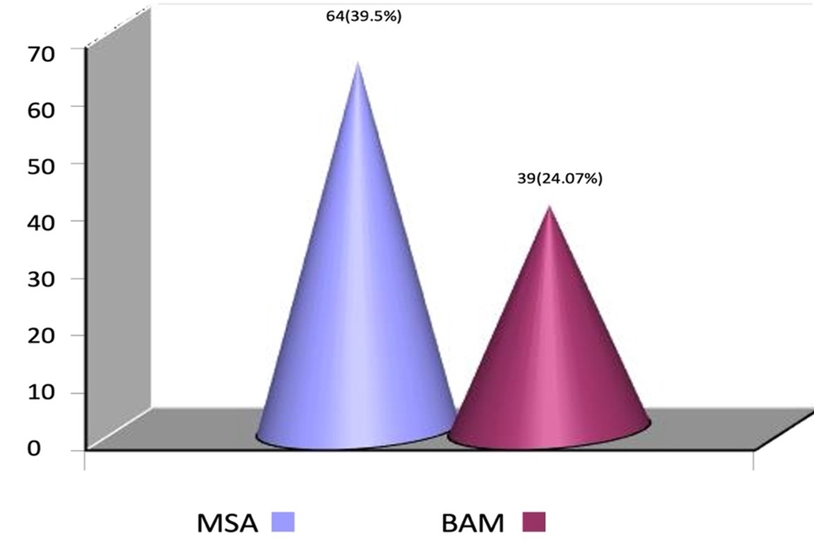
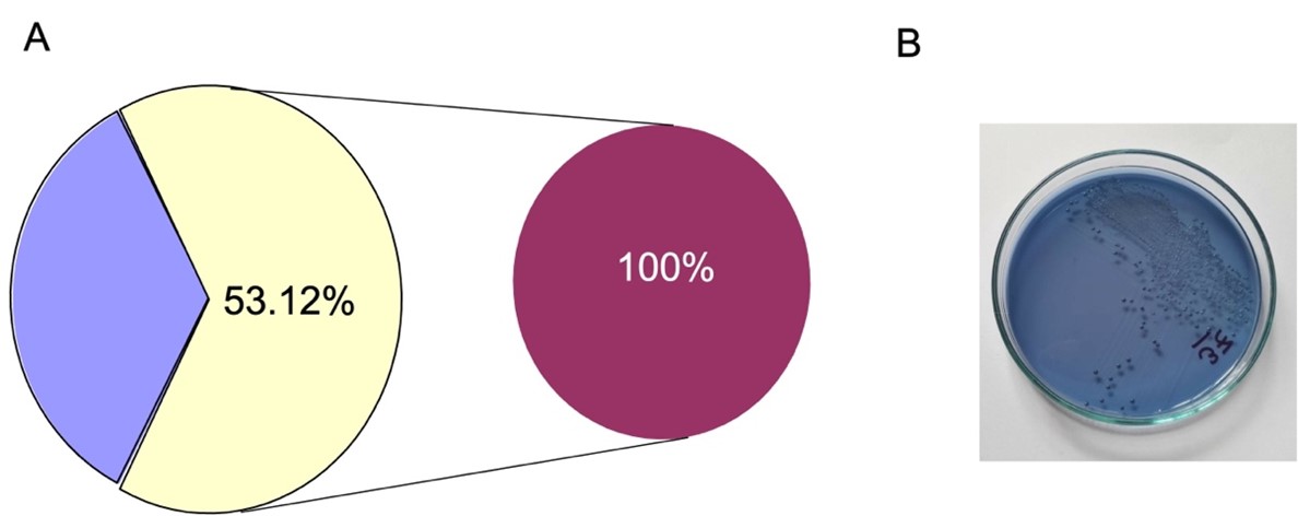
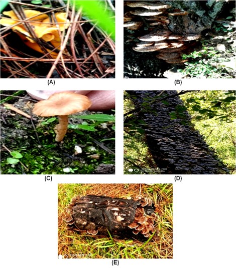
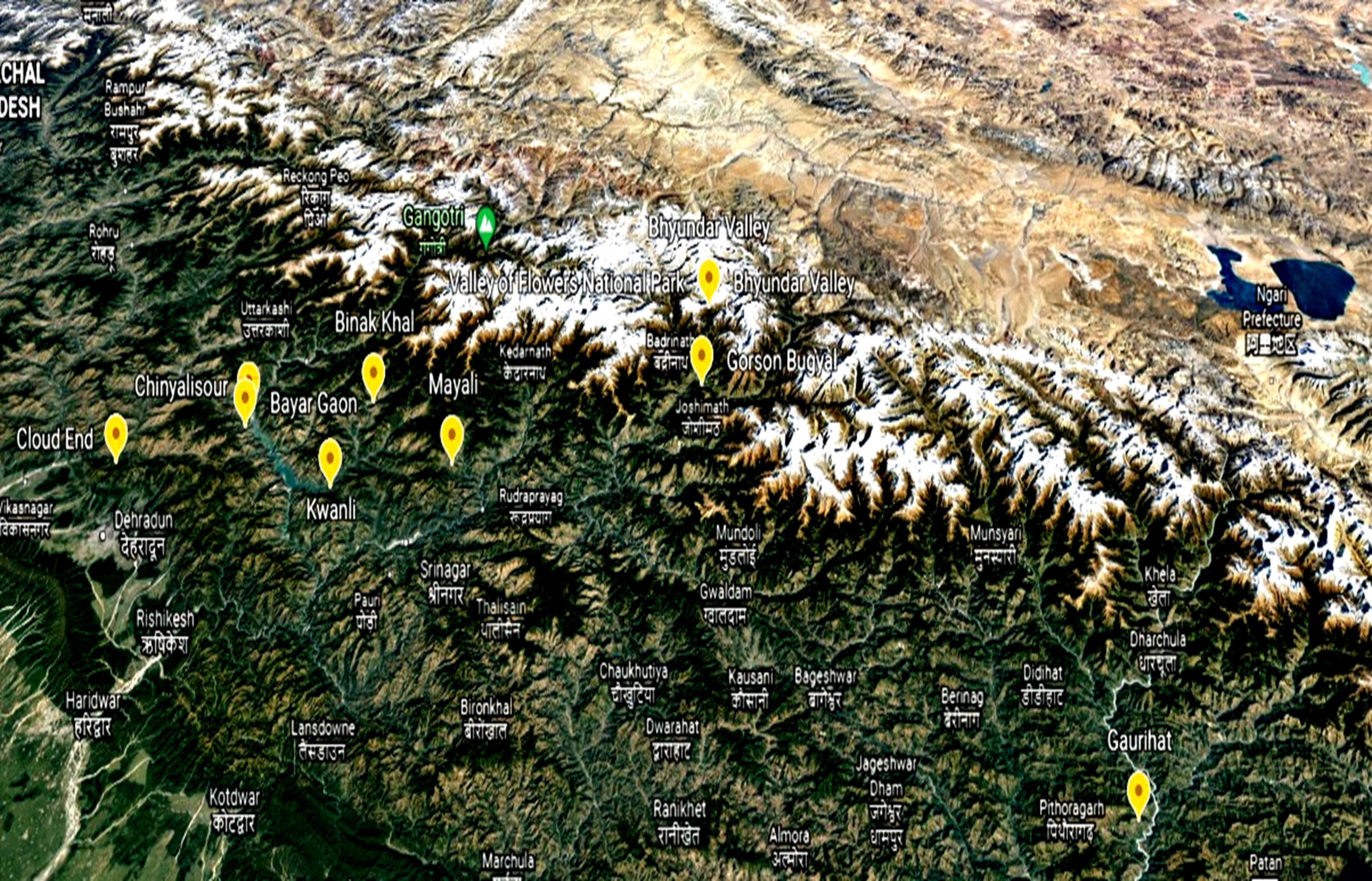
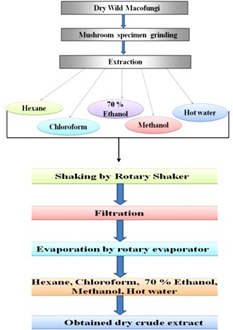
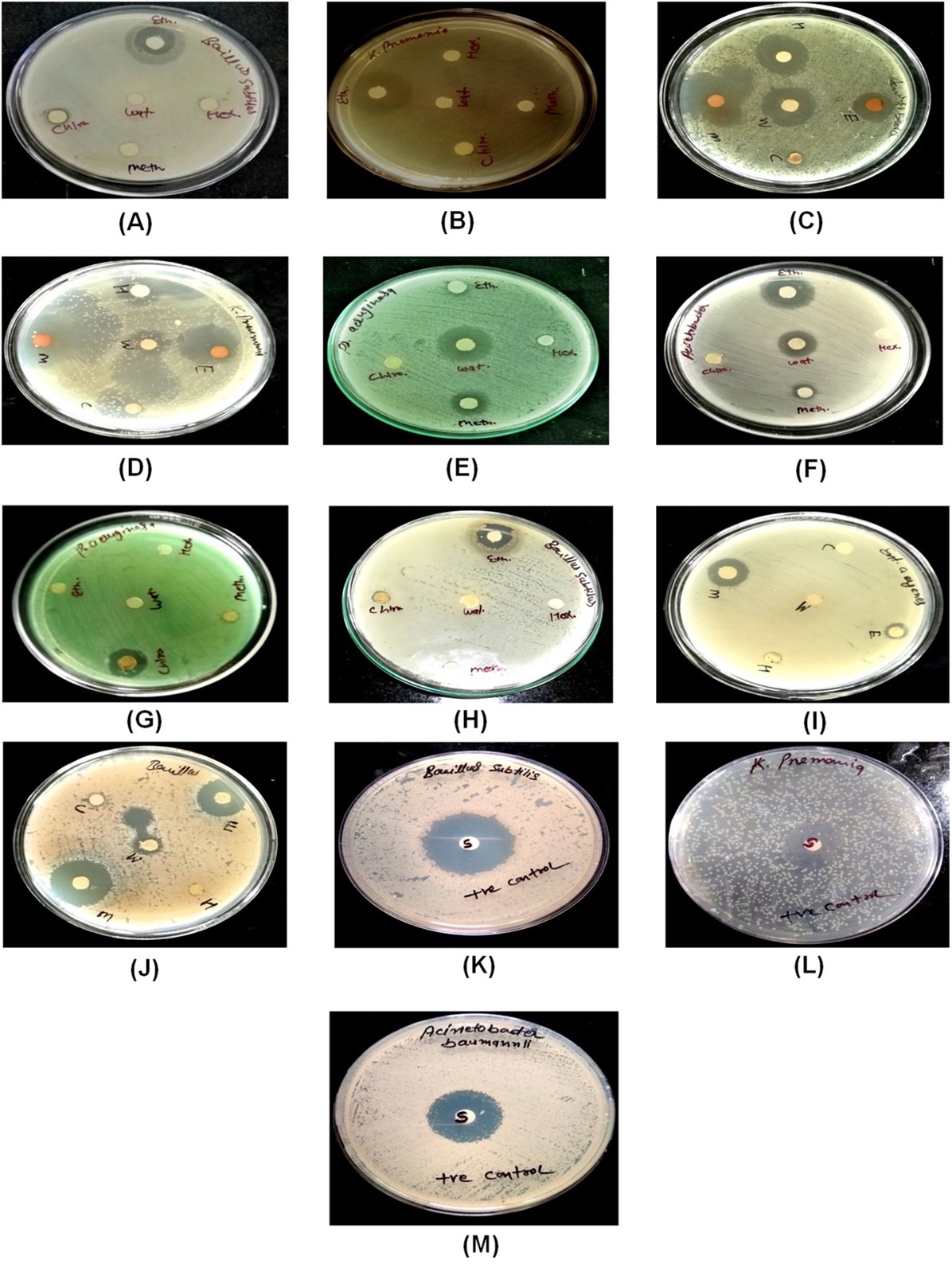
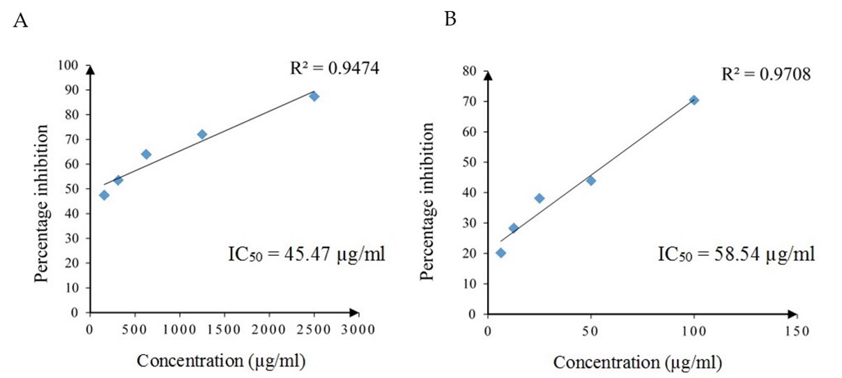
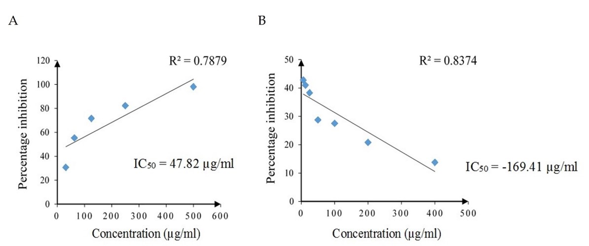
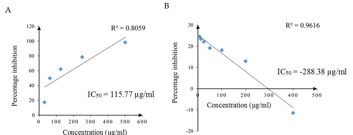
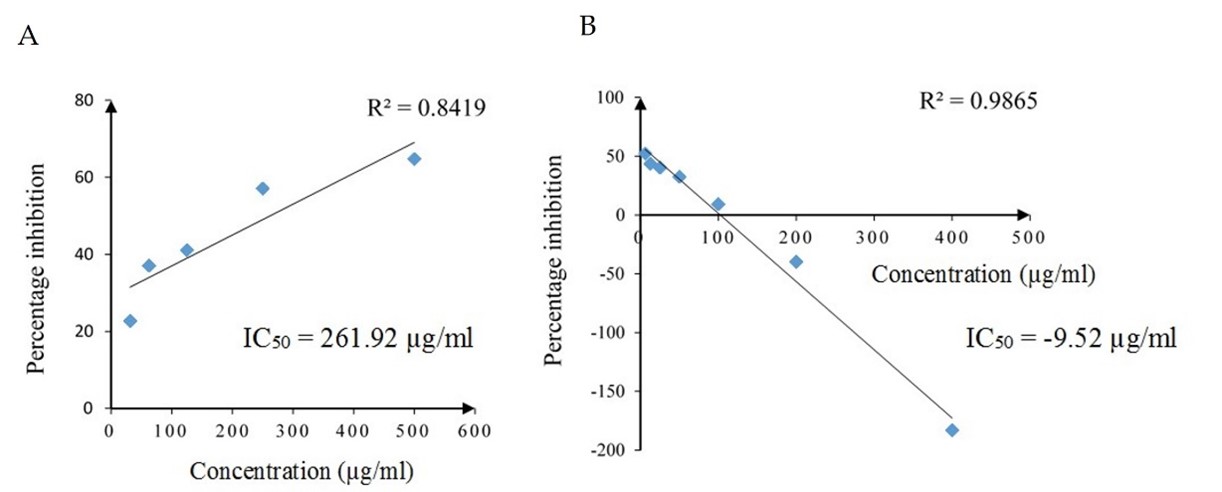
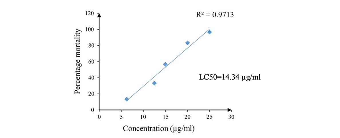
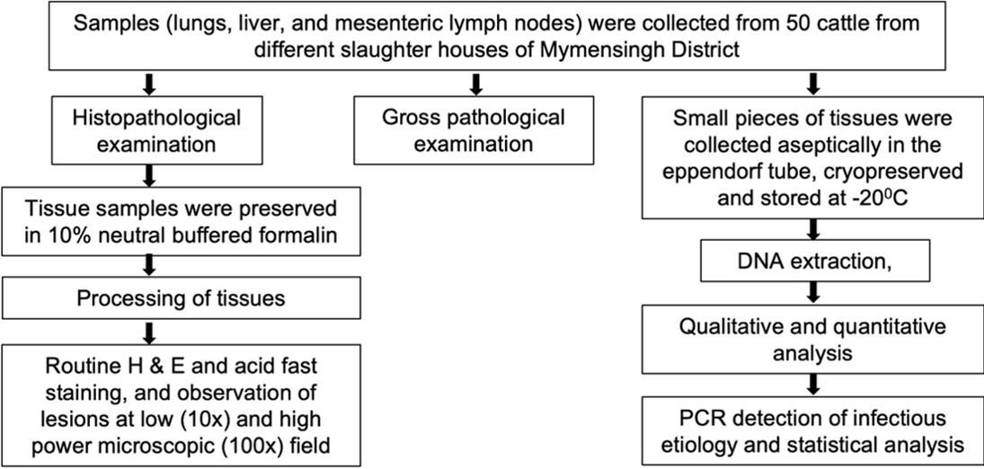
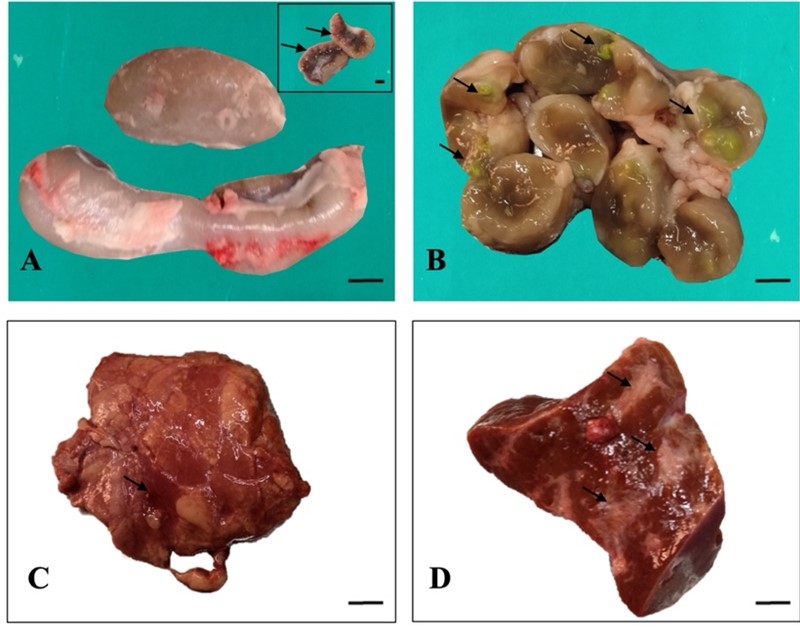
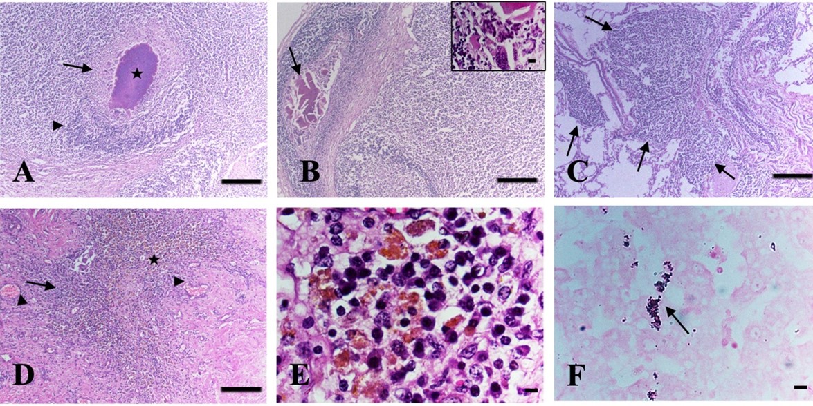
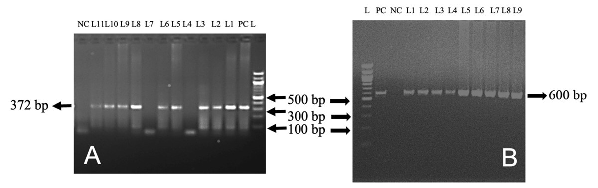
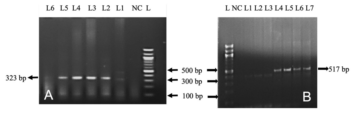
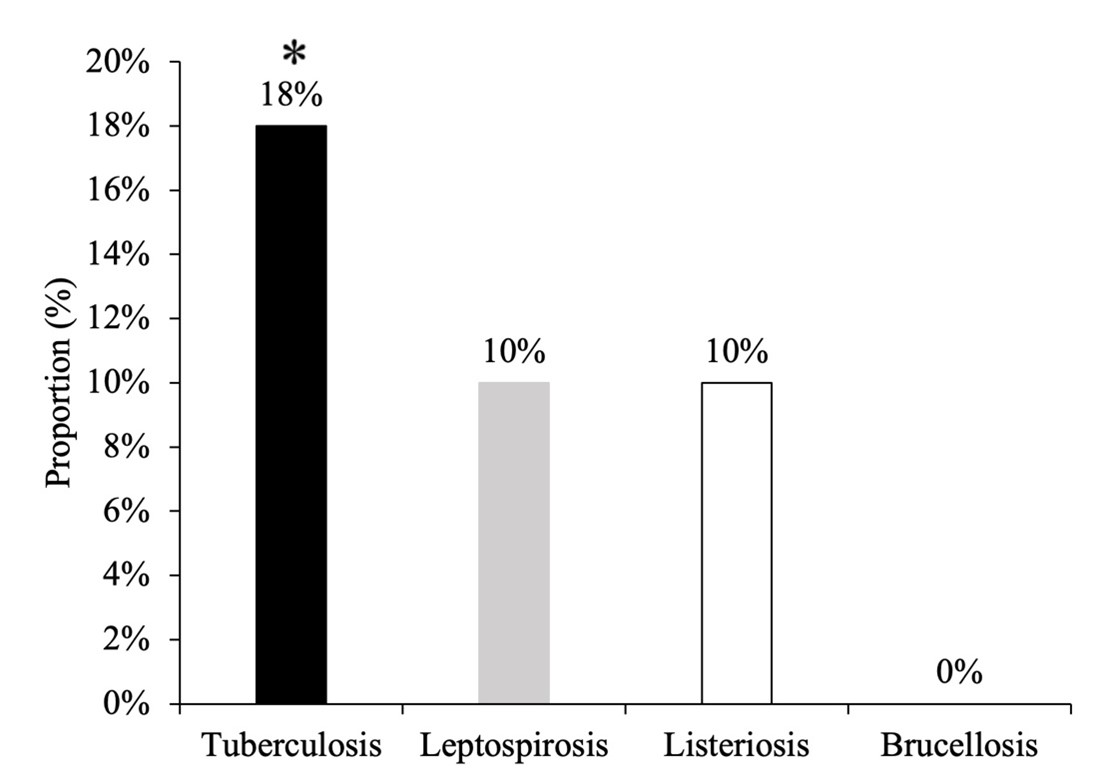
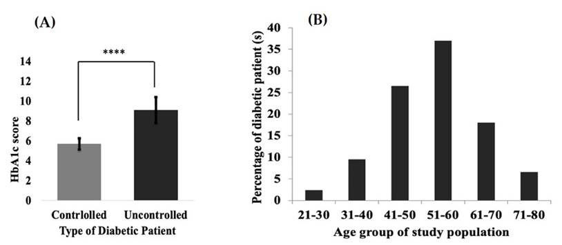
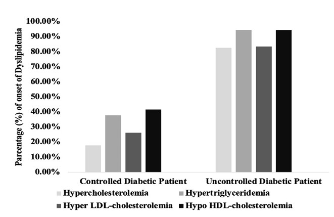
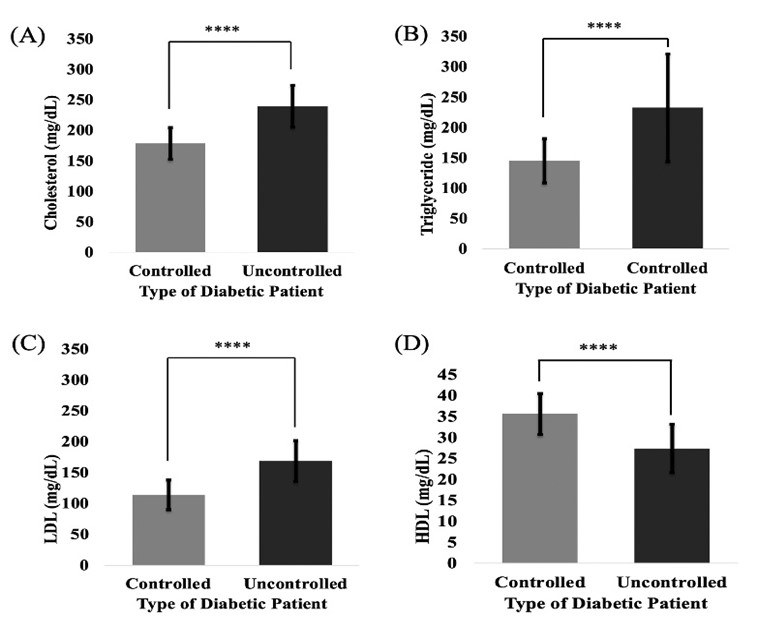
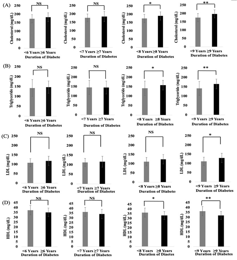
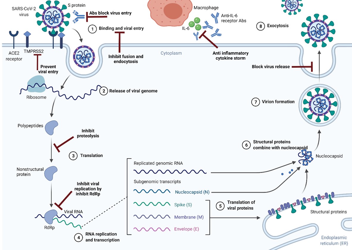
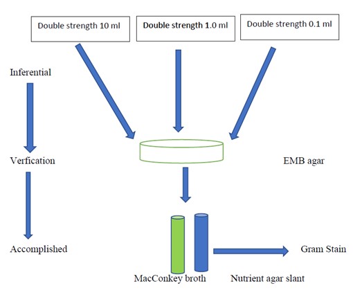
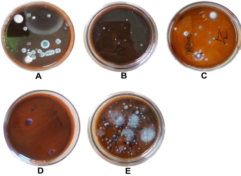
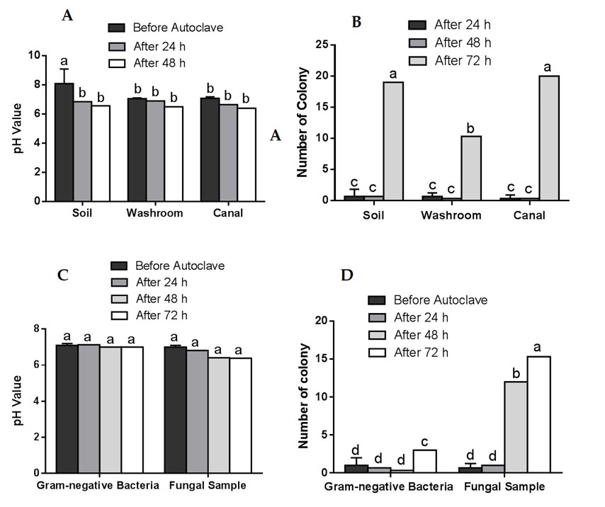
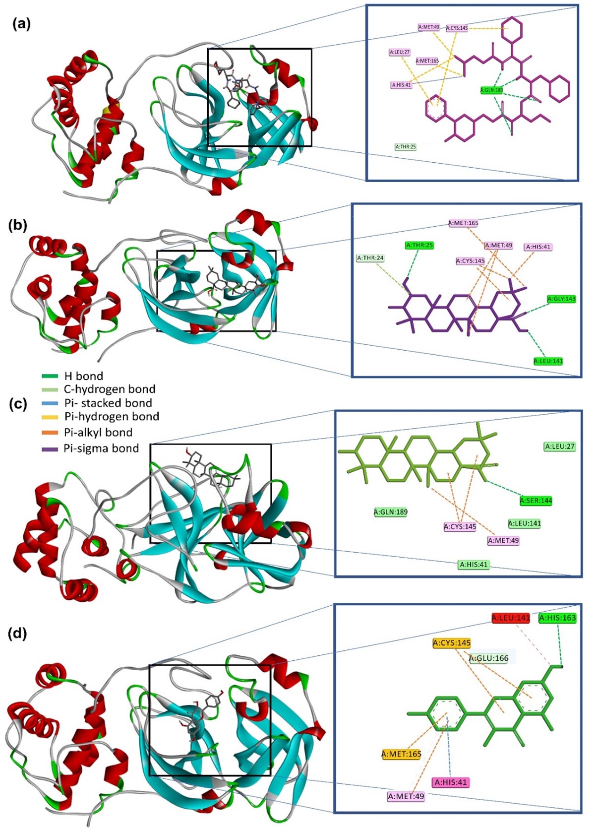
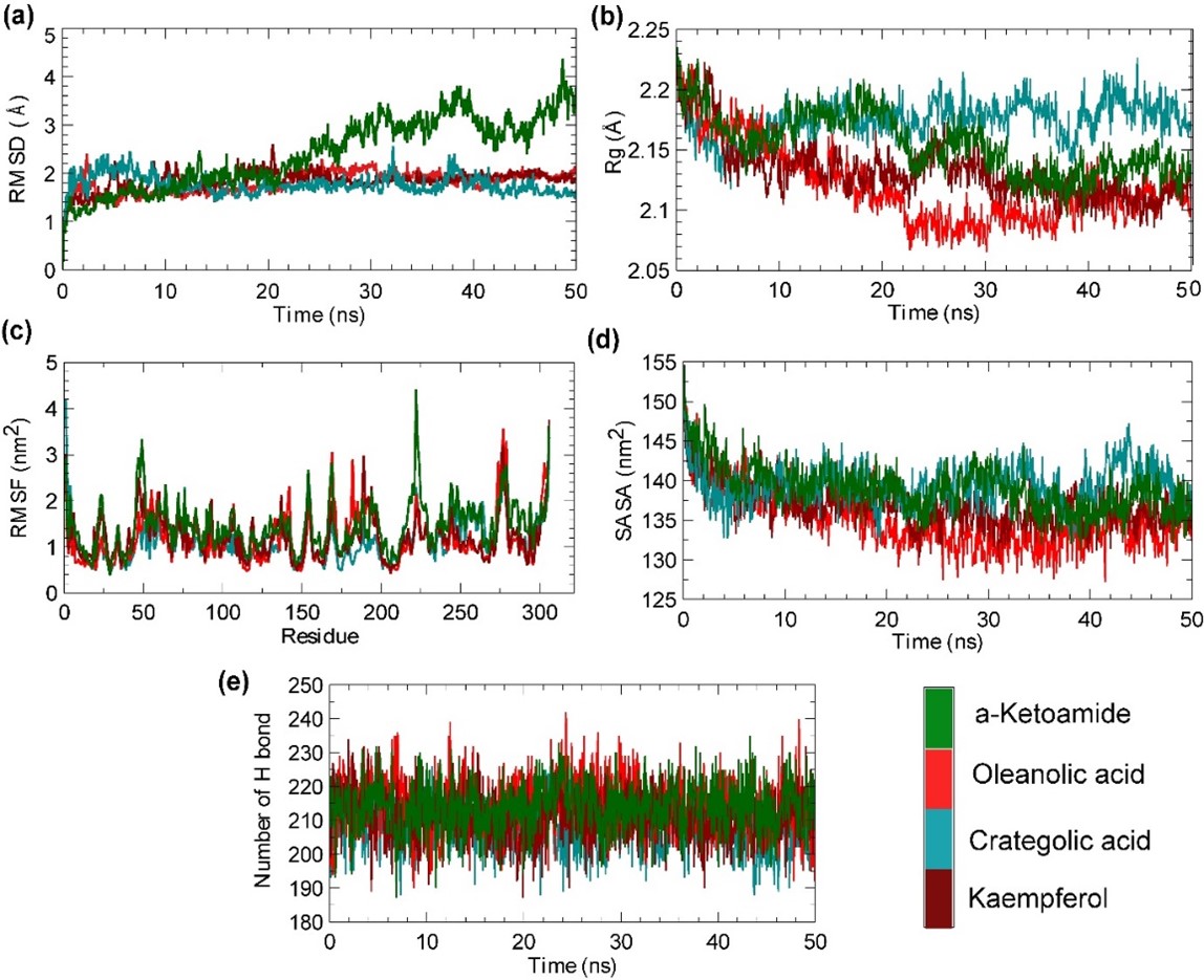
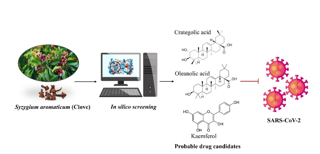
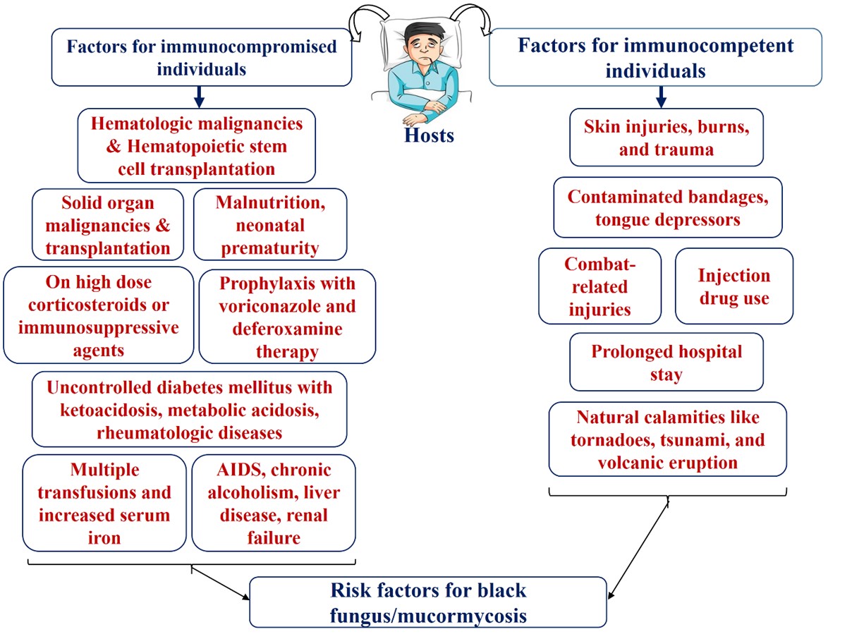
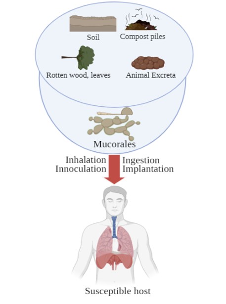
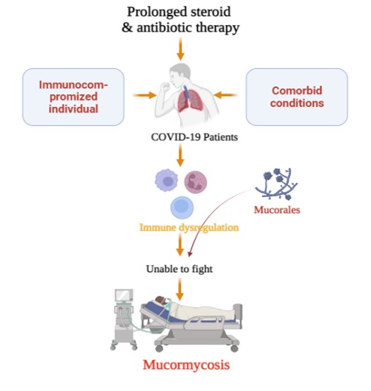
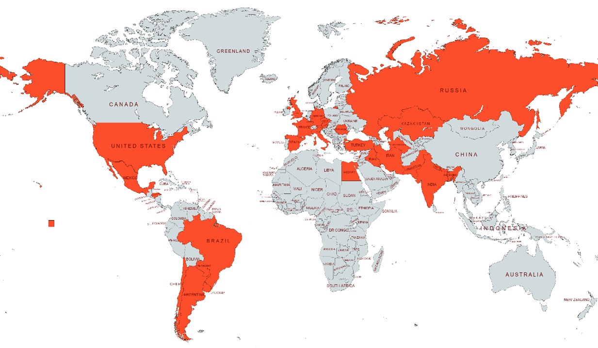
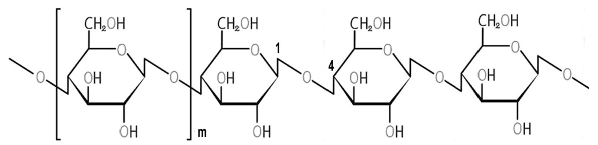
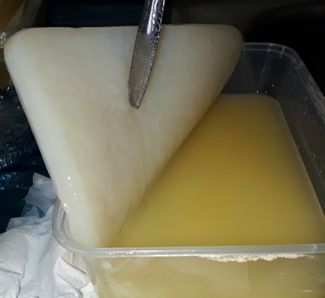
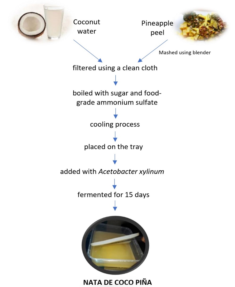