HTTP 200 OK
Allow: GET, HEAD, OPTIONS
Content-Type: application/json
Vary: Accept
{
"count": 319,
"next": "https://jabet.bsmiab.org/articles/?format=api&page=2",
"previous": null,
"results": [
{
"id": 334,
"slug": "178-1736575845-effect-of-fungal-microbiota-on-rankl-and-sclerostin-in-patients-with-crohns-disease",
"featured": false,
"slider": false,
"issue": "Vol8 Issue2",
"type": "original_article",
"manuscript_id": "178-1736575845",
"recieved": "2025-01-11",
"revised": null,
"accepted": "2025-03-22",
"published": "2025-04-13",
"pdf_file": "https://jabet.bsmiab.org/media/pdf_file/2025/56/178-1736575845.pdf",
"title": "Effect of fungal microbiota on RANKL and sclerostin in patients with Crohn's disease",
"abstract": "<p>The etiology of Crohn's disease (CD) is still unknown. However, many factors, including a dysregulated immune system, altered microbiota, inheritance, and environmental factors, have been implicated. This work was conducted to estimate the effect of fungal microbiota on two bone mineral density markers, RANKL and sclerostin, in addition to the correlation between these markers and vitamin B12, D3, and zinc in CD patients, along with their potential effect on fungal microbiota and vice versa. Peripheral blood and carry-Blair Stool samples were collected from 88 participants (60 newly diagnosed with CD without treatment and 28 healthy controls) to detect serum levels of RANKL and sclerostin, and culture media were used to grow, isolate, and identify fungi attendant to CD and its effect on RANKL and sclerostin levels. Sociodemographic data (vitamin B12, D3, and zinc levels) were collected from patients' medical records. The results showed significant differences in RANKL and sclerostin levels in various types of fungal microbiota in CD patients along with a significant increase in RANKL and sclerostin levels in these patients. Moreover, RANKL levels were negatively significantly correlated with Zinc, while sclerostin levels correlated negatively with vit D3. The findings of this study suggest that fungal microbiota may play a role in the inflammatory process and interactions with bone density by affecting levels of RANKL and sclerostin, vitamin D3, and zinc, suggesting that the use of the fungal microbiota in the monitoring and treatment of CD patients.<strong> </strong></p>",
"journal_reference": "J Adv Biotechnol Exp Ther. 2025; 8(2): 316-327",
"academic_editor": "Md. Abdul Hannan, PhD; Bangladesh Agricultural University, Bangladesh",
"cite_info": "Al-Rubaye AH, Alabassi HM, et al. Effect of fungal microbiota on RANKL and sclerostin in patients with Crohn's disease. J Adv Biotechnol Exp Ther. 2025; 8(2): 316-327.",
"keywords": [
"inflammation",
"Fungal microbiota",
"Crohn's disease",
"Sclerostin",
"RANKL"
],
"DOI": "10.5455/jabet.2025.26",
"sections": [
{
"section_number": 1,
"section_title": "INTRODUCTION",
"body": "<p>The intestinal mucosa is the site of origin and manifestation for the two primary phenotypes of inflammatory bowel disease (IBD), Crohn's disease (CD), and ulcerative colitis (UC) [<a href=\"#r-1\">1</a>]. CD inflammation can affect any part of the digestive system, but the terminal ileum, colon, and perianal areas are the most commonly implicated in a discontinuous pattern. The CD is distinguished from ulcerative colitis by submucosal thickening, transmural inflammation, and chronic granulomas [<a href=\"#r-2\">2</a>]. CD inflammation can affect any part of the digestive tract, from the mouth to the anus, and is accompanied by discontinuous transmural lesions of the gut wall [<a href=\"#r-3\">3</a>]. Compared to the general population, patients with CD are more likely to develop osteoporosis and sustain osteoporotic fractures [<a href=\"#r-4\">4</a>]. Chronic inflammation, diminished vitamin and mineral absorption, severe small-bowel disease or resection, corticosteroid use, advanced age, inactivity, smoking, and nutritional inadequacies are all factors that contribute to bone loss in CD patients [<a href=\"#r-5\">5</a>]. The relation between CD and lower bone density is still unclear. However, it seems to be directly related to inflammation or other osteoporosis risk factors, such as vitamin deficiency, that are frequently observed in CD. However, corticosteroids have been found to increase the risk of poor bone mineral density and osteoporosis [<a href=\"#r-6\">6</a>]. The etiology of CD is complex, involving environmental variables, genetic predisposition, and the host's impaired immunological interaction with the gut microbiota [<a href=\"#r-7\">7</a>].</p>\r\n\r\n<p>Most research on host-associated microbiota in health and disease has concentrated on bacterial microbiota even though fungal illnesses cause a significant infectious disease burden. As a result, little has been realized about the relevance and function of the fungal microbiota [<a href=\"#r-8\">8</a>]. Fungi are considered an important component of the human flora because they represent a dynamic and ecologically diverse microbial population. In a healthy gastrointestinal tract, the diversity of bacterial and fungal microbiota is inversely correlated, and the two are dynamically balanced [<a href=\"#r-9\">9</a>]. Aside from bacterial dysbiosis, earlier research has identified a distinct fungal microbiome dysbiosis in CD, which is characterized by changes in biodiversity and composition [<a href=\"#r-10\">10</a>]. Shifts in fungal microbiota modify immune response and disease status [<a href=\"#r-11\">11</a>]. The development of skeletal disorders is significantly influenced by gut microbiota, which includes bacteria, fungi, and viruses, and may be a target for treatment [<a href=\"#r-12\">12</a>]. A dynamic equilibrium known as physiological bone remodeling is the outcome of several biological processes that are tightly orchestrated and controlled by the complex interactions between the various cell types that comprise bone, principally osteoclasts and osteoblast lineage cells [<a href=\"#r-13\">13</a>]. RANKL (receptor activator of nuclear factor kappa-b ligand) is a transmembrane protein generated by stromal and osteoblast cells. It belongs to the tumor necrosis factor receptor family. It is mostly found on chromosome 18q22.1. It is made up of 616 amino acids and is found in osteoclast precursors and mature osteoclasts [<a href=\"#r-14\">14</a>]. RANKL regulates osteoclast development and function via binding to the RANK on osteoclast progenitor cells [<a href=\"#r-15\">15</a>].</p>\r\n\r\n<p>Moreover, osteocytes, the most prevalent type of bone cell, play a critical role in the formation of bones and resorption. They also serve as endocrine cells, secreting proteins that regulate skeletal metabolism as well as global mineral and nutrient balance [<a href=\"#r-16\">16</a>]. Sclerostin SOST is a bone tissue protein that is encoded by the SOST gene [<a href=\"#r-17\">17</a>]. It is a glycoprotein released by osteocytes that prevents osteoblasts from forming new bone, which results in bone loss [<a href=\"#r-18\">18</a>]. Several research on bone health have found that vitamin (Vit) B12, (vit) D3, and zinc all contribute to the quality of human bone growth, homeostasis, and bone diseases [<a href=\"#r-19\">19-21</a>]. A few studies focused on the effect of microbiota on Iraqi patients with CD, so that, the current study aims to assess the impact of fungal microbiota on bone mineral density, with an emphasis on its potential effect on RANKL and sclerostin levels, as well as their correlation to vitamin B12, D3, and zinc levels in Iraqi CD patients.</p>"
},
{
"section_number": 2,
"section_title": "MATERIALS AND METHODS",
"body": "<p><strong>Study design</strong></p>\r\n\r\n<p>A total of 88 Iraqi individuals (male and female) were enrolled in this study. Sixty patients were clinically early diagnosed with CD without treatment during their attendance at Al-Kindy Teaching Hospital in Baghdad, Iraq. The study extended from January 2022 to November 2022. The age of CD patients ranges from (15–65) years. In addition, 28 healthy individuals; their age matched the patient group, and the participants had no CD or other gastrointestinal diseases. Sociodemographic information (fungal infection kinds, vitamin B12, D3, and zinc levels), as well as patients' height, weight, age, sex, blood groups, and smoking habits, were obtained from their medical records.</p>\r\n\r\n<p> </p>\r\n\r\n<p><strong>Ethical considerations</strong></p>\r\n\r\n<p>This work has been approved by the ethical committee of the biology department at the University of Bagdad, College of Education for Pure Sciences/ Ibn Al-Haitham in Baghdad, Iraq. On 2/1/2022, the authorization was acquired under reference number EC-45.</p>\r\n\r\n<p> </p>\r\n\r\n<p><strong>Collection of blood samples</strong></p>\r\n\r\n<p>The venous blood sample was drawn from CD patients and controls. Each participant had a vein punctured to draw 5 mL of blood, which was then carefully pumped into disposable serum tubes filled with separating gel. After allowing the blood in the gel tubes to coagulate for 15 minutes at room temperature, it was centrifuged for another 15 minutes at 3000 rpm. The serum is kept for later use at -20°C.</p>\r\n\r\n<p><strong> </strong></p>\r\n\r\n<p><strong>Determination of RANKL and sclerostin levels by using ELISA </strong></p>\r\n\r\n<p>Commercial enzyme-linked immunosorbent assay (ELISA) kits from Cloud Clone Crop (USA) were used to measure the levels of RANKL and sclerostin in serum samples, employing the sandwich enzyme immunoassay concept. A microplate has been pre-coated with antibodies unique to each parameter. A biotin-conjugated antibody that is specific to the parameters is then pipetted into the wells along with the samples and standards. Each well was then filled with Avidin coupled with Horseradish Peroxidase (HRP) and incubated. After adding tetramethylbenzidine (TMB) substrate solution, only the wells containing the investigated parameter, biotin-conjugated antibody, and enzyme-conjugated Avidin showed a color shift. A sulfuric acid solution was added to halt the enzyme-substrate reaction, and the color shift was detected using spectrophotometry at 450 nm ± 10 nm. The concentration of the parameter studied in the samples was ascertained by comparing the optical density (OD) of the samples to the standard curve.</p>\r\n\r\n<p><strong> </strong></p>\r\n\r\n<p><strong>Quantitative measurements of vitamin D3, vitamin B12, and Zinc in serum samples </strong></p>\r\n\r\n<p>The quantitative measurements of vitamin D3, vitamin B12, and zinc in the sample of human serum were performed using an ELISA kit (BioSource, USA) following the manufacturer’s instructions. The human kit used in this study was a sandwich enzyme-linked immunosorbent assay. A microplate was pre-coated with the antibody unique to each parameter. A biotin-conjugated antibody specific to the parameters was pipetted into the wells along with the samples and standards. Next, HRP conjugate was then added. Following incubation, the unbound avidin-HRP conjugate was eliminated during a wash step. A substrate solution reactive with HRP was added to the wells. A colored product was formed in proportion to the sample's parameters. The color development was monitored with an ELISA plate reader, and absorbance was measured at a wavelength of 405 nm.</p>\r\n\r\n<p> </p>\r\n\r\n<p><strong>Stool sampling and analysis</strong></p>\r\n\r\n<p>In order to identify the fungus linked to CD, 120 Carry-Blair stool samples were obtained from patients and controls. A variety of culture media were made in accordance with guidelines for sterilizing culture media and were used to grow, isolate, and identify fungi. 15 minutes at 121° C and 15 Psi of pressure.</p>\r\n\r\n<p><strong> </strong></p>\r\n\r\n<p><strong>Isolates and culture samples</strong></p>\r\n\r\n<p>After homogenizing the sample in a tube with sterile saline and leaving it to rest for 30 minutes, 100 μl of the supernatant was transferred to potato and sabouraud dextrose agar (Oxoid, UK) and incubated for a week at 25–37° C. The features of mold colonies were based on color constancy and the reverse; direct microscope examination was used to diagnose fungi [<a href=\"#r-22\">22</a>].</p>\r\n\r\n<p><strong> </strong></p>\r\n\r\n<p><strong>Preparation of sabouraud dextrose agar </strong></p>\r\n\r\n<p>Sabouraud dextrose agar (SDA) medium was prepared according to the manufacturer's instructions (Oxoid, UK) by dissolving 51 g of powder medium in 5 liters of distilled water and putting it on the electric heater for a minute. After the powder was mixed with water and chloramphenicol to prevent bacterial growth, it was autoclaved at 121 °C and 15 psi of pressure for 15 minutes, then left to cool to 41 °C before being poured into 250 mg/L dishes. The solution is placed in dishes until solidified and becomes ready to culture the samples.</p>\r\n\r\n<p><strong> </strong></p>\r\n\r\n<p><strong>Preparation of potato dextrose agar </strong></p>\r\n\r\n<p>Potato dextrose agar (PDA) medium was prepared according to the manufacturer's specifications (Oxoid, UK) by dissolving 39 g of it in 1 liter of distilled water. The pH was set at 6.8. After that, the medium was sterilized in an autoclave at 121°C and a pressure of 15 psi for 15 minutes. After cooling the medium, the antibiotic chloramphenicol was added to prevent bacterial growth at a concentration of 250 mg/L.</p>\r\n\r\n<p> </p>\r\n\r\n<p><strong>Determination of body mass index </strong></p>\r\n\r\n<p>The formula of Body Mass Index (BMI) = weight (kg) / height<sup>2</sup> (m)<sup>2</sup> is used to calculate the body mass index (BMI). Underweight is defined as having a BMI beneath 18.5, normal weight as being between 18.5 and 24.9, overweight as being between 25 and 29.9, and obese as being over 30 [<a href=\"#r-23\">23</a>].</p>\r\n\r\n<p><strong> </strong></p>\r\n\r\n<p><strong>Statistical analysis</strong></p>\r\n\r\n<p>SPSS version 23 was used for statistical analysis of the acquired data. A difference was considered statistically significant if its P value was less than 0.05. The information was presented as Mean ± Standard Error (SE). The t-test and analysis of variance (ANOVA) were used to compare the groups statistically. Utilizing the Pearson correlation coefficient, the relationship among RANKL, sclerostin, zinc, vitamin D3, and vitamin B12 was investigated.</p>"
},
{
"section_number": 3,
"section_title": "RESULTS",
"body": "<p><strong>Serum levels of RANKL and sclerostin in CD patients</strong></p>\r\n\r\n<p>The present study revealed a highly significant increase (P≤0.001) in RANKL levels in CD patients compared to control as illustrated in <a href=\"#figure1\">Figure 1</a>A. Sclerostin level showed a slightly significant (p≤0.05) increase in patients with CD compared to the control group (<a href=\"#figure1\">Figure 1</a>B).</p>\r\n\r\n<div id=\"figure1\">\r\n<figure class=\"image\"><img alt=\"\" height=\"341\" src=\"/media/article_images/2025/43/19/178-1736575845-Figure1.jpg\" width=\"500\" />\r\n<figcaption><strong>Figure 1. </strong>A) Serum levels of RANKL. B) Serum levels of sclerostin. Mean ± standard error (SE). ** High significant increase in RANKL levels in CD patients compared to control at probability level p≤0.001. *A slightly significant increase in patients with CD compared to controls at probability at probability (P≤0.05).</figcaption>\r\n</figure>\r\n\r\n<p> </p>\r\n</div>\r\n\r\n<p><strong>Serum levels of RANKL and sclerostin among different fungal microbiomes in CD patients</strong></p>\r\n\r\n<p>A highly significant (p≤0.001) variation in RANKL levels among the several fungal microbiome types, is shown in <a href=\"#Table-1\">Table 1</a>. Additionally, the sclerostin level was slightly significant (p ≤ 0.05) in the different types of fungal infection as shown in <a href=\"#Table-2\">Table 2</a>.</p>\r\n\r\n<p style=\"text-align:center\"><strong>Table 1.</strong> RANKL levels with different types of fungi in CD patients.</p>\r\n\r\n<div id=\"Table-1\">\r\n<p style=\"text-align:center\"><a href=\"https://jabet.bsmiab.org/table/178-1736575845-table1/\">Table-1</a></p>\r\n</div>\r\n\r\n<p> </p>\r\n\r\n<p style=\"text-align:center\"><strong>Table 2.</strong> Sclerostin levels with different types of fungi in CD patients.</p>\r\n\r\n<div id=\"Table-2\">\r\n<p style=\"text-align:center\"><a href=\"https://jabet.bsmiab.org/table/178-1736575845-table2/\">Table-2</a></p>\r\n</div>\r\n\r\n<p> </p>\r\n\r\n<p><strong>Serum levels of RANKL and sclerostin among different fungal microbiomes in controls </strong></p>\r\n\r\n<p>The results also showed a non-significant difference p>0.05 in RANKL and sclerostin among control in different Fungi as shown in <a href=\"#Table-3\">Tables 3</a> and <a href=\"#Table-4\">4</a>.</p>\r\n\r\n<p style=\"text-align:center\"><strong>Table 3.</strong> Levels of RANKL in control with different types of fungi.</p>\r\n\r\n<div id=\"Table-3\">\r\n<p style=\"text-align:center\"><a href=\"https://jabet.bsmiab.org/table/178-1736575845-table3/\"> Table-3</a></p>\r\n</div>\r\n\r\n<p> </p>\r\n\r\n<p style=\"text-align:center\"><strong>Table 4.</strong> Levels of sclerostin in control with different types of fungi.</p>\r\n\r\n<div id=\"Table-4\">\r\n<p style=\"text-align:center\"><a href=\"https://jabet.bsmiab.org/table/178-1736575845-table4/\"> Table-4</a></p>\r\n</div>\r\n\r\n<p> </p>\r\n\r\n<p><strong>Comparison of RANKL and sclerostin levels between patient and control groups with different fungal infections </strong></p>\r\n\r\n<p>In addition, our results showed a slightly significant difference (p ≤ 0.05) in RANKL levels between patients and the control group with <em>Candida </em>spp., as shown in <a href=\"#Table-5\">Table 5</a>. Moreover, the sclerostin level was slightly significantly higher (p ≤ 0.05) in patients with <em>Candida albicans</em>, but not significantly different (p > 0.05) in patients with other <em>Candida </em>spp. compared to the control group with <em>Candida</em> spp. as shown in <a href=\"#Table-6\">Table 6</a>.</p>\r\n\r\n<p style=\"text-align:center\"><strong>Table 5.</strong> Comparison of RANKL with different fungal infections in CD patients.</p>\r\n\r\n<div id=\"Table-5\">\r\n<p style=\"text-align:center\"><a href=\"https://jabet.bsmiab.org/table/178-1736575845-table5/\">Table-5</a></p>\r\n</div>\r\n\r\n<p> </p>\r\n\r\n<p style=\"text-align:center\"><strong>Table 6.</strong> Comparison in sclerostin with different fungal infections in CD patients.</p>\r\n\r\n<div id=\"Table-6\">\r\n<p style=\"text-align:center\"><a href=\"https://jabet.bsmiab.org/table/178-1736575845-table6/\">Table-6</a></p>\r\n</div>\r\n\r\n<p> </p>\r\n\r\n<p><strong>Serum levels of RANKL and sclerostin</strong> <strong>are based on blood groups</strong></p>\r\n\r\n<p>There were no significant differences between the mean rank of the RANKL, and sclerostin levels based on blood groups as demonstrated in <a href=\"#figure2\">Figures 2</a>A and B.</p>\r\n\r\n<div id=\"figure2\">\r\n<figure class=\"image\"><img alt=\"\" height=\"300\" src=\"/media/article_images/2025/43/19/178-1736575845-Figure2.jpg\" width=\"500\" />\r\n<figcaption><strong>Figure 2</strong>. A) Serum levels of RANKL based on blood groups. B) Serum levels of sclerostin based on blood groups. Data are presented as Mean ± standard error (SE). No significant difference between the mean rank of the RANKL based on blood groups. No significant difference between the mean rank of sclerostin, based on blood groups as demonstrated.</figcaption>\r\n</figure>\r\n</div>\r\n\r\n<p> </p>\r\n\r\n<p><strong>Comparison of serum levels of RANKL and sclerostin</strong> <strong>based on gender, cigarette smoking, age, and BMI</strong></p>\r\n\r\n<p>Also, our results found no significant difference between studied parameters when distributed according to gender, cigarette smoking, age, and BMI as illustrated in <a href=\"#Table-7\">Tables</a> 7 and<a href=\"#Table-8\"> 8</a>.</p>\r\n\r\n<p style=\"text-align:center\"><strong>Table 7.</strong> RANKL levels according to sociodemographic data in CD patients.</p>\r\n\r\n<div id=\"Table-7\">\r\n<p style=\"text-align:center\"><a href=\"https://jabet.bsmiab.org/table/178-1736575845-table7/\">Table-7</a></p>\r\n</div>\r\n\r\n<p> </p>\r\n\r\n<p style=\"text-align:center\"><strong>Table 8.</strong> Sclerostin levels according to sociodemographic data in CD patients.</p>\r\n\r\n<div id=\"Table-8\">\r\n<p style=\"text-align:center\"><a href=\"https://jabet.bsmiab.org/table/178-1736575845-table8/\"> Table-8</a></p>\r\n</div>\r\n\r\n<p> </p>\r\n\r\n<p><strong>Correlation of RANKL and sclerostin levels with Vit B12, Zinc, and Vit D3 in CD patients</strong></p>\r\n\r\n<p>A Pearson correlation coefficient was also calculated to analyze the linear correlation between RANKL, and sclerostin levels in CD patients and Vit B12, Zinc, and Vit D3 levels. Table 9 shows a negative connection between RANKL levels and zinc levels. Sclerostin was found to be negatively associated with Vit D3 in this research as shown in <a href=\"#Table-9\">Table 9</a>.</p>\r\n\r\n<p style=\"text-align:center\"><strong>Table 9.</strong> Correlation of RANKL and sclerostin levels with Vit B12, Zinc, and Vit D3 in CD patients.</p>\r\n\r\n<div id=\"Table-9\">\r\n<p style=\"text-align:center\"><a href=\"https://jabet.bsmiab.org/table/178-1736575845-table9/\">Table-9</a></p>\r\n</div>"
},
{
"section_number": 4,
"section_title": "DISCUSSION",
"body": "<p>The results showed that fungus infections, particularly those caused by <em>Candida </em>spp<em>.</em>, which are the most prevalent pathogens in CD patients, significantly affect the levels of RANKL and sclerostin in CD patients. These results may be attributed to <em>Candida</em>'s inflammatory properties. The pro-inflammatory effects of <em>Candida albicans</em> in mouse colitis models are evidence linking between fungal microbiome and CD [<a href=\"#r-24\">24</a>].</p>\r\n\r\n<p><em>Candida</em> is assumed to be able to infiltrate the gut's compromised epithelial barrier and induce invasive illness, which makes sense in the situation of IBD, especially with concurrent immunosuppression [<a href=\"#r-25\">25</a>]. Other studies have shown that alteration in the gut microbiota has a significant effect on bone loss [<a href=\"#r-26\">26</a>]. Since intestinal microbes indirectly stimulate or repress osteoblasts and osteoclasts, they might alter the balance between bone formation and resorption. Furthermore, gut microbes affect bone metabolism by altering the immunological condition of bones or controlling signaling proteins that promote tissue healing, inflammation, cell division, and survival (growth factors)., thereby influencing bone mass [<a href=\"#r-27\">27</a>]. Dysbiotic gut microbial flora identified in IBD may have an indirect effect on bone by several possible mechanisms. T cells activated by the gut microbiota may serve as “inflammatory shuttles” between the intestine and bone. Secondly, microbe-associated molecular patterns released into the circulation in IBD may activate immunological responses in the bone marrow by immune cells as well as osteocytes, osteoblasts, and osteoclasts, resulting in decreased bone production and increased resorption [<a href=\"#r-28\">28</a>]. Bone homeostasis may be impacted by gut microbiota, which is recognized to be crucial in controlling the host's health and physiology. By boosting the synthesis of circulating cytokines including interleukin (IL)-17, TNF, and receptor activator of nuclear factor (NF)-kB ligand (RANKL), it may contribute to the pathophysiology of osteoporosis [<a href=\"#r-29\">29</a>]. The results of this study indicated that there was a highly significant rise in P<0.001 in the level of RANKL in CD patients compared to the control. This elevation in RANKL levels may be attributed to increased RANKL expression in response to proinflammatory cytokines, specifically TNF and interleukins 1 and 17, emphasizing the importance of inflammation in RANKL-mediated effects on bone [<a href=\"#r-30\">30</a>]. Thus, proinflammatory cytokines could impact bone metabolism by increasing RANKL expression. Osteoclasts express RANKL, which binds to an osteoclast precursor that expresses the RANKL receptor, RANK (receptor activator of NF-B) receptors, and the osteoprotegerin (OPG) receptor. Osteoclasts grow and differentiate when RANKL binds to RANK receptors, which increases bone loss [<a href=\"#r-31\">31</a>]. It is well documented that RANKL is secreted by various immune system cell types, including T and B cells, dendritic cells, and macrophages. Notably, RANKL production is influenced by a variety of variables, including proinflammatory cytokines [<a href=\"#r-32\">32</a>].</p>\r\n\r\n<p>On the other hand, our results show a slight increase in P<0.05 in sclerostin levels in CD patients compared to control. Numerous cell types contain the Wnt signaling pathway, a signaling system that controls a range of biological processes (including bone remodeling, cell differentiation, and tissue regeneration). The Wnt/-catenin pathway has been shown to have anti-inflammatory properties in IBD, and its role is presently being investigated [<a href=\"#r-33\">33</a>]. Bone homeostasis is mediated by Wnt signaling. Sclerostin (SOST) is the endogenous suppressor of the Wnt pathway. SOST, a monomeric glycoprotein with a cysteine-knot pattern is produced by osteoblasts. By binding to low-density lipoprotein-related proteins 5 and 6 (LRP5 and LRP6), Seclerostin improves its suppressive effects on the Wnt pathway and blocks the canonical Wnt signaling [<a href=\"#r-34\">34</a>]. Through this mechanism, SOST dose-dependently decreases osteoprotegerin (OPG), increasing the receptor activator of the nuclear factor-kB ligand/OPG mRNA ratio. This catabolic action is achieved by encouraging the osteocyte to produce and activate osteoclasts [<a href=\"#r-35\">35</a>].</p>\r\n\r\n<p>In inflammatory conditions like IBD, active T lymphocytes produce TNF-α, which also stimulates the formation of sclerostin [<a href=\"#r-36\">36</a>]. In addition, decreased SOST levels are linked to increased osteoblast activity through stimulation of the Wnt/β-catenin signaling pathway [<a href=\"#r-37\">37</a>]. Furthermore, sclerostin may increase the production of RANK-L, and the binding of RANKL to its receptor RANK is a critical step in the formation of osteoclasts from hematopoietic progenitor cells, as well as the activation of mature osteoclasts [<a href=\"#r-38\">38</a>]. Shifts in the diversity of fungal microbiota in CD patients are linked to inflammation of the mucosa [<a href=\"#r-39\">39</a>]. Inflammatory conditions can affect both bone production and resorption, although they most frequently affect both [<a href=\"#r-40\">40</a>]. A slightly significant difference p≤0.05 in RANKL levels between patients and the control group with <em>Candida </em>spp. This difference may be attributed to the small number of patient cases with <em>Candida </em>spp. only compared to control or due to environmental factors like lifestyle and diet which contribute to osteoporosis and bone loss [<a href=\"#r-41\">41</a>]. The present study shows that sclerostin negatively correlated with Vit D3 (r = -0.678, p= 0.05). Vitamin D has systemic effects; it regulates innate and adaptive immunological responses and affects calcium homeostasis, which is implicated in bone metabolism. Vitamin D deficiency has negative effects on the immune system in IBD patients, causing dysregulation and inflammation-related loss of bone mineral density BMD [<a href=\"#r-42\">42</a>]. Serum Vitamin D has been linked to alterations in the gut microbiome associated with inflammatory immune responses [<a href=\"#r-43\">43</a>]. It has been reported that TNF-α acts as an activator for sclerostin expression [<a href=\"#r-44\">44</a>]. Vitamin D therapy has been shown to inhibit the TNF pathway in IBD patients [<a href=\"#r-45\">45</a>], which may have an impact on sclerostin levels.</p>\r\n\r\n<p>A Pearson correlation coefficient was computed to assess the linear relationship between RANKL and Zinc. There was a strong negative correlation between the two assessed variables (r = -.744, p= 0.022). Further, the trace element zinc (Zn) is absorbed in the small intestine and serves as a cofactor for a number of growth-related enzymes, immunological function, and tissue repair. Zn functions as an antioxidant and regulates the stability of biological membranes [<a href=\"#r-46\">46</a>]. Patients with IBD frequently suffer from zinc deficiency with prevalence rates ranging from 15% to 40%. In IBD patients, zinc insufficiency could be contributing to mucosal inflammation, which is a hallmark of CD disease [<a href=\"#r-47\">47</a>]. Zn also stimulates osteoblastic cells and inhibits osteoclast activity in bone tissue via the zinc-regulated RANKL/RANK/OPG pathway [<a href=\"#r-48\">48</a>]. Thus, alterations in zinc levels may have an effect on RANKL serum levels.</p>"
},
{
"section_number": 5,
"section_title": "CONCLUSIONS",
"body": "<p>CD disease is associated with bone metabolism alterations. However, the signals that influence bone metabolism are not fully understood. The current results suggest that alterations in gut microbiota could affect the systemic immune response and provide signals that impact bone metabolism via RANKL and sclerostin levels. One significant limitation of the study was the CD sample size. Also, the causes of CD remain unknown despite its long history.</p>"
},
{
"section_number": 6,
"section_title": "ACKNOWLEDGMENTS",
"body": "<p>The authors would like to thank all patients who participated in this study and the family members of Al-Kindy Teaching Hospital for their wonderful collaboration and assistance during the study.</p>"
},
{
"section_number": 7,
"section_title": "AUTHOR CONTRIBUTIONS",
"body": "<p>HMA designed outlines and drafted the manuscript. RHKA and ASH performed the experiments and analyzed the data. RHKA and AKI wrote the initial draft of the manuscript. HMA and RHKAR reviewed the scientific contents described in the manuscript. All authors read and approved the final submitted version of the manuscript.</p>"
},
{
"section_number": 8,
"section_title": "CONFLICTS OF INTEREST",
"body": "<p>There is no conflict of interest among the authors.</p>"
}
],
"figures": [
{
"figure": "https://jabet.bsmiab.org/media/article_images/2025/43/19/178-1736575845-Figure1.jpg",
"caption": "Figure 1. A) Serum levels of RANKL. B) Serum levels of sclerostin. Mean ± standard error (SE). ** High significant increase in RANKL levels in CD patients compared to control at probability level p≤0.001. *A slightly significant increase in patients with CD compared to controls at probability at probability (P≤0.05).",
"featured": false
},
{
"figure": "https://jabet.bsmiab.org/media/article_images/2025/43/19/178-1736575845-Figure2.jpg",
"caption": "Figure 2. A) Serum levels of RANKL based on blood groups. B) Serum levels of sclerostin based on blood groups. Data are presented as Mean ± standard error (SE). No significant difference between the mean rank of the RANKL based on blood groups. No significant difference between the mean rank of sclerostin, based on blood groups as demonstrated.",
"featured": false
}
],
"authors": [
{
"id": 1682,
"affiliation": [
{
"affiliation": "Department of Biology, College of Education for Pure Sciences/ Ibn Al-Haitham, University of Baghdad, Iraq"
}
],
"first_name": "Rana Hanan",
"family_name": "Al-Rubaye",
"email": "rana.h.k@ihcoedu.uobaghdad.edu.iq",
"author_order": 1,
"ORCID": "https://orcid.org/0009-0002-2822-712X",
"corresponding": true,
"co_first_author": false,
"co_author": false,
"corresponding_author_info": "Rana Hanan Al-Rubaye, Department of Biology, College of Education for Pure Sciences/ Ibn Al-Haitham, University of Baghdad, Iraq. Email: rana.h.k@ihcoedu.uobaghdad.edu.iq",
"article": 334
},
{
"id": 1683,
"affiliation": [
{
"affiliation": "Department of Biology, College of Education for Pure Sciences/ Ibn Al-Haitham, University of Baghdad, Iraq"
}
],
"first_name": "Hazima Mossa",
"family_name": "Alabassi",
"email": null,
"author_order": 2,
"ORCID": "https://orcid.org/0000-0002-9218-8762",
"corresponding": false,
"co_first_author": false,
"co_author": false,
"corresponding_author_info": "",
"article": 334
},
{
"id": 1684,
"affiliation": [
{
"affiliation": "Department of Analytic Pathology, College of Applied Science, University of Fallujah, Al-Fallujah, Iraq"
}
],
"first_name": "Anwar Khalil",
"family_name": "Ismael",
"email": null,
"author_order": 3,
"ORCID": "https://orcid.org/0000-0001-9031-9856",
"corresponding": false,
"co_first_author": false,
"co_author": false,
"corresponding_author_info": "",
"article": 334
},
{
"id": 1685,
"affiliation": [
{
"affiliation": "Cancer Research Department Iraqi center for cancer and Genetic Research, Mustansiryah University, Baghdad, Iraq"
}
],
"first_name": "Alaa Saad",
"family_name": "Hasan",
"email": null,
"author_order": 4,
"ORCID": null,
"corresponding": false,
"co_first_author": false,
"co_author": false,
"corresponding_author_info": "",
"article": 334
}
],
"views": 146,
"downloads": 27,
"references": [
{
"id": 14024,
"serial_number": 1,
"pmc": null,
"reference": "Hasan AS, Alabassi HM, et al. The multifaceted role of dectin-1 and card9 in inflammatory bowel disease iraqi patients. History of Medicine. 2023;9:1763–88.",
"DOI": null,
"article": 334
},
{
"id": 14025,
"serial_number": 2,
"pmc": null,
"reference": "Ibraheem ZF, Muhsin HY. Roles of IL-36 in the pathogenesis of inflammatory bowel disease in a sample of iraqi patients. Pakistan Journal of Medical & Health Sciences. 2022;16:548-51.",
"DOI": null,
"article": 334
},
{
"id": 14026,
"serial_number": 3,
"pmc": null,
"reference": "Al-Abassi HM, Nazal MF, et al. Serum profile of cytokines in iraqi inflammatory bowel disease patients. Mustansiriya Medical Journal. 2015;14:11-6.",
"DOI": null,
"article": 334
},
{
"id": 14027,
"serial_number": 4,
"pmc": null,
"reference": "Targownik LE, Bernstein CN, et al. Inflammatory bowel disease and the risk of osteoporosis and fracture. Maturitas. 2013;76:315-9.",
"DOI": null,
"article": 334
},
{
"id": 14028,
"serial_number": 5,
"pmc": null,
"reference": "Jones K, Baker K, et al. Randomised clinical trial: Combined impact and resistance training in adults with stable crohn's disease. Alimentary pharmacology & therapeutics. 2020;52:964-75.",
"DOI": null,
"article": 334
},
{
"id": 14029,
"serial_number": 6,
"pmc": null,
"reference": "Chedid VG, Kane SV. Bone health in patients with inflammatory bowel diseases. Journal of Clinical Densitometry. 2020;23:182-9.",
"DOI": null,
"article": 334
},
{
"id": 14030,
"serial_number": 7,
"pmc": null,
"reference": "Chang JT. Pathophysiology of inflammatory bowel diseases. New England Journal of Medicine. 2020;383:2652-64.",
"DOI": null,
"article": 334
},
{
"id": 14031,
"serial_number": 8,
"pmc": null,
"reference": "Zhang I, Pletcher SD, et al. Fungal microbiota in chronic airway inflammatory disease and emerging relationships with the host immune response. Frontiers in Microbiology. 2017;8:2477.",
"DOI": null,
"article": 334
},
{
"id": 14032,
"serial_number": 9,
"pmc": null,
"reference": "Li Q, Wang C, et al. Dysbiosis of gut fungal microbiota is associated with mucosal inflammation in crohn’s disease. Journal of clinical gastroenterology. 2014;48:513-23.",
"DOI": null,
"article": 334
},
{
"id": 14033,
"serial_number": 10,
"pmc": null,
"reference": "Hoarau G, Mukherjee P, et al. Bacteriome and mycobiome interactions underscore microbial dysbiosis in familial crohn’s disease. MBio. 2016;7:10.",
"DOI": null,
"article": 334
},
{
"id": 14034,
"serial_number": 11,
"pmc": null,
"reference": "Gouba N, Hien YE, et al. Digestive tract mycobiota and microbiota and the effects on the immune system. Human Microbiome Journal. 2019;12:100056.",
"DOI": null,
"article": 334
},
{
"id": 14035,
"serial_number": 12,
"pmc": null,
"reference": "Tu Y, Yang R, et al. The microbiota-gut-bone axis and bone health. Journal of leukocyte biology. 2021; 110(3), 525-537.",
"DOI": null,
"article": 334
},
{
"id": 14036,
"serial_number": 13,
"pmc": null,
"reference": "Lerner UH, Kindstedt E, et al. The critical interplay between bone resorbing and bone forming cells. Journal of clinical periodontology. 2019;46:33-51.",
"DOI": null,
"article": 334
},
{
"id": 14037,
"serial_number": 14,
"pmc": null,
"reference": "Naik S, Sahu S, et al. Serum levels of osteoprotegerin, rank-l & vitamin d in different stages of osteoarthritis of the knee. Indian Journal of Medical Research. 2021;154:491-6.",
"DOI": null,
"article": 334
},
{
"id": 14038,
"serial_number": 15,
"pmc": null,
"reference": "Ismael AK, Alabassi HM. The dynamic role of pd-1, vitamin d, rankl, and sclerostin in iraqi patients with systemic lupus erythematosus. Ibn AL-Haitham Journal For Pure and Applied Sciences. 2024;37:9-18.",
"DOI": null,
"article": 334
},
{
"id": 14039,
"serial_number": 16,
"pmc": null,
"reference": "Munir A, Reseland JE, et al. Osteocyte‐like cells differentiated from primary osteoblasts in an artificial human bone tissue model. Journal of Bone and Mineral Research Plus. 2023;7:e10792.",
"DOI": null,
"article": 334
},
{
"id": 14040,
"serial_number": 17,
"pmc": null,
"reference": "Oniszczuk A, Kaczmarek A, et al. Sclerostin as a biomarker of physical exercise in osteoporosis: A narrative review. Frontiers in endocrinology. 2022;13:954895.",
"DOI": null,
"article": 334
},
{
"id": 14041,
"serial_number": 18,
"pmc": null,
"reference": "Compton JT, Lee FY. A review of osteocyte function and the emerging importance of sclerostin. JBJS. 2014;96:1659-68.",
"DOI": null,
"article": 334
},
{
"id": 14042,
"serial_number": 19,
"pmc": null,
"reference": "Belal A, Mahmoud R, et al. Therapeutic potential of zeolites/vitamin b12 nanocomposite on complete freund’s adjuvant-induced arthritis as a bone disorder: In vivo study and bio-molecular investigations. Pharmaceuticals. 2023;16:285.",
"DOI": null,
"article": 334
},
{
"id": 14043,
"serial_number": 20,
"pmc": null,
"reference": "Rizzoli R, Biver E. Are probiotics the new calcium and vitamin d for bone health? Current osteoporosis reports. 2020;18:273-84.",
"DOI": null,
"article": 334
},
{
"id": 14044,
"serial_number": 21,
"pmc": null,
"reference": "Huang T, Yan G, et al. Zinc homeostasis in bone: Zinc transporters and bone diseases. International journal of molecular sciences. 2020;21:1236.",
"DOI": null,
"article": 334
},
{
"id": 14045,
"serial_number": 22,
"pmc": null,
"reference": "Granato PA, Granato PA. Laboratory manual and workbook in microbiology: Applications to patient care: McGraw-Hill; 2011.",
"DOI": null,
"article": 334
},
{
"id": 14046,
"serial_number": 23,
"pmc": null,
"reference": "Mohajan D, Mohajan HK. Body mass index (bmi) is a popular anthropometric tool to measure obesity among adults. Journal of Innovations in Medical Research. 2023;2:25-33.",
"DOI": null,
"article": 334
},
{
"id": 14047,
"serial_number": 24,
"pmc": null,
"reference": "Liguori G, Lamas B, et al. Fungal dysbiosis in mucosa-associated microbiota of crohn’s disease patients. Journal of Crohn's and Colitis. 2016;10:296-305.",
"DOI": null,
"article": 334
},
{
"id": 14048,
"serial_number": 25,
"pmc": null,
"reference": "Stamatiades GA, Ioannou P, et al. Fungal infections in patients with inflammatory bowel disease: A systematic review. Mycoses. 2018;61:366-76.",
"DOI": null,
"article": 334
},
{
"id": 14049,
"serial_number": 26,
"pmc": null,
"reference": "Wang H, Liu J, et al. Gut microbiota signatures and fecal metabolites in postmenopausal women with osteoporosis. Gut Pathogens. 2023;15:33.",
"DOI": null,
"article": 334
},
{
"id": 14050,
"serial_number": 27,
"pmc": null,
"reference": "Zhang J, Lu Y, et al. The impact of the intestinal microbiome on bone health. Intractable & rare diseases research. 2018;7:148-55.",
"DOI": null,
"article": 334
},
{
"id": 14051,
"serial_number": 28,
"pmc": null,
"reference": "Sylvester FA. Inflammatory bowel disease: Effects on bone and mechanisms. Understanding the Gut-Bone Signaling Axis: Mechanisms and Therapeutic Implications. 2017:133-50.",
"DOI": null,
"article": 334
},
{
"id": 14052,
"serial_number": 29,
"pmc": null,
"reference": "Chen Y, Wang X, et al. Gut microbiota and bone diseases: A growing partnership. Frontiers in Microbiology. 2022;13:877776.",
"DOI": null,
"article": 334
},
{
"id": 14053,
"serial_number": 30,
"pmc": null,
"reference": "Schett G. Effects of inflammatory and anti‐inflammatory cytokines on the bone. European journal of clinical investigation. 2011;41:1361-6.",
"DOI": null,
"article": 334
},
{
"id": 14054,
"serial_number": 31,
"pmc": null,
"reference": "Palatianou ME, Karamanolis G, et al. Signaling pathways associated with bone loss in inflammatory bowel disease. Annals of Gastroenterology. 2023;36:132.",
"DOI": null,
"article": 334
},
{
"id": 14055,
"serial_number": 32,
"pmc": null,
"reference": "Onal M, Xiong J, et al. Receptor activator of nuclear factor κb ligand (rankl) protein expression by b lymphocytes contributes to ovariectomy-induced bone loss. Journal of Biological Chemistry. 2012;287:29851-60.",
"DOI": null,
"article": 334
},
{
"id": 14056,
"serial_number": 33,
"pmc": null,
"reference": "Jridi I, Canté-Barrett K, et al. Inflammation and wnt signaling: Target for immunomodulatory therapy? Frontiers in cell and developmental biology. 2021;8:615131.",
"DOI": null,
"article": 334
},
{
"id": 14057,
"serial_number": 34,
"pmc": null,
"reference": "Chin K-Y, Ekeuku SO, et al. Sclerostin in the development of osteoarthritis: A mini review. The Malaysian Journal of Pathology. 2022;44:1-18.",
"DOI": null,
"article": 334
},
{
"id": 14058,
"serial_number": 35,
"pmc": null,
"reference": "Luchetti MM, Ciccia F, et al. Sclerostin and antisclerostin antibody serum levels predict the presence of axial spondyloarthritis in patients with inflammatory bowel disease. The Journal of rheumatology. 2018;45:630-7.",
"DOI": null,
"article": 334
},
{
"id": 14059,
"serial_number": 36,
"pmc": null,
"reference": "Briot K, Geusens P, et al. Inflammatory diseases and bone fragility. Osteoporosis International. 2017;28:3301-14.",
"DOI": null,
"article": 334
},
{
"id": 14060,
"serial_number": 37,
"pmc": null,
"reference": "Sgambato D, Gimigliano F, et al. Bone alterations in inflammatory bowel diseases. World journal of clinical cases. 2019;7:1908.",
"DOI": null,
"article": 334
},
{
"id": 14061,
"serial_number": 38,
"pmc": null,
"reference": "Fernandez-Roldan C, Genre F, et al. Sclerostin serum levels in patients with systemic autoimmune diseases. BoneKEy reports. 2016;5.",
"DOI": null,
"article": 334
},
{
"id": 14062,
"serial_number": 39,
"pmc": null,
"reference": "Li Q, Wang C, et al. Dysbiosis of gut fungal microbiota is associated with mucosal inflammation in Crohn’s disease. Journal of clinical gastroenterology. 2014; 48(6), 513-523",
"DOI": null,
"article": 334
},
{
"id": 14063,
"serial_number": 40,
"pmc": null,
"reference": "Hardy R, Cooper MS. Bone loss in inflammatory disorders. Journal of Endocrinology, 2009;201(3), 309-320.",
"DOI": null,
"article": 334
},
{
"id": 14064,
"serial_number": 41,
"pmc": null,
"reference": "Duffuler P, Bhullar KS et al. Targeting gut microbiota in osteoporosis: Impact of the microbial-based functional food ingredients. Food Science and Human Wellness.2024;13(1), 1-15..",
"DOI": null,
"article": 334
},
{
"id": 14065,
"serial_number": 42,
"pmc": null,
"reference": "Nielsen OH, Rejnmark L, et al. Role of vitamin d in the natural history of inflammatory bowel disease. Journal of Crohn's and Colitis. 2018;12:742-52.",
"DOI": null,
"article": 334
},
{
"id": 14066,
"serial_number": 43,
"pmc": null,
"reference": "Luthold RV, Fernandes GR, et al. Gut microbiota interactions with the immunomodulatory role of vitamin d in normal individuals. Metabolism. 2017;69:76-86.",
"DOI": null,
"article": 334
},
{
"id": 14067,
"serial_number": 44,
"pmc": null,
"reference": "Kim J-H, Kim AR, et al. Tumor necrosis factor-α antagonist diminishes osteocytic rankl and sclerostin expression in diabetes rats with periodontitis. PLoS One. 2017;12:e0189702.",
"DOI": null,
"article": 334
},
{
"id": 14068,
"serial_number": 45,
"pmc": null,
"reference": "Bafutto M, Oliveira EC, et al. Use of vitamin d with anti-tumor necrosis factor therapy for crohn’s disease. Gastroenterology Research. 2020;13:101.",
"DOI": null,
"article": 334
},
{
"id": 14069,
"serial_number": 46,
"pmc": null,
"reference": "Weyh C, Krüger K, et al. The role of minerals in the optimal functioning of the immune system. Nutrients. 2022;14:644.",
"DOI": null,
"article": 334
},
{
"id": 14070,
"serial_number": 47,
"pmc": null,
"reference": "Siva S, Rubin DT, et al. Zinc deficiency is associated with poor clinical outcomes in patients with inflammatory bowel disease. Inflammatory Bowel Diseases. 2017;23:152-7.",
"DOI": null,
"article": 334
},
{
"id": 14071,
"serial_number": 48,
"pmc": null,
"reference": "Amin N, Clark CC, et al. Zinc supplements and bone health: The role of the rankl-rank axis as a therapeutic target. Journal of Trace Elements in Medicine and Biology. 2020;57:126417.",
"DOI": null,
"article": 334
}
]
},
{
"id": 333,
"slug": "178-1740468828-intermittent-androgen-therapy-in-prostate-cancer-reveals-the-pro-apoptotic-roles-of-androgenandrogen-receptor-an-overview",
"featured": false,
"slider": false,
"issue": "Vol8 Issue2",
"type": "review_article",
"manuscript_id": "178-1740468828",
"recieved": "2025-02-25",
"revised": null,
"accepted": "2025-03-25",
"published": "2025-04-01",
"pdf_file": "https://jabet.bsmiab.org/media/pdf_file/2025/04/178-1740468828.pdf",
"title": "Intermittent androgen therapy in prostate cancer reveals the pro-apoptotic roles of androgen/androgen receptor: an overview",
"abstract": "<p>For the treatment of advanced prostate cancer, either castration alone or in conjunction with androgen ablation is a crucial therapeutic strategy. Patients initially respond favorably to the treatment but eventually reach a hormone-resistant stage known as an androgen refractory tumor, which has an aggressive propensity to spread. Changes in the control of apoptotic pathways have been linked to this development towards androgen unresponsiveness. Reduction in apoptosis sensitivity or elevation in resistance to it seems to be a significant indicator of oncogenic transformation that cannot be treated. Resuming androgen levels after intermittent androgen therapy has been proposed to change tumor cells' growth behavior and make them more sensitive to pro-apoptotic drugs. This review offers an overview of the current understanding of the therapeutic benefits of androgen/androgen receptor-induced apoptotic induction. It also sheds light on the implications of activating novel apoptotic pathways in prostate cancer cells with regard to resistance targeting.<strong> </strong></p>",
"journal_reference": "J Adv Biotechnol Exp Ther. 2025; 8(2): 301-315",
"academic_editor": "Md. Abdul Hannan, PhD; Bangladesh Agricultural University, Bangladesh",
"cite_info": "Altuwaijri S, Albarrak SM, et al. Intermittent androgen therapy in prostate cancer reveals the pro-apoptotic roles of androgen/androgen receptor: an overview. J Adv Biotechnol Exp Ther. 2025; 8(2): 301-315.",
"keywords": [
"Apoptosis",
"Prostate cancer",
"Prospects",
"Androgen",
"Therapeutic advances"
],
"DOI": "10.5455/jabet.2025.25",
"sections": [
{
"section_number": 1,
"section_title": "INTRODUCTION",
"body": "<p>With its high death rates, prostate cancer is among the most serious illnesses worldwide [<a href=\"#r-1\">1</a>,<a href=\"#r-2\">2</a>]. Millions of men are impacted by this disease each year, making it the second cause of cancer-related mortality in the United States, with an annual incidence of 300,000 cases [<a href=\"#r-3\">3</a>]. The majority of prostate malignancies are treated with androgen deprivation therapy (ADT) as a cornerstone [<a href=\"#r-4\">4</a>]. But after a short while, the tumor often returns in a more aggressive form, leading to the development of androgen-independent prostate cancer (AIPC)<sup> </sup>[<a href=\"#r-5\">5</a>]. A change in the apoptotic pathways has been suggested as the most likely mechanism for the advancement of AIPC<sup> </sup>[<a href=\"#r-6\">6</a>]. Thus, it would be interesting to investigate and comprehend how androgen/androgen receptor (A/AR) signaling cascades induce apoptotic pathways in prostate malignancies.</p>\r\n\r\n<p>The regulation of gene expression is facilitated by the ligand-activated transcription factor known as the androgen receptor (AR)<sup> </sup>[<a href=\"#r-7\">7</a>]. AR is composed of various domains both structurally and functionally. The C-terminus's ligand-binding domain (LBD) is where androgen binding occurs. The N-terminal domain (NTD) is where the transactivation activity of AR mainly takes place, and the DNA-binding domain (DBD) is where functional dimerization and recognition of Androgen Response Elements (AREs) take place<sup> </sup>[<a href=\"#r-8\">8-11</a>]. AR is prevented from binding DNA and rendered inactive in the cytosol prior to ligand binding by its interaction with heat shock proteins. The androgen receptor undergoes a transformation upon binding its ligand, releasing it from the heat shock proteins and allowing it to be translocated into the nucleus. There, it binds DNA as a homodimer on AREs, forming a variety of complexes with the basal transcription apparatus to control the expression of different genes involved in the growth, differentiation, and apoptosis of prostate cells [<a href=\"#r-12\">12</a>,<a href=\"#r-13\">13</a>].</p>\r\n\r\n<p>Homeostasis is developed and maintained in large part by the process of controlled cell death known as apoptosis. Genetic and epigenetic changes are linked to cancer cells' ability to evade apoptosis<sup> </sup>[<a href=\"#r-14\">14</a>]. One of the key stages in the development of tumors is abnormalities in the apoptotic signaling pathways<sup> </sup>[<a href=\"#r-15\">15</a>]. It is clear that compromised apoptosis increases prostate cancer cells' resistance to many types of treatment [<a href=\"#r-16\">16</a>]. During intermittent androgen therapy (IAT), alterations in the growth behavior of tumor cells are linked to the restoration of androgen levels<sup> </sup>[<a href=\"#r-17\">17</a>]. Early on, intermittent androgen therapy increases the propensity of cancer cells to undergo apoptosis and preserves some degree of differentiation through regeneration<sup> </sup>[<a href=\"#r-18\">18,19</a>]. These altered behaviors may increase their androgen reliance, which could postpone the onset of hormone-independent cancer while minimizing damage and enhancing response to treatment drugs<sup> </sup>[<a href=\"#r-18\">18</a>, <a href=\"#r-20\">20</a>, <a href=\"#r-21\">21</a>].</p>\r\n\r\n<p>Reduced expression of AR led to increased kinase activity and Ca2+/calmodulin-dependent protein kinases II (CaMkII) gene expression. This caused the PI3k/Akt pathway to become activated, which in turn caused apoptosis to be evaded<sup> </sup>[<a href=\"#r-19\">19</a>]. It has been proposed that androgen deprivation eliminates their growth-inhibiting function<sup> </sup>[<a href=\"#r-22\">22</a>]. Research has demonstrated that giving androgen to castrated mice has the ability to change androgen-independent prostate cancer into an androgen-stimulated form<sup> </sup>[<a href=\"#r-23\">23</a>]. In order to replace malfunctioning apoptotic pathways and treat prostate cancer, androgen therapy can be viewed as a therapeutic strategy. This could slow the disease's progression and incidence<sup> </sup>[<a href=\"#r-24\">24</a>]. The data reported in this review indicate that medicines that enhance apoptosis may be able to further enhance the apoptotic tendency of tumor cells, which may be the case when using IAT instead of ADT in the treatment of prostate cancer<sup> </sup>[<a href=\"#r-25\">25–27</a>].</p>"
},
{
"section_number": 2,
"section_title": "METHODS",
"body": "<p>The material was gathered using the keywords "androgen on prostate cancer" and "androgen on apoptosis" from published online research databases (1979–2023), including PubMed and Google Scholar. The Microsoft PowerPoint was used to generate the figures.</p>"
},
{
"section_number": 3,
"section_title": "APOPTOTIC ROLE OF A/AR IN PROSTATE CANCER CELL MODELS",
"body": "<p>Different levels of AR expression, ranging from low (PC-3(AR)13) to high (PC-3(AR)10), were observed in a variety of PC-3 clonal cell lines transfected with a cytomegalovirus (CMV) promoter<sup> </sup>[<a href=\"http://28\">28</a>]. It revealed a relationship between the androgenic effect on growth inhibition and the expression level of AR [<a href=\"#r-29\">29</a>]. AR negative cell lines, PC-3(AR) cells<sup> </sup>[<a href=\"#r-30\">30,31</a>],<sup> </sup>and DU145<sup> </sup>[<a href=\"#r-32\">32,33</a>] were similarly shown to have an apoptotic response upon stable transfection with AR in the presence of androgens<sup> </sup>[<a href=\"#r-30\">30</a>,<a href=\"#r-34\">34</a>]. LNCaP-derived sublines, including MOP<sup> </sup>[<a href=\"#r-19\">19</a>], R2 [<a href=\"#r-32\">32</a>], 104-R<sup> </sup>[<a href=\"#r-33\">33</a>], ARCaP [<a href=\"#r-35\">35</a>], and LNCaP-LNO cells [36], demonstrated decreased cell cycle progression with an elevated apoptotic index when treated with androgens [<a href=\"#r-22\">22</a>]. These results demonstrated the relationship between AR expression and growth inhibition in various cell lines.</p>\r\n\r\n<p>The differentiation effects of androgen to cause apoptosis were proven by stable transfected AR in the non-tumorigenic cell line HPr-1<sup> </sup>[<a href=\"#r-37\">37</a>]. When treated with androgens, it demonstrated growth arrest between the G1 and S phases with a more differentiated morphology in the HPr-1AR cell line as compared to HPr-1[<a href=\"#r-37\">37</a>]. Nevertheless, the androgenic growth suppressive action was totally eliminated by pre-treating these cells with anti-androgens such as hydroxyflutamide (HF), indicating a potential communication between the apoptotic signaling cascade and AR/A [<a href=\"#r-37\">37</a>]. Therefore, evaluating the molecular and cellular interactions of A/AR in different PCa cell lines (<a href=\"#Table-1\">Table 1</a>) is crucial in order to have a better knowledge of apoptotic induction in prostate cancer. This will undoubtedly hold great promise in the creation of efficient therapeutic approaches.</p>\r\n\r\n<p style=\"text-align:center\"><strong>Table 1. </strong>The role of A/AR in apoptosis of prostate cancer in cellular models.</p>\r\n\r\n<div id=\"Table-1\">\r\n<p style=\"text-align:center\"><a href=\"https://jabet.bsmiab.org/table/178-1740468828-table1/\">Table-1</a></p>\r\n</div>"
},
{
"section_number": 4,
"section_title": "ANDROGEN/AR-MEDIATED MOLECULAR TARGETING OF APOPTOSIS",
"body": "<p>This section summarized various mechanisms of apoptosis, including the direct mechanism of A/AR induces apoptosis (<a href=\"#figure1\">Figure 1</a>) and the indirect mechanism of A/AR induces apoptosis (<a href=\"#figure2\">Figure 2</a>). </p>\r\n\r\n<div id=\"figure1\">\r\n<figure class=\"image\"><img alt=\"\" height=\"570\" src=\"/media/article_images/2025/53/09/178-1740468828-Figure1.jpg\" width=\"500\" />\r\n<figcaption><strong>Figure 1.</strong> The direct mechanism of A/AR induces apoptosis. BRCA 1 is a Tumor suppressor coregulator in androgen-induced apoptosis. BRCA1 stimulates AR transactivation activity by binding directly with AR through two intact pockets within the BRCA 1. This binding is ligand-dependent. BRCA1 can lower the number of cells and raise the proportion of dead cells. BRCA1 can form an important link between androgens/AR for the activation of AR target genes (such as p21) involved in cell cycle control and cell death. Upregulation of p21 in an A/AR-dependent manner enhances the apoptotic response in prostate cancer cells. Retinoblastoma protein (RB) suppresses tumor growth by regulating cell cycle progression. BRCA1 binds preferentially to the hypophosphorylated form of RB and the growth-suppressive phenotype of BRCA1 depends on the presence of a functional RB protein. The transactivational activity of AR and the intact pocket domains of RB are critical determinants for the synergistic functional interaction between AR and RB to induce apoptosis in prostate cancer. RB can only suppress tumors when it is in its active, hypophosphorylated form. Androgens are known to activate RB by inhibiting its phosphorylation at serine 780 (Ser780) and serine 795 (Ser795).</figcaption>\r\n</figure>\r\n</div>\r\n\r\n<p> </p>\r\n\r\n<div id=\"figure2\">\r\n<figure class=\"image\"><img alt=\"\" height=\"327\" src=\"/media/article_images/2025/53/09/178-1740468828-Figure2.jpg\" width=\"500\" />\r\n<figcaption><strong>Figure 2. </strong>The indirect mechanism of A/AR induces apoptosis. Androgens increase and enhance steady p27 levels, also called cyclin-dependent kinase inhibitor 1B (Cip/Kip 1), through specific modulation of its degradation by SKP2 (Androgens in an AR-dependent manner downregulate SKP2 levels. Elevation of p27 expression in positive AR prostate cancer cell lines by androgens led to reduced cell proliferation. Androgens, at concentrations that inhibit cell growth, suppress c-myc activity in some AR-dependent cell lines. Androgenic control of c-myc levels can be considered a probable mechanism in p27 up-regulation. Bcl-2–associated X protein (Bax), Bcl-2 to Bax ratio in prostate cancer can be an important factor in determining the sensitivity to Bax-induced cell death, Androgens have been shown to reverse this ratio in favor of Bax by downregulating the levels of Bcl2. Up-regulation and modulation of protein kinase C delta (PKCδ) by androgens in an AR-dependent manner serves as a crucial factor in determining the apoptotic response in prostate cancer. Protein kinase C alpha (PKC-α) can induce apoptosis in LNCaP cells by inactivating the survival factor, Akt through modulation of its phosphorylation. upregulation of PKCα by androgens can lead to reduced Akt activity with progression towards cell death. The inhibition of the Akt/PI3k pathway and modification of caspase 8 activity downstream of the death-inducing signaling complex (DISC) underlie TRAIL-dependent stimulation of apoptosis. Androgen ablation in the presence of a PI3k/Akt inhibitor made them resistant to TRAIL treatment. Sensitivity of the cells to TRAIL-induced apoptosis was completely restored in the presence of androgens, suggesting that androgens potentiate the capacity of TRAIL to induce DISC formation which in turn upregulates the expression level of TRAIL R1 and TRAIL R2 conferring them to susceptible to apoptosis. TNF-α and TRAIL are also known as death receptors. When supplemented with androgens demonstrated sensitivity to TNF-α induced apoptosis. AR in a ligand-dependent manner was shown to decrease this resistance by up-regulating TNF-α expression and suppressing NF-kB activity to reduce cell sensitivity to TNF-α mediated apoptosis. Androgens downregulate NF-kB levels by down-regulating IκB kinase (IKK) expression and reducing the phosphorylation of the IkBα protein. This in turn reduces NF-kB's constitutive activity in prostate cancer and its transfer to the nucleus and an increase in the JNK activity, and JNK activation contributes to an increase in caspases 3 to induce apoptosis and Bcl2 inactivation. Androgenic suppression of Bcl-2 expression could involve E2F, androgens/AR down-regulate E2F1 protein levels and decrease the binding of E2F1 to the promoter site in bcl2. As a result, decreasing bcl2.</figcaption>\r\n</figure>\r\n</div>\r\n\r\n<p><strong>Retinoblastoma protein </strong></p>\r\n\r\n<p>Retinoblastoma protein (RB) controls the course of the cell cycle to prevent tumor growth through processes including apoptosis and differentiation [<a href=\"#r-43\">43</a>]. RB overexpression in LNCaP cells has been shown to be linked to apoptosis [<a href=\"#r-44\">44</a>]. It was discovered that tumors lacking in RB alone did not grow as well in castrated mice with decreased RB levels [<a href=\"#r-45\">45</a>], suggesting a potential relationship between RB and AR. RB's activation of AR requires the ligand, even though binding has been shown to be ligand-independent [<a href=\"#r-46\">46</a>]. The synergistic functional relationship between AR and RB to trigger apoptosis in prostate cancer is largely dependent on the transcriptional activity of AR and the intact pocket domains of RB [<a href=\"#r-40\">40</a>]. A considerable increase in AR transcriptional activity was seen in a study using co-transfected Rb and AR in DU-145 cells (lacking both AR and RB). This was followed by the development of a mitochondrial-dependent apoptotic pathway when androgen was added to the medium [<a href=\"#r-40\">40</a>, <a href=\"#r-46\">46</a>]. The phosphorylation and dephosphorylation states of RB may be a mechanism tying the functional connection between RB and A/AR [<a href=\"#r-46\">46</a>]. Rb can only suppress tumors when it is in its active, hypophosphorylated form [<a href=\"#r-47\">47</a>]. Through the upregulation of CDKIs, androgens are known to activate RB by inhibiting its phosphorylation at serine 780 (Ser780) and serine 795 (Ser795) [<a href=\"#r-48\">48</a>]. Consequently, a poor treatment response is associated with the disruption of Rb function that is seen during androgen deprivation [<a href=\"#r-49\">49</a>]. These findings highlight the significance of the synergy between RB and AR in tumor suppression</p>\r\n\r\n<p><strong> </strong></p>\r\n\r\n<p><strong>Bcl-2–associated X protein </strong></p>\r\n\r\n<p>A proapoptotic member of the Bcl-2 family, Bcl-2-associated X protein (Bax), induces apoptosis via an intrinsic mechanism [<a href=\"#r-50\">50</a>]. Bax sensitizes the cells to numerous stimuli that induce cell death in addition to its role as a tumor suppressor in prostate cancer [<a href=\"#r-51\">51, 52</a>]. In order to cause cell death, the androgen receptor stimulates the translocation of Bax and its interaction with its targets in the outer mitochondrial membrane in a ligand-dependent manner [53]. Bax sensitizes the cells to numerous stimuli that induce cell death in addition to its role as a tumor suppressor in prostate cancer [<a href=\"#r-52\">52</a>, <a href=\"#r-54\">54</a>]. To cause cell death, the androgen receptor stimulates the translocation of Bax and its interactions with additional molecules in the outer membrane of the mitochondria in a ligand-dependent manner [<a href=\"#r-53\">53</a>]. By creating heterodimers with Bax, Bcl-2 blocks the intrinsic pore-forming activity of Bax, which may be the source of its pro-apoptotic actions [<a href=\"#r-55\">55</a>]. Therefore, in prostate cancer, the Bcl-2 to Bax ratio plays a significant role in regulating the sensitivity to Bax-induced cell death [<a href=\"#r-56\">56</a>]. It has been demonstrated that androgens can change this ratio in Bax's favor by lowering Bcl-2 levels. The Bcl-2 pathway is covered in greater detail later in this article. Despite the androgen absence, AR can cause Bax-mediated apoptosis via a non-genotropic effect, but androgen is required to make the cells more sensitive to Bax's apoptotic effects [<a href=\"#r-53\">53</a>, <a href=\"#r-57\">57</a>, <a href=\"#r-58\">58</a>]. This impact was mediated by the transcriptional activity of AR triggered by androgens, which was eliminated when AR expression in AR-positive LNCaP 104-R1, 104-S, and CDXR cells was suppressed by employing AR siRNA [<a href=\"#r-59\">59</a>]. Simultaneously, the resistant PC-3 cells were rendered susceptible to Bax-induced death by transfection of AR expression [<a href=\"#r-57\">57-60</a>].</p>\r\n\r\n<p><strong> </strong></p>\r\n\r\n<p><strong>p27<em> </em>(Cip/Kip 1)</strong></p>\r\n\r\n<p>Additionally, known as cyclin-dependent kinase inhibitor 1B (Cip/Kip 1), p27 functions as a tumor suppressor by inducing differentiation and inhibiting the cell cycle [<a href=\"#r-61\">61</a>]. A reduction in the level of p27 expression was seen in most cases of advanced prostate cancer and androgen-refractory prostate cancer [<a href=\"#r-39\">39</a>, <a href=\"#r-62\">62</a>]. Studies have shown a correlation between the elevation of p27 expression in these cells and the androgenic reduction of 104-R1 and 104-R2 proliferation [<a href=\"#r-63\">63</a>]. Androgens have been shown to produce a long-lasting increase in p27 levels, even at low dosages [<a href=\"#r-64\">64</a>]. A successful cell cycle exit has been associated with androgens' enhanced p27 levels [<a href=\"#r-65\">65</a>]. Studies have revealed that the level of p27 in prostate cancer cells is mainly regulated by the ubiquitin-proteasome system by means of the ubiquitin ligase SCF (SKP1/F-box) SKP2 complex [<a href=\"#r-66\">66</a>, <a href=\"#r-67\">67</a>]. Androgens specifically modulate the degradation of p27 by SKP2, which in turn raises and stabilizes its levels [<a href=\"#r-68\">68</a>]. Androgens inhibit SKP2 levels in an AR-dependent manner, which stops SKP2 from ubiquitylating and degrading phosphorylated p27 at Thr187 [<a href=\"#r-67\">67</a>, <a href=\"#r-69\">69</a>, <a href=\"#r-70\">70</a>]. Nevertheless, SKP2 overexpression is sufficient to overcome androgen-induced G1 block but can override androgen-induced p27 accumulation and facilitate entrance into the S phase [<a href=\"#r-71\">71</a>].</p>\r\n\r\n<p>Another plausible mediator of the androgen-regulated p27 is c-myc [<a href=\"#r-72\">72</a>]. Research has shown that c-myc overexpression increased p27 proteolysis and reduced p27's cell cycle-inhibiting effect [<a href=\"#r-73\">73- 75</a>]. Androgens at levels that inhibit cell development in some AR-dependent cell lines decrease c-myc activity, suggesting that the androgenic control of c-myc levels may account for a significant portion of p27 up-regulation [<a href=\"#r-39\">39</a>, <a href=\"#r-76\">76</a>].</p>\r\n\r\n<p><strong> </strong></p>\r\n\r\n<p><strong>P21 </strong></p>\r\n\r\n<p>p21 (WAF1/CIP1) is a member of the CDK inhibitors and has been identified as an AR target gene [<a href=\"#r-77\">77-79</a>]. Several studies have demonstrated that p21 induces an apoptotic effect in prostate cancer [<a href=\"#r-39\">39</a>]. This was noted in PC-3 (AR2) cells [<a href=\"#r-30\">30</a>], 104-R2 cells [<a href=\"#r-80\">80</a>], and AR-positive LNCaP-fast growth colony (FGC) cells [<a href=\"#r-81\">81</a>], but not in AR-negative PC-3 cells [<a href=\"#r-82\">82</a>], indicating AR involvement. By selectively binding AR to the consensus ARE, which is situated at the -200 bp in the proximal segment of the p21 promoter, androgens have been demonstrated to raise the levels of p21 transcription [<a href=\"#r-82\">82</a>]. Furthermore, research has demonstrated that the androgenic increase of p21 expression is mediated by the formation of a complex between AR and Specificity Protein 1 (SP1), particularly the SP1-3, which is situated in the p21 promoter [<a href=\"#r-83\">83</a>]. It is hypothesized that prostate cancer cells' apoptotic response will be strengthened by this p21 overexpression, which is dependent on A/AR.</p>\r\n\r\n<p><strong> </strong></p>\r\n\r\n<p><strong>Breast cancer susceptibility gene 1 </strong></p>\r\n\r\n<p>In addition to its role as a tumor suppressor [<a href=\"#r-84\">84, 85</a>], breast cancer susceptibility gene 1 (BRCA 1) 1 is a key regulator in androgen-induced apoptosis. Previous studies have shown that BRCA 1 stimulates AR transactivation activity by binding directly with AR through two intact pockets within BRCA 1. It is revealed that this binding is ligand-dependent. It has been noted that while DHT or BRCA 1 by themselves can lower the number of cells, their simultaneous addition can raise the proportion of dead cells [<a href=\"#r-84\">84–87</a>]. This impact was seen in DU145, PC-3, and LNCaP cells that had been stably transfected with AR and BRCA 1 [<a href=\"#r-84\">84</a>, <a href=\"#r-88\">88</a>]. By serving as a link between androgens/AR and RNA polymerase I (transcriptional machinery), BRCA 1 is crucial in mediating the activation of AR target genes, such as p21, that are involved in cell cycle regulation [<a href=\"#r-84\">84</a>, <a href=\"#r-89\">89</a>].</p>\r\n\r\n<p><strong> </strong></p>\r\n\r\n<p><strong>Protein kinase C </strong></p>\r\n\r\n<p>Different cellular responses are known to be regulated by pro-apoptotic Protein Kinase C (PKC) isoenzymes. Protein kinase C delta (PKCδ) is one member of the PKC family that has been demonstrated to cause apoptosis in cells [<a href=\"#r-90\">90-92</a>]. In the presence of androgen, it is hypothesized that AR binds to the ARE, which is situated at 4.7 kb of the human PKCδ gene promoter, to upregulate PKCδ mRNA and protein levels in prostate cancer cells [<a href=\"#r-93\">93</a>]. Significantly, lower levels of PKCδ are observed in LNCaP under androgen deprivation or castration, which is accompanied by the development of resistance to therapies that induce apoptosis [<a href=\"#r-94\">94</a>]. To make androgen-dependent prostate cancer cells more susceptible to PKC's activation of apoptosis, an androgenic pretreatment is therefore necessary. Thus, it is possible to speculate that one of the main factors influencing the apoptotic response in prostate cancer is the up-regulation and modulation of PKCδ by androgens in an AR-dependent way.</p>\r\n\r\n<p><strong> </strong></p>\r\n\r\n<p><strong>TRAIL and TNF-α receptors</strong></p>\r\n\r\n<p>TRAIL and TNF-α, popularly referred to as death receptors, are members of the TNF-R family. When activated by a ligand, these receptors cause either cell division, proliferation, or apoptosis [<a href=\"#r-60\">60</a>, <a href=\"#r-95\">95</a>]. TNF-α threshold levels are a decisive factor in cell cycle control [<a href=\"#r-60\">60</a>]. TNF-α induces apoptosis at greater levels, whereas at lower levels it activates the NF-kB pro-survival pathway to inhibit apoptosis [<a href=\"#r-96\">96</a>]. Androgens, in addition to the androgen-dependent cell lines LNCaP and CWR22RV1, made them more susceptible to TNF-α-induced apoptosis [<a href=\"#r-97\">97</a>]. However, without androgens, the AR-expressing cells became less sensitive. Conversely, even with the testosterone supplementation to the media, the androgen-independent cell lines PC-3, DU145, and JCA-1 demonstrated insensitivity to TNF-α-induced apoptosis [<a href=\"#r-98\">98, 99</a>]. One theory was that this resistance resulted from low levels of TNF stimulating anti-apoptotic genes triggered by NF-kB [<a href=\"#r-98\">98</a>]. It was demonstrated that AR might lessen this resistance in a ligand-dependent fashion by upregulating TNF-α expression and inhibiting NF-kB activity, which would increase the sensitivity of cells to TNF-α-mediated apoptosis [<a href=\"#r-100\">100</a>]. Thus, androgens can affect TNF-α mediated apoptosis.</p>\r\n\r\n<p>Tumor necrosis factor (TNF)-related apoptosis-inducing ligand (TRAIL) is a member of the TNF superfamily that may inhibit cancer [<a href=\"#r-101\">101-103</a>]. It has been demonstrated that agonistic antibodies targeting TRAIL receptors exhibit strong anti-tumor action by inducing apoptosis in mouse xenograft models [<a href=\"#r-104\">104</a>]. The inhibition of the Akt/PI3k pathway and modification of caspase 8 activity downstream of the death-inducing signaling complex (DISC) underlie TRAIL-dependent stimulation of apoptosis [<a href=\"#r-101\">101</a>, <a href=\"#r-105\">105</a>]. Research has shown that androgen ablation in the presence of a PI3k/Akt inhibitor made them resistant to TRAIL treatment [<a href=\"#r-106\">106</a>]. This resistance was linked to lower levels of TRAIL-R1 and TRAIL-R2 in PCa as well as the inability to produce TRAIL-DISC in the absence of androgens [<a href=\"#r-107\">107</a>]. However, in the presence of androgens, the cells' sensitivity to TRAIL-induced apoptosis was fully restored, indicating that androgens enhance TRAIL's ability to cause DISC formation [<a href=\"#r-108\">108</a>]. This, in turn, increases the expression of TRAIL R1 and TRAIL R2, making the cells more susceptible to apoptosis [<a href=\"#r-109\">109</a>]. Consequently, it is possible that sensitivity to TRAIL may improve treatment and, in turn, the clinical management of prostate cancer by combining anti-cancer therapy with androgens.</p>\r\n\r\n<p><strong> </strong></p>\r\n\r\n<p><strong>Epidermal growth factor</strong></p>\r\n\r\n<p>According to reports, individuals with elevated levels of the epidermal growth factor receptor (EGFR) exhibit a decreased AR staining index and a heightened likelihood of recurrence [<a href=\"#r-110\">110</a>, <a href=\"#r-111\">111</a>]. Research has shown that AR modifies EGFR's phosphorylated state and signaling in response to EGF [<a href=\"#r-112\">112-114</a>]. By decreasing the tyrosine phosphorylation of the β4 subunit of integrin α6β4 in PC-3 AR cells, AR reduced the co-localization between EGFR and integrin α6β4v in a ligand-dependent manner. This resulted in a less malignant and more differentiated phenotype in androgen-sensitive prostate cancer cells as well as those transfected with AR [<a href=\"#r-110\">110</a>]. According to recent research, AR inhibits EGFR tyrosine kinase activity by interacting with plasma membrane-associated proteins involved in EGFR signaling, including caveolin-1 and the proto-oncogene tyrosine-protein kinase (Src kinase) family [<a href=\"#r-115\">115</a>]. Furthermore, it has been demonstrated that androgens lessen PC-3AR cells' internalization of EGFR, which lowers the amount of active EGFR inside the cells [<a href=\"#r-113\">113</a>, <a href=\"#r-115\">115</a>, <a href=\"#r-116\">116</a>]. All of these results suggested that the deregulation of growth factor signaling pathways in prostate cancer may lead to new and promising therapeutic targets.</p>\r\n\r\n<p><strong> </strong></p>\r\n\r\n<p><strong>C-myc </strong></p>\r\n\r\n<p>Numerous studies have reported that an increase in c-myc expression during the early stages of prostate cancer stimulates antiapoptotic genes that contribute to the disease's aggressiveness [<a href=\"#r-117\">117, 118</a>]. It has been shown that the AR, reduces c-myc expression levels in 104-R1 and LNCaP cells in a ligand-dependent manner, resulting in a more differentiated phenotype with an increase in apoptotic indicators [<a href=\"#r-119\">119, 120</a>]. Research findings indicate that castrated rats treated with anti-androgen had higher levels of c-myc mRNA than uncastrated rats or those administered with androgen [<a href=\"#r-121\">121</a>]. Further research is required to determine whether AR binding to its putative binding site in the c-myc gene's first intron controls the expression of the gene and whether this binding can change the rate at which AR regulates the c-myc transcriptional pathway [<a href=\"#r-119\">119</a>].</p>\r\n\r\n<p><strong> </strong></p>\r\n\r\n<p><strong>Nuclear factor kappa B </strong></p>\r\n\r\n<p>It has been reported that nuclear factor kappa B (NF-kB) induces the expression of target genes that contribute to tumor progression. These genes include immuno-regulatory, inflammatory, and antiapoptotic genes as well as genes that regulate cell proliferation [<a href=\"#r-122\">122</a>]. Adverse crosstalk between the AR and NF-kB pathways has been suggested by the continuous activation of NF-kB reported in the androgen-resistant DU-145, PC-3, and Du-Pro cell lines, and the extremely low activity of NF-kB observed in the androgen-responsive LNCaP cells [<a href=\"#r-123\">123-127</a>]. It has been proposed that NF-kB excessive expressions shield LNCaP-RelAp65 (LNCaP cells overexpressing NF-kB) cells from apoptosis caused by androgen treatment [<a href=\"#r-128\">128, 129</a>]. IkBα is the inhibitory protein that keeps NF-kB dormant in the cytoplasm. NF-kB can be translocated into the nucleus through the phosphorylation of IkBα by the IκB kinase complex (IKK) in response to various stimuli [<a href=\"#r-130\">130</a>]. Research has shown that in prostate cancer, comparatively high levels of NF-kB located in the nucleus are linked to a poor prognosis and recurring tumors [<a href=\"#r-131\">131, 132</a>]. It has been demonstrated that AR, through a ligand-dependent manner, down-regulates IκB kinase (IKK) expression and reduces the phosphorylation of the IkBα protein [<a href=\"#r-129\">129</a>]. This in turn reduces NF-kB's constitutive activity in prostate cancer and its transfer to the nucleus. Thus, resuming androgen levels during intermittent androgen therapy may therefore make tumor cells more sensitive to proapoptotic substances, which could increase the efficacy of the treatment.</p>\r\n\r\n<p><strong> </strong></p>\r\n\r\n<p><strong>B cell lymphoma 2</strong></p>\r\n\r\n<p>The potent pro-survival protein B cell lymphoma 2 (Bcl-2), which is the mammalian counterpart of ced-9, is frequently overexpressed in aggressive and metastatic prostate cancer cells [<a href=\"#r-133\">133, 134</a>]. When androgen ablation occurs, as in the case of androgen-independent (AI) prostate cancer, elevated levels of Bcl-2 have been recorded [<a href=\"#r-135\">135</a>]. This could be partially caused by androgens' inhibitory loss of Bcl-2 [<a href=\"#r-136\">136, 137</a>]. The castrated rats' advancement toward the refractory state was postponed and their Bcl-2 levels were effectively lowered by the administration of androgens. Increased expression of death promoters may be involved in androgen-mediated modulation of Bcl-2 levels [<a href=\"#r-138\">138</a>]. The increasing amounts of Bax caused by androgens create heterodimers with Bcl-2 neutralizing its antiapoptotic effects [<a href=\"#r-139\">139-142</a>]. Bcl2 must be in an active, dephosphorylated state in order to form homodimers with other death inhibitors [<a href=\"#r-143\">143</a>]. Phosphorylation of Bcl-2 by JNK (Jun N-terminal kinase) activation may aid in its deactivation [<a href=\"#r-144\">144, 145</a>]. Therefore, it would be interesting to investigate how A/AR contributes to the inactivation of Bcl-2 in prostate cancer via upregulating JNK. </p>\r\n\r\n<p><strong> </strong></p>\r\n\r\n<p><strong>E2F transcription factor 1 </strong></p>\r\n\r\n<p>E2F transcription factor 1 (E2F1) plays a role in the androgenic reduction of Bcl-2 expression. Increased Bcl-2 protein and mRNA levels in LNCaP cells are a result of ectopic expression of E2F1. Research has indicated that there is a reciprocal relationship between the AR and E2F pathway [<a href=\"#r-146\">146, 147</a>]. E2F1 protein levels have been found to be downregulated by androgens/AR, as is the degree to which E2F1 binds to the Bcl-2 promoter. It has been shown that LNCaP and PC-3AR cells express fewer Bcl-2 and other E2F1-regulated genes [<a href=\"#r-146\">146, 147</a>]. Thus, it stands to reason that changed E2F1 levels could account for the treatment resistance linked to AR dysregulation.</p>"
},
{
"section_number": 5,
"section_title": "THERAPEUTIC ADVANCES AND PROSPECTS OF ANDROGEN/AR",
"body": "<p>For advanced prostate cancer, castration is an effective therapy option, either by itself or in conjunction with androgen ablation (<a href=\"#figure3\">Figure 3</a>). After responding favorably to treatment at first, patients progress to an androgen-refractory tumor, a hormone-resistant stage that is very likely to propagate. Additionally, androgen insensitivity has been linked to modifications in the regulation of apoptotic pathways. One important sign of incurable oncogenic transformation seems to be a decrease in apoptosis sensitivity or an increase in resistance. It has been suggested that restoring testosterone levels during intermittent androgen therapy can alter the way tumor cells proliferate and increase their susceptibility to pro-apoptotic agents.</p>\r\n\r\n<p>The role of androgen/AR in apoptosis in advanced prostate cancer is revealed by intermittent androgen deprivation therapy (IAD). Compared to continuous androgen deprivation therapy (ADT), IAD aids patients with their symptoms and slows the growth of tumors [<a href=\"#r-148\">148</a>]. Moreover, IAD therapy can lessen the development of castration-resistant prostate cancer in men with prostate cancer who have undergone primary treatment, merely biochemically progressed, and were chosen for ADT [<a href=\"#r-149\">149</a>]. A/AR's apoptotic function during IAD therapy can lessen the adverse effects of ADT, which worsen as therapy duration increases. In addition, IAD has several other benefits such as postponing androgen independence, lowering therapy morbidity, improving life satisfaction (e.g., sexual desire, urinary discomfort), maintaining bone density, and is cost-effective [<a href=\"#r-150\">150</a>]. The existing clinical research shows that both treatment approaches were safe and effective, although IAD did not surpass continuous ADT in minimizing prostate cancer's progression and mortality. Intermittent androgen deprivation therapy was not inferior to continuous androgen deprivation therapy regarding overall survival. Intermittent therapy appeared to be associated with better quality-of-life outcomes. The utilization of intermittent androgen deprivation may be recommended for individuals with recurrent or metastatic prostate cancer [<a href=\"#r-151\">151</a>].</p>\r\n\r\n<div id=\"figure3\">\r\n<figure class=\"image\"><img alt=\"\" height=\"252\" src=\"/media/article_images/2025/53/09/178-1740468828-Figure3.jpg\" width=\"500\" />\r\n<figcaption><strong>Figure 3. </strong>Androgen ablation is a valuable treatment option for advanced prostate cancer. Patients initially respond well to treatment but eventually advance to a hormone-resistant stage known as an androgen refractory tumor, which is highly likely to metastasize. Resuming androgen levels after intermittent androgen therapy has been proposed to change tumor cell development behavior and make them more vulnerable to pro-apoptotic drugs.</figcaption>\r\n</figure>\r\n</div>"
},
{
"section_number": 6,
"section_title": "CONCLUSION",
"body": "<p>Therapeutic approaches to selectively induce apoptosis by manipulating apoptotic pathways are being explored in a variety of clinical circumstances, and such approaches have been suggested as possible strategies to enhance intermittent androgen therapy in the treatment of prostate cancer. In this review, we slight the mechanism of steroid hormones such androgen/AR pathway in the mechanism inducing apoptosis in prostate cancer.</p>"
},
{
"section_number": 7,
"section_title": "ACKNOWLEDGEMENTS",
"body": "<p>None.</p>"
},
{
"section_number": 8,
"section_title": "AUTHOR CONTRIBUTIONS",
"body": "<p>This work involved collaboration among all the authors. SA and SMA designed the outline, wrote the initial draft, and reviewed the scientific contents described in the manuscript. All the authors have approved the final version of the manuscript.</p>"
},
{
"section_number": 9,
"section_title": "CONFLICTS OF INTEREST",
"body": "<p>There is no conflict of interest among the authors.</p>"
}
],
"figures": [
{
"figure": "https://jabet.bsmiab.org/media/article_images/2025/53/09/178-1740468828-Figure1.jpg",
"caption": "Figure 1. The direct mechanism of A/AR induces apoptosis. BRCA 1 is a Tumor suppressor coregulator in androgen-induced apoptosis. BRCA1 stimulates AR transactivation activity by binding directly with AR through two intact pockets within the BRCA 1. This binding is ligand-dependent. BRCA1 can lower the number of cells and raise the proportion of dead cells. BRCA1 can form an important link between androgens/AR for the activation of AR target genes (such as p21) involved in cell cycle control and cell death. Upregulation of p21 in an A/AR-dependent manner enhances the apoptotic response in prostate cancer cells. Retinoblastoma protein (RB) suppresses tumor growth by regulating cell cycle progression. BRCA1 binds preferentially to the hypophosphorylated form of RB and the growth-suppressive phenotype of BRCA1 depends on the presence of a functional RB protein. The transactivational activity of AR and the intact pocket domains of RB are critical determinants for the synergistic functional interaction between AR and RB to induce apoptosis in prostate cancer. RB can only suppress tumors when it is in its active, hypophosphorylated form. Androgens are known to activate RB by inhibiting its phosphorylation at serine 780 (Ser780) and serine 795 (Ser795).",
"featured": false
},
{
"figure": "https://jabet.bsmiab.org/media/article_images/2025/53/09/178-1740468828-Figure2.jpg",
"caption": "Figure 2. The indirect mechanism of A/AR induces apoptosis. Androgens increase and enhance steady p27 levels, also called cyclin-dependent kinase inhibitor 1B (Cip/Kip 1), through specific modulation of its degradation by SKP2 (Androgens in an AR-dependent manner downregulate SKP2 levels. Elevation of p27 expression in positive AR prostate cancer cell lines by androgens led to reduced cell proliferation. Androgens, at concentrations that inhibit cell growth, suppress c-myc activity in some AR-dependent cell lines. Androgenic control of c-myc levels can be considered a probable mechanism in p27 up-regulation. Bcl-2–associated X protein (Bax), Bcl-2 to Bax ratio in prostate cancer can be an important factor in determining the sensitivity to Bax-induced cell death, Androgens have been shown to reverse this ratio in favor of Bax by downregulating the levels of Bcl2. Up-regulation and modulation of protein kinase C delta (PKCδ) by androgens in an AR-dependent manner serves as a crucial factor in determining the apoptotic response in prostate cancer. Protein kinase C alpha (PKC-α) can induce apoptosis in LNCaP cells by inactivating the survival factor, Akt through modulation of its phosphorylation. upregulation of PKCα by androgens can lead to reduced Akt activity with progression towards cell death. The inhibition of the Akt/PI3k pathway and modification of caspase 8 activity downstream of the death-inducing signaling complex (DISC) underlie TRAIL-dependent stimulation of apoptosis. Androgen ablation in the presence of a PI3k/Akt inhibitor made them resistant to TRAIL treatment. Sensitivity of the cells to TRAIL-induced apoptosis was completely restored in the presence of androgens, suggesting that androgens potentiate the capacity of TRAIL to induce DISC formation which in turn upregulates the expression level of TRAIL R1 and TRAIL R2 conferring them to susceptible to apoptosis. TNF-α and TRAIL are also known as death receptors. When supplemented with androgens demonstrated sensitivity to TNF-α induced apoptosis. AR in a ligand-dependent manner was shown to decrease this resistance by up-regulating TNF-α expression and suppressing NF-kB activity to reduce cell sensitivity to TNF-α mediated apoptosis. Androgens downregulate NF-kB levels by down-regulating IκB kinase (IKK) expression and reducing the phosphorylation of the IkBα protein. This in turn reduces NF-kB's constitutive activity in prostate cancer and its transfer to the nucleus and an increase in the JNK activity, and JNK activation contributes to an increase in caspases 3 to induce apoptosis and Bcl2 inactivation. Androgenic suppression of Bcl-2 expression could involve E2F, androgens/AR down-regulate E2F1 protein levels and decrease the binding of E2F1 to the promoter site in bcl2. As a result, decreasing bcl2.",
"featured": false
},
{
"figure": "https://jabet.bsmiab.org/media/article_images/2025/53/09/178-1740468828-Figure3.jpg",
"caption": "Figure 3. Androgen ablation is a valuable treatment option for advanced prostate cancer. Patients initially respond well to treatment but eventually advance to a hormone-resistant stage known as an androgen refractory tumor, which is highly likely to metastasize. Resuming androgen levels after intermittent androgen therapy has been proposed to change tumor cell development behavior and make them more vulnerable to pro-apoptotic drugs.",
"featured": true
}
],
"authors": [
{
"id": 1680,
"affiliation": [
{
"affiliation": "Department of Pathology and Laboratory Diagnosis, College of Veterinary Medicine, Qassim University, Buraydah City, Saudi Arabia"
}
],
"first_name": "Saleh",
"family_name": "Altuwaijri",
"email": null,
"author_order": 1,
"ORCID": null,
"corresponding": false,
"co_first_author": false,
"co_author": false,
"corresponding_author_info": "",
"article": 333
},
{
"id": 1681,
"affiliation": [
{
"affiliation": "Department of Pathology and Laboratory Diagnosis, College of Veterinary Medicine, Qassim University, Buraydah City, Saudi Arabia"
}
],
"first_name": "Saleh M.",
"family_name": "Albarrak",
"email": "Salbarrak@qu.edu.sa",
"author_order": 2,
"ORCID": "https://orcid.org/0000-0003-4965-6142",
"corresponding": true,
"co_first_author": false,
"co_author": false,
"corresponding_author_info": "Saleh M. Albarrak; Department of Pathology and Laboratory Diagnosis, Collage of Veterinary Medicine, Qassim University, Buraydah City, Saudi Arabia. Email: Salbarrak@qu.edu.sa",
"article": 333
}
],
"views": 148,
"downloads": 24,
"references": [
{
"id": 13873,
"serial_number": 1,
"pmc": null,
"reference": "Lang SH, Frame FM, et al. Prostate cancer stem cells. The Journal of pathology. 2009; 217(2):299-306.",
"DOI": null,
"article": 333
},
{
"id": 13874,
"serial_number": 2,
"pmc": null,
"reference": "Kirby RS. Prostate cancer. 7th ed. ed. Abingdon: Abingdon: Health Press. 2012",
"DOI": null,
"article": 333
},
{
"id": 13875,
"serial_number": 3,
"pmc": null,
"reference": "Swami U, McFarland TR, Nussenzveig R, Agarwal N. Advanced Prostate Cancer: Treatment Advances and Future Directions. Trends Cancer. 2020;6(8):702-715",
"DOI": null,
"article": 333
},
{
"id": 13876,
"serial_number": 4,
"pmc": null,
"reference": "Lim J, Onozawa M, et al. Recent trend of androgen deprivation therapy in newly diagnosed prostate cancer patients: Comparing between high‐and middle‐income Asian countries. Cancer Sci. 2021;112(6):2071-80.",
"DOI": null,
"article": 333
},
{
"id": 13877,
"serial_number": 5,
"pmc": null,
"reference": "Merkens L, Sailer V, et al. Aggressive variants of prostate cancer: underlying mechanisms of neuroendocrine transdifferentiation. J Exp Clin Cancer Res. 2022;41(1):46.",
"DOI": null,
"article": 333
},
{
"id": 13878,
"serial_number": 6,
"pmc": null,
"reference": "Scher HI, Sawyers CL. Biology of progressive, castration-resistant prostate cancer: directed therapies targeting the androgen-receptor signaling axis. J. Clin. Oncol. 2005;23(32):8253-61.",
"DOI": null,
"article": 333
},
{
"id": 13879,
"serial_number": 7,
"pmc": null,
"reference": "Takayama KI, Inoue S. Transcriptional network of androgen receptor in prostate cancer progression. Int J Urol .2013;20(8):756-68.",
"DOI": null,
"article": 333
},
{
"id": 13880,
"serial_number": 8,
"pmc": null,
"reference": "Davey RA, Grossmann M. Androgen receptor structure, function and biology: from bench to bedside. Clin Biochem Rev. 2016;37(1):3.",
"DOI": null,
"article": 333
},
{
"id": 13881,
"serial_number": 9,
"pmc": null,
"reference": "Messner EA, Steele TM, et al. The androgen receptor in prostate cancer: Effect of structure, ligands and spliced variants on therapy. Biomedicines. 2020;8(10):422.",
"DOI": null,
"article": 333
},
{
"id": 13882,
"serial_number": 10,
"pmc": null,
"reference": "Antonarakis ES, Chandhasin C, et al. Targeting the N-terminal domain of the androgen receptor: a new approach for the treatment of advanced prostate cancer. The oncologist. 2016; 21(12):1427-35.",
"DOI": null,
"article": 333
},
{
"id": 13883,
"serial_number": 11,
"pmc": null,
"reference": "Helsen C, Dubois V, et al. Evidence for DNA-binding domain–ligand-binding domain communications in the androgen receptor. Mol Cell Biol. 2012;32(15):3033–3043.",
"DOI": null,
"article": 333
},
{
"id": 13884,
"serial_number": 12,
"pmc": null,
"reference": "Ratajczak W, Lubkowski M, et al. Heat Shock Proteins in Benign Prostatic Hyperplasia and Prostate Cancer. Int J Mol Sci. 2022;23(2):897.",
"DOI": null,
"article": 333
},
{
"id": 13885,
"serial_number": 13,
"pmc": null,
"reference": "Yu X, Yi P, et al. Structural insights of transcriptionally active, full-length androgen receptor coactivator complexes. Mol. Cell. 2020;79(5):812-23.",
"DOI": null,
"article": 333
},
{
"id": 13886,
"serial_number": 14,
"pmc": null,
"reference": "Ozyerli-Goknar E, Bagci-Onder T. Epigenetic deregulation of apoptosis in cancers. Cancers. 2021;13(13):3210.",
"DOI": null,
"article": 333
},
{
"id": 13887,
"serial_number": 15,
"pmc": null,
"reference": "Singh R, Letai A, et al. Regulation of apoptosis in health and disease: the balancing act of BCL-2 family proteins. Nat. Rev. Mol. Cell Biol. 2019;20(3):175-93.",
"DOI": null,
"article": 333
},
{
"id": 13888,
"serial_number": 16,
"pmc": null,
"reference": "Neophytou CM, Trougakos IP, et al. Apoptosis deregulation and the development of cancer multi-drug resistance. Cancers. 2021;13(17):4363.",
"DOI": null,
"article": 333
},
{
"id": 13889,
"serial_number": 17,
"pmc": null,
"reference": "Abd. Wahab NA, H. Lajis N, et al. Mechanism of anti-cancer activity of curcumin on androgen-dependent and androgen-independent prostate cancer. Nutrients. 2020;12(3):679.",
"DOI": null,
"article": 333
},
{
"id": 13890,
"serial_number": 18,
"pmc": null,
"reference": "Maitland NJ. Resistance to antiandrogens in prostate cancer: is it inevitable, intrinsic or induced? Cancers. 2021;13(2):327.",
"DOI": null,
"article": 333
},
{
"id": 13891,
"serial_number": 19,
"pmc": null,
"reference": "Cohen MB, Rokhlin OW. Mechanisms of prostate cancer cell survival after inhibition of AR expression. J Cell Biochem. 2009;106(3):363-71.",
"DOI": null,
"article": 333
},
{
"id": 13892,
"serial_number": 20,
"pmc": null,
"reference": "Klotz LH, Herr HW, et al. Intermittent endocrine therapy for advanced prostate cancer. Cancer. 1986;58:2546-50.",
"DOI": null,
"article": 333
},
{
"id": 13893,
"serial_number": 21,
"pmc": null,
"reference": "Karantanos T, Corn PG, et al. Prostate cancer progression after androgen deprivation therapy: mechanisms of castrate resistance and novel therapeutic approaches. Oncogene. 2013; 32(49):5501-11.",
"DOI": null,
"article": 333
},
{
"id": 13894,
"serial_number": 22,
"pmc": null,
"reference": "Joly-Pharaboz MO, Ruffion A, et al. Inhibition of growth and induction of apoptosis by androgens of a variant of LNCaP cell line. J Steroid Biochem Mol Biol. 2000;73(5):237-49.",
"DOI": null,
"article": 333
},
{
"id": 13895,
"serial_number": 23,
"pmc": null,
"reference": "Chuu CP, Hiipakka RA, et al. Androgen causes growth suppression and reversion of androgen-independent prostate cancer xenografts to an androgen-stimulated phenotype in athymic mice. Cancer Res. 2005;65(6):2082-4.",
"DOI": null,
"article": 333
},
{
"id": 13896,
"serial_number": 24,
"pmc": null,
"reference": "Bruckheimer EM, Kyprianou N. Dihydrotestosterone enhances transforming growth factor-β-induced apoptosis in hormone-sensitive prostate cancer cells. Endocrinol. 2001;142(6):2419-26.",
"DOI": null,
"article": 333
},
{
"id": 13897,
"serial_number": 25,
"pmc": null,
"reference": "Kurek R, Renneberg H, et al. Intermittent complete androgen blockade in PSA relapse after radical prostatectomy and incidental prostate cancer. Eur. Urol. 1998;35(Suppl. 1):27-31.",
"DOI": null,
"article": 333
},
{
"id": 13898,
"serial_number": 26,
"pmc": null,
"reference": "Tunn UW, Bruchovsky N, et al. Intermittierende Androgendeprivation. Der Urologe A. 2000; 39:9-13.",
"DOI": null,
"article": 333
},
{
"id": 13899,
"serial_number": 27,
"pmc": null,
"reference": "Vachalovský V, Dvoracek J. Intermittent androgen blockade in prostatic carcinoma. Casopis Lekaru Ceskych. 2002;141(21):669-672.",
"DOI": null,
"article": 333
},
{
"id": 13900,
"serial_number": 28,
"pmc": null,
"reference": "Kaighn ME, Narayan KS, et al. Establishment and characterization of a human prostatic carcinoma cell line (PC-3). Invest. Urol.1979;17(1):16-23.",
"DOI": null,
"article": 333
},
{
"id": 13901,
"serial_number": 29,
"pmc": null,
"reference": "Bonaccorsi L, Carloni V, et al. Androgen receptor expression in prostate carcinoma cells suppresses α6β4 integrin-mediated invasive phenotype. Endocrinol. 2000;141(9):3172-82.",
"DOI": null,
"article": 333
},
{
"id": 13902,
"serial_number": 30,
"pmc": null,
"reference": "Heisler LE, Evangelou A, et al. Androgen-dependent cell cycle arrest and apoptotic death in PC-3 prostatic cell cultures expressing a full-length human androgen receptor. Mol. Cell. Endocrinol. 1997;126(1):59-73.",
"DOI": null,
"article": 333
},
{
"id": 13903,
"serial_number": 31,
"pmc": null,
"reference": "Yuan S, Trachtenberg J, et al. Androgen-induced inhibition of cell proliferation in an androgen-insensitive prostate cancer cell line (PC-3) transfected with a human androgen receptor complementary DNA. Cancer Res. 1993;53(6):1304-11.",
"DOI": null,
"article": 333
},
{
"id": 13904,
"serial_number": 32,
"pmc": null,
"reference": "Mickey DD, Stone KR, et al. Characterization of a human prostate adenocarcinoma cell line (DU 145) as a monolayer culture and as a solid tumor in athymic mice. Prog. Clin. Biol. Res. 1980; 37:67-84.",
"DOI": null,
"article": 333
},
{
"id": 13905,
"serial_number": 33,
"pmc": null,
"reference": "Stone KR, Mickey DD, et al. Isolation of a human prostate carcinoma cell line (DU 145). Int J Cancer. 1978;21(3):274-81.",
"DOI": null,
"article": 333
},
{
"id": 13906,
"serial_number": 34,
"pmc": null,
"reference": "Yuan S, Trachtenberg J, et al. Androgen-induced inhibition of cell proliferation in an androgen-insensitive prostate cancer cell line (PC-3) transfected with a human androgen receptor complementary DNA. Cancer Res. 1993;53(6):1304-11.",
"DOI": null,
"article": 333
},
{
"id": 13907,
"serial_number": 35,
"pmc": null,
"reference": "Hasenson M, Pousette Å, et al. Effect of hormones on growth and ATP content of a human prostatic carcinoma cell line, LNCaP‐r. The Prostate. 1985;7(2):183-94.",
"DOI": null,
"article": 333
},
{
"id": 13908,
"serial_number": 36,
"pmc": null,
"reference": "Kokontis J, Takakura K, et al. Increased androgen receptor activity and altered c-myc expression in prostate cancer cells after long-term androgen deprivation. Cancer Res. 1994; 54(6):1566-73.",
"DOI": null,
"article": 333
},
{
"id": 13909,
"serial_number": 37,
"pmc": null,
"reference": "Choo CK, Ling MT, et al. Immortalization of human prostate epithelial cells by HPV 16 E6/E7 open reading frames. The Prostate. 1999;40(3):150-8.",
"DOI": null,
"article": 333
},
{
"id": 13910,
"serial_number": 38,
"pmc": null,
"reference": "Altuwaijri S, Wu CC, et al. Expression of human AR cDNA driven by its own promoter results in mild promotion, but not suppression, of growth in human prostate cancer PC‐3 cells. Asian J. Androl. 2007;9(2):181-8.",
"DOI": null,
"article": 333
},
{
"id": 13911,
"serial_number": 39,
"pmc": null,
"reference": "Kokontis JM, Hay N, et al. Progression of LNCaP prostate tumor cells during androgen deprivation: hormone-independent growth, repression of proliferation by androgen, and role for p27Kip1 in androgen-induced cell cycle arrest. Mol Endocrinol. 1998;12(7):941-53.",
"DOI": null,
"article": 333
},
{
"id": 13912,
"serial_number": 40,
"pmc": null,
"reference": "Wang X, Deng H, et al. Induction of androgen receptor-dependent apoptosis in prostate cancer cells by the retinoblastoma protein. Cancer Res. 2004;15;64(4):1377-85.",
"DOI": null,
"article": 333
},
{
"id": 13913,
"serial_number": 41,
"pmc": null,
"reference": "Altuwaijri S, Lin HK, et al. Interruption of nuclear factor κB signaling by the androgen receptor facilitates 12-O-tetradecanoylphorbolacetate-induced apoptosis in androgen-sensitive prostate cancer LNCaP cells. Cancer Res. 2003;63(21):7106-12.",
"DOI": null,
"article": 333
},
{
"id": 13914,
"serial_number": 42,
"pmc": null,
"reference": "Ling MT, Chan KW, et al. Androgen induces differentiation of a human papillomavirus 16 E6/E7 immortalized prostate epithelial cell line. J. Endocrinol. 2001;170(1):287-96.",
"DOI": null,
"article": 333
},
{
"id": 13915,
"serial_number": 43,
"pmc": null,
"reference": "Vélez-Cruz R, Johnson DG. The retinoblastoma (RB) tumor suppressor: pushing back against genome instability on multiple fronts. Int. J. Mol. Sci. 2017;18(8):1776.",
"DOI": null,
"article": 333
},
{
"id": 13916,
"serial_number": 44,
"pmc": null,
"reference": "Day ML, Foster RG, et al. Cell anchorage regulates apoptosis through the retinoblastoma tumor suppressor/E2F pathway. J. Biol. Chem. 1997;272(13):8125-8.",
"DOI": null,
"article": 333
},
{
"id": 13917,
"serial_number": 45,
"pmc": null,
"reference": "Sharma A, Yeow WS, et al. The retinoblastoma tumor suppressor controls androgen signaling and human prostate cancer progression. J Clin Invest. 2010;120(12):4478-92.",
"DOI": null,
"article": 333
},
{
"id": 13918,
"serial_number": 46,
"pmc": null,
"reference": "Yeh S, Miyamoto H, et al. Retinoblastoma, a tumor suppressor, is a coactivator for the androgen receptor in human prostate cancer DU145 cells. Biochem Biophys Res. 1998; 248(2):361-7.",
"DOI": null,
"article": 333
},
{
"id": 13919,
"serial_number": 47,
"pmc": null,
"reference": "Chen PL, Scully P, et al. Phosphorylation of the retinoblastoma gene product is modulated during the cell cycle and cellular differentiation. Cell. 1989;58(6):1193-8.",
"DOI": null,
"article": 333
},
{
"id": 13920,
"serial_number": 48,
"pmc": null,
"reference": "Huang H, Zegarra-Moro OL, et al. Androgens repress Bcl-2 expression via activation of the retinoblastoma (RB) protein in prostate cancer cells. Oncogene. 2004;23(12):2161-76.",
"DOI": null,
"article": 333
},
{
"id": 13921,
"serial_number": 49,
"pmc": null,
"reference": "Knudsen ES, Knudsen KE. Tailoring to RB: tumour suppressor status and therapeutic response. Nat. Rev. Cancer. 2008;8(9):714-24.",
"DOI": null,
"article": 333
},
{
"id": 13922,
"serial_number": 50,
"pmc": null,
"reference": "Qian S, Wei Z, et al. The role of BCL-2 family proteins in regulating apoptosis and cancer therapy. Front. Oncol. 2022;12:985363.",
"DOI": null,
"article": 333
},
{
"id": 13923,
"serial_number": 51,
"pmc": null,
"reference": "Jeong SH, Kim HH, et al. The Apoptosis in Prostate Cancer of Three Flavones Selected as Therapeutic Candidate Models. Int. J. Mol. Sci. 2023;24(11):9240.",
"DOI": null,
"article": 333
},
{
"id": 13924,
"serial_number": 52,
"pmc": null,
"reference": "Gregory GL, Copple IM. Modulating the expression of tumor suppressor genes using activating oligonucleotide technologies as a therapeutic approach in cancer. Mol Ther Nucleic Acids. 2023;31:211-23.",
"DOI": null,
"article": 333
},
{
"id": 13925,
"serial_number": 53,
"pmc": null,
"reference": "Gross A, Jockel J, et al. Enforced dimerization of BAX results in its translocation, mitochondrial dysfunction and apoptosis. EMBO J. 1998;17(14):3878-85.",
"DOI": null,
"article": 333
},
{
"id": 13926,
"serial_number": 54,
"pmc": null,
"reference": "Lin Y, Kokontis J, et al. Androgen and its receptor promote Bax-mediated apoptosis. Mol Cell Biol. 2006;26(5),1908–1916.",
"DOI": null,
"article": 333
},
{
"id": 13927,
"serial_number": 55,
"pmc": null,
"reference": "Russo A, Cardile V, et al. Involvement of Bax and Bcl-2 in induction of apoptosis by essential oils of three Lebanese Salvia species in human prostate cancer cells. Int. J. Mol. Sci. 2018; 19(1):292.",
"DOI": null,
"article": 333
},
{
"id": 13928,
"serial_number": 56,
"pmc": null,
"reference": "Andriani F, Nan B, et al. Use of the probasin promoter ARR2PB to express Bax in androgen receptor-positive prostate cancer cells. J Natl Cancer Inst. 2001;93(17):1314-24.",
"DOI": null,
"article": 333
},
{
"id": 13929,
"serial_number": 57,
"pmc": null,
"reference": "Lowe SL, Rubinchik S, et al. Prostate-specific expression of Bax delivered by an adenoviral vector induces apoptosis in LNCaP prostate cancer cells. Gene Ther. 2001;8(18):1363-71.",
"DOI": null,
"article": 333
},
{
"id": 13930,
"serial_number": 58,
"pmc": null,
"reference": "Dostert C, Grusdat M, et al. The TNF family of ligands and receptors: communication modules in the immune system and beyond. Physiol Rev. 2019;99(1):115-160.",
"DOI": null,
"article": 333
},
{
"id": 13931,
"serial_number": 59,
"pmc": null,
"reference": "Lorenzo PI, Arnoldussen YJ, et al. Molecular mechanisms of apoptosis in prostate cancer. Crit Rev Oncog. 2007;13(1):1-38.",
"DOI": null,
"article": 333
},
{
"id": 13932,
"serial_number": 60,
"pmc": null,
"reference": "Lee J, Kim SS. The function of p27KIP1 during tumor development. Exp Mol Med. 2009; 41(11):765-71.",
"DOI": null,
"article": 333
},
{
"id": 13933,
"serial_number": 61,
"pmc": null,
"reference": "Chuu CP, Kokontis JM, et al. Androgens as therapy for androgen receptor-positive castration-resistant prostate cancer. J. Biomed. Sci. 2011;18:1-2.",
"DOI": null,
"article": 333
},
{
"id": 13934,
"serial_number": 62,
"pmc": null,
"reference": "Blain SW, Scher HI, et al. p27 as a target for cancer therapeutics. Cancer cell. 2003;3(2):111-5.",
"DOI": null,
"article": 333
},
{
"id": 13935,
"serial_number": 63,
"pmc": null,
"reference": "Fang Z, Zhang T, et al. Androgen receptor enhances p27 degradation in prostate cancer cells through rapid and selective TORC2 activation. J. Biol. Chem. 2012;287(3):2090-8.",
"DOI": null,
"article": 333
},
{
"id": 13936,
"serial_number": 64,
"pmc": null,
"reference": "Ben-Salem S, Venkadakrishnan VB, et al. Novel insights in cell cycle dysregulation during prostate cancer progression. Endocrine-related cancer. 2021;28(6):R141-55.",
"DOI": null,
"article": 333
},
{
"id": 13937,
"serial_number": 65,
"pmc": null,
"reference": "Rico-Bautista E, Yang CC, et al. Chemical genetics approach to restoring p27Kip1 reveals novel compounds with antiproliferative activity in prostate cancer cells. BMC Biol 2010;8:1-8.",
"DOI": null,
"article": 333
},
{
"id": 13938,
"serial_number": 66,
"pmc": null,
"reference": "Spruck C, Strohmaier H, Watson M, et al. A CDK-independent function of mammalian Cks1: targeting of SCFSkp2 to the CDK inhibitor p27Kip1. Molecular cell. 2001;7(3):639-50.",
"DOI": null,
"article": 333
},
{
"id": 13939,
"serial_number": 67,
"pmc": null,
"reference": "Michmerhuizen AR, Spratt DE, et al. ARe we there yet? Understanding androgen receptor signaling in breast cancer. NPJ breast cancer. 2020;6(1):47.",
"DOI": null,
"article": 333
},
{
"id": 13940,
"serial_number": 68,
"pmc": null,
"reference": "Song K, Wang H, et al. Androgenic control of transforming growth factor-β signaling in prostate epithelial cells through transcriptional suppression of transforming growth factor-β receptor II. Cancer Res. 2008;68(19):8173-82.",
"DOI": null,
"article": 333
},
{
"id": 13941,
"serial_number": 69,
"pmc": null,
"reference": "Montagnoli A, Fiore F, et al. Ubiquitination of p27 is regulated by Cdk-dependent phosphorylation and trimeric complex formation. Genes & development 1999;13(9):1181-9.",
"DOI": null,
"article": 333
},
{
"id": 13942,
"serial_number": 70,
"pmc": null,
"reference": "Lu L, Schulz H, et al. The F-box protein SKP2 mediates androgen control of p27 stability in LNCaP human prostate cancer cells. BMC cell Biol. 2002;3:1-3.",
"DOI": null,
"article": 333
},
{
"id": 13943,
"serial_number": 71,
"pmc": null,
"reference": "Shtivelman E, Beer TM, et al. Molecular pathways and targets in prostate cancer. Oncotarget. 2014;5(17):7217.",
"DOI": null,
"article": 333
},
{
"id": 13944,
"serial_number": 72,
"pmc": null,
"reference": "Vlach J, Hennecke S, et al. Growth arrest by the cyclin‐dependent kinase inhibitor p27Kip1 is abrogated by c‐Myc. The EMBO J. 1996;15(23):6595-604.",
"DOI": null,
"article": 333
},
{
"id": 13945,
"serial_number": 73,
"pmc": null,
"reference": "Rohan JN, Weigel NL. 1α, 25-dihydroxyvitamin D3 reduces c-Myc expression, inhibiting proliferation and causing G1 accumulation in C4-2 prostate cancer cells. Endocrinol. 2009; 150(5):2046-54.",
"DOI": null,
"article": 333
},
{
"id": 13946,
"serial_number": 74,
"pmc": null,
"reference": "Steiner P, Philipp A, Lukas J, et al. Identification of a Myc‐dependent step during the formation of active G1 cyclin‐cdk complexes. The EMBO J. 1995;14(19):4814-26.",
"DOI": null,
"article": 333
},
{
"id": 13947,
"serial_number": 75,
"pmc": null,
"reference": "Chatterjee P, Schweizer MT, et al. Supraphysiological androgens suppress prostate cancer growth through androgen receptor–mediated DNA damage J Clin Invest. 2019;129(10):4245-60.",
"DOI": null,
"article": 333
},
{
"id": 13948,
"serial_number": 76,
"pmc": null,
"reference": "Lu S, Liu M, et al. Androgen regulation of the cyclin-dependent kinase inhibitor p21 gene through an androgen response element in the proximal promoter. Mol Endocrinol. 1999; 13(3):376-84.",
"DOI": null,
"article": 333
},
{
"id": 13949,
"serial_number": 77,
"pmc": null,
"reference": "Harper JW, Adami GR, et al. The p21 Cdk-interacting protein Cip1 is a potent inhibitor of G1 cyclin-dependent kinases. Cell. 1993;75(4):805-16.",
"DOI": null,
"article": 333
},
{
"id": 13950,
"serial_number": 78,
"pmc": null,
"reference": "Xiong Y, Hannon GJ, et al. p21 is a universal inhibitor of cyclin kinases. Nature 1993; 366:701–704",
"DOI": null,
"article": 333
},
{
"id": 13951,
"serial_number": 79,
"pmc": null,
"reference": "Shamloo B, Usluer S. p21 in cancer research. Cancers. 2019;11(8):1178.",
"DOI": null,
"article": 333
},
{
"id": 13952,
"serial_number": 80,
"pmc": null,
"reference": "Lu S, Tsai SY, et al. Regulation of androgen-dependent prostatic cancer cell growth: androgen regulation of CDK2, CDK4, and CKI p16 genes. Cancer Res. 1997;57(20):4511-6.",
"DOI": null,
"article": 333
},
{
"id": 13953,
"serial_number": 81,
"pmc": null,
"reference": "Kokontis JM, Lin HP, et al. Androgen suppresses the proliferation of androgen receptor-positive castration-resistant prostate cancer cells via inhibition of Cdk2, CyclinA, and Skp2. PLoS One. 2014;9(10):e109170.",
"DOI": null,
"article": 333
},
{
"id": 13954,
"serial_number": 82,
"pmc": null,
"reference": "Abbas T, Dutta A. p21 in cancer: intricate networks and multiple activities. Nat. Rev. Cancer. 2009;9(6):400-14.",
"DOI": null,
"article": 333
},
{
"id": 13955,
"serial_number": 83,
"pmc": null,
"reference": "Yeh S, Hu YC, et al. Increase of androgen-induced cell death and androgen receptor transactivation by BRCA1 in prostate cancer cells. Proc. Natl. Acad. Sci. 2000;97(21):11256-61.",
"DOI": null,
"article": 333
},
{
"id": 13956,
"serial_number": 84,
"pmc": null,
"reference": "Harkin DP, Bean JM, et al. Induction of GADD45 and JNK/SAPK dependent apoptosis following inducible expression of BRCA1.Cell. 1999;97(5):575-86.",
"DOI": null,
"article": 333
},
{
"id": 13957,
"serial_number": 85,
"pmc": null,
"reference": "MacLachlan TK, Somasundaram K, et al. BRCA1 effects on the cell cycle and the DNA damage response are linked to altered gene expression. J Biol Chem. 2000;275(4):2777-85.",
"DOI": null,
"article": 333
},
{
"id": 13958,
"serial_number": 86,
"pmc": null,
"reference": "Somasundaram K, Zhang HB, et al. Arrest of the cell cycle by the tumor-suppressor BRCA1 requires the CDK-inhibitor p21/WAF1/CIP1. Nature. 1997;389:187–190.",
"DOI": null,
"article": 333
},
{
"id": 13959,
"serial_number": 87,
"pmc": null,
"reference": "Cleutjens KB, van der Korput HA, et al. An Androgen Response Element in a Far Upstream Enhancer Region Is Essential for High, Androgen-Regulated Activity of the Prostate-Specific Antigen Promoter. Mol Endocrinol. 1997;11:148–161.",
"DOI": null,
"article": 333
},
{
"id": 13960,
"serial_number": 88,
"pmc": null,
"reference": "Anderson SF, Schlegel BP, et al. BRCA1 protein is linked to the RNA polymerase II holoenzyme complex via RNA helicase A. Nat Genet 1998;19(3):254–256",
"DOI": null,
"article": 333
},
{
"id": 13961,
"serial_number": 89,
"pmc": null,
"reference": "Kawano T, Inokuchi J, et al. Protein kinase C (PKC) isozymes as diagnostic and prognostic biomarkers and therapeutic targets for cancer. Cancers. 2022;14(21):5425.",
"DOI": null,
"article": 333
},
{
"id": 13962,
"serial_number": 90,
"pmc": null,
"reference": "Kang JH. Protein kinase C (PKC) isozymes and cancer. New J. Sci. 2014; (1);231418.",
"DOI": null,
"article": 333
},
{
"id": 13963,
"serial_number": 91,
"pmc": null,
"reference": "Black JD, Affandi T, et al. PKCα and PKCδ: friends and rivals. J. Biol. Chem. 2022;298(8).",
"DOI": null,
"article": 333
},
{
"id": 13964,
"serial_number": 92,
"pmc": null,
"reference": "Gavrielides MV, Gonzalez-Guerrico AM, et al. Androgens regulate protein kinase Cδ transcription and modulate its apoptotic function in prostate cancer cells. Cancer Res. 2006; 66(24):11792-801.",
"DOI": null,
"article": 333
},
{
"id": 13965,
"serial_number": 93,
"pmc": null,
"reference": "Montalvo L, Sanchez-Chapado M, et al. Regulation of the expression of protein kinase C isoenzymes in rat ventral prostate: effects of age, castration and flutamide treatment. Life Sci. 2002;71:2257–66.",
"DOI": null,
"article": 333
},
{
"id": 13966,
"serial_number": 94,
"pmc": null,
"reference": "Fulda S, Debatin KM. Extrinsic versus intrinsic apoptosis pathways in anticancer chemotherapy. Oncogene. 2006;25:4798–811.",
"DOI": null,
"article": 333
},
{
"id": 13967,
"serial_number": 95,
"pmc": null,
"reference": "Koliaki C, Katsilambros N. Repositioning the role of tumor necrosis factor-related apoptosis-inducing ligand (TRAIL) on the TRAIL to the development of diabetes mellitus: An update of experimental and clinical evidence. Int. J. Mol. Sci. 2022;23(6):3225.",
"DOI": null,
"article": 333
},
{
"id": 13968,
"serial_number": 96,
"pmc": null,
"reference": "Ting AT, Bertrand MJ. More to life than NF-κB in TNFR1 signaling. Trends Immunol. 2016; 37(8):535-45.",
"DOI": null,
"article": 333
},
{
"id": 13969,
"serial_number": 97,
"pmc": null,
"reference": "Shankar E, Song K, et al. A signaling network controlling androgenic repression of c-Fos protein in prostate adenocarcinoma cells. J. Biol. Chem. 2016; 291(11):5512-26.",
"DOI": null,
"article": 333
},
{
"id": 13970,
"serial_number": 98,
"pmc": null,
"reference": "Rokhlin OW, Taghiyev AF, et al. Androgen regulates apoptosis induced by TNFR family ligands via multiple signaling pathways in LNCaP. Oncogene. 2005;24(45):6773-84.",
"DOI": null,
"article": 333
},
{
"id": 13971,
"serial_number": 99,
"pmc": null,
"reference": "Yu H, Lin L, et al. Targeting NF-κB pathway for the therapy of diseases: mechanism and clinical study. Signal Transduct and Tar Ther. 2020;5(1):209.",
"DOI": null,
"article": 333
},
{
"id": 13972,
"serial_number": 100,
"pmc": null,
"reference": "Bucur O, Ray S, et al. APO2 ligand/tumor necrosis factor-related apoptosis-inducing ligand in prostate cancer therapy. Front Biosci. 2006;11:1549-68.",
"DOI": null,
"article": 333
},
{
"id": 13973,
"serial_number": 101,
"pmc": null,
"reference": "Bouralexis S, Findlay DM, et al. Death to the bad guys: targeting cancer via Apo2L/TRAIL. Apoptosis. 2005;10:35–51.",
"DOI": null,
"article": 333
},
{
"id": 13974,
"serial_number": 102,
"pmc": null,
"reference": "Diaz Arguello OA, Haisma HJ. Apoptosis-inducing TNF superfamily ligands for cancer therapy. Cancers. 2021;13(7):1543.",
"DOI": null,
"article": 333
},
{
"id": 13975,
"serial_number": 103,
"pmc": null,
"reference": "Fulda S, Debatin KM. Signaling through death receptors in cancer therapy. Curr Opin Pharmacol. 2004;4(4):327-32.",
"DOI": null,
"article": 333
},
{
"id": 13976,
"serial_number": 104,
"pmc": null,
"reference": "Tummers B, Green DR. Caspase‐8: regulating life and death. Immunol. Rev. 2017; 277(1):76-89.",
"DOI": null,
"article": 333
},
{
"id": 13977,
"serial_number": 105,
"pmc": null,
"reference": "Ehsani M, David FO, et al. Androgen receptor-dependent mechanisms mediating drug resistance in prostate cancer. Cancers. 2021;13(7):1534.",
"DOI": null,
"article": 333
},
{
"id": 13978,
"serial_number": 106,
"pmc": null,
"reference": "Staniek J, Lorenzetti R, et al. TRAIL-R1 and TRAIL-R2 mediate TRAIL-dependent apoptosis in activated primary human B lymphocytes. Front Immunol. 2019;10:951.",
"DOI": null,
"article": 333
},
{
"id": 13979,
"serial_number": 107,
"pmc": null,
"reference": "Farooqi AA, Gadaleta CD, et al. Restoring trail induced apoptosis using naturopathy. Hercules joins hand with nature to triumph over lernaean hydra. Curr Genomics. 2017;18(1):27.",
"DOI": null,
"article": 333
},
{
"id": 13980,
"serial_number": 108,
"pmc": null,
"reference": "Sarhan D, D’Arcy P, et al. Regulation of TRAIL-receptor expression by the ubiquitin-proteasome system. Int J Mol Sci. 2014;15(10):18557-73.",
"DOI": null,
"article": 333
},
{
"id": 13981,
"serial_number": 109,
"pmc": null,
"reference": "Schäfer W, Funke PJ, et al. Intensity of androgen and epidermal growth factor receptor immunoreactivity in samples of radical prostatectomy as prognostic indicator: correlation with clinical data of long-term observations. Urol. J. 2006;176(2):532-7.",
"DOI": null,
"article": 333
},
{
"id": 13982,
"serial_number": 110,
"pmc": null,
"reference": "Zhimin Ji, Lili Yang, et al. Correlation of epidermal growth factor receptor (EGFR), androgen receptor (AR) and 14-3-3 sigma expression in breast cancer. Int. J Clin Exp. Pathol. 2017; 10(10):10419-30.",
"DOI": null,
"article": 333
},
{
"id": 13983,
"serial_number": 111,
"pmc": null,
"reference": "Chuu CP, Kokontis JM, et al. Androgens as therapy for androgen receptor-positive castration-resistant prostate cancer. J. Biomed. Sci. 2011;18:1-2.",
"DOI": null,
"article": 333
},
{
"id": 13984,
"serial_number": 112,
"pmc": null,
"reference": "Bonaccorsi L, Muratori M, et al. The androgen receptor associates with the epidermal growth factor receptor in androgen-sensitive prostate cancer cells. Steroids. 2004;69(8-9):549-52.",
"DOI": null,
"article": 333
},
{
"id": 13985,
"serial_number": 113,
"pmc": null,
"reference": "Wee P, Wang Z. Epidermal growth factor receptor cell proliferation signaling pathways. Cancers. 2017;9(5):52.",
"DOI": null,
"article": 333
},
{
"id": 13986,
"serial_number": 114,
"pmc": null,
"reference": "Bonaccorsi L, Carloni V, et al. EGF receptor (EGFR) signaling promoting invasion is disrupted in androgen‐sensitive prostate cancer cells by an interaction between EGFR and androgen receptor (AR). Int. J. Cancer. 2004;112(1):78-86.",
"DOI": null,
"article": 333
},
{
"id": 13987,
"serial_number": 115,
"pmc": null,
"reference": "Bonaccorsi L, Nosi D, et al. Altered endocytosis of epidermal growth factor receptor in androgen receptor positive prostate cancer cell lines. J. Mol. Endocrinol 2007;38(1):51-66.",
"DOI": null,
"article": 333
},
{
"id": 13988,
"serial_number": 116,
"pmc": null,
"reference": "Koh CM, Bieberich CJ, et al. MYC and Prostate Cancer. Genes Cancer. 2010;(6):617-28.",
"DOI": null,
"article": 333
},
{
"id": 13989,
"serial_number": 117,
"pmc": null,
"reference": "Yang G, Timme TL, et al. Combined c‐Myc and caveolin‐1 expression in human prostate carcinoma predicts prostate carcinoma progression. Cancer: Interdiscip Int J Am Cancer Soc. 2005;103(6):1186-94.",
"DOI": null,
"article": 333
},
{
"id": 13990,
"serial_number": 118,
"pmc": null,
"reference": "Williams K, Fernandez S, et al. Unopposed c‐MYC expression in benign prostatic epithelium causes a cancer phenotype. The Prostate. 2005;63(4):369-84.",
"DOI": null,
"article": 333
},
{
"id": 13991,
"serial_number": 119,
"pmc": null,
"reference": "Ellwood-Yen K, Graeber TG, et al. Myc-driven murine prostate cancer shares molecular features with human prostate tumors. Cancer cell. 2003;4(3):223-38.",
"DOI": null,
"article": 333
},
{
"id": 13992,
"serial_number": 120,
"pmc": null,
"reference": "Wolf DA, Kohlhuber F, et al. Transcriptional down-regulation of c-myc in human prostate carcinoma cells by the synthetic androgen mibolerone. Br. J. Cancer. 1992;65(3):376-82.",
"DOI": null,
"article": 333
},
{
"id": 13993,
"serial_number": 121,
"pmc": null,
"reference": "Chuu CP, Kokontis JM, et al. Androgen suppresses proliferation of castration‐resistant LNCaP 104‐R2 prostate cancer cells through androgen receptor, Skp2, and c‐Myc. Cancer Sci. 2011;102(11):2022-8.",
"DOI": null,
"article": 333
},
{
"id": 13994,
"serial_number": 122,
"pmc": null,
"reference": "Bai S, Cao S, et al. A positive role of c-Myc in regulating androgen receptor and its splice variants in prostate cancer. Oncogene. 2019;38(25):4977-89.",
"DOI": null,
"article": 333
},
{
"id": 13995,
"serial_number": 123,
"pmc": null,
"reference": "Karin M, Cao Y, et al. NF-κB in cancer: from innocent bystander to major culprit. Nat. Rev. Cancer. 2002;2(4):301-10.",
"DOI": null,
"article": 333
},
{
"id": 13996,
"serial_number": 124,
"pmc": null,
"reference": "Suh J, Rabson AB. NF-kB activation in human prostate cancer: important mediator or epiphenomenon? J Cell Biochem. 2004;91:100 –17.",
"DOI": null,
"article": 333
},
{
"id": 13997,
"serial_number": 125,
"pmc": null,
"reference": "Chen CD, Sawyers CL. NF-κB activates prostate-specific antigen expression and is upregulated in androgen-independent prostate cancer. Mol Cell Biol. 2002;22(8):2862-70.",
"DOI": null,
"article": 333
},
{
"id": 13998,
"serial_number": 126,
"pmc": null,
"reference": "Alimirah F, Chen J, et al. DU-145 and PC-3 human prostate cancer cell lines express androgen receptor: implications for the androgen receptor functions and regulation. FEBS letters. 2006;580(9):2294-300.",
"DOI": null,
"article": 333
},
{
"id": 13999,
"serial_number": 127,
"pmc": null,
"reference": "Péant B, Diallo JS, et al. Regulation of IκB kinase ε expression by the androgen receptor and the nuclear factor-κB transcription factor in prostate cancer. Mol Cancer Res. 2007;5(1):87-94.",
"DOI": null,
"article": 333
},
{
"id": 14000,
"serial_number": 128,
"pmc": null,
"reference": "Khurana N, Sikka SC. Targeting crosstalk between Nrf-2, NF-κB and androgen receptor signaling in prostate cancer. Cancers. 2018;10(10):352.",
"DOI": null,
"article": 333
},
{
"id": 14001,
"serial_number": 129,
"pmc": null,
"reference": "Basílio J, Hochreiter B, et al. Antagonistic functions of androgen receptor and NF-κB in prostate cancer—Experimental and computational analyses. Cancers. 2022;14(24):6164.",
"DOI": null,
"article": 333
},
{
"id": 14002,
"serial_number": 130,
"pmc": null,
"reference": "Palvimo JJ, Reinikainen P, et al. Mutual transcriptional interference between RelA and androgen receptor. J. Biol. Chem. 1996;271(39):24151-6.",
"DOI": null,
"article": 333
},
{
"id": 14003,
"serial_number": 131,
"pmc": null,
"reference": "Hayden MS, Ghosh S. Signaling to NF-kB. Genes Dev. 2004;18:219–224.",
"DOI": null,
"article": 333
},
{
"id": 14004,
"serial_number": 132,
"pmc": null,
"reference": "Ismail A H, Lessard L, et al. Expression of NF‐κB in prostate cancer lymph node metastases. The Prostate. 2004;58(3):308-13.",
"DOI": null,
"article": 333
},
{
"id": 14005,
"serial_number": 133,
"pmc": null,
"reference": "Zhang T, Ma C, et al. NF‐κB signaling in inflammation and cancer. MedComm. 2021; 2(4):618-53.",
"DOI": null,
"article": 333
},
{
"id": 14006,
"serial_number": 134,
"pmc": null,
"reference": "Waltregny D, Leav I, et al. Androgen-driven prostate epithelial cell proliferation and differentiation in vivo involve the regulation of p27. Mol Endocrinol. 2001;15(5):765-82.",
"DOI": null,
"article": 333
},
{
"id": 14007,
"serial_number": 135,
"pmc": null,
"reference": "Kirkin V, Joos S, et al. The role of Bcl-2 family members in tumorigenesis. Biochim. Biophys. Acta, Mol. Cell Res. 2004;1644(2-3):229-49.",
"DOI": null,
"article": 333
},
{
"id": 14008,
"serial_number": 136,
"pmc": null,
"reference": "Lin Y, Fukuchi J, et al. Up-regulation of Bcl-2 is required for the progression of prostate cancer cells from an androgen-dependent to an androgen-independent growth stage. Cell Res. 2007;17(6):531-6.",
"DOI": null,
"article": 333
},
{
"id": 14009,
"serial_number": 137,
"pmc": null,
"reference": "Cardillo M, Berchem G, et al. Resistance to apoptosis and up regulation of Bcl-2 in benign prostatic hyperplasia after androgen deprivation. J Urol. 1997;158(1):212-6.",
"DOI": null,
"article": 333
},
{
"id": 14010,
"serial_number": 138,
"pmc": null,
"reference": "Xiang Z, Sun Y, et al. Suppressing BCL-XL increased the high dose androgens therapeutic effect to better induce the Enzalutamide-resistant prostate cancer autophagic cell death. Cell Death Dis. 2021;12(1):68.",
"DOI": null,
"article": 333
},
{
"id": 14011,
"serial_number": 139,
"pmc": null,
"reference": "Lapointe J, Fournier A, et al. Androgens down-regulate bcl-2 protooncogene expression in ZR-75-1 human breast cancer cells. Endocrinol. 1999;140(1):416-21.",
"DOI": null,
"article": 333
},
{
"id": 14012,
"serial_number": 140,
"pmc": null,
"reference": "Oltval ZN, Milliman CL, et al. Bcl-2 heterodimerizes in vivo with a conserved homolog, Bax, that accelerates programed cell death. Cell. 1993;74(4):609-19.",
"DOI": null,
"article": 333
},
{
"id": 14013,
"serial_number": 141,
"pmc": null,
"reference": "Antonsson B, Conti F, et al. Inhibition of Bax channel-forming activity by Bcl-2. Science. 1997;277(5324):370-2.",
"DOI": null,
"article": 333
},
{
"id": 14014,
"serial_number": 142,
"pmc": null,
"reference": "Abd. Wahab NA, H. Lajis N, et al. Mechanism of anti-cancer activity of curcumin on androgen-dependent and androgen-independent prostate cancer. Nutrients. 2020;12(3):679.",
"DOI": null,
"article": 333
},
{
"id": 14015,
"serial_number": 143,
"pmc": null,
"reference": "Wen S, Niu Y, et al. Androgen receptor (AR) positive vs negative roles in prostate cancer cell deaths including apoptosis, anoikis, entosis, necrosis and autophagic cell death. Cancer Treat. Rev. 2014;40(1):31-40.",
"DOI": null,
"article": 333
},
{
"id": 14016,
"serial_number": 144,
"pmc": null,
"reference": "Kale J, Osterlund EJ, et al. BCL-2 family proteins: changing partners in the dance towards death. CELL DEATH DIFFER. 2018;25(1):65-80.",
"DOI": null,
"article": 333
},
{
"id": 14017,
"serial_number": 145,
"pmc": null,
"reference": "Yamamoto K, Ichijo H, et al. BCL-2 is phosphorylated and inactivated by an ASK1/Jun N-terminal protein kinase pathway normally activated at G2/M. Mol Cell Biol. 1999;19(12):8469-78.",
"DOI": null,
"article": 333
},
{
"id": 14018,
"serial_number": 146,
"pmc": null,
"reference": "Sharma A, Yeow WS, et al. The retinoblastoma tumor suppressor controls androgen signaling and human prostate cancer progression. J. Clin. invest. 2010;120(12):4478-92.",
"DOI": null,
"article": 333
},
{
"id": 14019,
"serial_number": 147,
"pmc": null,
"reference": "Thomas S, Quinn BA, et al. Targeting the Bcl-2 family for cancer therapy. Expert Opin Ther Targets. 2013;17(1):61-75.",
"DOI": null,
"article": 333
},
{
"id": 14020,
"serial_number": 148,
"pmc": null,
"reference": "Perera M, Roberts MJ, et al. Intermittent versus continuous androgen deprivation therapy for advanced prostate cancer. Nat Rev Urol. 2020;17(8):469-481",
"DOI": null,
"article": 333
},
{
"id": 14021,
"serial_number": 149,
"pmc": null,
"reference": "Salciccia S, Frisenda M, et al. Intermittent Versus Continuous Androgen Deprivation Therapy for Biochemical Progression After Primary Therapy in Hormone-Sensitive Nonmetastatic Prostate Cancer: Comparative Analysis in Terms of CRPC-M0 Progression. Clin. Genitourin. Cancer. 2024;22(2):74-83.",
"DOI": null,
"article": 333
},
{
"id": 14022,
"serial_number": 150,
"pmc": null,
"reference": "Dong Z, Wang H, et al. Intermittent hormone therapy versus continuous hormone therapy for locally advanced prostate cancer: a meta-analysis. The Aging Male. 2015;18(4): 233-237.",
"DOI": null,
"article": 333
},
{
"id": 14023,
"serial_number": 151,
"pmc": null,
"reference": "Magnan S, Zarychanski R, et al. Intermittent vs continuous androgen deprivation therapy for prostate cancer: a systematic review and meta-analysis. JAMA Oncol. 2015;1(9):1261-1269.",
"DOI": null,
"article": 333
}
]
},
{
"id": 332,
"slug": "178-1737300602-animal-model-based-adjunctive-herbal-therapy-in-autism-spectrum-disorder-therapeutic-advances-and-prospects",
"featured": false,
"slider": false,
"issue": "Vol8 Issue2",
"type": "review_article",
"manuscript_id": "178-1737300602",
"recieved": "2025-01-19",
"revised": null,
"accepted": "2025-03-16",
"published": "2025-03-22",
"pdf_file": "https://jabet.bsmiab.org/media/pdf_file/2025/47/178-1737300602.pdf",
"title": "Animal model-based adjunctive herbal therapy in autism spectrum disorder: Therapeutic advances and prospects",
"abstract": "<p>Autism spectrum disorder is a complex neurodevelopmental condition that poses major challenges to caregivers in contemporary societies. Since there is no established cure for this disorder so far, autistic patients of all ages are currently taken in charge of psychological and educational therapies that address their primary complaints while enhancing their quality of life. With the growing interest in herbal therapy globally, especially for illnesses with well-known underlying mechanisms, there have been clinical attempts to treat autism symptomatology using herbs or natural plant molecules in parallel with traditional follow-up. Nevertheless, basic research has only recently focused on studying the effects of herbal extracts in animal models of autism. This review emphasizes the animal studies that may provide credence to the adjunction of herbal therapy to conventional care strategies. Therefore, the study deduced a timeline chart combining promising herbal extracts such as <em>Camellia sinensis, Bacopa monniera</em>, and Korean red ginseng. This allows clinicians and caregivers to further evaluate the positive outcomes reported in autistic-like rodents. In conclusion, the study suggests employing herbal therapy as a clinical adjunct in the context of phytosupportive care.<strong> </strong></p>",
"journal_reference": "J Adv Biotechnol Exp Ther. 2025; 8(2): 283-300",
"academic_editor": "Md. Abdul Hannan, PhD; Bangladesh Agricultural University, Bangladesh",
"cite_info": "Toumi ML,Merzoug S, et al. Animal model-based adjunctive herbal therapy in autism spectrum disorder: Therapeutic advances and prospects. J Adv Biotechnol Exp Ther. 2025; 8(2): 283-300.",
"keywords": [
"Supportive care",
"Adjunctive treatment",
"Animal model",
"Autism",
"Herbal therapy"
],
"DOI": "10.5455/jabet.2025.24",
"sections": [
{
"section_number": 1,
"section_title": "INTRODUCTION",
"body": "<p>Autism spectrum disorder (ASD) is a neurodevelopmental condition whose symptoms are noticeable in early postnatal life, affecting how children interact with their social environment. The global prevalence of this complex disorder is estimated to be 1% among children, but regional estimates vary substantially across countries depending on a panoply of genetic and environmental factors [<a href=\"#r-1\">1</a>]. According to the Diagnostic and Statistical Manual of Mental Disorders, Fifth Edition, Text Revision (DSM-5-TR), autistic patients are diagnosed based on their difficulty communicating with people around them, their repetitive (stereotyped) behavior and restricted interests, and their inability to fulfill focus-demanding tasks at home and school. These clinical hallmarks of ASD are among the most difficult issues to deal with by both parents (or caregivers) and healthcare practitioners, thus posing a great socioeconomic burden worldwide [<a href=\"#r-2\">2</a>].</p>\r\n\r\n<p>Individuals with ASD are recognized to need particular care, compared to those with well-defined neuropsychiatric diseases, because the core communicative disabilities are mostly linked with a wide range of comorbidities, such as attention-deficit hyperactivity disorder (ADHD), anxiety, depression, irritability, and epilepsy [<a href=\"#r-3\">3</a>]. Due to this multi-aspect condition, there is a consensus that ASD cannot be completely cured. Current treatments seek to attenuate symptoms while improving social interaction capabilities in autistic subjects of all age ranges. However, though these treatments are basically behavioral, cognitive, and/or physical, many autistic patients are prescribed medications to reduce anxiety, depression, and hyperactivity or to help manage some difficult traits such as aggression and epileptic seizures [<a href=\"#r-4\">4</a>]. Even though these approved drugs are effective in targeting each comorbidity apart, there is strong evidence that their use in the context of ASD may be negatively impactful in the long term, interfering with therapeutic efforts that aim at enhancing social abilities, mainly due to the poor understanding of pathophysiological mechanisms underlying ASD, which in turn makes difficult the process of predicting the outcomes in later life.</p>\r\n\r\n<p>Psychotropic drugs used to treat mental health disorders, namely antidepressants, anti-anxiety medications, psychostimulants, antipsychotics, and mood stabilizers, are known for their acute and chronic toxicity, and their severe side effects can significantly alter the prognostic course in ASD patients, for whom physicians in charge should examine the best therapeutic option before a decision of giving them such drugs can be made [<a href=\"#r-5\">5</a>]. Interestingly, there are important studies whose authors attempted to take advantage of herbs as a natural source of medications for treating various health conditions such as kidney injuries [<a href=\"#r-6\">6</a>] and Alzheimer’s disease [<a href=\"#r-7\">7</a>], or even for low-toxicity contraception [<a href=\"#r-8\">8</a>], reflecting that herbal therapy is emerging as an alternative to many synthetic drugs including psychotropic ones [<a href=\"#r-9\">9</a>].</p>\r\n\r\n<p>While not limited in practice, as about 80% of the world’s population still relies on traditional medicines for their primary health care [<a href=\"#r-10\">10</a>], herbal therapy for ASD is considered a recent approach, as shown by the relative novelty of studies. Its objective is to test plant extracts of sufficient efficacy and lower toxicity to be administered to autistic patients, alone or in conjunction with standard medical therapies. A larger consideration of herbal therapy that includes isolated plant molecules was applied in many experiments and trials on autism pathology; however, this usage does not reflect the whole benefit of medicinal herbs as usually accounted for in alternative medicine. The main candidate molecules that gave promising results are resveratrol [<a href=\"#r-11\">11</a>] and sulforaphane [<a href=\"#r-12\">12</a>], which are widely distributed in fruits, vegetables, and herbs and whose benefits are usually attained when eaten or decocted as a whole.</p>\r\n\r\n<p>Therefore, the current report reviews animal studies relating to the use of plant extracts in autism therapy research and proposes a timeline chart for herbal therapy to aid in guiding future supportive care of autistic patients.</p>"
},
{
"section_number": 2,
"section_title": "METHODS",
"body": "<p>In order to collect research articles relevant to herbal remedies applied to autistic patients and/or tested on animals made autistic-like experimentally, the authors conducted a literature search using the Mendeley reference management software and online system, targeting a variety of databases. The keywords utilized were “autism”, “herbal”, “therapy”, “animal model”, “autistic-like”, “medicinal plant”, “supportive care”, and “alternative treatment”. A reference script of relevant studies was obtained, organized, and then exported as a BibTeX file to the JabRef software that allowed the creation of a detailed HTML table file showing the author(s), title, abstract, year of publication, journal title, and DOI for each study. Further screening of the obtained studies prompted the authors to exclude those concerned with cannabis extracts and cannabinoids because the application of these treatments is beyond the scope of this review.</p>"
},
{
"section_number": 3,
"section_title": "ADJUNCTIVE HERBAL THERAPY IN PRELIMINARY CLINICAL STUDIES",
"body": "<p>ASD is a complex brain dysfunction of developmental origin, meaning that many genetic and environmental factors combine <em>in utero</em> (and/or at the perinatal stage) to yield an unknown degree of damage to the brain structure and function, leading to a range of symptoms that generally appear in the first two years of life [<a href=\"#r-13\">13</a>]. Clinically, this disorder is not looked at as a single disease but, rather, as a group of disabilities that assembles mild-to-severe communicative, behavioral, and intellectual abnormalities in the affected individuals, making almost every autistic person unique from a clinical point of view [<a href=\"#r-14\">14</a>]. Therefore, although autistic patients share similarities, the clinical setting of follow-up and treatment can significantly differ between them, raising a major socioeconomic burden in modern societies. Patients with ASD are characterized by three coexisting neurobehavioral issues: deficient social communication and interaction skills, atypical repetitive behavior, and restricted and obsessive interests [<a href=\"#r-15\">15</a>]. More or less related characteristics were also reported in the literature but are not specific to ASD, making the whole condition challenging for patients themselves and their caregivers (<a href=\"#figure1\">Figure 1</a>).</p>\r\n\r\n<div id=\"figure1\">\r\n<figure class=\"image\"><img alt=\"\" height=\"215\" src=\"/media/article_images/2025/53/08/178-1737300602-Figure1.jpg\" width=\"500\" />\r\n<figcaption><strong>Figure 1.</strong> Overall signs and symptoms found in patients with ASD. This complex disease shares many signs and symptoms characteristic of other neuropsychiatric conditions, thus making both the diagnosis and subsequent treatment challenging for caregivers.</figcaption>\r\n</figure>\r\n\r\n<p> </p>\r\n</div>\r\n\r\n<p>Three levels of severity are described in the DSM-5-TR, correlating with the increasing support that autistic patients need. However, there is no clear-cut picture of which therapies should be assigned to patients diagnosed with a certain level. Evidence-based educational and behavioral therapies are the most resorted to, attempting to effectively relay social information to ASD subjects, who are known to inadequately perceive and process social cues from their adjacent environment. Given the developmental character of this disorder, non-pharmacological approaches are meant to be applied to children but are frequently extended to be used in adolescents and adults. <a href=\"#Table-1\">Table 1</a> summarizes the main conventional therapies and their most pertinent approaches that aim to reduce maladaptive behaviors while enhancing socialization and personal independence.</p>\r\n\r\n<p style=\"text-align:center\"><strong>Table 1. </strong>Main conventional therapies and approaches catering to patients with ASD.</p>\r\n\r\n<div id=\"Table-1\">\r\n<p style=\"text-align:center\"><a href=\"https://jabet.bsmiab.org/table/178-1737300602-table1/\">Table-1</a></p>\r\n</div>\r\n\r\n<p> </p>\r\n\r\n<p>Of interest, conventional therapies are not optional in the context of ASD but, rather, mandatory methodologies of chronic management of this lifelong condition. When necessary, pharmacological interventions are joined to the approach that appears to best fit the patient’s needs. This is often the case because ASD subjects are prone to distress and irritability due to their inability to fulfill correct reciprocal conversations, thus feeling that their needs and expressions are neglected or misunderstood by family members and peers. Accordingly, anxiety, depression, and aggression are among the most frequent comorbidities found [<a href=\"#r-16\">16</a>], prompting physicians to prescribe psychotropic drugs to help accomplish therapy goals, improve patients’ quality of life, and minimize family suffering. In this respect, while the use of such drugs in neuropsychiatric diseases is regarded as primary care, many reports claim that their administration to ASD patients can negatively interfere with educational and behavioral efforts to alleviate the causal core symptoms, prejudicing the desired rehabilitation [<a href=\"#r-17\">17</a>]. In addition, only two drugs have been approved by the Food and Drug Administration (FDA) for indication in autistic individuals, namely aripiprazole and risperidone, with the other psychoactive drugs being prescribed off-label to target certain symptoms due to the beneficial effects observed in clinical studies, thus limiting the range of medication choices and increasing the uncertainty associated with unapproved drugs in this context. Furthermore, there is currently no evidence regarding the efficacy of these drugs in ameliorating the core features of ASD. Yet, the overall toxicity and side effects of psychotropic drugs are widely reported [<a href=\"#r-18\">18</a>], making it possible to exacerbate the autistic condition through somatic ailments. For these reasons, some clinical studies have been undertaken to administer plant extracts to autistic patients, medicated or not with an approved drug.</p>\r\n\r\n<p>An adjunct therapy with <em>Ginkgo biloba </em>(EGb 761®, 100 mg twice a day) for at least 4 weeks was carried out on adult male patients (19.4-22.4 years old) already diagnosed with autistic disorder. Obvious improvement, though non-significant, was reported for scores relating to irritability, hyperactivity, inadequate eye contact, and inappropriate speech, suggesting a modest therapeutic effect [<a href=\"#r-19\">19</a>]. In a similar study conducted by the same author, using <em>Hypericum perforatum</em> (commonly known as St. John's wort) as an adjunct treatment (20 mg daily), those autistic symptoms were significantly attenuated [<a href=\"#r-20\">20</a>]. Moreover, when <em>Ginkgo biloba </em>(Ginko TD® tablets, 40 mg or 60 mg twice a day, depending on body weight) was administered daily to autistic children (4-10 years old) under risperidone treatment for 10 weeks, no significant behavioral changes were found in comparison to counterparts receiving risperidone alone, suggesting that the adjunction of this plant extract to risperidone did not affect the treatment outcome [<a href=\"#r-21\">21</a>]. The effect of a 4-week adjunctive <em>Panax ginseng</em> treatment (pure extract tablets, 250 mg once a day) was also observed in three male autistic patients (18.4-22.2 years old) undergoing educational and behavioral interventions. This herb, which is comparable with the neuroprotective and anticonvulsant drug Piracetam [<a href=\"#r-22\">22</a>], led to slight, non-significant improvement in the outcomes of interest (i.e., irritability, hyperactivity, inadequate eye contact, and inappropriate speech) [<a href=\"#r-23\">23</a>]. In another study, capsules of sulforaphane-rich broccoli sprout extract were administered daily to young male ASD patients (13-27 years old) for 18 weeks without any psychotropic medication. The authors reported a substantial improvement of behavior in these patients, with a significantly greater number of them showing better social interaction and verbal communication compared to placebo-treated counterparts, proposing that this low-toxicity compound can be addressed for the prenatal prevention of ASD as well as for the early treatment of young children with this disorder [<a href=\"#r-24\">24</a>]. Based on traditional Chinese medicine, clinical trials were also devoted to evaluating whether herbal preparations (as pills, capsules, or decoctions) help achieve better behavioral outcomes in ASD patients. Overall, when such herbal medicines were combined with conventional therapy, most findings indicated a significant improvement in the assessment scale scores (reviewed in [<a href=\"#r-25\">25</a>]). Taken together, these studies shed light on herbal therapy as a valuable supportive approach, making use of the beneficial effects herbs exert in various pathological conditions to treat ASD symptoms.</p>"
},
{
"section_number": 4,
"section_title": "HERBAL THERAPY IN AUTISTIC-LIKE ANIMALS: A PHYTOSUPPORTIVE CARE",
"body": "<p>Animal models play a major role in understanding the etiological mechanisms of diseases and discovering new drugs to efficiently prevent or treat them. Prior clinical observations and findings allowed researchers to create autistic-like animal models that fulfill the main features of ASD, particularly the impaired sociability of the animal toward its conspecific mates. Rodents are particularly preferred because of their similarity to human core autism phenotypes and their high suitability for drug screening and preclinical trials [<a href=\"#r-26\">26</a>]. Autistic-like rodents are generally produced by injection of chemicals, especially valproic acid (VPA), either to pregnant dams (i.e., prenatal approach) or to their newborn pups (i.e., postnatal approach). An off-gestational approach was also adopted, whereby induction of autism is performed in adult, non-pregnant animals. To our knowledge, only a few published studies explored the herbal extract effects in autistic animal models, meaning that herbal therapy in autism basic research is still in its embryonic stage but develops rapidly through clinical efforts to apply alternative medicines.</p>\r\n\r\n<p><em>Camellia sinensis</em>, commonly known as green tea, was among the first herbs applied in animal studies seeking anti-autistic remedies. Using postnatal induction of autism-like condition, through subcutaneous (SC) injection of VPA to newborn albino mice aged 14 days, Banji et al. [<a href=\"#r-27\">27</a>] administered oral doses of <em>Camellia sinensis</em> extract (75 and 300 mg/kg/day) to both male and female offspring from postnatal day (PND) 13 to 40. This chronic treatment, particularly at the high dose, significantly attenuated the proprioceptive, motor, nociceptive, and spatial memory anomalies, thus preventing hyperactivity, high anxiety, and disorientation in a spatial recognition context. Evaluation of cerebellar areas in autistic-like animals further revealed damage to the Purkinje cell layer, which was mostly avoided by <em>Camellia sinensis </em>extract. It was concluded that this plant is capable of attenuating autism-related changes in motor coordination, cognition, and anxiety, thereby exerting a neuroprotective effect, which is partly explained by the antioxidant action revealed by the significant decrease in plasma malondialdehyde (MDA), a marker of lipid peroxidation. These preliminary findings were recently corroborated by another study centered on the neurochemistry of ASD. Male autistic-like rats were obtained through intraperitoneal (IP) administration of VPA to pregnant females on gestational day (GD) 12.5. Beginning on PND 15, oral treatment with <em>Camellia sinensis</em> extract (300 mg/kg/day) was provided for 20 days. Post-experiment neurochemical assays in untreated animals showed a significant decrease in the cerebellar and cerebral (cortical) levels of serotonin (5-HT), dopamine (DA), norepinephrine (NE), γ-aminobutyric acid (GABA), serine, and taurine, with a concomitant decrease in whole brain rates of cholesterol and antioxidant markers (GSH, SOD, and CAT). On the contrary, a significant increase in MAO, AChE, glutamate, aspartate, glycine, oxidative stress markers (MDA and NO), and proinflammatory cytokines (TNF-α and IL-6) was registered. These disturbances in VPA-exposed rats were significantly mitigated in those receiving <em>Camellia sinensis </em>extract, except for cortical 5-HT, cerebellar serine, and cortical/cerebellar NE, DA, glycine, and aspartate [<a href=\"#r-28\">28</a>].</p>\r\n\r\n<p><em>Bacopa monniera</em>, a prominent Ayurvedic plant largely employed as neuroprotective, was also tested and retested in autistic-like animals. Intraperitoneal injection of VPA to pregnant rats (GD 12.5) led to offspring manifesting impaired olfactory discrimination on PND 9, delayed eye opening on PND 13 and 14, and impaired motor development on PND 8-12 as early signs of abnormal neurodevelopment. Upon weaning (PND 21), autistic-like male pups were divided into saline-treated and <em>Bacopa monniera</em> (L.)-treated groups. The aqueous extract of this plant, at a dose of 300 mg/kg/day, was administered orally from PND 21 to 35. When submitted to various tests during adolescence (PND 30-40) and adulthood (PND 90-110), autistic animals showed pain hyposensitivity, hyperactivity, low exploratory/social behaviors, and high anxiety attitudes. The authors reported cytoarchitectural alterations of the cerebellum along with a significant increase in hippocampal serotonin levels, among other neurochemical changes. Treatment with <em>Bacopa monniera</em> was found to significantly improve behavioral alterations, ameliorate neurochemical markers, and preserve cerebellum tissue integrity [<a href=\"#r-29\">29</a>]. The neuroprotective features of <em>Bacopa monniera </em>were recently recalled through in-depth analyses. Using a standardized methanolic extract marketed under the name of BacoMind®, various aspects of the autism-like condition were targeted in male Wistar rats prenatally exposed to VPA (on GD 12.5). Developmental assessment of newborn rats on PND 7-23 revealed a delay in eye-opening, lower body weight, and deficient vestibule-related sensorimotor reflex in a tilting plane test. Hallmarks of neurobehavioral dysfunction determined between PND 44 and 53, were social deficits, anxiety-like and repetitive behaviors, learning and memory impairments, and poor motor coordination. Inherent in these visible disabilities are intense oxidative stress, proinflammatory cytokines rise (IL-1β, IL-6, and TNF-α), anti-inflammatory cytokines decline (IL-10), high neuronal injury scores, and glutamatergic α-amino-3-hydroxy-5-methyl-4-isoxazole propionic acid (AMPA) receptor overexpression in the hippocampus and prefrontal cortex. Post-weaning oral treatment with the standardized extract (20, 40, and 80 mg/kg), which was carried on from PND 23 to 43, resulted in a significant improvement in almost all parameters at the dose of 80 mg/kg. It is worth noting that while a much lesser extent of prevention was registered at 40 mg/kg, both extract doses proved efficient when compared to risperidone as a reference drug, which failed to significantly relieve the autistic status [<a href=\"#r-30\">30</a>].</p>\r\n\r\n<p>The advantage of reference drugs in such experiments was revealed by another recent study about the anti-autistic potential of <em>Asparagus racemosus</em>. Intraperitoneal injection of VPA to pregnant Wistar rats (GD 13) was shown to produce metabolic and enzymatic disturbances in the brains of their offspring. Autistic symptoms were manifested as a decrease in body weight (PND 7, 14, 21, 28, and 35), delayed eye-opening (observed once daily), and impaired olfactory discrimination (PND 9). <em>Asparagus racemosus</em> extract (50, 100, and 200 mg/kg/day) was introduced upon weaning (PND 21-35) by oral route. This two-week treatment, only at 100 and 200 mg/kg/day, was significantly effective in ameliorating the high plasma nitrite levels, low brain GSH and catalase activity, and high monoamine oxidase A (MAO-A) and acetylcholinesterase (AchE) activities. The use of fluoxetine (selective serotonin reuptake inhibitor) and donepezil (acetylcholinesterase inhibitor) as reference drugs allowed to demonstrate that <em>Asparagus racemosus</em> extract at the higher dose (300 mg/kg/day) is powerful at preventing the biochemical deficits measured in autistic-like preadolescent animals [<a href=\"#r-31\">31</a>].</p>\r\n\r\n<p>Korean Red Ginseng (KRG), a popular herbal remedy in South Korea, is reportedly shown to enhance central nervous system (CNS) functions. Pregnant ICR mice, subcutaneously injected with VPA on GD 10, delivered male offspring with a socially impaired phenotype, as indicated by lower sociability indices and sniffing scores, reflecting a poor social interaction of autistic-like animals with stranger mice. VPA-exposed animals also showed increased repetitive behavior and locomotor activity and decreased working memory scores and electroshock seizure threshold, while their motor coordination and balance measures did not differ from their control counterparts. These altered behavioral patterns, as assessed from PND 28 through 38, were normalized (except motor coordination ability) when autistic-like mice were given daily oral KRG solution (100 and 200 mg/kg) upon weaning (PND 21), revealing the potential therapeutic effects of KRG when administered post-weaning in animal models of ASD [<a href=\"#r-32\">32</a>]. Using the same traditional remedy, Kim et al. [<a href=\"#r-33\">33</a>] attempted to counteract VPA-induced autistic signs via concomitant administration of VPA and KRG during pregnancy. Pregnant Sprague-Dawley rats were subcutaneously injected with PVA on GD 12 while receiving daily oral doses of diluted KRG (20, 50, 100, and 200 mg/kg) from GD 10 to 15. Male autistic-like offspring showed hyperactivity (PND 28), social impairments (PND 30), and a lower electrical seizure threshold (PND 42) compared to their control counterparts. Poor social interaction was so evident that VPA-exposed pups significantly avoided conspecific strangers and preferred, thereafter, familial over novel rats. Depending on the dosage and variables measured, KRG treatment considerably improved the abnormal locomotor activity and sociability indices and prevented hypersensitivity to electric shock. Furthermore, KRG acted as an anti-teratogenic agent, counteracting the VPA-induced tail deformation in these animals. Those findings suggest that KRG may have positive therapeutic applications in ASD and serve as a preventive supplement against mild neural tube defects (NTD) in many contexts.</p>\r\n\r\n<p><em>Oryza sativa</em>, known within the Thai community as purple rice, was also explored for potential efficacy against autism symptoms. VPA was subcutaneously injected into newborn male and female rats on PND 14, followed by oral administration of the herbal extract (combined with pupae of the silkworm <em>Bombyx mori</em>) at doses of 50, 100, or 200 mg/kg between PND 14 and 40. A battery of repeated behavioral tests was applied throughout the treatment duration, with the cerebellum being isolated on PND 41 to assess biochemical markers and histomorphology. The extract was found to significantly reduce sensorimotor (reflex) deficits, anxiety-like behavior, social avoidance, and spatial memory deficiency shown in VPA-exposed offspring. However, this extract failed to counteract pain induced by thermal stimuli as a sign of nociceptive sensitivity following VPA exposure. Signs of severe oxidative stress, as well as a significant decrease in Purkinje cell density, were also unveiled in the cerebellum of autistic-like rats, whose treated counterparts had much less damage and even no apparent damage (at the dose of 200 mg/kg) at the end of the experiment [<a href=\"#r-34\">34</a>].</p>\r\n\r\n<p>Traditionally used for the treatment of many diseases, the leaf extract of <em>Morus alba</em> was recently shown to mitigate spatial memory deficit and hippocampal oxidative stress-related damage in Wistar rats. Subcutaneous VPA administration to 14-day-old male and female rats was later associated with pain hyposensitivity (PND 37-39), anxiety-like behavior, social avoidance, and spatial disorientation (PND 40). Neuronal density in hippocampal subregions (CA1, CA2, CA3, and dentate gyrus) was significantly decreased (PND 41), a finding that is linked with substantial oxidative stress. Applied by oral route (PND 14-40), daily treatment with <em>Morus alba</em> extract (25, 50, and 100 mg/kg) was unsuccessful in relieving social avoidance. While only low and medium doses differently affected nociceptive, oxidative, and cell density measures, all three doses resulted in a significant improvement in spatial memory performance [<a href=\"#r-35\">35</a>].</p>\r\n\r\n<p><em>Prangos ferulacea</em> (L.) is a medicinal herb available in the Mediterranean and Middle East regions. Its various benefits in folk medicine prompted Saadat et al. [<a href=\"#r-36\">36</a>] to evaluate its efficiency in male Wistar rats made autistic by exposing them to VPA <em>in utero</em>. Dams receiving this drug on GD 12.5 (by IP route) were allowed to raise their pups until PND 30, from which <em>Prangos ferulacea</em> extract (100 and 200 mg/kg) was intraperitoneally administered to the male offspring until PND 58. Several behavioral tasks were used at the start and end points of treatment (PND 30 and 58), then the animals were euthanized on PND 65 for histopathological and biochemical assays in the brain. Signs of increased anxiety, decreased motor coordination, pain hypersensitivity, and hippocampal oxidative stress were found in the VPA-offspring-vehicle group compared to control groups. Interestingly, hippocampal levels of the apoptosis regulator proteins BAX and BCL-2 were significantly changed in autistic-like animals, with the BAX/BCL-2 ratio being increased, pointing to apoptotic damage within the brain. Indeed, the hippocampal CA1, CA3, and DG subregions manifested a high percentage of neuronal death. <em>Prangos ferulacea</em> extract showed mild anxiolytic and high antioxidant effects. With no significant change in BAX and BCL-2 levels, this extract largely prevented neuronal cell death in the hippocampus, particularly at the dose of 200 mg/kg.</p>\r\n\r\n<p>Moreover, the hydroalcoholic extract of <em>Passiflora incarnata</em> was also explored for possible anti-autistic effects in a Wistar rat model. This plant extract, traditionally used to treat patients with anxiety and sleep disorders, was dissolved in drinking water and then given <em>ad libitum</em> to adolescent male offspring (PND 35-81) prenatally exposed to VPA. Untreated autistic-like rats showed anxiety-like and repetitive (stereotypic) behaviors, learning and recognition memory impairments, and low-score sociability measures. Serum oxidative stress markers were significantly changed, while damage to hippocampal CA1 and prefrontal neurons was considerable. <em>Passiflora incarnata</em> extract was found to alleviate some autistic-like behaviors, relating this finding to the neuroprotective and antioxidant action of its chemical constituents [<a href=\"#r-37\">37</a>].</p>\r\n\r\n<p>Postnatal induction of autism-like phenotype through administration of VPA to newborn mice was further performed by Tejano et al. [<a href=\"#r-38\">38</a>], who evaluated the anti-autistic potential of the ethanolic leaf extract of Balakat tree (<em>Ziziphus talanai</em> (Blanco) Merr.). On PND 14-16, mice pups were orally treated with VPA syrup followed by the plant extract (300 and 400 mg/kg/day). Autistic-like pups submitted to motor behavioral tests and cerebellar histological analyses, meanwhile, exhibited a significant increase in geotactic latency scores and a reduction in Purkinje cell layer density, both of which are signs of inappropriate proprioceptive and motor processing. Post-VPA treatment with <em>Ziziphus talanai</em> extract was successful in keeping most behavioral scores at control-group levels while preserving the number and size of Purkinje cells in the cerebellar cortex, suggesting that <em>Ziziphus talanai</em> possesses ameliorative effects against behavioral aberrations and altered cerebellar histology in murine models of ASD.</p>\r\n\r\n<p>Notably, in contrast to earlier moderate results reported in the clinical setting [<a href=\"#r-19\">19</a>], daily treatment of newborn albino male mice with <em>Ginkgo biloba</em> extract (100 mg/kg, IP route, PND 13-40) was very favorable. Post-VPA (PND 14) behavioral, biochemical, histological, and immunohistochemical analyses provided insight into the complexity of ASD. Signs of high anxiety, poor social interaction with stranger conspecifics, and deficient working memory were observed in VPA-exposed animals not receiving the plant extract. This autistic-like condition was accompanied by significant neurochemical changes (increase in MDA, IL-6, and IL-17, and decrease in GSH and TGF-β1), damage in the Purkinje cell layer, and low expression of myelin basic protein (MBP) and serotonin in the molecular and granular layers of the cerebellum. The intensity of these neurobehavioral aberrations was significantly reduced by the chronic <em>Ginkgo biloba</em> treatment, reflecting its therapeutic potential mainly through anti-inflammatory and antioxidant effects [<a href=\"#r-39\">39</a>].</p>\r\n\r\n<p>Interestingly, using an off-gestational approach whereby the chemical inducer propionic acid (PPA) was administered through intracerebroventricular (ICV) infusion, Jiji and Muralidharan [<a href=\"#r-40\">40</a>] evaluated the ability of <em>Clitoria ternatea L. </em>root extract to counteract memory deficit and behavioral impairments seen in adult Wistar male rats (aged 4-8 weeks at the beginning of the study). Vehicle (1% Tween-80) or extract (250 and 500 mg/kg) solutions were provided orally for 28 consecutive days, with PPA being injected on days 22-28 in parallel to oral treatment. Altered learning and memory performances, along with a significant increase in serotonin and glutamate brain levels, were registered in PPA-exposed rats. Relating particularly to dampened object recognition and working memory, this animal model may mimic cognitive disabilities encountered in severe autism cases. Treatment with <em>Clitoria ternatea L. </em>extract significantly prevented the behavioral and neurochemical changes in a dose-dependent manner, highlighting that these significant nootropic effects are worthy of consideration in favor of patients with ASD.</p>\r\n\r\n<p><a href=\"#Table-2\">Table 2</a> presents a summary underlining the main variables and findings of the reviewed animal studies. While we only mentioned positive results, it is noteworthy that half the reviewed studies enclose negative outcomes. <em>Camellia sinensis</em> did not alter certain neurotransmitters in the cerebral cortex (5-HT, aspartate, and glycine) and cerebellum (DA, NE, aspartate, glycine, and serine) [<a href=\"#r-28\">28</a>]. <em>Bacopa monniera</em> failed to counteract thermal nociception in adolescent rats [<a href=\"#r-29\">29</a>]. Treatment with Korean red ginseng did not reverse the impaired motor coordination and balance [<a href=\"#r-32\">32</a>]. Treatment with <em>Oryza sativa</em> failed to significantly reduce thermal nociception and stereotypical behavior [<a href=\"#r-34\">34</a>]. <em>Morus alba</em> was ineffective in improving anxiety, neuronal density, and SOD activity [<a href=\"#r-35\">35</a>]. <em>Prangos ferulacea (L.) </em>did not affect the BCL-2/BAX apoptotic pathway, motor imbalance, anxiety, and aberrant responses to painful stimuli [<a href=\"#r-36\">36</a>]. Moreover, treatment with <em>Ziziphus talanai</em> was unsuccessful in improving muscle strength in mouse pups [<a href=\"#r-38\">38</a>]. Consequently, a more realistic and balanced view of the potential of herbal therapies for ASD is gained by taking into account the negative findings of the reviewed studies.</p>\r\n\r\n<p style=\"text-align:center\"><strong>Table 2. </strong>Summary of the animal studies relating to using herbal extracts in autistic-like rodents.</p>\r\n\r\n<div id=\"Table-2\">\r\n<p style=\"text-align:center\"><a href=\"https://jabet.bsmiab.org/table/178-1737300602-table2/\">Table-2</a></p>\r\n</div>"
},
{
"section_number": 5,
"section_title": "PROSPECTS AND LIMITATIONS OF THE STUDY",
"body": "<p>Overall, the reviewed animal studies tried to replicate the complexities of human ASD by targeting the main pathophysiological factors thought to play a pivotal role in the occurrence and lifelong manifestations of this disease [<a href=\"#r-41\">41</a>]. The autistic-like behaviors, which are relevant to most clinical observations in people with ASD, were associated with maternal use of medications during pregnancy (represented by valproic acid and propionic acid), as well as with neuroinflammation, brain oxidative stress, and neuronal cell damage at key cerebral regions in the offspring. Though describing a chemically induced autism-like condition, these studies provide interesting findings indicating that herbal therapy merits consideration as an adjunct treatment to conventional therapies in patients with ASD. <a href=\"#Table-3\">Table 3</a> summarizes the relevant mechanisms of action afforded by herbal extracts and the expected effects in patients with ASD.</p>\r\n\r\n<p>To encourage the widespread use of herbal therapy in this context, we propose a timeline chart that relies on the animal developmental stage equivalent to a certain human age range (<a href=\"#figure2\">Figure 2</a>). This equivalence, yet approximative, is based on studies that compare rodents’ and humans’ age at different phases of their lives [<a href=\"#r-442\">42</a>]. As shown, herbal extract supplementation covers both prenatal and postnatal phases, with prenatal and/or perinatal utilization being considered preventive as no autism diagnostic procedures exist during pregnancy and childbirth. Caregivers should, therefore, rely on the medical history of pregnant women and any known risk factors to justify preventive herbal therapy. Accounting for the global use of herbal remedies and the fact that many herbal remedies used traditionally have become modern medicines through drug development [<a href=\"#r-43\">43</a>], plant extracts reviewed herein are worthy of testing in ASD to aid in ameliorating psychological and educational program outcomes in persons already diagnosed.</p>\r\n\r\n<div id=\"figure2\">\r\n<figure class=\"image\"><img alt=\"\" height=\"235\" src=\"/media/article_images/2025/53/08/178-1737300602-Figure2.jpg\" width=\"500\" />\r\n<figcaption><strong>Figure 2.</strong> A timeline chart of adjunctive herbal therapies in patients with ASD. The time points indicated for each herb correspond to a certain human age, approximately equivalent to the animal's developmental stage. Herbal therapies are independent of each other, and their overlap in time does not signify herbal mixtures. * The dashed line indicates that the treatment endpoint was not specified in the corresponding animal study. The treatment with <em>Clitoria ternatea L.</em> begins at 4 to 8 weeks of age in animals, meaning that the minimum human age for administering this herb is approximately 6.8 years.</figcaption>\r\n</figure>\r\n\r\n<p> </p>\r\n</div>\r\n\r\n<p style=\"text-align:center\"><strong>Table 3. </strong>Protective mechanisms of herbal extracts in patients with ASD.</p>\r\n\r\n<div id=\"Table-3\">\r\n<p style=\"text-align:center\"><a href=\"https://jabet.bsmiab.org/table/178-1737300602-table3/\">Table-3</a></p>\r\n</div>\r\n\r\n<p> </p>\r\n\r\n<p>Nevertheless, the proposed timeline chart is not meant to be blindly applied to human subjects due to ethical considerations and toxicological measures regarding the dosage, route of administration, and duration of treatment. Rather, it can be referred to as setting appropriate clinical trials within the framework of ethical practice. The context of herbal therapy in ASD has, therefore, some limitations and practical issues that need to be thoroughly addressed in future research.</p>\r\n\r\n<p>Certainly, the most significant issue to solve is the animal-human correlation in herbal efficacy. As stated above, herbal therapy in ASD is a recent approach worldwide, creating a big challenge to the extrapolation from rodents to humans. Even though the few clinical trials made so far are advantageous in this regard, considering prenatal and/or early postnatal phases as the main therapeutic window in most animal studies would add complexity to that challenge. Another limitation lies in the fact that herbal extracts are chemically heterogeneous, containing a wide range of active constituents that may interact with each other or with other metabolites inside the organism. While this property might allow patients to gain the whole benefit (in contrast to isolated molecules), heterogeneity means that even if improvement in autistic symptoms is observed, strict medical surveillance of patients under therapy would be mandatory to prevent potential side effects. Added to this is the likelihood that any prescribed herbal extract may alleviate some symptoms while exacerbating others. Thus, the appropriate use of herbal therapy for the sake of autistic people's well-being would remain speculative unless the extract heterogeneity is neatly explored to understand the corresponding pharmacokinetics and drug interactions. Furthermore, a substantial issue resides in the route of administration and doses applied, which are manifold among the reviewed animal studies. Accounting for the interspecies variation in toxicity, researchers should determine and put into practice a safety factor to achieve an acceptable oral dosage for humans. However, there are two studies [36, 39] whose authors opted for the intraperitoneal route of administration, a choice that is inconsistent with the common human consumption of herbal extracts. So, it is highly desirable to reproduce the same experimental protocols using the oral route instead.</p>\r\n\r\n<p>Otherwise, clinical attempts to implement herbal treatments for ASD should give particular attention to the poor communication skills of patients, who could be unable to express eventual discomfort (notably gastrointestinal). Their atypical behavior and sensitivity can make compliance with herbal ingestion a difficult task, particularly if the extract's taste or aroma is unpleasant. In these conditions, patient education is an integral part of herbal therapy, aiming at introducing it as a complementary and helpful care program that improves the quality of life.</p>\r\n\r\n<p>On the way to overcoming these limitations, researchers should always bear in mind that autistic profiles are highly various, claiming the need for personalized schedules of herbal therapy in conjunction with conventional care. In this sense, biotechnology tools can be devoted to establishing patient clusters that share the same range of symptoms and severity, for whom standardized extract fractions fulfilling their therapeutic requirements are developed and manufactured. Refinement of the organoleptic properties of these extracts to facilitate ingestion and compliance should take place in the development of new biotechnological tools for ASD management.</p>"
},
{
"section_number": 6,
"section_title": "CONCLUSIONS",
"body": "<p>Patients with ASD are currently handled by conventional therapies that aim to reduce their maladaptive behavior and enhance their communication skills. When needed, pharmacological interventions are called out by caregivers to control some comorbidities such as anxiety, depression, and aggressive behavior, but alternatives to psychoactive drugs are paving the way for herbal treatment of neuropsychiatric diseases. For instance, <em>Ginkgo biloba</em>, <em>Hypericum perforatum</em>, and <em>Panax ginseng</em> have shown promise in helping people with ASD. The emergence of animal studies focusing on the exploration of the anti-autistic effects of herbal extracts has further highlighted the forthcoming integration of herbal therapy into conventional healthcare programs, but this perspective presently confronts certain issues and limitations. In this review, we underlined the animal studies devoted to testing medicinal plants on autistic-like rodents, targeting the behavioral, histopathological, and molecular aspects of human ASD. A visual conclusion illustrating the key elements of this review is presented in <a href=\"#figure3\">Figure 3</a>. To emphasize the developmental feature of these herbal remedy-based experiments, we deduced a timeline chart modeling the adjunctive herbal therapies in patients with ASD (<a href=\"#figure2\">Figure 2</a>). Future research should be directed to analytical studies, in which predictive models of the relationships between herbal treatment and outcomes of interest are produced. Such statistical models would require a sufficiently large sample size, which is relatively small in the reviewed studies, and the comparison of herbal efficacy in males and females for better inference and precision. In this respect, extensive experiments covering the core symptoms and comorbidities of ASD should apply a variety of herbal fractions and therapeutic windows, allowing clinicians to systematically identify the best options for conducting cohort studies. Standardization of dosages and treatment durations among distinct experimental protocols is crucial in defining pharmacokinetics for clinical use. Taken together, both clinical and animal studies performed thus far should be endorsed, minimizing the time gap in the translation of their findings into approved herbal medications for patients with ASD.</p>\r\n\r\n<div id=\"figure3\">\r\n<figure class=\"image\"><img alt=\"\" height=\"368\" src=\"/media/article_images/2025/53/08/178-1737300602-Figure3.jpg\" width=\"500\" />\r\n<figcaption><strong>Figure 3.</strong> Current research relies on animal models that describe the behavioral, histopathological, and molecular aspects of ASD. In autistic-like rodents, herbal treatments have led to symptom improvement, thus opening the way for analytical studies that yield predictive models in males and females, accounting for a sufficiently large sample size. The established models would underlie the implementation of cohort studies and help ultimately approve herbal treatments for clinical use.</figcaption>\r\n</figure>\r\n</div>"
},
{
"section_number": 7,
"section_title": "ACKNOWLEDGMENTS",
"body": "<p>This work is a part of the project PRFU–Biotechnology Doctoral Training at Chadli Bendjedid University – El Tarf (Project Number: D00L05UN360120230003).</p>"
},
{
"section_number": 8,
"section_title": "AUTHOR CONTRIBUTIONS",
"body": "<p>MLT conceived the core idea, structured the review layout, performed the literature search and data extraction, and prepared the manuscript draft. SM prepared the manuscript draft, created the figures, and validated the final version. GS revised the draft and validated the final version. The authors read and approved the submitted version of the manuscript.</p>"
},
{
"section_number": 9,
"section_title": "CONFLICTS OF INTEREST",
"body": "<p>There is no conflict of interest among the authors.</p>"
}
],
"figures": [
{
"figure": "https://jabet.bsmiab.org/media/article_images/2025/53/08/178-1737300602-Figure1.jpg",
"caption": "Figure 1. Overall signs and symptoms found in patients with ASD. This complex disease shares many signs and symptoms characteristic of other neuropsychiatric conditions, thus making both the diagnosis and subsequent treatment challenging for caregivers.",
"featured": true
},
{
"figure": "https://jabet.bsmiab.org/media/article_images/2025/53/08/178-1737300602-Figure2.jpg",
"caption": "Figure 2. A timeline chart of adjunctive herbal therapies in patients with ASD. The time points indicated for each herb correspond to a certain human age, approximately equivalent to the animal's developmental stage. Herbal therapies are independent of each other, and their overlap in time does not signify herbal mixtures. * The dashed line indicates that the treatment endpoint was not specified in the corresponding animal study. The treatment with Clitoria ternatea L. begins at 4 to 8 weeks of age in animals, meaning that the minimum human age for administering this herb is approximately 6.8 years.",
"featured": false
},
{
"figure": "https://jabet.bsmiab.org/media/article_images/2025/53/08/178-1737300602-Figure3.jpg",
"caption": "Figure 3. Current research relies on animal models that describe the behavioral, histopathological, and molecular aspects of ASD. In autistic-like rodents, herbal treatments have led to symptom improvement, thus opening the way for analytical studies that yield predictive models in males and females, accounting for a sufficiently large sample size. The established models would underlie the implementation of cohort studies and help ultimately approve herbal treatments for clinical use.",
"featured": false
}
],
"authors": [
{
"id": 1677,
"affiliation": [
{
"affiliation": "Laboratory of Functional and Evolutionary Ecology, Department of Biology, Faculty of Natural and Life Sciences, Chadli Bendjedid University, El-Tarf, Algeria"
}
],
"first_name": "Mohamed Lamine",
"family_name": "Toumi",
"email": "toumi-mhd-lamine@univ-eltarf.dz",
"author_order": 1,
"ORCID": "https://orcid.org/0000-0002-0103-8524",
"corresponding": true,
"co_first_author": false,
"co_author": false,
"corresponding_author_info": "Mohamed Lamine Toumi, Laboratory of Functional and Evolutionary Ecology, Department of Biology, Faculty of Natural and Life Sciences, Chadli Bendjedid University, El-Tarf, Algeria.\r\nEmail: toumi-mhd-lamine@univ-eltarf.dz",
"article": 332
},
{
"id": 1678,
"affiliation": [
{
"affiliation": "Laboratory of Functional and Evolutionary Ecology, Department of Biology, Faculty of Natural and Life Sciences, Chadli Bendjedid University, El-Tarf, Algeria"
}
],
"first_name": "Sameha",
"family_name": "Merzoug",
"email": null,
"author_order": 2,
"ORCID": "https://orcid.org/0000-0002-4940-938X",
"corresponding": false,
"co_first_author": false,
"co_author": false,
"corresponding_author_info": "",
"article": 332
},
{
"id": 1679,
"affiliation": [
{
"affiliation": "Laboratory of Functional and Evolutionary Ecology, Department of Biology, Faculty of Natural and Life Sciences, Chadli Bendjedid University, El-Tarf, Algeria"
}
],
"first_name": "Ghada",
"family_name": "Senane",
"email": null,
"author_order": 3,
"ORCID": null,
"corresponding": false,
"co_first_author": false,
"co_author": false,
"corresponding_author_info": "",
"article": 332
}
],
"views": 280,
"downloads": 42,
"references": [
{
"id": 13798,
"serial_number": 1,
"pmc": null,
"reference": "Zeidan J, Fombonne E, et al. Global prevalence of autism: A systematic review update. Autism Res. 2022;15(5):778-790.",
"DOI": null,
"article": 332
},
{
"id": 13799,
"serial_number": 2,
"pmc": null,
"reference": "Okoye C, Obialo-Ibeawuchi CM, et al. Early diagnosis of autism spectrum disorder: A review and analysis of the risks and benefits. Cureus. 2023;15(8):e43226.",
"DOI": null,
"article": 332
},
{
"id": 13800,
"serial_number": 3,
"pmc": null,
"reference": "Mutluer T, Aslan Genç H, et al. Population-based psychiatric comorbidity in children and adolescents with autism spectrum disorder: A meta-analysis. Front Psychiatry. 2022;13.",
"DOI": null,
"article": 332
},
{
"id": 13801,
"serial_number": 4,
"pmc": null,
"reference": "Hellings J. Pharmacotherapy in autism spectrum disorders, including promising older drugs warranting trials. World J Psychiatry. 2023;13(6):262-277.",
"DOI": null,
"article": 332
},
{
"id": 13802,
"serial_number": 5,
"pmc": null,
"reference": "Turner M. The role of drugs in the treatment of autism. Aust Prescr. 2020;43(6):185-190.",
"DOI": null,
"article": 332
},
{
"id": 13803,
"serial_number": 6,
"pmc": null,
"reference": "Hannan MA, Zahan MS, et al. Protective Effects of Black Cumin (Nigella sativa) and Its Bioactive Constituent, Thymoquinone against Kidney Injury: An Aspect on Pharmacological Insights. Int J Mol Sci. 2021;22(16):9078.",
"DOI": null,
"article": 332
},
{
"id": 13804,
"serial_number": 7,
"pmc": null,
"reference": "Rahman MA, Rahman MH, et al. Potential Therapeutic Role of Phytochemicals to Mitigate Mitochondrial Dysfunctions in Alzheimer’s Disease. Antioxidants. 2021;10(1):23.",
"DOI": null,
"article": 332
},
{
"id": 13805,
"serial_number": 8,
"pmc": null,
"reference": "Bhakta S, Awal A, et al. Herbal contraceptive effect of Abrus precatorius, Ricinus communis, and Syzygium aromaticum on anatomy of the testis of male Swiss albino mice. J Adv Biotechnol Exp Ther. 2019;2(2):36-43.",
"DOI": null,
"article": 332
},
{
"id": 13806,
"serial_number": 9,
"pmc": null,
"reference": "Tang SW, Tang WH, et al. Herbal medicine for psychiatric disorders: Psychopharmacology and neuroscience-based nomenclature. World J Biol Psychiatry. 2019;20(8):586-604.",
"DOI": null,
"article": 332
},
{
"id": 13807,
"serial_number": 10,
"pmc": null,
"reference": "Lewis WH, Elvin-Lewis MPF. Medical Botany: Plants Affecting Human Health. 2nd Ed. Wiley: NJ, USA, 2003.",
"DOI": null,
"article": 332
},
{
"id": 13808,
"serial_number": 11,
"pmc": null,
"reference": "Malaguarnera M, Khan H, et al. Resveratrol in autism spectrum disorders: Behavioral and molecular effects. Antioxidants (Basel). 2020;9(3):188.",
"DOI": null,
"article": 332
},
{
"id": 13809,
"serial_number": 12,
"pmc": null,
"reference": "Magner M, Thorová K, et al. Sulforaphane treatment in children with autism: A prospective randomized double-blind study. Nutrients. 2023;15(3):718.",
"DOI": null,
"article": 332
},
{
"id": 13810,
"serial_number": 13,
"pmc": null,
"reference": "Cangialose A, Allen PJ. Screening for autism spectrum disorders in infants before 18 months of age. Pediatr Nurs. 2014;40(1):33-37.",
"DOI": null,
"article": 332
},
{
"id": 13811,
"serial_number": 14,
"pmc": null,
"reference": "Waizbard-Bartov E, Fein D, et al. Autism severity and its relationship to disability. Autism Res. 2023;16(4):685-696.",
"DOI": null,
"article": 332
},
{
"id": 13812,
"serial_number": 15,
"pmc": null,
"reference": "Parmeggiani A, Corinaldesi A, et al. Early features of autism spectrum disorder: a cross-sectional study. Ital J Pediatr. 2019;45(1).",
"DOI": null,
"article": 332
},
{
"id": 13813,
"serial_number": 16,
"pmc": null,
"reference": "Baweja R, Waschbusch DA, et al. Physical aggression toward others and self: Correlates in autism, attention-deficit/hyperactivity disorder, and population-based child samples. JAACAP Open. 2023;1(4):274-283.",
"DOI": null,
"article": 332
},
{
"id": 13814,
"serial_number": 17,
"pmc": null,
"reference": "Linke AC, Olson L, et al. Psychotropic medication use in autism spectrum disorders may affect functional brain connectivity. Biol Psychiatry Cogn Neurosci Neuroimaging. 2017;2(6):518-527.",
"DOI": null,
"article": 332
},
{
"id": 13815,
"serial_number": 18,
"pmc": null,
"reference": "Estancial Fernandes CS, de Azevedo RCS, et al. Psychotropic use patterns: Are there differences between men and women? PLoS One. 2018;13(11):e0207921.",
"DOI": null,
"article": 332
},
{
"id": 13816,
"serial_number": 19,
"pmc": null,
"reference": "Niederhofer H. First preliminary results of an observation of Ginkgo Biloba treating patients with autistic disorder. Phytother Res. 2009;23(11):1645-1646.",
"DOI": null,
"article": 332
},
{
"id": 13817,
"serial_number": 20,
"pmc": null,
"reference": "Niederhofer H. St John’s Wort treating patients with autistic disorder. Phytother Res. 2009;23(11):1521-1523.",
"DOI": null,
"article": 332
},
{
"id": 13818,
"serial_number": 21,
"pmc": null,
"reference": "Hasanzadeh E, Mohammadi MR, et al. A double-blind placebo controlled trial of ginkgo biloba added to risperidone in patients with autistic disorders. Child Psychiatry Hum Dev. 2012;43(5):674-682.",
"DOI": null,
"article": 332
},
{
"id": 13819,
"serial_number": 22,
"pmc": null,
"reference": "Banerjee U, Izquierdo JA. Antistress and antifatigue properties of Panax ginseng: comparison with piracetam. Acta Physiol Lat Am. 1982;32(4):277-285.",
"DOI": null,
"article": 332
},
{
"id": 13820,
"serial_number": 23,
"pmc": null,
"reference": "Niederhofer H. First preliminary results of an observation of Panax ginseng treatment in patients with autistic disorder. J Diet Suppl. 2009;6(4):342-346.",
"DOI": null,
"article": 332
},
{
"id": 13821,
"serial_number": 24,
"pmc": null,
"reference": "Singh K, Connors SL, et al. Sulforaphane treatment of autism spectrum disorder (ASD). Proc Natl Acad Sci U S A. 2014;111(43):15550-15555.",
"DOI": null,
"article": 332
},
{
"id": 13822,
"serial_number": 25,
"pmc": null,
"reference": "Bang M, Lee SH, et al. Herbal medicine treatment for children with autism spectrum disorder: A systematic review. Evid Based Complement Alternat Med. 2017;2017(1).",
"DOI": null,
"article": 332
},
{
"id": 13823,
"serial_number": 26,
"pmc": null,
"reference": "Lázaro MT, Golshani P. The utility of rodent models of autism spectrum disorders. Curr Opin Neurol. 2015;28(2):103-109.",
"DOI": null,
"article": 332
},
{
"id": 13824,
"serial_number": 27,
"pmc": null,
"reference": "Banji D, Banji OJF, et al. Amelioration of behavioral aberrations and oxidative markers by green tea extract in valproate induced autism in animals. Brain Res. 2011;1410:141-151.",
"DOI": null,
"article": 332
},
{
"id": 13825,
"serial_number": 28,
"pmc": null,
"reference": "Salem FEH, Abdel-Gaber R, et al. Therapeutic potency of Camellia sinensis extract on neurochemical and oxidative changes correlates to autistic disorder in rat pups model. Indian J Anim Res. 2023;57(12):1652-1661.",
"DOI": null,
"article": 332
},
{
"id": 13826,
"serial_number": 29,
"pmc": null,
"reference": "Sandhya T, Sowjanya J, et al. Bacopa monniera (L.) wettst ameliorates behavioral alterations and oxidative markers in sodium valproate induced autism in rats. Neurochem Res. 2012;37(5):1121-1131.",
"DOI": null,
"article": 332
},
{
"id": 13827,
"serial_number": 30,
"pmc": null,
"reference": "Abhishek M, Rubal S, et al. Neuroprotective effect of the standardised extract of Bacopa monnieri (BacoMind) in valproic acid model of autism spectrum disorder in rats. J Ethnopharmacol. 2022;293(115199):115199.",
"DOI": null,
"article": 332
},
{
"id": 13828,
"serial_number": 31,
"pmc": null,
"reference": "Joon P, Dhingra D, et al. Biochemical evidence for anti-autistic potential of Asparagus racemosus. Int J Plant Sci. 2020;15(1):42-51.",
"DOI": null,
"article": 332
},
{
"id": 13829,
"serial_number": 32,
"pmc": null,
"reference": "Gonzales ELT, Jang JH, et al. Supplementation of Korean Red Ginseng improves behavior deviations in animal models of autism. Food Nutr Res. 2016;60(1):29245.",
"DOI": null,
"article": 332
},
{
"id": 13830,
"serial_number": 33,
"pmc": null,
"reference": "Kim P, Park JH, et al. Effects of Korean red ginseng extracts on neural tube defects and impairment of social interaction induced by prenatal exposure to valproic acid. Food Chem Toxicol. 2013;51:288-296.",
"DOI": null,
"article": 332
},
{
"id": 13831,
"serial_number": 34,
"pmc": null,
"reference": "Morakotsriwan N, Wattanathorn J, et al. Autistic‐like behaviors, oxidative stress status, and histopathological changes in cerebellum of valproic acid rat model of autism are improved by the combined extract of purple rice and silkworm pupae. Oxid Med Cell Longev. 2016;2016(1).",
"DOI": null,
"article": 332
},
{
"id": 13832,
"serial_number": 35,
"pmc": null,
"reference": "Wattanathorn J, Klongrum J, et al. Morus alba leaves extract improves memory impairment, oxidative stress and neurodegeneration in hippocampus of the VPA-rat model of autism. J Med Assoc Thai. 2020;103(Suppl.1):97-104.",
"DOI": null,
"article": 332
},
{
"id": 13833,
"serial_number": 36,
"pmc": null,
"reference": "Saadat M, Taherian AA, et al. Prangos ferulacea (L.) ameliorates behavioral alterations, hippocampal oxidative stress markers, and apoptotic deficits in a rat model of autism induced by valproic acid. Brain Behav. 2023;13(11).",
"DOI": null,
"article": 332
},
{
"id": 13834,
"serial_number": 37,
"pmc": null,
"reference": "Amini F, Amini-Khoei H, et al. Hydroalcoholic extract of Passiflora incarnata improves the autistic-like behavior and neuronal damage in a valproic acid-induced rat model of autism. J Tradit Complement Med. 2023;13(4):315-324.",
"DOI": null,
"article": 332
},
{
"id": 13835,
"serial_number": 38,
"pmc": null,
"reference": "Tejano AC, Sula-David LF, et al. Amelioration of motor behavioral aberrations and cerebellar abnormalities by ethanol leaf extract of balakat tree (Ziziphus talanai (Blanco) Merr.) in valproic acid mice model of autism. Pharm Sci Asia. 2020;47(4):329-339.",
"DOI": null,
"article": 332
},
{
"id": 13836,
"serial_number": 39,
"pmc": null,
"reference": "Al-Gholam MA, Ameen O. The neuroprotective effect of Ginkgo biloba extract on valproic acid induced autistic features in mice. J Clin Diagn Res. 2020;14(8):KF01-KF06.",
"DOI": null,
"article": 332
},
{
"id": 13837,
"serial_number": 40,
"pmc": null,
"reference": "Jiji KN, Muralidharan P. Neuroprotective effects of Clitoria ternatea L. against propionic acid-induced behavior and memory impairment in autistic rat model. Futur J Pharm Sci. 2021;7(1).",
"DOI": null,
"article": 332
},
{
"id": 13838,
"serial_number": 41,
"pmc": null,
"reference": "Usui N, Kobayashi H, et al. Neuroinflammation and Oxidative Stress in the Pathogenesis of Autism Spectrum Disorder. Int J Mol Sci. 2023;24(6):5487.",
"DOI": null,
"article": 332
},
{
"id": 13839,
"serial_number": 42,
"pmc": null,
"reference": "Ghasemi A, Jeddi S, et al. The laboratory rat: Age and body weight matter. EXCLI J. 2021;20:1431-1445.",
"DOI": null,
"article": 332
},
{
"id": 13840,
"serial_number": 43,
"pmc": null,
"reference": "WHO (2010). Regional Office for South-East Asia, Traditional herbal remedies for primary health care. Available at: https://iris.who.int/handle/10665/206024/",
"DOI": null,
"article": 332
},
{
"id": 13841,
"serial_number": 44,
"pmc": null,
"reference": "Smith T. Discrete trial training in the treatment of autism. Focus Autism Other Dev Disabl. 2001;16(2):86-92.",
"DOI": null,
"article": 332
},
{
"id": 13842,
"serial_number": 45,
"pmc": null,
"reference": "Duifhuis EA, den Boer JC, et al. The effect of pivotal response treatment in children with autism spectrum disorders: A non-randomized study with a blinded outcome measure. J Autism Dev Disord. 2017;47(2):231-242.",
"DOI": null,
"article": 332
},
{
"id": 13843,
"serial_number": 46,
"pmc": null,
"reference": "Blackwell W, Stockall N. Incidental teaching of conversational skills for students with autism spectrum disorder. Teach Except Child. 2021;54(2):116-123.",
"DOI": null,
"article": 332
},
{
"id": 13844,
"serial_number": 47,
"pmc": null,
"reference": "Reichow B, Hume K, et al. Early intensive behavioral intervention (EIBI) for young children with autism spectrum disorders (ASD). Cochrane Libr. 2018;2018(10).",
"DOI": null,
"article": 332
},
{
"id": 13845,
"serial_number": 48,
"pmc": null,
"reference": "Konstantinidou I, Dillenburger K, et al. Positive behaviour support: a systematic literature review of the effect of staff training and organisational behaviour management. Int J Dev Disabil. 2023;69(1):29-44.",
"DOI": null,
"article": 332
},
{
"id": 13846,
"serial_number": 49,
"pmc": null,
"reference": "Nicolosi M, Dillenburger K. The University of California at Los Angeles‐Young Autism Project: A systematic review of replication studies. Behav Interv. 2022;37(2):415-464.",
"DOI": null,
"article": 332
},
{
"id": 13847,
"serial_number": 50,
"pmc": null,
"reference": "Fuller EA, Oliver K, et al. The effects of the Early Start Denver Model for children with autism spectrum disorder: A meta-analysis. Brain Sci. 2020;10(6):368.",
"DOI": null,
"article": 332
},
{
"id": 13848,
"serial_number": 51,
"pmc": null,
"reference": "Zhou K, Liu X, et al. The use of Treatment and Education of Autistic and Related Communication Handicapped Children in schools to improve the ability of children with autism to complete tasks independently: A single‐case meta‐analysis. Child Care Health Dev. 2024;50(2).",
"DOI": null,
"article": 332
},
{
"id": 13849,
"serial_number": 52,
"pmc": null,
"reference": "Yi J, Kim W, et al. Effectiveness of the SCERTS Model–based interventions for autistic children: A systematic review. J Speech Lang Hear Res. 2022;65(7):2662-2676.",
"DOI": null,
"article": 332
},
{
"id": 13850,
"serial_number": 53,
"pmc": null,
"reference": "Malhotra S, Rajender G, et al. Effects of picture exchange communication system on communication and behavioral anomalies in autism. Indian J Psychol Med. 2010;32(2):141-143.",
"DOI": null,
"article": 332
},
{
"id": 13851,
"serial_number": 54,
"pmc": null,
"reference": "Divya KY, Begum F, et al. DIR/floor time in engaging autism: A systematic review. Iran J Nurs Midwifery Res. 2023;28(2):132-138.",
"DOI": null,
"article": 332
},
{
"id": 13852,
"serial_number": 55,
"pmc": null,
"reference": "Gutstein SE, Burgess AF, et al. Evaluation of the Relationship Development Intervention program. Autism. 2007;11(5):397-411.",
"DOI": null,
"article": 332
},
{
"id": 13853,
"serial_number": 56,
"pmc": null,
"reference": "Randell E, McNamara R, et al. Sensory integration therapy versus usual care for sensory processing difficulties in autism spectrum disorder in children: study protocol for a pragmatic randomised controlled trial. Trials. 2019;20(1).",
"DOI": null,
"article": 332
},
{
"id": 13854,
"serial_number": 57,
"pmc": null,
"reference": "Hanley GP, Iwata BA, et al. Functional analysis of problem behavior: A review. J Appl Behav Anal. 2003;36(2):147-185.",
"DOI": null,
"article": 332
},
{
"id": 13855,
"serial_number": 58,
"pmc": null,
"reference": "Campbell P, Rooney S, et al. Speech and language therapy interventions for speech problems in Parkinson’s disease. Cochrane Libr. 2022;2022(6).",
"DOI": null,
"article": 332
},
{
"id": 13856,
"serial_number": 59,
"pmc": null,
"reference": "Bitu Pinto N, da Silva Alexandre B, et al. Neuroprotective Properties of the Standardized Extract from Camellia sinensis (Green Tea) and Its Main Bioactive Components, Epicatechin and Epigallocatechin Gallate, in the 6-OHDA Model of Parkinson's Disease. Evid Based Complement Alternat Med. 2015;2015:161092.",
"DOI": null,
"article": 332
},
{
"id": 13857,
"serial_number": 60,
"pmc": null,
"reference": "Shinomol GK, Muralidhara, et al. Exploring the Role of “Brahmi” (Bacopa monnieri and Centella asiatica) in Brain Function and Therapy. Recent Pat Endocr Metab Immune Drug Discov. 2011;5(1):33-49.",
"DOI": null,
"article": 332
},
{
"id": 13858,
"serial_number": 61,
"pmc": null,
"reference": "Mathew J, Gangadharan G, et al. Behavioral deficit and decreased GABA receptor functional regulation in the hippocampus of epileptic rats: effect of Bacopa monnieri. Neurochem Res. 2011;36(1):7-16.",
"DOI": null,
"article": 332
},
{
"id": 13859,
"serial_number": 62,
"pmc": null,
"reference": "Aguiar S, Borowski T. Neuropharmacological review of the nootropic herb Bacopa monnieri. Rejuvenation Res. 2013;16(4):313-326.",
"DOI": null,
"article": 332
},
{
"id": 13860,
"serial_number": 63,
"pmc": null,
"reference": "Pahwa P, Goel RK. Ameliorative effect of Asparagus racemosus root extract against pentylenetetrazol-induced kindling and associated depression and memory deficit. Epilepsy Behav. 2016;57(Pt A):196-201.",
"DOI": null,
"article": 332
},
{
"id": 13861,
"serial_number": 64,
"pmc": null,
"reference": "Lalert L, Kruevaisayawan H, et al. Neuroprotective effect of Asparagus racemosus root extract via the enhancement of brain-derived neurotrophic factor and estrogen receptor in ovariectomized rats. J Ethnopharmacol. 2018;225:336-341.",
"DOI": null,
"article": 332
},
{
"id": 13862,
"serial_number": 65,
"pmc": null,
"reference": "Iqbal H, Kim SK, et al. Korean Red Ginseng alleviates neuroinflammation and promotes cell survival in the intermittent heat stress-induced rat brain by suppressing oxidative stress via estrogen receptor beta and brain-derived neurotrophic factor upregulation. J Ginseng Res. 2020;44(4):593-602.",
"DOI": null,
"article": 332
},
{
"id": 13863,
"serial_number": 66,
"pmc": null,
"reference": "Choi JH, Lee MJ, et al. Panax ginseng exerts antidepressant-like effects by suppressing neuroinflammatory response and upregulating nuclear factor erythroid 2 related factor 2 signaling in the amygdala. J Ginseng Res. 2018;42(1):107-115.",
"DOI": null,
"article": 332
},
{
"id": 13864,
"serial_number": 67,
"pmc": null,
"reference": "Chou TW, Huang HS, et al. Korean red ginseng water extract produces antidepressant-like effects through involving monoamines and brain-derived neurotrophic factor in rats. J Ginseng Res. 2023;47(4):552-560.",
"DOI": null,
"article": 332
},
{
"id": 13865,
"serial_number": 68,
"pmc": null,
"reference": "Rungratanawanich W, Cenini G, et al. γ-Oryzanol Improves Cognitive Function and Modulates Hippocampal Proteome in Mice. Nutrients. 2019;11(4):753.",
"DOI": null,
"article": 332
},
{
"id": 13866,
"serial_number": 69,
"pmc": null,
"reference": "Gupta G, Afzal M, et al. Anticonvulsant activity of Morus alba and its effect on brain gamma-aminobutyric acid level in rats. Pharmacognosy Res. 2014;6(2):188-189.",
"DOI": null,
"article": 332
},
{
"id": 13867,
"serial_number": 70,
"pmc": null,
"reference": "Gupta G, Dua K, et al. Anticonvulsant activity of Morusin isolated from Morus alba: Modulation of GABA receptor. Biomed Aging Pathol. 2014;4:29–32.",
"DOI": null,
"article": 332
},
{
"id": 13868,
"serial_number": 71,
"pmc": null,
"reference": "Sadraei H, Shokoohinia Y, et al. Antispasmodic effects of Prangos ferulacea acetone extract and its main component osthole on ileum contraction. Res Pharm Sci. 2013;8(2):137-144.",
"DOI": null,
"article": 332
},
{
"id": 13869,
"serial_number": 72,
"pmc": null,
"reference": "Zizzo MG, Cicio A, et al. Essential oil of Sicilian Prangos ferulacea (L.) Lindl. and its major component, β-ocimen, affect contractility in rat small and large intestine. J Ethnopharmacol. 2023;313:116531.",
"DOI": null,
"article": 332
},
{
"id": 13870,
"serial_number": 73,
"pmc": null,
"reference": "Grundmann O, Wang J, et al. Anxiolytic activity of a phytochemically characterized Passiflora incarnata extract is mediated via the GABAergic system. Planta Med. 2008;74(15):1769-1773.",
"DOI": null,
"article": 332
},
{
"id": 13871,
"serial_number": 74,
"pmc": null,
"reference": "Yoshitake T, Yoshitake S, et al. The Ginkgo biloba extract EGb 761(R) and its main constituent flavonoids and ginkgolides increase extracellular dopamine levels in the rat prefrontal cortex. Br J Pharmacol. 2010;159(3):659-668.",
"DOI": null,
"article": 332
},
{
"id": 13872,
"serial_number": 75,
"pmc": null,
"reference": "Damodaran T, Cheah PS, et al. The nootropic and anticholinesterase activities of Clitoria ternatea Linn. root extract: Potential treatment for cognitive decline. Neurochem Int. 2020;139:104785.",
"DOI": null,
"article": 332
}
]
},
{
"id": 331,
"slug": "178-1731771562-thymoquinone-mitigates-phthalate-induced-reproductive-toxicities-in-male-swiss-albino-mice",
"featured": false,
"slider": false,
"issue": "Vol8 Issue2",
"type": "original_article",
"manuscript_id": "178-1731771562",
"recieved": "2024-11-16",
"revised": null,
"accepted": "2025-03-11",
"published": "2025-03-20",
"pdf_file": "https://jabet.bsmiab.org/media/pdf_file/2025/31/178-1731771562.pdf",
"title": "Thymoquinone mitigates phthalate-induced reproductive toxicities in male Swiss albino mice",
"abstract": "<p>Phthalates (PHA) are common environmental pollutants used extensively in the plastics sector. Exposure to PHA negatively impacts both human and animal health. Thymoquinone (TQ), an active ingredient of black cumin seed, exhibits potential pharmacological effects against many illnesses. The study aimed to investigate how TQ affected the reproductive parameters and histo-morphology of the testes in male Swiss albino mice that has been given PHA. Twenty-four male mice, aged 28 to 30 days, were utilized and allocated into three groups, each including eight animals. Group A (the control) received standard mice pellets combined with olive oil; Group B was supplied PHA; and Group C was given both TQ and PHA mixed with mice pellets. All mice were reared at 26–30°C for 60 days. Blood, serum, and organs were obtained and processed using established protocols. PHA induced an elevation in body weight in male mice. The administration of TQ normalized body weight in PHA-treated mice. Administration of PHA to male mice resulted in a significant decrease in blood levels of thyroxine (T4) and testosterone (p <0.01), whereas the administration of TQ led to an increase in these two hormones. PHA resulted in a substantial (p<0.01) decrease in sperm count and motility, accompanied by an increase in abnormalities, whereas TQ mitigated these sperm characteristics. Degenerative and necrotic alterations were observed in the seminiferous tubules of the testis in PHA-treated male mice which was altered by TQ. In conclusion, the integration of TQ may alleviate the adverse effects generated by PHA.<strong> </strong></p>",
"journal_reference": "J Adv Biotechnol Exp Ther. 2025; 8(2): 272-282",
"academic_editor": "Md Jamal Uddin, PhD; ABEx Bio-Research Center, Dhaka, Bangladesh",
"cite_info": "Anjir M, Miah MA, et al. Thymoquinone mitigates phthalate-induced reproductive toxicities in male Swiss albino mice. J Adv Biotechnol Exp Ther. 2025; 8(2): 272-282.",
"keywords": [
"Testis",
"Thymoquinone",
"Testosterone",
"Thyroxine",
"Phthalates"
],
"DOI": "10.5455/jabet.2025.23",
"sections": [
{
"section_number": 1,
"section_title": "INTRODUCTION",
"body": "<p>The influence of endocrine-disrupting chemicals (EDCs), such as phthalates (PHA), bisphenol A (BPA), and other plasticizers, on the reproductive health of both humans and animals is evident [<a href=\"#r-1\">1</a>]. PHAs are widely present in our environment and extensively utilized in various consumer products such as plastics, cosmetics, personal care items (e.g., shampoo, lotions, makeup, perfume), toys, PVC pipes, medical devices e.g., IV tubing, IV fluid, total parenteral nutritional bags, catheters, ready foods, formulation of insecticide, and high-fat dairy and meats [<a href=\"#r-2\">2</a>]. Animals are consistently subjected to PHAs via direct contact [<a href=\"#r-3\">3</a>]. The United States Environmental Protection Agency has classified six well-known phthalate compounds, namely di (2-ethylhexyl) phthalate (DEHP), diethyl phthalate (DEP), dibutyl phthalate (DBP), dibenzyl phthalate (DBzP), and di-isopropanol phthalate (DiPP), as environmental pollutants [<a href=\"#r-4\">4</a>]. Metabolites derived from DEHP, DEP, DMP<strong>, </strong>and<strong> </strong>DBP<strong> </strong>have been consistently identified in urine obtained from pregnant women [<a href=\"#r-5\">5</a>, <a href=\"#r-6\">6</a>]. In addition, maternal plasma and urine samples have been found to contain urinary levels of DEHP and its metabolite, mono-ethylhexyl phthalate (MEHP), which have been linked to a reduction in gestational age [<a href=\"#r-7\">7</a>]. PHA metabolites are frequently detected in amniotic fluid and cord blood samples [<a href=\"#r-8\">8</a>]. There exists a correlation between levels of MEHP during pregnancy and a decrease in testosterone, estradiol, progesterone, inhibin B, and insulin-like factor 3. Additionally, studies have found higher levels of DEHP metabolites in the autism’s urine samples compared to those without autism [<a href=\"#r-9\">9</a>]. The impact of gestational exposure to DEHP on animals' anxiety and depression-like behavior has been observed in previous studies [<a href=\"#r-10\">10</a>]. DEHP exerts its effects on multiple tiers of the hypothalamic-pituitary-adrenal (HPA) axis. Prolonged exposure to PHA and BPA has been associated with a decline in fertility and the occurrence of embryo damage in animals, with the severity of these effects being directly proportional to the dosage administered [<a href=\"#r-11\">11</a>].</p>\r\n\r\n<p>The pharmacological efficacy of thymoquinone (TQ), which is obtained from <em>Nigella sativa</em><em> </em>seed (usually known as black seed), has been demonstrated in the treatment of diverse medical conditions encompassing anti-parasitic, antimicrobial<strong>, </strong>antioxidant, and anti-inflammatory<strong> </strong>activities [<a href=\"#r-12\">12–15</a>]. By enhancing the oxidant scavenger system via NF-κB signaling, caspase pathways, and TGF-β signaling, TQ has been shown to consistently have antioxidant effects. Black seed oil has demonstrated remarkable kidney-protective effects in clinical trials, normalizing blood and urine parameters and improving disease outcomes in patients with advanced chronic renal disease [<a href=\"#r-16\">16</a>]. TQ has the potential to enhance fertility by augmenting the quantity of healthy sperm and mitigating the occurrence of sperm abnormalities [<a href=\"#r-17\">17</a>]. TQ, in a dose-dependent manner, has positive effects on bleomycin (BL)-induced toxicity of the testis in mouse models [<a href=\"#r-18\">18</a>]. Prepubertal administration of DBP led to testicular damage, while vitamin E and selenium restored normal spermatogenesis [<a href=\"#r-19\">19</a>]. It was observed that the administration of a suspension containing <em>Nigella sativa</em> seeds resulted in a notable augmentation of ejaculation volume, sperm activities, and motility in rams [<a href=\"#r-20\">20</a>]. Similarly, Sujan <em>et al.</em> [<a href=\"#r-21\">21</a>] found that male rats exhibited improved testes function and a reduction in sperm abnormalities upon the consumption of <em>Nigella sativa</em>.</p>\r\n\r\n<p>Therefore, the present study examined the impact of TQ on hormonal levels, reproductive functions, and testicular histopathology in mice treated with PHA.</p>"
},
{
"section_number": 2,
"section_title": "MATERIALS AND METHODS",
"body": "<p><strong>Chemicals</strong></p>\r\n\r\n<p>PHA metabolites and TQ were acquired from Sigma Aldrich Company, Hamburg, Germany, and subsequently dissolved in olive oil (vehicle) from F. Faiges SL, Tortosa, Tarragona, Spain, to create a stock solution before administration.</p>\r\n\r\n<p><strong> </strong></p>\r\n\r\n<p><strong>Ethical approval statement</strong></p>\r\n\r\n<p>The study was carried out in the Department of Physiology of Bangladesh Agricultural University, Mymensingh, Bangladesh, with the approval of the Animal Welfare and Experimentation Ethics Committee of Bangladesh Agricultural University, Mymensingh 2202, Bangladesh [No: AWEEC/BAU/2022(22)].</p>\r\n\r\n<p><strong> </strong></p>\r\n\r\n<p><strong>Animals</strong></p>\r\n\r\n<p>A total of twenty-four male Swiss Albino mice (<em>Mus musculus</em>), of 28-30 days of age were used. The mice were procured from the International Centre for Diarrhoeal Disease Research, Bangladesh (ICDDR’B) in Dhaka, Bangladesh. Prior to being utilized in the experiment, mice were subjected to a 7-day period of acclimation in order to adapt to their new environment. During this time, mice were housed in mouse cages located in a well-ventilated room that received natural daylight. The environmental conditions within the room were maintained at a temperature of 28 ± 2<sup>◦</sup>C and relative humidity ranging from 70% to 80%. The body weight of mice was measured and documented prior to their categorization. The mice were subsequently divided into three distinct groups, denoted as A, B, and C, with each group comprising a total of eight mice. Group A was considered as non-treated control and was given a normal mice pellet combined with olive oil. Group B was administered a PHA mixture composed of DEP 33%, DBP 32%, DIBP 31%, and DPP 4% formulated in olive oil and dose rate was 0.2 ml/kg/day (212 mg/kg/day), while group C received the PHA mixture and TQ at doses 0.2ml/kg/day (212 mg/kg/day) and 50 mg/kg/day, respectively. Mice from each group were kept together in individual cages. The experiment was conducted over a period of 60 days. The mice were provided with commercially available standard mice pellets obtained from ICDDR’B in Dhaka. The diet was provided on a daily basis, and unrestricted access to both diet and clean drinking water was ensured. The feed was stored in hermetically sealed polyethylene bags to maintain its freshness. The provision of feed and drinking water was consistently maintained throughout the entire 24 h period.</p>\r\n\r\n<h3><strong> </strong></h3>\r\n\r\n<p><strong>Body weight measurement</strong></p>\r\n\r\n<p>A digital balance was employed to measure the body weight of each mouse. Body weight was measured on the first day of treatment and repeated every 15 days until the experiment was completed.</p>\r\n\r\n<p><strong> </strong></p>\r\n\r\n<p><strong>Blood collection and serum preparation</strong></p>\r\n\r\n<p>Blood collection and serum separation were done as per standard protocol [<a href=\"#r-21\">21</a>]. Briefly, the mice fasted overnight before being individually placed in a vacuum-sealed container containing cotton soaked with diethyl ether. Following confirmation of unconsciousness, blood was drawn directly from the heart with a sterile syringe. To separate serum, approximately 1.5 milliliters of blood were drawn and transferred to an Eppendorf tube devoid of anticoagulant. The blood tubes were kept in an inclined upright position at room temperature for 6 h. The samples were incubated overnight at 4ºC in a refrigerator. Serum samples were centrifuged for separation and collected with 200 µl pipettes. Serum samples were maintained in sealed tubes at -20ºC for further hormonal analysis.</p>\r\n\r\n<h3><strong> </strong></h3>\r\n\r\n<p><strong>Hormonal assay</strong></p>\r\n\r\n<p>Serum levels of T4 and testosterone were assessed at the Institute of Nuclear Medicine & Allied Sciences (INMAS; Mymensingh Medical College, Mymensingh, Bangladesh using a radioimmunoassay kit (Berthold, Bad Wildbad, Germany & Co. KG) following the standard protocol [<a href=\"#r-21\">21</a>], with an assay sensitivity of 0.10 nmol (0.0288 ng/ml). The inter-assay coefficient of variation for testosterone was 12.2%.</p>\r\n\r\n<h3> </h3>\r\n\r\n<p><strong>Analysis of sperm physiological parameters</strong></p>\r\n\r\n<p>Upon ending the experiment, all mice were euthanized under anesthesia, and their testes were excised to obtain the epididymis for sperm parameter analysis. The motility of sperm was assessed by applying a tiny volume of the diluted suspension onto a pre-warmed microscope slide and thereafter covering it with a cover slip. The percent of motility was recorded [<a href=\"#r-22\">22</a>]. Mice cauda epididymis was collected and minced with sharp scissors in a petri dish. The ruptured epididymis was inserted in a test tube with 4 mL of physiological saline at 37°C. The sperms were then allowed to disseminate for 5-10 minutes before counting them with a pipette in a Neubauer's counting chamber. Sperms were observed at a high magnification using a microscope. Sperm morphology was assessed using the Williams staining method.</p>\r\n\r\n<p> </p>\r\n\r\n<p><strong>Histopathological studies and microscopy</strong></p>\r\n\r\n<p>Testicles were collected from each group of mice, and complete blood removal was carried out through perfusion with phosphate-buffered saline. The testicles obtained were subsequently immersed in 10% neutral buffered formalin for 15 days. Subsequent to the preservation period, the adequately fixed tissues underwent processing, sectioning, and staining (hematoxylin and eosin) in conjunction with the Department of Pathology at Bangladesh Agricultural University, Mymensingh, according to the standard protocol described [<a href=\"#r-23\">23</a>]. The stained slides were analyzed using an Olympus Photomicroscope (Model CX43) at the Department of Physiology, Bangladesh Agricultural University, Mymensingh, and images of these slides were captured.</p>\r\n\r\n<p> </p>\r\n\r\n<p><strong>Statistical analysis</strong></p>\r\n\r\n<p>The data was recorded and saved in Microsoft Excel-2010 (Microsoft, USA) before being imported into GraphPad Prism 8 software (GraphPad Software Inc., USA) for statistics. The analysis was conducted using a one-way analysis of variance (ANOVA) with a Bonferroni multiple comparison test. P-value ≤ 0.05 is considered as statistical significance.</p>"
},
{
"section_number": 3,
"section_title": "RESULTS",
"body": "<p><strong>Effect of TQ on body weight in PHA-treated mice</strong></p>\r\n\r\n<p>The average body weight gain in male mice upon being treated with PHA and PHA with TQ is shown in <a href=\"#figure1\">Figure 1</a>. PHA treatment resulted in a significant increase in body weight in male mice in a time-dependent manner compared to the control. The higher body weight gain was observed in the PHA group in 60 days (54.0 ± 4.58 g), as compared to the PHA+TQ and control groups (43.33 ± 1.52 g and 42.33 ± 2.51 g). Interestingly, the addition of TQ to PHA treatment resulted in a decrease in body weight, indicating a potential interaction between these two factors. The data were statistically significant at p <0.01 and p< 0.05 levels.</p>\r\n\r\n<div id=\"figure1\">\r\n<figure class=\"image\"><img alt=\"\" height=\"356\" src=\"/media/article_images/2025/25/08/178-1731771562-Figure1.jpg\" width=\"500\" />\r\n<figcaption><strong>Figure 1</strong>. Effect of TQ on body weights (grams) (Mean± SEM) in PHA-treated male mice in a time-dependent manner. Swiss albino mice were treated with TQ and PHA daily for 60 days (details in materials and methods). Body weights were measured at every 15-day interval. * p<0.05 (control versus PHA / PHA+TQ at day 45) ** P<0.01 (control versus PHA/PHA+TQ at day 60 of age).</figcaption>\r\n</figure>\r\n\r\n<p> </p>\r\n</div>\r\n\r\n<p><strong>Effects of TQ</strong><strong> on reproductive parameters in PHA-treated mice</strong></p>\r\n\r\n<p>The effects of TQ on sperm parameters in male mice treated with PHA are depicted in <a href=\"#figure2\">Figure 2</a>. Mice that were administered PHA for a duration of 60 days exhibited a statistically significant reduction (p<0.01) in sperm count (21.86 ± 0.32 million/ml), a decrease in sperm motility (48.4 ± 4.39 %), and an increase in sperm abnormalities (38.66 ± 3.05 %) when compared to the control group of mice (27.2 ± 0.8 million/ml; 72.4 ± 5.03 % and 23.0 ± 2.64%, respectively) (<a href=\"#figure2\">Figure 2</a>A-C). Normal sperm morphology was observed in both the non-treated control group and the TQ group (<a href=\"#figure3\">Figure 3</a>A-C). However, in the group of mice treated with PHA, various abnormalities were observed in the sperm, including those without a hook and with a folded or coiled tail, small or immature sperm, decapitated sperm, sperm with two heads and a single tail, abnormal head with a coiled tail, folded sperm, and sperm without a head (<a href=\"#figure3\">Figure 3</a>D-J). The parameters exhibited a significant improvement (p<0.05) toward normal levels when treated concurrently with TQ and PHA (<a href=\"#figure2\">Figure 2</a>A-C).</p>\r\n\r\n<div id=\"figure2\">\r\n<figure class=\"image\"><img alt=\"\" height=\"170\" src=\"/media/article_images/2025/25/08/178-1731771562-Figure2.jpg\" width=\"500\" />\r\n<figcaption><strong>Figure 2</strong>. Effects of TQ on (A) sperm count, (B) sperm motility, and (C) sperm abnormalities in PHA-treated male mice. Swiss albino male mice were treated with TQ and PHA daily for 60 days (details in materials and methods). Testes were isolated at day 60 and analyzed for sperm parameters *p<0.05 PHA+TQ versus PHA); #p<0.01 (control versus PHA); ns = not significant (control versus PHA+TQ).</figcaption>\r\n</figure>\r\n</div>\r\n\r\n<div id=\"figure3\">\r\n<figure class=\"image\"><img alt=\"\" height=\"470\" src=\"/media/article_images/2025/25/08/178-1731771562-Figure3.jpg\" width=\"500\" />\r\n<figcaption><strong>Figure 3</strong>. Representative images of normal and abnormal sperm in male albino mice treated with PHA and TQ. Swiss albino male mice were treated with TQ and PHA daily for 60 days (details in materials and methods). Testes were isolated at day 60; sperms were separated and stained with William staining. A, B, and H represent normal sperms, and D-F, I, and J represent abnormal sperms. C, sperm without a hook and a folded or coiled tail; D, small or immature sperm; E, folded sperm; F, sperm without a head; G, decapacitated sperm; I, two heads with a single tail; and J, an abnormal head with a coiled tail.</figcaption>\r\n</figure>\r\n\r\n<p> </p>\r\n</div>\r\n\r\n<p><strong>Effects of TQ on reproductive hormone levels in PHA-treated mice </strong></p>\r\n\r\n<p><a href=\"#figure4\">Figure 4</a> shows how TQ changed the amount of testosterone and T4 in male mice that had been given PHA. The mean value of testosterone in control mice was 2.97 ± 1.31 ng/ml. The values were 0.76 ± 0.26 ng/ml and 1.83 ± 0.35 ng/ml in the PHA and PHA+TQ groups, respectively (<a href=\"#figure4\">Figure 4</a>A). Results demonstrated that mice treated with PHA had a significantly (p<0.01) lower amount of testosterone, which was prevented by the treatment with TQ. Similarly, serum T4 levels also significantly decreased in PHA-treated mice (61.37 ± 5.89 nmol/L) compared to the values in control mice (81.28 ± 5.97 nmol/L) and PHA-and TQ-treated mice (69.56 ± 6.62 nmol/L) (<a href=\"#figure4\">Figure 4B</a>).</p>\r\n\r\n<div id=\"figure4\">\r\n<figure class=\"image\"><img alt=\"\" height=\"241\" src=\"/media/article_images/2025/25/08/178-1731771562-Figure4.jpg\" width=\"500\" />\r\n<figcaption><strong>Figure 4</strong>. Effects of TQ on (A)Testosterone and (B) T4 level in PHA-treated male mice. Swiss albino mice were treated with TQ and PHA daily for 60 days (details in materials and methods). Blood samples were collected on day 60; sera were separated and analyzed for testosterone and T4 by radioimmunoassay. *p<0.05 (control versus PHA+TQ for both and PHA versus PHA+TQ only for T4); ** p<0.01 (control versus PHA for both and PHA versus PHA+TQ only for testosterone).</figcaption>\r\n</figure>\r\n\r\n<p> </p>\r\n</div>\r\n\r\n<p><strong>Effect of TQ on histo-structure of testis in PHA-treated mice</strong></p>\r\n\r\n<p>Histological studies of the testis were performed to determine the effects of PHA and TQ at the tissue level (<a href=\"#figure5\">Figure 5</a>). The control group exhibited seminiferous tubules within the testis that displayed a typical morphology and organization of Sertoli cells (<a href=\"#figure5\">Figures 5</a>A1 and A2). The presence of observable spermatozoa within the seminiferous tubules exhibited intact tissue structures with no discernible alterations observed. However, in the group treated with PHA, degenerative and necrotic modifications were evident in the seminiferous tubules, leading to a reduction in both tubular and luminal diameters. Sertoli cells exhibit an irregular morphology characterized by the presence of vacuoles (<a href=\"#figure5\">Figure 5</a>B1, B2). The groups treated with PHA and TQ exhibited a restoration in germ-line cells with spermatozoa, and slight discernible alterations were observed in the seminiferous tubules' lumen (<a href=\"#figure5\">Figures 5</a>C1 and C2).</p>\r\n\r\n<div id=\"figure5\">\r\n<figure class=\"image\"><img alt=\"\" height=\"565\" src=\"/media/article_images/2025/25/08/178-1731771562-Figure5.jpg\" width=\"500\" />\r\n<figcaption><strong>Figure 5</strong>. Effect of TQ on testis in PHA-treated male albino mice. Photomicrograph of histopathological sections of testicular tissues of different groups of mice after 60 days of study. Swiss albino male mice were treated with TQ and PHA daily for 60 days (details in materials and methods). Testis was isolated at day 60; formalin-fixed, sectioned, and stained with Hematoxylin and Eosin staining non-treated control (A1, A2); PHA (B1, B2); PHA+TQ (C1, C2) at 100X and 400X magnification. S, spermatozoa; SG, spermatogonium.</figcaption>\r\n</figure>\r\n</div>"
},
{
"section_number": 4,
"section_title": "DISCUSSION",
"body": "<p>The observed increased body weight in PHA-treated mice and the counteracting action of TQ on PHA-induced body weight in the present study are consistent with those of the researchers who claimed that PHA has a significant effect on body weight by increasing adipose tissue deposition and lipid accumulation in mice [<a href=\"#r-4\">4</a>, <a href=\"#r-24\">24</a>]. In DEHP exposure groups, there was a significant reduction in adiponectin mRNA expression in adipose tissue [<a href=\"#r-25\">25</a>]. Because of the presence of the phytochemical TQ, black seed oil has anti-obesity and lipid peroxidation activity [<a href=\"#r-26\">26</a>]. Black seed oil might assist in losing weight by suppressing appetite [<a href=\"#r-27\">27</a>]. According to Bano et al. [<a href=\"#r-28\">28</a>], appetite suppression could be linked to neural circuits that regulate the catecholaminergic, serotonergic, and peptidergic systems or to circulating leptin hormone signaling the brain's satiety center to produce hypophagic effects. It may also reduce body weight by decreasing the amount of glucose absorbed by the intestines, lowering blood glucose levels, serum cholesterol, and triglyceride levels, and inhibiting gluconeogenesis in the liver.</p>\r\n\r\n<p>Exposure to PHA resulted in hormonal and reproductive abnormalities in male mice, whereas TQ compensated or prevented such abnormalities. The present findings are in line with the findings of Abd-Ellah et al. [<a href=\"#r-29\">29</a>], which found that the treatment with DEHP resulted in a significant decrease in sperm count, daily sperm production, and decrease in serum testosterone levels. Chiu et al. [<a href=\"#r-30\">30</a>] found a significant reduction in estradiol levels as a result of exposure to PHA. Furthermore, it has been observed that DEP induces significant fluctuations in testosterone levels, leading to the manifestation of these abnormalities [<a href=\"#r-31\">31</a>], and alters the hemato-biochemical parameters in goats [<a href=\"#r-32\">32</a>]. The decline in plasma testosterone levels may be attributed to reduced expressions of enzymes and proteins, as well as decreased levels of plasma LH. The administration of TQ resulted in a substantial elevation in both plasma testosterone levels and epididymal sperm count. TQ is implicated in the augmentation of Leydig cell quantity and testosterone concentrations, ultimately leading to enhancements in spermatogenesis [<a href=\"#r-12\">12</a>]. The potential increase in testosterone hormone levels may be attributed to the impact of TQ on key enzymes involved in testicular metabolism and steroid secretion. TQ demonstrated the ability to improve the adverse impacts caused by lead acetate [<a href=\"#r-33\">33</a>] and morphine [<a href=\"#r-34\">34</a>]. TQ has been found to induce the release of follicle-stimulating hormone (FSH) and testosterone, as reported by Al-Sa'aidi [<a href=\"#r-36\">36</a>]. There was a gradual deterioration of the seminiferous tubules of the testis of the PHA group, and TQ prevented the degeneration of seminiferous tubules caused by PHA.</p>\r\n\r\n<p>The current study indicates that TQ might possess protective properties against the detrimental effects induced by PHA. This is supported by the observed restoration of sperm motility and concentration, normal histo-morphology of the testis, and the maintenance of normal testosterone levels.</p>"
},
{
"section_number": 5,
"section_title": "CONCLUSIONS",
"body": "<p>It can be concluded that PHA has detrimental effects on male reproductive health in mice. TQ may play a vital role in improving reproductive health in males by regulating the hypothalamo-pituitary-gonadal hormone axis. This research work opens a new window for future research areas and may act as a research track for filling information gaps regarding PHA and TQ. Further investigation is needed to determine the specific mechanisms by which PHA and TQ affect male reproductive health. Additionally, studying the long-term effects of TQ supplementation on male fertility could provide valuable insights for potential therapeutic interventions. </p>"
},
{
"section_number": 6,
"section_title": "ACKNOWLEDGEMENTS",
"body": "<p>This research work was funded by the Bangladesh Agricultural University Research Fund (BAURES), Bangladesh Agricultural University, Mymensingh-2202.</p>"
},
{
"section_number": 7,
"section_title": "AUTHOR CONTRIBUTIONS",
"body": "<p>MAM designed the experiment, supervised it, analyzed the data, and revised the final draft of the manuscript. MA, SH carried out the experiment, analyzed data, and wrote the first draft of the manuscript; AM critically revised the manuscript; MAHNAK performed the histology of the mouse testis and revised the manuscript; FYB performed staining of the mouse sperm and revised the manuscript.</p>"
},
{
"section_number": 8,
"section_title": "CONFLICTS OF INTEREST",
"body": "<p>There is no conflict of interest among the authors.</p>"
}
],
"figures": [
{
"figure": "https://jabet.bsmiab.org/media/article_images/2025/25/08/178-1731771562-Figure1.jpg",
"caption": "Figure 1. Effect of TQ on body weights (grams) (Mean± SEM) in PHA-treated male mice in a time-dependent manner. Swiss albino mice were treated with TQ and PHA daily for 60 days (details in materials and methods). Body weights were measured at every 15-day interval. * p<0.05 (control versus PHA / PHA+TQ at day 45) ** P<0.01 (control versus PHA/PHA+TQ at day 60 of age).",
"featured": false
},
{
"figure": "https://jabet.bsmiab.org/media/article_images/2025/25/08/178-1731771562-Figure2.jpg",
"caption": "Figure 2. Effects of TQ on (A) sperm count, (B) sperm motility, and (C) sperm abnormalities in PHA-treated male mice. Swiss albino male mice were treated with TQ and PHA daily for 60 days (details in materials and methods). Testes were isolated at day 60 and analyzed for sperm parameters *p<0.05 PHA+TQ versus PHA); #p<0.01 (control versus PHA); ns = not significant (control versus PHA+TQ).",
"featured": false
},
{
"figure": "https://jabet.bsmiab.org/media/article_images/2025/25/08/178-1731771562-Figure3.jpg",
"caption": "Figure 3. Representative images of normal and abnormal sperm in male albino mice treated with PHA and TQ. Swiss albino male mice were treated with TQ and PHA daily for 60 days (details in materials and methods). Testes were isolated at day 60; sperms were separated and stained with William staining. A, B, and H represent normal sperms, and D-F, I, and J represent abnormal sperms. C, sperm without a hook and a folded or coiled tail; D, small or immature sperm; E, folded sperm; F, sperm without a head; G, decapacitated sperm; I, two heads with a single tail; and J, an abnormal head with a coiled tail.",
"featured": false
},
{
"figure": "https://jabet.bsmiab.org/media/article_images/2025/25/08/178-1731771562-Figure4.jpg",
"caption": "Figure 4. Effects of TQ on (A)Testosterone and (B) T4 level in PHA-treated male mice. Swiss albino mice were treated with TQ and PHA daily for 60 days (details in materials and methods). Blood samples were collected on day 60; sera were separated and analyzed for testosterone and T4 by radioimmunoassay. *p<0.05 (control versus PHA+TQ for both and PHA versus PHA+TQ only for T4); ** p<0.01 (control versus PHA for both and PHA versus PHA+TQ only for testosterone).",
"featured": false
},
{
"figure": "https://jabet.bsmiab.org/media/article_images/2025/25/08/178-1731771562-Figure5.jpg",
"caption": "Figure 5. Effect of TQ on testis in PHA-treated male albino mice. Photomicrograph of histopathological sections of testicular tissues of different groups of mice after 60 days of study. Swiss albino male mice were treated with TQ and PHA daily for 60 days (details in materials and methods). Testis was isolated at day 60; formalin-fixed, sectioned, and stained with Hematoxylin and Eosin staining non-treated control (A1, A2); PHA (B1, B2); PHA+TQ (C1, C2) at 100X and 400X magnification. S, spermatozoa; SG, spermatogonium.",
"featured": true
}
],
"authors": [
{
"id": 1671,
"affiliation": [
{
"affiliation": "Department of Physiology, Bangladesh Agricultural University, Mymensingh-2202, Bangladesh"
},
{
"affiliation": "Department of Physiology, Pharmacology & Toxicology, Habiganj Agricultural University, Habiganj-3300, Bangladesh"
}
],
"first_name": "Musfika",
"family_name": "Anjir",
"email": null,
"author_order": 1,
"ORCID": null,
"corresponding": false,
"co_first_author": false,
"co_author": false,
"corresponding_author_info": "",
"article": 331
},
{
"id": 1672,
"affiliation": [
{
"affiliation": "Department of Physiology, Bangladesh Agricultural University, Mymensingh-2202, Bangladesh"
}
],
"first_name": "Mohammad Alam",
"family_name": "Miah",
"email": "mam74@bau.edu.bd",
"author_order": 2,
"ORCID": "https://orcid.org/0000-0003-2778-9614",
"corresponding": true,
"co_first_author": false,
"co_author": false,
"corresponding_author_info": "Mohammad Alam Miah, Department of Physiology Bangladesh Agricultural University Mymensingh-2202, Bangladesh. Email: mam74@bau.edu.bd",
"article": 331
},
{
"id": 1673,
"affiliation": [
{
"affiliation": "Department of Physiology, Bangladesh Agricultural University, Mymensingh-2202, Bangladesh"
}
],
"first_name": "Sajibul",
"family_name": "Hasan",
"email": null,
"author_order": 3,
"ORCID": null,
"corresponding": false,
"co_first_author": false,
"co_author": false,
"corresponding_author_info": "",
"article": 331
},
{
"id": 1674,
"affiliation": [
{
"affiliation": "Department of Physiology, Bangladesh Agricultural University, Mymensingh-2202, Bangladesh"
}
],
"first_name": "Afrina",
"family_name": "Mustari",
"email": null,
"author_order": 4,
"ORCID": null,
"corresponding": false,
"co_first_author": false,
"co_author": false,
"corresponding_author_info": "",
"article": 331
},
{
"id": 1675,
"affiliation": [
{
"affiliation": "Department of Pathology, Bangladesh Agricultural University, Mymensingh 2202, Bangladesh"
}
],
"first_name": "Md. Abu Hadi Noor Ali",
"family_name": "Khan",
"email": null,
"author_order": 5,
"ORCID": null,
"corresponding": false,
"co_first_author": false,
"co_author": false,
"corresponding_author_info": "",
"article": 331
},
{
"id": 1676,
"affiliation": [
{
"affiliation": "Department of Surgery & Obstetrics, Bangladesh Agricultural University, Mymensingh-2202, Bangladesh"
}
],
"first_name": "Farida Yeasmin",
"family_name": "Bari",
"email": null,
"author_order": 6,
"ORCID": null,
"corresponding": false,
"co_first_author": false,
"co_author": false,
"corresponding_author_info": "",
"article": 331
}
],
"views": 505,
"downloads": 36,
"references": [
{
"id": 13762,
"serial_number": 1,
"pmc": null,
"reference": "Hasan S, Mustari A, et al. Phthalate plasticizer affects blood electrolytes , hormones , and reproductive parameters of black Bengal goats. Journal of Advanced Veterinary and Animal Research 2024;7710:1051–6.",
"DOI": null,
"article": 331
},
{
"id": 13763,
"serial_number": 2,
"pmc": null,
"reference": "Koniecki D, Wang R, et al. Phthalates in cosmetic and personal care products: concentrations and possible dermal exposure. Environmental Research 2011;111:329–36.",
"DOI": null,
"article": 331
},
{
"id": 13764,
"serial_number": 3,
"pmc": null,
"reference": "Li K, Liszka M, et al. Prenatal exposure to a phthalate mixture leads to multigenerational and transgenerational effects on uterine morphology and function in mice. Reproductive Toxicology 2020;93:178–90.",
"DOI": null,
"article": 331
},
{
"id": 13765,
"serial_number": 4,
"pmc": null,
"reference": "Ara C, Asmatullah, et al. Evaluation of sex steroid hormones and reproductive irregularities in diethyl phthalate-exposed premature mice: modulatory effect of raw honey against potential anomalies. Environmental Science and Pollution Research 2021;28:55265–76.",
"DOI": null,
"article": 331
},
{
"id": 13766,
"serial_number": 5,
"pmc": null,
"reference": "Adibi JJ, Whyatt RM, et al. Characterization of phthalate exposure among pregnant women assessed by repeat air and urine samples. Environmental Health Perspectives 2008;116:467–73.",
"DOI": null,
"article": 331
},
{
"id": 13767,
"serial_number": 6,
"pmc": null,
"reference": "Meeker JD, Ferguson KK. Relationship between urinary phthalate and bisphenol A concentrations and serum thyroid measures in U.S. adults and adolescents from the National Health and Nutrition Examination Survey (NHANES) 2007-2008. Environmenrtal Health Perspectives 2011;119:1396–402.",
"DOI": null,
"article": 331
},
{
"id": 13768,
"serial_number": 7,
"pmc": null,
"reference": "Weinberger B, Vetrano AM, et al. Effects of maternal exposure to phthalates and bisphenol A during pregnancy on gestational age. The Journal of Maternal-Fetal & Neonatal Medicine 2014;27:323–7.",
"DOI": null,
"article": 331
},
{
"id": 13769,
"serial_number": 8,
"pmc": null,
"reference": "Huang PC, Kuo PL, et al. Associations between urinary phthalate monoesters and thyroid hormones in pregnant women. Human Reproduction 2007;22:2715–22.",
"DOI": null,
"article": 331
},
{
"id": 13770,
"serial_number": 9,
"pmc": null,
"reference": "Testa C, Nuti F, et al. Di-(2-ethylhexyl) phthalate and autism spectrum disorders. ASN Neuro 2012;4:223–9.",
"DOI": null,
"article": 331
},
{
"id": 13771,
"serial_number": 10,
"pmc": null,
"reference": "Xu X, Yang Y, et al. Perinatal exposure to di-(2-ethylhexyl) phthalate affects anxiety- and depression-like behaviors in mice. Chemosphere 2015;124:22–31.",
"DOI": null,
"article": 331
},
{
"id": 13772,
"serial_number": 11,
"pmc": null,
"reference": "Khasin LG, Della Rosa J, et al. The Impact of Di-2-Ethylhexyl Phthalate on Sperm Fertility. Frontiers in Cell and Developmental Biology 2020;8:426.",
"DOI": null,
"article": 331
},
{
"id": 13773,
"serial_number": 12,
"pmc": null,
"reference": "Rosli FD, Hashim NH, et al. Ameliorative effects of thymoquinone on sperm parameters and testosterone level of nicotine-treated sprague dawley rats. Brazilian Archives of Biology and Technology 2019;62:1–11.",
"DOI": null,
"article": 331
},
{
"id": 13774,
"serial_number": 13,
"pmc": null,
"reference": "Chaieb K, Kouidhi B, et al. Antibacterial activity of Thymoquinone, an active principle of Nigella sativa and its potency to prevent bacterial biofilm formation. BMC Complementary and Alternative Medicine 2011;11:29.",
"DOI": null,
"article": 331
},
{
"id": 13775,
"serial_number": 14,
"pmc": null,
"reference": "Entok E, Ustuner MC, et al. Anti-inflammatuar and anti-oxidative effects of Nigella sativa L.: 18FDG-PET imaging of inflammation. Molecular Biology Reports 2014;41:2827–34.",
"DOI": null,
"article": 331
},
{
"id": 13776,
"serial_number": 15,
"pmc": null,
"reference": "Hannan A, Saleem S, et al. Anti bacterial activity of Nigella sativa against clinical isolates of methicillin resistant Staphylococcus aureus. Journal of Ayub Medical College, Abbottabad : JAMC 2008;20:72–4.",
"DOI": null,
"article": 331
},
{
"id": 13777,
"serial_number": 16,
"pmc": null,
"reference": "Hannan A, Zahan S, et al. Protective Effects of Black Cumin ( Nigella sativa ) and Its Bioactive Constituent , Thymoquinone against Kidney Injury : An Aspect on Pharmacological Insights. International Journal of Molecualr Sciences 2021;22:9078.",
"DOI": null,
"article": 331
},
{
"id": 13778,
"serial_number": 17,
"pmc": null,
"reference": "Tüfek NH, Altunkaynak ME, Altunkaynak BZ, Kaplan S. Effects of thymoquinone on testicular structure and sperm production in male obese rats. Systems Biology in Reprodive Medicine 2015;61:194–204.",
"DOI": null,
"article": 331
},
{
"id": 13779,
"serial_number": 18,
"pmc": null,
"reference": "Nezhad Y, H MK, et al. Thymoquinone ameliorates bleomycin-induced reproductive toxicity in male Balb/c mice. Human and Experimental Toxicology 2021;40:S611-s621.",
"DOI": null,
"article": 331
},
{
"id": 13780,
"serial_number": 19,
"pmc": null,
"reference": "Alam MS, Hoque N. Prophylactic effects of vitamin E and selenium on di ( n-butyl ) phthalate-induced testicular damage in prepubertal rats. Journal of Advanced Biotechnology and Experimental Therapeutics 2018;1:65–71.",
"DOI": null,
"article": 331
},
{
"id": 13781,
"serial_number": 20,
"pmc": null,
"reference": "HAA A-Z. The impact of black bean seeds Nigella sativa L. in some physiological traits and sperm in male sheep Awassi. Baghdad University, Iraq, 2008.",
"DOI": null,
"article": 331
},
{
"id": 13782,
"serial_number": 21,
"pmc": null,
"reference": "Sujan KM, Miah MA, et al. Beneficial Effects of Black Seed Oil on Blood, Hormone Profile, Sperm Parameters and Histotexture of Testis of Male Mice Exposed to Bisphenol-A. Journal of Bangladesh Agricultural University 2021;19:223–9.",
"DOI": null,
"article": 331
},
{
"id": 13783,
"serial_number": 22,
"pmc": null,
"reference": "Saalu LC, Kpela T, et al, Oyewopo A. The dose-dependent testiculoprotective and testiculotoxic potentials of Telfairia occidentalis Hook f. leaves extract in rat. International Journal of Applied Research in Natural Products 2010;3:27–38.",
"DOI": null,
"article": 331
},
{
"id": 13784,
"serial_number": 23,
"pmc": null,
"reference": "Rakib MS, Sujan KM, et al. Deleterious effects of mercuric chloride on blood biochemistry, liver and kidney histology in female albino mice. Journal of Agriculture, Food and Environment 2021;2:18–23.",
"DOI": null,
"article": 331
},
{
"id": 13785,
"serial_number": 24,
"pmc": null,
"reference": "Mei Y, Rongshuang M, et al. Effects of Dimethyl Phthalate (DMP) on Serum Sex Hormone Levels and Apoptosis in C57 Female Mice. International Journal of Endocrinology and Metabolism 2019;17:e82882.",
"DOI": null,
"article": 331
},
{
"id": 13786,
"serial_number": 25,
"pmc": null,
"reference": "Schmidt JS, Schaedlich K, et al. Effects of di(2-ethylhexyl) phthalate (DEHP) on female fertility and adipogenesis in C3H/N mice. Environmental Health Perspectives 2012;120:1123–9.",
"DOI": null,
"article": 331
},
{
"id": 13787,
"serial_number": 26,
"pmc": null,
"reference": "Fararh KM, Atoji Y, et al. Isulinotropic properties of Nigella sativa oil in Streptozotocin plus Nicotinamide diabetic hamster. Research in Veterinary Science 2002;73:279–82.",
"DOI": null,
"article": 331
},
{
"id": 13788,
"serial_number": 27,
"pmc": null,
"reference": "Mustari A, Sujan KM, et al. Growth performance, hematological parameters and lipid profile of mice treated with black seed oil and vitamin-E. Asian Journal of Medical and Biological Research 2021;7:132–7.",
"DOI": null,
"article": 331
},
{
"id": 13789,
"serial_number": 28,
"pmc": null,
"reference": "Bano F, Wajeeh M, et al. Antiobesity, antihyperlipidemic and hypoglycemic effects of the aqueous extract of Nigella Sativa seeds (Kalongi). Pakistan Journal of Biochmeistry and Molecular Biology 2009;42:136–40.",
"DOI": null,
"article": 331
},
{
"id": 13790,
"serial_number": 29,
"pmc": null,
"reference": "Abd-Ellah MF, Aly HA, et al. Quercetin attenuates di-(2-ethylhexyl) phthalate-induced testicular toxicity in adult rats. Human and Experimental Toxicology 2016;35:232–43.",
"DOI": null,
"article": 331
},
{
"id": 13791,
"serial_number": 30,
"pmc": null,
"reference": "Chiu K, Bashir ST, et al. Subacute exposure to di-isononyl phthalate alters the morphology, endocrine function, and immune system in the colon of adult female mice. Scientific Reports 2020;10.",
"DOI": null,
"article": 331
},
{
"id": 13792,
"serial_number": 31,
"pmc": null,
"reference": "Naureen I, Saleem A, et al. Study the harmful effects of diethyl phthalate on liver and testes of male mice (Mus musculus). . International Journal of Engineering Applied Sciences and Technology 2021;6:47–60.",
"DOI": null,
"article": 331
},
{
"id": 13793,
"serial_number": 32,
"pmc": null,
"reference": "Hasan S, Miah MA, et al. Exposure to environmentally relevant phthalate mixture during pregnancy alters the physical and hemato-biochemical parameters in Black Bengal goats. Heliyon 2024;10:e25852.",
"DOI": null,
"article": 331
},
{
"id": 13794,
"serial_number": 33,
"pmc": null,
"reference": "Mabrouk A, Ben Cheikh H. Thymoquinone supplementation ameliorates lead-induced testis function impairment in adult rats. Toxicology and Industrial Health 2016;32:1114–21.",
"DOI": null,
"article": 331
},
{
"id": 13795,
"serial_number": 34,
"pmc": null,
"reference": "Salahshoor MR, Haghjoo M, et al. Effect of Thymoquinone on Reproductive Parameter in Morphine-treated Male Mice. Advanced Biomedical Research 2018;7:18.",
"DOI": null,
"article": 331
},
{
"id": 13796,
"serial_number": 35,
"pmc": null,
"reference": "Sayed MM, Hassanein KMA, et al. Protective effects of thymoquinone and l-cysteine on cadmium-induced reproductive toxicity in rats. Toxicology Reports 2014;1:612–20.",
"DOI": null,
"article": 331
},
{
"id": 13797,
"serial_number": 36,
"pmc": null,
"reference": "Al-Sa’aidi JAA, Al-Khuzai ALD, et al. Effect of alcoholic extract of Nigella sativa on fertility in male rats. Iraqi Journal of Veterinary Sciences 2009;23",
"DOI": null,
"article": 331
}
]
},
{
"id": 330,
"slug": "178-1733198863-development-of-gelatin-particles-encapsulating-pdgf-bb-aptamer-for-the-efficient-drug-delivery-system",
"featured": false,
"slider": false,
"issue": "Vol8 Issue2",
"type": "original_article",
"manuscript_id": "178-1733198863",
"recieved": "2024-12-03",
"revised": null,
"accepted": "2025-03-11",
"published": "2025-03-17",
"pdf_file": "https://jabet.bsmiab.org/media/pdf_file/2025/11/178-1733198863.pdf",
"title": "Development of gelatin particles encapsulating PDGF-BB aptamer for the efficient drug delivery system",
"abstract": "<p>Drug delivery systems (DDSs) improve therapy through controlled release and targeted delivery, utilizing recognition elements, therapeutic agents, and carriers. Among various recognition elements, aptamers have gained attention for their high affinity and specificity. Gelatin, a biocompatible and biodegradable material, shows potential for DDSs due to its favorable properties. This study investigated gelatin-based particles for aptamer delivery, focusing on their preparation, characterization, and evaluation. Gelatin particles loaded with platelet-derived growth factor-BB (PDGF-BB) aptamer were prepared using glutaraldehyde as a crosslinker, with sodium dodecyl sulfonate (SDS) and Tween 20 as stabilizers. The particles were characterized through spectroscopy techniques, dynamic light scattering (DLS), and scanning electron microscopy (SEM). The reaction between aldehyde and amine groups was confirmed, and the effect of surfactants on particle formation was examined. Aptamer release and stability were also studied. The ninhydrin assay and Fourier-transform infrared (FTIR) analysis confirmed successful crosslinking between gelatin and glutaraldehyde, forming stable particles. Zeta potential measurements showed that both SGP and TGP maintained stability within the range of -30 to -60 mV. Particle size analysis via DLS and SEM revealed that increasing surfactant concentration led to larger particles, with sizes ranging from 95 to 120 nm. Weight loss studies demonstrated that SGP followed zero-order kinetics while TGP exhibited first-order degradation, with both retaining over 90% of their weight within 72 hours. Aptamer release studies showed that SGP had a higher release rate than TGP, correlating with their degradation profiles. Finally, electrophoresis confirmed the integrity of released aptamers, emphasizing the protective role of gelatin particles. Overall, gelatin-based particles show promise for aptamer delivery, offering stability and controlled release. These findings contribute to the advancement of effective DDSs for targeted therapeutic delivery.<strong> </strong></p>",
"journal_reference": "J Adv Biotechnol Exp Ther. 2025; 8(2): 259-271",
"academic_editor": "Hasan-Al-Faruque, PhD; University of Utah,\r\nUSA",
"cite_info": "Pikulthong W, Aunkitkancharoen P, et al. Development of gelatin particles encapsulating PDGF-BB aptamer for the efficient drug delivery system. J Adv Biotechnol Exp Ther. 2025; 8(2): 259-271.",
"keywords": [
"Surfactant",
"PDGF-BB aptamer",
"Gelatin particle",
"Drug delivery systems"
],
"DOI": "10.5455/jabet.2025.22",
"sections": [
{
"section_number": 1,
"section_title": "INTRODUCTION",
"body": "<p>Drug delivery systems (DDSs) have been applied in various therapeutic drugs because they can be modified to control the drug release rate and deliver drugs to target areas or organs in the body [<a href=\"#r-1\">1</a>]. In addition, the use of DDSs offers several advantages over traditional free drugs, including improvement of water solubility, enhancement of cellular penetration, and controllability of pharmacokinetics and body distribution [<a href=\"#r-1\">1</a>]. DDSs commonly consist of recognition elements, therapeutic components, and drug-carrier materials [<a href=\"#r-2\">2</a>]. The recognition ability of DDSs comes from binding ligands that have been attached to the systems. Some reported binding ligands include monoclonal antibodies [<a href=\"#r-3\">3</a>], transferrin [<a href=\"#r-4\">4</a>], various peptides [<a href=\"#r-5\">5</a>], folate [<a href=\"#r-6\">6</a>], and aptamers [<a href=\"#r-7\">7</a>]. The therapeutic components are active ingredients used for specific treatments such as cancers [<a href=\"#r-8\">8</a>], disorders [<a href=\"#r-9\">9</a>], and diseases [<a href=\"#r-10\">10</a>]. The active ingredients could be small drug molecules (e.g., anti-cancer drugs) or bio-macromolecules (e.g., proteins and oligonucleotides) [<a href=\"#r-11\">11</a>]. The drug-carrier materials should exhibit biocompatible and biodegradable properties with less toxicity [<a href=\"#r-12\">12</a>]. In this regard, natural-based biomaterials such as gelatin [<a href=\"#r-13\">13</a>], chitosan [<a href=\"#r-14\">14</a>], and alginate [<a href=\"#r-15\">15</a>] are common and attractive candidates for developing DDSs. Though DDSs have promising advantages, there are a few drawbacks to oligonucleotide delivery, such as limited absorption, loading efficiency, and release kinetics [<a href=\"#r-16\">16</a>]. Besides, natural-based biomaterials might not be feasible to use due to their limitations on enzymatic degradation or physical encapsulation [<a href=\"#r-17\">17</a>]. Therefore, this work aims to seek a better understanding of DDSs in a platform of gelatin-based particles for the delivery of aptamers, a binding functional oligonucleotide that could enhance targeted therapeutic delivery efficacy.</p>\r\n\r\n<p>Gelatin is a polypeptide-based biological material derived from collagen [<a href=\"#r-18\">18</a>]. It exhibits nonimmunogenic, biocompatible, and biodegradable properties [<a href=\"#r-18\">18</a>]. Gelatin is used in various applications, including the food industry, pharmaceutical drugs, and medical materials [<a href=\"#r-19\">19</a>]. Many researches have focused on further improving gelatin properties by modifying its functional groups with chemical reactions [<a href=\"#r-20\">20</a>]. In addition, gelatin has been prepared as nano/micro particles applied for the delivery system for a number of molecules such as growth factors and plasmid DNA [<a href=\"#r-21\">21</a>, <a href=\"#r-22\">22</a>]. This inspires us that gelatin is promising material for the delivery of aptamers, nucleic acid-based macromolecules.</p>\r\n\r\n<p>Aptamers are single-stranded DNA (ssDNA) or RNA sequences that fold into secondary and three-dimensional conformation and inherit recognition ability with high affinity and specificity [<a href=\"#r-23\">23</a>]. Targets for aptamer binding include proteins, small molecules, metal ions, bacterial cells, viruses, cancer cells, and even live cells [<a href=\"#r-24\">24</a>]. Aptamers also have many features, including small size, easy modification, low immunogenicity, and high selectivity [<a href=\"#r-25\">25</a>]. These properties make aptamers good candidates for replacing antibodies. The aptamers’ nano-drug delivery systems have been modified and developed in recent years to improve the therapeutic efficacy and decrease unwanted side effects. Zhan et al. constructed a nanomedicine in a platform of a DNA tetrahedron composed of AS1411 aptamer and 5-fluorouracil (5-FU), an antimetabolite drug [<a href=\"#r-26\">26</a>]. Their results showed that this nanomedicine could exhibit high therapeutic efficacy on target cells when compared to free 5-FU. The effective recognition ability of aptamers to various proteins on the cell membranes or in the blood circulation modulators makes aptamers promising therapeutic agents [<a href=\"#r-27\">27</a>].</p>\r\n\r\n<p>This research aimed to prepare PDGF-BB aptamer-loaded gelatin particles by using glutaraldehyde as a cross-linker and sodium dodecyl sulfonate (SDS) and tween 20 as particle stabilizers. The particles were characterized by spectroscopy techniques, light scattering, and scanning electron microscopy. The reaction between aldehyde and amine groups was confirmed by ninhydrin assay and FTIR spectroscopy. The effect of surfactant on particle formation was investigated. The PDGF-BB aptamer was incorporated into the prepared particles. The aptamer released from the particles and its stability were studied.</p>"
},
{
"section_number": 2,
"section_title": "MATERIALS AND METHODS",
"body": "<p><strong>Reagents</strong></p>\r\n\r\n<p>Acetic acid, analytical-grade ethanol, gelatin from bovine skin, glutaraldehyde, potassium bromide (KBr), and ninhydrin reagent were purchased from Sigma-Aldrich (Saint Louis, Missouri). Glycine and SDS were purchased from Bio-Rad laboratories (Hercules, California). Phosphate-buffered saline (PBS) and Tween 20 were obtained from Fisher Scientific (Rockford, Illinois). The PDGF aptamer was sourced from Integrated DNA Technologies (Asia Pacific Production, Singapore), and the sequence is 5’-ACA GGC TAC GGC ACG TAG AGC ATC ACC ATG ATC CTG-3’.</p>\r\n\r\n<p><strong> </strong></p>\r\n\r\n<p><strong>Preparation of gelatin particle</strong></p>\r\n\r\n<p>Gelatin was dissolved in deionized (DI) water at a concentration of 40% w/v. The resulting solution was then diluted to 2% w/v using surfactant solutions (SDS and Tween 20) with surfactant-to-critical micelle concentration (CMC) ratios of 0.4, 2.0, and 10.0. CMC of SDS and Tween 20 reported in the product description was 8 and 0.02 mM, respectively. Subsequently, 20 mL of the gelatin-surfactant solution was mixed with 100 mL of ethanol using a hot plate stirrer (SCILOGEX, Rocky Hill, Connecticut), stirred at 1,500 rpm, 60°C for 15 minutes, and further combined with 150 µL glutaraldehyde for crosslinking. The reaction proceeded for 2 hours, after which cross-linked gelatin particles were separated via centrifugation at 3000 rpm (GYROZEN Mini centrifuge, Seoul, South Korea) for 20 minutes and freeze-dried overnight (Christ Alpha 2-4 LSCplus, Osterode am Harz, Germany). The gelatin particles prepared in the presence of SDS and Tween 20 were named SGP and TGP, respectively. Here, SGP and TGP represent particles synthesized in the presence of SDS and Tween 20, respectively.</p>\r\n\r\n<p><strong> </strong></p>\r\n\r\n<p><strong>Determination of amine group in gelatin particle by ninhydrin assay</strong></p>\r\n\r\n<p>Unmodified gelatin and gelatin particles were suspended in 0.05% (v/v) acetic acid at a concentration of 0.01% (w/v). These suspensions (2 mL) were mixed with 1 mL of 2% (w/v) ninhydrin solution and heated in a boiling water bath for 10 minutes. After cooling to room temperature, the mixtures were transferred to mL of 95% ethanol, and absorbance was recorded at 570 nm using a UV-Vis spectrophotometer (PharmaSpec, Shimadzu, Japan).</p>\r\n\r\n<p> </p>\r\n\r\n<p><strong>Fourier transform infrared spectrometer </strong></p>\r\n\r\n<p>Unmodified gelatin and gelatin particles, mixed with KBr and pressed into discs, were analyzed using a Fourier transform infrared (FTIR) spectrometer (Perkin Elmer, Spectrum GX) to characterize the functional groups of the gelatin particles.</p>\r\n\r\n<p><strong> </strong></p>\r\n\r\n<p><strong>Dynamic light scattering and Zeta potential measurement</strong></p>\r\n\r\n<p>The size distribution and zeta potential of gelatin particles were measured using a Nano Particle Analyzer (HORIBA, Nano Partica SZ-100) at 25 °C with a scattering angle of 173°. The particles were suspended in DI water at a concentration of 0.02 mg/mL for measurements.</p>\r\n\r\n<p><strong> </strong></p>\r\n\r\n<p><strong>Scanning electron microscopy </strong></p>\r\n\r\n<p>Dried particles adhered to an adhesive scanning electron microscopy (SEM) stub and coated with gold were examined for particle morphology using SEM (JSM 5410, JEOL) at 40000x magnification, 5kV. The size of gelatin particles was manually measured from the SEM images.</p>\r\n\r\n<p> </p>\r\n\r\n<p><strong>Examination of weight loss</strong></p>\r\n\r\n<p>0.02 g of gelatin particles were placed in microcentrifuge tubes containing 500 µL of PBS and incubated at 25°C. During the incubation period, samples were shaken by an orbital shaker (N-Biotek, South Korea). At predetermined times, the particle suspension was centrifuged at 3,000 rpm. The particles were removed from the PBS, dried in a vacuum chamber overnight using a single-stage pump (SR Brand, SPG-1A model, Bangkok), and weighed (TC205, Denver Instrument Company, Denver, Colorado) to determine the weight loss. The percentage weight loss was calculated from the ratio between the weight of dried particles and the initial weight. The experiment was conducted in triplicate.</p>\r\n\r\n<p><strong> </strong></p>\r\n\r\n<p><strong>Preparation of aptamer loading gelatin particle</strong></p>\r\n\r\n<p>Gelatin particles (0.50g) were incubated in 5 mL of 5 µM aptamer under agitating conditions overnight. The suspension was centrifuged at 3,000 rpm, and a supernatant (4.5 mL) was transferred to an additional test tube, with 4.5 mL fresh PBS refilled. The amount of PDGF-BB aptamer in the supernatant was determined by absorbance at 260 nm using a Nanodrop spectrophotometer (BecThai, Thailand). Aptamer loading was calculated by the following equation, , where is an initial amount of PDGF-BB aptamer (mg), is an amount of the aptamer remained in supernatant after particle incubation [<a href=\"#r-28\">28</a>].</p>\r\n\r\n<p><strong> </strong></p>\r\n\r\n<p><strong>Aptamer release</strong></p>\r\n\r\n<p>Aptamer-loaded gelatin particles were incubated in 5 mL of PBS as a release medium. This particle suspension was kept in a 15 mL capped tube and continuously agitated with the orbital shaker at 25 °C. At predetermined time points, 100 µL of release medium was collected, and fresh PBS was refilled. Aptamer concentration in the release medium was quantified by the spectrophotometer. The aptamer cumulative release was calculated using the ratio between the amount of aptamer detected at the predetermined time and the total amount of aptamer released. All experiments were performed in triplicate [<a href=\"#r-29\">29</a>].</p>\r\n\r\n<p><strong> </strong></p>\r\n\r\n<p><strong>Stability of released aptamer</strong></p>\r\n\r\n<p>To confirm the stability of released aptamers, the aptamer solution collected at each specified time during the release study was subjected to 10% polyacrylamide gel electrophoresis, and the gel was imaged using Gel documentation system (GelDoc Go, Bio-Rad Laboratories).</p>\r\n\r\n<p><strong> </strong></p>\r\n\r\n<p><strong>Statistical analysis</strong></p>\r\n\r\n<p>All data are presented as the mean ± standard deviation (SD) from at least three replicate samples. Comparisons between the control and study groups were conducted using Student’s <em>t</em>-test, with statistical significance defined as <em>P</em> < 0.05.</p>"
},
{
"section_number": 3,
"section_title": "RESULTS",
"body": "<p><strong>Role of surfactant on<strong> </strong>crosslink reaction</strong></p>\r\n\r\n<p>According to the ninhydrin assay results (<a href=\"#figure1\">Figure 1</a>), the unmodified gelatin (UG) exhibited a higher count of amine groups compared to gelatin particles (e.g., SGP and TGP). This observation suggests a crosslink reaction involving amino and aldehyde groups, resulting in the formation of solid gelatin particles. While surfactant concentrations were altered, the quantity of glutaraldehyde remained constant across all preparation formulations. As a result, the remaining amine groups detected after the reaction in all formulations were consistent in terms of magnitude. The crosslink reaction was further validated through FTIR analysis (<a href=\"#figure2\">Figure 2</a>). For TGP and SGP, a distinct peak between 1640-1690 cm⁻¹ was identified, corresponding to C=N stretching. Notably, this peak was absent in UG. The C=N stretching signifies the presence of an aldimine bond formed by the reaction between amino groups from gelatin and aldehyde groups from glutaraldehyde. Additionally, gelatin displayed amide peaks around 1652 cm⁻¹, 1539 cm⁻¹, and 1241 cm<sup>−1</sup>[<a href=\"#r-30\">30</a>]. A marginal shift in these peaks observed in TGP and SGP serves as another indication of the crosslink reaction [<a href=\"#r-31\">31</a>].</p>\r\n\r\n<div id=\"figure1\">\r\n<figure class=\"image\"><img alt=\"\" height=\"262\" src=\"/media/article_images/2025/57/07/178-1733198863-Figure1.jpg\" width=\"500\" />\r\n<figcaption><strong>Figure 1</strong>. Quantification of amine groups on particles using ninhydrin assay: UG denotes unmodified gelatin, while SGP and TGP represent particles synthesized in the presence of SDS and Tween 20, respectively. The error bars indicate one standard deviation (n=3). *P < 0.05 indicates a significant difference.</figcaption>\r\n</figure>\r\n\r\n<p> </p>\r\n</div>\r\n\r\n<div id=\"figure2\">\r\n<figure class=\"image\"><img alt=\"\" height=\"386\" src=\"/media/article_images/2025/57/07/178-1733198863-Figure2.jpg\" width=\"500\" />\r\n<figcaption><strong>Figure 2.</strong> FTIR spectra of UG, SGP, and TGP indicate functional groups available on the gelatin particles. UG denotes unmodified gelatin, while SGP and TGP represent particles synthesized in the presence of SDS and Tween 20, respectively. %T refers to percent transmittance.</figcaption>\r\n</figure>\r\n\r\n<p> </p>\r\n</div>\r\n\r\n<p><strong>Effect of surfactant concentration to CMC ratio on Zeta potential of gelatin particle </strong></p>\r\n\r\n<p>From studying the particle stability by determining the zeta potential on the particle surfaces, it was found that the zeta potential of TGP and SGP measured falls within the range of -30 to -60 millivolts (<a href=\"#figure3\">Figure 3</a>). Zeta potential has long been considered a reliable indicator for evaluating the stability of colloidal systems. In general, particles with zeta potential above +30 mV or below −30 mV are deemed stable [<a href=\"#r-32\">32</a>]. This suggested that gelatin particles were not aggregated because of the negative charge on each particle. The zeta potential of SGP was significantly lower than that of TGP. In addition, the surfactant concentration to CMC ratio showed no statistical differences on the measured zeta potential.</p>\r\n\r\n<div id=\"figure3\">\r\n<figure class=\"image\"><img alt=\"\" height=\"327\" src=\"/media/article_images/2025/57/07/178-1733198863-Figure3.jpg\" width=\"500\" />\r\n<figcaption><strong>Figure 3</strong>. Effect of surfactant concentration to CMC ratio on particle zeta potential. The gelatin particles were suspended in DI water. CMC denotes critical micelle concentration. The error bars indicate one standard deviation (n=3). *P < 0.05 indicates a significant difference.</figcaption>\r\n</figure>\r\n\r\n<p> </p>\r\n</div>\r\n\r\n<p><strong>Effect of surfactant concentration to CMC ratio on size of gelatin particle </strong></p>\r\n\r\n<p>DLS technique is particularly useful for studying particles in the nanometer to submicron size range, making it suitable for applications such as measuring the size of gelatin particles (<a href=\"#figure4\">Figure 4</a>). After studying the size of TGP prepared at different tween20 concentrations, it was found that the particle sizes were relatively close and did not differ significantly, and they are approximately 95 nm. The prepared particles also exhibited a uniform distribution. For SGP, their sizes ranged between 95-120 nm and increased upon increasing SDS concentration.</p>\r\n\r\n<p>The SEM was used to identify and determine the morphology of the gelatin particles. The results showed that SGP and TGP had a round shape with a smooth surface (<a href=\"#figure5\">Figure 5</a>). From SEM images, average diameters of TGP prepared at 0.4, 2, and 10 surfactant concentration to CMC ratio were 84.3, 90.0, and 98.6 nm, respectively, while, average diameters of SGP prepared at 0.4, 2, and 10 surfactant concentration to CMC ratio were 99.0, 101.4, and 120.0 nm, respectively. It is seen that the size of the gelatin particle increased upon increasing the surfactant concentration. The size analyzed by SEM was also the same magnitude as the size detected by DLS reported in the earlier section. However, agglomerated particles were less found in SGP prepared at 10 surfactant concentration to CMC ratio and TGP prepared at two surfactant concentration to CMC ratio. This suggested that these two surfactant formulations were the optimal conditions for preparing stable gelatin particles for further particle degradation and aptamer loading studies.</p>\r\n\r\n<div id=\"figure4\">\r\n<figure class=\"image\"><img alt=\"\" height=\"309\" src=\"/media/article_images/2025/57/07/178-1733198863-Figure4.jpg\" width=\"500\" />\r\n<figcaption><strong>Figure 4.</strong> Effect of surfactant concentration to CMC ratio on particle diameter. The gelatin particles were suspended in DI water. CMC denotes critical micelle concentration. The error bars indicate one standard deviation (n=3). *P < 0.05 indicates a significant difference.</figcaption>\r\n</figure>\r\n\r\n<p> </p>\r\n</div>\r\n\r\n<div id=\"figure5\">\r\n<figure class=\"image\"><img alt=\"\" height=\"267\" src=\"/media/article_images/2025/57/07/178-1733198863-Figure5.jpg\" width=\"500\" />\r\n<figcaption><strong>Figure 5.</strong> SEM images of gelatin particles prepared at varying surfactant concentration to CMC ratios. The particles were coated with gold and observed at 40,000x magnification. The upper row depicts SGP at (A) 10, (B) 2, and (C) 0.4 ratios. The lower row illustrates TGP at (D) 10, (E) 2, and (F) 0.4 ratios. The scale bar indicates 100 nm in length.</figcaption>\r\n</figure>\r\n\r\n<p> </p>\r\n</div>\r\n\r\n<p><strong>Degradation characteristics of gelatin particle</strong></p>\r\n\r\n<p>To further investigate the degradation characteristics of the gelatin particles prepared by using the two different surfactants, a weight loss study in PBS was conducted under ambient conditions. At one hour of incubation, SGP and TGP showed weight losses of 9.33% and 4.67%, respectively (<a href=\"#figure6\">Figure 6</a>). Thereafter, SGP showed a linear dependence between weight loss and time of incubation with overall rates of 0.066 %/hour, which followed zero-order degradation kinetics. Whereas TGP demonstrated its degradation characteristics fitting with first-order kinetics with a degradation rate of 0.96%. As described in the literature, the weight loss kinetics of gelatin-based materials depended on preparation conditions [<a href=\"#r-33\">33</a>]. In addition, within 72 hours of the weight loss study, both SGP and TGP regained their weight over 90 %, indicating the particle stability improvement due to the chemical crosslink reaction compared to the degradation of unmodified gelatin reported in the previous study [<a href=\"#r-34\">34, 35</a>].</p>\r\n\r\n<div id=\"figure6\">\r\n<figure class=\"image\"><img alt=\"\" height=\"316\" src=\"/media/article_images/2025/57/07/178-1733198863-Figure6.jpg\" width=\"500\" />\r\n<figcaption><strong>Figure 6.</strong> Weight loss of SGP and TGP. The particles were incubated in PBS at 25°C. At set intervals, suspensions were centrifuged, and the particles were removed, vacuum-dried overnight, and weighed to assess weight loss. The error bars indicate one standard deviation (n=3).</figcaption>\r\n</figure>\r\n\r\n<p> </p>\r\n</div>\r\n\r\n<p><strong>Aptamer release from gelatin particle </strong></p>\r\n\r\n<p>Aptamer loading in SGP and TGP was 59.6 and 53.6 mg/g of particles, respectively. The release study was further investigated. Aptamer release from SGP and TGP was tested in neutral (pH 7.4) PBS solutions, and the release of aptamer was recorded for up to 72 hours (<a href=\"#figure7\">Figure 7</a>). After 1 hour, the majority of aptamers were released from the particles: 87% for SGP and 78% for TGP. The cumulative release from SGP reached a plateau after 2 hours, while that from TGP kept constant and maximum level after 1 hour of the release study. The cumulative release level from SGP was higher than that from TGP. The release results did agree with the results from the weight loss study. SGP exhibited higher levels in both degradation and release rate than TGP did. This indicated the impact of surfactants used for particle preparation. Moreover, the abrupt release rates with 2 hours of incubation suggested that the aptamer was successfully loaded onto the surface of the prepared particles. As reported in the literature, SDS and Tween 20 can impact particle size, affecting the loading and release of drugs or bioactive compounds from gelatin particles [<a href=\"#r-36\">36</a>]. Their presence can influence the affinity of molecules for the gelatin matrix, affecting their encapsulation efficiency and release kinetics [<a href=\"#r-37\">37</a>]. Especially, SDS may enhance particle stability depending on the acid-basic environment [<a href=\"#r-38\">38</a>].</p>\r\n\r\n<div id=\"figure7\">\r\n<figure class=\"image\"><img alt=\"\" height=\"320\" src=\"/media/article_images/2025/57/07/178-1733198863-Figure7.jpg\" width=\"500\" />\r\n<figcaption><strong>Figure 7.</strong> Cumulative release of PDGF-BB aptamer from SGP and TGP. The gelatin particles were incubated in PBS at 25°C. At set intervals, 100 µL of medium was sampled and replaced with fresh PBS. Aptamer concentration was measured via spectrophotometry. The error bars indicate one standard deviation (n=3).</figcaption>\r\n</figure>\r\n\r\n<p> </p>\r\n</div>\r\n\r\n<p><strong>Integrity and stability of released aptamers from gelatin particle</strong></p>\r\n\r\n<p>The integrity and stability of released aptamers from gelatin particles are paramount for their efficacy across various applications. Our analysis demonstrated the continuous presence of intact aptamers, evidenced by a consistent single band detected via electrophoresis assay throughout the release period. Notably, each band exhibited comparable fluorescent intensity, as depicted in <a href=\"#figure8\">Figure 8</a>. This indicated the protective role of both SGP and TGP in maintaining aptamer integrity, shielding them from degradation by external factors. Our findings parallel those of a previous long-term storage study involving a nano-formulation incorporating Tween 80 as a co-surfactant for etoposide delivery [<a href=\"#r-39\">39</a>]. This similarity highlighted the significance of surfactants in formulations, as they played a crucial role in promoting emulsification properties. Such protective mechanisms provided by surfactant-modified gelatin particles underscored their potential in preserving the integrity of bioactive molecules, thus enhancing their utility in biomedical applications [<a href=\"#r-40\">40</a>].</p>\r\n\r\n<div id=\"figure8\">\r\n<figure class=\"image\"><img alt=\"\" height=\"563\" src=\"/media/article_images/2025/57/07/178-1733198863-Figure8.jpg\" width=\"500\" />\r\n<figcaption><strong>Figure 8.</strong> Gel electrophoresis of PDGF-BB aptamer release from (A) SGP and (B) TGP at specified time points. The aptamer solution was collected at each specified time during the release study.</figcaption>\r\n</figure>\r\n</div>"
},
{
"section_number": 4,
"section_title": "DISCUSSION",
"body": "<p>While numerous approaches exist, this study focuses on the use of gelatin in drug delivery systems based on particle formulations. Various strategies have been developed for preparing gelatin-based particles, including desolvation, emulsion, chemical crosslinking, nanoprecipitation, and coacervation [<a href=\"#r-41\">41</a>]. Gelatin particles offer a large surface area suitable for functionalization with specific ligands, enabling tailored applications. Additionally, they exhibit rapid drug absorption and release [<a href=\"#r-42\">42</a>]. Recent advancements in gelatin-based drug delivery systems include the following. Gelatin was crosslinked with chitosan using glutaraldehyde to form nanoparticles, which were tested for doxorubicin delivery to H22 cells [<a href=\"#r-43\">43</a>]. Gelatin/alginate particles with basic fibroblast growth factor (bFGF) were<strong> </strong>prepared via coacervation. These particles effectively stabilized bFGF and enhanced fibroblast viability, migration, and pro-collagen synthesis [<a href=\"#r-44\">44</a>]. Hybrid lipid particles with gelatin, fatty acid, and siRNA were developed. The methacrylate-modified hybrid particles protected siRNA from enzymatic degradation at tumor sites, improving its therapeutic efficacy [<a href=\"#r-45\">45</a>].</p>\r\n\r\n<p>Aptamers are promising recognition molecules with advantageous properties, including ease of chemical modification, low immunogenicity, commercial availability, and broad target specificity. Aptamers have been integrated into various applications. Aptamers labeled with chromophores or fluorophores have been used as aptamer beacons for biomolecule monitoring [<a href=\"#r-46\">46</a>]. Alternatively, coupling aptamers with dye-binding assays preserves the aptamer's recognition domain structure by avoiding direct sequence modifications [<a href=\"#r-47\">47</a>]. Aptamers have demonstrated therapeutic potential for diseases such as ocular disorders, hematological conditions, and cancers [<a href=\"#r-48\">48</a>]. In 2004, the FDA approved a VEGF-targeting aptamer for treating Age-Related Macular Degeneration (AMD) [<a href=\"#r-49\">49</a>]. Additionally, a PDGF-BB-targeting aptamer was shown to suppress colon cancer cell proliferation by downregulating the Ras/Raf/MEK/ERK signaling pathway [<a href=\"#r-50\">50</a>]. </p>\r\n\r\n<p>Although our study highlights the crucial role of surfactant selection and preparation conditions in optimizing gelatin particles for drug delivery applications, several challenges remain. First, the degradation and aptamer release studies were limited to 72 hours, which may not fully capture long-term stability and release dynamics. Extending the study duration could provide deeper insights into sustained release and degradation kinetics. Additionally, aptamer release was primarily assessed in a neutral PBS environment (pH 7.4), whereas physiological conditions vary, such as acidic tumor microenvironments or enzymatic degradation in biological fluids. Evaluating particle behavior under diverse pH and enzymatic conditions would enhance the relevance of the findings for real applications.</p>"
},
{
"section_number": 5,
"section_title": "CONCLUSIONS",
"body": "<p>This work elucidated the successful preparation and characterization of gelatin particles, comparing unmodified gelatin with surfactant-modified gelatin particles (SGP and TGP). The ninhydrin assay and FTIR analysis confirmed effective crosslinking between gelatin and glutaraldehyde, resulting in stable particles. Zeta potential measurements indicated that both SGP and TGP remained stable within a range of -30 to -60 mV. DLS and SEM analyses showed that higher surfactant concentrations led to larger particles, measuring between 95 and 120 nm. Weight loss studies revealed that SGP followed zero-order kinetics, while TGP exhibited first-order degradation, with both retaining over 90% of their weight after 72 hours. Aptamer release studies demonstrated a higher release rate for SGP compared to TGP, aligning with their degradation profiles. Lastly, electrophoresis confirmed the integrity of released aptamers, highlighting the protective function of gelatin particles. Overall, these findings underscore the importance of surfactant choice and preparation conditions in tailoring the properties of gelatin particles for various biomedical applications.</p>"
},
{
"section_number": 6,
"section_title": "ACKNOWLEDGEMENTS",
"body": "<p>The authors gratefully acknowledge the financial support provided by the Thammasat University Research Fund, Contract No. TUFT 48/2566. KJ received a scholarship from the Faculty of Science and Technology, Thammasat University, and the National Nanotechnology Center (NANOTEC), National Science and Technology Development Agency (NSTDA).</p>"
},
{
"section_number": 7,
"section_title": "AUTHOR CONTRIBUTIONS",
"body": "<p>BS and WP were involved in the conception and design of the experiments. WP, PA, and KJ contributed to performing the experiments. WP, PR, and BS analyzed the data. WP and BS contributed to drafting the article. JA, PR, and BS contributed to revising it critically for important intellectual content. All authors made the final approval of the version to be published.</p>"
},
{
"section_number": 8,
"section_title": "CONFLICTS OF INTEREST",
"body": "<p>There is no conflict of interest among the authors.</p>"
}
],
"figures": [
{
"figure": "https://jabet.bsmiab.org/media/article_images/2025/57/07/178-1733198863-Figure1.jpg",
"caption": "Figure 1. Quantification of amine groups on particles using ninhydrin assay: UG denotes unmodified gelatin, while SGP and TGP represent particles synthesized in the presence of SDS and Tween 20, respectively. The error bars indicate one standard deviation (n=3). *P < 0.05 indicates a significant difference.",
"featured": false
},
{
"figure": "https://jabet.bsmiab.org/media/article_images/2025/57/07/178-1733198863-Figure2.jpg",
"caption": "Figure 2. FTIR spectra of UG, SGP, and TGP indicate functional groups available on the gelatin particles. UG denotes unmodified gelatin, while SGP and TGP represent particles synthesized in the presence of SDS and Tween 20, respectively. %T refers to percent transmittance.",
"featured": false
},
{
"figure": "https://jabet.bsmiab.org/media/article_images/2025/57/07/178-1733198863-Figure3.jpg",
"caption": "Figure 3. Effect of surfactant concentration to CMC ratio on particle zeta potential. The gelatin particles were suspended in DI water. CMC denotes critical micelle concentration. The error bars indicate one standard deviation (n=3). *P < 0.05 indicates a significant difference.",
"featured": false
},
{
"figure": "https://jabet.bsmiab.org/media/article_images/2025/57/07/178-1733198863-Figure4.jpg",
"caption": "Figure 4. Effect of surfactant concentration to CMC ratio on particle diameter. The gelatin particles were suspended in DI water. CMC denotes critical micelle concentration. The error bars indicate one standard deviation (n=3). *P < 0.05 indicates a significant difference.",
"featured": false
},
{
"figure": "https://jabet.bsmiab.org/media/article_images/2025/57/07/178-1733198863-Figure5.jpg",
"caption": "Figure 5. SEM images of gelatin particles prepared at varying surfactant concentration to CMC ratios. The particles were coated with gold and observed at 40,000x magnification. The upper row depicts SGP at (A) 10, (B) 2, and (C) 0.4 ratios. The lower row illustrates TGP at (D) 10, (E) 2, and (F) 0.4 ratios. The scale bar indicates 100 nm in length.",
"featured": false
},
{
"figure": "https://jabet.bsmiab.org/media/article_images/2025/57/07/178-1733198863-Figure6.jpg",
"caption": "Figure 6. Weight loss of SGP and TGP. The particles were incubated in PBS at 25°C. At set intervals, suspensions were centrifuged, and the particles were removed, vacuum-dried overnight, and weighed to assess weight loss. The error bars indicate one standard deviation (n=3).",
"featured": false
},
{
"figure": "https://jabet.bsmiab.org/media/article_images/2025/57/07/178-1733198863-Figure7.jpg",
"caption": "Figure 7. Cumulative release of PDGF-BB aptamer from SGP and TGP. The gelatin particles were incubated in PBS at 25°C. At set intervals, 100 µL of medium was sampled and replaced with fresh PBS. Aptamer concentration was measured via spectrophotometry. The error bars indicate one standard deviation (n=3).",
"featured": false
},
{
"figure": "https://jabet.bsmiab.org/media/article_images/2025/57/07/178-1733198863-Figure8.jpg",
"caption": "Figure 8. Gel electrophoresis of PDGF-BB aptamer release from (A) SGP and (B) TGP at specified time points. The aptamer solution was collected at each specified time during the release study.",
"featured": true
}
],
"authors": [
{
"id": 1663,
"affiliation": [
{
"affiliation": "Department of Chemistry, Faculty of Science and Technology, Thammasat University, Pathumthani, Thailand"
}
],
"first_name": "Wannapha",
"family_name": "Pikulthong",
"email": null,
"author_order": 1,
"ORCID": null,
"corresponding": false,
"co_first_author": false,
"co_author": false,
"corresponding_author_info": "",
"article": 330
},
{
"id": 1664,
"affiliation": [
{
"affiliation": "Department of Chemistry, Faculty of Science and Technology, Thammasat University, Pathumthani, Thailand"
}
],
"first_name": "Phattharawadee",
"family_name": "Aunkitkancharoen",
"email": null,
"author_order": 2,
"ORCID": null,
"corresponding": false,
"co_first_author": false,
"co_author": false,
"corresponding_author_info": "",
"article": 330
},
{
"id": 1665,
"affiliation": [
{
"affiliation": "Department of Chemistry, Faculty of Science and Technology, Thammasat University, Pathumthani, Thailand"
}
],
"first_name": "Wilawan",
"family_name": "Chaothum",
"email": null,
"author_order": 3,
"ORCID": null,
"corresponding": false,
"co_first_author": false,
"co_author": false,
"corresponding_author_info": "",
"article": 330
},
{
"id": 1666,
"affiliation": [
{
"affiliation": "Department of Chemistry, Faculty of Science and Technology, Thammasat University, Pathumthani, Thailand"
}
],
"first_name": "Napassawan",
"family_name": "Jorntrakan",
"email": null,
"author_order": 4,
"ORCID": null,
"corresponding": false,
"co_first_author": false,
"co_author": false,
"corresponding_author_info": "",
"article": 330
},
{
"id": 1667,
"affiliation": [
{
"affiliation": "Department of Chemistry, Faculty of Science and Technology, Thammasat University, Pathumthani, Thailand"
},
{
"affiliation": "National Nanotechnology Center, National Science and Technology Development Agency, Thailand Science Park, Pathumthani, Thailand"
}
],
"first_name": "Kanpitcha",
"family_name": "Jiramitmongkon",
"email": null,
"author_order": 5,
"ORCID": null,
"corresponding": false,
"co_first_author": false,
"co_author": false,
"corresponding_author_info": "",
"article": 330
},
{
"id": 1668,
"affiliation": [
{
"affiliation": "Division of Biochemistry, Department of Preclinical Science, Faculty of Medicine, Thammasat University, Pathumthani, Thailand"
},
{
"affiliation": "Thammasat University Research Unit in Innovation of Molecular Hybrid for Biomedical Application, Pathumthani, Thailand"
}
],
"first_name": "Pichayanoot",
"family_name": "Rotkrua",
"email": null,
"author_order": 6,
"ORCID": "https://orcid.org/0000-0003-2672-5782",
"corresponding": false,
"co_first_author": false,
"co_author": false,
"corresponding_author_info": "",
"article": 330
},
{
"id": 1669,
"affiliation": [
{
"affiliation": "Department of Chemistry, Faculty of Science and Technology, Thammasat University, Pathumthani, Thailand"
},
{
"affiliation": "Thammasat University Research Unit in Innovation of Molecular Hybrid for Biomedical Application, Pathumthani, Thailand"
}
],
"first_name": "Jiraporn",
"family_name": "Arunpanichlert",
"email": null,
"author_order": 7,
"ORCID": "https://orcid.org/0000-0003-3914-213X",
"corresponding": false,
"co_first_author": false,
"co_author": false,
"corresponding_author_info": "",
"article": 330
},
{
"id": 1670,
"affiliation": [
{
"affiliation": "Department of Chemistry, Faculty of Science and Technology, Thammasat University, Pathumthani, Thailand"
},
{
"affiliation": "Thammasat University Research Unit in Innovation of Molecular Hybrid for Biomedical Application, Pathumthani, Thailand"
}
],
"first_name": "Boonchoy",
"family_name": "Soontornworajit",
"email": "sbooncho@tu.ac.th",
"author_order": 8,
"ORCID": "https://orcid.org/0000-0002-2680-3456",
"corresponding": true,
"co_first_author": false,
"co_author": false,
"corresponding_author_info": "Boonchoy Soontornworajit Department of Chemistry, Faculty of Science and Technology, Thammasat University, Pathumthani ,Thailand. \r\nEmail: sbooncho@tu.ac.th",
"article": 330
}
],
"views": 199,
"downloads": 59,
"references": [
{
"id": 13712,
"serial_number": 1,
"pmc": null,
"reference": "Torchilin VP. Multifunctional, stimuli-sensitive nanoparticulate systems for drug delivery. Nat Rev Drug Discov. 2014;13:813-27.",
"DOI": null,
"article": 330
},
{
"id": 13713,
"serial_number": 2,
"pmc": null,
"reference": "Rotkrua P, Lohlamoh W, et al. A molecular hybrid comprising as1411 and pdgf-bb aptamer, cholesterol, and doxorubicin for inhibiting proliferation of sw480 cells. J Mol Recognit. 2021;34.",
"DOI": null,
"article": 330
},
{
"id": 13714,
"serial_number": 3,
"pmc": null,
"reference": "Tan HS, Hou N, et al. Cd133 antibody targeted delivery of gold nanostars loading 18820 and docetaxel for multimodal imaging and near-infrared photodynamic/photothermal/chemotherapy against castration resistant prostate cancer. Nanomed-Nanotechnol Biol Med. 2020;27:11.",
"DOI": null,
"article": 330
},
{
"id": 13715,
"serial_number": 4,
"pmc": null,
"reference": "Mourtas S, Lazar AN, et al. Multifunctional nanoliposomes with curcumin-lipid derivative and brain targeting functionality with potential applications for alzheimer disease. Eur J Med Chem. 2014;80:175-83.",
"DOI": null,
"article": 330
},
{
"id": 13716,
"serial_number": 5,
"pmc": null,
"reference": "Ruan HT, Yao SY, et al. Stapled rap12 peptide ligand of lrp1 for micelles-based multifunctional glioma-targeted drug delivery. Chem Eng J. 2021;403:12.",
"DOI": null,
"article": 330
},
{
"id": 13717,
"serial_number": 6,
"pmc": null,
"reference": "Chen ML, Amerigos J, et al. Folate receptor-targeting and reactive oxygen species-responsive liposomal formulation of methotrexate for treatment of rheumatoid arthritis. Pharmaceutics. 2019;11:23.",
"DOI": null,
"article": 330
},
{
"id": 13718,
"serial_number": 7,
"pmc": null,
"reference": "Fletcher NL, Houston ZH, et al. Designed multifunctional polymeric nanomedicines: Long-term biodistribution and tumour accumulation of aptamer-targeted nanomaterials. Chem Commun. 2018;54:5.",
"DOI": null,
"article": 330
},
{
"id": 13719,
"serial_number": 8,
"pmc": null,
"reference": "Yan XF, Yang Y, et al. Gambogic acid grafted low molecular weight heparin micelles for targeted treatment in a hepatocellular carcinoma model with an enhanced anti-angiogenesis effect. Int J Pharm. 2017;522:110-8.",
"DOI": null,
"article": 330
},
{
"id": 13720,
"serial_number": 9,
"pmc": null,
"reference": "Huang SS, Wei SC, et al. Gold nanoparticles modified with self-assembled hybrid monolayer of triblock aptamers as a photoreversible anticoagulant. J Control Release. 2016;221:9-17.",
"DOI": null,
"article": 330
},
{
"id": 13721,
"serial_number": 10,
"pmc": null,
"reference": "Tseng YC, Mozumdar S, et al. Lipid-based systemic delivery of sirna. Adv Drug Deliv Rev. 2009;61:721-31.",
"DOI": null,
"article": 330
},
{
"id": 13722,
"serial_number": 11,
"pmc": null,
"reference": "Oh EJ, Park K, et al. Target specific and long-acting delivery of protein, peptide, and nucleotide therapeutics using hyaluronic acid derivatives. J Control Release. 2010;141:2-12.",
"DOI": null,
"article": 330
},
{
"id": 13723,
"serial_number": 12,
"pmc": null,
"reference": "Islam N, Dmour I, et al. Degradability of chitosan micro/nanoparticles for pulmonary drug delivery. Heliyon. 2019;5:9.",
"DOI": null,
"article": 330
},
{
"id": 13724,
"serial_number": 13,
"pmc": null,
"reference": "Segers VFM, Lee RT. Local delivery of proteins and the use of self-assembling peptides. Drug Discov Today. 2007;12:561-8.",
"DOI": null,
"article": 330
},
{
"id": 13725,
"serial_number": 14,
"pmc": null,
"reference": "Sun N, Liu M, et al. Chitosan nanofibers for specific capture and nondestructive release of ctcs assisted by pcbma brushes. Small. 2016;12:5090-7.",
"DOI": null,
"article": 330
},
{
"id": 13726,
"serial_number": 15,
"pmc": null,
"reference": "Hao XJ, Silva EA, et al. Angiogenic effects of sequential release of vegf-a(165) and pdgf-bb with alginate hydrogels after myocardial infarction. Cardiovascular Research. 2007;75:178-85.",
"DOI": null,
"article": 330
},
{
"id": 13727,
"serial_number": 16,
"pmc": null,
"reference": "Venkatesh S, Wower J, et al. Nucleic acid therapeutic carriers with on-demand triggered release. Bioconjugate Chem. 2009;20:1773-82.",
"DOI": null,
"article": 330
},
{
"id": 13728,
"serial_number": 17,
"pmc": null,
"reference": "Ma YK, Ge YX, et al. Advancement of multifunctional hybrid nanogel systems: Construction and application in drug co-delivery and imaging technique. Mater Sci Eng C-Mater Biol Appl. 2017;71:1281-92.",
"DOI": null,
"article": 330
},
{
"id": 13729,
"serial_number": 18,
"pmc": null,
"reference": "Tungkavet T, Sirivat A, et al. Stress relaxation behavior of (ala-gly-pro-arg-gly-glu-4hyp-gly-pro-) gelatin hydrogels under electric field: Time-electric field superposition. Polymer. 2013;54:2414-21.",
"DOI": null,
"article": 330
},
{
"id": 13730,
"serial_number": 19,
"pmc": null,
"reference": "Hudson D, Margaritis A. Biopolymer nanoparticle production for controlled release of biopharmaceuticals. Critical Reviews in Biotechnology. 2014;34:161-79.",
"DOI": null,
"article": 330
},
{
"id": 13731,
"serial_number": 20,
"pmc": null,
"reference": "Tungkavet T, Seetapan N, et al. Improvements of electromechanical properties of gelatin hydrogels by blending with nanowire polypyrrole: Effects of electric field and temperature. Polymer International. 2012;61:825-33.",
"DOI": null,
"article": 330
},
{
"id": 13732,
"serial_number": 21,
"pmc": null,
"reference": "Ozeki M, Ishii T, et al. Controlled release of hepatocyte growth factor from gelatin hydrogels based on hydrogel degradation. Journal of Drug Targeting. 2001;9:461-71.",
"DOI": null,
"article": 330
},
{
"id": 13733,
"serial_number": 22,
"pmc": null,
"reference": "Fukunaka Y, Iwanaga K, et al. Controlled release of plasmid DNA from cationized gelatin hydrogels based on hydrogel degradation. Journal of Controlled Release. 2002;80:333-43.",
"DOI": null,
"article": 330
},
{
"id": 13734,
"serial_number": 23,
"pmc": null,
"reference": "Sun SG. Technology evaluation: Selex, gilead sciences inc. Current Opinion in Molecular Therapeutics. 2000;2:100-5.",
"DOI": null,
"article": 330
},
{
"id": 13735,
"serial_number": 24,
"pmc": null,
"reference": "Proske D, Blank M, et al. Aptamers--basic research, drug development, and clinical applications. Appl Microbiol Biotechnol. 2005;69:367-74.",
"DOI": null,
"article": 330
},
{
"id": 13736,
"serial_number": 25,
"pmc": null,
"reference": "Soontornworajit B, Wang Y. Nucleic acid aptamers for clinical diagnosis: Cell detection and molecular imaging. Anal Bioanal Chem. 2011;399:1591-9.",
"DOI": null,
"article": 330
},
{
"id": 13737,
"serial_number": 26,
"pmc": null,
"reference": "Zhan Y, Ma W, et al. DNA-based nanomedicine with targeting and enhancement of therapeutic efficacy of breast cancer cells. ACS Applied Materials and Interfaces. 2019;11:15354-65.",
"DOI": null,
"article": 330
},
{
"id": 13738,
"serial_number": 27,
"pmc": null,
"reference": "Yazdian-Robati R, Bayat P, et al. Therapeutic applications of as1411 aptamer, an update review. Int J Biol Macromol. 2020;155:1420-31.",
"DOI": null,
"article": 330
},
{
"id": 13739,
"serial_number": 28,
"pmc": null,
"reference": "Soliman AG, Mahmoud B, et al. Optimized synthesis characterization and protective activity of quercetin and quercetin–chitosan nanoformula against cardiotoxicity that was induced in male wister rats via anticancer agent: Doxorubicin. Cancer Nanotechnology. 2023;14.",
"DOI": null,
"article": 330
},
{
"id": 13740,
"serial_number": 29,
"pmc": null,
"reference": "Soontornworajit B, Zhou J, et al. Aptamer-functionalized in situ injectable hydrogel for controlled protein release. Biomacromolecules. 2010;11:2724-30.",
"DOI": null,
"article": 330
},
{
"id": 13741,
"serial_number": 30,
"pmc": null,
"reference": "Bahoor A, Ahmadi R, et al. Synthesis and evaluation of cross-linked gelatin nanoparticles for controlled release of an anti-diabetic drug: Gliclazide. Inorganic Chemistry Communications. 2023;154.",
"DOI": null,
"article": 330
},
{
"id": 13742,
"serial_number": 31,
"pmc": null,
"reference": "Dilruba Öznur KG, Ayşe Pınar TD. Statistical evaluation of biocompatibility and biodegradability of chitosan/gelatin hydrogels for wound-dressing applications. Polymer Bulletin. 2024;81:1563-96.",
"DOI": null,
"article": 330
},
{
"id": 13743,
"serial_number": 32,
"pmc": null,
"reference": "Zhang W, Shen H, et al. Preparation of type-a gelatin/poly-γ-glutamic acid nanoparticles for enhancing the stability and bioavailability of (-)-epigallocatechin gallate. Foods. 2023;12.",
"DOI": null,
"article": 330
},
{
"id": 13744,
"serial_number": 33,
"pmc": null,
"reference": "Santoro M, Tatara AM, et al. Gelatin carriers for drug and cell delivery in tissue engineering. J Control Release. 2014;190:210-8.",
"DOI": null,
"article": 330
},
{
"id": 13745,
"serial_number": 34,
"pmc": null,
"reference": "Aljaber MB, Verisqa F, et al. Influence of gelatin source and bloom number on gelatin methacryloyl hydrogels mechanical and biological properties for muscle regeneration. Biomolecules. 2023;13.",
"DOI": null,
"article": 330
},
{
"id": 13746,
"serial_number": 35,
"pmc": null,
"reference": "Soontornworajit B, Kerdsiri K, et al. Aptamer-gelatin composite material for prolonging pdgf-bb release. Songklanakarin Journal of Science and Technology. 2020;42:180-7.",
"DOI": null,
"article": 330
},
{
"id": 13747,
"serial_number": 36,
"pmc": null,
"reference": "Vaziri A, Warburton B. Some preparative variables influencing the properties of w/o/w multiple emulsions. Journal of Microencapsulation. 1994;11:649-56.",
"DOI": null,
"article": 330
},
{
"id": 13748,
"serial_number": 37,
"pmc": null,
"reference": "Özyildiz F, Karagönlü S, et al. Micro-encapsulation of ozonated red pepper seed oil with antimicrobial activity and application to nonwoven fabric. Letters in Applied Microbiology. 2013;56:168-79.",
"DOI": null,
"article": 330
},
{
"id": 13749,
"serial_number": 38,
"pmc": null,
"reference": "Zhang T, Ding M, et al. Droplet and creaming stability of fish oil-loaded gelatin/surfactant-stabilized emulsions depends on both the adsorption ways of emulsifiers and the adjusted ph. Food Science and Human Wellness. 2020;9:280-8.",
"DOI": null,
"article": 330
},
{
"id": 13750,
"serial_number": 39,
"pmc": null,
"reference": "Ma X, Wang T, et al. Formulation and physicochemical and biological characterization of etoposide-loaded submicron emulsions with biosurfactant of sophorolipids. AAPS PharmSciTech. 2022;23:181.",
"DOI": null,
"article": 330
},
{
"id": 13751,
"serial_number": 40,
"pmc": null,
"reference": "Mishra V, Nayak P, et al. Orally administered self-emulsifying drug delivery system in disease management: Advancement and patents. Expert Opin Drug Deliv. 2021;18:315-32.",
"DOI": null,
"article": 330
},
{
"id": 13752,
"serial_number": 41,
"pmc": null,
"reference": "Nitta SK, Numata K. Biopolymer-based nanoparticles for drug/gene delivery and tissue engineering. International Journal of Molecular Sciences. 2013;14:1629-54.",
"DOI": null,
"article": 330
},
{
"id": 13753,
"serial_number": 42,
"pmc": null,
"reference": "Aljabali AAA, Rezigue M, et al. Protein-based nanomaterials: A new tool for targeted drug delivery. Therapeutic Delivery. 2022;13:321-38.",
"DOI": null,
"article": 330
},
{
"id": 13754,
"serial_number": 43,
"pmc": null,
"reference": "Li K, Zhou D, et al. Size-transformable gelatin/nanochitosan/doxorubicin nanoparticles with sequentially triggered drug release for anticancer therapy. Colloids and Surfaces B: Biointerfaces. 2022;220.",
"DOI": null,
"article": 330
},
{
"id": 13755,
"serial_number": 44,
"pmc": null,
"reference": "Lee J, Ban E, et al. Stability enhancement of freeze-dried gelatin/alginate coacervates for bfgf delivery. Pharmaceutics. 2022;14.",
"DOI": null,
"article": 330
},
{
"id": 13756,
"serial_number": 45,
"pmc": null,
"reference": "Lei S, Gao Y, et al. Dual-rna controlled delivery system inhibited tumor growth by apoptosis induction and tme activation. J Control Release. 2022;344:97-112.",
"DOI": null,
"article": 330
},
{
"id": 13757,
"serial_number": 46,
"pmc": null,
"reference": "Hamaguchi N, Ellington A, et al. Aptamer beacons for the direct detection of proteins. Analytical Biochemistry. 2001;294:126-31.",
"DOI": null,
"article": 330
},
{
"id": 13758,
"serial_number": 47,
"pmc": null,
"reference": "Stojanovic MN, Kolpashchikov DM. Modular aptameric sensors. J Am Chem Soc. 2004;126:9266-70.",
"DOI": null,
"article": 330
},
{
"id": 13759,
"serial_number": 48,
"pmc": null,
"reference": "Keefe AD, Schaub RG. Aptamers as candidate therapeutics for cardiovascular indications. Current Opinion in Pharmacology. 2008;8:147-52.",
"DOI": null,
"article": 330
},
{
"id": 13760,
"serial_number": 49,
"pmc": null,
"reference": "Keefe AD, Pai S, et al. Aptamers as therapeutics. Nat Rev Drug Discov. 2010;9:537-50.",
"DOI": null,
"article": 330
},
{
"id": 13761,
"serial_number": 50,
"pmc": null,
"reference": "Sae-Lim S, Soontornworajit B, et al. Inhibition of colorectal cancer cell proliferation by regulating platelet-derived growth factor b signaling with a DNA aptamer. Asian Pac J Cancer Prev. 2019;20:487-94.",
"DOI": null,
"article": 330
}
]
},
{
"id": 329,
"slug": "178-1733537671-bovine-viral-diarrhea-virus-antigen-status-in-milk-and-blood-serum-implications-for-effective-screening-and-risk-factors-analysis-in-dairy-cows",
"featured": false,
"slider": false,
"issue": "Vol8 Issue2",
"type": "short_communication",
"manuscript_id": "178-1733537671",
"recieved": "2024-12-07",
"revised": null,
"accepted": "2025-03-08",
"published": "2025-03-11",
"pdf_file": "https://jabet.bsmiab.org/media/pdf_file/2025/13/178-1733537671.pdf",
"title": "Bovine viral diarrhea virus antigen status in milk and blood serum: Implications for effective screening and risk factors analysis in dairy cows",
"abstract": "<p>Bovine viral diarrhea virus (BVDV) is a significant viral pathogen affecting cow populations worldwide. This study was conducted in the Bagha Bari area of Sirajganj District, Bangladesh, and investigated the prevalence of BVDV antigen in 54 dairy cattle exhibiting reproductive, milk production, and congenital abnormalities. Milk and blood samples were collected from dairy cows without a history of BVDV vaccination. The research work employs indirect enzyme-linked immunosorbent assay (ELISA) to detect viral antigen, and the results reveal a significant discrepancy in BVDV antigen positivity between milk (55.56%) and serum (92.60%) samples from the same animals. Interestingly, animals negative in one sample type tested positive in the other, indicating potential localized viral presence or shedding variations. ELISA titers differed between serum and milk samples, highlighting variations in viral dynamics within bodily fluids. Risk factor analysis shows an association between BVDV positivity and pregnancy. In conclusion, this study advocates a comprehensive testing approach involving both serum and milk samples for effective BVDV screening. Future studies should involve larger, geographically varied populations and incorporate molecular diagnostic tools, such as PCR, to enhance accuracy.<strong> </strong></p>",
"journal_reference": "J Adv Biotechnol Exp Ther. 2025; 8(2): 251-258",
"academic_editor": "Md. Masudur Rahman, PhD; Obihiro University, Japan",
"cite_info": "Ullah MA, Hasan SMN, et al. Bovine viral diarrhea virus antigen status in milk and blood serum: Implications for effective screening and risk factors analysis in dairy cows. J Adv Biotechnol Exp Ther. 2025; 8(2): 251-258.",
"keywords": [
"Risk factors",
"Milk",
"BVDV",
"Bovine viral diarrhea",
"Blood",
"Dairy cows"
],
"DOI": "10.5455/jabet.2025.21",
"sections": [
{
"section_number": 1,
"section_title": "INTRODUCTION",
"body": "<p>Bovine viral diarrhea (BVD) or mucosal disease is an illness in cattle stemming from the BVDV. The virus is prevalent and poses a risk of infection to the majority of cattle herds, resulting in significant financial losses due to its severity [<a href=\"#r-1\">1</a>]. BVDV belongs to the family <em>Flaviviridae</em> and the genus <em>Pestivirus</em>, consisting of four species: BVD virus 1 (BVDV1), BVD virus 2 (BVDV2), classical swine fever virus, and border disease virus [<a href=\"#r-2\">2</a>]. A novel virus named HoBi-like BVDV3 has recently been identified in Thailand, Europe, and Brazil [<a href=\"#r-3\">3</a>, <a href=\"#r-4\">4</a>]. It is a small, enveloped virus with a single-stranded positive s-nse RNA, and its RNA genome size is approximately 12.5 kb [<a href=\"#r-5\">5</a>]. Due to the wide-ranging nature of the virus, its ease of transmission, and the absence of effective treatments, BVDV has become a pervasive and highly consequential disease in the global cattle population [<a href=\"#r-6\">6-8</a>].</p>\r\n\r\n<p>BVDV infection can lead to significant financial losses, negatively impacting various aspects of cattle health and productivity. This includes reduced fertility and milk production, delayed fetal growth, symptoms like diarrhea and respiratory issues, and reproductive problems such as abortion, birth defects, embryo loss, mummification, and stillbirth [9]. BVDV also disrupts the immune system, increases susceptibility to other infections, impairs overall herd performance, and is particularly concerning due to the development of persistent infection (PI) in calves [<a href=\"#r-10\">10</a>, <a href=\"#r-11\">11</a>]. In some instances, animals may not show any clinical signs but undergo immunosuppression [<a href=\"#r-12\">12</a>]. When animals contract noncytopathic BVDV during the early stages of pregnancy, it can result in the birth of persistently infected animals, contributing to viral transmission within the herd [<a href=\"#r-13\">13</a>].<strong> </strong>Therefore, identifying and removing these animals is crucial for successfully implementing eradication programs.</p>\r\n\r\n<p>Although healthy and immunocompetent cattle, including late-term fetuses, can experience acute BVDV infection and develop antibodies in response (seroconversion), the primary transmission and perpetuation of the disease within cattle populations occur through persistently infected (PI) individuals. The persistence of infection is established through fetal infection during the early stages of gestation [<a href=\"#r-14\">14</a>]. Cattle with immunotolerance can release the virus through secretions and waste over an extended period, significantly contributing to the spread of BVDV infections among herds. Due to the economic losses associated with BVD, it is crucial to have precise and sensitive diagnostic techniques that can quickly detect and eliminate persistently infected carriers within herds [<a href=\"#r-15\">15</a>].</p>\r\n\r\n<p>The initial documentation of BVDV infection in the Indian subcontinent was recorded in 1982 in the state of Orissa [16]. A study from 2000 to 2002 demonstrated that the BVDV-1 strain was prevalent in India. However, in 2011, Indian cattle were found to be infected with a different strain called BVDV-2 [<a href=\"#r-17\">17</a>]. The recent discovery of the uncommon HoBi-like pestivirus or BVDV-3 variant in Bangladesh necessitates additional monitoring to assess its influence on the livestock industry [<a href=\"#r-18\">18</a>]. On the other hand, the antigen-capture enzyme-linked immunosorbent assay exhibits high sensitivity, specificity, and repeatability in detecting BVDV antigens. It is a reliable and cost-effective technique for identifying persistently infected cattle, with ease of transfer and implementation [<a href=\"#r-19\">19</a>, <a href=\"#r-20\">20</a>]. Therefore, the current study was designed to detect BVDV Ag using antibody-coated ELISA from dairy cattle with histories associated with BVDV infection in the Sirajganj District of Bangladesh.</p>"
},
{
"section_number": 2,
"section_title": "MATERIALS AND METHODS",
"body": "<p><strong>Ethical approval </strong></p>\r\n\r\n<p>Ethical clearance for handling animals and experimental procedures was obtained from the institutional ethical committee (AWEEC/2023(64)).</p>\r\n\r\n<p><strong> </strong></p>\r\n\r\n<p><strong>Data collection</strong></p>\r\n\r\n<p>The study was conducted on a total of 54 dairy cattle in the Bagha Bari area of the Sirajganj District of Bangladesh. Milk and blood samples were obtained from 54 dairy cows of various ages with a history of either repeat breeding, abortion, or reduced milk production. None of the dairy cows had a history of BVDV vaccination. The breeding type and pregnancy condition were also recorded, and these data were collected using a predefined questionnaire.</p>\r\n\r\n<p><strong> </strong></p>\r\n\r\n<p><strong>Sample collection and preparation </strong></p>\r\n\r\n<p>The milk samples were centrifuged at 600 g for 5 minutes at a temperature of 4 °C to separate the somatic cell fat and fatless milk. The fatless milk was collected and used directly for ELISA or stored at -80 °C. The blood was collected from the jugular vein, and the animal was kept at room temperature for 1 hour to allow clotting. After clotting, the serum was separated. The clear straw-colored serum was transferred into a 1.5 ml Eppendorf tube and centrifuged at 600 g for 5 minutes at a temperature of 4 °C to remove the remaining red blood cells (RBC). The supernatant (serum) was then transferred into a new tube and stored at -80 °C or used directly for ELISA.</p>\r\n\r\n<div id=\"figure1\">\r\n<figure class=\"image\"><img alt=\"\" height=\"722\" src=\"/media/article_images/2025/06/22/178-1733537671-Figure1.jpg\" width=\"500\" />\r\n<figcaption><strong>Figure 1. </strong>The study area map was created by ArcGIS Pro (ESRI, USA) based on the Geographical Information System (GIS).</figcaption>\r\n</figure>\r\n\r\n<p> </p>\r\n</div>\r\n\r\n<p><strong>Enzyme-linked immunosorbent assay </strong></p>\r\n\r\n<p>The enzyme-linked immunosorbent assay (ELISA) (Tianjin Geneally Biotechnology Co., Ltd, Tianjin, China) was conducted using the manufacturer's recommended protocol with slight modifications. Briefly, all the steps of the ELISA were conducted at room temperature unless otherwise specified. First, 50 μl of each sample, positive control, and negative control were poured into the respective wells of the ELISA plate, with one well kept as a blank control. Then, 100μl of enzyme-conjugate was added to each well containing the sample, positive control, and negative control, excluding the blank one. The plate was covered with an adhesive strip, gently mixed for a few seconds using a shaker, and incubated for 60 minutes at 37 °C in a humidified incubator. The sample-enzyme-conjugate mixture was then carefully aspirated from every well, and the wells were washed with 1X wash buffer four times. Each well was filled with 350 μl wash buffer during each wash. The plate was flipped upside down to eliminate any remaining moisture by gently tapping it onto absorbent paper or paper towels until no visible dampness remained. Afterward, 50μl of substrate A and 50μl of substrate B were added to each well, gently mixed, and the plate was incubated for 15 minutes at 37°C. Subsequently, 50μl of stop solution was added to each well, and the plate was gently tapped to ensure proper mixing and uniformity. Finally, the optical density (OD) was measured at a wavelength of 450 nm using a microtiter plate reader (Stat Fax 4200, USA). The positivity of the samples was calculated based on the cut-off value according to the manufacturer's guidelines, and samples exhibiting S-N values of 0.277 or lower were categorized as negative for BVDV antigen, while samples with S-N values >0.277 were classified as positive.</p>\r\n\r\n<p> </p>\r\n\r\n<p><strong>Statistical analysis</strong></p>\r\n\r\n<p>Data was analyzed using MS Office Excel. The Chi-Square test was performed using an online tool (<a href=\"https://www.socscistatistics.com/\">https://www.socscistatistics.com/</a>). A statistical significance level of 0.05 was set for risk factor determination.</p>"
},
{
"section_number": 3,
"section_title": "RESULTS",
"body": "<p><strong>Status of BVDV antigen in milk and serum samples</strong></p>\r\n\r\n<p>A total of 108 samples (54 milk and 54 serum) from 54 cows with a history of repeat breeding, abortion, decreased milk production, and congenital defects were collected. These samples were subjected to an indirect ELISA to detect viral antigens (<a href=\"#figure2\">Figure 2</a>A). The BVDV antigen level, as detected by ELISA, demonstrated that 55.56% (30/54) of milk samples were positive, whereas 92.60% (50/54) were positive in the case of serum samples, even though both types of samples were collected from the same animals (<a href=\"#figure2\">Figure 2</a>B). Overall, 100% of the suspected animals were BVDV Ag positive based on both serum and milk sample analysis. Interestingly, animals that tested negative for BVDV Ag in serum samples were found to be positive by ELISA in milk samples. Similarly, animals that tested negative in milk samples showed BVDV Ag positivity in serum samples. However, the original titer of the ELISA in serum samples ranged from 0.458 to 2.332, whereas in milk samples, it ranged from 0.295 to 2.715 (<a href=\"#figure2\">Figure 2</a>A and <a href=\"https://jabet.bsmiab.org/media/supply_file/2025/10/16/178-1733537671-supplementary_materials.pdf\">Supplementary Table 1</a>). Therefore, it may be concluded that either serum alone or both serum and milk samples should be tested for screening BVDV-suspected animals.</p>\r\n\r\n<div id=\"figure2\">\r\n<figure class=\"image\"><img alt=\"\" height=\"472\" src=\"/media/article_images/2025/06/22/178-1733537671-Figure2.jpg\" width=\"500\" />\r\n<figcaption><strong>Figure 2.</strong> BVDV antigen status in milk and serum samples of the BVDV suspected cows and their prevalence. A) BVDV Ag ELISA was conducted according to the manufacturer's guidelines, and OD values were recorded. B) The percentages of positive and negative cows based on milk and serum samples.</figcaption>\r\n</figure>\r\n\r\n<p> </p>\r\n</div>\r\n\r\n<p><strong>Risk factors and clinical symptoms associated with BVDV infected cows</strong></p>\r\n\r\n<p>The various symptoms and factors associated with BVDV persistent infection, such as age, pregnancy, decreased milk production, repeat breeding abortion, and congenital defects, were analyzed [<a href=\"#r-21\">21</a>]. The age of the animals ranged from 3 to 13 years, and the data, along with samples, were collected randomly without any bias. Cows were categorized into two groups based on age: ≤7 years (38) and >7 years (16). Overall, 70.37% (38/54) of the cows were 7 years old or younger, while 29.65% (16/54) were older than 7 years. However, 52.63% of cows aged ≤7 years showed BVDV Ag positive, whereas 62.5% of cows older than 7 years tested positive for BVDV Ag (<a href=\"#figure3\">Figure 3</a>A). Among the 26 pregnant cows, 22 tested positive for BVDV antigen, while only 8 out of 28 nonpregnant animals were BVDV Ag positive (<a href=\"#figure3\">Figure 3</a>B). Therefore, it can be concluded that BVDV Ag positivity is significantly higher (<em>p</em> < 0.05) in pregnant animals.</p>\r\n\r\n<p>On the other hand, decreased milk production was observed in 30 cows, of which only 40% (12/30) tested positive for BVDV antigen. In contrast, 75% (18/24) of cows with no history of decreased milk production were found to be viral antigen negative (<a href=\"#figure3\">Figure 3</a>C). Lastly, we observed congenital defects in the cows, and only 10 out of 54 showed various congenital defects. Among cows with congenital defects, only 20% (2/10) tested positive for BVDV antigen, while 63.64% (28/44) of those without congenital defects showed BVDV antigen positivity (<a href=\"https://jabet.bsmiab.org/media/supply_file/2025/10/16/178-1733537671-supplementary_materials.pdf\">Supplementary table 1</a> and <a href=\"#figure3\">Figure 3</a>D).</p>\r\n\r\n<p>However, among 54 cows screened for BVDV Ag based on serum samples collected from the same cows, 50 tested positive (92.59%) (<a href=\"https://jabet.bsmiab.org/media/supply_file/2025/10/16/178-1733537671-supplementary_materials.pdf\">Supplementary table 1</a>). Considering the age groups, BVDV Ag positivity was observed in 94.7% of cows aged ≤7 years, while cows in the >7 years age category exhibited a BVDV antigen positivity rate of 87.5% (<a href=\"https://jabet.bsmiab.org/media/supply_file/2025/10/16/178-1733537671-supplementary_materials.pdf\">Supplementary Table 1</a>). Regarding pregnancy status, 84.6% (22/26) of pregnant cows were BVDV antigen-positive, whereas 100% (14/14) of non-pregnant cows tested positive. Decreased milk production was observed in 30 cows, out of which 86.7% (26 out of 30) tested positive for BVDV antigen. Conversely, only 10 cows had a history of congenital defects, and all of these were BVDV antigen positive (<a href=\"https://jabet.bsmiab.org/media/supply_file/2025/10/16/178-1733537671-supplementary_materials.pdf\">Supplementary Table 1</a>).</p>\r\n\r\n<div id=\"figure3\">\r\n<figure class=\"image\"><img alt=\"\" height=\"435\" src=\"/media/article_images/2025/13/22/178-1733537671-Figure3.jpg\" width=\"500\" />\r\n<figcaption><strong>Figure 3</strong>. Risk factors and clinical signs associated with BVDV infected cows include A) age, B) Pregnancy, C) milk production, and D) Congenital effects. Data were collected as mentioned in the questionnaire, and ELISA was conducted according to manufacturer’s guidelines, and samples exhibiting S-N values of 0.277 or lower were categorized as negative for BVDV antigen, while samples with S-N values >0.277 were classified as positive. Data were prepared in MS Office Excel, and a Chi-square test was performed for statistical significance analysis.</figcaption>\r\n</figure>\r\n\r\n<p> </p>\r\n</div>"
},
{
"section_number": 4,
"section_title": "DISCUSSION",
"body": "<p>BVDV infection in cattle typically causes mild or no symptoms in non-pregnant, healthy animals. However, infections during pregnancy can result in various reproductive complications, including failure to conceive, abortion, stillbirth, congenital deformities, and the birth of persistently infected calves [<a href=\"#r-22\">22</a>]. Acute BVDV infection is associated with immunosuppression, which increases the risk of secondary complications like mastitis and respiratory diseases in affected animals or herds [<a href=\"#r-23\">23</a>, <a href=\"#r-24\">24</a>]. These combined reproductive and immunosuppressive effects lead to significant economic losses, establishing BVDV as a major viral pathogen affecting cattle worldwide [<a href=\"#r-25\">25</a>]. This study examined 54 cows with reproductive issues, and discrepancies were observed between serum and milk sample results. These findings highlight the importance of considering both sample types for effective BVDV screening in suspected animals [<a href=\"#r-26\">26</a>]. The observed discrepancies in antigen positivity between serum and milk samples emphasize the need for a comprehensive testing approach to minimize false-negative results [<a href=\"#r-27\">27</a>]. Animals that tested negative in one sample type but positive in the other suggest a localized viral presence or variations in shedding patterns [<a href=\"#r-9\">9</a>]. The differences in ELISA titers between serum and milk samples suggest variations in viral dynamics within different bodily fluids [<a href=\"#r-22\">22</a>, <a href=\"#r-28\">28</a>]. Future research should investigate factors influencing variations in viral load between serum and milk.</p>\r\n\r\n<p>Older cows were more likely to have been exposed to BVDV multiple times throughout their lives, and the findings of this study align with previous reports [<a href=\"#r-29\">29</a>, <a href=\"#r-30\">30</a>]. The study also found significantly higher BVDV antigen positivity in pregnant cows. Accordingly, BVDV prevalence is higher in pregnant cows compared to non-pregnant cows, probably due to immunosuppression, fetal infection, abortion, calving difficulties, etc. [<a href=\"#r-29\">29</a>]. However, several limitations should be noted, including the sample size and geographic constraints, the lack of longitudinal data, and the absence of molecular confirmation. Therefore, future investigations should include larger, geographically diverse populations and integrate molecular diagnostics, such as PCR, to improve accuracy. Longitudinal studies and deeper analyses of viral shedding, tissue tropism, and herd-level risk factors are essential for a better understanding of BVDV dynamics and for enhancing control strategies.</p>"
},
{
"section_number": 5,
"section_title": "CONCLUSIONS",
"body": "<p>In conclusion, the study reveals a significant prevalence of BVDV antigen in both milk and serum samples, with a notable discrepancy in positivity rates between the two types of samples collected from the same animals. The findings emphasize the importance of a comprehensive approach, advocating for the testing of both serum and milk samples to effectively screen for BVDV in suspected animals. The observed variations in antigen positivity between sample types suggest the potential for localized viral presence or shedding pattern variations. Risk factor analysis indicates associations between BVDV positivity and pregnancy.</p>"
},
{
"section_number": 6,
"section_title": "ACKNOWLEDGEMENTS",
"body": "<p>The authors would like to acknowledge Bangladesh Agricultural University, Mymensingh 2202, Bangladesh, for funding the research grant (Project No. 2021/1380/BAU).</p>"
},
{
"section_number": 7,
"section_title": "AUTHOR CONTRIBUTIONS",
"body": "<p>MGH conceived the study. MAU, SMNH, and JZ collected samples; MGH and MAU conducted ELISA, analyzed data, and wrote the draft of the manuscript. MRI, SA, and SS reviewed the whole manuscript. All authors have read and agreed to the published version of the manuscript.</p>"
},
{
"section_number": 8,
"section_title": "CONFLICTS OF INTEREST",
"body": "<p>There is no conflict of interest among the authors.</p>"
},
{
"section_number": 9,
"section_title": "SUPPLEMENTARY MATERIALS",
"body": "<p>Supplementary Table 1. ELISA value variations in BVDV suspected cows (<a href=\"https://jabet.bsmiab.org/media/supply_file/2025/10/16/178-1733537671-supplementary_materials.pdf\">Supplementary materials</a>).</p>"
}
],
"figures": [
{
"figure": "https://jabet.bsmiab.org/media/article_images/2025/06/22/178-1733537671-Figure1.jpg",
"caption": "Figure 1. The study area map was created by ArcGIS Pro (ESRI, USA) based on the Geographical Information System (GIS).",
"featured": false
},
{
"figure": "https://jabet.bsmiab.org/media/article_images/2025/06/22/178-1733537671-Figure2.jpg",
"caption": "Figure 2. BVDV antigen status in milk and serum samples of the BVDV suspected cows and their prevalence. A) BVDV Ag ELISA was conducted according to the manufacturer's guidelines, and OD values were recorded. B) The percentages of positive and negative cows based on milk and serum samples.",
"featured": false
},
{
"figure": "https://jabet.bsmiab.org/media/article_images/2025/13/22/178-1733537671-Figure3.jpg",
"caption": "Figure 3. Risk factors and clinical signs associated with BVDV infected cows include A) age, B) Pregnancy, C) milk production, and D) Congenital effects. Data were collected as mentioned in the questionnaire, and ELISA was conducted according to manufacturer’s guidelines, and samples exhibiting S-N values of 0.277 or lower were categorized as negative for BVDV antigen, while samples with S-N values >0.277 were classified as positive. Data were prepared in MS Office Excel, and a Chi-square test was performed for statistical significance analysis.",
"featured": false
}
],
"authors": [
{
"id": 1656,
"affiliation": [
{
"affiliation": "Department of Microbiology and Hygiene, Bangladesh Agricultural University, Mymensingh-2202, Bangladesh"
}
],
"first_name": "Md Ashek",
"family_name": "Ullah",
"email": null,
"author_order": 1,
"ORCID": null,
"corresponding": false,
"co_first_author": true,
"co_author": false,
"corresponding_author_info": "",
"article": 329
},
{
"id": 1657,
"affiliation": [
{
"affiliation": "Department of Microbiology and Hygiene, Bangladesh Agricultural University, Mymensingh 2202, Bangladesh"
}
],
"first_name": "SM Nazmul",
"family_name": "Hasan",
"email": null,
"author_order": 2,
"ORCID": null,
"corresponding": false,
"co_first_author": true,
"co_author": false,
"corresponding_author_info": "",
"article": 329
},
{
"id": 1658,
"affiliation": [
{
"affiliation": "Department of Anatomy and Histology, Bangladesh Agricultural University, Mymensingh 2202, Bangladesh"
}
],
"first_name": "Md. Rafiqul",
"family_name": "Islam",
"email": null,
"author_order": 3,
"ORCID": null,
"corresponding": false,
"co_first_author": false,
"co_author": false,
"corresponding_author_info": "",
"article": 329
},
{
"id": 1659,
"affiliation": [
{
"affiliation": "Department of Microbiology and Hygiene, Bangladesh Agricultural University, Mymensingh 2202, Bangladesh"
}
],
"first_name": "Nurejunnati",
"family_name": "Jeba",
"email": null,
"author_order": 4,
"ORCID": null,
"corresponding": false,
"co_first_author": false,
"co_author": false,
"corresponding_author_info": "",
"article": 329
},
{
"id": 1660,
"affiliation": [
{
"affiliation": "Department of Physiology, Bangladesh Agricultural University, Mymensingh 2202, Bangladesh"
}
],
"first_name": "Sharmin",
"family_name": "Akter",
"email": null,
"author_order": 5,
"ORCID": null,
"corresponding": false,
"co_first_author": false,
"co_author": false,
"corresponding_author_info": "",
"article": 329
},
{
"id": 1661,
"affiliation": [
{
"affiliation": "Department of Microbiology and Hygiene, Bangladesh Agricultural University, Mymensingh 2202, Bangladesh"
}
],
"first_name": "Sukumar",
"family_name": "Saha",
"email": null,
"author_order": 6,
"ORCID": null,
"corresponding": false,
"co_first_author": false,
"co_author": false,
"corresponding_author_info": "",
"article": 329
},
{
"id": 1662,
"affiliation": [
{
"affiliation": "Department of Microbiology and Hygiene, Bangladesh Agricultural University, Mymensingh 2202, Bangladesh"
}
],
"first_name": "Md. Golzar",
"family_name": "Hossain",
"email": "mghossain@bau.edu.bd",
"author_order": 7,
"ORCID": "https://orcid.org/0000-0002-1487-5444",
"corresponding": true,
"co_first_author": false,
"co_author": false,
"corresponding_author_info": "Md. Golzar Hossain, PhD; Department of Microbiology and Hygiene, Bangladesh Agricultural University, Mymensingh 2202, Bangladesh.\r\nEmail: mghossain@bau.edu.bd",
"article": 329
}
],
"views": 203,
"downloads": 40,
"references": [
{
"id": 13681,
"serial_number": 1,
"pmc": null,
"reference": "Fray MD, Paton DJ, et al. The effects of bovine viral diarrhoea virus on cattle reproduction in relation to disease control. Animal reproduction science. 2000;60-61:615-27.",
"DOI": null,
"article": 329
},
{
"id": 13682,
"serial_number": 2,
"pmc": null,
"reference": "Mirosław P, Polak M. Increased genetic variation of bovine viral diarrhea virus in dairy cattle in poland. BMC veterinary research. 2019;15:278.",
"DOI": null,
"article": 329
},
{
"id": 13683,
"serial_number": 3,
"pmc": null,
"reference": "Ståhl K, Kampa J, et al. Natural infection of cattle with an atypical 'hobi'-like pestivirus--implications for bvd control and for the safety of biological products. Veterinary research. 2007;38:517-23.",
"DOI": null,
"article": 329
},
{
"id": 13684,
"serial_number": 4,
"pmc": null,
"reference": "Bauermann FV, Ridpath JF, et al. Hobi-like viruses: An emerging group of pestiviruses. Journal of veterinary diagnostic investigation : official publication of the American Association of Veterinary Laboratory Diagnosticians, Inc. 2013;25:6-15.",
"DOI": null,
"article": 329
},
{
"id": 13685,
"serial_number": 5,
"pmc": null,
"reference": "Zhang S, Tan B, et al. Complete genome sequence and pathogenesis of bovine viral diarrhea virus jl-1 isolate from cattle in china. Virology journal. 2014;11:67.",
"DOI": null,
"article": 329
},
{
"id": 13686,
"serial_number": 6,
"pmc": null,
"reference": "Tautz N, Thiel HJ. Cytopathogenicity of pestiviruses: Cleavage of bovine viral diarrhea virus ns2-3 has to occur at a defined position to allow viral replication. Archives of virology. 2003;148:1405-12.",
"DOI": null,
"article": 329
},
{
"id": 13687,
"serial_number": 7,
"pmc": null,
"reference": "Gunn GJ, Saatkamp HW, et al. Assessing economic and social pressure for the control of bovine viral diarrhoea virus. Preventive veterinary medicine. 2005;72:149-62; discussion 215-9.",
"DOI": null,
"article": 329
},
{
"id": 13688,
"serial_number": 8,
"pmc": null,
"reference": "Vilcek S, Durkovic B, et al. Genetic diversity of bvdv: Consequences for classification and molecular epidemiology. Preventive veterinary medicine. 2005;72:31-5; discussion 215-9.",
"DOI": null,
"article": 329
},
{
"id": 13689,
"serial_number": 9,
"pmc": null,
"reference": "Khodakaram-Tafti A, Farjanikish GH. Persistent bovine viral diarrhea virus (bvdv) infection in cattle herds. Iranian journal of veterinary research. 2017;18:154-63.",
"DOI": null,
"article": 329
},
{
"id": 13690,
"serial_number": 10,
"pmc": null,
"reference": "Deregt D, Tessaro SV, et al. Isolation of bovine viral diarrhoea viruses from bison. The Veterinary record. 2005;157:448-50.",
"DOI": null,
"article": 329
},
{
"id": 13691,
"serial_number": 11,
"pmc": null,
"reference": "Brock KV. The many faces of bovine viral diarrhea virus. The Veterinary clinics of North America Food animal practice. 2004;20:1-3.",
"DOI": null,
"article": 329
},
{
"id": 13692,
"serial_number": 12,
"pmc": null,
"reference": "Strong R, Errington J, et al. Increased phylogenetic diversity of bovine viral diarrhoea virus type 1 isolates in england and wales since 2001. Veterinary microbiology. 2013;162:315-20.",
"DOI": null,
"article": 329
},
{
"id": 13693,
"serial_number": 13,
"pmc": null,
"reference": "Brownlie J. The pathways for bovine virus diarrhoea virus biotypes in the pathogenesis of disease. Archives of virology Supplementum. 1991;3:79-96.",
"DOI": null,
"article": 329
},
{
"id": 13694,
"serial_number": 14,
"pmc": null,
"reference": "Grooms DL. Reproductive consequences of infection with bovine viral diarrhea virus. The Veterinary clinics of North America Food animal practice. 2004;20:5-19.",
"DOI": null,
"article": 329
},
{
"id": 13695,
"serial_number": 15,
"pmc": null,
"reference": "Safarpoor Dehkordi F. Prevalence study of bovine viral diarrhea virus by evaluation of antigen capture elisa and rt-pcr assay in bovine, ovine, caprine, buffalo and camel aborted fetuses in iran. AMB Express. 2011;1:32.",
"DOI": null,
"article": 329
},
{
"id": 13696,
"serial_number": 16,
"pmc": null,
"reference": "Sudharshana KJ, Suresh KB, et al. Prevalence of bovine viral diarrhoea virus antibodies in india. Revue scientifique et technique (International Office of Epizootics). 1999;18:667-71.",
"DOI": null,
"article": 329
},
{
"id": 13697,
"serial_number": 17,
"pmc": null,
"reference": "Behera SP, Mishra N, et al. Genetic and antigenic characterization of bovine viral diarrhoea virus type 2 isolated from cattle in india. Comparative immunology, microbiology and infectious diseases. 2011;34:189-96.",
"DOI": null,
"article": 329
},
{
"id": 13698,
"serial_number": 18,
"pmc": null,
"reference": "Haider N, Rahman MS, et al. Identification and epidemiology of a rare hobi-like pestivirus strain in bangladesh. Transboundary and emerging diseases. 2014;61:193-8.",
"DOI": null,
"article": 329
},
{
"id": 13699,
"serial_number": 19,
"pmc": null,
"reference": "Belák S, Ballagi-Pordány A. Experiences on the application of the polymerase chain reaction in a diagnostic laboratory. Molecular and Cellular Probes. 1993;7:241-8.",
"DOI": null,
"article": 329
},
{
"id": 13700,
"serial_number": 20,
"pmc": null,
"reference": "Pacheco JM, Lager I. Indirect method elisa for the detection of antibodies against bovine diarrhea virus in bovine serum. Revista Argentina de microbiologia. 2003;35:19-23.",
"DOI": null,
"article": 329
},
{
"id": 13701,
"serial_number": 21,
"pmc": null,
"reference": "Basqueira NS, Ramos JS, et al. An assessment of secondary clinical disease, milk production and quality, and the impact on reproduction in holstein heifers and cows from a single large commercial herd persistently infected with bovine viral diarrhea virus type 2. Viruses. 2020;12.",
"DOI": null,
"article": 329
},
{
"id": 13702,
"serial_number": 22,
"pmc": null,
"reference": "Lanyon SR, McCoy R, et al. Milk as a diagnostic sample for a commercially available elisa to identify bovine viral diarrhoea (bvd) antibodies in dairy herds. Australian veterinary journal. 2014;92:269-73.",
"DOI": null,
"article": 329
},
{
"id": 13703,
"serial_number": 23,
"pmc": null,
"reference": "Campbell JR. Effect of bovine viral diarrhea virus in the feedlot. The Veterinary clinics of North America Food animal practice. 2004;20:39-50.",
"DOI": null,
"article": 329
},
{
"id": 13704,
"serial_number": 24,
"pmc": null,
"reference": "Niskanen R, Emanuelson U, et al. Effects of infection with bovine virus diarrhoea virus on health and reproductive performance in 213 dairy herds in one county in sweden. Preventive veterinary medicine. 1995;23:229-37.",
"DOI": null,
"article": 329
},
{
"id": 13705,
"serial_number": 25,
"pmc": null,
"reference": "Lambot M, Douart A, et al. Characterization of the immune response of cattle against non-cytopathic and cytopathic biotypes of bovine viral diarrhoea virus. The Journal of general virology. 1997;78 ( Pt 5):1041-7.",
"DOI": null,
"article": 329
},
{
"id": 13706,
"serial_number": 26,
"pmc": null,
"reference": "Bachofen C, Bollinger B, et al. Diagnostic gap in bovine viral diarrhea virus serology during the periparturient period in cattle. Journal of veterinary diagnostic investigation : official publication of the American Association of Veterinary Laboratory Diagnosticians, Inc. 2013;25:655-61.",
"DOI": null,
"article": 329
},
{
"id": 13707,
"serial_number": 27,
"pmc": null,
"reference": "Fulton RW, Hessman BE, et al. Multiple diagnostic tests to identify cattle with bovine viral diarrhea virus and duration of positive test results in persistently infected cattle. Canadian journal of veterinary research = Revue canadienne de recherche veterinaire. 2009;73:117-24.",
"DOI": null,
"article": 329
},
{
"id": 13708,
"serial_number": 28,
"pmc": null,
"reference": "Fulton RW. Host response to bovine viral diarrhea virus and interactions with infectious agents in the feedlot and breeding herd. Biologicals : journal of the International Association of Biological Standardization. 2013;41:31-8.",
"DOI": null,
"article": 329
},
{
"id": 13709,
"serial_number": 29,
"pmc": null,
"reference": "Daves L, Yimer N, et al. Seroprevalence of bovine viral diarrhea virus (bvdv) infection and associated risk factors in cattle in selangor, malaysia. Vet Med Open J. 2016;1:22-8.",
"DOI": null,
"article": 329
},
{
"id": 13710,
"serial_number": 30,
"pmc": null,
"reference": "Scharnböck B, Roch FF, et al. A meta-analysis of bovine viral diarrhoea virus (bvdv) prevalences in the global cattle population. Scientific reports. 2018;8:14420.",
"DOI": null,
"article": 329
},
{
"id": 13711,
"serial_number": 31,
"pmc": null,
"reference": "Yue X, Steeneveld W, et al. The effect of bovine viral diarrhea virus introduction on milk production of dutch dairy herds. Journal of Dairy Science. 2021;104:2074-86.",
"DOI": null,
"article": 329
}
]
},
{
"id": 328,
"slug": "178-1734789996-prevalence-of-eating-disorders-and-their-association-with-depression-anxiety-and-stress-among-high-school-students-in-suburban-areas-in-vietnam",
"featured": false,
"slider": false,
"issue": "Vol8 Issue2",
"type": "original_article",
"manuscript_id": "178-1734789996",
"recieved": "2024-12-21",
"revised": null,
"accepted": "2025-03-01",
"published": "2025-03-10",
"pdf_file": "https://jabet.bsmiab.org/media/pdf_file/2025/01/178-1734789996.pdf",
"title": "Prevalence of eating disorders and their association with depression, anxiety, and stress among high school students in suburban areas in Vietnam",
"abstract": "<p>Eating disorders are a growing concern among adolescents, often associated with mental health issues like depression, anxiety, and stress. Limited evidence exists on their prevalence and contributing factors in suburban areas in Vietnam. This study aimed to assess the prevalence of eating disorders and their relationship with mental health factors among high school students in these areas. A descriptive cross-sectional survey was conducted with 426 high school students from suburban areas of major Vietnamese cities. The EAT-26 (eating attitudes test-26) and BITE (bulimic investigatory test Edinburgh) questionnaires were used to evaluate eating disorders, while the DASS-21 scale measured levels of depression, anxiety, and stress. The findings showed that 57.7% of participants exhibited signs of eating disorders. Logistic regression analysis identified significant associations between eating disorders and gender, eating habits, body image concerns, and mental health indicators (p-value < 0.05). In conclusion, this study highlights the high prevalence of eating disorders among suburban high school students in Vietnam. The strong associations with gender, eating habits, body image, and mental health factors emphasize the urgent need for targeted interventions and mental health support tailored to this vulnerable population.<strong> </strong></p>",
"journal_reference": "J Adv Biotechnol Exp Ther. 2025; 8(2): 242-250",
"academic_editor": "Md. Mahbubur Rahman, PhD; Gachon University Cancer and Diabetes Institute, South Korea",
"cite_info": "Anh TH, Truc LTT, et al. Prevalence of eating disorders and their association with depression, anxiety, and stress among high school students in suburban areas in Vietnam. J Adv Biotechnol Exp Ther. 2025; 8(2): 242-250.",
"keywords": [
"stress",
"Depression",
"Anxiety",
"High school students",
"Eating disorders"
],
"DOI": "10.5455/jabet.2025.20",
"sections": [
{
"section_number": 1,
"section_title": "INTRODUCTION",
"body": "<p>Eating disorders (ED) are a severe form of mental illness that significantly impacts both physical and psychological health, increasing the risk of comorbidities and mortality. ED are characterized by persistent disruptions in eating behaviors, leading to altered food intake and absorption that impair physical health and psychosocial functioning [<a href=\"#r-1\">1</a>]. Globally, the prevalence of ED has risen markedly in recent years, particularly in developed and high-income countries [<a href=\"#r-2\">2</a>]. For instance, a study in Spain reported a high-risk prevalence of ED at 14.9% in males and 20.8% in females [<a href=\"#r-3\">3</a>]. Moreover, the prevalence of ED has been increasing across Asia, with Japan reporting the highest rates, followed by Hong Kong, Singapore, Taiwan, South Korea, and other Southeast Asian countries [<a href=\"#r-2\">2</a>].</p>\r\n\r\n<p>In Vietnam, research on ED among adolescents remains limited despite its growing significance. A 2023 study at Duy Tan University found that 23.5% of students were at risk of ED [<a href=\"#r-4\">4</a>]. Similarly, a 2020 study conducted at Phu Cat No. 1 High School in Binh Dinh Province revealed that 44.5% of students had eating disorders [<a href=\"#r-5\">5</a>]. These findings underscore that ED is not confined to adults but is increasingly prevalent among adolescents, a population particularly vulnerable to its effects.</p>\r\n\r\n<p>Adolescence is a critical developmental stage marked by significant physical, psychological, and social changes. Factors such as failure to achieve expected weight or height, poor eating habits, academic pressures, and excessive concerns about body image during this period may contribute to the development of ED. These conditions are often associated with other mental health disorders, such as bipolar disorder, depression, and anxiety. Alarmingly, ED is one of the leading causes of mortality in this age group, second only to suicide [<a href=\"#r-4\">4,5</a>].</p>\r\n\r\n<p>Despite the growing global attention to ED, research in Vietnam, particularly in suburban areas near major cities, remains scarce. These areas, where socio-economic conditions may influence eating behaviors and overall health, are often overlooked in public health research. High school students in such settings may be particularly susceptible to ED due to the combined influence of academic pressures, societal standards of appearance, and limited access to health education and resources.</p>\r\n\r\n<p>This study aims to address this gap by determining the prevalence of ED and analyzing associated factors among high school students in a suburban area of Vietnam. The findings are expected to provide valuable scientific evidence to inform the development of targeted interventions and health education programs, ultimately contributing to the improvement of nutritional and mental health among students in similar contexts.</p>"
},
{
"section_number": 2,
"section_title": "MATERIALS AND METHODS",
"body": "<p><strong>Study design</strong></p>\r\n\r\n<p>A cross-sectional descriptive study was conducted from August 2024 to November 2024 in a suburban high school near a major city in Vietnam. The study aimed to investigate the prevalence of ED and its associated factors among adolescents.</p>\r\n\r\n<p><strong> </strong></p>\r\n\r\n<p><strong>Ethical approval</strong></p>\r\n\r\n<p>The Department of Research Methodology and Biostatistics, Institute of Preventive Medicine and Public Health, Hanoi Medical University, authorized this study by obtaining the informed consent of all participants (Approval number: PPNCK&TKYS-2024-07). Permission to conduct the research was also obtained from the school administration. Participants were informed about the study’s objectives, provided written informed consent, and were assured of confidentiality and anonymity. All data were used solely for research purposes.</p>\r\n\r\n<p> </p>\r\n\r\n<p><strong>Study subjects</strong></p>\r\n\r\n<p>The target population for this study included high school students aged 15 to 18 years enrolled in the 2024–2025 academic year. The minimum sample size was calculated using a formula to estimate the population's proportion. The parameters used included a 95% confidence level, an expected prevalence of eating disorders of 44.5% (based on a prior study conducted in 2020 at Phu Cat No. 1 High School, Binh Dinh Province), and a margin of error set at 0.05. Substituting these values, the calculated minimum sample size was 380 participants.</p>\r\n\r\n<p>To ensure adequate representation, stratified random sampling was employed. Students were stratified by grade level (10th, 11th, and 12th grades). Three classes per grade level were randomly selected using Excel software, and all students in these classes were invited to participate. The final sample comprised 426 students, exceeding the calculated minimum. Exclusion criteria included incomplete survey responses, absenteeism during data collection, and illness. This approach ensured a statistically rigorous and representative sample for the study.</p>\r\n\r\n<p><strong><em> </em></strong></p>\r\n\r\n<p><strong>Data collection</strong></p>\r\n\r\n<p>Data were gathered using a self-administered questionnaire completed within 30 minutes in a classroom setting. The questionnaire consisted of the following components:</p>\r\n\r\n<ol>\r\n\t<li>General demographics: Age, gender, grade level, and basic anthropometric data (height and weight).</li>\r\n\t<li>Meal and physical activity patterns: Number of meals per day, breakfast frequency, and exercise habits.</li>\r\n\t<li>Mental health assessment: Depression, anxiety, and stress levels were evaluated using the DASS-21 (Depression Anxiety Stress Scales-21) scale, validated in Vietnamese with a Cronbach’s alpha of 0.9.</li>\r\n\t<li>Eating disorder screening: EAT-26 (Eating Attitudes Test-26) and BITE (Bulimic Investigatory Test Edinburgh) questionnaires were used to assess disordered eating behaviors and tendencies toward bulimia nervosa or binge eating.</li>\r\n</ol>\r\n\r\n<p> </p>\r\n\r\n<p><em>Instruments' Validation and Reliability</em></p>\r\n\r\n<p>1. EAT-26 is commonly used to identify disordered eating patterns. Its validity and reliability have been established in various populations, with scores ≥20 indicating risk.</p>\r\n\r\n<p>2. BITE is designed to screen for bulimia nervosa and binge eating behaviors. A score ≥10 suggests a risk of bulimic tendencies. Both tools were translated into Vietnamese and piloted in a subset of the population to ensure cultural and linguistic appropriateness.</p>\r\n\r\n<p><strong> </strong></p>\r\n\r\n<p><strong>Statistical analysis</strong></p>\r\n\r\n<p>Data was managed and analyzed using STATA 17.0. Initial data cleaning involved identifying and correcting missing or inconsistent entries to ensure accuracy. Descriptive statistics were calculated to summarize demographic characteristics and key study variables. Continuous variables were reported as mean ± standard deviation (SD), and categorical variables were presented as frequencies and percentages. Logistic regression analysis was conducted to identify factors associated with eating disorders, with adjustments for potential confounders. Results were expressed as odds ratios (OR) with 95% confidence intervals (CI), and statistical significance was determined at a threshold of p < 0.05.</p>"
},
{
"section_number": 3,
"section_title": "RESULTS",
"body": "<p><strong>Demographic and eating behavior characteristics of participants</strong></p>\r\n\r\n<p>Females 270/426 (63.38%) are more represented than males 156/426 (36.62%). The distribution across grade levels is relatively even, with Grade 12 having the highest representation, 151/426 (35.45%), followed by Grade 10, 141/426 (33.10%), and Grade 11, 134/426 (31.46%). Most participants consume three meals per day 262/426 (61.50%), while a notable portion eats less than three meals 112/426 (26.30%), and a smaller group consumes more than three 52/426 (12.20%). Breakfast consumption varies, with 215/426 (50.50%) regularly eating breakfast, 168/426 (39.40%) occasionally, and 42/426 (10.10%) rarely. Late-night eating is reported by 170/426 (39.90%) of participants, while 256/426 (60.10%) do not engage in this behavior (<a href=\"#Table-1\">Table 1</a>).</p>\r\n\r\n<p style=\"text-align:center\"><strong>Table 1. </strong>Characteristics of participants. </p>\r\n\r\n<div id=\"Table-1\">\r\n<p style=\"text-align:center\"><a href=\"https://jabet.bsmiab.org/table/178-1734789996-table1/\">Table-1</a></p>\r\n</div>\r\n\r\n<p> </p>\r\n\r\n<p><strong>Physical health and body image concerns of participants</strong></p>\r\n\r\n<p>Body mass index (BMI) classification indicates that the majority are in the normal range 367/426 (86.2%), with 30/426 (7.04%) underweight and 29/426 (6.76%) overweight. Exercise participation is reported by 253/426 (59.4%) of participants, yet only 80/426 (18.8%) achieve the recommended ≥150 minutes of physical activity per week, suggesting limited adherence to physical activity guidelines. Self-perception shows that 181/426 (42.5%) consider themselves balanced, while similar proportions perceive themselves as thin 123/426 (28.87%) or overweight 122/426 (28.63%). Concerns about body shape or excess fat are reported frequently by 115/426 (27.0%), with occasional and rare concerns at 151/426 (35.45%) and 160/426 (37.55%), respectively. Experiences of teasing or pressure regarding body shape are rare for most 265/426 (62.2%), but 132/426 (30.98%) occasionally experience it, and 29/426 (6.8%) frequently do (<a href=\"#Table-2\">Table 2</a>).</p>\r\n\r\n<p style=\"text-align:center\"><strong>Table 2</strong>. Physical health and body image concerns of participants.</p>\r\n\r\n<div id=\"Table-2\">\r\n<p style=\"text-align:center\"><a href=\"https://jabet.bsmiab.org/table/178-1734789996-table2/\">Table-2</a></p>\r\n</div>\r\n\r\n<p> </p>\r\n\r\n<p><strong>Mental health characteristics of participants stratified by gender</strong></p>\r\n\r\n<p>Stress levels indicate that 301/426 (70.65%) of participants fall within the normal range, 112/426 (26.3%) experience mild to moderate stress, and 13/426 (3.05%) report severe stress. Females exhibit a higher prevalence of mild to moderate stress 82/270 (30.4%) than males 30/156 (19.23%). For anxiety, 190/426 (44.6%) of participants are classified as usual, while 185/426 (43.42%) experience mild to moderate levels, and 51/426 (11.97%) report severe anxiety. Severe anxiety is slightly more prevalent among females 35/270 (12.96%) than males 16/156 (10.26%). Depression is mainly within the normal range 326/426 (76.52%), though mild to moderate depression is observed in 88/426 (20.65%) of participants, and severe cases are minimal 12/426 (2.81%) (<a href=\"#Table-3\">Table 3</a>).</p>\r\n\r\n<p style=\"text-align:center\"><strong>Table 3</strong>. Mental health characteristics of participants.</p>\r\n\r\n<div id=\"Table-3\">\r\n<p style=\"text-align:center\"><a href=\"https://jabet.bsmiab.org/table/178-1734789996-table3/\">Table-3</a></p>\r\n</div>\r\n\r\n<p> </p>\r\n\r\n<p><strong>Characteristics of eating disorders among participants</strong></p>\r\n\r\n<p>Based on the EAT-26 (Eating Attitudes Test), 139/426 (32.62%) of participants scored ≥20, suggesting a substantial proportion may exhibit disordered eating behaviors. Additionally, the BITE (Bulimic Investigatory Test, Edinburgh) score indicates that 207/426 (48.6%) of participants scored ≥10, reflecting a significant prevalence of bulimic tendencies or behaviors within the population (<a href=\"#Figure-1\">Figure 1</a>).</p>\r\n\r\n<div id=\"Figure1\">\r\n<figure class=\"image\"><img alt=\"\" height=\"314\" src=\"/media/article_images/2025/05/20/178-1734789996-Figure1.jpg\" width=\"500\" />\r\n<figcaption><strong>Figure 1</strong>. Eating disorder characteristics of participants. n = 426.</figcaption>\r\n</figure>\r\n\r\n<p> </p>\r\n</div>\r\n\r\n<p><strong>Factors associated with eating disorders of participants</strong></p>\r\n\r\n<p>Significant associations are observed across multiple variables. Participants consuming fewer than three meals daily (OR = 1.67, 95% CI: 1.05–2.63, p-value < 0.05) or more than three meals (OR = 2.51, 95% CI: 1.3–4.86, p-value < 0.05) show elevated odds of eating disorders. Late-night eating increases the odds (OR = 1.48, 95% CI: 1.00–2.21, p-value < 0.05). Self-perception of being overweight (OR = 2.51, 95% CI: 1.54–4.09, p-value < 0.05) and frequent concern about body shape (OR = 8.21, 95% CI: 4.49–15.01, p-value < 0.05) demonstrate strong associations. Experiencing teasing or pressure about body shape frequently (OR = 5.29, 95% CI: 1.96–14.3, p-value < 0.05) and frequent thoughts about dieting (OR = 9.91, 95% CI: 4.13–24.1, p-value < 0.05) further increased the risk. Mental health indicators, particularly severe anxiety (OR = 5.76, 95% CI: 2.72–12.2, p-value < 0.05) and mild-to-moderate stress (OR = 3.12, 95% CI: 1.91–5.09, p-value < 0.05), show significant associations (<a href=\"#Table-4\">Table 4</a>). </p>\r\n\r\n<p style=\"text-align:center\"> <strong>Table 4</strong>. Factors associated with eating disorders among participants. </p>\r\n\r\n<div id=\"Table-4\">\r\n<p style=\"text-align:center\"><a href=\"https://jabet.bsmiab.org/table/178-1734789996-table4/\">Table-4</a></p>\r\n\r\n<p style=\"text-align:center\"> </p>\r\n</div>"
},
{
"section_number": 4,
"section_title": "DISCUSSION",
"body": "<p>The current study found a significant prevalence of disordered eating behaviors among high school students in suburban Vietnam, with 139/426 (32.62%) scoring ≥20 on the EAT-26 and 207/426 (48.6%) scoring ≥10 on the BITE scale. These results align with global trends indicating substantial rates of ED among adolescents. However, our study's prevalence was higher than in a survey conducted in Jordan, where 40.4% of participants reported disordered eating behaviors, with a higher prevalence among females [<a href=\"#r-6\">6</a>]. The discrepancy may be attributed to differences in assessment tools, cultural influences, and sample characteristics. Furthermore, global variations in ED prevalence reflect complex socio-cultural dynamics, emphasizing the universal nature of ED and the need for tailored interventions [<a href=\"#r-7\">7</a>].</p>\r\n\r\n<p>Several factors significantly associated with ED were identified in this study, including irregular meal patterns, body image concerns, and mental health status.</p>\r\n\r\n<p>Irregular eating habits emerged as significant risk factors for ED, with participants consuming fewer than three meals per day (OR = 1.67) or more than three meals (OR = 2.51), demonstrating increased vulnerability [<a href=\"#r-8\">8</a>]. These patterns may disrupt metabolic processes and circadian rhythms, as evidenced by a 27-year longitudinal study showing associations between adolescent meal irregularities and the development of metabolic syndrome in adulthood [<a href=\"#r-9\">9</a>]. Additionally, skipping meals can exacerbate anxiety, disrupt hunger cues, and lead to unhealthy food cravings, further increasing the risk of obesity, nutritional deficiencies, and ED.</p>\r\n\r\n<p>Body dissatisfaction, identified as a critical predictor of ED, was strongly associated with self-perceptions of being overweight (OR = 2.51) and frequent body shape concerns (OR = 8.21) [<a href=\"#r-10\">10</a>]. These findings align with global evidence linking distorted body image to harmful eating behaviors [<a href=\"#r-11\">11</a>]. In Vietnam, rapid economic development and increasing exposure to Western media ideals may amplify body dissatisfaction, particularly among adolescents. Cultural interventions that address these influences are essential to reducing ED prevalence.</p>\r\n\r\n<p>Psychological factors were also significant, with elevated stress, anxiety, and depression levels correlating strongly with ED. Severe anxiety (OR = 5.76) was the most prominent predictor. Adolescents often adopt disordered eating behaviors as coping mechanisms for negative emotions, exacerbating the psychological and physical impacts of ED [<a href=\"#r-12\">12</a>]. This aligns with prior research emphasizing the bidirectional relationship between mental health challenges and ED, underlining the necessity for integrated mental health interventions [<a href=\"#r-13\">13</a>].</p>\r\n\r\n<p>Given the high prevalence of ED and their associated factors, comprehensive interventions are urgently needed:</p>\r\n\r\n<ol>\r\n\t<li>Promoting regular eating habits: Educational programs emphasizing the benefits of consistent meal patterns should target adolescents and their families [<a href=\"#r-14\">14</a>]. Addressing socio-economic disparities contributing to irregular eating, such as out-of-home meals among higher-income families, is crucial [<a href=\"#r-15\">15</a>].</li>\r\n\t<li>Addressing body image concerns: Media literacy programs that challenge unrealistic beauty standards can help adolescents build resilience [<a href=\"#r-16\">16</a>]. Family involvement in fostering a positive body image is especially relevant in Vietnamese culture [<a href=\"#r-17\">17</a>].</li>\r\n\t<li>Mental health support: Embedding mental health services within school systems can provide accessible resources for students [<a href=\"#r-18\">18</a>]. Training teachers to identify early signs of psychological distress and establishing referral pathways to mental health professionals are critical steps [<a href=\"#r-19\">19</a>].</li>\r\n</ol>\r\n\r\n<p>This study provides valuable insights into ED prevalence and associated factors among suburban adolescents in Vietnam. Validated tools like EAT-26 and BITE ensure reliable data collection. However, the cross-sectional design limits causal inferences, and reliance on self-reported data introduces potential bias. Additionally, the focus on suburban areas restricts generalizability to urban or rural populations. Future research should employ longitudinal designs and objective measures, such as clinical diagnostics, to understand the temporal dynamics of ED better.<strong> </strong></p>"
},
{
"section_number": 5,
"section_title": "CONCLUSIONS",
"body": "<p>This study highlights a significant prevalence of disordered eating behaviors among adolescents in suburban Vietnam, strongly linked to irregular meal patterns, body image concerns, and mental health issues. These findings underscore the importance of culturally tailored, multi-pronged interventions to reduce ED burden and improve adolescent health outcomes.</p>"
},
{
"section_number": 6,
"section_title": "ACKNOWLEDGMENTS",
"body": "<p>The authors would like to sincerely thank the administration and students at My Duc B High School for their support and participation in this study. Special thanks to the research team from the Institute of Preventive Medicine and Public Health, Hanoi Medical University, for their guidance and collaboration.</p>"
},
{
"section_number": 7,
"section_title": "AUTHOR CONTRIBUTIONS",
"body": "<p>Anh TH and Truc LTH conceptualized the study, developed the study protocol, and coordinated data collection. Linh ND, Phuong NT, Le NM, and Phuong NM contributed to data acquisition and cleaning. Anh TH, Truc LTH, and Hung LX performed the statistical analyses and interpreted the results. Anh HA, Linh ND, and Hung LX drafted the introduction, methods, and results sections. All authors contributed to writing and revising the discussion. Hung LX critically reviewed and edited the final manuscript. All authors have read and approved the final version of the manuscript.</p>"
},
{
"section_number": 8,
"section_title": "CONFLICTS OF INTEREST",
"body": "<p>There is no conflict of interest among the authors.</p>"
}
],
"figures": [
{
"figure": "https://jabet.bsmiab.org/media/article_images/2025/05/20/178-1734789996-Figure1.jpg",
"caption": "Figure 1. Eating disorder characteristics of participants. n = 426.",
"featured": false
}
],
"authors": [
{
"id": 1649,
"affiliation": [
{
"affiliation": "Pre-Medical Post-Baccalaureate Students, Bryn Mawr College, USA"
}
],
"first_name": "Ton Hien",
"family_name": "Anh",
"email": null,
"author_order": 1,
"ORCID": null,
"corresponding": false,
"co_first_author": false,
"co_author": false,
"corresponding_author_info": "",
"article": 328
},
{
"id": 1650,
"affiliation": [
{
"affiliation": "Nutritional Science student, Institute of Preventive Medicine and Public Health, Hanoi Medical University, Vietnam"
}
],
"first_name": "Le Thi Thanh",
"family_name": "Truc",
"email": null,
"author_order": 2,
"ORCID": null,
"corresponding": false,
"co_first_author": false,
"co_author": false,
"corresponding_author_info": "",
"article": 328
},
{
"id": 1651,
"affiliation": [
{
"affiliation": "Nutritional Science student, Institute of Preventive Medicine and Public Health, Hanoi Medical University, Vietnam"
}
],
"first_name": "Nguyen Dieu",
"family_name": "Linh",
"email": null,
"author_order": 3,
"ORCID": null,
"corresponding": false,
"co_first_author": false,
"co_author": false,
"corresponding_author_info": "",
"article": 328
},
{
"id": 1652,
"affiliation": [
{
"affiliation": "Nutritional Science student, Institute of Preventive Medicine and Public Health, Hanoi Medical University, Vietnam"
}
],
"first_name": "Nguyen Thu",
"family_name": "Phuong",
"email": null,
"author_order": 4,
"ORCID": null,
"corresponding": false,
"co_first_author": false,
"co_author": false,
"corresponding_author_info": "",
"article": 328
},
{
"id": 1653,
"affiliation": [
{
"affiliation": "Nutritional Science student, Institute of Preventive Medicine and Public Health, Hanoi Medical University, Vietnam"
}
],
"first_name": "Nguyen My",
"family_name": "Le",
"email": null,
"author_order": 5,
"ORCID": null,
"corresponding": false,
"co_first_author": false,
"co_author": false,
"corresponding_author_info": "",
"article": 328
},
{
"id": 1654,
"affiliation": [
{
"affiliation": "Nutritional Science student, Institute of Preventive Medicine and Public Health, Hanoi Medical University, Vietnam"
}
],
"first_name": "Nguyen Mai",
"family_name": "Phuong",
"email": null,
"author_order": 6,
"ORCID": null,
"corresponding": false,
"co_first_author": false,
"co_author": false,
"corresponding_author_info": "",
"article": 328
},
{
"id": 1655,
"affiliation": [
{
"affiliation": "Department of Research Methodology and Biostatistics, Institute of Preventive Medicine and Public Health, Hanoi Medical University, Vietnam"
}
],
"first_name": "Le Xuan",
"family_name": "Hung",
"email": "lexuanhung@hmu.edu.vn",
"author_order": 7,
"ORCID": "https://orcid.org/0000-0002-1957-8529",
"corresponding": true,
"co_first_author": false,
"co_author": false,
"corresponding_author_info": "Le Xuan Hung, Department of Research Methodology and Biostatistics, Institute of Preventive Medicine and Public Health, Hanoi Medical University, Vietnam.\r\nE‑mail: lexuanhung@hmu.edu.vn",
"article": 328
}
],
"views": 260,
"downloads": 60,
"references": [
{
"id": 13662,
"serial_number": 1,
"pmc": null,
"reference": "Attia E, Walsh BT. (2022) Introduction to Eating Disorders - Psychiatric Disorders [Internet] Available at https://www.msdmanuals.com/professional/psychiatric-disorders/eating-disorders/introduction-to-eating-disorders. Modified Sept 2022.",
"DOI": null,
"article": 328
},
{
"id": 13663,
"serial_number": 2,
"pmc": null,
"reference": "Pike KM, Dunne PE. The rise of eating disorders in Asia: a review. J Eat Disord. 2015 Sep 17;3(1):33.",
"DOI": null,
"article": 328
},
{
"id": 13664,
"serial_number": 3,
"pmc": null,
"reference": "Sepulveda AR, Carrobles JA, et al. Gender, school and academic year differences among Spanish university students at high-risk for developing an eating disorder: An epidemiologic study. BMC Public Health. 2008 Mar 28;8:102.",
"DOI": null,
"article": 328
},
{
"id": 13665,
"serial_number": 4,
"pmc": null,
"reference": "Tran TTH, Le TH. Risk of eating disorders among nursing students at Duy Tan university 2023. Tạp Chí Khoa Học Điều Dưỡng. 2024 Apr 8;7(02):86–98.",
"DOI": null,
"article": 328
},
{
"id": 13666,
"serial_number": 5,
"pmc": null,
"reference": "Tân ĐD, Nguyên NT, et al. Eating disorders and associated factors among students at Phu Cat no. 1 high school, Phu Cat district, Binh Dinh province. J Prev Med. 2020;30(8):133–9.",
"DOI": null,
"article": 328
},
{
"id": 13667,
"serial_number": 6,
"pmc": null,
"reference": "Al‐Kloub MI, Al‐Khawaldeh OA, et al. Disordered eating in Jordanian adolescents. Int J Nurs Pract. 2019 Feb;25(1):e12694.",
"DOI": null,
"article": 328
},
{
"id": 13668,
"serial_number": 7,
"pmc": null,
"reference": "Kenny B, Fuller-Tyszkiewicz M, et al. Bi-directional associations between depressive symptoms and eating disorder symptoms in early adolescence. Body Image. 2022 Sep;42:246–56.",
"DOI": null,
"article": 328
},
{
"id": 13669,
"serial_number": 8,
"pmc": null,
"reference": "Pot GK, Almoosawi S, et al. Meal irregularity and cardiometabolic consequences: results from observational and intervention studies. Proc Nutr Soc. 2016 Nov;75(4):475–86.",
"DOI": null,
"article": 328
},
{
"id": 13670,
"serial_number": 9,
"pmc": null,
"reference": "Wennberg M, Gustafsson PE, et al. Irregular eating of meals in adolescence and the metabolic syndrome in adulthood: results from a 27-year prospective cohort. Public Health Nutr. 2016 Mar;19(4):667–73.",
"DOI": null,
"article": 328
},
{
"id": 13671,
"serial_number": 10,
"pmc": null,
"reference": "Mallaram GK, Sharma P, et al. Body image perception, eating disorder behavior, self-esteem and quality of life: a cross-sectional study among female medical students. J Eat Disord. 2023 Dec 15;11(1):225.",
"DOI": null,
"article": 328
},
{
"id": 13672,
"serial_number": 11,
"pmc": null,
"reference": "Schuck K, Munsch S, et al. Body image perceptions and symptoms of disturbed eating behavior among children and adolescents in Germany. Child Adolesc Psychiatry Ment Health. 2018 Dec;12(1):10.",
"DOI": null,
"article": 328
},
{
"id": 13673,
"serial_number": 12,
"pmc": null,
"reference": "Abaatyo J, Twakiire G, et al. Body image, eating distress and emotional-behavioral difficulties among adolescents in Mbarara, Southwestern Uganda. BMC Public Health. 2024 Jun 4;24(1):1493.",
"DOI": null,
"article": 328
},
{
"id": 13674,
"serial_number": 13,
"pmc": null,
"reference": "Chew KK, Temples HS. Adolescent Eating Disorders: Early Identification and Management in Primary Care. J Pediatr Health Care. 2022 Nov;36(6):618–27.",
"DOI": null,
"article": 328
},
{
"id": 13675,
"serial_number": 14,
"pmc": null,
"reference": "Ladrón-Arana S, Orzanco-Garralda R, et al. Efficacy of educational interventions in adolescent population with feeding and eating disorders: a systematic review. Eat Weight Disord. 2023;28(1):69.",
"DOI": null,
"article": 328
},
{
"id": 13676,
"serial_number": 15,
"pmc": null,
"reference": "Hazzard VM, Loth KA, et al. Food Insecurity and Eating Disorders: a Review of Emerging Evidence. Curr Psychiatry Rep. 2020;22(12):74.",
"DOI": null,
"article": 328
},
{
"id": 13677,
"serial_number": 16,
"pmc": null,
"reference": "Gordon CS, Jarman HK, et al. Outcomes of a Cluster Randomized Controlled Trial of the SoMe Social Media Literacy Program for Improving Body Image-Related Outcomes in Adolescent Boys and Girls. Nutrients. 2021 Oct 27;13(11):3825.",
"DOI": null,
"article": 328
},
{
"id": 13678,
"serial_number": 17,
"pmc": null,
"reference": "Hart LM, Cornell C, et al. Parents and prevention: a systematic review of interventions involving parents that aim to prevent body dissatisfaction or eating disorders. Int J Eat Disord. 2015 Mar;48(2):157–69.",
"DOI": null,
"article": 328
},
{
"id": 13679,
"serial_number": 18,
"pmc": null,
"reference": "Fazel M, Hoagwood K, et al. Mental health interventions in schools 1. Lancet Psychiatry. 2014 Oct;1(5):377–87.",
"DOI": null,
"article": 328
},
{
"id": 13680,
"serial_number": 19,
"pmc": null,
"reference": "Wiedermann CJ, Barbieri V, et al. Fortifying the Foundations: A Comprehensive Approach to Enhancing Mental Health Support in Educational Policies Amidst Crises. Healthcare. 2023 May 14;11(10):1423.",
"DOI": null,
"article": 328
}
]
},
{
"id": 327,
"slug": "178-1728471196-diversity-and-resistance-profile-of-bacteria-associated-with-washroom-surfaces-in-bangladesh-agricultural-university-residence-halls",
"featured": false,
"slider": false,
"issue": "Vol8 Issue2",
"type": "original_article",
"manuscript_id": "178-1728471196",
"recieved": "2024-10-09",
"revised": null,
"accepted": "2025-02-14",
"published": "2025-03-04",
"pdf_file": "https://jabet.bsmiab.org/media/pdf_file/2025/50/178-1728471196.pdf",
"title": "Diversity and resistance profile of bacteria associated with washroom surfaces in Bangladesh Agricultural University residence halls",
"abstract": "<p>Public washrooms in shared spaces, such as university residence halls, serve as potential reservoirs for pathogenic and multidrug-resistant bacteria, posing significant public health risks. This study aimed to assess bacterial diversity and evaluate antibiotic resistance profiles on commonly touched surfaces in washrooms and toilets of BAU residence halls. In total, 80 swab samples were obtained from the toilet and bathroom surfaces. Bacterial load was determined from each sample by total viable count (TVC), total coliform count (TCC), and total staphylococcal count (TSC). The bacterial isolates were identified using staining, biochemical testing, and subsequent molecular identification. Afterward, thirteen commonly available antibiotics were used to investigate the antibiotic sensitivity of the isolated organisms by disk diffusion methods. In the washroom samples, the greatest mean values were for TVC (log 5.91), TCC (log 5.75), and TSC (log 5.96). On the other hand, the toilet samples had the lowest mean values for TVC (log 5.39), TCC (log 5.13), and TSC (log 5.47). Notably, the floor surface samples had the highest levels of TVC, TCC, and TSC. The overall prevalence of <em>E. coli</em>, <em>Staphylococcus aureus</em>, and <em>Klebsiella</em> spp. was found to be 71%, 93.75%, and 87.5%. All isolated bacteria were found to be sensitive to chloramphenicol and gentamycin and resistant to ampicillin and amoxicillin. The study also found Methicillin-Resistant <em>Staphylococcus aureus</em> (MRSA), which poses a risk to public health. Therefore, the findings of this study can guide better hygiene practices, antibiotic usage, infrastructural improvements, and MRSA control in shared restrooms.<strong> </strong></p>",
"journal_reference": "J Adv Biotechnol Exp Ther. 2025; 8(2): 232-241",
"academic_editor": "Md. Masudur Rahman, PhD; Obihiro University, Japan",
"cite_info": "Islam MS, Rimi SS, et al. Diversity and resistance profile of bacteria associated with washroom surfaces in Bangladesh Agricultural University residence halls. J Adv Biotechnol Exp Ther. 2025; 8(2): 232-241.",
"keywords": [
"Antibiotic resistance",
"Diversity",
"MRSA",
"Washrooms",
"Residence halls",
"Toilets"
],
"DOI": "10.5455/jabet.2025.19",
"sections": [
{
"section_number": 1,
"section_title": "INTRODUCTION",
"body": "<p>Being the body's most exposed organ, hands play a significant role in physical manipulation and control of the environment; they quickly come into contact with various bacteria, many of which may be pathogens. They spread microorganisms between people and places [<a href=\"#r-1\">1</a>]. According to scientific studies, we use common materials like basins, water taps, toilet door handles, knobs, pans, dirty surfaces, mobile phones, laboratory equipment, paper coins, computers, books, ATM, vending machines, desks, and many others that can spread pathogenic bacteria during daily activities at university halls, workplaces, restaurants, and shopping malls [<a href=\"#r-2\">2-9</a>]. Public toilets are the major source of pathogenic bacteria, including MRSA, <em>Salmonella, Escherichia, Streptococcus, </em>and <em>Klebsiella</em>. In addition to being characterized by buildings with different purposes, university campuses are a special type of setting with a dense population. Most campus buildings have metal door handles, knobs, water taps, and showers that are often handled by several students, who are likely to exchange some of their own skin microbiota with those surfaces [<a href=\"#r-10\">10</a>].</p>\r\n\r\n<p>Commonly transmitted diseases, including the common cold, pneumonia, cold sores, giardiasis, diarrhoea, pinworm disease, conjunctivitis, and meningitis, may also be transmitted by contact with ambient surfaces such as computers, classrooms, restrooms, sinks, and chairs. Numerous pathogenic bacteria, including <em>Corynebacterium diphtheriae</em>, <em>Streptococcus pneumoniae</em>, <em>Shigella dysenteriae</em>, and <em>Escherichia coli,</em> can cause whooping cough, pneumonia, dysentery, food poisoning, and intoxication, respectively [<a href=\"#r-11\">11</a>]. These bacteria are easily spread by using the washroom and toilet and are responsible for many pathogenic diseases, for example, endocarditis, pneumonia (<em>S. aureus</em> and <em>K. pneumonia</em>), sore throats (<em>Streptococcus pyogenes</em>), food-borne illnesses (<em>S. aureus</em> and <em>E. coli</em>), urinary tract infections (UTI), and diarrhea (<em>E. coli</em> and <em>P. aeruginosa</em>) [<a href=\"#r-12\">12-14</a>]. </p>\r\n\r\n<p>Antibiotic-resistant microorganisms are becoming a growing global concern for the health of humans and animals in both acute care and long-term care settings. Methicillin-Resistant<em> Staphylococcus aureus </em>(MRSA),<em> Klebsiella pneumoniae </em>carbapenemase producing<em> </em>Gram-negatives, Extended-spectrum β-lactamase producing Gram-negative bacteria<em>, </em>Multidrug-Resistant Gram-negative rods (MDR GNR), MDRGN bacteria such as<em> Enterobacter </em>species,<em> E. coli, Acinetobacter baumannii, K. pneumoniae, P. aeruginosa </em>are common multidrug-resistant organisms<em> </em>[<a href=\"#r-15\">15</a>]<em>.</em> As antibiotic therapy becomes less effective, rising antimicrobial resistance poses the greatest danger to public health, thus increasing the morbidity and mortality rate and the cost of treatment [<a href=\"#r-16\">16</a>].</p>\r\n\r\n<p>In consideration of the aforementioned facts, the current study's objectives were to identify and isolate bacteria using conventional and molecular techniques, as well as to ascertain the antibiotic resistance profile of the isolated bacteria. It also aimed to quantify the bacteria present on frequently touched surfaces in the washroom and toilet by determining total viable count (TVC), total coliform count (TCC), and total staphylococcal count (TSC).</p>"
},
{
"section_number": 2,
"section_title": "MATERIALS AND METHODS",
"body": "<p><strong>Ethical statement</strong></p>\r\n\r\n<p>The Animal Welfare and Experimentation Ethics Committee of Bangladesh Agricultural University (BAU), Mymensingh, approved the methods described in this work [approval number AWEEC/BAU/2022(82)].</p>\r\n\r\n<p><strong> </strong></p>\r\n\r\n<p><strong>Collection of specimens</strong></p>\r\n\r\n<p>This research comprised a total of eighty (80) swab samples (10 from each hall). Sample was obtained from the door handle, pan, indoor and outdoor knob, dirty floor, water tap of the toilet, and washroom were collected randomly from eight girls' and boy’s halls of BAU, Mymensingh. Samples were taken aseptically, and a cool chain was maintained for transportation.</p>\r\n\r\n<p><strong> </strong></p>\r\n\r\n<p><strong>Calculation of total viable count </strong></p>\r\n\r\n<p>To evaluate the microbial quality of the target samples, TVC was calculated. The drop plate procedure was employed for this process [<a href=\"#r-17\">17</a>]. To determine the total bacterial count, utilizing a micropipette, 10µl of every 10-fold dilution was placed onto Plate Count Agar (Hi-media, India). The incubation period was overnight at 35-37°C. Following incubation, plates were removed, and colonies were counted on the dilution that produced between three and thirty colonies per 10µl drop. Viable cell counts were expressed as CFU per surface area.</p>\r\n\r\n<p> </p>\r\n\r\n<p><strong>Isolation and identification of associated bacteria</strong></p>\r\n\r\n<p>To promote bacterial growth, each sample was inoculated individually with nutrition broth (NB) (Hi-media, India). These media were all incubated overnight at 37°C. Until a pure culture with homogeneous colonies was achieved, the colonies growing on primary cultures were routinely subcultured using the streak plate technique. Subcultures were grown on different selective media such as Eosin Methylene Blue (EMB) agar (Hi-media, India), MacConkey agar (Hi-media, India) for <em>Escherichia coli</em> and <em>Klebsiella </em>spp., and Mannitol salt (MS) agar (Hi-media, India) for <em>Staphylococcus</em> spp. to isolate bacteria from the samples that were collected. The bacteria were identified by the colony morphology, Gram staining reaction, and biochemical tests. Following standard microbiological protocols, biochemical tests were carried out [<a href=\"#r-18\">18</a>].</p>\r\n\r\n<p><strong> </strong></p>\r\n\r\n<p><strong>Molecular detection of associated bacteria </strong></p>\r\n\r\n<p>A list of primers, along with their corresponding sequences, is in <a href=\"#Table-1\">Table 1</a>. The genomic DNA of bacterial isolates was extracted by boiling methods as described previously [<a href=\"#r-19\">19</a>]. To do the PCR, 25 μl of the reaction mixture was prepared by mixing 5 μl DNA, 1 μl of each forward and reverse primer, 12.5 μl PCR master mixture (Promega, Madison, WI), and 5.5 μl nuclease-free water.<strong> </strong></p>\r\n\r\n<p style=\"text-align:center\"><strong>Table 1. </strong>PCR primers with sequence.</p>\r\n\r\n<div id=\"Table-1\">\r\n<p style=\"text-align:center\"><a href=\"https://jabet.bsmiab.org/table/178-1728471196-table1/\"> Table-1</a></p>\r\n</div>\r\n\r\n<p> </p>\r\n\r\n<p><strong>Antibiogram study </strong></p>\r\n\r\n<p>Disc diffusion or the Kirby-Bauer technique was employed to test antimicrobial drug susceptibility against 13 commonly used antibiotics on Mueller-Hinton agar (Hi-media, India) [<a href=\"#r-26\">26</a>]. The isolated bacteria were incubated at 37°C for 24 hours after adjusting 0.5 McFarland standard. The antibiotics tested in this study, along with their respective classes, were: Amoxicillin (AMX, 30 µg/disc, Penicillin), Ampicillin (AM, 10 µg/disc, Penicillin), Azithromycin (AZM, 15 µg/disc, Macrolide), Cefixime (CFM, 5 µg/disc, Cephalosporin), Ciprofloxacin (CIP, 5 µg/disc, Fluoroquinolone), Chloramphenicol (C, 30 µg/disc, Chloramphenicol), Co-Trimoxazole (COT, 25 µg/disc, Sulfonamide/Trimethoprim), Colistin sulfate (CS, 10 µg/disc, Polymyxin), Gentamicin (GEN, 10 µg/disc, Aminoglycoside), Methicillin (MET, 5 µg/disc, Penicillin), Streptomycin (S, 10 µg/disc, Aminoglycoside), Tetracycline (TE, 10 µg/disc, Tetracycline), and Vancomycin (VA, 30 µg/disc, Glycopeptide). The findings of antimicrobial testing were categorized as sensitive, intermediate, and resistant based on the CLSI (2021) zone diameter interpretation [<a href=\"#r-27\">27</a>].</p>\r\n\r\n<p> </p>\r\n\r\n<p><strong>Statistical analysis </strong></p>\r\n\r\n<p>The data from this study were entered into Excel 365 for analysis. Data on antibiotic resistance among isolates is reported as frequencies or percentages.</p>"
},
{
"section_number": 3,
"section_title": "RESULTS",
"body": "<p><strong>Presence of bacterial load in various samples collected from different halls of BAU</strong></p>\r\n\r\n<p>The viable bacterial counts from toilet and washroom samples are shown in <a href=\"#Table-2\">Table 2</a> and <a href=\"#Table-3\">Table 3</a>, respectively. By TVC, the highest bacterial load was found in the washroom samples (log 5.91) compared to toilet samples (log 5.39). The TVC in washroom samples ranged between 5.64 log CFU/ml<sup> </sup>(Outdoor knob) to 6.16 log CFU/ml<sup> </sup>(Dirty floor). The lowest TVC in toilet samples was 2.76 log CFU/ml<sup> </sup>(Door handle), and the highest was 6.39 CFU/ml<sup> </sup>(Dirty floor). By TCC, the highest bacterial load was found in the washroom samples (log 5.75) compared to toilet samples (log 5.13). The TCC in washroom samples ranged between 5.48 log CFU/ml<sup> </sup>(Outdoor knob) to 6.05 log CFU/ml<sup> </sup>(Dirty floor). The lowest TCC in toilet samples was 2.64 log CFU/ml<sup> </sup>(Door handle), and the highest was 6.14 CFU/ml<sup> </sup>(Dirty floor). By TSC, the highest bacterial load was found in the washroom samples (log 5.96) compared to toilet samples (log 5.47). The TSC in washroom samples ranged between 5.73 log CFU/ml<sup> </sup>(Outdoor knob) to 6.17 log CFU/ml<sup> </sup>(Dirty floor). The lowest TSC in toilet samples was 2.89 log CFU/ml<sup> </sup>(Door handle), and the highest was 6.24 CFU/ml<sup> </sup>(Dirty floor, Water tap).</p>\r\n\r\n<p style=\"text-align:center\"><strong>Table 2. </strong>Bacterial load in toilet samples collected from different halls of BAU, Mymensingh.</p>\r\n\r\n<div id=\"Table-2\">\r\n<p style=\"text-align:center\"><a href=\"https://jabet.bsmiab.org/table/178-1728471196-table2/\">Table-2</a></p>\r\n</div>\r\n\r\n<p> </p>\r\n\r\n<p style=\"text-align:center\"><strong>Table 3. </strong>Bacterial load in washroom samples collected from different halls of BAU, Mymensingh.</p>\r\n\r\n<div id=\"Table-3\">\r\n<p style=\"text-align:center\"><a href=\"https://jabet.bsmiab.org/table/178-1728471196-table3/\">Table-3</a></p>\r\n</div>\r\n\r\n<p> </p>\r\n\r\n<p><strong>Comparison of bacterial load in toilet and washroom samples obtained from Lady’s and Gent’s halls, BAU, Mymensingh</strong></p>\r\n\r\n<p>The highest mean TVC and TCC in toilet samples was found in gent’s hall, and the lowest mean TVC and TCC was found in lady’s hall (<a href=\"#Table-4\">Table 4</a>). The highest mean TVC and TCC in washroom samples were found in the ladies' hall, and the lowest mean TVC and TCC were found in gent’s hall (<a href=\"#Table-4\">Table 4</a>). In contrast, the highest mean TSC in toilet washroom samples was found in lady’s hall, and the lowest was found in gent’s hall (<a href=\"#Table-4\">Table 4</a>).</p>\r\n\r\n<p style=\"text-align:center\"><strong>Table 4. </strong>Comparison of bacterial load in toilet and washroom from Lady’s and Gent’s halls.</p>\r\n\r\n<div id=\"Table-4\">\r\n<p style=\"text-align:center\"><a href=\"https://jabet.bsmiab.org/table/178-1728471196-table4/\">Table-4</a></p>\r\n</div>\r\n\r\n<p> </p>\r\n\r\n<p><strong>Prevalence of <em>E. coli</em>, <em>Klebsiella</em> spp., and <em>Staphylococcus</em> spp. from toilets and washroom samples of different halls of BAU</strong></p>\r\n\r\n<p>The highest prevalence of <em>E. coli</em> was found in the swab sample of L. hall-2, that is 90%. The overall prevalence was found to be 71%. The highest prevalence of <em>Klebsiella </em>spp. was found in the swab sample of L. hall-1, which is 100%. The overall prevalence was found to be 87.5%. The highest prevalence of <em>Staphylococcus </em>spp<em>.</em> was found from swab samples of L. hall-1, L. hall-3, G. hall-1, G. hall-2 is 100%. The overall prevalence was found to be 93.75% (<a href=\"#Table-5\">Table 5</a>).<strong> </strong></p>\r\n\r\n<p style=\"text-align:center\"><strong>Table 5</strong>. Summary of prevalence of <em>E. coli,</em> <em>Klebsiella </em>spp., and<em> Staphylococcus aureus</em> from Lady’s and Gent’s Hall in BAU campus.</p>\r\n\r\n<div id=\"Table-5\">\r\n<p style=\"text-align:center\"><a href=\"https://jabet.bsmiab.org/table/178-1728471196-table5/\"> Table-5</a></p>\r\n</div>\r\n\r\n<p> </p>\r\n\r\n<p><strong>Cultural, morphological, and biochemical characterization </strong></p>\r\n\r\n<p>After incubation at 37<sup>o </sup>C for 24 hours in nutrient broth, a loop full of broth is streaked onto different selective media, and incubated for 24 hours at 37<sup>o </sup>C. The growth of <em>E. coli</em> on EMB agar was indicated by smooth, circular, black or green-colored colonies with metallic sheen. On MacConkey agar the growth of <em>Klebsiella</em> spp. was indicated by circular, convex, mucoid, pink to red colored colonies. The growth of <em>Staphylococcus aureus </em>on Mannitol Salt agar was indicated by the smooth, circular, yellowish colony, changing the media color from pink to bright yellow. <em>E. coli</em> and <em>Klebsiella</em> spp. were found as Gram-negative short rods, while <em>Staphylococcus aureus</em> was found as Gram-positive cocci by Gram’s staining.</p>\r\n\r\n<p> </p>\r\n\r\n<p><strong>Molecular detection of bacteria</strong></p>\r\n\r\n<p>A total of 57 culture-positive isolates were screened by PCR using primers specific for <em>E. coli</em> targeting the <em>malB</em> gene. All the isolates were found positive by PCR, amplifying a band of 585 bp (<a href=\"#figure1\">Figure 1</a>A). All the genus-specific isolates were then screened for <em>Stx-1</em>, and 7 out of 57 isolates showed a positive band at 606 bp (<a href=\"#figure1\">Figure 1</a>B). In the case of <em>Klebsiella</em> spp., 70 isolates were found to be positive, targeting the Kleb_<em>gyrA </em>gene (<a href=\"#figure1\">Figure 1</a>C). Out of 75 culture-positive isolates, 35 were found positive for <em>Staphylococcus aureus,</em> targeting the <em>nuc</em> gene of 279 bp (<a href=\"#figure1\">Figure 1</a>D).</p>\r\n\r\n<div id=\"figure1\">\r\n<figure class=\"image\"><img alt=\"\" height=\"220\" src=\"/media/article_images/2025/07/04/178-1728471196-Figure1.jpg\" width=\"500\" />\r\n<figcaption><strong>Figure 1. </strong>Agarose gel electrophoresis results of PCR amplification targeting the (A) <em>malB </em>[585 bp] and (B)<em> stx-1</em> [606 bp] gene of <em>E. coli</em> isolates, (C) Kleb_<em>gyrA </em>[441 bp] gene of <em>Klebsiella</em> spp. isolates, (D) <em>nuc</em> [279 bp] gene of <em>Staphylococcus aureus</em> isolates. In all cases, Lane 1: 100 bp DNA ladder; blank lane represents negative control; and lanes with specific band represent positive isolates.</figcaption>\r\n</figure>\r\n\r\n<p> </p>\r\n</div>\r\n\r\n<p><strong>Antimicrobial resistance profile of the isolated bacteria</strong></p>\r\n\r\n<p>While 100% of the <em>E. coli</em> isolates were found to be resistant to Amoxicillin, 86.67% to Cefixime, Co-trimoxazole, and Tetracycline, 75% to Colistin sulfate, and 66.66% to Azithromycin, the isolates were shown to be 100% responsive to Gentamicin and Streptomycin, 92% to Chloramphenicol, and 62% to Ciprofloxacin (<a href=\"#figure2\">Figure 2</a>A)<em>.</em> <em>Klebsiella </em>spp<em>. </em>were completely susceptible to Ciprofloxacin and Gentamicin, 95% to Co-trimoxazole, 93% to Chloramphenicol, 88% to Tetracycline, and 66.66% to Streptomycin. Amoxicillin had a complete resistance rate of 100%, whereas Cefixime, Azithromycin, and Colistin sulfate showed resistance rates of 92%, 87.67%, and 53.33%, respectively (<a href=\"#figure2\">Figure 2</a>B). <em>Staphylococcus aureus </em>exhibited complete sensitivity (100%) to Vancomycin, with high sensitivity rates to Tetracycline (81.5%), Co-Trimoxazole (77.17%), Gentamicin (93.5%), and Chloramphenicol (93.25%). It showed moderate sensitivity to Azithromycin (52.5%), Ampicillin (80.5%), and Ciprofloxacin (56.16%). However, it was completely resistant (100%) to Methicillin (<a href=\"#figure2\">Figure 2</a>C).</p>\r\n\r\n<div id=\"figure2\">\r\n<figure class=\"image\"><img alt=\"\" height=\"299\" src=\"/media/article_images/2025/07/04/178-1728471196-Figure2.jpg\" width=\"500\" />\r\n<figcaption><strong>Figure 2. </strong>Sensitivity pattern of selected (A) <em>E. coli</em>, (B) <em>Klebsiella</em> spp., and (C) <em>Staphylococcus aureus</em> isolates<strong>.</strong></figcaption>\r\n</figure>\r\n</div>\r\n\r\n<p> </p>\r\n\r\n<p><strong>Detection of antibiotic-resistant genes by PCR</strong></p>\r\n\r\n<p>On antimicrobial resistance gene screening, 31 out of 57 <em>E. coli</em> isolates were found to contain <em>tetA </em>genes (<a href=\"#figure3\">Figure 3</a>A) by PCR amplifying a band of 831 bp, and 23 out of 35 <em>S. aureus</em> isolates were found to contain <em>mecA</em> genes (<a href=\"#figure3\">Figure 3</a>B) by PCR amplifying a band of 533 bp.</p>\r\n\r\n<div id=\"figure3\">\r\n<figure class=\"image\"><img alt=\"\" height=\"116\" src=\"/media/article_images/2025/07/04/178-1728471196-Figure3.jpg\" width=\"500\" />\r\n<figcaption><strong>Figure 3. </strong>Agarose gel electrophoresis results of PCR amplification targeting the (A) <em>tetA</em> [831 bp] gene of <em>E. coli</em> isolates and (B) <em>mecA</em> gene of <em>Staphylococcus aureus</em> isolates. In all cases, Lane 1: 100 bp DNA ladder; blank lane represents negative control; and lanes with specific band represent positive isolates.</figcaption>\r\n</figure>\r\n</div>"
},
{
"section_number": 4,
"section_title": "DISCUSSION",
"body": "<p>In the present work, 80 swab samples of different toilets and washrooms were used to determine microbial load by TVC, TCC, and TSC. The maximum mean TVC was determined in the washroom samples (log 5.91) compared to toilet samples (log 5.39). The highest mean of TVC and TCC in toilet samples was found in Gent’s Hall, and the lowest mean of TVC and TCC was found in Lady’s hall. The highest mean of TVC and TCC in washroom samples was found in Lady’s Hall, and the lowest mean TVC and TCC was in Gent’s Hall, whereas the highest mean TSC was found in Lady’s Hall and lowest in Gent’s Hall in both toilets and washroom samples. One study reported that the bacteria population density varied across different sampling points, with walls having a density of 6.3×10<sup>5</sup> CFU/ml to 2.35×10<sup>11</sup> CFU/ml, while floors of female hostel had a maximum density of 2.13×10<sup>9</sup> CFU/ml and door handles had a density of 1.12×10<sup>8</sup> CFU/ml in male hostel at Ambrose Alli University Ekpoma Nigeria [<a href=\"#r-28\">28</a>]. Another study obtained that the gas station bathroom doorknobs showed the most bacterial count, measuring 2.8 log10 CFU/ml, while the hospital pantry had the lowest at 1.3 log10 CFU/ml [<a href=\"#r-29\">29</a>]. In our study, we found the highest bacterial load on floor surfaces from toilets (log 6.39) and washrooms (log 6.16) and similar findings have also been described in previous study, where the highest bacterial load was (5.8 x 10<sup>4</sup>- 2.96 x 10<sup>7</sup>) CFU/ml on the washroom floor surface and (3.2 x 10<sup>4</sup> - 2.24 x 10<sup>6</sup>) CFU/ml in toilet seat, (3.1 x 10<sup>4</sup> - 2.96 x 10<sup>7</sup>) CFU/ml on door handles in the student’s hostel toilets at Sokoine University of Agriculture, Morogoro, Tanzania [<a href=\"#r-30\">30</a>].</p>\r\n\r\n<p>In this study, the overall prevalence of <em>E. coli,</em> <em>Klebsiella</em> spp., and <em>Staphylococcus</em> spp. were 71%, 93.75%, and 87.5%, respectively. The prevalence is much higher than in certain other studies [<a href=\"#r-15\">15</a>,<a href=\"#r-31\">31</a>]. This variation might be due to geographic location, population density, environment setup, etc. On the antibiogram, <em>E. coli</em> was 100% susceptible to Gentamicin and streptomycin and 100% resistant to Amoxicillin, followed by 86.67% to Cefixime and co-trimoxazole and Tetracycline, 75% to Colistin sulfate, and 66.66% to Azithromycin. Out of 57 <em>E. coli</em> isolates, 27 (47.37%) were <em>tet</em>A gene positive, which was greater than one study, which detected 30.4% from wastewater treated effluents in Eastern Cape, South Africa [<a href=\"#r-32\">32</a>]. Isolated <em>Klebsiella </em>spp. showed 100% sensitivity to Ciprofloxacin and Gentamicin, 95% to Co-trimoxazole, 88% to Tetracycline, and 100% resistance to Amoxicillin as well as 92%, 87.67%, and 53.33% against Cefixime, Azithromycin, and Colistin sulfate, respectively. A previous study also showed <em>Klebsiella </em>spp. sensitive to Gentamicin and tetracycline, and resistant to Amoxicillin [<a href=\"#r-33\">33</a>]. <em>Staphylococcus</em> <em>aureus</em> was sensitive to Vancomycin (100%), Gentamicin (93.5%), Chloramphenicol (93.25%) and resistant to Methicillin (100%), Ampicillin (80.5%), and Streptomycin (80%). This result matched previous studies, where they identified <em>Staphylococcus</em> spp. resistant to methicillin, sensitive to co-trimoxazole (100.00%), gentamycin (90.00%), and ciprofloxacin (80.00%) [<a href=\"#r-34\">34</a>]. In this investigation, 60% of <em>nuc</em> gene-positive <em>S. aureus</em> demonstrated <em>mecA</em> gene positivity, comparable to one previous report, where they isolated 49.3% <em>mecA</em>-positive from Shanghai hospital patients and personnel [<a href=\"#r-35\">35</a>].</p>\r\n\r\n<p>However, differences in geographic location, population density, sampling technique, environmental conditions, local antibiotic usage, and research design may all be responsible for the reported variances in microbial load, prevalence, and antibiotic resistance patterns. Therefore, the study's main drawback is the use of fewer samples, and each sample came from a single location.</p>"
},
{
"section_number": 5,
"section_title": "CONCLUSIONS",
"body": "<p>The study conducted on swab samples from toilets and washrooms of BAU, Mymensingh, highlights the prevalence of antibiotic-resistant bacteria, particularly <em>Staphylococcus </em>spp., <em>E. coli</em>, and <em>Klebsiella</em> spp., posing a significant health risk due to potential transmission through bathrooms. The identification of the <em>stx-</em>1 gene in some <em>E. coli</em> isolates indicates the existence of pathogenic strains. The antibiotic sensitivity profiles demonstrated diverse susceptibilities among the isolated bacteria, with several strains exhibiting concerning levels of resistance to frequently used medicines such as Amoxicillin. These results emphasize the immediate need for enhanced hygiene practices and infection control measures in student facilities to reduce the transmission of antibiotic-resistant bacteria and decrease related health hazards.</p>"
},
{
"section_number": 6,
"section_title": "ACKNOWLEDGMENTS",
"body": "<p>We thank the Ministry of Science and Technology, Government of the People's Republic of Bangladesh, for providing financial support (SRG-221106 for 2022–2023) to carry out this research.</p>"
},
{
"section_number": 7,
"section_title": "AUTHOR CONTRIBUTIONS",
"body": "<p>MSI was responsible for conceptualization, funding acquisition, project administration, supervision, reviewing, and editing the manuscript; SSR, MNA, and SHS were involved in methodology, data curation, formal analysis, and writing the original draft; SJ was involved in sample collection, methodology, investigation, and data curation; MPS was involved in validation, supervision, writing – review & editing; MTR was responsible for formal analysis, supervision as well as writing, reviewing and editing the manuscript. The manuscript's final submitted version was reviewed and approved by all authors.</p>"
},
{
"section_number": 8,
"section_title": "CONFLICTS OF INTEREST",
"body": "<p>There is no conflict of interest among the authors.</p>"
}
],
"figures": [
{
"figure": "https://jabet.bsmiab.org/media/article_images/2025/07/04/178-1728471196-Figure1.jpg",
"caption": "Figure 1. Agarose gel electrophoresis results of PCR amplification targeting the (A) malB [585 bp] and (B) stx-1 [606 bp] gene of E. coli isolates, (C) Kleb_gyrA [441 bp] gene of Klebsiella spp. isolates, (D) nuc [279 bp] gene of Staphylococcus aureus isolates. In all cases, Lane 1: 100 bp DNA ladder; blank lane represents negative control; and lanes with specific band represent positive isolates.",
"featured": false
},
{
"figure": "https://jabet.bsmiab.org/media/article_images/2025/07/04/178-1728471196-Figure2.jpg",
"caption": "Figure 2. Sensitivity pattern of selected (A) E. coli, (B) Klebsiella spp., and (C) Staphylococcus aureus isolates.",
"featured": false
},
{
"figure": "https://jabet.bsmiab.org/media/article_images/2025/07/04/178-1728471196-Figure3.jpg",
"caption": "Figure 3. Agarose gel electrophoresis results of PCR amplification targeting the (A) tetA [831 bp] gene of E. coli isolates and (B) mecA gene of Staphylococcus aureus isolates. In all cases, Lane 1: 100 bp DNA ladder; blank lane represents negative control; and lanes with specific band represent positive isolates.",
"featured": false
}
],
"authors": [
{
"id": 1642,
"affiliation": [
{
"affiliation": "Department of Microbiology and Hygiene, Faculty of Veterinary Science, Bangladesh Agricultural University, Mymensingh-2202, Bangladesh"
}
],
"first_name": "Md. Shafiqul",
"family_name": "Islam",
"email": null,
"author_order": 1,
"ORCID": "https://orcid.org/0000-0003-2390-737X",
"corresponding": false,
"co_first_author": false,
"co_author": false,
"corresponding_author_info": "",
"article": 327
},
{
"id": 1643,
"affiliation": [
{
"affiliation": "Department of Microbiology and Hygiene, Faculty of Veterinary Science, Bangladesh Agricultural University, Mymensingh-2202, Bangladesh"
}
],
"first_name": "Sabrina Sultana",
"family_name": "Rimi",
"email": null,
"author_order": 2,
"ORCID": "https://orcid.org/0009-0003-4388-0212",
"corresponding": false,
"co_first_author": false,
"co_author": false,
"corresponding_author_info": "",
"article": 327
},
{
"id": 1644,
"affiliation": [
{
"affiliation": "Department of Microbiology and Hygiene, Faculty of Veterinary Science, Bangladesh Agricultural University, Mymensingh-2202, Bangladesh"
}
],
"first_name": "Md. Nahid",
"family_name": "Ashraf",
"email": null,
"author_order": 3,
"ORCID": "https://orcid.org/0009-0003-3243-6191",
"corresponding": false,
"co_first_author": false,
"co_author": false,
"corresponding_author_info": "",
"article": 327
},
{
"id": 1645,
"affiliation": [
{
"affiliation": "Department of Microbiology and Hygiene, Faculty of Veterinary Science, Bangladesh Agricultural University, Mymensingh-2202, Bangladesh"
}
],
"first_name": "Sanzila Hossain",
"family_name": "Sigma",
"email": null,
"author_order": 4,
"ORCID": "https://orcid.org/0009-0008-4607-5610",
"corresponding": false,
"co_first_author": false,
"co_author": false,
"corresponding_author_info": "",
"article": 327
},
{
"id": 1646,
"affiliation": [
{
"affiliation": "Department of Microbiology and Hygiene, Faculty of Veterinary Science, Bangladesh Agricultural University, Mymensingh-2202, Bangladesh"
}
],
"first_name": "Sharika",
"family_name": "Jahan",
"email": null,
"author_order": 5,
"ORCID": "https://orcid.org/0009-0002-3116-8917",
"corresponding": false,
"co_first_author": false,
"co_author": false,
"corresponding_author_info": "",
"article": 327
},
{
"id": 1647,
"affiliation": [
{
"affiliation": "Department of Microbiology and Hygiene, Faculty of Veterinary Science, Bangladesh Agricultural University, Mymensingh-2202, Bangladesh"
}
],
"first_name": "Mahbubul Pratik",
"family_name": "Siddique",
"email": null,
"author_order": 6,
"ORCID": "https://orcid.org/0000-0001-7159-0747",
"corresponding": false,
"co_first_author": false,
"co_author": false,
"corresponding_author_info": "",
"article": 327
},
{
"id": 1648,
"affiliation": [
{
"affiliation": "Department of Microbiology and Hygiene, Faculty of Veterinary Science, Bangladesh Agricultural University, Mymensingh-2202, Bangladesh"
}
],
"first_name": "Md. Tanvir",
"family_name": "Rahman",
"email": "tanvirahman@bau.edu.bd",
"author_order": 7,
"ORCID": "https://orcid.org/0000-0001-5432-480X",
"corresponding": true,
"co_first_author": false,
"co_author": false,
"corresponding_author_info": "Md. Tanvir Rahman, Department of Microbiology and Hygiene, Faculty of Veterinary Science, Bangladesh Agricultural University, Mymensingh-2202, Bangladesh.\r\nEmail: tanvirahman@bau.edu.bd",
"article": 327
}
],
"views": 1048,
"downloads": 49,
"references": [
{
"id": 13627,
"serial_number": 1,
"pmc": null,
"reference": "Fakhoury S, Nawas T. Contamination of the Internal Handles/Knobs of Public Restroom Doors with Potentially Pathogenic Bacteria. Int J Curr Microbiol Appl Sci. 2018; 7:3434-3440.",
"DOI": null,
"article": 327
},
{
"id": 13628,
"serial_number": 2,
"pmc": null,
"reference": "Nworie A, Ayeni JA, et al. Bacterial contamination of door handles/ knobs in selected public convenience in Abuja metropolis, Nigeria: A Public Health Threat. Cont J Med Res. 2012; 6:7 – 11.",
"DOI": null,
"article": 327
},
{
"id": 13629,
"serial_number": 3,
"pmc": null,
"reference": "Joshaline CM. Automated teller machine (ATM) - a “pathogen city” – a surveillance report from locations in and around Madurai city, Tamil Nadu, India. Int. J Public Health Sci. 2014; 3:51-56.",
"DOI": null,
"article": 327
},
{
"id": 13630,
"serial_number": 4,
"pmc": null,
"reference": "Memišević S, Jerković-Mujkić A, et al. Bacterial contamination of public telephones in the downtown area of Sarajevo. Afr J Microbiol Res. 2013; 7:1664-1667.",
"DOI": null,
"article": 327
},
{
"id": 13631,
"serial_number": 5,
"pmc": null,
"reference": "Reynolds KA, Watt PM, et al. Occurrence of Bacteria and Bacteria Markers on Public Surfaces. Int J Environ Health Res. 2005; 15:225-234.",
"DOI": null,
"article": 327
},
{
"id": 13632,
"serial_number": 6,
"pmc": null,
"reference": "Selim HS, Abaza AF. Microbial contamination of mobile phones in a health care setting in Alexandria, Egypt. GMS Hyg Infect Control. 2015; 10:2-3.",
"DOI": null,
"article": 327
},
{
"id": 13633,
"serial_number": 7,
"pmc": null,
"reference": "Hamze F, Nawas T. Computer Mice in Public Centers: A Source of Pathogenic Bacteria. ARPN J Sci Technol. 2015; 5:537-539.",
"DOI": null,
"article": 327
},
{
"id": 13634,
"serial_number": 8,
"pmc": null,
"reference": "Sani NM. Isolation and Identification of Bacterial Contaminants from Door Handles of Public Toilets in Federal University Dutse, Jigawa State Nigeria. IOSR J Pharm Biol Sci. 2016; 11:53-57.",
"DOI": null,
"article": 327
},
{
"id": 13635,
"serial_number": 9,
"pmc": null,
"reference": "Dakroub R, Nawas T. Vending machine buttons and touch screens. A surface colonized by pathogenic bacteria. Int J Innov Appl Res. 2017; 5:82-88.",
"DOI": null,
"article": 327
},
{
"id": 13636,
"serial_number": 10,
"pmc": null,
"reference": "Ashley AR, Josh DN. Microbial biogeography of a university campus. Microbiome. 2015; 3:1-12.",
"DOI": null,
"article": 327
},
{
"id": 13637,
"serial_number": 11,
"pmc": null,
"reference": "Tsaku PA, Ehinmidu JO, et al. Antibiotic Susceptibility and Plasmid Profile of Escherichia coli from Door Handles in Two Tertiary Institutions in Nasarawa State, Nigeria. J Adv Microbiol. 2017; 3:1-13.",
"DOI": null,
"article": 327
},
{
"id": 13638,
"serial_number": 12,
"pmc": null,
"reference": "Vogt RL, Dippold L. Escherichia coli O157:H7 outbreak associated with consumption of ground beef, June-July 2002. Public Health Rep. 2005; 120:174-78.",
"DOI": null,
"article": 327
},
{
"id": 13639,
"serial_number": 13,
"pmc": null,
"reference": "Khan MF. Brief History of Staphylococcus aureus: A Focus to Antibiotic Resistance. EC Microbiology. 2017; 5:36-39.",
"DOI": null,
"article": 327
},
{
"id": 13640,
"serial_number": 14,
"pmc": null,
"reference": "Martin RM, Bachman MA. Colonization, infection and accessory genome of Klebsiella pneumonia. Front cell infect microbiol. 2018; 8:1-15.",
"DOI": null,
"article": 327
},
{
"id": 13641,
"serial_number": 15,
"pmc": null,
"reference": "Ajayi A, Ekozien M. Sensitivity profile of bacterial flora isolated from bathroom. Elite Res J Biotechnol Microbiol. 2014; 2:1–3.",
"DOI": null,
"article": 327
},
{
"id": 13642,
"serial_number": 16,
"pmc": null,
"reference": "Coast J, Smith R, et al. Superbugs: should antimicrobial resistance be included as a cost in economic evaluation? Health Econ. 1996; 5:217-26.",
"DOI": null,
"article": 327
},
{
"id": 13643,
"serial_number": 17,
"pmc": null,
"reference": "Herigstad B, Hamilton M, et al. How to optimize the drop plate method for enumerating bacteria. J Microbiol Methods. 2001; 44:121–129.",
"DOI": null,
"article": 327
},
{
"id": 13644,
"serial_number": 18,
"pmc": null,
"reference": "Cheesbrough M. Microbiology. In: Medical laboratory manual for tropical countries. English Language Book Society, London. 1985; 2:225-392.",
"DOI": null,
"article": 327
},
{
"id": 13645,
"serial_number": 19,
"pmc": null,
"reference": "Queipo-Ortuno MI, De Dios Colmenero J, et al. Preparation of bacterial DNA template by boiling and effect of immunoglobulin G as an inhibitor in real-time PCR for serum samples from patients with brucellosis. Clin Vaccine Immunol. 2008; 15:293–296.",
"DOI": null,
"article": 327
},
{
"id": 13646,
"serial_number": 20,
"pmc": null,
"reference": "Fratamico PM, Bagi LK, et al. A multiple polymerase chain reaction assay for rapid detection and identification of Escherichia coli 0157: H7 in foods and bovine feces. J Food Prot. 2000; 63:1032-1037.",
"DOI": null,
"article": 327
},
{
"id": 13647,
"serial_number": 21,
"pmc": null,
"reference": "Talukdar PK, Rahman M, et al. Antimicrobial resistance, virulence factors and genetic diversity of Escherichia coli isolates from household water supply in Dhaka, Bangladesh. Plos one. 2013;8:e61090.",
"DOI": null,
"article": 327
},
{
"id": 13648,
"serial_number": 22,
"pmc": null,
"reference": "Chen S, Zhao S, et al. Characterization of multiple-antimicrobial-resistant Salmonella serovars isolated from retail meats. Appl Environ Microbiol. 2004; 70:1–7.",
"DOI": null,
"article": 327
},
{
"id": 13649,
"serial_number": 23,
"pmc": null,
"reference": "Kalorey DR, Shanmugam Y, et al. PCRbased detection of genes encoding virulence determinants in Staphylococcus aureus from bovine subclinical mastitis cases. J Vet Sci. 2007; 8:151-154.",
"DOI": null,
"article": 327
},
{
"id": 13650,
"serial_number": 24,
"pmc": null,
"reference": "Lee JH. Methicillin (oxacillin)-resistant Staphylococcus aureus strains isolated from major food animals and their potential transmission to humans. Appl Environ Microbiol. 2003; 69:6489-94.",
"DOI": null,
"article": 327
},
{
"id": 13651,
"serial_number": 25,
"pmc": null,
"reference": "Brisse S, Verhoef J. Phylogenetic diversity of Klebsiella pneumoniae and Klebsiella oxytoca clinical isolates revealed by randomly amplified polymorphic DNA, gyrA and parC genes sequencing and automated ribotyping. Int J Syst Evol Microbio. 2001; 51:915–924.",
"DOI": null,
"article": 327
},
{
"id": 13652,
"serial_number": 26,
"pmc": null,
"reference": "Bauer AW, Kirby WM, et al. Antibiotic susceptibility testing by a standardized single disk method. Am J Clin Pathol. 1966; 45:493–496.",
"DOI": null,
"article": 327
},
{
"id": 13653,
"serial_number": 27,
"pmc": null,
"reference": "CLSI. Performance Standards for Antimicrobial Susceptibility Testing (31st Ed). CLSI Supplement M100. Wayne, PA: Clinical and laboratory standards institute; 2021.",
"DOI": null,
"article": 327
},
{
"id": 13654,
"serial_number": 28,
"pmc": null,
"reference": "Al-Harbi M, Anderson A, et al. Evaluation of microbial contamination in frequently used Fomites in Kuwait. Biodivers Int J. 2017; 1: 80‒86.",
"DOI": null,
"article": 327
},
{
"id": 13655,
"serial_number": 29,
"pmc": null,
"reference": "Chengula A, Lushino A, et al. Determination of bacterial load and antibiotic susceptibility testing of bacteria isolated from students toilets at Sokoine University of Agriculture, Morogoro, Tanzania. J Health Med Nurs. 2014; 5:1-11.",
"DOI": null,
"article": 327
},
{
"id": 13656,
"serial_number": 30,
"pmc": null,
"reference": "Sampson T, Esheyigba AP, et al. Bacteriological Assessment of Toilet Seats in a Nigerian University. J Adv Microbiol. 2019; 19:1-11.",
"DOI": null,
"article": 327
},
{
"id": 13657,
"serial_number": 31,
"pmc": null,
"reference": "Adefisoye M, Okoh AI. Identification and antimicrobial resistance prevalence of pathogenic Escherichia coli strains from treated wastewater effluents in Eastern Cape, South Africa. Microbiologyopen. 2016; 5:143-151.",
"DOI": null,
"article": 327
},
{
"id": 13658,
"serial_number": 32,
"pmc": null,
"reference": "Chakraborty S, Mohsina K, et al. Prevalence, antibiotic susceptibility profiles and ESBL production in Klebsiella pneumoniae and Klebsiella oxytoca among hospitalized patients. Period Biol. 2016; 118:53–58.",
"DOI": null,
"article": 327
},
{
"id": 13659,
"serial_number": 33,
"pmc": null,
"reference": "Thaker HC, Brahmbhatt MN, et al. Study on occurrence and antibiogram pattern of Escherichia coli from raw milk sample in Anand, Gujarat, India. Vet World. 2012; 5:556-559.",
"DOI": null,
"article": 327
},
{
"id": 13660,
"serial_number": 34,
"pmc": null,
"reference": "Soomro AH, Arain MA, et al. Isolation of Staphylococcus aureus from milk products sold at sweet meat shops of Hyderabad. Online J Bio. Sci. 2003; 3:91-94.",
"DOI": null,
"article": 327
},
{
"id": 13661,
"serial_number": 35,
"pmc": null,
"reference": "Wang X, Lin O, et al. Methicillin-resistant Staphylococcus aureus isolates in a hospital of Shanghai. Oncotarget. 2017; 8:6079-6084.",
"DOI": null,
"article": 327
}
]
},
{
"id": 326,
"slug": "178-1730828138-selection-of-efficient-broiler-strain-for-productive-performances-and-immunity-under-local-farming-system-in-bangladesh",
"featured": false,
"slider": false,
"issue": "Vol8 Issue2",
"type": "original_article",
"manuscript_id": "178-1730828138",
"recieved": "2024-11-05",
"revised": null,
"accepted": "2025-02-17",
"published": "2025-03-02",
"pdf_file": "https://jabet.bsmiab.org/media/pdf_file/2025/24/178-1730828138.pdf",
"title": "Selection of efficient broiler strain for productive performances and immunity under local farming system in Bangladesh",
"abstract": "<p>The poultry sector has emerged as an important contributor to the national economy of Bangladesh, meeting essential protein needs with quality products and progressing steadily toward a poultry-sufficient nation. This study investigated the performance of various broiler strains Cobb 500, Indian River (IR), Ross 308, and Efficiency Plus (EP) under the local farming system in Bangladesh, with a focus on growth, carcass characteristics, and immune response. A randomized controlled trial (RCT) was performed using 240 chicks for 42-day experimental trials. Data were collected weekly on body weight, feed intake, carcass yield, and humoral immune responses. These immune responses were evaluated using the hemagglutination inhibition (HI) test and the enzyme-linked immunosorbent assay (ELISA). The key findings indicated that Cobb 500 and Ross 308 exhibited superior growth performance, with Cobb 500 achieving the highest final body weight (2820.67 g) and breast weight (708.00 g). Ross 308 demonstrated a superior feed conversion ratio (FCR) and immune response, exhibiting higher antibody titers against Newcastle Disease and H9N2. Notably, while Ross displayed initially higher antibody levels for Infectious Bursal Disease, Cobb 500 ultimately surpassed it by day 42. Both Cobb 500 and Ross 308 were shown to have strong performance traits, with Cobb 500 leading in body weight and carcass characteristics, while Ross 308 excelled in feed efficiency and immune response. These findings underscored the importance of selecting broiler strains based on comprehensive performance metrics to enhance production efficiency in the poultry sector of Bangladesh. <strong> </strong></p>",
"journal_reference": "J Adv Biotechnol Exp Ther. 2025; 8(2): 218-231",
"academic_editor": "Md. Abdul Hannan, PhD; Bangladesh Agricultural University, Bangladesh",
"cite_info": "Rahman M, Hossain H, et al. Selection of efficient broiler strain for productive performances and immunity under local farming system in Bangladesh. J Adv Biotechnol Exp Ther. 2025; 8(2): 218-231.",
"keywords": [
"Broiler",
"Growth performance",
"Cobb 500",
"Immunity",
"Ross 308"
],
"DOI": "10.5455/jabet.2025.18",
"sections": [
{
"section_number": 1,
"section_title": "INTRODUCTION",
"body": "<p>Food security for today’s global population resides with an efficient and sustainable supply of good protein sources in which poultry holds a pivotal role. Human numbers will expand by another 20%, reaching nearly 10 billion by the year 2050 [<a href=\"#r-1\">1</a>]. The poultry sector in Bangladesh has emerged as an important contributor to the country’s economy, delivering essential and quality protein needs and a continuous recursive process toward a poultry-sufficient nation [<a href=\"#r-2\">2</a>]. This sector has experienced significant growth, with an annual increase in commercial poultry farms of about 15%. The industry produces 1.46 million tons of poultry meat annually [<a href=\"#r-3\">3</a>]. This sector has shown remarkable resilience despite facing challenges such as rising feed prices and climate impacts. On a global scale, the Middle East poultry meat market is estimated to reach USD 16.25 billion in 2025, with a projected growth rate of 1.87% CAGR from 2025 to 2030 [<a href=\"#r-4\">4</a>]. The region has experienced a significant influx of expatriates, resulting in a higher demand for poultry meat. Similarly, China's poultry meat imports are projected to rise, with chicken meat imports (excluding paws) expected to reach 930,000 metric tons in 2022 [<a href=\"#r-5\">5</a>]. The demand for poultry meat in China remains strong despite challenges such as import constraints and avian influenza-related issues [<a href=\"#r-1\">1</a>]. These data points highlight the optimism of Bangladeshi entrepreneurs in exporting poultry meat by 2024, driven by profitable and high-demand markets in the Middle East and China, despite potential challenges [<a href=\"#r-5\">5</a>].</p>\r\n\r\n<p>With the rise and trends in protein consumption in the entire world, it has become important to find affordable strategies for producing animal protein in a more cost-effective pattern. In Bangladesh, per capita animal protein consumption has increased from 79.1 g in 2010 to 88 g in 2017 and 201.9 g per head in 2022 [<a href=\"#r-1\">1</a>]. This rise is not just because of an improved understanding of nutrition but also due to transformative changes in the economic base management. Today, poultry farming has become a significant part of the agricultural sector in Bangladesh [<a href=\"#r-6\">6</a>].</p>\r\n\r\n<p>These changes in domestic demand and export potential make it crucial to identify higher-performance broiler strains that can maximize productivity and support the economic growth of Bangladeshi farmers. Advancements in growth performance, carcass yield, and composition have been closely linked to the prosperity of poultry production [<a href=\"#r-6\">6</a>]. Broiler strains are developed through a selective breeding process where specific genes governing traits like rapid growth, feed efficiency, and meat yield are targeted and emphasized. Different broiler strains are developed through selective breeding to emphasize traits such as rapid growth, feed efficiency, and meat yield, optimizing them for specific industry needs. Genetic selection aims to create strains with distinct characteristics, optimizing them for desired attributes such as accelerated growth and high meat production within the poultry industry [<a href=\"#r-7\">7</a>]. When selecting the appropriate broiler strain, it is essential to consider the growth rate, which ensures rapid body growth to reach market weight quickly. Feed efficiency is another critical factor, as it determines the ability to convert feed into body weight effectively, thus impacting cost-efficiency [<a href=\"#r-6\">6,7</a>]. Additionally, meat yield with desirable carcass quality is important for profitability, and strains that demonstrate strong resistance to common poultry diseases help reduce mortality and veterinary costs [<a href=\"#r-7\">7</a>].</p>\r\n\r\n<p>Broiler production in Bangladesh is influenced by several factors, including economic, environmental, and management practices. Financial aspects play a significant role, as the cost of feed, chicks, and medication represents major expenses [<a href=\"#r-8\">8</a>]. Environmental factors such as temperature and rainfall also impact production, with cold winters and heavy rains posing significant challenges [<a href=\"#r-9\">9</a>]. Management practices, including farmers' education, experience, and access to training and resources, as well as housing patterns, significantly affect broiler performance. Additionally, the availability of credit and the quality of feed and chicks are crucial determinants [<a href=\"#r-10\">10</a>]. Addressing these factors can help to improve broiler production efficiency and profitability in Bangladesh. In the case of the rearing pattern, semi-intensive farming allows chickens access to outdoor areas for foraging, improving their health and welfare while reducing costs and environmental impact compared to intensive farming, which confines birds to limited spaces with higher disease and stress risks [<a href=\"#r-8\">8</a>, <a href=\"#r-9\">9</a>].</p>\r\n\r\n<p>Previous studies have reported differences in the diversity of chicken productivity depending on strain and gender [<a href=\"#r-11\">11</a>]. In recent years, studies have focused on optimizing poultry production through strain selection, advanced nutrition, and the integration of cutting-edge technology [<a href=\"#r-12\">12</a>]. However, as the demand of the consumer changes, it is also important to find ways of changing production that can enhance the efficiency of the broiler. Various broiler strains have different growth rates and market ages in overall yield, and that is why it is important for both the researchers and the farmers to have accurate statistics for the purpose of the decision-making process [<a href=\"#r-13\">13</a>]. Previous data revealed that the standard weight at 42 days for Cobb 500, Ross 308, Efficiency Plus (EP), and Indian River (IR) was 2952 g, 2809 g, 3028 g & 2915 g, respectively [<a href=\"#r-14\">14-17</a>].</p>\r\n\r\n<p>The purpose of this study was to investigate the comparative growth performance and characteristics of four commercially available broiler strains Cobb 500, Ross 308, Efficiency Plus (EP), and Indian River (IR) of Bangladesh. The parameters used to assess growth performance included several key indicators. First, body weight gain was measured to track the increase in weight over time. Second, the feed conversion ratio (FCR) was calculated to determine the efficiency of feed utilization. Third, the specific growth rate (SGR) was analyzed to evaluate the relative growth rate over a specific period. Additionally, the average daily gain (ADG) was recorded to measure the daily weight increase. The broiler production efficiency factor was used to assess overall production efficiency. Finally, the overall performance index was calculated to provide a comprehensive evaluation of growth performance. This study also investigated the immune (Humoral) response against Newcastle Disease (ND), H5N1, H9N2, and Infectious Bursal Disease (IBD) of the four commercially available broiler strains.</p>"
},
{
"section_number": 2,
"section_title": "MATERIALS AND METHODS",
"body": "<p><strong>Ethical statement</strong></p>\r\n\r\n<p>This experiment holds an approved Animal Use Protocol #AUP2023048 from the Animal Experimentation and Ethics Committee (AEEC) at Sylhet Agricultural University, Bangladesh.</p>\r\n\r\n<p><strong> </strong></p>\r\n\r\n<p><strong>Experimental design and duration</strong></p>\r\n\r\n<p>A randomized controlled trial was conducted involving 240 chicks (Day old), which were divided into four groups: Cobb 500, IR, Ross 308, and EP. Each group comprised 60 chicks, further subdivided into three replications, with each replication containing 20 chicks. The study was conducted under farm conditions at Mahbub Poultry Farm in Kuliarchor, Kishoreganj, over a 42-day period.</p>\r\n\r\n<p><strong> </strong></p>\r\n\r\n<p><strong>Feeding and management</strong></p>\r\n\r\n<p>All chicks were provided with commercially available feed from Nourish Poultry and Hatchery Limited, Bangladesh. The chemical composition of the feed ingredients used in formulating the commercial diet is presented in <a href=\"#Table-1\">Table 1</a>. Chicks in all treatment groups had unrestricted access to feed and water throughout the 42-day experimental period.</p>\r\n\r\n<p>The birds were vaccinated against ND and Infectious Bronchitis (IB) using RaniVax Plus Vet (Incepta Pharmaceuticals Ltd., Bangladesh). The initial dose was administered as an eye drop on day 3, followed by a booster on day 21. RaniVax Plus Vet is a live, freeze-dried vaccine containing the NDV F strain and IBV Massachusetts strain, formulated to provide protection against ND and IB. Additionally, the IBD vaccine, GumboMed Plus Vet (Incepta Pharmaceuticals Ltd., Bangladesh), was administered via drinking water on day 10, with a booster dose given on day 17. This live, freeze-dried vaccine contains an Intermediate Plus strain of the IBD virus to ensure effective protection against IBD (Gumboro disease).</p>\r\n\r\n<p>The chicks were housed in twelve clean and disinfected pens, all of equal size, with floors covered by approximately 6 cm of fresh and dried rice husk. Each bird was allocated a floor space of 1 square foot. A 100-watt electric bulb was used to maintain warmth during the first week, achieving a temperature of 95°F. The temperature gradually decreased by 5°F each week until it reached the ambient conditions of the house. The electric bulb was used for lighting until the end of the study period to facilitate feeding and drinking for the chicks.</p>\r\n\r\n<p style=\"text-align:center\"><strong>Table 1.</strong> Nutrient Composition of supplied feed up to 42 days.</p>\r\n\r\n<div id=\"Table-1\">\r\n<p style=\"text-align:center\"><a href=\"https://jabet.bsmiab.org/table/178-1730828138-table1/\">Table-1</a></p>\r\n</div>\r\n\r\n<p><strong>Data collection</strong></p>\r\n\r\n<p>The initial weights of the chicks were measured at the start of the experiment and subsequently recorded weekly until the end of the 42-day period. The birds were weighed prior to the morning feeding sessions on a weighing scale (ACI-Weight scale, ACS-796, Dhaka-1208, Bangladesh). The average live weight for each treatment group (Cobb 500, Indian River, Efficiency Plus, and Ross 308) was documented weekly. The feed intake (FI) of the experimental birds from different replications within each treatment group was weighed at the end of each week, and weekly feed refusals were also recorded. The liveability of the broiler chicks was monitored, and differences in liveability were calculated for each treatment group. Additionally, the feed cost per kilogram of live broiler was calculated based on the market prices of feed ingredients during the experimental period.</p>\r\n\r\n<p><strong> </strong></p>\r\n\r\n<p><strong>Methods of determining the </strong><strong>performance parameters</strong></p>\r\n\r\n<p>After the calculation of the liveability percentage (%) and FCR, the Broiler performance efficiency factor (BPEF) and Broiler farm economy index were used to evaluate the growing performance of broilers as suggested by Murugan and Ragavan [<a href=\"#r-18\">18</a>].</p>\r\n\r\n<p><img alt=\"\" height=\"39\" src=\"https://jabet.bsmiab.org/media/ck_uploads/2025/03/04/image-20250304150616-4.png\" width=\"380\" /></p>\r\n\r\n<p><img alt=\"\" height=\"39\" src=\"https://jabet.bsmiab.org/media/ck_uploads/2025/03/04/image-20250304150921-6.png\" width=\"506\" /></p>\r\n\r\n<p><img alt=\"\" height=\"39\" src=\"https://jabet.bsmiab.org/media/ck_uploads/2025/03/04/image-20250304150854-5.png\" width=\"380\" /></p>\r\n\r\n<p><img alt=\"\" height=\"39\" src=\"https://jabet.bsmiab.org/media/ck_uploads/2025/03/04/image-20250304151032-7.png\" width=\"536\" /></p>\r\n\r\n<p><img alt=\"\" height=\"39\" src=\"https://jabet.bsmiab.org/media/ck_uploads/2025/03/04/image-20250304151105-8.png\" width=\"555\" /></p>\r\n\r\n<p> </p>\r\n\r\n<p><strong>Carcass characteristics</strong></p>\r\n\r\n<p>Prior to slaughter, all birds from each treatment group were withheld from feed and water for 12 hours. After complete bleeding, the birds were individually weighed. To facilitate defeathering, the carcasses were immersed in hot water of 51–55°C for 2 minutes. Final processing involved the removal of the head, shank, viscera, oil gland, kidney, and lungs. The heart and liver were extracted from the remaining viscera, with the gall bladder removed from the liver and the pericardial sac and arteries cut from the heart. The gizzard was separated from the intestine and cleaned with a knife, and the lining was removed by hand. During processing, the following parameters were recorded: live weight, carcass characteristics, giblet weight, and dressing percentages.</p>\r\n\r\n<p><strong> </strong></p>\r\n\r\n<p><strong>Hemagglutination and hemagglutination inhibition tests </strong></p>\r\n\r\n<p>All birds were given vaccines for IBD and ND for commercial broilers in Bangladesh. All the strains received vaccine against ND, while no vaccine was administered for H5N1 and H9N2.<strong> </strong>The Hemagglutination (HA) test was conducted following the protocol given by OIE. A 96-well V-bottom microtiter plate was prepared, with 50 μl of PBS added to each well in row 1. Equal amounts of ND viral antigen were mixed with PBS in the first well, and subsequent two-fold serial dilutions were performed across the remaining wells in row 1. After each dilution, the solution was thoroughly mixed. From the 11th well onward, 50 μl of the mixture was discarded, leaving 50 μl in each well. To facilitate hemagglutination, 50 μl of 1% chicken red blood cells (cRBC) were added to each well, and a control row containing only PBS and cRBC was included. The plate was incubated at room temperature for 30 minutes to allow the virus to agglutinate the cRBCs. The presence of agglutination was indicated by the formation of a hazy film in wells with sufficient antigen concentration, while wells with lower antigen levels or negative control wells exhibited a distinct button at the bottom. The plate was tilted to confirm buttoning, with the appearance of a teardrop shape indicating a negative result. The last well showing complete agglutination was considered one HA unit, and the HA titer was determined as the reciprocal value of the dilution that gave one HA unit.</p>\r\n\r\n<p>For the hemagglutination inhibition (HI) test, the serum samples were first serially diluted in a 96-well V-bottom microtiter plate, starting with a 1:2 dilution in PBS. A fixed volume (50 μl) of each diluted serum was mixed with an equal volume of the appropriate viral antigen (H9N2, H5N1, or ND) at a standardized concentration. The antigen-serum mixture was incubated for 30 minutes at room temperature to allow the serum antibodies to neutralize the viral antigen. Following incubation, 50 μl of 1% cRBC were added to each well, and the plate was again incubated at room temperature for an additional 30 minutes. The presence of hemagglutination was observed in each well, and inhibition of agglutination was recorded when antibodies in the serum neutralized the virus, preventing the cRBCs from agglutinating. The last serum dilution that fully inhibited hemagglutination was considered the HI titer, representing the highest concentration of antibodies capable of neutralizing the virus. The HI titer was calculated as the reciprocal of the highest dilution at which hemagglutination was inhibited, thus indicating the specific antibody concentration against H9N2, H5N1, or ND viruses in the sera samples.</p>\r\n\r\n<p> </p>\r\n\r\n<p><strong>Enzyme-linked immunosorbent assay </strong></p>\r\n\r\n<p>For IBD antibody detection, samples were prepared on a 96-well plate (Catalogue number; CK113 IBD, BioChek, Reeuwijk, Netherlands) to ensure uniform incubation times, with test and control samples transferred to an enzyme-linked immunosorbent assay (ELISA) microplate using a multichannel pipette (Catalog No. 3125000010, Eppendorf, Hamburg, Germany). The Wash Solution was prepared by diluting Wash Concentrate (20x) to 1x with distilled water. The testing procedure involved the addition of samples and controls to designated wells, followed by incubation for 30 minutes at 21°C. A diluted conjugate was added, another incubation was performed, and the wells were washed three times. Finally, a substrate solution was added, incubated in the dark for 15 minutes, and the reaction was stopped with a stop solution before the optical density was measured at 450 nm.</p>\r\n\r\n<p><strong> </strong></p>\r\n\r\n<p><strong>Statistical analysis</strong></p>\r\n\r\n<p>All recorded and calculated data were analyzed using SPSS program (IBM SPSS statistics Version 26). Analysis of variance (ANOVA) was performed, and least significant differences (LSD) were calculated for significant differences to compare parameters between the strains. Origin 2024b and GraphPad Prism 8.4 were used to make the graph based on the data.</p>"
},
{
"section_number": 3,
"section_title": "RESULTS",
"body": "<p><strong>Effect of strains on body weight and growth dynamics </strong></p>\r\n\r\n<p>The initial body weight of day-old chicks (DOC) across different strains was comparable. However, weekly body weight gains varied among the strains. After the first week, Cobb-500 exhibited a significantly higher body weight of 155.27 g and a corresponding body weight gain of 112.93 g, surpassing the other strains significantly (<em>p<</em>0.001). Both Cobb-500 and Ross showed a significant increase in body weight at the 5th and 6th weeks. In 42 days, Cobb 500 (2825.33 g) and Ross 308 (2725 g) strains gained more weight by feeding with commercial broiler feed (Nourish feed), which were significantly higher than those of EP and IR (<a href=\"#Table-2\">Table 2</a>).</p>\r\n\r\n<p style=\"text-align:center\"><strong>Table 2. </strong>The body weight and FCR of different broiler strains for 42 42-day period.</p>\r\n\r\n<div id=\"Table-2\">\r\n<p style=\"text-align:center\"><a href=\"https://jabet.bsmiab.org/table/178-1730828138-table2/\">Table-2</a></p>\r\n</div>\r\n\r\n<p> </p>\r\n\r\n<p>While EP and Ross strains demonstrated a gradual increase in weight gain, IR and Cobb-500 exhibited an irregular pattern. The BWG steadily increased until the 5th week but decreased in the 6th week. The highest weight gains were observed in Cobb-500 (640.8 g), Ross (650.83 g), IR (576.0 g), and EP (576.33 g), respectively. The final weight gain was significantly higher in Cobb 500 (2783 g) and Ross (2682 g) (<a href=\"https://jabet.bsmiab.org/media/supply_file/2025/20/04/178-1730828138-Supplementary_materials.pdf\">Supplementary Table 1</a>).</p>\r\n\r\n<p> </p>\r\n\r\n<p><strong>Impact of strains on FI and FCR</strong></p>\r\n\r\n<p>The initial range of weekly feed intake was 111.33 to 116.52 g/bird, as shown in <a href=\"https://jabet.bsmiab.org/media/supply_file/2025/20/04/178-1730828138-Supplementary_materials.pdf\">Supplementary Table 2</a>. As age increased, there was a gradual rise in feed intake by all strains. The FCR differed among the strains and displayed an ascending trend with increasing age. Notably, the EP strain exhibited the highest feed intake at 1131.83 g/bird/week, coupled with a relatively higher FCR of 1.96 at six weeks of age. Cobb-500, at the 6<sup>th</sup> week, had a feed intake of 1070 g/bird/week, accompanied by an FCR of 1.77. Conversely, the Ross strain demonstrated superior performance with a lower FCR of 1.55. However, the final FCR was 1.33 and 1.36 in Cobb 500 and Ross, respectively, at the end of 42 days (<a href=\"#Table-2\">Table 2</a>).</p>\r\n\r\n<p><strong> </strong></p>\r\n\r\n<p><strong>Impact on Growth Performance Efficiency Indicators</strong></p>\r\n\r\n<p>Various performance indicators, including ADG, BFEI, and BPEF, were presented in <a href=\"#figure1\">Figure 1</a>. Cobb-500 exhibited the highest ADG at 67.27 g, followed closely by Ross with 64.88 g. The indicators of overall farm efficiency, BFEI and BPEF, were notably higher in the Ross strain. Specifically, Ross showed a BFEI of 3.98% and a BPEF of 175.81%. On the other hand, the EP strain displayed the lowest BFEI at 2.65% and the lowest BPEF at 130.65%. The performance parameters indicate the comparative strengths and weaknesses of each strain, with Cobb-500 excelling in ADG, while Ross demonstrated superior efficiency indices, particularly in BFEI and BPEF.</p>\r\n\r\n<div id=\"figure1\">\r\n<figure class=\"image\"><img alt=\"\" height=\"293\" src=\"/media/article_images/2025/13/04/178-1730828138-Figure1.jpg\" width=\"500\" />\r\n<figcaption><strong>Figure 1. </strong>Comparative analysis of different performance parameters among different broiler strain. Broiler Performance Efficiency Factor (BPEF); Broiler Farm Economy Index (BFEI).</figcaption>\r\n</figure>\r\n\r\n<p> </p>\r\n</div>\r\n\r\n<p><strong>Carcass characteristics and yield differences among strains</strong></p>\r\n\r\n<p>The carcass characteristics of four broiler strains, including Cobb 500, Ross, IR, and EP, were systematically assessed, and the findings are presented in <a href=\"#Table-3\">Table 3</a>. Significant differences were found in both live weight and carcass weight among the various strains. Specifically, Cobb 500 demonstrated the highest values, registering a live weight of 2820.67 g and a carcass weight of 1688.67 g. Significant differences were also evident in drumstick and wing weights, underscoring distinct strain-specific variations. While the thigh weight exhibited numerical distinctions, statistical significance was not established (<em>p=</em>0.075), which emphasizes its advantage over the other strains in this specific parameter. The dressing % was found to be higher in Cobb 500 (59.87%) following EP strain (57.45%).</p>\r\n\r\n<p style=\"text-align:center\"><strong>Table 3.</strong> Carcass characteristics and organ weights of different broiler strains after 42 days trial.</p>\r\n\r\n<div id=\"Table-3\">\r\n<p style=\"text-align:center\"><a href=\"https://jabet.bsmiab.org/table/178-1730828138-table3/\">Table-3</a></p>\r\n</div>\r\n\r\n<p> </p>\r\n\r\n<p><strong>Comparison of organ weights across different strains</strong></p>\r\n\r\n<p>The weights of various organs in broilers from different strains Cobb 500, Ross, IR, and EP were measured and presented in <a href=\"#Table-3\">Table 3</a>. While there were some differences in the numbers, not all of them were statistically significant. For instance, liver weights showed a trend, being highest in Ross and lowest in EP, but the difference was not significant (<em>p-</em>value: 0.076). Similar non-significant trends were observed in the heart, spleen, gizzard, lungs, bursa, and thymus weights.</p>\r\n\r\n<p> </p>\r\n\r\n<p><strong>Strain-specific antibody response to Newcastle disease, H5N1, and H9N2</strong></p>\r\n\r\n<p>The comparative evaluation of humoral immune responses among different broiler strains revealed notable differences in antibody titers over the trial period (<a href=\"#figure2\">Figure 2</a> and <a href=\"https://jabet.bsmiab.org/media/supply_file/2025/20/04/178-1730828138-Supplementary_materials.pdf\">Supplementary Table</a> 3-5). In terms of ND maternal derived antibody (MDA), the Ross strain exhibited the highest levels, though the differences were not statistically significant compared to Cobb 500, IR, and EP. This pattern continued through the various weeks of the trial, with Ross consistently maintaining the highest antibody titers, yet without significant statistical differences across the strains. For H5N1 antibody titers, Ross again showed the highest MDA, but the differences remained non-significant until the fifth and sixth weeks, where significant differences were noted (<em>p</em><0.05) between the strains, with Cobb showing a notable decrease in titers. In contrast, for H9N2, the Ross strain significantly outperformed the others across all weeks, with the highest MDA observed at the beginning of the trial and consistently higher titers throughout the subsequent weeks. Statistical significance was achieved at <em>p<</em>0.05, indicating a clear distinction in the humoral response of the Ross strain compared to Cobb, IR, and EP.</p>\r\n\r\n<div id=\"figure2\">\r\n<figure class=\"image\"><img alt=\"\" height=\"410\" src=\"/media/article_images/2025/13/04/178-1730828138-Figure2.jpg\" width=\"500\" />\r\n<figcaption><strong>Figure 2. </strong>Comparative evaluation of immune (Humoral) response against NDV and avian influenza virus (AIV) among the different strains. A) Evaluation of immune titer against ND vaccine, B) Evaluation of immune titer against H5N1, C) Evaluation of immune titer against H9N2; (ANOVA, <em>*p<</em>0.05, <em>**p<</em>0.01, <em>***p<</em>0.001; ns: non-significant)</figcaption>\r\n</figure>\r\n</div>\r\n\r\n<p> </p>\r\n\r\n<p><strong>Immune response to infectious bursal disease among strains</strong></p>\r\n\r\n<p>The evaluation of humoral immune responses against IBD demonstrated distinct differences among various broiler strains (<a href=\"#Table-4\">Table 4</a>). Initially, the Ross strain exhibited significantly elevated MDA levels, followed by Cobb 500, EP, and IR, with statistical significance established (<em>p</em><0.001). By the 21st day, Cobb 500 showed the highest antibody titers, measured at 8575.33, followed by Ross at 7992.50, IR at 7772.50, and EP at 6476.33, all of which were statistically significant. By the 42nd day, Cobb 500 maintained the highest immunity levels at 9930, surpassing Ross at 8794.17, IR at 7972.17, and EP at 7453.50, with all differences remaining statistically significant. Overall, while Ross initially demonstrated higher MDA levels, Cobb 500 ultimately achieved the highest titers by the end of the evaluation period, indicating a notable shift in the immune response dynamics among the strains.</p>\r\n\r\n<p style=\"text-align:center\"><strong>Table 4.</strong> IBD antibody titer among the different broiler strains.</p>\r\n\r\n<div id=\"Table-4\">\r\n<p style=\"text-align:center\"><a href=\"https://jabet.bsmiab.org/table/178-1730828138-table4/\">Table-4</a></p>\r\n</div>"
},
{
"section_number": 4,
"section_title": "DISCUSSION",
"body": "<p>The findings of the current study confirmed that the Ross strain following Cobb-500 would be better to select for commercial production in Bangladesh under the local farming system. A similar type of work has been done, mainly focusing on growth performance and FCR in several studies [<a href=\"#r-19\">19-22</a>]. The current findings align with those of Husna <em>et al.</em> [<a href=\"#r-19\">19</a>], who similarly noted superior performance in growth and FCR parameters for both the Cobb-500 and Ross strains compared to four different strains. While previous research commonly identified the Cobb-500 as the top performer across various performance metrics in Bangladesh [<a href=\"#r-12\">12</a>, <a href=\"#r-19\">19</a>, <a href=\"#r-22\">22</a>], the current study found the recent effectiveness of Ross in commercial broiler farming under an open-house management system. However, a study indicated that Hubbard Classic exhibited superior performance compared to Lohman, Ross, and Hubbard JV strain in Jordan at the 42-day trial, which contradicts the current findings [<a href=\"#r-23\">23</a>]. These variations could have arisen from differences in geographical environments, trial durations, housing systems, management practices, biosecurity measures, and the immune status of the birds.</p>\r\n\r\n<p>The researchers in previous studies primarily concentrated on growth performance and FCR, often omitting the inclusion of the Ross strain in their analyses, which could have influenced their conclusions. However, the current study takes a comprehensive approach by evaluating the best performer based not only on performance parameters but also on carcass characteristics and immunity against IBD, ND, and Avian Influenza (H5N1, H9N2) through ELISA and HI tests, respectively.</p>\r\n\r\n<p>Husna <em>et al. </em>[<a href=\"#r-19\">19</a>] found that during a 30-day trial period, the Cobb-500 strain recorded its highest weight gain of 1553.67 g, accompanied by a FCR of 1.39. Conversely, Hossain <em>et al.</em> [<a href=\"#r-12\">12</a>] reported a weight gain of 1385.3 g for Cobb-500 with an FCR of 2.06 over 35 days. In contrast, the current investigation revealed that Cobb-500 attained a maximum weight gain of 2825.33 g, surpassing Ross 2725 g, after a 42-day experimental trial. The current results are consistent with previous reports [<a href=\"#r-12\">12</a>, <a href=\"#r-19\">19</a>]. The prolonged trial duration in the current study contributed to higher weight gain. However, there was variability in FCR, with Cobb-500 exhibiting an FCR of 1.77 and Ross showing an FCR of 1.55.</p>\r\n\r\n<p>Seasonal fluctuations, housing techniques, and variations in feed composition are recognized as influential factors affecting the performance of different broiler strains [<a href=\"#r-12\">12</a>, <a href=\"#r-19\">19</a>, <a href=\"#r-21\">21, 22</a>]. The current<strong> </strong>study demonstrated that the highest weight gain was observed in Cobb-500 and Ross strains under an open housing management system, consistent with previous findings<strong> </strong>[<a href=\"#r-12\">12</a>, <a href=\"#r-19\">19</a>]. Another study conducted in Pakistan found that Cobb-500 and Ross strains performed comparably under an open house management system, which fully aligns with the current study findings [<a href=\"#r-24\">24</a>]. Conversely, an optimal performance for Cobb-500 in a closed housing system was with an FCR of 1.38, whereas under an open housing system in Indonesia, the FCR was 1.83 [<a href=\"#r-21\">21</a>]. Ross strain performs better in different performance parameters like BEFI (%), BPEF (%), and liveability (%). As the growth performance was high in Cobb-500, the ADG was also high, but the BFEI was 3.04 in Cobb-500 and 3.98 in Ross. The BPEF was relatively higher in Ross (175.81), whereas the lower EP (130.65). Recently, BPEF (%) has been a comprehensive indicator for assessing the performance of broilers [<a href=\"#r-18\">18</a>]. The higher BPEF reflects a more efficient and productive broiler production system, while a lower BPEF suggests areas where improvements could be made to enhance efficiency [<a href=\"#r-25\">25</a>]. The liveability was also higher in Ross (100%) than in others.</p>\r\n\r\n<p>Carcass characteristics showed that the Cobb-500 had significantly higher carcass weight, dressing percentage, thigh and drumstick weight, and breast muscle weight compared to the Ross, EP, and IR strains. However, no significant differences were noted in the dressing percentage without edible organs. The higher body weight of Cobb-500 contributed to these enhanced carcass characteristics. Conversely, there were no significant variations observed in organ weight among the different strains. The current results align with the observations made<strong> </strong>by Sarker <em>et al</em>.<strong> </strong>[<a href=\"#r-26\">26</a>], indicating a significant disparity in live weight while observing no noteworthy variations in organ weight and overall dressing percentage. The differences in overall growth performance among the broiler strains, despite the non-significant variation in ADG, could be attributed to variations in genetic potential, feed efficiency, and immune response. Factors such as differences in nutrient utilization, metabolic rate, and adaptability to local farming conditions may have influenced final body weight and carcass characteristics. Additionally, variations in immune resilience and stress tolerance among strains could have contributed to differences in overall productive performance.</p>\r\n\r\n<p>MDA plays a crucial role in protecting day-old chicks from infectious diseases, including the ND, IBD, H5, and H9 viruses in poultry chickens. These antibodies are passively transferred from the hen to the chick through the egg yolk during embryonic development [<a href=\"#r-27\">27</a>]. This transfer provides temporary immune protection to the chick during its early stages of life, typically lasting several weeks to a few months, depending on the specific antibodies and their levels [<a href=\"#r-28\">28</a>]. This helps in preventing or reducing the severity of infections caused by these viruses during the critical early stages of the chick's life. However, as the chicks grow older, the levels of maternal antibodies gradually decline due to metabolic processes and dilution as the chicks' immune system starts to develop and produce their own antibodies.</p>\r\n\r\n<p>Immunologically, no significant variations were observed among the different strains of broiler chickens. The humoral immune titers, primarily the MDA titers of ND in day-old chicks, indicated that Cobb-500 and Ross exhibited higher antibody titers, ranging from Log2 (3.67 to 3.83), compared to IR and EP. However, it is challenging to determine which strain performed better immunologically solely based on MDA observations. MDA levels are largely dependent on the immune titer status of the parent stock [<a href=\"#r-29\">29, 30</a>]. If the titer is high during production, chicks from that batch inherit higher MDA levels from their parents. Gharaibeh <em>et al</em>. reported that approximately 30-40% of MDA is transferred from parent to chicks in cases of ND [<a href=\"#r-29\">29</a>].</p>\r\n\r\n<p>Following vaccination with a live vaccine, the titers gradually increased in the flock, reaching 5.33 for Ross and 5.00 for Cobb after 42 days, which was higher than IR and EP. Similar results for antibody levels against NDV were reported by Hossain <em>et al</em>. [<a href=\"#r-31\">31</a>] in indigenous Naked Neck chickens raised under local farming conditions in Bangladesh. It was evident that Ross exhibited a superior immune response against ND compared to the others. Additionally, the maternal-derived antibody titers for high pathogenic avian influenza (H5N1) were slightly higher in Ross. Since the chicks were not vaccinated, the titers gradually decreased over time, with a stable trend observed for H5 and H9. MDA HI titers ranged from 3.83 to 4.50 for H5 and 3.33 to 4.50 for H9, which is consistent with the findings<strong> </strong>of Gharaibeh <em>et al</em>. [<a href=\"#r-29\">29</a>]. The antibody levels against IBD were higher in the IR strain compared to Ross and Cobb-500. The maternal antibody levels against IBD ranged from 5667 to 7532, which aligns closely with findings<strong> </strong>by Alam <em>et al</em>. [<a href=\"#r-28\">28</a>]. By the end of the experimental trial, the antibody titers ranged from 7439 to 9943, as the birds were vaccinated against IBDV. Notably, Cobb-500 demonstrated a robust response with higher titers at the end of the trial. Therefore, determining which strain performs better immunologically under open-house management in Bangladesh proved challenging.</p>\r\n\r\n<p>To gain a proper understanding of immunological performance, it is recommended to source chicks from non-vaccinated parent stock to eliminate maternal antibody transfer. Additionally, evaluating different strains' antibody responses during experimental infection can provide valuable insights. Despite addressing various aspects, the study had limitations. Notably, it did not conduct qualitative tests on meat, nor did it assess hematological and biochemical parameters, which could have provided further insights.</p>\r\n\r\n<p>The study was conducted at a single farm location, which could affect the generalizability of the results to other regions or farm conditions. The seasonal variation was not considered, which may influence the overall findings. Future research should incorporate seasonal effects to provide a more comprehensive understanding of the results. The use of only four commercial broiler strains may limit the scope for broader comparison with other strains or genetic lines. Environmental factors such as temperature, humidity, and stocking density were controlled, but they may have varied outside the experimental parameters, influencing the results. Additionally, the study focused mainly on growth performance and immunity, with limited assessment of other health parameters or welfare aspects. Finally, while performance indicators were based on standard protocols, factors like variations in feed quality or small measurement errors could have impacted the precision of the results. </p>"
},
{
"section_number": 5,
"section_title": "CONCLUSIONS",
"body": "<p>The current evaluation of broiler strains Cobb-500, Ross 308, IR, and EP in Bangladesh revealed significant differences in production performance, carcass characteristics, and immune responses. The Ross strain demonstrated superior production metrics, including body weight gain and feed conversion ratio, while Cobb-500 excelled in carcass weight and dressing percentage. Both strains showed strong overall efficiency, with Ross leading in immune response to ND, evidenced by higher antibody titers. Although IR had higher maternal antibodies against Infectious Bursal Disease, Cobb-500 exhibited robust post-vaccination responses. This study underscores the need to consider environmental and management factors when selecting broiler strains. Ultimately, the Ross strain stands out as the optimal choice for production and economic viability in Bangladesh's poultry sector.</p>"
},
{
"section_number": 6,
"section_title": "ACKNOWLEDGEMENTS",
"body": "<p>The authors wish to express their gratitude to the Department of Dairy & Poultry Science, Hajee Mohammad Danesh Science and Technology University, Dinajpur, Bangladesh. Furthermore, our sincere appreciation goes to the Department of Medicine, Sylhet Agricultural University, Sylhet, Bangladesh, for their support and collaboration in the successful completion of this research.</p>"
},
{
"section_number": 7,
"section_title": "AUTHOR CONTRIBUTIONS",
"body": "<p>MR and HH – Conceptualization, Methodology, Data Curation, Software, Formal Analysis, Investigation, Writing – Original Draft, Writing – Review and Editing; MAIS, NA, MM, MJR, AAF, ZD, KAB and MSRC – Data Curation, Investigation, Laboratory work, Formal Analysis, Writing – Original Draft; Writing-reviewing and editing; MMR<sup>12</sup> and MMR<strong><sup>11</sup></strong> – Conceptualization, Methodology, Data Curation, Software, Validation, Visualization, Resources, Project Administration, Supervision, Writing – Original Draft, Writing – Review and Editing.</p>"
},
{
"section_number": 8,
"section_title": "CONFLICTS OF INTEREST",
"body": "<p>There is no conflict of interest among the authors.</p>"
},
{
"section_number": 9,
"section_title": "SUPPLEMENTARY MATERIALS",
"body": "<p>Supplementary Table 1. The body weight gain of different strains of broiler chicken up to 6 weeks; Supplementary Table 2. The average feed intake per bird per week; Supplementary Table 3. ND Titre among the different broiler strains; Supplementary Table 4. H5N1 Titre among the different broiler strains; and Supplementary Table 5. H9N2 Titre among the different broiler strains (<a href=\"https://jabet.bsmiab.org/media/supply_file/2025/20/04/178-1730828138-Supplementary_materials.pdf\">Supplementary materials</a>). </p>"
}
],
"figures": [
{
"figure": "https://jabet.bsmiab.org/media/article_images/2025/13/04/178-1730828138-Figure1.jpg",
"caption": "Figure 1. Comparative analysis of different performance parameters among different broiler strain. Broiler Performance Efficiency Factor (BPEF); Broiler Farm Economy Index (BFEI).",
"featured": false
},
{
"figure": "https://jabet.bsmiab.org/media/article_images/2025/13/04/178-1730828138-Figure2.jpg",
"caption": "Figure 2. Comparative evaluation of immune (Humoral) response against NDV and avian influenza virus (AIV) among the different strains. A) Evaluation of immune titer against ND vaccine, B) Evaluation of immune titer against H5N1, C) Evaluation of immune titer against H9N2; (ANOVA, *p<0.05, **p<0.01, ***p<0.001; ns: non-significant)",
"featured": false
}
],
"authors": [
{
"id": 1630,
"affiliation": [
{
"affiliation": "Department of Dairy & Poultry Science, Hajee Mohammad Danesh Science and Technology University, Dinajpur, Bangladesh"
},
{
"affiliation": "Faculty of Veterinary and Animal Sciences, Gono Bishwabidyalay, Dhaka, Bangladesh"
}
],
"first_name": "Mostafizor",
"family_name": "Rahman",
"email": null,
"author_order": 1,
"ORCID": "https://orcid.org/0009-0007-7815-9010",
"corresponding": false,
"co_first_author": true,
"co_author": false,
"corresponding_author_info": "",
"article": 326
},
{
"id": 1631,
"affiliation": [
{
"affiliation": "Department of Anatomy and Histology, Sylhet Agricultural University, Sylhet, Bangladesh"
}
],
"first_name": "Hemayet",
"family_name": "Hossain",
"email": null,
"author_order": 2,
"ORCID": "https://orcid.org/0000-0001-9785-2549",
"corresponding": false,
"co_first_author": true,
"co_author": false,
"corresponding_author_info": "",
"article": 326
},
{
"id": 1632,
"affiliation": [
{
"affiliation": "Faculty of Veterinary Medicine, Chattogram Veterinary and Animal Sciences University, Chattogram, Bangladesh"
}
],
"first_name": "Milon",
"family_name": "Mia",
"email": null,
"author_order": 3,
"ORCID": "https://orcid.org/0009-0007-8113-237X",
"corresponding": false,
"co_first_author": false,
"co_author": false,
"corresponding_author_info": "",
"article": 326
},
{
"id": 1633,
"affiliation": [
{
"affiliation": "Faculty of Veterinary Science, Bangladesh Agricultural University, Mymensingh, Bangladesh"
}
],
"first_name": "Md Jamilur",
"family_name": "Rahman",
"email": null,
"author_order": 4,
"ORCID": "https://orcid.org/0009-0001-0870-6919",
"corresponding": false,
"co_first_author": false,
"co_author": false,
"corresponding_author_info": "",
"article": 326
},
{
"id": 1634,
"affiliation": [
{
"affiliation": "Department of Microbiology and Veterinary Public Health, Chattogram Veterinary and Animal Sciences University, Chattogram, Bangladesh"
}
],
"first_name": "Abu Al",
"family_name": "Farabi",
"email": null,
"author_order": 5,
"ORCID": "https://orcid.org/0009-0004-9643-7262",
"corresponding": false,
"co_first_author": false,
"co_author": false,
"corresponding_author_info": "",
"article": 326
},
{
"id": 1635,
"affiliation": [
{
"affiliation": "Department of Microbiology and Public Health, Bangabandhu Sheikh Mujibur Rahman Agricultural University, Gazipur, Bangladesh"
}
],
"first_name": "Zhokhar",
"family_name": "Dudayev",
"email": null,
"author_order": 6,
"ORCID": "https://orcid.org/0000-0002-8186-5828",
"corresponding": false,
"co_first_author": false,
"co_author": false,
"corresponding_author_info": "",
"article": 326
},
{
"id": 1636,
"affiliation": [
{
"affiliation": "Department of Zoology (GSSC), University of Dhaka, Dhaka, Bangladesh"
}
],
"first_name": "Khadiza Akter",
"family_name": "Brishty",
"email": null,
"author_order": 7,
"ORCID": "https://orcid.org/0000-0002-4594-7703",
"corresponding": false,
"co_first_author": false,
"co_author": false,
"corresponding_author_info": "",
"article": 326
},
{
"id": 1637,
"affiliation": [
{
"affiliation": "Department of Medicine and Surgery, Chattogram Veterinary and Animal Sciences University, Chattogram, Bangladesh"
}
],
"first_name": "Nadia",
"family_name": "Afrin",
"email": null,
"author_order": 8,
"ORCID": "https://orcid.org/0009-0003-4888-6154",
"corresponding": false,
"co_first_author": false,
"co_author": false,
"corresponding_author_info": "",
"article": 326
},
{
"id": 1638,
"affiliation": [
{
"affiliation": "Faculty of Veterinary, Animal and Biomedical Sciences, Sylhet Agricultural University, Sylhet, Bangladesh"
}
],
"first_name": "Md. Arif Ishtiaq",
"family_name": "Shovon",
"email": null,
"author_order": 9,
"ORCID": "https://orcid.org/0009-0002-0944-6618",
"corresponding": false,
"co_first_author": false,
"co_author": false,
"corresponding_author_info": "",
"article": 326
},
{
"id": 1639,
"affiliation": [
{
"affiliation": "Department of Medicine, Sylhet Agricultural University, Sylhet, Bangladesh"
}
],
"first_name": "Md. Shahidur Rahman",
"family_name": "Chowdhury",
"email": null,
"author_order": 10,
"ORCID": "https://orcid.org/0000-0002-4594-7703",
"corresponding": false,
"co_first_author": false,
"co_author": false,
"corresponding_author_info": "",
"article": 326
},
{
"id": 1640,
"affiliation": [
{
"affiliation": "Department of Pathology, Sylhet Agricultural University, Sylhet, Bangladesh"
}
],
"first_name": "Md. Masudur",
"family_name": "Rahman",
"email": null,
"author_order": 11,
"ORCID": "https://orcid.org/0000-0001-7095-9303",
"corresponding": false,
"co_first_author": false,
"co_author": false,
"corresponding_author_info": "",
"article": 326
},
{
"id": 1641,
"affiliation": [
{
"affiliation": "Department of Medicine, Sylhet Agricultural University, Sylhet, Bangladesh"
}
],
"first_name": "Md. Mahfujur",
"family_name": "Rahman",
"email": "mahfuj.vetmed@sau.ac.bd",
"author_order": 12,
"ORCID": "https://orcid.org/0000-0003-2062-8471",
"corresponding": true,
"co_first_author": false,
"co_author": false,
"corresponding_author_info": "Md. Mahfujur Rahman, PhD; Department of Medicine, Sylhet Agricultural University, Sylhet, Bangladesh.\r\nEmail: mahfuj.vetmed@sau.ac.bd",
"article": 326
}
],
"views": 845,
"downloads": 55,
"references": [
{
"id": 13597,
"serial_number": 2,
"pmc": null,
"reference": "Tanni FY, Chowdhury MSR, et al. Prevalence and antimicrobial resistance of extended spectrum beta-lactamase (ESBL) producing Klebsiella spp. in poultry meat. Heliyon. 2025;11: e41748.",
"DOI": null,
"article": 326
},
{
"id": 13598,
"serial_number": 3,
"pmc": null,
"reference": "LightCastle Partners (2024). Bangladesh Poultry Sector: Efficiency & Growth Opportunities.",
"DOI": null,
"article": 326
},
{
"id": 13599,
"serial_number": 4,
"pmc": null,
"reference": "Mordor Intelligence (2024). Middle East Poultry Meat Market: Opportunity Analysis & Industry.",
"DOI": null,
"article": 326
},
{
"id": 13600,
"serial_number": 5,
"pmc": null,
"reference": "USDA Foreign Agricultural Service (2024). China: Poultry and Products Annual.",
"DOI": null,
"article": 326
},
{
"id": 13601,
"serial_number": 6,
"pmc": null,
"reference": "Torrey S, Mohammadigheisar M, et al. In pursuit of a better broiler: growth, efficiency, and mortality of 16 strains of broiler chickens. Poult Sci. 2021 Mar 1;100:100955.",
"DOI": null,
"article": 326
},
{
"id": 13602,
"serial_number": 7,
"pmc": null,
"reference": "Neeteson A-M, Avendaño S, et al. Evolutions in commercial meat poultry breeding. Animals. 2023;13:3150.",
"DOI": null,
"article": 326
},
{
"id": 13603,
"serial_number": 8,
"pmc": null,
"reference": "Ali MS, Hossain MM. Factors influencing the performance of farmers in broiler production of Faridpur District in Bangladesh. World's Poult Sci J. 2010; 66:123-31.",
"DOI": null,
"article": 326
},
{
"id": 13604,
"serial_number": 9,
"pmc": null,
"reference": "Kamruzzaman M, Islam S et al. Financial and factor demand analysis of broiler production in Bangladesh. Heliyon. 2021; 7: e07152.",
"DOI": null,
"article": 326
},
{
"id": 13605,
"serial_number": 10,
"pmc": null,
"reference": "Chowdhury SD. Family poultry production in Bangladesh: is it meaningful or an aimless journey? World's Poult Sci J. 2013; 69:649-65.",
"DOI": null,
"article": 326
},
{
"id": 13606,
"serial_number": 11,
"pmc": null,
"reference": "Broadbent LA, Wilson BJ, et al. The composition of the broiler chicken at 56 days of age: Output, components and chemical composition. Br Poult Sci. 1981;22:385-90.",
"DOI": null,
"article": 326
},
{
"id": 13607,
"serial_number": 12,
"pmc": null,
"reference": "Hossain M, Suvo K, et al. Performance and economic suitability of three fast-growing broiler strains raised under farming condition in bangladesh. Int J Agri Res Innov Tech. 2011;1:37-43.",
"DOI": null,
"article": 326
},
{
"id": 13608,
"serial_number": 13,
"pmc": null,
"reference": "Mehaffey JM, Pradhan SP, et al. Meat quality evaluation of minimally aged broiler breast fillets from five commercial genetic strains. Poult Sci. 2006;85:902-8.",
"DOI": null,
"article": 326
},
{
"id": 13609,
"serial_number": 14,
"pmc": null,
"reference": "Cobb500 broiler performance & nutrition supplement. 2022.",
"DOI": null,
"article": 326
},
{
"id": 13610,
"serial_number": 15,
"pmc": null,
"reference": "Ross broiler pocket guide. 2018.",
"DOI": null,
"article": 326
},
{
"id": 13611,
"serial_number": 16,
"pmc": null,
"reference": "Parent stock guide and nutrient specifications. 2022.",
"DOI": null,
"article": 326
},
{
"id": 13612,
"serial_number": 17,
"pmc": null,
"reference": "Indian river broiler pocket guide. 2018.",
"DOI": null,
"article": 326
},
{
"id": 13613,
"serial_number": 18,
"pmc": null,
"reference": "Murugan M, Ragavan A. Broiler performance efficiency factor (BPEF) in commercial broiler production facilities with special reference to climate. Indian Vet J. 2017; 94:11-14.",
"DOI": null,
"article": 326
},
{
"id": 13614,
"serial_number": 19,
"pmc": null,
"reference": "Husna A, Badruzzaman A, et al. Evaluation of productive performance of selected broiler strains under field condition at sylhet district of bangladesh. Ann Vet Anim Sci. 2017;4:104-10.",
"DOI": null,
"article": 326
},
{
"id": 13615,
"serial_number": 20,
"pmc": null,
"reference": "Iqbal J, Mian AA, et al. Comparative performance of different economic traits of four imported broiler strains under local conditions of pakistan. Pakistan J Agric Res Vol. 2012; 25:76-82.",
"DOI": null,
"article": 326
},
{
"id": 13616,
"serial_number": 21,
"pmc": null,
"reference": "Muharlien M, Sudjarwo E, et al. Comparative production performance of broiler under opened house and closed house system. J Ilmu-Ilmu Peternak. 2020;30:86-91.",
"DOI": null,
"article": 326
},
{
"id": 13617,
"serial_number": 22,
"pmc": null,
"reference": "Rokonuzzaman M, Jahan SS, et al. Growth performance of three broiler strains in winter seasons in bangladesh. Int J Agric Polic Res. 2015;3:308-13.",
"DOI": null,
"article": 326
},
{
"id": 13618,
"serial_number": 23,
"pmc": null,
"reference": "Abdullah AY, Al-Beitawi NA, et al. Growth performance, carcass and meat quality characteristics of different commercial crosses of broiler strains of chicken. J Poult Sci. 2010;47:13-21.",
"DOI": null,
"article": 326
},
{
"id": 13619,
"serial_number": 24,
"pmc": null,
"reference": "Khalid N, Ali MM, et al. Comparative productive performance of two broiler strains in open housing system. Adv Life Sci. 2021;8:124-7.",
"DOI": null,
"article": 326
},
{
"id": 13620,
"serial_number": 25,
"pmc": null,
"reference": "Kawathe SS, Gadegaonkar G, et al. Effect of dietary supplementation of creatine monohydrate powder on performance of broiler chicken. Indian J Anim Nutri. 2023;40:174-80.",
"DOI": null,
"article": 326
},
{
"id": 13621,
"serial_number": 26,
"pmc": null,
"reference": "Sarker M, Islam M, et al. Profitability and meat yield traits of different fast growing broiler strains in winter. J Biol Sci. 2002; 2:361-363.",
"DOI": null,
"article": 326
},
{
"id": 13622,
"serial_number": 27,
"pmc": null,
"reference": "Ali MZ, Hasan B. Follow up of maternally derived antibodies titer against economically important viral diseases of chicken. Polut Sci J. 2018; 6:149-154.",
"DOI": null,
"article": 326
},
{
"id": 13623,
"serial_number": 28,
"pmc": null,
"reference": "Alam J, Rahman M, et al. Giasuddin, and msk sarker. Effect of maternally derived antibody on vaccination against infectious bursal disease (gumboro) with live vaccine in broiler. Int J Poult Sci. 2002;1:98-102.",
"DOI": null,
"article": 326
},
{
"id": 13624,
"serial_number": 29,
"pmc": null,
"reference": "Gharaibeh S, Mahmoud K, et al. Field evaluation of maternal antibody transfer to a group of pathogens in meat-type chickens. Poult Sci. 2008;87:1550-5.",
"DOI": null,
"article": 326
},
{
"id": 13625,
"serial_number": 30,
"pmc": null,
"reference": "Kowalczyk J, Śmiałek M, et al. Field evaluation of maternal antibody transfer from breeder turkey hens to egg yolks, egg whites, and poults. Poult Sci. 2019;98:3150-7.",
"DOI": null,
"article": 326
},
{
"id": 13626,
"serial_number": 31,
"pmc": null,
"reference": "Hossain H, Nuradji H,et al. Impact of synbiotic on growth performance, histo-architectural modulation of lymphoid organ, hematology, blood biochemistry and humoral immune response in naked neck chicken. Trop Anim Health Prod. 2024; 57:39704773.",
"DOI": null,
"article": 326
},
{
"id": 13596,
"serial_number": null,
"pmc": null,
"reference": "Food and Agriculture Organization of the United Nations (2023). Meat Market Review: Emerging trends and outlook.",
"DOI": null,
"article": 326
}
]
},
{
"id": 325,
"slug": "178-1730094477-a-comprehensive-study-on-ph-sensitive-nanoparticles-for-the-efficient-delivery-of-drugs",
"featured": false,
"slider": false,
"issue": "Vol8 Issue2",
"type": "review_article",
"manuscript_id": "178-1730094477",
"recieved": "2024-10-28",
"revised": null,
"accepted": "2024-02-13",
"published": "2025-03-01",
"pdf_file": "https://jabet.bsmiab.org/media/pdf_file/2025/24/178-1730094477.pdf",
"title": "A comprehensive study on pH-sensitive nanoparticles for the efficient delivery of drugs",
"abstract": "<p>pH-sensitive nanoparticles are smart nanoparticles created to respond to changes in the pH of their surroundings. These nanoparticles contain polymers that undergo structure or chemical change in response to acidic and alkaline conditions. Their ability to alter their characteristics, including size, charge, and solubility, depends upon pH variation. This makes them highly useful in various applications, including targeted drug delivery, disease examination, improving therapeutic efficacy, and reducing systemic toxicity. pH-sensitive nanoparticles can release medication in a specific site with abnormal pH levels, such as a tumor or inflamed area. It also improved oral bioavailability and enhanced the residence time in the gastrointestinal tract, mucoadhesion, permeability of the gut, increased solubility, and faster dissolution of weakly soluble medications. This review article aims to explore the latest advancements in drug development and application of drug delivery systems, focusing on the mechanism of pH-sensitive Nanoparticles and their different types, as well as the challenges, limitations, and prospects in improving their efficacy for targeted and controlled drug release.<strong> </strong></p>",
"journal_reference": "J Adv Biotechnol Exp Ther. 2025; 8(2): 200-217",
"academic_editor": "Hasan-Al-Faruque, PhD; University of Utah, USA",
"cite_info": "Bachta P, Jakhmola V, et al. A comprehensive study on pH-sensitive nanoparticles for the efficient delivery of drugs. J Adv Biotechnol Exp Ther. 2025; 8(2): 200-217.",
"keywords": [
"pH-sensitive nanoparticles",
"Drug delivery",
"Tumor targeting",
"Therapeutic efficacy"
],
"DOI": "10.5455/jabet.2025.17",
"sections": [
{
"section_number": 1,
"section_title": "INTRODUCTION",
"body": "<p>Medicine development has two major challenges: first is synthesizing the drugs, and second is getting it into the affected area with the appropriate efficacy. Drugs are administered by injection or orally in a conventional technique. At their peak, drugs may become toxic to surrounding organs, but they may fail to be therapeutically effective at lower concentrations. Thus, efficient and economical devices or carriers are necessary for the productive administration of drugs [<a href=\"#r-1\">1</a>].</p>\r\n\r\n<p>Researchers have developed novel forms of nanoparticles that can adapt to changes in their surroundings to achieve even greater therapeutic outcomes [<a href=\"#r-2\">2</a>]. Nanoparticles are an advanced drug delivery system due to their distinct qualities, such as safeguarding pharmaceutical substances, regulating the release profiles of loaded medications, and modifying surface characteristics [<a href=\"#r-3\">3</a>]. It is described as a carrier capable of holding bioactive proteins and encapsulated drugs for long-term release at the targeted site [<a href=\"#r-4\">4</a>].</p>\r\n\r\n<p>A variety of targeting techniques, including environment-sensitive systems, active targeting, and passive targeting, have been used to prepare disease-targeted nanoparticles. They are extremely small particles, usually less than 100 nm in size, and their dimensions are measured in nanometers [<a href=\"#r-5\">5</a>]. They are made up of synthetic or semi-synthetic polymers. The two main classifications of nanomaterials are nanostructured and nanocrystalline: nano-structured and nanocrystalline. The different types of nanostructured substances are based on lipids, non-polymeric, and nanoparticle-based polymers. Examples of polymer-based nanoparticles include PLGA (poly (lactic-co-glycolic acid)) nanoparticles, chitosan nanoparticles, poly (lactic acid) (PLA) nanoparticles, PLGA-PEG nanoparticles, dendrimers, drug conjugates, polymeric micelles, nanogels, and protein nanoparticles. Metal nanoparticles, carbon nanotubes, nanodiamonds, and quantum dots are non-polymeric nanoparticles.</p>\r\n\r\n<p>To ensure the safe transportation of the encapsulated medications to their destinations, the nanoparticles need to remain in blood circulation for a long duration of time [6]. Nanoparticles have the ability to release drugs when triggered by specific stimuli such as light or heat, at the precise location where they are required. This implies that the medications will be more effective in their intended location. These external stimuli consist of two types: (1) chemical signals like pH, enzymatic activity, ionic strength, and redox potential; (2) physical signals like temperature, ultrasonic, electric, and magnetic field [<a href=\"#r-7\">7</a>].</p>\r\n\r\n<p>The use of nanoparticles in microbiology and biotechnology has increased due to their unique properties, which include their nature, antibacterial and anti-inflammatory properties, tumor targeting, bio-absorption, bioactivity, bioavailability, and efficient delivery of drugs. Conventional nano-delivery systems are unable to attain these objectives, such as increased drug concentration in targeted cells, longer duration in vivo drug retention times, and reduced adverse effects simultaneously. pH-sensitive drug delivery systems are more significant because they increase patient therapeutics efficacy and compliance by delivering the drug at a precise moment based on the pathophysiological requirements of the disease. Asthma, ulcerative colitis, cardiovascular disorders, cancer, and hypertension are among the conditions in which pH-sensitive drug delivery systems are active [<a href=\"#r-8\">8</a>].</p>\r\n\r\n<p>The term "pH-sensitive" refers to nanoparticles that can alter their physical characteristics, such as size, shape, charge, or solubility within a certain pH range [<a href=\"#r-9\">9</a>]. The nanoparticles can undergo certain alterations as the pH changes, which can be used for therapeutic drug delivery or other applications. It indicated the ability to disrupt the lysosomal/endosomal membrane [<a href=\"#r-10\">10</a>]. The pH of intracellular compartments (such as endocytic vesicles) in eukaryotic cells is regulated, which can directly affect membrane transit, receptor cycling, and lysosomal degradation into cells [<a href=\"#r-11\">11</a>].</p>\r\n\r\n<p>pH-sensitive nanoparticles generated research interest due to the change in pH that occurs when nanoparticles are endocytosed into a cell. The bloodstream’s pH decreases to around pH 6.5 in the early endosomal compartment from pH 7.4 and lower than pH 5 in the lysosomal compartment [<a href=\"#r-12\">12</a>, <a href=\"#r-13\">13</a>]. In addition, some extracellular regions have a slightly acidic pH (6.4-6.8), such as tumors and inflammation [<a href=\"#r-14\">14</a>]. At the molecular level, the pH gradient on the mitochondrial membrane is necessary for the production of Adenosine Triphosphate (ATP) [<a href=\"#r-15\">15, 16</a>].</p>\r\n\r\n<p>Another reason pH-sensitive materials are appealing is their functionality because they can easily integrate into various polymer configurations to create a variety of pH-sensitive nanoparticles. In addition to its physiological impacts, pH plays a role in pathological processes such as cancer, inflammation, and infection. As a result, pH-sensitive nano-delivery systems use pH as the stimulation signal, which is extremely important for disease imaging and treatment of associated illnesses [<a href=\"#r-15\">15</a>, <a href=\"#r-17\">17</a>].</p>\r\n\r\n<p>pH-sensitive drug delivery systems are polyelectrolytes that include ionizable groups in their backbone, side group, and end group and can display pH-dependent physiochemical characteristics [<a href=\"#r-18\">18</a>].<strong> </strong>Drugs taken orally can be more conveniently administered, but they are also more susceptible to degradation or inactivation due to the acidic conditions and biological enzymes in the gastrointestinal tract. The acidic and abnormal properties of the tumor microenvironment may also inhibit the bioactivity of drugs. Drugs must be protected from degradation by novel drugs in abnormal diseased tissues [<a href=\"#r-19\">19</a>]. Nanomaterials have many uses in drug delivery, such as isolating drugs and making stable conditions for bioavailability. Drugs can target cells in inflammatory regions since they can also pass through capillary tubes and endothelium. Drug release under particular conditions, such as an acidic environment, can be triggered by adding pH-sensitive properties to a nanoparticle. One of the benefits is that the release kinetics of the drug can be modified by applying an acidic pH as an external stimulus [<a href=\"#r-20\">20</a>].</p>\r\n\r\n<p>This study aims to review the latest advancements in pH-sensitive nanoparticles for drug delivery. It focuses on their application, types, and mechanism, including targeted drug release in specific areas with abnormal pH (like cancer cells), as well as the challenges, limitations, and prospects in improving their efficacy for targeted and controlled drug release.</p>"
},
{
"section_number": 2,
"section_title": "DIFFERENT TYPES OF pH-SENSITIVE NANOPARTICLES",
"body": "<p><strong>Liposomes</strong></p>\r\n\r\n<p>Liposomes are spherical vesicles constructed from lipid bilayers, capable of carrying water-soluble and fat-soluble drugs. When engineered with pH-sensitive materials, such as specific cationic and anionic lipids, these vesicles can alter their charge in response to acidic conditions, enabling site-specific drug release. For instance, research has demonstrated the integration of cationic Egg Phosphatidylcholine (EPC) and anionic Dioleoyl phosphatidylglycerol (DOPG) lipids into liposomes, resulting in enhanced antitumor efficacy and optimized drug delivery [<a href=\"#r-21\">21</a>].</p>\r\n\r\n<p><strong> </strong></p>\r\n\r\n<p><strong>Carbon dots</strong></p>\r\n\r\n<p>Functionalized carbon dots have been explored for their pH-sensitive luminescence properties, which can be utilized for imaging and drug delivery. Their fluorescence can change with pH, allowing for real-time monitoring of drug release and distribution within the body [<a href=\"#r-22\">22</a>].</p>\r\n\r\n<p><strong> </strong></p>\r\n\r\n<p><strong>Lipid nanoparticles</strong></p>\r\n\r\n<p>Lipid Nanoparticles are engineered to respond to acidic environments, facilitating the precise release of mRNA cargo within targeted cells like brain capillary endothelial cells. This approach addresses key challenges in drug delivery by enabling selective and efficient targeting of specialized cell types. By incorporating the advanced lipid-like material ss-cleavable Proton-activated Lipid-like Material (ssPalm), the nanoparticles achieve dual responsiveness to both pH and reductive conditions, making them adaptable for delivering various biomolecules, including nucleic acids and proteins [<a href=\"#r-23\">23</a>].</p>\r\n\r\n<p><strong> </strong></p>\r\n\r\n<p><strong>Nanogels</strong></p>\r\n\r\n<p>Nanogels are crosslinked polymeric structures that can adapt to external changes, such as variations in temperature and pH, to enable precise and controlled drug delivery. These systems have the remarkable ability to expand or contract in response to environmental stimuli, allowing them to release therapeutic agents such as anticancer drugs, proteins, peptides etc. The innovative advancements in nanogel development, including the integration of targeting ligands and functional groups that enable selective interaction with diseased cells, further optimize their therapeutic potential. Polymers used to craft these nanogels, such as poly (N-isopropyl acrylamide), change their structure when exposed to temperature and pH fluctuations, enabling controlled drug release [<a href=\"#r-24\">24</a>].</p>\r\n\r\n<p><strong> </strong></p>\r\n\r\n<p><strong>Multi-stimuli-responsive polymers in pH-sensitive nanoparticle systems</strong></p>\r\n\r\n<p>The integration of multi-stimuli responsive polymers into pH-sensitive nanoparticle systems offers a significant advancement in drug delivery technology. These polymers can respond to various internal and external stimuli, such as pH changes, temperature variations, light exposure, magnetic fields, and enzymatic activity, enabling precise control over drug delivery mechanisms [<a href=\"#r-25\">25</a>]. The polymers provide enhanced control over drug release, improve targeting accuracy, and allow for the design of more versatile and effective treatments [<a href=\"#r-26\">26</a>]. Polymers that respond to multiple stimuli play a crucial role in protecting encapsulated drugs from premature degradation caused by environmental factors. For example, hydrogels engineered to react to both pH and temperature fluctuations can securely hold therapeutic agents, ensuring they are released exclusively under specific conditions within diseased tissues. This targeted approach enhances drug stability while reducing systemic side effects, leading to safer and more efficient treatments [<a href=\"#r-26\">26</a>].</p>\r\n\r\n<p>For multi-stimulus responsive systems, linear block copolymers can be used [<a href=\"#r-27\">27</a>]. In <a href=\"#Table-1\">Table 1</a>, multi/dual responsive polymers are given.</p>\r\n\r\n<p style=\"text-align:center\"><strong>Table 1.</strong> Certain examples of multi/dual responsive polymers.</p>\r\n\r\n<div id=\"Table-1\">\r\n<p style=\"text-align:center\"><a href=\"https://jabet.bsmiab.org/table/178-1730094477-table1/\">Table-1</a></p>\r\n</div>\r\n\r\n<p> </p>\r\n\r\n<p><strong>Tissue-specific pH variations and responsive polymers in drug delivery</strong></p>\r\n\r\n<p>Under normal physiological conditions, pH values vary in different regions and remain constant, which is essential for the body’s physiological systems to work properly. The pH of diseased tissues is significantly different from that of normal tissue. For example, the pH of inflammatory areas, primary tumors, and metastatic tumors is lower [<a href=\"#r-30\">30</a>]. The pH value and type of polymer used in physiological conditions are listed in <a href=\"#Table-2\">Table 2</a>.</p>\r\n\r\n<p style=\"text-align:center\"><strong>Table 2.</strong> pH value and polymer used in physiological condition.</p>\r\n\r\n<div id=\"Table-2\">\r\n<p style=\"text-align:center\"><a href=\"https://jabet.bsmiab.org/table/178-1730094477-table2/\">Table-2</a></p>\r\n</div>"
},
{
"section_number": 3,
"section_title": "MECHANISM OF pH-SENSITIVE NANOPARTICLES",
"body": "<p>pH-sensitive nanoparticles have emerged as a promising approach for targeted drug delivery, particularly in cancer therapy. These nanoparticles are engineered to exploit the acidic microenvironment characteristic of tumor tissues, enabling controlled drug release at the desired site</p>\r\n\r\n<p>Drug release by stimulus-responsive delivery systems is often controlled by the local acidic environment of the tumor or inflammation [<a href=\"#r-31\">31</a>]. At the level of cell and organelle, an optimal pH gradient is provided by endosome acidification for acid-sensitive nano-delivery systems [<a href=\"#r-32\">32</a>]. In contrast to conventional polymeric micelles, pH-sensitive nanosystems undergo chemical or physical modifications in acidic environments, such as swelling, dissociation, and degradation, and effectively release the loaded drug to achieve accurate imaging or targeting therapy [<a href=\"#r-15\">15</a>], [<a href=\"#r-33\">33</a>]. Compared to small molecule sensors, responsive nanosystems generally have extremely high response sensitivity because of positive synergy. The responsiveness of pH-sensitive nanosystems may be attributed to hydrophobic interactions, hydrogen bonds, π-π stacking, or ionic bonds in the nano-core [<a href=\"#r-34\">34</a>]. In <a href=\"#figure1\">Figure 1</a>, the mode of response of pH-sensitive nanoparticles is given.</p>\r\n\r\n<div id=\"figure1\">\r\n<figure class=\"image\"><img alt=\"\" height=\"278\" src=\"/media/article_images/2025/41/03/178-1730094477-Figure1.jpg\" width=\"500\" />\r\n<figcaption><strong>Figure 1.</strong> Response modes of pH-sensitive nanoparticles for targeted drug delivery as previously described [<a href=\"#r-15\">15</a>].</figcaption>\r\n</figure>\r\n\r\n<p> </p>\r\n</div>\r\n\r\n<p><strong>Physical dissociation</strong></p>\r\n\r\n<p>Physical dissociation refers to the process where nanoparticles undergo structural disintegration when exposed to a specific pH environment. This process is triggered by protonation or ionization of the hydrophobic core, leading to the loss of interactions that maintain the nanoparticle's stability. As a result, the nanoparticle breaks apart, allowing for the controlled release of its payload [<a href=\"#r-35\">35</a>]. This property is particularly useful in the development of fluorescence probes and pH-sensitive drug delivery systems. By incorporating fluorescent dyes or therapeutic agents within the nanoparticle structure, they can function as switchable "ON-OFF" systems, responding dynamically to changes in pH. In an acidic environment, the protonation or ionization of hydrophobic segments weakens their interactions, leading to nanoparticle dissociation. This activates fluorescence or triggers drug release [<a href=\"#r-36\">36</a>].</p>\r\n\r\n<p><strong> </strong></p>\r\n\r\n<p><strong>Physical and chemical changes in pH-sensitive polymers</strong></p>\r\n\r\n<p>At Neutral pH, the polymer remains structurally compact and stable, maintaining its integrity and preventing premature drug release.</p>\r\n\r\n<p>At Acidic pH, functional groups such as carboxyl undergo ionization, leading to increased water absorption. This results in polymer expansion, which facilitates drug release.</p>\r\n\r\n<p>At Basic pH, when exposed to an alkaline environment, polymers containing amine groups experience deprotonation. This can lead to polymer contraction or aggregation, potentially affecting the drug release profile [<a href=\"#r-35\">35</a>].</p>\r\n\r\n<p>The solubility of pH-sensitive polymers is highly dependent on their protonation and deprotonation states. These changes influence polymer behavior and drug delivery efficiency. Example: PAA remains insoluble at low pH due to the protonation of carboxyl groups, reducing charge repulsion. However, at higher pH levels, deprotonation increases charge repulsion, enhances solubility, and facilitates drug release [<a href=\"#r-37\">37</a>].</p>\r\n\r\n<p><strong> </strong></p>\r\n\r\n<p><strong>Chemical bond cleavage that promotes payload release</strong></p>\r\n\r\n<p> In this method, hydrophobic regions of block copolymers are coupled with drug/fluorescent dyes via acid-labile chemical bonds to produce polymeric micelles [<a href=\"#r-38\">38</a>]. When the polymeric micelles are in normal physiological states, the polymer gets stable and does not leak drugs or fluorescent agents. However, when the polymer reaches acidic sites (like tumors), the acid-sensitive chemical linkages will be hydrolyzed and release the drug/fluorescent dyes. The breaking chemical bond pH-sensitive nanosystems are made from polymers that have acid-labile chemical linkages [<a href=\"#r-15\">15</a>].</p>\r\n\r\n<p><strong> </strong></p>\r\n\r\n<p><strong>Chemical bond cleavage</strong><strong> </strong><strong>leads to the dissociation of nanoparticles</strong></p>\r\n\r\n<p>This describes the process by which the hydrophobic and hydrophilic fragments cleave into nanoparticles, which leads to the dissolution of the nanoparticles. This method involves trapping drugs within self-forming nano-micelles by linking hydrophilic and hydrophobic parts of amphiphilic block copolymers using acid-sensitive chemical bonds. Under normal physiological pH conditions, the encapsulated drugs remain safely contained, and the micelles retain their stability without releasing their contents. Acid-labile bonds break when H+ penetrates the micelles in an acidic environment, causing the polymer to dissolve and the release of drugs. Hydrazone, imide, ester, ortho ester, acetal, and other acid-sensitive compounds are commonly used [<a href=\"#r-39\">39</a>].</p>\r\n\r\n<p><strong> </strong></p>\r\n\r\n<p><strong>Nanoparticle swelling</strong></p>\r\n\r\n<p>The technique is known as "swelling mode," in which pH-sensitive nanoparticle micelles expand to release their payload [<a href=\"#r-15\">15</a>, <a href=\"#r-31\">31</a>]. The concept behind the development of swelling polymeric micelles is the use of chemical bonds that are soluble in acid to connect hydrophobic segments to lengthy hydrophobic chains. Self-assembly in aqueous solutions is made possible by regulating the ratio of hydrophilic to hydrophobic blocks. H+ penetrates acidic environments, such as tumor tissues, where it helps to dissolve bonds, release hydrophobic fragments, increase solubility, enlarge the micelle volume, and leak payload [<a href=\"#r-35\">35</a>].</p>\r\n\r\n<p><strong> </strong></p>\r\n\r\n<p><strong>pH-sensitive nanoparticle delivery for tumor targeting</strong></p>\r\n\r\n<p>Nanoparticles that respond to the acidic pH levels of certain tissue: Acidic pH levels ranging from 5.7 to 6.8 have been observed in human tumors [<a href=\"#r-40\">40</a>].<strong> </strong>The majority of tumors have inadequate blood supply and poor lymphatic drainage, which further contributes to the acidic tumor environment<strong>. </strong>pH-sensitive nanoparticles can aggregate in tumor tissue through improved permeability as well as retention effects, which facilitate both active and passive targeting [<a href=\"#r-41\">41</a>]. The pH-dependent release of drugs through the use of polymers that vary their physical and chemical characteristics, such as swelling and solubility, according to local pH levels [<a href=\"#r-42\">42</a>]. In <a href=\"#figure2\">figure 2</a>, the delivery of the pH-sensitive nanoparticle at tumor cells is given.</p>\r\n\r\n<div id=\"figure2\">\r\n<figure class=\"image\"><img alt=\"\" height=\"179\" src=\"/media/article_images/2025/41/03/178-1730094477-Figure2.jpg\" width=\"500\" />\r\n<figcaption><strong>Figure 2.</strong> pH-sensitive nanoparticle for targeted drug release at tumor cells.</figcaption>\r\n</figure>\r\n\r\n<p> </p>\r\n</div>\r\n\r\n<p>Nanoparticles that respond to the basic environment: The gastrointestinal system keeps its pH levels consistent, ranging from the alkaline duodenum, ileum (pH 6.6-7.5) and cecum, rectum (6.0-7.0) for the stabilization of fluid to the acidic stomach lumen (pH 1-3) for digestion [<a href=\"#r-43\">43</a>]. When a drug is encapsulated inside pH-sensitive nanoparticles, the nanoparticles can shield the drug from the stomach's acidic environment. These nanoparticles are designed to be stable in the acidic conditions of the stomach but will only release the drug when they reach a more neutral or slightly basic environment. Recent developments in nanotechnology involved surface-functionalized receptors for cellular targeting, transepithelial transport, and pH-sensitive methods for enhancing systemic absorption by increased stomach retention [<a href=\"#r-44\">44</a>]. Making Nanoparticles with pH-dependent swelling is a commonly used strategy to accomplish organ-specific delivery of drugs. For example, when nanoparticles were carried with insulin, almost 90% of the insulin was released in its swollen form at pH 7.4 after two hours, but only 10% of the insulin was released in its collapsed condition at pH 1.2 [<a href=\"#r-43\">43</a>].</p>\r\n\r\n<p><strong> </strong></p>\r\n\r\n<p><strong>pH-sensitive delivery of nanoparticles at the transversal region</strong></p>\r\n\r\n<p>At endosomal pH, Nanoparticles uptake protons, which raise the osmotic pressure within the endosomal segment, followed by rupture of the plasma membrane, which causes the nanoparticles to leak into the cytoplasm, and the pKa of the polymer can be adjusted for better endosomal access using copolymers that are composed of non-ionic and pH-sensitive monomers. For instance, it was feasible to modify the nanoparticle pH transfection efficiency by employing copolymers derived from monomers that had different pKa (such as dimethylaminoethyl methacrylate) [<a href=\"#r-45\">45</a>]. Polymers with pH-sensitive properties in buffer endosomal compartments were grafted with additional functional regions for intracellular distribution. An example of amphiphilic and cationic triblock polymer was developed for siRNA transport and endosomal buffering, using monomethoxy polyethylene glycol (PEG), poly(ε-caprolactone), and poly (2-aminoethyl ethylene phosphate) [<a href=\"#r-46\">46</a>].</p>\r\n\r\n<p>Recent studies have focused on developing pH-sensitive polymeric nanocarriers that can effectively deliver antitumor agents. These smart nanocarriers are designed to permeate physiological barriers and release their payload in response to the acidic conditions found in tumor microenvironments. Such strategies aim to improve intracellular transport and target efficiency, addressing limitations associated with conventional chemotherapy delivery systems [<a href=\"#r-47\">47</a>].</p>\r\n\r\n<p>In the field of drug delivery, recent innovations in pH-sensitive nanoparticles have enhanced their precision and efficiency. Researchers have developed hybrid systems that combine polymers and lipids, enabling these nanoparticles to respond to acidic conditions found in specific body environments, such as the stomach or tumor tissues. These systems are particularly effective for delivering drugs like prednisolone, releasing the payload precisely at the desired site to minimize side effects and improve therapeutic outcomes. Additionally, advancements in polymer-based nanocomposites have led to dual-responsive systems that react to both acidic pH and redox conditions, which are common in tumor microenvironments. Such designs ensure a more targeted and efficient release of drugs, reducing the potential for off-target effects and improving treatment efficacy [<a href=\"#r-48\">48</a>].</p>\r\n\r\n<p>A promising development includes the introduction of cubosome liquid crystalline nanoparticles functionalized with pH-sensitive materials. These nanocarriers are capable of adapting to acidic environments, making them suitable for oral or topical drug delivery. This innovative approach has shown potential in addressing diseases where localized drug delivery is critical [<a href=\"#r-49\">49</a>]. Furthermore, advancements in nanoparticle stabilization techniques, including surface coating and the creation of core-shell nanostructures, have been explored to enhance the stability and functionality of pH-sensitive nanoparticles in biological systems. These approaches aim to improve the therapeutic outcomes of nanoparticle-based drug delivery systems [<a href=\"#r-49\">49</a>]. Various types of Smart drug delivery systems are listed in <a href=\"#Table-3\">Table 3</a>. The challenges of pH-sensitive nanoparticles in drug delivery are listed in <a href=\"#Table-4\">Table 4</a>.<strong> </strong></p>\r\n\r\n<p style=\"text-align:center\"><strong>Table 3. </strong>Comparison of pH-sensitive nanoparticles with other types of smart delivery systems.</p>\r\n\r\n<div id=\"Table-3\">\r\n<p style=\"text-align:center\"><a href=\"https://jabet.bsmiab.org/table/178-1730094477-table3/\">Table-3</a></p>\r\n</div>\r\n\r\n<p> </p>\r\n\r\n<p style=\"text-align:center\"><strong>Table 4. </strong>Challenges of pH-sensitive nanoparticles in drug delivery.</p>\r\n\r\n<div id=\"Table-4\">\r\n<p style=\"text-align:center\"><a href=\"https://jabet.bsmiab.org/table/178-1730094477-table4/\">Table-4</a></p>\r\n</div>"
},
{
"section_number": 4,
"section_title": "APPLICATION OF pH-SENSITIVE DRUG DELIVERY SYSTEM",
"body": "<p><strong>Gastrointestinal drug delivery</strong></p>\r\n\r\n<p>The gastrointestinal system is one of the most often used modes of administration since it is simple to administer, has a controlled dose, and is very effective<strong>.</strong> Furthermore, a drug delivery system that is orally absorbed must be responsive to the pH changes in the gastrointestinal tract besides the ability to bypass the stomach acid. Oral drug-delivery systems use pH-sensitive polymers to maintain stability, prevent drug leakage into stomach juice, and release the medication into the intestines within 8-10 hours [<a href=\"#r-15\">15</a>]. For example, Verapamil hydrochloride, a short-acting medication, is well-absorbed in the gastrointestinal tract, making it suitable for pH-sensitive formulations, particularly for treating diseases like gastritis and gastric cancer [<a href=\"#r-66\">66</a>].</p>\r\n\r\n<p><strong> </strong></p>\r\n\r\n<p><strong>Drug delivery to tumor tissues and chemotherapy</strong></p>\r\n\r\n<p>The rapid growth of cancer cells in tumor sites results in an acidic environment, leading to increased glucose consumption, lactic acid accumulation, and restricted blood flow [<a href=\"#r-67\">67</a>]. pH-sensitive nanoparticles have the potential to enhance the transportation of chemotherapeutic medicines to cancerous tumors, hence improving the efficacy of therapies. Molecular imaging, using target-specific probes, offers non-invasive viewing and statistical characterization of biological compounds in vivo, improving image-guided medication administration and surgical procedures [<a href=\"#r-68\">68, 69</a>].</p>\r\n\r\n<p>For example, nanomicelles monomethoxy polyethylene glycol-poly (lactic-co-glycolic acid) (mPEG-PLGA) with hydrophobic rare-earth nanoparticles. These nanoprobes, approximately 300 nanometers in size, are designed to respond to the acidic microenvironment of tumor tissues by breaking down and releasing chemotherapeutic drugs directly at the tumor site. Additionally, these nanoprobes enable imaging in the near-infrared II (NIR-II) range, providing high-resolution visualization for precise treatment monitoring and assisting in guiding surgical procedures [<a href=\"#r-70\">70</a>]<strong>.</strong></p>\r\n\r\n<p><strong> </strong></p>\r\n\r\n<p><strong>Immunotherapy</strong></p>\r\n\r\n<p>The term "nano vaccine" refers to a type of pH-sensitive nanomaterial that may stimulate humorous or cellular immunity by transmitting antigens or immune activators for immunotherapy [<a href=\"#r-15\">15</a>]. By using pH-sensitive compounds, different immunotherapy medications may be targeted more effectively, increasing their therapeutic impact and decreasing their harmful and adverse effects. For example, a Multifunctional oral nanoparticle delivery system loaded with anti-miR-301a (a microRNA inhibitor targeting miR-301a) aims to enhance targeting ability and therapeutic efficacy for inflammatory bowel disease [<a href=\"#r-70\">70</a>].</p>\r\n\r\n<p>pH-sensitive nano-delivery systems can conjugate or encapsulate fluorescent dyes, antigens, and small molecule medications, enabling medication distribution and disease site visualization [<a href=\"#r-43\">43</a>].</p>\r\n\r\n<p><strong> </strong></p>\r\n\r\n<p><strong>Biosensors and gene carriers</strong></p>\r\n\r\n<p>pH-sensitive polymers are widely used to create insulin delivery devices for those who are diabetic. In reaction to blood glucose levels, the glucose-sensitive polymer technique can produce maintaining insulin, which can regulate the amount of insulin within normal levels. For example, Insulin-releasing beads were prepared by putting them in an aqueous solution using pH-sensitive polymers. Using pH-sensitive polymers as non-viral gene carriers is one of their most fascinating uses. The negative charge and enormous size of naked DNA under physiological environments make it extremely difficult to integrate into cells [<a href=\"#r-71\">71</a>]. The transfer of genes intracellularly has garnered significant attention in recent times, owing to advancements in biotechnology and its potential medicinal applications. Gene transfection into vascular or cardiac cells has been explored using a variety of cationic polymers, such as linear or branched polyethylenimine, polyamidoamine dendrimer.</p>\r\n\r\n<p><strong> </strong></p>\r\n\r\n<p><strong>Advancements and strategies to enhance the targeting accuracy of pH-sensitive nanoparticles</strong></p>\r\n\r\n<p>The integration of both pH and light sensitivity into a single nanomaterial allows for more precise control of drug delivery, both through the body's natural pH changes and via external light sources, creating a more targeted and effective therapeutic strategy. As light-sensitive materials react to specific wavelengths of light, which is a powerful external trigger that can further activate or control the release of drugs. This dual-responsive behavior provides spatial and temporal control over the drug release. The system can be designed to respond to the acidic environment at the disease site, ensuring that the drug is released only when needed [<a href=\"#r-72\">72</a>].</p>\r\n\r\n<p>Temperature-sensitive nanoparticles, such as those made from poly (N-isopropyl acrylamide), undergo structural changes at specific temperatures, adjusting their solubility and permeability. This feature is useful for targeting tissues like tumors, which can be treated with hyperthermia. When combined with pH sensitivity, these nanoparticles can release drugs in tumor environments, ensuring targeted delivery. External heat or light sources can further refine drug release, enhancing precision.</p>\r\n\r\n<p>For example, mesoporous silica nanoparticles (MSNs) modified for both temperature and pH sensitivity allow controlled drug release in acidic, heated tumor environments. This dual stimulus ensures that the drug is released only at the target site, minimizing side effects on healthy tissues.</p>\r\n\r\n<p><strong> </strong></p>\r\n\r\n<p><strong>Development of advanced materials</strong></p>\r\n\r\n<p>Recent advancements have led to the emergence of various materials that respond to pH changes, which are essential in the development of nanoparticles. Polymers like polyacrylic acid, polyhistidine, and polyglutamic acid have gained attention due to their ability to undergo structural and solubility alterations when exposed to different pH levels. These pH-sensitive polymers facilitate the controlled release of therapeutic agents, ensuring that drugs are only released in specific, acidic environments, such as those found in tumors or inflamed tissues. This precise targeting helps improve the efficacy of the treatment while reducing potential side effects [<a href=\"#r-73\">73</a>].</p>\r\n\r\n<p><strong> </strong></p>\r\n\r\n<p><strong>Surface functionalization with targeting ligands</strong></p>\r\n\r\n<p>One of the most promising strategies to enhance the targeting accuracy of pH-sensitive nanoparticles is functionalization with targeting ligands. These ligands can recognize specific receptors on the surface of targeted cells, such as those found in tumors or inflamed tissues. This selective binding allows nanoparticles to accumulate at the site of interest, significantly improving the precision of drug delivery [<a href=\"#r-74\">74</a>]. Recent advancements in drug delivery technology have resulted in the development of lectin-conjugated pH-sensitive mesoporous silica nanoparticles. These specially designed nanoparticles have the ability to recognize and bind to unique markers on the surfaces of specific cells, making them highly effective for targeting bone cancer cells. By ensuring that therapeutic agents are delivered directly to diseased tissues, this approach enhances treatment effectiveness while minimizing unintended effects on healthy cells. As a result, this strategy significantly improves the precision, safety, and overall efficiency of nanoparticle-based drug delivery systems [<a href=\"#r-75\">75</a>].</p>\r\n\r\n<p>The various types of characterization techniques for pH-sensitive nanoparticles are listed in <a href=\"#Table-5\">Table 5</a>.</p>\r\n\r\n<p style=\"text-align:center\"><strong>Table 5.</strong> Characterization techniques of pH-sensitive nanoparticles.</p>\r\n\r\n<div id=\"Table-5\">\r\n<p style=\"text-align:center\"><a href=\"https://jabet.bsmiab.org/table/178-1730094477-table5/\">Table-5</a></p>\r\n</div>"
},
{
"section_number": 5,
"section_title": "RECENT RESEARCH AND PATENTS",
"body": "<p>In <a href=\"#Table-6\">Table 6</a>, recent research on pH-sensitive drug delivery systems is given. In <a href=\"#Table-7\">Table 7</a>, recent patents on pH-sensitive drug delivery systems are given.</p>\r\n\r\n<p style=\"text-align:center\"><strong>Table 6.</strong> Recent research on pH-sensitive drug delivery system.</p>\r\n\r\n<div id=\"Table-6\">\r\n<p style=\"text-align:center\"><a href=\"https://jabet.bsmiab.org/table/178-1730094477-table6/\"> Table-6</a></p>\r\n</div>\r\n\r\n<p> </p>\r\n\r\n<p style=\"text-align:center\"><strong>Table 7.</strong> Recent patent on pH-sensitive drug delivery system.</p>\r\n\r\n<div id=\"Table-7\">\r\n<p style=\"text-align:center\"><a href=\"https://jabet.bsmiab.org/table/178-1730094477-table7/\">Table-7</a></p>\r\n</div>"
},
{
"section_number": 6,
"section_title": "LIMITATIONS AND FUTURE PROSPECTS",
"body": "<p>One of the problems confronted by pH-sensitive nanoparticles is variability in pH throughout the tumor growth and its dependency according to the type and stage of cancer. This inconsistency in tumor acidity can lead to unpredictable drug release, reducing treatment effectiveness. Additionally, these nanoparticles often suffer from poor targeting accuracy, leading to off-target effects and potential toxicity in healthy tissues. In these situations, receptor-mediated active targeting can help mitigate systemic toxicity by enhancing precision and specificity [<a href=\"#r-93\">93</a>]. Another significant limitation lies in the biological barriers that hinder nanoparticle delivery. The body's mononuclear phagocyte system rapidly clears foreign particles from circulation, reducing the amount of nanoparticles that successfully reach tumor sites. Moreover, the tumor extracellular matrix presents a dense structural barrier that restricts deep penetration, making it difficult for nanoparticles to effectively distribute throughout the tumor mass. In addition, premature drug release triggered by pH variations in non-targeted areas can compromise the stability and effectiveness of these nanocarriers. Cancer cells often exhibit multidrug resistance (MDR), a mechanism that enables them to actively expel therapeutic compounds, decreasing treatment effectiveness. While pH-sensitive nanoparticles offer potential solutions, their ability to bypass MDR remains inadequate without additional modifications. Strategies such as altering nanoparticle composition and surface functionalization are being explored to improve intracellular drug retention. However, the materials used in these nanoparticles, typically synthetic polymers, raise concerns about their long-term accumulation and biodegradability, which could lead to unforeseen toxic effects over time. In oral drug delivery, maintaining bioavailability is challenging due to drug degradation in stomach acid, and large-scale production inconsistencies hinder clinical application. To overcome these limitations, researchers are developing multi-stimuli-responsive nanoparticles that combine pH sensitivity with other triggers like redox and enzymatic activity for better control and precision.</p>\r\n\r\n<p>The future of pH-sensitive nanoparticles for drug delivery focuses on enhancing efficiency, stability, and large-scale applicability. Advanced biodegradable polymers will improve biocompatibility while reducing toxicity, ensuring safer therapeutic use. Multi-stimuli-responsive nanoparticles, which react to pH, temperature, and enzymes, will enable precise drug release. Targeted drug delivery through ligand-functionalized nanoparticles will minimize off-target effects and enhance treatment effectiveness.</p>\r\n\r\n<p>Enhancing stability and shelf-life is crucial for clinical applications, ensuring nanoparticles remain effective under various conditions. Scaling Up Production- Creating pH-sensitive nanoparticles on a larger scale while maintaining consistency and quality is a significant challenge. So, focus on Mass Production Techniques and regulatory Standards. Additionally, personalized medicine will drive research into patient-specific nanoparticle designs, optimizing treatments based on individual profiles.</p>"
},
{
"section_number": 7,
"section_title": "CONCLUSIONS",
"body": "<p>Several advanced methods have been used to develop pH-sensitive nanoparticles that can be used for therapeutic purposes (<a href=\"#figure3\">Figure 3</a>). The fragments can engineer pH-sensitive nanoparticles and their response mechanisms, including chemical bond cleavage-induced dissociation, physical dissociation, and payload release via swelling nanoparticles. With the use of these techniques, the particles can precisely administer their medication in response to pH variations in the acidic regions. Furthermore, there is still much to learn about directing nanoparticles to certain areas. In vivo, many targeted systems exhibit little advantage over their non-targeted counterparts. The creation of standardized tests that could be used to compare newly produced particles to materials already in use would be beneficial for nanomedicine. To create a manual for improved material design, it is essential to integrate novel delivery methods with a deeper comprehension of the interactions between biological systems and nanoparticles.</p>\r\n\r\n<div id=\"figure3\">\r\n<figure class=\"image\"><img alt=\"\" height=\"287\" src=\"/media/article_images/2025/41/03/178-1730094477-Figure3.jpg\" width=\"500\" />\r\n<figcaption><strong>Figure 3.</strong> The summary demonstrates the role of pH-sensitive nanoparticles in drug delivery. At the top, it outlines the response mechanisms of these nanoparticles, including physical dissociation, nanoparticle swelling, chemical bond cleavage, and the release of therapeutic agents triggered by bond breakdown. On the left, the diagram focuses on the various applications of these nanoparticles, such as tumor targeting, gastroretentive drug delivery, immunotherapy, biosensing, and gene carriers. The right side illustrates the different levels at which pH-sensitive nanoparticles function: at the organ level, transversal region, and at the tissue level, where they specifically accumulate in the acidic tumor microenvironment. Finally, the bottom section depicts how the drug delivery process works, emphasizing the selective release of the drug payload in the tumor site, thereby improving therapeutic outcomes while minimizing side effects to healthy tissues.</figcaption>\r\n</figure>\r\n</div>"
},
{
"section_number": 8,
"section_title": "AUTHOR CONTRIBUTIONS",
"body": "<p>The manuscript was drafted by PB and VJ. NN provided supervision throughout the manuscript preparation. Other authors contributed directly or indirectly to the revision and publication of this manuscript.</p>"
},
{
"section_number": 9,
"section_title": "ACKNOWLEDGMENTS",
"body": "<p>The authors expressed gratitude to Mr. Jitender Joshi, President, Prof. (Dr.) Dharam Buddhi, Vice Chancellor of Uttaranchal Institute of Pharmaceutical Sciences, for their encouragement and guidance in the publication of this review work.</p>"
},
{
"section_number": 10,
"section_title": "CONFLICTS OF INTEREST",
"body": "<p>There is no conflict of interest among the authors.</p>"
}
],
"figures": [
{
"figure": "https://jabet.bsmiab.org/media/article_images/2025/41/03/178-1730094477-Figure1.jpg",
"caption": "Figure 1. Response modes of pH-sensitive nanoparticles for targeted drug delivery as previously described [15].",
"featured": false
},
{
"figure": "https://jabet.bsmiab.org/media/article_images/2025/41/03/178-1730094477-Figure2.jpg",
"caption": "Figure 2. pH-sensitive nanoparticle for targeted drug release at tumor cells.",
"featured": false
},
{
"figure": "https://jabet.bsmiab.org/media/article_images/2025/41/03/178-1730094477-Figure3.jpg",
"caption": "Figure 3. The summary demonstrates the role of pH-sensitive nanoparticles in drug delivery. At the top, it outlines the response mechanisms of these nanoparticles, including physical dissociation, nanoparticle swelling, chemical bond cleavage, and the release of therapeutic agents triggered by bond breakdown. On the left, the diagram focuses on the various applications of these nanoparticles, such as tumor targeting, gastroretentive drug delivery, immunotherapy, biosensing, and gene carriers. The right side illustrates the different levels at which pH-sensitive nanoparticles function: at the organ level, transversal region, and at the tissue level, where they specifically accumulate in the acidic tumor microenvironment. Finally, the bottom section depicts how the drug delivery process works, emphasizing the selective release of the drug payload in the tumor site, thereby improving therapeutic outcomes while minimizing side effects to healthy tissues.",
"featured": false
}
],
"authors": [
{
"id": 1621,
"affiliation": [
{
"affiliation": "Uttaranchal Institute of Pharmaceutical Sciences, Uttaranchal University, Premnagar, Dehradun, India"
}
],
"first_name": "Pallavi",
"family_name": "Bachta",
"email": null,
"author_order": 1,
"ORCID": null,
"corresponding": false,
"co_first_author": false,
"co_author": false,
"corresponding_author_info": "",
"article": 325
},
{
"id": 1622,
"affiliation": [
{
"affiliation": "Uttaranchal Institute of Pharmaceutical Sciences, Uttaranchal University, Premnagar, Dehradun, India"
}
],
"first_name": "Vikas",
"family_name": "Jakhmola",
"email": "Jakhmola.1979@gmail.com",
"author_order": 2,
"ORCID": null,
"corresponding": true,
"co_first_author": false,
"co_author": false,
"corresponding_author_info": "Vikas Jakhmola, PhD; Uttaranchal Institute of Pharmaceutical Sciences, Uttaranchal University, Premnagar, Dehradun, India.\r\nEmail: Jakhmola.1979@gmail.com",
"article": 325
},
{
"id": 1623,
"affiliation": [
{
"affiliation": "Uttaranchal Institute of Pharmaceutical Sciences, Uttaranchal University, Premnagar, Dehradun, India"
}
],
"first_name": "Nidhi",
"family_name": "Nainwal",
"email": "nidhi.nainwal87@gmail.com",
"author_order": 3,
"ORCID": "https://orcid.org/0000-0003-0117-1701",
"corresponding": true,
"co_first_author": false,
"co_author": false,
"corresponding_author_info": "Nidhi Nainwal, Uttaranchal Institute of Pharmaceutical Sciences, Uttaranchal University, Premnagar, Dehradun, India.\r\nEmail: nidhi.nainwal87@gmail.com",
"article": 325
},
{
"id": 1624,
"affiliation": [
{
"affiliation": "Department of Pharmaceutical Sciences, School of Health Sciences & Technology, University of Petroleum and Energy Studies, Bidholi, Premnagar, Dehradun 248007, Uttarakhand, India"
}
],
"first_name": "Poonam",
"family_name": "Joshi",
"email": null,
"author_order": 4,
"ORCID": null,
"corresponding": false,
"co_first_author": false,
"co_author": false,
"corresponding_author_info": "",
"article": 325
},
{
"id": 1625,
"affiliation": [
{
"affiliation": "Department of Pharmaceutical Sciences, School of Health Sciences & Technology, University of Petroleum and Energy Studies, Bidholi, Premnagar, Dehradun 248007, Uttarakhand, India"
}
],
"first_name": "Richa",
"family_name": "Bahuguna",
"email": null,
"author_order": 5,
"ORCID": "https://orcid.org/0000-0001-8408-9724",
"corresponding": false,
"co_first_author": false,
"co_author": false,
"corresponding_author_info": "",
"article": 325
},
{
"id": 1626,
"affiliation": [
{
"affiliation": "School of Pharmaceutical Sciences, Shri Guru Ram Rai University, Dehradun, India"
}
],
"first_name": "Meenu",
"family_name": "Chaudhary",
"email": null,
"author_order": 6,
"ORCID": null,
"corresponding": false,
"co_first_author": false,
"co_author": false,
"corresponding_author_info": "",
"article": 325
},
{
"id": 1627,
"affiliation": [
{
"affiliation": "Samrat Prithviraj Chauhan College of Pharmacy, Kashipur, Uttarakhand Technical University, Uttarakhand, India"
}
],
"first_name": "Kapil",
"family_name": "Kumar",
"email": null,
"author_order": 7,
"ORCID": null,
"corresponding": false,
"co_first_author": false,
"co_author": false,
"corresponding_author_info": "",
"article": 325
},
{
"id": 1628,
"affiliation": [
{
"affiliation": "Department of Pharmaceutical Sciences, Amrapali University Haldwani, Uttarakhand, India"
}
],
"first_name": "Arun Kumar",
"family_name": "Singh",
"email": null,
"author_order": 8,
"ORCID": null,
"corresponding": false,
"co_first_author": false,
"co_author": false,
"corresponding_author_info": "",
"article": 325
},
{
"id": 1629,
"affiliation": [
{
"affiliation": "Himalayan Institute of Pharmacy and Research, Dehradun, India"
}
],
"first_name": "Praveen",
"family_name": "Kumar",
"email": null,
"author_order": 9,
"ORCID": null,
"corresponding": false,
"co_first_author": false,
"co_author": false,
"corresponding_author_info": "",
"article": 325
}
],
"views": 739,
"downloads": 64,
"references": [
{
"id": 13503,
"serial_number": 1,
"pmc": null,
"reference": "Ghaffar A, Yameen B, et al. pH-sensitive drug delivery systems. Metal Nanoparticles for Drug Delivery and Diagnostic Applications. 2020; 259–278.",
"DOI": null,
"article": 325
},
{
"id": 13504,
"serial_number": 2,
"pmc": null,
"reference": "Stuart MAC, Huck WTS, et al. Emerging applications of stimuli-responsive polymer materials. Nature Materials. 2010 9:2 2010; 9: 101–113.",
"DOI": null,
"article": 325
},
{
"id": 13505,
"serial_number": 3,
"pmc": null,
"reference": "Yoo JW, Giri N, et al. pH-sensitive Eudragit nanoparticles for mucosal drug delivery. Int J Pharm. 2011; 403: 262–267.",
"DOI": null,
"article": 325
},
{
"id": 13506,
"serial_number": 4,
"pmc": null,
"reference": "Deirram N, Zhang C, et al. pH-Responsive Polymer Nanoparticles for Drug Delivery. Macromol Rapid Commun. 2019; 40: 1800917.",
"DOI": null,
"article": 325
},
{
"id": 13507,
"serial_number": 5,
"pmc": null,
"reference": "Ijaz I, Gilani E, et al. Detail review on chemical, physical and green synthesis, classification, characterizations and applications of nanoparticles. Green Chem Lett Rev. 2020; 13: 59–81.",
"DOI": null,
"article": 325
},
{
"id": 13508,
"serial_number": 6,
"pmc": null,
"reference": "Shikuku R, Hasnat MA, et al. Chitosan-based pH-sensitive semi-interpenetrating network nanoparticles as a sustained release matrix for anticancer drug delivery. Carbohydrate Polymer Technologies and Applications. 2024; 7: 100515.",
"DOI": null,
"article": 325
},
{
"id": 13509,
"serial_number": 7,
"pmc": null,
"reference": "Li YY, Dong HQ, et al. Stimulus-responsive polymeric nanoparticles for biomedical applications. Science China Chemistry. 2010 53:3 2010; 53: 447–457.",
"DOI": null,
"article": 325
},
{
"id": 13510,
"serial_number": 8,
"pmc": null,
"reference": "Balamurali V, Pramodkuma TM, et al. pH Sensitive Drug Delivery Systems: A Review. American Journal of Drug Discovery and Development. 2010; 1: 24–48.",
"DOI": null,
"article": 325
},
{
"id": 13511,
"serial_number": 9,
"pmc": null,
"reference": "Shi Z, Li Q, Mei L. pH-Sensitive nanoscale materials as robust drug delivery systems for cancer therapy. Chinese Chemical Letters. 2020; 31: 1345–1356.",
"DOI": null,
"article": 325
},
{
"id": 13512,
"serial_number": 10,
"pmc": null,
"reference": "Selby LI, Cortez-Jugo CM, et al. Nanoescapology: progress toward understanding the endosomal escape of polymeric nanoparticles. Wiley Interdiscip Rev Nanomed Nanobiotechnol. 2017; 9: e1452.",
"DOI": null,
"article": 325
},
{
"id": 13513,
"serial_number": 11,
"pmc": null,
"reference": "Izumi H, Torigoe T, et al. Cellular pH regulators: potentially promising molecular targets for cancer chemotherapy. Cancer Treat Rev. 2003; 29: 541–549.",
"DOI": null,
"article": 325
},
{
"id": 13514,
"serial_number": 12,
"pmc": null,
"reference": "Liu J, Huang Y, et al. pH-Sensitive nano-systems for drug delivery in cancer therapy. Biotechnol Adv. 2014; 32: 693–710.",
"DOI": null,
"article": 325
},
{
"id": 13515,
"serial_number": 13,
"pmc": null,
"reference": "Wang X, Yang Y, et al. Fabrication of pH-Responsive Nanoparticles with an AIE Feature for Imaging Intracellular Drug Delivery. Biomacromolecules. 2016; 17: 2920–2929.",
"DOI": null,
"article": 325
},
{
"id": 13516,
"serial_number": 14,
"pmc": null,
"reference": "Such GK, Yan Y, et al. Interfacing Materials Science and Biology for Drug Carrier Design. Advanced Materials. 2015; 27: 2278–2297.",
"DOI": null,
"article": 325
},
{
"id": 13517,
"serial_number": 15,
"pmc": null,
"reference": "Mu Y, Gong L, et al. Advances in pH-responsive drug delivery systems. OpenNano .2021; 5: 100031.",
"DOI": null,
"article": 325
},
{
"id": 13518,
"serial_number": 16,
"pmc": null,
"reference": "Casey JR, Grinstein S, Orlowski J. Sensors and regulators of intracellular pH. Nat Rev Mol Cell Biol. 2010; 11: 50–61.",
"DOI": null,
"article": 325
},
{
"id": 13519,
"serial_number": 17,
"pmc": null,
"reference": "Ma X, Wang Y, et al. Ultra-pH-sensitive nanoprobe library with broad pH tunability and fluorescence emissions. J Am Chem Soc. 2014; 136: 11085–11092.",
"DOI": null,
"article": 325
},
{
"id": 13520,
"serial_number": 18,
"pmc": null,
"reference": "Liu L, Yao WD, et al. pH-Responsive carriers for oral drug delivery: challenges and opportunities of current platforms. Drug Deliv. 2017; 24: 569–581.",
"DOI": null,
"article": 325
},
{
"id": 13521,
"serial_number": 19,
"pmc": null,
"reference": "Zhuo S, Zhang F, et al. pH-Sensitive Biomaterials for Drug Delivery. Molecules 2020, Vol 25, Page 5649 2020; 25: 5649.",
"DOI": null,
"article": 325
},
{
"id": 13522,
"serial_number": 20,
"pmc": null,
"reference": "Shi Z, Li Q, et al. pH-Sensitive nanoscale materials as robust drug delivery systems for cancer therapy. Chinese Chemical Letters. 2020; 31: 1345–1356.",
"DOI": null,
"article": 325
},
{
"id": 13523,
"serial_number": 21,
"pmc": null,
"reference": "Lin Z, Zhu H, et al. Direct introduction of cationic and anionic lipids to create pH-sensitive charge-reversible liposomes with optimized pharmacokinetics and antitumor effects. Journal of Nanoparticle Research. 2024; 26: 1–14.",
"DOI": null,
"article": 325
},
{
"id": 13524,
"serial_number": 22,
"pmc": null,
"reference": "Dilshener D, Parsons DF, et al. pH-sensitive spontaneous decay of functionalized carbon dots in solutions. The Journal of Chemical Physics.2024; 160; 212103",
"DOI": null,
"article": 325
},
{
"id": 13525,
"serial_number": 23,
"pmc": null,
"reference": "Sakurai Y, Watanabe H, et al. pH-Responsive Lipid Nanoparticles Achieve Efficient mRNA Transfection in Brain Capillary Endothelial Cells. Pharmaceutics. 2022; 14: 1560.",
"DOI": null,
"article": 325
},
{
"id": 13526,
"serial_number": 24,
"pmc": null,
"reference": "Jha A, Rama A, et al. Temperature and pH-responsive nanogels as intelligent drug delivery systems: A comprehensive review. J Appl Pharm Sci. 2021; 11: 001–016.",
"DOI": null,
"article": 325
},
{
"id": 13527,
"serial_number": 25,
"pmc": null,
"reference": "Torchilin V, et al. Multifunctional and stimuli-sensitive pharmaceutical nanocarriers. European Journal of Pharmaceutics and Biopharmaceutics. 2009; 71: 431–444.",
"DOI": null,
"article": 325
},
{
"id": 13528,
"serial_number": 26,
"pmc": null,
"reference": "Kumar N, Singh S, et al. Single-, Dual-, and Multi-Stimuli-Responsive Nanogels for Biomedical Applications. Gels. 2024, Vol 10, Page 61 2024; 10: 61.",
"DOI": null,
"article": 325
},
{
"id": 13529,
"serial_number": 27,
"pmc": null,
"reference": "Malikmammadov E, Hasirci N. Dual- and Multistimuli-Responsive Polymers for Biomedical Applications. Smart Polymers and Their Applications. 2019; 255–278.",
"DOI": null,
"article": 325
},
{
"id": 13530,
"serial_number": 28,
"pmc": null,
"reference": "[Sethuraman V, Janakiraman K, et al. Recent Progress in Stimuli-Responsive Intelligent Nano Scale Drug Delivery Systems: A Special Focus Towards pH-Sensitive Systems. Curr Drug Targets. 2021; 22: 947–966.",
"DOI": null,
"article": 325
},
{
"id": 13531,
"serial_number": 29,
"pmc": null,
"reference": "Singh J, Nayak P. pH-responsive polymers for drug delivery: Trends and opportunities. Journal of Polymer Science. 2023; 61: 2828–2850.",
"DOI": null,
"article": 325
},
{
"id": 13532,
"serial_number": 30,
"pmc": null,
"reference": "Ma X, Wang Y, et al. Ultra-pH-sensitive nanoprobe library with broad pH tunability and fluorescence emissions. J Am Chem Soc. 2014; 136: 11085–11092.",
"DOI": null,
"article": 325
},
{
"id": 13533,
"serial_number": 31,
"pmc": null,
"reference": "Wang Z, Deng X, et al. Mechanisms of drug release in pH-sensitive micelles for tumour targeted drug delivery system: A review. Int J Pharm. 2018; 535: 253–260.",
"DOI": null,
"article": 325
},
{
"id": 13534,
"serial_number": 32,
"pmc": null,
"reference": "Gupta P, Vermani K, et al. Hydrogels: from controlled release to pH-responsive drug delivery. Drug Discov Today. 2002; 7: 569–579.",
"DOI": null,
"article": 325
},
{
"id": 13535,
"serial_number": 33,
"pmc": null,
"reference": "Lu Y, Sun W, et al. Stimuli-responsive nanomaterials for therapeutic protein delivery. Journal of Controlled Release. 2014; 194: 1–19.",
"DOI": null,
"article": 325
},
{
"id": 13536,
"serial_number": 34,
"pmc": null,
"reference": "Li Y, Wang Z, et al. Non-covalent interactions in controlling pH-responsive behaviors of self-assembled nanosystems. Polym Chem. 2016; 7: 5949–5956.",
"DOI": null,
"article": 325
},
{
"id": 13537,
"serial_number": 35,
"pmc": null,
"reference": "Mu Y, Gong L, et al. Advances in pH-responsive drug delivery systems. OpenNano. 2021; 5: 100031.",
"DOI": null,
"article": 325
},
{
"id": 13538,
"serial_number": 36,
"pmc": null,
"reference": "Zhang G, Yue K, et al. Self-assembly and disassembly mechanisms of biomimetic peptides: Molecular dynamics simulation and experimental measurement. Int J Biol Macromol. 2022; 209: 785–793.",
"DOI": null,
"article": 325
},
{
"id": 13539,
"serial_number": 37,
"pmc": null,
"reference": "Chatterjee S, Hui PC leung. Review of Stimuli-Responsive Polymers in Drug Delivery and Textile Application. Molecules. 2019; 24:14; 2547.",
"DOI": null,
"article": 325
},
{
"id": 13540,
"serial_number": 38,
"pmc": null,
"reference": "Li Y, Zhao T, et al. Molecular basis of cooperativity in pH-triggered supramolecular self-assembly. Nat Commun7. 2016; 13214",
"DOI": null,
"article": 325
},
{
"id": 13541,
"serial_number": 39,
"pmc": null,
"reference": "Thambi T, Deepagan VG, et al. Synthesis and physicochemical characterization of amphiphilic block copolymers bearing acid-sensitive orthoester linkage as the drug carrier. Polymer (Guildf). 2011; 52: 4753–4759.",
"DOI": null,
"article": 325
},
{
"id": 13542,
"serial_number": 40,
"pmc": null,
"reference": "Vaupel P. Tumor microenvironmental physiology and its implications for radiation oncology. Semin Radiat Oncol. 2004; 14: 198–206.",
"DOI": null,
"article": 325
},
{
"id": 13543,
"serial_number": 41,
"pmc": null,
"reference": "Shinn J, Kwon N, et al. Smart pH-responsive nanomedicines for disease therapy. J Pharm Investig. 2022; 52: 427.",
"DOI": null,
"article": 325
},
{
"id": 13544,
"serial_number": 42,
"pmc": null,
"reference": "Gao W, Chan JM, et al. pH-responsive Nanoparticles for Drug Delivery. Mol Pharm. 2010; 7: 1913.",
"DOI": null,
"article": 325
},
{
"id": 13545,
"serial_number": 43,
"pmc": null,
"reference": "Gao W, Chan JM, et al. PH-responsive nanoparticles for drug delivery. Mol Pharm. 2010; 7: 1913–1920.",
"DOI": null,
"article": 325
},
{
"id": 13546,
"serial_number": 44,
"pmc": null,
"reference": "Yamanaka YJ, Leong KW. Engineering strategies to enhance nanoparticle-mediated oral delivery. J Biomater Sci Polym Ed. 2008; 19: 1549–1570.",
"DOI": null,
"article": 325
},
{
"id": 13547,
"serial_number": 45,
"pmc": null,
"reference": "You JO, Auguste DT. Nanocarrier cross-linking density and pH sensitivity regulate intracellular gene transfer. Nano Lett. 2009; 9: 4467–4473.",
"DOI": null,
"article": 325
},
{
"id": 13548,
"serial_number": 46,
"pmc": null,
"reference": "Sun TM, Du JZ, et al. Self-assembled biodegradable micellar nanoparticles of amphiphilic and cationic block copolymer for siRNA delivery. Biomaterials. 2008; 29: 4348–4355.",
"DOI": null,
"article": 325
},
{
"id": 13549,
"serial_number": 47,
"pmc": null,
"reference": "Qin J, Zhu Y, et al. pH-sensitive polymeric nanocarriers for antitumor biotherapeutic molecules targeting delivery. Bio-Design and Manufacturing. 2021 4:3 2021; 4: 612–626.",
"DOI": null,
"article": 325
},
{
"id": 13550,
"serial_number": 48,
"pmc": null,
"reference": "Gaddimath S, Payamalle S, et al. Recent Advances in pH and Redox Responsive Polymer Nanocomposites for Cancer Therapy. Journal of Composites Science. 2024, Vol 8, Page 28 2024; 8: 28.",
"DOI": null,
"article": 325
},
{
"id": 13551,
"serial_number": 49,
"pmc": null,
"reference": "Ezike TC, Okpala US, et al. Advances in drug delivery systems, challenges and future directions. Heliyon. 2023; 9: e17488.",
"DOI": null,
"article": 325
},
{
"id": 13552,
"serial_number": 50,
"pmc": null,
"reference": "Huh KM, Kang HC, et al. pH-sensitive polymers for drug delivery. Macromol Res. 2012; 20: 224–233.",
"DOI": null,
"article": 325
},
{
"id": 13553,
"serial_number": 51,
"pmc": null,
"reference": "Kanamala M, Wilson WR, et al. Mechanisms and biomaterials in pH-responsive tumour targeted drug delivery: A review. Biomaterials. 2016; 85: 152–167.",
"DOI": null,
"article": 325
},
{
"id": 13554,
"serial_number": 52,
"pmc": null,
"reference": "Bi H, Xue J, et al. Current developments in drug delivery with thermosensitive liposomes. Asian J Pharm Sci. 2019; 14: 365–379.",
"DOI": null,
"article": 325
},
{
"id": 13555,
"serial_number": 53,
"pmc": null,
"reference": "Priya S, Jain KK, et al. Revolutionizing rheumatoid arthritis treatment with emerging cutaneous drug delivery systems: overcoming the challenges and paving the way forward. Nanoscale. 2024; 17: 65–87.",
"DOI": null,
"article": 325
},
{
"id": 13556,
"serial_number": 54,
"pmc": null,
"reference": "Kapalatiya H, Madav Y, et al. Enzyme-responsive smart nanocarriers for targeted chemotherapy: an overview. Drug Deliv Transl Res. 2022; 12: 1293–1305.",
"DOI": null,
"article": 325
},
{
"id": 13557,
"serial_number": 55,
"pmc": null,
"reference": "Li M, Zhao G, et al. Enzyme-Responsive Nanoparticles for Anti-tumor Drug Delivery. Front Chem. 2020; 8: 556896.",
"DOI": null,
"article": 325
},
{
"id": 13558,
"serial_number": 56,
"pmc": null,
"reference": "Selim MM, El-Safty S, et al. A review of magnetic nanoparticles used in nanomedicine. APL Mater. 2024; 010601",
"DOI": null,
"article": 325
},
{
"id": 13559,
"serial_number": 57,
"pmc": null,
"reference": "Zhang H, Liu XL, et al. Magnetic nanoparticles based cancer therapy: current status and applications. Sci China Life Sci. 2018; 61: 400–414.",
"DOI": null,
"article": 325
},
{
"id": 13560,
"serial_number": 58,
"pmc": null,
"reference": "Samanta D, Meiser JL, et al. Polypyrrole nanoparticles for tunable, pH-sensitive and sustained drug release. Nanoscale. 2015; 7: 9497–9504.",
"DOI": null,
"article": 325
},
{
"id": 13561,
"serial_number": 59,
"pmc": null,
"reference": "Liu L, Yao WD, et al. pH-Responsive carriers for oral drug delivery: challenges and opportunities of current platforms. Drug Deliv. 2017; 24: 569–581.",
"DOI": null,
"article": 325
},
{
"id": 13562,
"serial_number": 60,
"pmc": null,
"reference": "Yaşayan G, Alarcin E, et al. Biocompatibility and toxicity challenges of nanomaterials. Functionalized Nanomaterials for Cancer Research. 2024; 603–631.",
"DOI": null,
"article": 325
},
{
"id": 13563,
"serial_number": 61,
"pmc": null,
"reference": "Li X, Wang L, et al. Biocompatibility and Toxicity of Nanoparticles and Nanotubes. J Nanomater. 2012; 2012: 548389.",
"DOI": null,
"article": 325
},
{
"id": 13564,
"serial_number": 62,
"pmc": null,
"reference": "Missaoui WN, Arnold RD, et al. Toxicological status of nanoparticles: What we know and what we don't know. Chem Biol Interact. 2018; 1;295:1-12.",
"DOI": null,
"article": 325
},
{
"id": 13565,
"serial_number": 63,
"pmc": null,
"reference": "Fu PP, Xia Q, et al. Mechanisms of nanotoxicity: generation of reactive oxygen species. 2014; 22(1):64-75.",
"DOI": null,
"article": 325
},
{
"id": 13566,
"serial_number": 64,
"pmc": null,
"reference": "Hoffman AS. Hydrogels for biomedical applications. Adv Drug Deliv Rev. 2012; 64: 18–23.",
"DOI": null,
"article": 325
},
{
"id": 13567,
"serial_number": 65,
"pmc": null,
"reference": "Huh KM, Kang HC, et al. pH-sensitive polymers for drug delivery. Macromol Res. 2012; 20: 224–233.",
"DOI": null,
"article": 325
},
{
"id": 13568,
"serial_number": 66,
"pmc": null,
"reference": "Garg T, Kumar A, et al. Gastroretentive Drug Delivery Systems for Therapeutic Management of Peptic Ulcer. Critical Reviews & trade; in Therapeutic Drug Carrier Systems. 2014; 31: 531–557.",
"DOI": null,
"article": 325
},
{
"id": 13569,
"serial_number": 67,
"pmc": null,
"reference": "Tang H, Zhao W, et al. Recent Development of pH-Responsive Polymers for Cancer Nanomedicine. Molecules. 2019, Vol 24, Page 4 2018; 24: 4.",
"DOI": null,
"article": 325
},
{
"id": 13570,
"serial_number": 68,
"pmc": null,
"reference": "Nayak AK, Hasnain MS, et al. Drug delivery using interpenetrating polymeric networks of natural polymers: A recent update. J Drug Deliv Sci Technol. 2021; 66: 102915.",
"DOI": null,
"article": 325
},
{
"id": 13571,
"serial_number": 69,
"pmc": null,
"reference": "Xu L, Bai E, et al. pH-Responsive Hydrogel as a Potential Oral Delivery System of Baicalin for Prolonging Gastroprotective Activity. Pharmaceutics. 2023, Vol 15, Page 257 2023; 15: 257.",
"DOI": null,
"article": 325
},
{
"id": 13572,
"serial_number": 70,
"pmc": null,
"reference": "Feng M, Wang Y, et al. Degradable pH-responsive NIR-II imaging probes based on a polymer-lanthanide composite for chemotherapy. Dalton Transactions. 2020; 49: 9444-9453.",
"DOI": null,
"article": 325
},
{
"id": 13573,
"serial_number": 71,
"pmc": null,
"reference": "Reyes-Ortega F. pH-responsive polymers: properties, synthesis and applications. Smart Polymers and their Applications. 2014; 45–92.",
"DOI": null,
"article": 325
},
{
"id": 13574,
"serial_number": 72,
"pmc": null,
"reference": "Shin Y, Husni P, et al. Recent Advances in pH- or/and Photo-Responsive Nanovehicles. Pharmaceutics. 2021; 13: 725.",
"DOI": null,
"article": 325
},
{
"id": 13575,
"serial_number": 73,
"pmc": null,
"reference": "Kocak G, Tuncer C, et al. pH-Responsive polymers. Polym Chem. 2016; 8: 144–176.",
"DOI": null,
"article": 325
},
{
"id": 13576,
"serial_number": 74,
"pmc": null,
"reference": "Emerich DF, Thanos CG. Targeted nanoparticle-based drug delivery and diagnosis. J Drug Target. 2007; 15: 163–183.",
"DOI": null,
"article": 325
},
{
"id": 13577,
"serial_number": 75,
"pmc": null,
"reference": "Martinez-Carmona M, Lozano D, et al. Lectin-conjugated pH-responsive mesoporous silica nanoparticles for targeted bone cancer treatment. Acta Biomater. 2021; 65: 393–404.",
"DOI": null,
"article": 325
},
{
"id": 13578,
"serial_number": 76,
"pmc": null,
"reference": "Khan Y, Sadia H, et al. Classification, Synthetic, and Characterization Approaches to Nanoparticles, and Their Applications in Various Fields of Nanotechnology: A Review. Catalysts. 2022; 12: 1386.",
"DOI": null,
"article": 325
},
{
"id": 13579,
"serial_number": 77,
"pmc": null,
"reference": "Griset AP, Walpole J, et al. Expansile nanoparticles: Synthesis, characterization, and in vivo efficacy of an acid-responsive polymeric drug delivery system. J Am Chem Soc. 2009; 131: 2469–2471.",
"DOI": null,
"article": 325
},
{
"id": 13580,
"serial_number": 78,
"pmc": null,
"reference": "Belman Flores, et al. Synthesis and characterization of pH sensitive hydrogel nanoparticles based on poly (N-isopropyl acrylamide-co-methacrylic acid). Journal of Materials Science: Materials in Medicine. 2020; 31: 61.",
"DOI": null,
"article": 325
},
{
"id": 13581,
"serial_number": 79,
"pmc": null,
"reference": "Rehman S, Jamil QA, et al. Preparation and Evaluation of pH-Sensitive Chitosan/Alginate Nanohybrid Mucoadhesive Hydrogel Beads: An Effective Approach to a Gastro-Retentive Drug Delivery System. Pharmaceutics. 2024; 16: 1451.",
"DOI": null,
"article": 325
},
{
"id": 13582,
"serial_number": 80,
"pmc": null,
"reference": "Lopez-Vidal L, Parodi P, et al. Formulation and optimization of pH-sensitive nanocrystals for improved oral delivery. Drug Deliv Transl Res. 2024; 14: 1301–1318.",
"DOI": null,
"article": 325
},
{
"id": 13583,
"serial_number": 81,
"pmc": null,
"reference": "Alejo T, Toro-Córdova A, et al. Comprehensive Optimization of a Freeze-Drying Process Achieving Enhanced Long-Term Stability and In Vivo Performance of Lyophilized mRNA-LNPs. Int J Mol Sci. 2024; 25: 10603.",
"DOI": null,
"article": 325
},
{
"id": 13584,
"serial_number": 82,
"pmc": null,
"reference": "Yannan D, et al. Prioritizing Formulation Strategies for Temperature-Sensitive Biotherapeutics. Journal of Controlled Release. 2017; 249: 63-73.",
"DOI": null,
"article": 325
},
{
"id": 13585,
"serial_number": 83,
"pmc": null,
"reference": "Unsoy G, Khodadust R, et al. Synthesis of Doxorubicin loaded magnetic chitosan nanoparticles for pH responsive targeted drug delivery. European Journal of Pharmaceutical Sciences. 2014; 62: 243–250.",
"DOI": null,
"article": 325
},
{
"id": 13586,
"serial_number": 84,
"pmc": null,
"reference": "Chang D, Gao Y, et al. Polydopamine-based surface modification of mesoporous silica nanoparticles as pH-sensitive drug delivery vehicles for cancer therapy. J Colloid Interface Sci. 2016; 463: 279–287.",
"DOI": null,
"article": 325
},
{
"id": 13587,
"serial_number": 85,
"pmc": null,
"reference": "Rao Z, Ge H, et al. Carboxymethyl cellulose modified graphene oxide as pH-sensitive drug delivery system. Int J Biol Macromol. 2018; 107: 1184–1192.",
"DOI": null,
"article": 325
},
{
"id": 13588,
"serial_number": 86,
"pmc": null,
"reference": "Zhao T, Maniglio D, et al. Development of pH-sensitive self-nanoemulsifying drug delivery systems for acid-labile lipophilic drugs. Chem Phys Lipids. 2016; 196: 81–88.",
"DOI": null,
"article": 325
},
{
"id": 13589,
"serial_number": 87,
"pmc": null,
"reference": "Lin YH, Chang CH, et al. Development of pH-responsive chitosan/heparin nanoparticles for stomach-specific anti-Helicobacter pylori therapy. Biomaterials. 2009; 30: 3332–3342.",
"DOI": null,
"article": 325
},
{
"id": 13590,
"serial_number": 88,
"pmc": null,
"reference": "Pohlit H, Bellinghausen I, et al. Sting activating nano vaccine for immunotherapy. Biomacromolecules. 2017; 16: 3103–3111.",
"DOI": null,
"article": 325
},
{
"id": 13591,
"serial_number": 89,
"pmc": null,
"reference": "Adriamycin prodrug antitumor preparation. 2023.",
"DOI": null,
"article": 325
},
{
"id": 13592,
"serial_number": 90,
"pmc": null,
"reference": "Lignin-based pH-responsive magnetic nano-drug carrier and preparation method and application thereof. 2022.",
"DOI": null,
"article": 325
},
{
"id": 13593,
"serial_number": 91,
"pmc": null,
"reference": "Schiff base copolymer coated mesoporous silica drug-loaded nanoparticle with pH responsiveness and preparation method and application thereof. 2020.",
"DOI": null,
"article": 325
},
{
"id": 13594,
"serial_number": 92,
"pmc": null,
"reference": "Oct. Ph-responsive silica metal organic framework nanoparticles for delivery of bioactive molecules. 2022.",
"DOI": null,
"article": 325
},
{
"id": 13595,
"serial_number": 93,
"pmc": null,
"reference": "Alsawaftah NM, Awad NS, et al. pH-Responsive Nanocarriers in Cancer Therapy. Polymers. 2022 ; 14: 936.",
"DOI": null,
"article": 325
}
]
}
]
}
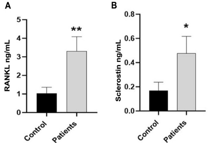
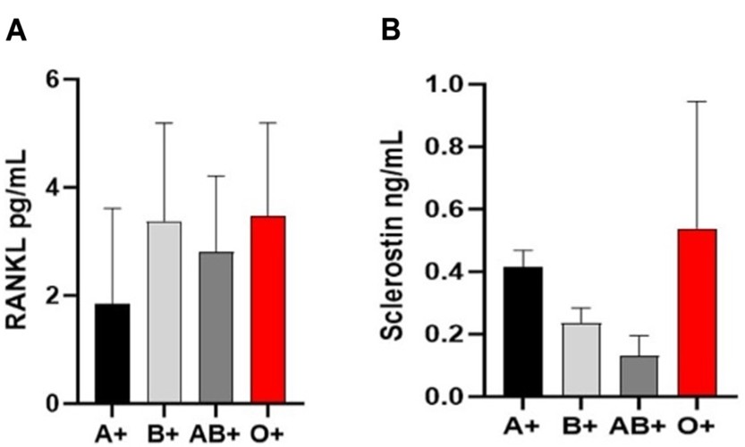
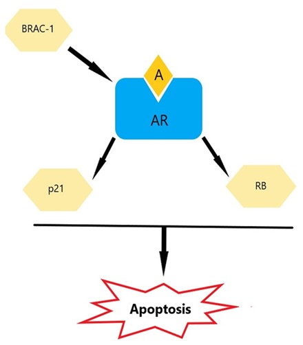
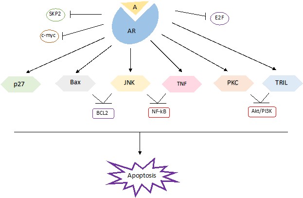
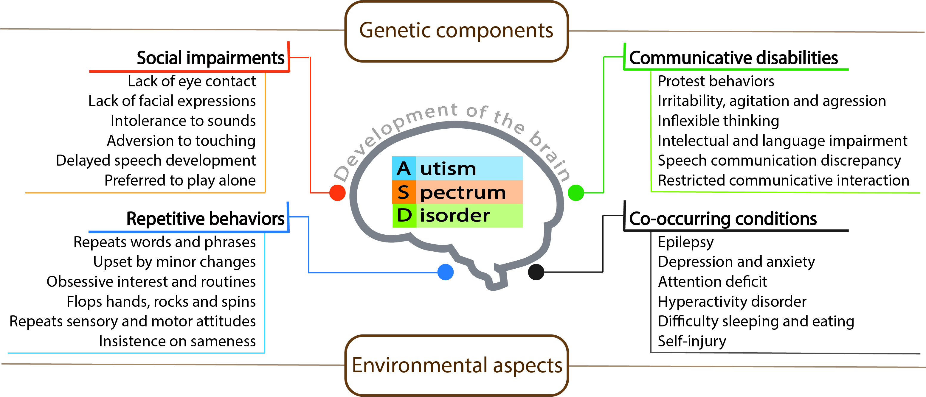
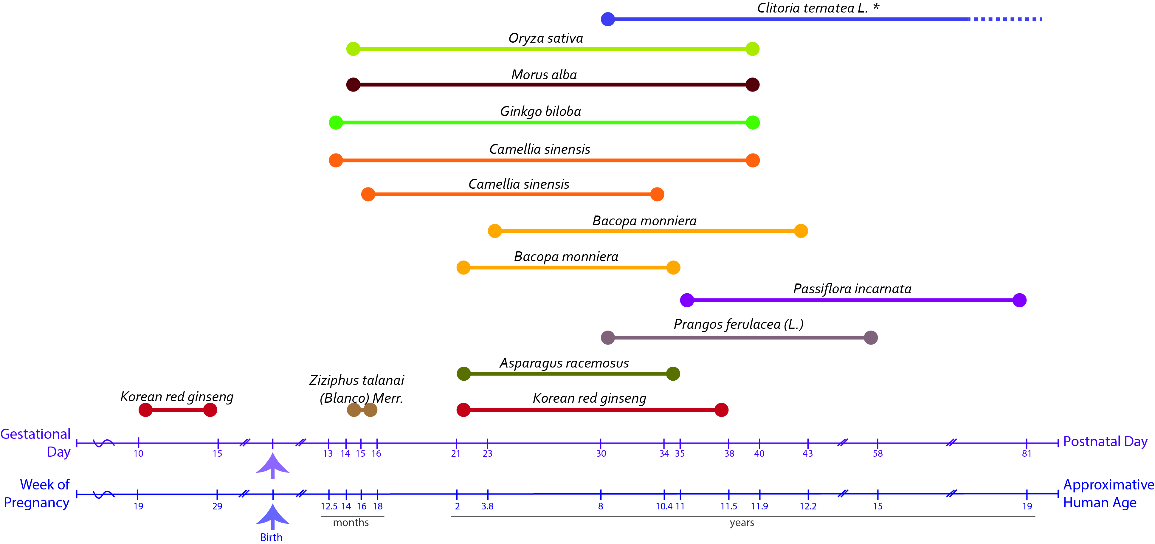
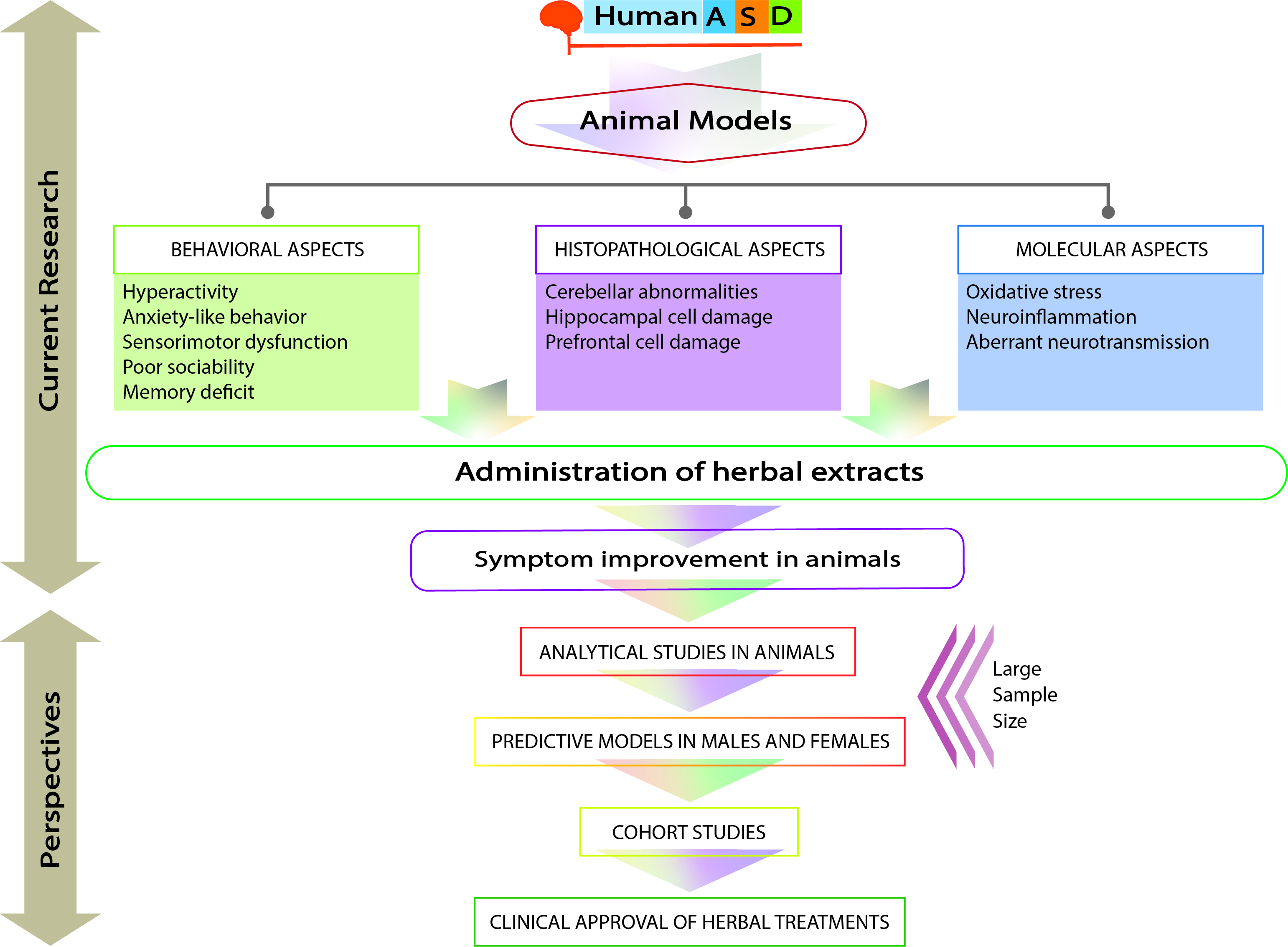
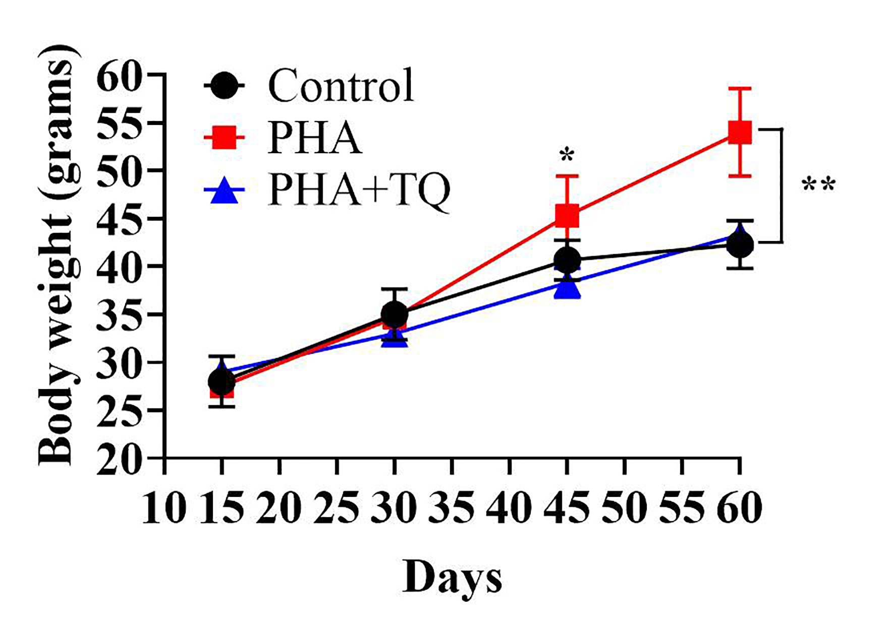
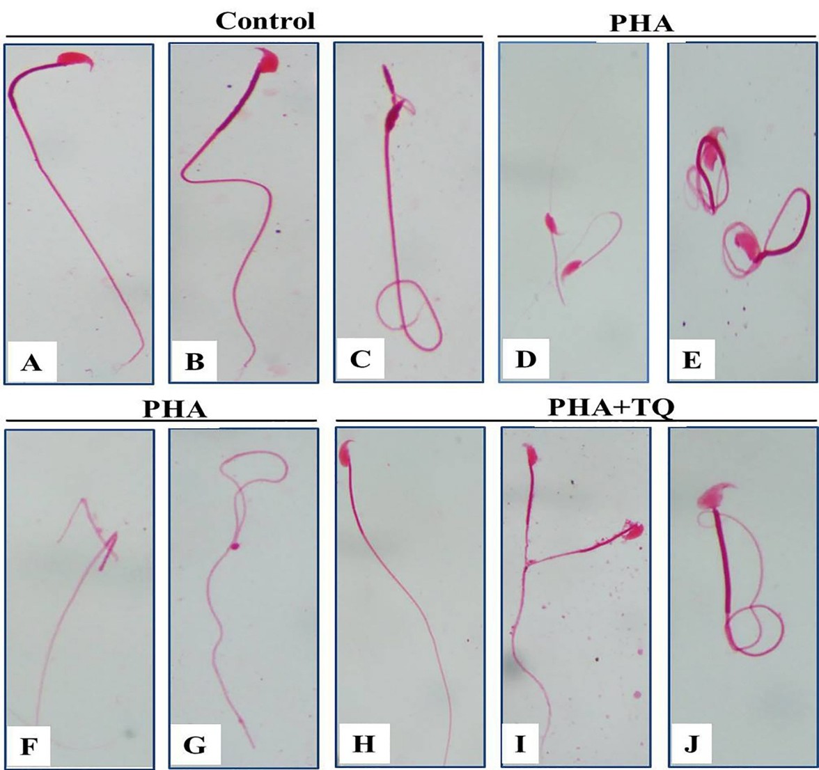
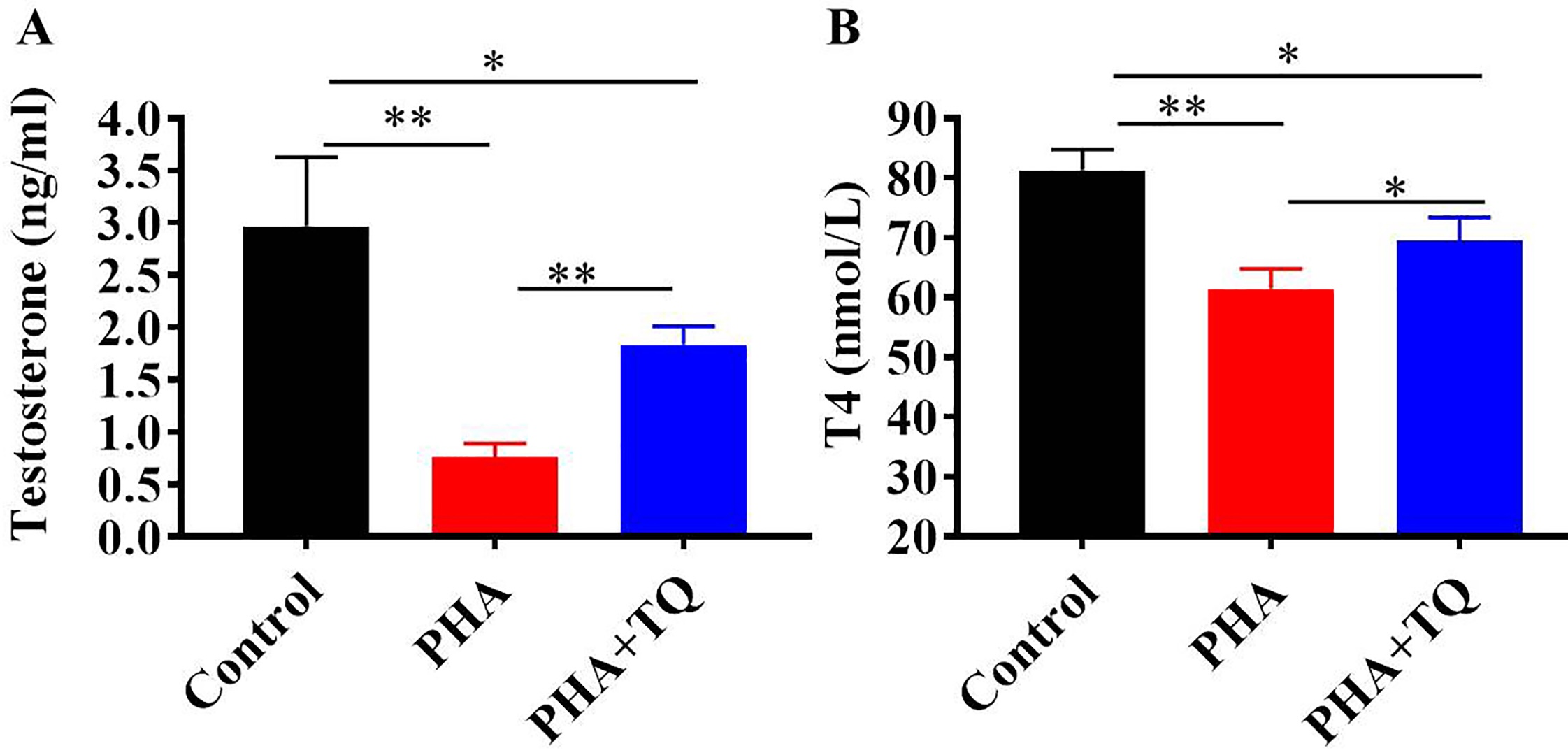
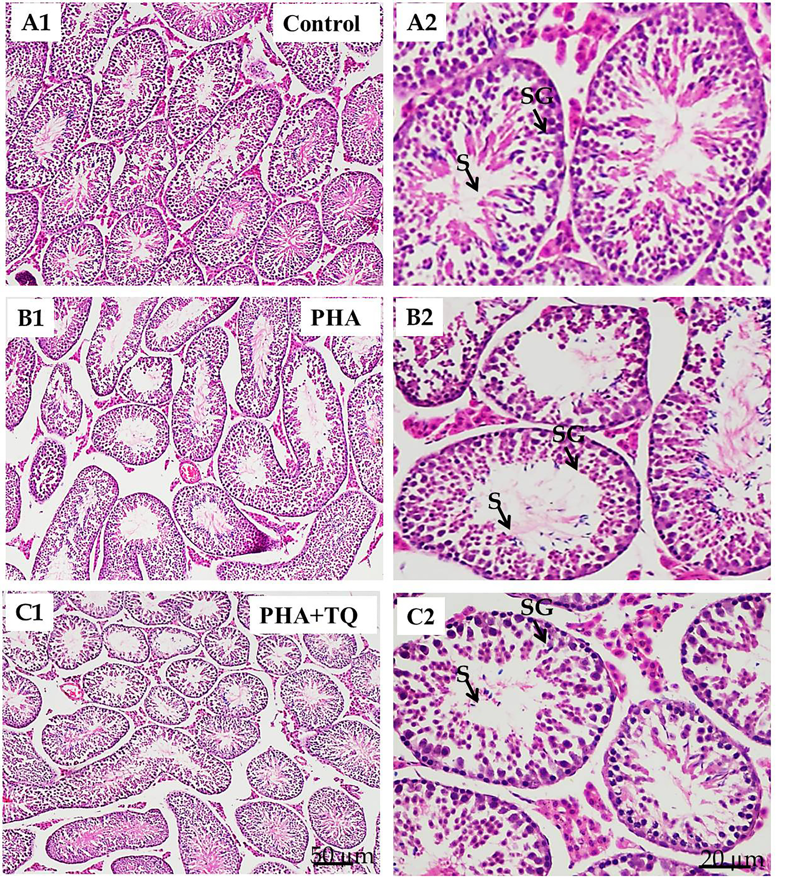
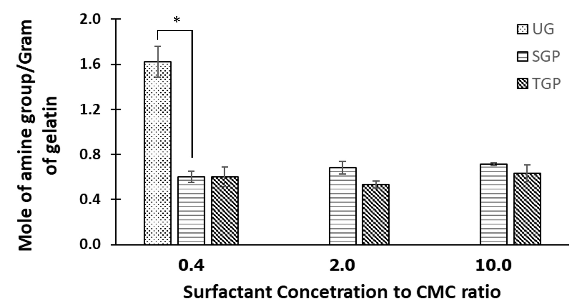
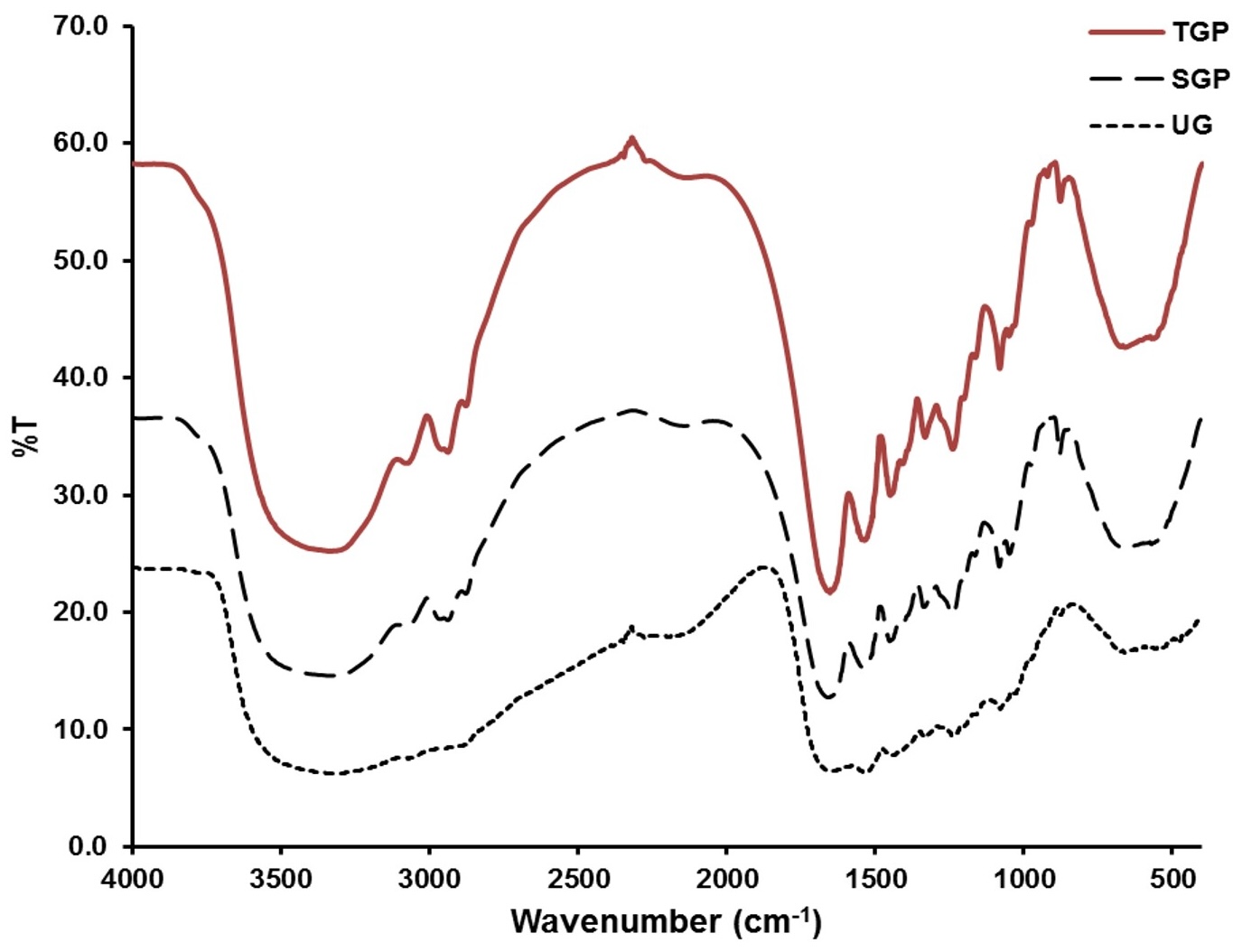
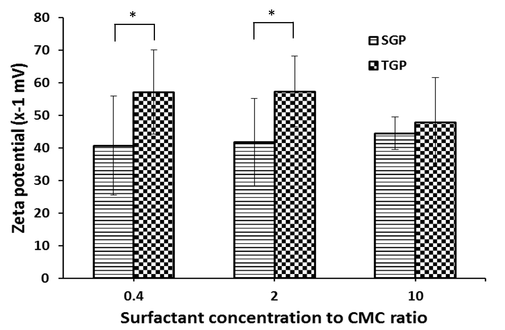
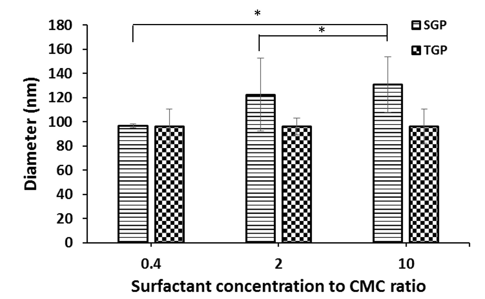
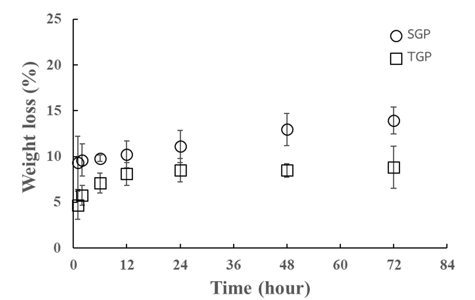
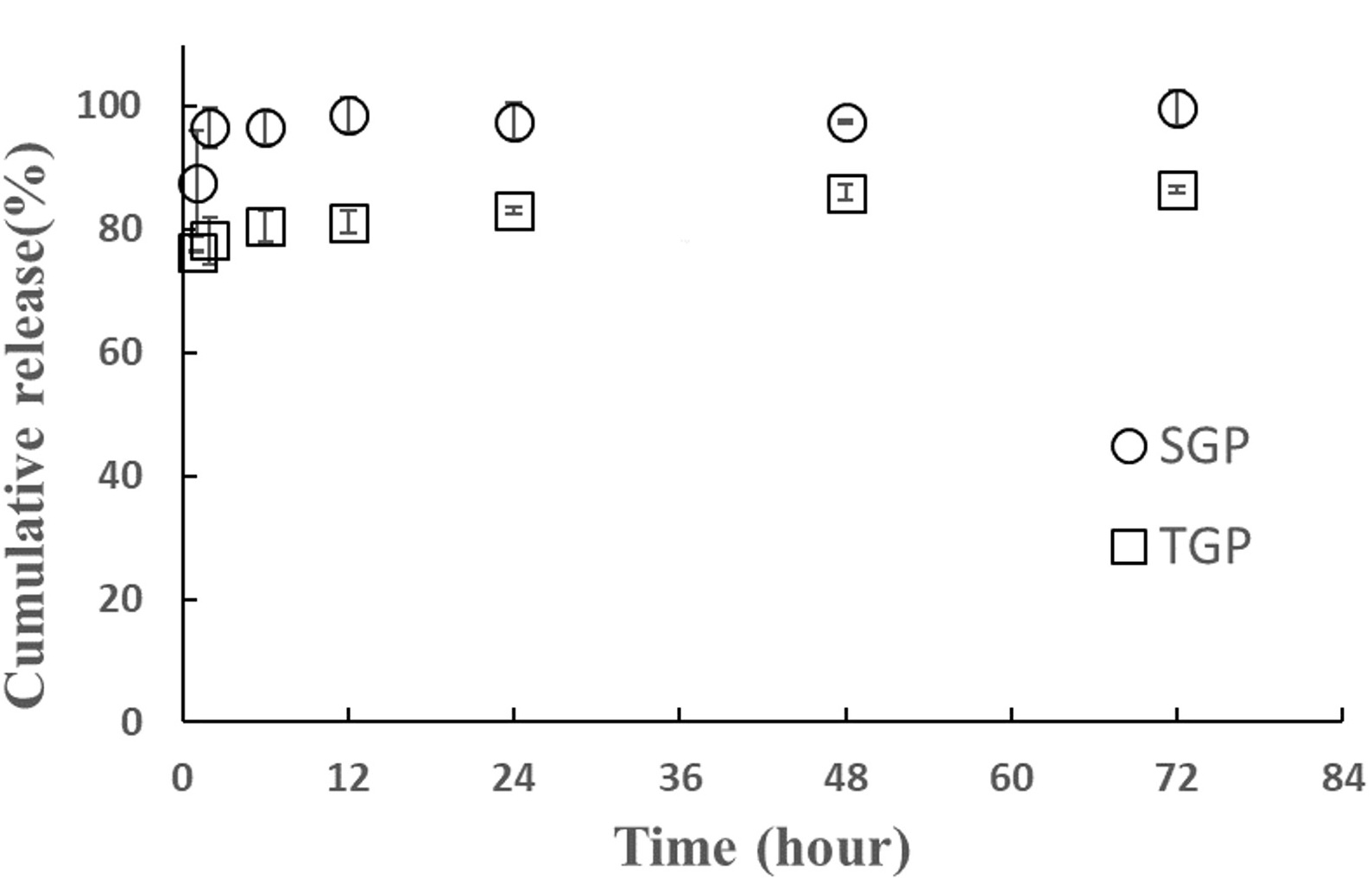
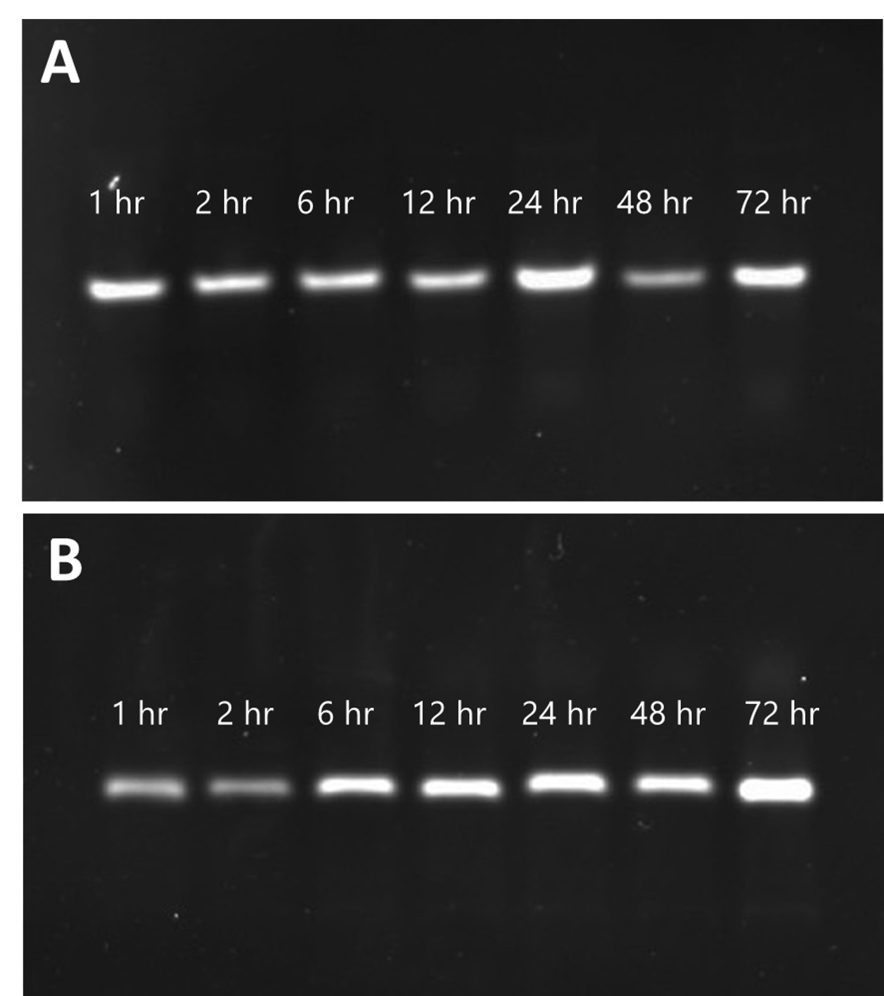
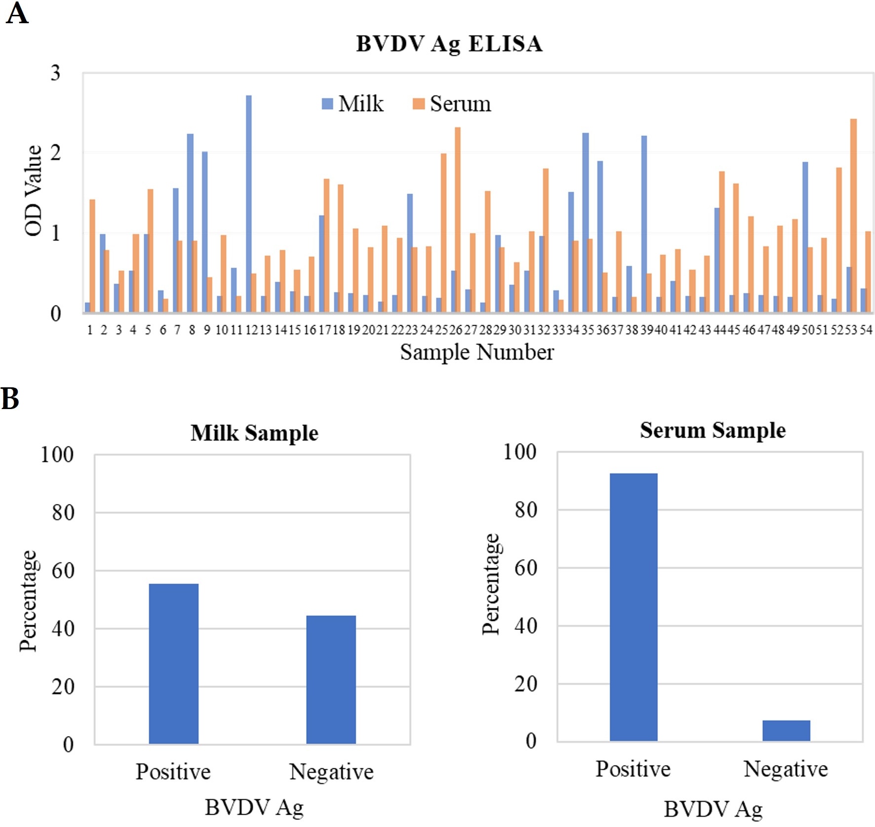
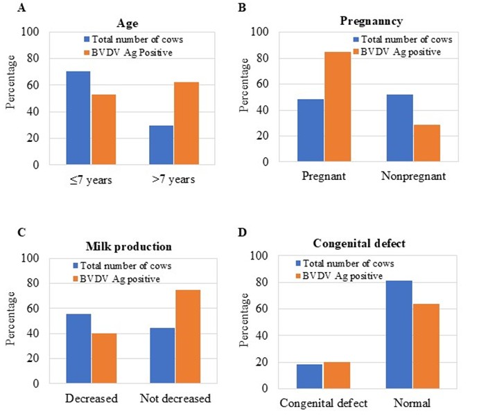
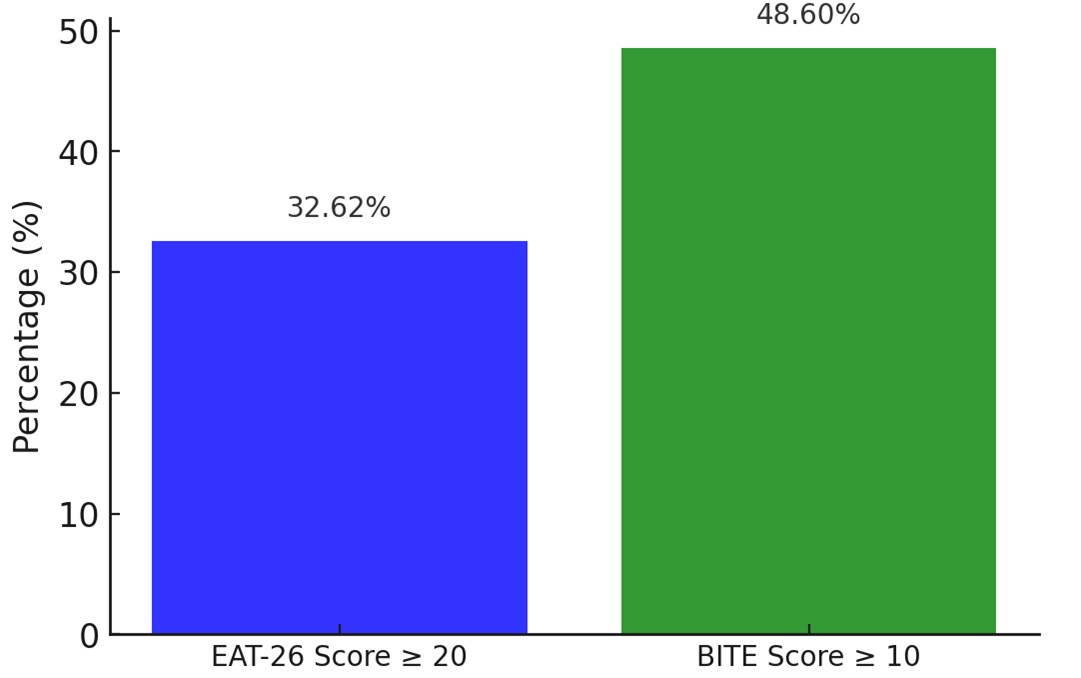
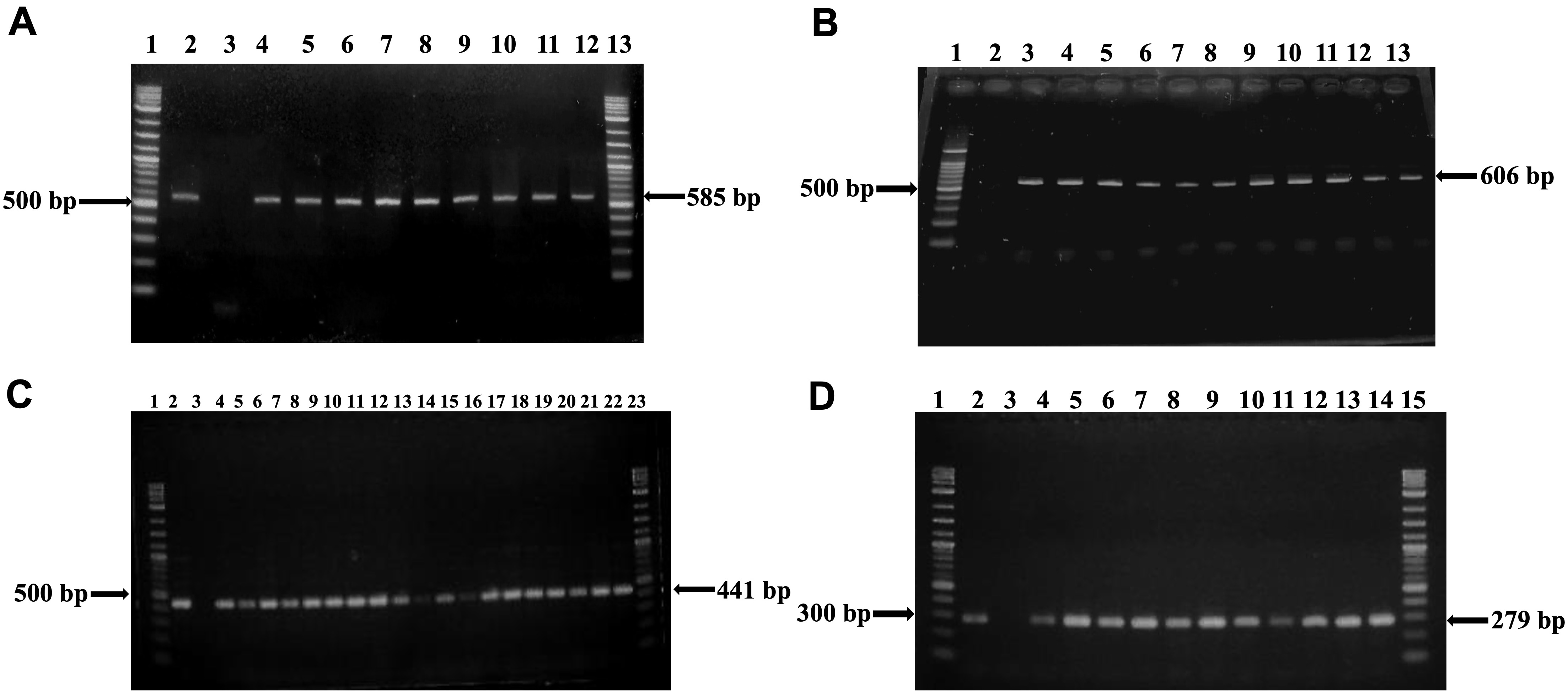
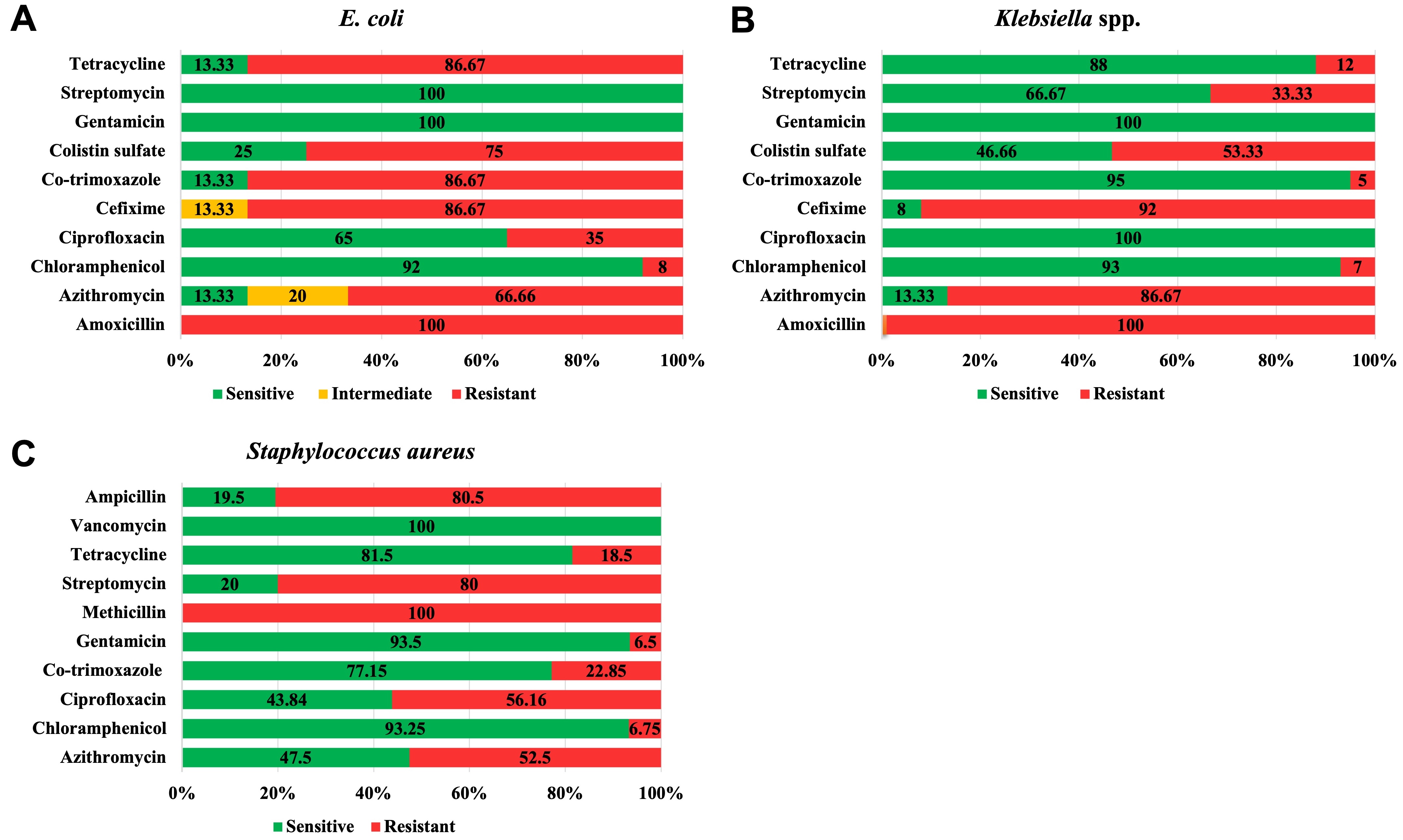
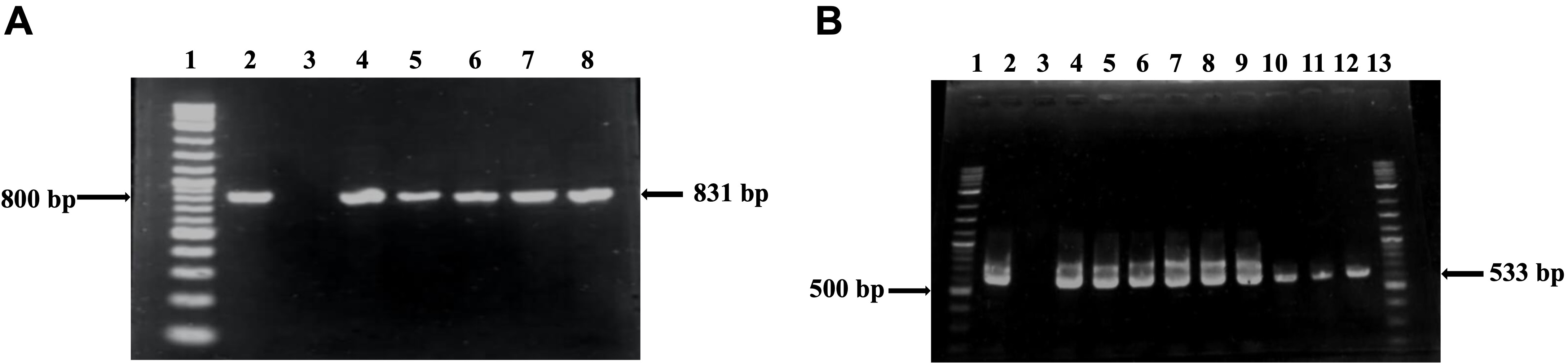
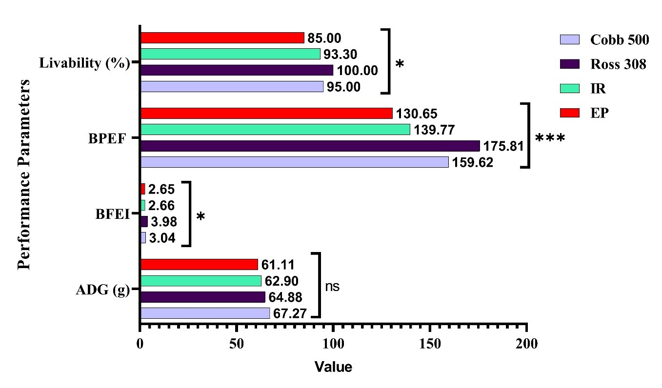
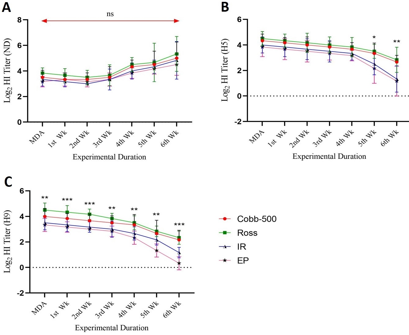
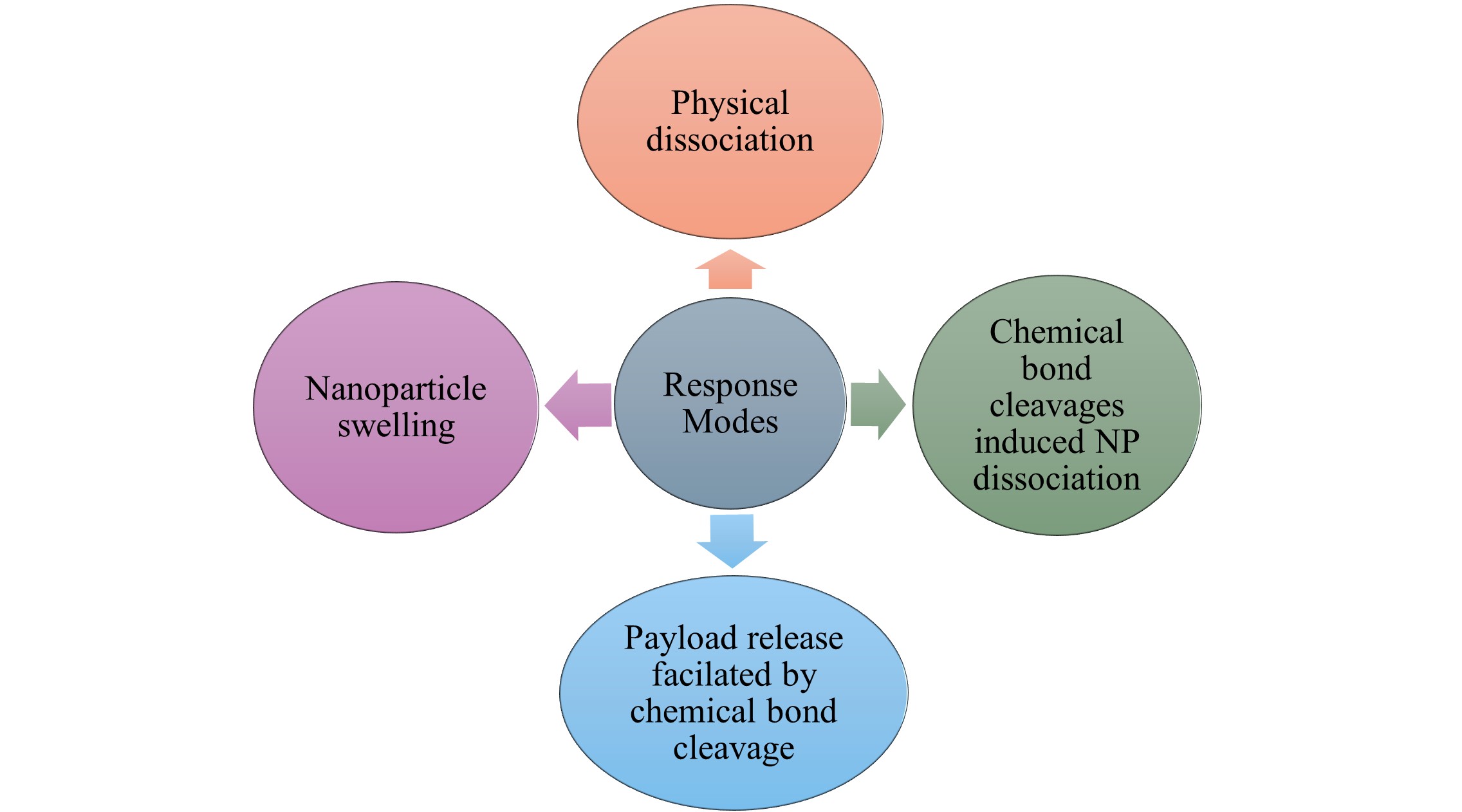
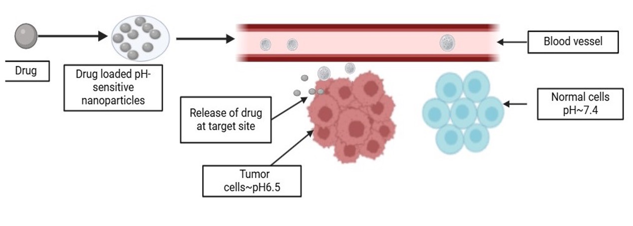