HTTP 200 OK
Allow: GET, HEAD, OPTIONS
Content-Type: application/json
Vary: Accept
{
"count": 319,
"next": "https://jabet.bsmiab.org/articles/?format=api&page=32",
"previous": "https://jabet.bsmiab.org/articles/?format=api&page=30",
"results": [
{
"id": 44,
"slug": "178-1539241375-insilico-analysis-of-g-aminobutyric-acid-transaminase-gaba-t-of-brassica-napus-rape",
"featured": false,
"slider": false,
"issue": "Vol2 Issue1",
"type": "original_article",
"manuscript_id": "178-1539241375",
"recieved": "2018-09-11",
"revised": null,
"accepted": "2018-11-26",
"published": "2019-01-05",
"pdf_file": "https://jabet.bsmiab.org/media/pdf_file/2024/54/178-1539241375.pdf",
"title": "Insilico analysis of γ- aminobutyric acid transaminase (GABA-T) of Brassica napus (Rape)",
"abstract": "<p>γ- aminobutyric acid (GABA) is an essential metabolite which plays a crucial role in signal transmission, stress metabolism, and some other activities also reported. Although the actual function of GABA shunt is not clearly understood. Three key enzymes, gamma aminobutyric acid transaminase (GABA-T), succinic semialdehyde dehydrogenase and succinic semialdehyde reductase are involved in GABA shunt mechanism en route from glutamate to the tricarboxylic acid cycle (TCA)which could pave the way of GABA shunt action. The enzyme gamma aminobutyric acid transaminase (GABA-T) could also play a key role in GABA shunt action by converting GABA to succinic semialdehyde (SSA).In this study, the protein sequence of γ-aminobutyric acid transaminase of <em>Brassica</em> <em>napus</em> <em>(Rape) was</em> <em>retrieved from </em>UniProt protein database and analyzed GABA-T enzyme using different bioinformatics tools and servers to analyze the physiochemical properties, amino acid composition, conformational states, and 3D structure. We found that our experimental protein sequence was very unstable, and the graph of Local Quality Estimate showed that the sequence was porn to mutation and value of Z score was above two in comparison with a non-redundant set of PDB structure. In addition, the phylogenetic tree revealed that GABA-T of <em>Dendrobium officinale</em><em>, </em><em>Phoenix dactylifera</em>, <em>Oryza sativa</em>, <em>Arabidopsis thaliana</em> and <em>Brassica napus </em>evolved from a common ancestor gene.</p>",
"journal_reference": "J Adv Biotechnol Exp Ther. 2019; 2(1) : 04-09.",
"academic_editor": "Dr. Md Mahmodul Hasan, Erciyes University, Turkey.",
"cite_info": "Rahman S, Shahjahan M. Insilico analysis of γ- aminobutyric acid transaminase (GABA-T) of \r\nBrassica napus (Rape). J Adv Biotechnol Exp Ther. 2019; 2(1) : 04-09.",
"keywords": [
"TCA cycle",
"GABA-T",
"transmission",
"metabolism",
"stress",
"UniPort"
],
"DOI": "10.5455/jabet.2018.d18",
"sections": [
{
"section_number": 1,
"section_title": "INTRODUCTION",
"body": "<p>γ-aminobutyric acid (GABA) is a ubiquitous, non-protein amino acid involved in the metabolism of stress and transmission of signal in plants [<a href=\"#r-1\">1</a>]. It is found in both unicellular and multicellular organisms and is involved in many aspects of plant life cycle [<a href=\"#r-2\">2</a>]. It first discovered in plants over half a century ago [<a href=\"#r-3\">3</a>]. This molecule has been intensively investigated in mammals in which it acts as a neurotransmitter in the central nervous system [<a href=\"#r-4\">4</a>]. Besides neurotransmission, it may work in carbon: nitrogen metabolism and responding during stress. However, much less is known about the role of GABA and its transport across the plasma membrane in plants [<a href=\"#r-5\">5</a>]. In plants, the enzyme gamma aminobutyric acid transaminase (GABA-T) involves in catalyzing for the conversion of GABA to succinic semialdehyde (SSA) [<a href=\"#r-6\">6</a>]. The catabolism occurs in the mitochondrial matrix of multi-cellular organisms by the action of GABA transaminase (GABA-T; EC 2.6.1.19) [<a href=\"#r-2\">2</a>]. Two branched pathways may catabolize this succinic semialdehyde (SSA): first case, it uses SSA dehydrogenase (SSADH; EC 1.2.1.16) to form succinate and enters into tricarboxylic acid cycle, and second case, utilizing SSA reductase to form α-hydroxybutyric acid [<a href=\"#r-7\">7</a>]. In this study, we focus on the activity of gamma aminobutyric acid transaminase. The result from Renault <em>et al.</em> (2013) showed that GABA-T deficiency during salt stress causes root and hypocotyl developmental defects and alterations of cell wall composition of Arabidopsis [<a href=\"#r-4\">4</a>]. (GABA-T) GABA is a metabolite en route from glutamate to the TCA cycle, which provides succinate and NADH to the respiratory machinery [<a href=\"#r-8\">8</a>]. The way from glutamate to succinate is known as the GABA shunt [3]. The activity of GABA shunt is induced by both abiotic and biotic stress [<a href=\"#r-8\">8</a>]. The GABA and the GABA shunt necessary for regulation of cytosolic p<sup>H</sup>, nitrogen storage and metabolism, protection against oxidative stress, development, and deterrence of insects [<a href=\"#r-7\">7</a>, <a href=\"#r-9\">9</a>, <a href=\"#r-10\">10</a>]. Interest in plant GABA increased mainly following observations of rapid elevation of its levels under abiotic stresses. Nevertheless, the roles of GABA under these conditions are not clear [<a href=\"#r-3\">3</a>]. It is postulated that due to the presence of stimuli or abiotic stress, accumulation of GABA is increased which enables in attachment of cell surface binding site that generate transient Ca<sup>2+</sup> increase and transport into cells via high affinity GABA transporters (e.g., GAT1;) [<a href=\"#r-5\">5</a>], which may activate glutamic acid decarboxylase enzyme via Ca<sup>2+</sup>/ calmodulin complex [<a href=\"#r-11\">11</a>]. GABA rapidly accumulates under various stress conditions such as low temperature, mechanical stimulation, and oxygen deficiency [<a href=\"#r-9\">9</a>]. In this research, the insilico analysis of the gamma-aminobutyric acid transaminase (GABA-T) may be helpful in understanding the molecular mechanism of underlying the γ-aminobutyric acid (GABA) action or any future genetic manipulation for their target application.</p>"
},
{
"section_number": 2,
"section_title": "MATERIALS AND METHODS",
"body": "<p><strong>Data retrieval</strong><br />\r\nThe protein sequences were retrieved from the web server UniProt (Universal Protein Resource, a database of protein sequence and functional information) which is a freely accessible database of protein sequence. Our target protein sequence was gamma-aminobutyric acid transaminase (GABA-T) of <em>Brassica napus</em> (Rape), a crucial enzyme of GABA shunt. The sequence was retrieved as FASTA file using accession code number A0A0H4AKW3. The protein sequence of Gamma-aminobutyric acid transaminase of <em>Malus domestica</em> (UniPort accession code J9XGP8), <em>Solanum lycopersicum</em> (UniPort accession code Q84P54), <em>Oryza sativa</em> (UniPort accession code (Q6ZH29) and <em>Arabidopsis thaliana</em> (UniPort accession code Q94CE5) and another 16 GABA-T sequences retrieved to construct phylogenetic tree using UniPort. The T-coffee [<a href=\"#r-12\">12</a>] (Tree based Consistency Objective Function for alignment Evaluation) multiple sequence alignment tool was used to generate phylogenetic tree.</p>\r\n\r\n<p> </p>\r\n\r\n<p><strong>Prediction of primary structure</strong><br />\r\nThe analyses of primary structure and physiochemical properties were performed using ProtParam [<a href=\"#r-13\">13</a>] tool from ExPasy (Expert Protein analysis system). Using the ProtParam tool, which allows the computation of various physical and chemical parameters for a given protein stored in Swiss-Prot or TrEMBL or for a user entered protein sequence. The total number of amino acids, molecular weight and atomic composition, total number of atoms, total number of negatively charged residues and total number of positively charged residues, extinction coefficients, estimated half-life, aliphatic index, instability index and grand average of hydropathicity (GRAVY) is computed.</p>\r\n\r\n<p> </p>\r\n\r\n<p><strong>Prediction of secondary structure</strong><br />\r\nThe secondary structure of a protein is predicted using SOPMA [<a href=\"#r-14\">14</a>] (Self- Optimized Prediction Method with Alignment) tool from PRABI Rhone-Alpes Bioinformatics Center (https://npsa-prabi.ibcp.fr/cgi bin/npsa_automat.pl?page=/NPSA/npsa_sopma.html). This tool evaluates the percentage of alpha helices, extended strand, beta turn and random coils. It uses homology methodology. According to percentage secondary structure is predicted. Number of conformational states can be given as either 4 (helix, sheet, turn, coil) or as 3 (helix, sheet, coil) [<a href=\"#r-15\">15</a>]. The SOPMA shows two graphs, the first graph of SOPMA result anticipates the prediction and the second graph consist of outcome curves for all of the predicted states.</p>\r\n\r\n<p> </p>\r\n\r\n<p><strong>3D structure prediction</strong><br />\r\nThe tertiary structure of the protein sequence was analyzed by SWISS MODELING [<a href=\"#r-16\">16</a>], is a structural bioinformatics web-server dedicated to homology modeling of protein 3D structures. The SWISS MODEL is very conservative but reliable. The reliability of a model depends on the availability of highly matched template and some other factors. The selection of a template relies on several parameters like sequence identity, oligomeric state, GMQE (Global Quality Model Estimate), ligand and QSQE (Quaternary Structure Prediction). It allows FASTA, Clustal, plain string, or a valid UniProtKB AC. SWISS-MODEL expert system features are automated modelling of homo-oligomeric assemblies, modeling of essential metal ions and biologically relevant ligands in protein structures, and model reliability estimates based on the QMEAN local score function. Global quality estimates based torsion angle, solvation potentiality, all atom angles, Cβ deviations, and QMEAN value. After building our model, we download as PDB format. This PDB data file subjected for PROCHECK analysis in <a href=\"http://www.ebi.ac.uk/thorntonsrv/databases/cgibin/pdbsum/GetPage.pl?pdbcode=m957&pdb_type=UPLOAD&code=174320&template=main.html\">PDBsum</a> [<a href=\"#r-17\">17</a>]. From PDBsum, we analyzed and validated our designed model based on different parameter like Ramachandran plot statistics, ligand, and protein-protein interaction. RAMPAGE [<a href=\"#r-18\">18</a>] tool was used to compare PROCHECK data of Ramachandran plot.</p>"
},
{
"section_number": 3,
"section_title": "RESULTS AND DISCUSSION",
"body": "<p><strong>Primary structure and phylogenetic analysis</strong><br />\r\nOur target protein sequence was 498 amino acids long, where highest amount of alanine (9.4%) and leucine (9.0%), and lowest amount of tryptophan (1.2%), and cysteine (1.2%) were presented (<a href=\"#Table-1\">Table 1</a>). We found negatively charged residues for (Asp + Glu) and positively charged residues for (Arg + Lys) where total number of atoms was 7750 including structural formula C2482H3879N651O719S19. Extinction coefficients were in units of M-1 cm-1 at 280 nm measured in water. The stability index was 50.4 which indicated that the protein was unstable (<a href=\"#Table-2\">Table 2</a>).<br />\r\nIn the phylogenetic tree (<a href=\"#figure1\">Figure 1</a>), we found that the GABA-T of <em>Brassica </em><em>napus</em>is was closely related with<em> Arabidopsis thaliana </em>where GABA-T of <em>Dendrobium officinale </em>was closely related with <em>Phoenix dactylifera. </em>The phylogenetic tree revealed that GABA-T of <em>Oryza sativa Dendrobium officinale, Phoenix dactylifera, </em><em>Arabidopsis thaliana</em> and <em>Brassica napus </em>were evolved from a common ancestor gene.</p>\r\n\r\n<div id=\"Table-1\">\r\n<p><strong><a href=\"https://jabet.bsmiab.org/table/178-1539241375-table1/\">T1 </a>Table 1</strong>. Amino acid composition of GABA-T obtained from ProtPram [13].</p>\r\n</div>\r\n\r\n<div id=\"Table-2\">\r\n<p><strong><a href=\"https://jabet.bsmiab.org/table/178-1539241375-table2/\">T2</a> Table 2</strong>. The different parameters of a primary structure obtained from ProtPram [13].</p>\r\n</div>\r\n\r\n<div id=\"figure1\">\r\n<figure class=\"image\"><img alt=\"\" height=\"303\" src=\"/media/article_images/2024/04/10/178-1539241375-Figure1.jpg\" width=\"500\" />\r\n<figcaption><strong>Figure 1.</strong> Phylogenetic tree of GABA-T from soil bacteria and plant sources.</figcaption>\r\n</figure>\r\n\r\n<p> </p>\r\n</div>\r\n\r\n<p><strong>Secondary structure prediction</strong><br />\r\nThe secondary structure of protein was predicted using SOPMA (Self- Optimized Prediction Method with Alignment) tool [<a href=\"#r-19\">19</a>]. This tool evaluated the percentage of alpha helices (43.17%), extended strand (13.65%), beta turn (7.43%) and random coils (35.74%). The windows output width, similarity threshold and number of conformational state were 17, 8 and 4, respectively. From PDBsum, we found the numbers of interface residues were 70 in chain A, and 68 in chain B, in addition, number of salt bridge, number of hydrogen bonds and number of non-bonded contracts were 2, 28 and 424, respectively. The topology of the secondary structure is presented in <a href=\"#figure2\">Figure 2</a>, designed by PDBsum.</p>\r\n\r\n<div id=\"figure2\">\r\n<figure class=\"image\"><img alt=\"\" height=\"360\" src=\"/media/article_images/2024/04/10/178-1539241375-Figure2.jpg\" width=\"500\" />\r\n<figcaption><strong>Figure 2.</strong> Protein topology of the secondary structure.</figcaption>\r\n</figure>\r\n</div>\r\n\r\n<p> </p>\r\n\r\n<p><strong>3D structure prediction</strong><br />\r\nOur target protein sequence found total 1041 templates for homology modeling. To reveal the high sequence similarity, 50 templates were displayed (<a href=\"#Table-3\">Table 3</a>) on the screen from SWISS-MODEL Template Library (SMTL) [<a href=\"#r-20\">20</a>]. The first templates (5ghg.1.A), showed the highest sequence identity (50.12) which was aminotransferase class-III and used for building target homology modeling. This template was found by Basic Local Alignment Search Tool (BLAST), for high Sequence similarity 0.44. The global quality estimation of the resulting model, (GMQE) is 0.71, the value of QSQE of the template, is 0.92, and the value of QMEAN is -1.67. The 3D structure of GABA-T showed in figure 2 including ligand.<br />\r\nIn <a href=\"#figure3\">Figure 3</a>. A is the target 3D structure of our protein developed by PDBsum, where chain A (green colored), chain B (red colored) and yellow colored in the center is the ligand (PMP, 4′-Deoxy-4′-aminopyridoxal-5′-Phosphate, PMP) where figure B. and C showed the Ramachandran Plot before PROCHECK. This plot favored 93.4%, which reflects an acceptable homology modeling of target protein. After PROCHECK, the Ramachandran Plot favored 85.9% in figure D and <a href=\"#Table-4\">Table 4</a>.</p>\r\n\r\n<div id=\"Table-3\">\r\n<p><strong><a href=\"https://jabet.bsmiab.org/table/178-1539241375-table3/\">T3</a> Table 3. </strong>The 50 templates from SWISS-MODEL Template Library (SMTL)</p>\r\n</div>\r\n\r\n<div id=\"figure3\">\r\n<figure class=\"image\"><img alt=\"\" height=\"419\" src=\"/media/article_images/2024/04/10/178-1539241375-Figure3.jpg\" width=\"500\" />\r\n<figcaption><strong>Figure 3.</strong> 3D structure of protein and Ramachandran plot. </figcaption>\r\n</figure>\r\n</div>\r\n\r\n<div id=\"Table-4\">\r\n<p><strong><a href=\"https://jabet.bsmiab.org/table/178-1539241375-table4/\">T4</a> Table 4.</strong> PROCHECK analyses of Ramachandran Plot [19].</p>\r\n</div>\r\n\r\n<p> </p>\r\n\r\n<p><strong>Ramachandran plot analyses</strong><br />\r\nIn Ramachandran Plot PROCHECK analyses, we found total 854 residues 854 including Glycine ( 66), Proline (53), End-residues (excl. Glycine and Proline) (4) and other highest number of residues were found in non-glycine and non-proline residues. The most favored region in Ramachandran plot was 85.9% (<a href=\"#Table-3\">Table 3</a>). It suggested that the percentage of most favored region (above 90%) could be desirable for good modeling.</p>\r\n\r\n<p> </p>\r\n\r\n<p><strong>Molprobity statistics</strong><br />\r\nIn MolProbity statistics, we found that the Ramachandran plot favoured 93.4% and the residues with bad angles were 0.0055 which was below 0.1% and values of bad bonds were 0.00029 (near to zero) (<a href=\"#Table-5\">Table 5</a>). But the C-Beta deviations were quite higher which might be zero or near to zero. The value of Ramachandran Outliers and Rotamer Outliers were very satisfactory.</p>\r\n\r\n<div id=\"Table-5\">\r\n<p><strong><a href=\"https://jabet.bsmiab.org/table/178-1539241375-table5/\">T5</a> Table 5. </strong>MolProbity results from Swiss Homology modeling using MolProbity in Phenix version 1.13. [20].</p>\r\n</div>\r\n\r\n<p> </p>\r\n\r\n<p><strong>Assessment of the Ramachandran Plot by Rampage</strong><br />\r\nIn RAMPAGE, we found number of residues in favored region 94.1%, (~98.0% expected) which was quite higher from Swiss Modeling and the number of residues in allowed region was 5.5%, (~2.0% expected). However, in PROCHECK analyses, we found 13.6% of residues in allowed region [Combined of additional allowed regions (a,b,l,p) and generously allowed regions (~a,~b,~l,~p)], and number of residues in outlier region, (0.4%).</p>"
},
{
"section_number": 4,
"section_title": "CONCLUSION",
"body": "<p>γ-aminobutyric acid (GABA) is not only a metabolite which plays a significant role in signal transmission, stress metabolism, regulation of cytosolic p<sup>H </sup> etc. The activity of GABA extensively studied in mammals but in plants the role of GABA is less known. This insilico analysis of the key enzyme gamma aminobutyric acid transaminase (GABA-T) could be helpful in understanding the role of GABA shunt. This protein sequence was unstable and porn to mutate and phylogenetic tree revealed that GABA-Tof <em>Malus domestica, Solanum lycopersicum</em>, <em>Oryza sativa</em>, <em>Arabidopsis thaliana</em> and <em>Brassica napus </em>were evolved from a common ancestor gene.</p>"
},
{
"section_number": 5,
"section_title": "CONFLICT OF INTEREST",
"body": "<p>The author declares that no conflict of interest exists.</p>"
}
],
"figures": [
{
"figure": "https://jabet.bsmiab.org/media/article_images/2024/04/10/178-1539241375-Figure1.jpg",
"caption": "Figure 1. Phylogenetic tree of GABA-T from soil bacteria and plant sources.",
"featured": false
},
{
"figure": "https://jabet.bsmiab.org/media/article_images/2024/04/10/178-1539241375-Figure2.jpg",
"caption": "Figure 2. Protein topology of the secondary structure.",
"featured": false
},
{
"figure": "https://jabet.bsmiab.org/media/article_images/2024/04/10/178-1539241375-Figure3.jpg",
"caption": "Figure 3. 3D structure of protein and Ramachandran plot.",
"featured": false
}
],
"authors": [
{
"id": 126,
"affiliation": [
{
"affiliation": "Bangladesh Livestock Research Institute Regional Station, Baghabari, Shahjadpur, Sirajganj-6770, Bangladesh"
}
],
"first_name": "Shahidur",
"family_name": "Rahman",
"email": "shahidur.blri@gmail.com",
"author_order": 1,
"ORCID": null,
"corresponding": true,
"co_first_author": false,
"co_author": false,
"corresponding_author_info": "Mr. Shahidur Rahman, Bangladesh Livestock Research Institute, Regional Station, Baghabari, Shahjadpur, Sirajganj\u00026770, Bangladesh, Email:shahidur.blri@gmail.com",
"article": 44
},
{
"id": 127,
"affiliation": [
{
"affiliation": "Bangladesh Livestock Research Institute Regional Station, Baghabari, Shahjadpur, Sirajganj-6770, Bangladesh"
}
],
"first_name": "Md",
"family_name": "Shahjahan",
"email": null,
"author_order": 2,
"ORCID": null,
"corresponding": false,
"co_first_author": false,
"co_author": false,
"corresponding_author_info": "",
"article": 44
}
],
"views": 530,
"downloads": 111,
"references": [
{
"id": 1123,
"serial_number": 1,
"pmc": null,
"reference": "Clark SM, Di Leo R, Van Cauwenberghe OR, Mullen RT, Shelp BJ. Subcellular localization and expression of multiple tomato γ-aminobutyrate transaminases that utilize both pyruvate and glyoxylate. J Exp Bot. 2009; 60(11): 3255–3267.",
"DOI": null,
"article": 44
},
{
"id": 1124,
"serial_number": 2,
"pmc": null,
"reference": "Michaeli S, Fromm H. Closing the loop on the GABA shunt in plants: are GABA metabolism and signaling entwined? Front Plant Sci. 2015; 6: 419.",
"DOI": null,
"article": 44
},
{
"id": 1125,
"serial_number": 3,
"pmc": null,
"reference": "Steward FC, Thompson JF, and Dent CE. γ-aminobutyricacid: a constituent of the potato tuber? Science 1949; 110: 439–440.",
"DOI": null,
"article": 44
},
{
"id": 1126,
"serial_number": 4,
"pmc": null,
"reference": "Renault H, El Amrani A, Berger A, Mouille G, Soubigou-Taconnat L, Bouchereau A, Deleu C.γ-Aminobutyric acid transaminase deficiency impairs central carbon metabolism and leads to cell wall defects during salt stress in Arabidopsis. Plant Cell Environ 2013; 36, 1009–1018.",
"DOI": null,
"article": 44
},
{
"id": 1127,
"serial_number": 5,
"pmc": null,
"reference": "Meyer A, Eskandari S, Grallath S, and Rentsch D. At GAT1, a high affinity transporter for γ-aminobutyricacidin Arabidopsisthaliana. J.Biol. Chem. 2006; 281: 7197–7204.",
"DOI": null,
"article": 44
},
{
"id": 1128,
"serial_number": 6,
"pmc": null,
"reference": "Shimajiri Y, Ozaki K, Kainou K, Akama K. Differential subcellular localization, enzymatic properties and expression patterns of γ-aminobutyric acid transaminases (GABA-Ts) in rice (Oryza sativa). J Plant Physiol.2013; 170 (2): 196-201.",
"DOI": null,
"article": 44
},
{
"id": 1129,
"serial_number": 7,
"pmc": null,
"reference": "Shelp BJ, Bown AW, and McLean MD. Metabolism and functions of gamma-aminobutyric acid. Trends Plant Sci. 1999; 4(11): 446-452.",
"DOI": null,
"article": 44
},
{
"id": 1130,
"serial_number": 8,
"pmc": null,
"reference": "Bouché N, Fait A, Bouchez D, Møller SG, Fromm H. Mitochondrial succinic-semialdehyde dehydrogenase of the γ-aminobutyrate shunt is required to restrict level of reactive oxygen intermediate in plants. Proc Natl Acad Sci U S A. 2003; 100. 6843-8.",
"DOI": null,
"article": 44
},
{
"id": 1131,
"serial_number": 9,
"pmc": null,
"reference": "Bouché N, Fromm H. GABA in plants: Just a metabolite? Trends plant sci. 2004; 9: 110-5.",
"DOI": null,
"article": 44
},
{
"id": 1132,
"serial_number": 10,
"pmc": null,
"reference": "Bouche N, Fait A, Bouchez D, Moller, SG, and Fromm, H. Natl. Acad. Sci. U. S. A.2003:100, 6843–6848.",
"DOI": null,
"article": 44
},
{
"id": 1133,
"serial_number": 11,
"pmc": null,
"reference": "Baum G, Chen Y, Arazi T, Takatsuji H, and Fromm H. A plant glutamate decarboxylase containing a calmodulin binding domain. Cloning, sequence, and function alanalysis. J. Biol. Chem. 1993; 268: 19610–19617.",
"DOI": null,
"article": 44
},
{
"id": 1134,
"serial_number": 12,
"pmc": null,
"reference": "Magis C, Taly JF, Bussotti G, Chang JM, Di Tommaso P, Erb I, Espinosa-Carrasco J, Notredame C.T-Coffee: Tree-based consistency objective function for alignment evaluation. Methods Mol Biol.2014; 1079: 117-29.",
"DOI": null,
"article": 44
},
{
"id": 1135,
"serial_number": 13,
"pmc": null,
"reference": "Elisabeth G, Christine H, Alexandre G, S’everine D, Marc RW, Ron DA, Amos B. Protein Identification and Analysis Tools on the ExPASy Server. The Proteomics Protocols Handbook pp 571-60.",
"DOI": null,
"article": 44
},
{
"id": 1136,
"serial_number": 14,
"pmc": null,
"reference": "Geourjon C, Deléage G SOPMA: significant improvements in protein secondary structure prediction by consensus prediction from multiple alignments. Comput Appl Biosci.1995; 11:681–684.",
"DOI": null,
"article": 44
},
{
"id": 1137,
"serial_number": 15,
"pmc": null,
"reference": "Singh N, Upadhyay S, Jaiswar A, Mishra N. In silico Analysis of Protein. J Bioinform, Genomics, Proteomics.2016: 1(2): 1007.",
"DOI": null,
"article": 44
},
{
"id": 1138,
"serial_number": 16,
"pmc": null,
"reference": "Schwede T, Kopp J, Guex N, Peitsch MC. “SWISS-MODEL: an automated protein homology-modeling server”. Nucleic Acids Res. 2003; 31: 3381–3385..",
"DOI": null,
"article": 44
},
{
"id": 1139,
"serial_number": 17,
"pmc": null,
"reference": "Laskowski RA, Hutchinson EG, Michie AD, Wallace AC, Jones ML, Thornton JM. “PDBsum: a Web-based database of summaries and analyses of all PDB structures”. Trends Biochem Sci. 1997; 22(12): 488–90.",
"DOI": null,
"article": 44
},
{
"id": 1140,
"serial_number": 18,
"pmc": null,
"reference": "Lovell SC1, Davis IW, Arendall WB 3rd, de Bakker PI, Word JM, Prisant MG, Richardson JS, Richardson DC. Structure validation by Calpha geometry: phi,psi and Cbeta deviation. Proteins: Structure, Function & Genetics. 2002; 50: 437-450.",
"DOI": null,
"article": 44
},
{
"id": 1141,
"serial_number": 19,
"pmc": null,
"reference": "Benkert P, Biasini M, Schwede T. Toward the estimation of the absolute quality of individual protein structure models. 2011. Bioinformatics. 2011; 27(3): 343-50.",
"DOI": null,
"article": 44
},
{
"id": 1142,
"serial_number": 20,
"pmc": null,
"reference": "Waterhouse A, Bertoni M, Bienert S, Studer G, Tauriello G, Gumienny R, Heer FT, de Beer, TAP, Rempfer C, Bordoli L, Lepore R, Schwede T. SWISS-MODEL: homology modelling of protein structures and complexes. 2018. Nucleic Acids Res. 46(W1): W296-W303.",
"DOI": null,
"article": 44
}
]
},
{
"id": 80,
"slug": "178-1570132696-hmg-coa-reductase-inhibitor-rosuvastatin-averted-carbon-tetrachloride-induced-oxidative-stress-inflammation-and-fibrosis-in-the-liver-of-rats",
"featured": false,
"slider": false,
"issue": "Vol3 Issue1",
"type": "original_article",
"manuscript_id": "178-1570132696",
"recieved": "2019-10-03",
"revised": null,
"accepted": "2019-11-10",
"published": "2019-01-05",
"pdf_file": "https://jabet.bsmiab.org/media/pdf_file/2023/17/178-1570132696.pdf",
"title": "HMG-CoA reductase inhibitor, rosuvastatin averted carbon tetrachloride-induced oxidative stress, inflammation and fibrosis in the liver of rats",
"abstract": "<p><strong> </strong>The aim of this study was to examine the effect of rosuvastatin in experimentally-induced hepatic inflammation and fibrosis in rats. Carbon tetra chloride (CCl4) was administered orally to induce liver damage in female Long Evans rats. Rats were treated with CCl4 alone twice a week over two weeks. Rosuvastatin (10 mg/kg) was also given daily to CCl4 treated rats concurrently by nasogastric gavage. After two weeks, various oxidative stress markers as well as liver markers enzymes were investigated in different animal groups tested in this study. Moreover, histological assessments were also done for inflammatory cell infiltration and fibrosis in the liver of all test groups. Plasma aspartate aminotransferase (AST), alanine aminotransferase (ALT) and alkaline phosphatase (ALP) activities were increased in the CCl4 group compared with the control group. Increased liver enzyme activities were significantly decreased by rosuvastatin treatment. Moreover, rosuvastatin treatment inhibited the formation of lipid peroxidation products in CCl4 administered rats. Rosuvastatin treatment also restored the decreased superoxide dismutase (SOD) activities as well as elevated the reduced glutathione concentration in CCl4 administered rats. Liver tissues from rats of control group also revealed no significant pathological changes, while CCl4 administered rats showed significant infiltration of inflammatory cells and liver fibrosis, which was further, normalized or significantly decreased by rosuvastatin treatment. This study revealed that, rosuvastatin treatment may ameliorate all necro-inflammatory and fibrotic changes in liver tissues of CCl4 induced rats and could be used as an alternative therapy for chemical or drug-induced liver fibrosis.</p>",
"journal_reference": "J Adv Biotechnol Exp Ther. 2020; 3(1): 01-08.",
"academic_editor": "Dr. Md Jamal Uddin, Ewha Womans University, South Korea.",
"cite_info": "Sikder B, Akter F, et al. HMG-CoA reductase inhibitor, rosuvastatin averted carbon tetrachloride-induced oxidative stress, inflammation and fibrosis in the liver of rats. J Adv Biotechnol Exp Ther. 2020; 3(1): 01-08.",
"keywords": [
"inflammation",
"Rosuvastatin",
"Carbon tetra chloride",
"Superoxide dismutase",
"Fibrosis"
],
"DOI": "10.5455/jabet.2020.d101",
"sections": [
{
"section_number": 1,
"section_title": "INTRODUCTION",
"body": "<p>Nowadays, an upsurge of persistent liver diseases, associated with metabolic syndrome, non-alcoholic fatty liver disease, steatosis, and primary biliary cirrhosis has become one of the major concerns in health care society. A statistics showed that annual deaths due to liver cancer and cirrhosis increased by 1.25 to 1.75 million from 1990 to 2010 worldwide. In most cases, advanced cirrhosis and hepatocellular carcinoma (HCC) are the main complications behind most of the liver-related mortality [<a href=\"#r-1\">1</a>]. Chronic liver injury leading to cirrhosis is occurred due to various insults like viral hepatitis, consumption of alcohol, irrational use of drugs, metabolic disease associated with iron or copper overload, autoimmune reaction of the hepatocyte, congenital abnormalities [<a href=\"#r-2\">2</a>] or due to the exposure of some chemical toxin e.g. ethanol, carbon tetrachloride etc. [<a href=\"https://www.bsmiab.org/jabet/178-1570132696-hmg-coa-reductase-inhibitor-rosuvastatin-averted-carbon-tetrachloride-induced-oxidative-stress-inflammation-and-fibrosis-in-the-liver-of-rats/#_ENREF_3\">3</a><a href=\"#r-3\">, 4</a>]. Liver malfunction is primarily associated with oxidative stress. Irreversible liver cirrhosis occurs due to persistent existence of inflammation. There are mainly two types of hepatic cells, including tissue macrophages (Kupffer cells), and a perivascular mesenchymal stellate cells, whose activation occurs following any liver injury of any etiology. Hepatocyte and kupffer cell is the primary source of ROS. In this study halo-alkane like carbon tetrachloride was used for the study of hepatotoxicity in animals. Trichloromethyl (<sup>•</sup>CCl<sub>3</sub>) and peroxy trichloromethyl (<sup>•</sup>OOCCl3) species are produced from carbon tetrachloride via the influence of phase-I cytochrome P450 enzyme [<a href=\"#r-5\">5</a>]. These excess ROSs are the main culprit for causing oxidative stress in the liver by raising the level of different oxidative stress markers (lipid peroxidation, nitric oxide, advanced oxidative protein products, myeloperoxidase).<br />\r\nAt present, lipid-lowering drugs have gained much more attention as an effective treatment approaches as these drugs lower cholesterol synthesis, reduce different reactive oxygen species and oxidative stress [<a href=\"https://www.bsmiab.org/jabet/178-1570132696-hmg-coa-reductase-inhibitor-rosuvastatin-averted-carbon-tetrachloride-induced-oxidative-stress-inflammation-and-fibrosis-in-the-liver-of-rats/#_ENREF_6\">6</a>]. Increasing investigational studies have revealed that statins have a beneficial effect on atherosclerosis by diminishing the function of NF-κB and by up-regulating the expression of peroxisome proliferator receptors (PPAR-α), both these activity of statins reduce inflammatory responses [<a href=\"#r-7\">7</a>]. Competitive inhibition of HMG-CoA reductase (rate limiting step for cholesterol synthesis), one of the major properties of rosuvastatin results in expression of hepatic low-density lipoprotein (LDL) receptors, reduction of oxidative stress by changing in oxidized LDL and reduction of pro-atherogenic circulating LDL cholesterol [<a href=\"#r-8\">8</a>]. Prominent characteristics like, enhanced binding of HMG-CoA reductase, relative hydrophilicity and minimal hepatic cytochrome P450 (CYP) metabolism aid rosuvastatin to become one of the most used drug worldwide compared to other statins [<a href=\"#r-9\">9</a>]. Besides that, several studies recommended that, rosuvastatin has some pleiotropic effects including refinement of endothelial dysfunction by upregulating endothelial synthesis (eNOS), reduction of vascular smooth muscle cell and macrophage proliferation, augmentation of nitric oxide systemic availability, antioxidant effects and immunomodulatory and anti-inflammatory properties which causes to altered phosphorylation of pro-inflammatory proteins, thus reduce the release of pro-inflammatory cytokines [<a href=\"#r-10\">10</a>]. Hepatoprotective activity of rosuvastatin was also reported in previous study. In a cholestasis-induced hepatic injury model in rats, rosuvastatin protected the liver injury by reducing lipid peroxides and nitric oxide level [<a href=\"#r-11\">11</a>]. However, conflicting results were also reported previously, stated that rosuvastatin treatment was not effective in preventing hepatic damage in rats induced by thioacetamide [<a href=\"#r-12\">12</a>]. Since there is less scrutiny of hepatoprotective effect of rosuvastatin, this study was conducted to appraise the effectiveness of rosuvastatin for preventing oxidative stress, hepatitis, and liver fibrosis actuated by CCl<sub>4</sub> administered rats.</p>"
},
{
"section_number": 2,
"section_title": "MATERIALS AND METHODS",
"body": "<p><strong>Animals</strong><br />\r\nTwenty four Long Evans female rats (180–210 g, ten to twelve weeks old) were used for this project, collected from Animal production unit of Animal House at Department of Pharmaceutical Sciences, North South University. According to the standard protocol these rats were kept with a 12 h dark/light cycles at room temperature in individual cages and provided with standard laboratory feed and water.<br />\r\nRats were grouped into four groups (I, II, III, IV; six rats in each group) to study the hepatoprotective effects of rosuvastatin. Animal group I and II were administered saline (0.85%, 1 ml/kg) and olive oil (3 ml/kg) orally twice a week for two weeks. Animals of group II also received rosuvastatin (10 mg/Kg) every day. Animals of group III and IV were administered CCl<sub>4</sub> (1:3 in olive oil) at a dose of 1 ml/kg in the same route, time interval and duration as of group I and II. In addition to CCl<sub>4</sub> treatment, animals of group IV received rosuvastatin (10 mg/kg) every day for two weeks. Body weight, food and water intake were monitored regularly for all the animals. All experimental protocols were approved by the Ethical Committee of North South University, for animal care and experimentation.</p>\r\n\r\n<p> </p>\r\n\r\n<p><strong>Animal sacrifice and tissue collection</strong><br />\r\nAt the end of two weeks of treatment, weight of all rats were measured and sacrificed using high dose (65 mg/kg) of pentobarbitone-Na (anesthetic agent). Seven mL blood was withdrawn from abdominal aorta from each rat and preserved in citrate buffer containing tube until centrifugation to collect the plasma at 4º C. All internal organs (heart, kidney, spleen and liver) were weighed and preserved in neutral buffered formalin (pH 7.4) immediately after collection for histological analysis and refrigerated at −20°C for further biochemical analysis. Plasma of collected blood samples were obtained by centrifugation (8000 rpm) and refrigerated at -20°C for future analysis.</p>\r\n\r\n<p> </p>\r\n\r\n<p><strong>Evaluation of hepatotoxicity</strong><br />\r\nHepatotoxicity was assessed by estimating various liver marker enzymes such as alanine aminotransferase (ALT), aspartate aminotransferase (AST), and alkaline phosphatase (ALP) in plasma by using DCI diagnostic kits (Hungary) according to the manufacturer’s protocol.</p>\r\n\r\n<p> </p>\r\n\r\n<p><strong>Preparation of tissue sample -assessment of oxidative stress markers</strong><br />\r\nLiver tissue, previously collected was homogenized in 10 volumes of Phosphate buffer (pH 7.4) and centrifuged at 10000 rpm for 30 min at controlled temperature (4°C). The supernatant from the centrifugation was collected and used for the assessment of protein and enzymatic studies as described below.</p>\r\n\r\n<p> </p>\r\n\r\n<p><strong>Estimation of lipid peroxidation</strong><br />\r\nLipid peroxidation in liver was assessed by following previously described mehtod of (author name,) [<a href=\"#r-13\">13</a>]. Thiobarbituric acid reactive substances were developed with the reaction of MDA which was measured by UV-spectrophotometer. The absorbance of the clear supernatant was measured against reference blank at 535 nm.</p>\r\n\r\n<p> </p>\r\n\r\n<p><strong>Assay of nitric oxide (NO)</strong><br />\r\nDetermination of Nitric oxide (NO) (as nitrate) was carried out according to the method described by Tracy et al. [<a href=\"#r-14\">14</a>]. In this study, Griess-Illosvoy reagent was modified by using naphthyl ethylene diamine dihydrochloride (0.1% w/v) instead of 1-naphthylamine (5%). A standard curve was prepared and NO level was measured from that standard curve and expressed as nmol/mL.</p>\r\n\r\n<p> </p>\r\n\r\n<p><strong>Advanced oxidation protein products (APOP) assay</strong><br />\r\nAPOP levels was assayed adopting the method of Witko-Sarsat [<a href=\"#r-15\">15</a>] and Tiwari [<a href=\"#r-16\">16</a>] with slight modification and AOPP concentrations were expressed as nmol·mL<sup>−1</sup> chloramine-T equivalents.</p>\r\n\r\n<p> </p>\r\n\r\n<p><strong>Catalase Assay (CAT)</strong><br />\r\nCatalase activities were evaluated followed by previously described method by Chance and Maehly [<a href=\"#r-17\">17</a>]. A change in absorbance of 0.01 unit/min considered as one unit of CAT activity.</p>\r\n\r\n<p> </p>\r\n\r\n<p><strong>Reduced glutathione assay (GSH)</strong><br />\r\nReduced glutathione was evaluated by the method of Jollow et al. [<a href=\"#r-18\">18</a>] and the concentration of GSH was expressed as ng/mg protein.</p>\r\n\r\n<p> </p>\r\n\r\n<p><strong>Histopathological determination</strong><br />\r\nImmobility of the liver tissues were achieved by using neutral buffered formalin and embedded in paraffin for the microscopic evaluation. These bloked tissues were sectioned at 5 μm with a microtome. To observe the architecture of hepatic tissue and infiltration of inflammatory cell tissue sections were stained with hematoxylin/eosin. Furthermore, fibrosis of liver tissue was evaluated by sirius red staining of liver sections. Sections were then studied and photographed under a light microscope (Zeiss Axioscope) at X40 magnifications.</p>\r\n\r\n<p> </p>\r\n\r\n<p><strong>Statistical analysis</strong><br />\r\nStatistical analysis of the results was carried out using One-way ANOVA followed by Newman-Keuls post hoc test using Graph Pad Prism Software, version 6. All values are expressed as a mean ± standard error of the mean (SEM), n=6. Statistical significance was considered <em>p </em>< 0.05 in all cases.</p>"
},
{
"section_number": 3,
"section_title": "RESULTS",
"body": "<p><strong>Effect of rosuvastatin on liver marker enzymes (AST, ALT and ALP) activities in CCl<sub>4</sub> administered rats</strong><br />\r\nThe outcome of prevention of hepatic injury produced by CCl<sub>4</sub> is shown in <a href=\"#figure1\">figure 1</a>. CCl<sub>4 </sub>usually increases the level of various liver marker enzyme including AST, ALT and ALP. In this experiment animal group treated with CCl<sub>4</sub> increased the level of AST, ALT and ALP compared to control group. On the other hand animal group treated with both CCl<sub>4</sub> and rosuvastatin significantly (p<0.05) reduces the level of AST, ALT and ALP (<a href=\"#figure1\">Figure 1 A-C</a>).</p>\r\n\r\n<div id=\"figure1\">\r\n<figure class=\"image\"><img alt=\"\" height=\"202\" src=\"/media/article_images/2024/09/23/178-1570132696-Figure1.jpg\" width=\"500\" />\r\n<figcaption><strong>Figure 1</strong>. Effect of rosuvastatin on liver marker enzyme function in CCl<sub>4</sub> administered rats. Data are presented as mean±SEM, n=6. Statistical analysis was done by One Way ANOVA followed by Newman-Keuls post hoc test. Statistical significance was considered as p<0.05 in all cases.</figcaption>\r\n</figure>\r\n</div>\r\n\r\n<p> </p>\r\n\r\n<p><strong>Effect of rosuvastatin on oxidative stress markers in CCl<sub>4</sub> administered rats</strong><br />\r\nPrevention of CCl<sub>4</sub> induced oxidative stress by rosuvastatin is shown in <a href=\"#figure2\">figure 2</a>. Typically CCl<sub>4</sub> increases several stress marker such as MDA (Malondialdehyde), NO (Nitric oxide) and APOP (Advanced protein oxidation product) in both liver and plasma. Here, animal group treated with CCl<sub>4</sub> showed significant (p<0.05) elevation of above mentioned stress markers in both liver and plasma compared to control group. However, treatment with rosuvastatin significantly (p<0.05) reduced the level of all the stress markers in both plasma and liver (<a href=\"#figure2\">Figure 2; A-F</a>).</p>\r\n\r\n<div id=\"figure2\">\r\n<figure class=\"image\"><img alt=\"\" height=\"388\" src=\"/media/article_images/2024/09/23/178-1570132696-Figure2.jpg\" width=\"500\" />\r\n<figcaption><strong>Figure 2</strong>. Effect of rosuvastatin on oxidative stress markers in CCl<s><sub>4</sub></s> administered rats. Data are presented as mean±SEM, n=6. Statistical analysis was done by One Way ANOVA followed by Newman-Keuls post hoc test. Statistical significance was considered as p<0.05 in all cases.</figcaption>\r\n</figure>\r\n</div>\r\n\r\n<p> </p>\r\n\r\n<p><strong>Effect of rosuvastatin on antioxidant enzyme function and inflammatory markers in CCl<sub>4</sub> administered rats</strong><br />\r\nPrevention of oxidative stress and lipid peroxidation is generally done by cellular antioxidant mechanism namely superoxide dismutase (SOD) and reduced glutathione (GSH). During oxidative stress level of both the enzymes in liver and plasma reduced. In this study CCl<sub>4</sub> significantly (p<0.05) reduced the level of SOD and reduced GSH in treated animals (<a href=\"#figure3\">Figure 3, A-D</a>). But, treatment with rosuvastatin regained the enzyme activity by increasing the level of SOD and reduced glutathione level (p<0.05) compared to CCl<sub>4</sub> treated animal group (<a href=\"#figure3\">Figure 3, A-D</a>). Furthermore, the activity of myeloperoxidase (MPO) in stressed and injured tissue is increased. In this experiment liver MPO activity significantly (p<0.05) increased in CCl<sub>4</sub> treated animal group (<a href=\"#figure3\">Figure 3, E</a>). Rosuvastatin significantly reduced the MPO activity in the treated animal group compared<br />\r\nto CCl<sub>4</sub> treated animal group (<a href=\"https://www.bsmiab.org/jabet/wp-content/uploads/sites/2/2019/11/178-1570132696.pdf\">Figure 3, E</a><a href=\"#figure3\">)</a></p>\r\n\r\n<div id=\"figure3\">\r\n<figure class=\"image\"><img alt=\"\" height=\"394\" src=\"/media/article_images/2024/09/23/178-1570132696-Figure3.jpg\" width=\"500\" />\r\n<figcaption><strong>Figure 3</strong>. Effect of rosuvastatin on antioxidant enzyme function and inflammatory markers in CCl<sub>4</sub> administered rats. Data are presented as mean±SEM, n=6. Statistical analysis was done by One Way ANOVA followed by Newman-Keuls post hoc test. Statistical significance was considered as p<0.05 in all cases.</figcaption>\r\n</figure>\r\n</div>\r\n\r\n<p> </p>\r\n\r\n<p><strong>Effect of rosuvastatin on hepatic inflammation and fibrosis in CCl<sub>4</sub> administered rats</strong><br />\r\nHistological staining of liver sections was presented in <a href=\"#figure4\">Figure 4</a>. Control rats showed normal structural orientation in the liver section in H and E staining (<a href=\"#figure4\">Figure 4 A</a>). Control rats treated with rosuvastatin also showed normal structural orientation in liver section (<a href=\"#figure4\">Figure 4B</a>). CCl<sub>4 </sub>administration induces hepatic damage and necrosis followed by infiltrating cells in the scar region (<a href=\"#figure4\">Figure 4C</a>). Rosuvastatin treatment in CCl<sub>4</sub> administered rats prevented the inflammatory cells infiltration and normalized the liver structure as shown in control rats (<a href=\"#figure4\">Figure 4 D</a>).<br />\r\nFurthermore, Sirius red staining was also conducted to evaluate the fibrosis in the liver section of CCl<sub>4</sub> administered rats. Control rats showed base line collagen deposition in the hepatic arterial and bile duct region (<a href=\"#figure4\">Figure 4 E</a>). Control rats treated with rosuvastatin also showed limited collagen deposition around blood vessel and bile duct region (<a href=\"#figure4\">Figure 4 F</a>). However, CCl<sub>4</sub> administered rats showed increased collagen deposition and fibrosis in liver (<a href=\"#figure4\">Figure 4 G</a>), which were further ameliorated by the rosuvastatin treatment (<a href=\"#figure4\">Figure 4 H</a>).</p>\r\n\r\n<div id=\"figure4\">\r\n<figure class=\"image\"><img alt=\"\" height=\"215\" src=\"/media/article_images/2024/09/23/178-1570132696-Figure4.jpg\" width=\"500\" />\r\n<figcaption><strong>Figure 4</strong>. Effect of rosuvastatin on hepatic inflammation (upper panel) and fibrosis (lower panel) in CCl<sub>4</sub> administered rats. A, E- control; B, F- Control+rosuvastatin; C, G- CCl<sub>4</sub> and D, H- CCl<sub>4</sub>+rosuvastatin. Magnification X40.</figcaption>\r\n</figure>\r\n</div>"
},
{
"section_number": 4,
"section_title": "DISCUSSION",
"body": "<p>Cellular life emerges in an exceedingly belligerent chemical environment with profuse electrophilic stress rendered by reactive oxygen species (ROS) and reactive nitrogen species (RNS) in the primitive biosphere [<a href=\"#r-19\">19</a>]. Oxidative stress, a common phenomenon in our body can result from an overabundance of reactive oxygen species (ROS) and lack of antioxidant potential [<a href=\"#r-20\">20</a>]. CCl<sub>4, </sub>an extremely toxic chemical is widely used for the study of investigational hepatic abnormalities. Several studies have proposed some of the fundamental mechanism involving tissue damage induced by CCl<sub>4, </sub>like lipid peroxidation, reactive free radical metabolites, metabolic activation, covalent binding and disturbance of calcium homeostasis [<a href=\"#r-21\">21</a>]. The present study suggests that rosuvastatin protected against hepatocyte injury evoked by administration of CCl<sub>4</sub> in the rat. Moreover, rosuvastatin restored the antioxidant enzymes function and prevented fibrosis in the liver of CCl<sub>4</sub> administered rats.<br />\r\nCCl<sub>4</sub> has been used as a hepatotoxin in the experimental animal model. The characteristic features of this noxious agent in the liver are the elevation of various liver maker activities such as AST, ALT and ALP [<a href=\"#r-22\">22</a>]. These enzymes are present in the hepatocyte and when any hepatocyte injury takes place in the liver, these enzymes will come to plasma. The present study also indicates that rosuvastatin protected against hepatocyte injury significantly by lowering plasma AST, ALT and ALP activities which were evoked by administration of CCl<sub>4</sub> in the rat.<br />\r\nOxidative stress, a common phenomenon in our body can result from an overabundance of oxygen free radical (ROS) and lack of antioxidant capability [<a href=\"#r-20\">20</a>]. Oxidation of CCl<sub>4</sub> by cytochrome P450 produces tri-chloromethyl free radicles which are the crucial factor of tissue damage induced by CCl<sub>4. </sub>In this study, CCl<sub>4</sub> administration increased the free radical generation and amplified lipid peroxidation significantly (p<0.05), which is comparable to the control. Rosuvastatin, one of the most well-known lipids lowering agent have some pleiotropic effects like restoring endothelial functions, diminishing oxidative stress and vascular inflammation, stabilizing atherosclerotic plaques etc. [<a href=\"#r-23\">23</a>]. Moreover, rosuvastatin increases PPAR expression and reduce oxidative stress, inflammation and atherosclerosis [<a href=\"#r-24\">24</a>]. Our current study exhibited the hepatoprotective effect of rosuvastatin by preventing oxidative stress by lowering MDA concentration in plasma and tissues of CCl<sub>4 </sub>administered animals.<br />\r\nNitric oxide is a crucial physiological molecule, acting as a signaling molecule in biological system. However, in association with other ROS (superoxide anion [<sup>–</sup>O<strong><sup>·</sup></strong><sub>2</sub>]), nitric oxide immediately reacts with superoxide to produce more reactive peroxynitrite (<sup>–</sup>ONOO<strong><sup>·</sup></strong>) and may cause nitrosative stress in tissues. Increased nitric oxide level was observed in plasma and liver tissue of CCl<sub>4</sub> administered rats significantly (p<0.05) compared to the control rats. Rosuvastatin treatment prevented the rise of nitric oxide in CCl<sub>4</sub> administered rats significantly (p<0.05).<br />\r\nAnother stress marker is known as advanced protein oxidation product (APOP). APOP concentration was also increased both in plasma and liver tissues in CCl<sub>4</sub> administered rats significantly (p<0.05) compared to the control rats, which were also reduced by rosuvastatin treatment in CCl<sub>4</sub> administered rats.<br />\r\nProtection against oxidative stress and lipid peroxidation can be exerted by cellular antioxidant defense present as superoxide dismutase (SOD) and reduced glutathione (GSH). CCl<sub>4</sub> administration in rats lowered the SOD activities both in plasma as well as liver tissues significantly (p<0.05), which is comparable to the control rats (<a href=\"https://www.bsmiab.org/jabet/wp-content/uploads/sites/2/2019/11/178-1570132696.pdf\">Figure 3</a>). Rosuvastatin restored the activity of antioxidant function of enzymes such as SOD activities in the liver of CCl<sub>4</sub> administered rats. Moreover, reduced GSH concentration was also decreased significantly (p<0.05) in CCl<sub>4</sub> administered rats, which was also restored by rosuvastatin treatment.<br />\r\nRecent studies revealed that liver plays an important role in inflammatory responses because of involvement of dietary components, and even, nutrition diets may increase inflammatory components in atherogenesis [<a href=\"https://www.bsmiab.org/jabet/178-1570132696-hmg-coa-reductase-inhibitor-rosuvastatin-averted-carbon-tetrachloride-induced-oxidative-stress-inflammation-and-fibrosis-in-the-liver-of-rats/#_ENREF_25\">25</a>]. Oxidative stress and tissue injury further attract enormous amount of inflammatory cells in to the injured site. Staining of liver section for microscopic study also indicated inflammatory cells infiltration alongside the portal vein in CCl<sub>4</sub> administered rats compared to the control rats. Moreover, tissue MPO activities were also found increased in CCl<sub>4</sub> administered rats significantly (p<0.05) compared to the control rats, which is a sign of inflammation in the tissue. Rosuvastatin prohibited infiltration of inflammatory cells and reduce the activity of MPO in CCl<sub>4</sub> administered rats.<br />\r\nOxidative stress and inflammation may also trigger extracellular matrix (ECM) deposition in the tissue. Oxidative stress also initiates dormant hepatic stellate cell activation and altered them into myofibroblast via the influence of transforming growth factor beta 1 (TGF-β1). Myofibroblasts responsible for the elevation of oxidative stress on hepatocyte by the NADPH oxidase pathway can also be activated by reactive oxygen species (ROS), cytokines, and chemokines released by an active kupffer cell that also increases production of nitric oxide (NO) by increasing inducible nitric oxide synthase (iNOS) which ultimately results in promoting nuclear factor Kappa B (NF-κB) [<a href=\"#r-26\">26</a>]. Pro-inflammatory cytokines that result in the development of hepatic inflammation, fibrosis and cirrhosis are also released by oxidative stress [<a href=\"#r-27\">27</a>]. When the liver becomes fibrotic a drastic change like 3-5 fold increase of collagens and non-collagenous components are found in hepatic ECM’s composition [<a href=\"#r-28\">28</a>]. Thus, development of hepatic cirrhosis is a consequence of progressive hepatic fibrosis and prolonged hepatic inflammation induced by oxidative stress [<a href=\"#r-29\">29</a>]. In liver, Kuffer cells and hepatic stellate cells (HSCs) are the responsible cell types to release ECM in the tissues and causes fibrosis [<a href=\"#r-30\">30</a>]. ROS and cytokines mediated signal may promote the HSCs to become activated and increases the collagen deposition in the scar site [<a href=\"#r-31\">31</a>]. In this study, CCl<sub>4</sub> mediated oxidative stress and inflammation also increased collagen deposition in the liver. Furthermore, rosuvastatin treatment decreased the collagen deposition in CCl<sub>4 </sub>administered rats.</p>"
},
{
"section_number": 5,
"section_title": "CONCLUSIONS",
"body": "<p>In conclusion, this study exhibits the hepatoprotective effect of rosuvastatin by reducing the liver enzyme activities, elevating the antioxidant enzyme activities and decreasing oxidative stress markers of CCl<sub>4</sub> administered animal models. This investigation revealed the beneficial role of rosuvastatin in hepatic dysfunction; this information can be utilized for the treatment of liver disorders in human such as fatty liver and non-alcoholic steatohepatitis. Further study is warranted to establish the efficacy in clinical trials.</p>"
},
{
"section_number": 6,
"section_title": "ACKNOWLEDGEMENT",
"body": "<p>Authors acknowledge the authority of Department of Pharmaceutical Sciences, North South University, Bangladesh for providing the logistic support to carry out the project.</p>"
},
{
"section_number": 7,
"section_title": "FUNDING",
"body": "<p>This research did not receive any grants/funds from any commercial, non- Government and/or Government organizations.</p>"
},
{
"section_number": 8,
"section_title": "CONFLICTS OF INTEREST",
"body": "<p>The authors declare no conflict of interest.</p>"
}
],
"figures": [
{
"figure": "https://jabet.bsmiab.org/media/article_images/2024/09/23/178-1570132696-Figure1.jpg",
"caption": "Figure 1: Effect of rosuvastatin on liver marker enzyme function in CCl4 administered rats. Data are presented as mean±SEM, n=6. Statistical analysis was done by One Way ANOVA followed by Newman-Keuls post hoc test. Statistical significance was considered as p<0.05 in all cases.",
"featured": false
},
{
"figure": "https://jabet.bsmiab.org/media/article_images/2024/09/23/178-1570132696-Figure2.jpg",
"caption": "Figure 2: Effect of rosuvastatin on oxidative stress markers in CCl4 administered rats. Data are presented as mean±SEM, n=6. Statistical analysis was done by One Way ANOVA followed by Newman-Keuls post hoc test. Statistical significance was considered as p<0.05 in all cases.",
"featured": false
},
{
"figure": "https://jabet.bsmiab.org/media/article_images/2024/09/23/178-1570132696-Figure3.jpg",
"caption": "Figure 3: Effect of rosuvastatin on antioxidant enzyme function and inflammatory markers in CCl4 administered rats. Data are presented as mean±SEM, n=6. Statistical analysis was done by One Way ANOVA followed by Newman-Keuls post hoc test. Statistical significance was considered as p<0.05 in all cases.",
"featured": false
},
{
"figure": "https://jabet.bsmiab.org/media/article_images/2024/09/23/178-1570132696-Figure4.jpg",
"caption": "Figure 4: Effect of rosuvastatin on hepatic inflammation (upper panel) and fibrosis (lower panel) in CCl4 administered rats. A, E- control; B, F- Control+rosuvastatin; C, G- CCl4 and D, H- CCl4+rosuvastatin. Magnification X40.",
"featured": false
}
],
"authors": [
{
"id": 291,
"affiliation": [
{
"affiliation": "Department of Pharmaceutical Sciences, North South University, Bangladesh"
}
],
"first_name": "Biswajit",
"family_name": "Sikder",
"email": null,
"author_order": 1,
"ORCID": null,
"corresponding": false,
"co_first_author": false,
"co_author": false,
"corresponding_author_info": "",
"article": 80
},
{
"id": 292,
"affiliation": [
{
"affiliation": "Department of Pharmaceutical Sciences, North South University, Bangladesh"
}
],
"first_name": "Farzana",
"family_name": "Akter",
"email": null,
"author_order": 2,
"ORCID": null,
"corresponding": false,
"co_first_author": false,
"co_author": false,
"corresponding_author_info": "",
"article": 80
},
{
"id": 294,
"affiliation": [
{
"affiliation": "Department of Pharmaceutical Sciences, North South University, Bangladesh"
}
],
"first_name": "Anayt",
"family_name": "Ulla",
"email": null,
"author_order": 3,
"ORCID": null,
"corresponding": false,
"co_first_author": false,
"co_author": false,
"corresponding_author_info": "",
"article": 80
},
{
"id": 293,
"affiliation": [
{
"affiliation": "Department of Pharmaceutical Sciences, North South University, Bangladesh"
}
],
"first_name": "Nusrat",
"family_name": "Subhan",
"email": null,
"author_order": 4,
"ORCID": null,
"corresponding": false,
"co_first_author": false,
"co_author": false,
"corresponding_author_info": "",
"article": 80
},
{
"id": 295,
"affiliation": [
{
"affiliation": "Pharmacy Discipline, Khulna University, Bangladesh"
}
],
"first_name": "Md. Iqbal",
"family_name": "Ahmed",
"email": "i.ahmed@pharm.ku.ac.bd",
"author_order": 5,
"ORCID": null,
"corresponding": true,
"co_first_author": false,
"co_author": false,
"corresponding_author_info": "Dr. Md. Iqbal Ahmed, Associate Professor, Pharmacy Discipline, Khulna University, Bangladesh, Email: i.ahmed@pharm.ku.ac.bd",
"article": 80
},
{
"id": 296,
"affiliation": [
{
"affiliation": "Department of Pharmaceutical Sciences, North South University, Bangladesh"
}
],
"first_name": "Md Ashraful",
"family_name": "Alam",
"email": "ashraful.alam@northsouth.edu",
"author_order": 6,
"ORCID": "https://orcid.org/0000-0001-7596-5868",
"corresponding": true,
"co_first_author": false,
"co_author": false,
"corresponding_author_info": "Dr. Md Ashraful Alam, Associate Professor, Department of Pharmaceutical Sciences, North South University, Bangladesh, Email: ashraful.alam@northsouth.edu",
"article": 80
}
],
"views": 1209,
"downloads": 190,
"references": [
{
"id": 2506,
"serial_number": 1,
"pmc": null,
"reference": "Younossi ZM, Stepanova M, Afendy M, Fang Y, Younossi Y, Mir H, et al. Changes in the prevalence of the most common causes of chronic liver diseases in the United States from 1988 to 2008. Clinical Gastroenterology and Hepatology. 2011;9:524-30.",
"DOI": null,
"article": 80
},
{
"id": 2507,
"serial_number": 2,
"pmc": null,
"reference": "Bataller R, Brenner DA. Liver fibrosis. Journal of clinical investigation. 2005;115:209.",
"DOI": null,
"article": 80
},
{
"id": 2508,
"serial_number": 3,
"pmc": null,
"reference": "Sawant SP, Dnyanmote AV, Shankar K, Limaye PB, Latendresse JR, Mehendale HM. Potentiation of Carbon Tetrachloride Hepatotoxicity and Lethality in Type 2 Diabetic Rats. Journal of Pharmacology and Experimental Therapeutics. 2004;308:694-704.",
"DOI": null,
"article": 80
},
{
"id": 2509,
"serial_number": 4,
"pmc": null,
"reference": "Wang AL, Wang JP, Wang H, Chen YH, Zhao L, Wang LS, et al. A dual effect of N-acetylcysteine on acute ethanol-induced liver damage in mice. Hepatology research : the official journal of the Japan Society of Hepatology. 2006;34:199-206.",
"DOI": null,
"article": 80
},
{
"id": 2510,
"serial_number": 5,
"pmc": null,
"reference": "Weber LWD, Boll M, Stampfl A. Hepatotoxicity and Mechanism of Action of Haloalkanes: Carbon Tetrachloride as a Toxicological Model. Critical Reviews in Toxicology. 2003;33:105-36.",
"DOI": null,
"article": 80
},
{
"id": 2511,
"serial_number": 6,
"pmc": null,
"reference": "Habibi J, Whaley-Connell A, Qazi MA, Hayden MR, Cooper SA, Tramontano A, et al. Rosuvastatin, a 3-hydroxy-3-methylglutaryl coenzyme a reductase inhibitor, decreases cardiac oxidative stress and remodeling in Ren2 transgenic rats. Endocrinology. 2007;148:2181-8.",
"DOI": null,
"article": 80
},
{
"id": 2512,
"serial_number": 7,
"pmc": null,
"reference": "Seo M, Inoue I, Ikeda M, Nakano T, Takahashi S, Katayama S, et al. Statins Activate Human PPAR PPAR research. 2008;2008.",
"DOI": null,
"article": 80
},
{
"id": 2513,
"serial_number": 8,
"pmc": null,
"reference": "Argo CK, Loria P, Caldwell SH, Lonardo A. Statins in liver disease: a molehill, an iceberg, or neither? Hepatology. 2008;48:662-9.",
"DOI": null,
"article": 80
},
{
"id": 2514,
"serial_number": 9,
"pmc": null,
"reference": "Olsson AG, McTaggart F, Raza A. Rosuvastatin: A Highly Effective New HMG‐CoA Reductase Inhibitor. Cardiovascular drug reviews. 2002;20:303-28.",
"DOI": null,
"article": 80
},
{
"id": 2515,
"serial_number": 10,
"pmc": null,
"reference": "Jasiñska M, Owczarek J, Orszulak-Michalak D. Statins: a new insight into their mechanisms of action and consequent pleiotropic effects. Pharmacological Reports. 2007;59:483.",
"DOI": null,
"article": 80
},
{
"id": 2516,
"serial_number": 11,
"pmc": null,
"reference": "Awad AS, Kamel R. Effect of rosuvastatin on cholestasis-induced hepatic injury in rat livers. Journal of biochemical and molecular toxicology. 2010;24:89-94.",
"DOI": null,
"article": 80
},
{
"id": 2517,
"serial_number": 12,
"pmc": null,
"reference": "Shirin H, Sharvit E, Aeed H, Gavish D, Bruck R. Atorvastatin and rosuvastatin do not prevent thioacetamide induced liver cirrhosis in rats. World journal of gastroenterology. 2013;19:241-8.",
"DOI": null,
"article": 80
},
{
"id": 2518,
"serial_number": 13,
"pmc": null,
"reference": "Niehaus WG, Samuelsson B. Formation of malonaldehyde from phospholipid arachidonate during microsomal lipid peroxidation. European Journal of Biochemistry. 1968;6:126-30.",
"DOI": null,
"article": 80
},
{
"id": 2519,
"serial_number": 14,
"pmc": null,
"reference": "Tracey WR, Tse J, Carter G. Lipopolysaccharide-induced changes in plasma nitrite and nitrate concentrations in rats and mice: pharmacological evaluation of nitric oxide synthase inhibitors. Journal of Pharmacology and Experimental Therapeutics. 1995;272:1011-5.",
"DOI": null,
"article": 80
},
{
"id": 2520,
"serial_number": 15,
"pmc": null,
"reference": "Witko-Sarsat V, Friedlander M, Capeillère-Blandin C, Nguyen-Khoa T, Nguyen A, Zingraff J, et al. Advanced oxidation protein products as a novel marker of oxidative stress in uremia. Kidney Int. 1996;49:1304-13.",
"DOI": null,
"article": 80
},
{
"id": 2521,
"serial_number": 16,
"pmc": null,
"reference": "Tiwari BK, Kumar D, Abidi AB, Rizvi SI. Efficacy of Composite Extract from Leaves and Fruits of Medicinal Plants Used in Traditional Diabetic Therapy against Oxidative Stress in Alloxan-Induced Diabetic Rats. ISRN Pharmacology. 2014;2014:7.",
"DOI": null,
"article": 80
},
{
"id": 2522,
"serial_number": 17,
"pmc": null,
"reference": "Khan RA. Protective effects of Sonchus asper (L.) Hill, (Asteraceae) against CCl4-induced oxidative stress in the thyroid tissue of rats. BMC Complementary and Alternative Medicine. 2012;12:181.",
"DOI": null,
"article": 80
},
{
"id": 2523,
"serial_number": 18,
"pmc": null,
"reference": "Jollow D, Mitchell J, Zampaglione N, Gillette J. Bromobenzene-induced liver necrosis. Protective role of glutathione and evidence for 3,4-bromobenzene oxide as the hepatotoxic metabolite. Pharmacology 1974;11:151-69.",
"DOI": null,
"article": 80
},
{
"id": 2524,
"serial_number": 19,
"pmc": null,
"reference": "Sporn MB, Liby KT, Yore MM, Fu L, Lopchuk JM, Gribble GW. New synthetic triterpenoids: potent agents for prevention and treatment of tissue injury caused by inflammatory and oxidative stress. Journal of natural products. 2011;74:537-45.",
"DOI": null,
"article": 80
},
{
"id": 2525,
"serial_number": 20,
"pmc": null,
"reference": "Ha H-L, Shin H-J, Feitelson MA, Yu D-Y. Oxidative stress and antioxidants in hepatic pathogenesis. World journal of gastroenterology: WJG. 2010;16:6035.",
"DOI": null,
"article": 80
},
{
"id": 2526,
"serial_number": 21,
"pmc": null,
"reference": "Zhu W, Fung P. The roles played by crucial free radicals like lipid free radicals, nitric oxide, and enzymes NOS and NADPH in CCl 4-induced acute liver injury of mice. Free Radical Biology and Medicine. 2000;29:870-80.",
"DOI": null,
"article": 80
},
{
"id": 2527,
"serial_number": 22,
"pmc": null,
"reference": "Chowdhury MRH, Sagor MAT, Tabassum N, Potol MA, Hossain H, Alam MA. Supplementation of Citrus maxima peel powder prevented oxidative stress, fibrosis, and hepatic damage in carbon tetrachloride (CCl4) treated rats. Evid Based Complement Alternat Med. 2015;2015:10.",
"DOI": null,
"article": 80
},
{
"id": 2528,
"serial_number": 23,
"pmc": null,
"reference": "Takemoto M, Liao JK. Pleiotropic effects of 3-hydroxy-3-methylglutaryl coenzyme a reductase inhibitors. Arteriosclerosis, thrombosis, and vascular biology. 2001;21:1712-9.",
"DOI": null,
"article": 80
},
{
"id": 2529,
"serial_number": 24,
"pmc": null,
"reference": "Camps J, García-Heredia A, Rull A, Alonso-Villaverde C, Aragones G, Beltrán-Debón R, et al. PPARs in regulation of paraoxonases: control of oxidative stress and inflammation pathways. PPAR research. 2012;2012.",
"DOI": null,
"article": 80
},
{
"id": 2530,
"serial_number": 25,
"pmc": null,
"reference": "Kleemann R, Verschuren L, van Erk MJ, Nikolsky Y, Cnubben N, Verheij ER, et al. Atherosclerosis and liver inflammation induced by increased dietary cholesterol intake: a combined transcriptomics and metabolomics analysis. Genome Biol. 2007;8:R200.",
"DOI": null,
"article": 80
},
{
"id": 2531,
"serial_number": 26,
"pmc": null,
"reference": "Czaja AJ. Hepatic inflammation and progressive liver fibrosis in chronic liver disease. World journal of gastroenterology: WJG. 2014;20:2515.",
"DOI": null,
"article": 80
},
{
"id": 2532,
"serial_number": 27,
"pmc": null,
"reference": "Vuppalanchi R, Juluri R, Bell LN, Ghabril M, Kamendulis L, Klaunig JE, et al. Oxidative stress in chronic liver disease: relationship between peripheral and hepatic measurements. The American journal of the medical sciences. 2011;342:314-7.",
"DOI": null,
"article": 80
},
{
"id": 2533,
"serial_number": 28,
"pmc": null,
"reference": "Friedman SL. Molecular regulation of hepatic fibrosis, an integrated cellular response to tissue injury. Journal of Biological Chemistry. 2000;275:2247-50.",
"DOI": null,
"article": 80
},
{
"id": 2534,
"serial_number": 29,
"pmc": null,
"reference": "Czaja AJ. Hepatic inflammation and progressive liver fibrosis in chronic liver disease. World J Gastroenterol. 2014;20:2515-32.",
"DOI": null,
"article": 80
},
{
"id": 2535,
"serial_number": 30,
"pmc": null,
"reference": "Elpek GÖ. Cellular and molecular mechanisms in the pathogenesis of liver fibrosis: An update. World Journal of Gastroenterology : WJG. 2014;20:7260-76.",
"DOI": null,
"article": 80
},
{
"id": 2536,
"serial_number": 31,
"pmc": null,
"reference": "Zhang C-Y, Yuan W-G, He P, Lei J-H, Wang C-X. Liver fibrosis and hepatic stellate cells: Etiology, pathological hallmarks and therapeutic targets. World Journal of Gastroenterology. 2016;22:10512-22.",
"DOI": null,
"article": 80
}
]
},
{
"id": 38,
"slug": "178-1536766636-the-possible-regulations-through-cross-generation-transmission-on-childhood-obesity",
"featured": false,
"slider": false,
"issue": "Vol2 Issue1",
"type": "editorial_article",
"manuscript_id": "178-1536766636",
"recieved": "2018-08-12",
"revised": null,
"accepted": "2018-09-11",
"published": "2019-01-01",
"pdf_file": "https://jabet.bsmiab.org/media/pdf_file/2023/15/178-1536766636.pdf",
"title": "The possible regulations through cross-generation transmission on childhood obesity",
"abstract": "",
"journal_reference": "J Adv Biotechnol Exp Ther. 2019; 2(1) : 01-03.",
"academic_editor": "Dr. Akhi Moni, ABEx Bio-Research, Azampur, Dakkhinkhan, Dhaka-1230, Banglades",
"cite_info": "Ngoc VTN, Chu DT. The possible regulations through cross-generation transmission on \r\nchildhood obesity. J Adv Biotechnol Exp Ther. 2019; 2(1) : 01-03.",
"keywords": [],
"DOI": "10.5455/jabet.2018.d17",
"sections": [
{
"section_number": 1,
"section_title": "EDITORIAL",
"body": "<p><strong>O</strong>besity and overweight are considered as one of the top health problem worldwide, obesity may induce both physical and mental health consequences [<a href=\"#r-1\">1</a>, <a href=\"#r-2\">2</a>]. Obesity and overweight is regulated by both genetics and environmental factors which can control the balance between the lipid accumulation and energy expenditure, thus several bio-functional markers [<a href=\"#r-3\">3</a>] of adipose tissues involve in the regulation of obesity, and obesity is also regulated by developmental age and nutrition [<a href=\"#r-4\">4</a>]. Two interesting findings in the flied of lipid metabolism and obesity have been recently released on <em>Pediatric Obesity</em> by R. Somerville et al [<a href=\"#r-5\">5</a>] and on <em>Nature Medicine</em> by Wenfei Sun et al [<a href=\"#r-6\">6</a>]. Both studies focused on a very important topic in childhood obesity that is “How do a life course approach and living environment of previous generations affect lipid metabolism and obesity in the child?”. The work done by R. Somerville et al [<a href=\"#r-5\">5</a>] showed that the central adiposity in grandmother, not grandfather, was positively consistent with that in children at the age of 5 and 9. However, cold exposure (CE) in the father, not mother, before conception may benefit for systemic metabolism as well as prevent overweight and obesity in the children as proved in the study done by Wenfei Sun et al [<a href=\"#r-6\">6</a>]. In both humans and animals, CE increases the browning of white adipocytes and the thermogenesis of both brite (the brown adipocytes induced in white fat depots) and classical brown adipocytes, these effects lead to reduction in lipid accumulation and body fat, but increase in glucose and insulin sensitivity as well as systemic metabolism [<a href=\"#r-7\">7</a>-<a href=\"#r-12\">12</a>]. As results, CE was proved to reduce overweight and diet-induced obesity [<a href=\"#r-10\">10</a>, <a href=\"#r-13\">13</a>].<br />\r\nThe first report was conducted in a prospective cross-generational cohort of 1094 children (5 and 9 years old), 1082 mothers, and 745 grandparents of these children [<a href=\"#r-5\">5</a>]. As waist circumference (WC) in one of indicator for determining obesity and overweight [<a href=\"#r-14\">14</a>, <a href=\"#r-15\">15</a>], authors have performed mediation analysis on WC of study cohort and found a significant positive relationship of grandmother WC and grandchildren WC, but they did not see that correlation in WC between grandfather and grandchildren. This result indicates that cross-generation transmission maybe one of factors regulating childhood obesity (<a href=\"https://jabet.bsmiab.org/media/article_images/2023/23/28/Figure_1.jpg\">Fig. 1A</a>).<br />\r\nThe second report shows that environmental effects on father lead to changes in the systemic metabolism of offspring [<a href=\"#r-6\">6</a>]. This has opened potential ways to control lipid metabolism and prevent obesity in human by optimizing ambient temperature at parents’ living places. It has been known that cold exposure activates the classical brown adipose tissue and induces functionally brite adipocytes in white fat depots in both human and animal models. The function of these thermogenic (brown and brite) adipocytes can improve the systemic metabolism and reduce fat mass [<a href=\"#r-16\">16</a>-<a href=\"#r-18\">18</a>, <a href=\"#r-12\">12</a>, <a href=\"#r-19\">19</a>], thus the cold exposure is considered as a potential therapy for controlling obesity and overweight [<a href=\"#r-16\">16</a>, <a href=\"#r-18\">18</a>, <a href=\"#r-12\">12</a>]. In the current report [<a href=\"#r-6\">6</a>], Wenfei Sun et al not only proved the anti-obesity effect of cold exposure but also went to a further step, because they showed that the browning effect of CE could transfer through from generations. Analyzing a cohort of 8,440 subjects, researchers found higher activities of thermogenic adipocytes in both brown (BAT) and white (WAT) fat tissues in children from the parents who were exposed to cold before impregnation or during pregnancy. Further investigations by authors showed that the CE effects on brown/brite adipocytes were only regulated through the paternal lineage. These findings in humans were supported by the studies in mice, they found that higher thermogenesis and respiration were partially induced by an increase in BAT activation of pups from the fathers exposed to the cold, and the paternal CE (P-CE) could improve systemic metabolism and protected mouse offspring from diet induced obesity. Mechanically, investigators proved that the increase in BAT function of offspring maybe due to the changes in brown adipogenesis and neurogenesis induced by P-CE, and the elevated formation of thermogenic cells in offspring stimulated by P-CE was a cell autonomous manner. Thus, the important findings by Wenfei Sun et al [<a href=\"#r-6\">6</a>] suggest that CE in the father before conception may benefit for systemic metabolism as well as prevent overweight and obesity in the child (<a href=\"https://jabet.bsmiab.org/media/article_images/2023/23/28/Figure_1.jpg\">Fig. 1B</a>).</p>\r\n\r\n<figure class=\"image\"><img alt=\"\" height=\"134\" src=\"/media/article_images/2023/23/28/Figure_1.jpg\" width=\"175\" />\r\n<figcaption><strong>Figure 1</strong>. Regulations through cross-generation transmission on childhood obesity. Significant relationship of grandmother adiposity and grandchildren adiposity (A). Preconception cold exposure increases the formation and activity of thermogenic adipocytes resulting in reduction of fat mass and obesity in offspring (B).</figcaption>\r\n</figure>\r\n\r\n<p> </p>"
},
{
"section_number": 1,
"section_title": "CONFLICT OF INTEREST",
"body": "<p>The authors have no conflicts of interest to declare.</p>"
}
],
"figures": [
{
"figure": "https://jabet.bsmiab.org/media/article_images/2023/23/28/Figure_1.jpg",
"caption": "Figure 1. Regulations through cross-generation transmission on childhood obesity. Significant relationship of grandmother adiposity and grandchildren adiposity (A). Preconception cold exposure increases the formation and activity of thermogenic adipocytes resulting in reduction of fat mass and obesity in offspring (B).",
"featured": true
}
],
"authors": [
{
"id": 96,
"affiliation": [
{
"affiliation": "School of Dentistry, Hanoi Medical University, Hanoi, Vietnam"
}
],
"first_name": "Vo Truong Nhu",
"family_name": "Ngoc",
"email": null,
"author_order": 1,
"ORCID": null,
"corresponding": false,
"co_first_author": false,
"co_author": false,
"corresponding_author_info": "",
"article": 38
},
{
"id": 97,
"affiliation": [
{
"affiliation": "Faculty of Biology, Hanoi National University of Education, Hanoi, Vietnam ."
}
],
"first_name": "Dinh-Toi",
"family_name": "Chu",
"email": "chudinhtoi.hnue@gmail.com",
"author_order": 2,
"ORCID": "https://orcid.org/0000-0003-0935-2646",
"corresponding": true,
"co_first_author": false,
"co_author": false,
"corresponding_author_info": "Dr. Dinh-Toi Chu, Faculty of Biology, Hanoi National University of Education, Hanoi, Email: chudinhtoi.hnue@gmail.com",
"article": 38
}
],
"views": 615,
"downloads": 106,
"references": [
{
"id": 1035,
"serial_number": 1,
"pmc": null,
"reference": "Chu DT, Minh Nguyet NT, Dinh TC, Thai Lien NV, Nguyen KH, Nhu Ngoc VT et al. An update on physical health and economic consequences of overweight and obesity. Diabetes Metab Syndr. 2018; 12(6): 1095-1100.",
"DOI": null,
"article": 38
},
{
"id": 1036,
"serial_number": 2,
"pmc": null,
"reference": "Chu DT, Minh Nguyet NT, Vu Thi N, Nguyen Vu TL, Duc Duy V, Nguyen L et al. An update on obesity: Mental consequences and psychological interventions. Diabetes & Metabolic Syndrome: Clinical Research & Reviews. 2019; 13:155-160.",
"DOI": null,
"article": 38
},
{
"id": 1037,
"serial_number": 3,
"pmc": null,
"reference": "Chu DT, Malinowska E, Gawronska-Kozak B, Kozak LP. Expression of Adipocyte Biomarkers in a Primary Cell Culture Models Reflects Preweaning Adipobiology. Journal of Biological Chemistry. 2014; 289:18478-18488.",
"DOI": null,
"article": 38
},
{
"id": 1038,
"serial_number": 4,
"pmc": null,
"reference": "Chu DT, Malinowska E, Jura M, Kozak LP. C57BL/6J mice as a polygenic developmental model of diet-induced obesity. Physiological Reports. 2017; 5:20.",
"DOI": null,
"article": 38
},
{
"id": 1039,
"serial_number": 5,
"pmc": null,
"reference": "Somerville R, Khalil H, Segurado R, Mehegan J, Viljoen K, Heinen M, et al. Childhood central adiposity at ages 5 and 9 shows consistent relationship with that of the maternal grandmother but not other grandparents. Pediatric Obesity. 2018; 13(12):778-785.",
"DOI": null,
"article": 38
},
{
"id": 1040,
"serial_number": 6,
"pmc": null,
"reference": "Sun W, Dong H, Becker AS, Dapito DH, Modica S, Grand G, et al. Cold-induced epigenetic programming of the sperm enhances brown adipose tissue activity in the offspring. Nat Med. 2018; 24(9):1372-1383.",
"DOI": null,
"article": 38
},
{
"id": 1041,
"serial_number": 7,
"pmc": null,
"reference": "Saito M, Okamatsu-Ogura Y, Matsushita M, Watanabe K, Yoneshiro T, Nio-Kobayashi J, et al. High Incidence of Metabolically Active Brown Adipose Tissue in Healthy Adult Humans: Effects of Cold Exposure and Adiposity. Diabetes. 2009; 58:1526-1531.",
"DOI": null,
"article": 38
},
{
"id": 1042,
"serial_number": 8,
"pmc": null,
"reference": "van Marken Lichtenbelt WD, Vanhommerig JW, Smulders NM, Drossaerts JM, Kemerink GJ, Bouvy ND, et al. Cold-Activated Brown Adipose Tissue in Healthy Men. New England Journal of Medicine. 2009; 360:1500-1508.",
"DOI": null,
"article": 38
},
{
"id": 1043,
"serial_number": 9,
"pmc": null,
"reference": "Ouellet V, Labbé SM, Blondin DP, Phoenix S, Guérin B, Haman F, et al. Brown adipose tissue oxidative metabolism contributes to energy expenditure during acute cold exposure in humans. J Clin Invest. 2012; 122(2):545-52.",
"DOI": null,
"article": 38
},
{
"id": 1044,
"serial_number": 10,
"pmc": null,
"reference": "Boon MR, van den Berg SA, Wang Y, van den Bossche J, Karkampouna S, Bauwens M, et al. BMP7 Activates Brown Adipose Tissue and Reduces Diet-Induced Obesity Only at Subthermoneutrality. PLoS One. 2013; 8(9):e74083.",
"DOI": null,
"article": 38
},
{
"id": 1045,
"serial_number": 11,
"pmc": null,
"reference": "Nagano G, Ohno H, Oki K, Kobuke K, Shiwa T, Yoneda M, et al. Activation of Classical Brown Adipocytes in the Adult Human Perirenal Depot Is Highly Correlated with PRDM16–EHMT1 Complex Expression. PLoS ONE. 2015; 10:e0122584.",
"DOI": null,
"article": 38
},
{
"id": 1046,
"serial_number": 12,
"pmc": null,
"reference": "Chu DT and Tao Y. Human thermogenic adipocytes: a reflection on types of adipocyte, developmental origin, and potential application. Journal of Physiology and Biochemistry. 2017; 73:1-4.",
"DOI": null,
"article": 38
},
{
"id": 1047,
"serial_number": 13,
"pmc": null,
"reference": "Ravussin Y, Xiao C, Gavrilova O, Reitman ML. Effect of Intermittent Cold Exposure on Brown Fat Activation, Obesity, and Energy Homeostasis in Mice. PLoS ONE. 2014; 9:e85876.",
"DOI": null,
"article": 38
},
{
"id": 1048,
"serial_number": 14,
"pmc": null,
"reference": "Janssen I, Heymsfield SB, Allison DB, Kotler DP, Ross R. Body mass index and waist circumference independently contribute to the prediction of nonabdominal, abdominal subcutaneous, and visceral fat. The American Journal of Clinical Nutrition. 2002; 75:683-688.",
"DOI": null,
"article": 38
},
{
"id": 1049,
"serial_number": 15,
"pmc": null,
"reference": "Hanh NTH, Tuyet LT, Dao DTA, Tao Y, Chu DT. Childhood Obesity Is a High-risk Factor for Hypertriglyceridemia: A Case-control Study in Vietnam. Osong Public Health and Research Perspectives. 2017; 8:138-146.",
"DOI": null,
"article": 38
},
{
"id": 1050,
"serial_number": 16,
"pmc": null,
"reference": "Chu DT, Tao Y, Son LH, Le DH. Cell source, differentiation, functional stimulation, and potential application of human thermogenic adipocytes in vitro. Journal of Physiology and Biochemistry. 2016; 73:315-321.",
"DOI": null,
"article": 38
},
{
"id": 1051,
"serial_number": 17,
"pmc": null,
"reference": "Chu DT and Gawronska-Kozak B. Brown and brite adipocytes: Same function, but different origin and response. Biochimie . 2017; 138:102-105.",
"DOI": null,
"article": 38
},
{
"id": 1052,
"serial_number": 18,
"pmc": null,
"reference": "Chu DT and Tao Y. A homologous stem cell therapy for obesity and its related metabolic disorders. Medical Hypotheses. 2017; 103:26-28.",
"DOI": null,
"article": 38
},
{
"id": 1053,
"serial_number": 19,
"pmc": null,
"reference": "Chu DT, Tao Y, Taskén K. OPA1 in Lipid Metabolism: Function of OPA1 in Lipolysis and Thermogenesis of Adipocytes Hormone and Metabolic Research. 2017; 49:276-285.",
"DOI": null,
"article": 38
}
]
},
{
"id": 26,
"slug": "178-1530154652-prophylactic-effects-of-vitamin-e-and-selenium-on-di-n-butyl-phthalate-induced-testicular-damage-in-prepubertal-rats",
"featured": false,
"slider": false,
"issue": "Vol1 Issue3",
"type": "original_article",
"manuscript_id": "178-1530154652",
"recieved": "2018-05-04",
"revised": null,
"accepted": "2018-07-28",
"published": "2018-09-25",
"pdf_file": "https://jabet.bsmiab.org/media/pdf_file/2023/21/178-1530154652.pdf",
"title": "Prophylactic effects of vitamin E and selenium on di (n-butyl) phthalate-induced testicular damage in prepubertal rats",
"abstract": "<p>Di (n-butyl) phthalate (DBP), a potential endocrine disruptor, adversely affects male reproductive function via activation of oxidative stress. This study was conducted to investigate the protective effects of vitamin E (Vit E) and selenium (Se) on DBP-induced testicular damage. Prepubertal rats were given 500 mg DBP/kg alone and in combination with Vit E (250 mg/kg) & Se (1 mg/kg) by oral gavage for 7 days and sacrificed at day 1 (D1), D30 and D45 after administration. Testicular section of DBP-treated rats showed markedly distorted seminiferous tubules, no spermatids and a reduction in the thickness of their epithelial lining, compared to the control. In contrast, co-administration of Vit E & Se protected the seminiferous tubules and regained its normal architecture to the control level. To clarify whether spermatogenic cells differentiate into mature spermatids in the treated testes at the end of first wave of spermatogenesis, immunostaining for Hsc 70t, a specific marker for spermatids, was carried out. As a result, the increase in maturation of spermatids in Vit E & Se+DBP-treated testes, compared to the DBP-treated, was demonstrated. For example, the most advanced spermatids in the tubules from rats in the DBP-treated groups were steps 8-9 at D45 of recovery, while those of the DBP+Vit E & Se-treated were steps 14-19 that were more or less similar to the control group. These results show for the first time that prepubertal administration of Vit E & Se have protective effects on DBP-induced testicular damage and restoration of normal spermatogenesis.</p>",
"journal_reference": "J Adv Biotechnol Exp Ther. 2018; 1(3) : 65-71",
"academic_editor": "Dr. Hasan-Al-Faruque, Daegu Gyeonbuk Institute of Science and Technology, South Korea",
"cite_info": "Alam MS, Hoque MN. Prophylactic effects of vitamin E and selenium on di (n-butyl) phthalate-induced testicular damage in prepubertal rats. J Adv Biotechnol Exp Ther. 2018; 1(3) : 65-71.",
"keywords": [
"Testis",
"Di (n-butyl) phthalate",
"Selenium",
"Vitamin E",
"Prepubertal rats"
],
"DOI": "10.5455/jabet.2018.d12",
"sections": [
{
"section_number": 1,
"section_title": "INTRODUCTION",
"body": "<p>Phthalates are widely distributed industrial chemicals that are used as plasticizers to soften polyvinyl chloride-based products. These are ubiquitous environmental contaminants due to large-scale production, widespread use, and their high concentration in cosmetics and personal care products, toys, pharmaceuticals, medical devices, food packaging, vinyl flooring, and building materials [<a href=\"#r-1\">1</a>]. Upon dietary exposure, phthalates are rapidly metabolized in the gut and other tissues by non-specific esterases and correspondingly produce monoester and alcohol [<a href=\"#r-2\">2</a>]. Human exposure occurs by ingestion of contaminated food, water, drugs and inhalation of indoor air or dermal contact. Phthalates including di(n butyl) phthalate (DBP) have been shown to induce testicular atrophy due to depletion of spermatogenic cells via apoptosis in rodents [<a href=\"#r-3\">3</a>-<a href=\"#r-8\">8</a>].</p>\r\n\r\n<p>Increased oxidative damage of protein, lipid and DNA is one of the underlying mechanisms of DBP-induced testicular toxicity [<a href=\"#r-9\">9</a>]. Oxidative stress is the imbalance between formation of reactive oxygen species (ROS) and antioxidant defense mechanisms. Oxidative stress and ROS play an important role in the modulation of several important physiological functions that lead to cellular damage and cell death [<a href=\"#r-10\">10</a>]. This oxidative stress has also been linked to spermatogenic cell apoptosis and to infertility [<a href=\"#r-11\">11</a>].</p>\r\n\r\n<p>Vitamin E (Vit E) is a well-known antioxidant that present mainly in cell membranes, plays an important role in scavenging free oxygen radicals [<a href=\"#r-12\">12</a>] and prevents cell membrane damage [<a href=\"#r-13\">13</a>]. A number of studies have shown that, Vit E supplementation significantly protects testis against oxidative damage [<a href=\"#r-14\">14</a>-<a href=\"#r-16\">16</a>]. Also, it decreases lipid peroxidation produced in oxidative stress and enhances the motility and fertilization potential of sperm [<a href=\"#r-17\">17</a>]. Selenium (Se) is another antidoxidant and it is involved in modulation of intracellular redox equilibrium with its several forms of cellular selenoproteins. Se takes part in fundamental biological processes ranging from cellular antioxidant defense to the protection and repair of DNA and apoptosis [<a href=\"#r-18\">18</a>, <a href=\"#r-19\">19</a>]. Se is also essential for the production of normal spermatozoa and thus plays a critical role in reproduction [<a href=\"#r-20\">20</a>]. As far as we are aware, there are no published reports regarding the effects of both Vit E and Se on the testes exposed to DBP.</p>"
},
{
"section_number": 2,
"section_title": "MATERIALS AND METHODS",
"body": "<p><strong>Animals and treatments</strong><br />\r\nForty prepubertal (3-week-old) Sprague-Dawley (SD) male rats were selected for this study. The rats were housed three to four per plastic cage, maintained on a 12 h light/dark cycle at a constant temperature (22ºC± 1ºC) with a humidity (45% to 70%), and provided water and rodent pellets ad libitum. Animals were maintained and handled humanely in accordance with the guidelines on animal experiments of the Institutional Animal Care and Use Committee (IACUC) of Bangabandhu Sheikh Mujibur Rahman Agricultural University, Gazipur, Bangladesh. The rats were randomly divided into four groups (I–IV) with ten (n=10) rats per group which were treated daily by gavage for 7 days using curved needle to be introduced directly into the stomach. Group I (Control): Rats in this group were administered the same amount of vehicle (mixture of 5% ethanol and 95% corn oil). Group II: Rats in this group were administered 500 mg DBP/kg (Sigma-Aldrich) which was dissolved in the vehicle. Group III: Rats in this group were administered 250 mg Vit E/kg + 1 mg Se/kg in the form of alpha-tocopherol acetate and sodium selenite respectively (E-Sel Powder, Square Pharmaceuticals Limited, Bangladesh) about 2 h prior to administration of DBP as group II. Group IV: This group received only Vit E+Se as group III. Four rats (n=4) in each group were sacrificed under diethyl ether anaesthesia at 24 h (D1) after last administration, three rats (n=3) were at D30 (58-day-old) and rest three rats (n=3) were at D45 (73-day-old) after treatment, and their testes were collected and subjected to histopathology. The first spermatogenic cycle occurs about 7 to 8 weeks after birth in rats; the experimental period was extended 10 weeks of postnatal life to achieve a maximum effect throughout the first wave of spermatogenesis. The dose of DBP (500 mg/kg) was based on our previous reports [<a href=\"#r-4\">4</a>, <a href=\"#r-5\">5</a>] testicular toxic for rats and Vit E & Se on the basis of previous report [<a href=\"#r-21\">21</a>].</p>\r\n\r\n<p> </p>\r\n\r\n<p><strong>Hematoxylin and eosin staining</strong><br />\r\nThe testes were excised and immersed in 10% neutrally buffered formalin for 48 h at room temperature. Then, the samples were washed in 1X PBS for 3 h, dehydrated through a graded series of ethanol, cleared in xylene, and embedded in paraffin. The paraffin blocks were cut into 5 µm. Sections were then stained with Meyer’s hematoxylin and eosin and/or periodic acid–Schiff (PAS)–hematoxylin.</p>\r\n\r\n<p> </p>\r\n\r\n<p><strong>Morphometrical study</strong><br />\r\nThe seminiferous tubules diameter and seminiferous epithelial height were measured by a computer-assisted system using Scion Image software (Scion Co., Frederick, MD, USA). From each rat, 5 microscopic fields were used. At least 50 seminiferous tubules diameter were measured from each rat. Precautions were taken to use round or nearly round tubules. The tubular diameter was measured and expressed in μm at 100X magnification with a semi-automatic system which is formed of a trinocular microscope (Olympus Corporation, Japan). Measurements were calibrated in term of micrometer using Nikon micrometer slide before performing any measurements. Seminiferous epithelial height was measured from the basement membrane to the latest stage of germinal cell (spermatids). Data were expressed as mean ± S.E.M.</p>\r\n\r\n<p> </p>\r\n\r\n<p><strong>TUNEL analysis</strong><br />\r\nIn order to quantitatively assess the incidence of apoptotic spermatogenic cells after treatment, in situ terminal deoxynucleotidyl transferase-mediated digoxigenin-dUTP nick-end-labeling (TUNEL) assay (TaKaRa, Tokyo, Japan) was performed by using an Apoptotic Detection Kit according to the manufacturer’s instructions. Briefly, the testes sections were deparaffinized and digested with 10 µg/ml proteinase K at 37ºC for 15 min. After being washed three to five times with PBS (pH 7.4), they were treated with terminal deoxynucleotidyl transferase (TdT) enzyme and Labeling Safe Buffer, which were included in the kit. The TdT reaction was conducted at 37ºC for 90 min. After further washing three to five times with PBS, they were incubated with horseradish peroxidase (HRP) goat anti-biotin at 37ºC for 30 min. The localization of HRP sites was determined by application of diaminobenzidine-hydrogen peroxide (DAB) solution. The sections were then counterstained with methyl green and mounted. Images of seminiferous tubules were obtained by using an OLYMPUS (BX50) light microscope connected to a digital camera. Under the microscope of 200X magnification, 3 fields in each section were randomly selected. The area of seminiferous tubules in all fields was measured by a computer assisted system using Scion Image software (Scion Co., Frederick, MD, USA). Then, TUNEL-positive (brown-stained) spermatogenic cells in all selected areas were counted. The number of TUNEL-positive cells was calculated as described in our previous study [<a href=\"#r-4\">4</a>]. Data were obtained from 4 rats in each group and were given as mean ± S.E.M.</p>\r\n\r\n<p> </p>\r\n\r\n<p><strong>Immunohistochemistry</strong><br />\r\nThe testes sections were deparaffinized, rinsed in PBS, treated with blocking solution (10% bovine serum albumin in PBS) and then incubated with anti-Hsc70t antibody (gift from Dr. Naoki Tsunekawa, 1:3000 dilution) in blocking solution at 4ºC overnight. After washing in PBS, the sections were incubated with biotinylated goat anti-rabbit IgG (BRL Gaithersbursg, MD, USA) at room temperature for 2 h. They were rinsed again and treated with Avidin Biotinylated Horseradish Peroxidase complex system (Elite ABC Peroxidase Kit, Vector Laboratories, Burlingame, CA, USA). The reaction was stain with DAB. They were counterstained with periodic acid-Schiff (PAS)-hematoxylin.</p>\r\n\r\n<p> </p>\r\n\r\n<p><strong>Statistical analysis</strong><br />\r\nStatistical analysis was performed using Scion Image software (Scion Co., Frederick, MD, USA). All results are represented as the mean ± S.E.M. For the comparison of apoptotic spermatogenic cell index, one-way analysis of variance (ANOVA) was carried out followed by Fisher’s PLSD as a post-hoc test. Student’s t-test was used to compare the mean values of testis weight, seminiferous epithelial height and the tubular diameter. Differences were considered to be statistically significant when the p value was less the 0.05.</p>"
},
{
"section_number": 3,
"section_title": "RESULTS",
"body": "<p><strong>Histopathology</strong><br />\r\nTesticular section from DBP-treated rats exhibited markedly distorted seminiferous tubules with very wide lumen, no spermatids and a reduction in the thickness of their epithelial lining, compared to the control (<a href=\"#figure1\">Figure 1</a>A, B). In addition, sloughing of spermatogenic cells into the lumen were also observed in some tubules (<a href=\"#figure1\">Figure 1</a>B). In contrast, co-administration of Vit E & Se regained its normal architecture to the control level (<a href=\"#figure1\">Figure 1</a>C). In Vit E & Se-treated rats, the histological profile was more or less comparable to that of control, group I (<a href=\"#figure1\">Figure 1</a>D).</p>\r\n\r\n<div id=\"figure1\">\r\n<figure class=\"image\"><img alt=\"\" height=\"379\" src=\"/media/article_images/2024/24/10/178-1530154652-Figure1.jpg\" width=\"500\" />\r\n<figcaption><strong>Figure 1</strong>. Histopathology of testes after treatment. Vehicle-treated control (A). Only DBP-treated group (B). DBP+Vit E & Se-treated group (C), Vit E & Se-treated (D). Arrows indicate sloughed spermatogenic cells. Scale bar = 30 µm.</figcaption>\r\n</figure>\r\n</div>\r\n\r\n<p> </p>\r\n\r\n<p><strong>Histomorphometry</strong><br />\r\nThe data given in <a href=\"#Table-1\">Table 1</a> show that epididymis weight was almost same in all the treated groups. However, the testes weight decreased in DBP- and DBP+Vit E & Se-treated group, compared to the control. The decrease was significant only in DBP-treated group (P<0.05). A significant reduction of seminiferous tubular diameter and their epithelial height were also observed in DBP-treated group, compared to control. Co-administration of Vit E & Se resulted in considerable increase in the tubular diameter and epithelial height, as well as testes weight gain as compared to the DBP administration alone. Thus, Vit E & Se protected testes from the DBP-induced toxicity and regained its normal architecture approximately close to control level.</p>\r\n\r\n<div id=\"Table-1\">\r\n<p><strong><a href=\"https://jabet.bsmiab.org/table/178-1530154652-table1/\">Table1.</a> Table 1</strong>. Testes and epididymis weights, and testes histomorphometry after treatment<br />\r\nParameters Treatments. Data represented as mean ± SEM, *P<0.05 and **P<0.001 compared with the control.</p>\r\n</div>\r\n\r\n<p> </p>\r\n\r\n<p><strong>Spermatogenic cell apoptosis</strong><br />\r\nA significant increase number of apoptotic spermatogenic cells were observed in DBP-treated group compared to that of the control group (<a href=\"#figure2\">Figure 2</a>A). In contrast, a significant decrease number of the apoptotic spermatogenic cells was observed in DBP+Vit E & Se-treated group compared to the only DBP-treated group (<a href=\"#figure2\">Figure 2</a>), and thus Vit E & Se administration was found to be protective against DBP-induced testicular damage.</p>\r\n\r\n<div id=\"figure2\">\r\n<figure class=\"image\"><img alt=\"\" height=\"331\" src=\"/media/article_images/2024/24/10/178-1530154652-Figure2.jpg\" width=\"500\" />\r\n<figcaption><strong>Figure 2</strong>. Spermatogenic cell apoptotic index in the study groups. Vehicle-treated control (A). Only DBP-treated group (B). DBP+Vit E & Se-treated group (C). Quantification of apoptotic spermatogenic cells (D). Arrows indicate apoptotic cells. Scale bar = 30 µm. Values representing the number of apoptotic cells/mm2 cross sectional area of seminiferous tubules are expressed as the mean±S.E.M (n=4). Statistically significant differences between means from the control and treated groups were determined by ANOVA followed by Fisher’s PLSD test (*P<0.05; ** p<0.01, versus control).</figcaption>\r\n</figure>\r\n</div>\r\n\r\n<p> </p>\r\n\r\n<p><strong>Effects on maturation of spermatogenic cells</strong><br />\r\nIn order to clarify whether spermatogenic cells differentiate into mature spermatids in the DBP- and DBP+Vit E & Se-treated testes, we performed PAS-hematoxylin and immunohistochemical staining of the testicular sections using Hsc 70t antibody. Hsc 70t protein is expressed in the cytoplasm of step 9 to the final step of spermatids, but not in less differentiated spermatogenic cells [<a href=\"#r-22\">22</a>]. Testicular sections of control rats contained the seminiferous tubules with fully matured spermatids, steps15-19. However, exposure to DBP delayed the maturation of spermatogenic cells in the tubules, the most advanced spermatids were steps 2-6. Moreover, loosely arranged spermatogenic cells, and wider tubular lumen were found in the tubules at D30 after treatment. At D45, development of spermatogenic cells in the DBP-treated rats proceeded further, and the most advanced spermatids were steps 8-9, while the co-administration of Vit E & Se showed better maturation of spermatids, compared with only DBP administration, the most advanced spermatids in the tubules were steps 14-19 that almost similar to the control group (<a href=\"#figure3\">Figure 3</a>A). As shown in <a href=\"#figure3\">figure 3</a>B in the control, a strong Hsc 70t immunological reaction was found in the elongated spermatids, step 14 in the adluminal region of tubule. The reaction was observed in rounded spermatids, steps 8-9 in the only DBP-treated rats testes, while combined DBP and Vit E & Se-treated rats testes showed strong immunoreaction in the elongated spermatids, step about 14 that was more or less comparable to that of control, indicating that Vit E and Se administration could alleviate DBP-induced testicular injuries.</p>\r\n\r\n<div id=\"figure3\">\r\n<figure class=\"image\"><img alt=\"\" height=\"386\" src=\"/media/article_images/2024/24/10/178-1530154652-Figure3.jpg\" width=\"500\" />\r\n<figcaption><strong>Figure 3</strong>. Maturation of spermatogenic cells in the study groups. (A) PAS-hematoxylin staining, in control, DBP-and DBP+Vit E & Se-treated rat testes at D30 (58-day-old) and D45 (73-day-old) after treatment. (B) Immunohistochemical staining for Hsc 70t protein, a specific marker for elongate spermatids, in control and DBP-and DBP+Vit E & Se-treated rat testes at D45 (73-day-old) after treatment. Note pretreatment with Vit E & Se could alleviate DBP-induced testicular injuries and significantly improved the maturation of spermagogenic cells after completion of first wave of spermatogenesis. Scale bar, A = 30 µm; B=20 µm.</figcaption>\r\n</figure>\r\n</div>"
},
{
"section_number": 4,
"section_title": "DISCUSSION",
"body": "<p>DBP was particularly chosen for this study due to its potential toxicity on human reproduction and the male reproductive system is the main target organ of the toxicity [<a href=\"#r-23\">23</a>]. In addition, it is used in a variety of consumer products, such as cosmetics, printing inks, and pharmaceutical coatings and can induce testicular atrophy [<a href=\"#r-4\">4</a>, <a href=\"#r-24\">24</a>]. We used prepubertal rats in the present study and observed the testicular alterations throughout the first wave of spermatogenesis. The first wave of spermatogenesis in rats occurs from the first to 7th/8th weeks of postnatal life, and is a key process in sexual maturation. During the first wave of spermatogenesis, a massive number of spermatogenic cells undergo apoptosis, and the highest rate of apoptosis occurs at 22-26 days of age to maintain an optimal Sertoli and spermatogenic cells ratio [<a href=\"#r-25\">25</a>]. In the present study, we have chosen 3-week-old rat as an animal model, because prepubertal stage is much more sensitive than adults [<a href=\"#r-26\">26</a>], and rats are more sensitive to phthalates than mice [<a href=\"#r-8\">8</a>].</p>\r\n\r\n<p>In the present study, rats treated with DBP showed markedly distorted seminiferous tubules with very wide lumen, no spermatids and a reduction in thickness of their epithelial lining. These findings were concordant with our previous report [<a href=\"#r-4\">4</a>] where detachment of spermatogenic cells, loss of stratification and disorganization of lining epithelium of the seminiferous tubules, and reduction in the tubular diameter were observed in 500 mg/kg DBP-treated rats, and complete loss of spermatocytes in the tubules was seen in 1000 mg/kg. However, the tubular epithelial lesions induced by DBP could be significantly improved by co-administration with Vit E & Se. This shows that Vit E & Se can protect testes from the DBP-induced toxicity and regain its normal architecture more or less similar to the control level. To our knowledge, this is the first investigation of prevention of DBP-induced testicular damage by Vit E & Se.</p>\r\n\r\n<p>DBP is a known endocrine disruptor. The most prominent effect of DBP is testicular atrophy. To date, several mechanisms have been proposed to explain the induction of testicular atrophy by DBP, such as the depletion of zinc [<a href=\"#r-27\">27</a>], activation of estrogen receptors and reduction of intratesticular testosterone level [<a href=\"#r-4\">4</a>], alteration of Sertoli cell vimentin filament organization [<a href=\"#r-5\">5</a>] and activation of oxidative stress [<a href=\"#r-9\">9</a>]. There have been some attempts at prevention of di(2-ethylhexyl) phthalate (DEHP), a compound quite similar to DBP-induced testicular atrophy. Co-administration of zinc, testosterone or gonadotrophin-releasing hormone failed to afford protection against DEHP-induced testicular atrophy in young rats [<a href=\"#r-16\">16</a>, <a href=\"#r-17\">17</a>, <a href=\"#r-28\">28</a>]. Prevention of DEHP-induced testicular disturbance was first achieved by administration of Vit B12 in rats [<a href=\"#r-29\">29</a>]. It has also been reported that co-administration of Vit C and E protected rat spermatogenesis from DEHP-induced toxicity, and that exert a preventive effect through their antioxidant activities [<a href=\"#r-30\">30</a>]. In another study, DEHP exposure caused disruption of vimentin filaments and significantly induced apoptotic death of spermatogenic cells, Se deficiency exacerbated the toxicity, whereas Se supplementation provides protection [<a href=\"#r-31\">31</a>]. Thus, like the Vit B12, Vit C and Vit E, Se is also essential for normal spermatogenesis judging from evidence that animals deficient in the Se exhibits spermatogenic disorders [<a href=\"#r-31\">31</a>]. In the present study, we demonstrated that administration of Vit E & Se significantly improved the spermatogenic status of DBP-treated rats. Although further study is needed to determine which agent, Vit E or Se is more important for the prevention of DBP-induced toxicity, both these have often been simultaneously used in clinical and experimental medicine, because these synergistically exert a potent antioxidant action which affords protection against mammary carcinogenesis [<a href=\"#r-21\">21</a>]. Ebeid, [<a href=\"#r-32\">32</a>] reported that the simultaneous administration of Vit E & Se synergistically increased the antioxidative status by minimizing lipid peroxidation in seminal plasma of the domestic fowl, which enhanced sperm motility and number. Obainime and Roberts, [<a href=\"#r-33\">33</a>] showed that supplementation with Vit C, E and Se caused a reversal of the cadmium-induced biochemical, hormonal and histological toxicities of the liver, kidney and testes in rats.</p>\r\n\r\n<p>Mammalian spermatogenesis is one of the most dynamic processes of cell proliferation, differentiation and morphogenesis and involves numerous cellular and molecular steps. In rats, spermatogenesis contains 14 stages and 19 steps of spermatids, and its completion requires about 7 to 8 weeks after birth. In the present study, at D30 (58-day-old) after treatment, the most advanced spermatids in the tubules from rats in the DBP-treated group were steps 3-7 and at D45 (73-day-old), the most advanced spermatids in tubules were steps 8-9, whereas combined exposure of DBP and Vit E & Se significantly improved the maturation of spermatids, the most advanced spermatids in the tubules were steps 14-19 that almost similar to the control group (Figure 3). These results show for the first time that prepubertal administration of Vit E & Se have protective effects on DBP-induced testicular damage and restoration of normal spermatogenesis.</p>"
},
{
"section_number": 5,
"section_title": "ACKNOWLEDGEMENT",
"body": "<p>We thank to Dr. Naoki Tsunekawa, the University of Tokyo for giving anti-Hsc70t antibody. This work was supported in part by Grants from RMC/UGC implemented by BSMRAU (to M.Shah Alam).</p>"
},
{
"section_number": 6,
"section_title": "AUTHOR CONTRIBUTIONS",
"body": "<p>M.S Alam designed and performed the experiments and wrote the manuscript, N. Hoque analyzed the data.</p>"
},
{
"section_number": 7,
"section_title": "CONFLICT OF INTERESTS",
"body": "<p>The authors declare no conflict of interest.</p>"
}
],
"figures": [
{
"figure": "https://jabet.bsmiab.org/media/article_images/2024/24/10/178-1530154652-Figure1.jpg",
"caption": "Figure 1. Histopathology of testes after treatment. Vehicle-treated control (A). Only DBP-treated group (B). DBP+Vit E & Se-treated group (C), Vit E & Se-treated (D). Arrows indicate sloughed spermatogenic cells. Scale bar = 30 µm.",
"featured": false
},
{
"figure": "https://jabet.bsmiab.org/media/article_images/2024/24/10/178-1530154652-Figure2.jpg",
"caption": "Figure 2. Spermatogenic cell apoptotic index in the study groups. Vehicle-treated control (A). Only DBP-treated group (B). DBP+Vit E & Se-treated group (C). Quantification of apoptotic spermatogenic cells (D). Arrows indicate apoptotic cells. Scale bar = 30 µm. Values representing the number of apoptotic cells/mm2 cross sectional area of seminiferous tubules are expressed as the mean±S.E.M (n=4). Statistically significant differences between means from the control and treated groups were determined by ANOVA followed by Fisher’s PLSD test (*P<0.05; ** p<0.01, versus control).",
"featured": false
},
{
"figure": "https://jabet.bsmiab.org/media/article_images/2024/24/10/178-1530154652-Figure3.jpg",
"caption": "Figure 3. Maturation of spermatogenic cells in the study groups. (A) PAS-hematoxylin staining, in control, DBP-and DBP+Vit E & Se-treated rat testes at D30 (58-day-old) and D45 (73-day-old) after treatment. (B) Immunohistochemical staining for Hsc 70t protein, a specific marker for elongate spermatids, in control and DBP-and DBP+Vit E & Se-treated rat testes at D45 (73-day-old) after treatment. Note pretreatment with Vit E & Se could alleviate DBP-induced testicular injuries and significantly improved the maturation of spermagogenic cells after completion of first wave of spermatogenesis. Scale bar, A = 30 µm; B=20 µm.",
"featured": false
}
],
"authors": [
{
"id": 47,
"affiliation": [
{
"affiliation": "Department of Anatomy and Histology, Bangabandhu Sheikh Mujibur Rahman Agricultural University, Gazipur-1706, Bangladesh"
}
],
"first_name": "Mohammad Shah",
"family_name": "Alam",
"email": "mdshahalam2004@yahoo.com",
"author_order": 1,
"ORCID": "https://orcid.org/0000-0003-2657-9121",
"corresponding": true,
"co_first_author": false,
"co_author": false,
"corresponding_author_info": "Dr. Mohammad Shah Alam, Department of Anatomy and Histology, Bangabandhu Sheikh Mujibur Rahman \r\nAgricultural University, Gazipur-1706, Bangladesh, Email: mdshahalam2004@yahoo.com",
"article": 26
},
{
"id": 48,
"affiliation": [
{
"affiliation": "Department of Gynecology, Obstetrics and Reproductive Health, Bangabandhu Sheikh Mujibur Rahman Agricultural University, Gazipur\u00021706, Banglades"
}
],
"first_name": "Md. Nazmul",
"family_name": "Hoque",
"email": null,
"author_order": 2,
"ORCID": null,
"corresponding": false,
"co_first_author": false,
"co_author": false,
"corresponding_author_info": "",
"article": 26
}
],
"views": 2178,
"downloads": 136,
"references": [
{
"id": 361,
"serial_number": 1,
"pmc": null,
"reference": "Hernandez-Diaz S, Mitchell AA, Kelley KE, Calafat AM, Hauser R. Medications as a potential source of exposure to phthalates in the US population. Environ. Health Perspect 2009; 117: 185–189.Hernandez-Diaz S, Mitchell AA, Kelley KE, Calafat AM, Hauser R. Medications as a potential source of exposure to phthalates in the US population. Environ. Health Perspect 2009; 117: 185–189.",
"DOI": null,
"article": 26
},
{
"id": 362,
"serial_number": 2,
"pmc": null,
"reference": "Wittassek M, Angerer J. Phthalates: metabolism and exposure. Int. J. Androl 2008; 31: 131–138.",
"DOI": null,
"article": 26
},
{
"id": 363,
"serial_number": 3,
"pmc": null,
"reference": "Alam MS, Kurohmaru M. Butylbenzyl phthalate induces spermatogenic cell apoptosis in prepubertal rats. Tissue Cell 2016; 48: 35-42.",
"DOI": null,
"article": 26
},
{
"id": 364,
"serial_number": 4,
"pmc": null,
"reference": "Alam MS, Ohsako S, Matsuwaki T, Zhu XB, Tsunekawa N, Kanai Y, Sone H, Tohyama C, Kurohmaru M. Induction of spermatogenic cell apoptosis in prepubertal rat testes irrespective of testicular steroidogenesis: a possible estrogenic effects of di(n-butyl) phthalate. Reproduction 2010; 139: 427-437.",
"DOI": null,
"article": 26
},
{
"id": 365,
"serial_number": 5,
"pmc": null,
"reference": "Alam MS, Ohsako S, Tay TW, Tsunekawa N, Kanai Y, Kurohmaru M. Di(n-butyl) phthalate induces vimentin filaments disruption in rat Sertoli cells: A possible relation with spermatogenic cell apoptosis. Anat. Histol Embryol 2010; 39: 186-193.",
"DOI": null,
"article": 26
},
{
"id": 366,
"serial_number": 6,
"pmc": null,
"reference": "Tay TW, Andriana BB, Ishii M, Choi EK, Zhu XB, Alam MS, Tsunekawa N, Kanai Y, Kurohmaru M. Phagocytosis plays an important role in clearing dead cells caused by mono(2-ethylhexyl) (MEHP) phthalate administration. Tissue Cell 2007; 39: 241-246.",
"DOI": null,
"article": 26
},
{
"id": 367,
"serial_number": 7,
"pmc": null,
"reference": "Tay TW, Andriana BB, Ishii M, Choi EK, Zhu XB, Alam MS, Tsunekawa N, Kanai Y, Kurohmaru M. An ultrastructural study on the effects of mono(2-ethylhexyl) phthalate on mice testes: Cell death and sloughing of spermatogenic cells. Okajimas Folia Anat. Jpn 2007; 83: 123-130.",
"DOI": null,
"article": 26
},
{
"id": 368,
"serial_number": 8,
"pmc": null,
"reference": "Zhu XB, Tay TW, Andriana BB, Alam MS, Choi EK, Tsunekawa N, Kanai Y, Kurohmaru M. Effects of di-iso-butyl phthalate on testes of prepubertal rats and mice. Okajimas Folia Anat. Jpn 2010; 86: 129-136.",
"DOI": null,
"article": 26
},
{
"id": 369,
"serial_number": 9,
"pmc": null,
"reference": "Aly HA, Hassan MH, El-Beshbishy HA, Alahdal AM, Osman AM. Dibutyl phthalate induces oxidative stress and impairs spermatogenesis in adult rats. Toxicol. Ind. Health 2016, 32: 1467-1477.",
"DOI": null,
"article": 26
},
{
"id": 370,
"serial_number": 10,
"pmc": null,
"reference": "Droge W. Free radicals in the physiological control of cell function. Physiol. Rev 2002; 82: 47–95.",
"DOI": null,
"article": 26
},
{
"id": 371,
"serial_number": 11,
"pmc": null,
"reference": "Kaushal N, Bansal MP. Dietary selenium variation-induced oxidative stress modulates CDC2/cyclin B1 expression and apoptosis of germ cells in mice testis. J. Nutr. Biochem 2007; 18: 553–564.",
"DOI": null,
"article": 26
},
{
"id": 372,
"serial_number": 12,
"pmc": null,
"reference": "Gurel A, Coskun O, Armutcu F, Kanter M, Ozen O. Vitamin E against oxidative damage caused by formaldehyde in frontal cortex and hippocampus: Biochemical and histological studies. J. Chem. Neuroanat 2005; 29: 173-178.",
"DOI": null,
"article": 26
},
{
"id": 373,
"serial_number": 13,
"pmc": null,
"reference": "Ozden S, Catalgol B, Gezginci-Oktayoglu S, Arda-Princci P, Bolkent S, Alpertunga B. Methiocarb induced oxidative damage following subacute exposure and the protective effects of vitamin E and taurine in rats. Food Chem. Toxicol 2009; 47: 1676-1684.",
"DOI": null,
"article": 26
},
{
"id": 374,
"serial_number": 14,
"pmc": null,
"reference": "Murugesan P, Muthusamy T, Balasubramanian K, Arunakaran J. Studies on the protective role of vitamin C and E against polychlorinated biphenyl (Aroclor 1254)- induced oxidative damage in Leydig cells. Free Radic. Res 2005; 39: 1259-1272.",
"DOI": null,
"article": 26
},
{
"id": 375,
"serial_number": 15,
"pmc": null,
"reference": "Erat M, Ciftci M, Gumustekin K, Gul M. Effect of nicotin and vitamin E on glutathione reductase activity in some rat tissues in vivo and in vitro. Eur. J. Pharmacol 2007; 554: 92–97.",
"DOI": null,
"article": 26
},
{
"id": 376,
"serial_number": 16,
"pmc": null,
"reference": "Yousef MI, Abdallah GA, Kamel KI. Effect of ascorbic acid and vitamin E supplementation on semen quality and biochemical parameters of male rabbits. Anim. Reprod. Sci2003; 76: 99–111.",
"DOI": null,
"article": 26
},
{
"id": 377,
"serial_number": 17,
"pmc": null,
"reference": "Naseem M, Goh YM, Hafandi A, Amal NM, Kufli CN, Rajion MA. Effect of vitamin E and soybean oil supplementation on sperm parameters in male sprague-Dawley rats. Trop. Biomed 2007; 24: 45–48.",
"DOI": null,
"article": 26
},
{
"id": 378,
"serial_number": 18,
"pmc": null,
"reference": "Fischer JL, Lancia JK, Mathur A, Smith ML. Selenium protection from DNA damage involves a Ref1/p53/Brca1 protein complex. Anticancer Res 2006; 26: 899–904.",
"DOI": null,
"article": 26
},
{
"id": 379,
"serial_number": 19,
"pmc": null,
"reference": "Ganther DE. Selenium metabolism, selenoproteins and mechanisms of cancer prevention: complexities with thioredoxin reductase. Carcinogenesis 1999; 20: 1657–1666.",
"DOI": null,
"article": 26
},
{
"id": 380,
"serial_number": 20,
"pmc": null,
"reference": "Flohe L. Selenium in mammalian spermiogenesis. Biol. Chem 2007; 388: 987–995.",
"DOI": null,
"article": 26
},
{
"id": 381,
"serial_number": 21,
"pmc": null,
"reference": "Horvath PM, Ip C. Synergistic effect of vitamin E and selenium in the chemoprevention of mammary carcinogenesis in rats. Cancer Res 1983; 43: 5335-5341.",
"DOI": null,
"article": 26
},
{
"id": 382,
"serial_number": 22,
"pmc": null,
"reference": "Tsunekawa N, Matsumoto M, Tone S, Nishida T, Fujimoto H. The HSP70 homolog gene, Hsc 70t, is expressed under translational control during mouse spermatogenesis. Mol. Reprod. Dev 1999; 52: 383-391.",
"DOI": null,
"article": 26
},
{
"id": 383,
"serial_number": 23,
"pmc": null,
"reference": "Howdeshell KL, Rider CV, Wilson VS, Gray LEJr. Mechanisms of action of phthalate esters, individually and in combination, to induce abnormal reproductive development in male laboratory rats. Environ. Res 2008; 108: 168–176.",
"DOI": null,
"article": 26
},
{
"id": 384,
"serial_number": 24,
"pmc": null,
"reference": "Zhang Y, Jiang X, Chen B. Reproductive and developmental toxicity in F1 Sprague–Dawley male rats exposed to di-n-butyl phthalate in utero and during lactation and determination of its NOAEL. Reprod. Toxicol 2004; 18: 669–676.",
"DOI": null,
"article": 26
},
{
"id": 385,
"serial_number": 25,
"pmc": null,
"reference": "Yan W, Suominen J, Samson M, Jegou B, Tappari J. Involvement of Bcl-2 family proteins in germ cell apoptosis during testicular development in the rat and pro-survival effect of stem cell factor on germ cells in vitro. Mol. Cell. Endocrinol 2000; 165: 115-129.",
"DOI": null,
"article": 26
},
{
"id": 386,
"serial_number": 26,
"pmc": null,
"reference": "Kondo T, Shono T, Suita S. Age-specific effect of phthalate ester on testicular development in rats. J. Pediatr. Surg 2006; 41: 1290–1293.",
"DOI": null,
"article": 26
},
{
"id": 387,
"serial_number": 27,
"pmc": null,
"reference": "Oishi S, Hiraga K. Testicular atrophy induced by phthalic acid monoesters: effects of zinc and testosterone concentrations. Toxicology 1980; 15: 197–202.",
"DOI": null,
"article": 26
},
{
"id": 388,
"serial_number": 28,
"pmc": null,
"reference": "Oishi S. Enhancing effects of luteinizing hormonereleasing hormone on testicular damage induced by di-(2-ethylhexyl) phthalate in rats. Toxicol. Lett 1989; 47: 271-277.",
"DOI": null,
"article": 26
},
{
"id": 389,
"serial_number": 29,
"pmc": null,
"reference": "Oishi S. Prevention of di(2-ethylhexyl) phthalate- induced testicular atrophy in rats by co- administration of the vitamin B12 derivative adenosylcobalamin. Arch. Environ. Contam. Toxicol 1994; 26497-26503.",
"DOI": null,
"article": 26
},
{
"id": 390,
"serial_number": 30,
"pmc": null,
"reference": "Ishihara M, Itoh M, Miyamoto K, Suna S, Takeuchi Y, Takenaka I, Jitsunari F. Spermatogenic disturbance induced by di-(2-ethylhexyl) phthalate is significantly prevented by treatment with antioxidant vitamins in the rat. Int. J. Androl 2000; 23: 85-94.",
"DOI": null,
"article": 26
},
{
"id": 391,
"serial_number": 31,
"pmc": null,
"reference": "Erkekoglu P, Zeybek ND, Giray B, Asan E, Hincal F. The effects of di (2-ethylhexyl) phthalate exposure and selenium nutrition on sertoli cell vimentin structure and germ-cell apoptosis in rat testis. Arch. Environ. Contam. Toxicol 2012; 62: 539-547.",
"DOI": null,
"article": 26
},
{
"id": 392,
"serial_number": 32,
"pmc": null,
"reference": "Ebeid TA. Vitamin E and organic selenium enhances the antioxidative status and quality of chicken semen under high ambient temperature. Br. Poult. Sci 2012; 53: 708-714.",
"DOI": null,
"article": 26
},
{
"id": 393,
"serial_number": 33,
"pmc": null,
"reference": "Obianime AW, Roberts II. Antioxidants, cadmium-induced toxicity, serum biochemical and the histological abnormalities of the kidney and testes of the male Wistar rats. Niger. J. Physiol. Sci 2009; 24: 177-185.",
"DOI": null,
"article": 26
}
]
},
{
"id": 35,
"slug": "178-1536554529-ocimum-herb-species-a-potential-treatment-strategy-for-diabetic-kidney-disease",
"featured": false,
"slider": false,
"issue": "Vol1 Issue3",
"type": "review_article",
"manuscript_id": "178-1536554529",
"recieved": "2018-07-15",
"revised": null,
"accepted": "2018-09-15",
"published": "2018-09-25",
"pdf_file": "https://jabet.bsmiab.org/media/pdf_file/2024/15/178-1536554529.pdf",
"title": "Ocimum herb species: a potential treatment strategy for diabetic kidney disease.",
"abstract": "<p>Ocimum genus most commonly termed as Tulsi, an aromatic plants belongs to family Lamiaceae. In addition to anti bacterial activity, anti-oxidant, anti diabetic, cardio protective, neuroprotective, radio-protective, anti-cancer, and free radical scavenging properties, quite a few studies have reported that this traditionally grown medicinal herb may become a potential therapy for diabetic kidney disease (DKD). This paper will significantly review Tulsi plants role in treatment of DKD to provide reference for future research.</p>",
"journal_reference": "J Adv Biotechnol Exp Ther. 2018; 1(3) : 88-91.",
"academic_editor": "Dr. Masud Parvez, Department of Phamacology, Inje University, Busan, South Korea.",
"cite_info": "Indira, P. Akhi, M. Ocimum herb species: a potential treatment strategy for diabetic \r\nkidney disease. J Adv Biotechnol Exp Ther. 2018; 1(3) : 88-91.",
"keywords": [
"Medicinal plant",
"Ocimum herb",
"Kidney disease"
],
"DOI": "10.5455/jabet.2018.d16",
"sections": [
{
"section_number": 1,
"section_title": "INTRODUCTION",
"body": "<p>Ocimum sp or Tulsi is one of the precious medicinal plants in India which is considered as a queen of herbs. The genus Ocimum includes at least 30 species which have established in tropics and sub-tropical regions. Leaves and flowering tops are vital in extracting the essential oil. Oil of O. sanctum is discovered to have five fatty acids (stearic, palmitic,oleic and linoleic and linolenic acids). It is said to be resource of beta carotene, calcium,vitamin C. Eugenol, beta-cymne, Carvacol are naturally occurring chemicals in tulsi leaves which have tremendous use as anti bacterial activity. Linalool and Rosamarinic acid have anti oxidant and mosquito repellents. It can assist in normalizing blood glucose, blood pressure and lipid profiles and also dealing with psychological and immunological stress [<a href=\"#r-1\">1</a>,<a href=\"#r-2\">2</a>]. Different species of this medicinal plant incorporated with herbal medicine.</p>\r\n\r\n<p>Since, plants are considered to be source of medicines and of those medicinal plants measured as loaded with secondary metabolites and oils which are of healing significance. In this review, we summarized the currently potential and possible mechanisms underlying the action of tulsi oils and metabolites for therapy of diabetic kidney disease and expect many more studies for further verification.</p>"
},
{
"section_number": 2,
"section_title": "THERAPEUTIC USES OF OCIMUM SPECIES",
"body": "<p>A variety of medicinal properties have been credited to O. sanctum. Whole tulsi plant has been found to possess several healing properties. Flower, Leaf, Stem, Root, every part of the plant is used as an expectorant, analgesic, anticancer, anti asthmatic, anti-emetic, diaphroretic, anti-diabetic, anti-fertility, hepato protective, hypotensive, hypolipidemic etc [<a href=\"#r-1\">1</a>].</p>\r\n\r\n<p>Ocimum gratissimum could significantly reduce cell viability of lung adeno carcinoma even at the lower concentrations of 500-800 micrograms per ml. Ocimum gratissimum inhibits the activation of ERK but can enhances activation of JNK, P38 to trigger apoptotic cascade in lung adenocarcinoma [<a href=\"#r-3\">3</a>]. Few other studies related with epoxide forms of lutein, eugenol and lupeol induce p53 thereby decreasing expression of bcl-2 and increasing expression of BAX proteins to trigger apoptosis in MCF-7 and MDA-MB-231 cells [<a href=\"#r-4\">4</a>]. Tulsi essential oils largely composed of monoterpenes which have been proved as cytotoxic in various cell lines HePG2, HU60, MCF7, Hela, U 251 as well as VERO cell [<a href=\"#r-5\">5</a>]. When LNCaP cells were treated with ethanolic extracts of tulsi, 24 h and 48 h of treatment significantly resulted in apoptosis via activation of caspase3 and caspase9, thereby resulted in DNA fragmentation and cell death [<a href=\"#r-6\">6</a>]. Oflate, researchers are drawing importance of tulsi in eradication of various kinds of cancer in possible ways.</p>\r\n\r\n<p>Leaf extract of Ocimum sanctum significantly decreases blood glucose in glucose induced hyperglycemic and STZ-induced diabetic rats. The result was more effective with the normal and hyperglycemic conditions of in-vivo studies. Dropping down blood glucose and uronic acid and increasing the lipid profile is the consequence feature. Overall tulsi plants have anti-diabetic potentials in many ways [<a href=\"#r-7\">7</a>–<a href=\"#r-9\">9</a>].<br />\r\nThe administration of extract of Ocimum sanctum in combination with Ginkgo biloba showed cardioprotective effect by protecting ventricular dysfunction via increasing level of antioxidant enzyme and decreasing the production of free radicals [<a href=\"#r-10\">10</a>]. Another study revealed that the administration of homogenate of leaves of Ocimum sanctum for 30 days showed preventive action in isoproterenol-induced myocardial necrosis in wistar rats through increasing the level of endogenous antioxidants [<a href=\"#r-11\">11</a>].</p>\r\n\r\n<p>Few studies also confirmed that supplement of Ocimum products could successively lower the fasting blood glucose and postprandial glucose and HbA1c thereby resulting the hypoglycemic effect [<a href=\"#r-12\">12</a>,<a href=\"#r-13\">13</a>]. Striking feature is that the increasing diabetes is prone to cancer commencement. Further, fresh leaf and stem extracts of Ocimum own very good antioxidant properties so they have involved in prevention of excess oxidation, increasing SOD, CAT, GSH, GPx anti oxidant enzymes thus protecting cells from damage by scavenging highly free radicals [<a href=\"#r-14\">14</a>]. Besides, anti-inflammatory potential of ocimum has been found [<a href=\"#r-15\">15</a>–<a href=\"#r-17\">17</a>].</p>\r\n\r\n<p>Antihypertensive effects of Ocimum basilicum L. on blood pressure in renovascular hypertensive rats have been reported [<a href=\"#r-18\">18</a>]. Ocimum gratissimum (L.) (Lamiaceae) and its ursolic acid and ellagic acid, were studied for their inhibitory activity against rat lens and kidney. In addition, in vivo inhibition of lens galactitol accumulation by the major constituents of the plants in galactose-fed rat has been studied. The plants and their major constituents as possible protective agents against long-term diabetic complications [<a href=\"#r-19\">19</a>]. In vitro anti-inflammatory, antioxidant and nephroprotective studies on leaves of Ocimum sanctum [<a href=\"#r-20\">20</a>]. In addition, protection against mercury-induced renal damage in Swiss albino mice by Ocimum sanctum [<a href=\"#r-21\">21</a>].</p>\r\n\r\n<p>Diabetic kidney disease (DKD) is the most common cause of end-stage kidney disease [<a href=\"#r-22\">22</a>]. Oxidative stress is a key factor responsible for progression of DKD [<a href=\"#r-23\">23</a>]. As clinical trials of several antioxidants against oxidative stress-associated tissue injury have failed [<a href=\"#r-24\">24</a>], inhibiting reactive oxygen species (ROS) generation might become a promising strategy to treat DKD. Current therapy including tight control of blood glucose and blood pressure and inhibition of angiotensin might delay but does not stop the development and progression of kidney injury in diabetes [<a href=\"#r-25\">25</a>]. Therefore, new and comparatively more effective therapeutic measures for diabetic nephropathy are essential. Considering all, ocimum herbs may become a potential therapeutic strategy for treatment of DKD.</p>"
},
{
"section_number": 3,
"section_title": "FUTURE PROPECTS AND CONCLUSIONS",
"body": "<p>Diabetes and kidney diseases are the diseases of multi factor type so we should always rely on view of multi dimensional treatment. In Asian countries medicinal herbs are of great importance. They are easily available and cost effective. Although there is no long term practice or large scale clinical trials, till date data suggested that Genus Ocimum is safe, effective herb towards normalizing glucose, blood pressure and lipid profiles and dealing with psychological and immunological stress. Also revealed that tulsi has a unique combination of actions that include: Antimicrobial (including antibacterial, antiviral, antifungal, antiprotozoal, antimalarial, anthelmintic), mosquito repellent, anti-diarrheal, anti-oxidant, anti-cataract, anti-inflammatory, chemopreventive, radioprotective, hepato-protective, neuro-protective, cardio-protective, anti-diabetic, anti-hypercholesterolemia, anti-hypertensive, anti-carcinogenic, analgesic, anti-pyretic, anti-allergic, immunomodulatory, central nervous system depressant, memory enhancement, anti-asthmatic, anti-tussive, diaphoretic, anti-thyroid, anti-fertility, anti-ulcer, anti-emetic, anti-spasmodic, anti-arthritic, adaptogenic, anti-stress, anti-cataract, anti-leukodermal and anti-coagulant activities [<a href=\"#r-26\">26</a>]. These are now summarized in <a href=\"#figure1\">Figure 1</a>.</p>\r\n\r\n<div id=\"figure1\">\r\n<figure class=\"image\"><img alt=\"\" height=\"304\" src=\"/media/article_images/2024/15/09/178-1536554529-Figure_1.jpg\" width=\"500\" />\r\n<figcaption><strong>Figure 1. </strong>Therapeutic uses of Ocimum herb.</figcaption>\r\n</figure>\r\n</div>\r\n\r\n<p>Furthermore, these studies indicate the daily addition of tulsi to the diet and/or as adjunct to drug therapy can potentially assist in prevention or reduction of various kidney diseases including DKD. Our suggested scheme for Ocimum herb induced anti-DKD effects have been shown in <a href=\"#figure2\">Figure 2.</a></p>\r\n\r\n<div id=\"figure2\">\r\n<figure class=\"image\"><img alt=\"\" height=\"337\" src=\"/media/article_images/2024/15/09/178-1536554529-Figure_2.jpg\" width=\"500\" />\r\n<figcaption><strong>Figure 2.</strong> Anti-DKD effects of Ocimum herbs.</figcaption>\r\n</figure>\r\n</div>"
},
{
"section_number": 4,
"section_title": "AUTHOR CONTRIBUTIONS",
"body": "<p>IP conceived the study; IP and AM collected the data; IP and AM wrote the paper.</p>"
},
{
"section_number": 5,
"section_title": "CONFLICT OF INTEREST",
"body": "<p>The authors declare that no conflicts of interest exist.</p>"
}
],
"figures": [
{
"figure": "https://jabet.bsmiab.org/media/article_images/2024/15/09/178-1536554529-Figure_1.jpg",
"caption": "Figure 1. Therapeutic uses of Ocimum herb.",
"featured": false
},
{
"figure": "https://jabet.bsmiab.org/media/article_images/2024/15/09/178-1536554529-Figure_2.jpg",
"caption": "Figure 2. Anti-DKD effects of Ocimum herbs.",
"featured": false
}
],
"authors": [
{
"id": 88,
"affiliation": [
{
"affiliation": "Department of Biological Sciences, University of Ulsan, South Korea."
}
],
"first_name": "Indira",
"family_name": "Pandiri",
"email": "indirassspatchineelm@gmail.com",
"author_order": 1,
"ORCID": "https://orcid.org/0000-0003-0963-9073",
"corresponding": true,
"co_first_author": false,
"co_author": false,
"corresponding_author_info": "Dr. Indira Pandiri, Department of Biological Sciences, University of Ulsan, South Korea, Email:\r\nindirassspatchineelm@gmail.com",
"article": 35
},
{
"id": 92,
"affiliation": [
{
"affiliation": "ABEx Bio-Research Center, Azampur, Dakkhinkhan, Dhaka-1230, Bangladesh"
}
],
"first_name": "Akhi",
"family_name": "Moni",
"email": null,
"author_order": 2,
"ORCID": null,
"corresponding": false,
"co_first_author": true,
"co_author": false,
"corresponding_author_info": "",
"article": 35
}
],
"views": 1069,
"downloads": 166,
"references": [
{
"id": 781,
"serial_number": 1,
"pmc": null,
"reference": "Ch M, Naz S, Sharif A, Akram M, Saeed M. Biological and Pharmacological Properties of the Sweet Basil (Ocimum basilicum). Br J Pharm Res 2015. doi:10.9734/BJPR/2015/16505.",
"DOI": null,
"article": 35
},
{
"id": 782,
"serial_number": 2,
"pmc": null,
"reference": "Bucktowar K, Bucktowar M, Devi Bholoa L. A review on sweet basil seeds: Ocimum basilicum. 554 Bucktowar Al World J Pharm Pharm Sci 2016. doi:10.20959/wjpps201612-8205.",
"DOI": null,
"article": 35
},
{
"id": 783,
"serial_number": 3,
"pmc": null,
"reference": "Chen H-M, Lee M-J, Kuo C-Y, Tsai P-L, Liu J-Y, Kao S-H. Ocimum gratissimum Aqueous Extract Induces Apoptotic Signalling in Lung Adenocarcinoma Cell A549. Evid Based Complement Alternat Med 2011. doi:10.1155/2011/739093.",
"DOI": null,
"article": 35
},
{
"id": 784,
"serial_number": 4,
"pmc": null,
"reference": "Behbahani M. Evaluation of in vitro anticancer activity of Ocimum basilicum, Alhagi maurorum, Calendula officinalis and their parasite Cuscuta campestris. PLoS One 2014. doi:10.1371/journal.pone.0116049.",
"DOI": null,
"article": 35
},
{
"id": 785,
"serial_number": 5,
"pmc": null,
"reference": "Ibrahim AY, Ibrahim FM, Mohamed EI. Evaluation of Antiproliferative and cytotoxic activities of Tulsi essential oils. Int J ChemTech Res 2015.",
"DOI": null,
"article": 35
},
{
"id": 786,
"serial_number": 6,
"pmc": null,
"reference": "Upadhyay RK. Tulsi: A holy plant with high medicinal and therapeutic value. Int J Green Pharm 2017",
"DOI": null,
"article": 35
},
{
"id": 787,
"serial_number": 7,
"pmc": null,
"reference": "Modak M, Dixit P, Londhe J, Ghaskadbi S, Paul a DT. Indian herbs and herbal drugs used for the treatment of diabetes. J Clin Biochem Nutr 2007. doi:10.3164/jcbn.40.163.",
"DOI": null,
"article": 35
},
{
"id": 788,
"serial_number": 8,
"pmc": null,
"reference": "Grover JK, Yadav S, Vats V. Medicinal plants of India with anti-diabetic potential. J Ethnopharmacol 2002. doi:10.1016/S0378-8741(02)00059-4.",
"DOI": null,
"article": 35
},
{
"id": 789,
"serial_number": 9,
"pmc": null,
"reference": "Malapermal V, Botha I, Krishna SBN, Mbatha JN. Enhancing antidiabetic and antimicrobial performance of Ocimum basilicum, and Ocimum sanctum (L.) using silver nanoparticles. Saudi J Biol Sci 2017. doi:10.1016/j.sjbs.2015.06.026.",
"DOI": null,
"article": 35
},
{
"id": 790,
"serial_number": 10,
"pmc": null,
"reference": "Sood S, Narang D, Dinda AK, Maulik SK. Chronic oral administration of Ocimum sanctum Linn. augments cardiac endogenous antioxidants and prevents isoproterenol-induced myocardial necrosis in rats. J Pharm Pharmacol 2005. doi:Doi 10.1211/0022357055146.",
"DOI": null,
"article": 35
},
{
"id": 791,
"serial_number": 11,
"pmc": null,
"reference": "Pattanayak P, Behera P, Das D, Panda S. Ocimum sanctum Linn. A reservoir plant for therapeutic applications: An overview. Pharmacogn Rev 2010. doi:10.4103/0973-7847.65323.",
"DOI": null,
"article": 35
},
{
"id": 792,
"serial_number": 12,
"pmc": null,
"reference": "Jamshidi N, Cohen MM. The Clinical Efficacy and Safety of Tulsi in Humans: A Systematic Review of the Literature. Evidence-Based Complement Altern Med 2017. doi:10.1155/2017/9217567.",
"DOI": null,
"article": 35
},
{
"id": 793,
"serial_number": 13,
"pmc": null,
"reference": "Somasundaram G, Manimekalai K. Evaluation of the antidiabetic effect of Ocimum sanctum in type 2 diabetic patients. Int J Life Sci Pharma Res 2012.",
"DOI": null,
"article": 35
},
{
"id": 794,
"serial_number": 14,
"pmc": null,
"reference": "Panda VS, Naik SR. Evaluation of cardioprotective activity of Ginkgo biloba and Ocimum sanctum in rodents. Altern Med Rev 2009.",
"DOI": null,
"article": 35
},
{
"id": 795,
"serial_number": 15,
"pmc": null,
"reference": "Rodrigues LB, Oliveira Brito Pereira Bezerra Martins A, Cesário FRAS, Ferreira e Castro F, de Albuquerque TR, et al. Anti-inflammatory and antiedematogenic activity of the Ocimum basilicum essential oil and its main compound estragole: In vivo mouse models. Chem Biol Interact 2016. doi:10.1016/j.cbi.2016.07.026.",
"DOI": null,
"article": 35
},
{
"id": 796,
"serial_number": 16,
"pmc": null,
"reference": "Kapewangolo P, Omolo JJ, Bruwer R, Fonteh P, Meyer D. Antioxidant and anti-inflammatory activity of Ocimum labiatum extract and isolated labdane diterpenoid. J Inflamm (United Kingdom) 2015. doi:10.1186/s12950-015-0049-4.",
"DOI": null,
"article": 35
},
{
"id": 797,
"serial_number": 17,
"pmc": null,
"reference": "Singh S, Majumdar DK, Rehan HMS. Evaluation of anti-inflammatory potential of fixed oil of Ocimum sanctum (Holybasil) and its possible mechanism of action. J Ethnopharmacol 1996. doi:10.1016/0378-8741(96)83992-4.",
"DOI": null,
"article": 35
},
{
"id": 798,
"serial_number": 18,
"pmc": null,
"reference": "Umar A, Imam G, Yimin W, Kerim P, Tohti I, Berké B, et al. Antihypertensive effects of Ocimum basilicum L. (OBL) on blood pressure in renovascular hypertensive rats. Hypertens Res 2010. doi:10.1038/hr.2010.64.",
"DOI": null,
"article": 35
},
{
"id": 799,
"serial_number": 19,
"pmc": null,
"reference": "Rao AR, Veeresham C, Asres K. In vitro and in vivo inhibitory activities of four Indian medicinal plant extracts and their major components on rat aldose reductase and generation of advanced glycation endproducts. Phytother Res 2013. doi:10.1002/ptr.4786.",
"DOI": null,
"article": 35
},
{
"id": 800,
"serial_number": 20,
"pmc": null,
"reference": "Reshma, Arun KP, Brindha P. In vitro anti-inflammatory, Antioxidant and nephroprotective studies on leaves of Aegle marmelos and Ocimum sanctum. Asian J Pharm Clin Res 2014.",
"DOI": null,
"article": 35
},
{
"id": 801,
"serial_number": 21,
"pmc": null,
"reference": "Sharma MK, Kumar M, Kumar A. Protection against mercury-induced renal damage in Swiss albino mice by Ocimum sanctum. Environ Toxicol Pharmacol 2005. doi:10.1016/j.etap.2004.06.002.",
"DOI": null,
"article": 35
},
{
"id": 802,
"serial_number": 22,
"pmc": null,
"reference": "Baynes JW, Thorpe SR. Role of Oxidative Stress in Diabetic Complications. Diabetes 1999. doi:10.2337/diabetes.48.1.1.",
"DOI": null,
"article": 35
},
{
"id": 803,
"serial_number": 23,
"pmc": null,
"reference": "Forbes JM, Coughlan MT, Cooper ME. Oxidative stress as a major culprit in kidney disease in diabetes. Diabetes 2008. doi:10.2337/db08-0057.",
"DOI": null,
"article": 35
},
{
"id": 804,
"serial_number": 24,
"pmc": null,
"reference": "Bjelakovic G, Nikolova D, Gluud LL, Simonetti RG, Gluud C. Mortality in randomized trials of antioxidant supplements for primary and secondary prevention: systematic review and meta-analysis. JAMA 2007. doi:10.1016/S0093-3619(08)70874-8.",
"DOI": null,
"article": 35
},
{
"id": 805,
"serial_number": 25,
"pmc": null,
"reference": "Brenner BM, Cooper ME, de Zeeuw D, Keane WF, Mitch WE, Parving HH, et al. Effects of losartan on renal and cardiovascular outcomes in patients with type 2 diabetes and nephropathy. N Engl J Med 2001. doi:10.1056/NEJMoa011161.",
"DOI": null,
"article": 35
},
{
"id": 806,
"serial_number": 26,
"pmc": null,
"reference": "Cohen M. Tulsi – Ocimum sanctum: A herb for all reasons. J Ayurveda Integr Med 2014. doi:10.4103/0975-9476.146554.",
"DOI": null,
"article": 35
}
]
},
{
"id": 29,
"slug": "178-1535114371-molecular-characterization-and-phylogenetic-analysis-of-two-minnows-puntius-sarana-and-barbodes-gonionotus",
"featured": false,
"slider": false,
"issue": "Vol1 Issue3",
"type": "original_article",
"manuscript_id": "178-1535114371",
"recieved": "2018-07-20",
"revised": null,
"accepted": "2018-09-12",
"published": "2018-09-25",
"pdf_file": "https://jabet.bsmiab.org/media/pdf_file/2023/55/178-1535114371_3.pdf",
"title": "Molecular characterization and phylogenetic analysis of two minnows, Puntius sarana and Barbodes gonionotus.",
"abstract": "<p>Two minnows, indigenous olive barb, Puntius sarana and exotic silver barb, Barbodes gonionotus are important fish species in Bangladesh. Therefore, it is essential to identify diversed population of these fish species for selective breeding programme. Sixty olive barb fish were collected from three different natural stocks (Mymensingh, Madaripur and Sylhet) and 20 silver barbs from hatchery stock of Jashore in Bangladesh. Out of 40 decamer primers tested, 5 primers were selected for the Polymerase Chain Reaction (PCR) based RAPD (Randomly Amplified Polymorphic DNA) analysis. Upon agarose gel electrophoresis, RAPD bands were scored as separately on the basis of their presence or absence for each sample and primer. A total of 43 polymorphic bands and highest proportion of polymorphic bands (62.79%) were found in the Madaripur populations. The gene diversity (0.2132±0.2067) and Shannon’s Information index (0.3161±0.2950) within populations were highest in Jashore stock. Among olive barb stocks, these values were higher in Sylhet stock. Besides, four populations segregated in two main clusters based on the Nei’s genetic distance. Indicating the segregation of two different species of minnows, the silver barb stock made one separate cluster while other three stocks of olive barb remained in another cluster. The present study exposed a distinct pattern of genetic variation and phylogenetic relatedness that would be helpful in selecting broodfish for genetic improvement as well as in conservation of these fish species.</p>",
"journal_reference": "J Adv Biotechnol Exp Ther. 2018; 1(3) : 83-87.",
"academic_editor": "Dr. Parvin Mostari, Bangladesh Livestock Research Institute, Savar, Dhaka, Bangladesh.",
"cite_info": "Sultana, S. Hossain, MS. Islam, MN. etal.Molecular characterization and phylogenetic analysis of two minnows, Puntius sarana and Barbodes gonionotus. J Adv Biotechnol Exp Ther. 2018; 1(3) : 83-87.",
"keywords": [
"Genetic diversity",
"Polymorphic loci",
"RAPD analysis",
"Olive barb",
"Silver barb"
],
"DOI": "10.5455/jabet.2018.d15",
"sections": [
{
"section_number": 1,
"section_title": "INTRODUCTION",
"body": "<p>Olive barb, Puntius sarana (Hamilton, 1822) and silver barb, Barbodes gonionotus (Bleeker, 1850) are two important minnows which are tropical and small freshwater fish belonging to the family Cyprinidae. The olive barb is a widely distributed cyprinid in the inland waters of South-East Asia [<a href=\"#r-1\">1</a>] and is used both as food fish and ornamental fish. This species is omnivorous and feeds on aquatic weeds, algae, protozoan, mud and sand [<a href=\"#r-2\">2</a>]. The natural abundance of olive barb has been reduced due to habitat fragmentation, injudicious usage of fertilizers in agricultural fields and their effluents mixing to water bodies and over-exploitation of water resources in Bangladesh and it is considered under vulnerable group in India [<a href=\"#r-3\">3</a>; <a href=\"#r-4\">4</a>]. On the other hand, silver barb (Thai sarpunti), B. gonionotus, a herbivorous [<a href=\"#r-5\">5</a>] and exotic fish introduced from Thailand to Bangladesh in 1977 that has been popular for its rapid growth, bright silvery outlook and taste [<a href=\"#r-6\">6</a>]. Among all the exotic fish species it becomes one of the suitable species for aquaculture owing to its high yield potential and market demand [<a href=\"#r-6\">6</a>].</p>\r\n\r\n<p>Genetic conservation and broodstock selection of a fish species may be helpful for good practice by breeders and hatchery owners. In these aspects, genetic methods have great potential to distinguish populations of fish species that cannot be identified by morphological and meristic characters [<a href=\"#r-7\">7</a>]. Data on genetic variation reflects the genetic condition of a species that can be used in designing proper selective breeding program for genetic improvement. DNA markers provide valuable and realistic genetic data that would be useful for investigation and monitoring of genetic conditions both in natural populations and in captive stocks. There are several types of DNA markers available among which PCR (Polymerase Chain Reaction) based RAPD (Randomly Amplified Polymorphic DNA) technique is very simple and quick to perform. The most important one that makes it simple from others technique, the genome specific sequence of the target organism is not required to design RAPD primers and only single primer is sufficient to amplify analyzable DNA fragments [<a href=\"#r-8\">8</a>; <a href=\"#r-9\">9</a>]. RAPD technique was successfully applied for phylogenetic studies [<a href=\"#r-10\">10</a>; <a href=\"#r-11\">11</a>], identification of subspecies [<a href=\"#r-12\">12</a>] and gene mapping studies of fish species [<a href=\"#r-13\">13</a>]. The objective of this research is to characterize different populations of P. sarana and one stock of B. gonionotus at molecular level and to indicate phylogenetic relatedness among the stocks of minnows in Bangladesh.</p>"
},
{
"section_number": 2,
"section_title": "MATERIALS AND METHODS",
"body": "<p><strong>Collection of fish sample and isolation of genomic DNA</strong><br />\r\nIndigenous olive barb fish samples (n= 60, 20 from each location) were collected from three sources namely Mymensingh, Madaripur and, Sylhet (Hakaluki Haor) in Bangladesh. The silver barb fish samples (n=20) were collected from a hatchery stock situated in Jashore district of Bangladesh. The locations of fish sampling are shown in the map of Bangladesh (<a href=\"#figure1\">Figure 1</a>). The samples were collected during July 2015 to March 2016. From individual fish, a small piece of caudal fin was cut with scissors and preserved in 95% alcohol. Genomic DNA was isolated from the caudal fin according to the method described by Islam and Alam [<a href=\"#r-14\">14</a>] and stored at -20 ºC.</p>\r\n\r\n<div id=\"figure1\">\r\n<figure class=\"image\"><img alt=\"\" height=\"626\" src=\"/media/article_images/2024/22/09/178-1535114371-Figure1.jpg\" width=\"500\" />\r\n<figcaption><strong>Figure 1</strong>. Sampling sites for natural (•) and hatchery (∆) populations of fish in the map of Bangladesh</figcaption>\r\n</figure>\r\n</div>\r\n\r\n<p><strong>Primer selection and PCR amplification</strong><br />\r\nPrimarily 40 decamer primers of random sequence (Bioserve Biotechnologies India Pvt Ltd. ACGI Company) were screened with one sample from each stock to test the performance of RAPD primers and resolution of bands generated. Finally five primers exhibiting highest quality banding patterns and sufficient variability were selected for analysis of all samples. DNA amplification was performed in a final volume of 25μl containing 12.5μl Master mix (Taq DNA Polymerase 2X-premix, GeneON), 2μl of template, 2μl of primer and 8.5μl of nuclease free water. The amplification was carried out in thermal cycler (Prime G Thermal Cycler, UK) programmed for initial heat denaturation in one step of 2 minutes at 94ºC. Subsequent 45 cycles of 1 minute at 94ºC, 30 seconds at 34ºC to 42ºC (OPA12: 38ºC, OPA17: 40ºC, OPB03: 34ºC, OPB07: 36ºC, OPB20: 42ºC) and 2 minutes at 72ºC; followed by one final step of primer extension at 72ºC for 7 minutes.</p>\r\n\r\n<p> </p>\r\n\r\n<p><strong>Agarose gel electrophoresis</strong><br />\r\nPCR product (10μl) was subjected to electrophoresis in 1.5% agarose gel containing ethidium bromide on 1X TBE buffer at 100 V for 1 hour. Molecular marker (1 Kb Plus) was used alongside of the sample. Finally, DNA bands were observed on a GelDoc system and photographs were recorded.</p>\r\n\r\n<p> </p>\r\n\r\n<p><strong>RAPD data Analysis</strong><br />\r\nThe obtained bands were scored separately on the basis of their presence (1) or absence (0) for each sample and primers. For accuracy, the scoring was done by two persons independently. The total scores were then used to create a single data matrix to estimate the proportion of polymorphic loci, Nei’s gene diversity [<a href=\"#r-15\">15</a>], gene flow and Nei’s genetic distance [<a href=\"#r-16\">16</a>] and to construct an unweighted pair group method of arithmetic mean (UPGMA) dendrogram among stocks with 1,000 simulated samples using the POPGENE (Version 1.31) program [<a href=\"#r-17\">17</a>]. Band sizes of the RAPD marker were estimated by using the software DNAfrag (Version 3.03) [<a href=\"#r-18\">18</a>].</p>"
},
{
"section_number": 3,
"section_title": "RESULTS",
"body": "<p>A total of 43 bands were scored and all of them are polymorphic (100% polymorphism) across all populations (<a href=\"#Table-1\">Table 1</a>). Number of bands ranged from 7 to 10 with an average 8.6 per primer. The highest (62.79%) polymorphic loci were found in Madaripur population and the lowest (51.16) in Mymensingh populations both of which are natural populations of olive barb (<a href=\"#Table-2\">Table 2</a>).The gene diversity was highest (0.2132±0.2067, Mean± SD) in Jashore followed by Sylhet (0.2021±0.2134), Madaripur (0.1850± 0.1883) and the Mymensingh (0.1597±0.1798) populations (<a href=\"#Table-2\">Table 2</a>). The Shannon’s Information index was also highest (0.3161±0.2950) in Jashore stock. In this research, the highest gene flow was found between Madaripur and Mymensingh populations and the lowest between Sylhet and Jashore populations (<a href=\"#Table-3\">Table 3</a>).</p>\r\n\r\n<div id=\"Table-1\">\r\n<p><strong><a href=\"https://jabet.bsmiab.org/table/178-1535114371-table1/\">Table 1</a>Table 1</strong>. Number and percentage of polymorphic loci of each RAPD marker tested in populations of olive barb <em>P. sarana</em> and silver barb, <em>B. gonionotus.</em></p>\r\n</div>\r\n\r\n<div id=\"Table-2\">\r\n<p><strong><a href=\"https://jabet.bsmiab.org/table/178-1535114371-table2/\">Table 2</a> Table 2</strong>. Number and percentage of polymorphic loci of each population of olive barb P. sarana and silver barb, B. gonionotus analyzed by five RAPD Primers.</p>\r\n</div>\r\n\r\n<div id=\"Table-3\">\r\n<p><strong><a href=\"https://jabet.bsmiab.org/table/178-1535114371-table3/\">Table 3</a> Table 3</strong>. Gene flow between populations of olive barb, P. sarana and silver barb, B. gonionotus analyzed by five RAPD primers</p>\r\n</div>\r\n\r\n<p> </p>\r\n\r\n<p>The genetic distance between Sylhet and Jashore populations was highest (0.3537) and was lowest (0.0559) between Madaripur and Mymensingh populations (<a href=\"#Table-4\">Table 4</a>). Both the values for genetic identity and gene flow between Madaripur and Mymensingh populations were highest. Based on the Nei’s genetic distance, the UPGMA dendrogram indicated the segregation of all four populations into two main clusters (<a href=\"#figure2\">Figure 2</a>). The three natural and indigenous barb (Sylhet, Mymensingh and Madaripur) populations made one cluster and exotic silver barb (Jashore) remained in another cluster. The Madaripur and Mymensingh populations made one sub cluster and Sylhet population belonged to another sub cluster.</p>\r\n\r\n<div id=\"Table-4\">\r\n<p><strong><a href=\"https://jabet.bsmiab.org/table/178-1535114371-table4/\">Table 4</a> Table 4</strong>. Nei’s (1972) genetic identity (above diagonal) and genetic distance (below diagonal) of olive barb, <em>P. sarana</em> and silver barb, <em>B. gonionotus</em> analyzed by five RAPD primers.</p>\r\n</div>\r\n\r\n<div id=\"figure2\">\r\n<figure class=\"image\"><img alt=\"\" height=\"174\" src=\"/media/article_images/2024/22/09/178-1535114371-Figure2.jpg\" width=\"500\" />\r\n<figcaption><strong>Figure 2.</strong> Phylogenetic dendrogram constructed based on Nei’s (1972) genetic distance of different populations of Olive barb and Silver barb analyzed by RAPD markers</figcaption>\r\n</figure>\r\n</div>"
},
{
"section_number": 4,
"section_title": "DISCUSSION",
"body": "<p>The P. sarana is a small indigenous species (SIS) in Bangladesh. Once the fish was available in natural shallow water bodies including rivers, beels, haors in Bangladesh but with increase of pollution, overharvesting, introduction of alien species makes it restricted in some specific region the country. Information on the genetic structure of fish species is useful for optimizing identification of stocks, stock enhancement, breeding programs, management for sustainable yield and preservation of genetic diversity [<a href=\"#r-19\">19</a>; <a href=\"#r-20\">20</a>; <a href=\"#r-21\">21</a>; <a href=\"#r-22\">22</a>]. Besides, the exotic Thai silver barb became a vital candidate for aquaculture in Bangladesh due to its high and fast growth rate over the indigenous olive barb. RAPD fingerprinting offers a rapid and efficient method for generating a new series of DNA markers in fishes [<a href=\"#r-23\">23</a>].</p>\r\n\r\n<p>DNA polymorphisms have been extensively employed as a means of assessing genetic diversity in aquatic organisms. In the present study polymorphism was 100% across all the primers. The highest proportion (62.79%) of polymorphic band was found Madaripur natural population. Akter et al. [<a href=\"#r-24\">24</a>] found 43.34% polymorphic loci in a hatchery stock of olive barb where the brood fish were collected from natural sources (a haor of Sunamgonj district) and the samples were first generations. Kabir et al. [<a href=\"#r-25\">25</a>] reported 53.84% polymorphism by RAPD marker analysis in three populations of P. sarana which were collected from natural sources where the primers were different from our study. The mean number of polymorphic loci was (26.6) found by Parvez et al. [<a href=\"#r-26\">26</a>] in the study of stock genetic variation of natural stock of critically endangered P. sarana through allozyme electrophoresis. The higher polymorphic loci (42.31%) was reported in natural population of Labeo kalbasu by Mostafa et al. [<a href=\"#r-27\">27</a>] used by RAPD marker. In this study, we also found the higher polymorphic loci in natural stock. The gene diversity was found to be higher (0.2132±0.2067) in the Jashore population which is the indication of higher genetic variation of this stock and it was a hatchery and silver barb stock. The higher genetic variation parameters can be used in the selective breeding programs. The higher gene diversity (0.3703±0.114, mean±SD) was found in natural population (Mohongonj haor) by Sultana et al. [<a href=\"#r-28\">28</a>] in Heteropneustes fossilis fish. Gopalakrishnan et al. [<a href=\"#r-29\">29</a>] found the mean gene diversity of 0.1848 from the study of genetic differentiation of Malabar carp Labeodus sumieri revealed by five RAPD primers. The highest genetic identity (0.9456) between Madaripur and Mymensingh populations indicates the same gene shared by these two populations though they are geographically isolated. The gene flow (3.8236) was also highest between these two populations. However, Jashore population maintained minimal gene flow with the other three populations. Since, samples collected from Jashore were of a captive stock of silver barb and are of a unique species, it is usual that there might have lower gene flow with the other three stocks of olive barb species.</p>\r\n\r\n<p>Nei’s genetic distance was highest between Sylhet and Jashore populatios. In phylogenetic dendrogram, the Jashore population made one cluster and the remaining Sylhet, Madaripur, Mymensingh populations made another cluster which clearly indicated the segregation of two different species of minnows. Since Jashore population represents a captive stock of silver barb, it can be maintained in the hatchery for brood stock management in sustainable manners. Among the olive barb populations, Sylhet population showed better genetic diversity, because this fish population belongs to the large inland water bodies, Hakaluki haor in the eastern region of Bangladesh. As olive barb is critically endangered, our present study recommends conserving this population to protect from extinction of the species.</p>\r\n\r\n<p>It can be concluded that remarkable level of genetic variable parameters like polymorphic loci, gene diversity were observed in the present study. However, these parameters can be taken into consideration for raising genetically superior broodstock in selective breeding program as well as for conservation management of these species of minnows family.</p>"
},
{
"section_number": 5,
"section_title": "AUTHOR CONTRIBUTIONS",
"body": "<p>SS, MSH and MSI performed the experiment; SS, MS and JA conceived the study; SS and MNI analyzed the data; SS, MSH, MSI, MNI and JA wrote the paper.</p>"
},
{
"section_number": 6,
"section_title": "CONFLICT OF INTEREST",
"body": "<p>The authors declare that no conflicts of interest exist.</p>"
}
],
"figures": [
{
"figure": "https://jabet.bsmiab.org/media/article_images/2024/22/09/178-1535114371-Figure1.jpg",
"caption": "Figure 1. Sampling sites for natural (•) and hatchery (∆) populations of fish in the map of Bangladesh",
"featured": false
},
{
"figure": "https://jabet.bsmiab.org/media/article_images/2024/22/09/178-1535114371-Figure2.jpg",
"caption": "Figure 2. Phylogenetic dendrogram constructed based on Nei’s (1972) genetic distance of different populations of Olive barb and Silver barb analyzed by RAPD markers.",
"featured": false
}
],
"authors": [
{
"id": 55,
"affiliation": [
{
"affiliation": "National Institute of Biotechnology, Ganakbari, Ashulia, Savar, Dhaka-1349, Bangladesh"
}
],
"first_name": "Shirin",
"family_name": "Sultana",
"email": null,
"author_order": 1,
"ORCID": null,
"corresponding": false,
"co_first_author": false,
"co_author": false,
"corresponding_author_info": "",
"article": 29
},
{
"id": 56,
"affiliation": [
{
"affiliation": "National Institute of Biotechnology, Ganakbari, Ashulia, Savar, Dhaka-1349, Bangladesh"
}
],
"first_name": "Mohammad Shahdat",
"family_name": "Hossain",
"email": null,
"author_order": 2,
"ORCID": null,
"corresponding": false,
"co_first_author": false,
"co_author": false,
"corresponding_author_info": "",
"article": 29
},
{
"id": 57,
"affiliation": [
{
"affiliation": "Department of Biotechnology, Sher-e-Bangla Agricultural University, Dhaka-1207, Bangladesh"
}
],
"first_name": "Mohammad Nazrul",
"family_name": "Islam",
"email": null,
"author_order": 3,
"ORCID": null,
"corresponding": false,
"co_first_author": false,
"co_author": false,
"corresponding_author_info": "",
"article": 29
},
{
"id": 58,
"affiliation": [
{
"affiliation": "National Institute of Biotechnology, Ganakbari, Ashulia, Savar, Dhaka-1349, Bangladesh"
}
],
"first_name": "Md. Shahidul Islam",
"family_name": "Bhuiyan",
"email": null,
"author_order": 4,
"ORCID": null,
"corresponding": false,
"co_first_author": false,
"co_author": false,
"corresponding_author_info": "",
"article": 29
},
{
"id": 59,
"affiliation": [
{
"affiliation": "National Institute of Biotechnology, Ganakbari, Ashulia, Savar, Dhaka-1349, Bangladesh"
}
],
"first_name": "Md.",
"family_name": "Salimulla",
"email": null,
"author_order": 5,
"ORCID": null,
"corresponding": false,
"co_first_author": false,
"co_author": false,
"corresponding_author_info": "",
"article": 29
},
{
"id": 60,
"affiliation": [
{
"affiliation": "National Institute of Biotechnology, Ganakbari, Ashulia, Savar, Dhaka-1349, Bangladesh"
}
],
"first_name": "Jahangir",
"family_name": "Alam",
"email": "alamjahan@yahoo.com",
"author_order": 6,
"ORCID": "https://orcid.org/0000-0002-2693-419X",
"corresponding": true,
"co_first_author": false,
"co_author": false,
"corresponding_author_info": "Dr. Jahangir Alam, National Institute of Biotechnology, Ganakbari, Ashulia, Savar, Dhaka-1349, Bangladesh, e-mail: \r\nalamjahan@yahoo.com",
"article": 29
}
],
"views": 580,
"downloads": 86,
"references": [
{
"id": 656,
"serial_number": 1,
"pmc": null,
"reference": "Jena JK, Das PC, Das R, Mondal S. Performance of olive barb, Puntius sarana (Hamilton) in fingerling rearing with rohu, Labeo rohita (Hamilton) and mrigal, Cirrhinus mrigala (Hamilton). Aquacult 2007: 265(1): 305-308.",
"DOI": null,
"article": 29
},
{
"id": 657,
"serial_number": 2,
"pmc": null,
"reference": "Mookerjee HK, Gupta SNS, Choudhury DNR. Food and its percentage composition of the common adult food fishes of Bengal. Sci Cult 1946; 47(12): 247.",
"DOI": null,
"article": 29
},
{
"id": 658,
"serial_number": 3,
"pmc": null,
"reference": "Mijkherjee, M, Praharaj A, Das S. Conservation of endangered fish stocks through artificial propagation and larval rearing technique in West Bengal, India. Aquacul Asia 2002; 7(2): 8-11.",
"DOI": null,
"article": 29
},
{
"id": 659,
"serial_number": 4,
"pmc": null,
"reference": "Chakraborty BK, Miah MI, Mirza M JA, Habib MAB. Rearing and nursing of local Sarpunti, Puntius sarana,(Hamilton) at different stocking densities. Pakistan J Biol Sci 2003; 6 (9): 797—800.",
"DOI": null,
"article": 29
},
{
"id": 660,
"serial_number": 5,
"pmc": null,
"reference": "Haroon AKY, Pittman KA. Rice-fish culture: feeding, growth and yield of two size classes of Puntius gonionotus Bleeker and Oreochromis spp. in Bangladesh. Aqucult 1997; 154(3-4): 261-281.",
"DOI": null,
"article": 29
},
{
"id": 661,
"serial_number": 6,
"pmc": null,
"reference": "Islam MS, Azim ME, Rahman MM, Mazid MA. Culture prospects of sharpunti (Puntius gonionotus Bleeker) and common carp (Cyprinus carpio Linnaeus) in backyard ditches at farmer level. Prog Agricult 1998; 8(1-2): 191-194.",
"DOI": null,
"article": 29
},
{
"id": 662,
"serial_number": 7,
"pmc": null,
"reference": "Cadrin SX, Friedland KD, Waldman J. (Ed.). Stock identification methods: Applications in fishery science. Elsevier Academic Press, Burlington, MA 01803, USA, 2005; 719 pp.",
"DOI": null,
"article": 29
},
{
"id": 663,
"serial_number": 8,
"pmc": null,
"reference": "Welsh J McClelland M. Fingerprinting genome using PCR with arbitrary primers. Nucleic Acids Res 1990; 18: 7213–7218.",
"DOI": null,
"article": 29
},
{
"id": 664,
"serial_number": 9,
"pmc": null,
"reference": "Williams JGK, Kublik AR, Livak KJ, Rafalski JA, Tingey SV. DNA polymorphism amplified by arbitrary primers are useful as genetic markers. Nucleic Acids Res 1990; 18: 6531–6535.",
"DOI": null,
"article": 29
},
{
"id": 665,
"serial_number": 10,
"pmc": null,
"reference": "Bardakci F, Skibinski DOF. Application of the RAPD technique in tilapia fish: species and subspecies identification. Heredity, 1994; 73: 117–123.",
"DOI": null,
"article": 29
},
{
"id": 666,
"serial_number": 11,
"pmc": null,
"reference": "Sultmann H, Mayer WE, Figueroa F, Tichy H, Klein J. Phylogenetic analysis of cichlid fishes using nuclear DNA markers. Mol Biol Evol 1995; 12: 1033–1047.",
"DOI": null,
"article": 29
},
{
"id": 667,
"serial_number": 12,
"pmc": null,
"reference": "Partis L, Wells RJ. Identification of fish species using random amplified polymorphic DNA (RAPD). Mol Cell Probes 1996; 10: 435–441.",
"DOI": null,
"article": 29
},
{
"id": 668,
"serial_number": 13,
"pmc": null,
"reference": "Postlethwait JH, Johnson SL, Midson CN, Talbot WS, Gates M, Ballinger EW, Africa D, Andrews R, Carl T, Eisen JS. A genetic linkage map for the zebrafish. Science 1994; 264: 699–703.",
"DOI": null,
"article": 29
},
{
"id": 669,
"serial_number": 14,
"pmc": null,
"reference": "Islam MS, Alam MS. Randomly amplified polymorphic DNA analysis of four different populations of the Indian major carp, Labeo rohita (Hamilton). J Applied Ichthyol 2004; 20(5): 407-412.",
"DOI": null,
"article": 29
},
{
"id": 670,
"serial_number": 15,
"pmc": null,
"reference": "Nei M. Analysis of gene diversity in sub divided populations. Proceedings of the Nationall Academy of Sciences USA 1973.",
"DOI": null,
"article": 29
},
{
"id": 671,
"serial_number": 16,
"pmc": null,
"reference": "Nei M. Genetic distance between populations. American Naturalist 1972; 106: 283-292.",
"DOI": null,
"article": 29
},
{
"id": 672,
"serial_number": 17,
"pmc": null,
"reference": "Yeh FC, Yang RC, Boyl T. POPGENE version 1.31.Microsoft window-based freeware for population enetic analysis.University of Alberta, Edmonton, Canada 1999.",
"DOI": null,
"article": 29
},
{
"id": 673,
"serial_number": 18,
"pmc": null,
"reference": "Nash JHE. DNAfrag, Version 3.03. Institute for Biological Sciences, National Research Council of Canada, Ottawa, Ontario, Canada 1991.",
"DOI": null,
"article": 29
},
{
"id": 674,
"serial_number": 19,
"pmc": null,
"reference": "Dinesh KR, Lim TM, Chua KL, Chan WK, Phang VPE. RAPD analysis: an efficient method of DNA fingerprinting in fishes. Zool Sci 1993; 10(5): 849-854.",
"DOI": null,
"article": 29
},
{
"id": 675,
"serial_number": 20,
"pmc": null,
"reference": "Garcia DK, Benzie JAH. RAPD markers of potential use in penaeid prawn (Penaeus monodon) breeding programs. Aquacult 1995; 130(2-3): 137-144.",
"DOI": null,
"article": 29
},
{
"id": 676,
"serial_number": 21,
"pmc": null,
"reference": "Tassanakajon A, Pongsomboon S, Rimphanitchayakit V, Jarayabhand P, Boonsaeng V. Random amplified polymorphic DNA (RAPD) markers for determination of genetic variation in wild populations of the black tiger prawn (Penaeus monodon) in Thailand. Mol Mar Biol Biotechnol 1997; 6(2): 110-115.",
"DOI": null,
"article": 29
},
{
"id": 677,
"serial_number": 22,
"pmc": null,
"reference": "Tassanakajon A, Pongsomboon S, Jarayabhand P, Klinbunga S, Boonsaeng V. Genetic structure in wild populations of black tiger shrimp (Penaeus monodon) using randomly amplified polymorphic DNA analysis. J Mar Biotechnol 1998; 6: 249-254.",
"DOI": null,
"article": 29
},
{
"id": 678,
"serial_number": 23,
"pmc": null,
"reference": "Foo C L, Dinesh KR, Lim TM, Chan WK, Phang VPE. Inheritance of RAPD markers in the guppy fish, Poecilia reticulata. Zool. Sci 1995; 12(5): 535-541.",
"DOI": null,
"article": 29
},
{
"id": 679,
"serial_number": 24,
"pmc": null,
"reference": "Akter S, Sultana S, Khan MSR, Nahiduzzaman M, Hossain MAR, Alam MS. Genetic Characterization of Critically Endangered Puntius sarana (Hamilton) and the Exotic Barbonymus gonionotus (Bleeker)(Cyprinidae: Cypriniformes) by DNA Fingerprinting. Int J BioSci Agricult Technol 2010; 2(3): 21-27.",
"DOI": null,
"article": 29
},
{
"id": 680,
"serial_number": 25,
"pmc": null,
"reference": "Kabir M, Habib M, Hossain A, Mandal SC. Genetic diversity of olive barb (Systomus sarana, Hamilton, 1822) from different locations of Bangladesh. Croatian J Fish 2015; 73(1): 6-12.",
"DOI": null,
"article": 29
},
{
"id": 681,
"serial_number": 26,
"pmc": null,
"reference": "Parvez I, Alam MA, Amin AR, Islam MR, Khan MMR. Genetic variation and differentiation of wild stocks of critically endangered Puntius sarana (Hamilton) and their F1 crossbreed through allozyme electrophoresis. Int J Biosci 2015; 7(5): 47-57.",
"DOI": null,
"article": 29
},
{
"id": 682,
"serial_number": 27,
"pmc": null,
"reference": "Mostafa MG, Ahmed ASI, Mustafa MG, Rabbane MG, Islam MN, Rafiquzzaman SM. Genetic diversity of wild and farmed Kalibaus (Labeo calbasu, Hamilton, 1822) by RAPD analysis of the genomic DNA. Croatian J Fish 2009; 67(2): 41-52.",
"DOI": null,
"article": 29
},
{
"id": 683,
"serial_number": 28,
"pmc": null,
"reference": "Sultana S, Akter S, Hossain MAR, Alam MS. DNA fingerprinting of the Asian stinging catfish (Heteropneustes fossilis, Bloch) by Random Amplified Polymorphic DNA markers. Int J Biotechnol Appl 2010; 2(2): 5-10.",
"DOI": null,
"article": 29
},
{
"id": 684,
"serial_number": 29,
"pmc": null,
"reference": "Gopalakrishnan A, Musammilu KK, Basheer, VS, John L, Padmakumar KG, Lal KK, Ponniah AG. Low genetic differentiation in the populations of the Malabar carp Labeo dussumieri as revealed by allozymes, microsatellites and RAPD. Asian Fish Sci 2009; 22(2): 359-391.",
"DOI": null,
"article": 29
}
]
},
{
"id": 28,
"slug": "178-1533298541-resveratrol-attenuates-inflammation-through-tristetraprolin-expression-in-human-hepatocytes",
"featured": false,
"slider": false,
"issue": "Vol1 Issue3",
"type": "original_article",
"manuscript_id": "178-1533298541",
"recieved": "2018-05-02",
"revised": null,
"accepted": "2018-07-12",
"published": "2018-09-25",
"pdf_file": "https://jabet.bsmiab.org/media/pdf_file/2024/30/178-1533298541.pdf",
"title": "Resveratrol attenuates inflammation through tristetraprolin expression in human hepatocytes",
"abstract": "<p>Resveratrol (Resv), a polyphenolic compound has the anti-inflammatory and antioxidant effects. Tristetraprolin (TTP) destabilizes mRNAs of pro-inflammatory cytokines. However, it was not clear how Resv exerts anti-inflammatory effects on hepatocytes. In this study, we used human hepatoma cell line (HepG2 cells) and analyzed samples using qRT-PCR. Resv increases TTP expression in hepatocytes. Resv attenuates LPS-induced inflammation through TTP expression in hepatocytes. In TTP deficient hepatocytes, LPS failed to attenuate TNFα, iNOS, and IL-6 mRNA expression whereas TTP level was still decreased. Our results suggest that Resv may attenuate inflammation in hepatocytes through TTP induction at least in part. This novel Resv-TTP signaling pathway provides new possibilities for the treatment of inflammatory diseases in liver.</p>",
"journal_reference": "J Adv Biotechnol Exp Ther. 2018; 1(3):78-82",
"academic_editor": "Hemant K. Sharma, PhD;\r\nCha University, South Korea",
"cite_info": "Moni A, Iqbal A, Uddin MJ. Resveratrol attenuates inflammation through tristetraprolin expression in human hepatocytes. J Adv Biotechnol Exp Ther. 2018; 1(3) : 78-82.",
"keywords": [
"TTP",
"hepatocytes.",
"inflammation",
"Resveratrol"
],
"DOI": "10.5455/jabet.2018.d14",
"sections": [
{
"section_number": 1,
"section_title": "INTRODUCTION",
"body": "<p>Resveratrol (Resv; 3,5,4′-trihydroxy-trans-stilbene), a polyphenolic antioxidant compound derived from grape skin is present in red wine and certain medicinal plants. Recently, it is found to possess hepatoprotective effect [<a href=\"#r-1\">1</a>]. It provides protection against various infections and stress [<a href=\"#r-2\">2</a>]. It also various kind of pharmacological effects against heart disorders, lipoprotein oxidation, free radical, apoptosis, as well as platelet aggregation [<a href=\"#r-3\">3</a>]. Further, it can defend against EtOH-induced lipid peroxidation and protects liver in rat [<a href=\"#r-4\">4</a>].</p>\r\n\r\n<p>Inflammation is associated with the progression of different kinds of diseases. Gram-negative bacteria activate macrophages by secreting lipopolysaccharides (LPS). Activated macrophages mediate excessive production of proinflammatory cytokines including tumor necrosis factor α (TNFα), which leads to serious systemic disorders. The antioxidant and anti-inflammatory effects of Resv has been established [<a href=\"#r-5\">5</a>]. Resv inhibits the increased levels of TNFα and interleukin (IL)-6 in diabetic kidneys [<a href=\"#r-6\">6</a>].</p>\r\n\r\n<p>The inflammatory response modulated by post-transcriptional control [<a href=\"#r-7\">7</a>] is dependent on AU-rich element (ARE)–mediated mechanisms [<a href=\"#r-8\">8</a>]. The ARE-binding protein, tristetraprolin (TTP) binds and destabilizes pro-inflammatory mRNAs including TNFα [<a href=\"#r-9\">9</a>]. TTP-deficient mice show severe inflammatory arthritis, autoimmune dysfunction, and myeloid hyperplasia [<a href=\"#r-10\">10</a>], indicating the importance role of TTP in inhibiting inflammatory responses. These mice produce increasing amount of the proinflammatory cytokine TNFα by macrophages leading to polyarticular arthritis.</p>\r\n\r\n<p>It is also reported that LPS treatment induces the expression of TTP [<a href=\"#r-11\">11</a>]. LPS-induced TTP seems to exert an inhibitory function against LPS-induced lung inflammation, because TTP deficiency aggravates LPS-induced lung inflammation. Resv stimulates HO-1 expression and this activation is related to the anti-inflammatory and antioxidant effects [<a href=\"#r-12\">12</a>,<a href=\"#r-13\">13</a>]. HO1 and TTP are functionally linked in mediating the anti-inflammatory effects; HO1 is necessary for the induction of TTP [<a href=\"#r-14\">14</a>,<a href=\"#r-15\">15</a>]. These observations suggest that Resv may have protective function in inflammation through TTP induction in hepatocytes.<br />\r\nTherefore, in our study, we used Resv to check the effect of TTP on LPS-induced inflammation in HepG2 cells.</p>"
},
{
"section_number": 2,
"section_title": "MATERIALS AND METHODS",
"body": "<p><strong>Reagents and antibodies</strong><br />\r\nSmall interfering RNA (siRNAs) against human TTP, and control siRNA were purchased from Santa Cruz Biotechnology (Santa Cruz, CA, USA). LPS was purchased from Sigma Aldrich (St. Louis, MO, USA). Dulbecco’s Modified Egale Medium (DMEM), fetal bovine serum (FBS), penicillin–streptomycin and sodium pyruvate were purchased from Invitrogen (Grand Island, NY, USA). All other chemicals were purchased from Sigma-Aldrich.</p>\r\n\r\n<p> </p>\r\n\r\n<p><strong>Cell culture</strong><br />\r\nThe human hepatocellular carcinoma cell line, HepG2, was purchased from ATCC (Manassas, VA, USA) and maintained with DMEM supplemented with 10% fetal bovine serum (FBS), 1% penicillin/streptomycin. Cells were seeded in 6-well plates at a density of 5×10⁵/ml.</p>\r\n\r\n<p> </p>\r\n\r\n<p><strong>Transfection</strong><br />\r\nHepG2 cells (5×10⁵/ml) were cultured in 6-well plate for 18h and transfected with TTP siRNA (100 nM) using Lipofectamine 2000 (Invitrogen, CA, USA). Cells were pretreated with or without Resv and stimulated in the presence or absence of LPS. The expression levels of TTP, TNFα, iNOS, and IL-6 mRNA were analyzed by q-RT-PCR.</p>\r\n\r\n<p> </p>\r\n\r\n<p><strong>Reverse transcription-polymerase chain reaction (RT-PCR)</strong><br />\r\nTotal RNA was extracted using TRIzol reagent (Invitrogen) according to manufacturer’s instructions. DNase I-treated total RNA (2 μg) was reversed transcribed using M-MLV reverse transcriptase (Promega Corporation, WI, USA) and oligo-dT (Promega Corporation, WI, USA). Real-time PCR was performed using SYBR Green PCR Master Mix (QIAGEN) on an ABI 7500 Fast Real-Time PCR System (Applied Biosystems). Semi-quantitative RT-PCR was performed using Taq polymerase (Solgent, Daejeon, Korea). PCR primer pairs for human were as follows: TTP: 5-cgctacaagactgagctat-3, 5-gag-gtagaacttgtgacaga-3; TNF-α: 5’-tcacaagcacctcccaaagc-3’, 5’-agcaagctgtgccactttcc-3’; iNOS: 5’-gcagaatgtgaccatcatgg-3’, 5’-acaaccttggtgttgaaggc-3’; IL-6: 5’-aagccagagctgtgcagatgagta-3’, 5’-cttggtcaccgacgtcctgt-3’; GAPDH: 5’-acatcaagaaggtggtgaag-3’, 5’-ctgttgctgtagccaaattc-3’. PCR cycles were determined according to a kinetic profile. GAPDH was used as an internal loading control.</p>\r\n\r\n<p> </p>\r\n\r\n<p><strong>Statistical analysis</strong><br />\r\nStatistical differences between groups were evaluated by one-way ANOVA or student’s t-test. A p value of <0.05 was considered statistically significant.</p>"
},
{
"section_number": 3,
"section_title": "RESULTS",
"body": "<p><strong>Resveratrol increases TTP expression in hepatocytes</strong><br />\r\nTo explore the effects of Resv on TTP expression, time and dose dependent treatment of Resv was employed in HepG2 cells. Resv significantly increased mRNA of TTP at different time points (0, 2, 4, 8, and 12 h) by using RT-PCR (<a href=\"#figure1\">Figure 1</a>A) and qRT-PCR (<a href=\"#figure1\">Figure 1</a>C). Based on TTP mRNA expression patterns, we used 4 h treatment time for out subsequent experiments. Similarly, Resv significantly increased mRNA of TTP at different doses (0, 100, 200, and 400 ng/ml) by using RT-PCR (Figure 1B) and qRT-PCR (<a href=\"#figure1\">Figure 1</a>D). These data suggest that Resv increases TTP expression in human hepatocytes.</p>\r\n\r\n<div id=\"figure1\">\r\n<figure class=\"image\"><img alt=\"\" height=\"272\" src=\"/media/article_images/2024/03/09/178-1533298541-Figure1.jpg\" width=\"500\" />\r\n<figcaption><strong>Figure 1.</strong> Resv increases TTP expression in HepG2 cells. Cells were treated with Resv (200 ng/ml) in time dependant manner (0, 2, 4, 8, and 12 h and the mRNA levels of TTP were measured by RT-PCR (A) and real time-RT-PCR analysis (C). Cells were treated with Resv in dose dependant manner (0, 50, 100, 200, and 400 ng/ml) and the mRNA levels of TTP were measured by RT-PCR (B) and real time-RT-PCR analysis (D). The representative bands are shown. Values are mean ± SEM (n = 3).</figcaption>\r\n</figure>\r\n</div>\r\n\r\n<p> </p>\r\n\r\n<p><strong>Resveratrol attenuates LPS-induced inflammation through TTP expression in hepatocytes</strong><br />\r\nTo determine whether Resv has any effect on LPS-induced pro-inflammatory cytokine expression as well as TTP expression. HepG2 cells were pretreated with Resv for 4 h and again stimulated with LPS for 6 h. Results showed that Resv significantly increased TTP expression (<a href=\"#figure2\">Figure 2</a>A) as well as decreased pro-inflammatory cytokines such as TNFα, IL-6, and iNOS (<a href=\"#figure2\">Figure 2</a>B-D) in LPS-stimulated cells.</p>\r\n\r\n<div id=\"figure2\">\r\n<figure class=\"image\"><img alt=\"\" height=\"325\" src=\"/media/article_images/2024/03/09/178-1533298541-Figure2.jpg\" width=\"500\" />\r\n<figcaption><strong>Figure 2</strong>. Resv inhibits inflammation in HepG2 cells. Cells were pretreated with Resv (200 ng/ml) for 4 h and stimulated with LPS for 6 h. The mRNA levels of TTP (A), TNFα (B), IL-6 (C), and iNOS (D) were measured by real time-RT-PCR analysis. Values are mean ± SEM (n = 3).</figcaption>\r\n</figure>\r\n</div>\r\n\r\n<p>To confirm the involvements of TTP on inhibition of pro-inflammatory cytokines, we used TTP siRNA. Under con siRNA, treatment of Resv significantly increased TTP mRNA expression while under TTP siRNA, Resv failed to increase the mRNA level of TTP in LPS-stimulated hepatocytes (<a href=\"#figure3\">Figure 3</a>A), suggesting the effectiveness of transfection. Further, we checked the role of TTP on pro-inflammatoy cytokines by using TTP siRNA. We found that Resv significantly inhibited the pro-inflammatory cytokines such as TNFα, IL-6, and iNOS under con siRNA, where these were not affected by TTP siRNA (<a href=\"#figure3\">Figure 3</a>B-D). Data suggest that Resv-induced TTP may have anti-inflammatory effects in human hepatocytes.</p>\r\n\r\n<div id=\"figure3\">\r\n<figure class=\"image\"><img alt=\"\" height=\"347\" src=\"/media/article_images/2024/03/09/178-1533298541-Figure3.jpg\" width=\"500\" />\r\n<figcaption><strong>Figure 3.</strong> Silencing TTP failed to inhibit inflammation by Resv in HepG2 cells. (A) Cells were transfected with control siRNA and TTP siRNA for 24 h. Cells were pretreated with Resv (200 ng/ml) for 4 h and stimulated with LPS for 6 h. The mRNA levels of TTP (A), TNFα (B), IL-6 (C), and (D) iNOS were measured by real time-RT-PCR analysis. Values are mean ± SEM (n = 3).</figcaption>\r\n</figure>\r\n</div>"
},
{
"section_number": 4,
"section_title": "DISCUSSION",
"body": "<p>Resv has well-known antioxidant and anti-inflammatory properties and exerts numerous pharmacological effects including hepatoprotection. The present study provides the direct evidence that TTP activation by Resv represents a novel therapeutic mechanism by which TTP prevents LPS-induced inflammation in hepatocytes.</p>\r\n\r\n<p>Resv significantly increased mRNA of TTP at different time points and doses in HepG2 cells. These data suggest that Resv increases TTP expression in human hepatocytes. In addition, Resv attenuated LPS-induced inflammation through TTP expression in hepatocytes. Consistently, TTP activation by Resv inhibits the proliferation and metastasis of colorectal cancer cells [<a href=\"#r-16\">16</a>] and induces glioma cell apoptosis [<a href=\"#r-17\">17</a>].</p>\r\n\r\n<p>LPS-signaling pathways have been shown to be affected by several proteins. TTP suppresses inflammation by destabilizing the mRNAs of pro-inflammatory cytokines [<a href=\"#r-7\">7</a>]. LPS-induced TTP seems to exert an inhibitory function against LPS-induced lung inflammation, because TTP deficiency aggravates LPS-induced lung inflammation. On the other hand, HO1 is a stress-inducible protein and the anti-inflammatory effects of HO1 may have therapeutic potential in inflammatory conditions such as arthritis [<a href=\"#r-18\">18</a>] and inflammatory bowel disease [<a href=\"#r-19\">19</a>]. However, HO1 and TTP are functionally linked in mediating the anti-inflammatory effects [<a href=\"#r-14\">14</a>]. Recently, our group report that the effects of HO1 on the regulation of TTP expression on LPS tolerance under pro-inflammatory conditions. In our study, Resv significantly inhibited the pro-inflammatory cytokines such as TNFα, IL-6, and iNOS under con siRNA, where these were not affected by TTP siRNA, suggest that Resv-induced TTP may have anti-inflammatory effects in human hepatocytes.</p>\r\n\r\n<p>However, the major limiting factor in preclinical dissolution testing for drug and health food applications, the water solubility and adsorption of Resv is poor [<a href=\"#r-20\">20</a>]. Therefore, red wine is not recommended for patients with alcohol-related disorders due to poor dissolution property and adsorption of Resv [<a href=\"#r-21\">21</a>].</p>\r\n\r\n<p>In summary, these results suggest that Resv may attenuate inflammation in hepatocytes through TTP induction at least in part. This study identified a novel Resv-TTP signaling pathway responsible for the inhibition of LPS-driven inflammation and potentially provides the rationale for novel therapeutic approaches for the management of inflammatory liver disorders.</p>"
},
{
"section_number": 5,
"section_title": "ACKNOWLEDGEMENT",
"body": "<p>This work is supported by Korea Research Fellowship Program through National Research Foundation (NRF) funded by Korean Ministry of Education, Science, and Technology (2015H1D3A1062189).</p>"
},
{
"section_number": 6,
"section_title": "AUTHOR CONTRIBUTIONS",
"body": "<p>AM designed and performed the experiments. AM and MJU wrote the manuscript. AM and AI analyzed the data.</p>"
},
{
"section_number": 7,
"section_title": "CONFLICT OF INTEREST",
"body": "<p>The authors declare that no conflicts of interest exist.</p>"
}
],
"figures": [
{
"figure": "https://jabet.bsmiab.org/media/article_images/2024/03/09/178-1533298541-Figure1.jpg",
"caption": "Figure 1. Resv increases TTP expression in HepG2 cells. Cells were treated with Resv (200 ng/ml) in time dependant manner (0, 2, 4, 8, and 12 h and the mRNA levels of TTP were measured by RT-PCR (A) and real time-RT-PCR analysis (C). Cells were treated with Resv in dose dependant manner (0, 50, 100, 200, and 400 ng/ml) and the mRNA levels of TTP were measured by RT-PCR (B) and real time-RT-PCR analysis (D). The representative bands are shown. Values are mean ± SEM (n = 3).",
"featured": false
},
{
"figure": "https://jabet.bsmiab.org/media/article_images/2024/03/09/178-1533298541-Figure2.jpg",
"caption": "Figure 2. Resv inhibits inflammation in HepG2 cells. Cells were pretreated with Resv (200 ng/ml) for 4 h and stimulated with LPS for 6 h. The mRNA levels of TTP (A), TNFα (B), IL-6 (C), and iNOS (D) were measured by real time-RT-PCR analysis. Values are mean ± SEM (n = 3).",
"featured": false
},
{
"figure": "https://jabet.bsmiab.org/media/article_images/2024/03/09/178-1533298541-Figure3.jpg",
"caption": "Figure 3. Silencing TTP failed to inhibit inflammation by Resv in HepG2 cells. (A) Cells were transfected with control siRNA and TTP siRNA for 24 h. Cells were pretreated with Resv (200 ng/ml) for 4 h and stimulated with LPS for 6 h. The mRNA levels of TTP (A), TNFα (B), IL-6 (C), and (D) iNOS were measured by real time-RT-PCR analysis. Values are mean ± SEM (n = 3).",
"featured": false
}
],
"authors": [
{
"id": 25,
"affiliation": [
{
"affiliation": "ABEx Bio-Research Center, Azampur, Dakkhinkhan, Dhaka-1230, Bangladesh"
}
],
"first_name": "Akhi",
"family_name": "Moni",
"email": null,
"author_order": 1,
"ORCID": "https://orcid.org/0000-0002-5812-7755",
"corresponding": false,
"co_first_author": false,
"co_author": false,
"corresponding_author_info": "",
"article": 28
},
{
"id": 26,
"affiliation": [
{
"affiliation": "Department of Genetic Engineering and Biotechnology, Shahjalal University of Science & Technology, Bangladesh"
}
],
"first_name": "Asif",
"family_name": "Iqbal",
"email": null,
"author_order": 2,
"ORCID": null,
"corresponding": false,
"co_first_author": false,
"co_author": true,
"corresponding_author_info": "",
"article": 28
},
{
"id": 27,
"affiliation": [
{
"affiliation": "ABEx Bio-Research Center, Azampur, Dakkhinkhan, Dhaka-1230, Bangladesh"
},
{
"affiliation": "Graduate School of Pharmaceutical Sciences, College of Pharmacy, Ewha Womans University, Seoul, Korea"
}
],
"first_name": "Md Jamal",
"family_name": "Uddin",
"email": "hasan800920@gmail.com",
"author_order": 3,
"ORCID": "https://orcid.org/0000-0003-2911-3255",
"corresponding": true,
"co_first_author": false,
"co_author": false,
"corresponding_author_info": "Md Jamal Uddin, PhD; Graduate School of Pharmaceutical Sciences, College of Pharmacy, Ewha Womans University, Seoul, South Korea. email: hasan800920@gmail.com",
"article": 28
}
],
"views": 648,
"downloads": 190,
"references": [
{
"id": 426,
"serial_number": 1,
"pmc": null,
"reference": "Liu S, Hou W, Yao P, Zhang B, Sun S, Nüssler AK, et al. Quercetin protects against ethanol-induced oxidative damage in rat primary hepatocytes. Toxicol Vitr 2010; 24(2):516-522. doi:10.1016/j.tiv.2009.03.006.",
"DOI": null,
"article": 28
},
{
"id": 427,
"serial_number": 2,
"pmc": null,
"reference": "Gonthier B, Allibe N, Cottet-Rousselle C, Lamarche F, Nuiry L, Barret L. Specific conditions for resveratrol neuroprotection against ethanol-induced toxicity. J Toxicol 2012; 2012:973134. doi:10.1155/2012/973134.",
"DOI": null,
"article": 28
},
{
"id": 428,
"serial_number": 3,
"pmc": null,
"reference": "Shakibaei M, Harikumar KB, Aggarwal BB. Review: Resveratrol addiction: To die or not to die. Mol Nutr Food Res 2009. doi:10.1002/mnfr.200800148.",
"DOI": null,
"article": 28
},
{
"id": 429,
"serial_number": 4,
"pmc": null,
"reference": "Kasdallah-Grissa A, Mornagui B, Aouani E, Hammami M, Gharbi N, Kamoun A, et al. Protective effect of resveratrol on ethanol-induced lipid peroxidation in rats. Alcohol Alcohol 2006; 41(3):236-239. doi:10.1093/alcalc/agh256.",
"DOI": null,
"article": 28
},
{
"id": 430,
"serial_number": 5,
"pmc": null,
"reference": "Frémont L. Biological effects of resveratrol. Antioxid Redox Signal 2001. doi:10.1089/152308601317203567.",
"DOI": null,
"article": 28
},
{
"id": 431,
"serial_number": 6,
"pmc": null,
"reference": "Kumar A, Sharma SS. NF-κB inhibitory action of resveratrol: A probable mechanism of neuroprotection in experimental diabetic neuropathy. Biochem Biophys Res Commun 2010. doi:10.1016/j.bbrc.2010.03.014.",
"DOI": null,
"article": 28
},
{
"id": 432,
"serial_number": 7,
"pmc": null,
"reference": "Stoecklin G, Anderson P. Posttranscriptional Mechanisms Regulating the Inflammatory Response. Adv Immunol 2006. doi:10.1016/S0065-2776(05)89001-7.",
"DOI": null,
"article": 28
},
{
"id": 433,
"serial_number": 8,
"pmc": null,
"reference": "Carpenter S, Ricci EP, Mercier BC, Moore MJ, Fitzgerald KA. Post-transcriptional regulation of gene expression in innate immunity. Nat Rev Immunol 2014. doi:10.1038/nri3682.",
"DOI": null,
"article": 28
},
{
"id": 434,
"serial_number": 9,
"pmc": null,
"reference": "Carballo E, Lai WS, Blackshear PJ. Feedback inhibition of macrophage tumor necrosis factor-alpha production by tristetraprolin. Science 1998. doi:10.1126/science.281.5379.1001.",
"DOI": null,
"article": 28
},
{
"id": 435,
"serial_number": 10,
"pmc": null,
"reference": "Taylor GA, Carballo E, Lee DM, Lai WS, Thompson MJ, Patel DD, et al. A pathogenetic role for TNFα in the syndrome of cachexia, arthritis, and autoimmunity resulting from tristetraprolin (TTP) deficiency. Immunity 1996. doi:10.1016/S1074-7613(00)80411-2.",
"DOI": null,
"article": 28
},
{
"id": 436,
"serial_number": 11,
"pmc": null,
"reference": "Mahtani KR, Brook M, Dean JL, Sully G, Saklatvala J, Clark AR. Mitogen-activated protein kinase p38 controls the expression and posttranslational modification of tristetraprolin, a regulator of tumor necrosis factor alpha mRNA stability. Mol Cell Biol 2001. doi:10.1128/MCB.21.9.6461.",
"DOI": null,
"article": 28
},
{
"id": 437,
"serial_number": 12,
"pmc": null,
"reference": "Zhuang H, Kim YS, Koehler RC, Doré S. Potential mechanism by which resveratrol, a red wine constituent, protects neurons. Ann. N. Y. Acad. Sci., 2003. doi:10.1111/j.1749-6632.2003.tb07534.x.",
"DOI": null,
"article": 28
},
{
"id": 438,
"serial_number": 13,
"pmc": null,
"reference": "Juan S-H, Cheng T-H, Lin H-C, Chu Y-L, Lee W-S. Mechanism of concentration-dependent induction of heme oxygenase-1 by resveratrol in human aortic smooth muscle cells. Biochem Pharmacol 2005. doi:10.1016/j.bcp.2004.09.015.",
"DOI": null,
"article": 28
},
{
"id": 439,
"serial_number": 14,
"pmc": null,
"reference": "Jamal Uddin M, Joe Y, Zheng M, Blackshear PJ, Ryter SW, Park JW, et al. A functional link between heme oxygenase-1 and tristetraprolin in the anti-inflammatory effects of nicotine. Free Radic Biol Med 2013. doi:10.1016/j.freeradbiomed.2013.09.027.",
"DOI": null,
"article": 28
},
{
"id": 440,
"serial_number": 15,
"pmc": null,
"reference": "Akhi Moni. Lipopolysaccharide tolerance attenuates inflammatory responses by increasing hemeoxygenase 1 and tristetraprolin expression in Raw264.7 macrophages. J Adv Biotechnol Exp Ther 2018;1:07-10. doi:10.5455/jabet.2018.d5.",
"DOI": null,
"article": 28
},
{
"id": 441,
"serial_number": 16,
"pmc": null,
"reference": "Lee SR, Jin H, Kim WT, Kim WJ, Kim SZ, Leem SH KS. Tristetraprolin activation by resveratrol inhibits the proliferation and metastasis of colorectal cancer cells. Int J Oncol 2018. doi:10.3892/ijo.2018.4453.",
"DOI": null,
"article": 28
},
{
"id": 442,
"serial_number": 17,
"pmc": null,
"reference": "Ryu J, Yoon NA, Seong H, Jeong JY, Kang S, Park N, et al. Resveratrol Induces Glioma Cell Apoptosis through Activation of Tristetraprolin. Mol Cells 2015. doi:10.14348/molcells.2015.0197.",
"DOI": null,
"article": 28
},
{
"id": 443,
"serial_number": 18,
"pmc": null,
"reference": "Devesa I, Ferrándiz ML, Terencio MC, Joosten LAB, Van Den Berg WB, Alcaraz MJ. Influence of heme oxygenase 1 modulation on the progression of murine collagen-induced arthritis. Arthritis Rheum 2005. doi:10.1002/art.21356.",
"DOI": null,
"article": 28
},
{
"id": 444,
"serial_number": 19,
"pmc": null,
"reference": "Hegazi RAF, Rao KN, Mayle A, Sepulveda AR, Otterbein LE, Plevy SE. Carbon monoxide ameliorates chronic murine colitis through a heme oxygenase 1–dependent pathway. J Exp Med 2005. doi:10.1084/jem.20051047.",
"DOI": null,
"article": 28
},
{
"id": 445,
"serial_number": 20,
"pmc": null,
"reference": "Rodrigo R, Miranda A, Vergara L. Modulation of endogenous antioxidant system by wine polyphenols in human disease. Clin Chim Acta 2011. doi:10.1016/j.cca.2010.11.034.",
"DOI": null,
"article": 28
},
{
"id": 446,
"serial_number": 21,
"pmc": null,
"reference": "Lee CW, Yen FL, Huang HW, Wu TH, Ko HH, Tzeng WS, et al. Resveratrol nanoparticle system improves dissolution properties and enhances the hepatoprotective effect of resveratrol through antioxidant and anti-inflammatory pathways. J. Agric. Food Chem., 2012. doi:10.1021/jf2050137.",
"DOI": null,
"article": 28
}
]
},
{
"id": 27,
"slug": "178-1532086031-analysis-of-telomere-length-in-the-coronary-artery-to-determine-the-pathogenesis-of-coronary-artery-disease",
"featured": false,
"slider": false,
"issue": "Vol1 Issue3",
"type": "original_article",
"manuscript_id": "178-1532086031",
"recieved": "2018-06-02",
"revised": null,
"accepted": "2018-07-30",
"published": "2018-09-20",
"pdf_file": "https://jabet.bsmiab.org/media/pdf_file/2023/24/178-1532086031.pdf",
"title": "Analysis of telomere length in the coronary artery to determine the pathogenesis of coronary artery disease",
"abstract": "<p>Endothelial dysfunction is one of the earliest pathological features in atherosclerosis. The current study designed to analyse the telomere length of human coronary artery derived endothelial cells to identify the relationship between telomere shortening and coronary artery disease (CAD)s. Coronary artery endothelial cells obtained from 30 patients with CAD and cell line were analysed by real time PCR method. Relative telomere and single-copy 36B4 gene (T/S) ratio which corresponds to the telomere length in coronary artery was significantly smaller in coronary artery disease patients than compared with control cell line. Flow cytometry analysis resulted endothelial cell count ranged from 0.55 x 10⁶ to 0.96 x 10⁶. Findings of this study suggest that telomere shortening in coronary artery endothelial cells play important role in the coronary artery disease.</p>",
"journal_reference": "J Adv Biotechnol Exp Ther. 2018; 1(3) : 72-77",
"academic_editor": "Dr. Md. Nabiul Islam, Graduate School of Medicine, Yamaguchi University, Yamaguchi, Japan.",
"cite_info": "Shaik, MV. G, S. Analysis of telomere length in the coronary artery to determine the pathogenesis of coronary artery disease. J Adv Biotechnol Exp Ther. 2018; 1(3) : 72-77.",
"keywords": [
"endothelial dysfunction",
"telomere length",
"Coronary artery disease"
],
"DOI": "10.5455/jabet.2018.d13",
"sections": [
{
"section_number": 1,
"section_title": "INTRODUCTION",
"body": "<p>There are many unanswered questions about what causes the adverse effects associated with aging, and even what aging actually is, including the potential for continuing creativity and contributions to society. Answers to these questions will give us powerful insights into the mechanisms of aging and the causes and treatment of age-related diseases. A continuing debate has been whether a pattern of aging free of disease can be dissected away from the overlapping development of age-related diseases such as cancer, cardiovascular disease, osteoporosis, osteoarthritis, diabetes, and a large variety of neurodegenerative diseases, including Alzheimer’s disease. It is clear that some age-related changes are risk factors for disease and that these changes can be both intrinsic and extrinsic, and either random or programmed (genetic). Intrinsic stochastic factors in aging includes oxidative damage to proteins, DNA, and lipids; glycation; changes in protein conformation; and induction of mutations. Genetic factors in aging includes genes implicated in regulating longevity and aging includes genes for proteins with antioxidant activities, e.g., superoxide dismutase, catalase and thioredoxin; and signal transduction proteins, e.g., insulin receptor, phosphatidyl inositol-3-kinase, ras and GTP-binding protein (the methuselah gene product) and telomerase deficiency/ telomere length.</p>\r\n\r\n<p>In spite of maintenance of systolic function during rest, there may be changes takes place in the diastolic phase of the cardiac cycle that occurs along with ageing. During ageing diastolic dysfunction is widely recognized and their association with cellular senescence is clearly not understood. Cardiovascular risk factors such as atherosclerosis, heart failure and hypertension, are highly associated with leukocyte telomere shortening, but it remains undetermined. Telomere length alone cannot satisfy the criteria as biomarker for ageing, but this adds the predictive to that of chronological age and can be considered as marker for cardiovascular ageing [<a href=\"#r-1\">1</a>]. The telomeres become critically short, the cell is no longer able to replicate and enters cellular senescence [<a href=\"#r-2\">2</a>,<a href=\"#r-3\">3</a>,<a href=\"#r-4\">4</a>]. Cardiac tissue from animal models shows substantial loss of telomere length with age and telomerase knockout in mice lead to heart failure, suggesting a possible role for telomere shortening in the development of heart ageing. The association of leukocyte telomere length (LTL) with the other tissues was also consistently high [<a href=\"#r-5\">5</a>]. The average LTL is inversely correlated with age and is associated with age-related disorders, including CVD [<a href=\"#r-6\">6</a>].<br />\r\nIschemic heart disease or coronary artery disease (CAD) is a representative atherosclerosis associated disorder. Cellular senescence and endothelial dysfunction were suggested to be important in its pathological mechanism [<a href=\"#r-7\">7</a>,<a href=\"#r-8\">8</a>,<a href=\"#r-9\">9</a>]. Even though the telomere shortening in leukocytes of CAD patients has been reported, the telomere length of coronary artery endothelial cells of such CAD patients has not been directly measured. Because of the small size of coronary arteries, it is very difficult to extract optimum quantity of DNA from coronary endothelial cells for telomere length measurement using southern hybridization analysis. In the current study, we analysed the telomere shortening in coronary artery endothelial cells of patients with CAD using a PCR method, by which the relative telomere length could be evaluated in a small amount of DNA.</p>"
},
{
"section_number": 2,
"section_title": "MATERIALS AND METHODS",
"body": "<p><strong>Study Population</strong><br />\r\nAn observational study of 30 CAD patients as confirmed by angiography were enrolled in this study. CAD with 1-3 vessel disease were included. Cardiovascular risk factors and other factors were evaluated. Clinical characteristics of all patients with CAD, including age, gender, types of CAD, and presence of risk factors (cigarette smoking, hypertension as defined by the Joint National Committee V, diabetes mellitus as defined by the WHO Study Group and hypercholesterolemia [<a href=\"#r-8\">8</a>]. Study subjects were categorized according to age into 2 groups, of 32 to 55 years (group 1, n = 18) and of 56 to 68 years (group 2, n = 12).</p>\r\n\r\n<p> </p>\r\n\r\n<p><strong>Tissue samples and isolation of endothelial cells</strong><br />\r\nIn this study, resected coronary arteries after Coronary Artery Bypass Grafting (CABG) were obtained in sterile containers with DMEM medium. The study carried out by collecting the patient’s samples, those will be disposed after the surgery. All these samples were processed within 3 hours after collection. The collected artery measurement ranged from 15 to 35 mm in length. In the patients with CAD, coronary segments containing the culprit lesion responsible for acute or old myocardial infarction were excluded. Isolation of the coronary endothelial cells was performed according to techniques previously reported [<a href=\"#r-9\">9</a>,<a href=\"#r-10\">10</a>,<a href=\"#r-11\">11</a>]. Briefly, each of the frozen coronary artery samples was cut longitudinally, and its luminal surface with endothelial cells was scraped carefully with a scalpel. The isolated endothelial cells were processed directly for flow cytometry.</p>\r\n\r\n<p> </p>\r\n\r\n<p><strong>Analysis of endothelial cells-population using flow cytometry</strong><br />\r\nCells were resuspended in the buffer solution. Cell suspensions were analyzed by FACScanto II system (Becton Dickinson, US) as previously reported [<a href=\"#r-11\">11</a>-<a href=\"#r-14\">14</a>]. CD146, is a well-described adhesion marker of endothelial cells, has also been identified on a limited number of other cell types and in tissues on both cellular components of vessel wall. Briefly, cells staining with 10 µl of FITC-conjugated anti-human CD146mAb (BD Biosciences, US) and kept for incubation at 4°C for 30 minutes in dark. Cells were washed twice with FACS buffer. The percentage of positivity against CD146 antibody was determined by side scatter fluorescence dot plot analysis after appropriate gating. Data processed using the FACS Diva software (BD Biosciences, San Jose, CA). For each analysis, a corresponding negative control with IgG–FITC antibody was used. The number of endothelial cells as a percentage of the total live cells was calculated for each sample and was further normalized by subtracting the percentage of the relevant isotype control.</p>\r\n\r\n<p> </p>\r\n\r\n<p><strong>Measurement of telomere length and activity</strong><br />\r\nReal time quantitative polymerase chain reaction (RT-q-PCR) used to determine relative telomere lengths from samples of patients and cell line DNA. Telomere length was determined according to the method described by Cawthon [<a href=\"#r-15\">15</a>]. DNA extraction from coronary artery intimal material was analysed using DNeasy tissue kit (Qiagen, Germany) according to the manufacturer’s recommendations. SYBR Green RT-qPCR Core Reagent Kit (Bio-Rad Laboratories, India) was used in this assay. Two plates of unknowns were run; one for the telomere reaction and one for the 36B4 reaction. All DNA samples, including the Jurkat cell line DNA control tubes, were run in triplicates. One tube of no template control (NTC), consisting of only PCR master mix and distilled water, was also run on each plate to assess the presence of primer dimer. A standard curve using Jurkat DNA was run on each plate as duplicates of five serially diluted wells ranging in 20 final well concentrations from 12.6 to 100ng, as described by Cawthon.</p>\r\n\r\n<p>The ratio of the telomere (T) and single-copy 36B4 gene (S) matrices reflect the length of telomeres.<br />\r\nSimultaneously stock mix 1,25 x (1x mixture: PCR buffer 1x (Fermentas 10X PCR Hotstartbuf + KCl), MgCl2 2 mM, dNTP 0.2 mM, 0.5 μM of each primer, 0.05 units / μl of Taq polymerase Maxima (Fermentas), SYBR Green I 0.2x) have been prepared. Thermal profiling modifications for the 36B4 primer occurred at Step 2, Stage 2: 58.0o C for 1 min 30s. Thermocycler is set to read SYBR green fluorescence during Stage 2, at which SYBR Green is bound to double stranded DNA (dsDNA)<br />\r\nThe primer sequences were:<br />\r\nTel1-GGTTTTTGAGGGTGAGGGTGAGGGTGAGGGTGAGGGT;<br />\r\nTel2-TCCCGACTATCCCTATCCCTATCCCTATCCCTATCCCTA;<br />\r\n36B4u-CAAGTGGGAAGGTGTAATCC;<br />\r\n36B4d- CCCATTCTATCATCAACGGGTACAA.</p>\r\n\r\n<p>Samples were amplified in a thermocycler CFX96 (BIORAD). We calculated the difference between cycle thresholds of amplification of the telomere and single copy of the gene (∆Ct), and based on these results appreciated relative telomere lengths. The genomic DNA of the endothelial cell line and control cell sample was used as a reference point.</p>\r\n\r\n<p> </p>\r\n\r\n<p><strong>Calculating T/S and Relative T/S Ratio</strong><br />\r\nThe relative T/S ratio is used to report relative telomere lengths among samples and is proportional to the average telomere length. Basically, by using a single gene of known sequence number, sample amplification can be normalized against single gene amplification. This normalization of unknown sample telomere repeat content vs. known single gene repeat content establishes a scale for comparison, a ratio of value. The telomere length is reported in relation to the 36B4. 36B4, encodes the acidic ribosomal phosphoprotein P0; also termed as b-globin, located on chromosome 12. We chose the 36B4 gene, it has been validated for gene dosage studies. The relative ratio of 36B4 gene copies to β-globin gene copies in the experimental DNAs versus the reference DNA. Cawthon [<a href=\"#r-15\">15</a>] gives the following formula to determine a T/S ratio, which must be established for the experimental samples, reference DNA, and for the 35ng Jurkat control:<br />\r\n[2Ct (telomeres)/2Ct(36B4)]-1 = 2-ΔCt<br />\r\nCawthon gives the following formula to calculate the relative T/S ratio for the experimental samples:<br />\r\n2-(ΔCt1-ΔCt2) = 2-ΔΔ Ct<br />\r\nHowever, these same values can be obtained by means of less complicated equations. For this study, the T/S ratio was established using this formula:<br />\r\nΔCt = Ct (telomere) – Ct (36B4)<br />\r\nThe relative T/S ratio was then calculated using this formula:<br />\r\nΔΔCt = ΔCt (unknown) – ΔCt (control)<br />\r\nOnce T/S ratio values are established for unknowns, controls, and standards, they can be entered into Microsoft Excel, with the necessary formulas, to calculate the relative T/S ratio. The greater the relative T/S ratio is, the longer the telomere.</p>\r\n\r\n<p> </p>\r\n\r\n<p><strong>Statistical analysis</strong><br />\r\nAll values were expressed as means ± SD. The intergroup comparisons were performed using Student’s t-test or ANOVA when appropriate. The association endothelial cell telomere length with CAD and other parameters were evaluated using Spearman’s and Pearson coefficients, linear and multivariate regression analysis. Differences were considered statistically significant at p < 0.05. Statistical analysis was performed with a commercially available statistics package (IBM SPSS Statistics 20.0; Chicago, IL, USA).</p>"
},
{
"section_number": 3,
"section_title": "RESULTS",
"body": "<p><strong>Population study</strong><br />\r\nOverall the mean age of the cases were 42.5±7.3 years (range, 32-68). Of the study group, 10 subjects was overweight (mean BMI 26.5±5.8 kg/m2) and 20 were smokers. Study subjects were categorized according to age into 2 groups, of 32 to 55 years (group 1, n = 18) and of 56 to 68 years (group 2, n = 12). The mean age was 39±7.9 year and 62±6.4 year, respectively (p<0.001). There were no significant differences in the sex, BMI and the levels of BP between the groups. Septal wall thickness (SWT) and posterior wall thickness (PWT) were larger in older individuals (p<0.01), but there was no difference between groups in LV mass index (LVMI). Older (60-68 yrs) subjects had lower values of LV diameter at the end of diastole (LVDD), LV end-diastolic volume (LVDV) and end-systolic volume (LVSV).</p>\r\n\r\n<p> </p>\r\n\r\n<p><strong>Endothelial cell isolation and flow cytometry</strong><br />\r\nEndothelial cells were removed from the coronary artery by scraping, and its scraped luminal surface material contained linear endothelial cells. All coronary artery specimens after separation of the luminal surface by scraping shows no endothelial cell lining at the luminal surface site (<a href=\"#figure1\">Figure 1</a>). After flow cytometry with anti-human CD146mAb, endothelial cell count was observed ranged from 0.55 x 10⁶ to 0.96 x 10⁶.</p>\r\n\r\n<div id=\"figure1\">\r\n<figure class=\"image\"><img alt=\"\" height=\"386\" src=\"/media/article_images/2024/45/10/178-1532086031-Figure1.jpg\" width=\"500\" />\r\n<figcaption><strong>Figure 1.</strong> Images of unprocessed and processed coronary arteries which were collected from the disposals after CABG in the patients with coronary artery disease.</figcaption>\r\n</figure>\r\n</div>\r\n\r\n<p> </p>\r\n\r\n<p><strong>PCR data analysis</strong><br />\r\nDNA isolation was followed by the employment of 1.5% agarose gel electrophoresis to confirm intact human genomic DNA before use in qPCR, as shown in <a href=\"#figure2\">Figure 2</a>. Genomic DNA is present when DNA samples do not migrate to bottom of agarose gel, as the gel has pores in it and DNA is a large molecule that will not move through the pores. Both samples migration, after both DNA isolations, was significantly higher than the 5,000 base pair band, indicating samples consisted of intact, whole genomic DNA.</p>\r\n\r\n<p>Relative T/S ratio which corresponds to the telomere length was less compared with cell line (<a href=\"#figure3\">Figure 3</a>). Analysis by two-way ANOVA of telomere length from previously isolated DNA from endothelial cells of cell line and patients along with DNA isolated from sub-cultured cells showed significant differences (p < 0.01) between age, extent of sub-culturing, and interaction effects.</p>\r\n\r\n<div id=\"figure2\">\r\n<figure class=\"image\"><img alt=\"\" height=\"227\" src=\"/media/article_images/2024/45/10/178-1532086031-Figure2.jpg\" width=\"500\" />\r\n<figcaption><strong>Figure 2.</strong> Agarose gel electrophoresis images of PCR DNA product derived from standard cell line and endothelial cells derived from coronary artery disease patients. A & B images are from two different samples and standards.<br />\r\nResults showed that telomere length in endothelial cells from Cell line was longer compared to telomere length in endothelial cells from patients (p < 0.01; Students t – test).</figcaption>\r\n</figure>\r\n</div>\r\n\r\n<div id=\"figure3\">\r\n<figure class=\"image\"><img alt=\"\" height=\"382\" src=\"/media/article_images/2024/45/10/178-1532086031-Figure3.jpg\" width=\"500\" />\r\n<figcaption><strong>Figure 3.</strong> Relative T/S ratio, which must be established for the experimental samples (coronary artery disease patients), reference DNA. The greater the relative T/S ratio is, the longer the telomere.</figcaption>\r\n</figure>\r\n</div>"
},
{
"section_number": 4,
"section_title": "DISCUSSION",
"body": "<p>The results of this study demonstrate that Cawthon’s primers and RT-PCR protocol can be applied to samples of patient’s and cell line to successfully assess telomere length in recently isolated or cells sequentially “aged” in the laboratory. In congruence with Cawthon’s results, there was no occurrence of primer dimer-derived products, as evidenced by the absence of detectable fluorescence in the reaction tubes containing only master mix [<a href=\"#r-15\">15</a>]. As expected, cell line endothelial cells demonstrated longer telomeres than patient’s endothelial cells.</p>\r\n\r\n<p>Despite increasing evidence of cellular senescence in patients with CAD no comprehensive study examining telomere length of coronary endothelial cells has been reported [<a href=\"#r-16\">16</a>]. Only a few studies have dealt with the measurement of telomere length in the iliac artery or abdominal aorta [<a href=\"#r-10\">10</a>,<a href=\"#r-17\">17</a>,<a href=\"#r-18\">18</a>]. These previous studies revealed that the telomere length of endothelial cells was shortened with aging. The present results that the telomere length of coronary endothelial cells were reduced as a patient’s age are compatible with these previous findings. However, several studies have suggested that aging induces endothelial dysfunction, which is recognized to be a factor in cardiovascular diseases [<a href=\"#r-18\">18</a>-<a href=\"#r-22\">22</a>]. Because telomere shortening and endothelial dysfunction are age related phenomena, telomere shortening in coronary endothelial cells may be related directly to coronary endothelial dysfunction. Recently, our study examined coronary arteries obtained from patients with CAD who underwent CABG found that the circulatory coronary endothelial progenitor cells were senescent and functionally deteriorated [<a href=\"#r-4\">4</a>].The present findings of the direct measurement of telomere contents in coronary endothelial cells complement previous findings and support the concept that telomere shortening in coronary endothelial cells with aging may contribute to coronary endothelial dysfunction and the development of CAD in humans. The present analysis showed that telomeres of coronary endothelial cells were shorter in patients with CAD than in the cell line, suggesting that other factors in telomere shortening other than aging had been acting on patients with CAD. Two mechanisms of telomere reduction have been proposed: (1) human telomeres undergo progressive shortening with cell division through replication dependent sequence loss at DNA termini [<a href=\"#r-21\">21</a>-<a href=\"#r-25\">25</a>] and (2) persistent mild oxidative stress leads to telomere shortening. Free radicals generated by hypertension [<a href=\"#r-25\">25</a>,<a href=\"#r-26\">26</a>], diabetes mellitus, and cigarette smoking can injure endothelial cells [<a href=\"#r-27\">27</a>-<a href=\"#r-32\">32</a>].</p>\r\n\r\n<p>In conclusion, we demonstrated that the telomeres of endothelial cells implicated in coronary artery atherosclerosis were markedly shortened. This finding suggests that telomere shortening and cellular senescence in coronary artery endothelial cells are focal phenomena and may play pathogenisis of CAD.</p>"
},
{
"section_number": 5,
"section_title": "ACKNOWLEDGEMENT",
"body": "<p>The authors extend their appreciation to management of Narayana Medical College & hospital for funding the research work.</p>"
},
{
"section_number": 6,
"section_title": "AUTHOR CONTRIBUTIONS",
"body": "<p>MVS designed and performed the experiment. MVS and SG analyzed the data; MVS wrote the draft, MVS and SG critically revised the manuscript; MRI supervised the study.</p>"
},
{
"section_number": 7,
"section_title": "CONFLICT OF INTERESTS",
"body": "<p>The authors declare no conflict of interest.</p>"
}
],
"figures": [
{
"figure": "https://jabet.bsmiab.org/media/article_images/2024/45/10/178-1532086031-Figure1.jpg",
"caption": "Figure 1. Images of unprocessed and processed coronary arteries which were collected from the disposals after CABG in the patients with coronary artery disease.",
"featured": false
},
{
"figure": "https://jabet.bsmiab.org/media/article_images/2024/45/10/178-1532086031-Figure2.jpg",
"caption": "Figure 2. Agarose gel electrophoresis images of PCR DNA product derived from standard cell line and endothelial cells derived from coronary artery disease patients. A & B images are from two different samples and standards.\r\nResults showed that telomere length in endothelial cells from Cell line was longer compared to telomere length in endothelial cells from patients (p < 0.01; Students t – test). Relative T/S ratio which corresponds to the telomere length was less compared with cell line (Figure 3). Analysis by two-way ANOVA of telomere length from previously isolated DNA from endothelial cells of cell line and patients along with DNA isolated from sub-cultured cells showed significant differences (p < 0.01) between age, extent of sub-culturing, and interaction effects.",
"featured": false
},
{
"figure": "https://jabet.bsmiab.org/media/article_images/2024/45/10/178-1532086031-Figure3.jpg",
"caption": "Figure 3. Relative T/S ratio, which must be established for the experimental samples (coronary artery disease patients), reference DNA. The greater the relative T/S ratio is, the longer the telomere.",
"featured": false
}
],
"authors": [
{
"id": 54,
"affiliation": [
{
"affiliation": "Head, Genetics, Advanced Research Center, Narayana Medical College, Nellore, Andhrapradesh, India"
}
],
"first_name": "Mahaboob",
"family_name": "V Shaik",
"email": "drmahaboobvs@gmail.com",
"author_order": 1,
"ORCID": "https://orcid.org/0000-0003-4059-1709",
"corresponding": true,
"co_first_author": false,
"co_author": false,
"corresponding_author_info": "Dr. Mahaboob V Shaik, Head, Genetics, Advanced Research Center, Narayana Medical College, Nellore,\r\nAndhrapradesh, India, Email: drmahaboobvs@gmail.com",
"article": 27
},
{
"id": 86,
"affiliation": [
{
"affiliation": "Director & Cardiologist, Narayana Medical Institutions, Nellore, Andhrapradesh, India."
}
],
"first_name": "Subrahmanyam",
"family_name": "G",
"email": null,
"author_order": 2,
"ORCID": null,
"corresponding": false,
"co_first_author": false,
"co_author": false,
"corresponding_author_info": "",
"article": 27
}
],
"views": 1285,
"downloads": 136,
"references": [
{
"id": 394,
"serial_number": 1,
"pmc": null,
"reference": "Akasheva DU, Plokhova EV, Tkacheva ON, Strazhesko ID, Dudinskaya EN, Kruglikova AS, Pykhtina VS, Brailova NV, Pokshubina IA, Sharashkina NV, Agaltsov MV. Age-related left ventricular changes and their association with leukocyte telomere length in healthy people. PloS one. 2015;10(8):e0135883.",
"DOI": null,
"article": 27
},
{
"id": 395,
"serial_number": 2,
"pmc": null,
"reference": "Butler RN, Warner HR, Williams TF. The aging factor in health and disease: The promise of basic research on aging. Aging Clin Exp Res. 2004;16(2):104-12.",
"DOI": null,
"article": 27
},
{
"id": 396,
"serial_number": 3,
"pmc": null,
"reference": "Nilsson PM, Tufvesson H, Leosdottir M, Melander O. Telomeres and cardiovascular disease risk: an update 2013. Transl Res. 2013;162(6):371-80.",
"DOI": null,
"article": 27
},
{
"id": 397,
"serial_number": 4,
"pmc": null,
"reference": "Shaik MV, Gangapatnam S, Edwin R. A study of Analysis of Endothelial Progenitor cells in Peripheral blood in patients with coronary artery disease. J Cardiovasc. Dis. Res. 2016;7(1):27.",
"DOI": null,
"article": 27
},
{
"id": 398,
"serial_number": 5,
"pmc": null,
"reference": "Hastings R, Li N-C, Lacy PS, Patel H, Herbert KE, Stanley AG, Williams B. Rapid telomere attrition in cardiac tissue of the ageing Wistar rat. Exp Gerontol. 2004, 39:855-857.",
"DOI": null,
"article": 27
},
{
"id": 399,
"serial_number": 6,
"pmc": null,
"reference": "Daniali L, Benetos A, Susser E, Kark JD, Labat C, Kimura M, Desai KK, Granick M, Aviv A. Telomeres shorten at equivalent rates in somatic tissues of adults. Nat Commun. 2013, 4:1597.",
"DOI": null,
"article": 27
},
{
"id": 400,
"serial_number": 7,
"pmc": null,
"reference": "Serrano AL, Andrés V. Telomeres and Cardiovascular Disease Does Size Matter?. Circ Res. 2004; 94:575-584.",
"DOI": null,
"article": 27
},
{
"id": 401,
"serial_number": 8,
"pmc": null,
"reference": "Minamino T, Miyauchi H, Yoshida T, Ishida Y, Yoshida H, Komuro I. Endothelial cell senescence in human atherosclerosis role of telomere in endothelial dysfunction. Circulation 2002; 105:1541-1544.",
"DOI": null,
"article": 27
},
{
"id": 402,
"serial_number": 9,
"pmc": null,
"reference": "Chobanian AV, Bakris GL, Black HR, Cushman WC, Green LA, Izzo JL, Jones DW, Materson BJ, Oparil S, Wright JT, Roccella EJ. Seventh report of the joint national committee on prevention, detection, evaluation, and treatment of high blood pressure. HYPERTENSION-DALLAS-.2003;42(6):1206-52.",
"DOI": null,
"article": 27
},
{
"id": 403,
"serial_number": 10,
"pmc": null,
"reference": "Chang E, Harley CB. Telomere length and replicative aging in human vascular tissues. Proc Natl Acad Sci U S A. 1995; 92:11190-11194.",
"DOI": null,
"article": 27
},
{
"id": 404,
"serial_number": 11,
"pmc": null,
"reference": "Ryan US. Isolation and culture of pulmonary endothelial cells. Environ Health Perspect. 1984; 56:103.",
"DOI": null,
"article": 27
},
{
"id": 405,
"serial_number": 12,
"pmc": null,
"reference": "Vasa M, Fichtlscherer S, Aicher A, Adler K, Urbich C, Martin H, Zeiher AM, Dimmeler S. Number and migratory activity of circulating endothelial progenitor cells inversely correlate with risk factors for coronary artery disease. Circ Res. 2001; 89:e1-e7",
"DOI": null,
"article": 27
},
{
"id": 406,
"serial_number": 13,
"pmc": null,
"reference": "Kondo T, Hayashi M, Takeshita K, Numaguchi Y, Kobayashi K, Iino S, Inden Y, Murohara T. Smoking cessation rapidly increases circulating progenitor cells in peripheral blood in chronic smokers. Arterioscler Thromb Vasc Biol. 2004; 24:1442-1447.",
"DOI": null,
"article": 27
},
{
"id": 407,
"serial_number": 14,
"pmc": null,
"reference": "Lambiase PD, Edwards RJ, Anthopoulos P, Rahman S, Meng YG, Bucknall CA, Redwood SR, Pearson JD, Marber MS. Circulating humoral factors and endothelial progenitor cells in patients with differing coronary collateral support. Circulation. 2004; 109:2986-2992",
"DOI": null,
"article": 27
},
{
"id": 408,
"serial_number": 15,
"pmc": null,
"reference": "Cawthon RM. Telomere measurement by quantitative PCR. Nucleic Acids Res. 2002; 30:e47-e47.",
"DOI": null,
"article": 27
},
{
"id": 409,
"serial_number": 16,
"pmc": null,
"reference": "Samani NJ, Boultby R, Butler R, Thompson JR, Goodall AH. Telomere shortening in atherosclerosis. Lancet. 2001;358:472–473.",
"DOI": null,
"article": 27
},
{
"id": 410,
"serial_number": 17,
"pmc": null,
"reference": "Aviv H, Khan MY, Skurnick J, Okuda K, Kimura M, Gardner J, Priolo L, Aviv A. Age dependent aneuploidy and telomere length of the human vascular endothelium. Atherosclerosis. 2001;159:281–287.",
"DOI": null,
"article": 27
},
{
"id": 411,
"serial_number": 18,
"pmc": null,
"reference": "Okuda K, Khan MY, Skurnick J, Kimura M, Aviv H, Aviv A. Telomere attrition of the human abdominal aorta: relationships with age and atherosclerosis. Atherosclerosis. 2000;152:391–398.",
"DOI": null,
"article": 27
},
{
"id": 412,
"serial_number": 19,
"pmc": null,
"reference": "Chauhan A, More RS, Mullins PA, Taylor G, Petch C, Schofield PM. Aging-associated endothelial dysfunction in humans is reversed by L-arginine. J Am Coll Cardiol. 1996;28:1796–1804.",
"DOI": null,
"article": 27
},
{
"id": 413,
"serial_number": 20,
"pmc": null,
"reference": "Gerhard M, Roddy MA, Creager SJ, Creager MA. Aging progressively impairs endothelium-dependent vasodilation in forearm resistance vessels of humans. Hypertension 1996;27:849–853.",
"DOI": null,
"article": 27
},
{
"id": 414,
"serial_number": 21,
"pmc": null,
"reference": "Celermajer DS, Sorensen KE, Spiegelhalter DJ, Georgakopoulos D,Robinson J, Deanfield JE. Aging is associated with endothelial dysfunction in healthy men years before the age-related decline in women. J Am Coll Cardiol. 1994;24:471–476.",
"DOI": null,
"article": 27
},
{
"id": 415,
"serial_number": 22,
"pmc": null,
"reference": "Quyyumi AA. Endothelial function in health and disease: new insights into the genesis of cardiovascular disease. Am J Med 1998;105:32S–39S.",
"DOI": null,
"article": 27
},
{
"id": 416,
"serial_number": 23,
"pmc": null,
"reference": "Watson JD. Origin of concatemeric T7 DNA. Nature New Biol. 1972; 239:197–201.",
"DOI": null,
"article": 27
},
{
"id": 417,
"serial_number": 24,
"pmc": null,
"reference": "Harley CB, Futcher AB, Greider CW. Telomeres shorten during ageing of human fibroblasts. Nature. 1990;345:458–460.",
"DOI": null,
"article": 27
},
{
"id": 418,
"serial_number": 25,
"pmc": null,
"reference": "Allsopp RC, Chang E, Kashefi-Aazam M, Rogaev EI, Piatyszek MA,Shay JW, Harley CB. Telomere shortening is associated with cell division in vitro and in vivo. Exp Cell Res. 1995;220:194–200.",
"DOI": null,
"article": 27
},
{
"id": 419,
"serial_number": 26,
"pmc": null,
"reference": "von Zglinicki T, Saretzki G, Docke W, Lotze C. Mild hyperoxia shortens telomeres and inhibits proliferation of fibroblasts: a model for senescence? Exp Cell Res. 1995;220:186–193.",
"DOI": null,
"article": 27
},
{
"id": 420,
"serial_number": 27,
"pmc": null,
"reference": "Lacy F, O’Connor DT, Schmid-Schonbein GW. Plasma hydrogen peroxide production in hypertensives and normotensive subjects at genetic risk of hypertension. J Hypertens. 1998;16:291–303.",
"DOI": null,
"article": 27
},
{
"id": 421,
"serial_number": 28,
"pmc": null,
"reference": "Perticone F, Ceravolo R, Pujia A, Ventura G, Iacopino S, Scozzafava A, Ferraro A, Chello M, Mastroroberto P, Verdecchia P, Schillaci G. Prognostic significance of endothelial dysfunction in hypertensive patients. Circulation. 2001;104:191–196.",
"DOI": null,
"article": 27
},
{
"id": 422,
"serial_number": 29,
"pmc": null,
"reference": "Kasper M, Funk RH. Age-related changes in cells and tissues due to advanced glycation end products (AGEs). Arch Gerontol Geriatr. 2001; 32:233–243.",
"DOI": null,
"article": 27
},
{
"id": 423,
"serial_number": 30,
"pmc": null,
"reference": "Wautier JL, Guillausseau PJ. Diabetes, advanced glycation end products and vascular disease. Vasc Med. 1998;3:131–137.",
"DOI": null,
"article": 27
},
{
"id": 424,
"serial_number": 31,
"pmc": null,
"reference": "Raij L, DeMaster EG, Jaimes EA. Cigarette smoke-induced endothelium dysfunction: role of superoxide anion. J Hypertens 2001;19:891–897.",
"DOI": null,
"article": 27
},
{
"id": 425,
"serial_number": 32,
"pmc": null,
"reference": "Michael Pittilo R. Cigarette smoking, endothelial injury and cardiovascular disease. Int J Exp Pathol 2000;81:219–230.",
"DOI": null,
"article": 27
}
]
},
{
"id": 25,
"slug": "178-1525624911-role-of-auxin-and-nitric-oxide-on-growth-and-development-of-lateral-root-of-plants-possible-involvement-of-exogenously-induced-phot1",
"featured": false,
"slider": false,
"issue": "Vol1 Issue2",
"type": "review_article",
"manuscript_id": "178-1525624911",
"recieved": "2018-04-25",
"revised": null,
"accepted": "2018-05-08",
"published": "2018-05-11",
"pdf_file": "https://jabet.bsmiab.org/media/pdf_file/2023/16/178-1525624911.pdf",
"title": "Role of auxin and nitric oxide on growth and development of lateral root of plants: possible involvement of exogenously induced Phot1",
"abstract": "<p>Development of root architecture including lateral root formation is a critical event for successful growth of plants. Auxin promotes the production of nitric oxide (NO) in roots, which is required for auxin-mediated root organogenesis. Inhibition of auxin transport reduced phototropin1 (Phot1)-GFP expression at the basal and central part of LRs, indicating role of auxin in Phot1 expression in lateral roots. However, the involvement of auxin-mediated NO on Phot1 expression in LR growth and development yet to be explored. This review gives an insight that auxin-mediated NO may lead to LR growth and development through interacting with Phot1.</p>",
"journal_reference": "J Adv Biotechnol Exp Ther. 2018; 1(2) : 61-64.",
"academic_editor": "Dr. Md. Nabiul Islam, Yamaguchi University Graduate School of Medicine, Yamaguchi, Japan.",
"cite_info": "Moni A, Islam MN, Uddin MJ. Role of auxin and nitric oxide on growth and development of lateral root of plants: possible involvement of exogenously induced Phot1. J Adv Biotechnol Exp Ther. 2018; 1(2) : 61-64.",
"keywords": [
"LR development",
"NO",
"Phot1",
"Auxin"
],
"DOI": "10.5455/jabet.2018.d11",
"sections": [
{
"section_number": 1,
"section_title": "LATERAL ROOT IN PLANT GROWTH AND DEVELOPMENT",
"body": "<p>Lateral root (LR) development in Arabidopsis can be used as a model for the study of factors that regulate organogenesis of plants. Development of root architecture including LR formation is a critical event for successful growth of plants [<a href=\"#r-1\">1</a>]. Whereas the structure of the LR itself is highly predictable, the number, placement and direction of growth of LRs in the system are all highly variable. Expansion of LRs in root system help to increase the surface area for absorption of water/nutrient from environment and enrich the capability to anchorage [<a href=\"#r-2\">2</a>] ;[<a href=\"#r-3\">3</a>]. Generally, LR primordia and the youngest LRs are located close to the root tip as well as older LRs are come upon higher in the root [<a href=\"#r-4\">4</a>]. In most of the plants, LRs grow horizontally and then start to grow at vertically and however, this phenomenon is not because of gravitropic response in Arabidopsis plants [<a href=\"#r-5\">5</a>].</p>"
},
{
"section_number": 2,
"section_title": "PHOTOTROPIN1 IN PLANT GROWTH AND DEVELOPMENT",
"body": "<p>Plant growth is regulated by many kinds of factor including environmental and climatic. Generally, plants respond to stimuli or signals from their environment in order to live successfully. These include light, wind and gravity. Among them, light is the most important environmental signal that is involved in various responses. After receiving signals by photoreceptor, the plant converts them into different physiological responses. The nature of the photoreceptor depends upon the stimulus and the receptor is a pigment/molecule that absorbs light.<br />\r\nPhototropism is a process through which plant organs can respond to changes in light direction to maximize photosynthetic potential, and support root growth for water/nutrient acquisition [<a href=\"#r-6\">6</a>]. The photoreceptor kinase phototropin 1 (Phot1) was the first of two phototropin photoreceptors mediating phototropism and other blue light responses described [<a href=\"#r-7\">7</a>]. The A. thalianaPhot1-5 mutant was characterized as defective in both shoot and root phototropism [<a href=\"#r-8\">8</a>]. Galen and co-workers showed that the abundance of blue light receptor Phot1 in roots is correlated with enhanced root growth efficiency [<a href=\"#r-9\">9</a>]. As in the case for positive shoot phototropism in etiolated Arabidopsis hypocotyls, negative root phototropism is mediated by Phot1[<a href=\"#r-8\">8</a>]. Another group reported that the endogenous Phot1 has negative effect on LR formation and root growth [<a href=\"#r-10\">10</a>]. Inhibition of auxin transport by NPA reduced Phot1-GFP expression at the basal and central part of LRs, indicating role of auxin in Phot1 expression in LRs [<a href=\"#r-11\">11</a>].</p>"
},
{
"section_number": 3,
"section_title": "AUXIN IN PLANT GROWTH AND DEVELOPMENT",
"body": "<p>In multicellular organisms, growth and development, including proper pattern formation and organogenesis, must be tightly regulated. In plants, the phytohormone auxin plays a prominent role in controlling nearly every step in growth and development [<a href=\"#r-12\">12</a>]. Additionally, auxin is an important mediator of LR development [<a href=\"#r-13\">13</a>]; [<a href=\"#r-14\">14</a>]. Considerable progress has recently been made describing the role of auxin during LR formation in A. thaliana [<a href=\"#r-15\">15</a>]; [<a href=\"#r-16\">16</a>]. In Arabidopsis, mutation in auxin regulatory genes are shown to arrest LR development at various stages [<a href=\"#r-17\">17</a>].<br />\r\nThe light environment has been known to regulate polar auxin transport that drives developmental programs of plants [<a href=\"#r-18\">18</a>]. Pre-initiation, initiation, and post-initiation are considered as three major steps of LR formation [<a href=\"#r-16\">16</a>]. The precise role of auxin deposition has been observed in LR initiation as well as in the production of a new root [<a href=\"#r-19\">19</a>], and also measured in both pre- and post initiation actions including emergence [<a href=\"#r-16\">16</a>]. LR initiation is regulated by auxin originating from the root tip [<a href=\"#r-20\">20</a>] ; [<a href=\"#r-21\">21</a>] , whereas emergence depends exclusively on auxin derived from the shoot [<a href=\"#r-22\">22</a>]. In addition, after emergence LRs can synthesize their own auxin [<a href=\"#r-23\">23</a>]. Further, inhibition of emergence is involved with elimination of the leaves and cotyledons [<a href=\"#r-24\">24</a>]. Additionally (Utsuno et al.[<a href=\"#r-25\">25</a>] and Marchant et al. [<a href=\"#r-26\">26</a>] demonstrated that auxin transport plays an important function in root growth and development. Further, application of auxin transporter, IAA to plants enhance LR development and LR elongation [<a href=\"#r-25\">25</a>]; [<a href=\"#r-26\">26</a>]; [<a href=\"#r-27\">27</a>]. On the other hand, use of auxin-transport inhibitors, NPA reduces the number of LR in tomato grown on agar [<a href=\"#r-27\">27</a>]. Further, development of LR may be affected by variation in auxin transports and inhibitors. Normally, blue light induces Phot1 expression which ultimately inhibits LR formation through decreasing the effects of auxin. Phot1 and phot2 both are functional serine/threonine protein kinase photoreceptors [<a href=\"#r-28\">28</a>]. The phototropins absorb blue light and activate the kinase domain [<a href=\"#r-7\">7</a>]. Though perception of directional blue light leads to phototropic curvature through differential accumulation of auxin [<a href=\"#r-29\">29</a>], the mechanism by which phototropin activation initiates signaling leading to this signal output remains elusive. However, recent report suggest that exogenous auxin and Phot1 directly helps in formation of blue light through binding auxin to Phot1 promoter region and mediates transcription of various effects on blue light formation [<a href=\"#r-11\">11</a>]. Additionally, inhibition of auxin transport by NPA reduces Phot1-GFP expression at the basal and central part of LRs, indicating role of auxin in Phot1 expression in LRs [<a href=\"#r-11\">11</a>].<br />\r\nIn addition, fungal contamination is a common problem in culture of Arabidopsis seedling in laboratory condition and many phytopathogenic fungi produce most common natural auxin (indole-3-acetic acid, IAA) in culture [<a href=\"#r-30\">30</a>] ; [<a href=\"#r-31\">31</a>]. Therefore, it is suggested that the effects of fungal contamination on expression of 1 in LR at different developmental stages may occur. Surprisingly, we found that fungal contamination significantly decreased Phot1 expression with the progress of days (our preliminary data).</p>"
},
{
"section_number": 4,
"section_title": "NITRIC OXIDE IN PLANT GROWTH AND DEVELOPMENT",
"body": "<p>Nitric oxide (NO) is a well-known stress signaling molecule that plays a crucialrole during plant defense against pathogens [<a href=\"#r-14\">14</a>]. Recently, a more fundamental role in basic growth processes has been discussed. Rather surprisingly, NO has been reported to function as a downstream signaling molecule of auxin-induced lateral and adventitious root formation [<a href=\"#r-32\">32</a>] ; [<a href=\"#r-33\">33</a>]. Moreover, gravistimulation of roots not only induces auxin accumulation at the lower root flank, but also of NO [<a href=\"#r-34\">34</a>] ; [<a href=\"#r-35\">35</a>] ; [<a href=\"#r-36\">36</a>], and a reduction in the NO level inhibits gravitropic bending of gravistimulated root apices [<a href=\"#r-35\">35</a>].<br />\r\nDevelopment of root architecture including LR formation is a critical event for successful growth of plants [<a href=\"#r-1\">1</a>]. Auxin promotes the production of nitric oxide (NO) in roots, which is required for auxin-mediated root organogenesis [<a href=\"#r-37\">37</a>] ; [<a href=\"#r-38\">38</a>]. NO inhibits blue light-induced stomatal opening by regulating the K+ influx in guard cells. Inhibition of auxin transport by NPA reduced 1-GFP expression at the basal and central part of LRs, indicating role of auxin in Phot1 expression in LRs [<a href=\"#r-11\">11</a>]. However, roles of auxin-mediated NO on Phot1 expression in LR growth and development need to be explored.</p>"
},
{
"section_number": 5,
"section_title": "PROSPECTS AND FUTURE DIRECTIONS",
"body": "<p>Throughout this review we highlighted the diverse mechanisms by which Phot1 has been involved in LR growth and development in plants. Blue light induces Phot1 expression which ultimately inhibits LR formation through decreasing the effects of auxin. On the other hand, exogenous auxin and NO directly helps in formation of LR. Auxin binds to Phot1 promoter region and mediates transcription of various effects on LR formation. These evidences suggest that auxin-mediated LR formation may be regulated by interact between Phot1 and NO (<a href=\"#figure1\">Figure 1</a>). To advance our understanding on the mechanisms of LR formation through determining the role of NO to influence the regulation of Phot1 would be of matter of interest in order to develop strategy to enhance plant growth and development.</p>\r\n\r\n<div id=\"figure1\">\r\n<figure class=\"image\"><img alt=\"\" height=\"451\" src=\"/media/article_images/2024/03/08/178-1525624911-Figure1.jpg\" width=\"500\" />\r\n<figcaption><strong>Figure 1. </strong>Suggested schematic diagram. Blue light induces Phot1 expression which inhibits LR formation. On the other hand, exogenous auxin and NO directly helps in formation of LR. Exogenous source of auxin enhances Phot1expression which helps LR formation. Evidences indicate that there is a functional involvement of Auxin, NO, and Phot1 in LR growth.</figcaption>\r\n</figure>\r\n</div>"
},
{
"section_number": 6,
"section_title": "ACKNOWLEDGEMENT",
"body": "<p>This work did not need any financial support.</p>"
},
{
"section_number": 7,
"section_title": "CONFLICTS OF INTERESTS",
"body": "<p>None of the authors declared any competing interest.</p>"
}
],
"figures": [
{
"figure": "https://jabet.bsmiab.org/media/article_images/2024/03/08/178-1525624911-Figure1.jpg",
"caption": "Figure 1. Suggested schematic diagram. Blue light induces Phot1 expression which inhibits LR formation. On the other hand, exogenous auxin and NO directly helps in formation of LR. Exogenous source of auxin enhances Phot1expression which helps LR formation. Evidences indicate that there is a functional involvement of Auxin, NO, and Phot1 in LR growth.",
"featured": false
}
],
"authors": [
{
"id": 44,
"affiliation": [
{
"affiliation": "ABEx Bio-Research Center, Azampur, Dakkhinkhan, Dhaka-1230, Bangladesh"
}
],
"first_name": "Akhi",
"family_name": "Moni",
"email": null,
"author_order": 1,
"ORCID": "https://orcid.org/0000-0002-5812-7755",
"corresponding": false,
"co_first_author": false,
"co_author": false,
"corresponding_author_info": "",
"article": 25
},
{
"id": 45,
"affiliation": [
{
"affiliation": "Department of Biotechnology, Sher-e-Bangla Agricultural University, Dhaka-1207, Bangladesh"
}
],
"first_name": "Mohammad Nazrul",
"family_name": "Islam",
"email": null,
"author_order": 2,
"ORCID": null,
"corresponding": false,
"co_first_author": false,
"co_author": false,
"corresponding_author_info": "",
"article": 25
},
{
"id": 46,
"affiliation": [
{
"affiliation": "ABEx Bio-Research Center, Azampur, Dakkhinkhan, Dhaka-1230, Bangladesh"
},
{
"affiliation": "Graduate School of Pharmaceutical Sciences, College of Pharmacy, Ewha Womans University, Seoul, Korea"
}
],
"first_name": "Md Jamal",
"family_name": "Uddin",
"email": "hasan800920@gmail.com",
"author_order": 3,
"ORCID": "https://orcid.org/0000-0003-2911-3255",
"corresponding": true,
"co_first_author": false,
"co_author": false,
"corresponding_author_info": "Md Jamal Uddin, PhD; Graduate School of Pharmaceutical Sciences, College of Pharmacy, Ewha Womans University, Seoul, Korea, Email: hasan800920@gmail.com",
"article": 25
}
],
"views": 1116,
"downloads": 89,
"references": [
{
"id": 322,
"serial_number": 1,
"pmc": null,
"reference": "Woodward A.W. BB. Auxin: Regulation, Action, and Interaction. Ann Bot 2005;95:707–735. doi:doi:10.1093/aob/mci083.",
"DOI": null,
"article": 25
},
{
"id": 323,
"serial_number": 2,
"pmc": null,
"reference": "Zhang H, Forde1 BG. Regulation of Arabidopsis root development by nitrate availability. J Exp Bot MP Spec Issue 2000. doi:10.1093/jexbot/51.342.51.",
"DOI": null,
"article": 25
},
{
"id": 324,
"serial_number": 3,
"pmc": null,
"reference": "Beeckman T, Burssens S, Inzé D. The peri-cell-cycle in Arabidopsis. J Exp Bot 2001. doi:10.1093/jexbot/52.suppl_1.403.",
"DOI": null,
"article": 25
},
{
"id": 325,
"serial_number": 4,
"pmc": null,
"reference": "Fahn A. Plant Anatomy. Plant Anat., 1982. doi:10.2134/agronj1953.00021962004500070024x.",
"DOI": null,
"article": 25
},
{
"id": 326,
"serial_number": 5,
"pmc": null,
"reference": "Mullen JL, Hangarter RP. Genetic analysis of the gravitropic set-point angle in lateral roots of Arabidopsis. Adv Sp Res 2003. doi:10.1016/S0273-1177(03)00249-7.",
"DOI": null,
"article": 25
},
{
"id": 327,
"serial_number": 6,
"pmc": null,
"reference": "Iino M. Chapter 23 Phototropism in higher plants. Compr Ser Photosciences 2001;1:659–811. doi:10.1016/S1568-461X(01)80027-2.",
"DOI": null,
"article": 25
},
{
"id": 328,
"serial_number": 7,
"pmc": null,
"reference": "Christie JM. Phototropin Blue-Light Receptors. Annu Rev Plant Biol 2007;58:21–45. doi:10.1146/annurev.arplant.58.032806.103951.",
"DOI": null,
"article": 25
},
{
"id": 329,
"serial_number": 8,
"pmc": null,
"reference": "Liscum E. Mutations in the NPH1 Locus of Arabidopsis Disrupt the Perception of Phototropic Stimuli. Plant Cell Online 1995;7:473–85. doi:10.1105/tpc.7.4.473.",
"DOI": null,
"article": 25
},
{
"id": 330,
"serial_number": 9,
"pmc": null,
"reference": "Galen C, Rabenold JJ, Liscum E. Functional ecology of a blue light photoreceptor: Effects of phototropin-1 on root growth enhance drought tolerance in Arabidopsis thaliana. New Phytol 2007;173:91–9. doi:10.1111/j.1469-8137.2006.01893.x.",
"DOI": null,
"article": 25
},
{
"id": 331,
"serial_number": 10,
"pmc": null,
"reference": "Wan YL, Eisinger W, Ehrhardt D, Kubitscheck U, Baluska F, Briggs W. The subcellular localization and blue-light-induced movement of phototropin 1-GFP in etiolated seedlings of Arabidopsis thaliana. Mol Plant 2008. doi:10.1093/mp/ssm011.",
"DOI": null,
"article": 25
},
{
"id": 332,
"serial_number": 11,
"pmc": null,
"reference": "Moni A, Lee AY, Briggs WR, Han IS. The blue light receptor Phototropin 1 suppresses lateral root growth by controlling cell elongation. Plant Biol 2015;17:34–40. doi:10.1111/plb.12187.",
"DOI": null,
"article": 25
},
{
"id": 333,
"serial_number": 12,
"pmc": null,
"reference": "Vanneste S, Friml J. Auxin: A Trigger for Change in Plant Development. Cell 2009. doi:10.1016/j.cell.2009.03.001.",
"DOI": null,
"article": 25
},
{
"id": 334,
"serial_number": 13,
"pmc": null,
"reference": "Benková E, Hejátko J. Hormone interactions at the root apical meristem. Plant Mol Biol 2009;69:383–96. doi:10.1007/s11103-008-9393-6.",
"DOI": null,
"article": 25
},
{
"id": 335,
"serial_number": 14,
"pmc": null,
"reference": "Fukaki H, Tasaka M. Hormone interactions during lateral root formation. Plant Mol Biol 2009;69:437–49. doi:10.1007/s11103-008-9417-2.",
"DOI": null,
"article": 25
},
{
"id": 336,
"serial_number": 15,
"pmc": null,
"reference": "Galen C, Rabenold JJ, Liscum E. Light-sensing in roots. Plant Signal Behav 2007;2:106–8. doi:10.4161/psb.2.2.3638.",
"DOI": null,
"article": 25
},
{
"id": 337,
"serial_number": 16,
"pmc": null,
"reference": "Péret B, De Rybel B, Casimiro I, Benková E, Swarup R, Laplaze L, et al. Arabidopsis lateral root development: an emerging story. Trends Plant Sci 2009;14:399–408. doi:10.1016/j.tplants.2009.05.002.",
"DOI": null,
"article": 25
},
{
"id": 338,
"serial_number": 17,
"pmc": null,
"reference": "Celenza JL, Grisafi PL, Fink GR. A pathway for lateral root formation in Arabidopsis thaliana. Genes Dev 1995. doi:10.1101/gad.9.17.2131.",
"DOI": null,
"article": 25
},
{
"id": 339,
"serial_number": 18,
"pmc": null,
"reference": "Sassi M, Wang J, Ruberti I, Vernoux T, Xu J. Shedding light on auxin movement: Light-regulation of polar auxin transport in the photocontrol of plant development. Plant Signal Behav 2013. doi:10.4161/psb.23355.",
"DOI": null,
"article": 25
},
{
"id": 340,
"serial_number": 19,
"pmc": null,
"reference": "Benková E, Michniewicz M, Sauer M, Teichmann T, Seifertová D, Jürgens G, et al. Local, Efflux-Dependent Auxin Gradients as a Common Module for Plant Organ Formation. Cell 2003. doi:10.1016/S0092-8674(03)00924-3.",
"DOI": null,
"article": 25
},
{
"id": 341,
"serial_number": 20,
"pmc": null,
"reference": "Casimiro I, Marchant A, Bhalerao RP, Beeckman T, Dhooge S, Swarup R, et al. Auxin Transport Promotes Arabidopsis Lateral Root Initiation. Plant Cell 2001;13:843. doi:10.2307/3871344.",
"DOI": null,
"article": 25
},
{
"id": 342,
"serial_number": 21,
"pmc": null,
"reference": "De Smet I, Tetsumura T, De Rybel B, Frey NF d., Laplaze L, Casimiro I, et al. Auxin-dependent regulation of lateral root positioning in the basal meristem of Arabidopsis. Development 2007;134:681–90. doi:10.1242/dev.02753.",
"DOI": null,
"article": 25
},
{
"id": 343,
"serial_number": 22,
"pmc": null,
"reference": "Bhalerao RP, Eklöf J, Ljung K, Marchant A, Bennett M, Sandberg G. Shoot-derived auxin is essential for early lateral root emergence in Arabidopsis seedlings. Plant J 2002;29:325–32. doi:10.1046/j.0960-7412.2001.01217.x.",
"DOI": null,
"article": 25
},
{
"id": 344,
"serial_number": 23,
"pmc": null,
"reference": "Ljung K. Sites and Regulation of Auxin Biosynthesis in Arabidopsis Roots. PLANT CELL ONLINE 2005. doi:10.1105/tpc.104.029272.",
"DOI": null,
"article": 25
},
{
"id": 345,
"serial_number": 24,
"pmc": null,
"reference": "Swarup K, Benková E, Swarup R, Casimiro I, Péret B, Yang Y, et al. The auxin influx carrier LAX3 promotes lateral root emergence. Nat Cell Biol 2008. doi:10.1038/ncb1754.",
"DOI": null,
"article": 25
},
{
"id": 346,
"serial_number": 24,
"pmc": null,
"reference": "Torrey JG. The induction of lateral roots by indoleacetic acid and root decapitation. Am J Bot 1950. doi:10.2307/2437843.",
"DOI": null,
"article": 25
},
{
"id": 347,
"serial_number": 25,
"pmc": null,
"reference": "Blakely LM, Durham M, Evans TA, Blakely RM. Experimental Studies on Lateral Root Formation in Radish Seedling Roots. I. General Methods, Developmental Stages, and Spontaneous Formation of Laterals. Bot Gaz 1982. doi:10.1086/337308.",
"DOI": null,
"article": 25
},
{
"id": 348,
"serial_number": 26,
"pmc": null,
"reference": "Muday GK, Haworth P. Tomato root growth, gravitropism, and lateral development: correlation with auxin transport. Plant Physiol Biochem 1994;32(2): 193-203",
"DOI": null,
"article": 25
},
{
"id": 349,
"serial_number": 27,
"pmc": null,
"reference": "Huala E, Oeller PW, Liscum E, Han IS, Larsen E, Briggs WR. Arabidopsis NPH1: A protein kinase with a putative redox-sensing domain. Science 1997; 80-88. doi:10.1126/science.278.5346.2120.",
"DOI": null,
"article": 25
},
{
"id": 350,
"serial_number": 28,
"pmc": null,
"reference": "Esmon CA, Tinsley AG, Ljung K, Sandberg G, Hearne LB, Liscum E. A gradient of auxin and auxin-dependent transcription precedes tropic growth responses. Proc Natl Acad Sci U S A 2006. doi:10.1073/pnas.0507127103.",
"DOI": null,
"article": 25
},
{
"id": 351,
"serial_number": 29,
"pmc": null,
"reference": "Gruen H. Auxin and fungi. Plant Physiol 1959;10:405–440.",
"DOI": null,
"article": 25
},
{
"id": 352,
"serial_number": 30,
"pmc": null,
"reference": "Gruen H. Auxin and fungi. Plant Physiol 1959;10:405–440.",
"DOI": null,
"article": 25
},
{
"id": 353,
"serial_number": 31,
"pmc": null,
"reference": "Tsavkelova EA, Klimova SY, Cherdyntseva TA, Netrusov AI. Microbial producers of plant growth stimulators and their practical use: A review. Appl Biochem Microbiol 2006;42:117–26. doi:10.1134/S0003683806020013.",
"DOI": null,
"article": 25
},
{
"id": 354,
"serial_number": 32,
"pmc": null,
"reference": "Pagnussat GC, Lanteri ML, Lamattina L. Nitric oxide and cyclic GMP are messengers in the indole acetic acid-induced adventitious rooting process. Plant Physiol 2003. doi:10.1104/pp.103.022228.",
"DOI": null,
"article": 25
},
{
"id": 355,
"serial_number": 33,
"pmc": null,
"reference": "Pagnussat GC, Lanteri ML, Lombardo MC, Lamattina L. Nitric oxide mediates the indole acetic acid induction activation of a mitogen-activated protein kinase cascade involved in adventitious root development. Plant Physiol 2004. doi:10.1104/pp.103.038554.",
"DOI": null,
"article": 25
},
{
"id": 356,
"serial_number": 34,
"pmc": null,
"reference": "Joo JH, Bae YS, Lee JS. Role of auxin-induced reactive oxygen species in root gravitropism. Plant Physiol 2001. doi:10.1104/pp.126.3.1055.",
"DOI": null,
"article": 25
},
{
"id": 357,
"serial_number": 35,
"pmc": null,
"reference": "Joo JH, Yoo HJ, Hwang I, Lee JS, Nam KH, Bae YS. Auxin-induced reactive oxygen species production requires the activation of phosphatidylinositol 3-kinase. FEBS Lett 2005. doi:10.1016/j.febslet.2005.01.018.",
"DOI": null,
"article": 25
},
{
"id": 358,
"serial_number": 36,
"pmc": null,
"reference": "Hu X, Neill SJ, Tang Z, Cai W. Nitric oxide mediates gravitropic bending in soybean roots. Plant Physiol 2005. doi:10.1104/pp.104.054494.",
"DOI": null,
"article": 25
},
{
"id": 359,
"serial_number": 37,
"pmc": null,
"reference": "Pagnussat GC. Nitric Oxide Is Required for Root Organogenesis. PLANT Physiol 2002. doi:10.1104/pp.004036.",
"DOI": null,
"article": 25
},
{
"id": 360,
"serial_number": 38,
"pmc": null,
"reference": "Correa-Aragunde N, Graziano M, Lamattina L. Nitric oxide plays a central role in determining lateral root development in tomato. Planta 2004;218:900–5. doi:10.1007/s00425-003-1172-7.",
"DOI": null,
"article": 25
}
]
},
{
"id": 23,
"slug": "178-1519813435-antibacterial-and-cytotoxic-activity-of-seeds-of-white-hyacinth-bean-lablab-purpureus-l-sweet-white",
"featured": false,
"slider": false,
"issue": "Vol1 Issue2",
"type": "original_article",
"manuscript_id": "178-1519813435",
"recieved": "2018-03-04",
"revised": null,
"accepted": "2018-03-23",
"published": "2018-05-08",
"pdf_file": "https://jabet.bsmiab.org/media/pdf_file/2023/11/178-1519813435.pdf",
"title": "Antibacterial and cytotoxic activity of seeds of white hyacinth bean (Lablab purpureus L. sweet ‘white’)",
"abstract": "<p>Seeds of white hyacinth bean available in Khulna, Bangladesh were investigated to assess bioactivities such as antibacterial and cytotoxic properties. For the evaluation of bioactivities solvent extraction was performed by using 50% ethanol and 50% methanol. In the present study, in vitro antibacterial screening was done by the method known as disc diffusion assay. Additionally, the cytotoxic activity was screened by using brine shrimp lethality bioassay. Both the extracts exhibited excellent activity against both gram positive and gram negative bacteria. For ethanolic extract of white hyacinth bean, maximum zone of inhibition was found 17.75 mm at 1000 μg/disc against Staphylococcus epidermidis and 16.75 mm against Staphylococcus aureus. On the other hand, for methanolic extract the maximum zone of inhibition was found as 17.25 mm for 1000 μg/disc against Escherichia coli and 16.75 mm against Pseudomonas aeruginosa. In comparison to ethanolic extract, methanolic extract was found to be more active against all tested microorganisms. Compared to vincristine sulphate (with LC50 of 0.99 μg/ml) both ethanolic and methanolic extracts of hyacinth bean seeds showed toxicity lower than 100 μg/ml and they were 34.67 μg/ml and 45.5 μg/ml, respectively. The LC50 values suggest moderate cytotoxicity of the tested samples. The experimental findings could be correlated with the traditional medicinal uses of the seeds of this plant and showed the rational for further investigation which would be required for isolating the possible bioactive constituents responsible for such activities.</p>",
"journal_reference": "J Adv Biotechnol Exp Ther. 2018; 1(2) : 49-54",
"academic_editor": "Dr. Shahed Uddin Ahmed Shazib, University of Ulsan, South Korea.",
"cite_info": "Rahman SA, Akhter MS. Antibacterial and cytotoxic activity of seeds of white hyacinth bean (Lablab purpureus L. sweet ‘white’). J Adv Biotechnol Exp Ther. 2018; 1(2) : 49-54.",
"keywords": [
"brine shrimp lethality bioassay",
"white hyacinth bean",
"Antibacterial activity"
],
"DOI": "10.5455/jabet.2018.d9",
"sections": [
{
"section_number": 1,
"section_title": "INTRODUCTION",
"body": "<p>The wild forms of hyacinth bean (Lablab purpureus L.) which have originated in India or South-East Asia, and is introduced into Africa from Southeast Asia during the eighth century. It was widely distributed to many tropical and subtropical countries [<a href=\"#r-1\">1</a>]. Hyacinth bean also known as Dolichos bean or field bean is one of the most ancient crops among cultivated plants. It is mainly cultivated either as a pure crop or mixed with finger millet, groundnut, castor, corn, bajra or sorghum in Asia and Africa. As it is considered as a multipurpose crop, it is grown for the pulse, vegetable, and forage. The traditional use of this plant includes treatment of worm, treatment of inflammation to the uterus, treatment of cholera and leucorrhoea. The flower and leaf have the antibacterial potentiality against Staphylococcus aureus [<a href=\"#r-2\">2</a>]. Different parts of this plant also have the proof of its potential antioxidant, anti-diabetic, anti-inflammatory, analgesic, anti-fungal, hepatoprotective activity [<a href=\"#r-3\">3</a>-<a href=\"#r-6\">6</a>].<br />\r\nFor determination of antibacterial activity of a substance, there are a number of methods available. Majority of the researchers use one of the following in vitro assays: disc diffusion, broth dilution, and agar dilution method to determine antibacterial activity. But the most popular method is disc diffusion [<a href=\"#r-9\">9</a>] method probably due to its easy setup, low cost and requirements of substances are relatively small. Cytotoxicity of a sample can be determined by ‘brine shrimp lethality’ assay [<a href=\"#r-12\">12</a>] which has a good correlation with cytotoxic activity on tumors in the human body [<a href=\"#r-13\">13</a>].<br />\r\nThe objective of this study was to find out the antibacterial potentiality and cytotoxic activity of crude seed extracts of Lablab purpureus L.sweet ‘white’.</p>"
},
{
"section_number": 2,
"section_title": "MATERIALS AND METHODS",
"body": "<p><strong>Collection of seeds</strong><br />\r\nSeeds of Lablab purpureus were purchased from the local seed market of Daulatpur, Khulna. For hyacinth bean Lablab purpureus L. sweet ‘white’ which is also known as ‘white hyacinth bean’ was chosen.</p>\r\n\r\n<p> </p>\r\n\r\n<p><strong>Preparation of sample/extract</strong><br />\r\nSeeds were washed three to four times rapidly with distilled water. Then seeds were dried 2 to 3 days under sunlight with shades. Dried seeds were powdered by using grinder. Fifty gm of powder sample was soaked into 100 ml of 50% ethanol and similarly, 50 gm of powder sample was soaked into 100 ml of 50% methanol. Contents were then kept in the water bath at 50⁰C for two hours. Then the contents were kept out for a while for cooling down. After that, the contents were filtered through Whatman filter paper. The filtrates obtained were evaporated by rotary evaporator and then air dried. The air-dried extract was weighted and 3.2 gm of hyacinth bean seed extracts were obtained from ethanolic and 2.9gm from methanolic extraction. The crude extracts were then stored in the refrigerator at 4⁰C for further experiment. For the antibacterial assay, sterile filter paper discs of 5 mm were impregnated with 500 μg and 1000 μg of each of the test substances and dried under the aseptic condition to evaporate residual solvent.</p>\r\n\r\n<p> </p>\r\n\r\n<p><strong>Antibacterial screening</strong><br />\r\nAntibacterial screening of crude extracts was tested by the agar disc diffusion method. Seven pathogenic bacterial strain including five gram-negative and two gram-positive bacteria were chosen for testing as they were maintained on nutrient agar media by streak plate method [<a href=\"#r-10\">10</a>]. Standard azithromycin (30 μg/disc) were used as positive control and blank discs were used as negative control. Nutrient agar plate was prepared by pouring 15 ml nutrient agar media into Perti plates (100mm x 15 mm). After solidification of nutrient agar media, the media was inoculated with bacteria, cultured previously on liquid broth. The colony forming unit of tested bacteria was ranged from 1.17 to 2.29×10⁸ CFU/ml. The sample discs, antibiotic discs, negative control discs were gently placed on to bacteria-inoculated nutrient agar plate. The plates were inversely kept in an incubator at 37⁰C for 24 hours. The antibacterial activity was determined by measuring the diameter of the zone of inhibition [<a href=\"#r-11\">11</a>].</p>\r\n\r\n<p> </p>\r\n\r\n<p><strong>Cytotoxic activity testing</strong><br />\r\nThe brine shrimp lethality test was used to predict the presence of cytotoxic activity in the extracts [<a href=\"#r-13\">13</a>]. For the experiment 5mg of each extract were dissolved in 1ml of seawater and one drop Tween-80 and adjusted to a final concentration of 5μg/μl. Then 4ml of seawater was given to each of the test tubes. With the help of micropipette specific volumes (5, 10, 20, 40, 80, 160 and 320 μl) of samples were transferred from the stock solution to the test tubes by serial dilution and adjusted to 10 ml with saline water to get the final concentration of 2.5, 5, 10, 20, 40, 80, 160 μg/ml, respectively. Finally, with the help of a Pasteur pipette 10 live brine shrimp nauplii were taken into each of the test tubes [<a href=\"#r-13\">13</a>]. Vincristine sulfate was used as positive control. After 24 hours the test tubes were inspected to count mortality and a graph of % mortality and log concentration was plotted and median lethal concentration (LC50) were calculated by using MS Office 2007. Test of ethanolic and methanolic extracts were done in triplicates to get a reliable result.</p>"
},
{
"section_number": 3,
"section_title": "RESULTS",
"body": "<p><strong>Effect on antibacterial activity</strong><br />\r\nBoth ethanolic and methanolic extracts of hyacinth bean seeds had shown activity against all tested bacteria. 1000 μg/disc of seed extract was found to be more potent against bacteria as there were some significant differences between the zone of inhibition of the same extract at a different concentration as p-value was less than 0.05 for all the tested bacteria except Pseudomonas aureginosa and Vibrio cholerae for ethanolic extract (<a href=\"#Table-1\">Table 1</a>). Both the extracts showed significant zone of inhibition (p<0.05) against Staphylococcus aureus, Staphylococcus epidermidis, E.coli and Proteus vulgaris. At the concentration of 1000 μg/disc ethanolic extract of hyacinth bean showed the maximum zone of inhibition against Staphylococcus epidermidis (17.75 mm) (<a href=\"#figure1\">Figure 1A</a>) and for methanolic extract, it was Escherichia coli (17.25 mm) (<a href=\"#figure1\">Figure 1B</a>). The results for positive control were ranged from 32 to 37 mm for all the tests.</p>\r\n\r\n<div id=\"Table-1\">\r\n<p><strong><a href=\"https://jabet.bsmiab.org/table/178-1519813435-table1/\">Table 1.</a> Table 1.</strong> Result of significance test (p < 0.05) for mean of zone of inhibition at two different concentration</p>\r\n</div>\r\n\r\n<div id=\"figure1\">\r\n<figure class=\"image\"><img alt=\"\" height=\"428\" src=\"/media/article_images/2024/53/09/178-1519813435-Figure1.jpg\" width=\"500\" />\r\n<figcaption><strong>Figure 1. </strong>Antibacterial activity of (A) ethanolic extract and (B) methanolic extract of Lablab purpureus L. sweet ‘white’ (Microorganisms vs. Zone of inhibition with standard error)</figcaption>\r\n</figure>\r\n\r\n<p> </p>\r\n</div>\r\n\r\n<p><strong>Effect on cytotoxic activity by brine shrimp lethality test</strong><br />\r\nBrine shrimp lethality tests results (<a href=\"#Table-2\">Table 2</a>) showed that both the extract of Lablab purpureus L. had moderate toxicity to brine shrimp as the LC50 values were below 100 μg/ml and over 30 μg/ml [<a href=\"#r-13\">13</a>]. The LC50 values were 34.67 μg/ml for ethanolic extract (<a href=\"#figure2\">Figure 2A</a>) and 45.5 μg/ml for methanolic extract (<a href=\"#figure2\">Figure 2B</a>), respectively. For positive control, the LC50 value was 0.997 μg/ml (<a href=\"#Table-3\">Table 3</a>).</p>\r\n\r\n<div id=\"Table-2\">\r\n<p><strong><a href=\"https://jabet.bsmiab.org/table/178-1519813435-table2/\">Table 2.</a> Table 2.</strong> Effect of ethanolic and methanolic extract of hyacinth bean seeds on brine shrimp</p>\r\n</div>\r\n\r\n<div id=\"figure2\">\r\n<figure class=\"image\"><img alt=\"\" height=\"561\" src=\"/media/article_images/2024/53/09/178-1519813435-Figure2.jpg\" width=\"500\" />\r\n<figcaption><strong>Figure 2.</strong> Plot of log concentration of (A) ethanolic and (B) methanolic extract of hyacinth bean seeds vs. Percent shrimp mortality.</figcaption>\r\n</figure>\r\n\r\n<p> </p>\r\n</div>\r\n\r\n<div id=\"Table-3\">\r\n<p><strong><a href=\"https://jabet.bsmiab.org/table/178-1519813435-table3/\">Table 3. </a>Table 3. </strong>Effect of ethanolic and methanolic extract of hyacinth bean seeds on brine shrimp</p>\r\n</div>"
},
{
"section_number": 4,
"section_title": "DISCUSSION",
"body": "<p>The use of plant derived drugs and search for dietary supplements from plants have advanced in recent years. Traditional healers use plants to prevent or cure infectious diseases. Plant is a rich of secondary metabolites, which have been found to have both in vitro and in vivo antimicrobial activities [<a href=\"#r-14\">14</a>]. For their own protection, plants accumulate an armory of antimicrobial secondary metabolites where some metabolites represent as constitutive chemical barriers to microbial attack and others are inducible antimicrobials [<a href=\"#r-15\">15</a>].The results of the present study clearly indicated that all the extract of seeds showed potent antibacterial activity against tested pathogens. Seeds of hyacinth bean have been traditionally used to treat cholera, diarrhea, poisoning by bacteria [<a href=\"#r-7\">7</a>]. This study provides scientific proof of using seeds of white hyacinth bean as antibacterial agents. The ethanolic extracts showed the maximum zone of inhibition against both the gram-positive bacteria Staphylococcus epidermidis and Staphylococcus aureus. The diameter of the zone of inhibition found against these two microorganisms can be evaluated as the extract showed susceptible result against the growth of these two microorganisms. According to the result, we found it can be told that the extract has potential antibacterial activity against all tested microorganisms [<a href=\"#r-16\">16</a>]. As it showed a good zone of inhibition against gram-negative bacteria, the possible causes of excellent potentiality showed against gram-positive bacteria due to the presence of outer membrane of gram-negative bacteria which act as a barrier against numerous antibiotic molecules and the enzymes of the periplasmic spaces which have the ability to break down foreign molecules [<a href=\"#r-17\">17</a>].<br />\r\nOn the other hand methanolic extract showed the maximum zone of inhibition against gram-negative bacteria Escherichia coli and Pseudomonas aeruginosa. The diameter of the zone of inhibition found against these two microorganisms can be evaluated as the extract showed susceptible result against the growth of these two microorganisms [<a href=\"#r-16\">16</a>]. The extract also showed good zone of inhibition (p<0.05) against other tested microbes including gram-positive bacteria. The variation of the result may be due to the potentiality to extract out molecules by ethanol and methanol, respectively.Analyzing the result of the antibacterial activity of ethanolic and methanolic extracts of seeds of white hyacinth bean is a clear indication of the potentiality of bean seeds as an antibacterial agent. The seeds can be used to treat disease like the skin infection and the infection after replacement surgery which are caused by S. aureus [<a href=\"#r-18\">18</a>, <a href=\"#r-19\">19</a>] as there was a significant zone of inhibition against that bacteria. The disease like septicemia and meningitis caused by Pseudomonas aeruginosa [<a href=\"#r-20\">20</a>] can also be treated by using hyacinth bean seeds as extracts of seeds showed a significant zone of inhibition (p < 0.05) against Pseudomonas aeruginosa. The seeds can be a potential agent for treating catheter infection caused by Staphylococcus epidermidis [<a href=\"#r-21\">21</a>]. Thus this study suggests isolation of active ingredients from seeds of hyacinth bean to develop potential antibacterial agents.<br />\r\nAnother part of this research was to find out the potential cytotoxic activity of seeds of white hyacinth bean. The brine shrimp lethality bioassay is normally conducted to draw inferences on the safety of the plant extracts and to further depict trends of their biological activities and considered as a useful tool for the preliminary assessment toxicity [<a href=\"#r-21\">21</a>]. The dried seeds extracts showed cytotoxicity at such a level that it could be termed as moderate cytotoxicity compare to positive control [<a href=\"#r-13\">13</a>]. The inhibitory effect of the extracts might be due to the presence of toxic compounds in the active fraction that possess ovicidal properties. The toxicity may be due to the presence of cyanogenic glucosides present in the dried seeds [<a href=\"#r-7\">7</a>]. Considerable interest arose regarding the use of a synthetic cyanogenic glucoside as an alternative anticancer compound [<a href=\"#r-8\">8</a>], so the in vitro cytotoxic effect showed by hyacinth bean seed extract can be an initial indicator of in vivo antitumor and anticancer activity.<br />\r\nCytotoxic effect can be occurred due to other active compounds because a wide range of phytochemicals have capability to exhibit nonspecific cytotoxicity. There is correlation between cytotoxicity and activity against the brine shrimp nauplii using plant extracts [<a href=\"#r-23\">23</a>], therefore isolation of active compounds and further cell line assay is required to eliminate cytotoxic compounds and to develop potential anticancer agent. The findings of this study also support the previous study which was done to find out the potential cytotoxic activity of two hyacinth bean seeds which were Lablab purpureus L. sweet purple and white and they found the white one as more potent [<a href=\"#r-24\">24</a>]. Again pod of white hyacinth bean has potent cytotoxic activity [<a href=\"#r-24\">24</a>]. So, it is suggested that effective anticancer agent can be developed by combining pods and seeds together.<br />\r\nHere in vitro studies provide scientific footing to enhance confidence in the traditional claims of L.purpureus seeds. The antibacterial screening in this study supports traditional medicinal practices of this plant. Another part of this study was to investigate cytotoxic potentialities of seeds of white hyacinth bean. The result obtained suggests that it has potent cytotoxic activity. The antibacterial and cytotoxic activity of hyacinth bean seed extracts suggests further isolation of active ingredients through bioassays. In-vivo trials would help to sort out active compounds of the seed as pharmaceutical and therapeutic agents.</p>"
},
{
"section_number": 5,
"section_title": "CONFLICT OF INTEREST",
"body": "<p>The author declares that no conflict of interest exists.</p>"
}
],
"figures": [
{
"figure": "https://jabet.bsmiab.org/media/article_images/2024/53/09/178-1519813435-Figure1.jpg",
"caption": "Figure 1: Antibacterial activity of (A) ethanolic extract and (B)\r\nmethanolic extract of Lablab purpureus L. sweet ‘white’\r\n(Microorganisms vs. Zone of inhibition with standard error)",
"featured": false
},
{
"figure": "https://jabet.bsmiab.org/media/article_images/2024/53/09/178-1519813435-Figure2.jpg",
"caption": "Figure 2: Plot of log concentration of (A) ethanolic and (B) methanolic extract of hyacinth bean seeds vs. Percent shrimp mortality",
"featured": false
}
],
"authors": [
{
"id": 42,
"affiliation": [
{
"affiliation": "Biotechnology and Genetic Engineering Discipline, Khulna University, Khulna-9208, Bangladesh."
}
],
"first_name": "Sheikh Ashikur",
"family_name": "Rahman",
"email": null,
"author_order": 1,
"ORCID": null,
"corresponding": false,
"co_first_author": false,
"co_author": false,
"corresponding_author_info": "",
"article": 23
},
{
"id": 43,
"affiliation": [
{
"affiliation": "Biotechnology and Genetic Engineering Discipline, Khulna University, Khulna-9208, Bangladesh."
}
],
"first_name": "Md. Shamim",
"family_name": "Akhter",
"email": "shamim11akhter@gmail.com",
"author_order": 2,
"ORCID": "https://orcid.org/0000-0001-5630-3906",
"corresponding": true,
"co_first_author": false,
"co_author": false,
"corresponding_author_info": "Dr. Md. Shamim Akhter, Biotechnology and Genetic Engineering Discipline, Khulna University, Khulna, \r\nBangladesh. E-mil: shamim11akhter@gmail.com\r\nAcademic Editor and Affiliation: Dr. Shahed Uddin Ahm",
"article": 23
}
],
"views": 1222,
"downloads": 193,
"references": [
{
"id": 266,
"serial_number": 1,
"pmc": null,
"reference": "Murphy, AM, Colucci, PE. A tropical forage solution to poor quality ruminant diets: A review of Lablab purpureus. Livestock Research for Rural Development 1999; 11(2): 96-113.",
"DOI": null,
"article": 23
},
{
"id": 267,
"serial_number": 2,
"pmc": null,
"reference": "Priya, S, Jenifer, S. Antibacterial activity of leaf and flower extract of Lablab purpureus against clinical isolates of Staphylococcus aureus. Research & Reviews: A Journal of Drug Design & Discovery 2014; 1(2), 1–4.",
"DOI": null,
"article": 23
},
{
"id": 268,
"serial_number": 3,
"pmc": null,
"reference": "Habib, MAM, Hasan, R, Nayeem, J, Uddin, N, Rana, S. Anti-inflammatory, antioxidant and cytotoxic potential of methanolic extract of two Bangladeshi bean Lablab purpureus L. sweet white and purple. International journal of pharmaceutical sciences and research 2012; 3(3): 776-781.",
"DOI": null,
"article": 23
},
{
"id": 269,
"serial_number": 4,
"pmc": null,
"reference": "Fakhoury, AM, Woloshuk, CP. Inhibition of growth of Aspergillus flavus and fungal alpha-amylases by a lectin-like protein from Lablab purpureus. Mol Plant Microbe Interact 2001; 14(8):955-961.",
"DOI": null,
"article": 23
},
{
"id": 270,
"serial_number": 5,
"pmc": null,
"reference": "Im, AR, Kim, YH, Lee, HW, Song, KH. Water extract of Dolichos lablab attenuates hepatic lipid accumulation in a cellular nonalcoholic fatty liver disease model. J Med Food 2016; 19(5):495-503.",
"DOI": null,
"article": 23
},
{
"id": 271,
"serial_number": 6,
"pmc": null,
"reference": "Ahmed, M, Trisha, UK, Shaha, SR, Dey, AK, Rahmatullah, M. An initial report on the antihyperglycemic and antinociceptive potential of Lablab purpureus beans. World Journal of Pharmacy and Pharmaceutical Sciences 2015; 4(10): 95-105.",
"DOI": null,
"article": 23
},
{
"id": 272,
"serial_number": 7,
"pmc": null,
"reference": "Al-Snafi, AE. The pharmacology and medical importance of Dolichos lablab (Lablab purpureus)-A review. IOSR Journal of Pharmacy 2017; 7(2), 22-30.",
"DOI": null,
"article": 23
},
{
"id": 273,
"serial_number": 8,
"pmc": null,
"reference": "Bruneton, J. Principles of herbal treatment, Principles and Practice of Phytotherapy 2013.",
"DOI": null,
"article": 23
},
{
"id": 274,
"serial_number": 9,
"pmc": null,
"reference": "Bauer AW, Kirby WM, Sherris JC, Turck, M. Antibiotic susceptibility testing by a standardized single disc method. Am J Clin Pathol 1966; 45:493-6.",
"DOI": null,
"article": 23
},
{
"id": 275,
"serial_number": 10,
"pmc": null,
"reference": "Bauman, RW. Microbiology: International Edition 2004.",
"DOI": null,
"article": 23
},
{
"id": 276,
"serial_number": 11,
"pmc": null,
"reference": "Wilkinson JM. Methods for testing the antimicrobial activity of extracts. In: Ahmad, I, Aqil, F, Owais, M, editors. Modern phytomedicine: Turning medicinal plants into drugs. Germany: Wiley-VCH 2007; p. 157-69.",
"DOI": null,
"article": 23
},
{
"id": 277,
"serial_number": 12,
"pmc": null,
"reference": "Michael, AS, Thompson, CG, Abramovitz, M. Artemia salina as a test organism for a bioassay. Science 1956;123:464.",
"DOI": null,
"article": 23
},
{
"id": 278,
"serial_number": 13,
"pmc": null,
"reference": "Meyer, BN, Ferrign, RN, Putnam, JE, Jacobson, LB, Nicholas, DE, McLaughlin, JL. Brine shrimp: A convenient general bioassay for active plant constituents. Planta Medica 1982; 45:31-34.",
"DOI": null,
"article": 23
},
{
"id": 279,
"serial_number": 14,
"pmc": null,
"reference": "Cowan, MM. Plant products as antimicrobial agents. Clinical microbiology reviews 1999; 12(4), 564-582.",
"DOI": null,
"article": 23
},
{
"id": 280,
"serial_number": 15,
"pmc": null,
"reference": "González-Lamothe, R, Mitchell, G, Gattuso, M, Diarra, MS, Malouin, F, Bouarab, K. Plant antimicrobial agents and their effects on plant and human pathogens. International Journal of Molecular Sciences 2009; 10(8), 3400-3419.",
"DOI": null,
"article": 23
},
{
"id": 281,
"serial_number": 16,
"pmc": null,
"reference": "Johnson, T, Case, C. Chemical Methods of Control, Adapted From Laboratory Experiments in Microbiology, Brief Edition, Redwood City 1995.",
"DOI": null,
"article": 23
},
{
"id": 282,
"serial_number": 17,
"pmc": null,
"reference": "Neidhardt, FC, Ingraham, JL, Schaechter, M. Physiology of the bacterial cell: a molecular approach (Vol. 20). Sunderland, MA: Sinauer Associates 1990.",
"DOI": null,
"article": 23
},
{
"id": 283,
"serial_number": 18,
"pmc": null,
"reference": "Tong, SY, Davis, JS., Eichenberger, E, Holland, TL, Fowler, VG. Staphylococcus aureus infections: epidemiology, pathophysiology, clinical manifestations, and management. Clinical microbiology reviews 2015; 28(3), 603-661.",
"DOI": null,
"article": 23
},
{
"id": 284,
"serial_number": 19,
"pmc": null,
"reference": "Rasmussen, RV, Fowler Jr, VG, Skov, R, Bruun, NE. Future challenges and treatment of Staphylococcus aureus bacteremia with emphasis on MRSA. Future microbiology 2011; 6(1), 43-56.",
"DOI": null,
"article": 23
},
{
"id": 285,
"serial_number": 20,
"pmc": null,
"reference": "Bodey, GP, Bolivar, R, Fainstein, V, Jadeja, L. Infections caused by Pseudomonas aeruginosa. Reviews of infectious diseases 1983; 5(2), 279-313.",
"DOI": null,
"article": 23
},
{
"id": 286,
"serial_number": 21,
"pmc": null,
"reference": "Hedin G. Staphylococcus epidermidis — hospital epidemiology and the detection of methicillin resistance. Scandinavian Journal of Infectious Diseases Supplementum 1993; 90: 1–59.",
"DOI": null,
"article": 23
},
{
"id": 287,
"serial_number": 22,
"pmc": null,
"reference": "Solis, PN, Wright, CW, Anderson, MM, Gupta, MP, Phillipson, JD. A Microwell cytotoxicity assay using Artemia salina (brine shrimp). Planta medica 1993; 59(03), 250-252.",
"DOI": null,
"article": 23
},
{
"id": 288,
"serial_number": 23,
"pmc": null,
"reference": "Manilal, A, Sujith, S, Kiran, GS, Selvin, J, Shakir, C. Cytotoxic potentials of red alga, Laurencia brandenii collected from the Indian coast. Global J Pharmacol 2009; 3(2), 90-94.",
"DOI": null,
"article": 23
},
{
"id": 289,
"serial_number": 24,
"pmc": null,
"reference": "Momin, MAM, Habib, MR, Hasan, MR, Nayeem, J, Uddin, N, Rana, MS. Anti-inflammatory, antioxidant and cytotoxicity potential of methanolic extract of two Bangladeshi bean Lablab purpureus (l.) sweet white and purple. International Journal of Pharmaceutical Sciences and Research. 2012; 3(3), 776.",
"DOI": null,
"article": 23
}
]
}
]
}
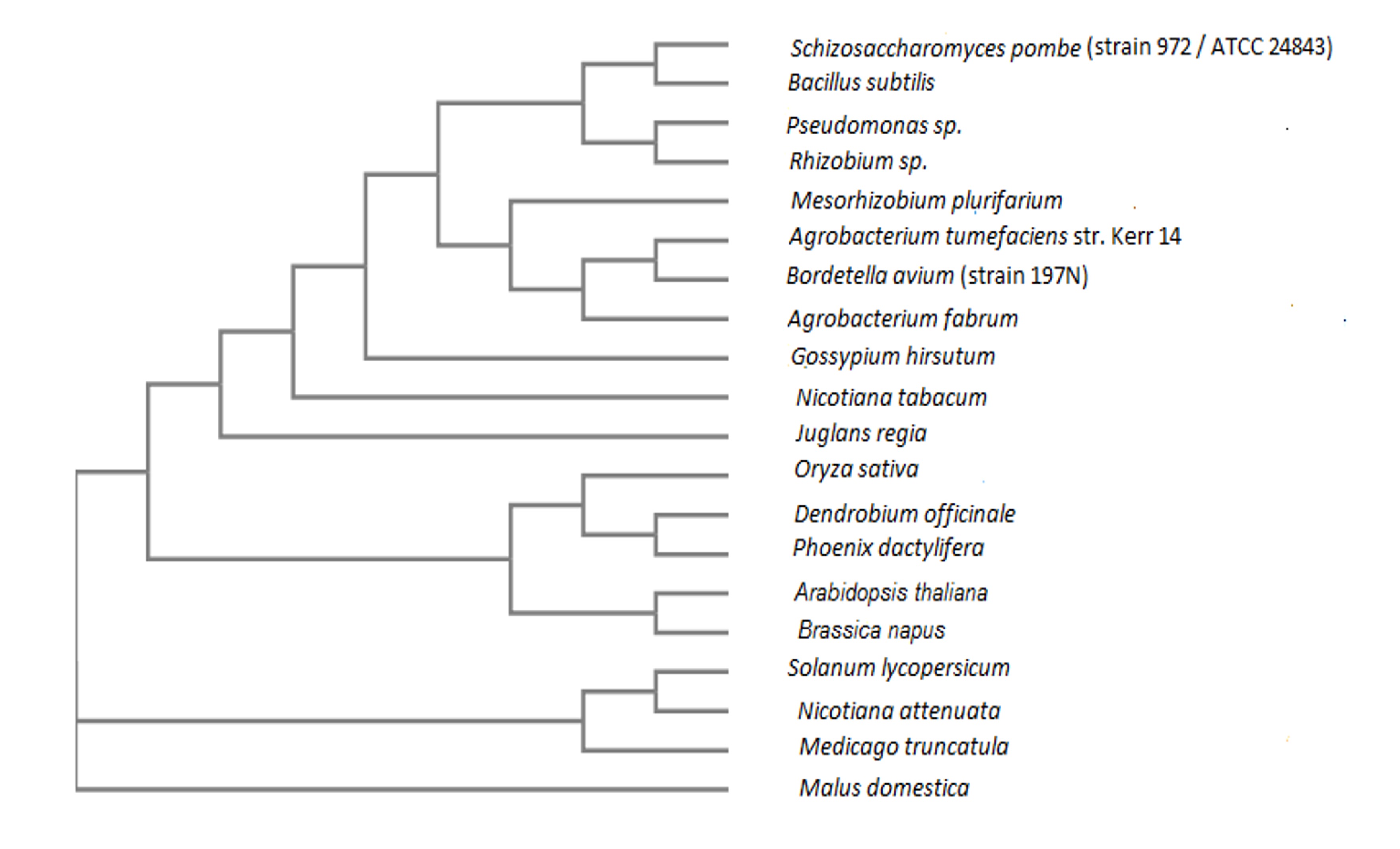
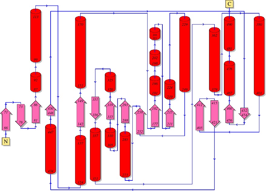
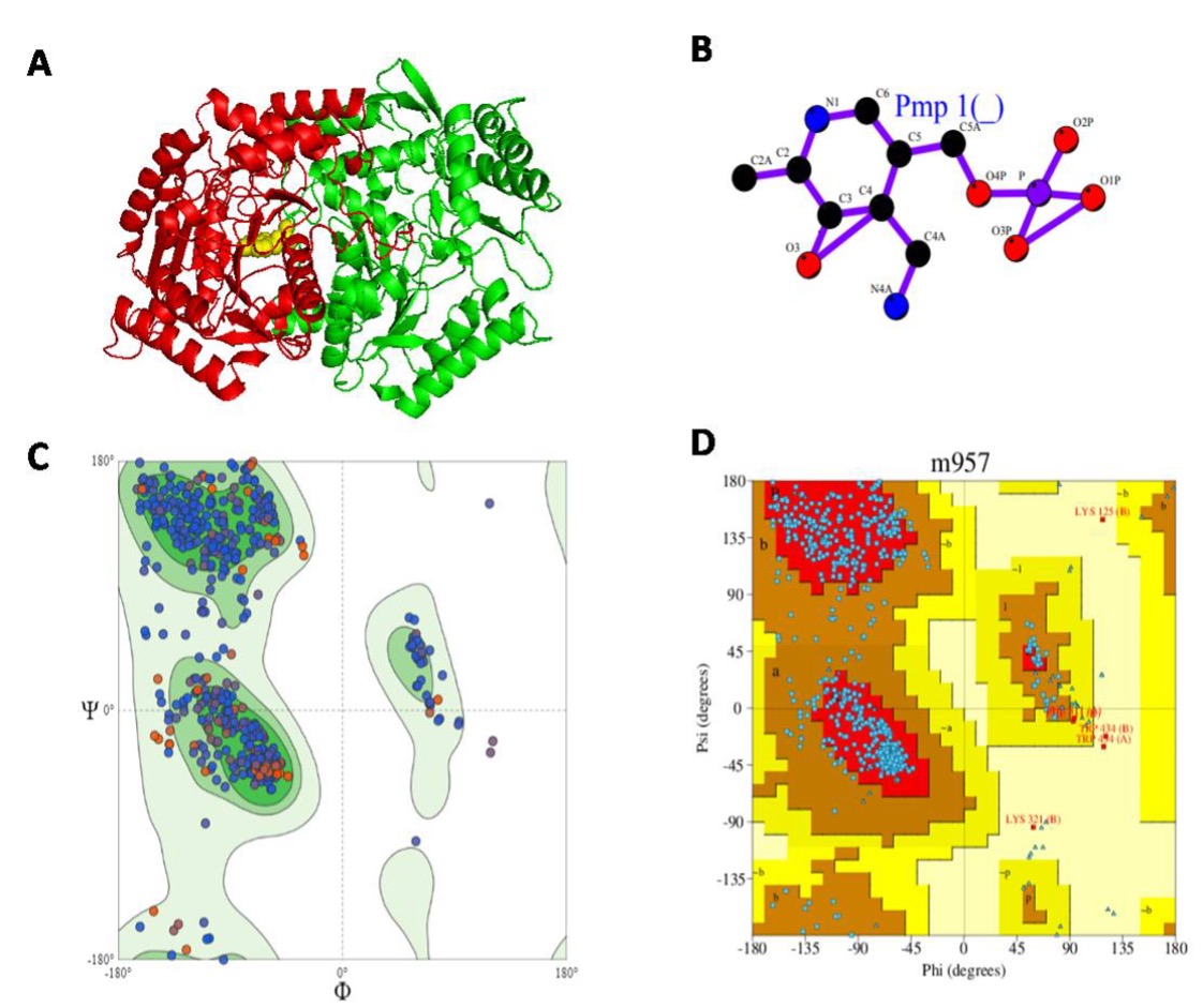
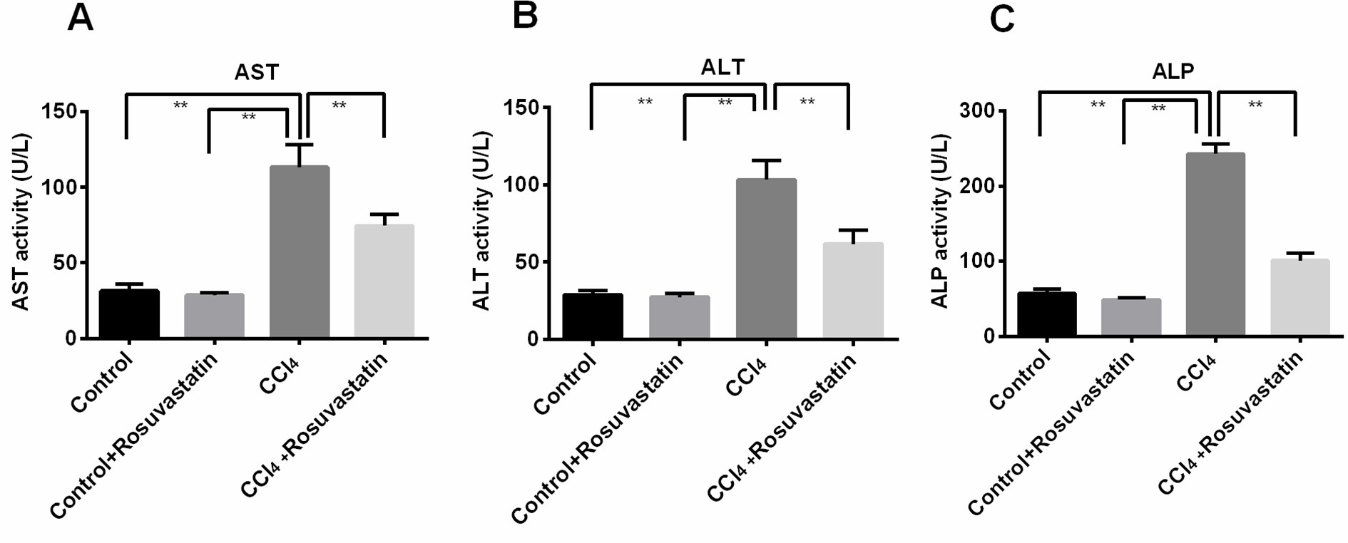
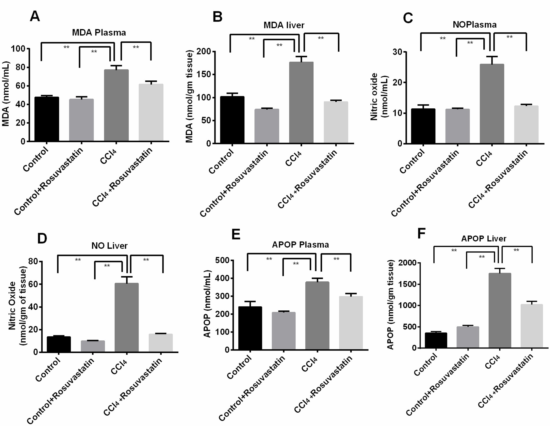
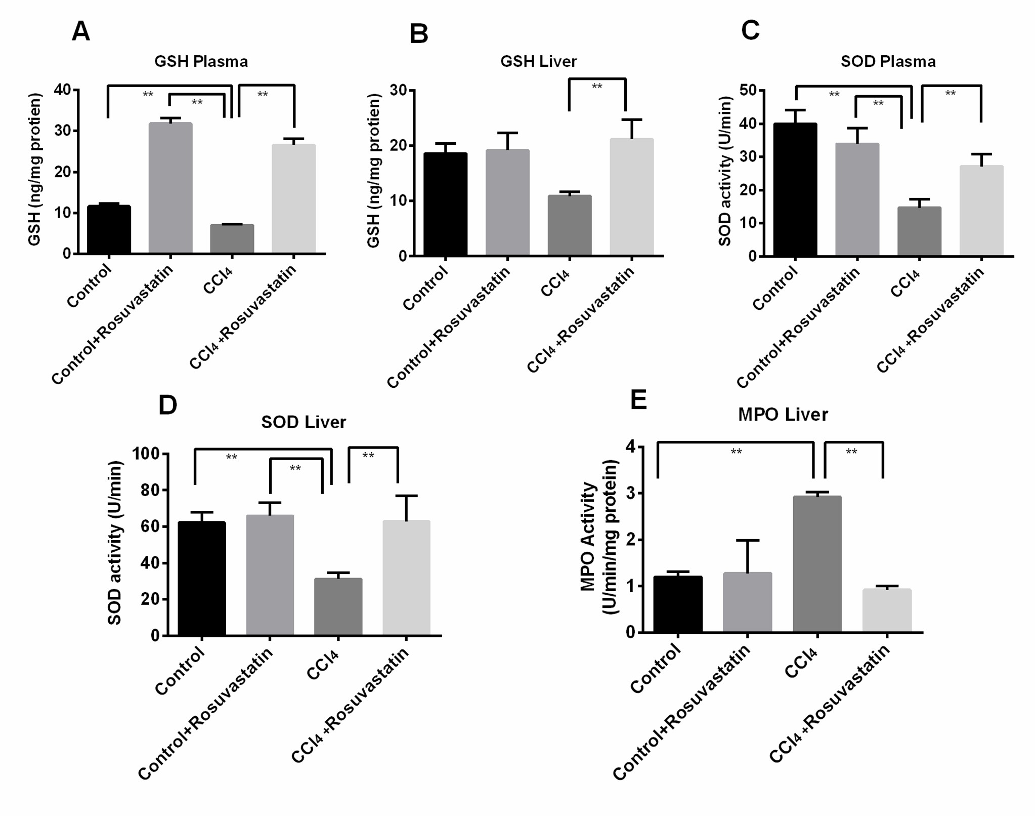
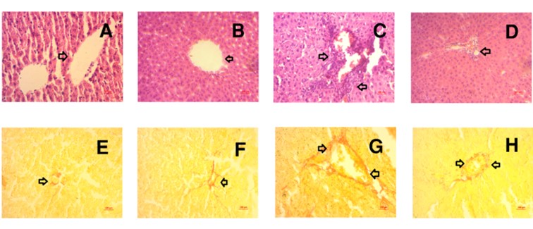
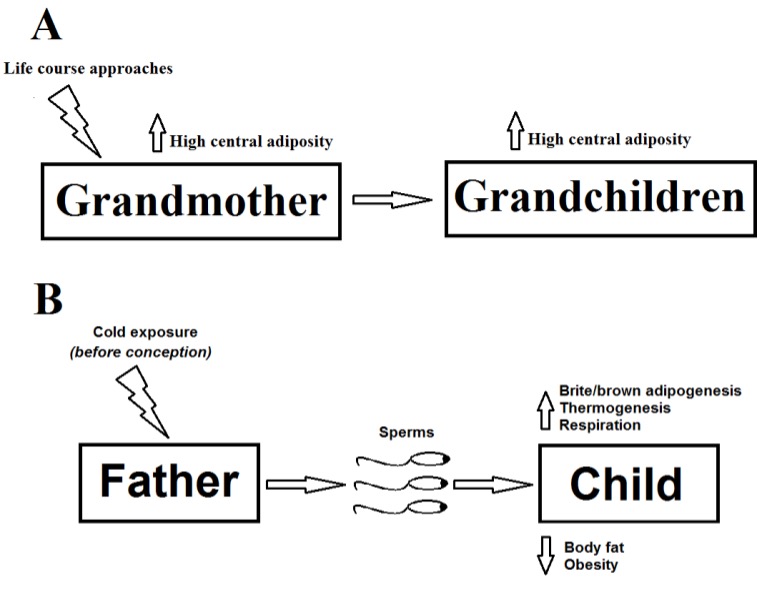
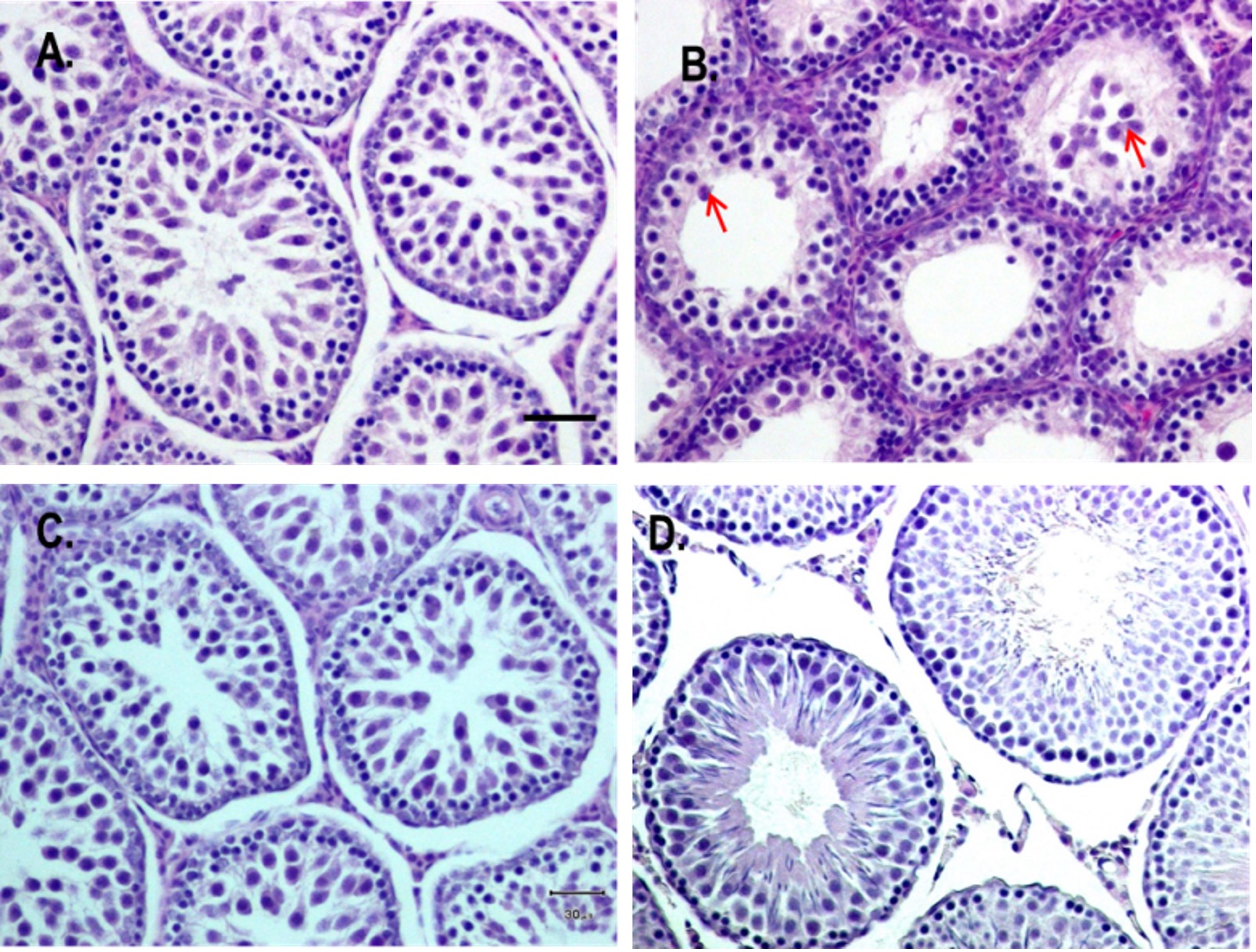
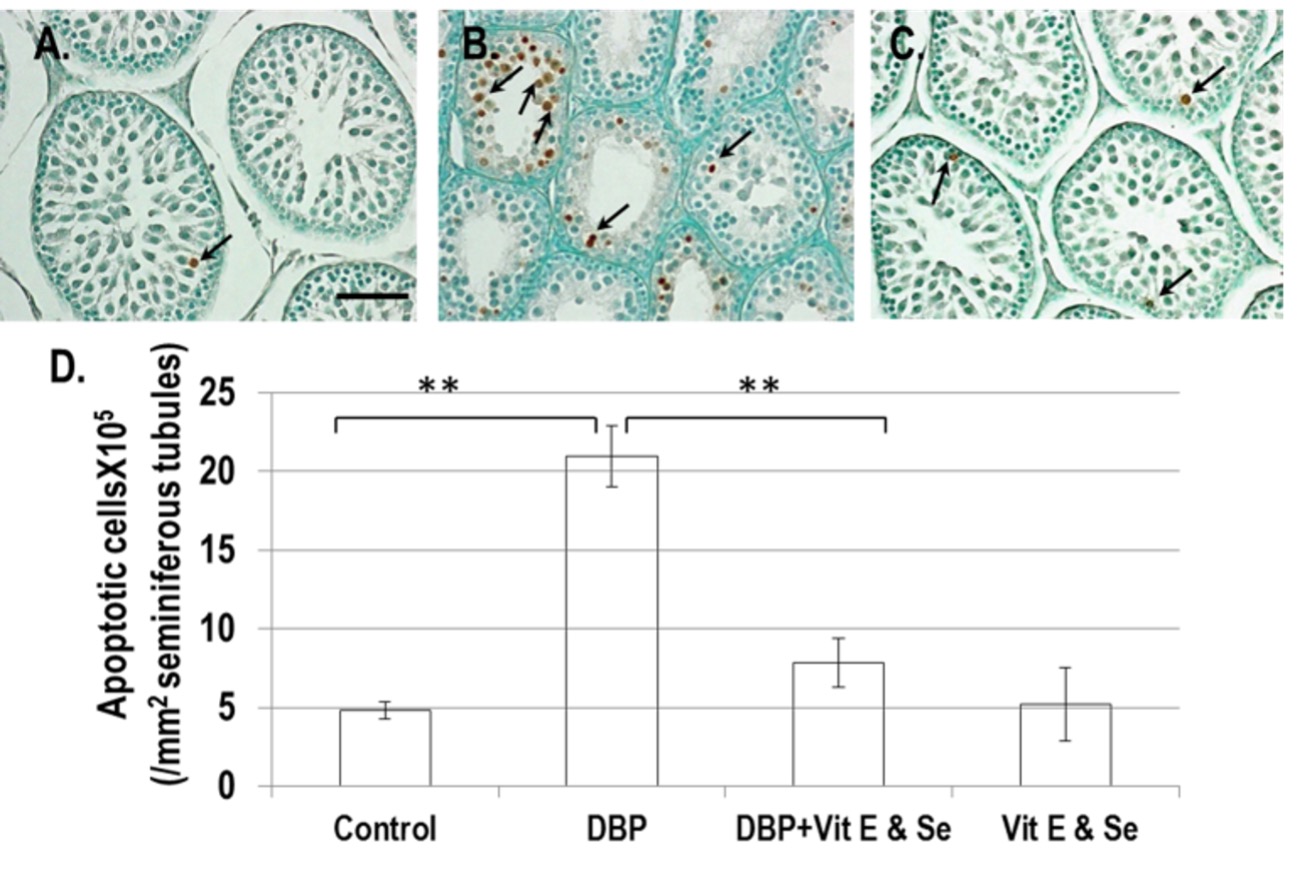
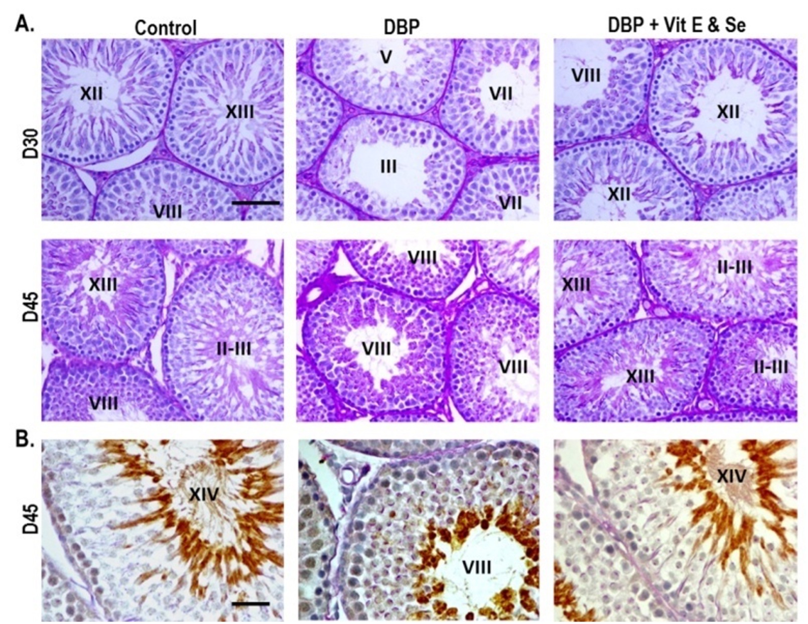
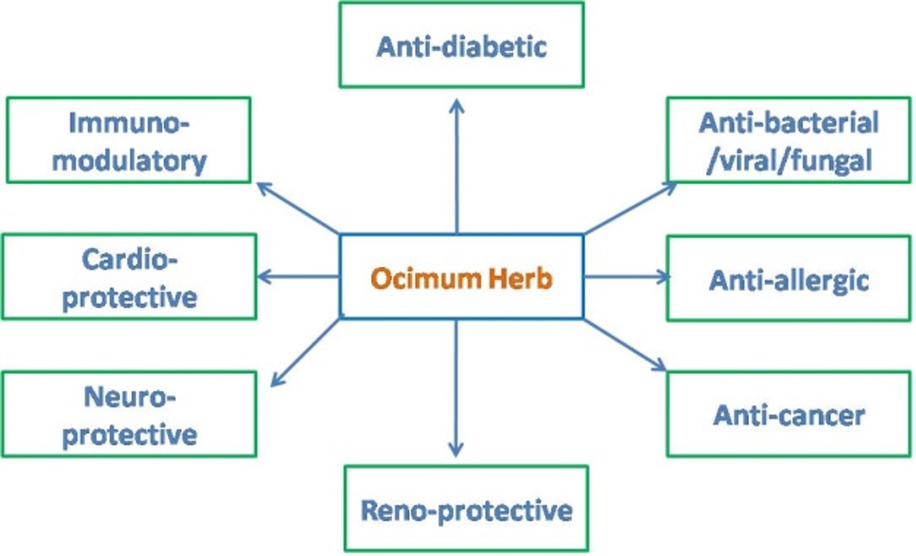
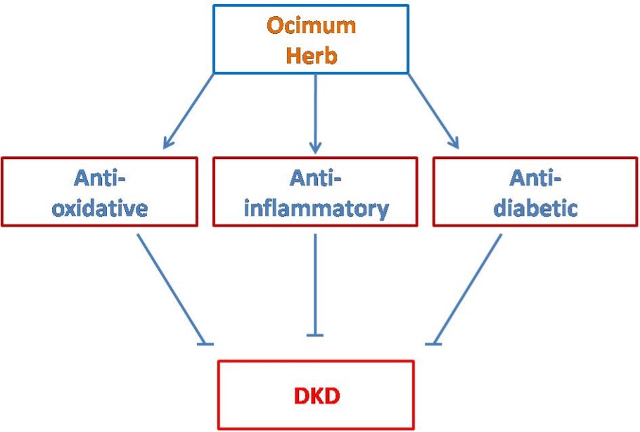
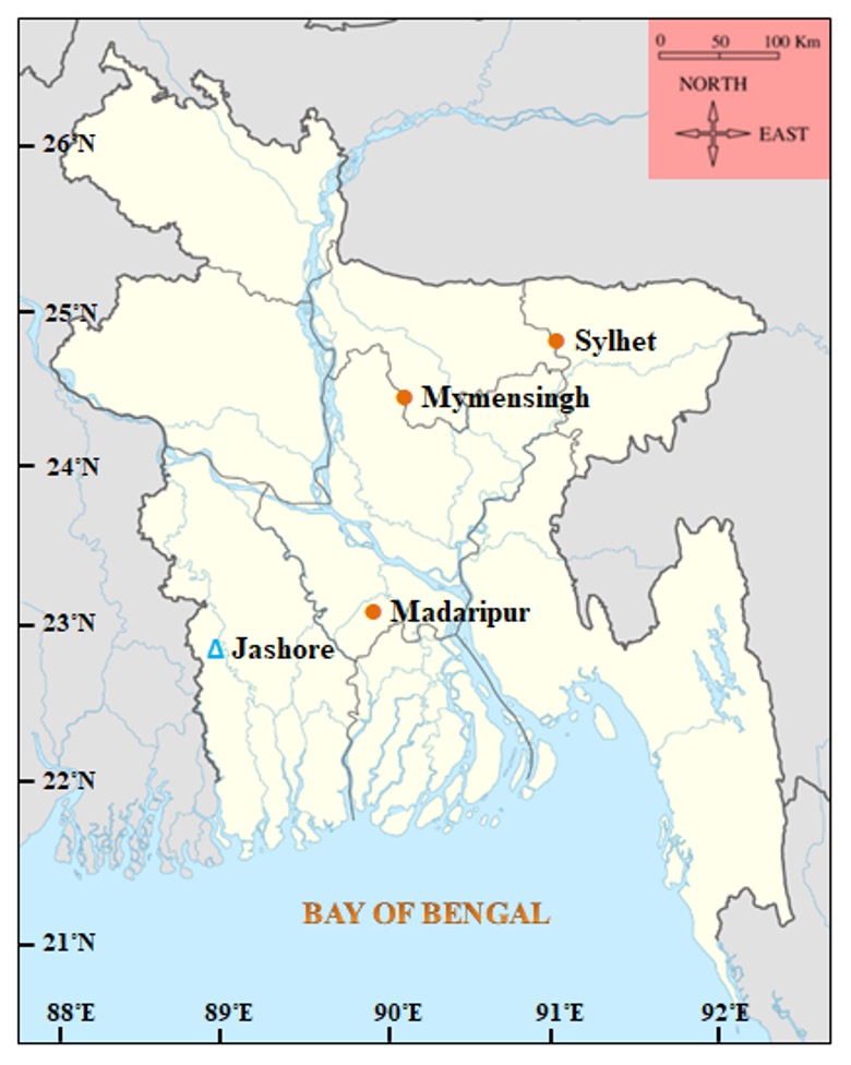
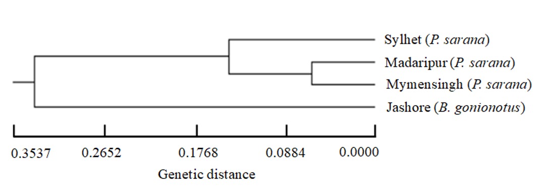
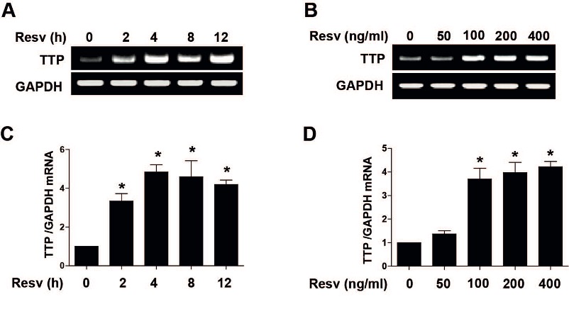
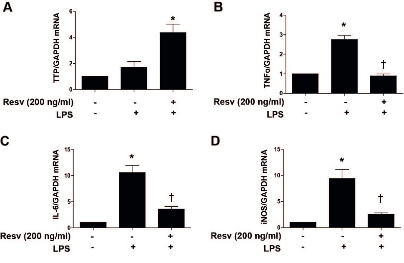
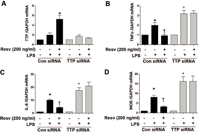
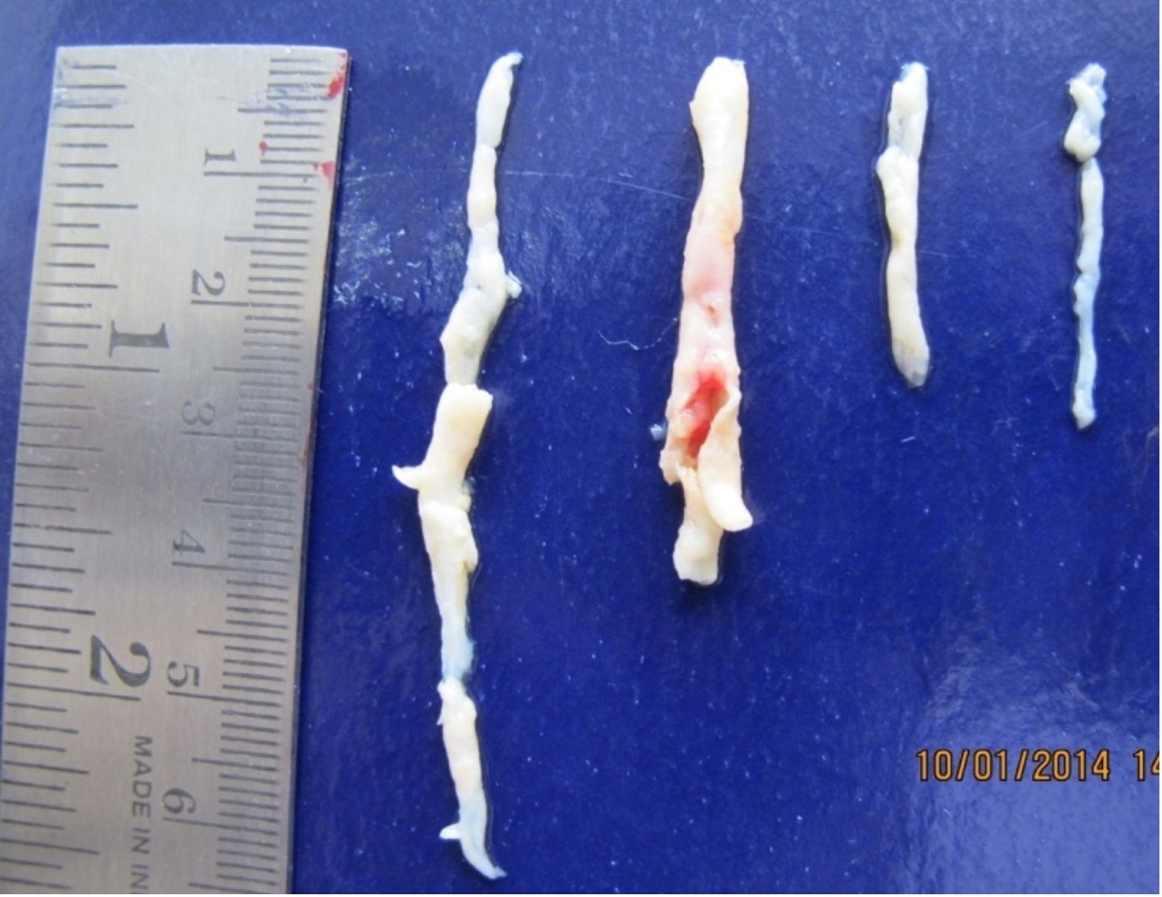
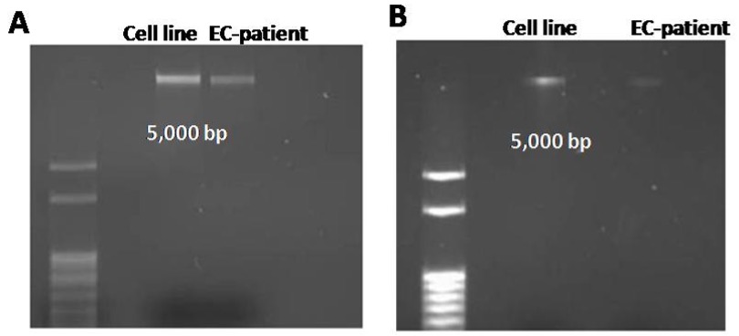
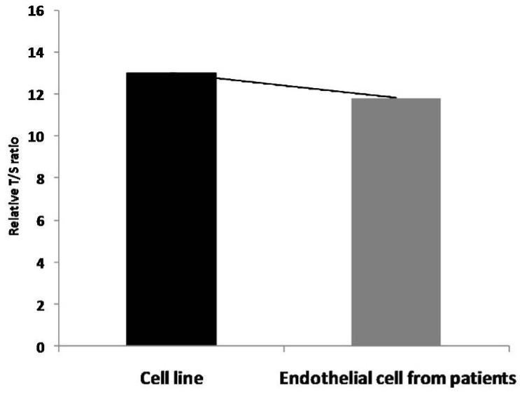
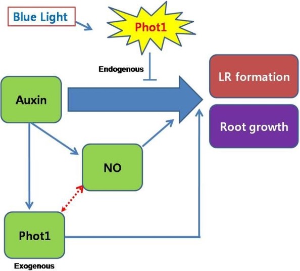
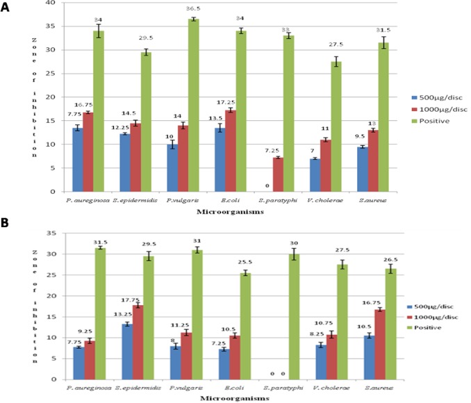
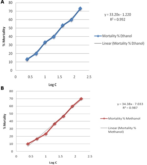