HTTP 200 OK
Allow: GET, HEAD, OPTIONS
Content-Type: application/json
Vary: Accept
{
"count": 319,
"next": "https://jabet.bsmiab.org/articles/?format=api&page=31",
"previous": "https://jabet.bsmiab.org/articles/?format=api&page=29",
"results": [
{
"id": 63,
"slug": "178-1555480720-influence-of-socio-demographic-factors-on-the-diarrheal-disease-management-approaches-taken-by-two-distinct-communities-of-bangladesh",
"featured": false,
"slider": false,
"issue": "Vol2 Issue2",
"type": "original_article",
"manuscript_id": "178-1555480720",
"recieved": "2019-03-19",
"revised": null,
"accepted": "2019-05-16",
"published": "2019-05-21",
"pdf_file": "https://jabet.bsmiab.org/media/pdf_file/2023/22/178-1555480720.pdf",
"title": "Influence of socio-demographic factors on the diarrheal disease management approaches taken by two distinct communities of Bangladesh",
"abstract": "<p>Diarrhea is one of the major determinants of childhood mortality in the world. This study was aimed to provide a demographically representative description on the influence of socio-demographic factors e.g. caretakers’ education, occupation, family income and living standard on the diarrheal disease management approaches taken by the slum dwellers and middle class families of Bangladesh. We have visited 90 middle class families and 120 slum dwellers to obtain information. Children of slum dwellers are more likely to be affected by diarrhea. In both classes, significantly more females were affected by diarrhea than males. This scenario is even more prominent among slum dwellers, where 1.5 times more females were affected by diarrhea than their male counterparts. As a primary approach to manage diarrhea, 63.8% caretakers chose Oral rehydration solution (ORS) whereas 31.5% preferred the salt-molasses fluid. All caretakers knew the use of ORS and antibiotics as a preventive measure against diarrhea. However, this scenario dramatically turned when the caretakers were asked whether they know how to prepare ORS. All the caretakers (100%) in middle class families knew how to prepare ORS in contrast to only 25% caretakers among the slum dwellers. Private sectors specially pharmacies were often the first line of health care in both of these classes during diarrhea. But this is most prevalent among the middle class families (50%), compared to the slum dwellers (35%). Finally, it is apparent that the education, family income, living standard and good food help the middle class families to fight diarrhea more efficiently and scientifically than the slum dwellers.</p>",
"journal_reference": "J Adv Biotechnol Exp Ther. 2019; 2(2): 78-86.",
"academic_editor": "Dr. Masud Parvez, Inje University, South Korea.",
"cite_info": "Khan G, Akter N, Uddin MD, etal. Influence of socio-demographic factors on the diarrheal disease \r\nmanagement approaches taken by two distinct communities of Bangladesh. J Adv Biotechnol Exp Ther. 2019; 2(2): 78-86.",
"keywords": [
"socio-demographic factors",
"Diarrhea management",
"education",
"ORS preparation.",
"family income",
"living standard"
],
"DOI": "10.5455/jabet.2019.d29",
"sections": [
{
"section_number": 1,
"section_title": "INTRODUCTION",
"body": "<p>Diarrhea the second leading cause of under-five child mortality, responsible for almost 2 million worldwide deaths per year [<a href=\"#\">1,2</a>]. Globally, up to 5 billion of diarrhea cases occur every year [<a href=\"#r-3\">3, 4, 5</a>]. This incident is most common in developing countries, where children at their younger ages suffer from diarrhea on average three times per year [<a href=\"#r-3\">3</a>]. High frequency of diarrheal episodes is the most common cause of malnutrition in those children [<a href=\"#r-3\">3</a>]. Other long term side effects of frequent episodes of diarrhea include poor intellectual development and stunted growth [<a href=\"#r-6\">6</a>].<br />\r\nDespite the increasing incidence of diarrhea and public concern about hygiene, there are still significant discrepancies among the caretakers about the knowledge and management of diarrhea. Socio-demographic factors such as caretaker’s education, employment status, family size and income, place of residence are strongly linked to the high frequency of diarrhea and play a crucial role on the approaches chosen by caretakers to efficiently manage diarrhea. However, majority of the death, less than one-third of children in sub-Saharan Africa and South Asia, caused by diarrhea can be prevented by proper management approaches e.g. continued feeding, timely use of oral rehydration solution (ORS) [<a href=\"#r-7\">7</a>]. Significant reduction in childhood diarrheal death by appropriate management practices on the basis of socioeconomic status, gender and where the children live is a renewed effort in response to millennium development goal #4 which is targeted to achieve by two-thirds by the year 2015 in developing countries [<a href=\"#r-8\">8</a>].<br />\r\nWith the right combination of water and sanitation facilities, with behavioral characteristics of the household, diarrheal disease is almost preventable. Two terms can be used (economic/ behavioral and infrastructure) for identifying socio-demographic factors linked with the incidence and severity of diarrheal illness [<a href=\"#r-9\">9</a>]. The economic/behavior view highlights the attention and interpretation of household behavior. The lack of awareness and insufficient knowledge of mothers about hygiene leads to frequent exposure of children to diarrhea [<a href=\"#r-10\">10-12</a>]. The second perspective, infrastructural intervention, has very less effect in lowering diarrhea than the behavioral factors.<br />\r\nOur present study tried to focus on the management approaches against diarrheal diseases taken by the slum dwellers and middle class families of Bangladesh, which is a completely new aspect. It is apparent that the education, family income, living standard and good food help the middle class families to fight against diarrhea more efficiently and scientifically than the lower families living in the slums. We have identified varied patterns of practice and equity by geographic location. The survey was stratified by urban and inner-city slum and non-slum populations. Finally, we tried to provide a baseline to monitor the effectiveness of chosen practices caused by education, family income, living standard and good food throughout major cities of Bangladesh to competently fight against diarrhea.</p>"
},
{
"section_number": 2,
"section_title": "MATERIALS AND METHODS",
"body": "<p><strong>Study area and Sampling</strong><br />\r\nAbout 120 slums dwellers and 90 households from the middle class families within the Dhaka and Chittagong division were interviewed. The Dhaka Metropolitan Area (DMA) was chosen for studying the slum dwellers. Four slums located in Mirpur, Mohammadpur, Tejgaog and Kamalapur were chosen to conduct the household survey. On average, each slum had 30 households. The middle class families are located both in Dhaka and Chittagong divisions. This survey was conducted from 1st May to 31st July 2018. The principal respondents to questionnaire were women because it was felt they were more aware of the children’s health condition compared to the men of the household. The data were collected on household members, household status, household knowledge on diarrhea and its management, the cost of treating diarrhea, awareness of and practices relating to personal hygiene.</p>\r\n\r\n<p> </p>\r\n\r\n<p><strong>Measurement</strong><br />\r\nThe interview was conducted with the proper consent from caretakers in every household we surveyed. Our questionnaire covered socio-demographic factors and management approaches taken by the household during the episode of diarrhea. Socioeconomic status was determined by considering education, monthly income, dwelling characteristics, and other household characteristics that are related to wealth status.</p>\r\n\r\n<p> </p>\r\n\r\n<p><strong>Survey methodology</strong><br />\r\nThe authors followed the recommendations of the Division for the Control of Diarrheal and Acute Respiratory Disease of the World Health Organization (WHO, 1995) in the designing of the survey on diarrhea. The opportunity of having direct contact with the target population (care seekers) was utilized.</p>\r\n\r\n<p> </p>\r\n\r\n<p><strong>Ethical approval</strong><br />\r\nEthical approval for this analysis was obtained from the patients and caretaker. All participants gave informed consent and signed a consent form prior to participation in the study.</p>\r\n\r\n<p> </p>\r\n\r\n<p><strong>Data analysis</strong><br />\r\nData were primarily analyzed using IBM SPSS V22 and Microsoft Excel 2016, cleaned for any inconsistencies and analyzed for standard distribution measurements. P-value < 0.05 was considered significant (not shown here).</p>"
},
{
"section_number": 3,
"section_title": "RESULTS",
"body": "<p><strong>Prevalence of diarrhea</strong><br />\r\nIn total, we have visited 90 middle class families and 120 slum dwellers, who were suffered from diarrhea in recent times, both in Dhaka and Chittagong district to obtain information. Diarrhea occurrence is higher in children and adults with age group between 1-10 and 40-60 years, respectively (<a href=\"#figure1\">Figure 1</a>). Children of slum dwellers are more likely to be affected by diarrhea than the children of middle class families. In all households visited in the slums, 36% (30) children under 10 years of age were reported to have suffered from diarrhea compared to only 6% (4) children in middle class families (<a href=\"#figure1\">Figure 1</a>). In both cases, significantly more females were affected by diarrhea than males. This scenario was even more prominent among slum dwellers, where 1.5 times more females were affected by diarrhea than their male counterparts (<a href=\"#figure2\">Figure 2</a>).</p>\r\n\r\n<div id=\"figure1\">\r\n<figure class=\"image\"><img alt=\"\" height=\"119\" src=\"/media/article_images/2024/43/23/178-1555480720-Figure1.jpg\" width=\"335\" />\r\n<figcaption><strong>Figure 1</strong>. Comparison of the age distribution of patients among middle class families and slum dwellers. The occurrence of diarrhea is higher in children and adults with age group between 1-10 and 40-60 years.</figcaption>\r\n</figure>\r\n</div>\r\n\r\n<div id=\"figure2\">\r\n<figure class=\"image\"><img alt=\"\" height=\"125\" src=\"/media/article_images/2024/43/23/178-1555480720-Figure2.jpg\" width=\"335\" />\r\n<figcaption><strong>Figure 2</strong>. Comparison of the gender distribution of patients among middle class families and slum dwellers. In both types of families, significantly more females were affected by diarrhea than males, which was even more prominent among slum dwellers.</figcaption>\r\n</figure>\r\n\r\n<p> </p>\r\n</div>\r\n\r\n<p><strong>Demographic characteristics of the study population</strong><br />\r\nThere were positive correlations between caretaker’s knowledge about diarrhea and mothers’ age and education, family size and husbands’ income. A caretaker with higher education and family income has better knowledge about preventing and managing diarrhea. These findings are true throughout our entire survey. The management of diarrhea is significantly better among the middle class families due to their higher family income and advanced education compared to the slum dwellers (<a href=\"#Table-1\">Table 1</a>).</p>\r\n\r\n<div id=\"Table-1\">\r\n<p><a href=\"https://jabet.bsmiab.org/table/178-1555480720-table1/\">Table-1</a><strong>Table 1</strong>. Socio-demographic factors.</p>\r\n\r\n<p> </p>\r\n</div>\r\n\r\n<p><strong>Knowledge on diarrhea management</strong><br />\r\n<em>i) Caretaker’s knowledge about the preparation and use of ORS</em><br />\r\nThe most common response of the caretakers about ORS was that it mainly decreases the frequency of diarrhea while in some cases frequency may increase. Caretakers were also asked what is the best for managing diarrhea. 63.8% chose ORS and 31.5% preferred the salt-molasses fluid. The use of rice-based fluid was not well known or not preferred as only 0.2% chose this alternative (<a href=\"#figure3\">Figure 3</a>). All caretakers (100%) knew the use of ORS and antibiotics as a preventive measure against diarrhea in both classes. However, this scenario dramatically turned when the caretakers were asked whether they know how to prepare ORS. All the caretakers (100%) in middle class families knew how to prepare ORS. In contrast, only 25% caretakers among the slum dwellers knew how to prepare ORS (<a href=\"#figure4\">Figure 4</a>). Three-fourth of the caretakers was unable to mention all the four steps of correct preparation of ORS solution. The main reason for using an incorrect volume of water during the preparation of the ORS solution was due to the use of local uncalibrated water-measuring devices. Many caretakers among slum dwellers gave the wrong volume of ORS solution to the patients during diarrhea.</p>\r\n\r\n<div id=\"figure3\">\r\n<figure class=\"image\"><img alt=\"\" height=\"289\" src=\"/media/article_images/2024/43/23/178-1555480720-Figure3.jpg\" width=\"270\" />\r\n<figcaption><strong>Figure 3</strong>. A combined view of the choice of fluid intake in both of these communities. Most of the caretakers from both families preferred ORS to provide proper hydration for patients during an episode of diarrhea. One-third of the respondents preferred salt-molasses, but very few knew about the rice-based fluids.</figcaption>\r\n</figure>\r\n</div>\r\n\r\n<div id=\"figure4\">\r\n<figure class=\"image\"><img alt=\"\" height=\"206\" src=\"/media/article_images/2024/43/23/178-1555480720-Figure4.jpg\" width=\"316\" />\r\n<figcaption><strong>Figure 4</strong>. Comparison of the knowledge on how to prepare ORS among the middle class families and slum dwellers. All the caretakers in middle class families knew how to prepare ORS compared to the families living in slums where only one-fourth of them knew how to prepare ORS.</figcaption>\r\n</figure>\r\n</div>\r\n\r\n<p> </p>\r\n\r\n<p><em>ii) Caretaker’s knowledge about diarrhea and its danger signs</em><br />\r\nMost of the caretakers, in both classes, were aware of thin watery diarrhea being the most serious type of diarrhea. However, almost 90% caretakers in middle class families pointed out that thin watery stool with repeated vomiting and febrile conditions as indicative of more serious diarrhea. Among slum dwellers, only 30% of the caretakers were aware of other dangerous signs of dehydration such as sunken eyes, thirst (eagerly drinking), skin pinch receding slowly, the passage of concentrated or dark-colored urine, a drowsy child and the child not getting better after three days (<a href=\"#figure5\">Figure 5</a>)</p>\r\n\r\n<div id=\"figure5\">\r\n<figure class=\"image\"><img alt=\"\" height=\"359\" src=\"/media/article_images/2024/43/23/178-1555480720-Figure5.jpg\" width=\"500\" />\r\n<figcaption><strong>Figure 5</strong>. Comparison of the knowledge about the danger signs of diarrhea among the middle class families and slum dwellers. We investigated whether caretakers recognized danger signs of diarrhea and if they knew when to bring the patients to the health facility. We found that caretakers’ knowledge on the danger sign of diarrhea almost 3 times higher among middle class families compared to the caretakers living in the slum.</figcaption>\r\n</figure>\r\n</div>\r\n\r\n<p> </p>\r\n\r\n<p><strong>Caretaker behavior during an episode of diarrhea</strong><br />\r\nAppropriate case management of diarrhea at home is one of the capital importance because this intervention reduces significantly the risk of dying by dehydration. The perceived seriousness of diarrhea as a life-threatening condition made 60.5% of caretakers who suffered from diarrhea in the two weeks prior to the study seek care outside the house.<br />\r\nThere is still a gap between knowledge of diarrhea management and its practice. For the categories of suspending and giving less fluid, the percentage of caretakers practicing it was higher among slum dwellers than the middle class families. While for the categories of the same amount and more fluid, the proportion of caretakers practicing it was higher among middle class families than the slum dwellers (<a href=\"#Table-2\">Table 2</a>). A similar pattern can be seen concerning food intake. In general, the management of diarrhea has improved significantly among middle class families, particularly concerning fluid and food intake. Currently, about 2.3 times more caretakers among middle class families give at least the same amount of food to their patients during an episode of diarrhea as compared to slum dwellers (<a href=\"#Table-2\">Table 2</a>).</p>\r\n\r\n<div id=\"Table-2\">\r\n<p><a href=\"https://jabet.bsmiab.org/table/178-1555480720-table2/\">Table-2</a><strong>Table 2</strong>. Caretaker’s knowledge and practice on fluids and food intake during diarrhea.</p>\r\n\r\n<p> </p>\r\n</div>\r\n\r\n<p><strong>Care-seeking behavior of caretakers in case of diarrhea</strong><br />\r\nDisparities based on income in care seeking behavior were identified among the slum dwellers and middle class families. Surprisingly, care-seeking behavior shows strong similarities in these two classes of society. In middle class families, 50% of the patients receive their medications from pharmacy and drug dealers, whereas only 35% patients from slum receive their treatment from the pharmacy and drug dealers (<a href=\"#figure6\">Figure 6</a>). However, this scenario is completely opposite in case of receiving the treatment from government services, 42% slum dwellers receive government services compared to only 30% of patients from the middle class families (<a href=\"#figure6\">Figure 6</a>). It is noteworthy that, no patients from the middle class families receive their treatment from traditional healers (<a href=\"#Table-3\">Table 3</a>). We have also investigated the pattern of antibiotic utilization in both of these communities. In this survey, we have found that 85% of the patients in middle class families consumed antibiotics compared to only 52% of the patients from slums (<a href=\"#figure7\">Figure 7</a>).</p>\r\n\r\n<div id=\"figure6\">\r\n<figure class=\"image\"><img alt=\"\" height=\"289\" src=\"/media/article_images/2024/43/23/178-1555480720-Figure6.jpg\" width=\"500\" />\r\n<figcaption><strong>Figure 6</strong>. Comparison in the care-seeking behavior among middle class families and slum dweller. We found that most of the caretakers did not consult a licensed health provider and drug shops were often the first line of health care in both classes of the family (50% and 35% cases from middle class families and slum dwellers, respectively).</figcaption>\r\n</figure>\r\n</div>\r\n\r\n<div id=\"figure7\">\r\n<figure class=\"image\"><img alt=\"\" height=\"222\" src=\"/media/article_images/2024/43/23/178-1555480720-Figure7.jpg\" width=\"500\" />\r\n<figcaption><strong>Figure 7</strong>. Comparison of the use of antibiotics among middle class families and slum dwellers. Here, almost 85% of the patients in middle class families consumed antibiotics compared to only 52% of the patients from slums.</figcaption>\r\n</figure>\r\n</div>\r\n\r\n<div id=\"Table-3\">\r\n<p><a href=\"https://jabet.bsmiab.org/table/178-1555480720-table3/\">Table-3</a><strong>Table 3</strong>. Care-seeking behavior of the patients from middle class families and slums during an episode of diarrhea.</p>\r\n\r\n<p> </p>\r\n</div>"
},
{
"section_number": 4,
"section_title": "DISCUSSION",
"body": "<p>Socio-demographic factors such as caretaker’s education and occupation, caretaker’s employment status, family size and income are linked with caretaker’s knowledge about diarrhea and its management. Although caretakers from both middle class families and slum dwellers were aware of diarrhea and its home management, the level of awareness was insufficient among the slum dwellers. In total, 90 middle class families and 120 slum dwellers were visited to obtain information. Most of the caretakers (80%) among the middle class have received advanced education, whereas, most of the caretakers (58%) among the slum dwellers studied only up to primary level (<a href=\"#Table-1\">Table 1</a>). In terms of household income, the middle class family’s income almost 4 times higher on an average compared to the slum dwellers. Children of slum dwellers are more likely to be affected by diarrhea than the children of middle class families. In all households visited in the slums, 36% of children under 10 years of age were reported to have suffered from diarrhea compared to only 6% of children in middle class families (<a href=\"#figure1\">Figure 1</a>). In both cases, significantly more females were affected by diarrhea than males. This scenario is even more prominent among slum dwellers, where 1.5 times more females were affected by diarrhea than their male counterparts (<a href=\"#figure2\">Figure 2</a>).<br />\r\nEffective management of diseases depends on correct knowledge on causes, symptoms and treatments. The frequency of correct answers in the interviews increased with the level of education. When we asked about the management of diarrhea, most of the caretakers from the slums were unable to mention all the steps for the correct and complete preparation of ORS solution. This study found that approximately 100% caretakers from the middle class families and only 25% of the caretakers from the slums were able to prepare ORS solution correctly and completely (<a href=\"#figure4\">Figure 4</a>). This study shows strong similarity with the studies conducted on the prior knowledge of the mothers which found approximately 20% to 50% of the mothers could prepare ORS solution correctly and completely [<a href=\"#r-13\">13-16</a>]. This might be due to caretaker’s lack of prior experience, a lack of proper education about the concerned matters and their ethnicity itself. Regarding the use of ORS, almost all the caretakers among the slum dwellers were lacking the knowledge of giving the correct volume of ORS to the patients with diarrhea which is completely different from the caretakers of the middle class families. The poor knowledge among the caretakers about the role of ORS in diarrhea is due to their poor knowledge about the concept of dehydration and rehydration.<br />\r\nThe ability of caretakers to recognize signs and symptoms of severe illness is believed to be an important predictor of timely and appropriate care seeking in developing countries. An intervention directed at improving caretaker recognition of danger signs in their patients resulted in substantial increases the timely use of qualified providers [<a href=\"#r-17\">17, 18</a>]. The knowledge about the danger signs of diarrhea, determined by our questionnaire, are almost 3 times higher among middle class families compared to the knowledge of caretakers living in the slum (<a href=\"#figure5\">Figure 5</a>).<br />\r\nDisparities based on income in care seeking behavior were identified among the slum dwellers and middle class families [<a href=\"#r-19\">19</a>]. This was true for any provider as well as for a licensed allopath from private sectors with significant trends favoring higher income households occurring throughout Bangladesh. This survey documents that throughout Bangladesh health-seeking behaviors for diarrhea are dominated by utilization of private sector providers (<a href=\"#Table-3\">Table 3</a>). The most crucial factor within urban households for seeing a licensed allopath was higher education of caretakers, higher income and longer duration of illness. In slums, the most important predictors were the longer duration of illness, age of the child, and caretaker’s education.<br />\r\nEven though nearly all caretakers sought care outside the home when their patients had diarrhea, the majority did not consult a licensed health provider, a trend that is increasingly reported in Bangladesh [<a href=\"#r-20\">20</a>] (<a href=\"#figure6\">Figure 6</a>). Drug shops were often the first line of health care in these both classes of the family. But this is most prevalent among the middle class families (50%), compared to the slum dwellers (35%) (Figure 6). Unexpectedly, it was found that slum when compared with middle class urban families, were twice as likely to seek services from the public sector. Surprisingly it was found that more patients among slum dwellers (42%) seek for government services than the patients from middle class families (30%) (<a href=\"#figure6\">Figure 6</a>). The reasons behind this may be hassle associated with the government care facilities. This survey did not inquire about reasons for choosing a particular provider; however, earlier surveys have documented that the use of public services is hindered by the unavailability of health providers and unofficial payments [<a href=\"#r-21\">21</a>]. However, these utilization patterns, in part, can be explained by the fact that private providers far outnumber the other sectors, are easily accessed, and are available at all hours of the day and late into evenings. Government clinics and hospitals, in contrast, require longer waiting, complex registration and accessible within limited daytime hours. In addition, caretakers usually seek a quick cure for their children and less interested in complex governmental settings unless a child is perceived as severely ill. Drugs are more efficiently and simply obtained through private providers as well.<br />\r\nFinally, the current survey found that most patients who received care from a hospital or health center were appropriately treated with ORS, and approximately half received antibiotics. These rates are compatible with other reports of antibiotics, zinc and ORS usage in rural Bangladesh [<a href=\"#r-22\">22</a>]. Many children in the older age groups were also given antibiotics. Antibiotics are extensively used in diarrhea case management [<a href=\"#r-23\">23-24</a>], even though they are only recommended in a few cases of diarrhea. The tendency to use antibiotics is even more serious among the caretaker from middle class families. In the present study over 85% of the caretakers from the middle class families used antibiotics together with ORS compared to 52% in case of slum dwellers (<a href=\"#figure7\">Figure 7</a>). Antibiotics are often attained from other sources than from trained medical personnel [<a href=\"#r-25\">25</a>]. Overuse can potentially cause harmful side effects and contributes to bacterial resistance development [<a href=\"#r-26\">26</a>].</p>"
},
{
"section_number": 5,
"section_title": "CONCLUSIONS",
"body": "<p>The improvement in the prevalence and management of diarrhea, as found in the study, seem to be predominantly due to extensive awareness-raising and educational activities. The present study finds that socio-economic variables such as education of the caretaker, family income, site of their residence play a crucial role in the management of diarrhea (<a href=\"#figure8\">Figure 8</a>). The study findings show that in relatively short time a significant reduction in mortality due to diarrhea through behavior change in a mostly poor and illiterate population can be achieved if intensive sensibilization and education strategies are deployed.</p>\r\n\r\n<div id=\"figure8\">\r\n<figure class=\"image\"><img alt=\"\" height=\"248\" src=\"/media/article_images/2024/43/23/178-1555480720-Figure8.jpg\" width=\"500\" />\r\n<figcaption><strong>Figure 8</strong>. Schematic representation of summary. Our study suggests that knowledge about diarrhea and its management was poor among the caretakers from the slum dwellers compared to the middle class families. Although caretakers from slum dwellers were aware of diarrhea and its home management, their knowledge on vital issues such as complete and correct preparation of ORS, danger signs of dehydration and actual role of oral rehydration fluids during diarrhea was very poor due to the associated socio-demographic factors.</figcaption>\r\n</figure>\r\n</div>"
},
{
"section_number": 6,
"section_title": "ACKNOWLEDGEMENT",
"body": "<p>This research received no external funding.</p>"
},
{
"section_number": 7,
"section_title": "AUTHOR CONTRIBUTIONS",
"body": "<p>GK and RUZ were involved in conception and design of the experiments. GK, NA and DU contributed to perform the experiments. GK and RUZ analyzed data and contributed to drafting the article. NA, DU and RUZ contributed to revising it critically for important intellectual content. RUZ made the final approval of the version to be published.</p>"
},
{
"section_number": 8,
"section_title": "CONFLICTS OF INTEREST",
"body": "<p>The author declares that no conflict of interest exists.</p>"
}
],
"figures": [
{
"figure": "https://jabet.bsmiab.org/media/article_images/2024/43/23/178-1555480720-Figure1.jpg",
"caption": "Figure 1. Comparison of the age distribution of patients among middle class families and slum dwellers. The occurrence of diarrhea is higher in children and adults with age group between 1-10 and 40-60 years.",
"featured": false
},
{
"figure": "https://jabet.bsmiab.org/media/article_images/2024/43/23/178-1555480720-Figure2.jpg",
"caption": "Figure 2. Comparison of the gender distribution of patients among middle class families and slum dwellers. In both types of families, significantly more females were affected by diarrhea than males, which was even more prominent among slum dwellers.",
"featured": false
},
{
"figure": "https://jabet.bsmiab.org/media/article_images/2024/43/23/178-1555480720-Figure3.jpg",
"caption": "Figure 3. A combined view of the choice of fluid intake in both of these communities. Most of the caretakers from both families preferred ORS to provide proper hydration for patients during an episode of diarrhea. One-third of the respondents preferred salt-molasses, but very few knew about the rice-based fluids.",
"featured": false
},
{
"figure": "https://jabet.bsmiab.org/media/article_images/2024/43/23/178-1555480720-Figure4.jpg",
"caption": "Figure 4. Comparison of the knowledge on how to prepare ORS among the middle class families and slum dwellers. All the caretakers in middle class families knew how to prepare ORS compared to the families living in slums where only one-fourth of them knew how to prepare ORS.",
"featured": false
},
{
"figure": "https://jabet.bsmiab.org/media/article_images/2024/43/23/178-1555480720-Figure5.jpg",
"caption": "Figure 5. Comparison of the knowledge about the danger signs of diarrhea among the middle class families and slum dwellers. We investigated whether caretakers recognized danger signs of diarrhea and if they knew when to bring the patients to the health facility. We found that caretakers’ knowledge on the danger sign of diarrhea almost 3 times higher among middle class families compared to the caretakers living in the slum.",
"featured": false
},
{
"figure": "https://jabet.bsmiab.org/media/article_images/2024/43/23/178-1555480720-Figure6.jpg",
"caption": "Figure 6. Comparison in the care-seeking behavior among middle class families and slum dweller. We found that most of the caretakers did not consult a licensed health provider and drug shops were often the first line of health care in both classes of the family (50% and 35% cases from middle class families and slum dwellers, respectively).",
"featured": false
},
{
"figure": "https://jabet.bsmiab.org/media/article_images/2024/43/23/178-1555480720-Figure7.jpg",
"caption": "Figure 7. Comparison of the use of antibiotics among middle class families and slum dwellers. Here, almost 85% of the patients in middle class families consumed antibiotics compared to only 52% of the patients from slums.",
"featured": false
},
{
"figure": "https://jabet.bsmiab.org/media/article_images/2024/43/23/178-1555480720-Figure8.jpg",
"caption": "Figure 8. Schematic representation of summary. Our study suggests that knowledge about diarrhea and its management was poor among the caretakers from the slum dwellers compared to the middle class families. Although caretakers from slum dwellers were aware of diarrhea and its home management, their knowledge on vital issues such as complete and correct preparation of ORS, danger signs of dehydration and actual role of oral rehydration fluids during diarrhea was very poor due to the associated socio-demographic factors.",
"featured": false
}
],
"authors": [
{
"id": 212,
"affiliation": [
{
"affiliation": "Department of Biotechnology and Genetic Engineering, Islamic University of Kushtia, Bangladesh"
}
],
"first_name": "Gitika",
"family_name": "Khan",
"email": null,
"author_order": 1,
"ORCID": null,
"corresponding": false,
"co_first_author": false,
"co_author": false,
"corresponding_author_info": "",
"article": 63
},
{
"id": 213,
"affiliation": [
{
"affiliation": "Chittagong Medical College, Chittagong, Bangladesh;"
}
],
"first_name": "Nahid",
"family_name": "Akter",
"email": null,
"author_order": 2,
"ORCID": null,
"corresponding": false,
"co_first_author": false,
"co_author": false,
"corresponding_author_info": "",
"article": 63
},
{
"id": 214,
"affiliation": [
{
"affiliation": "District Family Planning Office of Chittagong, Ministry of Health and Family Welfare, Bangladesh"
}
],
"first_name": "Md. Dabir",
"family_name": "Uddin",
"email": null,
"author_order": 3,
"ORCID": null,
"corresponding": false,
"co_first_author": false,
"co_author": false,
"corresponding_author_info": "",
"article": 63
},
{
"id": 215,
"affiliation": [
{
"affiliation": "Biotechnology Program, Department of Mathematics and Natural Sciences, BRAC University, Dhaka, Bangladesh."
}
],
"first_name": "S M Rakib-Uz",
"family_name": "Zaman",
"email": "rakib.zaman@bracu.ac.bd",
"author_order": 4,
"ORCID": null,
"corresponding": true,
"co_first_author": false,
"co_author": false,
"corresponding_author_info": "S M Rakib-Uz-Zaman, Biotechnology Program, Department of Mathematics and Natural Sciences, BRAC University, Dhaka, Bangladesh. Email: rakib.zaman@bracu.ac.bd; Tel.: +880-2-9844051 Ext 4060.",
"article": 63
}
],
"views": 422,
"downloads": 92,
"references": [
{
"id": 1739,
"serial_number": 1,
"pmc": null,
"reference": "Murray C. J. L., & Lopez A. D. Alternative projections of mortality and disability by cause 1990-2020: Global Burden of Disease Study. Lancet. 1997; 349(9064), 1498–1504. https://doi.org/10.1016/S0140-6736(96)07492-2",
"DOI": null,
"article": 63
},
{
"id": 1740,
"serial_number": 2,
"pmc": null,
"reference": "Jones G., Steketee R. W., Black R. E., Bhutta Z. A., & Morris S. S. How many child deaths can we prevent this year? Lancet. 2003; (03)13811-1 https://doi.org/10.1016/S0140-6736(03)13811-1",
"DOI": null,
"article": 63
},
{
"id": 1741,
"serial_number": 3,
"pmc": null,
"reference": "Kirk M. D., Angulo F. J., Havelaar A. H., & Black R. E. Diarrhoeal disease in children due to contaminated food. Bulletin of the World Health Organization. 2017; 95(3), 233–234. https://doi.org/10.2471/blt.16.173229",
"DOI": null,
"article": 63
},
{
"id": 1742,
"serial_number": 4,
"pmc": null,
"reference": "Pope L. E. R., & Hobbs C. G. L. Epistaxis: An update on current management. Postgraduate Medical Journal. 2005. https://doi.org/10.1136/pgmj.2004.025007",
"DOI": null,
"article": 63
},
{
"id": 1743,
"serial_number": 5,
"pmc": null,
"reference": "Vos T., Barber R. M., Bell B., Bertozzi-Villa A., Biryukov S., Bolliger I., Murray C. J. L. Global, regional, and national incidence, prevalence, and years lived with disability for 301 acute and chronic diseases and injuries in 188 countries, 1990-2013: A systematic analysis for the Global Burden of Disease Study 2013. The Lancet. 2015; 386(9995), 743–800. https://doi.org/10.1016/S0140-6736(15)60692-4",
"DOI": null,
"article": 63
},
{
"id": 1744,
"serial_number": 6,
"pmc": null,
"reference": "Liu L., Johnson H. L., Cousens S., Perin J., Scott S., Lawn J. E. et al. Global, regional, and national causes of child mortality: An updated systematic analysis for 2010 with time trends since 2000. The Lancet. 2012; 379(9832), 2151–2161. https://doi.org/10.1016/S0140-6736(12)60560-1",
"DOI": null,
"article": 63
},
{
"id": 1745,
"serial_number": 7,
"pmc": null,
"reference": "Victora C. G., Bryce J., Fontaine O., & Monasch R. Reducing deaths from diarrhoea through oral rehydration therapy. Bulletin of the World Health Organization. 2012; 78(10), 1246–1255. ISSN: 00429686",
"DOI": null,
"article": 63
},
{
"id": 1746,
"serial_number": 8,
"pmc": null,
"reference": "UN Millennium Project. Investing in Development. A Practical Plan to Achieve the Millennium Development Goals. UN Millennium Project.2005; 329. https://doi.org/10.1088/1751-8113/44/8/085201",
"DOI": null,
"article": 63
},
{
"id": 1747,
"serial_number": 9,
"pmc": null,
"reference": "Alberini A., Eskeland G. S., Krupnick A., & McGranahan G. Determinants of diarrheal disease in Jakarta. Water Resources Research. 1996; 32(7), 2259–2269. https://doi.org/10.1029/96WR01102",
"DOI": null,
"article": 63
},
{
"id": 1748,
"serial_number": 10,
"pmc": null,
"reference": "Jalan J., & Ravallion M. Does piped water reduce diarrhea for children in rural India? Journal of Econometrics. 2003; 112(1), 153–173. https://doi.org/10.1016/S0304-4076(02)00158-6",
"DOI": null,
"article": 63
},
{
"id": 1749,
"serial_number": 11,
"pmc": null,
"reference": "Han A. M., & Hlaing T. Prevention of diarrhoea and dysentery by hand washing. Transactions of the Royal Society of Tropical Medicine and Hygiene. 1989; 83(1), 128–131.https://doi.org/10.1016/0035-9203(89)90737-2",
"DOI": null,
"article": 63
},
{
"id": 1750,
"serial_number": 12,
"pmc": null,
"reference": "Hussain A., Keramat Ali S. M., & Kvåle G. Determinants of mortality among children in the urban slums of Dhaka city, Bangladesh. Tropical Medicine and International Health. 1999; 4(11), 758–764. https://doi.org/10.1046/j.1365-3156.1999.00485.x",
"DOI": null,
"article": 63
},
{
"id": 1751,
"serial_number": 13,
"pmc": null,
"reference": "Jha N., Singh R., Baral D. Knowledge, attitude and practices of mothers regarding home management of acute diarrhea in Sunsari, Nepal. Nepal Medical College Journal. 2006; 8(1):27-30. PMID: 16827086",
"DOI": null,
"article": 63
},
{
"id": 1752,
"serial_number": 14,
"pmc": null,
"reference": "Rasania S. K., Singh D., Pathi S., Matta S., & Singh S. Knowledge and attitude of mothers about oral rehydration solution in few urban slums of Delhi. Health and Population: Perspectives and Issues. 2005; 28(2), 100–107. ISSN: 02536803",
"DOI": null,
"article": 63
},
{
"id": 1753,
"serial_number": 15,
"pmc": null,
"reference": "MacDonald S. E., Moralejo D. G., & Matthews M. K. Correct Preparation and Administration of Oral Rehydration Solution: Essential for Safe and Effective Home Treatment of Diarrhea in Indonesia. International Quarterly of Community Health Education. 2006; 24(3), 205–214. https://doi.org/10.2190/8prr-9qve-rquh-705u",
"DOI": null,
"article": 63
},
{
"id": 1754,
"serial_number": 16,
"pmc": null,
"reference": "Rehan H.S., Gautam K., Gurung K. Mothers needs to know more regarding management of childhood acute diarrhea. Indian Journal of Preventive and Social Medicine. 2003; 34(1–2), 40–45.",
"DOI": null,
"article": 63
},
{
"id": 1755,
"serial_number": 17,
"pmc": null,
"reference": "Choi Y., El Arifeen S., Mannan I., Rahman S. M., Bari S., Darmstadt G. L. et al. Can mothers recognize neonatal illness correctly? Comparison of maternal report and assessment by community health workers in rural Bangladesh. Tropical Medicine and International Health. 2010; 15(6), 743–753. https://doi.org/10.1111/j.1365-3156.2010.02532.x",
"DOI": null,
"article": 63
},
{
"id": 1756,
"serial_number": 18,
"pmc": null,
"reference": "Bari S., Mannan I., Rahman M. A., Darmstadt G. L., Seraji M. H. R., Baqui A. H. et al. Trends in use of referral hospital services for care of sick newborns in a community-based intervention in Tangail district, Bangladesh. Journal of Health, Population and Nutrition. 2006; 24(4), 519–529.",
"DOI": null,
"article": 63
},
{
"id": 1757,
"serial_number": 19,
"pmc": null,
"reference": "Edgeworth R., & Collins A. E. Self-care as a response to diarrhoea in rural Bangladesh: Empowered choice or enforced adoption? Social Science and Medicine. 2006; 63(10), 2686–2697. https://doi.org/10.1016/j.socscimed.2006.06.022",
"DOI": null,
"article": 63
},
{
"id": 1758,
"serial_number": 20,
"pmc": null,
"reference": "Ahmed S. M., Adams A. M., Chowdhury M., & Bhuiya A. Changing health-seeking behaviour in Matlab, Bangladesh: Do development interventions matter? Health Policy and Planning. 2003; 18(3), 306–315. https://doi.org/10.1093/heapol/czg037",
"DOI": null,
"article": 63
},
{
"id": 1759,
"serial_number": 21,
"pmc": null,
"reference": "Perry H.B. Health for All in Bangladesh. Dhaka, Bangladesh: The University Press Ltd. 2000; 14; 225-27.",
"DOI": null,
"article": 63
},
{
"id": 1760,
"serial_number": 22,
"pmc": null,
"reference": "Larson C. P., Saha U. R., & Nazrul H. Impact monitoring of the national scale up of zinc treatment for childhood diarrhea in Bangladesh: Repeat ecologic surveys. PLoS Medicine. 2009; 6(11). https://doi.org/10.1371/journal.pmed.1000175",
"DOI": null,
"article": 63
},
{
"id": 1761,
"serial_number": 23,
"pmc": null,
"reference": "Raghu M.B., Balasubramanian S., Balasubramanian G., Ramnath A. Drug therapy of acute diarrhea: in children- actual practice and recommendations. Indian Journal of Pediatrics. 1995; 62:433–437.",
"DOI": null,
"article": 63
},
{
"id": 1762,
"serial_number": 24,
"pmc": null,
"reference": "Tomson G., & Sterky G. Self-prescribing by way of pharmacies in three Asian developing countries. The Lancet. 1986; 328(8507), 620–622. https://doi.org/10.1016/S0140-6736(86)92438-4",
"DOI": null,
"article": 63
},
{
"id": 1763,
"serial_number": 25,
"pmc": null,
"reference": "Winch P., & FitzGerald M.F. Formative research in preparation for promotion of zinc treatment for childhood diarrhea: Cross-country comparison of diarrhea treatment practices and implications for programs. Johns Hopkins Bloomberg School of Public Health. 2004; 22–23.",
"DOI": null,
"article": 63
},
{
"id": 1764,
"serial_number": 26,
"pmc": null,
"reference": "Okeke I. N., Laxminarayan R., Bhutta Z. A., Duse A. G., Jenkins P., O’Brien T. F. Antimicrobial resistance in developing countries. Part I: Recent trends and current status. Lancet Infectious Diseases. 2005. https://doi.org/10.1016/S1473-3099(05)70189-4",
"DOI": null,
"article": 63
}
]
},
{
"id": 57,
"slug": "178-1552415618-mutation-detection-sensitivity-of-high-resolution-melting-in-clinical-samples-a-comparative-study-between-formamide-and-dimethyl-sulfoxide",
"featured": false,
"slider": false,
"issue": "Vol2 Issue2",
"type": "original_article",
"manuscript_id": "178-1552415618",
"recieved": "2019-02-12",
"revised": null,
"accepted": "2019-04-07",
"published": "2019-05-05",
"pdf_file": "https://jabet.bsmiab.org/media/pdf_file/2023/47/178-1552415618.pdf",
"title": "Mutation detection sensitivity of high resolution melting in clinical samples: a comparative study between formamide and dimethyl sulfoxide",
"abstract": "<p>High-resolution melting (HRM) is one of the widely used methods for mutation detection. Sometimes, mutations which are rare but clinically important may not be detected. Thus, increasing sensitivity of HRM based mutation detection method is essential. This study was particularly aimed to establish HRM based mutation detection method with improved sensitivity. However, we had taken an attempt to detect the mutations in transcription factor 7-like 2 (<em>TCF7L2</em>) gene by HRM analysis but we experienced poor sensitivity in mutation detection. Hence, we tried to increase the sensitivity of HRM by adding formamide and dimethyl sulfoxide (DMSO). We used the final concentration of formamide and DMSO at 0.2% and 7%, respectively and found that formamide had better efficacy in increasing HRM sensitivity than DMSO.</p>",
"journal_reference": "J Adv Biotechnol Exp Ther. 2019; 2(2): 51-54. (accepted).",
"academic_editor": "Dr. Md Mahmodul Hasan, Erciyes University, Turkey.",
"cite_info": "Roy D, Hasa MM, Haque A. Mutation detection sensitivity of high resolution melting in clinical samples: a comparative study between formamide and dimethyl sulfoxide. J Adv Biotechnol Exp Ther. 2019; 2(2): 51-54. (accepted).",
"keywords": [
"Mutation",
"Formamide",
"Melting",
"Clinical samples",
"Dimethyl sulfoxide"
],
"DOI": "10.5455/jabet.2019.d25",
"sections": [
{
"section_number": 1,
"section_title": "INTRODUCTION",
"body": "<p>High-resolution melting (HRM) are commonly used in clinical applications for mutation detection [<a href=\"#r-1\">1-2</a>]. HRM is normally conducted in a post-PCR fashion and can be easily done in a routine laboratory with a real-time PCR machine. However, PCR-HRM may often miss mutations which are less abundant than approximately 3%–10% [<a href=\"#r-3\">3</a>] that may be clinically significant. Therefore, increasing detection sensitivity in HRM mutation scanning can amplify its clinical value. Correct execution of a HRM genotyping experiment include type and quality of DNA material, template DNA preparation, primer and amplicon design, and pipetting consistencies, as well as physical limitations in melting curve distinction for alternative variants [<a href=\"#r-3\">3</a>].<br />\r\nHRM analysis of GC rich sequence is often a challenge due to its high stringency of hydrogen bonds. Dimethyl sulfoxide (DMSO) is used for increasing the amplification efficiency of GC-rich sequences [<a href=\"#r-4\">4-5</a>], because it helps open up secondary structures and weaken hydrogen bonds between base pairs [<a href=\"#r-6\">6-7</a>]. Formamide is another chemical added in PCR and was expected to mimic the effect like DMSO. However, both formamide and DMSO were previously shown to improve thermal profile of DNA sample in similar manner [<a href=\"#r-8\">8</a>].<br />\r\nThus, we designed this study to examine and compared the effect of both by adding DMSO (7%) and formamide (0.2%) to our HRM reaction.</p>"
},
{
"section_number": 2,
"section_title": "MATERIALS AND METHODS",
"body": "<p><strong>Chemicals and reagents</strong><br />\r\nFormamide (≥99.5%, BioScience-Grade, RNAse/ DNAse-free) and DMSO (≥99.5%, BioScience-Grade, for molecular biology) were bought from <em>Carl Roth (German)</em><em>.</em> GoTaq® qPCR master mix (2X) were collected from Promega (USA). Sodium acetate, ethanol, and glycogen were from Sigma-Aldrich (USA). Primer was custom synthesized from IDT (Integrated DNA Technologies), Malaysia.</p>\r\n\r\n<p> </p>\r\n\r\n<p><strong>Sample collection, DNA isolation and purification</strong><br />\r\nThis study included blood sample of 657 Bangladeshi individuals consisting 327 of type 2 diabetic patients and 330 of non-diabetic individual. The blood samples were collected to study the correlation between type 2 diabetes and <em>TCF7L2 </em>polymorphism. In this regard, consent of the individuals was taken. Approval regarding this study was given by the Institutional Animal, Medical Ethics, Biosafety and Biosecurity Committee (IAMEBBC) for Experimentations on Animal, Human, Microbes and Living Natural Sources (Memo number: 58/320/IAMEBBC/IBSc), Institute of Biological Sciences (IBSc), University of Rajshahi, Rajshahi, Bangladesh. However, isolation of genomic DNA was carried out by using Blood DNA Preparation Kit (Jena Bioscience, German) according to the manufacturer’s protocol. Then the isolated DNA samples were subjected to purification. 200 µL of DNA sample (previously dissolved in distilled water) was taken in 1.5 mL micro-centrifuge tube and 100 µL CHCl<sub>3 </sub>was added. Then 100 µL phenol was added slowly prior to vortex. The mixture was then spun at 13000 rpm for 5 min and 200 µL supernatant was transferred in a new 1.5 mL micro-centrifuge tube. Sodium acetate (20 µL), ethanol (550 µL), and glycogen (3 µL) were added with the supernatant and placed at -10°C for 30 min. The mixture was centrifuged at 13000 rpm for 10 min and the supernatant was discarded. A washing step with 70% ethanol was followed and the pellet was left to dry. Finally, the DNA pellet was dissolved in distilled water and the concentration of each sample was measured by using a nano-drop spectrophotometer (Nano Drop 2000, Thermo Scientific, USA). As concentration of DNA needed to be equal for HRM analysis, a template dilution series was carried out following the concentration of DNA of each sample.</p>\r\n\r\n<p> </p>\r\n\r\n<p><strong>Polymerase chain reaction (PCR) analysis</strong><br />\r\nA gradient PCR was performed to determine the optimum annealing temperature of the new primer set. Gradient PCR was carried out by using SureCycler 8800 (Agilent Technologies, USA). The <em>TCF7L2</em> primer sequence was as follows: forward: 5′-CTGTGCTGCCTAACACAACT-3’ and, reverse: 5’-GGCAAAAACGACACCTCTTG-3’. The PCR mixture (50 µL) comprises 25 µL GoTaq® Hot start PCR master mix (2X), 1 µL of both forward and reverse primer (10 mM), 1 µL of template DNA, and 22 µL of nuclease free water. Cycling conditions were initial PCR activation step of 3 min at 95°C, followed by 40 cycles of 95°C for 30 s, 55-62°C for 30 s, 72°C for 30 s and a final extension of 72°C for 10 min. Finally, PCR reactions were analyzed on 1.5% agarose gel using a gel documentation system (Red™ Imaging System, Alpha Innotech’s, USA) to check the temperature of corresponding reaction that revealed brighter band.<br />\r\nFor confirmation that the annealing temperature was optimized, PCR was run again with the conditions in which DNA amplification was appropriately occurred in gradient PCR. Other cycling conditions were same as gradient PCR and the PCR product was checked again on 1.5% agarose gel. The size of PCR product was tracked using 1 kb plus DNA ladder (Tiangen, Beijing, China) as DNA marker.</p>\r\n\r\n<p> </p>\r\n\r\n<p><strong>High-resolution melting (HRM) analysis</strong><br />\r\nHRM analysis was carried out by using Eco<sup>TM</sup> real-time PCR system (Illumina<sup>®</sup>, USA). The PCR mixture that served as control (10 µL) comprises 5 µL GoTaq® qPCR master mix (2X), 0.5 µL of both forward and reverse primer (10 mM), 1 µL of template DNA, and nuclease free water upto 10 µL. Other two different conditions were made by adding formamide and DMSO at 0.2% and 7% of the final concentration respectively. Cycling conditions were initial PCR step (polymerase activation) of 10 min at 95°C, followed by 40 cycles of 95°C for 10 s, 60°C for 30 s, 72°C for 15 s. PCR reaction specificity was confirmed by high resolution melt curve analysis at 95°C for 15 s, 55°C for 15 s, 95°C for 15 s.</p>"
},
{
"section_number": 3,
"section_title": "RESULTS",
"body": "<p><strong>Determination of optimum annealing temperature</strong><br />\r\nAfter gradient PCR with 8 different annealing temperatures (55°C to 62°C), we found that amplification of <em>TCF7L2</em> primer was occurred at all temperatures except 55°C (<a href=\"#figure1\">Figure 1A</a>). But, the highest amplification was resulted at 60°C showing the brightest band (<a href=\"#figure1\">Figure 1A</a>).<br />\r\nMoreover, when we ran PCR for confirmation of annealing temperature (60°C), we observed that amplification of <em>TCF7L2</em> primer was occurred in all of the replicates (<a href=\"#figure1\">Figure 1B</a>). Thereafter, we confirmed and finalized that the annealing temperature of <em>TCF7L2</em> primer was 60°C and conducted further experiment.</p>\r\n\r\n<div id=\"figure1\">\r\n<figure class=\"image\"><img alt=\"\" height=\"253\" src=\"/media/article_images/2024/53/25/178-1552415618-Figure1.jpg\" width=\"330\" />\r\n<figcaption><strong>Figure 1</strong>. Gel photograph after (A) gradient PCR at 55-62°C, and (B) normal PCR at only 60°C. In case of gradient PCR, lanes 1, 2, 3, 4, 5, 6, 7, and 8 represent PCR products of corresponding annealing temperature of 55, 56, 57, 58, 59, 60, 61, and 62°C respectively, where lane 6 (annealing temperature is 60°C) showed the brightest band. In case of normal PCR, lane 1 represents DNA ladder (1 kb plus), whereas lane 2, 3, 4, and 5 represent four different PCR products (~160 bp) at only 60°C annealing temperature.</figcaption>\r\n</figure>\r\n\r\n<p> </p>\r\n</div>\r\n\r\n<p><strong>High-resolution melting (HRM) analysis</strong><br />\r\nAccording to the result, when none of the DMSO and formamide was used (control), curves were compact and hardly distinguishable (<a href=\"#figure2\">Figure 2A</a>). But compare to control, the curves were readily distinguishable due to using 0.2% formamide (<a href=\"#figure2\">Figure 2B</a>). When we used both of the DMSO and formamide, the curve resulting from 7% DMSO sample was almost similar to the control, whereas 0.2% formamide containing sample revealed remarkably noticeable curve compared to 7% DMSO treated sample (<a href=\"#figure2\">Figure 2C</a>).</p>\r\n\r\n<figure class=\"image\"><img alt=\"\" height=\"428\" src=\"/media/article_images/2024/53/25/178-1552415618-Figure2.jpg\" width=\"500\" />\r\n<figcaption><strong>Figure 2</strong>. Normalized melt curve for (A) without both of formamide and DMSO, (B) without and with formamide, and (C) with both of formamide and DMSO. X axis represents temperature (°C) and Y axis represents normalized fluorescence.</figcaption>\r\n</figure>"
},
{
"section_number": 4,
"section_title": "DISCUSSION",
"body": "<p>Destabilization of DNA via addition of betaine before melting analysis of real-time PCR products generates a narrower melting peak for the probe-template duplex [<a href=\"#r-9\">9</a>], whereas addition of high-salt buffer may improve clustering in HRM analysis [<a href=\"#r-10\">10</a>]. Graziano et al., [<a href=\"#r-11\">11</a>] demonstrated that DMSO added before melting curve analysis with the saturating dye SYBR Green I increase the separation between wild type (WT) and mutant amplicons. Because HRM uses saturating dyes for high sensitivity and reproducibility [<a href=\"#r-12\">12-13</a>], we anticipated that addition of DMSO would have an even more pronounced effect than SYBR Green I on the thermodynamic difference between mutant and WT amplicons. Once we added DMSO to make a final concentration of 7% and formamide to make a final concentration of 0.2%, we found that both DMSO and formamide increased thermal profile of the amplicons. This finally created a physical separation of individual melting curves compared to non-treated HRM reaction. However, in terms of reproducibility and performance, we found formamide is better than DMSO.</p>"
},
{
"section_number": 5,
"section_title": "CONCLUSIONS",
"body": "<p>The overall study may attract a great deal of attention in using formamide as a HRM sensitivity enhancer in mutation detection.</p>"
},
{
"section_number": 6,
"section_title": "ACKNOWLEDGEMENT",
"body": "<p>This research received no external funding.</p>"
},
{
"section_number": 7,
"section_title": "AUTHOR CONTRIBUTIONS",
"body": "<p>AH was involved in conception and design of the experiments. DR and MMH contributed to perform the experiments. AH, DR and MMH analyzed data. MMH contributed to drafting the article. AH contributed to revising it critically for important intellectual content. AH made the final approval of the version to be published.</p>"
},
{
"section_number": 8,
"section_title": "CONFLICTS OF INTEREST",
"body": "<p>The authors declare no conflict of interest.</p>"
}
],
"figures": [
{
"figure": "https://jabet.bsmiab.org/media/article_images/2024/53/25/178-1552415618-Figure1.jpg",
"caption": "Figure 1. Gel photograph after (A) gradient PCR at 55-62°C, and (B) normal PCR at only 60°C. In case of gradient PCR, lanes 1, 2, 3, 4, 5, 6, 7, and 8 represent PCR products of corresponding annealing temperature of 55, 56, 57, 58, 59, 60, 61, and 62°C respectively, where lane 6 (annealing temperature is 60°C) showed the brightest band. In case of normal PCR, lane 1 represents DNA ladder (1 kb plus), whereas lane 2, 3, 4, and 5 represent four different PCR products (~160 bp) at only 60°C annealing temperature.",
"featured": false
},
{
"figure": "https://jabet.bsmiab.org/media/article_images/2024/53/25/178-1552415618-Figure2.jpg",
"caption": "Figure 2. Normalized melt curve for (A) without both of formamide and DMSO, (B) without and with formamide, and (C) with both of formamide and DMSO. X axis represents temperature (°C) and Y axis represents normalized fluorescence.",
"featured": false
}
],
"authors": [
{
"id": 177,
"affiliation": [
{
"affiliation": "Molecular Pathology Laboratory, Institute of Biological Sciences (IBSc), University of Rajshahi, Rajshahi-6205, Bangladesh"
}
],
"first_name": "Dipa",
"family_name": "Roy",
"email": null,
"author_order": 1,
"ORCID": null,
"corresponding": false,
"co_first_author": true,
"co_author": false,
"corresponding_author_info": "",
"article": 57
},
{
"id": 178,
"affiliation": [
{
"affiliation": "Molecular Biology and Protein Science Laboratory, Department of Genetic Engineering and Biotechnology, Faculty of Life and Earth Science, University of Rajshahi, Rajshahi-6205, Bangladesh"
}
],
"first_name": "Md. Mahmudul",
"family_name": "Hasan",
"email": null,
"author_order": 2,
"ORCID": null,
"corresponding": false,
"co_first_author": true,
"co_author": false,
"corresponding_author_info": "",
"article": 57
},
{
"id": 179,
"affiliation": [
{
"affiliation": "Molecular Pathology Laboratory, Institute of Biological Sciences (IBSc), University of Rajshahi, Rajshahi-6205, Bangladesh"
}
],
"first_name": "Ariful",
"family_name": "Haque",
"email": "haque@ru.ac.bd",
"author_order": 3,
"ORCID": null,
"corresponding": true,
"co_first_author": false,
"co_author": false,
"corresponding_author_info": "Dr. Ariful Haque, Molecular Pathology Laboratory, Institute of Biological Sciences (IBSc), University of Rajshahi, Rajshahi\u00026205, Bangladesh, e-mail: haque@ru.ac.bd",
"article": 57
}
],
"views": 623,
"downloads": 128,
"references": [
{
"id": 1604,
"serial_number": 1,
"pmc": null,
"reference": "Kennerson ML, Warburton T, Nelis E, Brewer M, Polly P, De Jonghe P, Timmerman V, Nicholson GA. Mutation scanning the GJB1 gene with high-resolution melting analysis: implications for mutation scanning of genes for Charcot-Marie-Tooth disease. Clin. Chem. 2007; 53: 349-352.",
"DOI": null,
"article": 57
},
{
"id": 1605,
"serial_number": 2,
"pmc": null,
"reference": "Margraf RL, Mao R, Highsmith WE, Holtegaard LM, Wittwer CT. Mutation scanning of the RET protooncogene using high-resolution melting analysis. Clin. Chem. 2006; 52: 138-141.",
"DOI": null,
"article": 57
},
{
"id": 1606,
"serial_number": 3,
"pmc": null,
"reference": "Ney JT, Froehner S, Roesler A, Buettner R, Merkelbach-Bruse S. High-resolution melting analysis as a sensitive prescreening diagnostic tool to detect KRAS, BRAF, PIK3CA, and AKT1 mutations in formalin-fixed, paraffin-embedded tissues. Arch. Pathol. Lab. Med. 2012; 136: 983-992.",
"DOI": null,
"article": 57
},
{
"id": 1607,
"serial_number": 4,
"pmc": null,
"reference": "Jensen MA, Fukushima M, Davis RW. DMSO and betaine greatly improve amplification of GC-rich constructs in de novo synthesis. PLOS One. 2010; 5:e11024.",
"DOI": null,
"article": 57
},
{
"id": 1608,
"serial_number": 5,
"pmc": null,
"reference": "Varadaraj K, Skinner DM. Denaturants or cosolvents improve the specificity of PCR amplification of a G+ C-rich DNA using genetically engineered DNA polymerases. Gene. 1994; 140: 1-5.",
"DOI": null,
"article": 57
},
{
"id": 1609,
"serial_number": 6,
"pmc": null,
"reference": "Chester N, Marshak DR. Dimethyl sulfoxide-mediated primer Tm reduction: a method for analyzing the role of renaturation temperature in the polymerase chain reaction. Anal. Biochem. 1993; 209: 284-290.",
"DOI": null,
"article": 57
},
{
"id": 1610,
"serial_number": 7,
"pmc": null,
"reference": "Escara JF, Hutton JR. Thermal stability and renaturation of DNA in dimethyl sulfoxide solutions: acceleration of the renaturation rate. Biopolymers. 1980; 19: 1315-1327.",
"DOI": null,
"article": 57
},
{
"id": 1611,
"serial_number": 8,
"pmc": null,
"reference": "Song C, Castellanos-Rizaldos E, Bejar R, Ebert BL, Makrigiorgos GM. DMSO increases mutation scanning detection sensitivity of high-resolution melting in clinical samples. Clin. Chem. 2015; 61: 1354-1362.",
"DOI": null,
"article": 57
},
{
"id": 1612,
"serial_number": 9,
"pmc": null,
"reference": "Luo T, Jiang L, Sun W, Fu G, Mei J, Gao Q. Multiplex real-time PCR melting curve assay to detect drug-resistant mutations of Mycobacterium tuberculosis. J. Clin. Microbio. 2011; 49: 3132-3138.",
"DOI": null,
"article": 57
},
{
"id": 1613,
"serial_number": 10,
"pmc": null,
"reference": "Vossen RH, Aten E, Roos A, den Dunnen JT. High‐resolution melting analysis (HRMA)-more than just sequence variant screening. Hum. Mutat. 2009; 30: 860-866.",
"DOI": null,
"article": 57
},
{
"id": 1614,
"serial_number": 11,
"pmc": null,
"reference": "Graziano C, Giorgi M, Malentacchi C, Mattiuz PL, Porfirio B. Sequence diversity within the HA-1 gene as detected by melting temperature assay without oligonucleotide probes. BMC Med. Genet. 2005; 6: 36-41.",
"DOI": null,
"article": 57
},
{
"id": 1615,
"serial_number": 12,
"pmc": null,
"reference": "Wittwer CT, Reed GH, Gundry CN, Vandersteen JG, Pryor RJ. High-resolution genotyping by amplicon melting analysis using LCGreen. Clin. Chem. 2003; 49: 853-860.",
"DOI": null,
"article": 57
},
{
"id": 1616,
"serial_number": 13,
"pmc": null,
"reference": "Monis PT, Giglio S, Saint CP. Comparison of SYTO9 and SYBR Green I for real-time polymerase chain reaction and investigation of the effect of dye concentration on amplification and DNA melting curve analysis. Anal. Biochem. 2005; 340: 24-34.",
"DOI": null,
"article": 57
}
]
},
{
"id": 61,
"slug": "178-1554885136-in-vitro-plant-regeneration-of-wild-eggplant-solanum-sisymbriifolium-to-produce-large-number-of-rootstocks-for-tomato-grafting",
"featured": false,
"slider": false,
"issue": "Vol2 Issue2",
"type": "original_article",
"manuscript_id": "178-1554885136",
"recieved": "2019-03-10",
"revised": null,
"accepted": "2019-04-27",
"published": "2019-05-05",
"pdf_file": "https://jabet.bsmiab.org/media/pdf_file/2023/13/178-1554885136.pdf",
"title": "In vitro plant regeneration of wild eggplant (Solanum sisymbriifolium) to produce large number of rootstocks for tomato grafting",
"abstract": "<p>The experiment was conducted to develop a suitable protocol for high frequency plant regeneration of wild eggplant (<em>Solanum sisymbriifolium</em>) in order to produce a large number of rootstocks for tomato grafting for the management of wilt disease. To obtain <em>in vitro</em> seedlings of<em> S. sisymbriifolium</em>, seeds were treated with various concentrations of GA<sub>3</sub> (Gibberellic acid) prior to place them in germination media (½ strength Murashige and Skoog) and 750 mg/L GA<sub>3</sub> was found as a suitable concentration resulting the highest (76.67%) germination rate. Various factors namely combination of plant growth regulators, explant types and explant age were investigated for development of an efficient plant regeneration system of <em>S. sisymbriifolium</em>. Cotyledon and hypocotyl explants of<em> S. sisymbriifolium</em> were cultured on Murashige and Skoog medium supplemented with various concentrations of BA (6-Benzylaminopurine), NAA (α-Naphthalene acetic acid) and 2,4-D (2,4-Dichlorophenoxy acetic acid), to determine suitable medium for callus and shoot initiation. Fourteen days old cotyledon explants were found more responsive than that of hypocotyl, both in callus and shoot induction. The highest callus initiation (100%) and shoot regeneration (73.33%) were observed in MS media supplemented with 0.5 mg/L NAA + 1.0 mg/L BA and 0.2 mg/L NAA + 3.0 mg/L BA, respectively. MS medium supplemented with 0.1 mg/L NAA showed the highest frequency (86.67%) of rooting. The regenerated plantlets were acclimatized in pot soil and eventually used as rootstock for tomato (<em>Solanum lycopersicum</em> cv. BARI hybrid 4) grafting. The grafted plants showed no wilt disease in field condition until maturity.</p>",
"journal_reference": "J Adv Biotechnol Exp Ther. 2019; 2(2): 65-72.",
"academic_editor": "Dr. Akhi Moni, ABEx Bio-Research, Dhaka-1230, Bangladesh.",
"cite_info": "Deb G, Sultana S, Bhuiyan MSU, etal. In vitro plant regeneration of wild eggplant (Solanum sisymbriifolium) to produce large number of rootstocks for tomato grafting 2019 Volume2 Issue2. J Adv Biotechnol Exp Ther. 2019; 2(2): 65-72.",
"keywords": [
"organogenesis.",
"wilt disease",
"tomato",
"Wild eggplant",
"grafting",
"rootstock"
],
"DOI": "10.5455/jabet.2019.d27",
"sections": [
{
"section_number": 1,
"section_title": "INTRODUCTION",
"body": "<p>Tomato and eggplant belong to the family Solanaceae, are the most important high value, widely consumed, palatable and nutritious vegetables in Bangladesh. They are cultivated commercially throughout the tropical and subtropical region of the world. In respect of, acreage and production eggplant (122,000 acres and 450,000 M. tons) is the second most and tomato (76,000 acres and 414,000 M. tons) is the third most important vegetable crop next to potato (1164,000 acres and 9254,000 M. tons) in Bangladesh [<a href=\"#r-1\">1</a>]. Traditionally, tomato and eggplant are highly consumed in Bangladesh and play a vital role in the national economy as a cash crop. However, the yield potential of these two vegetables is very low in Bangladesh compare to other countries. This lower production rate and higher consumer’s demand lead high price of these two vegetables. The production of these two crops is hampered due to different insects, pests and diseases that exert a deleterious effect on yield, market quality, and storability [<a href=\"#r-2\">2</a>]. Among the 13 different diseases of these crops so far recorded in Bangladesh [<a href=\"#r-3\">3<em>, </em>4, 5</a>], wilt is one of the major diseases in eggplant and tomato production in the country [<a href=\"#r-6\">6</a>] which causes devastating damage of both crops. Sometime 100% crop failure is noticed in kitchen gardens of Bangladesh due to wilt [<a href=\"#r-7\">7</a>]. Control of wilt is difficult for growers in Bangladesh, particularly for growers with limited capacity to rotate out the solanaceous crops. The wide host range of wilt causing pathogen further restricts rotational options, and effective crop rotation programs in severely infested soils may require multiple years out of tomato production [<a href=\"#r-8\">8</a>]. Even soil fumigants have little success against the causal pathogen of wilt [<a href=\"#r-9\">9, 10</a>]. However, to avoid circumvent of wilt by grafting the tomato on wilt-resistant rootstock is proven an effective technique especially where wilt disease is acute [<a href=\"#r-11\">11</a>]. Moreover, grafting has been utilized to manage wilt in tomato crops worldwide [<a href=\"#r-2\">2</a>, <a href=\"#r-12\">12, 13, 14</a>].<br />\r\nThere are some non-tuberous wild <em>Solanum</em> species and their amphidiploids are being considered to have high resistance against wilt disease and used as rootstocks of tomato and eggplant grafting. <em>S. sisymbriifolium </em>is known as Kata begun is resistant to biotic and abiotic stresses [<a href=\"#r-15\">15-17</a>] and this species is found effective as rootstock to control wilt disease [<a href=\"#r-17\">17, 18</a>]. But the availability of seedlings of <em>S. sisymbriifolium</em> for using as rootstocks is limited due to its poor and not uniform seed germination rate and strong dormancy [<a href=\"#r-11\">11</a>]. To overcome this situation, plant tissue culture offers an efficient method to produce a large number and year round availability of seedlings. Regeneration of valuable economic plants through tissue culture based on the principle of totipotency, individual plant cell is capable of regenerating new plantlets [<a href=\"#r-19\">19</a>]. However, till to date no report was found on tissue culture of <em>S. sisymbriifoliu</em>m.<br />\r\nTo minimize the wilt diseases grafting could be a valuable tool for eggplant and tomato growers in Bangladesh, which is of critical importance of the availability of wilt resistant rootstocks to growers. Therefore, the objective of this study was to establish a suitable protocol for high frequency plant regeneration of <em>S. sisymbriifolium </em>which is a pre-requisite to produce a large number of seedlings for the use of resistant rootstocks for tomato and eggplant cultivation around the year.</p>"
},
{
"section_number": 2,
"section_title": "MATERIALS AND METHODS",
"body": "<p><strong>Plant materials</strong><br />\r\nHealthy and disease free seeds of <em>S. sisymbriifolium</em> were collected from Kamalgonj and Rajnagar Upazila of Moulvibazar district of Bangladesh. The seeds were treated with various concentrations of GA<sub>3</sub> (90% TC, JiangXiXin Ruifeng Biochemical Company Ltd. China) for 24 h to determine optimal concentration for breaking seed dormancy. After that the seeds were sterilized in the solution of 70% ethyl alcohol (MERCK, Germany) for 5 min and 30% Clorox (Sodium hypochlorite, The Clorox Company, Oakland, USA) for 15 min followed by three rinses in sterilized distilled water. The seeds were then placed on germination medium comprising half strength MS [<a href=\"#r-20\">20</a>] salts and vitamins, 3% sucrose and 1% agar with a density of 10 seeds per culture vessels and incubated in 25±2°C temperature under 16 hours photoperiod provided by 144W white fluorescent lamps (culture condition).</p>\r\n\r\n<p> </p>\r\n\r\n<p><strong>Explant preparation and culture of explant</strong><br />\r\nCotyledon and hypocotyl explants were prepared from 14-d-old <em>in vitro</em> seedlings of <em>S. sisymbriifolium</em> (<a href=\"#figure1\">Figure 1a</a>) and they were cultured on MS (Murashige and Skoog, 1962) media supplemented with different concentrations of BA (99%, Duchefa Biochemie, the Netherlands) (0.5, 1.0, and 2.0 mg/L), NAA (98%, Duchefa Biochemie, the Netherlands) (0.1, 0.5 and 1.0 mg/L) and 2,4-D (96%, Duchefa Biochemie, the Netherlands) (0.1, 0.5 and 1.0 mg/L) to determine optimal medium for callus initiation. Cotyledons along with 1-2 mm petioles were very carefully excised from the hypocotyl and apical shoot meristems of seedlings. The hypocotyls were then discarded from the root tip and cut into 5-7 mm length segments. The whole procedure was carried out in laminar airflow cabinet. Ten explants were placed on each culture vessels containing 50 ml callus induction media. Cotyledons along with petioles were placed in upward direction with the petiole in contact with the media whereas hypocotyl segments were placed horizontally on the surface of the media (<a href=\"#figure1\">Figure 1b</a> & <a href=\"#figure1\">c</a>).<br />\r\nAfter 14 days of incubation of explants, when the calli attained a convenient size, were transferred in culture vessels containing shoot induction media (Figure 1d & e). Shoot induction media comprised MS salts and vitamins, 3% sucrose, 1% agar and various concentrations of BA (1.0, 2.0 and 3.0 mg/L), NAA (0.1, 0.2 and 0.5 mg/L) and 2,4-D (0.1, 0.2 and 0.5 mg/L). When the shoots were attained about 2-3 cm in length, these were excised from the callus and transferred into the new culture vessels containing freshly prepared root induction medium. Root induction media contained various concentrations of NAA (0, 0.1, 0.2 and 0.5 mg/L) to develop root. Each of the time, the cultured vessels were sealed with parafilm and marked with a permanent marker to indicate each treatment and were incubated in culture room. Three to four cm in length of plantlets with sufficient root system were taken out carefully from the culture vessels and washed gently in tap water to remove agar medium and sucrose trace elements to discourage infection by fungal contamination. The plantlets were then transplanted to moistened soil in pots and covered with glassware (beaker) for preventing desiccation. After proper hardening, the plantlets were transferred to natural environment. Thirty five days old plantlets of <em>S. sisymbriifolium</em> were used as rootstocks for tomato grafting (<em>S. lycopersicum</em> cv. BARI hybrid 4) and allowed to grown in natural environment.</p>\r\n\r\n<div id=\"figure1\">\r\n<figure class=\"image\"><img alt=\"\" height=\"500\" src=\"/media/article_images/2024/02/25/178-1554885136-Figure1.jpg\" width=\"500\" />\r\n<figcaption><strong>Figure 1</strong>. The regeneration process of S. sisymbriifolium. (a) 14 days old seedlings grown in half strength MS media, (b) hypocotyl explants on callus induction medium (MS + 0.5 mg/L NAA + 1 mg/L BA) at first day of culture, (c) cotyledon explants on callus induction medium at first day of culture, (d) 14-d-old callus obtained from cotyledon explants in callus induction medium, (e) shoot initiation in shoot induction medium (MS + 0.2 mg/L NAA + 3 mg/L BA), (f) initiation of roots in MS + 0.1 mg/L NAA medium, (g) acclimatized plant in soil, (h) Grafted tomato ( S. lycopersicum cv. BARI hybrid tomato 4) plant where S. sysimbriifolium was used as rootstock, (i) flowered grafted-tomato plant in natural environment. Scale bars represent 5mm (b, c, d), 1 cm (a, e, f, g), 2 cm (h), 5 cm (i).</figcaption>\r\n</figure>\r\n\r\n<p> </p>\r\n</div>\r\n\r\n<p><strong>Statistical analysis</strong><br />\r\nThe experiment was arranged in Completely Randomized Design (CRD) with 3 replications. The recorded data for different parameters were statistically analyzed to ascertain the significance of the experimental results. The mean and standard deviation for all treatments were calculated by using MS Excel 2010. The significance and difference between means were evaluated by Dunkan’s Multiple Range Test (DMRT) using R analysis software (version Rx64 3.4.3).</p>"
},
{
"section_number": 3,
"section_title": "RESULTS",
"body": "<p><strong>The optimal concentration of GA<sub>3</sub> for seed germination</strong><br />\r\nFor the optimum germination, seeds were treated with different concentrations of GA<sub>3</sub> for 24 h before placing them in germination media. Seeds treated with 750 or 1000 mg/L GA<sub>3</sub> showed the highest (76.67%) germination rate while seeds without treated with GA<sub>3</sub> showed the lowest germination rate (33.33%) as shown in <a href=\"#Table-1\">Table 1</a>.</p>\r\n\r\n<div id=\"Table-1\">\r\n<p><a href=\"https://jabet.bsmiab.org/table/178-1554885136-table1/\">Table-1</a><strong>Table 1</strong>. Influence of GA3 pretreatment for germination of <em>S. sisymbriifolium</em>.</p>\r\n\r\n<p> </p>\r\n</div>\r\n\r\n<p><strong>The optimal medium for callus initiation</strong><br />\r\nMS media supplemented with various concentrations of BA (0.5, 1.0 and 2.0 mg/L), NAA (0.1, 0.5 and 1.0 mg/L) and 2,4-D (0.1, 0.5 and 1.0 mg/L) were used to determine the suitable media combination for callus induction. Fourteen days old cotyledon and hypocotyl explants of <em>S. sisymbriifolium </em>were used for callus induction. Explants cultured in hormone free MS basal medium (control) did not produce any callus and died after a few days. From a total of 18 different combinations tested, cotyledon explants showed the highest (100%) callus initiation frequency in MS + 0.5 mg/L NAA + 1 mg/L BA combination and the lowest (33.33%) in MS + 0.1 mg/L 2,4-D + 0.5 mg/L BA combination whereas hypocotyl explants showed the highest (93.33%) callus initiation frequency in MS + 0.5 mg/L NAA + 1 mg/L BA combination and the lowest (30%) in MS + 0.1 mg/L 2,4-D + 0.5 mg/L BA combination (<a href=\"#figure2\">Figure 2</a>). A significant difference was found in callus initiation frequency between cotyledon and hypocotyl explants and it is clear that cotyledon explants showed better performance than that of hypocotyl.</p>\r\n\r\n<div id=\"figure2\">\r\n<figure class=\"image\"><img alt=\"\" height=\"321\" src=\"/media/article_images/2024/02/25/178-1554885136-Figure2.jpg\" width=\"500\" />\r\n<figcaption><strong>Figure 2</strong>. Frequency of callus initiation from 14 days old cotyledon and hypocotyl explants of S. sisymbriifolium on MS media supplemented with various concentrations of BA, NAA and 2,4-D. Data consist of three replications and 10 explants were used for each replication. Bars represent SD of means. Values with different letters are significantly different at P value = .05 (DMRT).</figcaption>\r\n</figure>\r\n\r\n<p> </p>\r\n</div>\r\n\r\n<p><strong>The optimal medium for </strong><strong>shoot regeneration</strong><br />\r\nTo observe the best shoot regeneration response, various combination of BA (1.0, 2.0, and 3.0 mg/L), NAA (0.1, 0.2 and 0.5 mg/L) and 2,4-D (0.1, 0.2 and 0.5 mg/L) were tested (<a href=\"#figure3\">Figure 3</a>). Two weeks old calli obtained from cotyledon and hypocotyl explants of <em>S. sisymbriifolium</em> were transferred in shoot regeneration media. A total of 18 combinations of supplements showed clear variation in shoot regeneration ability for both the explants. Among the combinations, MS + 0.2 mg/L NAA + 3.0 mg/L BA showed the highest 73.33% and 66.67% shoot regeneration from cotyledon and hypocotyl explants, respectively. The lowest shoot regeneration frequencies (20% in cotyledon explants and 13.13% in hypocotyl explants) were observed in MS + 0.1 mg/L 2,4-D + 1.0 mg/L BA for both cotyledon and hypocotyl explants.</p>\r\n\r\n<div id=\"figure3\">\r\n<figure class=\"image\"><img alt=\"\" height=\"274\" src=\"/media/article_images/2024/02/25/178-1554885136-Figure3.jpg\" width=\"500\" />\r\n<figcaption><strong>Figure 3</strong>. Frequency of shoot regeneration from callus (14 days old) originated from cotyledon and hypocotyl explants on shoot regeneration media after 21 days of culture. Data consist of three replications and 5 explants were used for each replication. Bars represent SD of means. Values with different letters are significantly different at P value = .05 (DMRT).</figcaption>\r\n</figure>\r\n</div>\r\n\r\n<p> </p>\r\n\r\n<p><strong>Effect of explant age</strong><br />\r\nCotyledon explants of different ages (12 to 16 days) were cultured on the best callus initiation media (MS+ 0.5 mg/L NAA + 1.0 mg/L BA) followed by the best shoot regeneration media (MS + 0.2 mg/L NAA + 3.0 mg/L BA) to investigate the effect of age of explant on shoot regeneration frequency. Explants below 12 days old seedlings were too small and was not used in this experiment. Cotyledon explants of 14 days old seedlings showed the highest (73.33%) shoot regeneration frequency whereas 16 days old seedlings showed the lowest (40 %) shoot regeneration frequency after two weeks of explant incubation (<a href=\"#figure4\">Figure 4</a>).</p>\r\n\r\n<div id=\"figure4\">\r\n<figure class=\"image\"><img alt=\"\" height=\"308\" src=\"/media/article_images/2024/02/25/178-1554885136-Figure4.jpg\" width=\"500\" />\r\n<figcaption><strong>Figure 4</strong>. Effect of explant age on shoot regeneration from cotyledon explants of S. sisymbriifolium. Data consist of three replications and 5 explants were used for each replication. Bars represent SD of means. Values with different letters are significantly different at P value = .05 (DMRT).</figcaption>\r\n</figure>\r\n\r\n<p> </p>\r\n</div>\r\n\r\n<p><strong>Root initiation, acclimatization and grafting compatibility</strong><br />\r\nIn the final stage of <em>in vitro </em>development, elongated shoots (2-3 cm) were excised out and transferred to rooting media, MS medium supplemented with different concentration of NAA (0, 0.1, 0.2 and 0.5 mg/L). Among the four tested media, the maximum rooting (86.67%) was observed in MS medium supplemented with 0.1 mg/L NAA whereas the lowest root formation frequency (26.67%) was observed in NAA free MS medium (<a href=\"#Table-2\">Table 2</a>). Within 6 days of culture, root formation was started and plantlets produced well developed root system within 15 days (Figure 1f). Plantlets produced well developed roots were transferred to pot soil and acclimatized accordingly (<a href=\"#figure1\">Figure 1g</a>). Then thirty five days old <em>S. sisymbriifolium</em> plants were used as rootstocks for tomato (<em>S. lycopersicum</em> cv BARI hybrid 4) grafting. The regenerated <em>S. sisymbiifolium</em> plants showed well compatibility as rootstocks in tomato grafting (<a href=\"#figure1\">Figure 1h</a>). Until fruit maturity the grafted plants showed no wilt disease (<a href=\"https://www.bsmiab.org/jabet/wp-content/uploads/sites/2/2019/04/178-1554885136.pdf\">Figure 1i</a>).</p>\r\n\r\n<div id=\"Table-2\">\r\n<p><a href=\"https://jabet.bsmiab.org/table/178-1554885136-table2/\">Table-2</a><strong>Table 2</strong>. Frequency of root initiation of <em>S. sisymbriifolium</em> on MS media supplemented with various concentrations of NAA.</p>\r\n\r\n<p> </p>\r\n</div>"
},
{
"section_number": 4,
"section_title": "DISCUSSION",
"body": "<p>Since wild <em>Solanum</em> species required long time to germinate and showed poor seed germination and strong dormancy, the seeds need pretreatment with GA<sub>3</sub> or any other chemicals for breaking seed dormancy and uniform seed germination. Although GA<sub>3</sub> treatment for breaking seed dormancy of some wild <em>Solanum</em> species was not so effective [<a href=\"#r-11\">11</a>], however, in our study GA<sub>3</sub> treatment was found to be the most influential factor for facilitating seed germination of <em>S. sisymbriifolium</em>. The germination percentage of seed was increased with the increasing concentrations of GA<sub>3</sub> up to 750 mg/L (76.67%) and it is constant in 1000 mg/L GA<sub>3</sub>. That means the treatment neutralized, further increased concentrations of GA<sub>3</sub> may give the same result.<br />\r\nResearchers had used wide range of explants for <em>in vitro</em> regeneration of <em>Solanum</em> species such as hypcotyl, cotyledon and young leaf explants for brinjal [<a href=\"#r-21\">21, 22, 23, 24</a>], and pedicel explants for tomato [<a href=\"#r-25\">25</a>]. Although a study was found on <em>in vitro</em> regeneration of <em>S. torvum </em>[<a href=\"#r-26\">26</a>], till to date there is no report found on <em>in vitro</em> plant regeneration of <em>S. sisymbriifolium</em>. In our investigation, cotyledon and hypocotyl segments excised from <em>in vitro</em> grown seedlings were used as explants to find out their callusing ability. Cotyledon explant was found to be more responsive than that of hypocotyl explant in both callus and shoot initiation. This result compares favorably with recent studies of <em>S. torvum</em> [<a href=\"#r-26\">26</a>] and <em>S. melongena</em> [<a href=\"#r-23\">23</a>]. The use of cotyledon explants for <em>in vitro</em> plant regeneration has several advantages. A large number of cotyledon explants can be obtained by germinating seeds under sterile conditions over a short period of time all year round [<a href=\"#r-27\">27</a>]. Moreover, cotyledon explants possess high morphogenic potential. The maximum 100% callus initiation frequency obtained from cotyledon explant in MS media supplemented with 0.5 mg/L NAA and 1.0 mg/L BA media combination. Plant growth regulator, NAA was more responsive for callus and shoot induction compared to 2,4-D (<a href=\"#figure2\">Figure 2</a> & <a href=\"#figure3\">3</a>). Among 9 combinations of 2,4-D and BA, cotyledon explant produced the highest 70% callus in MS + 0.5 mg/L 2,4-D + 1.0 mg/L BA combination. Hypocotyl explant showed maximum 93.33% callus initiation frequency in MS + 0.5 mg/L NAA + 1.0 mg/L BA media combination. Among the media combinations of 2,4-D and BA hypocotyl explant produced the highest 66.67% callus in MS + 0.5 mg/L 2,4-D + 1 mg/L BA media combination. Explants cultured on hormone free MS medium did not produce any callus. The callus initiation frequency of both cotyledon and hypocotyl explants increased with the increase of BA concentrations up to 1.0 mg/L then it declines when the concentration of NAA and 2,4-D increased. Combinations of BA with NAA gave the better result than BA and 2,4-D in case of callus initiation of <em>S. sisymbriifolium</em> (<a href=\"#figure2\">Figure 2</a>).<br />\r\nCytokinins are mainly responsible for cell division and differentiation of adventitious shoots from callus [<a href=\"#r-28\">28</a>]. These compounds overcome apical dominance and release lateral buds from dormancy, while added to shoot regeneration media [<a href=\"#r-29\">29</a>]. The proportion of growth regulators required for shoot induction varies numerously with the tissue and seems directly correlated to the amount of hormones synthesized at endogenous levels within the cells of the explant [<a href=\"#r-28\">28</a>]. Shoot production induces by higher cytokinin to auxin ratio and lower cytokinin to auxin ratio induces roots with few shoots [<a href=\"#r-30\">30</a>]. Similar as callus initiation, cotyledon explant showed better shoot regeneration frequency than hypocotyl explant and this result is compliant with previous work of <em>S. melongena</em> [<a href=\"#r-23\">23</a>]. In this study, maximum 73.33% shoot regeneration from cotyledon explant obtained in MS + 0.2 mg/L NAA + 3.0 mg/L BA media combination. MS medium supplemented with 3.0 mg/L BA was found to be suitable concentration for high frequency shoot regeneration of <em>S. torvum</em> [<a href=\"#r-26\">26</a>] which is compliant with the present study. The shoot regeneration frequency increased with the higher concentration of BA and lower concentration of NAA or 2,4-D which support the findings of previous work [<a href=\"#r-30\">30</a>].<br />\r\nCotyledon explants derived from 14-d-old seedlings showed the highest frequency of shoot regeneration of <em>S. sisymbriifolium</em> (<a href=\"#figure4\">Figure 4</a>). The cotyledons derived from 16-d-old and older seedlings exhibited yellowing of the lamina after 10-12 days of culture indicated that younger explants exhibit greater morphogenic potential than older explants, as they might have more metabolically active cells with hormonal and nutritional conditions that are responsible for increased organogenesis [<a href=\"#r-27\">27</a>, <a href=\"#r-31\">31</a>].<br />\r\nSince, lower cytokinin to auxin ratio induces roots with few shoots [<a href=\"#r-30\">30</a>], we only used auxin (NAA) for root initiation. Shoots regenerated from the cotyledon and hypocotyl explants cultured in MS media and MS media supplemented with various concentrations of NAA (0, 0.1, 0.2 and 0.5 mg/L) for root initiation. The highest (86.67%) root formation was found in MS medium supplemented with 0.1 mg/L NAA whereas the lowest (26.67%) root formation was occurred in MS media without NAA. Increasing concentration of NAA dramatically decreased the frequency of rooting. Most importantly, seedlings obtained from tissue culture technology showed grafting compatibility with tomato plants as a rootstock.</p>"
},
{
"section_number": 5,
"section_title": "CONCLUSIONS",
"body": "<p>The highest rate of seed germination was found in case of seeds treated with 750 mg/L GA<sub>3</sub> for 24 h before placing them in germination media. Fourteen days old cotyledon explants of <em>S. sisymbriifolium </em>showed higher callus and shoot formation frequency than hypocotyl explants. MS medium supplemented with 0.5 mg/L NAA and 1.0 mg/L BA was suitable medium for high frequency callus induction and MS medium supplemented with 0.2 mg/L NAA and 3.0 mg/L BA was the appropriate medium for high frequency shoot regeneration of <em>S sisymbriifolium</em>. MS medium supplemented with 0.1 mg/L NAA is the best rooting medium of <em>S. sisymbriifolium</em>. Finally, the plants established by tissue culture technology in this study showed well compatibility as a rootstock in tomato grafting and the grafted plants exhibited no wilt disease until fruit maturity.</p>"
},
{
"section_number": 6,
"section_title": "ACKNOWLEDGEMENT",
"body": "<p>The research work was supported by a grant (4829.1 for FY 2017-2018) from Sylhet Agricultural University Research System (SAURES), financed by the University Grants Commission (UGC) of Bangladesh.</p>"
},
{
"section_number": 7,
"section_title": "AUTHOR CONTRIBUTIONS",
"body": "<p>This work is carried out in collaboration of all the authors. Authors SS designed the experiment. Authors GD carried out laboratory work and performed the statistical analysis. Authors MSUB, KKS and ASP helped in conducting the lab work, collecting the data and interpreting the results. Author GD wrote the first draft of the manuscript and which was edited by the author SS. All authors read and approved the final manuscript.</p>"
},
{
"section_number": 8,
"section_title": "CONFLICTS OF INTEREST",
"body": "<p>The authors declare that no conflict of interest exists.</p>"
}
],
"figures": [
{
"figure": "https://jabet.bsmiab.org/media/article_images/2024/02/25/178-1554885136-Figure1.jpg",
"caption": "Figure 1. The regeneration process of S. sisymbriifolium. (a) 14 days old seedlings grown in half strength MS media, (b) hypocotyl explants on callus induction medium (MS + 0.5 mg/L NAA + 1 mg/L BA) at first day of culture, (c) cotyledon explants on callus induction medium at first day of culture, (d) 14-d-old callus obtained from cotyledon explants in callus induction medium, (e) shoot initiation in shoot induction medium (MS + 0.2 mg/L NAA + 3 mg/L BA), (f) initiation of roots in MS + 0.1 mg/L NAA medium, (g) acclimatized plant in soil, (h) Grafted tomato ( S. lycopersicum cv. BARI hybrid tomato 4) plant where S. sysimbriifolium was used as rootstock, (i) flowered grafted-tomato plant in natural environment. Scale bars represent 5mm (b, c, d), 1 cm (a, e, f, g), 2 cm (h), 5 cm (i).",
"featured": false
},
{
"figure": "https://jabet.bsmiab.org/media/article_images/2024/02/25/178-1554885136-Figure2.jpg",
"caption": "Figure 2. Frequency of callus initiation from 14 days old cotyledon and hypocotyl explants of S. sisymbriifolium on MS media supplemented with various concentrations of BA, NAA and 2,4-D. Data consist of three replications and 10 explants were used for each replication. Bars represent SD of means. Values with different letters are significantly different at P value = .05 (DMRT).",
"featured": false
},
{
"figure": "https://jabet.bsmiab.org/media/article_images/2024/02/25/178-1554885136-Figure3.jpg",
"caption": "Figure 3. Frequency of shoot regeneration from callus (14 days old) originated from cotyledon and hypocotyl explants on shoot regeneration media after 21 days of culture. Data consist of three replications and 5 explants were used for each replication. Bars represent SD of means. Values with different letters are significantly different at P value = .05 (DMRT).",
"featured": false
},
{
"figure": "https://jabet.bsmiab.org/media/article_images/2024/02/25/178-1554885136-Figure4.jpg",
"caption": "Figure 4. Effect of explant age on shoot regeneration from cotyledon explants of S. sisymbriifolium. Data consist of three replications and 5 explants were used for each replication. Bars represent SD of means. Values with different letters are significantly different at P value = .05 (DMRT).",
"featured": false
}
],
"authors": [
{
"id": 200,
"affiliation": [
{
"affiliation": "Department of Genetics and Plant Breeding, Sylhet Agricultural University, Sylhet-3100, Bangladesh"
}
],
"first_name": "Goutam",
"family_name": "Deb",
"email": null,
"author_order": 1,
"ORCID": null,
"corresponding": false,
"co_first_author": false,
"co_author": false,
"corresponding_author_info": "",
"article": 61
},
{
"id": 201,
"affiliation": [
{
"affiliation": "Department of Genetics and Plant Breeding, Sylhet Agricultural University, Sylhet-3100, Bangladesh"
}
],
"first_name": "Sayeda",
"family_name": "Sultana",
"email": "lopacnu@yahoo.com",
"author_order": 2,
"ORCID": null,
"corresponding": true,
"co_first_author": false,
"co_author": false,
"corresponding_author_info": "Sayeda Sultana, Assistant Professor, Department of Genetics and Plant Breeding, Sylhet Agricultural University, \r\nSylhet-3100, Bangladesh, Email: lopacnu@yahoo.com, Tel.: +880-1732036675.",
"article": 61
},
{
"id": 202,
"affiliation": [
{
"affiliation": "Department of Genetics and Plant Breeding, Sylhet Agricultural University, Sylhet-3100, Bangladesh"
}
],
"first_name": "Mohammed Shafi Ullah",
"family_name": "Bhuiyan",
"email": null,
"author_order": 3,
"ORCID": null,
"corresponding": false,
"co_first_author": false,
"co_author": false,
"corresponding_author_info": "",
"article": 61
},
{
"id": 203,
"affiliation": [
{
"affiliation": "Department of Genetics and Plant Breeding, Sylhet Agricultural University, Sylhet-3100, Bangladesh"
}
],
"first_name": "Kishore Kumar",
"family_name": "Sarker",
"email": null,
"author_order": 4,
"ORCID": null,
"corresponding": false,
"co_first_author": false,
"co_author": false,
"corresponding_author_info": "",
"article": 61
},
{
"id": 204,
"affiliation": [
{
"affiliation": "Department of Genetics and Plant Breeding, Sylhet Agricultural University, Sylhet-3100, Bangladesh"
}
],
"first_name": "Ashrafi Sultana",
"family_name": "Papry",
"email": null,
"author_order": 5,
"ORCID": null,
"corresponding": false,
"co_first_author": false,
"co_author": false,
"corresponding_author_info": "",
"article": 61
}
],
"views": 1039,
"downloads": 100,
"references": [
{
"id": 1690,
"serial_number": 1,
"pmc": null,
"reference": "BBS: Year Book of Agricultural Statistics of Bangladesh. Ministry of Planning, Government of the People Republic of Bangladesh, 2015.",
"DOI": null,
"article": 61
},
{
"id": 1691,
"serial_number": 2,
"pmc": null,
"reference": "Ray BP, Hassan L, Nasiruddin KM. In vitro regeneration of brinjal (Solanum melongena L.). Bangladesh J Agril Res. 2011; 36: 397-406.",
"DOI": null,
"article": 61
},
{
"id": 1692,
"serial_number": 3,
"pmc": null,
"reference": "Khan AA, Badshah K, Khan MW. Resistance of aubergine cultivars to root-knot nematodes. Tests of Agrochemicals and Cultivars. 1998; 19: 40-41.",
"DOI": null,
"article": 61
},
{
"id": 1693,
"serial_number": 4,
"pmc": null,
"reference": "Das GP, Ramaswamy S, Bari MA. Integrated crop management practices for the control of the brinjal shoot and fruit borer in Bangladesh. DAE-DANIDA, 2000.",
"DOI": null,
"article": 61
},
{
"id": 1694,
"serial_number": 5,
"pmc": null,
"reference": "Rashid MM. A Guidebook of Plant Pathology. Department of Plant Pathology. HSTU, Dinajpur, Bangladesh, 2000; pp 58.",
"DOI": null,
"article": 61
},
{
"id": 1695,
"serial_number": 6,
"pmc": null,
"reference": "Ali M. Workshop on Research and Development of vegetable crops. Institute of Post graduate Studies in Agriculture (IPSA), Gazipur, Bangladesh. 1993; pp 68-75.",
"DOI": null,
"article": 61
},
{
"id": 1696,
"serial_number": 7,
"pmc": null,
"reference": "Ali M, Alam MZ, Akanda MAM. Grafting, A technique of control soil borne diseases of tomato and eggplant. Institute of Post graduate Studies in Agriculture (IPSA), Gazipur, Bangladesh.1994; pp 10.",
"DOI": null,
"article": 61
},
{
"id": 1697,
"serial_number": 8,
"pmc": null,
"reference": "Lemaga B, Kanzikwera R, Kakuhenzire R, Hakiza J, Maniz G. The effect of crop rotation on bacterial wilt incidence and potato tuber yield. Afr Crop Sci. 2001; 9: 257-266.",
"DOI": null,
"article": 61
},
{
"id": 1698,
"serial_number": 9,
"pmc": null,
"reference": "Driver J, Louws FJ. Fumigants and varieties to manage southern bacterial wilt of tomato. Annu. Int. Res. Conf. Methyl Bromide Alternatives Emissions Reductions. Orlando, FL, 2002, pp228-232.",
"DOI": null,
"article": 61
},
{
"id": 1699,
"serial_number": 10,
"pmc": null,
"reference": "Enfinger JM, McCarter SM, Jaworski CA. Evaluation of chemicals and application methods for control of bacterial wilt of tomato transplants. J Phytopathol. 1979; 69: 637-640.",
"DOI": null,
"article": 61
},
{
"id": 1700,
"serial_number": 11,
"pmc": null,
"reference": "Ibhrahim M, Munira MK, Kabir MS, Islam AKMS, Miah MMU. Seed germination and graft compatibility of wild Solanum as rootstock of tomato. Online J Biol Sci. 2001; 1: 701-703.",
"DOI": null,
"article": 61
},
{
"id": 1701,
"serial_number": 12,
"pmc": null,
"reference": "Grimault V, Prior P. Grafting tomato cultivars resistant or susceptible to bacterial wilt-analysis of resistance mechanisms. J Phytopathol. 1994; 141: 330-334.",
"DOI": null,
"article": 61
},
{
"id": 1702,
"serial_number": 13,
"pmc": null,
"reference": "Lin C, Hsu S, Tzeng K, Wang J. Application of a preliminary screen to select locally adapted resistant rootstock and soil amendment for integrated management of tomato bacterial wilt in Taiwan Plant Dis. 2008; 92: 909-916.",
"DOI": null,
"article": 61
},
{
"id": 1704,
"serial_number": 14,
"pmc": null,
"reference": "Dhivya R, Sadasakthi A, Shivakumar M. Response of wild solanum rootstocks to root-knot nematode (Meloidogyne incognita Kofoid and White). Int J Plant Sci. 2014; 9: 117-122.",
"DOI": null,
"article": 61
},
{
"id": 1703,
"serial_number": 14,
"pmc": null,
"reference": "Tikoo SK, Mathai PJ, Kishan R. Successful graft culture of tomato in bacterial wilt sick soils. Curr Sci. 1979; 48: 259-260.",
"DOI": null,
"article": 61
},
{
"id": 1705,
"serial_number": 15,
"pmc": null,
"reference": "Hebert Y. Comparative resistance of 9 Solanum species to bacterial wilt (Pseudomonas solanacearum) and to the nematode Meloidogyne incognita: Importance for breeding aubergine (Solanum melongena L.) in a humid tropical zone. Agronomie. 1985; 5: 27-32.",
"DOI": null,
"article": 61
},
{
"id": 1706,
"serial_number": 16,
"pmc": null,
"reference": "Shetty KD, Reddy DDR. Resistance in Solanum species to root-knot nematode Meloidogyne incognita. Indian J Nematol. 1985; 15: 230.",
"DOI": null,
"article": 61
},
{
"id": 1707,
"serial_number": 17,
"pmc": null,
"reference": "Petran. interspecific grafting of tomato (Solanum lycopersicum) onto wild eggplant (Solanum torvum) for increased environmental tolerances. A thesis submitted for MS degree to the University of Minnesota, USA, 2013, pp 25-30.",
"DOI": null,
"article": 61
},
{
"id": 1708,
"serial_number": 18,
"pmc": null,
"reference": "Krikorian AD, DL Berquam. Plant cell and Tissue culture: The role of Haberlandt. Bot. Rev. 1969; 35: 59-88.",
"DOI": null,
"article": 61
},
{
"id": 1709,
"serial_number": 19,
"pmc": null,
"reference": "Murashige T, Skoog F. A revised medium for rapid growth and bioassay with tobacco tissue culture. Physiol Plant, 1962. 15:473-497.",
"DOI": null,
"article": 61
},
{
"id": 1710,
"serial_number": 20,
"pmc": null,
"reference": "Murashige T, Skoog F. A revised medium for rapid growth and bioassay with tobacco tissue culture. Physiol Plant, 1962. 15:473-497.",
"DOI": null,
"article": 61
},
{
"id": 1711,
"serial_number": 21,
"pmc": null,
"reference": "Solanki JJ, Pawar PK, Maheshwari VL. Efficient plant regeneration in Solanum melongena L. Physiol Mol Biol Plants. 2006; 12: 307-311.",
"DOI": null,
"article": 61
},
{
"id": 1712,
"serial_number": 22,
"pmc": null,
"reference": "Sharma P, Rajam MV. Genotype explant and position effects on organogenesis and embryogenesis in eggplant (Solanum melongena L.). J Exp Bot. 1995; 46: 135-141.",
"DOI": null,
"article": 61
},
{
"id": 1713,
"serial_number": 23,
"pmc": null,
"reference": "Dobariya KL, Kachhadiya JR. Role of genotype, explants and culture medium on in vitro morphogenesis in brinjal (Solanum melongena L.). Orissa J Hort. 2004; 32: 52-54.",
"DOI": null,
"article": 61
},
{
"id": 1714,
"serial_number": 24,
"pmc": null,
"reference": "Hendrix CR, Litz ER, Kirchoff KB. In vitro organogenesis and plant regeneration from leaves of Solanum candidum Lindl. S. quitoens Lam. (naranjilla) and S. sessiliflorum Dunal. Plant Cell Tissue Organ Cult. 1987; 11: 67-73.",
"DOI": null,
"article": 61
},
{
"id": 1715,
"serial_number": 25,
"pmc": null,
"reference": "Compton ME, Veillux RE. Shoot, root and flower morphogenesis on tomato inflorescence explants. Plant Cell Tissue Organ Cult. 1991; 24: 223-231.",
"DOI": null,
"article": 61
},
{
"id": 1716,
"serial_number": 26,
"pmc": null,
"reference": "Thirunahari U. In vitro plantlet regeneration from cotyledonary explants in Solanum torvum (Swartz) a medicinally important plant. European J Biomed Pharma Sci. 2016; 3: 207-213.",
"DOI": null,
"article": 61
},
{
"id": 1717,
"serial_number": 27,
"pmc": null,
"reference": "Bhuiyan MSU, Min SR, Choi KS, Lim, YP, Liu JR. Factors for high frequency plant regeneration in tissue cultures of Indian mustard (Brassica juncea). K J Plant Biotechnol. 2009; 36: 137-143.",
"DOI": null,
"article": 61
},
{
"id": 1718,
"serial_number": 28,
"pmc": null,
"reference": "Pampanna Y. Studies on in vitro regeneration and transformation of tomato (Lycopersicon esculentum L.) Cv. Vybhav with Chitinase gene. Department of horticulture University of Agricultural Sciences G.K.V.K. Campus, Bengaluru. 2002.",
"DOI": null,
"article": 61
},
{
"id": 1719,
"serial_number": 29,
"pmc": null,
"reference": "George EF. Plant Propagation by Tissue Culture. Exegetics Ltd: UK, 2008, pp 205-476.",
"DOI": null,
"article": 61
},
{
"id": 1720,
"serial_number": 30,
"pmc": null,
"reference": "Skoog F, Miller CO. Chemical regulation of growth and organ formation in plant tissue cultures in vitro. Symp Soc Exp Biol .1957; 11: 118-131.",
"DOI": null,
"article": 61
},
{
"id": 1721,
"serial_number": 31,
"pmc": null,
"reference": "Wayase UR, Shitole MG. Effect of Plant Growth Regulators on Organogenesis in Tomato (Lycopersicon esculentum Mill.) cv. Dhanashri. Int. J. Pure Appl. Sci. Technol. 2014; 20: 65-71.",
"DOI": null,
"article": 61
}
]
},
{
"id": 58,
"slug": "178-1553065003-dhp107-a-novel-oral-paclitaxel-formulation-induces-less-peripheral-neuropathic-pain-and-pain-related-molecular-alteration-than-intravenous-paclitaxel-preparation-in-rat",
"featured": false,
"slider": false,
"issue": "Vol2 Issue2",
"type": "original_article",
"manuscript_id": "178-1553065003",
"recieved": "2019-02-19",
"revised": null,
"accepted": "2019-04-12",
"published": "2019-05-05",
"pdf_file": "https://jabet.bsmiab.org/media/pdf_file/2023/57/178-1553065003.pdf",
"title": "DHP107, a novel oral paclitaxel formulation induces less peripheral neuropathic pain and pain-related molecular alteration than intravenous paclitaxel preparation in rat",
"abstract": "<p>Paclitaxel is used in the treatment of cancer especially in breast, stomach and ovarian cancer. However, peripheral neuropathic pain (PNP) induction is the most common devastating side effect of paclitaxel treatment. The objective of this study was to evaluate the PNP related behavioral changes in rats following oral administration of a novel oral paclitaxel formulation DHP107 (Liporaxel) in comparison with another popular intravenous paclitaxel preparation (Taxol). The rats were equally divided in to three groups namely, NC group (normal control): was treated with saline in a matched volume, LPX group (Liporaxel were administered orally) and TAX group (Taxol was administered intravenously). Less pain like behaviors were observed in LPX group in comparison with TAX group evidenced by significant higher level of hot plate paw withdrawal threshold (PWT), von Frey filament PWT and mechanical PWT than TAX group. Reduced lipid peroxidation and elevated antioxidant activities in serum, dorsal root ganglion (DRG) and sciatic nerve (SN) in LPX group than TAX group. In addition, cell apoptosis and expression of pain and neuropathy related proteins activation in LPX group was found lowered and myelin sheath thickness was higher in DRG and SN but not significantly different from the TAX group. Therefore, oral DHP107 could be a promising chemotherapeutic agent due to inducing less PNP.</p>",
"journal_reference": "J Adv Biotechnol Exp Ther. 2019; 2(2): 55-64.",
"academic_editor": "Dr. Shahed Uddin Ahmed Shazib, University of Ulsan, South Korea.",
"cite_info": "Rahman MM, Son M, Kim H,etal. DHP107, a novel oral paclitaxel formulation induces less peripheral \r\nneuropathic pain and pain-related molecular alteration than intravenous paclitaxel preparation in rat. J Adv Biotechnol Exp Ther. 2019; 2(2): 55-64.",
"keywords": [
"oral formulation",
"Paclitaxel",
"neuropathic pain."
],
"DOI": "10.5455/jabet.2019.d26",
"sections": [
{
"section_number": 1,
"section_title": "INTRODUCTION",
"body": "<p>Chemotherapy-induced peripheral neuropathic pain (CIPN) following neurotoxicity is one of the unwanted devastating dose-limiting side effects of antineoplastic drugs treatment. It triggers sensory, motor and autonomic system dysfunction as well as deteriorates the long-term quality of life of cancer survivors [<a href=\"#r-1\">1</a>]. It occurs up to 50% of patients treated with standard doses of chemotherapy but almost 100% of patients given with second dose or high doses [<a href=\"#r-2\">2, 3</a>]. This circumstance ultimately made a panic situation for patients to continue chemotherapy. In addition, many considerable strategies such as OPTIMOX (stop and go) have been proposed for CIPN control [<a href=\"#r-4\">4, 5</a>]. However, dose modification or halting can enhance cancer-related morbidity and mortality. So, overcoming the neuropathic pain induced by pain is crucial and challenging. For controlling CIPN, analgesic drugs such as amitriptyline or gabapentin have been using but not successful to alleviate CIPN in randomized, placebo-controlled clinical trials [<a href=\"#r-6\">6, 7</a>]. Moreover, chronic gabapentin treatment induces significant adverse effects, including peripheral edema, sedation, dizziness, and ataxia [<a href=\"#r-8\">8</a>]. Therefore, an alternative safe approach of chemotherapy is important to treat or prevent CIPN. Safe and less inducing neurotoxicity drugs could be the best solution of this problem.<br />\r\nPaclitaxel is a taxane derived microtubule-binding antineoplastic drug that is first line treatment for solid tumors [<a href=\"#r-9\">9</a>], most commonly used in the treatment of breast, stomach ovarian, non-small cell lung carcinomas, and Kaposi sarcoma [<a href=\"#r-10\">10-13</a>]. Paclitaxel is commonly used intravenous formula, such as, Taxol<sup>®</sup> and Abraxane<sup>®</sup> for the treatment of cancer. Unfortunately, Taxol<sup>®</sup> or Abraxane<sup>®</sup> induced peripheral neuropathy is a common side effect of anti-cancer treatment with an incidence of 30 to 50% following a single dose, elevating to more than 50% after a second dose [<a href=\"#r-3\">3</a>, <a href=\"#r-14\">14</a>]. Thus, the devastating effects of CIPN limit the choosing of cytostatic drugs, delays subsequent treatment cycles, and leads to discourage of therapy. DHP107 (Liporaxel<sup>®</sup>), is a novel lipid-based single-agent oral paclitaxel formulation developed by Daehwa Pharmaceutical Co. Ltd.(Seoul, Republic of Korea) that is systemically absorbed without the need for P-glycoprotein inhibitors or cremophor EL [<a href=\"#r-15\">15, 16</a>]. The absorption, mechanism and anti-cancer activities of Liporaxel<sup>® </sup>had been extensively studied previously [<a href=\"#r-15\">15-20</a>]. However, the objective of this study was to evaluate the CIPN related behavioral changes in rats following oral administration of a novel oral paclitaxel formulation (Liporaxel<sup>®</sup>) in comparison with another popular intravenous paclitaxel preparation Taxol<sup>®</sup>.</p>"
},
{
"section_number": 2,
"section_title": "MATERIALS AND METHODS",
"body": "<p><strong>Animals and experimental design</strong><br />\r\nWhite Sprague-Dawley male rats (Orient Bio, Gapyeong, Gyeonggi-do, Korea) were used for this study. The rats were housed in controlled environment with temperature of (23±2)°C, humidity of (50 ± 5)% with a 12–12 h light-dark cycle with lighting time with 150~300 Lux illumination and ventilation frequency of 10 to 20 times/hr. Food and water were available ad libitum before and after started experiment. After a week of adaptive feeding, average body weight was 264 ±1.18 g.<br />\r\nThe 24 rats were equally divided in to three groups namely, NC group (normal control): was treated with saline in a matched volume, LPX group (Liporaxel<sup>®</sup> soln were administered orally at 25 mg/kg body weight at 2.5 ml/kg, twice a week) and TAX group (Taxol<sup>®</sup>, Bristol-Myers Squibb Company, New York, United States) administered intravenously once a week, 10 mg/kg in at 10 ml/kg). The dosage and routes were selected in accordance with the clinical application and formulation of this drug in the field. All experimental protocols employed herein were approved by the committee on the care of laboratory animal resources, KNOTUS Co., Ltd, Republic of Korea, (Certificate number: IACUC 17-KE-355).</p>\r\n\r\n<p> </p>\r\n\r\n<p><strong>Pain related behavioral tests</strong><br />\r\n<em>Paw pressure test</em><br />\r\nThe nociceptive threshold in the rat was measured by an analgesiometer (Ugo Basile, Varese, Italy). Briefly, application of continuously increasing pressure to a small area of the dorsal surface of the paw was performed using a blunt conical probe by a mechanical device. The paw withdrawal threshold of mechanical pressure was expressed as gram (g) and applied in lightly restrained rats until vocalization or a withdrawal reflex occurred. Experiments were performed blindly for three times. Paw pressure test was performed before and after once a week after drug administration.</p>\r\n\r\n<p> </p>\r\n\r\n<p><em>Von Frey test</em><br />\r\nIn order to observe the painful behavior von Frey filament (diameter of about 0.5 mm) was used. The animal was placed in a plastic box with a wire net on the floor and stabilized for about 20 minutes to adapt to the environment. Mechanical stimulation is applied to the foot pad by increasing the strength gradually by avoiding sharpen or violent application. The flinching time for the filament stimulation was limited to the positive reaction and at that time the filament strength was measured. For accurate measurement, the von Frey filament was repeatedly pressed three times at intervals of about 10 seconds under pressure until it was bent slightly. Before the test substance administration, after once/week.</p>\r\n\r\n<p> </p>\r\n\r\n<p><em>Hot plate test</em><br />\r\nSensitivity to thermal stimulation was performed before and once a week after the drug administration by hot-plate test. The animals were placed on a transparent acrylic chamber with a hot plate metal floor, then record the time in seconds when the pain-related behaviors (ie, lifting and licking of the hind paw) response was first observed. The hot plate temperature was maintained at 49-50°C and the cut-off time was 60 seconds. The examination was carried out for three times and the mean was calculated at 10 seconds intervals.</p>\r\n\r\n<p> </p>\r\n\r\n<p><strong>Measurement of oxidative stress profiles</strong><br />\r\nAt 8 weeks after measurement of all pain related behaviors the rats were sacrificed after anesthesia in an induction chamber by isoflurane 3.5% for 2–4min as previously described [<a href=\"#r-21\">21</a>]. Blood serum, sciatic nerve and dorsal root ganglion of lumbar 5 and 6 vertebra were collected for the measurement of biochemical profiles analysis. Concentrations of malondialdehyde (MDA) in serum and cardiac tissue were measured with lipid Peroxidation (MDA) Colorimetric/Fluorometric Assay Kit (Biovision, Mountain View, CA, USA). Serum and tissue levels of superoxide dismutase (SOD) were quantified using Superoxide Dismutase (SOD) Activity Colorimetric Assay Kit; Glutathione Peroxidase (GPx) Activity Colorimetric Assay Kit and levels of glutathione (GSH) were measured using a Glutathione Colorimetric Assay Kit; (Biovision, Mountain View, CA, USA) according to the protocol instruction.</p>\r\n\r\n<p> </p>\r\n\r\n<p><strong>Histopathological examination</strong><br />\r\nTo observe the demyelination, glutaraldehyde fixed gluteal ganglion and sciatic nerve tissue (4 µm-thick) were fixed with osmium, and finally processed into resin-embedded plastic blocks. Blocks were sectioned to semithin thickness and stained with toluidine blue O. After the ROI (region of interest) was determined in a 400 × magnification image obtained with cellsense software, the area of stained Toluidine blue (% respectively).<br />\r\nImmunohistochemistry staining was performed using 4 µm-thick tissue sections to quantify the number of activated GFAP (Glial fibrillary acidic protein)-positive astrocytes and ionized calcium-binding adapter molecule (Iba-1)-positive microglia, to quantify protein oxidative modification by nitrotyrosine and MPZ (Myelin protein zero) to measure level of demyelination. The tissue sections were first washed twice with PBS and incubated with blocking reagent (4 % Bovine Serum Albumin in PBS containing 0.3 % Triton-X 100) for 30 min. The sections were then incubated overnight with the primary antibodies anti-GFAP (1:1000), anti-Iba-1 (1:1000), anti-nitrotyrosine (1:800) and anti-MPZ (1:800) at 4°C. The sections were washed and incubated with secondary anti-rabbit (GFAP and anti-Iba-1) or anti-mouse (nitrotyrosine) antibody for 30 min at room temperature. Sections were then washed and counter stain Mayers Hematoxylin, mounted on slides. Immunohistochemically staining was performed using an image analyzer (Zen 2.3 blue edition, Carl Zeiss, Germany), and histopathologic changes were observed using an optical microscope (Olympus BX53, Japan). Percentage of stained Area (%) was compared between groups.</p>\r\n\r\n<p> </p>\r\n\r\n<p><strong>TUNEL assay</strong><br />\r\nTUNEL assay of tissue sections used ApopTag<sup>®</sup> Peroxidase In Situ Apoptosis Detection Kit (S7100, Millipore, USA). Incubated the tissue slices with proteinase K for 10 min followed by 3 % H2O2 in methanol for 15 min to inactivation of endogenous peroxidase. TdT was added at room temperature and incubated overnight. Dark brown color showed DNA breaks after incubation with DAB (3, 3’- diaminobenzidine tetrachloride) and hydrogen peroxide, followed by counterstaining with methyl green. Percentages of positive TUNEL staining cells within cardiac areas were estimated.</p>\r\n\r\n<p> </p>\r\n\r\n<p><strong>Statistical Analysis</strong><br />\r\nData were expressed as means± standard error of the mean (SEM). Differences between groups were evaluated by Bonferronipost hoc test following one-way ANOVA or two-way ANOVA. We analyzed the differences by using Prism 5.03 (GraphPad Software Inc., San Diego, CA, USA). Statistical significance was set at p < 0.05.</p>"
},
{
"section_number": 3,
"section_title": "RESULT",
"body": "<p><strong>Effects of drugs administration on the body weight</strong><br />\r\nBefore the drug administration, the body weight (BW) of NC, LPX and TAX groups were 265.33±6.05, 264.20±5.40 and 264.11±6.55 g, respectively. However, after the drugs administration, the weekly body weights of LPX and TAX groups were lowered until the end of the experiment but BW of TAX group was significantly differed from 2 weeks (p <0.001) while LPX group from 5 weeks (p <0.05) than NC group. In addition the BW of TAX group was significantly lowered since 3 weeks (p <0.01) until the end of the experiment (p <0.01) than LPX group. At the end of the experiment (8 weeks) the BW of three groups were 520.89±33.87, 476.49±33.44 and 411.45±28.56 g, respectively (<a href=\"#figure1\">Figure 1 A</a>).</p>\r\n\r\n<div id=\"figure1\">\r\n<figure class=\"image\"><img alt=\"\" height=\"382\" src=\"/media/article_images/2024/55/28/178-1553065003-Figure1.jpg\" width=\"500\" />\r\n<figcaption><strong>Figure 1</strong>. Effect of oral paclitaxel (Liporaxel<sup>®</sup>) and intravenous paclitaxel (Taxol<sup>®</sup>) on body weight and mechanical thresholds in rats. NC group (normal control): was treated with saline in a matched volume; LPX group (Liporaxel<sup>® </sup>soln were administered orally at 25 mg/kg body weight at 2.5 ml/kg, twice a week) and TAX group (Taxol<sup>® </sup>was administered intravenously once a week, 10 mg /kg in at 10 ml/kg). HP, hot plate; PW, paw withdrawal. The data are reported as the mean ±SEM (n = 10).*, p < 0.05; **, p< 0.01; ***, p < 0.001, Bonferronipost hoc test following two-way ANOVA versus the NC group. #, p < 0.05; ##, p < 0.01 and ###, p < 0.001, Bonferronipost hoc test following two-way ANOVA versus the LPX group.</figcaption>\r\n</figure>\r\n</div>\r\n\r\n<p> </p>\r\n\r\n<p><strong>Effects of drugs administration on pain related behaviors</strong><br />\r\nThe pain related behavioral changes were measured by thermal paw withdrawal latency, von Frey filament test and analgesiometer before and after drug administration weekly. Before drug administration there were no significant differences among all groups in all tests (<a href=\"#figure1\">Figure 1 B, C, D</a>). However, after the drugs administration thermal paw withdrawal latency in both LPX and TAX groups significantly lowered when compared to NC group until the end of the experiment. Interestingly, the thermal latency of TAX group was significantly lowered than LPX from 7 weeks (p <0.01). At 8 weeks the time of latency of TAX group was significantly lowered (6.9±2.4 s, p <0.001) than LPX group (15.9±2.5s) where as in NC group it was 30.8±4.7s (<a href=\"#figure1\">Figure 1 B</a>). On the other hand paw withdrawal thresholds (PWT) measured by analgesiometer were also significantly lowered after drug administration in both LPX and TAX group significantly lowered in comparison with NC group from 1 weeks to the end of the experiment. Moreover, at 6 to 8 weeks the PWT of TAX group (32.9±9.1) was significantly reduced when compared LPX group. At 8 weeks the PWT of TAX group was significantly lowered (32.9±9.1g, p <0.01) than LPX group (58.0±10.1g) where as in NC group it was 118.5±10.6g (<a href=\"#figure1\">Figure 1 C</a>).<br />\r\nPain like behavior was also further confirmed by von Frey filament test and like before, similar tendency was also confirmed. After drug administration in both LPX and TAX group significantly lowered in comparison with NC group from 1 week to the end of the experiment. At 8 weeks the PWT of LPX group (5.1±0.8g) was higher than TAX group (2.9±1.0g) where as in NC group it was 17.8±4.0g (<a href=\"#figure1\">Figure 1 D</a>).</p>\r\n\r\n<p> </p>\r\n\r\n<p><strong>Effects of drugs administration oxidative stress and antioxidant activities</strong><br />\r\nDrug administration both in LPX and TAX group caused a significant increase in lipid per oxidation/MDA concentration in serum, DRG (L5 and L6) and sciatic nerve compared with the NC group. However, in TAX group the lipid per oxidation level were significantly higher in comparison with LPX group. In addition, the amounts of GSH, Gpx activity and SOD activity were significantly reduced in serum, DRG (L5 and L6) and sciatic nerve following drug administration in both LPX and TAX group when compared with NC group. Interestingly, these parameters were also significantly higher in the LPX group than TAX group (<a href=\"#Table-1\">Table 1</a>).</p>\r\n\r\n<div id=\"Table-1\">\r\n<p><a href=\"https://jabet.bsmiab.org/table/178-1553065003-table1/\">Table-1</a><strong>Table 1</strong>. Effect of oral paclitaxel (Liporaxel®) and intravenous paclitaxel (Taxol®) on antioxidant activities in rat.</p>\r\n\r\n<p> </p>\r\n</div>\r\n\r\n<p><strong>Effects of drugs administration on histopathology alterations</strong><br />\r\nThe analysis of the changes in the area of the nerve fibers using electron microscopy, the area of the nerve fibers in the sciatic nerve of the TAX group was significantly lower than the NC group (p <0.05). In the LPX group, there was no significant difference in the area of the dorsal root ganglion and the sciatic nerve fibers in the sciatic nerve compared to the normal control group (<a href=\"#figure2\">Figure 2</a>). In addition, as a result of analysis using immunohistochemical staining in this test, there was no significant difference in all test groups. The levels of GFAP, IBA1 and Nitro in LPX group were lower than the TAX group in posterior ganglion (DRG) tissues (<a href=\"#figure3\">Figure 3</a>) and left sciatic nerve (<a href=\"#figure4\">Figure 4</a>). In addition, TUNEL assay for confirming tissue apoptosis showed that the number of apoptotic-positive cells in sciatic nerve and DRG was also lower in LPX group than the TAX group (<a href=\"#figure5\">Figure 5</a>).</p>\r\n\r\n<div id=\"figure2\">\r\n<figure class=\"image\"><img alt=\"\" height=\"415\" src=\"/media/article_images/2024/55/28/178-1553065003-Figure2.jpg\" width=\"325\" />\r\n<figcaption><strong>Figure 2</strong>. Effect of oral paclitaxel (Liporaxel<sup>®</sup>) and intravenous paclitaxel (Taxol<sup>®</sup>) on morphometric changes of dorsal root ganglion and sciatic nerve. NC group (normal control): was treated with saline in a matched volume; LPX group (Liporaxel<sup>® </sup>soln were administered orally at 25 mg/kg body weight at 2.5 ml/kg, twice a week) and TAX group (Taxol<sup>® </sup>was administered intravenously once a week, 10 mg /kg in at 10 ml/kg). DRG, dorsal root ganglion; SN, sciatic nerve. The data are reported as the mean ±SEM (n = 10).*, p < 0.05 Bonferronipost hoc test following one-way ANOVA versus the NC group. #, p < 0.05 Bonferronipost hoc test following one-way ANOVA versus the LPX group but no significance change was observed.</figcaption>\r\n</figure>\r\n</div>\r\n\r\n<div id=\"figure3\">\r\n<figure class=\"image\"><img alt=\"\" height=\"422\" src=\"/media/article_images/2024/55/28/178-1553065003-Figure3.jpg\" width=\"335\" />\r\n<figcaption><strong>Figure 3</strong>. Effect of oral paclitaxel (Liporaxel<sup>®</sup>) and intravenous paclitaxel (Taxol<sup>®</sup>) on the expression of GFAP, Iba1, Nitro and MPZ in the dorsal root ganglion and sciatic nerve in rat. NC group (normal control): was treated with saline in a matched volume; LPX group (Liporaxel® soln were administered orally at 25 mg/kg body weight at 2.5 ml/kg, twice a week) and TAX group (Taxol® was administered intravenously once a week, 10 mg /kg in at 10 ml/kg). GFAP, glial fibrillary acidic protein; Iba-1, ionized calcium-binding adapter molecule-1; Nitro, nitrotyrosine and MPZ, Myelin protein zero. The data are reported as the mean ±SEM (n = 10).*, p < 0.05 Bonferronipost hoc test following one-way ANOVA versus the NC group. #, p < 0.05 Bonferronipost hoc test following one-way ANOVA versus the LPX group but no significance change was observed.</figcaption>\r\n</figure>\r\n</div>\r\n\r\n<div id=\"figure4\">\r\n<figure class=\"image\"><img alt=\"\" height=\"426\" src=\"/media/article_images/2024/55/28/178-1553065003-Figure4.jpg\" width=\"335\" />\r\n<figcaption><strong>Figure 4</strong>. Effect of oral paclitaxel (Liporaxel®) and intravenous paclitaxel (Taxol®) on the expression of GFAP, Iba1, Nitro and MPZ in the sciatic nerve in rat. NC group (normal control): was treated with saline in a matched volume; LPX group (Liporaxel® soln were administered orally at 25 mg/kg body weight at 2.5 ml/kg, twice a week) and TAX group (Taxol® was administered intravenously once a week, 10 mg /kg in at 10 ml/kg). GFAP, glial fibrillary acidic protein; Iba-1, ionized calcium-binding adapter molecule-1; Nitro, nitrotyrosine and MPZ, Myelin protein zero. The data are reported as the mean ±SEM (n = 10).*, p < 0.05 Bonferronipost hoc test following one-way ANOVA versus the NC group. #, p < 0.05 Bonferronipost hoc test following one-way ANOVA versus the LPX group but no significance change was observed.Caption</figcaption>\r\n</figure>\r\n</div>\r\n\r\n<div id=\"figure5\">\r\n<figure class=\"image\"><img alt=\"\" height=\"450\" src=\"/media/article_images/2024/55/28/178-1553065003-Figure5.jpg\" width=\"335\" />\r\n<figcaption><strong>Figure 5</strong>. Effect of oral paclitaxel (Liporaxel®) and intravenous paclitaxel (Taxol®) on the apoptosis in the dorsal root ganglion and sciatic nerve in rat. NC group (normal control): was treated with saline in a matched volume; LPX group (Liporaxel® soln were administered orally at 25 mg/kg body weight at 2.5 ml/kg, twice a week) and TAX group (Taxol® was administered intravenously once a week, 10 mg /kg in at 10 ml/kg). DRG, dorsal root ganglion; SN, sciatic nerve. (Scale bar in case of DRG = 200 μm and incase of SN 1000 μm). The data are reported as the mean ±SEM (n = 10).*, p < 0.05 Bonferronipost hoc test following one-way ANOVA versus the NC group. #, p < 0.05 Bonferronipost hoc test following one-way ANOVA versus the LPX group but no significance change was observed.</figcaption>\r\n</figure>\r\n</div>"
},
{
"section_number": 4,
"section_title": "DISCUSSION",
"body": "<p>Paclitaxel treatment induced neuropathic pain in normal rats in this experiment reflected by the significant increased pain like behaviors such as lowered PW latency in hot plate test, von Frey filament PWT threshold and mechanical PW threshold in comparison with NC group. Interestingly, when comparison in between LPX and TAX group, it was clearly showed that all of these parameters were significantly higher in the LPX group indicating the less neuropathic pain inducing effects. To elucidate the underlying molecular mechanism, lipid per oxidation, antioxidant activities in serum, DRG and sciatic nerve were measured. Neuron morphometry (myelinated area), expression of pain and neuropathy related activated proteins and cellular apoptosis were investigated in DRG and sciatic nerve in all groups.<br />\r\nThe oxidative stress is one of the key pathogenic mechanisms involved in nerve damage and pain following chemotherapy. It is the central mediator of neuro inflammation, apoptosis, mitochondrial dysfunction metabolic disturbances and bioenergetics failure in neurons [<a href=\"#r-22\">22</a>]. These mechanisms are directly related with cellular oxidative stress, which damages antioxidant defense system and neuronal damage and finally result in cell death [<a href=\"#r-22\">22, 23</a>]. Therefore, many studies measured the antioxidant activities and several oxidative stress markers to assess the state of oxidative stress after chemotherapy in animals, and concluded that chemotherapy can induce excessive oxidative stress and cause peripheral neurotoxicity [<a href=\"#r-16\">16</a>, <a href=\"#r-22\">22-24</a>]. Similar types of alteration after paclitaxel administration were also reported [<a href=\"#r-9\">9</a>, <a href=\"#r-25\">25</a>]. Based on this reports, stress-associated mechanism of CIPN was tested in this study. In this study it was also observed that lipid peroxidation increased significantly and GSH, Gpx and SOD activities were reduced significantly following paclitaxel administration in both LPX and TAX group in comparison with NC group in serum, DRG and sciatic nerve tissue. However, these alterations in LPX group were significantly better than TAX group which might be responsible for inducing less neuropathic pain. In addition, the expression of nitrotyrosine was also investigated in this study to evaluate the level of protein oxidative modification as nitrotyrosine is considered to be a marker of oxidative stress [<a href=\"#r-23\">23</a>, <a href=\"#r-26\">26, 27</a>]. The DRG and sciatic nerve sections of treated groups showed no significance difference but increased compared with NC group but it was lowered in LPX group in comparison with TAX group. This result also further confirmed that oral Liporaxel<sup>®</sup> treatment induced less oxidative damage on sciatic nerve and DRG.<br />\r\nFollowing oxidative stress, astrocytic cell are activated which contributes to mechanical allodynia in a rat chemotherapy induced neuropathic pain model [<a href=\"#r-28\">28</a>]. Similarly, glial cells also activated in chemotherapy induced neuropathic pain [29, 30]. Basically, astrocytes and microglia are important special glial cells populations in the nervous cells are involved in maintaining homeostasis regulation of pH and ionic balance, uptake of neurotrans­mitter and degradation, and neuro inflammation manipulation in physiological and pathophysiological condi­tions in the nervous systems [<a href=\"#r-30\">30, 31</a>]. GFAP is a specific marker of astrocytic activation and Iba‑1 is specific marker microglial activation in the nervous system [<a href=\"#r-31\">31, 32</a>]. Therefore, we have evaluated the expression of GFAP and Iba1 proteins in the sciatic nerve and DRG. It was found that the expression of GFAP and Iba1 were increased in both LPX and TAX groups than NC but both the markers were slightly lower in LPX group accompanied a lower pain threshold than TAX group.<br />\r\nFurthermore, paclitaxel induced peripheral neuropathy and pain is represented by decreased density of myelinated fibers, axonal degeneration, and demyelination of nerve fibers [<a href=\"#r-33\">33</a>]. In this study, such paclitaxel induced significant demyelination of nerve fibers were observed in sciatic nerve in the TAX group than NC group but in the LPX group demyelination was also observed but not markedly. These results also accurately reflected with the severity of neuropathic pain behavioral responses in the LPX and TAX groups of this study. In addition we have measured the MPZ (Myelin protein zero) expression in sciatic nerve and DRG. MPZ is a major component of the myelin sheath of the peripheral nervous system and decreased in chronic neuropathic pain state [<a href=\"#r-34\">34</a>]. Accordingly, linking with the neuropathic pain behavior and demyelination MPZ expression was also decreased in the treatment groups (LPX or TAX group) as compared to NC group, however, such difference is not significant.<br />\r\nApoptosis has taken main target of chemotherapy to induce programmed cell death in cancerous tissues. Unfortunately, conventional chemotherapeutic agents not only elicit apoptosis in cancerous cells but also in other normal tissue of body [<a href=\"#r-35\">35, 36</a>]. If any drugs inhibit the diversity of apoptosis in normal cell, it would be more effective and less toxic chemotherapeutic agent. In this study, we indeed have investigated apoptotic cells in rats and found that less apoptosis in DRG and sciatic nerve tissue in LPX group as compared to the TAX group. This result also consistent with the pain like behavior in the LPX and TAX groups.<br />\r\nIn the view of above results and discussion, it is concluded that Liporaxel<sup>®</sup> administration induced significant lowered pain behavior in rats compared to the Taxol<sup>®</sup>-treated group along with less oxidative stress, apoptosis, pain and neuropathy related molecular protein alterations than Taxol<sup>®</sup> treated group. Therefore, Liporaxel<sup>®</sup> could be a promising chemotherapeutic agent due to inducing less PNP as compared to the intravenous paclitaxel formula, e.g., Taxol<sup>®</sup>.</p>"
},
{
"section_number": 5,
"section_title": "ACKNOWLEDGEMENT",
"body": "<p>This research was supported by the R&D support project of High-tech Medical Complex through the OSONG Medical Innovation Foundation, funded by Ministry of Health & Welfare, Republic of Korea (grant number: HO16C0002). We also thank to KNOTUS Co., Ltd for their support.</p>"
},
{
"section_number": 6,
"section_title": "AUTHOR CONTRIBUTIONS",
"body": "<p>Md. Mahbubur Rahman, Minhee Son, In-Hyun Lee, Junhee Jang and Sokho Kim designed the experiment and draft the manuscript. Md. Mahbubur Rahman, Minhee Son, Hyun Kim, In-Hyun Lee, Ha-young Jeon Myung-Jin Kim, Hansol Kwon and Sung-Jin Park carried out the experiments and analyzed the data. The manuscript was carefully revised by all authors. In-Hyun Lee, Junhee Jang and Sokho Kim supervised the research work and finalized the manuscript.</p>"
},
{
"section_number": 7,
"section_title": "CONFLICTS OF INTEREST",
"body": "<p>The authors declare no conflict of interest.</p>"
}
],
"figures": [
{
"figure": "https://jabet.bsmiab.org/media/article_images/2024/55/28/178-1553065003-Figure1.jpg",
"caption": "Figure 1. Effect of oral paclitaxel (Liporaxel®) and intravenous paclitaxel (Taxol®) on body weight and mechanical thresholds in rats. NC group (normal control): was treated with saline in a matched volume; LPX group (Liporaxel® soln were administered orally at 25 mg/kg body weight at 2.5 ml/kg, twice a week) and TAX group (Taxol® was administered intravenously once a week, 10 mg /kg in at 10 ml/kg). HP, hot plate; PW, paw withdrawal. The data are reported as the mean ±SEM (n = 10).*, p < 0.05; **, p< 0.01; ***, p < 0.001, Bonferronipost hoc test following two-way ANOVA versus the NC group. #, p < 0.05; ##, p < 0.01 and ###, p < 0.001, Bonferronipost hoc test following two-way ANOVA versus the LPX group.",
"featured": false
},
{
"figure": "https://jabet.bsmiab.org/media/article_images/2024/55/28/178-1553065003-Figure2.jpg",
"caption": "Figure 2. Effect of oral paclitaxel (Liporaxel®) and intravenous paclitaxel (Taxol®) on morphometric changes of dorsal root ganglion and sciatic nerve. NC group (normal control): was treated with saline in a matched volume; LPX group (Liporaxel® soln were administered orally at 25 mg/kg body weight at 2.5 ml/kg, twice a week) and TAX group (Taxol® was administered intravenously once a week, 10 mg /kg in at 10 ml/kg). DRG, dorsal root ganglion; SN, sciatic nerve. The data are reported as the mean ±SEM (n = 10).*, p < 0.05 Bonferronipost hoc test following one-way ANOVA versus the NC group. #, p < 0.05 Bonferronipost hoc test following one-way ANOVA versus the LPX group but no significance change was observed.",
"featured": false
},
{
"figure": "https://jabet.bsmiab.org/media/article_images/2024/55/28/178-1553065003-Figure3.jpg",
"caption": "Figure 3. Effect of oral paclitaxel (Liporaxel®) and intravenous paclitaxel (Taxol®) on the expression of GFAP, Iba1, Nitro and MPZ in the dorsal root ganglion and sciatic nerve in rat. NC group (normal control): was treated with saline in a matched volume; LPX group (Liporaxel® soln were administered orally at 25 mg/kg body weight at 2.5 ml/kg, twice a week) and TAX group (Taxol® was administered intravenously once a week, 10 mg /kg in at 10 ml/kg). GFAP, glial fibrillary acidic protein; Iba-1, ionized calcium-binding adapter molecule-1; Nitro, nitrotyrosine and MPZ, Myelin protein zero. The data are reported as the mean ±SEM (n = 10).*, p < 0.05 Bonferronipost hoc test following one-way ANOVA versus the NC group. #, p < 0.05 Bonferronipost hoc test following one-way ANOVA versus the LPX group but no significance change was observed.",
"featured": false
},
{
"figure": "https://jabet.bsmiab.org/media/article_images/2024/55/28/178-1553065003-Figure4.jpg",
"caption": "Figure 4. Effect of oral paclitaxel (Liporaxel®) and intravenous paclitaxel (Taxol®) on the expression of GFAP, Iba1, Nitro and MPZ in the sciatic nerve in rat. NC group (normal control): was treated with saline in a matched volume; LPX group (Liporaxel® soln were administered orally at 25 mg/kg body weight at 2.5 ml/kg, twice a week) and TAX group (Taxol® was administered intravenously once a week, 10 mg /kg in at 10 ml/kg). GFAP, glial fibrillary acidic protein; Iba-1, ionized calcium-binding adapter molecule-1; Nitro, nitrotyrosine and MPZ, Myelin protein zero. The data are reported as the mean ±SEM (n = 10).*, p < 0.05 Bonferronipost hoc test following one-way ANOVA versus the NC group. #, p < 0.05 Bonferronipost hoc test following one-way ANOVA versus the LPX group but no significance change was observed.",
"featured": false
},
{
"figure": "https://jabet.bsmiab.org/media/article_images/2024/55/28/178-1553065003-Figure5.jpg",
"caption": "Figure 5. Effect of oral paclitaxel (Liporaxel®) and intravenous paclitaxel (Taxol®) on the apoptosis in the dorsal root ganglion and sciatic nerve in rat. NC group (normal control): was treated with saline in a matched volume; LPX group (Liporaxel® soln were administered orally at 25 mg/kg body weight at 2.5 ml/kg, twice a week) and TAX group (Taxol® was administered intravenously once a week, 10 mg /kg in at 10 ml/kg). DRG, dorsal root ganglion; SN, sciatic nerve. (Scale bar in case of DRG = 200 μm and incase of SN 1000 μm). The data are reported as the mean ±SEM (n = 10).*, p < 0.05 Bonferronipost hoc test following one-way ANOVA versus the NC group. #, p < 0.05 Bonferronipost hoc test following one-way ANOVA versus the LPX group but no significance change was observed.",
"featured": false
}
],
"authors": [
{
"id": 180,
"affiliation": [
{
"affiliation": "KNOTUS Co., Ltd. Research Center, 189 Donggureung-Ro, Guri-Si, Gyeonggi-Do, Republic of Korea"
}
],
"first_name": "Md. Mahbubur",
"family_name": "Rahman",
"email": null,
"author_order": 1,
"ORCID": null,
"corresponding": false,
"co_first_author": true,
"co_author": false,
"corresponding_author_info": "",
"article": 58
},
{
"id": 181,
"affiliation": [
{
"affiliation": "Daehwa Pharmaceutical Co. Ltd, C-9th, Korea BioPark, 700, Daew angpangyo-Ro, Bundang-Gu, Seongnam-Si, geongi-Do, Republic of Korea"
}
],
"first_name": "Minhee",
"family_name": "Son",
"email": null,
"author_order": 2,
"ORCID": null,
"corresponding": false,
"co_first_author": true,
"co_author": false,
"corresponding_author_info": "",
"article": 58
},
{
"id": 182,
"affiliation": [
{
"affiliation": "Daehwa Pharmaceutical Co. Ltd, C-9th, Korea BioPark, 700, Daew angpangyo-Ro, Bundang-Gu, Seongnam-Si, geongi-Do, Republic of Korea"
}
],
"first_name": "Hyun",
"family_name": "Kim",
"email": null,
"author_order": 3,
"ORCID": null,
"corresponding": false,
"co_first_author": false,
"co_author": false,
"corresponding_author_info": "",
"article": 58
},
{
"id": 183,
"affiliation": [
{
"affiliation": "Daehwa Pharmaceutical Co. Ltd, C-9th, Korea BioPark, 700, Daew angpangyo-Ro, Bundang-Gu, Seongnam-Si, geongi-Do, Republic of Korea"
}
],
"first_name": "In-Hyun",
"family_name": "Lee",
"email": null,
"author_order": 4,
"ORCID": null,
"corresponding": false,
"co_first_author": false,
"co_author": false,
"corresponding_author_info": "",
"article": 58
},
{
"id": 184,
"affiliation": [
{
"affiliation": "KNOTUS Co., Ltd. Research Center, 189 Donggureung-Ro, Guri-Si, Gyeonggi-Do, Republic of Korea"
}
],
"first_name": "Ha-young",
"family_name": "Jeon",
"email": null,
"author_order": 5,
"ORCID": null,
"corresponding": false,
"co_first_author": false,
"co_author": false,
"corresponding_author_info": "",
"article": 58
},
{
"id": 185,
"affiliation": [
{
"affiliation": "KNOTUS Co., Ltd. Research Center, 189 Donggureung-Ro, Guri-Si, Gyeonggi-Do, Republic of Korea"
}
],
"first_name": "Myung-Jin",
"family_name": "Kim",
"email": null,
"author_order": 6,
"ORCID": null,
"corresponding": false,
"co_first_author": false,
"co_author": false,
"corresponding_author_info": "",
"article": 58
},
{
"id": 186,
"affiliation": [
{
"affiliation": "KNOTUS Co., Ltd. Research Center, 189 Donggureung-Ro, Guri-Si, Gyeonggi-Do, Republic of Korea"
}
],
"first_name": "Hansol",
"family_name": "Kwon",
"email": null,
"author_order": 7,
"ORCID": null,
"corresponding": false,
"co_first_author": false,
"co_author": false,
"corresponding_author_info": "",
"article": 58
},
{
"id": 187,
"affiliation": [
{
"affiliation": "KNOTUS Co., Ltd. Research Center, 189 Donggureung-Ro, Guri-Si, Gyeonggi-Do, Republic of Korea"
}
],
"first_name": "Sung-Jin",
"family_name": "Park",
"email": null,
"author_order": 8,
"ORCID": null,
"corresponding": false,
"co_first_author": false,
"co_author": false,
"corresponding_author_info": "",
"article": 58
},
{
"id": 188,
"affiliation": [
{
"affiliation": "Daehwa Pharmaceutical Co. Ltd, C-9th, Korea BioPark, 700, Daew angpangyo-Ro, Bundang-Gu, Seongnam-Si, geongi-Do, Republic of Korea"
}
],
"first_name": "Junhee",
"family_name": "Jang",
"email": "jhjang@dhpharm.co.kr",
"author_order": 9,
"ORCID": null,
"corresponding": true,
"co_first_author": false,
"co_author": false,
"corresponding_author_info": "Daehwa Pharmaceutical Co. Ltd, C-9th, Korea BioPark, 700, Daew angpangyo-Ro, Bundang-Gu, Seongnam-Si, geongi-Do, Republic of Korea. e-mail: jhjang@dhpharm.co.kr",
"article": 58
},
{
"id": 189,
"affiliation": [
{
"affiliation": "KNOTUS Co., Ltd. Research Center, 189 Donggureung-Ro, Guri-Si, Gyeonggi-Do, Republic of Korea"
}
],
"first_name": "Sokho",
"family_name": "Kim",
"email": "skim@knotus.co.kr",
"author_order": 10,
"ORCID": null,
"corresponding": true,
"co_first_author": false,
"co_author": false,
"corresponding_author_info": "KNOTUS Co., Ltd. Research Center, 189 Donggureung-Ro, Guri-Si, Gyeonggi-Do, Republic of Korea. , E-mail: skim@knotus.co.kr",
"article": 58
}
],
"views": 465,
"downloads": 96,
"references": [
{
"id": 1639,
"serial_number": 1,
"pmc": null,
"reference": "Cheng XL, Liu HQ, Wang Q, Huo JG, Wang XN, Cao P. Chemotherapy-induced peripheral neurotoxicity and complementary and alternative medicines: progress and perspective. Frontiers in pharmacology. 2015; 6: 234.",
"DOI": null,
"article": 58
},
{
"id": 1640,
"serial_number": 2,
"pmc": null,
"reference": "Park HJ. Chemotherapy induced peripheral neuropathic pain. Korean journal of anesthesiology. 2014; 67(1): 4-7.",
"DOI": null,
"article": 58
},
{
"id": 1641,
"serial_number": 3,
"pmc": null,
"reference": "Farquhar-Smith P. Chemotherapy-induced neuropathic pain. Current opinion in supportive and palliative care. 2011; 5(1): 1-7.",
"DOI": null,
"article": 58
},
{
"id": 1642,
"serial_number": 4,
"pmc": null,
"reference": "Saif MW, Reardon J. Management of oxaliplatin-induced peripheral neuropathy. Therapeutics and clinical risk management. 2005; 1(4): 249-258.",
"DOI": null,
"article": 58
},
{
"id": 1643,
"serial_number": 5,
"pmc": null,
"reference": "Hershman DL, Lacchetti C, Dworkin RH, Lavoie Smith EM, Bleeker J, Cavaletti G, Cavaletti G, Chauhan C, Gavin P, Lavino A, Lustberg MB, Paice J, Schneider B, Smith ML, Smith T, Terstriep S, Wagner-Johnston N, Bak K, Loprinzi CL. Prevention and management of chemotherapy-induced peripheral neuropathy in survivors of adult cancers: American Society of Clinical Oncology clinical practice guideline. Journal of clinical oncology : official journal of the American Society of Clinical Oncology. 2014; 32(18): 1941-1967.",
"DOI": null,
"article": 58
},
{
"id": 1644,
"serial_number": 6,
"pmc": null,
"reference": "Rao RD, Michalak JC, Sloan JA, Loprinzi CL, Soori GS, Nikcevich DA, Warner DO, Novotny P, Kutteh LA, Wong GY. Efficacy of gabapentin in the management of chemotherapy-induced peripheral neuropathy: a phase 3 randomized, double-blind, placebo-controlled, crossover trial (N00C3). Cancer. 2007; 110(9): 2110-2118.",
"DOI": null,
"article": 58
},
{
"id": 1645,
"serial_number": 7,
"pmc": null,
"reference": "Kautio AL, Haanpaa M, Leminen A, Kalso E, Kautiainen H, Saarto T. Amitriptyline in the prevention of chemotherapy-induced neuropathic symptoms. Anticancer research. 2009; 29(7): 2601-2606.",
"DOI": null,
"article": 58
},
{
"id": 1646,
"serial_number": 8,
"pmc": null,
"reference": "Mathiesen O, Wetterslev J, Kontinen VK, Pommergaard HC, Nikolajsen L, Rosenberg J, Hansen MS, Hamunen K, Kjer JJ, Dahl JB. Adverse effects of perioperative paracetamol, NSAIDs, glucocorticoids, gabapentinoids and their combinations: a topical review. Acta anaesthesiologica Scandinavica. 2014; 58(10): 1182-1198.",
"DOI": null,
"article": 58
},
{
"id": 1647,
"serial_number": 9,
"pmc": null,
"reference": "Duggett NA, Griffiths LA, McKenna OE, de Santis V, Yongsanguanchai N, Mokori EB, Flatters SJ. Oxidative stress in the development, maintenance and resolution of paclitaxel-induced painful neuropathy. Neuroscience. 2016; 333: 13-26.",
"DOI": null,
"article": 58
},
{
"id": 1648,
"serial_number": 10,
"pmc": null,
"reference": "Pota V PM, Sansone P, Barbarisi M, Pace MC and Aurilio C. Taxane-induced neuropathic pain: Current evidence and treating strategies. Journal of Pain & Relief. 2018; 7:2.",
"DOI": null,
"article": 58
},
{
"id": 1649,
"serial_number": 11,
"pmc": null,
"reference": "Kamei J, Hayashi S, Sakai A, Nakanishi Y, Kai M, Ikegami M, Ikeda H. Rikkunshito prevents paclitaxel-induced peripheral neuropathy through the suppression of the nuclear factor kappa B (NFkappaB) phosphorylation in spinal cord of mice. PloS one. 2017; 12(2): e0171819.",
"DOI": null,
"article": 58
},
{
"id": 1650,
"serial_number": 12,
"pmc": null,
"reference": "Argyriou AA, Koltzenburg M, Polychronopoulos P, Papapetropoulos S, Kalofonos HP. Peripheral nerve damage associated with administration of taxanes in patients with cancer. Critical reviews in oncology/hematology. 2008; 66(3): 218-228.",
"DOI": null,
"article": 58
},
{
"id": 1651,
"serial_number": 13,
"pmc": null,
"reference": "Vyas DM, Kadow JF. Paclitaxel: a unique tubulin interacting anticancer agent. Progress in medicinal chemistry. 1995; 32: 289-337.",
"DOI": null,
"article": 58
},
{
"id": 1652,
"serial_number": 14,
"pmc": null,
"reference": "Kudlowitz D, Muggia F. Defining risks of taxane neuropathy: insights from randomized clinical trials. Clinical cancer research : an official journal of the American Association for Cancer Research. 2013; 19(17): 4570-7.",
"DOI": null,
"article": 58
},
{
"id": 1653,
"serial_number": 15,
"pmc": null,
"reference": "Ryu MH, Ryoo BY, Kim TW, Kim SB, Lim HS, Bae KS, Park SR, Jo YW, Cho HJ, Kang YK. A Phase I/IIa Study of DHP107, a Novel Oral Paclitaxel Formulation, in Patients with Advanced Solid Tumors or Gastric Cancer. The oncologist. 2017; 22(2): 129-e8.",
"DOI": null,
"article": 58
},
{
"id": 1654,
"serial_number": 16,
"pmc": null,
"reference": "Hong YS, Kim KP, Lim HS, Bae KS, Ryu MH, Lee JL, Chang HM, Kang YK, Kim H, Kim TW. A phase I study of DHP107, a mucoadhesive lipid form of oral paclitaxel, in patients with advanced solid tumors: crossover comparisons with intravenous paclitaxel. Investigational new drugs. 2013; 31(3): 616-622.",
"DOI": null,
"article": 58
},
{
"id": 1655,
"serial_number": 17,
"pmc": null,
"reference": "Kang YK, Ryu MH, Park SH, Kim JG, Kim JW, Cho SH, Kang SY, Roh SY, Ryoo BY, Nam BH, Jo YW, Yoon KE, Oh SC. Efficacy and safety findings from DREAM: a phase III study of DHP107 (oral paclitaxel) versus i.v. paclitaxel in patients with advanced gastric cancer after failure of first-line chemotherapy. Annals of oncology : official journal of the European Society for Medical Oncology. 2018; 29(5): 1220-6.",
"DOI": null,
"article": 58
},
{
"id": 1656,
"serial_number": 18,
"pmc": null,
"reference": "Hong JW, Lee IH, Kwak YH, Park YT, Sung HC, Kwon IC, Chung H. Efficacy and tissue distribution of DHP107, an oral paclitaxel formulation. Molecular cancer therapeutics. 2007; 6(12): 3239-3247.",
"DOI": null,
"article": 58
},
{
"id": 1657,
"serial_number": 19,
"pmc": null,
"reference": "Lim HS, Bae KS, Jung JA, Noh YH, Hwang AK, Jo YW, Hong YS, Kim K, Lee JL, Park SJ, Kim JE, Kang YK, Kim TW. Predicting the efficacy of an oral paclitaxel formulation (DHP107) through modeling and simulation. Clinical therapeutics. 2015; 37(2): 402-417.",
"DOI": null,
"article": 58
},
{
"id": 1658,
"serial_number": 20,
"pmc": null,
"reference": "Jang Y, Chung HJ, Hong JW, Yun CW, Chung H. Absorption mechanism of DHP107, an oral paclitaxel formulation that forms a hydrated lipidic sponge phase. Acta pharmacologica Sinica. 2017; 38(1): 133-145.",
"DOI": null,
"article": 58
},
{
"id": 1659,
"serial_number": 21,
"pmc": null,
"reference": "Rahman MM, Kim HK, Kim SE, Kim MJ, Kim DH, Lee HS. Chondroprotective Effects of a Standardized Extract (KBH-JP-040) from Kalopanax pictus, Hericium erinaceus, and Astragalus membranaceus in Experimentally Induced In Vitro and In Vivo Osteoarthritis Models. Nutrients. 2018; 10(3). pii: E356.",
"DOI": null,
"article": 58
},
{
"id": 1660,
"serial_number": 22,
"pmc": null,
"reference": "Areti A, Yerra VG, Naidu V, Kumar A. Oxidative stress and nerve damage: role in chemotherapy induced peripheral neuropathy. Redox biology. 2014; 2: 289-295.",
"DOI": null,
"article": 58
},
{
"id": 1661,
"serial_number": 23,
"pmc": null,
"reference": "Kim ST, Kyung EJ, Suh JS, Lee HS, Lee JH, Chae SI, Park ES, Chung YH, Bae J, Lee TJ, Lee WM, Sohn UD, Jeong JH. Phosphatidylcholine attenuated docetaxel-induced peripheral neurotoxicity in rats. Drug and chemical toxicology. 2018; 41(4): 476-485.",
"DOI": null,
"article": 58
},
{
"id": 1662,
"serial_number": 24,
"pmc": null,
"reference": "Di Cesare Mannelli L, Zanardelli M, Failli P, Ghelardini C. Oxaliplatin-induced oxidative stress in nervous system-derived cellular models: could it correlate with in vivo neuropathy? Free radical biology & medicine. 2013; 61: 143-150.",
"DOI": null,
"article": 58
},
{
"id": 1663,
"serial_number": 25,
"pmc": null,
"reference": "Duggett NA, Griffiths LA, Flatters SJL. Paclitaxel-induced painful neuropathy is associated with changes in mitochondrial bioenergetics, glycolysis, and an energy deficit in dorsal root ganglia neurons. Pain. 2017; 158(8): 1499-1508.",
"DOI": null,
"article": 58
},
{
"id": 1664,
"serial_number": 26,
"pmc": null,
"reference": "Azevedo MI, Pereira AF, Nogueira RB, Rolim FE, Brito GA, Wong DV, Lima-Junior RC, de Albuquerque Ribeiro R, Vale ML. The antioxidant effects of the flavonoids rutin and quercetin inhibit oxaliplatin-induced chronic painful peripheral neuropathy. Molecular pain. 2013; 9: 53.",
"DOI": null,
"article": 58
},
{
"id": 1665,
"serial_number": 27,
"pmc": null,
"reference": "Tanabe K, Kawai Y, Kitayama M, Akao H, Ishida R, Motoyama A, et al. Increased levels of the oxidative stress marker, nitrotyrosine in patients with provocation test-induced coronary vasospasm. Journal of cardiology. 2014; 64(2): 86-90.",
"DOI": null,
"article": 58
},
{
"id": 1666,
"serial_number": 28,
"pmc": null,
"reference": "Ji XT, Qian NS, Zhang T, Li JM, Li XK, Wang P, et al. Spinal astrocytic activation contributes to mechanical allodynia in a rat chemotherapy-induced neuropathic pain model. PloS one. 2013; 8(4): e60733.",
"DOI": null,
"article": 58
},
{
"id": 1667,
"serial_number": 29,
"pmc": null,
"reference": "Warwick RA, Hanani M. The contribution of satellite glial cells to chemotherapy-induced neuropathic pain. European journal of pain. 2013; 17(4): 571-580.",
"DOI": null,
"article": 58
},
{
"id": 1668,
"serial_number": 30,
"pmc": null,
"reference": "Boyette-Davis JA, Walters ET, Dougherty PM. Mechanisms involved in the development of chemotherapy-induced neuropathy. Pain management. 2015; 5(4): 285-296.",
"DOI": null,
"article": 58
},
{
"id": 1669,
"serial_number": 31,
"pmc": null,
"reference": "Wang LL, Shi DL, Gu HY, Zheng MZ, Hu J, Song XH, et al. Resveratrol attenuates inflammatory hyperalgesia by inhibiting glial activation in mice spinal cords. Molecular medicine reports. 2016; 13(5): 4051-7.",
"DOI": null,
"article": 58
},
{
"id": 1670,
"serial_number": 32,
"pmc": null,
"reference": "Guo J, Jia D, Jin B, Xu F, Yuan X, Shen H. Effects of glial cell line-derived neurotrophic factor intrathecal injection on spinal dorsal horn glial fibrillary acidic protein expression in a rat model of neuropathic pain. The International journal of neuroscience. 2012; 122(7): 388-394.",
"DOI": null,
"article": 58
},
{
"id": 1671,
"serial_number": 33,
"pmc": null,
"reference": "Chen YF, Chen LH, Yeh YM, Wu PY, Chen YF, Chang LY, et al. Minoxidil is a potential neuroprotective drug for paclitaxel-induced peripheral neuropathy. Scientific reports. 2017; 7: 45366.",
"DOI": null,
"article": 58
},
{
"id": 1672,
"serial_number": 34,
"pmc": null,
"reference": "Kallenborn-Gerhardt W, Schroder K, Del Turco D, Lu R, Kynast K, Kosowski J, et al. NADPH oxidase-4 maintains neuropathic pain after peripheral nerve injury. The Journal of neuroscience : the official journal of the Society for Neuroscience. 2012; 32(30): 10136-10145.",
"DOI": null,
"article": 58
},
{
"id": 1673,
"serial_number": 35,
"pmc": null,
"reference": "Ricci MS, Zong WX. Chemotherapeutic approaches for targeting cell death pathways. The oncologist. 2006; 11(4): 342-357.",
"DOI": null,
"article": 58
},
{
"id": 1674,
"serial_number": 36,
"pmc": null,
"reference": "Makin G, Hickman JA. Apoptosis and cancer chemotherapy. Cell and tissue research. 2000; 301(1): 143-152.",
"DOI": null,
"article": 58
}
]
},
{
"id": 51,
"slug": "178-1546820446-herbal-contraceptive-effect-of-abrus-precatorius-ricinus-communis-and-syzygium-aromaticum-on-anatomy-of-the-testis-of-male-swiss-albino-mice",
"featured": false,
"slider": false,
"issue": "Vol2 Issue2",
"type": "original_article",
"manuscript_id": "178-1546820446",
"recieved": "2018-12-17",
"revised": null,
"accepted": "2019-02-09",
"published": "2019-05-01",
"pdf_file": "https://jabet.bsmiab.org/media/pdf_file/2023/21/178-1546820446.pdf",
"title": "Herbal contraceptive effect of Abrus precatorius, Ricinus communis, and Syzygium aromaticum on anatomy of the testis of male Swiss albino mice",
"abstract": "<p>The present research was designed to investigate in vivo herbal contraceptive effects on the anatomy of the testis in male Swiss albino mice (<em>Mus musculus</em>) and to observe that whether the herbal extracts depart any baleful effects on the genitalia or not. Swiss albino mice at age of 30 days were divided into two groups: control and treated group. From day 60, the treated group was administered orally with an aqueous extract (4.4 mg/kg body weight) prepared from the herbal plants. At day 97 (after treatment for 6 weeks) both the control and treated mice were sacrificed. Testes were collected for anatomical (gross and histomorphological) studies. Hematoxylin and Eosin staining was performed for the histo-morphological studies. In the present study, the gross anatomy of the testis were significantly (p<0.05) reduced and pale in color in treated mice in comparison to the control group. Histologically, number of seminiferous tubules, sertoli & leydig cells and amount of spermatozoa within the lumen of seminiferous tubules were decreased. Derangement of the seminiferous tubules along with the presence of a thick fibrous layer and vacuoles were found within and between the seminiferous tubules in the treated group. The aforesaid changes with a good contraception rate (75%) and devoid of any baleful effects even on the female were found, where the contraceptive pills are known to have many side effects. Considering above all, the mixture of plants seems to be effectively worked on the male gonad (testis) suggesting that the herbal extracts might be future use-worthy.</p>",
"journal_reference": "J Adv Biotechnol Exp Ther. 2019; 2(2): 36-43.",
"academic_editor": "Dr. Md Nabiul Islam, Yamaguchi University, Japan.",
"cite_info": "Bhakta S, Awal A, Das SK. Herbal contraceptive effect of Abrus precatorius, Ricinus communis, and Syzygium aromaticum on anatomy of the testis of male Swiss albino mice. J Adv Biotechnol Exp Ther. 2019; 2(2): 36-43.",
"keywords": [
"anatomy",
"Testis",
"Swiss albino mice.",
"Herbal contraception"
],
"DOI": "10.5455/jabet.2019.d23",
"sections": [
{
"section_number": 1,
"section_title": "INTRODUCTION",
"body": "<p>Contraception directly affects the population size, a crucial issue for human communities as well as for the well-being of the women, as the available pills are mostly for the women which have negative impact on the different organs of human body. It would be even better if we could obtain figures on the pregnancy rate, for that would give us a better estimate of reproduction control and help us to distinguish contraception from the abortion and infanticide. Birth control was widely practiced in the pre-agricultural and nomadic societies also [<a href=\"#r-1\">1</a>]. Early oral contraceptives were initially approved for the indication of “menstrual regulation”, not contraception. Several dietary and herbal supplements can interfere with the efficacy of birth control pills [<a href=\"#r-2\">2</a>]. Herbal products can be used both in case of male and female so the method of contraception will be more effective. Some plants, flowers and seeds can also be used for this purpose [<a href=\"#r-3\">3</a>]. There are also many herbs that are very innocuous, which can be used by virtually human being without any baleful effects on the health [<a href=\"#r-4\">4, 5</a>]. It is eternal that nature makes our lives possible at every aspect as it is the chief depot of all the primary resources for human medicines [<a href=\"#r-6\">6</a>]. People are becoming more dependent on the herbal medicines rather than the synthetic or chemical drugs as the herbs are usually free from any side effects [<a href=\"#r-7\">7</a>]. Herbal extracts/products have been used for the medicinal values from the ancient time having a long history [<a href=\"#r-8\">8</a>]. China and India are the two countries that have done quite a bit of researches on herbal contraceptives. Thus an attempt has been made to review plants which have been used as herbal contraceptives [<a href=\"#r-9\">9</a>].<br />\r\nThe <em>Abrus precatorius</em> commonly known as rosary pea, was formerly used to weigh gems and precious stones. As per factual recordings the <em>Abrus precatorius </em>plant was used to weigh the famous Kohinoor diamond as well. In China the herb of <em>Abrus precatorius </em>is used as a folk-medicine for the treatment of bronchitis, aryngitis and hepatitis. Because of their platelet inhibiting activity, abrus and quinones present in the <em>Abrus precatorius</em> are supposed to be the active substances [<a href=\"#r-10\">10</a>].<br />\r\nThe seeds of <em>Abrus precatorius </em>yield alkaloids, a fixed oil, steroids, lectin, flavonoids, and anthocyanins. The alkaloids of the seeds are abrin, hypaphorine, choline and precatorine. The oil content of seed is only 2.5%, which is rich in oleic acid and linoleic acids. α-sitosterol, stigmasterol, 5α-cholanic acid, abricin, and cholesterol are the steroids present. The colour of the seed is due to glycosides of abranin, pelargonidin, cyaniding, and delphinidin [<a href=\"#r-11\">11</a>]. Abrin, abricin have the antifertility effect on the testis.<br />\r\nIn ancient times, farmers knew to keep their livestock away from the <em>Ricinus communis </em>or castor bean as it is highly toxic if the dose is not maintained properly there is the chance to lose it [<a href=\"#r-12\">12</a>]. The seeds have been also used in folk medicine against a wide variety of diseases [<a href=\"#r-13\">13</a>]. The use of these proteins in medical treatment since ancient times is reviewed.<br />\r\nA sapogenol, abrisapogenol J, sophoradiol, its 22-O-acetate, hederagenin methyl ether, kaikasaponin III methyl ester, flavones such as abrectorin and aknone are the active constituents of the seeds. Lectins are the chief constituents of the seeds, the principal ones being abrin. Lectins are both toxic (abrin) and non toxic (abrus agglutinin) [<a href=\"#r-14\">14</a>]. Abrin, saponins, sapongenols are considered to be used as the antifertility agent. Recently, these toxins have played important roles as experimental models to elucidate the intracellular trafficking of endocytosed proteins [<a href=\"#r-15\">15</a>].<br />\r\n<em>Syzygium aromaticum</em> or cloves have been found in vessels dating as far back as 1721 BC. Native to the Molucca Islands, as many spices, cloves were once a treasured commodity prized by the ancient Romans [<a href=\"#r-2\">2</a>]. The flowers are rich in kaempferol, quercetin, myricetin, isoquercetin (quercetin-3-glucoside), myricetin-3-L-arabinoside, quercetin-3-D-galactoside, dihydromyricetin, oleanolic acid, acetyl oleanolic acid, eugenol-triterpenoid A and eugenol-triterpenoid B [<a href=\"#r-16\">16</a>].<br />\r\nAlthough they are underappreciated for their medicinal uses today, cloves have been used historically to treat many diseases. They have antiseptic, antibacterial, antifungal, antispasmodic, antiviral, antiparasitic, analgesic, and simulative properties making them a great overall healer. They can be used to stimulate the mind as well as prevent nausea & diarrhea; ease coughs, aid in digestion and even treats conditions like malaria and cholera. They can also be used topically to treat acne, sties (pimples) and sores [<a href=\"#r-17\">17</a>].<br />\r\nTestis is the primary organ or gonad for the reproduction in case of male as it produces the spermatozoa (male gamete). In the previous studies of herbal contraception for male, gross anatomically, the size of the treated testes reduced and the weight also reduced comparing with the control [<a href=\"#r-18\">18</a>] in the rabbit.<br />\r\nNormal male reproductive function is dependent on the normal functioning of the male reproductive organ and other accessory organs/structures. The male reproductive organ is the testis, which is primarily responsible for the production of spermatozoa. Sperm production occurs in the seminiferous tubules of the testis, which is controlled by testosterone, produced by the Leydig (interstitial) cells of the testis. Testosterone production is directly dependent on the concentration (or activity) of leutinizing hormone (LH), in the milieu secreted by the anterior pituitary gland. Follicle stimulating hormone (FSH), released also by the anterior pituitary stimulates the Sertoli cells of the testis which give support and nourishment to developing spermatozoa. The quality and quantity of spermatozoa produced will therefore depend on normalfunctioning of the testicular structures and reproductive hormones [<a href=\"#r-19\">19</a>].<br />\r\nWhereas, some previous studies have mentioned that Oral administration of herbal extract over 4 weeks, caused significant (p≤0.05) effects on the serum levels of testosterone, LH and FSH. In our images we have also found that the number of leydig and sertoli cells are reduced as a result the production of LH, FSH and testosterone is hindered and cause reduction in the sperm production [<a href=\"#r-20\">20</a>].<br />\r\nSo we can say that with the chemical constituents act on the testis, reduce the hormonal level and cause the contraception.<br />\r\nHistologically, the treated seminiferous tubules showed reduction in number of the seminiferous tubules, mixing of spermatids of different stages of spermatogenesis within the lumen of the seminiferous tubules, intraepithelial vacuolation, loosening of germinal epithelium and occurrence of giant cells [<a href=\"#r-21\">21</a>] in the Wister mice. In the lumen of the seminiferous tubules showed no matured spermatozoa but spermatids were present but the number reduced as well [<a href=\"#r-22\">22</a>]. Outside of the seminiferous tubules leydig cells present and sertoli cells were present in between the spermatocytes within the seminiferous tubules. The reduction of the amount of the leydig and sertoli cells between and within the seminiferous tubules and their morphology also changed comparing with the control [<a href=\"#r-23\">23</a>]. These changes in the anatomy of the testes were due to the treatment by the herbal extracts that was used for the purpose of contraception.<br />\r\nTherefore, the present research on the herbal (<em>Abrus precatprius</em>,<em> Ricinus communis</em> and <em>Syzygium aromaticum</em>) contraceptive effects on the anatomy of the testis and ovary in Swiss albino mice might be a frontier one.</p>"
},
{
"section_number": 2,
"section_title": "MATERIALS AND METHODS",
"body": "<p><strong>Experimental animals</strong><br />\r\nTwenty Swiss albino mice (Mus musculus) male and female were divided into control group (C) and treated group (T), which were purchased at the age of 30 days (average body weight: 25-28 g) from the Animal Resource Center, International Centre for Diarrhoeal Disease Research, Bangladesh, Mohakhali, Dhaka. Before being used in the experiment, mice were reared for 15 days in order to be accustomed with the environment and also to reach the age of sexual maturity (since Swiss albino mice both male and female reach to their sexual maturity at 45-48 days). To observe the normal reproductive ability of the mice at the age of 45 days male and female mice were kept together and the first parity (first time delivery of offspring) was found at the age of 58 days (as the incubation period is 14 days). After that at the age of 60 days the experiment was started. The mice were housed in compartmentalized rectangular metallic cages (9x11x7 cube inches) wrapped with wire mesh and also in the deep bottom dishes to facilitate the sexual behavior. The mice had neither developmental disorders, detectable genital diseases nor other diseases that may cause any problem in the experiment or affect the result thereby.</p>\r\n\r\n<p> </p>\r\n\r\n<p><strong>Rearing and care</strong><br />\r\nThe mice were cared at Animal Care Room, Department of Anatomy and Histology, Faculty of Veterinary Science, Bangladesh Agricultural University in proper hygienic conditions, with experimental and normal feeding (standard pelleted feed for mice from ICDDR, B) ad libitum. During the experimental period, uniformity of the management practices was maintained. The ventilation of the rearing house of mice was sufficient as a standard one. The room temperature was 28±2<sup>0</sup>C and relative humidity 70-80% with natural day and light. Before starting the experiment the mice were reared and observed for a normal cycle to clarify that whether they were reproductive or not. For this reason both the male and female were kept together and fed the normal mice pellet and water ad libitum for at least 15 days.</p>\r\n\r\n<p> </p>\r\n\r\n<p><strong>Experimental plant</strong><br />\r\nThe plants those were used for the purpose are: Abrus precatorious (Abrus), Ricinus communis (Castor bean) and Syzygium aromaticum (Clove). The seeds of the Abrus precatorius and Ricinus communis, fruit of Syzygium aromaticum were used in the extraction and to observe their effects on the testis</p>\r\n\r\n<p> </p>\r\n\r\n<p><strong>Extraction of the plant</strong><br />\r\nThe plant material (seed and fruit) was collected and air dried for 10 days under an open shade and pulverized with the help of a mortar and pestle to fine powder. Then 50 gm of powder was dissolved in 1000 ml (1 L) of distilled water in a conical flask. The mixture was intermittently shaken throughout the period of extraction using glass rod stirrer, but allowed to stand overnight and filtered with whatman No 1 filter paper into measuring cylinder and concentrated at 60<sup>0</sup>C in an incubator and next stored in a refrigerator at 4<sup>0</sup>C until used and modified by method described by Sagnuwan and Onyeyili (2010) [<a href=\"#r-24\">24</a>].</p>\r\n\r\n<p> </p>\r\n\r\n<p><strong>Experimental treatment</strong><br />\r\nBefore starting the experiment the mice were fed for 15 days by normal feed (pellet) and water ad libitum and the regular record of the feed consumption and body weight gain were recorded to be accustomed with the environment. This recording was started at the 45th day of age of the mice. At the age of 60th day the treated groups were administered the aqueous extract of the herbal products (seed of Abrus precatorious, Ricinus communis and fruit of Syzygium aromaticum @ 4.4 mg/kg bwt.) orally for the purpose of birth control and on the previous day of treatment these groups were not given the 2<sup>nd</sup> meal. During the treatment the male and female were kept separately but after the treatment they were kept together. At the time of experiment the male and female were kept separated and after the treatment they were brought in contact to observe the efficacy of the extract. During the experimental period, the uniformity of the management practices was maintained.</p>\r\n\r\n<p> </p>\r\n\r\n<p><strong>Sample collection</strong><br />\r\nPhysiological data such as daily feeding status and body weight was taken on the 1st week, during the experiment and after end of the experiment. After 6 weeks of the experiment period, all the experimental animals of both the control and treated groups were sacrificed and the samples from the testis (left) were collected with the help of scalpel and forceps for the gross and histological observation. The average weight, length and diameter of the testes were measured (electrical weighing measure and scale). Then the collected samples were preserved in fixatives (10% formalin).</p>\r\n\r\n<p> </p>\r\n\r\n<p><strong>Tissue processing and staining</strong><br />\r\nFor the purpose of histological observation of the testis, 5 mm pieces were collected from testis and immersed in 10% formalin for 48 hours. Then, the sample was washed in 10% phosphate buffer solution for 3 hours, dehydration was done by passing the tissue in the ascending grade of alcohol, such as 70, 80, 90, 95, 100% (1), and 100% (2) each for 2 hour and finally 100% (3) for overnight, cleared in xylene and embedded in paraffin. Sections from the paraffin blocks were cut in 5 μm in thickness by using rotatory microtome. Then, the sections were stained with Meyer’s Hematoxylin and Eosin (H&E). The sections were protected by a thin cover slip attached to the slide with a mounting medium ‘DPX’. The samples were studied with the aid of light microscope.</p>\r\n\r\n<p> </p>\r\n\r\n<p><strong>Photography and illustration</strong><br />\r\nNecessary photography was done during gross morphological and histological investigation for better illustration of the result. The gross anatomical pictures were taken directly from the organs by using digital camera and the histological pictures were taken from light microscope. The Olympus-BX-51 microscope was used and necessary illustration was carried out by Adobe Photoshop®.</p>\r\n\r\n<p> </p>\r\n\r\n<p><strong>Statistical analyses</strong><br />\r\nDuring the study period we collected the data of daily feed intake and body weight weekly. After the study period we analyzed gross anatomical, histological and hematological data. Chi square test of all the collected data were then analyzed using IBM SPSS Statistics (version 21) software and revealed the results.</p>"
},
{
"section_number": 3,
"section_title": "RESULTS",
"body": "<p><strong>Gross Anatomy</strong><br />\r\n<em>Weight</em><br />\r\nIt was observed in the gross study that the mean weight of the testis was 0.08±0.02 gm in the control group (group C) and 0.07±0.00** gm in the treated mice (group T) (<a href=\"#figure1\">Figure 1</a>), respectively. Results revealed that the weight of the testis significantly decreased (p<0.05) in the herbal extract treated mice of group T in comparison to the mice of the control group (C) (<a href=\"#figure1\">Figure 1</a>).</p>\r\n\r\n<div id=\"figure1\">\r\n<figure class=\"image\"><img alt=\"\" height=\"154\" src=\"/media/article_images/2024/02/28/178-1546820446-Figure1.jpg\" width=\"500\" />\r\n<figcaption><strong>Figure 1</strong>. Gross anatomy (weight-A, length-B and diameter-C) of the testis of control (C) and treated (T) showing the reduction of size.</figcaption>\r\n</figure>\r\n</div>\r\n\r\n<p><em>Length</em><br />\r\nThe changes in length of testis during the experimental period in different groups including the control were given in <a href=\"#figure1\">Figure 1</a>. It was found in the gross study that the mean length of the testis was 0.70±0.05 cm in the mice of the control group and 0.60±0.20** cm in the treated mice (group T), respectively (<a href=\"#figure1\">Figure 1</a>). Results revealed that the length of the testis significantly decreased (p<0.05) in the treated mice of group T as compared to the control (<a href=\"#figure1\">Figure 1</a>).</p>\r\n\r\n<p> </p>\r\n\r\n<p><em>Diameter</em><br />\r\nRegarding the diameter of the testis during the experimental period the changes were given in<a href=\"#figure1\"> Figure 1</a>. The mean diameter of the testis that was observed from the gross study were 0.40±0.02 cm in the control mice and 0.30±0.05** cm in the treated mice (group T), respectively (<a href=\"#figure1\">Figure 1</a>). Results revealed that the diameter of the testis of group C and group D significantly decreased (p<0.05) among the treated mice in comparison to the control mice and this reduction was 25% (0.40 cm to 0.30 cm, <a href=\"#figure1\">Figure 1)</a>.</p>\r\n\r\n<p> </p>\r\n\r\n<p><em>Color</em><br />\r\nComparing to the normal testis of the control group, the treated group having a little pale colored testis.</p>\r\n\r\n<p> </p>\r\n\r\n<p><strong>Histo-morphological study</strong><br />\r\nThe light microscopic examination by H & E staining of testis in the tissue section of the control group C showed that the seminiferous tubules were surrounded by the connective tissue boundary as of normal histology of the testis. In a single focus, huge number of seminiferous tubules was present and the interstitial cells or the leydig cells in between the seminiferous tubules were also present. The matured spermatozoa were present within the lumen of the seminiferous tubules and the spermatocytes remained in a linear position from the periphery towards the center of the lumen. The sertoli cells were present within the seminiferous tubules (<a href=\"#figure2\">Figure 2B</a>).<br />\r\nOn the other hand, the histopathological changes in a single focus of the herbal extract treated testis showed that the number of the seminiferous tubules decreased in the treated group T comparing to the control (<a href=\"#figure2\">Figure 2B</a>). In the control group, average 35 seminiferous tubules were found in a single focus whereas, in the treated groups it was 16 (<a href=\"#figure2\">Figure 2B</a>).<br />\r\nThe amount of the spermatozoa within the lumen of the seminiferous tubules also reduced in the treated group T comparing with the control one (<a href=\"#figure2\">Figure 2B</a>). The amount of the spermatozoa in a single focus in the control group was 70% on an average, but in the treated group C and D it reduced to 35%. (<a href=\"#figure2\">Figure 2B</a>).<br />\r\nThe amount of the spermatozoa within the lumen of the seminiferous tubules decreased in the treated group T comparing with the control. In the control group (C), well organized seminiferous tubules were found but in the treated group (group T) the arrangement of the seminiferous tubules was distorted (<a href=\"#figure2\">Figure 2D</a>).<br />\r\nIn the treated group T, fibrous thickening of the membrane was found surrounding the seminiferous tubules and at the same time, vacuolization within the seminiferous tubules was also found in the treated group T due to the presence of the fat droplets (<a href=\"#figure2\">Figure 2F</a>).<br />\r\nIn the control group, huge number of the sertoli cells within the seminiferous tubules and the leydig cells in between the seminiferous tubules were found, but in the treated group T there were reduction of the number of the sertoli and Leydig cells(<a href=\"#figure2\">Figure 2H</a>).</p>\r\n\r\n<div id=\"figure2\">\r\n<figure class=\"image\"><img alt=\"\" height=\"263\" src=\"/media/article_images/2024/02/28/178-1546820446-Figure2.jpg\" width=\"500\" />\r\n<figcaption><strong>Figure 2</strong>. Histological architecture of testis showing the normal testis of the control group (A, C E, G) and (B) reduced number of seminiferous tubules (arrow) and spermatozoa (arrow head) in treated testis of the group T;(H&E, X 10); (D) derangement of the seminiferous tubules (arrow) in the treated group T (H&E, X 10); (F) presence of a fibrin thickening (arrow) & vacuoles (fat deposition) (arrow head) in the testis of the treated group (arrow) (H&E, X 40) and (H) reduction of the amount of the sertoli (arrow) and leydig cells (arrow head) in the treated group, but less in the group C (H&E, X 40).</figcaption>\r\n</figure>\r\n</div>"
},
{
"section_number": 4,
"section_title": "DISCUSSION",
"body": "<p>Results revealed that the weight of the testis significantly decreased (p<0.05) in the herbal extract treated mice of group T in comparison to the mice of the control group (C) (<a href=\"#figure1\">Figure 1</a>). These findings were consistent with that of Ralebona et al., 2012 [<a href=\"#r-19\">19</a>], Igweze et al., 2014 [<a href=\"#r-11\">11</a>], Boudou et al., 2013 [<a href=\"#r-26\">26</a>] and Mishra et al., 2008 [<a href=\"#r-21\">21</a>]. Although, Ralebona et al.,2012 [<a href=\"#r-119\">19</a>] and Mishra et al.,2015 [<a href=\"#r-21\">21</a>] used the ethanolic extract of Garcinia kola and Piper nigram, respectively for their experiments. Their explanation of decreasing the weight of the testis was due to the presence of the toxic constituents of the plants.<br />\r\nResults revealed that the length of the testis significantly decreased (p<0.05) in the treated mice of group T as compared to the control (<a href=\"#figure1\">Figure 1</a>). These findings resembled the observation of Igweze et al., 2014 [<a href=\"#r-25\">25</a>] and Dehghani et al., 2012 [<a href=\"#r-5\">5</a>]. Their explanation of decreasing the length of the testis was due to the presence of the toxic constituents of the plant. They used alcoholic extract of castor bean for their experiments and the rat as the animal model but in this present research a combination of three herbal extracts were used and the Swiss albino mice was the experimental animal.<br />\r\nResults revealed that the diameter of the testis of group C and group D significantly decreased (p<0.05) among the treated mice in comparison to the control mice and this reduction was 25% (0.40 cm to 0.30 cm). These findings were in consistent with the findings as described by Boudou et al., 2013 [<a href=\"#r-26\">26</a>], but did not resemble with that of the Saganuwan et al., 2010 as they did their experiment by using the leaf of the Abrus precatorius, but in the present research we used the seed of this plant rather than the leaf.<br />\r\nThis finding was consistent as the observation of Boudou et al., 2013 [<a href=\"#r-26\">26</a>]. Some chemical constituents present in the plant extracts used have spermicidal effect that might be a prime cause of the reduction of the amount of the spermatozoa within the lumen of the seminiferous tubules of the treated group T. This supported the findings of Ekwere et al., 2011 [<a href=\"#r-7\">7</a>], but they used Ricinus communis in their experiment as a single herbal plant without using it in a combination of several herbal extracts as used in the present research.<br />\r\nThe amount of the spermatozoa within the lumen of the seminiferous tubules decreased in the treated group T comparing with the control. Similar findings were also observed as stated by of Ekwere et al., 2011 [<a href=\"#r-22\">22</a>] and Mishra et al., 2015 [<a href=\"#r-21\">21</a>].<br />\r\nIn the control group (C), well organized seminiferous tubules were found but in the treated group (group T) the arrangement of the seminiferous tubules was distorted (<a href=\"#figure2\">Figure 2D</a>). The observations of Sharaw et al., 2014 [<a href=\"#r-27\">27</a>], Dehghani et al., 2006 [<a href=\"#r-28\">28</a>] and Raji et al., 2006 [<a href=\"#r-29\">29</a>] was supported by the finding of this research as well. But they did their experiment with the ethanolic extract of the Hibiscus rosasinensis and Syzygium cumini.<br />\r\nIn the treated group T, fibrous thickening of the membrane was found surrounding the seminiferous tubules and at the same time, vacuolization within the seminiferous tubules was also found in the treated group T due to the presence of the fat droplets (<a href=\"#figure2\">Figure 2F</a>). Similar findings were also found as described by Ekbujo et al., 2008 [<a href=\"#r-30\">30</a>] and Galaly et al., 2014 [<a href=\"#r-31\">31</a>] where they used the Hibiscus sandorfin (rossele) for their experiments.<br />\r\nIn the control group, huge number of the sertoli cells within the seminiferous tubules and the leydig cells in between the seminiferous tubules were found, but in the treated group T there were reduction of the number of the sertoli and Leydig cells (<a href=\"#figure2\">Figure 2H</a>). This histological finding supported the observation of Boudou et al., 2013 [<a href=\"#r-26\">26</a>].</p>"
},
{
"section_number": 5,
"section_title": "CONCLUSIONS",
"body": "<p>Considering the above mentioned results it might be praiseworthy to mention here that herbal extracts seems to hold great potential for in-depth investigation for contraception. The authors hope to attract the attention of the natural product researchers throughout the world to focus on this unexplored potential of herbal extracts, and it may be useful in developing new formulations with more therapeutic values for exploring new, affordable, available and above all safe herbal contraceptives especially in male.</p>"
},
{
"section_number": 6,
"section_title": "ACKNOWLEDGEMENT",
"body": "<p>The research work was supported with the grants from the Ministry of Education and Ministry of Science and Technology, Bangladesh.</p>"
},
{
"section_number": 7,
"section_title": "AUTHOR CONTRIBUTIONS",
"body": "<p>Sonali Bhakta carried out the experiments, analyzed the data and wrote the initial draft of the manuscript. Shonkor Kumar Das and Abdul Awal, designed and supervised the research work and revised the manuscript, and finalized the manuscript. The manuscript was carefully read by both the authors before the submission process.</p>"
},
{
"section_number": 8,
"section_title": "CONFLICTS OF INTEREST",
"body": "<p>The author declares that no conflict of interest exists.</p>"
}
],
"figures": [
{
"figure": "https://jabet.bsmiab.org/media/article_images/2024/02/28/178-1546820446-Figure1.jpg",
"caption": "Figure 1. Gross anatomy (weight-A, length-B and diameter-C) of the testis of control (C) and treated (T) showing the reduction of size.",
"featured": false
},
{
"figure": "https://jabet.bsmiab.org/media/article_images/2024/02/28/178-1546820446-Figure2.jpg",
"caption": "Figure 2. Histological architecture of testis showing the normal testis of the control group (A, C E, G) and (B) reduced number of seminiferous tubules (arrow) and spermatozoa (arrow head) in treated testis of the group T;(H&E, X 10); (D) derangement of the seminiferous tubules (arrow) in the treated group T (H&E, X 10); (F) presence of a fibrin thickening (arrow) & vacuoles (fat deposition) (arrow head) in the testis of the treated group (arrow) (H&E, X 40) and (H) reduction of the amount of the sertoli (arrow) and leydig cells (arrow head) in the treated group, but less in the group C (H&E, X 40).",
"featured": false
}
],
"authors": [
{
"id": 149,
"affiliation": [
{
"affiliation": "Department of Anatomy and Histology, Faculty of Veterinary Science, Bangladesh Agricultural University, Mymensingh-2202, Bangladesh"
}
],
"first_name": "Sonali",
"family_name": "Bhakta",
"email": "sonali.dvm@gmail.com",
"author_order": 1,
"ORCID": null,
"corresponding": true,
"co_first_author": false,
"co_author": false,
"corresponding_author_info": "Sonali Bhakta, Department of Anatomy and Histology, Faculty of Veterinary Science, Bangladesh Agricultural \r\nUniversity, Mymensingh-2202, Bangladesh, Email: sonali.dvm@gmail.com",
"article": 51
},
{
"id": 150,
"affiliation": [
{
"affiliation": "Department of Anatomy and Histology, Faculty of Veterinary Science, Bangladesh Agricultural University, Mymensingh-2202, Bangladesh"
}
],
"first_name": "Abdul",
"family_name": "Awal",
"email": null,
"author_order": 2,
"ORCID": null,
"corresponding": false,
"co_first_author": false,
"co_author": false,
"corresponding_author_info": "",
"article": 51
},
{
"id": 151,
"affiliation": [
{
"affiliation": "Department of Anatomy and Histology, Faculty of Veterinary Science, Bangladesh Agricultural University, Mymensingh-2202, Bangladesh"
}
],
"first_name": "Shonkor Kumar",
"family_name": "Das",
"email": null,
"author_order": 3,
"ORCID": null,
"corresponding": false,
"co_first_author": false,
"co_author": false,
"corresponding_author_info": "",
"article": 51
}
],
"views": 1471,
"downloads": 142,
"references": [
{
"id": 1370,
"serial_number": 1,
"pmc": null,
"reference": "Gupta MK, Sharma PK, Ansari SH. In-vitro antioxidant activity of the successive extracts of Ricinus communis leaves. International Journal of Plant Sciences. 2006; 1(2): 229–231.",
"DOI": null,
"article": 51
},
{
"id": 1371,
"serial_number": 2,
"pmc": null,
"reference": "Michele Noonan 2013: Herbs That Affect Birth Control Pills (http://www.livestrong.com/article/375674-herbs-that-affect-birth-control-pills/).",
"DOI": null,
"article": 51
},
{
"id": 1372,
"serial_number": 3,
"pmc": null,
"reference": "Kadiri AB. An Examination of the Usage of Herbal Contraceptives and Abortifacients in Lagos State, Nigeria. Ethnobotanical Leaflets. 2009; 13: 140-46.",
"DOI": null,
"article": 51
},
{
"id": 1373,
"serial_number": 4,
"pmc": null,
"reference": "Hannah Ransom 2013. Holistic Hormonal Health and Natural Birth Control. (http://oneradionetwork.com/women-%E2%80%93-children-vaccines/hannah-ransom-holistic-hormonal-health-and-natural-birth-control-december-10-2013/).",
"DOI": null,
"article": 51
},
{
"id": 1374,
"serial_number": 5,
"pmc": null,
"reference": "Okoko IE, yama OE. Cutoarchitectural variations in the ovary, oviduct and uterus following intra-gastric gavages of Abrus precatorius Linn. in albino rats. Int. J. Morphol. 2011; 29(4): 408-413.",
"DOI": null,
"article": 51
},
{
"id": 1375,
"serial_number": 6,
"pmc": null,
"reference": "Jena J, Gupta AK. Ricinus communis Linn. A phytopharmacological review. International Journal of Pharmacy and Pharmaceutical Sciences. 2012; 4(4): 25-29.",
"DOI": null,
"article": 51
},
{
"id": 1376,
"serial_number": 7,
"pmc": null,
"reference": "Das SK, Masuda M, Sakurai A, Sakakibara M. Medicinal uses of the mushroom Cordyceps militaris: Current state and prospects. Filoterapia. 2010; 81: 961-968.",
"DOI": null,
"article": 51
},
{
"id": 1377,
"serial_number": 8,
"pmc": null,
"reference": "Mukherjee TK. Protection of Indian traditional knowledge. Ethnomedicinal Plants. Point Publisher Jaipur 2004, pp. 1-303.",
"DOI": null,
"article": 51
},
{
"id": 1378,
"serial_number": 9,
"pmc": null,
"reference": "Kaur R, Sharma A, Kumar R, Kharb R. Rising trends towards herbal contraceptives. J nat. Prod. Resour. 2011; 1(4): 5-12.",
"DOI": null,
"article": 51
},
{
"id": 1379,
"serial_number": 10,
"pmc": null,
"reference": "Kuo SC, Chen LH. Potent antiplate let, anti-inflammatory and anti-allergic isoflavoquinones from the roots of Abrus precatorius plant. Med. 1995; 61: 307-312.",
"DOI": null,
"article": 51
},
{
"id": 1380,
"serial_number": 11,
"pmc": null,
"reference": "Sayeed MA, Hossain ABMM, Mondol AM, Islam MA. Antifertility studies on ethanolic extract of abrus precatorius on swiss male albino mice. International Journal of Pharmaceutical Sciences and Research. 2012; 3(1): 288-92",
"DOI": null,
"article": 51
},
{
"id": 1381,
"serial_number": 12,
"pmc": null,
"reference": "Raji Y, Oloyo AK, Morakinyo AO. Effect of methanol extract of Ricinus communis seed on reproduction of male rats. Asian J Androl. 2006; 8(1): 115-121.",
"DOI": null,
"article": 51
},
{
"id": 1382,
"serial_number": 13,
"pmc": null,
"reference": "Mark AP, Chad R, Kermit DH, David RF, Nancy KJ. Medical Aspects of Chemical and Biological Warfare (Ricin toxin). Text book of military medicine. 2007, pp. 1-607.",
"DOI": null,
"article": 51
},
{
"id": 1383,
"serial_number": 14,
"pmc": null,
"reference": "Bhakta S, Das SK. In praise of the medicinal plant Ricinus communis L.: a review. Global J Res. Med. Plants & Indigen. Med. 2015; 4(5): 95–105",
"DOI": null,
"article": 51
},
{
"id": 1384,
"serial_number": 15,
"pmc": null,
"reference": "Olsnes S, Saltvedt E, Pihl A. Isolation and comparison of galactose-binding lectins from Abrus precatorius and Ricinus communis. J Biol Chem. 1974; 249(3): 803-810.",
"DOI": null,
"article": 51
},
{
"id": 1385,
"serial_number": 16,
"pmc": null,
"reference": "Chaudhary B, Mukhopadhyay K. Syzygium cumini (L.) skeels: a potential source of nutraceuticals. International Journal of Pharmacy and Biological Sciences. 2012; 2(1): 46-53.",
"DOI": null,
"article": 51
},
{
"id": 1386,
"serial_number": 17,
"pmc": null,
"reference": "Muniappan A, Pandurangan S. Syzygium cumini (L.) Skeels: A review of its phytochemical constituents and traditional uses. Asian Pac J Trop Biomed. 2012; 2(3): 240–246.",
"DOI": null,
"article": 51
},
{
"id": 1387,
"serial_number": 18,
"pmc": null,
"reference": "Shah GM, Khan MA, Ahmad M, Zafar M, Khan AA. Observations on antifertility and abortifacient herbal drugs. African Journal of Biotechnology. 2009; 8 (9): 1959-1964.",
"DOI": null,
"article": 51
},
{
"id": 1388,
"serial_number": 19,
"pmc": null,
"reference": "Saganuwan SA, Onyeyili PA. Biochemical effects of aqueous leaf extract of Abrus precatorius (Jecquirity bean) in Swiss albino mice. Herba Rolonica. 2010; 56(3): 63-80.",
"DOI": null,
"article": 51
},
{
"id": 1389,
"serial_number": 20,
"pmc": null,
"reference": "Atuboyedia WO, Jonah SA, Chinagoro TOE. Antifertility effects of aqueous extract of Ocimum gratissimum L. leaves in male mice. Journal of Medicial plants Research. 2010; 4(9): 809-816.",
"DOI": null,
"article": 51
},
{
"id": 1390,
"serial_number": 21,
"pmc": null,
"reference": "Mishra RK, Singh SK. Reproductive effects of lipid soluble components of Syzygium aromaticum flower bud in male mice. Journal of Ayurveda and Integrative Medicine. 2013; 4(2): 94-98.",
"DOI": null,
"article": 51
},
{
"id": 1391,
"serial_number": 22,
"pmc": null,
"reference": "Ekwere EO, McNell RT, Okwuasaba FK. The effect of Ricinus communis-Linn (Ricom 1013-J) on semen parameters: a comparative study. Journal of Physics: Conference Series. 2011; 1: 7-11.",
"DOI": null,
"article": 51
},
{
"id": 1392,
"serial_number": 23,
"pmc": null,
"reference": "Prasenjit M, Prasanta KM, Tanaya G. Effect of Season on UV Absorbing Property of Syzygium cumini L. leaves. Glob J Pharmaceu Sci. 2018; 6(3): 555687.",
"DOI": null,
"article": 51
},
{
"id": 1393,
"serial_number": 24,
"pmc": null,
"reference": "Sharaw S, Ibrahim NS. The effects of Hibiscus rosasinensis flower extracts on spermatogenesis a sperm parameters of mice. Global Journal of Biology, Agriculture and Health science. 2014; 3(2): 32-35.",
"DOI": null,
"article": 51
},
{
"id": 1394,
"serial_number": 25,
"pmc": null,
"reference": "Igweze ZN, Orisakwe OE, Obianime AW. Reproductive parameters in a day toxicity study of smart herbal purifier-a poly herbal supplement in male rats. Journal of Applied Pharmaceutical Science. 2014; 4(10): 69-74.",
"DOI": null,
"article": 51
},
{
"id": 1395,
"serial_number": 26,
"pmc": null,
"reference": "Boudou F, Berroukche A, Salmi MB, Kandouci BA, Adili DEH, Tou N. Amoeliorative effects of Syzygium aromaticum essential oil on fertility in male rats exposed to manganese. Advanced in Sexual Medicine. 2013; 3: 85-91.",
"DOI": null,
"article": 51
},
{
"id": 1396,
"serial_number": 27,
"pmc": null,
"reference": "Dehghani F, Panjeshahin MR. The toxic effect of alcoholic extract of Citrullus colosynthis on Rat liver. Iranian Journal of Pharmacology and Therapeutics. 2006; 5(2): 117-119.",
"DOI": null,
"article": 51
},
{
"id": 1397,
"serial_number": 28,
"pmc": null,
"reference": "Sharma RK, Goyal AK, Yadav SK, Bhatt RA 2013. Anti-Fertility activity of Ficus religiosa fruits extract on goat uterus in vitro. Int. J. Drug Dev. & Res., 2013, 5(4): 330-335.",
"DOI": null,
"article": 51
},
{
"id": 1398,
"serial_number": 29,
"pmc": null,
"reference": "Ralebona N, Rusike CRS, Chungag BNN. Effects of ethanolic extract of Garcinia kola on sex behavior and sperm parameters in male Wistar rats. African Journal of Pharmacy and Pharmacology. 2012; 6(14): 1077-1082.",
"DOI": null,
"article": 51
},
{
"id": 1399,
"serial_number": 30,
"pmc": null,
"reference": "Ekbujo EC, Adisa OJ, Yahaya AB. A study of the staining effect of roselle (Hibiscus sabdariffa) on the histologic section of the testis. Int J. Morphol. 2008; 26(4): 927-930.",
"DOI": null,
"article": 51
},
{
"id": 1400,
"serial_number": 31,
"pmc": null,
"reference": "Galaly SR, Hozayen WG, Amin KA, Ramadan SM. Effects of Orlistat and herbal mixture extract on brain, testes functions and oxidative stress biomarkers in a rat model of high fat diet. Beni-Suef University Journal of Basic and Applied Sciences. 2014; 3(2): 93-105.",
"DOI": null,
"article": 51
}
]
},
{
"id": 56,
"slug": "178-1550814536-evaluation-of-density-metric-grading-of-agarwood-antioxidant-potentiality-in-agar-oil-and-prevalence-of-unknown-bacteria-in-agarwood-soaking-water",
"featured": false,
"slider": false,
"issue": "Vol2 Issue2",
"type": "original_article",
"manuscript_id": "178-1550814536",
"recieved": "2019-02-22",
"revised": null,
"accepted": "2019-04-07",
"published": "2019-05-01",
"pdf_file": "https://jabet.bsmiab.org/media/pdf_file/2023/24/178-1550814536.pdf",
"title": "Evaluation of density metric grading of agarwood, antioxidant potentiality in agar oil, and prevalence of unknown bacteria in agarwood soaking water",
"abstract": "<p>The experiments were conducted for grading unknown agarwood, to analyze the total phenolics, total flavonoids, antioxidant status of agarwood and its crude oil and also to identify the existing bacteria that assist in the fermentation of agarwood during soaking period. Grades of unknown agarwoods were determined based on density metric method and cross checked for ether extract. Total phenolics of agar wood and oils were analyzed through Folin-Ciocalteu method and total flavonoid contents of agar oils were estimated using Aluminium chloride colorimetric method. For the determination of antioxidant status of agarwood oil, the inhibitory effects of oil extracts on DPPH radical was measured. To identify the bacterial genus, several biochemical tests were conducted. Results showed that insect infested agarwood was classified as Grade-1 agarwood (Calculated density was 0.641 g/cm<sup>3</sup>) and contained the highest amount ether extract of 18.90 ± 0.60%, total phenolic content of 3.5 ± 0.06 (mg GAE/g) and flavonoid content of 7.82 ± 0.23 (µg QE/ml). IC<sub>50</sub> values obtained by DPPH activity for <em>Aquillaria malaccensis</em> oil extract was found to be 0.904 µg/ml. <em>Bacillus</em> spp. was recognized as the mostly prevalent bacteria responsible for fermentation of agarwood which exhibit special role in increasing the yield of agar oil. If agarwood and agar oil can be graded properly high market value will be obtained by exporting them.</p>",
"journal_reference": "J Adv Biotechnol Exp Ther. 2019; 2(2): 44-50.",
"academic_editor": "Dr. Md. Abdul Hannan, Dongguk University, South Korea.",
"cite_info": "Ahmed J, Khan MMH, Kashem MA, etal. Evaluation of density metric grading of agarwood, antioxidant potentiality in agar oil, and prevalence of unknown bacteria in agarwood soaking water. J Adv Biotechnol Exp Ther. 2019; 2(2): 44-50.",
"keywords": [
"phenolic",
"antioxidant.",
"flavonoid",
"fermentation",
"Essential oil"
],
"DOI": "10.5455/jabet.2019.d24",
"sections": [
{
"section_number": 1,
"section_title": "INTRODUCTION",
"body": "<p>Agarwood is a highly valuable commodity that has been traded in many parts of the world for hundreds of years [<a href=\"#r-1\">1</a>]. The heartwood is light and pale in color, when the wood becomes matured, the tree start to produce a dark aromatic resin in response to infection and results in a very dense, dark and resin embedded heartwood that is called “agarwood” [<a href=\"#r-2\">2</a>]. Formation of agarwood is characterized as a result of pathological and physiological process and only under specific condition as a tree becomes infected and wounded, the scented agarwood forms [<a href=\"#r-3\">3</a>]. There are 21 recognized <em>Aquillaria</em>s species and 13 of them are fragrant resin producers. In Bangladesh, <em>Aquillaria malaccensis</em> and <em>Aquillaria sinensis</em> are mainly cultivated in huge amount in Sylhet and Chittagong divisions [<a href=\"#r-4\">4</a>]. Production of agarwood started about 400 years ago in the Suzanagar union under Barlekha Upazila of Moulvibazar district in Bangladesh. Naturally, it is a time consuming process to find best quality agarwood. Therefore, those people in Suzanagar have started production of agarwood and agar oil from ten to fifteen years old trees. At present, there are more than 200 processing industries involved in the production of agarwood and agar oil in Bangladesh [<a href=\"#r-1\">1</a>]. Currently, people involved in agarwood and oil processing business are exporting TK. 5 million to 100 million per year. Presently, it has become Tk. 1000 crore business with agar wood and oil [<a href=\"#r-5\">5</a>]. The oil extracted from agarwood contains distinct aroma producing complex volatile compounds. In high quality agarwood oil three compounds of sesquiterpenes were detected namely,(-)-guaia-1(10), 11-dien-15-al, (-)-selina-3, 11-dien-9-one and (+)-selina-3, 11-dien, 9-ol [<a href=\"#r-6\">6</a>].<br />\r\nThe unique aromatic scent of agarwood makes it an important trading commodity in cosmetics, perfumery and incense industries as well as in medical field. Medicinally it is used as stimulant tonic and diuretic. Agarwood is used to treat small pox, rheumatism, illness during and after birth, abdominal pain, nausea, treatment of regurgitation. In northeast part of Bangladesh, agarwood is described as a stimulant, cardiac tonic and carminative [<a href=\"#r-7\">7</a>]. It is considered one of the costliest perfumery raw materials used in high-class perfumery and fixative [<a href=\"#r-8\">8</a>].<br />\r\nA number of scientific studies have revealed that essential oil extracts of Aquilaria possess antioxidant activity [<a href=\"#r-9\">9</a>]. The essential oil of agarwood had a protective effect against oxidative damage induced by hydrogen peroxide in PC12 cells [<a href=\"#r-10\">10</a>]. The most important bioactive constituents of agarwood plants are flavonoids, tannins and phenolic compounds [<a href=\"#r-11\">11</a>]. It has been proved through several research that group of compounds acting as antioxidants was phenolic and flavonoids while the group of compounds that contribute as antibacterial compounds was alkaloids and terpenoids [<a href=\"#r-12\">12</a>]. In some countries, chips of agarwood are usually polished and then colored for the purpose of attracting buyers. Also, many agarwood collectors are engaged in burying immature agarwood in soil with a view to accelerate decomposition. As a result of this kind of act, the product is changed into black and can be sold as higher agarwood. So, it has become increasingly difficult to access agarwood and to identify fake agarwood based on color [<a href=\"#r-13\">13</a>].<br />\r\nWith view above discussion, the present experiments were conducted to categorize the grading of agarwood, to determine the chemical compositions of agar wood and oil and to identify the bacteria responsible for fermentation of agarwood.</p>"
},
{
"section_number": 2,
"section_title": "MATERIALS AND METHODS",
"body": "<p><strong>Materials collection</strong><br />\r\nPlant materials and agarwood soaking water were collected from an agarwood industry in Barlekha, Moulvibazar. Among those wood some were naturally insect infested, some were artificially screw injected and some were white agar wood free from any infections.</p>\r\n\r\n<p> </p>\r\n\r\n<p><strong>Determination of density of agarwood for their grading</strong><br />\r\nAgarwood of three different categories were collected from Barlekha, Moulvibazar and were taken to the laboratory of Bangladesh Forest Research Institute, Chittagong. Collected agarwoods were dried for two hours at 50ºC to remove residual moisture from those woods. In this process of density metric method, a 200 ml cylinder was taken and filled with water. After that, each of individual samples was soaked in water and the final volume was measured (<a href=\"#figure1\">Figure 1</a>). According to the above-mentioned process, the density of different agar wood was determined for the purpose of grading them into desired class and then those graded woods were cross checked by evaluation of ether extract with three replications.</p>\r\n\r\n<div id=\"figure1\">\r\n<figure class=\"image\"><img alt=\"\" height=\"269\" src=\"/media/article_images/2024/25/28/178-1550814536-Figure1.jpg\" width=\"399\" />\r\n<figcaption><strong>Figure 1</strong>. Determination of density of agarwood through mass volume relationship.</figcaption>\r\n</figure>\r\n</div>\r\n\r\n<p><strong>Evaluation of ether extract from graded agarwood</strong><br />\r\nEther extract was evaluated with the help of soxhlet extraction apparatus. At first, boiling flask was dried in oven for 1.5 hour at 105ºC and cooled on desiccator, weighed and labeled. The condenser was fixed with water inlet and outlet pipes.<br />\r\nIn a thimble, a 2 g sample of grinded agarwood was taken and inside the thimble, a piece of absorbent tissue was placed. The thimble was then transferred into the soxhlet extractor and about 180 ml diethyl ether was poured inside the extractor. The boiling flask was then loaded for heating. The heater regulator was adjusted so that 2-3 drops of diethyl ether per second dribbled from the condenser to the boiling flask with a usual temperature of 40ºC [<a href=\"#r-14\">14</a>].</p>\r\n\r\n<p> </p>\r\n\r\n<p><strong>Determination of total phenolics</strong><br />\r\nFor the determination of total phenolics from agarwood oil, the Foli­n-Ciocalteu method was used and Gallic acid was used as standard [<a href=\"#r-15\">15</a>]. To prepare extract 0.1 g samples were suspended in 10 ml of 75% acetone in 50 ml falcon tubes followed by vortexing for 30 minutes using a vortex mixture. Each extract from individual aliquots were mixed with 0.4 ml of water, 0.25 ml of Folin-Ciocalteu reagent and 1.25 ml sodium carbonate solutions (20%). After waiting for 40 minutes at room temperature, the absorbance was taken at 660 nm wavelength using colorimeter [<a href=\"#r-14\">14</a>].</p>\r\n\r\n<p> </p>\r\n\r\n<p><strong>Estimation of total flavonoid content</strong><br />\r\nTo estimate total flavonoid content, Aluminium chloride colorimetric method was followed [<a href=\"#r-16\">16</a>]. Absorbance was taken at 415 nm against the suitable blank [<a href=\"#r-16\">16</a>].</p>\r\n\r\n<p> </p>\r\n\r\n<p><strong>Determination of antioxidant status</strong><br />\r\nFor the determination of antioxidant status of agarwood oil, the method described by Susanti and Sirat was followed [<a href=\"#r-17\">17</a>]. To a 3 ml of freshly prepared DPPH solution (0.004%) in methanol, 0.1 ml of each extract and control at various concentrations were added. The reaction was allowed to stand for 30 minutes and absorbance was measured at 515 nm. All experiments were repeated three times independently. The intensity of decolorization of DPPH from purple to yellow resembles the scavenging efficiency of the extract.<br />\r\nThe percentage inhibition of DPPH free radical scavenging activity was calculated using the following equation:<br />\r\nPercent inhibition = [(A<sub>DPPH</sub>–A <sub>sample</sub>)/A<sub>DPPH</sub>]*100<br />\r\nWhere: A<sub>DPPH</sub> = Absorbance of DPPH<br />\r\nA <sub>sample</sub>= Absorbance of sample (extract/ascorbic acid)</p>\r\n\r\n<p> </p>\r\n\r\n<p><strong>Biochemical tests for identification of unknown bacteria</strong><br />\r\nAgarwood soaking water was collected from Barlekha, Moulvibazar in order to identify the genus of unknown bacteria present in that water. After collection, serial dilutions from 1 x 10<sup>-1</sup> to 10<sup>-10</sup> of the agarwood soaking water was made by using the dilution fluid. The nutrient media and the petri dishes were sterilised by autoclaving at 121ºC for 30 minutes. After the autoclaving the petri dishes were dried and about 20 ml of the sterile media was poured into the petri dishes. About 1 ml inoculum from each of the 10<sup>-4</sup> to 10<sup>-10</sup> serial dilutions was inoculated into the petri dishes inside the Laminar Air Flow. After inoculation the petri dishes were kept inside the incubator at 37ºC for 24 hrs.<br />\r\nFollowing biochemical tests were performed for identifying genus of existing bacteria from the agarwood soaking water sample.</p>\r\n\r\n<p> </p>\r\n\r\n<p><strong>Gram-staining</strong><br />\r\nA clean, grease free slide was taken. The smear of suspension was prepared on the clean slide with a loopful of sample. The slide was air dried and heat fixed. Then Crystal Violet was poured and kept for about 30 seconds to 1 minute and rinsed with water. Gram’s iodine was flooded for 1 minute and washed with water. Next, 95% alcohol was used for about 10-20 seconds for washing and rinsed with water. After that, Safranin was added for about 1 minute and washed with water. Finally the slide was air dried, blot dried and observed under microscope [<a href=\"#r-18\">18</a>].</p>\r\n\r\n<p> </p>\r\n\r\n<p><strong>Catalase test</strong><br />\r\nCatalase is an enzyme produced by microorganisms that live in oxygenated environments to neutralize toxic forms of oxygen metabolites; H<sub>2</sub>O<sub>2</sub> and protects them.<br />\r\nA loop or sterile wooden stick was used to transfer a small amount of colony growth in the surface of a clean, dry glass slide. A drop of 3% H<sub>2</sub>O<sub>2</sub> was placed in the glass slide. Finally the slide was observed for the evolution of oxygen bubbles [<a href=\"#r-19\">19</a>].</p>\r\n\r\n<p> </p>\r\n\r\n<p><strong>Oxidase test</strong><br />\r\nAn oxidase is an enzyme that catalyzes an oxidation-reduction reaction, specially one involving oxygen (O<sub>2</sub>) as the electron acceptor.<br />\r\nStrip of Whatman’s No. 1 filter paper were soaked in a freshly prepared 1% solution of tertramethyl-p-phenylene-diamine-dihydrochloride. After draining for about 30 seconds, the strips were freeze dried and stored in a dark bottle tightly sealed with a screw cap. For use, a strip was removed, laid in a petri dish and moistened with distilled water. The colony to be tested was picked up with a platinum loop and smeared over the moist area [<a href=\"#r-19\">19</a>].</p>\r\n\r\n<p> </p>\r\n\r\n<p><strong>Indole test</strong><br />\r\nThe indole test is a biochemical test performed on bacterial species to determine the ability of the organism to convert tryptophan into indole.<br />\r\nA sterilized test tube was taken containing 4 ml of broth media. The tube was inoculated aseptically by taking the growth from 18 to 24 hours culture. The tube was then incubated at 37°C for 24-28 hours. A 0.5 ml of Kovac’s reagent was added to the broth culture. The tube was observed for the presence or absence of ring [<a href=\"#r-19\">19</a>].</p>\r\n\r\n<p> </p>\r\n\r\n<p><strong>Carbohydrate fermentation test</strong><br />\r\nGlucose fermentation is a biological technique utilized in microbiology to determine the way a microorganisms metabolizes a carbohydrate.<br />\r\nAt first, Trypticase, Sodium chloride, and Phenol red were weighed and dissolved in 100 ml distilled water and transferred into conical flasks. Then, 0.5% of dextrose was added into the flasks. The flask was autoclaved at 115<sup>o</sup>C for 15 minutes. The mixture was then transferred into fermentation tubes and label properly. Aseptically each labeled carbohydrate broth with bacterial culture was inoculated. The tubes were incubated at 18-24 hours at 37<sup>o</sup>C [<a href=\"#r-20\">20</a>].</p>\r\n\r\n<p> </p>\r\n\r\n<p><strong>MR (Methyl Red)</strong><br />\r\nMethyl Red (MR) test determines whether the microbe performs mixed acids fermentation when glucose is supplied.<br />\r\nBy using sterile inoculating loop, the unknown bacterial culture was inoculated into the fresh, sterile medium. The other broth was left without inoculating (considered as a control).The inoculated tube was incubated at 35-37ºC for two to five days. After incubation, the broths were obtained from the incubator and 5 drops of Methyl Red reagent was added to the broth. The color was observed [<a href=\"#r-20\">20</a>].</p>\r\n\r\n<p> </p>\r\n\r\n<p><strong>VP (Voges Proskuer) test</strong><br />\r\nVoges Proskauer or VP is a test used to detect acetoin in a bacterial broth culture. The test is performed by adding alpha-naphthol and potassium hydroxide to the Voges-Proskauer broth which has been inoculated with bacteria.<br />\r\nA tube was inoculated with MR/VP broth with a pure culture of the test organism and was incubated for 24 hours at 35ºC.At the end of this time, aliquot 1 ml of broth to clean test tube. A 0.6 ml of 5% alpha naphthol was added, followed by the addition of 0.2 ml of 40% KOH. The tube was gently shaken to expose the medium to atmospheric oxygen and allowed the tube to remain undisturbed for 10 to 15 minutes. A pink-red color at the surface within 30 min was observed. The tube was shaken vigorously during the 30-min period [<a href=\"#r-19\">19</a>].</p>\r\n\r\n<p> </p>\r\n\r\n<p><strong>Nitrate reduction test</strong><br />\r\nNitrate Reduction test is a microbiological test roughly named for its ability to test a microorganism’s ability to produce hydrogen sulfide.<br />\r\nNitrate broth was prepared and inoculated heavy inoculum of the samples of organism individually in the medium aseptically. Test tubes were incubated at 37°C for 4 hours. After 4 hours incubation, few drops of the reagent A (Sulfanilic acid) and B (α- napthylamine) were added in the culture tubes. Color change was observed due to reduction of nitrate to nitrite [<a href=\"#r-21\">21</a>].</p>\r\n\r\n<p>The genus present in the fermented water was identified. Data were analyzed and tabulated through the use of simple statistics like mean, standard deviation etc.</p>\r\n\r\n<p> </p>\r\n\r\n<p><strong>Statistical analyses</strong><br />\r\nAll the collected data were then analyzed using IBM SPSS statistics (version 21) software and revealed the results.</p>"
},
{
"section_number": 3,
"section_title": "RESULTS",
"body": "<p><strong>Determination of density of agarwood for their grading</strong><br />\r\nAfter determination of the density of supplied agarwood sample, those three categories of agarwood samples were graded with three replications. Density was highest in case of insect infested agarwoods (0.641 ± 0.85g/cm<sup>3</sup>) and was graded as Grade-1 agarwood, followed by Nailed (0.436 ± 0.052 g/cm<sup>3</sup>) and White agarwood (0.361 ± 0.46 g/cm<sup>3</sup>) and those two type agarwoods were classified as Grade-2 and Grade-3agarwood, respectively (<a href=\"#Table-1\">Table 1</a>). Determined densities of different agarwoods were completely similar with the result from eye estimation and also with the information obtained from different agarwood businessman.</p>\r\n\r\n<div id=\"Table-1\">\r\n<p><a href=\"https://jabet.bsmiab.org/table/178-1550814536-table1/\">Table-1</a><strong>Table 1</strong>. Density of different agarwood sample.</p>\r\n\r\n<p> </p>\r\n</div>\r\n\r\n<p><strong>Evaluation of ether extract from graded agarwood</strong><br />\r\nThe results of ether extract are shown in <a href=\"#Table-2\">Table 2</a>. The results indicate Insect infested agarwood contained highest amount of ether extract (18.90 ± 0.60%) than Nailed (11.03 ± 0.19%) and White agar wood (1.84 ± 0.04%). Through personal communication with agar industry entrepreneur it has been cleared that market price of insect infested agarwood oil is the highest which is supporting the present results of ether extract content.</p>\r\n\r\n<div id=\"Table-2\">\r\n<p><a href=\"https://jabet.bsmiab.org/table/178-1550814536-table2/\">Table-2</a><strong>Table 2</strong>. Ether extract content from agarwood samples.</p>\r\n\r\n<p> </p>\r\n</div>\r\n\r\n<p><strong>Determination of total phenolics and total flavonoid </strong><br />\r\nResults of total phenolics and flavonoid were expressed as mg GAE/g of agar wood samples [<a href=\"#r-22\">22</a>] which is shown in <a href=\"#Table-3\">Table 3</a> and µg QE/ml for agar oil samples [<a href=\"#r-23\">23</a>] which is shown in <a href=\"#Table-4\">Table 4</a>. The phenolic content in insect infested agarwood, nailed agarwood and white wood were 3.5 ± 0.06 mg GAE/g, 2.98 ± 0.07 mg GAE/g and 2.50 ± 0.05 mg GAE/g, respectively. Results indicated that phenolic content was the highest in insect infested agarwood and the lowest in white agarwood.<br />\r\nIn case of oil extracts, phenolic content in insect infested agarwood oil was 33.25 ± 0.66 µg GAE/ml and in nailed agarwood oil was 27.54 ± 1.97 µg GAE/ml. Among the two oil sample, insect infested agarwood oil possessed flavonoid content of 7.82 ± 0.23 µg QE/ml and nailed agarwood contained 6.58 ± 0.62 µg QE/ml.</p>\r\n\r\n<div id=\"Table-3\">\r\n<p><a href=\"https://jabet.bsmiab.org/table/178-1550814536-table3/\">Table-3</a><strong>Table 3</strong>. Total phenolics estimation from agarwood.</p>\r\n</div>\r\n\r\n<div id=\"Table-4\">\r\n<p><a href=\"https://jabet.bsmiab.org/table/178-1550814536-table4/\">Table-4</a><strong>Table 4</strong>. Total phenolics and total flavonoid of agar oil extract.</p>\r\n</div>\r\n\r\n<p> </p>\r\n\r\n<p><strong>Estimation of antioxidant status</strong><br />\r\nDPPH radical scavenging capacities of nailed agarwood oil extracts were tested at 0.50, 0.75 and 1.00μg/ml concentrations. The inhibitory concentration of oil extract on DPPH is summarized in <a href=\"#Table-5\">Table 5</a>. All oil extracts were found to possess concentration-dependent inhibitory activity against DPPH radical. The antioxidant capacity is also expressed as 50% inhibitory concentration (IC<sub>50</sub>). A lower IC<sub>50</sub> value corresponds to a higher antioxidant activity of the oil extract [<a href=\"#r-24\">24</a>]. The scavenging activity on 1, 1diphenyl-2-picryl hydrazyl (DPPH) radical shown by agarwood oil was 0.904 µg/ ml of IC<sub>50 </sub>value [<a href=\"#r-25\">25</a>]. IC<sub>50</sub>value of 0.904µg/ml was observed in oil extracts of nailed agar wood.</p>\r\n\r\n<div id=\"Table-5\">\r\n<p><a href=\"https://jabet.bsmiab.org/table/178-1550814536-table5/\">Table-5</a><strong>Table 5</strong>. The inhibitory concentration of oil extracts from nailed agarwood on DPPH radical.</p>\r\n\r\n<p> </p>\r\n</div>\r\n\r\n<p><strong>Biochemical tests for identification of genus of microorganisms</strong><br />\r\nAmong, thirty (30) isolates the prevalence percentage of different bacterial genus has been listed in <a href=\"#Table-6\">Table 6</a>. Most of the bacteria present in agarwood soaking water belong to <em>Bacillus spp. </em>Also genus of some other microorganism like <em>Staphylococcus, E. coli, Pseudomonas, Aeromonas</em> and <em>Klebsiella</em> were also found but with lower occurrence (<a href=\"#figure2\">Figure 2</a>). It can be concluded that the genus of the most of isolated bacteria was <em>Bacillus spp. </em>with identifying characteristics and may contribute in the fermentation of agarwood to increase agarwood yield [<a href=\"#r-26\">26</a>].</p>\r\n\r\n<div id=\"Table-6\">\r\n<p><a href=\"https://jabet.bsmiab.org/table/178-1550814536-table6/\">Table-6</a><strong>Table 6</strong>. No. of bacterial isolates found with their prevalence. </p>\r\n\r\n<p> </p>\r\n</div>\r\n\r\n<div id=\"figure2\">\r\n<figure class=\"image\"><img alt=\"\" height=\"310\" src=\"/media/article_images/2024/25/28/178-1550814536-Figure2.jpg\" width=\"500\" />\r\n<figcaption><strong>Figure 2</strong>. Overall prevalence of bacterial isolates in agarwood soaking water.</figcaption>\r\n</figure>\r\n</div>"
},
{
"section_number": 4,
"section_title": "DISCUSSION",
"body": "<p>The present study results can be concluded that <em>Aquilaria</em> plant contained significant amount of phenols, flavonoids which directly influence the quality of secondary metabolites. Determination of total phenolic and total flavonoid compounds represents that the antioxidant activity is due to the presence of these constituents and suggested that this plant have a potential source of natural antioxidant so it can combat against diseases in where increased free radical produced. Hoq et al. [<a href=\"#r-27\">27</a>] obtained similar result for total phenolics and ether extract in agarwood and oil from Moulvibazar, Bangladesh. Researcher from same laboratory observed 93.92% and 95.37% sesquiterpenes in different agar oil collected from Moulvibazar district [<a href=\"#r-28\">28</a>]. Therefore, same experiments have not been repeated here. There is no easily available and practically established grading system of agarwood. Thus, the present study aimed to develop an easily available grading system for agarwood so that anybody can grade agarwood with little technical knowledge or instruments.<br />\r\nAgarwood is a fragrant resin embedded wood that can sink down in water. Agarwood pieces that sink in water are assumed to have higher resin content and higher density [<a href=\"#r-13\">13</a>]. Therefore, the grading of agarwood was determined based on density metric method. Density was found highest in insect infested wood and classified as Grade-1 agarwood. This grading was also cross checked for ether extract content to increase the acceptance of the grading system. Among several bacterial isolates found in agar wood soaking water, <em>Bacillus</em> prevailed most and assists in agarwood fermentation during soaking period. It is important to identify the species of Bacillus that can help to improve the soaking of agarwood. If the species can be identified, this species can be additionally used during soaking period that would reduce soaking time.<br />\r\nResearch should be done to improve the quality of agarwood and agar oil. Extraction method needs to be improved. There is also scope to carry out research on soil quality and quality of agarwood and agarwood oil production. If it is possible to grade the agarwood more accurately, high market value can be obtained by exporting them. Proper regulatory support from the government can play a vital role to make agar sector one of the major foreign currency earning sectors for Bangladesh.</p>"
},
{
"section_number": 5,
"section_title": "ACKNOWLEDGMENT",
"body": "<p>The authors extend their appreciation to the Bangladesh Agricultural Research Council (BARC) (Project no.CRG-418, NATP Phase-II) for financing the research work.</p>"
},
{
"section_number": 6,
"section_title": "AUTHOR CONTRIBUTIONS",
"body": "<p>JA, MMHK and MAK designed the experiment. JA, SRA and MJH performed the experiments; JA, MMHK and SRA analyzed the data and wrote the draft. MMHK and AK critically revised the manuscript. MMHK contributed to drafting the article. JA and SRA contributed to revising it critically for important intellectual content.</p>"
},
{
"section_number": 7,
"section_title": "CONFLICTS OF INTEREST",
"body": "<p>The author declares that no conflict of interest exists.</p>"
}
],
"figures": [
{
"figure": "https://jabet.bsmiab.org/media/article_images/2024/25/28/178-1550814536-Figure1.jpg",
"caption": "Figure 1. Determination of density of agarwood through mass volume relationship.",
"featured": false
},
{
"figure": "https://jabet.bsmiab.org/media/article_images/2024/25/28/178-1550814536-Figure2.jpg",
"caption": "Figure 2. Overall prevalence of bacterial isolates in agarwood soaking water.",
"featured": false
}
],
"authors": [
{
"id": 172,
"affiliation": [
{
"affiliation": "Department of Biochemistry and Chemistry, Sylhet Agricultural University, Sylhet-3100"
}
],
"first_name": "Jamil",
"family_name": "Ahmed",
"email": null,
"author_order": 1,
"ORCID": null,
"corresponding": false,
"co_first_author": false,
"co_author": false,
"corresponding_author_info": "",
"article": 56
},
{
"id": 173,
"affiliation": [
{
"affiliation": "Department of Biochemistry and Chemistry, Sylhet Agricultural University, Sylhet-3100"
}
],
"first_name": "Mohammad Mehedi Hasan",
"family_name": "Khan",
"email": "mehedi2001bdbd@gmail.com",
"author_order": 2,
"ORCID": "https://orcid.org/0000-0002-4814-3065",
"corresponding": true,
"co_first_author": false,
"co_author": false,
"corresponding_author_info": "Mohammad Mehedi Hasan Khan, Department of Biochemistry and Chemistry, Sylhet Agricultural University, Sylhet-3100, E-mail: mehedi2001bdbd@gmail.com",
"article": 56
},
{
"id": 174,
"affiliation": [
{
"affiliation": "Department of Soil Science, Sylhet Agricultural \r\nUniversity, Sylhet-3100"
}
],
"first_name": "Md. Abul",
"family_name": "Kashem",
"email": null,
"author_order": 3,
"ORCID": null,
"corresponding": false,
"co_first_author": false,
"co_author": false,
"corresponding_author_info": "",
"article": 56
},
{
"id": 175,
"affiliation": [
{
"affiliation": "Department of Plant and Environmental Biotechnology, Sylhet Agricultural University, Sylhet-3100"
}
],
"first_name": "Sheikh Rashel",
"family_name": "Ahmed",
"email": null,
"author_order": 4,
"ORCID": null,
"corresponding": false,
"co_first_author": false,
"co_author": false,
"corresponding_author_info": "",
"article": 56
},
{
"id": 176,
"affiliation": [
{
"affiliation": "Department of Forest Chemistry, Bangladesh Forest Research Institute, Chittagong, Bangladesh"
}
],
"first_name": "Mohammad Jakir",
"family_name": "Hossain",
"email": null,
"author_order": 5,
"ORCID": null,
"corresponding": false,
"co_first_author": false,
"co_author": false,
"corresponding_author_info": "",
"article": 56
}
],
"views": 914,
"downloads": 155,
"references": [
{
"id": 1576,
"serial_number": 1,
"pmc": null,
"reference": "Abdin J. The agarwood industry: yet to utilize in Bangladesh. Int J Econ Manag Sci. 2014; 3: 163.",
"DOI": null,
"article": 56
},
{
"id": 1577,
"serial_number": 2,
"pmc": null,
"reference": "Abdin J. The agarwood industry: yet to utilize in Bangladesh. Int J Econ Manag Sci. 2014; 3: 163.",
"DOI": null,
"article": 56
},
{
"id": 1578,
"serial_number": 3,
"pmc": null,
"reference": "Yadav DK, Mudgal V, Agrawal J, Maurya AK, Bawankule DU, Chanotiya CS, Khan F,Thul ST. Molecular docking and ADME studies of natural compounds of Agarwood oil for topical anti-inflammatory activity. Curr Comput Aided Drug Des. 2013; 9(3):360-70.",
"DOI": null,
"article": 56
},
{
"id": 1579,
"serial_number": 4,
"pmc": null,
"reference": "Nag LT, Chang YS, Kadir AA. A review on agar producing Aquilaria species. J Trop For Prod. 1997; 2(2):272-285.",
"DOI": null,
"article": 56
},
{
"id": 1580,
"serial_number": 5,
"pmc": null,
"reference": "Hansen E. The hidden history of a scented wood.Saudi Aramco World. 2000; 51:1-13.",
"DOI": null,
"article": 56
},
{
"id": 1581,
"serial_number": 6,
"pmc": null,
"reference": "Ishihara M, Tsuneya T, Shiga M, Uneyama K. Three sesquiterpenes from agarwood. Phytochem. 1991; 30: 563–566.",
"DOI": null,
"article": 56
},
{
"id": 1582,
"serial_number": 7,
"pmc": null,
"reference": "Bhuiyan NI,Jaripa B, Bhuiyan NH. Analysis of essential oil of eaglewood tree by Gas chromatography mass spectroscopy. Bangladesh J Pharmacol. 2009; 4(1):24-28.",
"DOI": null,
"article": 56
},
{
"id": 1583,
"serial_number": 8,
"pmc": null,
"reference": "Burt S. Essential Oils: Their Antibacterial Properties and Potential Applications in Foods-A Review. Int J Food Microbiol. 2004; 94(3): 223-253.",
"DOI": null,
"article": 56
},
{
"id": 1584,
"serial_number": 9,
"pmc": null,
"reference": "Dahham SS, Saghir SM, Ahmed MBK, Al-Suede FS. Bioactive Essential Oil from Aquilaria crassana for cancer prevention and treatment. Glob J Pure Appl Sci. 2014; 4:26-31.",
"DOI": null,
"article": 56
},
{
"id": 1585,
"serial_number": 10,
"pmc": null,
"reference": "Wu YC, Lee KS, Song Y, Gehrke S, Lu B. The bantam micro RNA acts through numb to exert cell growth control and feedback regulation of notch in tumor forming stem cells in Drosophila brain. PLoS Genet. 2017; 13(5):e1006785.",
"DOI": null,
"article": 56
},
{
"id": 1586,
"serial_number": 11,
"pmc": null,
"reference": "Manasi D, Jayanta KP, Prasanna PP. Phytochemical and antimicrobial screening of extracts of Aquilaria agallochaRoxb. Afr J Biotechnol. 2008; 7(20):3531-3534.",
"DOI": null,
"article": 56
},
{
"id": 1587,
"serial_number": 12,
"pmc": null,
"reference": "Hideaki H. Agarwood extracts decrease high protein, high fat diet induced intestinal putrefaction toxins in mice. Pharm Anal Acta. 2012; 3: 152.",
"DOI": null,
"article": 56
},
{
"id": 1588,
"serial_number": 13,
"pmc": null,
"reference": "Yang-yang L, Jian-he W, Zhi-hui G, Zheng Z, Jun-chen L. A Review of Quality Assessment and Grading for Agarwood. Chinese Her Med. 2017; 9(1): 22-30.",
"DOI": null,
"article": 56
},
{
"id": 1589,
"serial_number": 14,
"pmc": null,
"reference": "Khan MMH. Novel supplements to improve utilization of low quality forages in ruminants. Muktodesh Prokashon, Islami Tower, 11/1 Banglabazar, Dhaka-1100, Bangladesh. 2012.",
"DOI": null,
"article": 56
},
{
"id": 1590,
"serial_number": 15,
"pmc": null,
"reference": "Khan MMH, Chaudhry AS. Chemical composition of selected forage and spices and the effect of these spices on in vitro rumen degradability of some forages. Asian Australas J Anim Sci. 2010; 23(8): 889-900.",
"DOI": null,
"article": 56
},
{
"id": 1591,
"serial_number": 16,
"pmc": null,
"reference": "Pekal A, Pyrzynska K. Evaluation of Aluminium Complexation Reaction for Flavonoid Content Assay. Food Anal Method. 2014; 7:1776-1782.",
"DOI": null,
"article": 56
},
{
"id": 1592,
"serial_number": 17,
"pmc": null,
"reference": "Susanti D, Sirat HM, Ahmad F, Ali RM. Antioxident and cytotoxic flavonoids from the flowers of Melastomamalabathricum. Food Chem. 2007; 103(3): 710-716.",
"DOI": null,
"article": 56
},
{
"id": 1593,
"serial_number": 18,
"pmc": null,
"reference": "Beveridge TJ. Use of the Gram stain in microbiology. Biotech Histochem. 2001; 76 (3): 111–118.",
"DOI": null,
"article": 56
},
{
"id": 1594,
"serial_number": 19,
"pmc": null,
"reference": "Aneja KR. Experiments in Microbiology, Plant Pathology and Biotechnology. New Age International Publishers, 2003.",
"DOI": null,
"article": 56
},
{
"id": 1595,
"serial_number": 20,
"pmc": null,
"reference": "Hemraj V, Sharma D, Gupta A. A review on commonly used biochemical test for bacteria. Innovare J Life Sci. 2013; 1(1): 1-7.",
"DOI": null,
"article": 56
},
{
"id": 1596,
"serial_number": 21,
"pmc": null,
"reference": "Campbell WH, Song P, Barbier GG. Nitrate reductase for nitrate analysis in water. Environ Chem. 2006; 4: 69-73.",
"DOI": null,
"article": 56
},
{
"id": 1597,
"serial_number": 22,
"pmc": null,
"reference": "Yadav P, Malpathak N. Estimation of Antioxidant Activity and Total Phenol, Flavonoid Content among Natural Populations of Caper (Capparismoonii) from Western Ghats Region, Indian J Pharm Educ. 2016; 50(3): 495-501",
"DOI": null,
"article": 56
},
{
"id": 1598,
"serial_number": 23,
"pmc": null,
"reference": "Benslama A, Harrar A. Free radicals scavenging activity and reducing power of two Algerian Sahara medicinal plants extracts. Int J Her Med. 2016; 4(6): 158-161.",
"DOI": null,
"article": 56
},
{
"id": 1599,
"serial_number": 24,
"pmc": null,
"reference": "Singh K, Deo B. Phytochemical evaluation and in vitro antioxidantactivity of GymnemasylvestreR.Br. J Med Plants Stud. 2014: 2(4): 19-23.",
"DOI": null,
"article": 56
},
{
"id": 1600,
"serial_number": 25,
"pmc": null,
"reference": "Nik NANW, Nor AMO, Noorhuda A. In vitro antioxidant activity and phytochemical screening of Aquilariamalaccensis leaf extracts, J Chem Pharm Res. 2014; 6(12): 688-693.",
"DOI": null,
"article": 56
},
{
"id": 1601,
"serial_number": 26,
"pmc": null,
"reference": "Bhore SJ, Preveena J, Kandasamy KI. Isolation and identification of bacterial endophytes from pharmaceutical agarwood-producing Aquilaria species. Pharmacogn Res. 2013; 5(2): 131-137.",
"DOI": null,
"article": 56
},
{
"id": 1602,
"serial_number": 27,
"pmc": null,
"reference": "Hoque MN,Khan MMH,Mondal MF. Insect infested agarwood: A newly prized product of agarwood market in Bangladesh. Fund Appl Agric. 2019; 4(1): 689-692.",
"DOI": null,
"article": 56
},
{
"id": 1603,
"serial_number": 28,
"pmc": null,
"reference": "Hoque MN, Khan MMH, Mondal MF. Chemical composition and Antimicrobial Activity of Essential Oils from Aquilariamalaccensis in Bangladesh. Saudi J Life Sci. 2018; 3(10): 600-608.",
"DOI": null,
"article": 56
}
]
},
{
"id": 50,
"slug": "178-1545186273-prophylactic-effects-of-vitamin-e-and-coriander-coriandrum-sativum-extract-on-lead-induced-testicular-damage-in-swiss-albino-mice",
"featured": false,
"slider": false,
"issue": "Vol2 Issue1",
"type": "original_article",
"manuscript_id": "178-1545186273",
"recieved": "2018-11-09",
"revised": null,
"accepted": "2019-01-04",
"published": "2019-01-13",
"pdf_file": "https://jabet.bsmiab.org/media/pdf_file/2023/41/178-1545186273.pdf",
"title": "Prophylactic effects of vitamin E and coriander (Coriandrum sativum) extract on lead-induced testicular damage in Swiss albino mice",
"abstract": "<p>Lead toxicity is the vital issue in the developing countries that causes serious health hazards in animals and humans. Prophylactic effects of vitamin E and coriander (<em>Coriandrum sativum</em>) extract on lead-induced testicular damage in Swiss albino mice were investigated in the present study by gross and histological studies in five groups (control, lead intoxicated, vitamin E-treated, coriander-treated, vitamin E and coriander-treated). Treatment was done for 42 days. In the present study, weight and length of left and right testes were significantly (p<0.05) reduced in lead-intoxicated mice in comparison to the control group. Lead acetate was found to cause separation of primary spermatocyte from spermatogonia, morphological changes of seminiferous tubule and irregular arrangement of spermatogenic cells in the seminiferous tubules of testis. Vitamin E and coriander extract were found effective in the treatment of lead intoxicated mice and testes histology were found normal. Combined antioxidative action of vitamin E and corriander extract was also found effective in the treatment of lead intoxicated mice. The present investigation may serve as baseline data about adverse effects of lead toxicity and efficacy of vitamin E and coriander extract against lead toxicity. Further research need to be done to isolate and purify the active principle involved in the antioxidant activity of this plant.</p>",
"journal_reference": "J Adv Biotechnol Exp Ther. 2019; 2(1) : 31-35.",
"academic_editor": "Dr. Hasan-Al-Faruque, Daegu Gyeonbuk Institute of Science and Technology, South Korea.",
"cite_info": "Islam MR, Jahid MA, et al. Prophylactic effects of vitamin E and coriander (Coriandrum sativum) extract on lead-induced testicular damage in Swiss albino mice. J Adv Biotechnol Exp Ther. 2019; 2(1) : 31-35.",
"keywords": [
"Testis",
"Vitamin E",
"coriander",
"lead",
"toxicity"
],
"DOI": "10.5455/jabet.2018.d22",
"sections": [
{
"section_number": 1,
"section_title": "INTRODUCTION",
"body": "<p>The humans and animals are exposed to various types of environmental contaminants at various stages of their life, majority of them are hazardous to health. Lead is one of them. Lead is considered the most common occupational and environmental toxicants and it has stern potential health hazards to humans (especially children) and animals. In Bangladesh, livestock are affected by lead toxicity. Mainly soft tissues like liver, kidney and testis are affected seriously. Most lead-associated renal effects or toxicity are a result of the ongoing chronic or current high acute exposure. Lead exposure causes progressive vascular, tubular and interstitial testicular damage [<a href=\"#r-1\">1</a>].<br />\r\nMany researchers have used a wide variety of antioxidants including vitamin C [<a href=\"#r-2\">2</a>] and vitamin E [<a href=\"#r-3\">3-4</a>] to prevent the incidence and to lessen oxidative stress in tissues. Vitamin E minimizes the impact of lead induced reduction in sperm count in Swiss mice. Similarly, Mishra and Acharya [<a href=\"#r-5\">5</a>] also observed that vitamin E reduced the impact of lead induced reduction in sperm count in Swiss mice. Vitamin E was found to exhibit a protective effect on the testis of rats [<a href=\"#r-6\">6</a>]. They also stated that vitamin E functions as a free radical scavenger, scavenging superoxide, hydrogen peroxide and hydroxyl radicals. Similarly, other group also reported that Vitamin E has beneficial effects to protect sperm DNA from oxidative stress of free radicals and to improve fertility [<a href=\"#r-7\">7</a>]. It is a chief chain-breaking antioxidant in the sperm membranes. Adding vitamin E in diet enhanced the dynamism of some antioxidant enzymes, decreased nitric oxide content and lipid peroxidation products in the testis of Boer goat [<a href=\"#r-8\">8</a>].<br />\r\nThe seed of coriander is one of the most valuable spices in the world. It is regularly used by South Asian kitchen. In addition to its cooking value, coriander is popular for its wide range of healing properties. It assists to remove toxic mineral residue such as mercury and lead from the body through the urine or faces. Coriander (<em>Coriandrum</em> <em>sativum</em>) promotes SOD, CAT functions and GSH content and decreases LPO level in lead induced mice tissues. The antioxidant property of coriander extracts could be directly related to both the scavenging function against ROS and elevation of antioxidant make up. A study shown that the formation of lipid peroxides declined and the activities of antioxidant enzymes (catalase, glutathione peroxidase) increased in rats treated by coriander extracts [<a href=\"#r-9\">9</a>].<br />\r\nThis study was designed to investigate the effects of lead toxicity on testis of mice and possible curable effects produced by vitamin E and extract of coriander seeds supplementation.</p>"
},
{
"section_number": 2,
"section_title": "MATERIALS AND METHODS",
"body": "<p><strong>Chemicals</strong><br />\r\nThe study was conducted in the Department of Anatomy and Histology, Faculty of Veterinary Science, Bangladesh Agricultural University, Mymensing-2202. Vitamin E was purchased from commercial source. Coriander extract was prepared in the Department of Pharmacology, Faculty of Veterinary Science, Bangladesh Agricultural University, Mymensingh.</p>\r\n\r\n<p> </p>\r\n\r\n<p><strong>Animals and Treatments</strong><br />\r\nThe experimental Swiss albino mice (male) were collected from Department of Pharmacy, Jahangirnagar University, Dhaka. Collected mice were 6 weeks of age and about 26-28 grams at the time of collection. All mice were managed and raised under confinement as an intensive system. Mice were kept cages at room temperature. The mice were fed with feed and water <em>ad libitum. </em>All experimental protocols were approved by the Animal Welfare and Ethical Committee (order no. sha 1/444/edu) Faculty of Veterinary Science, Bangladesh Agricultural University.<br />\r\nAfter 7 days, mice were divided into different groups according to the experimental design. At first there was two groups- Group A: Control group (10 mice) and Group B: Lead intoxicated group (25 mice). Control group was received only normal water and feed. Lead intoxicated group was treated with 60 mg lead acetate per kg body weight in every day orally for 6 weeks. After six weeks samples were collected from 5 mice of control group and 5 mice of intoxicated group. Remaining 5 mice of control group were kept as control for next 42 days. Five mice of intoxicated group were further intoxicated for next 42 days. Other 15 mice of intoxicated group were kept for treatment purpose. These mice were divided into three groups (C, D and E) each having 5 mice. Group C was treated with 150 mg vitamin E (diluted in soya oil) per kg body weight in every day orally for 6 weeks. Group D was treated with 300 mg coriander extract (diluted in distilled water) per kg body weight in every day orally for 6 weeks. Group E was treated with 150 mg vitamin E (diluted in soya oil) and 300 mg coriander extract (diluted in distilled water) per kg body weight in every day orally for 6 weeks. After completion of experiment, samples were collected from all the mice of different groups.</p>\r\n\r\n<p> </p>\r\n\r\n<p><strong>Gross and Histology</strong><br />\r\nIn gross study, parameters such as color, length and weight were taken into consideration. All kind of abnormalities were also observed. The color of testis was compared with the organs of control group by eye observation. Length of testis of different groups was measured by a graded scale. Unit of length measurement was milimeter. Weight was measured in gram by electronic balance.<br />\r\nAfter gross observation, samples were preserved in 10% formalin and Bouin’s fluid. After proper fixation, samples were processed for histological study. H & E staining protocol was applied. The details histological study was done using a light microscope.</p>\r\n\r\n<p> </p>\r\n\r\n<p><strong>Photomicrographs</strong><br />\r\nPhotographs for the present study were taken according to our previous study [<a href=\"#r-10\">10</a>]. Necessary photomicrographs were taken with Olympus BX 51 photographic light microscope and placed for better illustration of the result.</p>\r\n\r\n<p> </p>\r\n\r\n<p><strong>Data Analysis</strong><br />\r\nAll the collected data were then analyzed using Statistical Package for the Social Sciences (SPSS) software and disrobe the results in tabular form. The chi- squared test was used for the analytical assessment. The differences were considered statistically significant when the p values were less than 0.05.</p>"
},
{
"section_number": 3,
"section_title": "RESULTS",
"body": "<p><strong>Gross architectural changes</strong><br />\r\nAppearance of testis was found normal in control group, vitamin E treated group, coriander extract treated group and vitamin E and coriander extract (combined) treated group. In lead intoxicated group, testis was found in reduced size (<a href=\"#figure1\">Fig. 1A-E</a>). The mean weight of left testis in control group, intoxicated group, vitamin E treated group, coriander extract treated group and vitamin E and coriander extract (combined) treated group was 0.13 ± 0.01, 0.10 ± 0.01, 0.12 ± 0.01, 0.11 ± 0.01 and 0.12 ± 0.01 g, respectively (<a href=\"#figure1\">Fig. 1F</a>). Weight of left testis in lead intoxicated group was significantly (p<0.05) reduced in comparison to the control group. The mean weight of right testis in control group, intoxicated group, vitamin E treated group, coriander extract treated group and vitamin E and coriander extract (combined) treated group was 0.12 ± 0.01, 0.10 ± 0.02, 0.12 ± 0.01, 0.11 ± 0.01 and 0.11 ± 0.01 g, respectively (<a href=\"#figure1\">Fig.1G</a>). Weight of right testis in lead intoxicated group was significantly (p<0.05) reduced in comparison to the control group.<br />\r\nThe mean length of left testis in control group, intoxicated group, vitamin E treated group, coriander extract treated group and vitamin E and coriander extract (combined) treated group was 7.59 ± 0.23, 6.57 ± 0.17, 7.14 ± 0.24, 6.58 ± 0.14 and 7.07 ± 0.20 mm, respectively (<a href=\"#figure1\">Fig. 1H</a>). Length of left testis in lead intoxicated group was significantly (p<0.05) reduced in comparison to the control group. The mean length of right testis in control group, intoxicated group, vitamin E treated group, coriander extract treated group and vitamin E and coriander extract (combined) treated group was 7.43 ± 0.17, 6.50 ± 0.15, 6.93 ± 0.20, 6.79 ± 0.10 and 7.07 ± 0.20 mm, respectively (<a href=\"#figure1\">Fig. 1I</a>). Length of right testis in lead intoxicated group was significantly (p<0.05) reduced in comparison to the control group.</p>\r\n\r\n<div id=\"figure1\">\r\n<figure class=\"image\"><img alt=\"\" height=\"631\" src=\"/media/article_images/2024/46/23/178-1545186273-Fig1.jpg\" width=\"500\" />\r\n<figcaption><strong>Figure 1</strong>. Gross observation of left testis in mice (only left testes of different groups are presented here because there was no gross difference between left and right testes.). Normal appearance of testis in control group (A), vitamin E treated group (C), coriander extract treated group (D) and vitamin E and coriander extract treated group (E). Testis was found in reduced size in lead intoxicated group (B). *(p<0.05).</figcaption>\r\n</figure>\r\n</div>\r\n\r\n<p> </p>\r\n\r\n<p><strong>Microscopic architectural changes</strong><br />\r\nIn the present study, appearance of seminiferous tubules was found normal in control group. Spermatogenic cells were present in normal pattern (<a href=\"#figure2\">Fig. 2 A</a>). In lead intoxicated group, primary spermatocytes were found separated from spermatogonia (<a href=\"#figure2\">Fig. 2B</a>). In addition, morphological changes of seminiferous tubules and irregular arrangement of spermatogenic cells were also observed in this group (<a href=\"#figure2\">Fig. 2 C</a>). Seminiferous tubules were found normal in vitamin E treated group, coriander extract treated group and vitamin E and coriander extract treated group. Primary spermatocytes were not found separated from spermatogonia in these groups. Spermatogenic cells were present in normal pattern. Morphological changes were not observed in the seminiferous tubules (<a href=\"#figure2\">Fig. 2 D-F</a>).</p>\r\n\r\n<div id=\"figure2\">\r\n<figure class=\"image\"><img alt=\"\" height=\"373\" src=\"/media/article_images/2024/46/23/178-1545186273-Fig2.jpg\" width=\"500\" />\r\n<figcaption><strong>Figure 2</strong>. Histological observation of testis in mice (H&E). Histological observation of left testis is presented here because there was no histological differentiation between left and right testes. Normal appearance of seminiferous tubules in control group. Spermatogenic cells were present in normal pattern (A). Primary spermatocytes were found separated from spermatogonia (black arrowhead) in lead intoxicated group (B). In addition, morphological changes of seminiferous tubules (double arrow head) and irregular arrangement of spermatogenic cells (white arrow) were observed in this group (C). Appearance of seminiferous tubules was found normal in vitamin E treated group (D), coriander extract treated group (E) and vitamin E and coriander extract treated group (F). Spermatogenic cells were present in normal pattern in these groups. Scale bar = 100 µm.</figcaption>\r\n</figure>\r\n</div>"
},
{
"section_number": 4,
"section_title": "DISCUSSION",
"body": "<p>In the present study, the prophylactic effects of vitamin E and coriander (<em>Coriandrum sativum</em>) extract on lead-induced testicular damage in Swiss albino mice were investigated in detail by gross and histological studies in five groups (control, lead intoxicated, vitamin E-treated, coriander-treated, vitamin E and coriander-treated).<br />\r\nTestis was found in reduced size in lead intoxicated group. This is similar to the findings of other groups [<a href=\"#r-11\">11-12</a>] who found that the weight of the testis, epididymis and accessory sex glands was significantly decreased in rats treated with lead compared to the control group. They also reported an increase in the relative weights of the testis and epididymides in rats poisoned with 8mg/Kg/day by I.P route for five weeks. The weight of the testis is largely dependent on the mass of differentiated spermatogenic cells. Hence a reduction in its weight might be due to the decreased number of germ cells and elongated spermatids [<a href=\"#r-13\">13</a>].<br />\r\nIn the present study, primary spermatocyte was found to be separated from spermatogonia in lead intoxicated group. Morphological change in the seminiferous tubules and irregular arrangement of spermatogenic cells were also found in this group. This result is similar or nearly similar to the findings of previous works. It has been reported that lead causes degeneration and necrosis of spermatogonia and interstitial cells and abnormal distribution of spermatozoa [<a href=\"#r-14\">14</a>]. In addition it was showed that lead exposure caused progressive vascular, tubular and interstitial testicular damage [<a href=\"#r-1\">1</a>]. Other group of researcher found more conspicuous degenerative changes in testicular tissues and an increase in sperm head abnormalities in mice exposed to lead acetate [<a href=\"#r-15\">15</a>]. It has also been reported that lead is a male reproductive toxicant and lead causes morphological changes in the seminiferous tubules [<a href=\"#r-11\">16</a>].<br />\r\nThe seminiferous tubules were found normal in vitamin E treated group and coriander extract treated group as well as vitamin E and coriander extract (combined) treated group in the present study. This is due to antioxidative properties of vitamin E and coriander extract. However, vitamin E has many other properties, which was found to prevent the incidence and to lessen oxidative stress in tissues [<a href=\"#r-3\">3</a>], protective effect on the testis of rats [6] primary component of the antioxidant system of the spermatozoa [<a href=\"#r-17\">17</a>], dynamism of some antioxidant enzymes [<a href=\"#r-8\">8</a>] and improvement of reproductive efficiency of male rats [<a href=\"#r-18\">18-19</a>]. The coriander extract has antioxidative properties on the testis of mice exposed to lead toxicity in the present study [<a href=\"#r-20\">20</a>]. They suggested that aqueous and ethanolic extracts of <em>Coriandrum sativum </em>can prevent or slow down the oxidative damage induced by lead in mice. They also reported that the coriander mediated suppression of the increased AST and ALT activities and cholesterol level suggests the possibility of the extract to give protection against hepatic, renal and testicular injury upon lead induction.</p>"
},
{
"section_number": 5,
"section_title": "CONCLUSION",
"body": "<p>The present findings revealed that lead has detrimental effects on testis of mice. Lead was found to cause decrease weight of testis, separation of primary spermatocyte from spermatogonia, morphological changes of seminiferous tubule and irregular arrangement of spermatogenic cells in the seminiferous tubules. Treatment with vitamin E as well as coriander extract is very effective in lead intoxication. After treatment with vitamin E and coriander extract, gross and histoarchitecture of testis were found normal.</p>"
},
{
"section_number": 6,
"section_title": "ACKNOWLEDGEMENT",
"body": "<p>The authors extend their appreciation to the Ministry of Science and Technology, Bangladesh (MoST; Project no. BS 54/58/2017-18) for funding the research works.</p>"
},
{
"section_number": 7,
"section_title": "AUTHORS CONTRIBUTION",
"body": "<p>MRI and MAJ designed the experiment. MRI and MAJ performed the experiments; MAJ and MNI analyzed the data and wrote the draft, MRI and MZIK critically revised the manuscript.</p>"
},
{
"section_number": 8,
"section_title": "CONFLICT OF INTEREST",
"body": "<p>The author declares that no conflict of interest exists.</p>"
}
],
"figures": [
{
"figure": "https://jabet.bsmiab.org/media/article_images/2024/46/23/178-1545186273-Fig1.jpg",
"caption": "Figure 1. Gross observation of left testis in mice (only left testes of different groups are presented here because there was no gross difference between left and right testes.). Normal appearance of testis in control group (A), vitamin E treated group (C), coriander extract treated group (D) and vitamin E and coriander extract treated group (E). Testis was found in reduced size in lead intoxicated group (B). *(p<0.05).",
"featured": false
},
{
"figure": "https://jabet.bsmiab.org/media/article_images/2024/46/23/178-1545186273-Fig2.jpg",
"caption": "Figure 2. Histological observation of testis in mice (H&E). Histological observation of left testis is presented here because there was no histological differentiation between left and right testes. Normal appearance of seminiferous tubules in control group. Spermatogenic cells were present in normal pattern (A). Primary spermatocytes were found separated from spermatogonia (black arrowhead) in lead intoxicated group (B). In addition, morphological changes of seminiferous tubules (double arrow head) and irregular arrangement of spermatogenic cells (white arrow) were observed in this group (C). Appearance of seminiferous tubules was found normal in vitamin E treated group (D), coriander extract treated group (E) and vitamin E and coriander extract treated group (F). Spermatogenic cells were present in normal pattern in these groups. Scale bar = 100 µm.",
"featured": false
}
],
"authors": [
{
"id": 145,
"affiliation": [
{
"affiliation": "Department of Anatomy & Histology, Bangladesh Agricultural University"
}
],
"first_name": "Mohammad Rafiqul",
"family_name": "Islam",
"email": "rafiqah77@yahoo.com",
"author_order": 1,
"ORCID": "https://orcid.org/0000-0002-8852-265X",
"corresponding": true,
"co_first_author": false,
"co_author": false,
"corresponding_author_info": "Dr. Mohammad Rafiqul Islam, Department of Anatomy & Histology, Bangladesh Agricultural University,\r\nMymensingh-2202, Bangladesh, email: rafiqah77@yahoo.com",
"article": 50
},
{
"id": 146,
"affiliation": [
{
"affiliation": "Department of Anatomy & Histology, Bangladesh Agricultural University"
}
],
"first_name": "Md. Anwar",
"family_name": "Jahid",
"email": null,
"author_order": 2,
"ORCID": null,
"corresponding": false,
"co_first_author": false,
"co_author": false,
"corresponding_author_info": "",
"article": 50
},
{
"id": 147,
"affiliation": [
{
"affiliation": "Department of Anatomy & Histology, Bangladesh Agricultural University"
}
],
"first_name": "Md Zahirul Islam",
"family_name": "Khan",
"email": null,
"author_order": 3,
"ORCID": null,
"corresponding": false,
"co_first_author": false,
"co_author": false,
"corresponding_author_info": "",
"article": 50
},
{
"id": 148,
"affiliation": [
{
"affiliation": "Division of Neuroanatomy, Yamaguchi University School of Medicine, Ube, Japan"
}
],
"first_name": "Md. Nabiu",
"family_name": "Islam",
"email": null,
"author_order": 4,
"ORCID": null,
"corresponding": false,
"co_first_author": false,
"co_author": false,
"corresponding_author_info": "",
"article": 50
}
],
"views": 942,
"downloads": 129,
"references": [
{
"id": 1350,
"serial_number": 1,
"pmc": null,
"reference": "Moniem AEA, Dkhil MA, Al-Quraishy S. Protective role of flaxseed oil against lead acetate induced oxidative stress in testes of adult rats. Afr. J. Biotechnol. 2010; 9: 7216-7223.",
"DOI": null,
"article": 50
},
{
"id": 1351,
"serial_number": 2,
"pmc": null,
"reference": "Hsu CP, Guo LY. Antioxidant nutrients and lead toxicity. Toxicol. 2002; 180: 33-44.",
"DOI": null,
"article": 50
},
{
"id": 1352,
"serial_number": 3,
"pmc": null,
"reference": "Patra RC, Rautray AK, Swarup D. Oxidative stress in lead and cadmium toxicity and its amelioration. Veterinary Medicine International p.1-9. 2011.",
"DOI": null,
"article": 50
},
{
"id": 1353,
"serial_number": 4,
"pmc": null,
"reference": "Alam MS, Hoque MN. Prophylactic effects of vitamin E and selenium on di (n-butyl) phthalate-induced testicular damage in prepubetral rats. J Adv Biotechnol Exp Ther. 2018; 1 (3): 65-71.",
"DOI": null,
"article": 50
},
{
"id": 1354,
"serial_number": 5,
"pmc": null,
"reference": "Mishra M, Acharya UR. Protective action of vitamins on the spermatogenesis in lead-treated Swiss mice. J. Trace Elements Med. Biol. 2004; 18:173-178.",
"DOI": null,
"article": 50
},
{
"id": 1355,
"serial_number": 6,
"pmc": null,
"reference": "Kartikeya M, Ashok A, Rakesh S. Oxidative stress & male infertility. Indian J Med Res. 2009; 129:357–367.",
"DOI": null,
"article": 50
},
{
"id": 1356,
"serial_number": 7,
"pmc": null,
"reference": "Tarin JJ, Brines J, Cano A. Antioxidants may protect against infertility. Hum. Reprod. 1998; 13: 1415-1416.",
"DOI": null,
"article": 50
},
{
"id": 1357,
"serial_number": 8,
"pmc": null,
"reference": "Hong Z, Hailing L, Hui M, Guijie Z. Effect of Vitamin E supplementation on development of reproductive organs in Boer goat. Anim. Reprod. Sci. 2009; 113: 93-101.",
"DOI": null,
"article": 50
},
{
"id": 1358,
"serial_number": 9,
"pmc": null,
"reference": "Chithra V, Leelamma S. Coriandrum sativum changes the levels of lipid peroxidase and activity of antioxidant enzymes in experimental animals. Ind J Biochem Biophys. 1999; 36:59-61.",
"DOI": null,
"article": 50
},
{
"id": 1359,
"serial_number": 10,
"pmc": null,
"reference": "Jannat N, Sultana N, Jahan MR, Islam MR. Long term administration of gentamicin affects hemato-biochemical parameters and liver archiytecture of Swiss Albino Mice. J Adv Biotechnol Exp Ther. 2018; 1 (2): 29-35.",
"DOI": null,
"article": 50
},
{
"id": 1360,
"serial_number": 11,
"pmc": null,
"reference": "Hamadouche NA, Sadi N, Kharoubi O, Slimani M, Aoues A.: The protective effect of vitamin E against genotoxicity of lead acetate intraperitoneal administration in male rat. Arch. Biol. Sci. Belgrade. 2013; 65:1435–1445.",
"DOI": null,
"article": 50
},
{
"id": 1361,
"serial_number": 12,
"pmc": null,
"reference": "Thoreux-Manlay A, Vélez de la Calle JF, Olivier MF. Impairment of testicular endocrine function after lead intoxication in the adult rat. Toxicology. 1995;100: 101-109.",
"DOI": null,
"article": 50
},
{
"id": 1362,
"serial_number": 13,
"pmc": null,
"reference": "Chapin RE, Harris MW, Davis BJ, Ward SM, Wilson RE, Mauney MA, Lockhart AC, Smialowicz RJ, Moser VC, Burka LT and Collins BJ. The effects of perinatal/ juvenile methoxychlor exposure on adult rat nervous, immune and reproductive system function. National Toxicology Program, NIEHS, North Carolina, USA. 1997.",
"DOI": null,
"article": 50
},
{
"id": 1363,
"serial_number": 14,
"pmc": null,
"reference": "Nadia AH, Sadi Nesrine, Kharoubi O, Slimani M and Aoues A. The Protective Effect of Vitamin E Against Genotoxicity of Lead Acetate Intraperitoneal Administration in Male Rat. Arch. Biol. Sci. 2913; 65(4): 1435-1445.",
"DOI": null,
"article": 50
},
{
"id": 1364,
"serial_number": 15,
"pmc": null,
"reference": "Gautam AK, Agarwal K, Shah BA, Kumar S, Saiyed HN. Lead induced sperm toxicity in mouse and MPG treatment. J. Environ. Biol. 2001; 22: 287–291.",
"DOI": null,
"article": 50
},
{
"id": 1365,
"serial_number": 16,
"pmc": null,
"reference": "Winder C. Reproductive and chromosomal effect of occupational exposure to lead on the male. Reproductive Toxicology. 1989; 3(4): 221-233.",
"DOI": null,
"article": 50
},
{
"id": 1366,
"serial_number": 17,
"pmc": null,
"reference": "Hsu PC, MY Liu, CC Hsu, LY Chen, YL Guo. Effects of vitamin E and/or C on reactive oxygen species-related lead toxicity in the rat sperm. Toxicology. 1998; 128:169-179.",
"DOI": null,
"article": 50
},
{
"id": 1367,
"serial_number": 18,
"pmc": null,
"reference": "Kutlubay R, Can E, Nancy L. Vitamin E Protection from Testicular Damage caused in traperitoneal aluminium. International J. of toxicology. 2006; 26 (4) 297-306.",
"DOI": null,
"article": 50
},
{
"id": 1368,
"serial_number": 19,
"pmc": null,
"reference": "Jamilah M. Pumpkin Seed Oil and Vitamin E improve Reproductive Function of male as significant by Testicular Injury. World Applied Sci. J. 2013; 23(10): 1351-1359.",
"DOI": null,
"article": 50
},
{
"id": 1369,
"serial_number": 20,
"pmc": null,
"reference": "Kansal L, Sharma A, Lodi S. Remedial effect of Coriandrum sativum (coriander extracts on lead induced oxidative damage in soft tissues of Swiss albino mice. International Journal of Pharmacy and Pharmaceutical Sciences. 2012; 4:729-736.",
"DOI": null,
"article": 50
}
]
},
{
"id": 48,
"slug": "178-1541252802-a-brief-review-on-japanese-encephalitis-in-swine-with-its-diagnostic-strategies",
"featured": false,
"slider": false,
"issue": "Vol2 Issue1",
"type": "review_article",
"manuscript_id": "178-1541252802",
"recieved": "2018-11-03",
"revised": null,
"accepted": "2018-12-18",
"published": "2019-01-12",
"pdf_file": "https://jabet.bsmiab.org/media/pdf_file/2023/52/178-1541252802.pdf",
"title": "A brief review on Japanese Encephalitis in Swine with its diagnostic strategies",
"abstract": "<p>Japanese encephalitis is a viral disease affecting humans, mostly the children and is a major concern to public health. The single stranded flavivirus is mainly transmitted by mosquitoes of the Culex genus and causes neurological disorders with high fatality. The virus is well amplified in animals like pigs (swine) that act as a host. Close proximity of the domesticated pigs to human settlements increases the risk of human infection. The animal to human zoonoses of this antigen is occurred through the mosquitoes bite. Recent developments have led to discovery of various vaccines for the treatment of the disease. This review discusses various aspects of the Japanese encephalitis disease with their transmission, pathogenesis and diagnostic strategies. Moreover it highlights the importance of prevention of transmission of the disease over its cure by proposing a hypothetical kit design model to detect presence of the Japanese encephalitis antigen in the swine populations. This detection process may help to detect infected animals and thus help to keep such animals away from the human settlements so as to prevent transmission as well as outbreak of the disease. This may further help to create a disease free environment, thus saving many lives.</p>",
"journal_reference": "J Adv Biotechnol Exp Ther. 2019; 2(1) : 23-30.",
"academic_editor": "Dr. Md Jamal Uddin, Ewha Womans University, South Korea.",
"cite_info": "Ghana M, Das PK. A brief review on Japanese Encephalitis in Swine with its diagnostic \r\nstrategies. J Adv Biotechnol Exp Ther. 2019; 2(1) : 23-30.",
"keywords": [
"Japanese encephalitis",
"Kit design.",
"Flavivirus",
"Vector",
"Gold nanoparticles",
"Swine"
],
"DOI": "10.5455/jabet.2018.d21",
"sections": [
{
"section_number": 1,
"section_title": "INTRODUCTION",
"body": "<p>Japanese encephalitis is a viral disease affecting humans and is caused by the Japanese Encephalitis Virus (JEV). The disease is found worldwide and is mostly prominent in the south East Asian regions [<a href=\"#r-1\">1</a>]. Japanese encephalitis is a disease of major public concern and accounts for approximately 20-30 % of fatalities [<a href=\"#r-2\">2</a>]. Japanese encephalitis virus is a flavivirus belonging to the family flaviviridae. The virus is a single stranded positive sense RNA virus. Vertebrates like birds and pigs act as a host, playing an important role in the maintenance and growth of the virus while the invertebrate mosquitoes act as vector for transmission of the virus from the hosts to other living beings, especially to the humans [<a href=\"#r-3\">3</a>]. The disease mostly affects children aged between 0-14 years [<a href=\"#r-4\">4</a>], however children of 3 to 6 years of age are the victims of highest attack rates of the virus in the endemic areas [<a href=\"#r-5\">5-6</a>]. Non-immune adults are also prone to the disease in endemic areas [<a href=\"#r-7\">7</a>]. Japanese encephalitis is a disease of major public concern, thus causing an estimated 50,000 cases and accounting to 15,000 deaths (30%) per annum [<a href=\"#r-8\">8</a>]. Among the survivors, almost half (35%) of them suffer from various neurological diseases [<a href=\"#r-9\">9</a>] (<a href=\"#figure1\">Figure 1</a>).</p>\r\n\r\n<div id=\"figure1\">\r\n<figure class=\"image\"><img alt=\"\" height=\"303\" src=\"/media/article_images/2024/25/23/178-1541252802-Fig1.jpg\" width=\"500\" />\r\n<figcaption><strong>Figure 1.</strong> Reported cases of Japanese Encephalitis and the deaths involved therein.</figcaption>\r\n</figure>\r\n\r\n<p> </p>\r\n</div>\r\n\r\n<p>The disease is spread over three epidemiological regions – Endemic region, Intermediary subtropical region and temperate endemic region (<a href=\"#Table-1\">Table 1</a>). The disease transmission is variable in nature and is dependent on environmental temperature and conditions [<a href=\"#r-10\">10</a>]. Five genotypes of the Japanese encephalitis virus can be found – Ia, Ib, II, III and IV [<a href=\"#r-11\">11, 12</a>]. Genotype III was predominant among the early isolates and is now being gradually replaced by the genotype I [<a href=\"#r-13\">13</a>].<br />\r\nThe review focuses on various aspects of the Japanese encephalitis disease which includes its mode of transmission, various stages and pathogenesis in humans. Moreover, the review also briefly describes current approaches for detection of the viral antigen in animals and their shortcomings. The authors emphasize the use of citrate stabilized gold nanoparticles for possible and efficient detection of the antigen in serum samples of swine. A hypothetical kit design method has been proposed that makes use of these citrated gold nanoparticles for rapid, efficient and easy detection of the antigen in swine population. </p>\r\n\r\n<div id=\"Table-1\">\r\n<p><strong><a href=\"https://jabet.bsmiab.org/table/178-1541252802-table1/\">Table1</a>Table 1.</strong> Epidemiological regions and spread of Japanese encephalitis</p>\r\n</div>"
},
{
"section_number": 2,
"section_title": "DISEASE TRANSMISSION",
"body": "<p>Mosquitoes, especially the <em>Culex</em> species, generally act as a vector for the transmission of Japanese encephalitis to vertebrates [<a href=\"#r-14\">14</a>]. The Japanese encephalitis virus cycle mainly involves water birds and Culex mosquitoes. Besides, pigs also act as an amplifying host, thereby linking to the humans due to their close proximity to dwellings [<a href=\"#r-15\">15</a>]. In general, there are two recognized epidemiological patterns of Japanese encephalitis – the endemic pattern and the epidemic pattern. (<a href=\"#figure2\">Figure 2</a>) describes the details about occurrence of both the patterns.<br />\r\nThe transmission cycle of Japanese encephalitis (<a href=\"#figure3\">Figure 3</a>) makes it very much difficult to control the spread of the disease. The virus is mostly found in the aquatic birds that act as a reservoir. These viruses are taken up by the mosquitoes when they feed on these aquatic birds. On the other hand the virus is transmitted by the mosquitoes from the birds to animals like pigs which act as an amplifier, thus multiplying the virus inside their body cells. Mosquitoes bite these infected animals and take up the virus again and have more chances of transmitting the same to humans because of the close proximity of these animals to human dwellings. Moreover, these mosquitoes breed in stagnant water bodies or irrigated fields thus posing a greater chance of transmission to humans who act as a dead-end host. This is because the humans do not pose the ability to amplify the virus in sufficient amount to be taken up again by the mosquitoes. Further transmission of this disease to new geographical areas is mediated by the birds migrating from one place to another. Experiments have also confirmed that vector less transmission of the virus between pigs can occur via nasal secretion [<a href=\"#r-16\">16</a>].</p>\r\n\r\n<div id=\"figure2\">\r\n<figure class=\"image\"><img alt=\"\" height=\"302\" src=\"/media/article_images/2024/25/23/178-1541252802-Fig2.jpg\" width=\"500\" />\r\n<figcaption><strong>Figure 2. </strong>Epidemiological patterns of Japanese encephalitis.</figcaption>\r\n</figure>\r\n</div>\r\n\r\n<div id=\"figure3\">\r\n<figure class=\"image\"><img alt=\"\" height=\"473\" src=\"/media/article_images/2024/25/23/178-1541252802-Fig3.jpg\" width=\"500\" />\r\n<figcaption><strong>Figure 3. </strong>Transmission cycle of Japanese encephalitis virus.</figcaption>\r\n</figure>\r\n</div>"
},
{
"section_number": 3,
"section_title": "STAGES OF JAPANESE ENCEPHALITIS",
"body": "<p>Patients infected by the Japanese encephalitis virus show signs of ‘acute encephalitic syndrome’ [<a href=\"#r-17\">17</a>]. Stages of Japanese encephalitis can be broadly classified into 4 stages. The first stage also known as the prodromal stage, is marked by a sudden inception of high fever and headache along with certain other non-specific symptoms like malaise, nausea, vomiting and anorexia. The second stage is the ‘acute stage’ which includes decreased consciousness leading to mild clouding or even more adverse conditions like semi-coma or coma. These symptoms are also accompanied by convulsions, neck stiffness and weakness. Patients with more serious conditions generally die at this stage. The third stage or the ‘late stage’ is characterized by improved neurological conditions in patients who do not have any complications. The fourth and final stage also called the ‘sequelae stage’ includes complete recovery in mild cases. However, patients with high severity are found to be left with neurological defects even after improvement of their health conditions.</p>"
},
{
"section_number": 4,
"section_title": "PATHOGENESIS OF JAPANESE ENCEPHALITIS",
"body": "<p>Following inoculation of the virus into the skin by the mosquitoes, replication of the viral cells occur within the ‘Langerhans dendritic cells’ similar to that observed in case of dengue virus [<a href=\"#r-18\">18</a>] and or in the ‘Keratinocytes, as observed in case of West Nile virus. This is followed by transfer of the virus to local lymph nodes where further replication occurs. This leads to Viraemia and the virus crosses through the blood-brain barrier to enter the central nervous system (CNS). Once the virus reaches inside the CNS, uncontrolled replication takes place [<a href=\"#r-19\">19</a>]. Inside the CNS, the neurons are the principal target cells for replication of the virus [<a href=\"#r-20\">20-22</a>].<br />\r\nThe severity in pathogenesis of Japanese encephalitis has prompted many researchers worldwide to work towards efficient diagnosis of the disease and its possible prevention. Many diagnostic processes have been applied either as single or in combination with each other for the purpose. The section below describes some of the current major diagnostic processes being followed.</p>"
},
{
"section_number": 5,
"section_title": "CURRENT DIAGNOSTIC SCENARIO",
"body": "<p>The Japanese encephalitis infection is generally asymptomatic in nature and thereby presents a challenge as far as the diagnostic perspective is concerned. The diagnostic procedures rely highly on a combination of clinical, serological and molecular findings [<a href=\"#r-23\">23</a>], thus making the process highly complicated.</p>\r\n\r\n<p> </p>\r\n\r\n<p><strong>Clinical diagnosis</strong><br />\r\nIn vitro isolation of the viral antigen from the central nervous system samples of the animals is possible using tissue culture techniques. The isolated cells can be visualized using a cell dye such as crystal violet. In vivo isolation and detection is also carried out in 2-4 day old mice by administering an intracerebral injection of the homogenized central nervous system tissue sample obtained from the affected animals. If the sample contains JEV antigen then, the mice would show neurological symptoms and death occurs within 14 days. Following the death, the brain of the mice can be removed and the isolation and detection of the antigen can be carried out using cell culture techniques. However, proper identification of the viral antigen requires further serological or molecular steps.</p>\r\n\r\n<p> </p>\r\n\r\n<p><strong>Serological diagnosis</strong><br />\r\nThe serological diagnostic procedures mostly rely on the reaction of antibodies with the antigen present in either cerebrospinal fluid or serum samples of the infected animals. Manual ELISA (Enzyme-linked immunosorbent assay) assays and commercially available IgM ELISA kits are mostly used for the diagnosis of Japanese encephalitis [<a href=\"#r-24\">24</a>]. However, these methods require significantly high concentration of antibody in the serum sample for proper detection. The processes may also pose a chance of giving false results due to cross-reactivity between other flaviviruses if present in the serum samples [<a href=\"#r-25\">25</a>], and thus requires separate serological methodologies for accurate interpretation of the results [<a href=\"#r-26\">26</a>].</p>\r\n\r\n<p> </p>\r\n\r\n<p><strong>Molecular diagnostic methods</strong><br />\r\nSeveral molecular methods have been described and tested for detection of Japanese encephalitis virus by reverse transcription polymerase chain reaction (RT-PCR) [<a href=\"#r-27\">27-28</a>]. Molecular assays compatible for application directly in the field where there is limited availability of equipments have been developed. Parida et al., 2006 described the reverse transcription loop-mediated isothermal amplification (RT-LAMP) assay for the above purpose [<a href=\"#r-29\">29</a>]. Multiplex assays involving detection and differentiation of a wide range of viruses along with Japanese encephalitis virus has acquired greater interest [<a href=\"#r-30\">30</a>]. Whole genome sequencing techniques have been able to provide a more thorough genetic identification of the virus [<a href=\"#r-31\">31</a>] along with their geographical origin. The molecular techniques are highly specific. However, they simultaneously require highly skilled personnel for their proper execution and also involve high cost in terms of the various equipments, chemicals and primers used.</p>"
},
{
"section_number": 6,
"section_title": "CITRATE STABILIZED GOLD NANOPARTICLES IN THE DIAGNOSIS OF JAPANESE ENCEPHALITIS",
"body": "<p>Nanoparticles are defined as ‘very small particles which behave as a complete unit in terms of their transport and properties’ [<a href=\"#r-32\">32</a>] with a diameter ranging below 100 nm [<a href=\"#r-33\">33</a>]. These particles are widely studied and have wide range of applications [<a href=\"#r-34\">34, 35</a>] which include applications in the field of biology and medicine too (<a href=\"#Table-2\">Table 2</a>).<br />\r\nAmong the nanoparticles being used, gold nanoparticles (Au NPs) carry high significance which can be mainly attributed to the unique optical properties possessed by them. Au NPs are currently in trend for use in various biomedical applications because of their biocompatibility, functionalization, lesser toxicity and easy detection [<a href=\"#r-45\">45</a>]. These AuNPs undergo colour changes depending on the environment [<a href=\"#r-46\">46</a>] and can thus be used as a sensor for detection of antigens in blood serum. Citrate (in the form of trisodium citrate or citric acid) plays a major role in providing stability to the gold nanoparticles. Apart from stabilizing, it also acts as a reducing agent during formation of various sized AuNPs. Citrate stabilized AuNPs have been a subject of major focus which may be attributed to their wide range of utilities in various fields. Citrated AuNPs have been used to develop a rapid and easy colorimetric method for detection of cyromazine contamination in environmental samples [<a href=\"#r-47\">47</a>]. Citrate stabilized AuNPs have also been found to hinder fibrillogenesis of globular proteins that are responsible for many severe disorders like the Parkinson’s and Alzheimer’s disease [<a href=\"#r-48\">48</a>]. Colorimetric assay for detection of creatinine in urine has been developed that makes use of citrate stabilized AuNPs resulting in a cross-linking reaction leading to aggregation of the nanoparticles and thus a characteristic color change from red wine to blue [<a href=\"#r-49\">49</a>]. This method could be possibly used to diagnose any disease or malfunctioning related to the kidneys. Quantitative comparison between citrated AuNPs and transferrin-coated nanoparticles showed greater uptake of the citrated AuNPs into the mammalian cells. This indicates that these nanoparticles can be used efficiently as vehicle for drug delivery inside mammalian cells [<a href=\"#r-50\">50</a>]. Citrated stabilized AuNPs coated with anti epidermal growth factor receptors have been used successfully to target squamous cells in human oral carcinoma [<a href=\"#r-51\">51</a>]. Citrate capped AuNPs are also used for general detection of bacterial pathogens. Gold nanoparticles synthesized by citrate mediated reduction of gold salts have been used in a simple spectroscopic assay for detection of <em>Cronobacter sakazakii</em> [<a href=\"#r-52\">52</a>], a bacterium that is known to cause fatal infection of the bloodstream and central nervous system. Keeping in mind the wide utilities of citrate stabilized AuNPs, we provide a hypothetical method for designing a kit that utilizes these nanoparticles and may prove to be useful in detecting the Japanese encephalitis virus in pigs. Gold nanoparticles can be synthesized by Turkevich method [<a href=\"#r-53\">53</a>] and then utilized to develop a sensor with the ability as well as accuracy to detect the Japanese encephalitis antigen in animals especially pigs (<a href=\"#figure4\">Figure 4</a>). Citrate stabilized gold nanoparticles can be used for the purpose as they give a red colour in solution and are also detectable at low concentrations [<a href=\"#r-54\">54</a>]. A lateral flow paper that is made up of nitrocellulose membrane can be generally used for the purpose. The strip of paper should consist of a sample pad at one end followed by a conjugation pad, a test zone, a control zone and finally an absorbent pad at the other end. The conjugation pad will have gold nanoparticles conjugated on to absorbed primary antibodies specific for the Japanese encephalitis antigen. The test zone or the test line will contain adsorbed monoclonal antibodies specific for the antigen and the control line will contain secondary antibodies (Anti-IgG) that can easily bind to the IgG present in the blood serum.<br />\r\nBlood samples obtained from animals (pigs) can be directly put on the sample pad. The sample would start to flow laterally to the opposite direction. Once the blood sample reaches the conjugation pad, gold nanoparticles conjugated primary monoclonal antibodies specific for the Japanese encephalitis antigen will bind to the viral antigen (if present) in the sample and move forward towards the test zone. Upon reaching the test zone the monoclonal antibodies present will bind to the viral antigen-gold nanoparticle complex. Deposition of gold nanoparticle on the test line will give a red coloured line [<a href=\"#r-55\">55</a>] indicating a positive result i.e. presence of Japanese encephalitis antigen in the sample. The control line that consists of anti – IgG secondary antibodies will then bind to the IgG present in the blood serum to give another red line. The control line is just to confirm that the kit is working properly and there is unidirectional flow of sample from the sample pad to the absorbent.<br />\r\nThe proposed model may prove to be beneficial and may possibly overcome the limitations faced by the current diagnostic methods in practice. The proposed kit may help in detecting the Japanese encephalitis antigen from swine serum samples with high specificity and low cost. The kit can also be easily marketed and can be easily afforded by marginal people living in villages. Further, it does not require a well established lab nor any high-end specific instruments and skilled workforce for its operation. It will also provide results in quick time and will thus facilitate to control the spread of the disease to humans in a timely manner.</p>\r\n\r\n<div id=\"Table-2\">\r\n<p><strong><a href=\"https://jabet.bsmiab.org/table/178-1541252802-table2/\">Table 2.</a>Table 2.</strong> Applications of nanoparticles in the field of biology and medicine</p>\r\n</div>\r\n\r\n<div id=\"figure4\">\r\n<figure class=\"image\"><img alt=\"\" height=\"411\" src=\"/media/article_images/2024/25/23/178-1541252802-Fig4.jpg\" width=\"500\" />\r\n<figcaption><strong>Figure 4.</strong> Hypothetical Kit design using citrate stabilized gold nanoparticles for detection of Japanese encephalitis in swine.</figcaption>\r\n</figure>\r\n</div>"
},
{
"section_number": 7,
"section_title": "CONCLUSIONS",
"body": "<p>Many vaccines have been developed for the treatment of Japanese encephalitis. However, most of the vaccines are associated with certain side-effects. Keeping in mind the severity of Japanese encephalitis and the death rates involved therein, there arises an immediate need to prevent the disease transmission from swine to humans. As such, the hypothetical model may help to design a kit that may successfully detect the antigen in the serum of swine. The current diagnostic methods in practice involve a large cost of analysis, skilled work force, consume more time and sometimes provide results with very low specificity. The current model may prove as an easy, rapid, cost-effective and user friendly approach for detection of Japanese encephalitis in animals like swine. Timely detection of the virus in the swines could possibly help contain future outbreaks and thus save many lives.</p>"
},
{
"section_number": 8,
"section_title": "CONFLICTS OF INTEREST",
"body": "<p>The authors declare no conflict of interest.</p>"
},
{
"section_number": 8,
"section_title": "AUTHOR CONTRIBUTIONS",
"body": "<p>Madhusmita Ghana was involved in collection and sorting of data from various databases like Scopus, Science Direct and Google Scholar. Madhusmita Ghana and Pratyush Kumar Das were involved in framing the outline of the review and drafting of the article. Both the authors were equally involved in designing the hypothetical model. Pratyush Kumar Das contributed to revising it critically for important intellectual content and made the final approval of the version to be published.</p>"
}
],
"figures": [
{
"figure": "https://jabet.bsmiab.org/media/article_images/2024/25/23/178-1541252802-Fig1.jpg",
"caption": "Figure 1. Reported cases of Japanese Encephalitis and the deaths involved therein.",
"featured": false
},
{
"figure": "https://jabet.bsmiab.org/media/article_images/2024/25/23/178-1541252802-Fig2.jpg",
"caption": "Figure 2. Epidemiological patterns of Japanese encephalitis.",
"featured": false
},
{
"figure": "https://jabet.bsmiab.org/media/article_images/2024/25/23/178-1541252802-Fig3.jpg",
"caption": "Figure 3. Transmission cycle of Japanese encephalitis virus.",
"featured": false
},
{
"figure": "https://jabet.bsmiab.org/media/article_images/2024/25/23/178-1541252802-Fig4.jpg",
"caption": "Figure 4. Hypothetical Kit design using citrate stabilized gold nanoparticles for detection of Japanese encephalitis in swine.",
"featured": false
}
],
"authors": [
{
"id": 136,
"affiliation": [
{
"affiliation": "Centre for Biotechnology, Siksha ‘O’ Anusandhan , Bhubaneswar, Odisha – 751003, India."
}
],
"first_name": "Madhusmita",
"family_name": "Ghana",
"email": null,
"author_order": 1,
"ORCID": null,
"corresponding": false,
"co_first_author": false,
"co_author": false,
"corresponding_author_info": "",
"article": 48
},
{
"id": 137,
"affiliation": [
{
"affiliation": "Centre for Biotechnology, Siksha ‘O’ Anusandhan , Bhubaneswar, Odisha – 751003, India."
}
],
"first_name": "Pratyush Kumar",
"family_name": "Das",
"email": "pratyushdas@soa.ac.in",
"author_order": 2,
"ORCID": "https://orcid.org/0000-0002-7252-380X",
"corresponding": true,
"co_first_author": false,
"co_author": false,
"corresponding_author_info": "Pratyush Kumar Das, Centre for Biotechnology, Siksha ‘O’ Anusandhan, Bhubaneswar, Odisha – 751003, India. \r\nEmail: pratyushdas@soa.ac.in",
"article": 48
}
],
"views": 1321,
"downloads": 101,
"references": [
{
"id": 1295,
"serial_number": 1,
"pmc": null,
"reference": "Erlanger TE, Weiss S, Keiser J, Utzinger J, Wiedenmayer K. Past, present, and future of Japanese encephalitis. Emerg Infect Dis. 2009; 15: 1-7.",
"DOI": null,
"article": 48
},
{
"id": 1296,
"serial_number": 2,
"pmc": null,
"reference": "Malhotra S, Sharma S, Kumar P, Hans C. Japanese Encephalitis and its Epidemiology. J Infect Dis Ther. 2015; 3(5): 243. doi:10.4172/2332-0877.1000243.",
"DOI": null,
"article": 48
},
{
"id": 1297,
"serial_number": 3,
"pmc": null,
"reference": "Halstead SB, Jacobson J. Japanese encephalitis vaccines. In: PlotkinSA, Orenstein WA, Offit (eds.) Vaccines (5thedn.) Elsevier, Philadelphia, PA, 2008; pp. 311-352.",
"DOI": null,
"article": 48
},
{
"id": 1298,
"serial_number": 4,
"pmc": null,
"reference": "Tiwari S, Singh RK, Tiwari R, Dhole TN. Japanese Encephalitis: a review of the Indian perspective. Braz j infect dis. 2012; 16 (6):564–573.",
"DOI": null,
"article": 48
},
{
"id": 1299,
"serial_number": 5,
"pmc": null,
"reference": "Grossman RA, Edelman R, Chiewanich P, Voodhikul P, SiriwanC. Study of Japanese encephalitis virus in Chiangmai valley,Thailand. II. Human clinical infections. Am J Epidemiol. 1973; 98:121–32.",
"DOI": null,
"article": 48
},
{
"id": 1300,
"serial_number": 6,
"pmc": null,
"reference": "Hoke Jr CH, Vaughn DW, Nisalak A,Intralawan P, Poolsuppasit S, Jongsawas V et al. Effect of high-dosedexamethasone on the outcome of acute encephalitis due toJapanese encephalitis virus. J Infect Dis. 1992; 165:631–7.",
"DOI": null,
"article": 48
},
{
"id": 1301,
"serial_number": 7,
"pmc": null,
"reference": "Hills SL, Griggs AC, Fischer M. Japanese encephalitis in travelers from non-endemic countries, 1973–2008. Am. J. Trop. Med. Hyg.2010; 82: 930–936.",
"DOI": null,
"article": 48
},
{
"id": 1302,
"serial_number": 8,
"pmc": null,
"reference": "Tsai TF. Factors in the changing epidemiology of Japaneseencephalitis and West Nile fever. In: Saluzzo JF, editor. Factorsin the Emergence of Arboviral Diseases. Amsterdam: Elsevier; 1997. p. 179–89.",
"DOI": null,
"article": 48
},
{
"id": 1303,
"serial_number": 9,
"pmc": null,
"reference": "Solomon T, Dung NM, Kneen R,Gainsborough M, Vaughn DW, Khanh VT. Japanese encephalitis. JNeurolNeurosurg Psychiatry.2000;68:405–15.",
"DOI": null,
"article": 48
},
{
"id": 1304,
"serial_number": 10,
"pmc": null,
"reference": "Tiwari PM, VigK , Dennis VA , Singh SR . Functionalized goldnanoparticles and their biomedical applications .Nanomaterials.2011; 1 : 31 – 63 .",
"DOI": null,
"article": 48
},
{
"id": 1305,
"serial_number": 11,
"pmc": null,
"reference": "Solomon T, Ni H, Beasley DW, Ekkelenkamp M, Cardosa MJ, Barrett AD. Origin and evolution of Japanese encephalitis virus in southeast Asia.J. Virol.2003;77: 3091–3098.",
"DOI": null,
"article": 48
},
{
"id": 1306,
"serial_number": 12,
"pmc": null,
"reference": "Schuh AJ, Ward MJ, Brown AJ, Barrett AD. Phylogeography of Japanese encephalitis virus: genotype is associated with climate. PLoSNegl. Trop. Dis. 2013; 7: e2411. doi: 10.1371/journal.pntd.0002411",
"DOI": null,
"article": 48
},
{
"id": 1307,
"serial_number": 13,
"pmc": null,
"reference": "Pan XL, Liu H, Wang HY, Fu SH, Liu HZ, Zhang HL et al. Emergence of genotype I of Japanese encephalitis virus as the dominant genotype in Asia. J. Virol.2011; 85: 9847–9853.",
"DOI": null,
"article": 48
},
{
"id": 1308,
"serial_number": 14,
"pmc": null,
"reference": "Kanojia PC, Shetty PS, Geevarghese G. A long-term study onvector abundance & seasonal prevalence in relation to theoccurrence of Japanese encephalitis in Gorakhpur district,Uttar Pradesh. Indian J Med Res. 2003; 117:104–10.",
"DOI": null,
"article": 48
},
{
"id": 1309,
"serial_number": 15,
"pmc": null,
"reference": "Kabilan L, Rajendran R, Arunachalam N,Ramesh S, Srinivasan S, Samuel PP et al. Japaneseencephalitis in India: an overview. Indian J Pediatr.2004; 71:609–15.",
"DOI": null,
"article": 48
},
{
"id": 1310,
"serial_number": 16,
"pmc": "1",
"reference": "Ricklin ME, Nicolas OG, Brechbuhl D, Python S, Zumkehr B, Nougairede A et al. Vector-free transmission and persistence of Japanese encephalitis virus in pigs. Nat. Commun. 2016; 7: 10832. doi: 10.1038/ncomms10832.",
"DOI": null,
"article": 48
},
{
"id": 1311,
"serial_number": 17,
"pmc": null,
"reference": "Ghosh D, Basu A. Japanese encephalitis-a pathological andclinical perspective. PLoSNegl Trop Dis. 2009; 3:e437. doi:10.1371/journal.pntd.0000437",
"DOI": null,
"article": 48
},
{
"id": 1312,
"serial_number": 18,
"pmc": null,
"reference": "Wu SJ, Grouard-Vogel G, Sun W, Mascola JR, Brachtel E, Putvatana R et al. Human skin Langerhans cells are targets of dengue virus infection. Nat. Med.2000;6: 816–820.",
"DOI": null,
"article": 48
},
{
"id": 1313,
"serial_number": 19,
"pmc": null,
"reference": "Huang CH, Wong C. Relation of the peripheral multiplication of Japanese B encephalitis virus to the pathogenesis of the infection in mice. ActaVirol.1963;7: 322–330.doi:10.1038/77553",
"DOI": null,
"article": 48
},
{
"id": 1314,
"serial_number": 20,
"pmc": null,
"reference": "German AC, Myint KS, Mai NT, Pomeroy I, Phu NH, Tzartos J et al. A preliminary neuropathological study of Japanese encephalitis in humans and a mouse model.Trans. R. Soc. Trop. Med. Hyg. 2006; 100: 1135–1145.doi:10.1016/j.trstmh.2006.02.008",
"DOI": null,
"article": 48
},
{
"id": 1315,
"serial_number": 21,
"pmc": null,
"reference": "Johnson RT, Burke DS, Elwell M, Leake CJ, Nisalak A, Hoke CH et al. Japanese encephalitis: immunocytochemical studies of viral antigen and inflammatory cells in fatal cases. Ann. Neurol. 1985; 18: 567–573.doi:10.1002/ana.410180510",
"DOI": null,
"article": 48
},
{
"id": 1316,
"serial_number": 22,
"pmc": null,
"reference": "Desai A, Shankar SK, Ravi V, Chandramuki A,Gourie-Devi M.Japanese encephalitis virus antigen in the human brain and its topographic distribution.ActaNeuropathol. 1995; 89: 368–373.",
"DOI": null,
"article": 48
},
{
"id": 1317,
"serial_number": 23,
"pmc": null,
"reference": "OIE, 2012. Manual of Diagnostic Tests and Vaccines for Terrestrial Animals, 7th edition World Health Organisation for Animal Health (OIE), Paris, France.",
"DOI": null,
"article": 48
},
{
"id": 1318,
"serial_number": 24,
"pmc": null,
"reference": "Litzba N, Klade CS, Lederer S, Niedrig M. Evaluation of serological diagnostic test systems assessing the immune response to Japanese encephalitis vaccination. PLoS Trop. Negl. Dis. 2010; 4: e883.",
"DOI": null,
"article": 48
},
{
"id": 1319,
"serial_number": 25,
"pmc": null,
"reference": "Mansfield KL, Horton DL, Johnson N, Li L, Barrett AD, Smith DJ, Galbraith SE, Solomon T, Fooks AR. Flavivirus-induced antibody cross-reactivity. J. Gen. Virol. 2011; 92: 2821–2829.",
"DOI": null,
"article": 48
},
{
"id": 1320,
"serial_number": 26,
"pmc": null,
"reference": "Yeh JY, Lee JH, Park JY, Seo HJ, Moon JS, Cho IS, et al. A diagnostic algorithm to serologically differentiate West Nile virus from Japanese encephalitis virus infections and its validation in field surveillance of poultry and horses. Vector Borne Zoonotic Dis. 2012; 12: 372–379.",
"DOI": null,
"article": 48
},
{
"id": 1321,
"serial_number": 27,
"pmc": null,
"reference": "Gao X, Lui H, Wang H, Fu S, Guo Z, Liang G. Southernmost Asia is the source of Japanese encephalitis virus (genotype 1) diversity from which the viruses disperse and evolve throughout Asia. PLoS Negl. Trop. Dis. 2013; 19: e2459.",
"DOI": null,
"article": 48
},
{
"id": 1322,
"serial_number": 28,
"pmc": null,
"reference": "Do LP, Bui TM, Hasebe F, Morita K, Phan NT. Molecular epidemiology of Japanese encephalitis in northern Vietnam, 1964–2011: genotype replacement. Virol. J. 2015; 12: 51.",
"DOI": null,
"article": 48
},
{
"id": 1323,
"serial_number": 29,
"pmc": null,
"reference": "Parida MM, Santhosh SR, Dash PK, Tripathi NK, Saxena P, Ambuj S, Sahni AK, Lakshmana Rao PV, Morita K. Development and evaluation of reverse transcription-loop-mediated isothermal amplification assay for rapid and real-time detection of Japanese encephalitis virus. J. Clin. Microbiol. 2006; 44: 4172–4178.",
"DOI": null,
"article": 48
},
{
"id": 1324,
"serial_number": 30,
"pmc": null,
"reference": "Zeng Z, Liu Z, Wang W, Tang D, Liang H, Liu Z. Establishment and application of a multiplex PCR for rapid and simultaneous detection of six viruses in swine. J. Virol. Methods. 2014; 208: 102–106.",
"DOI": null,
"article": 48
},
{
"id": 1325,
"serial_number": 31,
"pmc": null,
"reference": "Li MH, Fu SH, Chen WX, Wang HY, Cao YX, Liang GD. Molecular characterization of full-length genome of Japanese encephalitis virus genotype V isolated from Tibet, China. Biomed. Environ. Sci. 2014; 27: 231–239.",
"DOI": null,
"article": 48
},
{
"id": 1326,
"serial_number": 32,
"pmc": null,
"reference": "Buzea C, Pacheco I, Robbie K. “Nanomaterials and Nanoparticles: Sources and Toxicity”. Biointerphases2.2007; 2: MR17–MR71.doi:10.1116/1.2815690.",
"DOI": null,
"article": 48
},
{
"id": 1327,
"serial_number": 33,
"pmc": null,
"reference": "Das PK. Phytoremediation and Nanoremediation : Emerging Techniques for Treatment of Acid Mine Drainage Water. Def. Lif. Sci. J. 2018; 3(2) : 190 – 196. doi: 10.14429/dlsj.3.11346.",
"DOI": null,
"article": 48
},
{
"id": 1328,
"serial_number": 34,
"pmc": null,
"reference": "Geethalakshmi R, Sarada DVL. Gold and silver nanoparticlesfrom Trianthemadecandra: synthesis, characterization, and antimicrobialproperties .Int J Nanomed.2012;7 : 5375 – 5384 .",
"DOI": null,
"article": 48
},
{
"id": 1329,
"serial_number": 35,
"pmc": null,
"reference": "MateiA ,Cernica I, Cadar O , Roman C , Schiopu V . Synthesisand characterization of ZnO-polymer nanocomposites .Int J MaterForm.2008;1 : 767 – 770.",
"DOI": null,
"article": 48
},
{
"id": 1330,
"serial_number": 36,
"pmc": null,
"reference": "Mah C, Zolotukhin I, Fraites TJ, Dobson J, Batich C, Byrne BJ: Microsphere- mediated delivery of recombinant AAV vectors invitro and in vivo. Mol Therapy.2000;1:S239.doi:10.1006/mthe.2001.0636",
"DOI": null,
"article": 48
},
{
"id": 1331,
"serial_number": 37,
"pmc": null,
"reference": "Panatarotto D, Prtidos CD, Hoebeke J, Brown F, Kramer E, Briand JP, Muller S, Prato M, Bianco A: Immunization with peptide-functionalizedcarbon nanotubes enhances virus-specific neutralizingantibody responses. Chemistry&Biology.2003;10:961-966.",
"DOI": null,
"article": 48
},
{
"id": 1332,
"serial_number": 38,
"pmc": null,
"reference": "Nam JM, Thaxton CC, Mirkin CA.Nanoparticles-based bio-barcodes for the ultrasensitive detection of proteins. Science.2003;301:1884-1886.",
"DOI": null,
"article": 48
},
{
"id": 1333,
"serial_number": 39,
"pmc": null,
"reference": "Mahtab R, Rogers JP, Murphy CJ.Protein-sized quantum dot luminescence can distinguish between “straight”, “bent”, and “kinked” oligonucleotides. J Am Chem Soc.1995;117:9099-9100.",
"DOI": null,
"article": 48
},
{
"id": 1334,
"serial_number": 40,
"pmc": null,
"reference": "Yoshida J, Kobayashi T.Intracellular hyperthermia for cancerusing magnetite cationic liposomes. J MagnMagnMaterI.1999;194:176-184.",
"DOI": null,
"article": 48
},
{
"id": 1335,
"serial_number": 41,
"pmc": null,
"reference": "Ma J, Wong H, Kong LB, Peng KW.Biomimetic processing ofnanocrystallite bioactive apatite coating on titanium.Nanotechnology.2003;14:619-623.",
"DOI": null,
"article": 48
},
{
"id": 1336,
"serial_number": 42,
"pmc": null,
"reference": "de la Isla A, Brostow W, Bujard B, Estevez M, Rodriguez JR, Vargas S,CastanoVM:Nanohybrid scratch resistant coating for teethand bone viscoelasticity manifested in tribology. Mat ResrInnovat.2003;7:110-114.",
"DOI": null,
"article": 48
},
{
"id": 1337,
"serial_number": 43,
"pmc": null,
"reference": "Molday RS, MacKenzie D.Immunospecific ferromagnetic irondextran reagents for the labeling and magnetic separation ofcells.J Immunol Methods.1982;52:353-367.",
"DOI": null,
"article": 48
},
{
"id": 1338,
"serial_number": 44,
"pmc": null,
"reference": "Edelstein RL, Tamanaha CR, Sheehan PE, Miller MM, Baselt DR, WhitmanLJ, Colton RJ.The BARC biosensor applied to the detectionof biological warfare agents. Biosensors Bioelectron.2000;14:805-813.",
"DOI": null,
"article": 48
},
{
"id": 1339,
"serial_number": 45,
"pmc": null,
"reference": "Tiwari PM, Vig K , Dennis VA , Singh SR. Functionalized goldnanoparticles and their biomedical applications . Nanomaterials.2011; 1 : 31 – 63.",
"DOI": null,
"article": 48
},
{
"id": 1340,
"serial_number": 46,
"pmc": null,
"reference": "Ahmed S, Bui MPN, Abbas, A. Paper-based chemical and biological sensors: Engineering aspects. Biosensors and Bioelectronics.2016;77: 249–263. doi:10.1016/j.bios.2015.09.038.",
"DOI": null,
"article": 48
},
{
"id": 1341,
"serial_number": 47,
"pmc": null,
"reference": "Bai W, Zhu C, Zhang G, Huang Y, Yan J, Yan M, Chen A. Visual colorimetric detection of cyromazine in river water using citrate-stabilized gold nanoparticles. Anal. Methods. 2016; 8: 5869-587. doi: 10.1039/C6AY01845A.",
"DOI": null,
"article": 48
},
{
"id": 1342,
"serial_number": 48,
"pmc": null,
"reference": "Cantarutti C, Raj G, Fogolari F, Giorgetti S, Corazza A, Bellotti V et al. Interference of citrate – stabilized gold nanoparticles with β2 – microglobulin oligomeric association. Chem. Commun. 2018; 54: 5422-5425. doi: 10.1039/C8CC01053F.",
"DOI": null,
"article": 48
},
{
"id": 1343,
"serial_number": 49,
"pmc": null,
"reference": "He Y, Zhang X, Yu H. Gold nanoparticles – based colorimetric and visual creatinine assay. Microchimica Acta. 2015; 182 (11-12): 2037 – 2043. doi: 10.1007/s00604-015-1546-0.",
"DOI": null,
"article": 48
},
{
"id": 1344,
"serial_number": 50,
"pmc": null,
"reference": "Chithrani BD, Ghazani AA, Chan WCW. Determining the size and shape dependence of gold nanoparticle uptake into mammalian cells. Nano Lett. 2006; 6(4): 662–668. doi: 10.1021/nl052396o.",
"DOI": null,
"article": 48
},
{
"id": 1345,
"serial_number": 51,
"pmc": null,
"reference": "Ghosh P, Han G, De M, Kim CK, Rotello VM. Gold nanoparticles in delivery applications. Advanced Drug Delivery Reviews. 2008; 60: 1307–1315. doi:10.1016/j.addr.2008.03.016.",
"DOI": null,
"article": 48
},
{
"id": 1346,
"serial_number": 52,
"pmc": null,
"reference": "Aly MA, Domig KJ, Kneifel W, Reimhult E. Immunogold nanoparticles for rapid plasmonic detection of C.sakazakii. Sensors. 2018; 18: 2028. doi: 10.3390/s18072028.",
"DOI": null,
"article": 48
},
{
"id": 1347,
"serial_number": 53,
"pmc": null,
"reference": "Kimling J, Maier M, Okenve B, Kotaidis V, Ballot H, Plech A. Turkevich method for gold nanoparticle synthesis revisited. The Journal of Physical Chemistry B. 2006;110(32): 15700–15707. doi:10.1021/jp061667w",
"DOI": null,
"article": 48
},
{
"id": 1348,
"serial_number": 54,
"pmc": null,
"reference": "Saha K, Agasti SS, Kim C, Li X, Rotello VM. Gold nanoparticles in chemical and biological sensing.Chemical Reviews. 2012;112(5): 2739–79. doi:10.1021/cr2001178.",
"DOI": null,
"article": 48
},
{
"id": 1349,
"serial_number": 55,
"pmc": null,
"reference": "Su J, Zhou Z, Li H, Liu S. Quantitative detection of human chorionic gonadotropin antigen via immunogold chromatographic test strips. Anal.Methods.2014; 6(2): 450–455. doi:10.1039/C3AY41708E.",
"DOI": null,
"article": 48
}
]
},
{
"id": 47,
"slug": "178-1541767950-a-reliable-and-easy-method-to-isolate-a-pure-population-of-bovine-granulosa-cells-from-slaughterhouse-ovaries-for-in-vitro-studies",
"featured": false,
"slider": false,
"issue": "Vol2 Issue1",
"type": "original_article",
"manuscript_id": "178-1541767950",
"recieved": "2018-10-09",
"revised": null,
"accepted": "2018-12-10",
"published": "2019-01-10",
"pdf_file": "https://jabet.bsmiab.org/media/pdf_file/2023/51/178-1541767950.pdf",
"title": "A reliable and easy method to isolate a pure population of bovine granulosa cells from slaughterhouse ovaries for in vitro studies",
"abstract": "<p>In a growing follicle, granulosa cells (GCs) are the major cellular component and perform several important functions. GCs are an excellent choice for in vitro experimentation to understand their functions in vivo. However, the isolation of a pure fraction of GCs from slaughterhouse ovaries remains a challenge. The current study describes a simpler yet reliable method for isolating GCs from slaughterhouse ovaries of domestic animals. For this, bovine ovaries were collected from a nearby slaughterhouse and GCs were collected by aspiration method, one fraction of fresh GCs was snap frozen and stored at -80°C for further analysis and another fraction was used in culture purpose. The purity of GCs was determined by quantifying follicle stimulating hormone receptor (FSHR) and CYP17A1 transcripts and analyzing the cultured cells under a microscope. The viability of GCs ranged from 66 – 35 % depending on the time and media used in transportation-processing. The results showed that FSHR gene was highly expressed as evidenced by the presence of a strong band while CYP17A1 is completely absent in the GCs isolated using the current method. In addition, the characteristics of the cultured cells further confirm the purity of isolated GCs. The current method isolates a pure population of GCs from slaughterhouse ovary without any contamination of thecal cells.</p>",
"journal_reference": "J Adv Biotechnol Exp Ther. 2019; 2(1) : 17-22.",
"academic_editor": "Dr. Md Nabiul Islam, Yamaguchi University, Japan.",
"cite_info": "Sohel MMH. A reliable and easy method to isolate a pure population of bovine \r\ngranulosa cells from slaughterhouse ovaries for in vitro studies. J Adv Biotechnol Exp Ther. 2019; 2(1) : 17-22.",
"keywords": [
"Granulosa",
"Bovine;",
"cells;",
"expression.",
"Isolation",
"Gene",
"method;",
"Ovary;"
],
"DOI": "10.5455/jabet.2018.d20",
"sections": [
{
"section_number": 1,
"section_title": "INTRODUCTION",
"body": "<p>Follicular development, also known as folliculogenesis, is a dynamic process which involved different morphological changes of follicular cells. The whole process is orchestrated by different molecules in a tightly organized manner [<a href=\"#r-1\">1,2</a>]. A developmentally competent oocyte is released for fertilization at the end of follicular development. Among the cellular constituents, granulosa cells (GCs) are the major cell type in a follicular microenvironment. GCs are the somatic cells closely associated with the developing female gamete or oocyte. At the beginning of follicular development, GCs undergo a rapid proliferation and apoptosis in order to create a fluid-filled cavity called follicular antrum [<a href=\"#r-3\">3</a>]. It also performs several other important functions including converting androgen to estradiol, the supply of nutrients to oocyte during growth, support and protects oocyte during ovulation. In addition, GCs contribute significantly contribute to the extracellular vesicles content of the follicular fluid, however, the function of these vesicles in follicular microenvironment has yet to be confirmed [<a href=\"#r-4\">4</a>]. At the end of follicular development, after ovulation, GCs are transformed into granulosa-lutein cells by a preovulatory pituitary luteinizing hormone (LH) surge [<a href=\"#r-5\">5</a>]. The transformation of GCs to granulosa-lutein cells is particularly very important for a successful ovulation and subsequent formation of corpus luteum to maintain the pregnancy. Therefore, it appears that the function and integrity of GCs are crucial for mammalian female fertility [<a href=\"#r-6\">6</a>].<br />\r\nTo understand and unmasking the functions of GCs, in vitro cell culture studies played a significant role. More than half a century ago GCs were cultured and proliferation was reported in both human and other experimental animals [<a href=\"#r-7\">7</a>]. Since then many studies have conducted by isolating GCs from growing follicles to understand various aspects of GC function. We have been working on slaughterhouse originated bovine GCs for a long time and published several articles regarding the function and molecular aspects of GCs [<a href=\"#r-4\">4,5</a>,<a href=\"#r-8\">8</a>]. Researchers have tried different methods to collect GCs in their study. However, isolating a pure population of GCs from slaughterhouse ovaries using a simpler method remains a significant challenge. In this study, based on my previous experiences of isolation of GCs [<a href=\"#r-4\">4,5</a>,<a href=\"#r-8\">8,9</a>], I reported a comparatively easy and reliable method for GCs isolation from slaughterhouse originated bovine ovaries.</p>"
},
{
"section_number": 2,
"section_title": "MATERIALS AND METHODS",
"body": "<p><strong>Chemicals and reagents</strong><br />\r\nChemicals and reagents were purchased from Sigma-Aldrich unless otherwise stated.</p>\r\n\r\n<p> </p>\r\n\r\n<p><strong>Workflow</strong><br />\r\nFor a quick understanding of the isolation procedure of GCs from slaughterhouse ovaries, the whole workflow is presented in <a href=\"#figure1\">Fig. 1</a>.</p>\r\n\r\n<div id=\"figure1\">\r\n<figure class=\"image\"><img alt=\"\" height=\"417\" src=\"/media/article_images/2024/08/11/178-1541767950-Figure1.jpg\" width=\"500\" />\r\n<figcaption><strong>Figure 1. </strong>The whole workflow.Caption</figcaption>\r\n</figure>\r\n</div>\r\n\r\n<p> </p>\r\n\r\n<p><strong>Collection of ovaries</strong><br />\r\nA total of 30 ovaries were collected from 15 cattle (<em>Bos Taurus</em>) at random stages of the estrus cycle from a local slaughterhouse on a single visit. Collected ovaries were kept immediately in a thermos-flask containing collection medium (Tissue Culture Medium 199 + 100 μg/mL streptomycin and 100 U/mL penicillin) at 38.5°C and brought to the laboratory for processing. Ovaries were divided into three groups and each group consists of 10 ovaries. Ovaries were trimmed and washed twice with warm PBS to remove dirt. After that, ovaries were dipped in 70% ethanol for 30 seconds. Ovaries were washed twice with warm PBS and kept in collection medium at 38.5°C during processing.</p>\r\n\r\n<p> </p>\r\n\r\n<p><strong>Isolation of GCs</strong><br />\r\nFollicular materials including GCs, oocyte-cumulus complex, and follicular fluid were collected from medium-sized follicles (4-8 mm diameter) using a 5 mL syringe with an 18 G needle attached to it. Follicular materials from different follicles were put in a 50 mL falcon tube containing 10 mL serum-free DMEM/F-12 with 1.25 mg/mL collagenase and 0.5 mg/mL DNase as previously described [<a href=\"#r-10\">10</a>]. Collagenase and DNase were used to prevent cell clumping. In the case of collagenase and DNase unavailability, serum-free DPBS can also be used, however, it may result in lower cell viability. After collecting follicular materials from all ovaries, the falcon tubes containing follicular materials were thoroughly shaken and kept at 38.5°C for 15 min to allow cellular debris and cumulus-oocyte complex to be settled down at the bottom of the tube. The upper liquid fraction containing GCs were then carefully collected in a new 50 mL falcon tube and centrifuged at 500 × g for 7 min to obtain GCs pellet at the bottom of the falcon tube. To remove the red blood cells, GCs were resuspended 1-mL of red blood cell lysis buffer (8.26 g/L NH<sub>4</sub>Cl in 10 mM Tris-HCl) for 30 sec. Immediately 10 mL of DMEM/F-12 media containing 10% FBS (Pan-Biotech GmbH, Aidenbach, Germany), 100 μg/mL streptomycin and100 U/mL penicillin was added to restore the isotonicity. Following another wash with culture media, one fraction of cells was resuspended with 2 mL of culture media and kept at 38.5°C to use in cell culture and another fraction was kept at -80°C for further genetic analysis.</p>\r\n\r\n<p> </p>\r\n\r\n<p><strong>Cell counting and viability</strong><br />\r\nBefore seeding the cells, the viability of cells was determined using the trypan blue dye exclusion test as described previously [<a href=\"#r-11\">11</a>] with few modifications. Briefly, 300 µl of DPBS, 100 µl trypan blue (0.4%) dye, and 100 µl of cell suspension were added to a microcentrifuge tube and mixed gently (dilution factor is 5) and incubated for 1-2 min. On the top of the counting chamber of a hemocytometer, a cover slide was placed and typically, 10-20 µl of trypan blue cell mix was placed each side of the hemocytometer. Under the microscope, live cells were appeared as clear (unstained) while dead cells appeared as blue (stained). Upon counting the cells in large chambers in four corners, the percentage of the viable cell was calculated as {%viable cells = (average number of viable cells/numbers of total cells) × 100}. Total number of viable cells in 1 mL was counted as follows- (Total number of viable cells/mL = average number of viable cells × dilution factor × 10<sup>4</sup>).</p>\r\n\r\n<p> </p>\r\n\r\n<p><strong>Cell culture</strong><br />\r\nApproximately 6 × 10<sup>5</sup> viable cells were seeded in each well of 6-well culture plate (Corning Incorporated, Kennebunk, ME, USA) and cultured in the culture media. Cells were cultured in a humidified environment of 5% CO<sub>2</sub> at 38.5°C for different time-points. Culture media was changed at every 24 h time-point. Cells were routinely checked for their morphology and growth throughout the culture period using a phase-contrast light microscope (Nikon Eclipse TS100, Tokyo, Japan). Following the culture period, cells were washed twice with warm calcium-magnesium free PBS and detached by adding 700 µL 0.25% trypsin EDTA (Biological Industries, Kibbutz Beit Haemek, Israel) followed by an incubation at 38.5°C for 5-7 min. When all cells are detached from the floor, 3 mL of culture media was added to each well to stop the activity of trypsin EDTA. Cells were collected by centrifuging at 500 × g for 7 min and subjected to RNA isolation.</p>\r\n\r\n<p> </p>\r\n\r\n<p><strong>RNA isolation and cDNA synthesis</strong><br />\r\nmiRNeasy mini kit (Qiagen, Hilden, Germany) was used to isolate total RNA from GCs following the manufacturer’s protocol. Briefly, 700 µl of Qiazol lysis buffer was added and vortexed vigorously until all the cell clumps were dissolved and incubated for 6 min on the benchtop at room temperature. Following addition of 140 µl of chloroform, tubes were vigorously shaken for 15 s and centrifuged at 4°C for 15 min at 12,000 × g. The clear aqueous phase at the top was collected and transferred to a new 2-mL microcentrifuge tube and 1.5 volume of absolute ethanol (Merck KGaA, Darmstadt, Germany) was added and mixed by pipetting. RNA was captured in mini spin column’s membrane (provided by the company) and washed with washing buffer. In between the washing steps, residual DNA was removed by an on-column DNase digestion step using RNase-Free DNase set (Qiagen, Hilden, Germany). The concentration and integrity were determined using BioSpec-nano spectrophotometer (Shimadzu Biotech, Japan).<br />\r\nMaxima H Minus First Strand cDNA Synthesis Kit (Thermo Scientific, Massachusetts, USA) was used to synthesized complementary DNA from 1 µg of total RNA according to the previously described protocol [5]. Briefly, 1 μL 10 mM dNTP mix, 4 μL of 5× RT Buffer, 1 μL Maxima H Minus Enzyme mix, and 0.25μL oligo dT and random hexamer primers (each) was used in the cDNA synthesis reaction. First, in 1 µg of total RNA, dNTP mix, Random primer, and oligo dT was added and by adding nuclease-free water the reaction volume was adjusted to 15 μL and incubated at 65 °C for 5min. Following the incubation remaining components were added to the reaction mixture and incubated at room temperature (⁓ 25°C) for 10 min followed by 15 min at 50 °C. Finally, the reaction was terminated by heating at 85 °C for 5 min.</p>\r\n\r\n<p> </p>\r\n\r\n<p><strong>Polymerase chain reaction and gel documentation</strong><br />\r\nPCR was performed using sequence-specific primers of three genes namely GAPDH (internal control), Cytochrome P450 17A1 (<em>CYP17A1</em>– theca specific), and Follicle Stimulating Hormone Receptor (<em>FSHR</em>– GC specific) were investigated for their expressions in GCs and theca cells. The sequence-specific primers were obtained from primer3web version 4.0.4 (http://bioinfo.ut.ee/primer3/) and the sequences are as follows- GAPDH- F 5´ CCAGGGCTGCTTTTAATTCT, R 5´- ATGGCCTTTCCATTGATGAC; FSH- F 5´- GCAGTCGAACTGAGGTTTGT, R 5´- AGATATCGGAGGTTGGGAAG; CYP17A1- F 5´- CTACTTGCCCTTTGGAGCAG, R 5´- GGAGTCATGAGGTGCTACCC. The PCR reaction was set up by adding 10 μl 2× Easy Taq PCR (Civic Bioscience, QC, Canada) master mix, 0.3 μM of each reverse and forward primers, 7.4 μL deionized water and 2 μL cDNA template. The thermal cycling conditions were 5 min at 95 °C followed by 35 cycles of denaturation at 95°C for 30 s, annealing at 56°C for <em>GAPDH</em>, 57°C for <em>CYP17A1 </em>55°C, and <em>FSHR</em> for 30 s, 72°C for 1 min for extension and at 72°C for 10 min for a final extension. The resulted PCR products were mixed with 6X loading dye (Thermo Scientific, Vilnius, Lithuania) and loaded into 2% agarose gel (Bio-Roc, Mainz, Germany) containing Ethidium bromide (EtBr) and visualized using Chemidoc XRS instrument (Bio-Rad, München, Germany).</p>"
},
{
"section_number": 3,
"section_title": "RESULTS AND DISCUSSION",
"body": "<p>Collection of bovine ovaries from local slaughterhouse sometimes may involve long transportation time to the laboratory which may result in possible postmortem changes in the ovarian tissues and subsequently in the GCs. The duration of holding time, the temperature, and the media where the ovaries are held may potentially affect the outcome of an experiment. To understand these effects on bovine GCs, firstly, the impact of collection-transportation-processing time on the viability of GCs was measured. After the collection of cells, the viability of GCs was assessed using trypan blue exclusion test and the results showed that the viability ranges from 55-66% <a href=\"#figure2\">(Fig. 2</a>) when the transportation-processing time is 2 h. A similar percentage of viable cells was previously obtained by [<a href=\"#r-12\">12</a>] using a similar method of GC isolation. The viability of GCs crucially depends on the time spent in collection-transportation-isolation. In the current experiment, the GC viability was highest at 2 h time point (collection-transportation-processing) and the viability decreases as the time processing time increases. Significantly lower viability was obtained when the collection and transportation time is typically more than 4 h (Fig. 2b) and the lowest viability was observed in 6 h time point. Secondly, the effects of different types of collection media on the viability of GCs was assessed. It is important to note that the use of DPBS or water over collection media could also result in lower GC viability (35%) which has been shown in Fig. 2b, although the collection-transportation-processing time is less than 2 h. Therefore, the collection-processing time and collection media are crucial for obtaining a higher percentage of viable GCs. Apoptosis is a process of programmed cell death which may be responsible for the lower cell viability if the ovaries are maintained under suboptimal conditions for a long time. Indeed, the regulatory genes of apoptosis may be activated in the nucleus by the suboptimal environmental conditions including temperature, culture medium, and prolonged holding time could initiate the apoptosis process form the nucleus [<a href=\"#r-13\">13</a>]. Apoptosis in GCs of feline ovaries significantly increased after 8h when stored in 4°C [<a href=\"#r-14\">14</a>], while holding equine ovaries for 2-4 h after slaughter did not change the apoptosis level in GCs [<a href=\"#r-15\">15</a>]. Interestingly, lower holding temperature may delay the apoptosis in granulosa cells as the enzymes are efficient at body temperature or higher [<a href=\"#r-16\">16</a>]. However, the present study did not investigate the effect of holding temperature on the viability of GCs.</p>\r\n\r\n<div id=\"figure2\">\r\n<figure class=\"image\"><img alt=\"\" height=\"289\" src=\"/media/article_images/2024/08/11/178-1541767950-Figure2.jpg\" width=\"500\" />\r\n<figcaption><strong>Figure 2. </strong>The viability of GCs using trypan blue exclusion test.</figcaption>\r\n</figure>\r\n</div>\r\n\r\n<p>The shape and morphology of GCs in culture were evaluated using a phase-contrast microscopy and presented in <a href=\"#figure3\">Fig. 3</a>. A total of 15 observations from each well of the 6-well plates were taken randomly and analyzed for possible theca cell contamination in the culture. The images revealed that GCs were apparently healthy and formed clusters of the flattened monolayer with typical epithelioid morphology. Similar morphology was previously described for healthy GCs in culture [<a href=\"#r-17\">17,18</a>]. However, no theca cell (strip-like elongated cells) morphologies were observed in the cultured cells. The current GC isolation method efficiently recovered healthy GCs and avoid theca cell contamination during GC collection.<br />\r\nThe expression of <em>FSHR</em> and <em>CYP17A1</em> genes were checked for the presence and absence, respectively, in both freshly isolated and cultured GCs. <em>FSHR</em> gene is expressed highly in freshly isolated GCs while <em>CYP17A1</em> predominantly expressed in theca cells and doesn’t express in GCs [<a href=\"#r-19\">19</a>]. Therefore, the expression of these two genes was checked in GCs isolated using the current method. The results showed that <em>FSHR</em> was highly expressed in freshly isolated GCs and the expression is reduced in a time-dependent manner and the lowest expression was observed in 48 h cultured GCs (<a href=\"#figure4\">Fig. 4</a>). <em>FSHR</em> mRNA is expressed in GCs obtained from all follicles. However, the expression is highest in the GCs of growing follicles, typically 6-8 mm in diameter, while the expression is lowest in small follicles and preovulatory follicles [<a href=\"#r-20\">20</a>]. The GCs of the current experiment were collected from follicles ranged 4-8 mm in diameter, therefore, a strong expression of FSHR was seen in freshly isolated granulosa cells. The expression of FSHR mRNA in cultured hen GCs significantly decreased after 20 h [<a href=\"#r-20\">20</a>] which is strongly support the findings of the current experiment as a time-dependent decrease was also seen in the cultured bovine GCs. Furthermore, no expression was observed for <em>CYP17A1</em> in neither freshly isolated or cultured GCs at different time-points (<a href=\"#figure4\">Fig. 4</a>).</p>\r\n\r\n<div id=\"figure3\">\r\n<figure class=\"image\"><img alt=\"\" height=\"309\" src=\"/media/article_images/2024/08/11/178-1541767950-Figure3.jpg\" width=\"500\" />\r\n<figcaption><strong>Figure 3.</strong> The shape and morphology of GCs in culture were evaluated using a phase-contrast microscopy.</figcaption>\r\n</figure>\r\n</div>\r\n\r\n<div id=\"figure4\">\r\n<figure class=\"image\"><img alt=\"\" height=\"192\" src=\"/media/article_images/2024/08/11/178-1541767950-Figure4.jpg\" width=\"500\" />\r\n<figcaption><strong>Figure 4.</strong> FSHR and CYP17A1 mRNA expression in freshly isolated GCs using PCR analysis.</figcaption>\r\n</figure>\r\n</div>"
},
{
"section_number": 4,
"section_title": "CONCLUSION",
"body": "<p>The results of the current article confirm that the viability of granulosa cells crucially depends on both the transportation-processing time and the media used during transportation-isolation. Finally, considering all the factors, this article reported an easy and reliable method of isolating a pure population of GCs without theca cell contamination from slaughterhouse ovary that can be used further <em>in</em> <em>vitro</em> studies to understand GC biology.</p>"
},
{
"section_number": 5,
"section_title": "CONFLICT OF INTEREST",
"body": "<p>The author declares that no conflict of interest exists.</p>"
}
],
"figures": [
{
"figure": "https://jabet.bsmiab.org/media/article_images/2024/08/11/178-1541767950-Figure1.jpg",
"caption": "Figure 1. The whole workflow.",
"featured": false
},
{
"figure": "https://jabet.bsmiab.org/media/article_images/2024/08/11/178-1541767950-Figure2.jpg",
"caption": "Figure 2. The viability of GCs using trypan blue exclusion test.",
"featured": false
},
{
"figure": "https://jabet.bsmiab.org/media/article_images/2024/08/11/178-1541767950-Figure3.jpg",
"caption": "Figure 3. The shape and morphology of GCs in culture were evaluated using a phase-contrast microscopy.",
"featured": false
},
{
"figure": "https://jabet.bsmiab.org/media/article_images/2024/08/11/178-1541767950-Figure4.jpg",
"caption": "Figure 4. FSHR and CYP17A1 mRNA expression in freshly isolated GCs using PCR analysis.",
"featured": false
}
],
"authors": [
{
"id": 135,
"affiliation": [
{
"affiliation": "Genome and Stem Cell Centre, Erciyes University, Kayseri 38039, Turkey."
},
{
"affiliation": "Department of Animal Science, Faculty of Agriculture, Erciyes University, Kayseri 38039, Turkey."
}
],
"first_name": "Md Mahmodul Hasan",
"family_name": "Sohel",
"email": "sohel@erciyes.edu.tr",
"author_order": 1,
"ORCID": null,
"corresponding": true,
"co_first_author": false,
"co_author": false,
"corresponding_author_info": "Dr. Md Mahmodul Hasan Sohel, Department of Animal Science, Faculty of Agriculture, Erciyes University, Kayseri \r\n38039, Turkey. Email: sohel@erciyes.edu.tr",
"article": 47
}
],
"views": 1380,
"downloads": 130,
"references": [
{
"id": 1275,
"serial_number": 1,
"pmc": null,
"reference": "Sohel MMH, Cinar MU. Advancement in Molecular Genetics to understand the molecular reproduction of Livestock – follicular development, Res. Agric. Livest. Fish. 2015; 1: 47–60. doi:10.3329/ralf.v1i1.22355.",
"DOI": null,
"article": 47
},
{
"id": 1276,
"serial_number": 2,
"pmc": null,
"reference": "Yada H, K. Hosokawa, Tajima K, Hasegawa Y, Kotsuji F. Role of ovarian theca and granulosa cell interaction in hormone productionand cell growth during the bovine follicular maturation process., Biol. Reprod. 1999; 61: 1480–6. http://www.ncbi.nlm.nih.gov/pubmed/10569992.",
"DOI": null,
"article": 47
},
{
"id": 1277,
"serial_number": 3,
"pmc": null,
"reference": "Matsuda F, Inoue N, Manabe N, Ohkura S. Follicular Growth and Atresia in Mammalian Ovaries: Regulation by Survival and Death of Granulosa Cells, J. Reprod. Dev. 2012; 58: 44–50. doi:10.1262/jrd.2011-012.",
"DOI": null,
"article": 47
},
{
"id": 1278,
"serial_number": 4,
"pmc": null,
"reference": "Sohel MMH, Hoelker M, Noferesti SS, Salilew-Wondim D, Tholen E, Looft C, et al., Exosomal and non-exosomal transport of extra-cellular microRNAs in follicular fluid: Implications for bovine oocyte developmental competence, PLoS One. 2013; 8. doi:10.1371/journal.pone.0078505.",
"DOI": null,
"article": 47
},
{
"id": 1279,
"serial_number": 5,
"pmc": null,
"reference": "Sohel MMH, Konca Y, Akyuz B, Arslan K, Sariozkan S, Cinar MU. Concentration dependent antioxidative and apoptotic effects of sulforaphane on bovine granulosa cells in vitro, Theriogenology. 2017; 97: 17–26. doi:10.1016/j.theriogenology.2017.04.015.",
"DOI": null,
"article": 47
},
{
"id": 1280,
"serial_number": 6,
"pmc": null,
"reference": "Tesfaye D, Salilew-Wondim D, Gebremedhn S, Sohel MMH, Pandey HO, Hoelker M, et al., Potential role of microRNAs in mammalian female fertility, Reprod. Fertil. Dev. 2017; 29. doi:10.1071/RD16266.",
"DOI": null,
"article": 47
},
{
"id": 1281,
"serial_number": 7,
"pmc": null,
"reference": "Mansani FE, Observations on the histogenesis of human granulosa cells cultured and proliferated in vitro., Quad. Clin. Ostet. Ginecol. 1959; 14: 630–47. http://www.ncbi.nlm.nih.gov/pubmed/14420735.",
"DOI": null,
"article": 47
},
{
"id": 1282,
"serial_number": 8,
"pmc": null,
"reference": "Sohel MMH, Cinar MU, Kalibar M, Arslan K, Sariozkan S, Akyuz B, et al., Appropriate concentration of Hydrogen Peroxide and Sulforaphane for granulosa cells to study oxidative stress in vitro, J. Biotechnol. 2016; 231: S24. doi:10.1016/j.jbiotec.2016.05.104.",
"DOI": null,
"article": 47
},
{
"id": 1283,
"serial_number": 9,
"pmc": null,
"reference": "Sohel MMH, Amin A, Prastowo S, Linares-Otoya L, Hoelker M, Schellander K, et al., Sulforaphane protects granulosa cells against oxidative stress via activation of NRF2-ARE pathway, Cell Tissue Res. 2018; 1–13. doi:10.1007/s00441-018-2877-z.",
"DOI": null,
"article": 47
},
{
"id": 1284,
"serial_number": 10,
"pmc": null,
"reference": "Langhout DJ, Spicer LJ, Geisert RD. Development of a culture system for bovine granulosa cells: effects of growth hormone, estradiol, and gonadotropins on cell proliferation, steroidogenesis, and protein synthesis., J. Anim. Sci. 1991; 69: 3321–34. http://www.ncbi.nlm.nih.gov/pubmed/1894569.",
"DOI": null,
"article": 47
},
{
"id": 1285,
"serial_number": 11,
"pmc": null,
"reference": "Strober W. Trypan blue exclusion test of cell viability., Curr. Protoc. Immunol. 2001; Appendix 3, Appendix 3B. doi:10.1002/0471142735.ima03bs21.",
"DOI": null,
"article": 47
},
{
"id": 1286,
"serial_number": 12,
"pmc": null,
"reference": "Feng T, Schütz LF, Morrell BC, Perego MC, Spicer LJ. Effects of N-carbamylglutamate and L-arginine on steroidogenesis and gene expression in bovine granulosa cells, Anim. Reprod. Sci. 2018; 188: 85–92. doi:10.1016/J.ANIREPROSCI.2017.11.012.",
"DOI": null,
"article": 47
},
{
"id": 1287,
"serial_number": 13,
"pmc": null,
"reference": "Tilly JL. Apoptosis and ovarian function. Rev. Reprod. 1996; 1: 162–72. http://www.ncbi.nlm.nih.gov/pubmed/9414454.",
"DOI": null,
"article": 47
},
{
"id": 1288,
"serial_number": 14,
"pmc": null,
"reference": "Jewgenow K, Wood TC, Wildt DE. DNA degradation in mural granulosa cells of non- and slightly atretic follicles of fresh and cold-stored domestic cat ovaries, Mol. Reprod. Dev. 1997; 48: 350–355. doi:10.1002/(SICI)1098-2795(199711)48:3<350::AID-MRD8>3.0.CO;2-Q.",
"DOI": null,
"article": 47
},
{
"id": 1289,
"serial_number": 15,
"pmc": null,
"reference": "Dell’Aquila ME, Albrizio M, Maritato F, Minoia P, Hinrichs K. Meiotic Competence of Equine Oocytes and Pronucleus Formation after Intracytoplasmic Sperm Injection (ICSI) as Related to Granulosa Cell Apoptosis1, Biol. Reprod. 2003; 68: 2065–2072. doi:10.1095/biolreprod.102.009852.",
"DOI": null,
"article": 47
},
{
"id": 1290,
"serial_number": 16,
"pmc": null,
"reference": "Pedersen HG, Watson ED, Telfer EE. Effect of ovary holding temperature and time on equine granulosa cell apoptosis, oocyte chromatin configuration and cumulus morphology, Theriogenology. 2004; 62: 468–480. doi:10.1016/J.THERIOGENOLOGY.2003.10.006.",
"DOI": null,
"article": 47
},
{
"id": 1291,
"serial_number": 17,
"pmc": null,
"reference": "Lawrence TS, Ginzberg RD, Gilula NB, Beers WH. Hormonally induced cell shape changes in cultured rat ovarian granulosa cells., J. Cell Biol. 1979. 80: 21–36. doi:10.1083/JCB.80.1.21.",
"DOI": null,
"article": 47
},
{
"id": 1292,
"serial_number": 18,
"pmc": null,
"reference": "Sangha GK, Kaur H, Khera KS. Morphological changes in cultured granulosa cells from different sized follicles of goat ovary, Indian J. Anim. Reprod. 2012;33. http://citeseerx.ist.psu.edu/viewdoc/download?doi=10.1.1.879.6101&rep=rep1&type=pdf.",
"DOI": null,
"article": 47
},
{
"id": 1293,
"serial_number": 19,
"pmc": null,
"reference": "Nielsen ME, Rasmussen IA, Kristensen SG, Christensen ST, Mollgard K, Wreford Andersen E, et al., In human granulosa cells from small antral follicles, androgen receptor mRNA and androgen levels in follicular fluid correlate with FSH receptor mRNA, Mol. Hum. Reprod. 2011; 17: 63–70. doi:10.1093/molehr/gaq073.",
"DOI": null,
"article": 47
},
{
"id": 1294,
"serial_number": 20,
"pmc": null,
"reference": "Woods DC, Johnson AL. Regulation of Follicle-Stimulating Hormone-Receptor Messenger RNA in Hen Granulosa Cells Relative to Follicle Selection1, Biol. Reprod. 2005; 72: 643–650. doi:10.1095/biolreprod.104.033902.",
"DOI": null,
"article": 47
}
]
},
{
"id": 45,
"slug": "178-1542113052-spectrochemical-characterization-of-vero-cell-line-against-ppr-virus-infection",
"featured": false,
"slider": false,
"issue": "Vol2 Issue1",
"type": "original_article",
"manuscript_id": "178-1542113052",
"recieved": "2018-09-02",
"revised": null,
"accepted": "2018-11-18",
"published": "2019-01-08",
"pdf_file": "https://jabet.bsmiab.org/media/pdf_file/2023/24/178-1542113052.pdf",
"title": "Spectrochemical characterization of Vero cell line against PPR virus infection",
"abstract": "<p>An in vitro virus infectivity assay based on the newly introduced spectrochemical method was performed using <em>Peste des Petits</em> Ruminants (PPR) virus infected Vero cell line as a model infection. Herein, the mitochondrial reductase enzyme activity was monitored using 3-(4,5-Dimethylthiazol-2-yl)-2,5-diphenyltetrazolium bromide (MTT) reagent as a substrate which was reduced to a insoluble formazan products. The purple to darker color was achieved upon hydrolysis of the insoluble formazan with DMSO solution. The absorbance (OD values) of colored solution was measured by spectrophotometer at a wave length of 570nm. The infectivity/viability data were achieved from the OD values of different doses of virus infected or non-infected cells using the standard formulae. The OD values obtained from spectrochemical assay were compared with traditional Plaque assay and validated with Trypan blue assay. The data obtained from spectrochemical analysis showed similar trend as was achieved with traditional methods with a little variation in the sensitivity. The sensitivity variations are obvious due to the marked differences in the measurement unit and detection methods. However the newly introduced spectrochemical method showed superiority over the traditional methods because of its simple, label free, less time consuming measurement method and its suitability in the monitoring of large number samples.</p>",
"journal_reference": "J Adv Biotechnol Exp Ther. 2019; 2(1) : 10-16.",
"academic_editor": "Dr. Md Masud Parvez, Inje University, South Korea",
"cite_info": "Rahman MM, Roy KJ, Aktar MK, etal. Spectrochemical characterization of Vero cell line against PPR virus infection. J Adv Biotechnol Exp Ther. 2019; 2(1) : 10-16.",
"keywords": [
"MTT assay",
"Trypan blue assay.",
"Plaque assay",
"Spectrochemical-Assay",
"PPR virus"
],
"DOI": "10.5455/jabet.2018.d19",
"sections": [
{
"section_number": 1,
"section_title": "INTRODUCTION",
"body": "<p>In vitro cell culture system is a useful alternative to animal based research including drug effect study or pathogenesis of intracellular pathogen particularly virus, chlamydia and rickettsia. Present study focuses on analysis of cellular responses against viral exposure using spectrochemical analysis based on mitochondrial reductase enzyme activity assay. The current <em>in vitro </em>virus infectivity analysis is based on trypan blue exclusion assays, optical microscopic analysis of cytopathic effect (CPE) or plaque assay. These traditional systems lack accuracy and sensitivity in studying cellular changes resulting from infection of an intracellular pathogen. Hence, accurate and sensitive detection system is required for the investigation cellular response to intracellular organism’s infection or other influences. Very recently electrochemical analysis of mammalian cell appears as a very fast, sensitive and effective tool for the analysis of in vitro cytotoxicity [<a href=\"#r-1\">1</a>;<a href=\"#r-2\">2</a>;<a href=\"#r-3\">3</a>;<a href=\"#r-4\">4</a>]. However, this emerging diagnostic tool requires sophisticated transducer and recorder system, where physiologic responses of cell acquired and transduced as electrical signal [<a href=\"#r-5\">5</a>;<a href=\"#r-6\">6</a>]. The intensities of electrical signals represent physiologic state of the corresponding cell population [<a href=\"#r-2\">2</a>;<a href=\"#r-7\">7</a>;<a href=\"#r-8\">8</a>]. In spite of the accuracy and high sensitivity and strict cell line specificity [<a href=\"#r-9\">9</a>], the electrochemical detection system is not suitable for Bangladesh due to the unavailability of detector device and transducer system that requires huge cost involvement [<a href=\"#r-2\">2</a>;<a href=\"#r-10\">10</a>;<a href=\"#r-11\">11</a>]. Therefore, a convenient detection system for the viability of cell against infectious agents, toxicants, and pollutants is required for Bangladesh perspective. Particularly, the onsite monitoring of <em>in vitro </em>cellular response for the monitoring of viral infectivity is our current demand.<br />\r\nConsidering the severity of illness and huge economic impacts to the small ruminant keepers and marginal farmers, <em>Peste des Petits </em>Ruminants (PPR) was considered as a model virus in this research [<a href=\"#r-12\">12</a>]. The PPR literally named as “Plague of small ruminants” is an economically significant viral disease of sheep and goats because of its huge morbidity and severe mortality [<a href=\"#r-13\">13</a>]. Therefore, a rapid, accurate and onsite monitoring system for this deadly viral disease is required to suggest earliest strategy to combat and save the sheep and goat population of Bangladesh. There are various traditional and conventional diagnostic methods for PPR virus isolation and identification confirmed by morphological characterization, clinical findings, postmortem lesions, cell cultures isolation, plaque assay, serological study, and followed by molecular detection (i.e. PCR, RT-PCR). Although the traditional and conventional methods are accurate and sensitive for the diagnosis of virus but all of them are time consuming, laborious and lacks economic feasibility. Therefore, herein this study, spectrochemical monitoring of PPR virus infection was performed as an alternative to traditional methods. This newly developed method was based on the mitochondrial reductase enzyme activity on 3-(4,5-Dimethylthiazol-2-yl)-2,5-diphenyltetrazolium bromide (MTT)[<a href=\"#r-7\">7</a>]. The MTT reagent based cytotoxicity determination is a well-established spectroscopic detection method for the environmental toxicity analysis and drug effect study [<a href=\"#r-14\">14</a>;<a href=\"#r-15\">15</a>;<a href=\"#r-16\">16</a>]. Where, MTT reagent is reduced by mitochondrial reductase and produces purple insoluble formazan in living cells [<a href=\"#r-16\">16</a>]. Colored solution was achieved when dimethyl sulfoxide was employed [<a href=\"#r-7\">7</a>;<a href=\"#r-14\">14</a>]. Finally, the absorbance of the colored solution measured by spectrophotometer at a defined wavelength (570nm) indicates the mitochondrial activity of cells [<a href=\"#r-15\">15</a>]. Based on this principle cytotoxicity of Rat Pheochromocytoma (PC-12), Neuroblastoma (SHS-Y5Y), HEK293, HeLa, HepG2 cell were investigated successfully against Pentachlorobenzine (PCB), Bisphenyl A, dichloro-dimethyl-trichloroethane (DDT) and other potential toxicant [<a href=\"#r-1\">1</a>;<a href=\"#r-2\">2</a>;<a href=\"#r-4\">4</a>;<a href=\"#r-8\">8</a>]. Very recently nanoparticle toxicity has also been investigated using MTT based mitochondrial reductase enzyme activity assays [<a href=\"#r-17\">17</a>]. But no study has earlier been carried out on the MTT based spectrochemical characterization of cell against infectivity of intracellular pathogen. Therefore, the present research is aimed to establish spectrochemical characterization of PPR virus infection on vero cell line that can be used as an alternative to traditional, time consuming optical based CPE or plaque assay method for viral infectivity assay.<br />\r\nIn the present research, MTT reagent based mitochondrial reductase enzyme activity assay [<a href=\"#r-7\">7</a>;<a href=\"#r-14\">14</a>] was employed with the aim of monitoring <em>in vitro </em>cellular response against viral infections. Herein PPR virus sample was considered as a candidate for this <em>in vitro</em> viral infectivity assay based on spectrochemical method. The detail experimental processes are schematically illustrated in the <a href=\"#figure1\">Figure 1</a>.</p>\r\n\r\n<div id=\"figure1\">\r\n<figure class=\"image\"><img alt=\"\" height=\"265\" src=\"/media/article_images/2024/11/10/178-1542113052-Figure1.jpg\" width=\"500\" />\r\n<figcaption><strong>Figure 1.</strong> Schematic illustrations of spectrochemical analysis of PPR infected vero cell.</figcaption>\r\n</figure>\r\n\r\n<p> </p>\r\n</div>"
},
{
"section_number": 2,
"section_title": "MATERIALS AND METHODS",
"body": "<p><strong>Materials and reagents</strong><br />\r\nVero cell passage-17 and PPR virus passage-30 were obtain from SAARC RLDL-PPR; Dimethyl Sulfoxide (DMSO) purchased from Sigma-Aldrich, USA; 3-(4, 5-Dimethylthiazol-2-yl)-2, 5-diphenyltetrazolium bromide (MTT) reagent purchased from Sigma-Aldrich, USA; Cell culture plate purchased from NUNCLON, Denmark; Trypsin-EDTA (0.25%,100mL) and FBS (500 ml) were purchased from Gibco, South America; Agarose (500g) was purchased from Agarose-Seakem (R) GTG (R), Rockland, USA; Crystal violet (100g) and Trypan blue(25g) were purchased from BDH VWR International Ltd, Poole, BH15, Ltd, England; Hemocytometer purchased from HBG Germany; Spectrophotometer was purchased from Erba(R) Erba Lisa Scan II tm, Erba, Mannheim, Germany.</p>\r\n\r\n<p> </p>\r\n\r\n<p><strong>Formula</strong><strong>e</strong> <strong>used for</strong> <strong>deriving Infectivity and Viability </strong></p>\r\n\r\n<p style=\"text-align:justify\"><span style=\"font-size:10px\"><span style=\"font-family:Calibri,sans-serif\"><span dir=\"ltr\" lang=\"EN-US\" style=\"font-family:"Times New Roman",serif\">% Viability=</span><em><span dir=\"ltr\" lang=\"EN-US\" style=\"font-family:"Cambria Math",serif\">OD</span></em><em><span dir=\"ltr\" lang=\"EN-US\" style=\"font-family:"Cambria Math",serif\"> </span></em><em><span dir=\"ltr\" lang=\"EN-US\" style=\"font-family:"Cambria Math",serif\">of</span></em><em><span dir=\"ltr\" lang=\"EN-US\" style=\"font-family:"Cambria Math",serif\"> </span></em><em><span dir=\"ltr\" lang=\"EN-US\" style=\"font-family:"Cambria Math",serif\">infected</span></em><em><span dir=\"ltr\" lang=\"EN-US\" style=\"font-family:"Times New Roman",serif\">-</span></em><em><span dir=\"ltr\" lang=\"EN-US\" style=\"font-family:"Cambria Math",serif\">OD</span></em><em><span dir=\"ltr\" lang=\"EN-US\" style=\"font-family:"Cambria Math",serif\"> </span></em><em><span dir=\"ltr\" lang=\"EN-US\" style=\"font-family:"Cambria Math",serif\">of</span></em><em><span dir=\"ltr\" lang=\"EN-US\" style=\"font-family:"Cambria Math",serif\"> </span></em><em><span dir=\"ltr\" lang=\"EN-US\" style=\"font-family:"Cambria Math",serif\">Blank</span></em><em><span dir=\"ltr\" lang=\"EN-US\" style=\"font-family:"Cambria Math",serif\">OD</span></em><em><span dir=\"ltr\" lang=\"EN-US\" style=\"font-family:"Cambria Math",serif\"> </span></em><em><span dir=\"ltr\" lang=\"EN-US\" style=\"font-family:"Cambria Math",serif\">of</span></em><em><span dir=\"ltr\" lang=\"EN-US\" style=\"font-family:"Cambria Math",serif\"> </span></em><em><span dir=\"ltr\" lang=\"EN-US\" style=\"font-family:"Cambria Math",serif\">control</span></em><em><span dir=\"ltr\" lang=\"EN-US\" style=\"font-family:"Times New Roman",serif\">-</span></em><em><span dir=\"ltr\" lang=\"EN-US\" style=\"font-family:"Cambria Math",serif\">OD</span></em><em><span dir=\"ltr\" lang=\"EN-US\" style=\"font-family:"Cambria Math",serif\"> </span></em><em><span dir=\"ltr\" lang=\"EN-US\" style=\"font-family:"Cambria Math",serif\">of</span></em><em><span dir=\"ltr\" lang=\"EN-US\" style=\"font-family:"Cambria Math",serif\"> </span></em><em><span dir=\"ltr\" lang=\"EN-US\" style=\"font-family:"Cambria Math",serif\">Blank</span></em><em><span dir=\"ltr\" lang=\"EN-US\" style=\"font-family:"Times New Roman",serif\">×</span></em><em><span dir=\"ltr\" lang=\"EN-US\" style=\"font-family:"Cambria Math",serif\">100</span></em><span dir=\"ltr\" lang=\"EN-US\" style=\"font-family:"Times New Roman",serif\">……</span><span dir=\"ltr\" lang=\"EN-US\" style=\"font-family:"Cambria Math",serif\">1</span><span dir=\"ltr\" lang=\"EN-US\"><span style=\"font-family:"Calibri",sans-serif\"><img alt=\"\" src=\"file:////Users/mdjamaluddin/Library/Group%20Containers/UBF8T346G9.Office/TemporaryItems/msohtmlclip/clip_image001.emz\" /> </span></span></span></span></p>\r\n\r\n<p style=\"text-align:justify\"><span style=\"font-size:10px\"><span style=\"font-family:Calibri,sans-serif\"><span dir=\"ltr\" lang=\"EN-US\" style=\"font-family:"Times New Roman",serif\">% Infectivity =</span><span dir=\"ltr\" lang=\"EN-US\" style=\"font-family:"Cambria Math",serif\">100</span><span dir=\"ltr\" lang=\"EN-US\" style=\"font-family:"Times New Roman",serif\">-</span><span dir=\"ltr\" lang=\"EN-US\" style=\"font-family:"Cambria Math",serif\">% Viability</span><span dir=\"ltr\" lang=\"EN-US\" style=\"font-family:"Times New Roman",serif\">…………………</span><span dir=\"ltr\" lang=\"EN-US\" style=\"font-family:"Cambria Math",serif\"> 2</span><span dir=\"ltr\" lang=\"EN-US\"><span style=\"font-family:"Calibri",sans-serif\"><img alt=\"\" src=\"file:////Users/mdjamaluddin/Library/Group%20Containers/UBF8T346G9.Office/TemporaryItems/msohtmlclip/clip_image002.emz\" /> </span></span></span></span></p>\r\n\r\n<p> </p>\r\n\r\n<p><strong>Cell </strong><strong>line</strong><strong> and viral sample selection</strong><br />\r\nConsidering the susceptibility of <em>Peste des Petits</em> Ruminants (PPR) virus, vero cell line was used as a candidate for the present spectrochemical investigation. The infectivity of PPR on vero cell was spectrochemically investigated in parallel maintaining vero cell in virus free medium as a negative control.</p>\r\n\r\n<p> </p>\r\n\r\n<p><strong>Inoculum preparation</strong><br />\r\nPPR virus infected vero cells (passage-30) obtained from SAARC RLDL-PPR were scrapped with the sterilized cell scraper and collected in a centrifuged tube. The tubes were spinned at 5000 rpm for 10 min. The supernatant was collected and remaining cell suspension was vortexed and centrifuged again for the collection of supernatant. The collected fluid was filterred in using syringe filter (0.22𝞵m) and treated with 1% penicillin and streptromycin solution. Thus inoculums were prepared using 10-fold dilution for experimental use.</p>\r\n\r\n<p> </p>\r\n\r\n<p><strong>Maintenance of cell culture and virus inoculation</strong><br />\r\nVero cells were maintained in Minimal Essential medium (MEM) supplemented and were cultured at 37<sup>0</sup>C in MEM supplemented with 10% heat inactivated fetal bovine serum (FBS), 1% antibiotics (streptomycin and penicillin), 2% L-glutamine, 2% Sodium bicarbonate. Similarly, maintenance media contained 2% FBS in place of 10%. Cells were maintained under standard cell culture conditions at 37<sup>0</sup>C in an atmosphere of 5% CO<sub>2</sub>. The medium was changed twice in a week. At 80-90% confluence the cells were sub-cultured at a density of 2×10<sup>6</sup> cells/ml on culture plates, and then incubated for 4–5days. When the cells reached sub-confluence, they were harvested with trypsin and sub-cultured. The cells from passages were used in the experiments. The cell cultured plate with confluent layer was subjected to virus inoculation. For that cell cultred plates were washed with PBS and virus inoculum was added spread throughout the plate by swirling movement left at 37<sup>0</sup>C for 30min. Then maintenance medium was added and kept in incubator for experimental investigations.</p>\r\n\r\n<p> </p>\r\n\r\n<p><strong>Spectrochemical investigation</strong><br />\r\nFor spectrochemical assay, the medium was removed and replaced with 100µL of fresh culture medium. Then 10µL of 12mM MTT stock solution was added to each well and allowed for incubation at 37<sup>0</sup>C for 4 hours. Afterwards 100µL of the SDS-HCl solution was added to each well and mix thoroughly using the pipette to stop reaction. Then medium was removed gently and DMSO was added as a solubilizing agent to dissolve the insoluble formazan product. Finally the solution was gently pipetted and absorbance (OD values) was determined spectroscopically at 570 nm. The percent infectivity of the cell was determined by analyzing and quantifying the OD values obtained using the spectrophotometer.</p>"
},
{
"section_number": 3,
"section_title": "RESULTS",
"body": "<p><strong>Titration of virus </strong><strong>(</strong><strong>TCID<sub>50</sub></strong><strong>)</strong><br />\r\nFor the titration of PPR virus TCID<sub>50 </sub>was determination using vero cell seeded 96-well plates at a density of 0.4×10<sup>-6</sup> cells/well and maintained in cell culture incubator. The cell cultured wells were infected with six different dilutions ranging from 10<sup>-1</sup> to 10<sup>-6</sup> of a virus sample and observed twice daily for 7 days. Four wells of the cellcultured plates were infected with each dilution. All the infected wells of dilutions 10<sup>-1</sup>, 10<sup>-2</sup>, and 10<sup>-3</sup> showed CPE (100%) and three wells of dilution 10<sup>-4</sup> (75%) and one well of dilution 10<sup>-5</sup> (25%) showed CPE whereas none of the well was infected with dilution 10<sup>-6</sup> showed CPE (0%) as illustrated in the <a href=\"#figure2\">Figure 2</a>.</p>\r\n\r\n<figure class=\"image\"><img alt=\"\" height=\"257\" src=\"/media/article_images/2024/11/10/178-1542113052-Figure2.jpg\" width=\"500\" />\r\n<figcaption><strong>Figure 2.</strong> (a) Illustrations of the results of results of TCID50, (b) Optical microscopic image (5x) of non-infected vero cells and (c) PPR virus infected vero cells.</figcaption>\r\n</figure>\r\n\r\n<p> </p>\r\n\r\n<p><strong>Plaque assay for virus infectivity analysis</strong><br />\r\nFor plaque assay, three 96-well plates were seeded with 0.4×10<sup>-6</sup> cells/well and maintained in the cell culture incubator. The cell seeded wells were infected with four different concentrations of a PPR virus sample and observed twice daily for 7 days. For each dilution, three wells of tissue culture plates with 80-90% confluence were used with control non-infected wells of similar confluence. The original virus concentration 10<sup>3.5</sup> TCID<sub>50</sub> showed numerous plaques which was difficult to count them individually because one plaque coalesced with another forming a large plaque. Hence, the<a href=\"#figure3\"> figure 3</a> showed the number of plaques obtained from concentrations ranging from 10<sup>2.5</sup>to 0 TCID<sub>50</sub>. The plaque assay showed significant variation (**p= 0.000145) between infected and noninfected cells. However for various doses of virus infections the differences in infectivity was non-significant (NS) indicating that the traditional plaque assays are not sensitive for virus infectivity analysis.</p>\r\n\r\n<figure class=\"image\"><img alt=\"\" height=\"382\" src=\"/media/article_images/2024/11/10/178-1542113052-Figure3.jpg\" width=\"500\" />\r\n<figcaption><strong>Figure 3. </strong>TCID50 of the inoculum obtained from PPR virus infected vero cell culture fluid.</figcaption>\r\n</figure>\r\n\r\n<p> </p>\r\n\r\n<p><strong>MTT based spectrochemical analysis of virus infectivity</strong><br />\r\nFor spectrochemical analysis of virus infectivity, three 96-well plates seeded with 0.4×10<sup>-6</sup> cells/well were maintained in cell culture incubator. The cell seeded wells were infected with four different concentrations of a PPR virus and observed twice daily for 7 days. For each dilution, three wells of tissue culture plates with 80-90% confluence were used keeping non infected control groups. At day-7 of post-infection, MTT reagents were treated at a dose of 10µl/well and allowed for 4 hours incubation for mitochondrial reductase enzyme activity. Mitochondrial reductase enzyme reduces MTT reagent to a non-soluble formazan product. After 4 hours media was pipetted out and DMSO was added in each well and allowed for 15 minutes to hydrolyze the insoluble formazan product. The <a href=\"#figure4\">Figure 4a</a> showed formazan hydrolyzed product where a gradual increase in color intensity with the increasing concentrations of virus suspensions and the highest color intensity in the control non infected groups which was clearly visible even with the naked eyes. Whereas in the <a href=\"#figure4\">Figure 4b</a>, the cell culture medium was taken out prior to the DMSO treatment which showed no changes in color intensity irrespective of virus concentrations but the wells of control group showed variations in color intensities due to trace amount of formazan that came out during the media removal because of the huge amount of formazan accumulation in the control groups. Optical Density (OD) value obtained from the hydrolyzed formazan product of the representative concentrations of virus infected groups and non-infected control groups with the corresponding blank were used for determining the percent viability and infectivity using the equation-1 and equation-2 (shown in methodology section), respectively. <a href=\"#figure5\">Figure 5</a> represents percent infectivity and viability with respect to the corresponding doses of virus infections ranging from 10<sup>3.5 </sup>to 0 TCID<sub>50</sub>. The spectrochemical assays showed highly significant (**p=0.00317) differences in infectivity was observed between the PPR virus infected and non-infected cells. The infectivity decreases significantly (*p= 0.049) with the decreasing concentrations of viruses. However, the viability increases with decreased concentrations of viruses which is obvious phenomenon in cell virus interactions.</p>\r\n\r\n<figure class=\"image\"><img alt=\"\" height=\"222\" src=\"/media/article_images/2024/11/10/178-1542113052-Figure4.jpg\" width=\"500\" />\r\n<figcaption><strong>Figure 4.</strong> (a) The image represents hydrolyzed formazan products with DMSO showing color intensity with decreasing concentrations (TCID50) of virus suspension, (b) represents MTT treated cell culture fluid taken out prior to the solubilization of formazan.</figcaption>\r\n</figure>\r\n\r\n<figure class=\"image\"><img alt=\"\" height=\"353\" src=\"/media/article_images/2024/11/10/178-1542113052-Figure5.jpg\" width=\"500\" />\r\n<figcaption><strong>Figure 5.</strong> Comparison between percent viability and percent infectivity obtained from Optical Density (OD) based spectrochemical analysis where X-axis represents virus concentrations (TCID50) and Y-axis represent percent viability/percent infectivity.</figcaption>\r\n</figure>\r\n\r\n<p> </p>\r\n\r\n<p><strong>Trypan blue exclusion assay for cell viability</strong><br />\r\nFor the confirmation of spectrochemical analysis of virus infectivity, similar sets of cells were seeded and infectivity/viability was determined using trypan blue based traditional cell counting method. Percent viability of the infected tissue culture plate with various concentrations of virus maintaining non infected control groups are presented in the <a href=\"#\">figure 6</a>. The trypan blue exclusion assay showed similar trend of enhancement of infectivity with the treated virus infections where the infected and non-infected cell showed significant differences (*p=0.013). However, infectivity levels among the virus concentrations do not show any significance differences indicating the less sensitivity of this traditional assay. The corresponding percent viability also showed similar observations with respective concentrations of virus infections.</p>\r\n\r\n<figure class=\"image\"><img alt=\"\" height=\"307\" src=\"/media/article_images/2024/20/10/178-1542113052-Figure6.jpg\" width=\"500\" />\r\n<figcaption><strong>Figure 6.</strong> Comparison between percent viability and percent infectivity obtained from Trypan blue exclusion assay where X-axis represents virus concentrations (TCID50) and Y-axis represent percent viability/percent infectivity.</figcaption>\r\n</figure>\r\n\r\n<p> </p>\r\n\r\n<p><strong>Validation of spectrochemical assay with conventional trypan blue assay</strong><br />\r\nFor the validation of optical density based spectrochemical assay with conventional trypan blue based optical cell counting method, two sets of experiments were carried out with similar concentration of cells and treated with the similar concentration of virus suspensions and maintained in an identical conditions.<br />\r\nData obtained from both the experiments were presented in the <a href=\"#figure7\">Figure 7</a> where two linearly fitted trend lines were shown to validate the newly developed method. The trend lines obtained from both methods showed similar trend with little variations in sensitivity. The sensitivity variations are obvious due to the marked differences in the measurement unit and detection methods.</p>\r\n\r\n<figure class=\"image\"><img alt=\"\" height=\"290\" src=\"/media/article_images/2024/20/10/178-1542113052-Figure7.jpg\" width=\"500\" />\r\n<figcaption><strong>Figure 7.</strong> Validation of spectrochemical assay with conventional trypan blue assay.</figcaption>\r\n</figure>"
},
{
"section_number": 4,
"section_title": "DISCUSSION",
"body": "<p>In the present study, it was focused a spectrochemical based viral infectivity assay which was based on the mitochondrial reductase enzyme activity of live non infected healthy cells. MTT reagent was used as the substrate of reductase enzyme. The dissolved MTT reagent when employed to a live cell, the mitochondrial reductase enzyme reduced the MTT forming insoluble formazan products as the precipitated [<a href=\"#r-16\">16</a>]. Afterwards the media with unused MTT reagent was decanted out leaving the insoluble formazan products at the bottom of the tissue culture plate. The purple to darker color (Figure 4a) was achieved upon hydrolysis of the insoluble formazan with DMSO solution [<a href=\"#r-7\">7</a>;<a href=\"#r-14\">14</a>]. The intensity of the color varies from purple to dark due to the number of viable cells. However the MTT treated cell culture medium do not show such color intensity variations with exception to the control wells (Figure 4b). The control well might have some formazan products because of their huge accumulation in the control wells. As the concentration of viable cells increased the color was darker because of the activity of mitochondrial reductase enzyme was increased. Therefore, the color intensity directly correlates with the viability of cells.<br />\r\nThe measurement of optical density (OD) value has long been used as a sensitive method to quantify the density of a suspension [<a href=\"#r-15\">15</a>]. Keeping this in mind, herein this experiment, OD values were measured at a wavelength of 570nm (As per manufacturer’s instruction) which reflects the concentration of viable cells in a tissue culture plate. The optical density measurement was performed conveniently using a spectrophotometer where all the wells of a 96-well plate were measured at a time within few seconds. Thereby the number of samples can be measured within short time which most important for analyzing and quantifying the viral infectivity of a population sample. Whereas the traditional methods employ microscopic quantification of Plaques or Trypan blue based exclusion assay which is time consuming, laborious and difficult to analyze accurately [<a href=\"#r-18\">18</a>]. Therefore, the newly developed method can be suitable for monitoring viral infectivity of a population to suggest appropriate measures to combat with the outbreak and save the population.<br />\r\nThe developed spectrochemical method was compared with the traditional plaque assay [<a href=\"#r-19\">19</a>]. In the plaque assay, quantification of highly concentrated virus suspension (10<sup>3.5</sup> TCID<sub>50</sub>) was not possible because the numerous plaques formed and coalesced with other forming a large empty area (Figure 3). However, the diluted suspensions were quantified by counting plaques whereas a lot of plaques count might be missed or duplicated due to the optical limitations, where the developed spectrochemical method does not possess such limitations because the quantification of optical density performed using a spectrophotometer which is accurate and sensitive at a definite wavelength (570 nm).<br />\r\nThe newly developed method was further verified and validated with the traditional trypan blue based cell counting method where the trend line developed from both methods were fitted completely with a little variations in their sensitivity. The variation in sensitivity is obvious due to the marked differences in their measurement unit and detection methods. However, the developed method showed complete fitting with the optical based cell counting method proving the accuracy of the newly developed spectrochemical detection method. Considering the suitability of label free detection tools, the newly developed method is convenient, less time consuming and effective for monitoring of PPR virus infection from large number of samples.</p>"
},
{
"section_number": 5,
"section_title": "ACKNOWLEDGEMENTS",
"body": "<p>This work was supported by the BAURES research grants (Project No.: 2015/11/AU-GC) and with the help of SAARC-RLDL for PPR at BLRI.</p>"
},
{
"section_number": 6,
"section_title": "CONFLICT OF INTEREST",
"body": "<p>The author declares that no conflict of interest exists.</p>"
}
],
"figures": [
{
"figure": "https://jabet.bsmiab.org/media/article_images/2024/11/10/178-1542113052-Figure1.jpg",
"caption": "Figure 1. Schematic illustrations of spectrochemical analysis of PPR infected vero cell.",
"featured": false
},
{
"figure": "https://jabet.bsmiab.org/media/article_images/2024/11/10/178-1542113052-Figure2.jpg",
"caption": "Figure 2. (a) Illustrations of the results of results of TCID50, (b) Optical microscopic image (5x) of non-infected vero cells and (c) PPR virus infected vero cells.",
"featured": false
},
{
"figure": "https://jabet.bsmiab.org/media/article_images/2024/11/10/178-1542113052-Figure3.jpg",
"caption": "Figure 3. TCID50 of the inoculum obtained from PPR virus infected vero cell culture fluid.",
"featured": false
},
{
"figure": "https://jabet.bsmiab.org/media/article_images/2024/11/10/178-1542113052-Figure4.jpg",
"caption": "Figure 4. (a) The image represents hydrolyzed formazan products with DMSO showing color intensity with decreasing concentrations (TCID50) of virus suspension, (b) represents MTT treated cell culture fluid taken out prior to the solubilization of formazan.",
"featured": false
},
{
"figure": "https://jabet.bsmiab.org/media/article_images/2024/11/10/178-1542113052-Figure5.jpg",
"caption": "Figure 5. Comparison between percent viability and percent infectivity obtained from Optical Density (OD) based spectrochemical analysis where X-axis represents virus concentrations (TCID50) and Y-axis represent percent viability/percent infectivity.",
"featured": false
},
{
"figure": "https://jabet.bsmiab.org/media/article_images/2024/20/10/178-1542113052-Figure6.jpg",
"caption": "Figure 6. Comparison between percent viability and percent\r\ninfectivity obtained from Trypan blue exclusion assay where X-axis\r\nrepresents virus concentrations (TCID50) and Y-axis represent percent viability/percent infectivity.",
"featured": false
},
{
"figure": "https://jabet.bsmiab.org/media/article_images/2024/20/10/178-1542113052-Figure7.jpg",
"caption": "Figure 7. Validation of spectrochemical assay with conventional trypan blue assay.",
"featured": false
}
],
"authors": [
{
"id": 130,
"affiliation": [
{
"affiliation": "Department of Microbiology and Hygiene, Bangladesh Agricultural University, Mymensingh-2202, Bangladesh"
}
],
"first_name": "Md. Mizanur",
"family_name": "Rahman",
"email": null,
"author_order": 1,
"ORCID": null,
"corresponding": false,
"co_first_author": false,
"co_author": false,
"corresponding_author_info": "",
"article": 45
},
{
"id": 131,
"affiliation": [
{
"affiliation": "Department of Microbiology and Hygiene, Bangladesh Agricultural University, Mymensingh-2202, Bangladesh"
}
],
"first_name": "Kumar Jyotirmoy",
"family_name": "Roy",
"email": null,
"author_order": 2,
"ORCID": null,
"corresponding": false,
"co_first_author": false,
"co_author": false,
"corresponding_author_info": "",
"article": 45
},
{
"id": 132,
"affiliation": [
{
"affiliation": "Department of Microbiology and Hygiene, Bangladesh Agricultural University, Mymensingh-2202, Bangladesh"
}
],
"first_name": "Mst. Khudishta",
"family_name": "Aktar",
"email": null,
"author_order": 3,
"ORCID": null,
"corresponding": false,
"co_first_author": false,
"co_author": false,
"corresponding_author_info": "",
"article": 45
},
{
"id": 133,
"affiliation": [
{
"affiliation": "Bangladesh Agricultural Research Council, Farmgate, Dhaka 1215, Bangladesh"
}
],
"first_name": "Md. Rafiqul",
"family_name": "Islam",
"email": null,
"author_order": 4,
"ORCID": null,
"corresponding": false,
"co_first_author": false,
"co_author": false,
"corresponding_author_info": "",
"article": 45
},
{
"id": 134,
"affiliation": [
{
"affiliation": "Department of Microbiology and Hygiene, Bangladesh Agricultural University, Mymensingh-2202, Bangladesh"
}
],
"first_name": "Md. Abdul",
"family_name": "Kafi",
"email": "makafi2003@bau.edu.bd",
"author_order": 5,
"ORCID": "https://orcid.org/0000-0001-8126-024X",
"corresponding": true,
"co_first_author": false,
"co_author": false,
"corresponding_author_info": "Dr. Md. Abdul Kafi, Department of Microbiology and Hygiene, Bangladesh Agricultural University, Mymensingh\u00022202, Bangladesh, Email:makafi2003@bau.edu.bd",
"article": 45
}
],
"views": 1008,
"downloads": 312,
"references": [
{
"id": 1213,
"serial_number": 1,
"pmc": null,
"reference": "Kafi MA, Kim TH, Yea CH, KimH, Choi JW, Effects of nanopatterned RGD peptide layer on electrochemical detection of neural cell chip, BiosensBioelectron. 2010a; 26:1359–1365.",
"DOI": null,
"article": 45
},
{
"id": 1214,
"serial_number": 2,
"pmc": null,
"reference": "Kafi MA, Kim TH, Yagati AK, Kim H, Choi JW, Nanoscale fabrication of a peptide layer in cell chip to detect effects of environmental toxins on HEK293 cells, Biotechnol Lett. 2010; 32:1797–1802.",
"DOI": null,
"article": 45
},
{
"id": 1215,
"serial_number": 3,
"pmc": null,
"reference": "Kafi MA, Kim TH, An JH, Choi JW, Fabrication of Cell Chip for Detection of Cell Cycle Progression Based on Electrochemical Method, Anal. Chem.2011; 83:2104–2111.",
"DOI": null,
"article": 45
},
{
"id": 1216,
"serial_number": 4,
"pmc": null,
"reference": "Kafi MA, Yea CH, Kim TH, Yagati AK, Choi JW, Electrochemical cell chip to detect environmental toxicants based on cell cycle arrest technique, BiosensBioelectron. 2013; 41:192-198.",
"DOI": null,
"article": 45
},
{
"id": 1217,
"serial_number": 5,
"pmc": null,
"reference": "Kafi MA, Choi JW, Book chapter entitled “Application of cell chip consisted of nanostructured layer for diagnosis and sensing environmental toxicity” published in Nanobiotechnology, Editor Professor Alexander Seifalian,2014 University College London (UK), One Central Press, One Central Park, Northampton Road, Manchester, M40 5BP, United Kingdom.",
"DOI": null,
"article": 45
},
{
"id": 1218,
"serial_number": 6,
"pmc": null,
"reference": "Kaneko T, Nomura F, Hamada T, Abe Y, Takamori H, Sakakura T, Takasuna K, Sanbuissho A, Hyllner J, Sartipy P, Yasuda K, On-chip in vitro cell-network pre-clinical cardiac toxicity using spatiotemporal human cardiomyocyte measurement on a chip,Sci Rep. 2014; 4:4670.",
"DOI": null,
"article": 45
},
{
"id": 1219,
"serial_number": 7,
"pmc": null,
"reference": "Kafi MA, Kim TH, An JH, Choi JW, Electrochemical cell-based chip for the detection of toxic effects of Bisphenol-A,BiosensBioelectron. 2011; 26:3371–3375.",
"DOI": null,
"article": 45
},
{
"id": 1220,
"serial_number": 8,
"pmc": null,
"reference": "El-Said, WA, Yea CH, Kim H, Oh BK, Choi JW, Cell-based Chip for the Detection of Anticancer Effect on HeLa Cells using Cyclic Voltammetry,BiosensBioelectron. 2009; 24(5):1259-1265",
"DOI": null,
"article": 45
},
{
"id": 1221,
"serial_number": 9,
"pmc": null,
"reference": "Kafi MA, Kim TH, Choi JW, Fabrication of Cell-chip to characterize living cell based on electrochemical method, Second International Conference on Sensor Device Technologies and Applications; Sensor Device IARIA. 2011; ISBN: 978-1-61208-145-8.",
"DOI": null,
"article": 45
},
{
"id": 1222,
"serial_number": 10,
"pmc": null,
"reference": "Kafi MA, Kim TH, An JH, Kim TH, Choi JW, Fabrication of Cell Chip to Detect Dopamine Secretion in PC12 Cells Using Electrochemical Method, Sensor Lett. 2011;9:147–151",
"DOI": null,
"article": 45
},
{
"id": 1223,
"serial_number": 11,
"pmc": null,
"reference": "Kafi MA, Kim TH, Lee T, Choi JW, Fabrication of cell chip with nano-scale peptide layer to detect dopamine secretion from neuronal cells, J NanosciNanotechnol. 2011;11:7086-7090.",
"DOI": null,
"article": 45
},
{
"id": 1224,
"serial_number": 12,
"pmc": null,
"reference": "Dhar P, Sreenivasa BP, Barrett T, Corteyn M, Singh RP, Bandyopadhyay SK, Recent epidemiology of Peste des Petits Ruminants virus (PPRV),Vet Microbiol. 2002; 88:153-159.",
"DOI": null,
"article": 45
},
{
"id": 1225,
"serial_number": 13,
"pmc": null,
"reference": "Hamdy FM, Dardiri AB, Nduaka O, Breese SS and Themelandu EC, Etiology of the stomatitis pneumoenteritis complex in Nigerian Dwarf Goats, Can J Camp Med. 1976; 40:276-284.",
"DOI": null,
"article": 45
},
{
"id": 1226,
"serial_number": 14,
"pmc": null,
"reference": "Mosmann T,Rapid colorimetric assay for cellular growth and survival: Application to proliferation and cytotoxicity assays, Immunol. Meth. 1983;65:55–63.",
"DOI": null,
"article": 45
},
{
"id": 1227,
"serial_number": 15,
"pmc": null,
"reference": "Scudiero DA, Shoemaker RH, Paull KD, Monks A, Tierney S, Nofziger TH, Currens MJ, Seniff D. Boyd MR, Evaluation of a soluble tetrazolium/formazan assay for cell growth and drug sensitivity in culture using human and other tumor cell lines. Cancer Res. 1988;48(17):4827–4833.",
"DOI": null,
"article": 45
},
{
"id": 1228,
"serial_number": 16,
"pmc": null,
"reference": "Berridge MV, Herst PM, Tan AS, Tetrazolium dyes as tools in cell biology: New insights into their cellular reduction, Biotechnol Annu Rev. 2005;11:127-52.",
"DOI": null,
"article": 45
},
{
"id": 1229,
"serial_number": 17,
"pmc": null,
"reference": "Shah P, Kaushik A, Zhu X, Zhang C,Li CZ, Chip based single cell analysis for nanotoxicity assessment, Analyst. 2014;139:2088-2098.",
"DOI": null,
"article": 45
},
{
"id": 1230,
"serial_number": 18,
"pmc": null,
"reference": "Strober W, Trypan blue exclusion test of cell viability. Curr. Protoc. Immunol. 2015:111; 1-3.",
"DOI": null,
"article": 45
},
{
"id": 1231,
"serial_number": 19,
"pmc": null,
"reference": "Mikhail M, Tatyana M, Garten W, Klenk HD, New low viscosity overlay medium for viral plaque assays. J. Virol. 2006: 3; 63.",
"DOI": null,
"article": 45
}
]
}
]
}
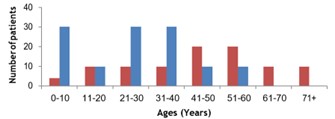
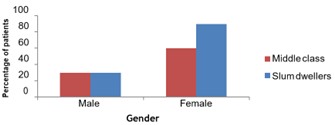
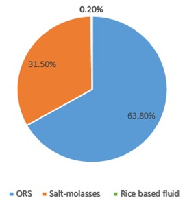
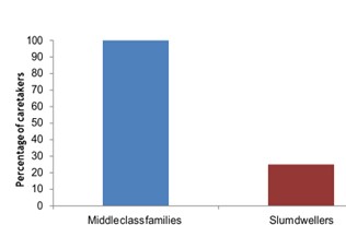
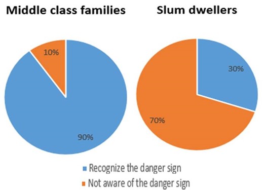
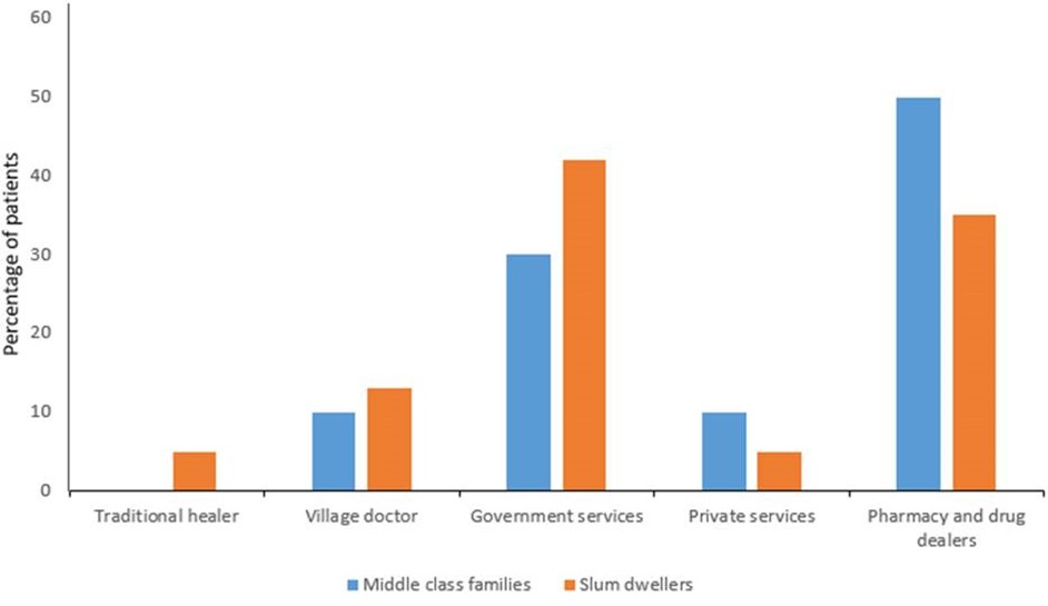
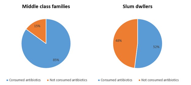
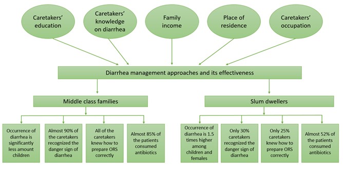
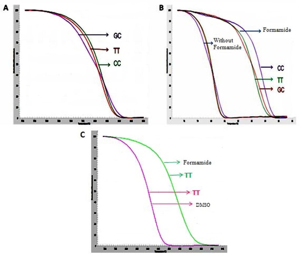
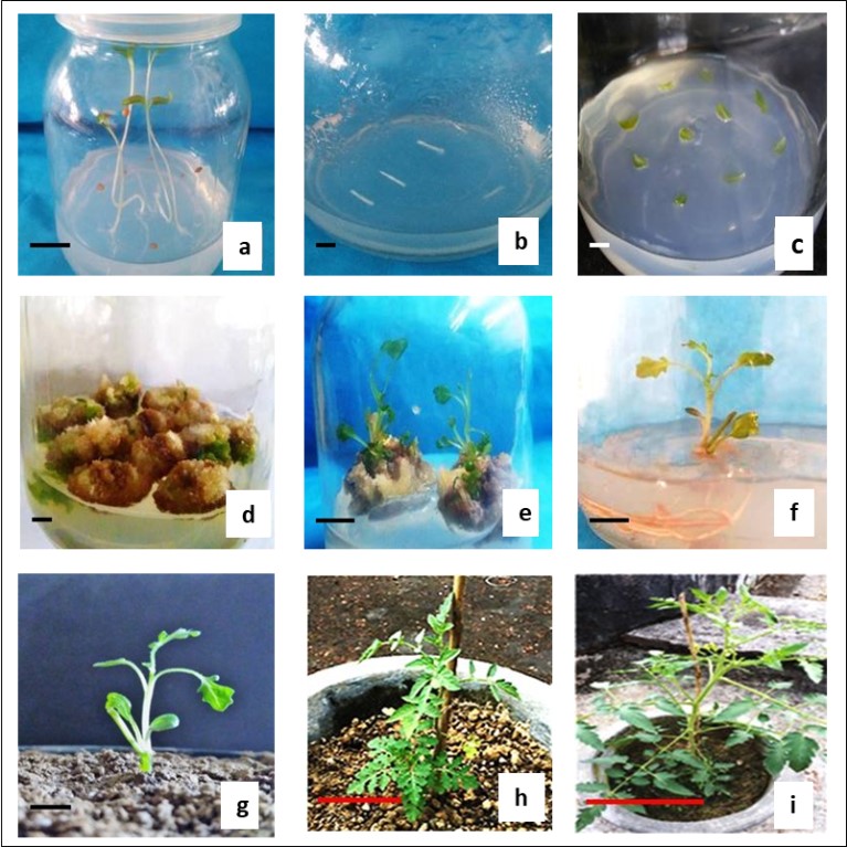
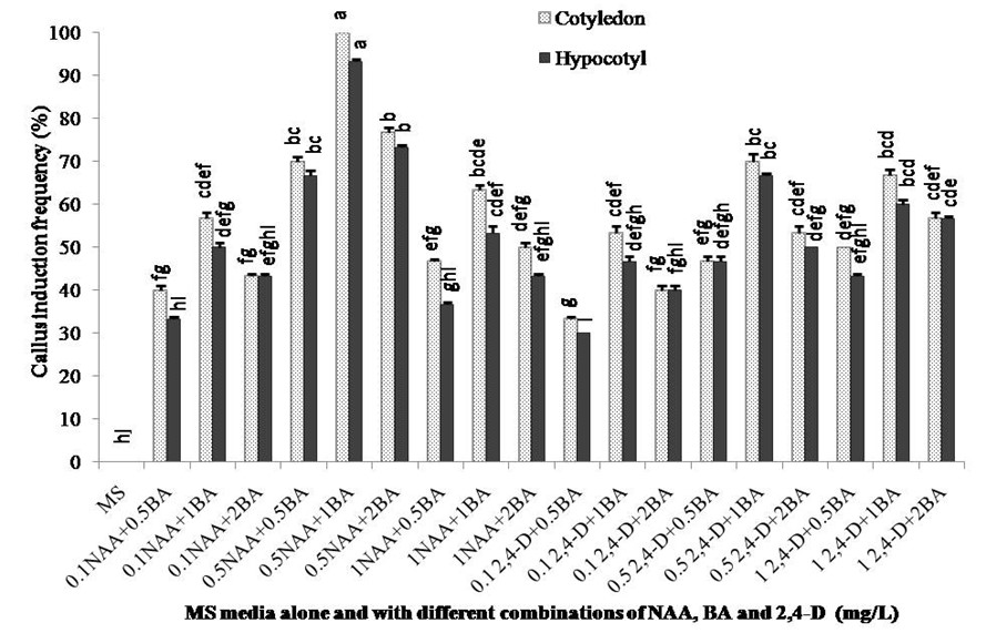
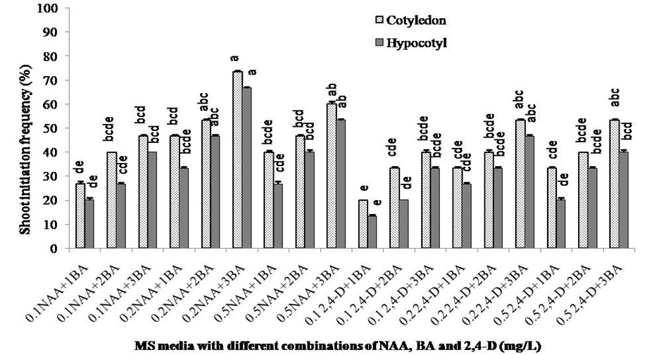
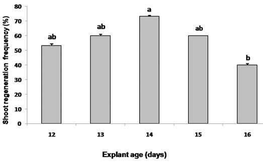
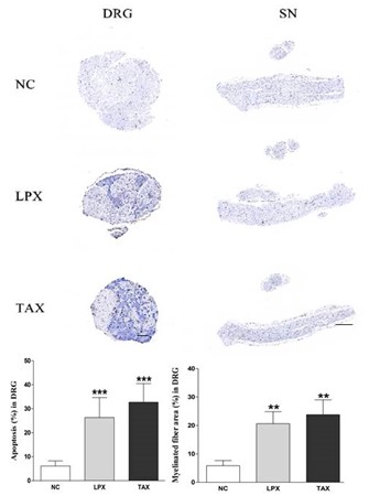
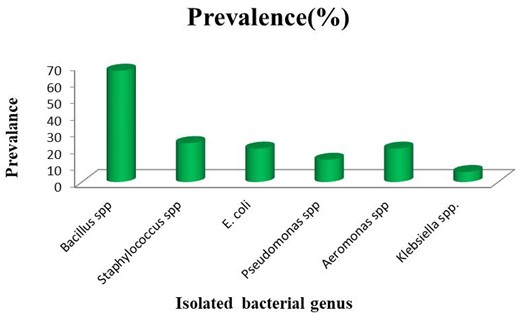
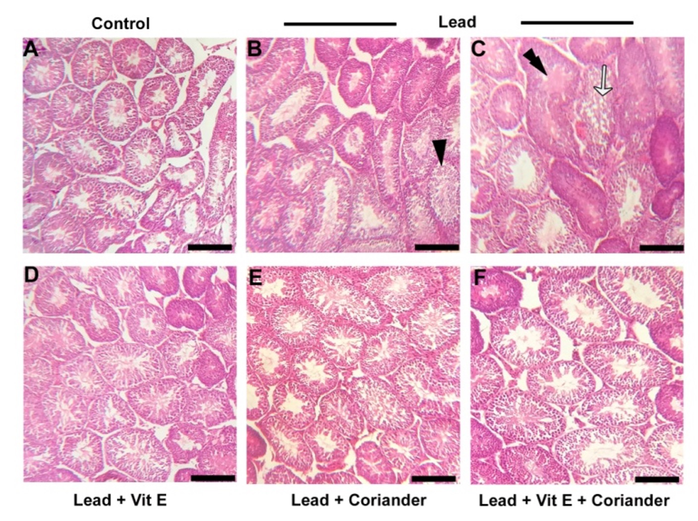
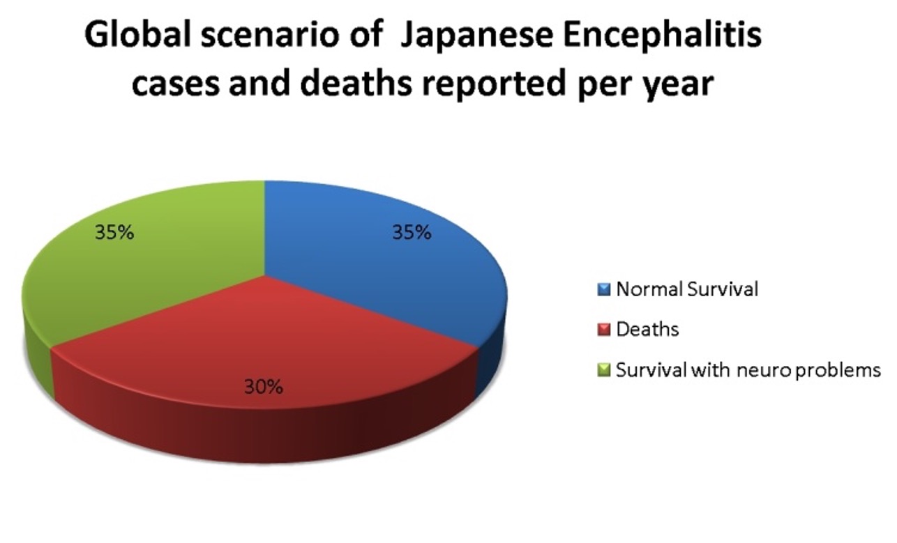
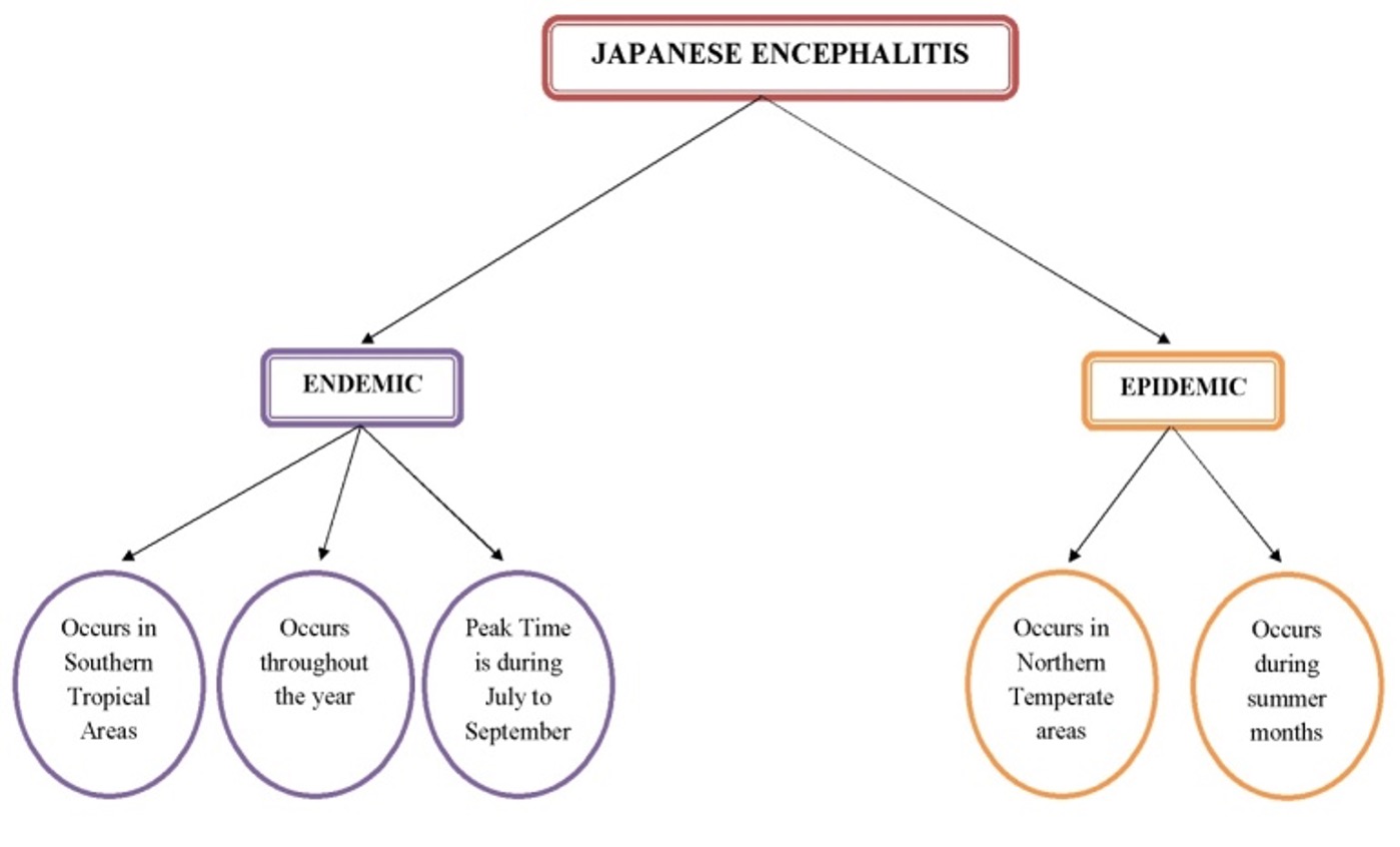
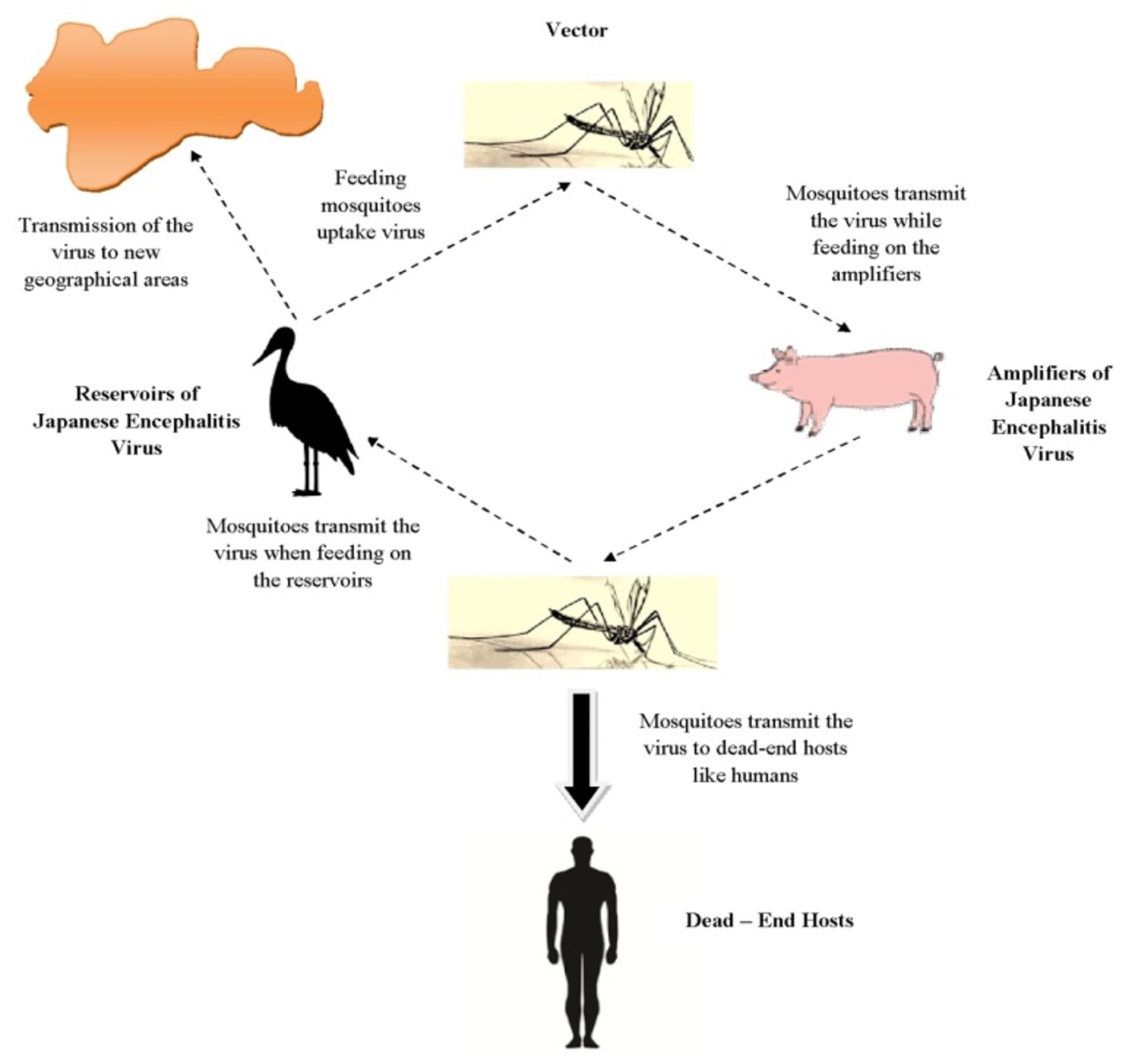
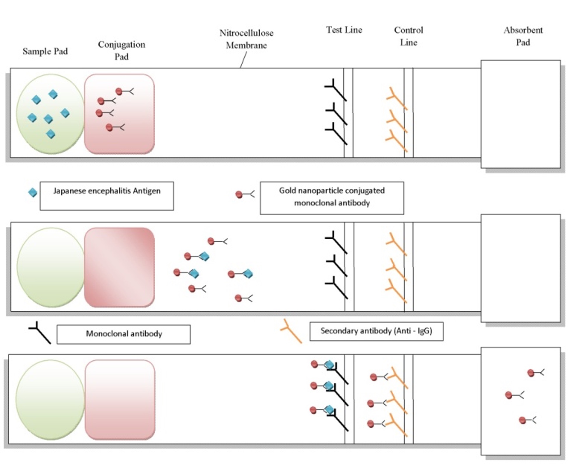
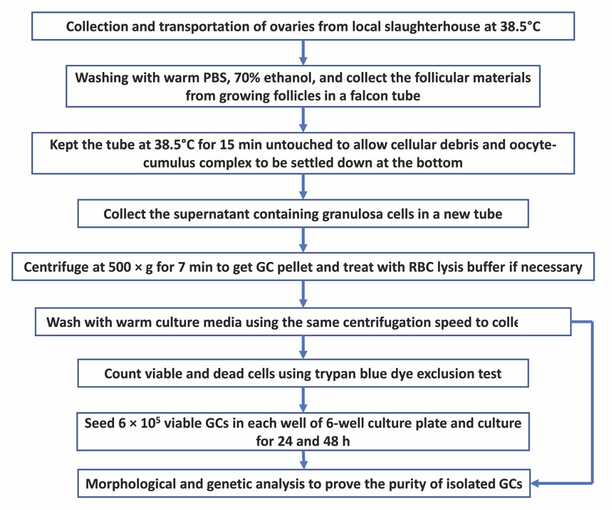
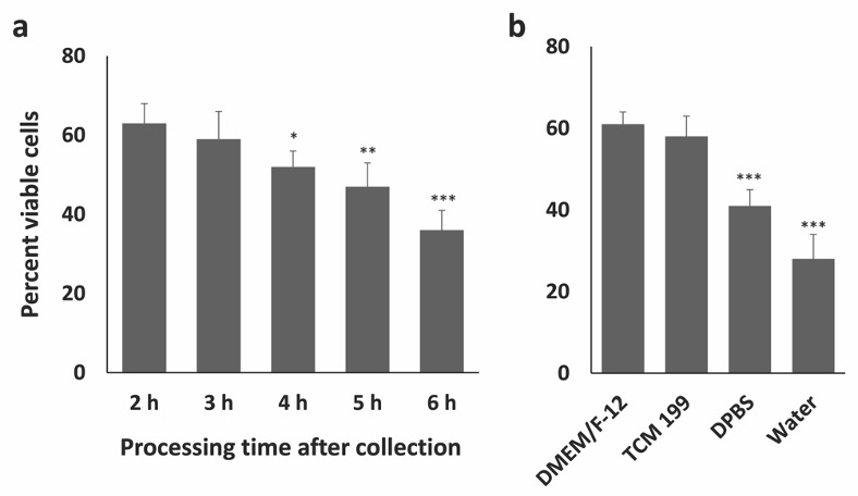
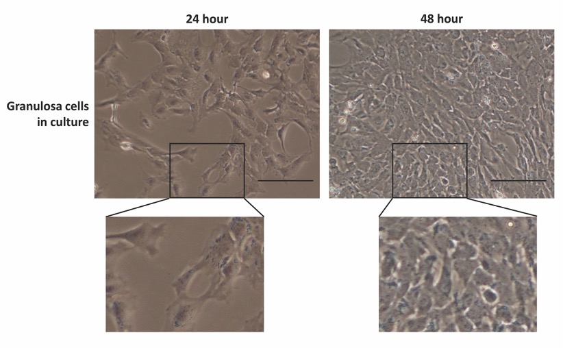
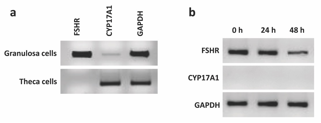
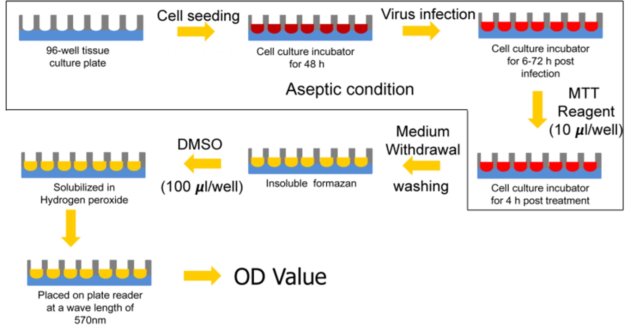
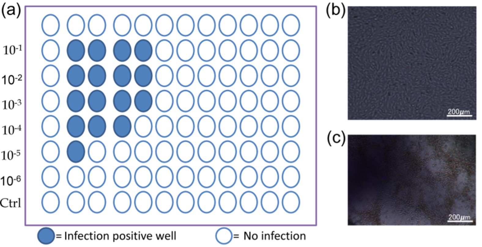
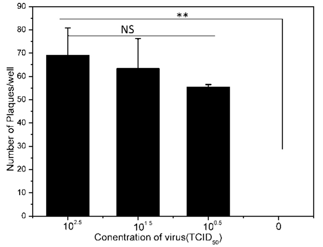
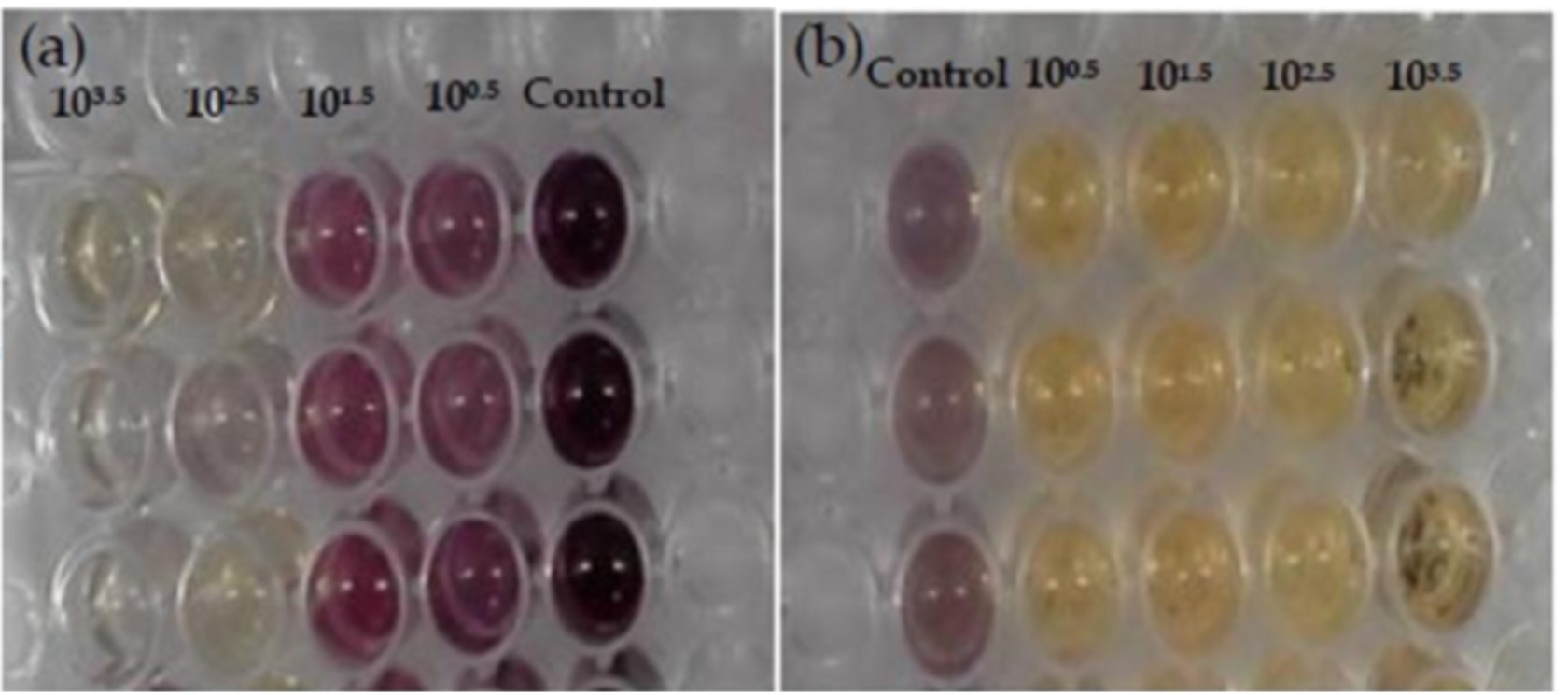
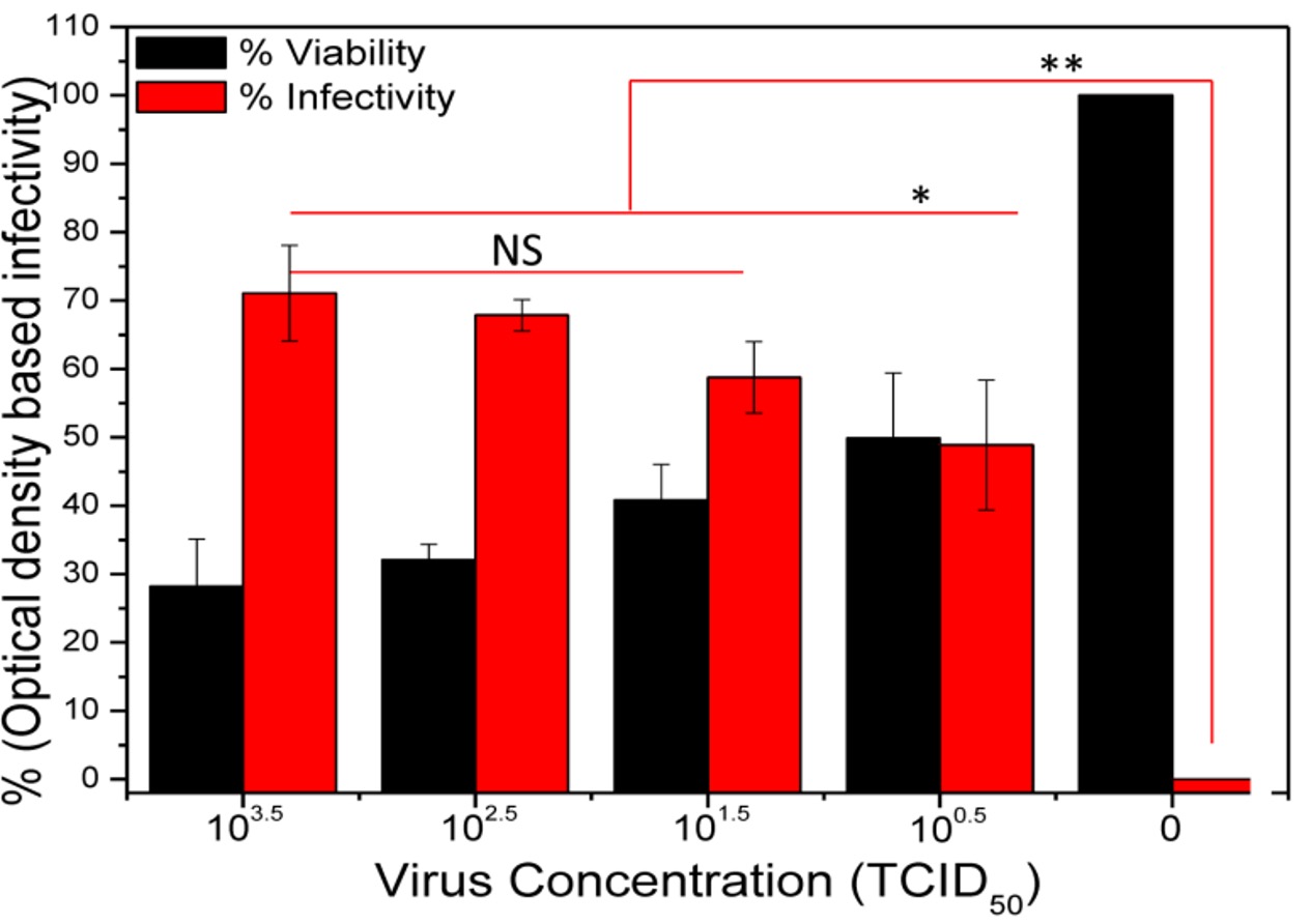
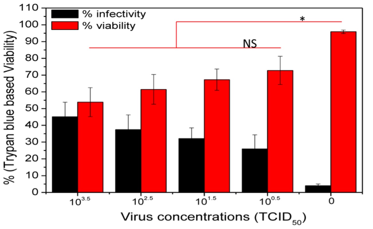