HTTP 200 OK
Allow: GET, HEAD, OPTIONS
Content-Type: application/json
Vary: Accept
{
"count": 319,
"next": "https://jabet.bsmiab.org/articles/?format=api&page=30",
"previous": "https://jabet.bsmiab.org/articles/?format=api&page=28",
"results": [
{
"id": 75,
"slug": "178-1566176524-analysis-of-maize-profilin-4-isoform-as-an-allergen",
"featured": false,
"slider": false,
"issue": "Vol2 Issue3",
"type": "original_article",
"manuscript_id": "178-1566176524",
"recieved": "2019-07-19",
"revised": null,
"accepted": "2019-09-18",
"published": "2019-09-25",
"pdf_file": "https://jabet.bsmiab.org/media/pdf_file/2023/57/178-1566176524.pdf",
"title": "Analysis of maize profilin-4 isoform as an allergen",
"abstract": "<p>Profilin is an actin monomer-binding protein that controls the dynamic turnover of actin filaments and is ubiquitously present in different organisms ranging from prokaryotes to higher eukaryotes. Maize (<em>Zea mays</em>) profilin-4 isoform is a pollen-specific protein. Birch profilin isoform is a known allergen but maize profilin is yet to be characterized. In this study, we investigated the properties of maize profilin-4 isoform’s allergenicity. To this end, we first analyzed profilin-4 isoform’s physicochemical properties, including molecular weight (~14kD), theoretical pI (4.63), and amino acids composition; and found that it might have allergenic potency. Then we tested the potential B cell epitope candidates using different immune-informatics tools housed at IEDB analysis resource. For the B cell epitope prediction, potential antigenic sites on the protein surface were predicted by both propensity scale and machine learning method followed by their mapping of 3D structure prediction. Our findings suggest that profilin-4 isoform is a potential allergen and can induce allergic responses.</p>",
"journal_reference": "J Adv Biotechnol Exp Ther. 2019; 2(3): 134-139.",
"academic_editor": "Dr. Akhi Moni, ABEx Bio-Research Center, Azampur, Dhaka 1230, Bangladesh.",
"cite_info": "Alam S, Hasan MK, et al. Analysis of maize profilin-4 isoform as an allergen. J Adv Biotechnol Exp Ther. 2019; 2(3): 134-139.",
"keywords": [
"Profilin-4",
"Allergen",
"Zea mays",
"Allergenicity",
"Epitope",
"In silico"
],
"DOI": "10.5455/jabet.2019.d36",
"sections": [
{
"section_number": 1,
"section_title": "INTRODUCTION",
"body": "<p>Allergens are small proteins or glycoproteins wavering a molecular weight range of 15 to 40 kDa [<a href=\"#r-1\">1</a>]. Allergens appear from different sources, for instance, pollen allergens from plants, venom allergens from insects, food allergens from various food items, mite allergens from dust, etc. [<a href=\"#r-2\">2</a>]. They can induce IgA, IgE, IgG, and IgM antibody-mediated immune responses [<a href=\"#r-3\">3</a>]. Besides, they can induce Th2 (Helper T) cell-mediated immune response in the human body [<a href=\"#r-4\">4</a>]. Allergens produce an enzymatic or immunogenic reaction to cause allergenicity [<a href=\"#r-5\">5</a>].<br />\r\n<em>Zea mays</em> (maize), a Poaceae family member, is one of the most cultivated crop plants around the world. Maize has both nutritional and medicinal importance. The maize kernel is the nutritive part of the plant that contains all the different vitamins, fatty acids, minerals, etc. Maize is a great source of phytochemicals that are used to treat chronic diseases, HIV, even cancer, etc. [<a href=\"#r-29\">29</a>]. There is an increasing trend of maize production over the last decade. It has been estimated that about 187.95 million hectors of land were used for maize cultivation [<a href=\"#r-6\">6</a>].<br />\r\nIn this study, we predict profilin-4 as a potential allergen. As the cultivation rate of maize increases keeping the pace with the demands, it also provokes the concern of allergenicity of its pollen. Wind-pollinated seed plants produce pollens which encompass crucial sign of Type-I allergy [<a href=\"#r-7\">7</a>]. Profilin is known as panallergen due to its widespread cross-reactivity [<a href=\"#r-11\">11</a>]. The allergenic properties of pollens have no association with biological function but the enzymatic and immunogenic actions of allergens cause the allergic reaction and inflammation [<a href=\"#r-8\">8</a>]. The profilin of birch pollen (called Bet v 2) [<a href=\"#r-10\">10</a>] and latex [<a href=\"#r-9\">9</a>] are documented as allergens, but not the maize-specific profilin isoform, profilin-4. Using the Bioinformatics tools and database [<a href=\"#r-12\">12–16</a>], here we analyzed the allergenicity of profilin-4.</p>"
},
{
"section_number": 2,
"section_title": "MATERIALS AND METHODS",
"body": "<p><strong>Protein Sequence retrieval</strong><br />\r\nProfilin-4 protein sequence (O22655.1) for <em>Zea mays </em>was retrieved in FASTA format from the NCBI protein database (<a href=\"http://www.ncbi.nlm.nih.gov/protein\">http://www.ncbi.nlm.nih.gov/protein</a>). This protein sequence was the basis for further use to perform different computational analysis from linear amino acid residues.</p>\r\n\r\n<p> </p>\r\n\r\n<p><strong>Prediction of physicochemical properties</strong><br />\r\nDifferent physicochemical properties for profilin-4 protein were predicted from its linear amino acid sequence using the ProtParam tool (<a href=\"https://web.expasy.org/protparam/\">https://web.expasy.org/protparam/</a>) web server. Protparam predicts the molecular weight, theoretical pI, atomic composition, amino acid composition, instability index, extinction coefficient, grand average of hydropathicity (GRAVY), estimated half-life, and aliphatic index of any given amino acid sequences [<a href=\"#r-17\">17</a>].</p>\r\n\r\n<p> </p>\r\n\r\n<p><strong>Potential antigenic sites prediction</strong><br />\r\nThe hydrophobic and hydrophilic regions were determined to predict the antigenicity of profilin-4. The hydrophilic portions are exposed to the surface of the protein and display reactivity to the B cell. Kolaskar-Tongaonkar antigenicity and Parker’s hydrophilicity methods were employed to predict the antigenicity of profilin-4. Antigenic propensity, as well as hydrophilicity, was then analyzed from the plots generated [<a href=\"#r-18\">18, 19</a>].</p>\r\n\r\n<p> </p>\r\n\r\n<p><strong>Potential B cell epitope prediction</strong><br />\r\nNot all the regions exposed to outer surface react with B cell, that’s why to predict the B cell epitopes a machine learning tool was used (<a href=\"http://tools.iedb.org/bcell/)\">http://tools.iedb.org/bcell/)</a>, a web server where Bepipred linear epitope prediction method was chosen [20]. Bepipred linear epitope prediction method uses an algorithm comprising both hidden Markov model and antigenic propensity and thus allowed to cross-check the predicted result from Kolaskar-Tongaonkar antigenicity and Parker’s hydrophilicity prediction method [<a href=\"#r-18\">18, 19</a>].</p>\r\n\r\n<p> </p>\r\n\r\n<p><strong>Prediction of the 3D structure of profilin-4 and mapping of B cell epitopes on the predicted structure</strong><br />\r\nFor 3-D structure prediction of profilin-4 from its linear amino acid sequence an online web service Phyre2 (<a href=\"http://www.sbg.bio.ic.ac.uk/~phyre2/html/page.cgi?id=index\">http://www.sbg.bio.ic.ac.uk/~phyre2/html/page.cgi?id=index</a>) was used. Phyre2 does several alignments of the target protein sequence with different protein templates from its database to predict a good quality model [<a href=\"#r-21\">21-22</a>]. We found that the structure (PDB ID: O22655) of profilin-4 showed maximum alignment score with its target template. Swiss PDB tool was used for the energy minimization of the structure [<a href=\"#r-23\">23</a>]. To validate the structure predicted structure a Ramachandran plot was generated at ‘PDBsum generate’ (<a href=\"http://www.ebi.ac.uk/thornton-srv/databases/pdbsum/Generate.html\">http://www.ebi.ac.uk/thornton-srv/databases/pdbsum/Generate.html</a>) web server which measures the stereochemical properties of the protein structure [<a href=\"#r-24\">24</a>]</p>"
},
{
"section_number": 3,
"section_title": "RESULTS AND DISCUSSION",
"body": "<p><strong>Physicochemical properties predict the allergenic property of profilin-4 protein</strong><br />\r\nSometimes physicochemical properties of a protein can determine the allergenic property of a protein [<a href=\"#r-25\">25</a>]. The maize profilin-4 consists of 131 amino acids with a molecular weight of approximately 14 kD (<a href=\"#Table-1\">Table 1</a>). The total amino acid distribution of profilin-4 protein (<a href=\"#figure1\">Figure 1</a>) shows that asparagine present in the lowest amount and glutamate, glycine, isoleucine, and valine predominate among the 20 amino acids of profilin-4 protein (<a href=\"#figure1\">Figure 1</a>). Due to abundant acidic amino acids, this suggests the protein’s theoretical pI is to be acidic and theoretical pI is found 4.63, which mean the profilin-4 protein is highly acidic and tends to be allergenic [<a href=\"#r-25\">25</a>]. Hence negatively charged residues (Asp + Glue) is twice the total number of positively charged residues (Arg + Lys) (<a href=\"#Table-1\">Table 1</a>) in profilin-4, there is a probability to be processed by dendritic cells via scavenger receptor [<a href=\"#r-26\">26</a>]. From predicted half-life and instability index it indicates that profilin-4 is quite stable [<a href=\"#r-27\">27</a>]. From the predicted negative grand average of hydropathicity value, it can be assumed that most of the amino acid residues of the profilin-4 protein are likely to be present on the surface of the folded profilin-4.</p>\r\n\r\n<div id=\"figure1\">\r\n<figure class=\"image\"><img alt=\"\" height=\"262\" src=\"/media/article_images/2024/44/28/178-1566176524-Figure1.jpg\" width=\"500\" />\r\n<figcaption><strong>Figure 1</strong>: The amino acids composition in profilin-4. Glycine (Gly) and Asparagine (Asn) are the major (14.5%) and the least (~0.8%) constituents, respectively.</figcaption>\r\n</figure>\r\n</div>\r\n\r\n<div id=\"Table-1\">\r\n<p><a href=\"https://jabet.bsmiab.org/table/178-1566176524-table1/\">Table-1</a><strong>Table 1</strong>. ProtParam predicted physicochemical properties for profilin-4 protein from Zea mays.</p>\r\n\r\n<p> </p>\r\n</div>\r\n\r\n<p><strong>Prediction of Potential antigenic sites on the surface of the profilin-4 protein</strong><br />\r\nFor the prediction of profilin-4 allergenicity, Kolaskar and Tongaonkar prediction method were employed which functions based on physicochemical properties of amino acids in proteins and abundances in experimentally known epitopes [<a href=\"#r-18\">18</a>]. In <a href=\"#figure2\">Figure 2</a>, the x-axis represents the amino acid position and the y-axis represents the antigenic propensity of the protein. The average antigenic propensity of profilin-4 protein is found to be 1.027. So all residues having a value greater than 1.027 are potential antigenic determinant. Seven peptides (<a href=\"#Table=2\">Table 2</a>) are found to be a potential antigen because they satisfy the set threshold value (1.00). The peptide regions “EGQHLSAAAIVGHDGSVWAQ” ranging from 16 to 35 amino acid residues and 100 to 108 amino acid residues (“SLIIGVYDE”) are predicted to have the highest antigenic propensity score. Both of them comprise about more than one-fifth (22.13%) of profilin-4 protein. The hydrophilic portion of a protein tends to be exposed on the outer surface of the protein that makes them vulnerable to be engaged with B cell. The average score of hydrophilicity of profilin-4 is found to be 1.421 (<a href=\"#figure3\">Figure 3</a>). The regions highlighted yellow have a hydrophilicity score of above the average and are likely to be present on the surface of the profilin-4 protein, while the regions highlighted green have a hydrophilicity score of below the average and are unlikely to be exposed on the surface. To predict the hydrophilic regions of profilin-4, we adopted Parker hydrophilicity prediction method [<a href=\"#r-19\">19</a>]. For making a better prediction decision, we have also used a more reliable machine learning tool that follows the Bepipred linear epitope prediction method [<a href=\"#r-28\">28</a>].</p>\r\n\r\n<div id=\"figure2\">\r\n<figure class=\"image\"><img alt=\"\" height=\"259\" src=\"/media/article_images/2024/44/28/178-1566176524-Figure2.jpg\" width=\"441\" />\r\n<figcaption><strong>Figure 2</strong>: Kolaskar and Tongaonkar antigenicity graphical plot. The protein sequences that satisfied the set antigenic propensity threshold value of 1.00 are predicted to be potential antigenic region.</figcaption>\r\n</figure>\r\n</div>\r\n\r\n<div id=\"figure3\">\r\n<figure class=\"image\"><img alt=\"\" height=\"267\" src=\"/media/article_images/2024/44/28/178-1566176524-Figure3.jpg\" width=\"452\" />\r\n<figcaption><strong>Figure 3</strong>: Parker hydrophilicity plot. The x-axis represents the amino acid position and the y-axis represents the hydrophilicity score.</figcaption>\r\n</figure>\r\n</div>\r\n\r\n<div id=\"Table-2\">\r\n<p><a href=\"https://jabet.bsmiab.org/table/178-1566176524-table2/\">Table-2</a><strong>Table 2</strong>. The list of the Peptide sequences having at least 1.0 antigenic propensity score, predicted from Kolaskar and Tongaonkar antigenicity plot.</p>\r\n\r\n<p> </p>\r\n</div>\r\n\r\n<p><strong>Potential B cell epitopes overlap the antigenic sites of profilin-4</strong><br />\r\nWe have applied the BepiPred tool to predict the potential B cell epitopes. The Bepipred linear epitope prediction method uses an algorithm that links the Hidden Markov Model (HMM) and the antigenic propensity to make the prediction more trustworthy [<a href=\"#r-20\">20</a>]. BepiPred predicted four potential B cell epitopes highlighted in yellow for profilin-4 protein sequence (<a href=\"#figure4\">Figure 4</a>) and the maximum predicted score is 1.630. Predicted epitopes are summarized in <a href=\"#Table-3\">Table 3</a>.</p>\r\n\r\n<div id=\"figure4\">\r\n<figure class=\"image\"><img alt=\"\" height=\"270\" src=\"/media/article_images/2024/44/28/178-1566176524-Figure4.jpg\" width=\"450\" />\r\n<figcaption><strong>Figure 4</strong>: Potential B cell epitopes predicted from BepiPred tool. Threshold value for potential epitope was set 0.350. Regions shaded with yellow colour are predicted as potential epitopes. The maximum score predicted here is 1.630.</figcaption>\r\n</figure>\r\n</div>\r\n\r\n<div id=\"Table-3\">\r\n<p><a href=\"https://jabet.bsmiab.org/table/178-1566176524-table3/\">Table-3</a><strong>Table 3</strong>. Predicted B cell epitope sequences and their position along with their length.</p>\r\n\r\n<p> </p>\r\n</div>\r\n\r\n<p><strong>Mapping of the B cell epitopes in the modeled structure confirms their presence on the surface of profilin-4</strong><br />\r\nThe predicted 3-D structure of profilin-4 was visualized (<a href=\"#figure5\">Figure 5 A</a>) using Swiss PDB viewer tool [<a href=\"#r-7\">7</a>]. Ramachandran plot was generated using an online tool PDBsum generate which validates the predicted structure (<a href=\"#figure5\">Figure 5 B</a>) each blue dots indicates the amino acid distribution in different quadrants of the plot. The amino acid residue distribution reveals that only 1 amino acid residue (tyrosine) which contributes less than 1% is positioned in the disallowed region of the Ramachandran plot that corroborates the high quality of the predicted model. The Predicted B cell epitopes of the profilin-4 protein are mapped on the predicted 3D structure of the profilin-4 protein (<a href=\"#figure6\">Figure 6</a>). The different colored balls on the surface of the protein other than pink represent the 4 predicted B cell epitopes and regions in pink represents the core of the protein.</p>\r\n\r\n<div id=\"figure5\">\r\n<figure class=\"image\"><img alt=\"\" height=\"289\" src=\"/media/article_images/2024/44/28/178-1566176524-Figure5.jpg\" width=\"500\" />\r\n<figcaption><strong>Figure 5</strong>: 3D structure and its validation using the Ramachandran plot for profilin-4 protein. (A) Cartoon representation of the predicted structure of the profilin-4 protein. This image has been developed using Swiss PDB viewer tool (B) amino acid residues are distributed in the Ramachandran plot.</figcaption>\r\n</figure>\r\n</div>\r\n\r\n<div id=\"figure6\">\r\n<figure class=\"image\"><img alt=\"\" height=\"256\" src=\"/media/article_images/2024/44/28/178-1566176524-Figure6.jpg\" width=\"500\" />\r\n<figcaption><strong>Figure 6</strong>: Mapping of B cell epitopes on the 3D structure of the profilin-4. BepiPred predicted epitopes are mapped on the surface of the profilin-4 protein structure where B cell epitopes are highlighted in red (epitope-1), olive (epitope-2), green (epitope-3) and yellow (epitope-4) and the rest of the non-reactive portion highlighted in pink.</figcaption>\r\n</figure>\r\n</div>"
},
{
"section_number": 4,
"section_title": "CONCLUSIONS",
"body": "<p>Due to the increasing trend of maize production in the world (<a href=\"#figure7\">Figure 7</a>), it is urgent to analyze the potency of maize profilin-4 isoform as an allergen. In this study, it is evident that profilin-4 is a potential antigen. Our investigation is suggestive of modifying maize crop excluding profilin-4 isoform. We believe that our findings will raise awareness among crop scientists and will help to further validate our findings in <em>in</em><em> vitro</em> settings.</p>\r\n\r\n<div id=\"figure7\">\r\n<figure class=\"image\"><img alt=\"\" height=\"255\" src=\"/media/article_images/2024/44/28/178-1566176524-Figure7.jpg\" width=\"500\" />\r\n<figcaption><strong>Figure 7</strong>: World corn production from 1988 to 2018. This bar diagram shows a gradual increase in corn production over three decades around the world. (Modified from <a href=\"https://www.indexmundi.com/agriculture/?country=us&commodity=corn&graph=production\">https://www.indexmundi.com/agriculture/?country=us&commodity=corn&graph=production</a>).</figcaption>\r\n</figure>\r\n</div>"
},
{
"section_number": 5,
"section_title": "CONFLICTS OF INTEREST",
"body": "<p>The authors have declared no conflict of interest with any parties which may arise from this publication.</p>"
}
],
"figures": [
{
"figure": "https://jabet.bsmiab.org/media/article_images/2024/44/28/178-1566176524-Figure1.jpg",
"caption": "Figure 1: The amino acids composition in profilin-4. Glycine (Gly) and Asparagine (Asn) are the major (14.5%) and the least (~0.8%) constituents, respectively.",
"featured": false
},
{
"figure": "https://jabet.bsmiab.org/media/article_images/2024/44/28/178-1566176524-Figure2.jpg",
"caption": "Figure 2: Kolaskar and Tongaonkar antigenicity graphical plot. The protein sequences that satisfied the set antigenic propensity threshold value of 1.00 are predicted to be potential antigenic region.",
"featured": false
},
{
"figure": "https://jabet.bsmiab.org/media/article_images/2024/44/28/178-1566176524-Figure3.jpg",
"caption": "Figure 3: Parker hydrophilicity plot. The x-axis represents the amino acid position and the y-axis represents the hydrophilicity score.",
"featured": false
},
{
"figure": "https://jabet.bsmiab.org/media/article_images/2024/44/28/178-1566176524-Figure4.jpg",
"caption": "Figure 4: Potential B cell epitopes predicted from BepiPred tool. Threshold value for potential epitope was set 0.350. Regions shaded with yellow colour are predicted as potential epitopes. The maximum score predicted here is 1.630.",
"featured": false
},
{
"figure": "https://jabet.bsmiab.org/media/article_images/2024/44/28/178-1566176524-Figure5.jpg",
"caption": "Figure 5: 3D structure and its validation using the Ramachandran plot for profilin-4 protein. (A) Cartoon representation of the predicted structure of the profilin-4 protein. This image has been developed using Swiss PDB viewer tool (B) amino acid residues are distributed in the Ramachandran plot.",
"featured": false
},
{
"figure": "https://jabet.bsmiab.org/media/article_images/2024/44/28/178-1566176524-Figure6.jpg",
"caption": "Figure 6: Mapping of B cell epitopes on the 3D structure of the profilin-4. BepiPred predicted epitopes are mapped on the surface of the profilin-4 protein structure where B cell epitopes are highlighted in red (epitope-1), olive (epitope-2), green (epitope-3) and yellow (epitope-4) and the rest of the non-reactive portion highlighted in pink.",
"featured": false
},
{
"figure": "https://jabet.bsmiab.org/media/article_images/2024/44/28/178-1566176524-Figure7.jpg",
"caption": "Figure 7: World corn production from 1988 to 2018. This bar diagram shows a gradual increase in corn production over three decades around the world. (Modified from https://www.indexmundi.com/agriculture/?country=us&commodity=corn&graph=production).",
"featured": false
}
],
"authors": [
{
"id": 259,
"affiliation": [
{
"affiliation": "Department of Biochemistry and Molecular Biology, University of Dhaka, Dhaka, Bangladesh"
}
],
"first_name": "Saruar",
"family_name": "Alam",
"email": null,
"author_order": 1,
"ORCID": null,
"corresponding": false,
"co_first_author": false,
"co_author": false,
"corresponding_author_info": "",
"article": 75
},
{
"id": 260,
"affiliation": [
{
"affiliation": "Department of Biochemistry and Molecular Biology, University of Dhaka, Dhaka, Bangladesh"
}
],
"first_name": "Md. Kamrul",
"family_name": "Hasan",
"email": null,
"author_order": 2,
"ORCID": null,
"corresponding": false,
"co_first_author": false,
"co_author": false,
"corresponding_author_info": "",
"article": 75
},
{
"id": 261,
"affiliation": [
{
"affiliation": "Department of Biology, St. John's University, Queens, New York 11439"
}
],
"first_name": "Md. Faruk",
"family_name": "Hossain",
"email": "farukbmb16@gmail.com",
"author_order": 3,
"ORCID": "https://orcid.org/0000-0002-3457-7903",
"corresponding": true,
"co_first_author": false,
"co_author": false,
"corresponding_author_info": "Department of Biology, St. John's University, Queens, New York 11439\r\n E-mail: farukbmb16@gmail.com",
"article": 75
}
],
"views": 798,
"downloads": 137,
"references": [
{
"id": 2048,
"serial_number": 1,
"pmc": null,
"reference": "S. B. Lehrer and J. E. Salvaggio, Allergens: Standardization and Impact of Biotechnology—A Review, Allergy Asthma Proc., 1990, 11(5): 197–208.",
"DOI": null,
"article": 75
},
{
"id": 2049,
"serial_number": 2,
"pmc": null,
"reference": "C. Ozdemir, M. Akdis, and C. A. Akdis, “T-Cell Response to Allergens, in Anaphylaxis, Basel: KARGER, 2010, 95: 22–44.",
"DOI": null,
"article": 75
},
{
"id": 2050,
"serial_number": 3,
"pmc": null,
"reference": "A. Vojdani, Detection of IgE, IgG, IgA and IgM antibodies against raw and processed food antigens, Nutr. Metab. (Lond)., 2009, 6 (1): 22.",
"DOI": null,
"article": 75
},
{
"id": 2051,
"serial_number": 4,
"pmc": null,
"reference": "J. A. Woodfolk, T-cell responses to allergens, J. Allergy Clin. Immunol., 2007, 119(2): 280–294.",
"DOI": null,
"article": 75
},
{
"id": 2052,
"serial_number": 5,
"pmc": null,
"reference": "D. A. O. Taketomi, Ernesto A., 1Almeida, Karine C., Pereira, Fernando L., Silva, Allergens: sources, exposure and sensitization levels, diagnostic tools and immunotherapeutical applications. J. Med. Med. Sci. 2010, 1(12): 580-588.",
"DOI": null,
"article": 75
},
{
"id": 2053,
"serial_number": 6,
"pmc": null,
"reference": "FAOSTAT.[Online]. Available: http://www.fao.org/faostat/en/#home. [Accessed: 05-Jan-2018].",
"DOI": null,
"article": 75
},
{
"id": 2054,
"serial_number": 7,
"pmc": null,
"reference": "Behrendt H, Becker WM, Fritzsche C, Sliwa-Tomczok W, Tomczok J, Friedrichs KH et al., Air pollution and allergy: experimental studies on modulation of allergen release from pollen by air pollutants., Int Arch Allergy Immunol. 1997, 113(1-3): 69-74.",
"DOI": null,
"article": 75
},
{
"id": 2055,
"serial_number": 8,
"pmc": null,
"reference": "A. Bufe, The biological function of allergens: relevant for the induction of allergic diseases? Int Arch Allergy Immunol. 1998,117(4):215-9.",
"DOI": null,
"article": 75
},
{
"id": 2056,
"serial_number": 9,
"pmc": null,
"reference": "P. Vallier, S. Balland, R. Harf, R. Valenta, and P. Deviller. Identification of profilin as an IgE-binding component in latex from Hevea brasiliensis: clinical implications. Clin. Exp. Allergy, 1995, 25(4): 332–339.",
"DOI": null,
"article": 75
},
{
"id": 2057,
"serial_number": 10,
"pmc": null,
"reference": "Valenta R, Duchêne M, Pettenburger K, Sillaber C, Valent P, Bettelheim P et al., Identification of profilin as a novel pollen allergen; IgE autoreactivity in sensitized individuals.,” Science. 1991, 253(5019):557-60.",
"DOI": null,
"article": 75
},
{
"id": 2058,
"serial_number": 11,
"pmc": null,
"reference": "Asero R, Mistrello G, Roncarolo D, Amato S, Zanoni D, Barocci F et al., Detection of clinical markers of sensitization to profilin in patients allergic to plant-derived foods. J. Allergy Clin. Immunol., vol. 112, no. 2, pp. 427–32, Aug. 2003.",
"DOI": null,
"article": 75
},
{
"id": 2059,
"serial_number": 12,
"pmc": null,
"reference": "S. L. Taylor and S. L. Hefle, “Will genetically modified foods be allergenic?,” J. Allergy Clin. Immunol., vol. 107, no. 5, pp. 765–771, May 2001.",
"DOI": null,
"article": 75
},
{
"id": 2060,
"serial_number": 13,
"pmc": null,
"reference": "S. M. Gendel, “Sequence databases for assessing the potential allergenicity of proteins used in transgenic foods.,” Adv. Food Nutr. Res., vol. 42, pp. 63–92, 1998.",
"DOI": null,
"article": 75
},
{
"id": 2061,
"serial_number": 14,
"pmc": null,
"reference": "S. M. Gendel, “The use of amino acid sequence alignments to assess potential allergenicity of proteins used in genetically modified foods.,” Adv. Food Nutr. Res., vol. 42, pp. 45–62, 1998.",
"DOI": null,
"article": 75
},
{
"id": 2062,
"serial_number": 15,
"pmc": null,
"reference": "J. D. Astwood and R. L. Fuchs, “Allergenicity of foods derived from transgenic plants.,” Monogr. Allergy, vol. 32, pp. 105–20, 1996.",
"DOI": null,
"article": 75
},
{
"id": 2063,
"serial_number": 16,
"pmc": null,
"reference": "D. D. Metcalfe, J. D. Astwood, R. Townsend, H. A. Sampson, S. L. Taylor, and R. L. Fuchs, “Assessment of the allergenic potential of foods derived from genetically engineered crop plants.,” Crit. Rev. Food Sci. Nutr., vol. 36 Suppl, pp. S165-86, 1996.",
"DOI": null,
"article": 75
},
{
"id": 2064,
"serial_number": 17,
"pmc": null,
"reference": "E. Gasteiger et al., “Protein Identification and Analysis Tools on the ExPASy Server,” in The Proteomics Protocols Handbook, Totowa, NJ: Humana Press, 2005, pp. 571–607.",
"DOI": null,
"article": 75
},
{
"id": 2065,
"serial_number": 18,
"pmc": null,
"reference": "A. S. Kolaskar and P. C. Tongaonkar, “A semi-empirical method for prediction of antigenic determinants on protein antigens.,” FEBS Lett., vol. 276, no. 1–2, pp. 172–4, Dec. 1990.",
"DOI": null,
"article": 75
},
{
"id": 2066,
"serial_number": 19,
"pmc": null,
"reference": "J. M. Parker, D. Guo, and R. S. Hodges, “New hydrophilicity scale derived from high-performance liquid chromatography peptide retention data: correlation of predicted surface residues with antigenicity and X-ray-derived accessible sites.,” Biochemistry, vol. 25, no. 19, pp. 5425–32, Sep. 1986.",
"DOI": null,
"article": 75
},
{
"id": 2067,
"serial_number": 20,
"pmc": null,
"reference": "J. Larsen, O. Lund, and M. Nielsen, “Improved method for predicting linear B-cell epitopes.,” Immunome Res., vol. 2, no. 1, p. 2, Apr. 2006.",
"DOI": null,
"article": 75
},
{
"id": 2068,
"serial_number": 21,
"pmc": null,
"reference": "Kelley, Lawrence A et al. “The Phyre2 web portal for protein modeling, prediction and analysis.” Nature protocols vol. 10,6 (2015): 845-58.",
"DOI": null,
"article": 75
},
{
"id": 2069,
"serial_number": 22,
"pmc": null,
"reference": "J. Ma, J. Peng, S. Wang, and J. Xu, “A conditional neural fields model for protein threading,” Bioinformatics, vol. 28, no. 12, pp. i59–i66, Jun. 2012.",
"DOI": null,
"article": 75
},
{
"id": 2070,
"serial_number": 23,
"pmc": null,
"reference": "N. Guex and M. C. Peitsch, “SWISS-MODEL and the Swiss-Pdb Viewer: An environment for comparative protein modeling,” Electrophoresis, vol. 18, no. 15, pp. 2714–2723, Dec. 1997.",
"DOI": null,
"article": 75
},
{
"id": 2071,
"serial_number": 24,
"pmc": null,
"reference": "Laskowski, R A. “PDBsum: summaries and analyses of PDB structures.” Nucleic acids research vol. 29,1 (2001): 221-2.",
"DOI": null,
"article": 75
},
{
"id": 2072,
"serial_number": 25,
"pmc": null,
"reference": "S. Singh, B. Taneja, S. S. Salvi, and A. Agrawal, “Physical Properties of Intact Proteins May Predict Allergenicity or Lack Thereof,” PLoS One, vol. 4, no. 7, p. e6273, Jul. 2009.",
"DOI": null,
"article": 75
},
{
"id": 2073,
"serial_number": 26,
"pmc": null,
"reference": "K. Shakushiro, Y. Yamasaki, M. Nishikawa, and Y. Takakura, “Efficient scavenger receptor-mediated uptake and cross-presentation of negatively charged soluble antigens by dendritic cells.,” Immunology, vol. 112, no. 2, pp. 211–8, Jun. 2004.",
"DOI": null,
"article": 75
},
{
"id": 2074,
"serial_number": 27,
"pmc": null,
"reference": "A. Bachmair, D. Finley, and A. Varshavsky, “In vivo half-life of a protein is a function of its amino-terminal residue.,” Science, vol. 234, no. 4773, pp. 179–86, Oct. 1986.",
"DOI": null,
"article": 75
},
{
"id": 2075,
"serial_number": 28,
"pmc": null,
"reference": "J. Söllner and B. Mayer, “Machine learning approaches for prediction of linear B-cell epitopes on proteins,” J. Mol. Recognit., vol. 19, no. 3, pp. 200–208, May 2006.",
"DOI": null,
"article": 75
},
{
"id": 2076,
"serial_number": 29,
"pmc": null,
"reference": "Tajamul Rouf Shah, Kamlesh Prasad, Pradyuman Kumar & Fatih Yildiz. “Maize-A potential source of human nutrition and health: A review”, Cogent Food & Agriculture Vol. 2, Iss. 1, 2016",
"DOI": null,
"article": 75
}
]
},
{
"id": 77,
"slug": "178-1568893361-comparable-preventive-effects-of-laboratory-grown-spirulina-and-market-spirulina-against-arsenic-induced-alterations-in-the-liver-of-adult-rats",
"featured": false,
"slider": false,
"issue": "Vol2 Issue3",
"type": "original_article",
"manuscript_id": "178-1568893361",
"recieved": "2019-07-04",
"revised": null,
"accepted": "2019-09-20",
"published": "2019-09-25",
"pdf_file": "https://jabet.bsmiab.org/media/pdf_file/2023/25/178-1568893361.pdf",
"title": "Comparable preventive effects of laboratory-grown spirulina and market spirulina against arsenic-induced alterations in the liver of adult rats",
"abstract": "<p>Arsenic (As) is a naturally occurring ubiquitous environmental toxicant. It has been reported that spirulina has protective effects against As toxicity. In the present study, we compared the prophylactic effects of spirulina [laboratory grown agro-based spirulina (Ab-Sp) and market spirulina (M-Sp)] against the histopathological changes in liver induced by inorganic arsenic (iAs) in male rats. Three doses (1.0g, 1.5g and 2.0g/kg feed) of both the Ab-Sp and M-Sp with feed and 3.0mg NaAsO<sub>2</sub>/kg body weight (BW) in drinking water were given simultaneously to six groups (T4, T5, T6, T7, T8 and T9) of rats daily for 90 days. Same dose of NaAsO<sub>2</sub> (3.0mg/kg; T1) and highest dose (2.0g/kg) of each of Ab-Sp (T2) and M-Sp (T3) were given individually to other 3 groups keeping the rest one as control (T0) with normal feed and water. As feeding resulted in a variety of histopathological changes in liver, including congestion in the central veins, hemorrhage in the hepatic lobules and lobular tissues, higher numbers of hypertrophic hepatocytes with hypertrophic nucleus, hepatocytes with visible chromatin in the nucleus and vacuolated hepatocytes. The Ab-Sp treatment successfully improved all the histopathological conditions. In contrast, the M-Sp improved the conditions by combating all the histopathological conditions including vacuolated hepatocytes, erosions and hemorrhages in the liver. Taken together, the spirulina was found as an effective agent in prevention of the histopathological changes while we first clarified that Ab-Sp had better result than the M-Sp and finally, 2.0g Ab-Sp/kg feed was found as the best dose.</p>",
"journal_reference": "J Adv Biotechnol Exp Ther. 2019; 2(3): 146-152.",
"academic_editor": "Dr. Hasan-Al-Faruque, Daegu Gyeonbuk Institute of Science and Technology, South Korea.",
"cite_info": "Khair A, Awal MA, et al. Comparable preventive effects of laboratory-grown spirulina and market spirulina against arsenic-induced alterations in the liver of adult rats. J Adv Biotechnol Exp Ther. 2019; 2(3): 146-152.",
"keywords": [
"Liver",
"Spirulina",
"Rats",
"Arsenic",
"Hepatic architecture"
],
"DOI": "10.5455/jabet.2019.d38",
"sections": [
{
"section_number": 1,
"section_title": "INTRODUCTION",
"body": "<p>Arsenic (As) is a naturally occurring ubiquitous environmental toxicant widely distributed in the earth crust. Normally, As occurs in air, natural water, soil, vegetation, plants, forests and marine products and its concentrations are much higher depending on geographic locations. Arsenic occurs in both inorganic and organic forms and can exist in the environment in 4 redox states: arsenite (+3), arsenate (+5), arsine (-3) and elemental (0) forms [<a href=\"#r-1\">1</a>]. Inorganic arsenicals have detrimental impacts on life supporting functions [<a href=\"#r-2\">2</a>] while most of the organic arsenicals have negligible health effects and excreted unchanged [<a href=\"#r-3\">3</a>]. Arsenite is 10 times more toxic than arsenate and 70 times more toxic than the methylated species, i.e., organic arsenicals [<a href=\"#r-4\">4</a>].<br />\r\nDrinking water polluted by high level of arsenic is one of the most serious worldwide environmental problems [<a href=\"#r-5\">5</a>]. It has been reported from at least 70 countries including 14 Asian countries that an estimated of 140 million people around the world are drinking water contaminated with arsenic exceeding 10.0 µg As/L (WHO standard; [<a href=\"#r-6\">6,7</a>]. Although, drinking of high arsenic contaminated water is the primary pathway of arsenic exposure to humans [<a href=\"#r-8\">8,9</a>], arsenic can also enter into the human food chain directly or indirectly due to its wide spread distribution in both the plant and animal kingdoms [<a href=\"#r-10\">10</a>]. As a result, it has been reported that 85.0 million peoples are at risk of arsenic toxicity for consuming both arsenic rich drinking water and foodstuffs in Bangladesh [<a href=\"#r-11\">11,12</a>].<br />\r\nArsenicosis caused serious public health problems including melanosis, leukomelanosis, hyperkeratosis, black foot disease, hepatomegaly, neuropathy, cancer and gangrene [<a href=\"#r-9\">9</a>], weakness, anemia, burning sensation of eyes, liver fibrosis, chronic lung disease, bone marrow depression [<a href=\"#r-13\">13</a>], vascular remodeling, portal hypertension, noncirrhotic liver fibrosis [<a href=\"#r-14\">14</a>].<br />\r\nArsenic causes histopathological changes in tissues and organs of the exposed subject. The hepatotoxic action of arsenic includes cirrhotic portal fibrosis [<a href=\"#r-15\">15</a>], hepatomegaly, non-cirrhotic portal fibrosis and portal hypertension [<a href=\"#r-16\">16</a>,<a href=\"#r-14\">14</a>], oxidative damage of liver [<a href=\"#r-14\">14</a>,<a href=\"#r-17\">17</a>], fatty accumulation, parenchymal cell degeneration [<a href=\"#r-18\">18</a>], steatosis, hepatocyte degeneration [<a href=\"#r-19\">19</a>], hepatic fibrosis [<a href=\"#r-14\">14</a>], and liver proliferative lesions [<a href=\"#r-18\">18</a>]. Fibrosis of liver may progress to cirrhosis and even to liver cancers [<a href=\"#r-20\">20</a>]. Necrosis of hepatocytes and cytoplasmic blebbing and expanded sinusoidal spaces due to shrinkage and necrosis of hepatocytes were found on arsenite-exposure [<a href=\"#r-21\">21</a>]. However, there were significant vascular remodeling with increased sinusoidal endothelial cell capillarization, vascularization of the peribiliary vascular plexus and constriction of hepatic arterioles [<a href=\"#r-22\">22</a>]. Few focal areas of necrosis, Kupffer cell hyperplasia, and localized fibrosis in the periportal region were observed [<a href=\"#r-23\">23</a>]. Ballooning of hepatocytes in the periportal and parenchymal areas, chromatin fragmentation and drop-out necrosis [<a href=\"#r-24\">24</a>], and swollen hepatocytes near the centrilobular vein were observed in liver [<a href=\"#r-25\">25</a>].<br />\r\nHowever, the first important step to control arsenicosis is the cessation of drinking arsenic contaminated water [<a href=\"#r-17\">17</a>] and ingesting arsenic rich foods. Then, supplementation of potential antioxidants and feeding of arsenic burden reducing agent seem to be beneficial for remedy of arsnicosis [<a href=\"#r-26\">26,27</a>]. Antioxidant supplement may have preventive effects in arsenicosis [<a href=\"#r-28\">28,29</a>,<a href=\"#r-26\">26</a>]. Intake of cysteine, methionine, niacin, vitamin B<sub>12</sub> and choline facilitates arsenic methylation by modulating its metabolism [<a href=\"#r-30\">30</a>].<br />\r\nSpirulina (<em>Spirulina platensis</em>), a blue-green alga, has been considered as an excellent whole food ever known to mankind [<a href=\"#r-31\">31,32</a>] having antioxidant properties [<a href=\"#r-33\">33,34</a>]. It has the corrective properties against heavy metal toxicity, nephrotoxicity induced by heavy metals and drugs and also against cancer, tumor growth and malnutrition [<a href=\"#r-32\">32</a>,<a href=\"#r-35\">35</a>]. As arsenic induces the generation of ROS [<a href=\"#r-36\">36</a>], alters DNA methylation pattern in many genes [<a href=\"#r-37\">37</a>] and also inactivate enzymes in the cellular energy pathway [<a href=\"#r-38\">38</a>], as a result, antioxidants could be helpful in altering the cytotoxicity of arsenic [<a href=\"#r-39\">39</a>]. β-carotene is a very important antioxidant [<a href=\"#r-40\">40</a>] and β-carotene from spirulina pose more antioxidant properties than synthetic one. Phycocyanin, a pigment of spirulina, stimulates the immune system and assists detoxification with the ability to inhibit oxidative damage in DNA [<a href=\"#r-41\">41</a>]. Spirulina is rich in protein with all the essential amino acids, antioxidants, galaxy of phytonutrients and polysaccharides that trigger enzyme systems to enhance detoxification of arsenic [<a href=\"#r-42\">42</a>]. Spirulina contains a lot of minerals including zinc, which is essential for a strong immune system and for powering antioxidant enzyme systems and acts synergistically with β-carotene. Micronutrients of Spirulina interact with toxic metals at several points in the body like absorption and excretion of toxic metals; transport of metals in the body; binding to target proteins; metabolism and sequestration of toxic metals; and in secondary mechanisms of toxicity such as oxidative stress [<a href=\"#r-43\">43</a>]. Hence, spirulina intake can reduce the effects of arsenicosis by modulating its methylation, reducing oxidative stress, binding to target proteins, enhancing arsenic excretion from the body, and finally by reducing susceptibility to arsenicosis by combating malnutrition.<br />\r\nTherefore, this work has been taken with a view to evaluate the histopathological changes induced by inorganic arsenic and to clarify the comparative effects of laboratory grown agro-based spirulina (Ab-Sp) and market spirulina (M-Sp) in prevention of histopathological alterations in liver.</p>"
},
{
"section_number": 2,
"section_title": "MATERIALS AND METHODS",
"body": "<p><strong>Animals</strong><br />\r\nApparently healthy 40 male <em>Long Evans</em> rats with minimum of 350.0g BW were selected and randomly divided into 10 groups consisting of 4 rats in each group. The rats of all groups, except that of T0, T2 and T3 groups, were fed with NaAsO<sub>2</sub> (Merck, Darmstadt, Germany: 0.58 mg iAs/mg) at 3.0 mg/kg BW/day in drinking water. Simultaneously, the rats of T4, T5 and T6 group were individually given 3 doses of the Ab-Sp at 1.0g, 1.5g and 2.0g/kg feed respectively while that of the T7, T8 and T9 group were similarly supplied M-Sp at 1.0g, 1.5g and 2.0g/kg feed respectively in feed. The rats of T2 were treated with only 2.0g Ab-Sp/kg feed while the T3 group rats were treated with the same dose of M-Sp (2.0g/kg feed) in feed as Ab-Sp and M-Sp treated control respectively. The rats of the T1 group were fed only with 3.0 mg NaAsO<sub>2</sub>/kg BW/day in drinking water as arsenic treated control whereas; the rats of T0 group were supplied only normal feed and drinking water as non-treated control. The trial was continued for 90 days. All experimental protocols were approved by the Animal Welfare and Ethical Committee, Faculty of Veterinary Science, Bangladesh Agricultural University. All efforts were made to minimize the number of mice used and their sufferings.</p>\r\n\r\n<p> </p>\r\n\r\n<p><strong>Feeding trial</strong><br />\r\nA 0.2% NaAsO<sub>2</sub> stock solution was prepared with deionized water and preserved at 4°C to feed the trial rats for use of maximum 7 days. Dried powder of Ab-Sp obtained from the laboratory production and the dried powder of M-Sp collected from the local market. Required amounts of the respective doses (1.0g, 1.5g and 2.0g/kg feed) of both the Ab-Sp and M-Sp were individually mixed with pellet feed and dried at 50°C in an electric oven for at least 20 hours. Then, the spirulina mixed feed was taken into air tight polypropylene container for supplying to the trial rats for 5 days from the preparation. A new lot of spirulina mixed feed was prepared generally 12 hours before the 5<sup>th</sup> day from the previous feed preparation.<br />\r\nIn every morning, required amount of the prepared NaAsO<sub>2</sub> solution (0.2%) for the rats of a group per day at the rate of 3 mg As/kg BW/day was calculated and taken into a previously washed and sterilized waterer. Then, a small amount of drinking water was added to the solution so as not to exceed the half of the daily requirement of drinking water for the rats of that particular group. This was done to ensure drinking of the total amount of NaAsO<sub>2</sub> solution within 6 to 8 hours of supply. After drinking the total amount of NaAsO<sub>2</sub> solution, the rats were allowed to drink normal drinking water <em>ad libitum</em>. This As feeding process was done for the rats of T1, T4, T5, T6, T7, T8 and T9 groups. Concomitantly to the feeding of NaAsO<sub>2</sub> solution, the spirulina mixed feed with the respective doses of the individual spirulina (Ab-Sp and M-Sp) were supplied <em>ad libitum</em> to the rats of all six groups and the rats of T2 and T3 groups<em>.</em> This feeding process was continued for 90 days.</p>\r\n\r\n<p> </p>\r\n\r\n<p><strong>Sample collection</strong><br />\r\nOn Day 90, all the trial rats were euthanized with high dose of chloroform (Fisher Scientific UK Limited, UK) and samples were collected by opening the carcass immediate after euthanization. An intact piece of liver was collected from the sample rats. The collected samples were immediately washed in buffered neutral formalin and taken into individual stoppered glass vial filled with adequate amount of buffered neutral formalin (10 x sample volume) for fixation and kept at room temperature until processed for histopathological investigation.<br />\r\nAfter 48 hours, the fixed tissues were trimmed with a sharp blade into a suitable size and shape (about 0.5cmX0.5cm). Immediately after trimming, the liver was preserved in fresh buffered neutral formalin into new clean and dried stoppered glass vials.</p>\r\n\r\n<p> </p>\r\n\r\n<p><strong>Staining of tissue sections and photomicrograph</strong><br />\r\nRoutine hematoxylin and eosin (H & E) staining was used for the tissue sections. Preparation of stains and other necessary chemical solutions, and staining procedures were described previously [<a href=\"#r-44\">44</a>]. Tissue sections were examined under compound microscope and photographs of tissue sections were taken under a Differential Interference Contrast (DIC) Microscope.</p>\r\n\r\n<p> </p>\r\n\r\n<p><strong>Examination of liver sections</strong><br />\r\nThe degree of changes, if any in the hepatocytes, was expressed by the number of hepatocytes with changes in respect to the total number following counting of hepatocytes.</p>\r\n\r\n<p> </p>\r\n\r\n<p><strong>Statistical analysis</strong><br />\r\nThe designs of the experiments were Randomized Complete Block Design (RCBD). The data were tabulated into a preliminary data sheet of a computer and compared with computer spread sheets to ensure the accuracy of the data. Then the data were analyzed with computer programs: Microsoft Excel and SPSS (Statistical Package for Social Science), and also using the analysis of variance technique by a computer using of MSTAT computer package program in according to the principles of RCBD. Least Significance Difference (LSD) was done to compare the variations between treatments where ANOVA showed significant differences.</p>"
},
{
"section_number": 3,
"section_title": "RESULTS",
"body": "<p><strong>Overall histopathological findings</strong><br />\r\nWith routine H&E staining, the liver showed normal in structures with mitotic phases in few nuclei, although a considerable number of hepatocytes showed hypertrophic hepatocytes with hypertrophic nucleus and hepatocytes with visible chromatin in the rats of all the trial groups in a variable degree (<a href=\"#Table-1\">Table 1</a>). In the rats of the T1 group, the liver was found with vacuolated hepatocytes, blood accumulation in some of the central veins, and erosions of lobular epithelium, and consequently hemorrhages in some hepatic lobules and lobular tissues as extravasations while no vacuolated hepatocytes, erosion and hemorrhage was found in rats of other trial groups (<a href=\"#figure1\">Figure 1A-D</a>).</p>\r\n\r\n<div id=\"figure1\">\r\n<figure class=\"image\"><img alt=\"\" height=\"408\" src=\"/media/article_images/2024/14/28/178-1568893361-Figure1.jpg\" width=\"500\" />\r\n<figcaption><strong>Figure 1</strong>. Representative photomicrographs of H & E stained histological section of liver of group T0 (A) and T1 (C). B and D are the enlargement of CV areas in A and B, respectively. Black arrows indicate nucleus with visible chromatin, White arrows indicate mitotic phase, black arrowheads indicate vacuolated hepatocytes, double arrowheads indicates congestion and hemorrhage in the hepatic matrix, white arrowheads indicates hypertrophic hepatocytes with hypertrophic nucleus. CV, central veins. Scale bar in A, C = 100 μm; B, D= 10μ m.</figcaption>\r\n</figure>\r\n</div>\r\n\r\n<div id=\"Table-1\">\r\n<p><a href=\"https://jabet.bsmiab.org/table/178-1568893361-table1/\">Table-1</a><strong>Table 1</strong>. Incidence of hypertrophic hepatocytes with hypertrophic nucleus and hepatocytes with visible chromatin in the nucleus of the liver of trial rats.</p>\r\n\r\n<p> </p>\r\n</div>\r\n\r\n<p><strong>Hypertrophic hepatocytes with hypertrophic nucleus in the liver</strong><br />\r\nThe livers of the T8 group rats had the highest numbers of hypertrophic hepatocytes with hypertrophic nucleus while the lowest number was in the T0 group. However, the number of hypertrophic hepatocytes with hypertrophic nucleus in the liver of the rats of all of the groups differed significantly (p<0.01) among the groups. Arsenic induction significantly increased the numbers of hypertrophic hepatocytes with hypertrophic nucleus in the liver compared to the control. However, the numbers of that type of cell between T0 and T5, and among T1, T2, T3, T6 and T7 groups did not vary significantly (<a href=\"#Table-1\">Table 1</a>). The findings (based on percent values) show that none of the doses of any of the spirulina improved this histopathological condition at the control level. However, the lowest and intermediate doses of the Ab-Sp decreased (T4: 19.60% and T5: 48.57%) the number of the hypertrophic hepatocytes with hypertrophic nucleus in the liver of the As induced rats compared to the T1 group, but none of the M-Sp doses could do (<a href=\"#Table-1\">Table 1</a>).</p>\r\n\r\n<p> </p>\r\n\r\n<p><strong>Hepatocytes with v</strong><strong>isible chromatin in the nucleus in the liver</strong><br />\r\nThe rats of the T7 group showed the highest numbers of the hepatocytes with visible chromatin in the nucleus while the lowest number of that was in the T5. However, the numbers of the hepatocytes with visible chromatin in the nucleus varied significantly (p<0.01) among the trial groups. Although, the numbers of that type of hepatocytes did not differ significantly between the T1 and T2, among the T0, T4, T5 and T6 groups, and among the T3, T7, T8 and T9 groups. The data (based on percent values) show that all of the M-Sp doses increased compared to both T0 and T1 while all of the Ab-Sp doses decreased this histopathological condition at the control level. However, the average decreasing rate of the Ab-Sp was 34.50% vs. 67.41% compared to T0 vs. to T1 and the dose of 1.5g Ab-Sp/kg feed (52.57% vs. 76.40% decreased compared to T0 vs. to T1) was found best in reducing the numbers of hepatocytes with visible chromatin in the liver of the As induced rats (<a href=\"#Table-1\">Table 1</a>).</p>"
},
{
"section_number": 4,
"section_title": "DISCUSSION",
"body": "<p>In the present study we the first to compare the effect of Ab-Sp and M-Sp on the histopathological changes in liver induced by iAs in rats. iAs induced variety of histopathological changes in liver tissues and spirulina was found effective in prevention of these histopathological changes.<br />\r\nIn the present study the livers of the trial rats with arsenic dosing alone resulted in vacuolated hepatocytes, erosions of the lobular epithelium, and hemorrhages in some of the central veins and hepatic lobules including lobular tissues as extravasations. In accordance to the present findings, similar hemorrhages were found to be frequent throughout the liver [<a href=\"#r-21\">21</a>] hepatocyte with hypertrophy and fatty infiltration as widespread vacuoles consistent with fatty droplets were observed after chronic arsenic exposure [<a href=\"#r-18\">18</a>, <a href=\"#r-21\">21</a>]. However, the spirulina feeding with arsenic in rats resulted in full recovery from these conditions and the Spirulina treatments without arsenic in rats did not induce such histological changes in the liver. Besides these, hypertrophic hepatocytes with hypertrophic nucleus and hepatocytes with visible chromatin were found in the liver of rats of all the groups with variable degrees.<br />\r\nArsenic feeding alone (T1) and treatments of both the spirulina without arsenic (T2 and T3) showed significantly higher (p<0.01) numbers of both hypertrophic hepatocytes with hypertrophic nucleus and hepatocytes with visible chromatin in the livers of the trial rats compared to the control. This finding indicates that arsenic dosing and both the spirulina treatments without arsenic in the rats induced the increased numbers of both hypertrophic hepatocytes with hypertrophic nucleus and hepatocytes with visible chromatin in the livers of rats above the control level.<br />\r\nThe numbers of both hypertrophic hepatocytes with hypertrophic nucleus and hepatocytes with visible chromatin in the livers of the trial rats in all the As plus Ab-Sp groups were found lower compared to all of the As plus M-Sp groups as well as T1 groups. But, the numbers of the hypertrophic hepatocytes with hypertrophic nucleus were higher while the numbers of the hepatocytes with visible chromatin were lower in all the As plus Ab-Sp groups compared to the control. However, none of the M-Sp doses was found to reduce the numbers of both the types of the hepatocytes compared to the control and T1 groups. These findings reveal that all the doses of the Ab-Sp improved both these histological conditions from the intensity caused by arsenic dosing in the liver. It was evident that all the Ab-Sp doses decrease the numbers of the hepatocytes with visible chromatin below the control level, although the numbers of the hypertrophic hepatocytes with hypertrophic nucleus were decreased with all the Ab-Sp doses but could not return at the control level with any of the Ab-Sp doses. The Ab-Sp treatment with arsenic at the dose of 1.5g Ab-Sp/kg feed (T5) was found as the best dose among Ab-Sp doses in reducing the numbers of both types of the hepatocytes. On the other hand, none of the doses of the M-Sp reduce the number of any type of the cells at the control level and even at the T1 group level.<br />\r\nIn conclusions, In the present study, we the first to clarify the comparative effects of Ab-Sp and M-Sp. Arsenic induced histopathological changes in liver were fully or partially recovered by spirulina feeding to the arsenic induced rats, and the Ab-Sp was found better in majority of the cases than the M-Sp in prevention of the changes.</p>"
},
{
"section_number": 5,
"section_title": "ACKNOWLEDGEMENTS",
"body": "<p>The authors are thankful to the United States Department of Agriculture, USA, for financial support (Grant No. USDA/31/2006, BGARS-117) through the research project entitled “Detection of arsenic in the food chains and animal samples and study the preventive measure using the best cost-effective agricultural products-based spirulina against arsenicosis in man and livestock” under the Department of Pharmacology, Bangladesh Agricultural University, Mymensingh, Bangladesh. The authors also grateful to the Field Fertility Clinic (FFC) Laboratory, Department of Surgery and Obstetrics, BAU, Mymensingh for supporting the study providing DIC microscope.</p>"
},
{
"section_number": 6,
"section_title": "AUTHOR CONTRIBUTION",
"body": "<p>AK, MAA and MZIK designed the experiment, AK, MRI, MRJ performed experiments, AK analyzed the data. AK drafted the manuscript. MZIK and MNI critically revised the manuscript.</p>"
},
{
"section_number": 7,
"section_title": "CONFLICT OF INTERSEST",
"body": "<p>The authors declare no conflict of interest.</p>"
}
],
"figures": [
{
"figure": "https://jabet.bsmiab.org/media/article_images/2024/14/28/178-1568893361-Figure1.jpg",
"caption": "Figure 1. Representative photomicrographs of H & E stained histological section of liver of group T0 (A) and T1 (C). B and D are the enlargement of CV areas in A and B, respectively. Black arrows indicate nucleus with visible chromatin, White arrows indicate mitotic phase, black arrowheads indicate vacuolated hepatocytes, double arrowheads indicates congestion and hemorrhage in the hepatic matrix, white arrowheads indicates hypertrophic hepatocytes with hypertrophic nucleus. CV, central veins. Scale bar in A, C = 100 μm; B, D= 10μ m.",
"featured": false
}
],
"authors": [
{
"id": 268,
"affiliation": [
{
"affiliation": "Department of Livestock Services, Bangladesh"
},
{
"affiliation": "Department of Pharmacology, Bangladesh Agricultural University, Mymensingh, Bangladesh"
}
],
"first_name": "Abul",
"family_name": "Khair",
"email": null,
"author_order": 1,
"ORCID": null,
"corresponding": false,
"co_first_author": false,
"co_author": false,
"corresponding_author_info": "",
"article": 77
},
{
"id": 269,
"affiliation": [
{
"affiliation": "Department of Pharmacology, Bangladesh Agricultural University, Mymensingh, Bangladesh"
}
],
"first_name": "Md Abdul",
"family_name": "Awal",
"email": null,
"author_order": 2,
"ORCID": null,
"corresponding": false,
"co_first_author": false,
"co_author": false,
"corresponding_author_info": "",
"article": 77
},
{
"id": 270,
"affiliation": [
{
"affiliation": "Department of Anatomy and Histology, Bangladesh Agricultural University, Mymensingh 2202, Bangladesh"
}
],
"first_name": "Md Zahirul Islam",
"family_name": "Khan",
"email": "zahirul@umk.edu.my",
"author_order": 3,
"ORCID": null,
"corresponding": true,
"co_first_author": false,
"co_author": false,
"corresponding_author_info": "Md Zahirul Islam Khan, Department of Anatomy and Histology, Bangladesh Agricultural University, Mymensingh, Bangladesh , Email: zahirul@umk.edu.my",
"article": 77
},
{
"id": 288,
"affiliation": [
{
"affiliation": "Department of Anatomy and Histology, Bangladesh Agricultural University, Mymensingh, Bangladesh"
}
],
"first_name": "Md Rafiqul",
"family_name": "Islam",
"email": null,
"author_order": 4,
"ORCID": null,
"corresponding": false,
"co_first_author": false,
"co_author": false,
"corresponding_author_info": "",
"article": 77
},
{
"id": 289,
"affiliation": [
{
"affiliation": "Department of Anatomy and Histology, Bangladesh Agricultural University, Mymensingh, Bangladesh"
},
{
"affiliation": "Division of Neuroanatomy, Yamaguchi University School of Medicine, Ube, Japan"
}
],
"first_name": "Mir Rubayet",
"family_name": "Jahan",
"email": null,
"author_order": 5,
"ORCID": null,
"corresponding": false,
"co_first_author": false,
"co_author": false,
"corresponding_author_info": "",
"article": 77
},
{
"id": 290,
"affiliation": [
{
"affiliation": "Division of Neuroanatomy, Yamaguchi University School of Medicine, Ube, Japan"
}
],
"first_name": "Md Nabiul",
"family_name": "Islam",
"email": null,
"author_order": 6,
"ORCID": null,
"corresponding": false,
"co_first_author": false,
"co_author": false,
"corresponding_author_info": "",
"article": 77
}
],
"views": 773,
"downloads": 126,
"references": [
{
"id": 2221,
"serial_number": 1,
"pmc": null,
"reference": "Oremland RS, Stolz JF 2003: The ecology of arsenic. Available at: http//www.sciencemag.org/cgi",
"DOI": null,
"article": 77
},
{
"id": 2222,
"serial_number": 2,
"pmc": null,
"reference": "Mahimairaja S, Bolan NS, Adriano DC, Robinson B. Arsenic contamination and its risk management in complex environmental settings. Adv Agron. 2005; 86: 1-82.",
"DOI": null,
"article": 77
},
{
"id": 2223,
"serial_number": 3,
"pmc": null,
"reference": "Navas-Acien A, Silbergeld EK, Pastor-Barriuso R, Guallar E. Arsenic exposure and prevalence of type 2 diabetes in US adults. Journal of American Medical Association. 2008; 300: 814-822.",
"DOI": null,
"article": 77
},
{
"id": 2224,
"serial_number": 4,
"pmc": null,
"reference": "Squibb KS Fowler BA: The toxicity of arsenic and its compounds. In: BA Fowler (Editor), Biological and Environmental Effects of Arsenic, Elsevier, Amsterdam. 1983; pp. 233-269.",
"DOI": null,
"article": 77
},
{
"id": 2225,
"serial_number": 5,
"pmc": null,
"reference": "IARC 2004: International Agency for Research on Cancer, Some drinking-water disinfectants and contaminants, including arsenic, IARC, Lyon, France. 84 pp. 41-267.",
"DOI": null,
"article": 77
},
{
"id": 2226,
"serial_number": 6,
"pmc": null,
"reference": "Mukherjee AB, Bhattacharya P, Jacks G, Banerjee DM, Ramanathan AL, Mahanta C, Chandrashekharam D, Chatterjee D and Naidu R. Groundwater arsenic contami nation in India: Extent and severity. In: R Naidu, E Smith, G Owens, P Bhattacharya, P Nadebaum (Editors), Managing Arsenic in the Environment: From Soil to Human Health. CSIRO Publishing, Melbourne, Australia. 2006; pp. 553–593.",
"DOI": null,
"article": 77
},
{
"id": 2227,
"serial_number": 7,
"pmc": null,
"reference": "Bagchi S: Arsenic threat reaching global dimensions. Can Med Assoc J. 2007; 177(11): 1344–1345.",
"DOI": null,
"article": 77
},
{
"id": 2228,
"serial_number": 8,
"pmc": null,
"reference": "Smith AH, Arroyo AP, Mazumder DN, Kosnett MJ, Hernandez AL, Beeris M, Smith MM, Moore LE. Arsenic-induced skin lesions among Atacameno people in Northern Chile despite good nutrition and centuries of exposure. Environ Health Perspect. 2000; 108: 617–620.",
"DOI": null,
"article": 77
},
{
"id": 2229,
"serial_number": 9,
"pmc": null,
"reference": "Kapaj S, Peterson H, Liber K, Bhattacharya P. Human health effects from chronic arsenic poisoning- A Review. J Environ Sci Heal A. 2006; 41: 2399-2428.",
"DOI": null,
"article": 77
},
{
"id": 2230,
"serial_number": 10,
"pmc": null,
"reference": "Kile ML, Houseman AE, Breton CV, Smith T, Quamruzzaman Q, Rahman M, Mahiuddin G, Christiani DC. Dietary arsenic exposure in Bangladesh. Environ Health Perspect. 2007; 115(6): 889-93.",
"DOI": null,
"article": 77
},
{
"id": 2231,
"serial_number": 11,
"pmc": null,
"reference": "Hossain, MF Arsenic contamination in Bangladesh—an overview. Agr Ecosyst Environ. 2006; 113(1-4): 1-16.",
"DOI": null,
"article": 77
},
{
"id": 2232,
"serial_number": 12,
"pmc": null,
"reference": "Wahidur R .Arsenic Exposure in Bangladesh: The Reproductive and Developmental Health Effects in Humans. Philadelphia Annual Meeting held on 22–25 October, 2006; Paper No. 67-3.",
"DOI": null,
"article": 77
},
{
"id": 2233,
"serial_number": 13,
"pmc": null,
"reference": "Ng JC, Wang J, Shraim A. A global health problem caused by arsenic from natural sources. Chemosphere 2003; 52: 1353-1359.",
"DOI": null,
"article": 77
},
{
"id": 2234,
"serial_number": 14,
"pmc": null,
"reference": "Mazumder DN. Effect of chronic intake of arsenic-contaminated water on liver. Toxicol Appl Pharm. 2005; 206(2): 169-75.",
"DOI": null,
"article": 77
},
{
"id": 2235,
"serial_number": 15,
"pmc": null,
"reference": "Santra A, Das Gupta J, De BK, Roy B, Guha Mazumder DN. Hepatic manifestations in chronic arsenic toxicity. Ind l of Gastrol. 1999; 18: 152-155.",
"DOI": null,
"article": 77
},
{
"id": 2236,
"serial_number": 16,
"pmc": null,
"reference": "Santra A, Maiti A, Das S, Lahiri S, Charkaboty SK, Mazumder DN. Hepatic damage caused by chronic arsenic toxicity in experimental animals. J. Toxicol.Clin. Toxicol. 2000; 38: 395-405.",
"DOI": null,
"article": 77
},
{
"id": 2237,
"serial_number": 17,
"pmc": null,
"reference": "Das, NK, Sengupta SR. Arsenicosis: Diagnosis and treatment. Indian Journal of Dermatology, Venereology and Leprology 2008; 74: 571-581.",
"DOI": null,
"article": 77
},
{
"id": 2238,
"serial_number": 18,
"pmc": null,
"reference": "Chen H, Li S, Liu J, Diwan BA, Barrett JC, Waalkes MP. Chronic inorganic arsenic exposure induces hepatic global and individual gene hypomethylation: implications for arsenic hepatocarcinogenesis. Carcinogenesis 2004; 25: 1779–1786.",
"DOI": null,
"article": 77
},
{
"id": 2239,
"serial_number": 19,
"pmc": null,
"reference": "Wu J, Liu J, Waalkes MP, Cheng ML, Li L, Li CX, Yang Q: High dietary fat exacerbates arsenic-induced liver fibrosis in mice. Exp Biol Med. 2008; 233: 377-384.",
"DOI": null,
"article": 77
},
{
"id": 2240,
"serial_number": 20,
"pmc": null,
"reference": "Centeno JA, Mullick FG, Martinez L, Page NP, Gibb H, Longfellow D, Thompson C, Ladich ER. Pathology related to chronic arsenic exposure. Environ Health Perspect. 2002; 110 (Suppl. 5): 883–886.",
"DOI": null,
"article": 77
},
{
"id": 2241,
"serial_number": 21,
"pmc": null,
"reference": "Ferzand R, Gadahi JA, Saleha S, Ali Q Histological and haematological disturbance caused by arsenic toxicity in mice model. Pak J Biol Sci. 2008; 11(11): 1405-1413.",
"DOI": null,
"article": 77
},
{
"id": 2242,
"serial_number": 22,
"pmc": null,
"reference": "Straub AC, Stolz DB, Ross MA, Hernández-Zavala A, Soucy NV, Klei LR, Barchowsky A. Arsenic Stimulates Sinusoidal Endothelial Cell Capillarization and Vessel Remodeling in Mouse Liver. Hepatology. 2007; 45(1): 205–212.",
"DOI": null,
"article": 77
},
{
"id": 2243,
"serial_number": 23,
"pmc": null,
"reference": "Mandal AK, Das S, Basu MK, Chakrabarti RN, Das N: Hepatoprotective Activity of Liposomal Flavonoid against Arsenite-Induced Liver Fibrosis. J Pharmacol Exp Ther. 2007; 320: 994–1001.",
"DOI": null,
"article": 77
},
{
"id": 2244,
"serial_number": 24,
"pmc": null,
"reference": "Bashir S, Sharma Y, Irshad M, Gupta SD, Dogra TD. Arsenic-induced cell death in liver and brain of experimental rats. Bas Clin Pharmacol Toxicol. 2006; 98: 38–43.",
"DOI": null,
"article": 77
},
{
"id": 2245,
"serial_number": 25,
"pmc": null,
"reference": "Carmignani M, Boscolo P, Iannaccone A. Effects of chronic exposure to arsenate on the cardiovascular function of rats. Br J Ind Med. 1983; 40: 280–284.",
"DOI": null,
"article": 77
},
{
"id": 2246,
"serial_number": 26,
"pmc": null,
"reference": "Khandker S, Dey RK, Islam AZM, Ahmad SA, Ifthaker-Al-Mahmud. Arsenic-safe drinking water and antioxidants for the management of arsenicosis patients. Bangl J Pharmacol. 2006; 1: 42-50",
"DOI": null,
"article": 77
},
{
"id": 2247,
"serial_number": 27,
"pmc": null,
"reference": "McCall MR, Balz F. Can antioxidant vitamins materially reduce oxidative damage in humans. Free Radical Bio Med. 1999; 26: 1034–1053.",
"DOI": null,
"article": 77
},
{
"id": 2248,
"serial_number": 28,
"pmc": null,
"reference": "Lee TC, Ho IC. Differential cytotoxic effects of arsenic on human and animal cells. Environ Health Perspect. 1994; 102: 101–105.",
"DOI": null,
"article": 77
},
{
"id": 2249,
"serial_number": 29,
"pmc": null,
"reference": "Ratnaike RN. Acute and chronic arsenic toxicity. Postgrad. Med. J. 2003; 79(933): 391-396.",
"DOI": null,
"article": 77
},
{
"id": 2250,
"serial_number": 30,
"pmc": null,
"reference": "Heck JE, Gamble MV, Chen Y, Graziano JH, Slavkovich V, Parvez F, Baron JA Howe GR, Ahsan H. Consumption of folate-related nutrients and metabolism of arsenic in Bangladesh. Am. J. Clin. Nutr.. 2007; 85(5): 1367-1374.",
"DOI": null,
"article": 77
},
{
"id": 2251,
"serial_number": 31,
"pmc": null,
"reference": "Switzer L. Spirulina, the whole food revolution. Proteus Corporation, USA. 1980; pp. 1-69.",
"DOI": null,
"article": 77
},
{
"id": 2252,
"serial_number": 32,
"pmc": null,
"reference": "Johson PE, Shubert LE. Accumulation of mercury and other elements by Spirulina (cyanophyceae ). Nutr. Rep. Int. 1986; 34: 1063-1071.",
"DOI": null,
"article": 77
},
{
"id": 2253,
"serial_number": 33,
"pmc": null,
"reference": "Benedetti SF, Benvenuti S, Pagliarani S, Francogli S, Scoglio, Canestrari F. Antioxidant properties of a novel phycocyanin extract from the blue-green alga Aphanizomenon flos-aquae. Life Sci. 2004; 75: 2353-2362.",
"DOI": null,
"article": 77
},
{
"id": 2254,
"serial_number": 34,
"pmc": null,
"reference": "Dartsch PC. Antioxidant potential of selected Spirulina platensis preparations. Phytother Res. 2008; 22(5): 627-633.",
"DOI": null,
"article": 77
},
{
"id": 2255,
"serial_number": 35,
"pmc": null,
"reference": "Belay A, Ota Y, Miyakawa K, Shimamatsu H. Production of high quality Spirulina at Earthrise Farms. In: Phang et al (Editors), Proceedings of the Algal Biotechnology in the Asia-Pacific Region, 1994, University of Malaya, Malaysia. 1994; pp 92-102.",
"DOI": null,
"article": 77
},
{
"id": 2256,
"serial_number": 36,
"pmc": null,
"reference": "Barchowsky A, Klei LP, Dudek EJ, Swartz HM, James PE. Stimulation of reactive oxygen, but not reactive nitrogen species, in vascular endothelial cells exposed to low level of arsenite. Free Radical Bio Med. 1999; 27: 1405-1412",
"DOI": null,
"article": 77
},
{
"id": 2257,
"serial_number": 37,
"pmc": null,
"reference": "Chanda S, Dasgupta UB, Guha DN, Guha Mazumder DN, Gupta M, Chaudhuri U, Lahiri S, Das S, Ghosh N, Chatterjee D. DNA hypermethylation of promoter of gene p53 and p16 in arsenic-exposed people with and without malignancy. Toxicol. Sci. 2006; 89(2): 431-437.",
"DOI": null,
"article": 77
},
{
"id": 2258,
"serial_number": 38,
"pmc": null,
"reference": "Vahidnia A, van der Voet GB, Wolff de FA. Arsenic neurotoxicity — a review. Hum. and Ex. Toxicol. 2007; 26: 823-832.",
"DOI": null,
"article": 77
},
{
"id": 2259,
"serial_number": 39,
"pmc": null,
"reference": "Hei TK, Filipic M. Role of oxidative damage in the genotoxicity of arsenic.Free Radical Bio. Med. 2004; 37(5): 574-581.",
"DOI": null,
"article": 77
},
{
"id": 2260,
"serial_number": 40,
"pmc": null,
"reference": "Schwartz J, Flynn E, Shklar G. The effect of carotenoids on the antitumor immune response in vivo and in vitro with hamster and mouse immune effectors. In: A Bendich, R Chandra, K Gerard, A Cerami, F Takaku (Editors) Micronutrients and immune functions – Cytokines and metabolism. New York Academy of Sciences. 1990; pp. 92-109.",
"DOI": null,
"article": 77
},
{
"id": 2261,
"serial_number": 41,
"pmc": null,
"reference": "Bhat VB, Madyastha KM. Scavening of peroxynitrite by phycocyanin and phycocyanobilin from Spirulina platensis: protection against oxidative damage to DNA. Biochem. Biophys. Res. Commun. 2001; 285: 262-266.",
"DOI": null,
"article": 77
},
{
"id": 2262,
"serial_number": 42,
"pmc": null,
"reference": "Dasgupta T, Banerjee S, Yadav PK, Rao AR. Chemomodulation of carcinogen metabolizing enzymes, antioxidant profiles and skin and fore stomach papillomagenesis by Spirulina platensis. Mol. Cell. Biochem. 2001; 226: 27-38.",
"DOI": null,
"article": 77
},
{
"id": 2263,
"serial_number": 43,
"pmc": null,
"reference": ". Marjorie MA, Ayala-Fierro F, Barber DS, Casarez E, Rael LT. Effects of micronutrients on metal toxicity. Environ. Health Perspect. 1998; 10: 203-216.",
"DOI": null,
"article": 77
},
{
"id": 2264,
"serial_number": 44,
"pmc": null,
"reference": "Jannat N, Amin T, Sultana N, Jahan MR, Islam MR: Long term administration affects hemato-biochemical parameters and liver architecture of swiss Albino mice. J Adv Biotechnol Exp Ther. 2018; 1(2):29-35.",
"DOI": null,
"article": 77
}
]
},
{
"id": 76,
"slug": "178-1565175151-phytochemical-profiling-and-antioxidant-potentiality-of-medicinal-plants-along-with-their-antibacterial-efficacy",
"featured": false,
"slider": false,
"issue": "Vol2 Issue3",
"type": "original_article",
"manuscript_id": "178-1565175151",
"recieved": "2019-07-15",
"revised": null,
"accepted": "2019-09-18",
"published": "2019-09-25",
"pdf_file": null,
"title": "Phytochemical profiling and antioxidant potentiality of medicinal plants along with their antibacterial efficacy",
"abstract": "<p><strong> </strong>The aim of this study was to explore phytochemical profiling, antioxidant and antibacterial activity of four medicinal plants including <em>Catharanthus roseus, Aegle marmelos, Moringa oleifera, </em>and <em>Ageratum conyzoids</em> grown in Sylhet district, Bangladesh. In this study, total 11 phytochemicals were screened from methanol extract of four medicinal plants, wherein flavonoid, tannin, sterol, phenol were present in all four medicinal plants. <em>In vitro</em>, antioxidant activity of these medicinal plants extract was investigated by DPPH-radical scavenging assay. The <em>Aegle marmelos</em> exhibited the highest antioxidant activity followed by <em>Moringa oleifera, Ageratum conyzoids, </em>and <em>Catharanthus roseus extract</em>. Methanolic extracts of same medicinal plants were subjected to a test of their antibacterial activities against <em>Staphylococcus aureus, Escherichia coli, Klebsiella sp.<strong>, </strong>Pseudomonas sp .and Salmonella sp</em>. by agar disc diffusion method. The highest antibacterial potential was observed in the extract of <em>Aegle marmelos </em>against <em>Salmonella sp</em>. followed by <em>Catharanthus roseus against Pseudomonas sp .</em>with zone of inhibition of 18.67 mm, 15.0 mm, respectively. This study confirmed the efficacy of some native medicinal plants extract as potential source of phytochemicals, along with natural antioxidant and antimicrobials, which provide new possibilities to employing them against disease causing test organisms.</p>",
"journal_reference": "J Adv Biotechnol Exp Ther. 2019; 2(3): 140-145.",
"academic_editor": "Dr. Md. Atikur Rahman, National Institute of Animal Science, South Korea",
"cite_info": "Ahmed SR, Romi IJ, et al. Phytochemical profiling and antioxidant potentiality of medicinal plants along with their antibacterial efficacy. J Adv Biotechnol Exp Ther. 2019; 2(3): 140-145.",
"keywords": [
"phytochemicals",
"DPPH",
"Extract.",
"Methanol"
],
"DOI": "10.5455/jabet.2019.d37",
"sections": [
{
"section_number": 1,
"section_title": "INTRODUCTION",
"body": "<p>Natural products have been a part and parcel of phytomedicines to treat diseases subsequently from time ancient [<a href=\"#r-1\">1</a>]. Medicinal plants are a big source for a wide range of chemical ingredients as drug candidate [<a href=\"#r-2\">2</a>]. The World Health Organization (WHO) estimates that 65-80% of the people relied upon traditional medicine, notable from plant origin, to combat diverse ailments [<a href=\"#r-3\">3</a>].<br />\r\nMedicinal plants possess some organic compounds which influence certain physiological action on the human body and these bioactive substances include alkaloids, tannins, carbohydrates, terpenoids, steroids and flavonoids [<a href=\"#r-4\">4</a>]. These compounds are synthesized in living organisms by means of primary or rather secondary metabolism. Bioactive compounds derived from plants exhibit various biological activities. They are widely used in many areas such as human therapy, agriculture, veterinary, scientific research [<a href=\"#r-5\">5</a>].<br />\r\nAntioxidants play a major role in protecting the biological systems against oxidative stress, which is associated with development of many chronic diseases and disorders [<a href=\"#r-6\">6</a>]. Antioxidants protect our body against free radicals that leads to various pathological conditions such as anaemia, asthma, arthritis, ischemia, inflammation, neurodegeneration etc. It also delays ageing process [<a href=\"#r-7\">7</a>]. Due to the harmful side effect of synthetic antioxidant, the search for safe, nutritional and therapeutic natural antioxidant has increasingly demanded for future prospect [<a href=\"#r-8\">8</a>]. On the other hand, pathogen, which creates major health problem by causing infectious diseases, has acquired resistance to the available antibiotics. In recent years, there has gained a great interest to discover new drugs especially from plants with a view to minimizing remarkable side effects associated with synthetic antimicrobials [<a href=\"#r-9\">9</a>].<br />\r\n<em>Moringa oleifera</em> having multiple utilities is cultivated all over the world. Every part of Moringa is used for certain medicinal propose. Moreover, being an excellent source of protein, vitamins, oils, fatty acids, micro-macro minerals elements and various phenolics [<a href=\"#r-10\">10</a>]. Both <em>Aegle marmelos</em> and <em>Moringa oleifera</em> plant have great potential to treat diseases like antimicrobial, peptic ulcer, inflammation, anticancer, hepatoprotective, antioxidant, cardio protective and many more [<a href=\"#r-10\">10</a>] [<a href=\"http://#r-11\">11</a>]. <em>Cathranthus roseus</em>, one of the best-studied medicinal plants, has immense medicinal value for its alkaloids. All parts of the plant including leaf, root, shoot and stem are used for therapeutic purposes against several diseases [<a href=\"#r-12\">12</a>] [<a href=\"#r-13\">13</a>]. <em>Ageratum conyzoides</em>, a herb with a long history, has been used as a traditional medicine in many countries, especially in the tropical and subtropical regions. Extracts and metabolites from this plant have been found to exert several pharmacological activities [<a href=\"#r-14\">14</a>].<br />\r\nTherefore, the current study aimed to evaluate the phytochemical constituents, antioxidant, and antibacterial activity of mentioned plants of Sylhet region in Bangladesh.</p>"
},
{
"section_number": 2,
"section_title": "MATERIALS AND METHODS",
"body": "<p><strong>Collection of plant materials</strong><br />\r\nFresh leaves of <em>Aegle Marmelos, Catharanthus roseus, Moringa oleifera </em>and<em> Ageratum conyzoids </em>were collected from different location of Sylhet district and identified with great care by plant specialist in the department of plant and environmental biotechnology, Sylhet Agricultural University.</p>\r\n\r\n<p> </p>\r\n\r\n<p><strong>Preparation of crude extract</strong><br />\r\nThe leaves were sun dried for three days following their collection and blended using electric blender. 10 g of each powdered leaves were placed in conical flask and 200 ml of methanol was added and plugged with cotton and soaked with methanol for 72 hours at room temperature with continuous stirring. After 72 hours the supernatant was collected by filtration using clean cloth and Whatmann filter paper. Finally, the solvent was evaporated to make the crude extract which was considered as 100% concentrated extract and used for further analysis.</p>\r\n\r\n<p> </p>\r\n\r\n<p><strong>Phytochemical screening</strong><br />\r\nThe crude plant extracts were subjected to various biochemical tests for phytochemical analysis using standard procedures as described previously [<a href=\"#r-15\">15</a>] [<a href=\"#r-16\">16</a>].</p>\r\n\r\n<p> </p>\r\n\r\n<p><strong>Total Phenolic content</strong><br />\r\nFolin-Ciocalteu reagent assay was conducted to determined total phenolic contents where gallic acid was used as standard active compound. In this assay, a volume of 1ml extract was added to 0.5 ml of 10 fold diluted Follin-Ciocalteu reagent followed by adding 1 ml Na<sub>2</sub>CO<sub>3 </sub>(7.5%) after 10 minutes and 4.5 ml distilled water to make reaction mixture. After 30 minutes of reaction occur, the absorbance was taken at 680 nm against reagent blank which was prepared by mixing all reagents except plant extract [<a href=\"#r-16\">16</a>] [<a href=\"#r-17\">17</a>].</p>\r\n\r\n<p> </p>\r\n\r\n<p><strong>Total flavonoid content</strong><br />\r\nTotal flavonoid content was determined by aluminium chloride containing colorimetric assay [<a href=\"#r-18\">18</a>]. Briefly, 1ml extract or 1ml varying concentration of standard solution (12.5, 25, 50, 75, 100 μg/ml) mixed with equal volume (200 ul) of 10% Aluminium chloride and 1M potassium acetate (1:1), followed by adding distilled water. All the prepared solutions were filtered through Whatmann filter paper before measuring their absorbance. Similar manner was adopted to prepare sample blank by replacing aluminium chloride solution with distilled water. The absorbance was taken at 510 nm. The content was quantified from Standard calibration curve of quercetin and expressed in mg of quercetin equivalent (QE) per gram of dry extract.</p>\r\n\r\n<p> </p>\r\n\r\n<p><strong>Antioxidant activity</strong><br />\r\nVarying concentration (25, 50, 75, 100 µg/ml) of standard or crude extract was made by serial dilution with methanol from stock solution where the concentration was 1 mg/ml. At a concentration of 0.004%, DPPH solution was freshly prepared by mixing with methanol solvent. The reaction mixture comprised with 1 ml crude extract or 1 ml standard, 3 ml DPPH solution and 1ml methanol. The reference standard compound was ascorbic acid, whereas 1 ml methanol added to 3 ml solution of DPPH was used as control. Blank was made with methanol. After incubating the reaction mixture in dark condition for 30 minutes at room temperature, the absorbance of control, crude extract and standard was measured at 517 nm [<a href=\"#r-19\">19</a>] [<a href=\"#r-20\">20</a>].</p>\r\n\r\n<p>Free radical scavenging activity was expressed by using following formula:<br />\r\n%scavenging = [(A517<sub>CONTROL</sub> –A517<sub>SAMPLE</sub>)/A517<sub>CONTROL</sub>]×100<br />\r\nWhere, A517<sub>CONTROL</sub> = Absorbance of DPPH solution<br />\r\nA517<sub>SAMPLE</sub> = Absorbance of sample<br />\r\n[The IC<sub>50</sub> value means the concentration of extract to scavenge 50% of DPPH]</p>\r\n\r\n<p> </p>\r\n\r\n<p><strong>Antibacterial activity</strong><br />\r\nMedicinal plants extract were subjected to a test of their antibacterial activities against five pathogenic bacteria based on agar disc diffusion method [<a href=\"#r-21\">21</a>] [<a href=\"#r-22\">22</a>]. The growth of test organism was maintained by sub culturing on nutrient broth and incubating overnight at 35<sup>0 </sup>C. Gentamycin was used as positive control whereas methanol without plant extract was used as negative control. Methanol extract containing discs (5 mm in diameter) were placed on spread culture of bacterial agar plate subsequently plates were kept in an incubator at 35<sup>0 </sup>C for incubation at least 24 hours.</p>\r\n\r\n<p> </p>\r\n\r\n<p><strong>Statistical analysis</strong><br />\r\nAll experiments were conducted three times. Linear Regression analysis was used to calculate IC<sub>50 </sub>values of antioxidant. All data were analysed using Microsoft Excel 2007 software.</p>"
},
{
"section_number": 3,
"section_title": "RESULTS",
"body": "<p><strong>Phytochemical screening of four different medicinal plants in methanol extracts</strong><br />\r\nIn this study, total 11 phytochemicals were screened from methanol extract of four medicinal plants, wherein flavonoid, tannin, sterol, phenol were present in all four medicinal plants. Saponins and terpenoids were detected in three extracts out of four. Quinone was present only in <em>Aegle marmelos</em> extract and coumarin was present in three extract except <em>Aegle marmelos</em>. Anthraquinones was present in <em>Moringa oleifera</em> whereas cardiac glycosides were absent in same extract out of four extract (<a href=\"#Table-1\">Table 1</a>).<br />\r\nThe total phenolic contents and total flavonoids contents in four leaves extracts were examined in mg GAE/g respectively. The leaves of <em>A. marmelos</em> and <em>M. oleifera</em> contained highest amount of total phenolics, amounting 41.45 mg GAE/g and 36.51 mg GAE/g and the highest amount of total flavonoid content was exhibited in <em>M. oleifera</em> extract (15.11 mg QE/g) followed by <em>A. marmelos</em> extract having 11.37 mg QE/g (<a href=\"#Table-2\">Table 2</a>).</p>\r\n\r\n<div id=\"Table-1\">\r\n<p><a href=\"https://jabet.bsmiab.org/table/178-1565175151-table1/\">Table-1</a><strong>Table 1</strong>. Phytochemical profiling of four medicinal plants in methanol extract.</p>\r\n</div>\r\n\r\n<div id=\"Table-2\">\r\n<p><a href=\"https://jabet.bsmiab.org/table/178-1565175151-table2/\">Table-2</a><strong>Table 2</strong>. Estimation of total phenolic and total flavonoid content in methanol extract.</p>\r\n</div>\r\n\r\n<p> </p>\r\n\r\n<p><strong>Determination of antioxidant activity</strong><br />\r\nHigher DPPH radical scavenging percentage and lower IC<sub>50</sub> value indicates higher antioxidant activity, In <em>Aegle marmelos</em>, <em>Moringa oleifera, Ageratum conyzoids </em>and <em>Catharanthus roseus</em> leaves extract, the DPPH radical scavenging (%) were 67.14%, 57.14%, 42.86%, 35.71% and IC<sub>50</sub> value were determined to be 70.83 μg/ml, 81.01 μg/ml, 117.03 μg/ml and 142.85 μg/ml respectively (<a href=\"#Table-3\">Table 3</a>). IC<sub>50</sub> values of these four medicinal plants indicated that <em>Aegle marmelos </em>and <em>Moringa oleifera </em>had higher antioxidant activity and then a little bit low in <em>Ageratum conyzoids, </em>and lower antioxidant activity was recorded in <em>Catharanthus roseus</em>.</p>\r\n\r\n<div id=\"Table-3\">\r\n<p><a href=\"https://jabet.bsmiab.org/table/178-1565175151-table3/\">Table-3</a><strong>Table 3</strong>. Antioxidant activity of medicinal plant in MM extract.</p>\r\n\r\n<p> </p>\r\n</div>\r\n\r\n<p><strong>Determination of antibacterial activity:</strong><br />\r\nThe highest antibacterial activity against <em>S. aureus</em> and <em>Salmonella sp. </em>was exhibited in <em>Aegle marmelos</em>, having zone of inhibition with 10.66 mm and 18.67 mm respectively, while <em>C. roseus</em> exhibited highest antibacterial efficacy against <em>Pseudomonas sp</em>. with zone of inihibition of 15 mm. As described by <a href=\"#figure1\">figure 1</a> and<a href=\"#Table-4\"> table 4</a>, methanolic extract of <em>Moringa oleifera </em>exerted highest antibacterial activity against <em>Klebsiella sp</em>. followed by <em>E. coli</em>, having zone of inhibition of 12.67 mm and 11 mm respectively, whereas, overall, <em>Ageratum conyzoids</em> extract exerted lowest antibacterial efficacy.</p>\r\n\r\n<div id=\"figure1\">\r\n<figure class=\"image\"><img alt=\"\" height=\"233\" src=\"/media/article_images/2024/25/28/178-1565175151-Figure1.jpg\" width=\"500\" />\r\n<figcaption><strong>Figure 1</strong>. Antibacterial activity of a) Ageratum conyzoids against S. aureus, b) Moringa oleifera against Klebsiella sp.</figcaption>\r\n</figure>\r\n</div>\r\n\r\n<div id=\"Table-4\">\r\n<p><a href=\"https://jabet.bsmiab.org/table/178-1565175151-table4/\">Table-4</a><strong>Table 4</strong>. Zone of inhibition of plant samples.</p>\r\n\r\n<p> </p>\r\n</div>"
},
{
"section_number": 4,
"section_title": "DISCUSSION",
"body": "<p>This research work was carried out on four selected medicinal plants which showed various phytochemical constituent’s i.e., terpenoids, flavonoids, alkaloids, phenols, cardiac glycosides, tannins , saponins and quinones were either present or absent in these plants. In previous studies it was reported that flavonoids and terpenoids were present in leaves extract of the <em>A. marmelos, C. roseus, A. conyzoids </em>that is similar with the present study result [<a href=\"#r-23\">23</a>]. In case of alkaloids and tannins, the present research result and previous research results were slightly different, it might be due to the change in location and genetic variation and that’s why their genetic makeup was changed and showed the different results. It was observed that the antioxidant values were increased with increase in concentration of crude extracts which indicated that antioxidant values may be dependent on the presence of different<br />\r\nphytochemicals such as alkaloids, flavonoids, saponins, tannins etc. It has been determined that the antioxidant effect of plant products is mainly due to radical scavenging activity of phenolic compound [<a href=\"#r-24\">24</a>]. It is well known that polyphenols are widely distributed in the plant kingdom and that they are sometimes present in high concentrations [<a href=\"#r-25\">25</a>]. Since structural features of phenolic compounds are reportedly responsible for antioxidant activity, measurements of phenols in infusions may be related to their antioxidant properties [<a href=\"#r-26\">26</a>].<br />\r\nIn the present study, a colorimetric quantification of flavonoids with aluminum chloride was used, which has previously been described for the quantification of flavonoids in propolis extracts [<a href=\"#r-27\">27</a>]. Medicinal plants are a very important and widely available resource for primary healthcare and complementary healthcare systems large numbers of plants are constantly being screened for their antibacterial effects [<a href=\"#r-28\">28</a>]. All the plant extracts showed antibacterial activity against both Gram-positive and Gram-negative organisms and this was conformity with earlier findings that plant extracts have a significant scope to develop a novel broad spectrum of antibacterial herbal formulations [<a href=\"#r-29\">29</a>]. The present study illustrated that Gram-negative bacteria were more susceptible to plant extract as compared to Gram-positive bacterial species. This is probably due to the differences in chemical composition and structure of cell wall of both types of microorganisms [<a href=\"#r-30\">30</a>].</p>"
},
{
"section_number": 5,
"section_title": "CONCLUSIONS",
"body": "<p>In conclusion, the present study revealed the presence of diverse active constituents in medicinal plants, including <em>Catharanthus roseus</em>, <em>Moringa oleifera</em>, <em>Aegle marmelos</em> and <em>Ageratum conyzoids</em>. <em>Aegle marmelos</em> and <em>Moringa oleifera</em> can be considered as promising resources for antioxidants and potent antimicrobials. Further study is needed to isolate chemical compounds responsible for such natural bioactivities and may lead to their use as safe alternatives to synthetic drugs.</p>"
},
{
"section_number": 6,
"section_title": "ACKNOWLEDGEMENT",
"body": "<p>The authors would like to acknowledge Dean, Faculty of Biotechnology and Genetic Engineering, Sylhet Agricultural University and Chairman, Department of Biochemistry and Chemistry for allowing using the laboratory for conducting the experiment.</p>"
},
{
"section_number": 7,
"section_title": "AUTHOR CONTRIBUTIONS",
"body": "<p>SRA, MMHK and MH designed the experiment. IJR, SRA, JA and RR performed the experiments; MH and SRA analyzed the data and wrote the draft. SRA and JA critically revised the manuscript. SRA contributed to drafting the article. JA and SRA contributed to revising it critically for important intellectual content.</p>"
},
{
"section_number": 8,
"section_title": "CONFLICTS OF INTEREST",
"body": "<p>The author declares that no conflict of interest exists.</p>"
}
],
"figures": [
{
"figure": "https://jabet.bsmiab.org/media/article_images/2024/25/28/178-1565175151-Figure1.jpg",
"caption": "Figure 1. Antibacterial activity of a) Ageratum conyzoids against S. aureus, b) Moringa oleifera against Klebsiella sp.",
"featured": false
}
],
"authors": [
{
"id": 262,
"affiliation": [
{
"affiliation": "Faculty of Biotechnology and Genetic Engineering, Sylhet Agricultural University, Sylhet-3100, Bangladesh."
},
{
"affiliation": "Department of Plant and Environmental Biotechnology, Sylhet Agricultural University, Sylhet-3100, Bangladesh."
}
],
"first_name": "Sheikh Rashel",
"family_name": "Ahmed",
"email": null,
"author_order": 1,
"ORCID": null,
"corresponding": false,
"co_first_author": false,
"co_author": false,
"corresponding_author_info": "",
"article": 76
},
{
"id": 263,
"affiliation": [
{
"affiliation": "Faculty of Biotechnology and Genetic Engineering, Sylhet Agricultural University, Sylhet-3100, Bangladesh."
}
],
"first_name": "Ismat Jahan",
"family_name": "Romi",
"email": null,
"author_order": 2,
"ORCID": null,
"corresponding": false,
"co_first_author": false,
"co_author": false,
"corresponding_author_info": "",
"article": 76
},
{
"id": 264,
"affiliation": [
{
"affiliation": "Faculty of Biotechnology and Genetic Engineering, Sylhet Agricultural University, Sylhet-3100, Bangladesh."
},
{
"affiliation": "Department of Biochemistry and Chemistry, Sylhet Agricultural University, Sylhet-3100, Bangladesh."
}
],
"first_name": "Jamil",
"family_name": "Ahmed",
"email": null,
"author_order": 3,
"ORCID": null,
"corresponding": false,
"co_first_author": false,
"co_author": false,
"corresponding_author_info": "",
"article": 76
},
{
"id": 265,
"affiliation": [
{
"affiliation": "Faculty of Biotechnology and Genetic Engineering, Sylhet Agricultural University, Sylhet-3100, Bangladesh."
},
{
"affiliation": "Department of Pharmaceuticals and Industrial Biotechnology, Sylhet Agricultural University, Sylhet-3100, Bangladesh."
}
],
"first_name": "Mahmudul",
"family_name": "Hasan",
"email": null,
"author_order": 4,
"ORCID": null,
"corresponding": false,
"co_first_author": false,
"co_author": false,
"corresponding_author_info": "",
"article": 76
},
{
"id": 266,
"affiliation": [
{
"affiliation": "Faculty of Biotechnology and Genetic Engineering, Sylhet Agricultural University, Sylhet-3100, Bangladesh."
}
],
"first_name": "Rubel",
"family_name": "Roy",
"email": null,
"author_order": 5,
"ORCID": null,
"corresponding": false,
"co_first_author": false,
"co_author": false,
"corresponding_author_info": "",
"article": 76
},
{
"id": 267,
"affiliation": [
{
"affiliation": "Faculty of Biotechnology and Genetic Engineering, Sylhet Agricultural University, Sylhet-3100, Bangladesh."
},
{
"affiliation": "Department of Biochemistry and Chemistry, Sylhet Agricultural University, Sylhet-3100, Bangladesh."
}
],
"first_name": "Mohammad Mehedi Hasan",
"family_name": "Khan",
"email": null,
"author_order": 6,
"ORCID": "https://orcid.org/0000-0002-4814-3065",
"corresponding": true,
"co_first_author": false,
"co_author": false,
"corresponding_author_info": "Mohammad Mehedi Hasan Khan, Department of Biochemistry and Chemistry, Sylhet Agricultural University, Sylhet\u00023100, Bangladesh. E-mail: mehedi2001bdbd@gmail.com, Tel.: +8801680592166",
"article": 76
}
],
"views": 1706,
"downloads": 0,
"references": [
{
"id": 2077,
"serial_number": 1,
"pmc": null,
"reference": "Bandyopadhyay U, Biswas K, Chattopadhyay I, Banerjee RK. Biological activities and medicinal properties of neem (Azadirachta indica). Currnt Sci. 2002; 82(11):1336-1345.",
"DOI": null,
"article": 76
},
{
"id": 2078,
"serial_number": 2,
"pmc": null,
"reference": "Mustafa G, Arif R, Atta A, Sharif S, Jamil A. Bioactive compounds from medicinal plants and their importance in drug discovery in Pakistan. Mat Sc Pharm. 2017; 1(1): 17-26.",
"DOI": null,
"article": 76
},
{
"id": 2079,
"serial_number": 3,
"pmc": null,
"reference": "Kaur GJ, Arora DS. Antibacterial and phytochemical screening of Anethum graveolens, Foeniculum vulgare and Trachyspermum ammi. BMC Complement Altern Med. 2009; 9: 30.",
"DOI": null,
"article": 76
},
{
"id": 2080,
"serial_number": 4,
"pmc": null,
"reference": "Edoga, HO, Okwu DE, Mbaebie BO. Phytochemicals constituents of some Nigerian medicinal plants. Afr J Biotechnol. 2005; 4(7): 685-688.",
"DOI": null,
"article": 76
},
{
"id": 2081,
"serial_number": 5,
"pmc": null,
"reference": "Vasu K, Goud JV, Suryam A, Singara Chary MA. Biomolecular and phytochemical analyses of three aquatic angiosperms. Afr J Microbiol Res. 2009; 3(8):418-421.",
"DOI": null,
"article": 76
},
{
"id": 2082,
"serial_number": 6,
"pmc": null,
"reference": "Abhishek RU, Mohana DC, Thippeswamy S, Manjunath K. Antioxidant properties of some selected Indian medicinal plants: Their correlation with total phenolic contents. Int J Green Pharm. 2013; 7(2): 117-121.",
"DOI": null,
"article": 76
},
{
"id": 2083,
"serial_number": 7,
"pmc": null,
"reference": "Polterait O. Antioxidant and free-radical Scavengers of Natural origin. Curr Org Chem. 1997; 1: 415-440.",
"DOI": null,
"article": 76
},
{
"id": 2084,
"serial_number": 8,
"pmc": null,
"reference": "Gupta VK, Sharma SK. Plants as Natural antioxidants. Nat Prod Rad. 2006; 5(4): 326-334",
"DOI": null,
"article": 76
},
{
"id": 2085,
"serial_number": 9,
"pmc": null,
"reference": "Cunha BA. Antibiotics side effects. Med Clin North Am. 2001; 85: 149 185.",
"DOI": null,
"article": 76
},
{
"id": 2086,
"serial_number": 10,
"pmc": null,
"reference": "Farooq F, Rai M , Tiwari A , Khan A A, Farooq S. Medicinal properties of Moringa oleifera: An overview of promising healer. J Med Plants Res. 2012; 6(27): 4368-4374.",
"DOI": null,
"article": 76
},
{
"id": 2087,
"serial_number": 11,
"pmc": null,
"reference": "Patel P K, Sahu J , Sahu L , Prajapati N K, Dubey B K. Aegle marmelos: A Review on its Medicinal Properties. Int J Pharm Phytopharmacol Res. 2012; 1(5): 332-341.",
"DOI": null,
"article": 76
},
{
"id": 2088,
"serial_number": 12,
"pmc": null,
"reference": "Van der Heijden R, Jacobs DI, Snoeijer W, Hallard D, Verpoorte R. The Catharanthus alkaloids: pharmacognosy and biotechnology. Curr Med Chem. 2004; 11: 607-628.",
"DOI": null,
"article": 76
},
{
"id": 2089,
"serial_number": 13,
"pmc": null,
"reference": "Kaushik S, Singh R, Monika T, Raghvendra G, Mishra K. An overview of Catharanthus roseus and medicinal properties of their metabolites against important diseases. Eur Acad Res, 2017; 2: 1237-1247.",
"DOI": null,
"article": 76
},
{
"id": 2090,
"serial_number": 14,
"pmc": null,
"reference": "Okunade A L. Ageratum conyzoides L. Asteraceae. Fitoterapia. 2002; 73: 1-16",
"DOI": null,
"article": 76
},
{
"id": 2091,
"serial_number": 15,
"pmc": null,
"reference": "Bhuiyan FR, Hasan M, Imran AS, Ahmed SR, Shanzana P,Marufatuzzahan. Antimicrobial activity screening for three Citrus pulp extract and phytochemical constituency profiling. J PharmacogPhytochem. 2019; 8(4): 157-161.",
"DOI": null,
"article": 76
},
{
"id": 2092,
"serial_number": 16,
"pmc": null,
"reference": "Ahmed SR, Roy R, Romi IJ, Hasan M, Bhuiyan MK, Khan MM. Phytochemical Screening, Antioxidant and Antibacterial Activity of Some Medicinal Plants Grown In Sylhet Region. IOSR J Pharm Biol Sci. 2019; 14(1): 26-37.6",
"DOI": null,
"article": 76
},
{
"id": 2093,
"serial_number": 17,
"pmc": null,
"reference": "Saeed N, Khan MR, Shabbir M. 2012. Antioxidant activity, total phenolic and total flavonoid contents of whole plant extracts Torilisleptophylla L. BMC Comp. Altern Med. 2012; 12: 221.",
"DOI": null,
"article": 76
},
{
"id": 2094,
"serial_number": 18,
"pmc": null,
"reference": "Chang CC, Yang MH, Wen HM, Chern JC. Estimation of total flavonoid content in propolis by two complementary colorimetric methods.J Food Drug Anal.2012; 10: 178-182.",
"DOI": null,
"article": 76
},
{
"id": 2095,
"serial_number": 19,
"pmc": null,
"reference": "Susanti D, Sirat HM, Ahmad F, Ali RM, Aimi N. Antioxidant and cytotoxic flavonoids from the flowers of Melastoma malabathricumL. Food Chem. 2007; 103: 710–716 .",
"DOI": null,
"article": 76
},
{
"id": 2096,
"serial_number": 20,
"pmc": null,
"reference": "Braca A, Tommasi ND, Bari LD, Pizza C, Politi M, Morelli I. Antioxidant principles from Bauhinia terapotensis. J Nat Prod.2001; 64: 892–895.",
"DOI": null,
"article": 76
},
{
"id": 2097,
"serial_number": 21,
"pmc": null,
"reference": "Bauer AW, Kirby WM, Sheris JC, Turck M. Antibiotic susceptibility testing by a standardized single disc method.Am J ClinPathol.1966; 45:149–158.",
"DOI": null,
"article": 76
},
{
"id": 2098,
"serial_number": 22,
"pmc": null,
"reference": "Gulluce M, Sahin F, Sokmen M, Ozer H, Daferera D, Sokmen A. Antimicrobial and antioxidant properties of the essential oils and methanol extract from Menthalongifolia L. ssp. longifolia. Food Chem. 2007; 103(4):1449-56.",
"DOI": null,
"article": 76
},
{
"id": 2099,
"serial_number": 23,
"pmc": null,
"reference": "Pietta PG. Flavonoids as antioxidants. J Nat Prod. 2000; 63(7): 1035-1042.",
"DOI": null,
"article": 76
},
{
"id": 2100,
"serial_number": 24,
"pmc": null,
"reference": "Lacine A, Erdi K, Yasin A, Zeyneb A, Mustafa K. Free radical scavenging activity, total phenolic content, total antioxidant status, and total oxidant status of endemic Thermopsis turcica. Saudi J Biol 2013; 20(3): 235–239.",
"DOI": null,
"article": 76
},
{
"id": 2101,
"serial_number": 25,
"pmc": null,
"reference": "Harbone JB, Mabry TJ. The flavonoids: advances in research. Chapman &Hall, New York, 1982.",
"DOI": null,
"article": 76
},
{
"id": 2102,
"serial_number": 26,
"pmc": null,
"reference": "Katalinic VM, Milos T, Kulisic, Jukic M. Screening of 70 medicinal plant extracts for antioxidant capacity and total phenols. Food Chem. 2006; 94: 550-557.",
"DOI": null,
"article": 76
},
{
"id": 2103,
"serial_number": 27,
"pmc": null,
"reference": "Chang C, Yang M, Wen H, Chern, Estimation of total flavonoid content in propolis by two complementary colorimetric methods. J. Food Drug Anal. 2002; 10: 178-182.",
"DOI": null,
"article": 76
},
{
"id": 2104,
"serial_number": 28,
"pmc": null,
"reference": "Raina R, Parwez S, Verma PK, Pankaj NK. Medicinal plants and their role in wound healing. Onl Vet J. 2008; 3(1): 21.",
"DOI": null,
"article": 76
},
{
"id": 2105,
"serial_number": 29,
"pmc": null,
"reference": "Mizanur R, Raquib S, Nigar SM, Tasneema I, Pravas CR. Antimicrobial activity of some medicinal plant extracts against Gram positive and Gram negative bacteria in Bangladesh. Asian J Med Biol Res. 2017; 3(4): 405-411.",
"DOI": null,
"article": 76
},
{
"id": 2106,
"serial_number": 30,
"pmc": null,
"reference": "Kumar U, Kumar K, Hindumathy CK. Study of antimicrobial activity of Rosa indica against gram positive and gram negative microorganisms. Int J Microbiol Res. 2012; 4(3): 182-185.",
"DOI": null,
"article": 76
}
]
},
{
"id": 74,
"slug": "178-1565811282-isolation-and-characterization-of-osmophilic-fermentative-yeasts-from-bangladeshi-honeys",
"featured": false,
"slider": false,
"issue": "Vol2 Issue3",
"type": "original_article",
"manuscript_id": "178-1565811282",
"recieved": "2019-07-14",
"revised": null,
"accepted": "2019-08-31",
"published": "2019-09-08",
"pdf_file": "https://jabet.bsmiab.org/media/pdf_file/2023/22/178-1565811282.pdf",
"title": "Isolation and characterization of osmophilic fermentative yeasts from Bangladeshi honeys",
"abstract": "<p>Despite the medicinal values and economic importance, honey is also a source of osmophilic fermentative yeasts. So, it can be utilized for the isolation of local fermentative yeast. Here, 1ml honey was inoculated in yeast extract peptone dextrose (YPD) broth containing 4% dextrose. After overnight incubation, the broth was streaked on an YPD agar plate to purify yeast colonies. In this study, total eight strains were isolated from seven physio-chemically defined florally diversified honey samples of Bangladesh. Microscopic morphology, plasmid profile, growth pattern and fermentative capacity of these isolates were determined. Under light microscope, these yeasts had one of three distinct shapes: ovoid, spherical, or cylindrical. The cytoplasm in young, actively reproducing cells occupied most of the interior and looked homogeneous. Two plasmids of around 3kb and 2.1 kb were common in these strains except for one. While compared with Baker’s yeast, these strains showed faster growth. Five of them were attributed to high fermentative potency. Yeast 2 showed the highest fermentative potency yielding 33.48% (v/v) ethanol. We suggest that these strains have potentialities for efficient bioethanol production to meet the increasing demand of biofuel.</p>",
"journal_reference": "J Adv Biotechnol Exp Ther. 2019; 2(3): 127-133.",
"academic_editor": "Dr. Md Murshidul Ahsan, National University of Singapore, Singapore.",
"cite_info": "Mukti RF, Chowdhury MMK, et al. Isolation and characterization of osmophilic fermentative yeasts from Bangladeshi honeys. J Adv Biotechnol Exp Ther. 2019; 2(3): 127-133.",
"keywords": [
"Plasmid profile.",
"Honey",
"Osmophilic fermentative yeast",
"Growth curve"
],
"DOI": "10.5455/jabet.2019.d35",
"sections": [
{
"section_number": 1,
"section_title": "INTRODUCTION",
"body": "<p>World production of ethanol, often referred to as biofuel, has increased dramatically in recent years since it offers a valuable energy alternative to fossil fuels which are nonrenewable and significantly contribute to atmospheric pollution. Owing to the depleting reserves and competing industrial needs of petrochemical feed stocks, there is global emphasis in bio-ethanol production by fermentation process [<a href=\"#r-1\">1, 2</a>]. Though <em>Saccharomyces cerevisiae</em> is regarded as an industrial working horse for ethanol production, still its ethanol productivity and fermentation rate are not satisfactory to meet up the increasing demand of bioethanol production worldwide [<a href=\"#r-3\">3, 4</a>]. Therefore, it has become essential to find out potential alternative strains of yeast which will provide a valuable means of increasing the efficiency of ethanol production. Besides, a serious economic concern is that most industrial microorganisms are patented and may not be available for use outside their country of origin which does not allow for rapid expansion of fermentation industries [<a href=\"#r-5\">5</a>]. Hence, the need to source for indigenous and suitable yeast strains from local substrates for sustainable ethanol production has become a serious issue.<br />\r\nHistory of isolating various strains of indigenous yeasts capable of producing ethanol from different local sources started back to more than 3500 years [<a href=\"#r-6\">6</a>]. Yeasts have been isolated from soil and food samples [<a href=\"#r-7\">7, 8</a>], cereal based foods [<a href=\"#r-9\">9-11</a>], various milk [<a href=\"#r-12\">12-16</a>], or cheese [<a href=\"#r-11\">17</a>] and ripe banana peels [<a href=\"#r-18\">18</a>] in various regions of the world.<br />\r\nHoney contains yeasts naturally and primary sources of honey yeasts are likely to include pollen, the digestive tracts of honey bees, dust, air, earth and nectar [<a href=\"#r-19\">19</a>]. Honey yeasts are osmophilic and protect themselves against the high osmotic pressure by the synthesis of osmoprotectants such as alcohols and amino acids [<a href=\"#r-20\">20</a>]. Various yeast strains have been isolated from honey of different regions in the world. A total of 30 osmophilic yeast colonies from 45 honey samples were recovered in a study in Saudi Arabia [<a href=\"#r-21\">21</a>]. Yeasts were detected in three honeys in Serbia [<a href=\"#r-22\">22</a>]. Yeasts were found in four out of 30 samples from the honey of Argentina in a recent study [<a href=\"#r-23\">23</a>].<br />\r\nSince honeys of various floral sources are readily available in Bangladesh, it can be utilized as a potential source for the isolation of local fermentative yeast strains for efficient bioethanol production. But so far, it has been underutilized as potential growth medium for isolating local yeast strains. Therefore, this study was carried out to isolate and characterize osmophilic fermentative yeasts from different honey samples of Bangladesh.</p>"
},
{
"section_number": 2,
"section_title": "MATERIALS AND METHODS",
"body": "<p><strong>Sample collection and preparation</strong><br />\r\nSeven different amber honey samples were collected from different regions of Bangladesh. Natural honeys were collected from the comb and commercially available honeys were bought from the market (<a href=\"#Table-1\">Table 1</a>). All samples were stored at room temperature (20-25°C) before analysis and were treated similarly. Individual samples were properly mixed with cold milli-Q water as 1:1 (v/v), and then were filtered through 0.45μm syringe filter to remove particles.</p>\r\n\r\n<div id=\"Table-1\">\r\n<p><a href=\"https://jabet.bsmiab.org/table/178-1565811282-table1/\">Table-1</a><strong>Table 1</strong>. Details of seven different honey samples (BDH=Bangladeshi Honey).</p>\r\n\r\n<p> </p>\r\n</div>\r\n\r\n<p><strong>Isolation and maintenance of honey yeasts</strong><br />\r\nIsolation and maintenance of yeasts from the honey samples were done according to methods of Nasir <em>et al</em><em>.,</em> 2017 [<a href=\"#r-8\">8</a>] and Qvirist <em>et al</em><em>.,</em> 2016 [<a href=\"#r-16\">16</a>] respectively with slight modifications. Each honey sample was inoculated in both of YPD (Yeast Peptone Dextrose) broths (Sigma, USA) and 4% dextrose (Oxoid, UK) solution from which one set was incubated aerobically for seven days, and other set was incubated anaerobically for fifteen days at 37<sup>o</sup>C. After that, the cultures were streaked on YPD agar plates and then overnight incubation at 37<sup>o</sup>C was done both aerobically and anaerobically. Individual colonies were observed under microscope to identify the yeast colonies. Yeast single colonies were picked and transferred to fresh YPD plates and YPD broths. Stock cultures of the isolates were prepared in 80% glycerol (Scharlau, Spain) solution and preserved at -20<sup>o</sup>C and at -80<sup>o</sup>C in cryotubes. Isolates were also maintained in YPD slants at 4<sup>o</sup>C.</p>\r\n\r\n<p> </p>\r\n\r\n<p><strong>Observation of morphology and growth of yeast isolates</strong><br />\r\nThe morphology of the yeasts was examined according to the modified method of Nofemele <em>et al.</em> 2012 [<a href=\"#r-24\">24</a>]. Single colony was picked and a light suspension of cells from very young cultures was prepared in normal saline 0.9% (w/v) NaCl (normal saline) and then examined under a 40X objective of the phase contrast microscope (Nikon, UK). Photomicrograph was done using Nikon E995 digital camera.<br />\r\nTo observe the growth characteristics of the isolates, after overnight growth, each of the yeast cultures was diluted in fresh sterile YPD broths to match 0.5 McFarland turbidity standard and the optical density (OD) was adjusted to 0.1 at 600 nm, which corresponds to 10<sup>8</sup>cfu/ml. Then it was kept incubated at 37<sup>o</sup>C with agitation at 120 rpm and sampling was done time to time at every 30 minutes intervals under aseptic condition to measure the absorbance at 600 nm using spectrophotometer until it reached to the stationary phase. The experiment was repeated three times and a mean of the three readings was taken where baker’s yeast (<em>S. cerevisiae</em>) was used as control. Then the growth curve was drawn by plotting time along the X-axis and OD value along Y-axis using Microsoft Excel 2007.</p>\r\n\r\n<p> </p>\r\n\r\n<p><strong>Fermentation capacity determination of yeast isolates</strong><br />\r\nFermentation was carried out following the method of modified Periyasamy <em>et al</em><em>.,</em> 2009 [<a href=\"#r-25\">25</a>]. 1ml of yeast culture (10<sup>8</sup>cfu/ml) was transferred in an autoclaved 250ml conical flask containing 240ml fresh sterile fermentation broth containing Sucrose 10% (w/v), NaCl 0.05% (w/v) and Lemon Extract 4% (v/v) of pH 6.5±0.2 under aseptic condition and kept incubated at 37<sup>o</sup>C anaerobically with agitation (120 rpm) inside the fermenter (Fermentec, Korea) for 21 days for fermentation. Then the ethanol produced by the fermentation was extracted using IKA<sup>®</sup> RV 10 rotary evaporator (200mBar pressure, 70<sup>o</sup>C temperature and 100 rpm) and the volume of ethanol was measured. We used baker’s yeast (<em>S. cerevisiae</em>) as our control. All the tests were conducted in triplicates.</p>\r\n\r\n<p> </p>\r\n\r\n<p><strong>Plasmid profile of yeast isolates</strong><br />\r\n1.5 ml of yeast culture, after overnight growth, was centrifuged at 10,000 rpm for 10 minutes using a bench top centrifuge machine (Hettich, UK) and the supernatant was removed. The pellet was washed with normal saline that is NaCl 0.85% (w/v). Then 200µl lysis buffer comprising 100mM NaCl, 10mM Tris-HCl of pH 8.0, 1mM EDTA of pH 8.0, and SDS 0.1% (w/v) (Sigma, USA) was added and mixed properly. An equal amount of acid washed glass beads of 0.45mm diameter (Ace Glass Inc., USA) was added just before the solution surface and vortexed at top speed for 2 minutes. 200µl PCI; Phenol:Chloroform:Isoamylalcohol (25:24:1) (Merck, India) was added and mixed well. After centrifugation at 10,000 rpm for 10 minutes, the aquous phase was collected. Plasmid DNA was extracted with equal amount of isopropanol (Merck, India) and washed with 70% ehanol (v/v) (Scharlau, Spain). The DNA pellet was allowed for air dry at room temperature for 20 minutes and then dissolved in TE buffer (10 mM Tris-HCl, pH 8.0, 1 mM EDTA). Finally, it was stored at -20<sup>o</sup>C. Plasmid was isolated for three times for each yeast isolate.<br />\r\nIsolated plasmid was analyzed on a 0.8% agarose gel containing ethidium bromide (Amresco, USA) added to a final concentration of 0.5, followed by electrophoresis using 0.5X TAE buffer system (20 mM Tris, 20 mM acetic acid, 1 mM EDTA) (Amresco, USA). 5µl plasmid was added to 1µl 6X loading dye (TOYOBO, Japan, containing 0.03% bromophenol blue, 0.03% xylene cyanol FF, 15% Ficoll<sup>®</sup> 400, 10 mM Tris-HCl, pH 7.5 and 50 mM EDTA) before loading and 1Kb plus DNA was used as ladder. Visualization was performed under UV transilluminator (Hettich, UK) after electrophoresis where Photomicrograph was done. Electrophoresis was done for three times with each of three individual plasmid samples.</p>\r\n\r\n<p> </p>\r\n\r\n<p><strong>Statistical analysis</strong><br />\r\nThe assays were carried out in triplicate, and the results were expressed as mean values and the standard deviation (SD). The data were analyzed using Microsoft Excel spread sheet 2007. All the statistical analysis like average, standard deviation, standard error of mean was performed using Microsoft Excel 2007. All the graphs and bar diagram were drawn using Microsoft Excel 2007.</p>"
},
{
"section_number": 3,
"section_title": "RESULTS",
"body": "<p><strong>Morphology and growth characteristics of yeast isolates</strong><br />\r\nTotal eight yeasts were isolated from the five raw honey samples and no yeast was found in the two commercial honey samples in this study. Colonies of the yeast isolates 1 to 7 were off white colored while isolate 8 colony was a bit pinkish. Also, the colony size was larger for the yeast isolates of 1, 2, 6 and 8 whereas isolates 3, 4, 5 and 7 had comparatively smaller colonies. Moreover, seven isolates, from 1 to 7, grew well at a temperature between 30<sup>o</sup>C to 37<sup>o</sup>C and the remaining isolate 8 grew better at a temperature between 4<sup>o</sup>C to 20<sup>o</sup>C. Furthermore, cells of the isolates were of 4 to 8 µ in diameter where the yeast isolates 1, 2 and 6 were ovoid shaped; isolates 3, 4 and 5 were spherical shaped; and yeast isolates 7 and 8 had cylindrical shaped cells (<a href=\"#figure1\">Figure 1</a>). The three main parts of the cell were the cell wall, the cytoplasm, and the nucleus, bedded into the cytoplasm. The cytoplasm in young, actively reproducing cells occupied most of the interior and looked homogeneous for yeast isolates 1, 2 and 6 or somewhat granular for yeast 3, 4, 5, 7 and 8. Yeast isolate 8 was longer and contained two large vacuoles of equal size symmetrically located toward the two ends. The vacuoles were spherical in shape filled with transparent homogeneous liquid containing bodies with various sizes from cell to cell, small and hardly visible in young cells and large and quite conspicuous in older or resting cells. Additionally, budding cells showed an outgrowth from the parent cell pinches off, producing a daughter cell (<a href=\"#figure2\">Figure 2A</a>). In old cultures, yeasts also formed filament-like, elongated sausage shaped cells to split evenly into two daughter cells by fission (<a href=\"#figure2\">Figure 2B</a>).<br />\r\nIn addition, the yeast isolates were observed, in this study, to enter the stationary phase earlier than Baker’s yeast (control) which indicated that these isolates could start fermentation earlier and thus could help to increase the yield of ethanol production as well as lessen the time and cost required for fermentation. Results showed that yeast 1, yeast 2, yeast 3, yeast 4, yeast 5, yeast 6, yeast 7 and yeast 8 entered the stationary phase within 6.92±0.02 hours, 6.12±0.01 hours, 9.5±0.04 hours, 10.42±0.01 hours, 9±0.04 hours, 9±0.03 hours, 9.42±0.02 hours and 14±0.03 hours respectively whereas Baker’s yeast (<em>S. cerevisae</em>) enters into the stationary phase after 14±0.02 hours (<a href=\"#figure3\">Figure 3</a>). As Yeast 2 entered the stationary phase within the shortest time, it could be assumed that this yeast could have the best fermentation capacity. Yeast 1, Yeast 3, Yeast 5 and Yeast 6 could also attribute better fermentation potency as their growth rates were also higher.</p>\r\n\r\n<div id=\"figure1\">\r\n<figure class=\"image\"><img alt=\"\" height=\"230\" src=\"/media/article_images/2024/28/29/178-1565811282-Figure1.jpg\" width=\"500\" />\r\n<figcaption><strong>Figure 1</strong>. Microscopic images of the yeast isolates.</figcaption>\r\n</figure>\r\n</div>\r\n\r\n<div id=\"figure2\">\r\n<figure class=\"image\"><img alt=\"\" height=\"151\" src=\"/media/article_images/2024/29/29/178-1565811282-Figure2.jpg\" width=\"500\" />\r\n<figcaption><strong>Figure 2</strong>. Yeast cells become elongated and cylindrical with elliptical edges to split evenly into two daughter cells by fission in old cultures (A). Chain of yeast cells (pseudomycelium) produced by budding in young cultures (B).</figcaption>\r\n</figure>\r\n</div>\r\n\r\n<div id=\"figure3\">\r\n<figure class=\"image\"><img alt=\"\" height=\"332\" src=\"/media/article_images/2024/29/29/178-1565811282-Figure3.jpg\" width=\"500\" />\r\n<figcaption><strong>Figure 3</strong>. Comparison of the growth curves of the yeast isolates.</figcaption>\r\n</figure>\r\n</div>\r\n\r\n<p> </p>\r\n\r\n<p><strong>Fermentation capacity of yeast isolates</strong><br />\r\nFive of the eight yeast isolates attributed very high fermentation capacity (<a href=\"#figure4\">Figure 4</a>). Yeast 2 attributed the highest fermentative potency yielding 33.48±1.35% (v/v) ethanol. Besides, Yeast 1, Yeast 3, Yeast 5 and Yeast 6 were found to have high fermentative potency, yielding 23.91±1.64% (v/v), 31.31±1.68% (v/v), 24.35±1.65% (v/v) and 20.44±1.85% (v/v) ethanol respectively, while baker’s yeast was found to yield only 4.35±1.31% (v/v) ethanol. However, the yeast isolates 4, 7 and yeast 8 were found to be non-fermentative with a poor yield of ethanol 0.98±0.36% (v/v), 2.18±0.83% (v/v) and 0.34±0.29% (v/v) respectively.</p>\r\n\r\n<div id=\"figure4\">\r\n<figure class=\"image\"><img alt=\"\" height=\"318\" src=\"/media/article_images/2024/29/29/178-1565811282-Figure4.jpg\" width=\"500\" />\r\n<figcaption><strong>Figure 4</strong>. Fermentation capacity of the yeast isolates expressed as percent (v/v) of produced ethanol.</figcaption>\r\n</figure>\r\n</div>\r\n\r\n<p> </p>\r\n\r\n<p><strong>Plasmid profile of yeast isolates</strong><br />\r\nEight yeast isolates derived from honey samples were screened for the presence of plasmid DNA. Yeast 8 was plasmid free and seven yeasts (yeast 1, yeast 2, yeast 3, yeast 4, yeast 5, yeast 6 and yeast 7) possessed plasmids with molecular weights ranging from around 1 kb to 12 kb as extrapolated from 1kb+ DNA ladder for electrophoresis (<a href=\"#figure5\">Figure 5</a>). A total of three different molecular weights of plasmids (around 1kb, around 1.65 kb and around 12 kb) were obtained for the five isolates (yeast 1, yeast 2, yeast 3, yeast 4 and yeast 5). Yeast 6 contained plasmids of around 12 kb molecular weights. Yeast 7 contained plasmids of two different molecular weights (around 1kb and around 1.65kb). The molecular weights of the plasmids were ranging from 2.1 kb to 30 kb using previous data obtained from <em>E. coli</em> V517 molecular weight marker.</p>\r\n\r\n<div id=\"figure5\">\r\n<figure class=\"image\"><img alt=\"\" height=\"348\" src=\"/media/article_images/2024/29/29/178-1565811282-Figure5.jpg\" width=\"500\" />\r\n<figcaption><strong>Figure 5</strong>. Agarose gel electrophoresis of the isolated plasmids from the yeast isolates.</figcaption>\r\n</figure>\r\n</div>"
},
{
"section_number": 4,
"section_title": "DISCUSSION",
"body": "<p>Specific growth rate is one of the important process parameters which represent the dynamic behavior of yeasts in a fermenter. It is important to find out which yeast strain reaches to the stationary phase earlier to assume whether the yeast has good fermentative potency. If the yeast requires a low period of time to reach the stationary phase, it attributes better fermentation potency. Therefore, the growth characteristics of the yeast isolates were observed in this study. From the results, yeast 2 was found to have the fastest growth rate following the yeast isolates 1, 5, 6 and 3 (<a href=\"#figure3\">Figure 3</a>). Surprisingly, the data from fermentation capacity measurement showed yeast 2 to possessing the best fermentative potency following the yeasts 1, 5, 6 and 3 among the isolates (<a href=\"#figure4\">Figure 4</a>). So, it was found in this study that there is a significant relation between the required time by yeasts to reach stationary phase of growth and the fermentative capacity. Yeast 2 reached stationary phase most rapidly and it attributed the highest fermentation potency, while Yeast 8 entered the stationary phase most slowly and possessed the lowest fermentation potency. The arrangement for the time required to reach stationary phase by the isolates was: yeast 2 < yeast 1 < yeast 5 < yeast 6 < yeast 3 < yeast 7 < yeast 4 < yeast 8; while the arrangement for the yield of ethanol fermentation by the isolates was: yeast 2 > yeast 1 > yeast 5 > yeast 6 > yeast 3 >yeast 7 > yeast 4 > yeast 8. However, as yeast 8 grew well at low temperature (4<sup>o</sup>C to 20<sup>o</sup>C), its growth rate became slow with increasing temperature and thus it entered the stationary phase after a long period. So, this yeast isolate cannot be used as a good fermentative agent for industrial ethanol production because an ideal microorganism used for ethanol production must have good thermo-tolerance for rapid fermentative potential.<br />\r\nIn a previous study, the ethanol production capacity of yeast strains <em>Saccharomyces unisporous</em> (P), <em>Saccharomyces cerevisiae</em> (C) and (T), isolated from agro-industrial waste of Bangladesh, were found 15.00% for P, 12.50% for C and 10.15% for T [<a href=\"#r-26\">26</a>]. In another study in Bangladesh, the ethanol production rate of the yeasts isolated from sugarcane, date juice and vegetable peels, ranged from 1.71% to 6.23% [<a href=\"#r-27\">27</a>]. However, in this present study, the ethanol production capacity of five yeast isolates exhibiting high fermentative potency ranged from 23.91% to 33.48% (<a href=\"#figure4\">Figure 4</a>). Therefore, the ethanol production potency of the yeast isolated from honey is much better than that of yeasts isolated from agro-industrial or kitchen wastes. Our findings also showed that the fermentation capacity of the yeast isolates was much higher than that of yeast strains from Nigeria and Greece where <em>S. cerevisiae</em> R-2, <em>S. cerevisiae</em> R-8, <em>S. cerevisiae</em> T-7, <em>S. kluyveri</em> K-6 and <em>D. hansenii</em> B-2 strains were isolated from banana peel and their ethanol fermentation yield was 4.6%, 7.2%, 4.3%, 4.8% and 3.6% [<a href=\"#r-18\">18</a>]. Other study reported 7.8% of (m/v) ethanol production using <em>Saccharomyces cerevisiae</em> strains from sugarcane molasses [<a href=\"#r-24\">24</a>].<br />\r\nMany metabolic engineering and genetic engineering applications in yeast rely on the use of its plasmids [<a href=\"#r-28\">28</a>]. Since five of the yeast isolates in this study attributed high fermentative potency, information regarding their plasmid profile could be helpful to determine the reason for their efficient ethanol fermentation and the molecular relatedness of the yeast isolates. Therefore, plasmid profiling of the isolated honey yeasts was performed in this study. Seven of the eight isolates possessed plasmids with molecular weights ranging from around 1 kb to 12 kb (<a href=\"#figure5\">Figure 5</a>).</p>"
},
{
"section_number": 5,
"section_title": "CONCLUSION",
"body": "<p>This study indicated that Bangladeshi honeys could be utilized as potential sources for local fermentative yeast strains. Since the ethanol production capacity of the yeast isolates was higher than baker’s yeast, they could be used for increased biofuel production to meet up the increasing demand of biofuel due to the scarcity of fossil fuels. The osmotolerant character makes honey yeasts a good fermentative agent for industrial ethanol production because during industrial fermentation yeasts have to tolerate a high sugar substrate concentration. The study also showed that some yeast isolates from honey possessed plasmids indicating that further studies on plasmid and molecular identification of yeast isolates from honey could be carried out. However, the study is ongoing for the molecular characterization of the yeast isolates. 16S rDNA sequencing and phylogenetic analysis is under consideration for the strain identification of the isolates to develop novel fermentative strains with efficient ethanol production capability.</p>"
},
{
"section_number": 6,
"section_title": "ACKNOWLEDGEMENT",
"body": "<p>We acknowledge the University Grants Commission of Bangladesh for funding this research, and Higher Education Quality Enhancement Project, Ministry of Education, Government of Bangladesh for their support with the laboratory infrastructure.</p>"
},
{
"section_number": 7,
"section_title": "AUTHOR CONTRIBUTIONS",
"body": "<p>We declare that this work was done by the authors named in this article and all liabilities pertaining to claims relating to the content of this article will be borne by the authors. MMKC and RFM were involved in conception and design of the experiments. RFM and MMKC contributed to perform the experiments. RFM and MMKC analyzed data. RFM contributed to drafting the article. MMKC and MAU contributed to revising it critically for important intellectual content. All authors reviewed and approved the manuscript for publication.</p>"
},
{
"section_number": 8,
"section_title": "CONFLICT OF INTEREST",
"body": "<p>The authors declare no conflict of interest.</p>"
}
],
"figures": [
{
"figure": "https://jabet.bsmiab.org/media/article_images/2024/28/29/178-1565811282-Figure1.jpg",
"caption": "Figure 1. Microscopic images of the yeast isolates.",
"featured": false
},
{
"figure": "https://jabet.bsmiab.org/media/article_images/2024/29/29/178-1565811282-Figure2.jpg",
"caption": "Figure 2. Yeast cells become elongated and cylindrical with elliptical edges to split evenly into two daughter cells by fission in old cultures (A). Chain of yeast cells (pseudomycelium) produced by budding in young cultures (B).",
"featured": false
},
{
"figure": "https://jabet.bsmiab.org/media/article_images/2024/29/29/178-1565811282-Figure3.jpg",
"caption": "Figure 3. Comparison of the growth curves of the yeast isolates.",
"featured": false
},
{
"figure": "https://jabet.bsmiab.org/media/article_images/2024/29/29/178-1565811282-Figure4.jpg",
"caption": "Figure 4. Fermentation capacity of the yeast isolates expressed as percent (v/v) of produced ethanol.",
"featured": false
},
{
"figure": "https://jabet.bsmiab.org/media/article_images/2024/29/29/178-1565811282-Figure5.jpg",
"caption": "Figure 5. Agarose gel electrophoresis of the isolated plasmids from the yeast isolates.",
"featured": false
}
],
"authors": [
{
"id": 256,
"affiliation": [
{
"affiliation": "Department of Genetic Engineering and Biotechnology, East West University, Aftabnagar, Dhaka-1212"
}
],
"first_name": "Roushney Fatima",
"family_name": "Mukti",
"email": "roushney@ewubd.edu",
"author_order": 1,
"ORCID": "https://orcid.org/0000-0001-7059-8165",
"corresponding": true,
"co_first_author": false,
"co_author": false,
"corresponding_author_info": "Roushney Fatima Mukti, Department of Genetic Engineering and Biotechnology, East West University, A/2, Jahurul Islam City, Aftabnagar, Dhaka-1212, Bangladesh, Email: roushney@ewubd.edu , Tel.: +8801719273371",
"article": 74
},
{
"id": 257,
"affiliation": [
{
"affiliation": "Department of Genetic Engineering and Biotechnology, University of Dhaka, Dhaka-1000"
}
],
"first_name": "Md. Miraj Kobad",
"family_name": "Chowdhury",
"email": null,
"author_order": 2,
"ORCID": null,
"corresponding": false,
"co_first_author": false,
"co_author": false,
"corresponding_author_info": "",
"article": 74
},
{
"id": 258,
"affiliation": [
{
"affiliation": "Department of Genetic Engineering and Biotechnology, University of Dhaka, Dhaka-1000"
}
],
"first_name": "Md. Aftab",
"family_name": "Uddin",
"email": null,
"author_order": 3,
"ORCID": null,
"corresponding": false,
"co_first_author": false,
"co_author": false,
"corresponding_author_info": "",
"article": 74
}
],
"views": 649,
"downloads": 120,
"references": [
{
"id": 2020,
"serial_number": 1,
"pmc": null,
"reference": "Hansen AC, Zhang Q, Lyne PW. Ethanol-diesel fuel blends- a review. Bioresour Technol. 2005; 96(3): 277-85.",
"DOI": null,
"article": 74
},
{
"id": 2021,
"serial_number": 2,
"pmc": null,
"reference": "Balat M, Balat, H Oz, C. Progress in bioethanol processing. Energ. Combust. 2008; 34: 551–573.",
"DOI": null,
"article": 74
},
{
"id": 2022,
"serial_number": 3,
"pmc": null,
"reference": "Echegaray O, Carvalho J, Fernandes A, Sato S, Aquarone E, Vitolo M: Fed batch culture of Saccharomyces cerevisiae in sugarcane blackstrap molasses: invertase activity of intact cells in ethanol fermentation. Biomass Bioenergy. 2000; 19: 39–50.",
"DOI": null,
"article": 74
},
{
"id": 2023,
"serial_number": 4,
"pmc": null,
"reference": "Sujin K, Seung-Ho B, Kyusung L and Ji-Sook H. Cellulosic ethanol production using a yeast consortium displaying a minicellulosome and β-glucosidase. Microbial Cell Factories 2013, 12: 14, doi: 10.1186/1475-2859-12-14.",
"DOI": null,
"article": 74
},
{
"id": 2024,
"serial_number": 5,
"pmc": null,
"reference": "Fortman JL, Chhabra S, Mukhopadhyay A, Chou H, Lee TS, Steen E, Keasling JD. Biofuel alternatives to ethanol: pumping the microbial well. Trends Biotechnol, 2008; 26: 375–381.",
"DOI": null,
"article": 74
},
{
"id": 2025,
"serial_number": 6,
"pmc": null,
"reference": "Cavalieri D, McGovern PE, Hartl DL, Mortimer R. Evidence for S. cerevisiae fermentation in ancient wine., Polsinelli M J Mol Evol. 2003; 57 Suppl 1:S226-32.",
"DOI": null,
"article": 74
},
{
"id": 2026,
"serial_number": 7,
"pmc": null,
"reference": "Thapa S, Shrestha R, Tibrewal A, Sharma A, Yuvraj K.C. Isolation of yeast from soil and different food samples and its characterization based on fermentation. Nepal Journal of Biotechnology. 2015, 3(1): 29-34.",
"DOI": null,
"article": 74
},
{
"id": 2027,
"serial_number": 8,
"pmc": null,
"reference": "Nasir, A., Rahman, S. S., Hossain, M. M., & Choudhury, N. Isolation of Saccharomyces Cerevisiaefrom Pineapple and Orange and Study of Metal’s Effectiveness on Ethanol Production. European journal of microbiology & immunology. 2017; 7(1): 76–91. doi:10.1556/1886.2016.00035",
"DOI": null,
"article": 74
},
{
"id": 2028,
"serial_number": 9,
"pmc": null,
"reference": "Hellström A. M., Vázques-Juárez R., Svanberg U., Andlid T. A.. Biodiversity and phytase capacity of yeasts isolated from Tanzanian togwa. Int. J. Food Microbiol. 2010; 136: 352–358. 10.1016/j.ijfoodmicro.2009.10.011",
"DOI": null,
"article": 74
},
{
"id": 2029,
"serial_number": 10,
"pmc": null,
"reference": "Ogunremi O. R., Sanni A. I., Agrawal R. Probiotic potentials of yeasts isolated from some cereal-based Nigerian traditional fermented food products. J. Appl. Microbiol. 2015; 119: 797–808. 10.1111/jam.12875",
"DOI": null,
"article": 74
},
{
"id": 2030,
"serial_number": 11,
"pmc": null,
"reference": "Todorov S. D., Holzapfel W. H. 6 – Traditional cereal fermented foods as sources of functional microorganisms, in Advances in Fermented Foods and Beverages, ed Holzapfel W., editor. (Sawston; Cambridge: Woodhead Publishing;), 2015: 123–153.",
"DOI": null,
"article": 74
},
{
"id": 2031,
"serial_number": 12,
"pmc": null,
"reference": "Gadaga T. H., Mutukumira A. N., Narvhus J. A. The growth and interaction of yeasts and lactic acid bacteria isolated from Zimbabwean naturally fermented milk in UHT milk. Int. J. Food Microbiol. 2001; 68: 21–32. 10.1016/S0168-1605(01)00466-4",
"DOI": null,
"article": 74
},
{
"id": 2032,
"serial_number": 13,
"pmc": null,
"reference": "Mathara J. M., Schillinger U., Kutima P. M., Mbugua S. K., Holzapfel W. H. Isolation, identification and characterisation of the dominant microorganisms of kule naoto: the Maasai traditional fermented milk in Kenya. Int. J. Food Microbiol. 2004; 94: 269–278. 10.1016/j.ijfoodmicro.2004.01.008",
"DOI": null,
"article": 74
},
{
"id": 2033,
"serial_number": 14,
"pmc": null,
"reference": "Bai M., Qing M., Guo Z., Zhang Y., Chen X., Bao Q. S., et al. Occurrence and dominance of yeast species in naturally fermented milk from the tibetan plateau of China. Can. J. Microbiol. 2010; 56: 707–714. 10.1139/W10-056",
"DOI": null,
"article": 74
},
{
"id": 2034,
"serial_number": 15,
"pmc": null,
"reference": "Yun Li T. L., Guoqing H. E. Isolation and identification of yeasts from Tibet Kefir. Adv. J. Food Sci. Technol. 2015; 7: 199–203. 10.19026/ajfst.7.1294",
"DOI": null,
"article": 74
},
{
"id": 2035,
"serial_number": 16,
"pmc": null,
"reference": "Qvirist, L. A., De Filippo, C., Strati, F., Stefanini, I., Sordo, M., Andlid, T.,Cavalieri, D.. Isolation, Identification and Characterization of Yeasts from Fermented Goat Milk of the Yaghnob Valley in Tajikistan. Frontiers in microbiology, 2016; 7: 1690. doi:10.3389/fmicb.2016.01690",
"DOI": null,
"article": 74
},
{
"id": 2036,
"serial_number": 17,
"pmc": null,
"reference": "Fasoli G., Tofalo R., Lanciotti R., Schirone M., Patrignani F., Perpetuini G., et al. Chromosome arrangement, differentiation of growth kinetics and volatile molecule profiles in Kluyveromyces marxianus strains from Italian cheeses. Int. J. Food Microbiol. 2015; 214: 151–158. 10.1016/j.ijfoodmicro.2015.08.001",
"DOI": null,
"article": 74
},
{
"id": 2037,
"serial_number": 18,
"pmc": null,
"reference": "Brooks A. Ethanol production potential of local yeast strains isolated from ripe banana peels. African J. of Biotechnol.. 2008; 7(20): 3749-3752.",
"DOI": null,
"article": 74
},
{
"id": 2038,
"serial_number": 19,
"pmc": null,
"reference": "Al-Waili NK. Salom A. Al-Ghamdi, Ansari MJ. Antibiotic pesticide, and microbial contaminants of honey: human health hazards, Sci World J; 2012: 1-9",
"DOI": null,
"article": 74
},
{
"id": 2039,
"serial_number": 20,
"pmc": null,
"reference": "Ancasi EG, Carrillo L, Benítez Ahrendts MR. Moulds and yeasts in bottled water and soft drinks. Rev Argent Microbiol. 2006, 38(2): 93-96.",
"DOI": null,
"article": 74
},
{
"id": 2040,
"serial_number": 21,
"pmc": null,
"reference": "Laila AN.. Isolation and characterization of fungi contaminating packaged honey commonly consumed in Saudi Arabia. Univ. Bull. Environ. Res. 2004, 7(1): 1-7.",
"DOI": null,
"article": 74
},
{
"id": 2041,
"serial_number": 22,
"pmc": null,
"reference": "Matović K, Ćirić J, Kaljević V et al. Physicochemical parameters and microbiological status of honey produced in an urban environment in Serbia. Environ Sci Pollut Res. 2018; 25: 14148. 10.1007/s11356-018-1659-1",
"DOI": null,
"article": 74
},
{
"id": 2042,
"serial_number": 23,
"pmc": null,
"reference": "Gallez LM, Fernández LA. Honeys from the Ventania mountain range: microbiological quality evaluation at different points of the honey-processing plant. Rev Argent Microbiol. 2009;41(3):163-7.",
"DOI": null,
"article": 74
},
{
"id": 2043,
"serial_number": 24,
"pmc": null,
"reference": "Nofemele Z, Shukla P, Trussler A, Permaul K, Singh S: Improvement of ethanol production from sugarcane molasses through enhanced nutrient supplementation using Saccharomyces cerevisiae. J Brew Distill. 2012, 3(2), 29–35. DOI: 10.5897/JBD12.003",
"DOI": null,
"article": 74
},
{
"id": 2044,
"serial_number": 25,
"pmc": null,
"reference": "Periyasamy S, Venkatachalam S, Ramasamy S, Srinivasan V: Production of bio-ethanol from sugar molasses using Saccharomyces Cerevisiae. Mod Appl Sci. 2009, 3(8), 32–37.",
"DOI": null,
"article": 74
},
{
"id": 2045,
"serial_number": 26,
"pmc": null,
"reference": "Fakruddin M, Islam MA, Quayum MA, Ahmed MM, Chowdhury N. Characterization of stress tolerant high potential ethanol producing yeast from agro-industrial waste. American Journal of BioScience. 2013; 1(2): 24-34. doi: 10.11648/j.ajbio.20130102.11",
"DOI": null,
"article": 74
},
{
"id": 2046,
"serial_number": 27,
"pmc": null,
"reference": "Promon SK. Studies on Isolation of Yeasts from Natural Sources for Bioethanol production from Vegetable Peels and the Role of Cellulose Degrading Bacteria (Bacillus subtilis) on Ethanol Production. 2015; https://pdfs.semanticscholar.org/2378/37f19e0f3e1fa3db79854119294bdd66c558.pdf.",
"DOI": null,
"article": 74
},
{
"id": 2047,
"serial_number": 28,
"pmc": null,
"reference": "T Si, R Chao, Y Min, Y Wu, W Ren, H Zhao. Automated multiplex genome-scale engineering in yeast. Nature communications. 2017, 8: 15187. doi: 10.1038/ncomms15187",
"DOI": null,
"article": 74
}
]
},
{
"id": 69,
"slug": "178-1563256003-development-of-high-frequency-in-vitro-plant-regeneration-protocol-of-brassica-napus",
"featured": false,
"slider": false,
"issue": "Vol2 Issue3",
"type": "original_article",
"manuscript_id": "178-1563256003",
"recieved": "2019-06-10",
"revised": null,
"accepted": "2019-07-30",
"published": "2019-09-05",
"pdf_file": "https://jabet.bsmiab.org/media/pdf_file/2023/41/178-1563256003.pdf",
"title": "Development of high frequency in vitro plant regeneration protocol of Brassica napus",
"abstract": "<p>Various factors like plant growth regulator combinations, explant type and explant age were examined for establishing a convenient protocol for high frequency plant regeneration of <em>Brassica napus</em>. Cotyledon and hypocotyl explants of <em>B. napus</em> cv. BARI sarisha-8 were cultured on Murashige and Skoog (MS) medium fortified by different strengths of 6-Benzylaminopurine (BA), 2,4-Dichlorophenoxy Acetic Acid (2,4-D) and α-Napthalene Acetic Acid (NAA) to determine the suitable callus induction and shoot regeneration media. MS medium supplemented with 0.5 mg/L NAA and 3.0 mg/L BA was the best combination as regeneration medium because of showing highest frequency for both callus (80% for cotyledon and 53.33% for hypocotyl explants) and shoot initiation (73.33% for cotyledon and 40% for hypocotyl explants). Four days old cotyledon explants showed the highest (73.33%) frequency of shoot regeneration and highest shoot number to each explant (3.13) when 3-7 days old cotyledon cultured. Among the four tested genotypes of <em>B. napus</em>, BARI sarisha-8 showed the highest shoot regeneration frequency (73.33%) with maximum shoot number to each explant (3.13) while the lowest frequency of shoot regeneration was found in BINA sarisha-4 (46.66%) with minimum shoot number per explant (1.66). The ideal rooting medium was MS media comprised with 0.1 mg/L NAA that offered the maximum frequency (100%) of rooting. The regenerated plantlets were shifted to pot soil, acclimatized and grown until maturity in natural conditions. All plants were fertile and morphologically identical with the source plants. This protocol for high frequency regeneration of <em>B. napus</em> could be used for genetic transformation experiments.</p>",
"journal_reference": "J Adv Biotechnol Exp Ther. 2019; 2(3): 114-119.",
"academic_editor": "Dr. Akhi Moni, ABEx Bio-Research Center, Dhaka-1230, Bangladesh.",
"cite_info": "Dina MMA, Sultana S, et al. Development of high frequency in vitro plant regeneration protocol of Brassica napus. J Adv Biotechnol Exp Ther. 2019; 2(3): 114-119.",
"keywords": [
"Organogenesis",
"Cotyledon",
"Brassica napus",
"Tissue culture",
"Plant growth regulators"
],
"DOI": "10.5455/jabet.2019.d33",
"sections": [
{
"section_number": 1,
"section_title": "INTRODUCTION",
"body": "<p>Brassica is an important oil-yielding crop under Brassicaceae family. Among the vegetable oil crops, the ranking of <em>Brassica</em> is 3<sup>rd</sup> following palm oil and soya bean oil in the world. It also ranks the 5<sup>th</sup> position among economically essential crops, after rice, wheat, maize and cotton [<a href=\"#r-1\">1-3</a>]. It is the second leading source of protein meal [<a href=\"#r-3\">3</a>]. <em>Brassica </em>seeds contain 40-45% oil [<a href=\"#r-4\">4</a>] and 20-25% protein [<a href=\"#r-5\">5</a>]. Rapeseed and mustard are being cultivated in 0.325 million hectares in Bangladesh that is about 60% of total oilseed crop cultivation area [<a href=\"#r-6\">6, 7</a>]. About 70% of the total oil production in Bangladesh is covered by mustard [<a href=\"#r-7\">7</a>]. Oilseed production of Bangladesh is about 0.254 million tons, which is 39- 40% of the country’s need [<a href=\"#r-5\">5</a>, <a href=\"#r-8\">8</a>]. The production of edible oil is being decreased because of delayed harvesting of high yielding transplant Aman rice, enhanced plantation of boro rice, lack of land and oil processing industry. But the demand for oil is increasing day by day with the increasing population. The present per capita oil consumption is only 10 g/day, but need is 22 g/day [<a href=\"#r-9\">9</a>]. So, it is very essential to develop high yielding variety to ensure more production and to fulfill our need.<br />\r\nThe mustard cultivars contain high erucic acid and glucosinolates that makes it lower market choice. Although rapeseed-mustard oil contains two essential fatty acids like linoleic and linolenic and trace amount of unhealthy saturated fatty acids, the existence of erucic acid and glucosinolates makes it undesirable. Erucic acid causes health complications and high glucosinolates are not wished in the oil cake of animal feed [<a href=\"#r-10\">10</a>]. So we have to promote mustard production with limited land by using high yielding variety that contains low erucic acid and glucosinolates.<br />\r\nFor developing high yielding crop variety of <em>B. napus</em> with low erucic acid and glucosinolates, conventional breeding program is not convenient because it is laborious and very time-consuming method. Development of a new crop variety by conventional breeding program needs eight to ten generations [<a href=\"#r-11\">11</a>]. Recently, genetic transformation techniques have been utilized to develop desired character containing crop plants. However, it is essential to develop an <em>in vitro </em>plant regeneration system of that individual plant before starting such a program on genetic transformation. <em>B. napus </em>has become an object of extensive tissue culture studies and breeding. Tissue culture techniques can play an important role for improvement of genetic variability by initiating variation (somaclonal variation) or mutation at an unusually high rate [<a href=\"#r-12\">12-14</a>].<br />\r\nConsidering the above, this study was carried out to set up an effective and high frequency regeneration system of <em>B. napus </em>plants for further crop improvement program by biotechnological manipulation and to optimize this system for regeneration of a number of <em>B. napus</em> cultivars.</p>"
},
{
"section_number": 2,
"section_title": "MATERIALS AND METHODS",
"body": "<p><strong>Plant materials</strong><br />\r\nFour <em>B. napus</em> cultivars namely BARI sarisha-8, BARI sarisha-13, BINA sarisha-4 and BINA sarisha-8) were collected from Regional Agriculture Research Station (RARS), Jamalpur and Bangladesh Institute of Nuclear Agriculture (BINA), Mymensingh. Among these cultivars BARI sarisha-8 was used to standardize the regeneration protocol for <em>B. napus</em> and other cultivars were used to evaluate their plantlet regeneration potentiality.</p>\r\n\r\n<p> </p>\r\n\r\n<p><strong>Explant and media preparation, and culture method</strong><br />\r\nThe seeds of <em>B. napus</em> were surface sterilized by submerging them into 70% ethyl alcohol (MERCK, Germany) for 2 minutes, 10% Clorox (Sodium hypochlorite, The Clorox Company, Oakland, USA) for 10 minutes and then seeds were rinsed for three minutes by sterilized distilled H<sub>2</sub>O for 3 times. For raising <em>in vitro</em> seedlings, the seeds were then placed on germination medium comprising 1/2 strength of MS (Murashige and Skoog) salts and vitamins [<a href=\"#r-15\">15</a>], 3% sucrose and 1% agar with a density of 10 seeds per callus and incubated in 25±2°C temperature under 16/8 hours (light/dark) photoperiods provided by 144 W white fluorescent lamps (culture condition).<br />\r\nCotyledon and hypocotyl explants were prepared from four days aged <em>in vitro</em> seedling of <em>B. napus</em> and they were cultured on MS medium supplemented with several strengths BA (99%, Duchefa Biochemie, the Netherlands) (1.0, 2.0 and 3.0 mg/L), 2,4-D (96%, Duchefa Biochemie, the Netherlands (0.1, 0.5 and 1.0 mg/L) and NAA (98%, Duchefa Biochemie, the Netherlands) (0.1, 0.5 and 1.0 mg/L) to determine optimal medium for callus initiation. Cotyledons along with 1-2 mm petioles were very carefully excised from the hypocotyl and apical shoot meristems of seedlings. The hypocotyls were then discarded from the root tip and cut into 4-5 mm length segments. The whole procedure was carried out in laminar airflow cabinet. Ten explants were placed on each callus containing 50 ml callus induction media. Cotyledons along with petioles were placed in upward direction with the petiole in contact with the media whereas hypocotyl segments were cultured on top of the media horizontally (<a href=\"#figure1\">Figure 1 a & b</a>).<br />\r\nThe cultured vessels were then marked with permanent marker to indicate specific treatment after sealing with Parafilm. After that the callus were incubated in previously discussed culture condition at culture room. After 14 days of incubation of explants, when the calli became proper size, then these were excised from explants inside the laminar airflow cabinet. Then, calli were cultured in callus having freshly prepared shoot regeneration medium (MS salts and vitamins, 1% agar, 3% sucrose and several strengths of BA (1.0, 2.0 and 3.0 mg/L), 2,4-D (0.5 and 1.0 mg/L) and NAA (0.1, 0.5 and 1.0 mg/L). When the shoot grew properly and became 2-3 cm long, these were separated from the callus aseptically within the laminar airflow cabinet and again cultured in callus containing freshly prepared root induction medium (half strength MS salts and vitamins, 1% agar, 3% sucrose and various strengths of NAA (0.1, 0.2 and 0.5 mg/L).</p>\r\n\r\n<div id=\"figure1\">\r\n<figure class=\"image\"><img alt=\"\" height=\"168\" src=\"/media/article_images/2024/04/29/178-1563256003-Figure1.jpg\" width=\"500\" />\r\n<figcaption><strong>Figure 1</strong>. In vitro organogenesis of B. napus cv. BARI sarisha-8. (a) cotyledon explants at 1<sup>st</sup> day of culture, (b) hypocotyl explants at 1<sup>st</sup> day of culture, (c) callus and shoot induction from cotyledon explants after 14 days of culture on regeneration media (MS + 0.5 mg/L NAA + 3.0 mg/L BA), (d) callus and shoot induction from hypocotyl explants after 14 days of culture on regeneration media (MS + 3.0 mg/L BA + 0.5 mg/L NAA), (e) shoot elongation on shoot induction medium from cotyledon explants, (f) shoot elongation from hypocotyl explants on shoot induction medium, (g, h) root induction on MS + 0.1 mg/L NAA in regenerated shoots obtained from cotyledon and hypocotyl explants, respectively, (i) flowered plants in natural environment. Scale bars represent 5mm (a & b), 1cm (c, d, e, f, g & h), 10cm (i).</figcaption>\r\n</figure>\r\n</div>\r\n\r\n<p><strong>Hardening and transplantation</strong><br />\r\nWhen the plantlets developed enough root system and became 5-7 cm in length, these were removed from the callus without disturbing rooting system and gently washed in tap H<sub>2</sub>O to get rid of agar medium and sucrose traces to reduce the risk of infection by fungal contamination. The plantlets were then transplanted to moistened soil in pots and covered with moist polythene bags to prevent desiccation and put in culture room for 4-5 days. Then acclimatized plants were shifted to natural environment and allowed to grow until maturity.</p>\r\n\r\n<p> </p>\r\n\r\n<p><strong>Statistical analysis</strong><br />\r\nThe research was operated in Completely Randomized Design (CRD) with three replications. Data were recorded on the percentage of callus initiation, shoot regeneration percentage and shoot number in each explant and statistically analyzed to confirm the significance of the experimental results. The standard deviation and mean for all treatments were calculated by using MS Excel 2010. The significance and difference between means were evaluated by Dunkan’s Multiple Range Test (DMRT) at 5% significance level by R software [<a href=\"#r-16\">16</a>].</p>"
},
{
"section_number": 3,
"section_title": "RESULTS AND DISCUSSION",
"body": "<p><strong>The optimal medium for callus induction</strong><br />\r\nFor inducing callus of <em>B. napus</em> cv. BARI sarisha-8, total 19 combinations of BA, 2, 4-D and NAA were used. These combinations were grouped into three classes like (a) MS + 2,4-D (b) MS + BA + 2,4-D and (c) MS + BA + NAA (<a href=\"#Table-1\">Table 1</a>). Typically, the explants had swollen after 3-4 days of culture and calli appeared within a week (<a href=\"#figure1\">Figure 1</a> c & d), however, explants cultured on hormone-free basal medium (control) did not produce any callus and died after a few days. Among the combinations tested, cotyledon explants showed the highest 80% callus initiation frequency in MS + 0.5 mg/L NAA+ 3.0 mg/L BA combination and the lowest 6.66% in MS + 0.5 mg/L 2,4-D combination whereas hypocotyl explants showed the highest 53.33% callus initiation frequency in MS + 0.5 mg/L NAA + 3.0 mg/L BA combination and the lowest 3.33% in MS + 0.5 mg/L 2,4-D combination (Table 1). There was a significant difference between cotyledon and hypocotyl explants on callus initiation frequency. Cotyledon explants showed better performance than hypocotyl explants in terms of callus initiation in the same concentrations. Similar trends in callus initiation were also reported previously that cotyledon explants produced higher frequency of calli than the hypocotyls in <em>B. napus</em> [<a href=\"#r-17\">17</a>], <em>B. juncea</em> [<a href=\"#r-18\">18</a>], and <em>B. campestris</em> [<a href=\"#r-19\">19</a>].</p>\r\n\r\n<div id=\"Table-1\">\r\n<p><a href=\"https://jabet.bsmiab.org/table/178-1563256003-table1/\">Table-1</a><strong>Table 1</strong>. Frequency of callus initiation of B. napus cv. BARI sarisha-8.</p>\r\n\r\n<p> </p>\r\n</div>\r\n\r\n<p><strong>The optimal medium for shoot induction</strong><br />\r\nShoot bud formation started from the calli after two weeks of explants incubation. Both the calli and calli with the shoot buds were cultured on shoot induction media to obtain complete shoot buds (<a href=\"#figure1\">Figure 1 e & f</a>). From a total of 17 mixtures of BA, 2,4-D and NAA tested, both cotyledon and hypocotyl explants did not produce any shoot in MS + 0.5 mg/L 2,4-D, MS + 1.0 mg/L 2,4-D combinations (<a href=\"#Table-2\">Table 2</a>). This result indicates that shoot does not regenerate without BA. So, we must use BA in combination with 2,4-D or NAA. For cotyledon explants, the maximum 73.33% and the minimum 13.33% shoot formation frequency were obtained in MS + 0.5 mg/L NAA + 3.0 mg/L BA and MS + 1.0 mg/L 2,4-D + 1.0 mg/L BA combinations, respectively (Table 2). On the other hand, the highest 40% and lowest 6.66% shoot regeneration frequency for hypocotyl explant were found in same media as cotyledon.<br />\r\nIt was observed that cotyledon explants showed more regeneration frequency than the hypocotyl explants in all the combinations used. This agrees with previous reports that stated that cotyledon explants showed greater shoot regeneration ability than that of hypocotyl [<a href=\"#r-19\">19, 20</a>]. In previous report, the maximum 31.42% shoot regeneration frequency was observed in <em>B. napus</em> [<a href=\"#r-21\">21</a>]. However, we observed maximum 73.33% shoot regeneration frequency from cotyledon explants in this study.</p>\r\n\r\n<div id=\"Table-2\">\r\n<p><a href=\"https://jabet.bsmiab.org/table/178-1563256003-table2/\">Table-2</a><strong>Table 2</strong>. Frequency of shoot regeneration.</p>\r\n\r\n<p> </p>\r\n</div>\r\n\r\n<p><strong>Influence of explants age</strong><br />\r\nTo inspect the impact of age of explant supply (seedling) on shoot regeneration, at first cotyledon explants of different ages (3 to 7 days) were cultivated on the optimum callus initiation medium (MS + 0.5 mg/L NAA + 3.0 mg/L BA) followed by the best shoot induction media same as the callus induction media. Explants excised from 2 days aged plants were very small and that’s why these were not used in this experiment. After two weeks of callus culture, cotyledon explants collected from seed plant of four days old seedlings showed the highest (73.33%) shoot regeneration frequency and explants from seven days aged seedlings showed the lowest (26.66%) shoot regeneration frequency (<a href=\"#figure2\">Figure 2</a>). There was no noticeable significant difference between shoot regeneration frequencies of explant from 4 days (73.33%) and 5 days (60%) old seedling, but a steady reduction in shoot regeneration frequency was observed in the explants used from 4 days to 7 days old seedlings (<a href=\"#figure2\">Figure 2</a>). So, the result indicates that seedling age affects the shoot regeneration frequency and most range of shoot is produced from 4 days old seedling explants. This investigation is identical with the previous reports on <em>B. napus</em> [<a href=\"#r-22\">22</a>], <em>B. juncea</em> [<a href=\"#r-18\">18</a>], <em>B. campestris</em> [<a href=\"#r-19\">19</a>] and <em>Solanum sisymbriifolium</em> [<a href=\"#r-23\">23</a>]. Younger explants exhibited greater morphogenic potential than older explants as they might have more metabolically active cells with hormonal and nutritional conditions that are responsible for increased organogenesis.</p>\r\n\r\n<div id=\"figure2\">\r\n<figure class=\"image\"><img alt=\"\" height=\"254\" src=\"/media/article_images/2024/04/29/178-1563256003-Figure2.jpg\" width=\"500\" />\r\n<figcaption><strong>Figure 2</strong>. Effects of explant age on shoot regeneration of B. napus cv. BARI sarisha-8 from cotyledon explant. Data consist of three replications and 10 explants were used for each replication. Bars represent SD of means. Values with distinct letters are significantly diverse at P = 0.05 (DMRT).</figcaption>\r\n</figure>\r\n</div>\r\n\r\n<p> </p>\r\n\r\n<p><strong>Genotypic variation</strong><br />\r\nTo investigate shoot regeneration capacity, Cotyledon explants excised from 4 days old seedlings of four genotypes of <em>B. napus</em> were cultured on the best regeneration media (MS + 0.5 mg/L NAA + 3.0 mg/L BA). Shoot regeneration frequency was 73.33%, 70%, 46.66%, 63.33% in BARI sarisha-8, BARI sarisha-13, BINA sarisha-4 and BINA sarisha-8, respectively. Again, shoot number in each explant was 3.13, 2.73, 1.66 and 2.06 in BARI sarisha-8, BARI sarisha-13, BINA sarisha-4 and BINA sarisha-8, respectively (<a href=\"#3\">Figure 3</a>).<br />\r\nIt is clear from the above results that the frequency of shoot regeneration and shoot number per explants were affected by the genotypic variation. It is also found that BARI sarisha-8 showed the highest (73.33%) frequency of shoot growth and maximum shoot number in each explant (3.13) while BINA sarisha-4 had the lowest (46.66%) frequency of shoot regeneration and lowest shoots number per explant (1.66) (<a href=\"#figure3\">Figure 3</a>).</p>\r\n\r\n<div id=\"figure3\">\r\n<figure class=\"image\"><img alt=\"\" height=\"275\" src=\"/media/article_images/2024/04/29/178-1563256003-Figure3.jpg\" width=\"500\" />\r\n<figcaption><strong>Figure 3</strong>. Influence of genotypes on shoot regeneration from 4 days old cotyledon explants of B. napus. Data consist of three replications and 10 explants were used for each replication. Bars represent SD of means. Values with distinct letters are significantly diverse at P = 0.05 (DMRT).</figcaption>\r\n</figure>\r\n</div>\r\n\r\n<p> </p>\r\n\r\n<p><strong>Initiation of roots</strong><br />\r\nFor root initiation the reproduced shoots were placed into ½ MS medium, MS medium and MS medium complemented with various combination of NAA (0, 0.1, 0.2 and 0.5 mg/L). Within 5 days root formation started (<a href=\"#figure4\">Figure 1 g & h</a>). The highest (100%) root formation frequency was found in MS medium fortified by 0.1 mg/L NAA and the lowest root initiation (40%) was occurred in ½ MS medium (<a href=\"#figure4\">Figure 4</a>). It is found that root formation frequency varies with the different concentrations of NAA. This result is identical to previous report found in <em>B. juncea</em> [<a href=\"#r-18\">18</a>]. Plantlets produced well developed root system in 10 to 15 days and then plantlets were acclimatized. The acclimatized plantlets were grown successfully in natural environment until maturity.</p>\r\n\r\n<div id=\"figure4\">\r\n<figure class=\"image\"><img alt=\"\" height=\"288\" src=\"/media/article_images/2024/04/29/178-1563256003-Figure4.jpg\" width=\"500\" />\r\n<figcaption><strong>Figure 4</strong>. Root development of regenerated shoots from cotyledon explants of B. napus cv. BARI sarisha-8. Data consist of three replications and 10 regenerated plants were used for each replication. Bars represent SD of means. Values with distinct letters are significantly diverse at P = 0.05 (DMRT).Caption</figcaption>\r\n</figure>\r\n</div>"
},
{
"section_number": 4,
"section_title": "CONCLUSIONS",
"body": "<p>From the above results it can be concluded that cotyledon explant from four days old seedlings is better for callus initiation and subsequent shoot development than hypocotyl. Age of explants and genotypes of <em>B. napus</em> have great influence on shoot regeneration. MS medium fortified by 3.0 mg/L BA and 0.5 mg/L NAA is the best medium for both callus and shoot induction. The best rooting medium for <em>B. napus </em>is MS medium comprised with 0.1 mg/L NAA. This protocol can be used for future research such as developing transgenic <em>B. napus</em> plants with desired genes.</p>"
},
{
"section_number": 5,
"section_title": "ACKNOWLEDGEMENT",
"body": "<p>The authors are grateful to Dr. Manjurul Kadir (Principal Scientific Officer, RARS, Jamalpur, Bangladesh) for providing <em>B. napus</em> seeds.</p>"
},
{
"section_number": 6,
"section_title": "AUTHOR CONTRIBUTIONS",
"body": "<p>Mst Maiful Akter Dina and Mohammed Shafi Ullah Bhuiyan designed the experiment and draft the manuscript. Mst Maiful Akter Dina and Sayeda Sultana carried out the experiments and analyzed the data. Mohammed Shafi Ullah Bhuiyan and Sayeda Sultana supervised the research work and finalized the manuscript. The final manuscript was carefully revised and approved by all authors.</p>"
},
{
"section_number": 7,
"section_title": "CONFLICTS OF INTEREST",
"body": "<p>The authors declare that no conflict of interest exists.</p>"
}
],
"figures": [
{
"figure": "https://jabet.bsmiab.org/media/article_images/2024/04/29/178-1563256003-Figure1.jpg",
"caption": "Figure 1. In vitro organogenesis of B. napus cv. BARI sarisha-8. (a) cotyledon explants at 1st day of culture, (b) hypocotyl explants at 1st day of culture, (c) callus and shoot induction from cotyledon explants after 14 days of culture on regeneration media (MS + 0.5 mg/L NAA + 3.0 mg/L BA), (d) callus and shoot induction from hypocotyl explants after 14 days of culture on regeneration media (MS + 3.0 mg/L BA + 0.5 mg/L NAA), (e) shoot elongation on shoot induction medium from cotyledon explants, (f) shoot elongation from hypocotyl explants on shoot induction medium, (g, h) root induction on MS + 0.1 mg/L NAA in regenerated shoots obtained from cotyledon and hypocotyl explants, respectively, (i) flowered plants in natural environment. Scale bars represent 5mm (a & b), 1cm (c, d, e, f, g & h), 10cm (i).",
"featured": false
},
{
"figure": "https://jabet.bsmiab.org/media/article_images/2024/04/29/178-1563256003-Figure2.jpg",
"caption": "Figure 2. Effects of explant age on shoot regeneration of B. napus cv. BARI sarisha-8 from cotyledon explant. Data consist of three replications and 10 explants were used for each replication. Bars represent SD of means. Values with distinct letters are significantly diverse at P = 0.05 (DMRT).",
"featured": false
},
{
"figure": "https://jabet.bsmiab.org/media/article_images/2024/04/29/178-1563256003-Figure3.jpg",
"caption": "Figure 3. Influence of genotypes on shoot regeneration from 4 days old cotyledon explants of B. napus. Data consist of three replications and 10 explants were used for each replication. Bars represent SD of means. Values with distinct letters are significantly diverse at P = 0.05 (DMRT).",
"featured": false
},
{
"figure": "https://jabet.bsmiab.org/media/article_images/2024/04/29/178-1563256003-Figure4.jpg",
"caption": "Figure 4. Root development of regenerated shoots from cotyledon explants of B. napus cv. BARI sarisha-8. Data consist of three replications and 10 regenerated plants were used for each replication. Bars represent SD of means. Values with distinct letters are significantly diverse at P = 0.05 (DMRT).",
"featured": false
}
],
"authors": [
{
"id": 242,
"affiliation": [
{
"affiliation": "Department of Genetics and Plant Breeding, Sylhet Agricultural University, Sylhet-3100, Bangladesh"
}
],
"first_name": "Mst Maiful Akter",
"family_name": "Dina",
"email": null,
"author_order": 1,
"ORCID": null,
"corresponding": false,
"co_first_author": false,
"co_author": false,
"corresponding_author_info": "",
"article": 69
},
{
"id": 243,
"affiliation": [
{
"affiliation": "Department of Genetics and Plant Breeding, Sylhet Agricultural University, Sylhet-3100, Bangladesh"
}
],
"first_name": "Sayeda",
"family_name": "Sultana",
"email": null,
"author_order": 2,
"ORCID": null,
"corresponding": false,
"co_first_author": false,
"co_author": false,
"corresponding_author_info": "",
"article": 69
},
{
"id": 244,
"affiliation": [
{
"affiliation": "Department of Genetics and Plant Breeding, Sylhet Agricultural University, Sylhet-3100, Bangladesh"
}
],
"first_name": "Mohammed Shafi Ullah",
"family_name": "Bhuiyan",
"email": "msubhuiyan@gmail.com",
"author_order": 3,
"ORCID": null,
"corresponding": true,
"co_first_author": false,
"co_author": false,
"corresponding_author_info": "Mohammed Shafi Ullah Bhuiyan, Associate Professor, Department of Genetics and Plant Breeding, Sylhet Agricultural University, Sylhet-3100, Bangladesh, Email: msubhuiyan@gmail.com , Tel.: +880-1727618980.",
"article": 69
}
],
"views": 701,
"downloads": 64,
"references": [
{
"id": 1949,
"serial_number": 1,
"pmc": null,
"reference": "Sovero M. Rapeseed, a new oilseed crop for the United States. In: Janick J, Simon JE (ed) New crops. Wiley, New York, 1993, pp 302-307.",
"DOI": null,
"article": 69
},
{
"id": 1950,
"serial_number": 2,
"pmc": null,
"reference": "Cardoza V, Stewart NC. Increased Agrobacterium-mediated transformation and rooting efficiencies in canola (Brassica napus L.) from hypocotyl segment explants. Plant Cell Rep. 2003; 21: 599-604.",
"DOI": null,
"article": 69
},
{
"id": 1951,
"serial_number": 3,
"pmc": null,
"reference": "Gupta SK. Breeding oilseed crops for sustainable production. In: Gupta SK (ed) Brassicas. Elsevier, 2016, pp 33-53.",
"DOI": null,
"article": 69
},
{
"id": 1952,
"serial_number": 4,
"pmc": null,
"reference": "Sharafi Y, Majidi M, Goli S, Rashidi F. Oil content and fatty acids composition in Brassica Int J Food Prop. 2015; 18: 2145-2154.",
"DOI": null,
"article": 69
},
{
"id": 1953,
"serial_number": 5,
"pmc": null,
"reference": "Bhuiyan MSH, Malek MA, Mondal MMA. The role of morpho-physiological attributes on the seed yield of Brassica juncea. Acta Sci Agricult. 2018; 2: 22-26.",
"DOI": null,
"article": 69
},
{
"id": 1954,
"serial_number": 6,
"pmc": null,
"reference": "Razzaque M, Karim MA. Salinity problems and crop production in Bangladesh. Bangladesh J Agric Sci. 2007; 18: 15-19.",
"DOI": null,
"article": 69
},
{
"id": 1955,
"serial_number": 7,
"pmc": null,
"reference": "Akhter D, Hassan L, Nath UK, Mia MS. Selection for salt tolerant Brassica campestris genotypes through in vitro Bangladesh J Prog Sci & Tech. 2012; 10: 25-28.",
"DOI": null,
"article": 69
},
{
"id": 1956,
"serial_number": 8,
"pmc": null,
"reference": "Production Year Book for 1999. FAO. UN. Rome, Italy, 2001; pp118.",
"DOI": null,
"article": 69
},
{
"id": 1957,
"serial_number": 9,
"pmc": null,
"reference": "Tel Fasaler Utpadan Prajukti (In Bangla) Oil Seed Res. Centre, Bangladesh Agril Res Inst, Joydebpur, Gazipur, 2000; pp1-102.",
"DOI": null,
"article": 69
},
{
"id": 1958,
"serial_number": 10,
"pmc": null,
"reference": "Kumar A, Sharma P, Thomas L, Agnihotri A, Banga SS. Canola cultivation in India: scenario and future strategy. 16th Australian Research Assembly on Brassicas Ballarat Victoria, Australia, 2009.",
"DOI": null,
"article": 69
},
{
"id": 1959,
"serial_number": 11,
"pmc": null,
"reference": "Cardoza V, Stewart C. Canola (Brassica napus). Agrobacterium Protocols 343, 2006: 257-266.",
"DOI": null,
"article": 69
},
{
"id": 1960,
"serial_number": 12,
"pmc": null,
"reference": "Scowcroft WR, Brettell RIS, Ryan SA, Davies PA, Pallota MA. Somaclonal variation and genomic flux, in plant tissue and cell culture. In: Green CE, Somers DA, Hackett WP, Biesboer DD (ed) A. R. Liss, New York, 1987; pp 275-286.",
"DOI": null,
"article": 69
},
{
"id": 1961,
"serial_number": 13,
"pmc": null,
"reference": "Jain JM. Tissue culture-derived variation in crop improvement. Euphytica, 2001; 118: 153-166.",
"DOI": null,
"article": 69
},
{
"id": 1962,
"serial_number": 14,
"pmc": null,
"reference": "Krishna H, Alizadeh M, Singh D, Singh U, Chauhan N, Eftekhari M, Sadh R. Somaclonal variations and their applications in horticultural crops improvement. 3 Biotech. 2016; 6(1): 54.",
"DOI": null,
"article": 69
},
{
"id": 1963,
"serial_number": 15,
"pmc": null,
"reference": "Murashige T, Skoog F. A revised growth regulators for rapid growth and bio-assays with tobacco tissue culture. Physiol Plant. 1962; 15: 473-497.",
"DOI": null,
"article": 69
},
{
"id": 1964,
"serial_number": 16,
"pmc": null,
"reference": "R Core Team R: A language and environment for statistical R Foundation for Statistical Computing, Vienna, Austria, 2013.",
"DOI": null,
"article": 69
},
{
"id": 1965,
"serial_number": 17,
"pmc": null,
"reference": "Kamal GB, Illich KG, Asadollah A. Effects of genotype, explant type and nutrient medium components on canola (Brassica napus ) shoot in vitro organogenesis. African J Biotechnol. 2007; 6: 861-867.",
"DOI": null,
"article": 69
},
{
"id": 1966,
"serial_number": 18,
"pmc": null,
"reference": "Bhuiyan MSU, Min SR, Choi KS, Lim YP, Liu JR. Factors for high frequency plant regeneration in tissue cultures of Indian mustard (Brassica juncea). J Plant Biotechnol. 2009; 36: 137-143.",
"DOI": null,
"article": 69
},
{
"id": 1967,
"serial_number": 19,
"pmc": null,
"reference": "Sarker KK, Monshi FI, Sultana S, Akhter D, Bhuiyan MSU. Development of an efficient plant regeneration system of field mustard (Brassica campestris). British Biotechnol J. 2016; 10(2): 1-10.",
"DOI": null,
"article": 69
},
{
"id": 1968,
"serial_number": 20,
"pmc": null,
"reference": "Dhawan AK, Jain A, Singh J. An efficient plant regeneration protocol from seedling explants of Brassica juncea 781, a freeze tolerant cultivar. Cruciferae Newslett. 2002; 22: 21-22.",
"DOI": null,
"article": 69
},
{
"id": 1969,
"serial_number": 21,
"pmc": null,
"reference": "Khan, MMA, Robin ABMAHK, Nazim-ud-dowla MAN, Talukder SK, Hassan L. In vitro regeneration potentiality of Brassica genotypes in differential growth regulators. Bangladesh J Agril Res. 2010; 35: 189-199.",
"DOI": null,
"article": 69
},
{
"id": 1970,
"serial_number": 22,
"pmc": null,
"reference": "Tang GX, Zhou WJ, Li HZ, Mao BZ, He ZH, Yoneyama K. Medium, explant and genotype factors influencing shoot regeneration in oilseed Brassica J Agron Crop Sci. 2003; 189: 351-358.",
"DOI": null,
"article": 69
},
{
"id": 1971,
"serial_number": 23,
"pmc": null,
"reference": "Deb G, Sultana S, Bhuiyan MSU, Sarker KK, Papry AS. In vitro plant regeneration of wild eggplant (Solanum sisymbriifolium) to produce large number of rootstocks for tomato grafting. J Adv Biotechnol Ext Ther. 2019; 2(2): 65-72.",
"DOI": null,
"article": 69
}
]
},
{
"id": 72,
"slug": "178-1565245703-biochemical-and-molecular-identification-of-antibiotic-producing-bacteria-from-waste-dumpsite-soil",
"featured": false,
"slider": false,
"issue": "Vol2 Issue3",
"type": "original_article",
"manuscript_id": "178-1565245703",
"recieved": "2019-06-02",
"revised": null,
"accepted": "2019-08-18",
"published": "2019-09-05",
"pdf_file": "https://jabet.bsmiab.org/media/pdf_file/2023/21/178-1565245703.pdf",
"title": "Biochemical and molecular identification of antibiotic-producing bacteria from waste dumpsite soil",
"abstract": "<p>Antibiotics are the secondary metabolites produced by bacteria and fungi to defend themselves from other pathogens. These secondary metabolites are being produced and used as a drug to cure different diseases. However, antibiotic resistance is a common problem that demands an urgent need to discover new antibiotics routinely. Several approaches have been performed to develop novel and potent antibiotics from natural sources against pathogenic bacteria. Among the different sources soil has been considered as the potent natural source of obtaining bacteria with the ability to produce novel antibiotics. The present work has been focused on the isolation of antibiotic producing bacteria from the soil samples collected from waste dumpsite. Among the 5 microbial isolates, 2 were shown to have inhibitory activities against <em>Escherichia coli</em> and <em>Salmonella paratyphi</em>. Morphological and biochemical tests revealed that both strains were <em>Bacillus </em>species with some differences in cultural characteristics. Molecular identification was performed by sequencing of the amplified 16S rRNA PCR products. The result showed that the two microbes were <em>Bacillus subtilis</em> and <em>Bacillus cereus</em> with 98% and 97% similarity score, respectively. This study suggests that <em>Bacillus</em> species have the potential to produce antibiotics against a broad spectrum of microbial growth and will be helpful in improving these strains for better production.</p>",
"journal_reference": "J Adv Biotechnol Exp Ther. 2019; 2(3): 120-126.",
"academic_editor": "Dr. Md. Niamul Haque, ABEx Bio-Research Center, Dhaka-1230, Bangladesh.",
"cite_info": "Mandal C, Tabassum T, et al. Biochemical and molecular identification of antibiotic-producing bacteria from waste dumpsite soil. J Adv Biotechnol Exp Ther. 2019; 2(3): 120-126.",
"keywords": [
"Bacteria",
"Bacillus",
"Dumpsite soil",
"Antibiotic",
"Phylogenetic."
],
"DOI": "10.5455/jabet.2019.d34",
"sections": [
{
"section_number": 1,
"section_title": "INTRODUCTION",
"body": "<p>Microorganisms produce different secondary metabolites, known as antibiotics to maintain their territory from other microorganisms. Antibiotics are used for the cure of diseases of humans [<a href=\"#r-1\">1</a>]. After the discovery of antibiotics it has revolutionized the field of medicine [<a href=\"#r-2\">2</a>]. However, the misemployment or immense use of it is responsible for drug resistant property of the relevant pathogens [<a href=\"https://www.bsmiab.org/jabet/178-1565245703-biochemical-and-molecular-identification-of-antibiotic-producing-bacteria-from-waste-dumpsite-soil/#_ENREF_3\">3</a><a href=\"#r-3\">, 4</a>]. The ratio of resistant bacteria is increasing and it is threatening treatment methods of modern medicine [<a href=\"#r-5\">5</a>]. So, there is enormous importance of searching new antibiotics that can kill or destroy the resistant bacteria.<br />\r\nThere are several key reasons of becoming a drug resistant as available source of antibiotics, their immense usage and random disposal. Therapeutic uses of the antibacterial products in human, animal and agriculture also favour the survival and migration of resistant bacteria [<a href=\"#r-6\">6</a>]. The improper uses of animal wastes as fertilizers are also responsible for spreading of resistance bacteria in soil. Consumption of animal products is the way of transmission of drug resistant bacteria to human [<a href=\"#r-7\">7</a>]. It is a situation demand to develop new, effective and safe antibiotics to combat the possible danger of drug-resistant pathogens.<br />\r\nThe microbial secondary metabolites are considered as the most promising resources for the novel antibiotics [<a href=\"https://www.bsmiab.org/jabet/178-1565245703-biochemical-and-molecular-identification-of-antibiotic-producing-bacteria-from-waste-dumpsite-soil/#_ENREF_8\">8</a><a href=\"#r-8\">, 9</a>]. Antibiotic producing bacteria are abundant in soil where they produce antimicrobial metabolites with specific activities against co-existing microorganisms [<a href=\"#r-10\">10</a>]. The antibiotic diversities in the soil depend on the availability of nutrient, soil moisture, temperature and pH [<a href=\"#r-4\">4</a>]. It was reported earlier that among the all antibiotic producing bacteria, <em>Bacillus</em> species are the most abundant in soil. They can adapt very easily in any habitat by their ability to form spores to survive a range of adverse environments [<a href=\"#r-11\">11</a>]. To identify the diversified physiology of <em>Bacillus</em> species, elaborate biochemical tests are required. Molecular identification techniques such as 16S rRNA and gyrase B sequence analysis are considered as promising methods for phylogenetic analysis of bacteria [<a href=\"#r-11\">11</a>]. In the present study, we aimed to isolate and characterized antibiotic producing bacteria from waste dumpsite soil in Khulna city, Bangladesh.</p>"
},
{
"section_number": 2,
"section_title": "MATERIALS AND METHODS",
"body": "<p><strong>Collection of samples</strong><br />\r\nSoil samples were collected from waste dumpsites in Khulna city, Bangladesh. After removing all surface debris the site was dug into 4-5 cm and approximately 10-15 g of the soil was collected in a sterile plastic bag using a sterile spatula. Five different sites of the dumping area were used to collect soil samples. The bag contained the soil samples were labelled specifically, transported into laboratory and stored at 4 <sup>0</sup>C till further analysis.</p>\r\n\r\n<p> </p>\r\n\r\n<p><strong>Isolation and maintenance of soil bacteria</strong><br />\r\nSoil bacteria were isolated by the well-known ‘Crowded plate’ technique following standard serial dilution. One gram of each soil sample was weighed and soaked in 10 ml of sterile distilled water to get 1:10 dilution. After vigorous shaking it was allowed to settle for sediment. The supernatant was collected and then serially diluted. An amount of 100 µl from each dilution was aseptically spread on labelled nutrient agar plates using a glass rod. After few seconds the agar plates were incubated at 37 <sup>o</sup>C for 24 to 48 hrs. The distinguishable colonies were selected for streaking on agar plate separately to gain pure colonies. Pure culture was stored at 4 °C for subsequent studies.</p>\r\n\r\n<p> </p>\r\n\r\n<p><strong>Physiological characterization of the isolated bacteria</strong><br />\r\nThe morphology of each colony of the bacterial isolates on agar plates was observed under microscope. After an incubation of 24 hrs at 37 ºC, individual colonies were characterized based on their color, shape, appearance, colony diameter and transparency. Gram staining (Thermo Fisher Scientific, Massachusetts, USA) method was used to distinguish between gram +ve and gram –ve bacteria.</p>\r\n\r\n<p> </p>\r\n\r\n<p><strong>Secondary screening of antibiotic producing bacteria</strong><br />\r\nThe pure colonies from different bacterial isolates were tested for antibacterial activity against two gram negative bacteria, <em>Escherichia coli</em> and <em>Salmonella paratyphi</em>. The rationale of choosing gram negative bacteria is that they possess endotoxin and have a history of serious infections in humans. The agar diffusion method was used for secondary screening of antibiotic producing bacteria. An amount of 100 µl of the pathogenic culture was inoculated in each of the plates by spread plate technique. The overnight culture of the colonies isolated was centrifuged and the supernatant (250µl) was absorbed on the discs for final use. These discs were placed on the agar plates and then kept in the incubator overnight. Blank disc were used as a negative control. The diameter of each inhibited zone was measured and evaluated the intensity of antagonism against test pathogens.</p>\r\n\r\n<p> </p>\r\n\r\n<p><strong>Biochemical screening of the isolated bacteria</strong><br />\r\nThe bacterial isolates were characterized biochemically to evaluate their chemical nature. We conducted oxidase test, catalase test, Voges-Proskauer test, methyl red test, indole production test, starch hydrolysis test, citrate utilization test, carbohydrate fermentation tests and growth on MacConkey agar according to the standard protocols.</p>\r\n\r\n<p> </p>\r\n\r\n<p><strong>Extraction of genomic DNA from selected bacteria</strong><br />\r\nThe bacterial broth was centrifuged to discard supernatant for cell pellet. The pellet was washed with 0.9% saline and suspended in the digestion buffer. The genomic DNA extraction process was performed using automated DNA extractor (Invent Technologies Ltd., Dhaka, Bangladesh) according to the manufacturer instructions. The concentration of isolated DNA was measured using spectrophotometer at 260 and 280 nm of wavelength. The purity of genomic DNA was checked before further use.</p>\r\n\r\n<p> </p>\r\n\r\n<p><strong>Molecular identification of bacteria by 16S rRNA amplification and sequencing</strong><br />\r\nThe 16S rRNA gene fragments were amplified using GoTaq® Green Master Mix (Promega Corporation, Wisconsin, USA) according to the manufacturer instruction. The universal primers were used as forward (5’-AGA GTT TGA TCM TGG CTC AG-3’) and reverse (5’- CGG TTA CCT TGT TAC GAC TT-3’) primer. In brief, the PCR started with an initial denaturation for 3 min at 95 °C. The optimum denaturation was performed 32 cycles at 95 °C for 30 s for each cycle. The annealing temperature was 55 °C for 30 s followed by an extension at 72 °C for 1 min. The PCR was ended with the final step of extension for 5 min at 72 °C. The amplified PCR products were run on 1% agarose gel in the electrophoresis tank. The gel was stained in ethidium bromide and scanned using a trans-illuminator AlphaImager (Alpha Innotech, California, USA).<br />\r\nThe amplified gene fragments were purified and sequenced from Invent Technologies Ltd., Dhaka, Bangladesh. 16S rRNA gene sequences were aligned using a sequence alignment editor, BioEdit 7.2 and exported into BLAST to identify matches with existing reference sequences.</p>"
},
{
"section_number": 3,
"section_title": "RESULTS",
"body": "<p><strong>Crowded plate technique screened out distinct isolates</strong><br />\r\nColonies with distinguished morphology were found when soil samples cultured on media following crowded plate technique. Plates with crowd but well demarcated colonies were selected for screening. Five different colonies at dilution 10<sup>4</sup> showed distinguishable properties. Hence, these 5 antagonistic bacterial colonies (BI-1, BI-2, BI-3, BI-4 and BI-5) were isolated and their pure cultures were stored at 4 <sup>o</sup>C for further use.</p>\r\n\r\n<p> </p>\r\n\r\n<p><strong>Secondary screening of different isolates</strong><br />\r\nThe 5 bacterial isolates were tested for their antibiotic efficiency by disc diffusion assay against the pathogenic test strains <em>Escherichia coli</em> and <em>Salmonella paratyphi</em>. The result showed that except BI-5 all other isolates have antibacterial activity. BI-2, BI-3 and BI-4 showed prominent zone of inhibition against <em>E. coli</em> whereas, BI-1, BI-2, BI-3 and BI-4 showed zone of inhibition against <em>S. paratyphi</em> (<a href=\"#Table-1\">Table 1</a>). The best two isolates (BI-2 and BI-3) were selected for further analysis.</p>\r\n\r\n<div id=\"Table-1\">\r\n<p><a href=\"https://jabet.bsmiab.org/table/178-1565245703-table1/\">Table-1</a><strong>Table 1</strong>. Antagonistic activity of bacterial isolates against test pathogens.</p>\r\n\r\n<p> </p>\r\n</div>\r\n\r\n<p><strong>Morphological characterization of BI-2 and BI-3</strong><br />\r\nAnalysis of the colony morphology on agar plate is the conventional method used to characterize bacterial growth. Microscopic examination of BI-2 and BI-3 revealed circular shaped colonies with smooth surfaces. The detailed characteristics of their colony morphology are presented in <a href=\"#Table-2\">Table 2</a>. Both of the isolates were grown at the surface of the broth, so the growth in broth was pellicle. This result indicated that they were aerobic. Furthermore, gram staining technique was applied to identify their cell wall properties. Result showed that both isolates retained purple color which denoted them as gram positive bacteria. The microscopic observation of BI-2 and BI-3 suggested that their shape were bacilli in nature (<a href=\"#figure1\">Figure 1</a>). Several biochemical tests were conducted to confirm the identity of isolated strains which are listed in <a href=\"#Table-3\">Table 3</a>. The above results suggested that BI-2 and BI-3 belong to the gram positive <em>Bacillus</em> species.</p>\r\n\r\n<div id=\"figure1\">\r\n<figure class=\"image\"><img alt=\"\" height=\"271\" src=\"/media/article_images/2024/44/29/178-1565245703-Figure1.jpg\" width=\"500\" />\r\n<figcaption><strong>Figure 1</strong>. Visualisation of morphology of BI-2 and BI-3 using Gram<br />\r\nstaining. The rod shaped bacilli are clearly visible with endospores.</figcaption>\r\n</figure>\r\n</div>\r\n\r\n<div id=\"Table-2\">\r\n<p><a href=\"https://jabet.bsmiab.org/table/178-1565245703-table2/\">Table-2</a><strong>Table 2</strong>. Colony morphology and gram staining of bacterial isolates.</p>\r\n</div>\r\n\r\n<div id=\"Table-3\">\r\n<p><a href=\"https://jabet.bsmiab.org/table/178-1565245703-table3/\">Table-3</a><strong>Table 3</strong>. Biochemical test results for the identification of bacterial isolates.</p>\r\n</div>\r\n\r\n<p> </p>\r\n\r\n<p><strong>Molecular identification of BI-2 and BI-3</strong><br />\r\nThe PCR products of bacterial 16S rRNAs were purified and sequenced. The sequence of bacterial isolates BI-2 and BI-3 were subjected to NCBI BLAST. The bacterial isolate BI-2 showed 98% of similarity with the available database sequence of <em>Bacillus subtilis </em>in BLAST. On the other hand, isolate BI-3 showed 97% similarity with <em>Bacillus cereus</em>. The sequences of BI-2 and BI-3 were deposited in GenBank and the accession numbers were MK073920.1 and MK074711.1, respectively. The detailed sequences of BI-2 and BI-3 were presented in <a href=\"#Table-4\">Table 4</a>. The phylogenetic positions of the isolates were also evaluated by constructing a phylogenetic tree using neighbor-joining methods of MEGA7 software [<a href=\"#r-12\">12</a>] (<a href=\"#figure2\">Figure 2</a>).</p>\r\n\r\n<div id=\"figure2\">\r\n<figure class=\"image\"><img alt=\"\" height=\"354\" src=\"/media/article_images/2024/44/29/178-1565245703-Figure2.jpg\" width=\"500\" />\r\n<figcaption><strong>Figure 2</strong>. Evolutionary relationships of different taxa. The evolutionary history was inferred using the Neighbor-Joining method in MEGA7. A) and B) represents the phylogenetic tree of bacterial isolate BI-2 and BI-3, respectively. The percentage of replicate trees in which the associated taxa clustered together in the bootstrap test (500 replicates) is shown next to the branches. The evolutionary distances were computed using the number of differences method and are in the units of the number of base differences per sequence. The analysis involved 10 nucleotide sequences. All positions containing gaps and missing data were eliminated. The box indicates deposited sequences of BI-2 and BI-3.</figcaption>\r\n</figure>\r\n</div>\r\n\r\n<div id=\"Table-4\">\r\n<p><a href=\"https://jabet.bsmiab.org/table/178-1565245703-table4/\">Table-4</a><strong>Table 4</strong>. 16S rRNA gene sequence of antagonistic bacterial isolates BI-2 and BI-3.</p>\r\n</div>"
},
{
"section_number": 4,
"section_title": "DISCUSSION",
"body": "<p>Soil is one of the best sources for isolating novel antibiotics as many scientists have chosen it for their research [<a href=\"#r-13\">13-16</a>]. It was reported that the heterogeneity of soil environment results heterogeneous population of soil bacteria [<a href=\"https://www.bsmiab.org/jabet/178-1565245703-biochemical-and-molecular-identification-of-antibiotic-producing-bacteria-from-waste-dumpsite-soil/#_ENREF_17\">17</a>]. In this present study, isolation of the antibiotic producing microorganisms was performed from waste dumpsite soil. We have isolated two antibiotic producing bacteria and submitted their 16S rRNA sequences in the NCBI BLAST.<br />\r\nMorphological identification was done by observing the colony morphology and by gram staining. Gram staining method is widely used and unique conventional method of bacterial characterization [<a href=\"#r-9\">9</a>, <a href=\"#r-18\">18</a>]. The staining result showed that both of the bacterial isolates BI-2 and BI-3 were gram positive in nature. It was reported that most of the soil isolates are gram positive in nature [<a href=\"#r-14\">14</a>] which supports our result.<br />\r\nDuring secondary screening the bacterial culture filtrate was used to observe the antagonistic property of BI-2 and BI-3. The use of bacterial culture filtrate in agar diffusion method is a common practice among scientists [<a href=\"https://www.bsmiab.org/jabet/178-1565245703-biochemical-and-molecular-identification-of-antibiotic-producing-bacteria-from-waste-dumpsite-soil/#_ENREF_19\">19</a><a href=\"#r-19\">, 20</a>]. The secondary screening of the isolates BI-2 and BI-3 was done against the human pathogens, <em>E. coli</em> and <em>S.</em> <em>paratyphi</em>. The maximum zone of inhibition was shown by BI-2 against <em>E. coli</em>. <em>E. coli</em> is one of the common pathogenic bacteria in the soil. Recently the antibiotic resistant pathogenic <em>E. coli</em> were isolated from household soil [<a href=\"#r-21\">21</a>]. Additionally, they found that 42.3%, 12.6% and 10% <em>E. coli</em> isolates were single antibiotic resistant, multidrug resistant and potentially pathogenic, respectively. Hence, our bacterial isolate BI-2 would be potential against pathogenic <em>E. coli</em>.<br />\r\nFor molecular characterization of bacterial isolates 16S rRNA amplification and sequencing was performed. The 16S rRNA gene sequencing was considered as the improved method for identification of bacteria from different samples [<a href=\"#r-22\">22</a>]. The sequences of bacterial isolates BI-2 and BI-3 were matched with <em>Bacillus subtilis </em>and <em>Bacillus cereus</em>, respectively. It was reported that <em>Bacillus</em> species were one of the dominant bacterial species in soil [<a href=\"https://www.bsmiab.org/jabet/178-1565245703-biochemical-and-molecular-identification-of-antibiotic-producing-bacteria-from-waste-dumpsite-soil/#_ENREF_23\">23</a><a href=\"#r-23\">, 24</a>]. Gram positive bacteria are more susceptible to antibiotics than gram negative bacteria for their composition of outer surface. We observed bigger inhibited zones against <em>E. coli</em> and <em>S. paratyphi</em> which indicated the nobility of our isolated strains.<br />\r\nOne of the major reasons to be antibiotic-resistant is the excessive use of it as a drug. There is a consistent need for new antibiotics that would be effective against different pathogens. The natural products are the major sources of secondary metabolites in nature. Hence, we aimed to isolate and characterize soil bacteria with antibiosis activity. We have identified <em>Bacillus subtilis</em> and <em>Bacillus cereus</em> from waste dumpsite soil which has antibacterial activity against <em>E. coli</em> and <em>S.</em> <em>paratyphi</em>, respectively. We hope that our findings will help further the commercial production of new antibiotic drugs after proper down-stream processing.</p>"
},
{
"section_number": 5,
"section_title": "ACKNOWLEDGEMENT",
"body": "<p>This research was partially funded by Khulna University Research Cell; grant number KURC-RGP-13/2018. The authors are grateful to Laxmon Chandra Roy for his invaluable technical assistance.</p>"
},
{
"section_number": 6,
"section_title": "AUTHOR CONTRIBUTIONS",
"body": "<p>AH was involved in the conception and design of the experiments. TT and MJS contributed to perform the experiments and also analyzed data. CM contributed to drafting the article and contributed to revising it critically for important intellectual content. AH made the final approval of the version to be published.</p>"
},
{
"section_number": 7,
"section_title": "CONFLICTS OF INTEREST",
"body": "<p>The authors declare no conflict of interest. The founding sponsors had no role in the design of the study; in the collection, analyses, or interpretation of data; in the writing of the manuscript, and in the decision to publish the results.</p>"
}
],
"figures": [
{
"figure": "https://jabet.bsmiab.org/media/article_images/2024/44/29/178-1565245703-Figure1.jpg",
"caption": "Figure 1. Visualisation of morphology of BI-2 and BI-3 using Gram staining. The rod shaped bacilli are clearly visible with endospores.",
"featured": false
},
{
"figure": "https://jabet.bsmiab.org/media/article_images/2024/44/29/178-1565245703-Figure2.jpg",
"caption": "Figure 2. Evolutionary relationships of different taxa. The evolutionary history was inferred using the Neighbor-Joining method in MEGA7. A) and B) represents the phylogenetic tree of bacterial isolate BI-2 and BI-3, respectively. The percentage of replicate trees in which the associated taxa clustered together in the bootstrap test (500 replicates) is shown next to the branches. The evolutionary distances were computed using the number of differences method and are in the units of the number of base differences per sequence. The analysis involved 10 nucleotide sequences. All positions containing gaps and missing data were eliminated. The box indicates deposited sequences of BI-2 and BI-3.",
"featured": false
}
],
"authors": [
{
"id": 245,
"affiliation": [
{
"affiliation": "Biotechnology and Genetic Engineering Discipline, Khulna University, Khulna, Bangladesh"
}
],
"first_name": "Chanchal",
"family_name": "Mandal",
"email": null,
"author_order": 1,
"ORCID": null,
"corresponding": false,
"co_first_author": false,
"co_author": false,
"corresponding_author_info": "",
"article": 72
},
{
"id": 246,
"affiliation": [
{
"affiliation": "Biotechnology and Genetic Engineering Discipline, Khulna University, Khulna, Bangladesh"
}
],
"first_name": "Tasmina",
"family_name": "Tabassum",
"email": null,
"author_order": 2,
"ORCID": null,
"corresponding": false,
"co_first_author": false,
"co_author": false,
"corresponding_author_info": "",
"article": 72
},
{
"id": 247,
"affiliation": [
{
"affiliation": "Biotechnology and Genetic Engineering Discipline, Khulna University, Khulna, Bangladesh"
}
],
"first_name": "Md. Jamil",
"family_name": "Shuvo",
"email": null,
"author_order": 3,
"ORCID": null,
"corresponding": false,
"co_first_author": false,
"co_author": false,
"corresponding_author_info": "",
"article": 72
},
{
"id": 248,
"affiliation": [
{
"affiliation": "Biotechnology and Genetic Engineering Discipline, Khulna University, Khulna, Bangladesh"
}
],
"first_name": "Ahsan",
"family_name": "Habib",
"email": "ahsan_habib@ku.ac.bd",
"author_order": 4,
"ORCID": null,
"corresponding": true,
"co_first_author": false,
"co_author": false,
"corresponding_author_info": "Ahsan Habib, PhD, Associate Professor, Biotechnology and Genetic Engineering Discipline, Khulna University, Khulna, Bangladesh, E-mail: ahsan_habib@ku.ac.bd , Tel.: +88041720171 Ex. 2027",
"article": 72
}
],
"views": 1819,
"downloads": 155,
"references": [
{
"id": 1972,
"serial_number": 1,
"pmc": null,
"reference": "Mowafy EI, Attia M, Shaaban MT, Turky AS, Awad NM. Ecological studies on microorganisms producing antimicrobial agents from different soil types. Res J Pharm Biol Chem Sci. 2015; 6(5): 1020-30.",
"DOI": null,
"article": 72
},
{
"id": 1973,
"serial_number": 2,
"pmc": null,
"reference": "Wohlleben W, Mast Y, Stegmann E, Ziemert N. Antibiotic drug discovery. Microb Biotechnol. 2016; 9(5): 541-8.",
"DOI": null,
"article": 72
},
{
"id": 1974,
"serial_number": 3,
"pmc": null,
"reference": "Ventola CL. The antibiotic resistance crisis: part 1: causes and threats. P T. 2015; 40(4): 277-83.",
"DOI": null,
"article": 72
},
{
"id": 1975,
"serial_number": 4,
"pmc": null,
"reference": "Nasfi Z, Busch H, Kehraus S, Linares-Otoya L, Konig GM, Schaberle TF et al. Soil bacteria isolated from Tunisian arid areas show promising antimicrobial activities against gram-negatives. Front Microbiol. 2018; 9: 2742.",
"DOI": null,
"article": 72
},
{
"id": 1976,
"serial_number": 5,
"pmc": null,
"reference": "Frieri M, Kumar K, Boutin A. Antibiotic resistance. J Infect Public Health. 2017; 10(4): 369-378.",
"DOI": null,
"article": 72
},
{
"id": 1977,
"serial_number": 6,
"pmc": null,
"reference": "Gebreyohannes G, Moges F, Sahile S, Raja N. Isolation and characterization of potential antibiotic producing Actinomycetes from water and sediments of Lake Tana, Ethiopia. Asian Pac J Trop Biomed. 2013; 3(6): 426-35.",
"DOI": null,
"article": 72
},
{
"id": 1978,
"serial_number": 7,
"pmc": null,
"reference": "Marshall BM, Levy SB. Food animals and antimicrobials: impacts on human health. Clin Microbiol Rev. 2011; 24(4): 718-33.",
"DOI": null,
"article": 72
},
{
"id": 1979,
"serial_number": 8,
"pmc": null,
"reference": "Yunus FN, Khalid ZZ, Rashid F, Ashraf A, Iqbal MN, Hussain F. Isolation and Screening of Antibiotic producing Bacteria from soil in Lahore City. PSM Microbiology. 2016; 1(1): 01-04.",
"DOI": null,
"article": 72
},
{
"id": 1980,
"serial_number": 9,
"pmc": null,
"reference": "Kumar S, Chaurasia P, Kumar A. Isolation and characterization of microbial strains from textile industry effluents of Bhilwara, india: analysis with bioremediation. J Chem Pharm Res. 2016; 8(4): 143-150.",
"DOI": null,
"article": 72
},
{
"id": 1981,
"serial_number": 10,
"pmc": null,
"reference": "Cavalcanti, Maria AQO, Luciana GF, Maria JL, Débora M. Filamentous fungi isolated from soil in districts of the Xingó region, Brazil. Acta Botanica Brasilica. 2006; 20: 831-837.",
"DOI": null,
"article": 72
},
{
"id": 1982,
"serial_number": 11,
"pmc": null,
"reference": "Parvathi A, Krishna K, Jose J, Joseph N, Nair S. Biochemical and molecular characterization of Bacillus pumilus isolated from coastal environment in Cochin, India. Braz J Microbiol. 2009; 40(2): 269-75.",
"DOI": null,
"article": 72
},
{
"id": 1983,
"serial_number": 12,
"pmc": null,
"reference": "Kumar S, Stecher G, Tamura K. MEGA7: Molecular evolutionary genetics analysis version 7.0 for bigger datasets. Mol Biol Evol. 2016; 33(7): 1870-4.",
"DOI": null,
"article": 72
},
{
"id": 1984,
"serial_number": 13,
"pmc": null,
"reference": "Nike AR, Hassan SA, Ajijolakewu AFB. Soil screening for antibiotic – producing microorganisms. Adv Environ Biol. 2013; 7(1): 7-11.",
"DOI": null,
"article": 72
},
{
"id": 1985,
"serial_number": 14,
"pmc": null,
"reference": "Wadetwar RN, Patil AT. Isolation and characterization of bioactive Actinomycetes from soil in and around Nagpur. Int J Pharm Sci Res. 2013; 4: 1428-1433.",
"DOI": null,
"article": 72
},
{
"id": 1986,
"serial_number": 15,
"pmc": null,
"reference": "Kaur S, Kaur J, Pankaj PP. Isolation and characterization of antibiotic producing microorganisms from soil samples of certain area of Punjab region of India. Int J Pharm Clin Res. 2014; 6: 312-315.",
"DOI": null,
"article": 72
},
{
"id": 1987,
"serial_number": 16,
"pmc": null,
"reference": "Gislin D, Sudarsanam D, Antony Raj G, Baskar K. Antibacterial activity of soil bacteria isolated from Kochi, India and their molecular identification. J Genet Eng Biotechnol. 2018; 16(2): 287-294.",
"DOI": null,
"article": 72
},
{
"id": 1988,
"serial_number": 17,
"pmc": null,
"reference": "Constancias F, Terrat S, Saby NP, Horrigue W, Villerd J, Guillemin JP et al. Mapping and determinism of soil microbial community distribution across an agricultural landscape. Microbiologyopen. 2015; 4(3): 505-17.",
"DOI": null,
"article": 72
},
{
"id": 1989,
"serial_number": 18,
"pmc": null,
"reference": "Diddi K, Chaudhry R, Sharma N, Dhawan B. Strategy for identification & characterization of Bartonella henselae with conventional & molecular methods. Indian J Med Res. 2013; 137(2): 380-7.",
"DOI": null,
"article": 72
},
{
"id": 1990,
"serial_number": 19,
"pmc": null,
"reference": "Yoshida S, Hiradate S., Tsukamoto T, Hatakeda K, Shirata A. Antimicrobial activity of culture filtrate of Bacillus amyloliquefaciens RC-2 isolated from Mulberry leaves. Phytopathology. 2001; 91(2): 181-7.",
"DOI": null,
"article": 72
},
{
"id": 1991,
"serial_number": 20,
"pmc": null,
"reference": "Sheikh HMA. Antimicrobial activity of certain bacteria and fungi isolated from soil mixed with human saliva against pathogenic microbes causing dermatological diseases. Saudi J Biol Sci. 2010; 17(4): 181-7.",
"DOI": null,
"article": 72
},
{
"id": 1992,
"serial_number": 21,
"pmc": null,
"reference": "Montealegre MC, Roy S, Boni F, Hossain MI, Navab-Daneshmand T, Caduff L et al. Risk factors for detection, survival, and growth of antibiotic-resistant and pathogenic Escherichia coli in household soils in rural Bangladesh. Appl Environ Microbiol. 2018; 84(24): e01978-18.",
"DOI": null,
"article": 72
},
{
"id": 1993,
"serial_number": 22,
"pmc": null,
"reference": "Lau SKP, Teng JLL, Ho CC, Woo PCY. Gene amplification and sequencing for bacterial identification. Methods in Microbiology. 2015; 42: 433-464.",
"DOI": null,
"article": 72
},
{
"id": 1994,
"serial_number": 23,
"pmc": null,
"reference": "Singh AP, Singh RB, Mishra S. Studies on isolation and characterization of antibiotic producing microorganisms from industrial waste soil sample. Open Nutraceuticals J. 2012; 5(1): 169-173.",
"DOI": null,
"article": 72
},
{
"id": 1995,
"serial_number": 24,
"pmc": null,
"reference": "Abbas S, Senthilkumar R, Arjunan S. Isolation and molecular characterization of microorganisms producing novel antibiotics from soil sample. Eur J Experiment Biol. 2014; 4(5): 149-155.",
"DOI": null,
"article": 72
}
]
},
{
"id": 67,
"slug": "178-1557986892-the-possible-histo-toxicological-impacts-of-long-term-dietary-supplementation-of-soybean-and-canola-oil-on-liver-in-swiss-albino-mice",
"featured": false,
"slider": false,
"issue": "Vol2 Issue3",
"type": "original_article",
"manuscript_id": "178-1557986892",
"recieved": "2019-03-20",
"revised": null,
"accepted": "2019-06-27",
"published": "2019-09-02",
"pdf_file": "https://jabet.bsmiab.org/media/pdf_file/2023/54/178-1557986892.pdf",
"title": "The possible histo-toxicological impacts of long-term dietary supplementation of Soybean and Canola oil on liver in Swiss albino mice",
"abstract": "<p>Soybean and Canola oil are widely consumed cooking oils all over the world. Oils and fats are harmful to health so the experiment was conducted to study the possible histo-toxicological impacts of long term dietary supplementation of Soybean and Canola oil on liver in Swiss albino mice. A total of 30 male Swiss albino mice at 6 weeks old were used in the study and inconstantly prorated into 5 equivalent groups as group A was considered as control, group B1 (25 ml so/kg pellet) and B2 (35 ml so/kg pellet) were supplemented with Soybean oil (so) and group C1 (25 ml co/kg pellet) and C2 (35 ml co/kg pellet) supplemented with Canola oil (co) respectively in addition to pellet for 60 days. After completion of study period samples (blood and liver) were collected from the mice of each group and the biochemical, gross and histopathological study was performed. The biochemical study revealed that ALT and AST values were increased significantly in Soybean and Canola oil supplemented groups comparing that of the control group. Gross study revealed that significantly higher liver weight was found in Soybean and Canola oil supplemented groups of mice than the control group. Histopathological study revealed that congested portal vein and dilated bile duct was found in the liver of Soybean oil supplemented groups of mice. There was a congested portal vein was found in the liver of Canola oil supplemented groups of mice. From the present experiment, it could be concluded that Soybean and Canola oil have histo-toxicological effects on the liver of mice.</p>",
"journal_reference": "J Adv Biotechnol Exp Ther. 2019; 2(3): 98-102.",
"academic_editor": "Dr. Md Nabiul Islam, Yamaguchi University, Japan",
"cite_info": "Sharif MA, Haque Z, et al. The possible histo-toxicological impacts of long-term dietary supplementation of Soybean and Canola oil on liver in Swiss albino mice. J Adv Biotechnol Exp Ther. 2019; 2(3): 98-102.",
"keywords": [
"Mice",
"Biochemical",
"Canola oil",
"Histopathology",
"Soybean oil",
"Gross"
],
"DOI": "10.5455/jabet.2019.d31",
"sections": [
{
"section_number": 1,
"section_title": "INTRODUCTION",
"body": "<p>Fats and oils has significant function to provide sufficient energy and also valuable for the taste and adorable flavor of the food. Per gram fat or oil provides nine kcal energy which is double the energy is supplied by protein or carbohydrate. Fats and oils are essential for the transport and storage of fat soluble vitamins in the body. Fats and oils are necessary to produce phospholipids and it is the valuable constituent of liver. Long term consumption of fats and oils can cause complications in the body [<a href=\"#r-1\">1</a>]. Diet containing high amount of fat and oil consumption causes diseases like fatty liver, atherosclerosis, chronic nephritis, obesity, hypertension, diabetes etc. in the human and animal body [<a href=\"#r-2\">2</a>]. Vegetable oils are the most valuable source of fat in the human diet. Edible oils are vegetable oils (Soybean oil, Canola oil, etc.) which are extensively used for cooking all over the world. These edible oils are abundant in triglycerides, sterol, tocopherols, carotenes, and pigments. Fats and oils are esters formed from the condensation of glycerol and 3 carboxylic acids known as fatty acids and it may be saturated, monounsaturated or polyunsaturated [<a href=\"#r-3\">3</a>].<br />\r\nThe liver is an important internal organ of the body which detoxifies different metabolites, synthesizes proteins and induces biochemicals important for digestion [<a href=\"#r-4\">4</a>]. There are different type of complication occurs in the liver by consuming different type of vegetable oils. Fat and oils are the sources of high amount unsaturated fatty acids which are generally exposed to oxidation. Consumption of food bearing oxidized lipid accelerate the absorption of secondary peroxidized components in the liver. Metabolism of peroxidized components finally affects the function of various lipogenic enzymes and causes different kind of liver injury [<a href=\"#r-1\">1</a>]. Limited studies were performed to find out the effects of Soybean and Canola oil on the liver. It was reported that distinct enlargement of the central veins and portal veins with congestion and mononuclear cell infiltration were observed in the liver of soybean oil supplemented groups of rat [<a href=\"#r-5\">5</a>]. Severe hepatocyte ballooning and large lipid droplets were observed in the liver of mice that fed on the Soybean oil high feed diet [<a href=\"#r-6\">6</a>]. Soybean oil containing poly-unsaturated fatty acids accelerate liver damage in non-alcoholic fatty liver disease (NAFLD) induced by dietary cholesterol [<a href=\"#r-7\">7</a>]. There was reported that dilated and congested hepato-portal blood vessel and hyperplasia of the bile duct were found in the liver of canola oil supplemented groups of rat [<a href=\"#r-8\">8</a>]. So there is a great importance of Soybean and Canola oil on the liver of the human and animal body.<br />\r\nHigh quantity data and long term use of soybean oil and Canola oil in the humans and laboratory animals are very insufficient all over the world. As it is so tough to continue experimental research on human being and the laboratory animals (mice) were selected as the experimental research framework for this experimental study. Hence, the present research experiment was structured to study the possible histo-toxicological impacts of long term dietary supplementation of Soybean and Canola oil on liver in Swiss albino mice.</p>"
},
{
"section_number": 2,
"section_title": "MATERIALS AND METHODS",
"body": "<p><strong>Animals and treatments</strong><br />\r\nFor the scientific research, male Swiss albino mice were purchased from the Department of Pharmacy, Jahangirnagar University, Dhaka. The mice were 6 weeks of age and weight about 25-30 g at the time of collection. Mice were apparently good and free from any external injury. After acquisition, mice were observed carefully in order to adjust to the new habitat for a time of one week before starting the experiment. Experimental protocols were approved by the Animal Welfare and Ethical Committee, Faculty of Veterinary Science, Bangladesh Agricultural University. After 1-week acclimatization thirty male Swiss albino mice were inconstantly prorated into 5 equivalent groups, each group contains 6 mice and the groups were A, B1, B2, C1 & C<sub>2</sub>. Group A was considered as control group provided only rat pellet, group B1 was provided on rat pellet with Soybean oil and the dose rate was (25 ml Soybean oil : 1000 gm rat pellet), group B2 was provided on rat pellet with Soybean oil and the dose rate was (35 ml Soybean oil : 1000 gm rat pellet), group C1 was provided on rat pellet with Canola oil and the dose rate was (25 ml Canola oil : 1000 gm rat pellet) and group C2 was provided on rat pellet with Canola oil the dose rate was (35 ml Canola oil : 1000 gm rat pellet) for 60 day. We purchased Canola and Soybean oil from the local market of Mymensingh. We purchased rat pellet from Jahangirnagar University laboratory animal house. In order to inhibit spoilage rat pellet were kept in the plastic container. The experimental diet was prepared on a regular basis and provide as 5 gm/mice/day and water was supplied <em>ad libitum</em>. All grouped mice were housed in a mice cage and the cages were put up in a fresh-ventilated room at 27.8°C and relative humidity of 71-81% with a 12 hour light/dark cycle. The experimental research laboratory was cleaned and washed on daily basis and proper hygienic and sanitary safety procedures were also taken during the experimental research period.</p>\r\n\r\n<p> </p>\r\n\r\n<p><strong>Biochemical analysis</strong><br />\r\nAfter completion of the experimental period, each mouse was euthanized by using chloroform before 2 ml of blood was seized in 5 ml disposable syringe by cardiac puncture for measurement of different blood biochemical parameters such as alanine transaminase (ALT) and aspartate transaminase (AST). Then 2 ml of blood was put up in the sterile glass test tube. The blood containing glass test tubes were fixed in a slanting situation at room temperature for six hours then glass test tubes were incubated overnight in the refrigerator at 4°C. Serum from the samples were detached and centrifuged at 3000 rpm to eliminate unneeded blood cells. Serum samples were kept at -20°C for biochemical study. ALT and AST were measured by using Chemelex, S.A. Company ALT and AST test reagent.</p>\r\n\r\n<p> </p>\r\n\r\n<p><strong>Gross and histopathology</strong><br />\r\nAfter sacrificing of mice, the liver was collected from each group of mice and inspected for gross study. In the gross observation, the color and weight of the liver was taken into deliberation. Weight was measured in gram by electronic balance. After gross observation, liver samples were preserved in 10% formalin. After proper fixation, samples were processed for histopathological study. H & E staining protocol was applied for histopathology. The details histopathological study was performed by applying light microscope.</p>\r\n\r\n<p> </p>\r\n\r\n<p><strong>Photomicrographs</strong><br />\r\nPhotomicrographs for the present study were taken according to our previous study [<a href=\"#r-9\">9</a>]. Necessary photomicrographs were taken with Olympus BX 51 photographic light microscope and placed for better illustration of the result.</p>\r\n\r\n<p> </p>\r\n\r\n<p><strong>Statistical</strong><strong> analysis</strong><br />\r\nAll the collected data were stored in Microsoft Excel- 2013 and imported to the software Graph Pad Prism 7 for data analysis. All the research data were conferred as mean ± standard error and variation among the groups of mice were compared applying one-way ANOVA test. The variation was expressed statistically significant when the p values were less than 0.05.</p>"
},
{
"section_number": 3,
"section_title": "RESULTS",
"body": "<p><strong>Biochemical changes</strong><br />\r\nThe mean value of ALT in control group (A), group B1 (25 ml Soybean oil/kg rat pellet), group B2 (35 ml Soybean oil/kg rat pellet), group C1 (25 ml Canola oil/kg rat pellet) and group C2 (35ml Canola oil/kg rat pellet) were 15.44±0.35, 32.81 ± 0.39, 35.23 ±0.26 and 23.47 ± 0.28, 27.71 ±0.38 unit/litre, respectively (<a href=\"#figure1\">Figure 1</a>).<br />\r\nThe mean value of AST in control group (A), group B1 (25ml Soybean oil/kg rat pellet), group B2 (35ml Soybean oil/kg rat pellet), group C1 (25ml Canola oil/kg rat pellet) and group C2 (35ml Canola oil/kg rat pellet) were 41.77±0.32, 61.25 ± 0.2, 65.20 ±0.33 and 51.87 ± 0.33, 55.80 ±0.36 unit/litre, respectively (<a href=\"#figure2\">Figure 2</a>).<br />\r\nThe ALT and AST values were increased significantly in a dose-depended aspect in both the Soybean (B1, B2) and Canola (C1, C2) oil supplemented groups comparison to the control group (A).</p>\r\n\r\n<div id=\"figure1\">\r\n<figure class=\"image\"><img alt=\"\" height=\"316\" src=\"/media/article_images/2024/41/29/178-1557986892-Figure1.jpg\" width=\"500\" />\r\n<figcaption><strong>Figure 1</strong>. ALT value was increased significantly in both the Soybean (B1, B2) and Canola (C1, C2) oil supplemented groups compared to the control (A) group. Data represented as mean ± SEM, *P<0.05 compared with the control.</figcaption>\r\n</figure>\r\n</div>\r\n\r\n<div id=\"figure2\">\r\n<figure class=\"image\"><img alt=\"\" height=\"361\" src=\"/media/article_images/2024/41/29/178-1557986892-Figure2.jpg\" width=\"500\" />\r\n<figcaption><strong>Figure 2</strong>. AST value was increased significantly in both the Soybean (B1, B2) and Canola (C1, C2) oil supplemented groups compared to the control (A) group. Data represented as mean ± SEM, *P<0.05 compared with the control.</figcaption>\r\n</figure>\r\n</div>\r\n\r\n<p> </p>\r\n\r\n<p><strong>Gross changes in liver</strong><br />\r\nThe liver of the mice of all groups was reddish in color. The mean weight of liver of groups A, B1, B2<sub>, </sub>C1 and C2 was 2.41 ± 0.11, 3.29±0.07, 3.75±0.05, 2.79±0.08 and 3.08±0.07 g, respectively (<a href=\"#figure3\">Figure 3</a>). The mean weight of the liver was increased significantly in a dose-depended manner in both the Soybean and Canola oil supplemented groups compared to the control group.</p>\r\n\r\n<div id=\"figure3\">\r\n<figure class=\"image\"><img alt=\"\" height=\"308\" src=\"/media/article_images/2024/41/29/178-1557986892-Figure3.jpg\" width=\"500\" />\r\n<figcaption><strong>Figure 3</strong>. The mean weight of liver was increased significantly in a dose-depended manner in both the Soybean (B<sub>1</sub> and B<sub>2</sub>) and Canola oil (C<sub>1</sub> and C<sub>2</sub>) supplemented groups compared to control (A) group. Data represented as mean ± SEM, *P<0.05 compared with the control.</figcaption>\r\n</figure>\r\n</div>\r\n\r\n<p> </p>\r\n\r\n<p><strong>Histopathological changes in liver</strong><br />\r\nIn the present study, the liver was found with normal histological architecture in the control group of mice, (<a href=\"#figure4\">Figure 4</a> A). Congestion was found in the portal vein of the liver of B1<sub>, </sub>C1 and C2 groups of mice (<a href=\"#figure4\">Figure 4</a> B1<sub>, </sub>C1 and C2). Marked lymphocytic infiltration was also found in the liver of the B1 group of mice (<a href=\"#figure4\">Figure 4 </a>B1). The dilated bile duct was found in the liver of B2 grouped mice (<a href=\"#figure4\">Figure 4</a> B2).</p>\r\n\r\n<div id=\"figure4\">\r\n<figure class=\"image\"><img alt=\"\" height=\"331\" src=\"/media/article_images/2024/41/29/178-1557986892-Figure4.jpg\" width=\"500\" />\r\n<figcaption><strong>Figure 4</strong>. Histopathological observation of the liver of control and treated groups of mice. The normal morphological appearance was observed in the liver of the control group of mice (A). Marked lymphocytic infiltration (white arrow) was found in the liver of B1 grouped mice. Congested portal vein (black arrow) was found in the liver of B1<sub>, </sub>C1 and C2 groups of mice. There was also a dilated bile duct (green arrow) that was found in the liver of the B2 group of mice. PV= portal vein, BD= Bile duct, HA= Hepatic artery. Scale bar = 50 μm.</figcaption>\r\n</figure>\r\n</div>"
},
{
"section_number": 4,
"section_title": "DISCUSSION",
"body": "<p>In our present study, Swiss Albino mice were used to observe the biochemical and morphological alteration of the liver after dietary administration of Soybean and Canola oil. Serum enzymes such as ALT and AST are important liver function test to diagnose liver damage. ALT and AST enzymes are found in the heart, liver, kidney, pancreas, red blood cells and biliary ducts of the liver. AST and ALT level in the serum are used to identify body tissues particularly heart and liver is properly functioned or not. During damaging of body tissues, accessory AST and ALT are discharged into the bloodstream and increase the level of serum enzyme. Respectively, the extent of tissue damage is precisely associated with the AST and ALT level in the blood. Higher amount of AST and ALT ratio (>1.5) in acute viral hepatitis may indicate the serious condition [<a href=\"#r-10\">10</a>]. In the biochemical study, we observed that ALT and AST values were increased significantly (p<0.05) in a dose-depended aspect in both the Soybean (B1, B2) and Canola (C1, C2) oil supplemented groups compared to control group (A). The mean weight of the liver was increased significantly in a dose-depended manner in both the Soybean and Canola oil supplemented groups compared to the control group. The present research finding is partially consistent with [<a href=\"#r-1\">1</a>] reported that significantly higher liver weight was acquired in soybean oil supplemented groups than that of the control group.<br />\r\nConcerning the histopathological changes, the result of the present study revealed that there was various distortion observed in the liver of Soybean and Canola oil supplemented groups of mice. Marked lymphocytic infiltration with congestion in the portal vein was found in the liver of the B1 group of mice. The dilated bile duct was found in the liver of B2 grouped mice. The present result in soybean oil supplementation groups is partially consistent with [<a href=\"#r-5\">5</a>] reported that distinct enlargement of central veins and portal veins with congestion and mononuclear cell infiltration was observed in the liver of soybean oil supplementation grouped rat. The present research finding in Soybean oil treated groups is not appropriate with [<a href=\"#r-2\">2</a>] stated that the section of the liver of soybean oil supplemented group rat displayed normal histology of liver without exhibiting any observable lesions. Slight congestion of the portal vein was found in the liver of Canola oil supplemented group (C1) of mice. There was also severe congestion of the portal vein was found in the liver of Canola oil supplemented group (C2) of mice. The present research finding in Canola oil treated group (C1 and C2) is partially appropriate with [<a href=\"#r-8\">8</a>] reported that dilated and congested hepato-portal blood vessel was found in the liver of canola oil supplemented group rat. Lipid peroxidation is a physiological mechanism that occurs in all aerobic cells. Cell membranes structural part are unsaturated fatty acids and this unsaturated fatty acids are directly associated with the lipid peroxidation by a non-enzymatic and free-radical mediated reaction chain [<a href=\"#r-11\">11</a>]. Soybean and Canola oil are rich in unsaturated fatty acids. So more unsaturated fatty acids occur more lipid peroxidation. The toxicity of lipid peroxidation products in mammals generally involves hepatotoxicity [<a href=\"#r-12\">12</a>]. So lipid peroxidation may be an important factor to produce histopathological lesions in Soybean and Canola oil supplemented groups of mice.</p>"
},
{
"section_number": 5,
"section_title": "CONCLUSIONS",
"body": "<p>In the present research work, we found that ALT and AST values were increased in both the Soybean and Canola oil supplemented groups of mice comparison to the control group. Increased the level of ALT and AST values are the biomarker of liver injury and its reflection we found in the histopathological study. The different harmful histopathological lesion was found in both the Soybean and Canola oil supplemented groups of mice. The present study suggests that we should have conscious about the consumption of Soybean and Canola oil. Though oils and fats are detrimental to health but to evaluate the toxicity of Soybean and Canola oil, further studies with more laboratory animals, more duration and some other organ histopathology may be conducted.</p>"
},
{
"section_number": 6,
"section_title": "ACKNOWLEDGEMENT",
"body": "<p>The authors extend their appreciation to the Ministry of Science and Technology, Bangladesh (MoST; Project no. BS 41/52/2018-19) for funding the research works.</p>"
},
{
"section_number": 7,
"section_title": "AUTHOR CONTRIBUTIONS",
"body": "<p>MRI designed the experiment. MAS performed the experiments; MAS analyzed the data and wrote the draft, MRI and ZH critically revised the manuscript.</p>"
},
{
"section_number": 8,
"section_title": "CONFLICTS OF INTEREST",
"body": "<p>The author declares that no conflict of interest exists.</p>"
}
],
"figures": [
{
"figure": "https://jabet.bsmiab.org/media/article_images/2024/41/29/178-1557986892-Figure1.jpg",
"caption": "Figure 1. ALT value was increased significantly in both the Soybean (B1, B2) and Canola (C1, C2) oil supplemented groups compared to the control (A) group. Data represented as mean ± SEM, *P<0.05 compared with the control.",
"featured": false
},
{
"figure": "https://jabet.bsmiab.org/media/article_images/2024/41/29/178-1557986892-Figure2.jpg",
"caption": "Figure 2. AST value was increased significantly in both the Soybean (B1, B2) and Canola (C1, C2) oil supplemented groups compared to the control (A) group. Data represented as mean ± SEM, *P<0.05 compared with the control.",
"featured": false
},
{
"figure": "https://jabet.bsmiab.org/media/article_images/2024/41/29/178-1557986892-Figure3.jpg",
"caption": "Figure 3. The mean weight of liver was increased significantly in a dose-depended manner in both the Soybean (B1 and B2) and Canola oil (C1 and C2) supplemented groups compared to control (A) group. Data represented as mean ± SEM, *P<0.05 compared with the control.",
"featured": false
},
{
"figure": "https://jabet.bsmiab.org/media/article_images/2024/41/29/178-1557986892-Figure4.jpg",
"caption": "Figure 4. Histopathological observation of the liver of control and treated groups of mice. The normal morphological appearance was observed in the liver of the control group of mice (A). Marked lymphocytic infiltration (white arrow) was found in the liver of B1 grouped mice. Congested portal vein (black arrow) was found in the liver of B1, C1 and C2 groups of mice. There was also a dilated bile duct (green arrow) that was found in the liver of the B2 group of mice. PV= portal vein, BD= Bile duct, HA= Hepatic artery. Scale bar = 50 μm.",
"featured": false
}
],
"authors": [
{
"id": 232,
"affiliation": [
{
"affiliation": "Department of Anatomy & Histology, Bangladesh Agricultural University"
}
],
"first_name": "Md. Arman",
"family_name": "Sharif",
"email": null,
"author_order": 1,
"ORCID": null,
"corresponding": false,
"co_first_author": false,
"co_author": false,
"corresponding_author_info": "",
"article": 67
},
{
"id": 233,
"affiliation": [
{
"affiliation": "Department of Anatomy & Histology, Bangladesh Agricultural University"
}
],
"first_name": "Ziaul",
"family_name": "Haque",
"email": null,
"author_order": 2,
"ORCID": null,
"corresponding": false,
"co_first_author": false,
"co_author": false,
"corresponding_author_info": "",
"article": 67
},
{
"id": 234,
"affiliation": [
{
"affiliation": "Department of Anatomy & Histology, Bangladesh Agricultural University"
}
],
"first_name": "M. Rafiqul",
"family_name": "Islam",
"email": "rafiqah77@yahoo.com",
"author_order": 3,
"ORCID": null,
"corresponding": true,
"co_first_author": false,
"co_author": false,
"corresponding_author_info": "Dr. M. Rafiqul Islam, Professor, Department of Anatomy & Histology, Bangladesh Agricultural University, Mymensingh-2202, Bangladesh, e-mail: rafiqah77@yahoo.com",
"article": 67
}
],
"views": 1007,
"downloads": 149,
"references": [
{
"id": 1907,
"serial_number": 1,
"pmc": null,
"reference": "Ahmad N, Majumder S, Miah MA, Uddin MJ. Effects of edible fats and oils on the body weight gain and on weights of some selected organs in rats removing the impact of unequal feed intake. Bangl. J. Vet. Med. 2007; 5 (1 & 2): 107–110.",
"DOI": null,
"article": 67
},
{
"id": 1908,
"serial_number": 2,
"pmc": null,
"reference": "Hoque M, Kabir M, Hasan M, Rahman M, Rashid M, Ruba T. Biochemical and pathological effects of palm, mustard and soybean oils in rats. Bangl. J. Vet. Med. 2018; 16 (1): 107–114.",
"DOI": null,
"article": 67
},
{
"id": 1909,
"serial_number": 3,
"pmc": null,
"reference": "Bhattacharya S. Seeds as Herbal Drugs. In: Preedy VR, Watson RR, Patel VB (ed). Nuts & Seeds in Health and Disease Prevention. Elsevier Inc: USA, 2011, pp 15-24.",
"DOI": null,
"article": 67
},
{
"id": 1910,
"serial_number": 4,
"pmc": null,
"reference": "Abdel-Misih SR, Bloomston M. Liver Anatomy. Surg Clin North Am. 2010; 90(4):643–653.",
"DOI": null,
"article": 67
},
{
"id": 1911,
"serial_number": 5,
"pmc": null,
"reference": "Kosif R, Yilmaz F, Evrendilek GA, Diramali M. Histopathological effects of Aloe barbadensis and soybean oil on rat liver. Int. J. Morphol. 2010; 28 (4):1101-1106.",
"DOI": null,
"article": 67
},
{
"id": 1912,
"serial_number": 6,
"pmc": null,
"reference": "Deol P, Evans JR, Dhahbi J, Chellappa K, Han DS, Spindler S. Soybean oil is more obesogenic and diabetogenic than coconut oil and fructose in mouse: potential role for the liver. PloS one. 2015; 10 (7): e0132672.",
"DOI": null,
"article": 67
},
{
"id": 1913,
"serial_number": 7,
"pmc": null,
"reference": "Henkel J, Alfine E, Saín J, Jöhrens K, Weber D, Castro J. Soybean oil-derived poly-unsaturated fatty acids enhance liver damage in NAFLD induced by dietary cholesterol. Nutrients. 2018; 10 (9): 1326.",
"DOI": null,
"article": 67
},
{
"id": 1914,
"serial_number": 8,
"pmc": null,
"reference": "El-Reffaei WHM, El-Sebeay AS, Ragheb EM, El-Ghandour HMA, Badr SEA. Effect of deep-fat frying on canola oil, palmolein and sunflower oil blends: b) biologicaland nutritional studies. J. Food and Dairy Sci., Mansoura Univ. 2016; 7(2): 81 -96.",
"DOI": null,
"article": 67
},
{
"id": 1915,
"serial_number": 9,
"pmc": null,
"reference": "Jannat N, Sultana N, Jahan MR, Islam MR. Long term administration of gentamicin affects hemato-biochemical parameters and liver archiytecture of Swiss Albino Mice. J Adv Biotechnol Exp Ther. 2018; 1 (2): 29-35.",
"DOI": null,
"article": 67
},
{
"id": 1916,
"serial_number": 10,
"pmc": null,
"reference": "Hasan KMM, Tamanna N, Haque MA. Biochemical and histopathological profiling of Wistar rat treated with Brassica napus as a supplementary feed. Food Sci Hum Wellness. 2018; 7(1): 77–82.",
"DOI": null,
"article": 67
},
{
"id": 1917,
"serial_number": 11,
"pmc": null,
"reference": "Marisa R, Boveris A, Semprine J. Lipid Peroxidation: Chemical Mechanism, Biological Implications and Analytical Determination. In: Catala A (ed). Lipid Peroxidation. In tech: Rijeka, Croatia, 2012, 03-30.",
"DOI": null,
"article": 67
},
{
"id": 1918,
"serial_number": 12,
"pmc": null,
"reference": "Boveris A, Repetto MG, Bustamante J, Boveris AD, Valdez LB, The concept of oxidative stress in pathology. In: Álvarez S and Evelson P (ed). Free Radical Pathophysiology. Transworld Research Network: Kerala, India, 2008, 1-17.",
"DOI": null,
"article": 67
}
]
},
{
"id": 68,
"slug": "178-1560250921-optimization-of-production-and-partial-characterization-of-cellulase-and-protease-enzymes-from-aeromonas-hydrophila-asm-s32",
"featured": false,
"slider": false,
"issue": "Vol2 Issue3",
"type": "original_article",
"manuscript_id": "178-1560250921",
"recieved": "2019-05-10",
"revised": null,
"accepted": "2019-07-04",
"published": "2019-09-02",
"pdf_file": "https://jabet.bsmiab.org/media/pdf_file/2023/45/178-1560250921.pdf",
"title": "Optimization of production and partial characterization of cellulase and protease enzymes from Aeromonas hydrophila ASM-S32",
"abstract": "<p>Microbial hydrolytic enzymes, especially cellulase and protease, are widely used in industrial processes due to their low cost, large productivity, chemical stability, environmental protection, plasticity and vast availability. But existing cellulase and protease enzymes are not capable enough to fulfill the industrial demands due to the lack of adapting operational conditions. So, the present study focused on extracting enzymes from microbial sources to overcome the problems. 260 isolated bacterial strains from different areas of Sylhet, Bangladesh, were screened for their cellulolytic and proteolytic activities using carboxymethylcellulose (CMC) and skim milk agar respectively. Among them 6 cellulolytic and 15 proteolytic bacteria were initially identified. Based on the capability to degrade CMC and skim milk, strain S32 was found to be the most potential among the isolates. Biochemical tests and molecular identification revealed that S32 is a strain of <em>Aeromonas hydrophila</em> and was later named as <em>Aeromonas hydrophila</em> ASM-S32. Maximum cellulase production by the strain <em>A. hydrophila</em> ASM-S32 was obtained after 18 hours (h) of incubation in a fermentation medium with an initial pH of 6.5 at 37°C and that was 4-fold higher as compared to unoptimized conditions. Maximum cellulase activity was observed at 60°C with a pH of 6.5 in presence of Ca<sup>2+</sup> metal ion. In case of protease, optimum enzyme production was observed after 24 h of incubation with an initial pH of 8.5 at 37°C and protease production was increased by 1.2-folds when optimized conditions were used. Maximum protease activity resulted from pH 6.0, at 70°C and in presence of Cu<sup>2+</sup> ion.</p>",
"journal_reference": "J Adv Biotechnol Exp Ther. 2019; 2(3): 103-113.",
"academic_editor": "Dr. Akhi Moni, ABEx Bio-Research, Dhaka-1230, Bangladesh.",
"cite_info": "Chakraborty S, Joy ZF, et al. Optimization of production and partial characterization of cellulase and protease enzymes from Aeromonas hydrophila ASM-S32. J Adv Biotechnol Exp Ther. 2019; 2(3): 103-113.",
"keywords": [
"Cellulolytic and proteolytic activity",
"Hydrolytic enzymes",
"Aeromonas hydrophila"
],
"DOI": "10.5455/jabet.2019.d32",
"sections": [
{
"section_number": 1,
"section_title": "INTRODUCTION",
"body": "<p>Global attention has currently been attracted by enzymes due to their wide range of industrial applications in many fields including organic synthesis, clinical analysis, pharmaceuticals, detergents, food production and fermentation [<a href=\"#r-1\">1</a>]. Consequently, the uses of harsh chemicals in various industrial processes are gradually replaced by enzymes [<a href=\"#r-2\">2</a>]. As enzymes work under moderate conditions, such as warm temperatures and neutral pH, they reduce energy consumption by eliminating the need to maintain extreme environments. The reaction specificity of the enzymes lead to minimization of the production of by-products and thereby the application of enzymes offer minimal risk to the environment [<a href=\"#r-2\">2</a>]. Approximately 80% of all industrial enzymes are hydrolytic in nature and are used for depolymerization of natural substances [<a href=\"#r-3\">3</a>].<br />\r\nHydrolytic enzymes such as cellulases have been commercially available for more than 30 years, and these enzymes have represented a target for both academic as well as industrial research [<a href=\"#r-4\">4, 5</a>]. Cellulose is considered as one of the most important sources of carbon on this planet and its annual biosynthesis by both higher plants and marine algae occurs in many tons per annum. Moreover, numerous agricultural residues generated due to diverse agricultural practices and food processing such as rice straw, yam peels, cassava peels, banana peels represent one of the most important energy resources. The major components of these are cellulose and hemicellulose (75-80%) while lignin constitutes only 14%. Accumulation of these agricultural residues causes deterioration of the environment in each year and huge loss of potentially valuable nutritional constituents which when processed could yield food, feed, fuel, chemicals and minerals. In this case, cellulases can play an important role in different industries; e.g., food processing, animal feed production, pulp and paper production, detergent and textile industry [<a href=\"#r-6\">6</a>]. Bioconversion of biomass to biofuel is also dependent on the catalytic activity of cellulase [<a href=\"#r-5\">5</a>].<br />\r\nProtease is another demandable hydrolytic enzyme which executes a large variety of functions and has important biotechnological applications. Proteases represent one of the three largest groups of industrial enzymes and find application in detergents, leather industry, food industry, pharmaceutical industry and bioremediation processes [<a href=\"#r-7\">7</a>]. Proteases are widespread in nature and due to their key role in biological processes and in the life-cycle of many pathogens, have found immense importance in many industrial sectors. Microbes serve as a preferred source of these enzymes because of their rapid growth, the limited space required for their cultivation and the ease with which they can be genetically manipulated to generate new enzymes with altered properties that are desirable for their various applications [<a href=\"#r-8\">8</a>]. Most of the available proteases produced commercially are of microbial origin [<a href=\"#r-9\">9</a>]. As cellulases and proteases have numerous industrial applications, the demands for more stable, highly active and specific enzymes are growing rapidly.<br />\r\nThe present study was aimed to screen potential microorganisms from various sites of North Eastern part of Bangladesh for obtaining both protease and cellulase enzymes that is able to satisfy the present demands. The research work describes the effects of culture conditions on cellulase and protease production in batch experiments in shake flasks and under controlled conditions in a laboratory incubator. In this study, strategies were applied to optimize the production conditions. In addition, effects of temperature, pH and metal ions on the activity of both the enzymes were evaluated.</p>"
},
{
"section_number": 2,
"section_title": "MATERIALS AND METHODS",
"body": "<p><strong>Isolation of bacteria</strong><br />\r\nSamples were collected from different dumping sites of Sylhet region in Bangladesh. Samples were then enriched separately for both cellulolytic and proteolytic bacteria by culturing in nutrient broth along with 1% carboxymethylcellulose (CMC) and casein respectively. After 3 days incubation at 37°C and 120 rpm, single colonies were isolated by spread plate method in nutrient agar.</p>\r\n\r\n<p> </p>\r\n\r\n<p><strong>Screening of cellulolytic bacteria</strong><br />\r\nIsolates were grown in 10 ml of nutrient broth for 24 h at 120 rpm and 37°C. Slow growing isolates were left to incubate for an additional 24 h. Resulting broth cultures were tested for cellulase activity via the Gram’s iodine method [<a href=\"#r-10\">10</a>]. In this method, 5μl of each broth culture was singly dropped onto a petri dish containing CMC agar and then incubated for 48 h at 37°C. The CMC agar plates with the isolates were then stained with Gram’s iodine solution (2.0g KI and 1.0g I, per 300 ml ddH<sub>2</sub>O) to visualize the cellulase activity. This solution stains the agar containing CMC brown and leaves areas without CMC clear, described here as halos. The halo diameters were measured using a ruler for a semi-qualitative comparison of cellulase activity among the isolates. The halo measurement was used to identify comparatively better CMC degrading bacteria [<a href=\"#r-11\">11</a>].</p>\r\n\r\n<p> </p>\r\n\r\n<p><strong>Screening of proteolytic bacteria</strong><br />\r\nIsolates were grown in 10 ml of nutrient broth for 24 h at 120 rpm and 37°C. Slow growing isolates were left to incubate for an additional 24 h. Broth cultures were tested for protease activity by singly dropping 5μl of each broth culture onto a petri-dish containing skim milk agar (SMA) and then incubated for 24 h at 37°C. Bacterial isolates that were able to utilize skim milk form clear zone around the single droplets of bacterial culture. The zone diameters were measured using a ruler for a semi-qualitative comparison of proteolytic activity among the isolates. The zone diameter measurement was used to compare proteolytic bacteria.</p>\r\n\r\n<p> </p>\r\n\r\n<p><strong>Cultural characteristics and biochemical studies</strong><br />\r\nThe colony characteristics such as: size, shape, colour of bacterial colonies grown on agar medium was recorded. Morphological characters of the isolates such as shape and size were observed after Gram’s staining with a compound light microscope at 1000x magnification.<br />\r\nSeveral biochemical tests were performed for the confirmation of the genus of the isolate. The biochemical tests performed were Voge’s Proskauer (VP) test, methyl red (MR) test, catalase test, glucose fermentation tests, indole test and citrate utilization test.</p>\r\n\r\n<p> </p>\r\n\r\n<p><strong>Molecular identification of the selected bacterial isolate</strong><br />\r\nMolecular identification of the bacterial isolate S32 was performed by 16s rDNA sequence analysis by Invent Technologies Ltd., Bangladesh. Then the obtained sequences were compared with those of 16S rDNA deposited at GenBank by using BLAST program (http://blast.ncbi.nlm.nih.gov/Blast.cgi) and closely related sequences were multiple aligned using ClustalX 2.0 to identify organism with maximum similarity. Isolate S32 was found to have about 96% similarity with <em>Aeromonas hydrophila</em>. A phylogenetic tree was constructed using MEGA 5.2 software.</p>\r\n\r\n<p> </p>\r\n\r\n<p><strong>Optimization of pH and temperature for the growth of seed culture</strong><br />\r\nSelected isolate was grown in nutrient broth medium at different pH (6.0, 6.5, 7.0, 7.5, 8 and 8.5) to identify the optimum pH for its growth. Optimum temperature for the growth of the selected isolate was identified by culturing it in nutrient broth medium at different temperatures (30°, 33°, 35°, 37° and 40°C).</p>\r\n\r\n<p> </p>\r\n\r\n<p><strong>Production of crude cellulase enzyme</strong><br />\r\nSeed culture was prepared in nutrient broth with an incubation time of 24 h at 37°C and 120 rpm. For the production of cellulase from bacteria, Czapek’s basal media (K<sub>2</sub>HPO<sub>4</sub> 0.1%, MgSO<sub>4</sub>.7H<sub>2</sub>O 0.05%, KNO<sub>3</sub> 0.3%, FeSO<sub>4</sub>.5H<sub>2</sub>O 0.001%, (NH<sub>4</sub>)<sub>2</sub>SO<sub>4</sub> 0.15%, Yeast extract 0.12%, CMC (carboxymethylcellulose) 1.0%) was used as basal medium. The initial pH of the medium was adjusted to 7.0 with 0.1N NaOH and 0.1N HCl. Conical flasks (250 ml) containing 90 ml of medium were inoculated with 10 ml of an overnight seed culture and incubated at 37ºC in a rotary shaker incubator at 120 rpm for 24 h.</p>\r\n\r\n<p> </p>\r\n\r\n<p><strong>Assessment of cellulase activity</strong><br />\r\nCellulase activity was assayed using Miller method in which 1.5 ml reaction mixture containing 0.5 ml of diluted enzyme solution and 1 ml of 2% CMC suspension in 0.05 M citrate buffer (pH 6) was used. The reaction mixture was incubated at 45ºC for 20 minutes. Following incubation, the reaction was stopped by addition of 3 ml of di-nitrosalicylic acid (DNS) and boiling for 10 minutes at 99ºC. After cooling, optical density (OD) was measured at 540 nm by spectrophotometer (PG instrument LTD T80, UK). One unit (U) of enzyme activity was defined as the amount of enzyme that released 1 µmol of glucose per minute under the assay conditions.</p>\r\n\r\n<p> </p>\r\n\r\n<p><strong>Production of crude protease enzyme</strong><br />\r\nSeed culture was prepared in nutrient broth with an incubation time of 24 h at 37°C and 120 rpm. The enzyme production was carried out in the basal medium (Glucose 10.0g, Peptone 10.0g, K<sub>2</sub>HPO<sub>4</sub> 1.0g, MgSO<sub>4</sub> 0.2g, Na<sub>2</sub>CO<sub>3</sub> 5.0g, Distilled water 1000 ml). The initial pH of the medium was adjusted to 7.0 with 0.1N NaOH and 0.1N HCl. The medium was sterilized by autoclaving at 15 lbs /inch<sup>2</sup> pressure (121ºC) for 15 minutes. Conical flasks (250) containing 90 ml of medium were inoculated with 10 ml of an overnight seed culture and incubated at 37ºC in a rotary shaker incubator at 120 rpm for 24 h.</p>\r\n\r\n<p> </p>\r\n\r\n<p><strong>Protease activity assay</strong><br />\r\nProtease activity was determined by modified Anson method [<a href=\"#r-12\">12</a>] using 1% casein as substrate. 0.2 ml of enzyme solution was added to 0.8 ml of substrate solution (1% V/V, casein with 50 mM Glycine-NaOH buffer, pH 10.0) and incubated at 50˚C for 20 minutes independently with respective controls. The reaction was stopped by adding 1 ml of 10 % TCA followed by 10 min holding at room temperature which was then centrifuged at 8000 rpm for 15 min at 4°C.<br />\r\n1 ml of supernatant was added to 3 ml of 0.4M Na<sub>2</sub>CO<sub>3</sub> solution. Then 0.5 ml of Folin reagent was immediately added to each tube, vortexed and left for 30 min at room temperature. This provides coloration equivalent, measured at OD660 nm, to 1 μmol of tyrosine, in the presence of the Folin-Ciocalteau reagent by using a tyrosine standard curve [<a href=\"#r-13\">13</a>]. The absorbance was measured at 660 nm. The protease activity was expressed as the difference of absorbance at 660 nm between the control and the test sample.</p>\r\n\r\n<p> </p>\r\n\r\n<p><strong>Optimization of production period for cellulase and protease production</strong><br />\r\nCultivation period is an important parameter for enzyme production. In this study, to determine the optimum cultivation period for maximum cellulase and protease production, the seed cultures were inoculated in the respective basal media and incubated at 37°C and 120 rpm. Experiment was carried out up to 30 hours and enzyme activities were measured at 6 hours intervals according to the previously mentioned methods.</p>\r\n\r\n<p> </p>\r\n\r\n<p><strong>Optimization of pH for cellulase and protease production</strong><br />\r\nTo observe the effect of different initial pH on the production of cellulase by the selected bacterial isolate, media were adjusted to pH 6.0, 6.5, 7.0, 7.5, and 8.0 in different flasks using 0.05M buffer (phosphate buffer for 6.0 to 8.0 pH). In case of protease enzyme, cultivation was carried out in the medium of different initial pH values e.g. 6.0, 6.5, 7.0, 7.5, 8.0, 8.5, 9.0, 9.5, 10.0 and 10.5. The pH of the media was adjusted with different suitable buffers (phosphate buffer for 6.0 to 8.0 pH and Glycine-NaOH buffer for 8.5 to 10.5 pH). The production media were incubated at 37°C and 120 rpm. Cellulase and protease activities were determined at different pH according to the methods described previously.</p>\r\n\r\n<p> </p>\r\n\r\n<p><strong>Optimization of temperature for cellulase and protease production</strong><br />\r\nProduction medium at pH 6.5 was inoculated with overnight grown selected bacterial strain. The broth was incubated at different temperatures 30°, 35°, 37°, 40°C for 18 hours. At the end of incubation period, cellulase activities were measured by the previously mentioned method. In order to assess the optimum temperature for protease production by the selected isolate, the preparations were cultivated at different temperatures e.g. 30°C, 35°C, 37°C, 40°C, at 120 rpm. The pH of the media was adjusted to 8.5. After 24 hours of incubation, the activities of the crude enzyme solutions were determined according to the method described previously.</p>\r\n\r\n<p> </p>\r\n\r\n<p><strong>Optimization of pH for cellulase and protease assay condition</strong><br />\r\nThe effect of pH on cellulase activity was evaluated by carrying out cellulase activity assay in pH 6.0, 6.5, 7.0, 7.5, 8.0 and 8.5. These conditions were adjusted by using different buffers (phosphate buffer for 6.0 to 8.0 pH and Glycine-NaOH buffer for 8.5 pH). Cellulase activities were measured for individual pH condition by the previously stated method. On the other hand, protease activity assay was carried out in pH 6.0, 6.5, 7.0, 7.5, 8.0, 8.5, 9.0, 9.5, 10.0 and 10.5 using similar buffers (phosphate buffer for 6.0 to 8.0 pH and Glycine-NaOH buffer for 8.5 to 10.5 pH). Protease activities were measured for individual pH condition by the previously stated method.</p>\r\n\r\n<p> </p>\r\n\r\n<p><strong>Optimization of temperature for cellulase and protease assay condition</strong><br />\r\nCellulase activity assays were conducted at 45°, 50°, 55°, 60°, 65° and 70°C reaction temperatures. Reaction pH was adjusted to pH 6.5. Conversely, protease activity assays were conducted at 45°, 50°, 55°, 60°, 65°,70° and 75°C reaction temperatures. Reaction pH was adjusted to pH 6. Previously stated methods were used for measuring cellulase and protease activities.</p>\r\n\r\n<p> </p>\r\n\r\n<p><strong>Optimization of metal ions for cellulase and protease assay condition</strong><br />\r\nDifferent metal salts at a concentration of 10 mM were dissolved in phosphate buffer. Cellulase and protease activities were measured for reaction mixtures containing one of these metal ions. Metal salts that were used for determining the effects of respective metal ions were Mg<sup>2+</sup>, Ca<sup>2+</sup>, Cu<sup>2+</sup>, Zn<sup>2+</sup>, K<sup>+</sup>.</p>\r\n\r\n<p> </p>\r\n\r\n<p><strong>Statistical analysis</strong><br />\r\nData analysis was performed using SPSS software version10 (Chicago, USA).The results were presented as mean ± SE. In case of effects of metal ions on the activity of cellulase and protease enzymes, analysis of variance (ANOVA) was performed using SPSS Version 25 to investigate the significance of the treatment effect means.</p>"
},
{
"section_number": 3,
"section_title": "RESULTS",
"body": "<p><strong>Screening and identification of </strong><strong>cellulolytic and proteolytic bacterial isolates</strong><br />\r\nThe appearance of clear halos around the bacterial culture drops confirms bacterial cellulase activity. Among 260 bacterial isolates, only 6 isolates produced halos around their culture drops (<a href=\"#figure1\">Figure 1a</a>). Bacterial isolates that were able to utilize skim milk formed clear zone around the droplets of their culture. Among 260 bacterial isolates, 15 bacterial isolates produced clear zone around their culture drops (<a href=\"#figure1\">Figure 1b</a>). Interestingly, 6 bacterial isolates were found to have both cellulolytic and proteolytic activities. Moreover, among these 6 bacterial isolates S32 revealed as the best cellulolytic and proteolytic bacteria. Comparison of the cellulolytic and proteolytic activity of different bacterial isolates has been shown in <a href=\"#figure2\">Figure 2</a>.</p>\r\n\r\n<div id=\"figure1\">\r\n<figure class=\"image\"><img alt=\"\" height=\"264\" src=\"/media/article_images/2024/18/29/178-1560250921-Figure1.jpg\" width=\"500\" />\r\n<figcaption><strong>Figure 1</strong>. (a) Halos (zones) formed by the cellulolytic bacteria, (b) Zones formed by the proteolytic bacteria.</figcaption>\r\n</figure>\r\n</div>\r\n\r\n<div id=\"figure2\">\r\n<figure class=\"image\"><img alt=\"\" height=\"363\" src=\"/media/article_images/2024/18/29/178-1560250921-Figure2.jpg\" width=\"500\" />\r\n<figcaption><strong>Figure 2</strong>. Comparison of cellulolytic and proteolytic bacterial activity.</figcaption>\r\n</figure>\r\n</div>\r\n\r\n<p> </p>\r\n\r\n<p><strong>Cultural characteristics and biochemical studies</strong><br />\r\nThe bacterial isolate was Gram negative and rod shaped. The diameter of its smooth, convex, round, opaque colonies was 2-3mm. Results of several biochemical tests were Voges-Proskauer (VP) test negative, methyl red (MR) test positive, catalase test negative, glucose fermentation tests positive, indole test positive and citrate utilization test negative (data not shown).</p>\r\n\r\n<p> </p>\r\n\r\n<p><strong>Molecular identification of the selected bacterial isolate</strong><br />\r\n16s rDNA sequence of bacterial isolate S32 was 1422 bp long which is shown in <a href=\"#figure3\">Figure 3</a>. Phylogenetic Tree revealed that S32 exhibited maximum sequence similarity with <em>Aeromonas hydrophila</em> strain_IR32 (<a href=\"#figure4\">Figure 4</a>).</p>\r\n\r\n<div id=\"figure3\">\r\n<figure class=\"image\"><img alt=\"\" height=\"416\" src=\"/media/article_images/2024/18/29/178-1560250921-Figure3.jpg\" width=\"500\" />\r\n<figcaption><strong>Figure 3</strong>. Representative sequence chromatograms of 16S rDNA of isolate S32.</figcaption>\r\n</figure>\r\n</div>\r\n\r\n<div id=\"figure4\">\r\n<figure class=\"image\"><img alt=\"\" height=\"433\" src=\"/media/article_images/2024/18/29/178-1560250921-Figure4.jpg\" width=\"500\" />\r\n<figcaption><strong>Figure 4</strong>. Phylogenetic relationship of Aeromonas hydrophila ASM_S32 with other Aeromonas hydrophila strains.</figcaption>\r\n</figure>\r\n</div>\r\n\r\n<p> </p>\r\n\r\n<p><strong>Optimization of Cellulase and Protease Production</strong><br />\r\nCellulase activity of the bacterial isolate was maximum with 18 hours of production period (<a href=\"#figure5\">Figure 5</a>). Among the different pH and temperatures with pH 6.5 (<a href=\"#figure6\">Figure 6</a>) and 37°C temperature (<a href=\"#figure7\">Figure 7</a>) maximum cellulase activity was observed.<br />\r\nIn case of protease maximum activity was observed with 24 hours of production period (<a href=\"#figure8\">Figure 8</a>). The bacterial isolate provide maximum protease activity with pH 8.5 (<a href=\"#figure9\">Figure 9</a>) and 37°C temperature (<a href=\"#figure10\">Figure 10</a>).</p>\r\n\r\n<div id=\"figure5\">\r\n<figure class=\"image\"><img alt=\"\" height=\"362\" src=\"/media/article_images/2024/18/29/178-1560250921-Figure5.jpg\" width=\"500\" />\r\n<figcaption><strong>Figure 5</strong>. Optimization of production period for cellulase production. Bars represent means ± standard deviations for three replicates.</figcaption>\r\n</figure>\r\n</div>\r\n\r\n<div id=\"figure6\">\r\n<figure class=\"image\"><img alt=\"\" height=\"360\" src=\"/media/article_images/2024/18/29/178-1560250921-Figure6.jpg\" width=\"500\" />\r\n<figcaption><strong>Figure 6</strong>. Optimization of pH for cellulase production. Bars represent means ± standard deviations for three replicates.</figcaption>\r\n</figure>\r\n</div>\r\n\r\n<div id=\"figure7\">\r\n<figure class=\"image\"><img alt=\"\" height=\"359\" src=\"/media/article_images/2024/18/29/178-1560250921-Figure7.jpg\" width=\"500\" />\r\n<figcaption><strong>Figure 7</strong>. Optimization of temperature for cellulase production. Bars represent means ± standard deviations for three replicates.</figcaption>\r\n</figure>\r\n</div>\r\n\r\n<div id=\"figure8\">\r\n<figure class=\"image\"><img alt=\"\" height=\"362\" src=\"/media/article_images/2024/18/29/178-1560250921-Figure8.jpg\" width=\"500\" />\r\n<figcaption><strong>Figure 8</strong>. Optimization of production period for protease production. Bars represent means ± standard deviations for three replicates.</figcaption>\r\n</figure>\r\n</div>\r\n\r\n<div id=\"figure9\">\r\n<figure class=\"image\"><img alt=\"\" height=\"361\" src=\"/media/article_images/2024/18/29/178-1560250921-Figure9.jpg\" width=\"500\" />\r\n<figcaption><strong>Figure 9</strong>. Optimization of pH for protease production. Bars represent means ± standard deviations for three replicates.</figcaption>\r\n</figure>\r\n</div>\r\n\r\n<div id=\"figure10\">\r\n<figure class=\"image\"><img alt=\"\" height=\"363\" src=\"/media/article_images/2024/18/29/178-1560250921-Figure10.jpg\" width=\"500\" />\r\n<figcaption><strong>Figure 10</strong>. Optimization of temperature for protease production. Bars represent means ± standard deviations for three replicates.</figcaption>\r\n</figure>\r\n</div>\r\n\r\n<p> </p>\r\n\r\n<p><strong>Optimization of cellulase and protease assay condition</strong><br />\r\nThe effect of pH on cellulase and protease activities was evaluated by carrying out both activity assays in different pH conditions that were adjusted by using different buffers. Maximum cellulase activity was observed with pH 6.5 (<a href=\"#figure11\">Figure 11</a>) while in case of protease it was pH 6.<br />\r\nCellulase and protease activity assays were conducted in different reaction temperatures where maximum activities were observed with 60°C for cellulase (<a href=\"#figure12\">Figure 12</a>) and with 70°C for protease (<a href=\"#figure13\">Figure 13</a>).<br />\r\nCellulase and protease activities in presence of different metal ions were tested by using 5 important and common metal ions. Maximum cellulase activity was observed with Ca<sup>2+</sup> (<a href=\"#figure14\">Figure 14</a>) and maximum protease activity was observed with Cu<sup>2+</sup> (<a href=\"#figure15\">Figure 15</a>). In this study, Analysis of variance (ANOVA) was performed using SPSS Version 25 to investigate the significance of the treatment effect means. In addition, multiple comparison tests were also conducted to explore the pairwise comparison between or among treatment means. For multiple comparisons test most significant difference (MSD) was calculated and made pairwise comparison at t 0.01 with error degrees of freedom.</p>\r\n\r\n<div id=\"figure11\">\r\n<figure class=\"image\"><img alt=\"\" height=\"362\" src=\"/media/article_images/2024/18/29/178-1560250921-Figure11.jpg\" width=\"500\" />\r\n<figcaption><strong>Figure 11</strong>. Optimization of pH for cellulase assay. Bars represent means ± standard deviations for three replicates.</figcaption>\r\n</figure>\r\n</div>\r\n\r\n<div id=\"figure12\">\r\n<figure class=\"image\"><img alt=\"\" height=\"361\" src=\"/media/article_images/2024/18/29/178-1560250921-Figure12.jpg\" width=\"500\" />\r\n<figcaption><strong>Figure 12</strong>. Optimization of temperature for cellulase assay. Bars represent means ± standard deviations for three replicates.</figcaption>\r\n</figure>\r\n</div>\r\n\r\n<div id=\"figure13\">\r\n<figure class=\"image\"><img alt=\"\" height=\"362\" src=\"/media/article_images/2024/18/29/178-1560250921-Figure13.jpg\" width=\"500\" />\r\n<figcaption><strong>Figure 13</strong>. Optimization of temperature for protease assay. Bars represent means ± standard deviations for three replicates.</figcaption>\r\n</figure>\r\n</div>\r\n\r\n<div id=\"figure14\">\r\n<figure class=\"image\"><img alt=\"\" height=\"364\" src=\"/media/article_images/2024/18/29/178-1560250921-Figure14.jpg\" width=\"500\" />\r\n<figcaption><strong>Figure 14</strong>. Optimization of metal ions for cellulase assay. Bars represent means ± standard deviations for three replicates. *Asterisks with same letter do not differ significantly (P<0.01) whereas asterisks with different letter indicates treatment effects differ significantly (P<0.01) with each other.</figcaption>\r\n</figure>\r\n</div>\r\n\r\n<div id=\"figure15\">\r\n<figure class=\"image\"><img alt=\"\" height=\"411\" src=\"/media/article_images/2024/18/29/178-1560250921-Figure15.jpg\" width=\"500\" />\r\n<figcaption><strong>Figure 15</strong>. Optimization of metal ions for protease assay. Bars represent means ± standard deviations for three replicates. *Asterisks with same letter do not differ significantly (P<0.01) whereas asterisks with different letter indicates treatment effects differ significantly (P<0.01) with each other.</figcaption>\r\n</figure>\r\n</div>"
},
{
"section_number": 4,
"section_title": "DISCUSSION",
"body": "<p>Microorganisms have been regarded as treasure sources of useful enzymes. They are usually capable of digesting insoluble nutrient materials such as cellulose, protein and starch [<a href=\"#r-14\">14</a>]. Different microorganisms have been reported to produce cellulase but bacteria which have high growth rate as compared to fungi have good potential to be used in cellulase production [<a href=\"#r-15\">15</a>]. In recent years, there is renewed interest in the study of proteolytic enzymes, mainly due to the recognition that these enzymes not only play an important role in the cellular metabolic processes but have also gained considerable attention in the industrial community such as in detergents, leather industry, food industry, pharmaceutical industry, and bioremediation processes [<a href=\"#r-16\">16</a>, <a href=\"#r-7\">7</a>].<br />\r\nIn the present study, an attempt was made to screen cellulolytic and proteolytic bacteria from 260 bacterial isolates that were isolated from different locations of Sylhet, Bangladesh. Among 260 different colonies, 6 were identified as potential cellulolytic bacteria based on the halo zone formed by the isolates in CMC agar media. Proteolytic activities of the 260 bacterial isolates were also tested using skim milk agar and 15 bacterial isolates were identified as potential proteolytic bacteria. Interestingly, among the identified cellulolytic and proteolytic bacteria 6 were capable of degrading both cellulose and protein. In a previous research work, Ariole et al. (2014) identified 4 bacteria that had both cellulolytic and proteolytic activity [<a href=\"#r-17\">17</a>]. Among the 6 selected isolates, strain S32 was found to be the best for degrading both cellulose and protein.<br />\r\nS32 appeared as Gram negative, methyl red positive, Voges–Proskauer negative, catalase test negative, sugar fermentation test positive, indole test positive and citrate test negative. The biochemical tests results indicate that S32 bacterial strain is a species of the genus <em>Aeromonas </em>[<a href=\"#r-18\">18, 19</a>]<em>.</em><br />\r\nMolecular identification of the selected bacterial isolate was also done. 16S rDNA sequence analysis revealed maximum similarity with <em>Aeromonas hydrophila</em>. Bacterial isolate S32 is a strain of <em>Aeromonas hydrophila</em> and was named as <em>Aeromonas hydrophila</em> ASM-S32.<br />\r\nOptimization of culture conditions is very important for maximum microbial growth and enzyme production by microorganisms [<a href=\"#r-20\">20</a>]. Among the physical and chemical parameters, optimum temperature, pH ranges are the most important for enzyme production by microbes [<a href=\"#r-21\">21, 22</a>]. 18 hours cultivation period resulted in maximum cellulase production for <em>A. hydrophila</em> ASM-S32. To identify optimum pH for cellulase production, different pH (6 to 8.5) conditions were applied on cellulase production media and best result was obtained with pH 6.5. Another important parameter for enzyme production is temperature. Maximum cellulase production resulted from the production media kept at 37° C temperature. 4-fold higher cellulase production was found when optimized conditions were used. A research work conducted in Nigeria reported that isolates exhibited optimal cellulolytic activities at 32.6 ± 6.2°C and pH 6.29 ± 0.9 [<a href=\"#r-23\">23</a>]. Ariffin et al. (2008) reported optimal cellulase activity of <em>Bacillus pumulus</em> EB3 at 37°C and pH 7 which is also similar to the present study [<a href=\"#r-24\">24</a>].<br />\r\nActivity of the cellulase enzyme was measured with different pH (6 to 8.5), temperatures (45°, 50°, 55°, 60°, 65°, 70°C) and metal ions in the present study. Optimum cellulase activity was obtained with pH 6.5 and 60°C temperature. Almost similar results were obtained by Liang et al. (2010) who reported optimum cellulase activity for <em>Anoxybacillus sp. 527 </em>with pH 6.0 and 70°C temperature [<a href=\"#r-25\">25</a>]. Since thermostability of an enzyme is an important parameter for industrial utilization [<a href=\"#r-26\">26</a>], highest cellulolytic activity at 60°C temperature from our isolate can become industrially more demandable in the near future.<br />\r\nMetal ions also play an important role for cellulase activity. Liang et al. (2010) found maximum cellulase activity from <em>Anoxybacillus </em>sp. 527 in presence of Ca<sup>2+</sup> ion [<a href=\"#r-25\">25</a>]. Similar results have been found with <em>A. </em><em>hydrophila</em> ASM-S32 where Ca<sup>2+</sup> ion yielded maximum cellulase activity.<br />\r\nTo obtain the optimum conditions for protease production by <em>A. hydrophila</em> ASM-S32, experiments were also carried out for different cultivation periods, temperatures and pH. Among different cultivation periods, maximum protease production was found with 24 hours incubation. Protease production by <em>A. hydrophila</em> ASM-S32 was carried out with different pH and the best result was obtained with pH 8.5. Maximum protease production resulted from 37°C temperature. Protease production was increased by 1.2-fold at optimized conditions as compared to unoptimized conditions. Koka and Weimer, (2000) reported metalloprotease production from <em>Pseudomonas fluorescens</em> RO98 with an optimum pH 5.0 and incubation temperature of 35ºC [<a href=\"#r-27\">27</a>]. Singh et al. (2015) found that the optimum temperature for protease production from <em>Bacillus</em> strains BP1 and BP2 was 37±2°C [<a href=\"#r-28\">28</a>].<br />\r\nOptimum activity of protease enzyme was measured with different pH (6 to 10.5), temperatures (45, 50, 55, 60, 65, 70 and 75°C) and metal ions in this study. Maximum protease activity was obtained with pH 6.0 and 70°C temperature. In the case of metal ions, optimum protease activity was obtained with Cu<sup>2+</sup> ion. In a recent research maximum protease activity was obtained from <em>Aspergillus oryzae</em> with pH range 5.0–5.5 and temperature range for optimum activity was 55–60°C [<a href=\"#r-29\">29</a>]. Proteases with activity and stability at high temperature are attention-grabbing for bioengineering and biotechnological applications [<a href=\"#r-30\">30</a>]. In the present study, protease produced from the strain <em>A. hydrophila</em> ASM-S32 shows maximum proteolytic activity at 70°C temperature which can make it industrially more preferable as thermostable proteolytic enzyme.</p>"
},
{
"section_number": 5,
"section_title": "CONCLUSIONS",
"body": "<p>Among the identified 6 cellulolytic and 15 proteolytic bacteria strains, <em>A. hydrophila</em> ASM-S32 was found to be the most potential one in producing extracellular cellulase and protease enzymes. <em>A. hydrophila</em> ASM-S32 produced maximum cellulase and protease at 37°C after 18 and 24 hours cultivation period with an initial pH of 6.5 and 8.5 respectively. Optimum cellulase and protease activity was observed in presence of Ca<sup>2+</sup> and Cu<sup>2+</sup> ions respectively. Interestingly, both cellulase and protease enzymes produced by <em>A. hydrophila</em> ASM-S32 exhibited activity at a very high temperature of 60°C and 70°C respectively. Therefore, it can be concluded that <em>A. hydrophila</em> ASM-S32 can be a potential producer of extracellular cellulase and protease and can be highly demandable from industrial point of view.</p>"
},
{
"section_number": 6,
"section_title": "ACKNOWLEDGEMENT",
"body": "<p>The Authors are grateful to the Department of Genetic Engineering and Biotechnology, Shahjalal University of Science and Technology, Bangladesh for the laboratory facilities towards completing the study.</p>"
},
{
"section_number": 7,
"section_title": "AUTHOR CONTRIBUTIONS",
"body": "<p>SMAS planned the work and analyzed the experimental data that led to the manuscript; SC, ZFJ and AH produced and analyzed the experimental data; SA, SC, ZFJ, AI, PKS and SMAS participated in the interpretation of the results; SMAS, SA, SC and ZFJ wrote the paper. All authors read and approved the final manuscript.</p>"
},
{
"section_number": 8,
"section_title": "CONFLICTS OF INTEREST",
"body": "<p>The authors declare that no conflict of interest exists.</p>"
}
],
"figures": [
{
"figure": "https://jabet.bsmiab.org/media/article_images/2024/18/29/178-1560250921-Figure1.jpg",
"caption": "Figure 1. (a) Halos (zones) formed by the cellulolytic bacteria, (b) Zones formed by the proteolytic bacteria.",
"featured": false
},
{
"figure": "https://jabet.bsmiab.org/media/article_images/2024/18/29/178-1560250921-Figure2.jpg",
"caption": "Figure 2. Comparison of cellulolytic and proteolytic bacterial activity.",
"featured": false
},
{
"figure": "https://jabet.bsmiab.org/media/article_images/2024/18/29/178-1560250921-Figure3.jpg",
"caption": "Figure 3. Representative sequence chromatograms of 16S rDNA of isolate S32.",
"featured": false
},
{
"figure": "https://jabet.bsmiab.org/media/article_images/2024/18/29/178-1560250921-Figure4.jpg",
"caption": "Figure 4. Phylogenetic relationship of Aeromonas hydrophila ASM_S32 with other Aeromonas hydrophila strains.",
"featured": false
},
{
"figure": "https://jabet.bsmiab.org/media/article_images/2024/18/29/178-1560250921-Figure5.jpg",
"caption": "Figure 5. Optimization of production period for cellulase production. Bars represent means ± standard deviations for three replicates.",
"featured": false
},
{
"figure": "https://jabet.bsmiab.org/media/article_images/2024/18/29/178-1560250921-Figure6.jpg",
"caption": "Figure 6. Optimization of pH for cellulase production. Bars represent means ± standard deviations for three replicates.",
"featured": false
},
{
"figure": "https://jabet.bsmiab.org/media/article_images/2024/18/29/178-1560250921-Figure7.jpg",
"caption": "Figure 7. Optimization of temperature for cellulase production. Bars represent means ± standard deviations for three replicates.",
"featured": false
},
{
"figure": "https://jabet.bsmiab.org/media/article_images/2024/18/29/178-1560250921-Figure8.jpg",
"caption": "Figure 8. Optimization of production period for protease production. Bars represent means ± standard deviations for three replicates.",
"featured": false
},
{
"figure": "https://jabet.bsmiab.org/media/article_images/2024/18/29/178-1560250921-Figure9.jpg",
"caption": "Figure 9. Optimization of pH for protease production. Bars represent means ± standard deviations for three replicates.",
"featured": false
},
{
"figure": "https://jabet.bsmiab.org/media/article_images/2024/18/29/178-1560250921-Figure10.jpg",
"caption": "Figure 10. Optimization of temperature for protease production. Bars represent means ± standard deviations for three replicates.",
"featured": false
},
{
"figure": "https://jabet.bsmiab.org/media/article_images/2024/18/29/178-1560250921-Figure11.jpg",
"caption": "Figure 11. Optimization of pH for cellulase assay. Bars represent means ± standard deviations for three replicates.",
"featured": false
},
{
"figure": "https://jabet.bsmiab.org/media/article_images/2024/18/29/178-1560250921-Figure12.jpg",
"caption": "Figure 12. Optimization of temperature for cellulase assay. Bars represent means ± standard deviations for three replicates.",
"featured": false
},
{
"figure": "https://jabet.bsmiab.org/media/article_images/2024/18/29/178-1560250921-Figure13.jpg",
"caption": "Figure 13. Optimization of temperature for protease assay. Bars represent means ± standard deviations for three replicates.",
"featured": false
},
{
"figure": "https://jabet.bsmiab.org/media/article_images/2024/18/29/178-1560250921-Figure14.jpg",
"caption": "Figure 14. Optimization of metal ions for cellulase assay. Bars represent means ± standard deviations for three replicates. *Asterisks with same letter do not differ significantly (P<0.01) whereas asterisks with different letter indicates treatment effects differ significantly (P<0.01) with each other.",
"featured": false
},
{
"figure": "https://jabet.bsmiab.org/media/article_images/2024/18/29/178-1560250921-Figure15.jpg",
"caption": "Figure 15. Optimization of metal ions for protease assay. Bars represent means ± standard deviations for three replicates. *Asterisks with same letter do not differ significantly (P<0.01) whereas asterisks with different letter indicates treatment effects differ significantly (P<0.01) with each other.",
"featured": false
}
],
"authors": [
{
"id": 235,
"affiliation": [
{
"affiliation": "Department of Genetic Engineering and Biotechnology, Shahjalal University of Science and Technology, Sylhet, Bangladesh"
}
],
"first_name": "Sourav",
"family_name": "Chakraborty",
"email": null,
"author_order": 1,
"ORCID": null,
"corresponding": false,
"co_first_author": false,
"co_author": false,
"corresponding_author_info": "",
"article": 68
},
{
"id": 236,
"affiliation": [
{
"affiliation": "Department of Genetic Engineering and Biotechnology, Shahjalal University of Science and Technology, Sylhet, Bangladesh"
}
],
"first_name": "Ziaul Faruque",
"family_name": "Joy",
"email": null,
"author_order": 2,
"ORCID": null,
"corresponding": false,
"co_first_author": false,
"co_author": false,
"corresponding_author_info": "",
"article": 68
},
{
"id": 237,
"affiliation": [
{
"affiliation": "Department of Genetic Engineering and Biotechnology, Shahjalal University of Science and Technology, Sylhet, Bangladesh"
}
],
"first_name": "Ashequl",
"family_name": "Haque",
"email": null,
"author_order": 3,
"ORCID": null,
"corresponding": false,
"co_first_author": false,
"co_author": false,
"corresponding_author_info": "",
"article": 68
},
{
"id": 238,
"affiliation": [
{
"affiliation": "Department of Genetic Engineering and Biotechnology, Shahjalal University of Science and Technology, Sylhet, Bangladesh"
}
],
"first_name": "Asif",
"family_name": "Iqbal",
"email": null,
"author_order": 4,
"ORCID": null,
"corresponding": false,
"co_first_author": false,
"co_author": false,
"corresponding_author_info": "",
"article": 68
},
{
"id": 239,
"affiliation": [
{
"affiliation": "Department of Chemical Engineering and Polymer Science, Shahjalal University of Science and Technology, Sylhet, Bangladesh"
}
],
"first_name": "Salma",
"family_name": "Akhter",
"email": null,
"author_order": 5,
"ORCID": null,
"corresponding": false,
"co_first_author": false,
"co_author": false,
"corresponding_author_info": "",
"article": 68
},
{
"id": 240,
"affiliation": [
{
"affiliation": "Microbial Biotechnology Division, National Institute of Biotechnology, Savar, Dhaka, Bangladesh"
}
],
"first_name": "Palash Kumar",
"family_name": "Sarker",
"email": null,
"author_order": 6,
"ORCID": null,
"corresponding": false,
"co_first_author": false,
"co_author": false,
"corresponding_author_info": "",
"article": 68
},
{
"id": 241,
"affiliation": [
{
"affiliation": "Department of Genetic Engineering and Biotechnology, Shahjalal University of Science and Technology, Sylhet, Bangladesh"
}
],
"first_name": "S M Abu",
"family_name": "Sayem",
"email": "asayem08@yahoo.com",
"author_order": 7,
"ORCID": "https://orcid.org/0000-0002-9747-0724",
"corresponding": true,
"co_first_author": false,
"co_author": false,
"corresponding_author_info": "Dr. S M Abu Sayem, Department of Genetic Engineering and Biotechnology, Shahjalal University of Science and Technology, Sylhet, Bangladesh",
"article": 68
}
],
"views": 745,
"downloads": 84,
"references": [
{
"id": 1919,
"serial_number": 1,
"pmc": null,
"reference": "Kirk O, Borchert TV, Fuglsang CC. Industrial enzyme applications. Curr opin biotechnol. 2002; 13(4):345-351.",
"DOI": null,
"article": 68
},
{
"id": 1920,
"serial_number": 2,
"pmc": null,
"reference": "Kumar P, Sharma SM. Enzymes in green chemistry: The need for environment and sustainability. Int J Appl Res. 2016; 2:337-341.",
"DOI": null,
"article": 68
},
{
"id": 1921,
"serial_number": 3,
"pmc": null,
"reference": "Rao MB, Tanksale AM, Ghatge MS, Deshpande VV. Molecular and biotechnological aspects of microbial proteases. Microbiol Mol Biol Rev. 1998; 62(3):597-635.",
"DOI": null,
"article": 68
},
{
"id": 1922,
"serial_number": 4,
"pmc": null,
"reference": "Singh A. Engineering enzyme properties. Indian J Microbiol. 1999; 39(2):65-77.",
"DOI": null,
"article": 68
},
{
"id": 1923,
"serial_number": 5,
"pmc": null,
"reference": "Singh A, Kuhad RC, Ward OP. Industrial application of microbial cellulases. Lignocellulose Biotechnology: Future Prospects. 2007; 345-58.",
"DOI": null,
"article": 68
},
{
"id": 1924,
"serial_number": 6,
"pmc": null,
"reference": "Saranraj P, Stella D, Reetha D. Microbial cellulases and its applictions. Int J Biochem Biotechnol. Sci. 2012; 1:1-2.",
"DOI": null,
"article": 68
},
{
"id": 1925,
"serial_number": 7,
"pmc": null,
"reference": "Gupta R, Beg Q, Lorenz P. Bacterial alkaline proteases: molecular approaches and industrial applications. Appl Microbiol Biotechnol. 2002; 59(1):15-32.",
"DOI": null,
"article": 68
},
{
"id": 1926,
"serial_number": 8,
"pmc": null,
"reference": "Anwar A, Saleemuddin M. Alkaline‐pH‐acting digestive enzymes of the polyphagous insect pest Spilosoma obliqua: stability and potential as detergent additives. Biotechnol Appl Biochem. 1997; 25(1):43-6.",
"DOI": null,
"article": 68
},
{
"id": 1927,
"serial_number": 9,
"pmc": null,
"reference": "Oskouie SF, Tabandeh F, Yakhchali B, Eftekhar F. Enhancement of alkaline protease production by Bacillus clausii using Taguchi experimental design. Afr J Biotechnol. 2007; 6(22).",
"DOI": null,
"article": 68
},
{
"id": 1928,
"serial_number": 10,
"pmc": null,
"reference": "Kasana RC, Salwan R, Dhar H, Dutt S, Gulati A. A rapid and easy method for the detection of microbial cellulases on agar plates using Gram’s iodine. Curr Microbiol. 2008; 57(5):503-7.",
"DOI": null,
"article": 68
},
{
"id": 1929,
"serial_number": 11,
"pmc": null,
"reference": "Maki ML, Broere M, Leung KT, Qin W. Characterization of some efficient cellulase producing bacteria isolated from paper mill sludges and organic fertilizers. Int J Biochem Mol Biol. 2011; 2(2):146.",
"DOI": null,
"article": 68
},
{
"id": 1930,
"serial_number": 12,
"pmc": null,
"reference": "Bhunia B, Dutta D, Chaudhuri S. Selection of suitable carbon, nitrogen and sulphate source for the production of alkaline protease by Bacillus licheniformis NCIM-2042. Not Sci Biol. 2010; 2(2):56-9.",
"DOI": null,
"article": 68
},
{
"id": 1931,
"serial_number": 13,
"pmc": null,
"reference": "Folin O, Ciocalteu V. On tyrosine and tryptophane determinations in proteins. J Biol Chem. 1927; 73(2):627-50.",
"DOI": null,
"article": 68
},
{
"id": 1932,
"serial_number": 14,
"pmc": null,
"reference": "Ann Suji H, Palavesam TA, Immanuel G, & Suthin Raj. Production of different enzymes by gut microflora. Int J Sci Nat. 2014; 5(1), pp.28-32.",
"DOI": null,
"article": 68
},
{
"id": 1933,
"serial_number": 15,
"pmc": null,
"reference": "Immanuel G, Dhanusha R, Prema P, Palavesam A. Effect of different growth parameters on endoglucanase enzyme activity by bacteria isolated from coir retting effluents of estuarine environment. Int J Environ Sci Technol. 2006; 3(1):25-34.",
"DOI": null,
"article": 68
},
{
"id": 1934,
"serial_number": 16,
"pmc": null,
"reference": "Anwar A. and Saleemuddin M. Alkaline proteases: a review. Bioresour technol. 1998; 64(3), pp.175-83.",
"DOI": null,
"article": 68
},
{
"id": 1935,
"serial_number": 17,
"pmc": null,
"reference": "Ariole CN, Nwogu HA, Chuku PW. Enzymatic activities of intestinal bacteria isolated from farmed Clarias gariepinus. Intel J Aquacult. 2014; 4.",
"DOI": null,
"article": 68
},
{
"id": 1936,
"serial_number": 18,
"pmc": null,
"reference": "Von Graevenitz A. Practical substitution for the indole, methyl red, Voges-Proskauer, citrate system. J Appl Microbiol. 1971; 21(6):1107-9.",
"DOI": null,
"article": 68
},
{
"id": 1937,
"serial_number": 19,
"pmc": null,
"reference": "Erdem B, KARİPTAŞ E, Cil E, IŞIK K. Biochemical identification and numerical taxonomy of Aeromonas isolated from food samples in Turkey. Turkish J Biol. 2011; 35(4):463-72.",
"DOI": null,
"article": 68
},
{
"id": 1938,
"serial_number": 20,
"pmc": null,
"reference": "Kathiresan K, Manivannan S. Amylase production by Penicillium fellutanum isolated from mangrove rhizosphere soil. Afr J Biotechnol. 2006; 5(10).",
"DOI": null,
"article": 68
},
{
"id": 1939,
"serial_number": 21,
"pmc": null,
"reference": "Bose K, Das D. Thermostable alpha-amylase production using Bacillus licheniformis NRRL B14368. Indian J Exp Biol. 1996; 34(12):1279-82.",
"DOI": null,
"article": 68
},
{
"id": 1940,
"serial_number": 22,
"pmc": null,
"reference": "Gupta R, Gigras P, Mohapatra H, Goswami VK, Chauhan B. Microbial α-amylases: a biotechnological perspective. Process Biochem. 2003; 38(11):1599-616.",
"DOI": null,
"article": 68
},
{
"id": 1941,
"serial_number": 23,
"pmc": null,
"reference": "Otajevwo FD, Aluyi HS. Cultural conditions necessary for optimal cellulase yield by cellulolytic bacterial organisms as they relate to residual sugars released in broth medium. Mod Appl Sci. 2011; 5(3):141.",
"DOI": null,
"article": 68
},
{
"id": 1942,
"serial_number": 24,
"pmc": null,
"reference": "Ariffin H, Hassan MA, Shah UK, Abdullah N, Ghazali FM, Shirai Y. Production of bacterial endoglucanase from pretreated oil palm empty fruit bunch by Bacillus pumilus J Biosci Bioeng. 2008; 106(3):231-6.",
"DOI": null,
"article": 68
},
{
"id": 1943,
"serial_number": 25,
"pmc": null,
"reference": "Liang Y, Feng Z, Yesuf J, Blackburn JW. Optimization of growth medium and enzyme assay conditions for crude cellulases produced by a novel thermophilic and cellulolytic bacterium, Anoxybacillus 527. Appl Biochem Biotechnol. 2010; 160(6):1841-52.",
"DOI": null,
"article": 68
},
{
"id": 1944,
"serial_number": 26,
"pmc": null,
"reference": "Momeni MH, Goedegebuur F, Hansson H, Karkehabadi S, Askarieh G, Mitchinson C, Larenas EA, Ståhlberg J, Sandgren M. Expression, crystal structure and cellulase activity of the thermostable cellobiohydrolase Cel7A from the fungus Humicola grisea var. thermoidea. Acta Cryst. D. 2014; 70(9):2356-66.",
"DOI": null,
"article": 68
},
{
"id": 1945,
"serial_number": 27,
"pmc": null,
"reference": "Koka R, and Weimer BC. Isolation and characterization of a protease from Pseudomonas fluorescens J Appl Microbiol. 2000. 89(2), pp.280-8.",
"DOI": null,
"article": 68
},
{
"id": 1946,
"serial_number": 28,
"pmc": null,
"reference": "Singh P, Rani A, Chaudhary N. Isolation and characterization of protease producing Bacillus from soil. Int J Pharm Sci Res. 2015; 6(4), pp.633-9.",
"DOI": null,
"article": 68
},
{
"id": 1947,
"serial_number": 29,
"pmc": null,
"reference": "de Castro RJ, Sato HH. Protease from Aspergillus oryzae: biochemical characterization and application as a potential biocatalyst for production of protein hydrolysates with antioxidant activities. J Food Process. 2014; 2014.",
"DOI": null,
"article": 68
},
{
"id": 1948,
"serial_number": 30,
"pmc": null,
"reference": "Sellami-Kamoun A, Haddar A, Ali NE, Ghorbel-Frikha B, Kanoun S, Nasri M. Stability of thermostable alkaline protease from Bacillus licheniformis RP1 in commercial solid laundry detergent formulations. Microbiol Res. 2008; 163(3):299-306.",
"DOI": null,
"article": 68
}
]
},
{
"id": 66,
"slug": "178-1555579162-outbreak-of-salmonella-in-poultry-of-bangladesh-and-possible-remedy",
"featured": false,
"slider": false,
"issue": "Vol2 Issue2",
"type": "original_article",
"manuscript_id": "178-1555579162",
"recieved": "2019-04-12",
"revised": null,
"accepted": "2019-05-19",
"published": "2019-05-22",
"pdf_file": "https://jabet.bsmiab.org/media/pdf_file/2023/53/178-1555579162.pdf",
"title": "Outbreak of Salmonella in poultry of Bangladesh and possible remedy",
"abstract": "<p>Poultry sector is presently emerged as a great profitable sector in worldwide. The sector’s role in the immense development of the people related this sector. Eggs and hens of layer farms are a major protein source for the people in Bangladesh. Different types of zoonotic diseases prevent the development of the sector. Among the major anxiety related to the development are health issues that scolding not only animal production, but also the community using the food resulting from these animals. One of the most frequently and widely occurring disease is Salmonella. Small-scale commercial farms are predominating here as in Bangladesh where stocks range from several hundreds to thousands and kept in a semi-confined system with a minimum of bio security. In such a system, the birds might be more vulnerable to become exposed to <em>Salmonella</em>. Rainy season is the most suitable for salmonella infection then summer or winter. Prevalence of <em>Salmonella </em>spp. was significantly higher in egg shell compared to egg contents are associated with human illnesses during consumption of contaminated poultry eggs. Different survey notified that, salmonella occurrence in Bangladesh ranges from 20% to above 90% in different locations and seasons. Animals are recognized to be the major reservoir for salmonellae; modern methods of animal husbandry, food production and food handling may encourage the transmission of these organisms from animal products to man. Efforts including critical control point programs in food manufacture are needed to reduce the incidence of <em>Salmonella</em> in food. Consumers-awareness efforts would protect public health from foodborne Salmonellosis.</p>",
"journal_reference": "J Adv Biotechnol Exp Ther. 2019; 2(2): 87-97.",
"academic_editor": "Dr. Chu Dinh Toi, University of Oslo, Norway.",
"cite_info": "Hoque MN, Mohiuddin RB, et al. Outbreak of Salmonella in poultry of Bangladesh and possible remedy. J Adv Biotechnol Exp Ther. 2019; 2(2): 87-97.",
"keywords": [
"Zoonotic Disease",
"Salmonellosis",
"Poultry",
"Season"
],
"DOI": "10.5455/jabet.2019.d30",
"sections": [
{
"section_number": 1,
"section_title": "INTRODUCTION",
"body": "<p>Diseases and infections shared between animals and humans are mainly called zoonotic diseases which may be categorized as emerging, reemerging and neglected [<a href=\"#r-1\">1</a>]. All types of these diseases occur throughout the world including Bangladesh. Poultry production is considered one of the fastest growing livestock industries, as a result of its amenities in terms of ground use and elevation in the food shift rate of genetically superior poultry breeds. Among the major anxiety related to this development are health issues that scolding not only animal production, but also the community using the food resulting from these animals. The food-born zoonotic disease Salmonellosis in humans and animals of Bangladesh are analyzed from the published literatures and presented in this report. It appears from the journalism that there are about 1415 human pathogens of which 61% are zoonotic and most of the human pathogens can be classified as emerging, of which 75% of these are caused by zoonotic pathogens [<a href=\"#r-1\">1</a>]. From these results it seems that all types of emerging, reemerging and neglected zoonotic diseases are widely prevalent and pose a great threat to human health in Bangladesh [<a href=\"#r-1\">1</a>]. Ignorance of Veterinary medical profession and its extension services, poor people without any knowledge of zoonotic diseases who are in close contact with livestock and their products and unhygienic processing, maintaining and marketing the livestock and livestock products have made the situation graver in Bangladesh[<a href=\"#r-1\">1</a>].<br />\r\nAnimals are recognized to be the major reservoir for salmonellae; modern methods of animal husbandry, food production and food handling may encourage the transmission of these organisms from animal products to man. Salmonella is a major food-borne pathogen worldwide and contaminated poultry product, especially undercooked meat and uncooked eggs are important sources of it. Eggs and hens of layer farms are a major protein source for the people in Bangladesh. Small-scale commercial farms are predominating here as in the other South and South-East Asian countries where stocks range from several hundreds to a few thousands, kept in a semi-confined system with a minimum of bio security. In such a system, unlike large-scale commercial production systems seen in developed countries, the birds might be more vulnerable to become exposed to Salmonella [<a href=\"#r-2\">2</a>]. In future, it is needed to consider the variations in the epidemiologies of food-borne zoonotic infections and apply a quantitative risk analysis approach to ensure that the most cost-effective programs are developed. Balanced use of antibiotics needs to be adopted in commercial poultry farming system of Bangladesh to prevent the emergence of drug-resistance Salmonella to protect the public health consequences [<a href=\"#r-3\">3</a>].</p>"
},
{
"section_number": 2,
"section_title": "MAJOR ZOONOTIC DISEASES IN BANGLADESH",
"body": "<p>The most important zoonotic bacterial diseases recorded in Bangladesh are Anthrax, Tuberculosis, Brucellosis, Salmonellosis, Campylobacteriosis and Leptospirosis, of which only Anthrax has been reported as clinical outbreaks form in both the humans and cattle [<a href=\"#r-1\">1</a>]. During the period 2009 to 2012, anthrax caused death of hundreds of cattle and more than 650 cases of cutaneous anthrax in humans including fatalities in two humans associated with anthrax [<a href=\"#r-1\">1</a>]. The major reported viral zoonotic diseases in Bangladesh include avian influenza, Rabies, Nipah virus infection, Japanese encephalitis, Rotavirus and Dengue fever. Avian influenza caused by highly pathogenic H5N in humans and poultry in Bangladesh and about six humans affected with H5N but all of them have recovered. Since 27 March 2007 when Avian influenza was reported to have occurred for the first time in Bangladesh, this virus spread in 51 out of 64 districts with more than 480 outbreaks, culled more than two million poultry birds and cost Tk. 55 billion (US $ 757.9 million) in Bangladesh. Rabies is considered as a significance zoonosis in Bangladesh and it is mainly transmitted to humans and animals food [<a href=\"#r-28\">28</a>].Through dog bite nearly 100,000 people and at least 2000 died of rabies in 2009 in Bangladesh. Nipah virus infection is recognized as vital emerging infectious disease (63.58%) died in 2001 in Bangladesh [<a href=\"#r-1\">1</a>]. Japanese encephalitis (JE) is a vector borne zoonotic disease, first detected in Bangladesh since an outbreak in 1977 and 12.38% encephalitis patients had JE virus infection which was associated with mortality, physical disability and cognitive difficulties [<a href=\"#r-1\">1</a>]. Rotavirus is a worldwide distributed zoonotic disease affecting mammals and birds and it has been reported from Bangladesh in humans (23.75%), animals (12 to 43.78%) and broiler birds (13.15%) linked with diarrheic syndrome. Dengue fever was first reported in Bangladesh in 1964 and outbreak that began in 2000 predominantly caused by DENV-3 in which 5551 cases recovered and 93 Dengue related deaths were reported. The dermatomycosis has been reported in 9.3% cattle, 18.6% goats and 25.2% in contact humans [<a href=\"#r-1\">1</a>].</p>"
},
{
"section_number": 3,
"section_title": "SALMONELLA",
"body": "<p>Salmonella is a prominent water/foodborne fecal-oral zoonosis world wide [<a href=\"#r-64\">64,65</a>] is rod-shaped bacteria of the Enterobacteriaceae family [<a href=\"#r-14\">4, 5</a>]related to Escherichia and Shigella [66]. Salmonella was named after medical research scientist Theobald Smith who was working as a research laboratory assistant under Veterinary Division of the United States Department of Agriculture [<a href=\"#r-6\">6</a>, <a href=\"#r-22\">22</a>]. Generally, there are two species of Salmonella: Salmonella bongori and Salmonella enterica of which have six subspecies and in several serovars [<a href=\"#r-7\">7</a>]. Salmonellae are found commonly in both cold-blooded and warm-blooded animals and environments [<a href=\"#r-8\">8</a>]. They cause sicknesses such as typhoid fever, paratyphoid fever and food poisoning [7]. Salmonella are non-spore forming, mostly motile typed enterobacteria with diameters near about 0.7 to 1.5 μm, lengths from 2 to 5 μm, and having peritrichous flagella. They are chemo-organotrophs, obtain their energy from the oxidation and reduction reactions using organic sources and are facultative anaerobes [<a href=\"#r-5\">5</a>, <a href=\"#r-9\">9, 10</a>]<br />\r\nIt`s taxonomy has been revised and has the likely to confuse [<a href=\"#r-3\">3</a>, <a href=\"#r-7\">7</a>]. The taxonomic group covers more than 2500 serovars based on the somatic and flagellar H antigens [<a href=\"#r-3\">3</a>, <a href=\"#r-7\">7</a>]. The full name of a serovaris Salmonella enterica subsp. Enteric serovar Typhimuriu that can be shortened to Salmonella typhimurium[<a href=\"#r-10\">10</a>, <a href=\"#r-11\">11</a>]. Strains variations may be achieved by antibiogram and subgenomic techniques such as Pulsed Field Gel Electrophoresis (PFGE), Multi-Locus Sequence Typing (MLST) and by Whole Genome Sequencing (WGS) to support clinic epidemiological investigation [<a href=\"#r-10\">10</a>]. Based on host preference and disease manifestations in man, the Salmonellae have been clinically categorized as invasive (typhoidal) or non-invasive (non-typhoidal Salmonellae) [<a href=\"#r-12\">12</a>].A strain of Salmonella that has been spreaded in the United States is Salmonella javiana. Increasing number of Salmonella serotypes that were Multidrug Resistant (MDR) was identified by the CDC’s National Antimicrobial Resistance Monitoring System [<a href=\"#r-13\">13</a>, <a href=\"#r-21\">21</a>]. Contamination with non typhoidal serovars of Salmonella results in food poisoning [<a href=\"#r-3\">3</a>]. Infection generally occurs when a person ingests foods that contain a high concentration of the bacteria where infants are more susceptible to infection [<a href=\"#r-3\">3</a>].<br />\r\nSalmonella is a significant reason of infection, affecting both humans and animals [<a href=\"#r-14\">14</a>]. It is the causal agent of salmonellosis, a gastrointestinal disease of public health impact [<a href=\"#r-15\">15</a>]. Some Salmonella strains that exist in humans can make animals’ ill, and vice-versa [<a href=\"#r-43\">43</a>]. This bacterium survives in the gut of infected humans and animals. The organism enters through the digestive tract and cause disease in healthy adults. Infectious processes can occur after living salmonellae reach the gastrointestinal tract [<a href=\"#r-67\">67</a>]. Some of the microorganisms are killed in the stomach, while the surviving salmonellae enter the small intestine and multiply in tissues [<a href=\"#r-16\">16</a>]. Gastric acidity is responsible for the destruction of major ingested bacteria; however Salmonella has evolved a level of tolerance to acidic environments that allows subsequent ingested bacteria to survive [<a href=\"#r-16\">16</a>]. Bacterial colonies may become trapped in mucus produced in the oesophagus. With the end of the incubation period, the nearby cells are poisoned by endotoxins released from the dead salmonellae. The local response to the endotoxins is enteritis and gastrointestinal disorder [<a href=\"#r-16\">16</a>].</p>"
},
{
"section_number": 4,
"section_title": "SALMONELLOSIS",
"body": "<p>Salmonellosis is a typical zoonotic disease that occurs frequently in poultry flocks [<a href=\"#r-17\">17</a>].It is a disease caused by Salmonella. People who consume Salmonella contaminated unhygienic food can become ill with salmonellosis. The disease is more common in summer than in winter [<a href=\"#r-69\">69</a>]. Like other foodborne illnesses, the symptoms of salmonellosis can feel like stomach flu, but they can also develop into serious illness with long-lasting effects. Salmonellosis is a common illness in many developing countries, including Bangladesh. Hens and eggs produced from layer farms are a major source of protein for the people in Bangladesh [<a href=\"#r-2\">2</a>]. Salmonella contamination is one of the most critical restrictions in poultry farming that has slowed down its development in Bangladesh [<a href=\"#r-25\">25</a>]. Salmonella serotypes with multidrug resistant phenotypes are a threat to the poultry of Bangladesh [<a href=\"#r-26\">26</a>].Salmonella is a globally extensive food-borne pathogen having major impact on public health. Every motile serovars of Salmonella enterica of poultry derivation are zoonotic and contaminated meat and raw eggs are a significant source to human infections. The prevalence of Salmonella at farm holding level are increasing day by day in Bangladesh where small-scale commercial farms are predominant [<a href=\"#r-2\">2</a>].<br />\r\nSalmonella infection is one of the most concerning problems for poultry industry in Bangladesh that have public health importance [<a href=\"#r-26\">27-28</a>].Salmonella pullorum causes the disease pullorum, which is transmitted vertically from parent to offspring [<a href=\"#r-23\">23</a>]. Fowl typhoid, caused by Salmonella gallinarum, is an acute or chronic disease that most often affects mature birds and is a serious problem resulting in mortality and lowered egg production and hatchability. Salmonella gallinarum can create lesions in chicks, vague from those associated with Pullorum disease. In this study, data on salmonellosis has taken from different location of Bangladesh like, Mymensingh, Gazipur, Savar, Tangail, Rajshahi, Patuakhali, Sylhet regions and Cox`s Bazar district. Location wise occurrence of salmonella in Bangladesh is shown in <a href=\"#figure1\">Figure 1</a>.<br />\r\nThe seroprevalence of Salmonella infection is 45.9% in layer birds at Mymensingh district [<a href=\"#r-29\">29-30</a>]. Village chickens can act as a reservoir of salmonellosis. In Tangail districts of Bangladesh, 20.4% samples have found positive for Salmonella species in 2010,[<a href=\"#r-31\">31</a>] 36.67% incidence of Salmonella species in 2015 [<a href=\"#r-32\">32</a>]. Again 28.57% of Salmonella species were positive in transport swabs samples, 28.57% in feed samples and 18.75% in water samples in Tangail district of Bangladesh [<a href=\"#r-33\">33-35</a>]. Salmonellosis is the most prevalent disease followed by infectious bursal disease and mycoplasmosis in different kinds of poultry of Gazipur district of Bangladesh. The diseases encountered in layers were bacterial diseases 52.29% where salmonellosis was 38.56%. In case of broiler, bacterial disease 28.99% among this salmonellosis was 21.30% [<a href=\"#r-36\">36-37</a>].<br />\r\nThe prevalence of Salmonella spp. in poultry eggs from different retail markets of Savar was 86% in poultry eggs (83% from outer shell of eggs and 3% from egg contents) and was higher in spring (91%) than in winter (82%) [<a href=\"#r-38\">38</a>]. Prevalence of Salmonella spp. was significantly higher in egg shell compared to egg contents and may be associated with human illnesses during consumption of contaminated poultry eggs. In a serological survey on the prevalence of Salmonella spp. in Rajshahi and surrounding districts was 45.1%. Predominance of Salmonella was recorded the highest (37.6%) in adult compared to young (16.7%). For season wise highest was in summer (30.4%) followed by winter (23.7%), rainy (25.0%) and autumn (23.3%) [<a href=\"#r-39\">39</a>].The Seroprevalence of salmonella infection in six model breeder poultry farms (MBPFS) at Patuakhali district reported Salmonella infection was 23.46% which was highest in rainy season (25.0%) than the winter season (21.88) [<a href=\"#r-40\">40</a>].<br />\r\nIn pathological investigation on the occurrence of poultry diseases in Sylhet region of Bangladesh was performed in 2003 [<a href=\"#r-39\">39</a>].In this study Salmonellosis was found 6.73%, where highest number of Salmonella sp. in the age group of 8-21 days followed by 22-35 days age group, 36-60 days age group and over 60 days age group of Poultry [<a href=\"#r-39\">39</a>].<br />\r\nRecently, rapid serum plate agglutination test for salmonella identification was done in Cox`s Bazar where 42% chickens were found positive for Salmonella infection [<a href=\"#r-62\">62</a>]. Seroprevalence was higher in adults (68%) compared to young 20% chickens [<a href=\"#r-63\">63</a>]. Age dependent occurrence of Salmonella in Cox’s Bazar and Rajshahi is shown in <a href=\"#figure2\">Figure 2</a>.<br />\r\nAccording to several researches distribution and proportionate incidence of poultry disease of Bangladesh reveals that the poultry diseases occur mostly in rainy season followed by summer and the least in winter season [<a href=\"#r-38\">38</a>, <a href=\"#r-40\">40-41</a>]. Season wise occurrence of Salmonella in Bangladesh is shown in <a href=\"#figure3\">Figure 3</a>. Transmission is primarily through the egg but also via direct or indirect contact with infected birds. Infection transmitted via egg or hatchery contamination usually results in death up to 2-3 weeks of age. The birds that survive clinical disease when infected at a young age may show few signs of infection but can act as carriers. Environmental factors such as air, dirty litter and unclean facilities, and vectors, such as insects, humans, and rodents are responsible for Salmonella contamination in poultry farms. The prevalence of salmonellosis in breeder flocks and specially layer flocks is increasing in Bangladesh [<a href=\"#r-40\">40-41</a>].</p>\r\n\r\n<div id=\"figure1\">\r\n<figure class=\"image\"><img alt=\"\" height=\"325\" src=\"/media/article_images/2024/28/23/178-1555579162-Figure1.jpg\" width=\"500\" />\r\n<figcaption><strong>Figure 1</strong>. Location wise occurrence of <em>Salmonella</em> in Bangladesh.</figcaption>\r\n</figure>\r\n</div>\r\n\r\n<div id=\"figure2\">\r\n<figure class=\"image\"><img alt=\"\" height=\"224\" src=\"/media/article_images/2024/28/23/178-1555579162-Figure2.jpg\" width=\"500\" />\r\n<figcaption><strong>Figure 2</strong>. Age dependent occurrence of <em>Salmonella</em> in two regions in Bangladesh.</figcaption>\r\n</figure>\r\n</div>\r\n\r\n<div id=\"figure3\">\r\n<figure class=\"image\"><img alt=\"\" height=\"298\" src=\"/media/article_images/2024/28/23/178-1555579162-Figure3.jpg\" width=\"500\" />\r\n<figcaption><strong>Figure 3</strong>. Season wise occurrence of <em>Salmonella</em> in Bangladesh.</figcaption>\r\n</figure>\r\n</div>"
},
{
"section_number": 5,
"section_title": "RISK FACTORS",
"body": "<p>Salmonella infections are life-threatening especially for infants, pregnant women and their unborn babies [<a href=\"#r-18\">18</a>].But it can be prevented, if foods are cooked average 68–72°C (145–160°F), and liquids such as soups or gravies must be boiled. Freezing kills Salmonella, but it is not sufficient to reduce Salmonella below infectious levels. Salmonella is typically heat-sensitive; it does acquire heat resistance in high-fat environments such as peanut butter. Vaccines are available for typhoid fever but no vaccines are available for non-typhoidal salmonellosis [<a href=\"#r-68\">68</a>]. Generally, Salmonellosis treatments replace fluid loss by oral and intravenous route antibiotic administration. Typhoid fever and enteric fevers should be treated with antibiotics [<a href=\"#r-19\">19</a>].</p>"
},
{
"section_number": 6,
"section_title": "THE GLOBAL BURDEN OF SALMONELLA",
"body": "<p>Most people experience at least one episode of food poisoning during their lifetime. Among foodborne bacterial diseases salmonellosis causes huge economic losses in terms of massive morbidity and mortality. Food borne Salmonella is estimated to cause approximately 10.9 million illness, 116.8 thousands [<a href=\"#r-70\">70</a>] deaths in the year of 2017 in the world. In the USA, it was estimated that one in ten people was experienced bacteria-related food poisoning each year, of which the majority will be associated with Salmonella or Campylobacter [<a href=\"#r-3\">3</a>]. Similarly, a recent national surveillance study in England revealed that one in five people developed infectious intestinal disease each year and that Campylobacter and Salmonella were the most common bacterial pathogens isolated. Bacterial food-borne zoonotic infections are the most common cause of human intestinal disease in many countries of the world. Salmonella and Campylobacter account for over 90% of all cases of bacteria-related food poisoning world-wide [<a href=\"#r-20\">20</a>]. Poultry and poultry products incriminated in the majority of traceable food-borne illnesses caused by these bacteria are reservoirs of infection. Salmonella enteritidis caused pandemic in both poultry and humans during the final half of the 20th Century. Salmonella typhimurium and Campylobacter appeared more ubiquitous in the environment, colonising a greater variety of hosts and environmental niches [<a href=\"#r-20\">20</a>]. Outbreaks and sporadic cases of salmonellosis are frequently associated with the intake of infected hen eggs with Salmonella spp. The disease is endemic in many developing countries, particularly the Asian subcontinent and South and Central America [<a href=\"#r-3\">3</a>]. Antibiotic used as a therapeutic, prophylactic, or growth promoter of poultry in many developing countries including Bangladesh which deposit residues in meat and eggs. The emergence of antimicrobial-resistant, Salmonella strains is of great concern and emerging antimicrobial resistance has become a public health issue worldwide [<a href=\"#r-21\">21</a>].There are reports of high prevalence of resistance in Salmonella isolates from countries such as Bangladesh, India, and France. Similarly, there are various reports of multidrug-resistant Salmonella organisms isolated from chickens eggs in Bangladesh. In recent years, antibiotic resistance in Salmonella has assumed alarming issue, and most of the Salmonella isolated from layer birds detected earlier as are resistant to at least one antimicrobial. The eggshell surface contamination can occur through egg contact with fecal material, and feed or even during transportation, storage or handling. The results of researches indicated that the prevalence of Salmonella in eggshell surface is significantly higher [<a href=\"#r-22\">22</a>].<br />\r\nThe prevalence of Salmonella was reported 40% in eggshells in a previous study carried out in Pakistan and 6.1% in India [<a href=\"#r-23\">23</a>]. One possible cause of Salmonella contamination in developing countries is repeated use of same egg-storing trays. Egg-storing trays contamination might be due to chicken fecal material or due to the environmental factors [<a href=\"#r-23\">23</a>]. The results of Salmonella incidence in commercial egg-storing trays were 7.5% in India. Moreover, antibiotic treatment is considered the most important issue that promotes the emergence, selection and spreading of antibiotic-resistant microorganisms in both veterinary and human medicine. Evidence indicated that antimicrobial resistance among human Salmonellasis results from the presence of antimicrobials residues in the foods. Salmonella species are a principal bacterial cause of acute gastroenteritis. Although the global human health impact of Salmonella infections has not been estimated, gastroenteritis is a major cause of morbidity and mortality, worldwide, both in children < 5 years old and in the general population [<a href=\"#r-23\">23</a>].</p>"
},
{
"section_number": 7,
"section_title": "PREVALENCE AND MULTIDRUG RESISTANT PATTERN OF SALMONELLA IN BANGLADESH",
"body": "<p>Poultry seems to be one of the main reservoirs of Salmonella spp. in Bangladesh. The high level of contamination indicates an alarming situation, both for chicken farming and public health as well. A similar rate (25%) of Salmonella sp. was present in the samples from poultry and poultry environments in Bangladesh [<a href=\"#r-42\">42</a>]. That study also reported that 25% of the cloacal swab samples and 50% of intestinal fluid samples were contaminated with Salmonella spp. Salmonella contamination was found 26% and 60% in cloacal swab and intestinal fluid samples, respectively. The occurrence of Salmonella contamination in samples from poultry sources has also been reported from various parts of the world; 17%, 35%, 36%, 39%, and 53% in USA, Spain, Korea, Brazil and Vietnam, consecutively [<a href=\"#r-43\">43-45</a>]. The presence of Salmonella spp. in chicken handler indicated a potential breakdown of personal hygiene at the stage of chicken handling and processing. Studies have demonstrated that poultry feeds have been implicated as an important source of Salmonella spp. and may therefore be the consequence of the subsequent contamination of eggs [<a href=\"#r-43\">43</a>]. However, egg surface might have been contaminated with Salmonella spp. with feces during lay in unhygienic condition from infected poultry. Among the animal protein ingredients, a major ingredient of poultry feeds, locally processed cheap fish wastes were found to be a vital cause for bacterial contamination ofpoultry feeds. Moreover, Salmonella was reported as a common microflora in animal feedstuffs, raw feeding materials and poultry feeds [<a href=\"#r-45\">45</a>]. Careless and unhygienic handling process serves as a frequent source of contamination with Salmonella in pre-stuffed chickens in poultry shops. Poultry and poultry products like eggs and plastic-wrapped poultry meat found in various super shops and ready-to-eat foods become cross contaminated with Salmonella spp. and other pathogenic bacteria from food handlers with poor personal hygiene and may be from other raw poultry products. In addition, several study confirmed a high incidence of antibiotic resistance with the frequency of 20% to 100% among Salmonella spp. isolated from poultry and poultry environments in Bangladesh. Salmonella strains isolated from poultry sources were commonly resistant against ampicilin, tetracycline and chloramphenicol and susceptible to nalidixic acid and gentamicin as found in several studies in Bangladesh. Resistance against penicilin, ampicilin, tetracycline and erythromycin was often observed due to low cost, ready availability and for drug abuse [<a href=\"#r-44\">44, 45</a>]. Therefore, prudent use of antimicrobials in animal production system has been accepted worldwide as a means of preventing development of the antimicrobial resistance in pathogenic bacteria. Moreover, all the isolates exhibited multidrug resistance against more than five antibiotics. Similar findings on multidrug resistance among Salmonella strains have been reported from Bangladesh and various parts of the world [<a href=\"#r-44\">44</a>]. The ability of bacteria to acquire antibiotic resistance gene and subsequently spread them to many different bacterial species is now well known. Integrons play an important part in the transfer of resistance among Salmonella serotypes to a variety of antimicrobial drugs. Several surveys on antibiotic operation in Bangladesh have revealed that peoples are in the habit of consuming antibiotics familiarized in frequent uptake of antibiotics than necessary and antibiotics can be bought here easily from drugstores without any prescription. It may facilitate the development of multidrug resistant pathogens, as regular exploit of antimicrobials would put selective pressure for development and proliferation of resistance genes. In addition, low cost and available antimicrobials like ampicillin, penicillin, tetracycline and erythromycin are frequently used as growth promoters or feed additives or preservatives to the food producing animals and poultry flocks to assuage the escalating food requirements for the augmented population in Bangladesh. The results of the study have illustrated the extent of antibiotic resistance in Salmonella serotypes from poultry sources in Bangladesh. The domestic and commercial handlers of poultry and poultry products in chicken shops and household and the peoples engaged in the poultry farms need more attention to strictly follow the rules and guidelines of hygiene to reduce or eliminate the risk of antibiotic resistant Salmonella and other pathogenic microbes. In addition, the use of antibiotics both for farming and for medication should be astute to minimize the chance for organisms to develop resistance. Salmonellosis is one of the major bacterial agents that cause foodborne infections in humans worldwide. The majority of salmonellosis outbreaks have been attributed to food such as eggs, chicken, beef, and fish to human carriers. The outbreaks involving eggs, approximately all have occurred in the food service sector and have been the result of inadequate refrigeration and insufficient cooking. Salmonellosis is a major problem in layer poultry in Bangladesh and its prevalence ranged from 28% to 53.3% [<a href=\"#r-2\">2</a>].The fatality rate in people infected with antibiotic-resistant Salmonella is 21 times greater than that infected with non-antibiotic-resistant Salmonella strains. Different serotypes of Salmonella including Salmonella Typhimurium and Salmonella enteritid is are prevalent both in poultry and human and categorized as zoonotic pathogens. Salmonella spp. contamination in egg producing farms and market outlets may arise at any production stage by horizontal or vertical transmission. One probable cause of Salmonella contamination in developing countries is reusable egg trays [<a href=\"#r-2\">2</a>]. Outbreaks and sporadic cases of salmonellosis are frequently associated with the intake of infected hen eggs with Salmonella spp. Multidrug resistance of Salmonella spp. has increased in developing countries with the indiscriminate use of antibiotics in the poultry production system [<a href=\"#r-26\">26</a>]. Salmonella isolates showed multidrug-resistance pattern up to five of the eight antimicrobials tested [<a href=\"#r-3\">3</a>].<br />\r\nTherefore, widespread availability and uncontrolled use of antibiotics poses the antimicrobial resistance in food animals and their products which are the actual threat of public health. 3 Multidrug-resistant Salmonella typhimurium was already reported in the past few decades and was frequently reported from the Indian subcontinent. Ongoing infection with Salmonella organism and use of medication at breeder level could significantly amplify the prevalence of multiple resistant Salmonella in poultry rearing environment in Bangladesh [<a href=\"#r-3\">3</a>].<br />\r\nThe results indicated that Salmonella-contaminated eggs are common in the retail markets of Bangladesh. The poor storage and handling practices of eggs at the site of sale might be a source of contamination. The excess utilization of antibiotics in the poultry farms might be the cause of increased resistance. Balanced use of antibiotics in animal production and more careful use of drugs in humans are needed. It is important to take concentrated action to improve antibiotic resistance inspection worldwide with a view to monitoring the promising resistance genes and their transfer in both animal and human. Therefore, from above discussion based on references, Salmonella organisms were present in poultry egg and its environment and showed different antibiotic resistance pattern which may cause a serious public health problem in our country.</p>"
},
{
"section_number": 8,
"section_title": "CAUSES OF SALMONELLOSIS",
"body": "<p>Raw meat is a major cause of Salmonellosis. Salmonella exist in the guts of birds, humans and animals. Mostly, human contaminations are caused by eating food or drinking water that has been polluted by feces (dirt). Foods that are most frequently infected according to Mayo [<a href=\"#r-46\">46</a>] USA: Uncooked meat, seafood and poultry dirt commonly occurs during the slaughtering process [<a href=\"#r-47\">47</a>].Unprepared eggs shell may appear to be a great obstacle to contamination. Some unhygienic chickens may produce eggs that contain salmonella prior to the shell is yet formed. We eat fruits and vegetables, if fruits and vegetables are watered or washed in unhygienic water there is a much higher chance of contamination. Again, if the human being prepares the food managing raw meat and then touches the fruit devoid of washing hands it will create the chance of contamination. The US Food and Drug Administration peak out that some salmonella outbreaks have been traced to contaminants in spices [<a href=\"#r-47\">47</a>]. Unhygienic kitchen surfaces that lack of hand washing measures all through food preparation and be deficient in hand washing after going to the bathroom or changing a baby’s diapers are extensive path for contamination and infection. Domestic reptiles or amphibians may spread Salmonella in their gut without becoming ill. They throw the bacteria in their droppings, which can `quickly extend through their skin and then everything they come up to contact with, as well as cages, toys, clothes, and furniture or household surfaces. Salmonella bacteria exist in the intestines of people, animals and birds. A large number of peoples are infected with salmonella by eating foods that have been contaminated by feces.</p>"
},
{
"section_number": 9,
"section_title": "POSSIBLE REASONS FOR SALMONELLOSIS IN POULTRY SECTOR OF BANGLADESH",
"body": "<p>From FAO statement, production of poultry meat and eggs in Bangladesh is growing rapidly over the last 15 years. Poultry meat production has increased from 660 tons in 1990 to 6.2 million metric tons in 2016 and egg production has increased 11,912.4 million over the same period [<a href=\"#r-48\">48</a>] Growth rate of chicken production in Bangladesh was 5.3% per year and consumption of broiler meat and eggs could grow by 95% and 78% respectively, in the period of 2020 [<a href=\"#r-49\">49</a>]. This growth will be being driven by the increase in the market demand. As poultry is not an internationally marketed commercial product in Bangladesh, very few controlled vertical production systems have been established. Most poultry is sold in live bird markets and about 90% of the rural families maintain small numbers of chickens [<a href=\"#r-50\">50</a>].<br />\r\nTheir present two laws related to slaughter and meat, the Animal Slaughter and Meat Act (1957) and the Municipal Corporation Ordinance (1983). These two laws has cleared animal categories allowed for slaughtering, provisions for meatless days, etc. These had not set out minimum procedures for slaughter stated Svendsen [<a href=\"#r-51\">51</a>]. Again these did not cover guidelines for pre-slaughter and post-slaughter inspection. The Animal Disease Act and the Animal Products Quarantine Act were approved by the country’s parliament in 2005.<br />\r\nOne factor that responsible for the slow accomplishment of international regulatory tools such as HACCP in the poultry sector in Bangladesh is tremendously high start-up costs [<a href=\"#r-52\">52</a>]. A 10% occurrence of Salmonella in commercial poultry farms in Bangladesh from 1200 farms were tested by Hoque et al. [<a href=\"#r-53\">53</a>]. Salmonella is endemic throughout the country. With regard to chemical risk factors, the exploit of antibiotics is extensive all over the poultry sector. Antibiotics are used for therapeutic and as growth promoters in feed [<a href=\"#r-53\">53</a>].<br />\r\nIn spite of the efforts made by the government, there are major deficiencies with respect to food security in poultry production of Bangladesh. Consumer consciousness of foodborne illnesses is quite effortless. Purchaser organizations present in developed countries exercise pressure on producers to apply food-safety actions are weak or non-existent in Bangladesh. This hamper to accomplishment of existing policies. There is also lack of alertness of consumer rights and food-safety risks. Another problem is related to financial. Even if awareness is greater, financial limitations influence consumers’ choices and promote the consumption of poorer quality products [<a href=\"#r-54\">54</a>].Regardless of efforts to begin consumer-protection legislation, enforcement remains poor. In addition, the vertical links from the government to the villages are quite weak; may lack the information or the incentives essential to apply the food-safety regulations passed by parliament also responsible for Salmonellosis in Bangladesh.</p>"
},
{
"section_number": 10,
"section_title": "CONTROL OF SALMONELLOSIS",
"body": "<p>There is very little vaccine to prevent Salmonellosis [24]. Foods of animal source may be infected with Salmonella; for this people should not take raw or undercooked eggs, poultry, or meat. Chick and meat, plus hamburgers, should be well-cooked, not pink in the middle. Poultry products should be thoroughly washed.<br />\r\nCross-contamination of foods must be avoided. Fresh meats should bed is connected from produce cooked foods and ready-to-eat foods. All kinds of utensils must be washed thoroughly after touching raw foods. Hand should be washed prior to handling food and handling diverse food items. Peoples having salmonellosis should not prepare food or discharge water for others until their disease has resolved [<a href=\"#r-55\">55</a>]. Health departments need a stool for the test of Salmonella infection showing that restaurant workers are no longer carrying the Salmonella germs before they return to work. Public should wash their hands following contact with animal feces. As reptiles are mainly liable to have Salmonella can contaminate their skin, everybody should instantly wash their hands after handling reptiles. Reptiles are not appropriate pets for small children and should not be in the same house as a baby [<a href=\"#r-55\">56</a>]. Salmonella present in the intestines of chicks and ducklings contaminates their environment and the entire surface of the animal [<a href=\"#r-55\">55</a>] Kids can be attacked to the bacteria basically by holding, cuddling, or kissing the birds. Kids should not touch baby chicks or other young birds. Everybody should instantly wash their hands after touching birds, baby chicks and ducklings, or their environment.<br />\r\nFood-safety regulations and risk analysis are other way to control salmonellosis. In order to arrange food safety strategies for countries or regions, some basic frameworks have been designed by international regulators. The modern approach is to use risk analysis tools. According to Adams and Moss [<a href=\"#r-55\">57</a>], such tools include the following steps:</p>\r\n\r\n<ul>\r\n\t<li>Identification of the hazards (the risk factors).</li>\r\n\t<li>Exposure assessment–estimating the likely intake of the agents.</li>\r\n\t<li>Hazard categorization–quantitative and qualitative analysis of the risk factors.</li>\r\n\t<li>Risk characterization–estimating the probability and severity of the possible food-borne illness.</li>\r\n</ul>\r\n\r\n<p>According to FAO [<a href=\"#r-58\">58</a>], this approach in a large extent has been successfully implemented in the developed countries food production sector. But the proper implementation of risk analysis tools requires some fundamental components. These may include efficient public health institutions, sufficient laboratory facilities, skilled human resources and well-designed infrastructure for Food-safety concerns in the poultry sector of developing countries. Many countries are weak with respect to one or more of these mechanisms. A careful analysis of the country or region should be implemented before taking into consideration the application of such tools.<br />\r\nDeveloped world markets follow international sets of policy. The major group of actors in the international rule-setting forum are the FAO/WHO Codex Alimentations Commission with its Hazard Analysis and Critical Control Point guidelines. The World Organization for Animal Health (OIE) with its Terrestrial Animal Health Code and the World Trade Organization (WTO) which sets the sanitary and phytosanitary framework for inter¬national trade [<a href=\"#r-59\">59</a>].These sets of rules and practices are widely accepted in developed countries and international markets. As well as the main rule setters mentioned above, there are several other interna¬tional and regional bodies, for example, the International Atomic Energy Agency (IAEA), [<a href=\"#r-60\">60</a>]. European Food Safety Authority (EFSA) and the African Regional Standardization Organization (ARSO) which sets regulations concerning irradiation of food and feed. Although having so many regulatory authority 90% of the world’s livestock trade is within domestic markets. Several environmental factors like air, dirty litter and unhygienic facilities and vectors, such as insects, humans and rodents are also liable for Salmonella infection in poultry farm. There are numerous methods of identification of Salmonella in field level such as an indirect enzyme linked immune sorbent assay (ELISA), double ELISA, rapid plate agglutination and whole blood agglutination test [<a href=\"#r-63\">63</a>]. Rapid plate agglutination (RPA) test is frequently used in field condition to identify Salmonella as it can be performed simply and need less time as well as economic [<a href=\"#r-62\">62</a>].</p>"
},
{
"section_number": 11,
"section_title": "RECOMMENDATIONS",
"body": "<p>Eggs associated Salmonellosis is an important public health problem in the world. We have to consider some of the point that eggs offered for sale must be free of faeces, dirt and stains. Premises and equipment for handling and storage of eggs must be maintained in a sanitized state fit for the production of food for human consumption. Egg farms must be regularly visited by field inspectors to monitor bird health by recording feed and water intake, rate of lay, egg quality, bird’s behavior and appearance. Continuous monitoring and control methodologies, which should be applied in poultry farms for the control of spread and eradication of this pathogen, where possible, are strongly recommended. Efforts in critical control point programs of food production are needed to lessen the incidence of Salmonella in food. Consumer’s awareness efforts would protect public health from foodborne Salmonellosis.</p>"
},
{
"section_number": 12,
"section_title": "CONCLUSION",
"body": "<p>It can be concluded that the balanced use of antibiotics needs to be adopted in commercial poultry farming system to prevent the emergence of drug-resistance Salmonella to protect the public health consequences [<a href=\"#r-3\">3</a>]. In addition, salmonellosis is affected by age, location, season, processing, and many other factors in Poultry (<a href=\"#figure4\">Figure 4</a>). Poultry rearing can play a vital role for income generation and poverty reduction particularly for the distressed women, unemployed youths in Bangladesh by means of self-employment. For this purpose, a model of semi scavenging poultry rearing system known as MBPF has been developed under the Poultry Management Technology Improvement Projects (PMTIP), Partnership Livestock Development Project (PLDP) and Small Holder Livestock Development Project (SLDP-2). In model farming, very good breed of chickens need to be reared instead of indigenous local chicken because of their high productivity and increased resistant to diseases [<a href=\"#r-61\">61</a>]. Numerous research works has already made on the prevalence of Salmonella contamination by the previous author in different districts of Bangladesh. They mainly focused on isolation, identification and serological tests [<a href=\"#r-3\">3</a>, <a href=\"#r-28\">28</a>, <a href=\"#r-39\">39</a>, and <a href=\"#r-40\">40</a>]. However, more research on the seroprevalence of Salmonella infection covering extensive geographical areas of Bangladesh is vital to design effective control program. This review emphasized on the need to execute practical measures of hygienic practices, surveillance programs for laying hen flocks should be optimized. The use of Hazard Analysis and Critical Control Point (HACCP) in the preparation and processing of foods with the aim of reducing Salmonella contamination of eggs in trade and to reduce the risk of human infection.</p>\r\n\r\n<div id=\"figure4\">\r\n<figure class=\"image\"><img alt=\"\" height=\"263\" src=\"/media/article_images/2024/28/23/178-1555579162-Figure4.jpg\" width=\"500\" />\r\n<figcaption><strong>Figure 4</strong>. Factors affecting Salmonellosis in Bangladesh.</figcaption>\r\n</figure>\r\n</div>"
},
{
"section_number": 13,
"section_title": "AUTHOR CONTRIBUTION",
"body": "<p>Rasha Binte Mohiuddin and Md. Najmol Hoque were involved in collecting data, preparing the report according the format, drafting the article. Other authors were equally involved in designing the draft article, rechecking the reports authorization.</p>"
},
{
"section_number": 14,
"section_title": "CONFLICT OF INTEREST",
"body": "<p>The authors declare no conflict of interest.</p>"
}
],
"figures": [
{
"figure": "https://jabet.bsmiab.org/media/article_images/2024/28/23/178-1555579162-Figure1.jpg",
"caption": "Figure 1. Location wise occurrence of Salmonella in Bangladesh.",
"featured": false
},
{
"figure": "https://jabet.bsmiab.org/media/article_images/2024/28/23/178-1555579162-Figure2.jpg",
"caption": "Figure 2. Age dependent occurrence of Salmonella in two regions in Bangladesh.",
"featured": false
},
{
"figure": "https://jabet.bsmiab.org/media/article_images/2024/28/23/178-1555579162-Figure3.jpg",
"caption": "Figure 3. Season wise occurrence of Salmonella in Bangladesh.",
"featured": false
},
{
"figure": "https://jabet.bsmiab.org/media/article_images/2024/28/23/178-1555579162-Figure4.jpg",
"caption": "Figure 4. Factors affecting Salmonellosis in Bangladesh.",
"featured": false
}
],
"authors": [
{
"id": 227,
"affiliation": [
{
"affiliation": "Department of Biochemistry and Chemistry, Sylhet Agricultural University, Sylhet, Bangladesh"
}
],
"first_name": "Md. Najmol",
"family_name": "Hoque",
"email": null,
"author_order": 1,
"ORCID": null,
"corresponding": false,
"co_first_author": false,
"co_author": false,
"corresponding_author_info": "",
"article": 66
},
{
"id": 228,
"affiliation": [
{
"affiliation": "University of Hohenheim, Germany"
}
],
"first_name": "Rasha Binte",
"family_name": "Mohiuddin",
"email": null,
"author_order": 2,
"ORCID": null,
"corresponding": false,
"co_first_author": false,
"co_author": false,
"corresponding_author_info": "",
"article": 66
},
{
"id": 229,
"affiliation": [
{
"affiliation": "Department of Biochemistry and Chemistry, Sylhet Agricultural University, Sylhet, Bangladesh"
}
],
"first_name": "Mohammad Mehedi Hasan",
"family_name": "Khan",
"email": "mehedi2001bdbd@gmail.com",
"author_order": 3,
"ORCID": null,
"corresponding": true,
"co_first_author": false,
"co_author": false,
"corresponding_author_info": "Dr. Mohammad Mehedi Hasan Khan, Department of Biochemistry and Chemistry, Sylhet Agricultural University Sylhet, Bangladesh, Email: mehedi2001bdbd@gmail.com",
"article": 66
},
{
"id": 230,
"affiliation": [
{
"affiliation": "Department of Genetics and Plant Breeding, Bangladesh Agricultural University, Mymensingh"
}
],
"first_name": "Afsana",
"family_name": "Hannan",
"email": null,
"author_order": 4,
"ORCID": null,
"corresponding": false,
"co_first_author": false,
"co_author": false,
"corresponding_author_info": "",
"article": 66
},
{
"id": 231,
"affiliation": [
{
"affiliation": "Department of Animal Production & Management, Faculty of Animal Science and Veterinary Medicine, Sher-e-Bangla Agricultural University, Dhaka-1207, Bangladesh"
}
],
"first_name": "Md. Jahangir",
"family_name": "Alam",
"email": null,
"author_order": 5,
"ORCID": null,
"corresponding": false,
"co_first_author": false,
"co_author": false,
"corresponding_author_info": "",
"article": 66
}
],
"views": 2372,
"downloads": 193,
"references": [
{
"id": 1837,
"serial_number": 1,
"pmc": null,
"reference": "Samad MA. Public health threat caused by zoonotic diseases in Bangladesh, Bangl. J. Vet. Med.2011; 9 (2), 95 – 120.",
"DOI": null,
"article": 66
},
{
"id": 1838,
"serial_number": 2,
"pmc": null,
"reference": "Barua H, Biswas PK, Talukder KA, Olsen KEP and Christensen JP. Prevalence and Characterization of Motile Salmonella in Commercial Layer Poultry Farms in Bangladesh. PLoS ONE. 2012; 7(4): e35914.",
"DOI": null,
"article": 66
},
{
"id": 1839,
"serial_number": 3,
"pmc": null,
"reference": "Mahmud T, Hassan MM, Alam M, Khan MM, Bari MS and Islam A. Pattern of Salmonella from the eggs and egg-storing trays of retail markets of Bangladesh. Int. J. One Health, 2016; 2: 7-1.",
"DOI": null,
"article": 66
},
{
"id": 1840,
"serial_number": 4,
"pmc": null,
"reference": "Jeremy F, Peter JH, Thomas J, Gagandeep K, David L, Nicholas J. White Elsevier Health Sciences. Manson’s Tropical Diseases E-Book. Elsevier Health Sciences,Medical. 2013; p. 337.",
"DOI": null,
"article": 66
},
{
"id": 1841,
"serial_number": 5,
"pmc": null,
"reference": "Nesa MK, Khan MSR and Alam M. Isolation, identification and characterization of salmonella serovars from diarrhoeic stool samples of human. Bangl. Vet. J. 2011; 9(1): 85 -93.",
"DOI": null,
"article": 66
},
{
"id": 1842,
"serial_number": 6,
"pmc": null,
"reference": "FDA/CFSAN. Food Safety A to Z Reference Guide—Salmonella. FDA–Center for Food Safety and Applied Nutrition.2009. Archived from the original on 2009-03-02. Retrieved 2009-02-14.",
"DOI": null,
"article": 66
},
{
"id": 1843,
"serial_number": 7,
"pmc": null,
"reference": "Ryan KJ and Ray CG (editors). Sherris Medical Microbiology(4th ed.). McGraw Hill. 2004: 362–8.",
"DOI": null,
"article": 66
},
{
"id": 1844,
"serial_number": 8,
"pmc": null,
"reference": "Tim Sandle. Food poisoning scare from farmers’ markets. Digital Journal. 2015.",
"DOI": null,
"article": 66
},
{
"id": 1845,
"serial_number": 9,
"pmc": null,
"reference": "Fabrega A and Vila J. Salmonella entericaSerovar Typhimurium Skills to Succeed in the Host: Virulence and Regulation. Clinical Microbiology Reviews.2013; 26 (2): 308–341.",
"DOI": null,
"article": 66
},
{
"id": 1846,
"serial_number": 10,
"pmc": null,
"reference": "Jantsch J, Chikkaballi D and Hensel M. Cellular aspects of immunity to intracellular Salmonella enterica. Immunological Reviews. 2011; 240 (1): 185–195.",
"DOI": null,
"article": 66
},
{
"id": 1847,
"serial_number": 11,
"pmc": null,
"reference": "Piyush KP and Anju B. Cloning expression and characterization of heat shock protein 60 (Groel) of Salmonella entericaSerovarTyphimurium and its role in protective immunity against lethal Salmonella infection in mice. clinimmunol. 2008; 126: 89-96.",
"DOI": null,
"article": 66
},
{
"id": 1848,
"serial_number": 12,
"pmc": null,
"reference": "Okoro CK, Kingsley RA, Connor TR, Harris SR, Parry CM, Al-Mashhadani MN, Kariuki S, Msefula CL, Gordon MA, de Pinna E, Wain J, Heyderman RS, Obaro S, Alonso PL, Mandomando I, MacLennan CA, Tapia MD, Levine MM, Tennant SM, Parkhill J and Dougan G. Intracontinental spread of human invasive Salmonella Typhimuriumpathovariants in sub-Saharan Africa. Nature Genetics. 2012; 44 (11): 1215–1221.",
"DOI": null,
"article": 66
},
{
"id": 1849,
"serial_number": 13,
"pmc": null,
"reference": "Goldrick B. Emerging Infections: Foodborne Diseases. The American J. Nursing. 2003; 103 (3): 105–106",
"DOI": null,
"article": 66
},
{
"id": 1850,
"serial_number": 14,
"pmc": null,
"reference": "De Jong B, Andersson Y and Ekdahl K. Effect of regulation and education on reptile-associated salmonellosis. Emerging Infectious Diseases J. 2005; 11, 398-403.",
"DOI": null,
"article": 66
},
{
"id": 1851,
"serial_number": 15,
"pmc": null,
"reference": "Ao TT, Feasey NA, Gordon MA, Keddy KH, Angulo FJ and Crump JA. Global burden of invasive nontyphoidal Salmonella disease.Emerg. Infect. Dis. J. 2015; 21: 941–949.",
"DOI": null,
"article": 66
},
{
"id": 1852,
"serial_number": 16,
"pmc": null,
"reference": "Elward A, Grim A, Schroeder P, Phillips V, Bartow R, Mays D, Lawrence S, Seed P, Holzmann PG, Polish L, Leet T and Fraser V. Outbreak of Salmonella javiana Infection at a Children’s Hospital. Infection Control and Hospital Epidemiology. 2006;27 (6): 586–592.",
"DOI": null,
"article": 66
},
{
"id": 1853,
"serial_number": 17,
"pmc": null,
"reference": "Dar MA, Ahmad SM, Bhat SA, Ahmed R, Urwat U, Mumtaz PT, Bhat SA, Dar TA, Shah RA and Ganai NA. Salmonella typhimurium in poultry: a review. World’s Poul. Sci. J. 2017; 73 (2): 345-354.",
"DOI": null,
"article": 66
},
{
"id": 1854,
"serial_number": 18,
"pmc": null,
"reference": "Bryan W. A Mysterious Salmonella Outbreak Shows the Holes in Our Food Safety System. Ecocentric. 2011.",
"DOI": null,
"article": 66
},
{
"id": 1855,
"serial_number": 19,
"pmc": null,
"reference": "Baron S, editor. Galveston (TX). Salmonella Chapter 21. Medical Microbiology. 4th edition. University of Texas Medical Branch at Galveston.1996.",
"DOI": null,
"article": 66
},
{
"id": 1856,
"serial_number": 20,
"pmc": null,
"reference": "Thorns CJ. Bacterial food-borne zoonoses. Revue Scientifiqueet Technique-Office International des Epizooties. 2000; 19(1): 226-239.",
"DOI": null,
"article": 66
},
{
"id": 1857,
"serial_number": 21,
"pmc": null,
"reference": "Ahaduzzaman M, Hassan MM, Alam M, Islam SKMA and Uddin I. Antimicrobial resistance pattern against Staphylococcus aureus in environmental effluents. Res. J. Vet. Pract. 2014; 2(1): 13 – 16.",
"DOI": null,
"article": 66
},
{
"id": 1858,
"serial_number": 22,
"pmc": null,
"reference": "Akhtar F, Hussain I, Khan A and Rahman SU. Prevalence and antibiogram studies of Salmonella enteritidis isolated from human and poultry sources. Pakistan Vet J. 2010; 30(1): 25-28.",
"DOI": null,
"article": 66
},
{
"id": 1859,
"serial_number": 23,
"pmc": null,
"reference": "Suresh T, Hathab AAM, Sreenivasan D, Nathan S and Lashmana perumalsamy P. Prevalence and antimicrobial resistance of Salmonella enteritidis and other salmonellas in the eggs and egg-storing trays from retails markets of Coimbatore, South India. Food Microbiology.2006; 23(3): 294-9.",
"DOI": null,
"article": 66
},
{
"id": 1860,
"serial_number": 24,
"pmc": null,
"reference": "Majowicz SE, Musto J, Scallan E, Angulo F J, Kirk M, O’Brien SJ and Hoekstra RM. The global burden of non typhoidal Salmonella gastroenteritis, Clinical Infectious Diseases. 2010; 50(6): 882-889.",
"DOI": null,
"article": 66
},
{
"id": 1861,
"serial_number": 25,
"pmc": null,
"reference": "Barua H, Biswas PK, Talukder KA, Olsen KEP and Christensen JP. Poultry as a possible source of nontyphoidal salmonella entericaserovars in humans in Bangladesh. Vet. Micro. 2014;168 (2): 372-380.",
"DOI": null,
"article": 66
},
{
"id": 1862,
"serial_number": 26,
"pmc": null,
"reference": "Sultana M, Bilkis R, Diba F and Hossain MA. Predominance of Multidrug Resistant Zoonotic Salmonella Enteritidis Genotypes in Poultry of Bangladesh. Japan Poultry Science Association. 2014; 51, 424-434.",
"DOI": null,
"article": 66
},
{
"id": 1863,
"serial_number": 27,
"pmc": null,
"reference": "Islam MM, Islam MN, Sharifuzzaman, Fakhruzzaman M. Isolation and identification of Escherichia coli and Salmonella from poultry litter and feed. Int. J. Nat. Soc. Sci. 2014; 1(1): 1-7.",
"DOI": null,
"article": 66
},
{
"id": 1864,
"serial_number": 28,
"pmc": null,
"reference": "Haider MG, Chowdhury EH, Khan MAHNA, Hossain MT, Rahman MS, Song HJ, Hossain MM. Experimental pathogenesis of pullorum disease with local isolate of Salmonella entericaserovar. entericasubspecies pullor um in pullets in Bangladesh. Korean J. Poult. Sci. 2008; 35(4): 341-350.",
"DOI": null,
"article": 66
},
{
"id": 1865,
"serial_number": 29,
"pmc": null,
"reference": "Ahmed A, Islam M, Haider M. and Hossain M. Seroprevalence and pathology of naturally infected salmonellosis in poultry with isolation and identification of causal agents. J. Bang. Agri.Uni. 2008; 6(2): 327-334.",
"DOI": null,
"article": 66
},
{
"id": 1866,
"serial_number": 30,
"pmc": null,
"reference": "Talha AFSM, Hossain MM, Chowdhury EH, Bari ASM, Islam MR, and Das PM. Occurring poultry diseases in Mymensingh district of Bangladesh. Bangladesh Veterinarian. 2001; 18 (2): 20–23.",
"DOI": null,
"article": 66
},
{
"id": 1867,
"serial_number": 31,
"pmc": null,
"reference": "Muktaruzzaman M, Haider MG, Ahmed, AKM, Alam KJ, Rahman MM. Validation and refinement of Salmonella pullorum(SP) colored antigen for diagnosis of Salmonella infections in the field. Int.J. Poul. Sci.2010;9: 801-808.",
"DOI": null,
"article": 66
},
{
"id": 1868,
"serial_number": 32,
"pmc": null,
"reference": "Hossain M, Hoda N, Hossen MJ, Hassan MM, Rahman SME and Kabir SML. Assessment of bacterial load of poultry meat used at dining hall of Bangladesh Agricultural University campus. Asian J. Med. Biol. Res.B. 2015; 1(1):9-16.",
"DOI": null,
"article": 66
},
{
"id": 1869,
"serial_number": 33,
"pmc": null,
"reference": "Samanta I, Joardar SN, Das PK, Sar TK, Bandyopadhyay S, Dutta TK and Sarkar U. Prevalence and antibiotic resistance profiles of Salmonella serotypes isolated from backyard poultry flocks in West Bengal, India. J. Appl. Poult. Res. 2014; 23(3): 536-545.",
"DOI": null,
"article": 66
},
{
"id": 1870,
"serial_number": 34,
"pmc": null,
"reference": "Islam MJ, Mahbub-E-Elahi ATM, Ahmed T, Hasan MK. Isolation and identification of Salmonella spp. from broiler and their antibiogram study in Sylhet, Bangladesh. J. Appl. Biol. Biotechnol. 2016;4(3): 46-51.",
"DOI": null,
"article": 66
},
{
"id": 1871,
"serial_number": 35,
"pmc": null,
"reference": "Parvej MS, Nazir KHMNH, Rahman MB, Jahan M, Khan MFR, Rahman M. Prevalence and characterization of multi-drug resistant Salmonella entericaserovarGallinarumbiovarPullorum and Gallinarum from chicken. Vet. World. 2016; 9(1): 65-70.",
"DOI": null,
"article": 66
},
{
"id": 1872,
"serial_number": 36,
"pmc": null,
"reference": "Al-Ferdous T, Kabir SML, Amin MM, Hossain KMM. Identification and antimicrobial susceptibility of Salmonella species isolated from washing and rinsed water of broilers in pluck shops. Int. J. Anim. Vet. Adv. 2013; 5(1): 1-8.",
"DOI": null,
"article": 66
},
{
"id": 1873,
"serial_number": 37,
"pmc": null,
"reference": "Kabir SML. Avian colibacillosis and salmonellosis: A closer look at epidemiology, pathogenesis, diagnosis, control and public health concerns. Int. J. Environ. Res. Public Health. 2010; 7(1): 89-114.",
"DOI": null,
"article": 66
},
{
"id": 1874,
"serial_number": 38,
"pmc": null,
"reference": "Mahmud MS, Kabir ML, Alam SMS, Ali MM, Towhid ST. Prevalence of Salmonella spp. in Poultry eggs from different retail markets at Savar area, Bangladesh.American J. Food Science and Health. 2015; 1(2):27-31.",
"DOI": null,
"article": 66
},
{
"id": 1875,
"serial_number": 39,
"pmc": null,
"reference": "Hossain KMM, Hossain MT and Yamato I. Seroprevalenceof salmonella and mycoplasma gallisepticum infection in chickens in Rajshahi and surrounding districts of Int. J. Bio. 2010; 2(2).",
"DOI": null,
"article": 66
},
{
"id": 1876,
"serial_number": 40,
"pmc": null,
"reference": "Sikder AJ, Islam MA, Rahman MM and Rahman MB. Seroprevalence of salmonella and mycoplasma gallisepticum infection in the six model breeder poultry farms at Patuakhali district in Bangladesh.I nt. J. Poultry Sci. 2005; 4 (11): 905-910.",
"DOI": null,
"article": 66
},
{
"id": 1877,
"serial_number": 41,
"pmc": null,
"reference": "Islam MR, Das BC, Hossain K, Lucky NS and Mostafa MG. A Study on the occurrence of poultry diseases in Sylhet region of Bangladesh. Int. J. Poult. Sci. 2003; 2 (5): 354-356.",
"DOI": null,
"article": 66
},
{
"id": 1878,
"serial_number": 42,
"pmc": null,
"reference": "Hassan MK, Kabir MH, Hasan MAA, Sultana S, Khokon MSI and Kabir S ML. Prevalence of poultry diseases in Gazipur district of Bangladesh. Asian J. Biol. 2016; 2 (1): 107-112.",
"DOI": null,
"article": 66
},
{
"id": 1879,
"serial_number": 43,
"pmc": null,
"reference": "Plym LF, Wierup M. Salmonella contamination: a significant challenge to the global marketing of animal food products. Rev. Sci. Tech. 2006; 25(2): 541-54.",
"DOI": null,
"article": 66
},
{
"id": 1880,
"serial_number": 44,
"pmc": null,
"reference": "Lu Y, Wu CM, Wu GJ, Zhao HY, He T, Cao XY, Dai L, Xia LN, Qin SS and Shen JZ. Prevalence of antimicrobial resistance among Salmonella isolates from chicken in China. Food borne Pathog. Dis. 2011; 8(1): 45-53.",
"DOI": null,
"article": 66
},
{
"id": 1881,
"serial_number": 45,
"pmc": null,
"reference": "Merchant IA and Packer RA. Veterinary bacteriology and virology, 7thedn. The Iowa University Press, Ames, Iowa, USA. 1967; 286-306.",
"DOI": null,
"article": 66
},
{
"id": 1882,
"serial_number": 46,
"pmc": null,
"reference": "mayoclinic.org/ diseases conditions/ salmonella /basics/ causes /con-20029017 on 20th August 2016.",
"DOI": null,
"article": 66
},
{
"id": 1883,
"serial_number": 47,
"pmc": null,
"reference": "Nordqvist C. Salmonella: Causes, Diagnosis and Treatment Medical News Today. MediLexicon, Intl. 2017. (http://www.medicalnewstoday.com/ articles/160942/ php)",
"DOI": null,
"article": 66
},
{
"id": 1884,
"serial_number": 48,
"pmc": null,
"reference": "Rahman MS, Jang DH and Yu CJ. Poultry industry of Bangladesh: entering a new phase. Korean J. Agri. Sci. 2017; 44:272-282.",
"DOI": null,
"article": 66
},
{
"id": 1885,
"serial_number": 49,
"pmc": null,
"reference": "Quasem MA and Islam KMN. The emerging livestock sector in Bangladesh. inP. Dor-osh, ed. the 2008 floods and beyond: towards comprehensive food security in Bangladesh. Dhaka, the University Press. 2004; 335–358.",
"DOI": null,
"article": 66
},
{
"id": 1886,
"serial_number": 50,
"pmc": null,
"reference": "Das SC, Chowdhury SD, KhatunMA, NishiboriM, IsobeN and Yoshimura Y. Poultry Production profile and expected future projection in Bangladesh. World’s Poul. Sci. J. 2008; 64: 99-118.",
"DOI": null,
"article": 66
},
{
"id": 1887,
"serial_number": 51,
"pmc": null,
"reference": "Svendsen FU. Animal feed and food legislation. Participatory livestock development project Bangladesh. Copenhagen, DANIDA. 1999.",
"DOI": null,
"article": 66
},
{
"id": 1888,
"serial_number": 52,
"pmc": null,
"reference": "Cato JC and Santos D. European Union 1997 seafood safety ban. The eco¬nomic impact on Bangladesh shrimp processing. Marine resources economics. 1998; 3: 215–227.",
"DOI": null,
"article": 66
},
{
"id": 1889,
"serial_number": 53,
"pmc": null,
"reference": "Hoque MM, Biswas HR and Rahman L. Isolation, identification and production in Salmonella pullorum coloured antigen in Bangladesh for the rapid whole blood test. Asian-Australasian J. Animal Sci. 1997; 10(1): 141–146.",
"DOI": null,
"article": 66
},
{
"id": 1890,
"serial_number": 54,
"pmc": null,
"reference": "Amjad E. Protecting consumer rights – how helpful will be the enactment of a new law? Daily Star, No. 9, March 3, 2007.",
"DOI": null,
"article": 66
},
{
"id": 1891,
"serial_number": 55,
"pmc": null,
"reference": "Danielle A and Brands. Salmonella (Deadly Diseases and Epidemics, Hardcover) 1st Edition, I. Edward Alcamo (Editor),David Heymann(Foreword), Medical. 2006; pp. 83",
"DOI": null,
"article": 66
},
{
"id": 1892,
"serial_number": 56,
"pmc": null,
"reference": "ChetanaVaishnavi (Editor). Infections of the Gastrointestinal System. 1st Edition, Jaypee Brothers Medical Pub.2013; pp. 644",
"DOI": null,
"article": 66
},
{
"id": 1893,
"serial_number": 57,
"pmc": null,
"reference": "Adams MR and Moss MO.Food microbiology. second edition. Cambridge, UK, The Royal Society of Chemistry. 2004.",
"DOI": null,
"article": 66
},
{
"id": 1894,
"serial_number": 58,
"pmc": null,
"reference": "Bridging the gap between food safety policies and implementation. Document C 2007/INF/19. FAO Conference, 34th Session, November 2007. Rome. 2007.",
"DOI": null,
"article": 66
},
{
"id": 1895,
"serial_number": 59,
"pmc": null,
"reference": "https://www.wto.org/english/res_e/publications_e/wtr11_forum_e/wtr11_12july11_e.htm on 20th June 2018.",
"DOI": null,
"article": 66
},
{
"id": 1896,
"serial_number": 60,
"pmc": null,
"reference": "https://www.iaea.org 20th June 2018.",
"DOI": null,
"article": 66
},
{
"id": 1897,
"serial_number": 61,
"pmc": null,
"reference": "Rahman MM, Chowdhury TIMF and Hossain WIMA. Survillance of Salmonella and Escherichia Organisms in poultry feed. Ban. Vet. J.1997;15:59-62.",
"DOI": null,
"article": 66
},
{
"id": 1898,
"serial_number": 62,
"pmc": null,
"reference": "Abdullah AMS, Mizanur R, Nanda B, Zobayda FH, Amrita P, Kamrul I and Mohammad MH. “Seroprevalence of Salmonella Infection in Commercial Layer Chickens in Cox’s Bazar District, Bangladesh.” American Journal of Microbiological Research. 2019; 7(1): 19-23.",
"DOI": null,
"article": 66
},
{
"id": 1899,
"serial_number": 63,
"pmc": null,
"reference": "Feberwee A, De Vries TS, Hartman E, G De Wit, JJ, ElbersAR, De Jong WA, Vaccination against Salmonella enteritidisin Dutch commercial layer flocks with a vaccine based on a live Salmonella gallinarum9R strain: evaluation of efficacy, safety, and performance of serologic Salmonella tests. Avian diseases.2001; 45(1): 83-91.",
"DOI": null,
"article": 66
},
{
"id": 1900,
"serial_number": 64,
"pmc": null,
"reference": "Kirk MD, Pires SM, Black RE, Caipo M, Crump JA, Devleesschauwer B, Döpfer D, Fazil A, Fischer-Walker CL, Hald T, Hall, AJ, Keddy KH, Lake RJ, Lanata CF, Torgerson, PR, Havelaar, AH, Angulo FJ, World Health Organization Estimates of the Global and Regional Disease Burden of 22 Foodborne Bacterial, Protozoal, and Viral Diseases. 2010; A Data Synthesis. PLoS Med. 2015;12:e1001921.",
"DOI": null,
"article": 66
},
{
"id": 1901,
"serial_number": 65,
"pmc": null,
"reference": "Majowicz SE1, Musto J, Scallan E, Angulo FJ, Kirk M, O’Brien SJ, Jones TF, Fazil A, Hoekstra RM, The global burden of nontyphoidal Salmonella gastroenteritis. Clin. Infect. Dis. 2010;50:882–9.",
"DOI": null,
"article": 66
},
{
"id": 1902,
"serial_number": 66,
"pmc": null,
"reference": "Hata H, Natori T, Mizuno T, Kanazawa I, Eldesouky I, Hayashi M, Miyata M, Fukunaga H, Ohji S, Hosoyama A, Aono E, Yamazoe A, Tsuchikane K, Fujita N, Ezaki T, .Phylogenetics of family Enterobacteriaceae and proposal to reclassify Escherichia hermannii and Salmonella subterranea as Atlantibacterhermannii and Atlantibactersubterranea gen. nov., comb. nov. Microbiol. Immunol. 2016;60:303–11.",
"DOI": null,
"article": 66
},
{
"id": 1903,
"serial_number": 67,
"pmc": null,
"reference": "Batz MB, Hoffmann S, Morris Jr. JG. Ranking the disease burden of 14 pathogens in food sources in the United States using attribution data from outbreak investigations and expert elicitation. J. Food Prot. 2012;75:1278–91.",
"DOI": null,
"article": 66
},
{
"id": 1904,
"serial_number": 68,
"pmc": null,
"reference": "Kimberly S, Miriams TCL, Rafael DA, Julieta M, Jorge AAE, Miguel SH, Harish C, Alan H, John M, Richard W. Nontyphoidal Salmonella purulent pericarditis presenting with pericardial tamponade in a patient on infliximab therapy.Elsevier. 2018; 15.",
"DOI": null,
"article": 66
},
{
"id": 1905,
"serial_number": 69,
"pmc": null,
"reference": "Judd MC, Hoekstra RM, Mahon BE, Fields PI, Wong KK. Epidemiologic patterns of human Salmonella serotype diversity in the USA, 1996–2016. Epidemiology and Infection. 2019; 147: e187, 1–9.",
"DOI": null,
"article": 66
},
{
"id": 1906,
"serial_number": 70,
"pmc": null,
"reference": "Thomas F Wierzba, John W Sanders. The global burden of enteric fevers in the age of typhoid-conjugate vaccines. Elsevier. 2019; 19: 369.",
"DOI": null,
"article": 66
}
]
},
{
"id": 62,
"slug": "178-1555825906-moringa-oleifera-leaves-methanolic-extract-inhibits-angiotensin-converting-enzyme-activity-in-vitro-which-ameliorates-hypertension",
"featured": false,
"slider": false,
"issue": "Vol2 Issue2",
"type": "original_article",
"manuscript_id": "178-1555825906",
"recieved": "2019-03-25",
"revised": null,
"accepted": "2019-05-15",
"published": "2019-05-22",
"pdf_file": "https://jabet.bsmiab.org/media/pdf_file/2023/45/178-1555825906.pdf",
"title": "Moringa oleifera leaves methanolic extract inhibits angiotensin converting enzyme activity in vitro which ameliorates hypertension",
"abstract": "<p>Angiotensin Converting Enzyme (ACE) regulates blood pressure. ACE converts angiotensin I to angiotensin II which binds with its receptors and through a cascade of reactions constrict blood vesels, consequently results in increased blood pressure, called hypertension. Inhibition of ACE activity is considered as an useful therapeutic target that reduce hypertension. <em>Moringa oleifera</em> leaves have traditionally been used in Ayurvedic medicine for their antihypertensive activity and antihypertensive effect of Moringa leaves was reported on spontaneously hypertensive rats. So, we hypothesize that <em>Moringa oleifera</em> leaves methanolic extract (MOLME) might inhibit ACE activity. Inhibition of ACE activity by MOLME was estimated <em>in vitro</em>. In this study, inhibition of ACE by MOLME was evaluated by spectrophotometric method. MOLME inhibited ACE activity in the substrate hippuryl-L-histidyl-L-leucine (HHL) with an IC50 value of 226.37 μg/ml with a reference compound, captropril (CP), a potent ACE inhibitor with an IC50 value of 0.0289 μM. The mode of ACE inhibition in HHL with or without MOLME revealed that the Vmax (0.0857 and 0.0541 OD/30 min, respectively) was changed and the Km values were 4.671 and 4.41. The results indicate that MOLME acts as a non-competitive inhibitor for ACE. CP was found a competitive inhibitor of ACE. MOLME might be a potential natural inhibitor of ACE which reduces hypertension.</p>",
"journal_reference": "J Adv Biotechnol Exp Ther. 2019; 2(2): 73-77.",
"academic_editor": "Dr. Ataur Rahman, Korea Institute of Science & Technology, South Korea.",
"cite_info": "Aktar S, Das PK, Asha SY, etal. Moringa oleifera leaves methanolic extract inhibits angiotensin converting enzyme activity in vitro which ameliorates hypertension. J Adv Biotechnol Exp Ther. 2019; 2(2): 73-77.",
"keywords": [
"ACE",
"HHL",
"Hypertension",
"Moringa"
],
"DOI": "10.5455/jabet.2019.d28",
"sections": [
{
"section_number": 1,
"section_title": "INTRODUCTION",
"body": "<p>Hypertension is one of the most important risk factors for cardiovascular diseases, which are the leading cause of mortality worldwide. Globally cardiovascular disease accounts for approximately 17 million deaths per year, nearly one third of the total [<a href=\"#r-1\">1</a>]. In Bangladesh, approximately 20% of adult and 40–65% of elderly people suffer from hypertension [<a href=\"#r-2\">2</a>].<br />\r\nThe renin- angiotensin system (RAS) is a major clinical target for the treatment of hypertension [<a href=\"#r-3\">3</a>]. RAS-mediated hypertension is initiated when renin stimulates the conversion of angiotensinogen to angiotensin I. Then, angiotensin converting enzyme (ACE) stimulates the conversion of angiotensin I to angiotensin II, which is a potent vasoactive peptide that causes blood vessels to constrict, resulting in increased blood pressure [<a href=\"#r-4\">4</a>].<br />\r\n<em>Moringa oleifera</em> Lam (Moringaceae) is a highly valued plant, distributed in many countries of the tropics and subtropics including Bangladesh. In Bangladesh, the leaves, fruits, flowers and pods of this tree are used as a highly nutritive vegetables. Different parts of this plant contain a profile of important minerals, and are a good source of protein, vitamins, beta-carotene, amino acids and various phenolics. The Moringa plant provides a rich and rare combination of zeatin, quercetin, beta-sitosterol, caffeoylquinic acid and kaempferol. In addition to its compelling water purifying powers and high nutritional value, <em>Moringa oleifera</em> is very important for its medicinal value. Various parts of this plant such as the leaves, roots, seed, bark, fruit, flowers and immature pods act as cardiac and circulatory stimulants, possess antitumor, antipyretic, antiepileptic, anti-inflammatory, antiulcer, antispasmodic, diuretic, antihypertensive, cholesterol lowering, antioxidant, anti-diabetic, hepatoprotective, antibacterial and antifungal activities, and are being employed for the treatment of different ailments in the indigenous system of medicine, particularly in South Asia [<a href=\"#r-5\">5</a>]. <em>Moringa oleifera leaves</em> have traditionally been used in Ayurvedic medicine for their antihypertensive activity<em> </em>[<a href=\"#r-6\">6</a>]<em>. </em>Moreover, Antihypertensive effect of Moringa leaves also evaluated on spontaneously hypertensive rats [<a href=\"#r-7\">7</a>]. But the ACE inhibitory activity of Moringa leaves has not yet been studied.<br />\r\nMany drugs are available for the management of hypertension, even though most of them are rarely affordable. Currently, a tendency towards the use of natural products or alternative medicine, probably due to the fact that it is perceived to be a cheaper means of treatment. The objective of the study was to evaluate the ACE inhibitory activity of MOLME and its type of inhibition <em>in vitro</em>. Inhibition of ACE activity of <em>Moringa oleiofera</em> leaves extracts might be a cost effective and safer antihypertensive agent.</p>"
},
{
"section_number": 2,
"section_title": "MATERIALS AND METHODS",
"body": "<p><strong>Chemicals</strong><br />\r\nAngiotensin converting enzyme (ACE), Hippuryl-L- histidyl-L-leucine (HHL), <em>O</em>-Phthaldialdehyde (OPA), and Captoprill (CP) were purchased from Sigma, MO, USA. Sodium borate decahydrate (Borax), 2-mercapto ethanol were purchased from Carl Roth, Germany and all other reagents were analytical grade.</p>\r\n\r\n<p> </p>\r\n\r\n<p><strong><em>Moringa oleifera</em></strong><strong> leaves methanolic </strong><strong>extract (MOMLE) preparation</strong><br />\r\n<em>Moringa oleifera</em> leaves were collected and dried in open air in room temperature in the laboratory. The dried leaves were grinded to powder and kept in a refrigerator. 100 g of powder was extracted with 300 mL of methanol by mixing, using a magnetic stirrer at room temperature for 2 h. The extract was filtered through Whatman No.1 filter paper to obtain particle free extract. The residue was re-extracted twice and filtered. The extracts were pooled and concentrated using a rotary vacuum evaporator (Hahnshin Scientific, Korea). The extracts were kept in a refrigerator until analysis.</p>\r\n\r\n<p> </p>\r\n\r\n<p><strong>ACE inhibition assay <em>in vitro</em></strong><br />\r\nInhibition of ACE activity by different concentrations of MOLME and CP were measured by spectrophotometric method as described previously [<a href=\"#r-8\">8</a>]. Briefly, 20mM sodium borate buffer containing 0.3 M NaCl (pH 8.3) was used for the preparation of MOLME, CP, ACE, and substrate HHL solutions. The ACE-catalyzed reaction was performed for 30 min at 37°C in test tubes of the following compositions: 100 μL of MOLME or CP, 100 μL of ACE solution (40 mU/mL), and 100μL of HHL (15 mM) solutions (A1); 100 μL of MOLME or CP solution and 200 μL of borate buffer (A2); 100 μL of borate buffer, 100 μL of ACE solution, and 100 μL of HHL solution (A3); and 300 μL of borate buffer (A4). The enzymatic reaction was stopped by adding 3 mL of alkaline solution of OPA solution (pH 12.0). The absorbance of each reaction was measured at 390nm using a HITACHI U-1800 Spectrophotometer (Japan), after incubation for 20 min at 25°C. Inhibition of ACE by MOLME or CP was calculated using the following equation: inhibition (%) = [1−(A1−A2)/(A3−A4)]×100. The IC<sub>50</sub> value of ACE activity was calculated by the equation IC<sub>50</sub> = (50−b)/m derived from a linear regression graph of ACE activity, where b is the intercept and m is the slope of the equation.</p>\r\n\r\n<p> </p>\r\n\r\n<p><strong>Determination of kinetic parameters of ACE inhibition</strong><br />\r\nKinetic parameters of Vmax and Km values were determined according to the Michaelis-Menten kinetic model [<a href=\"#r-9\">9</a>]. The reaction rate for the formation of L-histidyl-L-leucine from HHL by ACE (40 mU/mL) was determined by the above-mentioned method with MOLME (226.37μg/ml) or CP (0.0289 μM) and without MOLME or CP to get the saturation curves and then plotted against HHL concentrations (1.85, 3.75, 7.50,15 mM). The Line weaver-Burk plot was derived using the saturation curves to determine the type of inhibition. Kinetic parameters (Km and Vmax) were calculated using MS Excel.</p>\r\n\r\n<p> </p>\r\n\r\n<p><strong>Statistical analysis</strong><br />\r\nThe results are expressed as the percentage of ACE inhibition. All data were expressed as the mean ± SD. The statistical analysis of data was performed using SPSS for Windows version 11 (SPSS, Inc., Chicago, IL, USA).</p>"
},
{
"section_number": 3,
"section_title": "RESULTS",
"body": "<p><strong>ACE inhibition</strong><br />\r\nACE plays a key role in blood pressure regulation [<a href=\"#r-4\">4</a>, <a href=\"#r-10\">10-12</a>]. ACE converts angiotensin I to angiotensin II which causes the contraction of blood vessels and thereby leads to hypertension. Inhibition of ACE activity by MOLME was measured by ACE inhibition assay with reference compound, CP, which is a potent inhibitor of ACE [<a href=\"#r-13\">13</a>]. <a href=\"#figure1\">Figure 1</a> and <a href=\"#figure2\">Figure 2</a> show ACE inhibition activities of MOLME and CP at different concentrations, respectively. The IC<sub>50 </sub>value for ACE inhibition by MOLME was found at 226.37 µg/ml. IC<sub>50</sub> of CP was 0.0289 μM. This result indicated that MOLME has ACE inhibitory activity which might be a candidate for lowering hypertension.</p>\r\n\r\n<div id=\"figure1\">\r\n<figure class=\"image\"><img alt=\"\" height=\"362\" src=\"/media/article_images/2024/17/23/178-1555825906-Figure1.jpg\" width=\"500\" />\r\n<figcaption><strong>Figure 1</strong>. Inhibition of ACE activity by MOLME. IC50 value of MOLME was calculated to be 226.37±0.021µg/ml. The results are expressed as the percentage of ACE inhibition. The plot represents the mean ± SD from three experiments.</figcaption>\r\n</figure>\r\n</div>\r\n\r\n<div id=\"figure2\">\r\n<figure class=\"image\"><img alt=\"\" height=\"385\" src=\"/media/article_images/2024/17/23/178-1555825906-Figure2.jpg\" width=\"500\" />\r\n<figcaption><strong>Figure 2</strong>. Inhibition of ACE activity by CP. IC50 value was calculated to be 0.0289 ± 0.011 µM. The results are expressed as the percentage of ACE inhibition. The plot represents the mean ± SD from three experiments.</figcaption>\r\n</figure>\r\n</div>\r\n\r\n<div id=\"figure3\">\r\n<figure class=\"image\"><img alt=\"\" height=\"357\" src=\"/media/article_images/2024/17/23/178-1555825906-Figure3.jpg\" width=\"500\" />\r\n<figcaption><strong>Figure 3</strong>. Lineweaver-Burk plots for the inhibition of ACE by MOLME (226.37 μg/ml) and CP (0.0289 µM). The plot represents the mean ± SD from three experiments.</figcaption>\r\n</figure>\r\n</div>\r\n\r\n<p><strong>Type of ACE inhibition mechanism</strong><br />\r\nThe mode of action of MOLME in ACE inhibition at various HHL substrate concentrations (<a href=\"#figure3\">Figure 3</a> and <a href=\"#Table-1\">Table 1</a>) was also determined. The Km values of the ACE activity were found to be 4.507 and 4.42 for no inhibitor and MOLME respectively and Vmax values were 0.0842 and 0.0463. These results indicate that ACE activity inhibition was by MOLME as a non-competitive inhibitor.</p>\r\n\r\n<div id=\"Table-1\">\r\n<p><a href=\"https://jabet.bsmiab.org/table/178-1555825906-table1/\">Table1</a><strong>Table 1</strong>. ACE inhibition and Kinetic Parameters by MOLME.</p>\r\n</div>"
},
{
"section_number": 4,
"section_title": "DISCUSSION",
"body": "<p>The objective of this study was to evaluate whether MOLME have ACE inhibitory activity. The present study revealed that MOLME has ACE inhibitory activity <em>in vitro</em> with an IC<sub>50</sub> of 226.37 µg/ml and CP has shown inhibition IC<sub>50</sub> of 0.0289μM, which is a strong competitive inhibitor of ACE [<a href=\"#r-8\">8</a>] as shown in<a href=\"#figure1\"> figure 1</a> and <a href=\"#figure2\">2</a>. Like CP, several ACE inhibitory agents, such as, eritadenine [<a href=\"#r-8\">8</a>], and snake venom [<a href=\"#r-14\">14</a>] were shown to have competitive inhibitory activity. In the present study<strong>, </strong>MOLME was found to have non-competitive ACE inhibitory activity as shown in <a href=\"#figure2\">Figure 2</a> and <a href=\"#Table-1\">Table 1</a> and this is the first report of MOLME as an ACE inhibitor which was compared with CP, a potent competitive ACE inhibitor.<br />\r\nAnalysis of kinetic properties gives the mechanism of MOLME to inhibit ACE activity, along with the quantity required either for the reaction to continue or for ACE activity to be inhibited [<a href=\"#r-8\">8</a>, <a href=\"#r-14\">14-17</a>]. Line Weaver-Burk plot for ACE reactions in various concentration of HHL with MOLME (226.37 μg/ml) or without MOLME revealed that Vmax 0.0541 and 0.0857 respectively. Vmax increased MOLME and Km increased from 4.41 to 4.671 mM HHL, indicating non-competitive inhibitor (<a href=\"#figure3\">Figure 3</a> and <a href=\"#Table-1\">Table 1</a>). There was no previous data MOLME in the literature to compare. Same Vmax and increased (variation) in Km 4.671 and 17.52 were found for no inhibitor and CP which indicate competitive inhibitor. In previous different studies, CP has shown competitive inhibitor of ACE similar to our study (<a href=\"#figure3\">Figure 3</a> and <a href=\"#Table-1\">Table 1</a>).<br />\r\nTherefore, the results indicate that MOLME is a non-competitive inhibitor which may bind different site of the enzyme other than the active site. MOLME may bind either with the free enzyme or enzyme-HHL complex.</p>"
},
{
"section_number": 5,
"section_title": "CONCLUSION",
"body": "<p>In conclusion, this study, first reported that MOLME have ACE inhibitory activity with an IC<sub>50</sub> of 226.37µg/ml. This result suggests that editable <em>moringa oleifera</em> leaves could be useful for the treatment of hypertension (<a href=\"#figure4\">Figure 4</a>). Further research is needed to clarify the antihypertensive action of MOLME <em>in vivo</em>.</p>\r\n\r\n<div id=\"figure4\">\r\n<figure class=\"image\"><img alt=\"\" height=\"362\" src=\"/media/article_images/2024/17/23/178-1555825906-Figure4.jpg\" width=\"500\" />\r\n<figcaption><strong>Figure 4</strong>. Diagram showing the possible effect of MOLME on ACE activity in hypertension.</figcaption>\r\n</figure>\r\n</div>"
},
{
"section_number": 6,
"section_title": "ACKNOWLEDGEMENT",
"body": "<p>This study was supported by a grant from Ministry of Science and Technology, Peoples Republic of Bangladesh under “Special Allocation for Science and Technology” program 2016-2017 (GO NO:39.00.0000.09.02.6916-17/14 MEDI’S, Gr. serial no. 249).</p>"
},
{
"section_number": 7,
"section_title": "AUTHOR CONTRIBUTIONS",
"body": "<p>Suraiya Aktar and Plabon Kumar Das were involved in conception and design and perform the experiments. Saharia Yeasmin Asha and Mst. Ayesha Siddika analyzed data. Md. Abdur Rakib contributed to drafting the article. Farhadul Islam and Jahan Ara Khanam contributed to revising it critically for important intellectual content. Md. Abdur Rakib made the final approval of the version to be published.</p>"
},
{
"section_number": 8,
"section_title": "CONFLICTS OF INTEREST",
"body": "<p>The authors declare that there is no conflict of interests.</p>"
}
],
"figures": [
{
"figure": "https://jabet.bsmiab.org/media/article_images/2024/17/23/178-1555825906-Figure1.jpg",
"caption": "Figure 1. Inhibition of ACE activity by MOLME. IC50 value of MOLME was calculated to be 226.37±0.021µg/ml. The results are expressed as the percentage of ACE inhibition. The plot represents the mean ± SD from three experiments.",
"featured": false
},
{
"figure": "https://jabet.bsmiab.org/media/article_images/2024/17/23/178-1555825906-Figure2.jpg",
"caption": "Figure 2. Inhibition of ACE activity by CP. IC50 value was calculated to be 0.0289 ± 0.011 µM. The results are expressed as the percentage of ACE inhibition. The plot represents the mean ± SD from three experiments.",
"featured": false
},
{
"figure": "https://jabet.bsmiab.org/media/article_images/2024/17/23/178-1555825906-Figure3.jpg",
"caption": "Figure 3. Lineweaver-Burk plots for the inhibition of ACE by MOLME (226.37 μg/ml) and CP (0.0289 µM). The plot represents the mean ± SD from three experiments.",
"featured": false
},
{
"figure": "https://jabet.bsmiab.org/media/article_images/2024/17/23/178-1555825906-Figure4.jpg",
"caption": "Figure 4. Diagram showing the possible effect of MOLME on ACE activity in hypertension.",
"featured": false
}
],
"authors": [
{
"id": 205,
"affiliation": [
{
"affiliation": "Department of Biochemistry and Molecular Biology, University of Rajshahi, Rajshahi-6205, Bangladesh."
}
],
"first_name": "Suraiya",
"family_name": "Aktar",
"email": null,
"author_order": 1,
"ORCID": null,
"corresponding": false,
"co_first_author": false,
"co_author": false,
"corresponding_author_info": "",
"article": 62
},
{
"id": 206,
"affiliation": [
{
"affiliation": "Department of Biochemistry and Molecular Biology, University of Rajshahi, Rajshahi-6205, Bangladesh."
}
],
"first_name": "Plabon Kumar",
"family_name": "Das",
"email": null,
"author_order": 2,
"ORCID": null,
"corresponding": false,
"co_first_author": false,
"co_author": false,
"corresponding_author_info": "",
"article": 62
},
{
"id": 207,
"affiliation": [
{
"affiliation": "Department of Biochemistry and Molecular Biology, University of Rajshahi, Rajshahi-6205, Bangladesh."
}
],
"first_name": "Saharia Yeasmin",
"family_name": "Ash",
"email": null,
"author_order": 3,
"ORCID": null,
"corresponding": false,
"co_first_author": false,
"co_author": false,
"corresponding_author_info": "",
"article": 62
},
{
"id": 208,
"affiliation": [
{
"affiliation": "Department of Biochemistry and Molecular Biology, University of Rajshahi, Rajshahi-6205, Bangladesh."
}
],
"first_name": "Mst. Ayesha",
"family_name": "Siddika",
"email": null,
"author_order": 4,
"ORCID": null,
"corresponding": false,
"co_first_author": false,
"co_author": false,
"corresponding_author_info": "",
"article": 62
},
{
"id": 209,
"affiliation": [
{
"affiliation": "Department of Biochemistry and Molecular Biology, University of Rajshahi, Rajshahi-6205, Bangladesh."
}
],
"first_name": "Farhadul",
"family_name": "Islam",
"email": null,
"author_order": 5,
"ORCID": null,
"corresponding": false,
"co_first_author": false,
"co_author": false,
"corresponding_author_info": "",
"article": 62
},
{
"id": 210,
"affiliation": [
{
"affiliation": "Department of Biochemistry and Molecular Biology, University of Rajshahi, Rajshahi-6205, Bangladesh."
}
],
"first_name": "Jahan Ara",
"family_name": "Khanam",
"email": null,
"author_order": 6,
"ORCID": null,
"corresponding": false,
"co_first_author": false,
"co_author": false,
"corresponding_author_info": "",
"article": 62
},
{
"id": 211,
"affiliation": [
{
"affiliation": "Department of Biochemistry and Molecular Biology, University of Rajshahi, Rajshahi-6205, Bangladesh."
}
],
"first_name": "Md. Abdur",
"family_name": "Rakib",
"email": "mar@ru.ac.bd",
"author_order": 7,
"ORCID": null,
"corresponding": true,
"co_first_author": false,
"co_author": false,
"corresponding_author_info": "Dr. Md. Abdur Rakib, Department of Biochemistry and Molecular Biology, University of Rajshahi, Rajshahi-6205, \r\nBangladesh, Email: mar@ru.ac.bd",
"article": 62
}
],
"views": 729,
"downloads": 192,
"references": [
{
"id": 1722,
"serial_number": 1,
"pmc": null,
"reference": "Papadogiannis DE, Protogerou AD. Blood pressure variability: A confounder and a cardiovascular risk factor. Hypertension Res. 2011; 34(2): 162–163.",
"DOI": null,
"article": 62
},
{
"id": 1723,
"serial_number": 2,
"pmc": null,
"reference": "Monwarul Islam AKM, Abdullah AS, Majumder. Hypertension in Bangladesh: a review. Indian Heart J. 2012; 64(03):319–323.",
"DOI": null,
"article": 62
},
{
"id": 1724,
"serial_number": 3,
"pmc": null,
"reference": "Ma TK, Kam KK, Yan BP, Lam YY. Renin-angiotensin aldosterone system blockade for cardiovascular diseases: current status. Br J Pharmacol. 2010; 160: 1273−1292.",
"DOI": null,
"article": 62
},
{
"id": 1725,
"serial_number": 4,
"pmc": null,
"reference": "Hammoud RA, Vaccari CS, Nagamia SH, Khan BV. Regulation of the renin-angiotensin system in coronary atherosclerosis: a review of the literature. Vasc Health Risk Manage 2007; 3: 937−945.",
"DOI": null,
"article": 62
},
{
"id": 1726,
"serial_number": 5,
"pmc": null,
"reference": "Anwar F, Latif S, Ashraf M, Gilani AH. Moringa oleifera: a food plant with multiple medicinal uses. Phytother Res. 2007; 21(1): 17-25.",
"DOI": null,
"article": 62
},
{
"id": 1727,
"serial_number": 6,
"pmc": null,
"reference": "Dangi SY, Jolly CI, Narayan S. Antihypertensive Activity of the Total Alkaloids from the Leaves of Moringa oleifera. Pharm. Biol. 2002; 40(2): 144−148.",
"DOI": null,
"article": 62
},
{
"id": 1728,
"serial_number": 7,
"pmc": null,
"reference": "Kajihara R, Nakatsu S, Shiono T, Shibatak K et al. Antihypertensive effect of water extracts from leaves of Moringa oleifera Lam. on spontaneously hypertensive rats. J Jpn Soc food Sci 2008; 55(4):183−185.",
"DOI": null,
"article": 62
},
{
"id": 1729,
"serial_number": 8,
"pmc": null,
"reference": "Sadia Afrin, Md. Abdur Rakib, Boh Hyun Kim, Jeong Ok Kim and Yeong Lae Ha. Eritadenine from Edible Muhrooms Inhibits Activity of Angiotensin Converting Enzyme in Vitro. J Agric Food Chem. 2016; 64: 2263−2268.",
"DOI": null,
"article": 62
},
{
"id": 1730,
"serial_number": 9,
"pmc": null,
"reference": "Ojeda D, Jimenez-Ferrer E, Zamilpa A, Herrera-Arellano A, Tortoriello J, Alvarez L. Inhibition of angiotensin convertin enzyme (ACE) activity by the anthocyanins delphinidin- and cyanidin-3-Osambubiosides from Hibiscus sabdariffa. J. Ethnopharmacol 2010; 127: 7−10.",
"DOI": null,
"article": 62
},
{
"id": 1731,
"serial_number": 10,
"pmc": null,
"reference": "Akif M, Georgiadis D, Mahajan A, Dive V, Sturrock ED, et al. High resolution crystal structures of Drosophila melanogaster angiotensin-converting enzyme in complex with novel inhibitors and antihypertensive drugs. J Mol Biol. 2010; 400:502–517.",
"DOI": null,
"article": 62
},
{
"id": 1732,
"serial_number": 11,
"pmc": null,
"reference": "Fleming I. Signaling by the angiotensin-converting enzyme. Cardiovasc Res.2006; 98: 887–896.",
"DOI": null,
"article": 62
},
{
"id": 1733,
"serial_number": 12,
"pmc": null,
"reference": "Koike H, Ito K, Miyamoto M, Nishino H. Effects of long-term blockade of angiotensin converting enzyme with captopril (SQ14, 225) on hemodynamics and circulating blood volume in SHR. Hypertension 1980; 2: 299–303.",
"DOI": null,
"article": 62
},
{
"id": 1734,
"serial_number": 13,
"pmc": null,
"reference": "Jurca T, Vicas L. 2010. Complexes of the ACE-inhibitor captopril. Farmacia 2010; 58: 198−202.",
"DOI": null,
"article": 62
},
{
"id": 1735,
"serial_number": 14,
"pmc": null,
"reference": "Bryan, J. From snake venom toACE inhibitor-the discovery and rise of captopril. Pharm. J. 2009; 282: 455.",
"DOI": null,
"article": 62
},
{
"id": 1736,
"serial_number": 15,
"pmc": null,
"reference": "Yahaya NFM, Rahman MA, Abdullah N. Therapeutic potential of mushrooms in preventing and ameliorating hypertension. Trends Food Sci. Technol. 2014; 39:104−115.",
"DOI": null,
"article": 62
},
{
"id": 1737,
"serial_number": 16,
"pmc": null,
"reference": "Girgih AT, Udenigwe CC, Li H, Adebiyi AP, Aluko RE. Kinetics of enzyme inhibition and antihypertensive effects of hemp seed (Cannabis sativaL.) protein hydrolysates. J Am Oil Chem Soc. 2011; 88: 1767−1774.",
"DOI": null,
"article": 62
},
{
"id": 1738,
"serial_number": 17,
"pmc": null,
"reference": "Onuh JO, Girgih AT, Malomo SA, Aluko RE, Aliani M. Kinetics of in vitro renin and angiotensin converting enzyme inhibition by chicken skin protein hydrolysates and their blood pressure lowering effects in spontaneously hypertensive rats. J Funct. Foods 2015; 14: 133−143.",
"DOI": null,
"article": 62
}
]
}
]
}
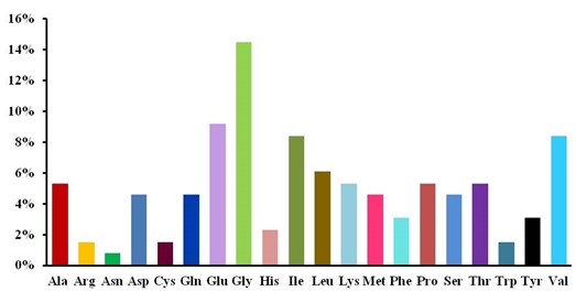
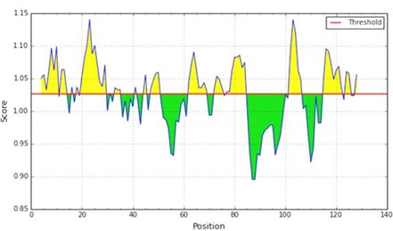
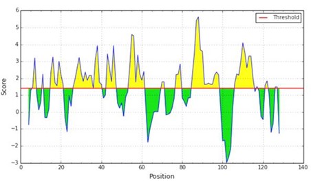
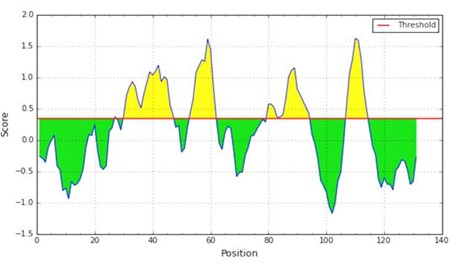
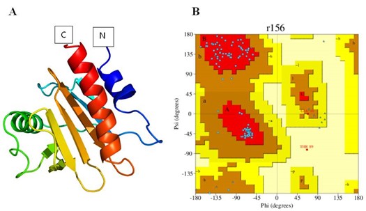
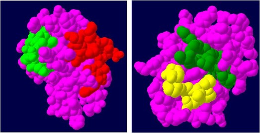
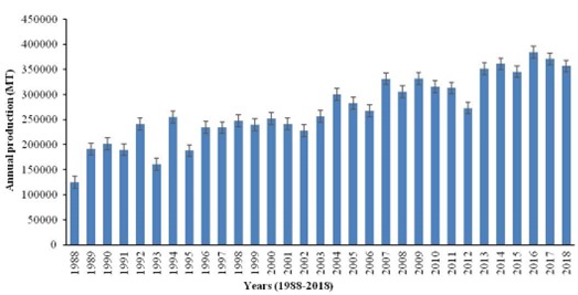
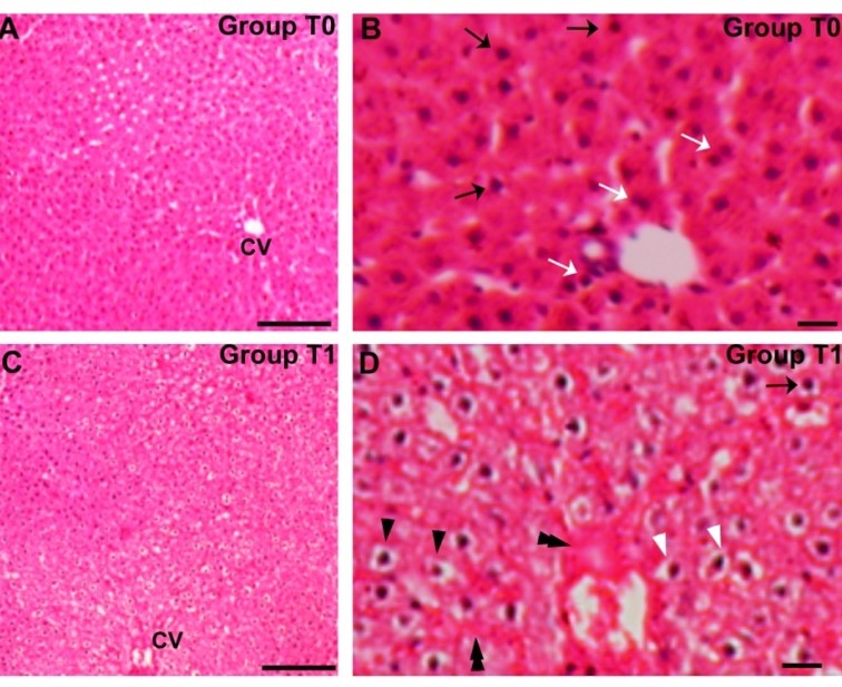
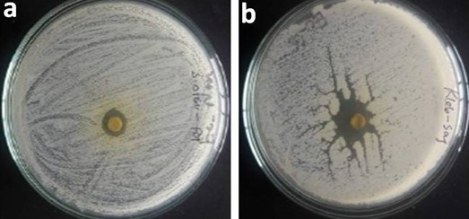
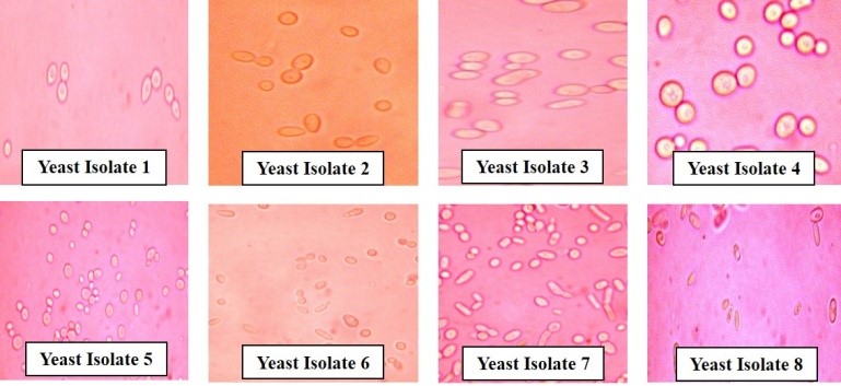
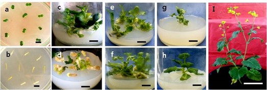
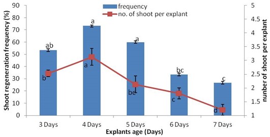
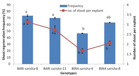
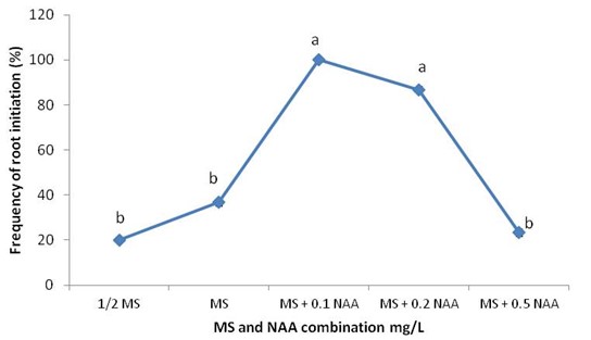
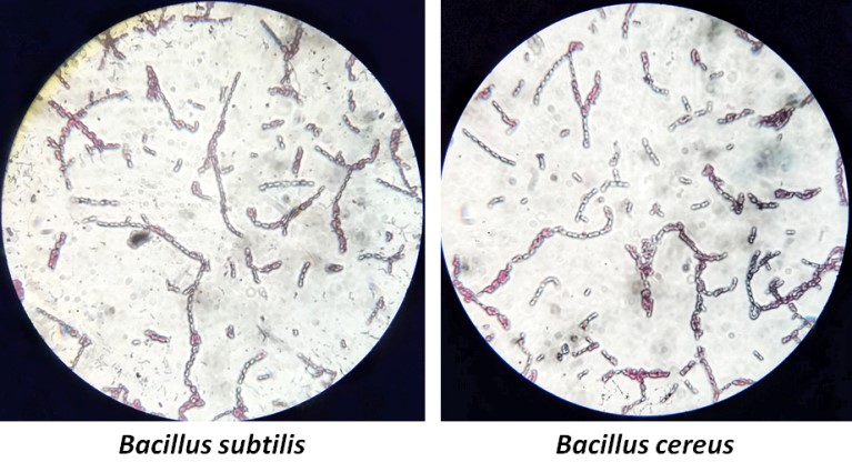
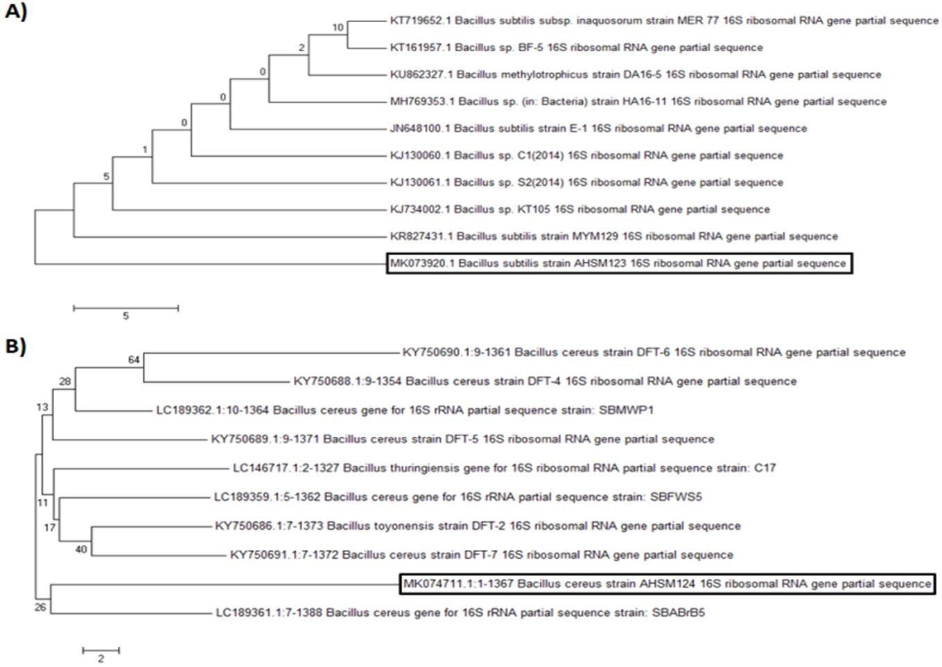
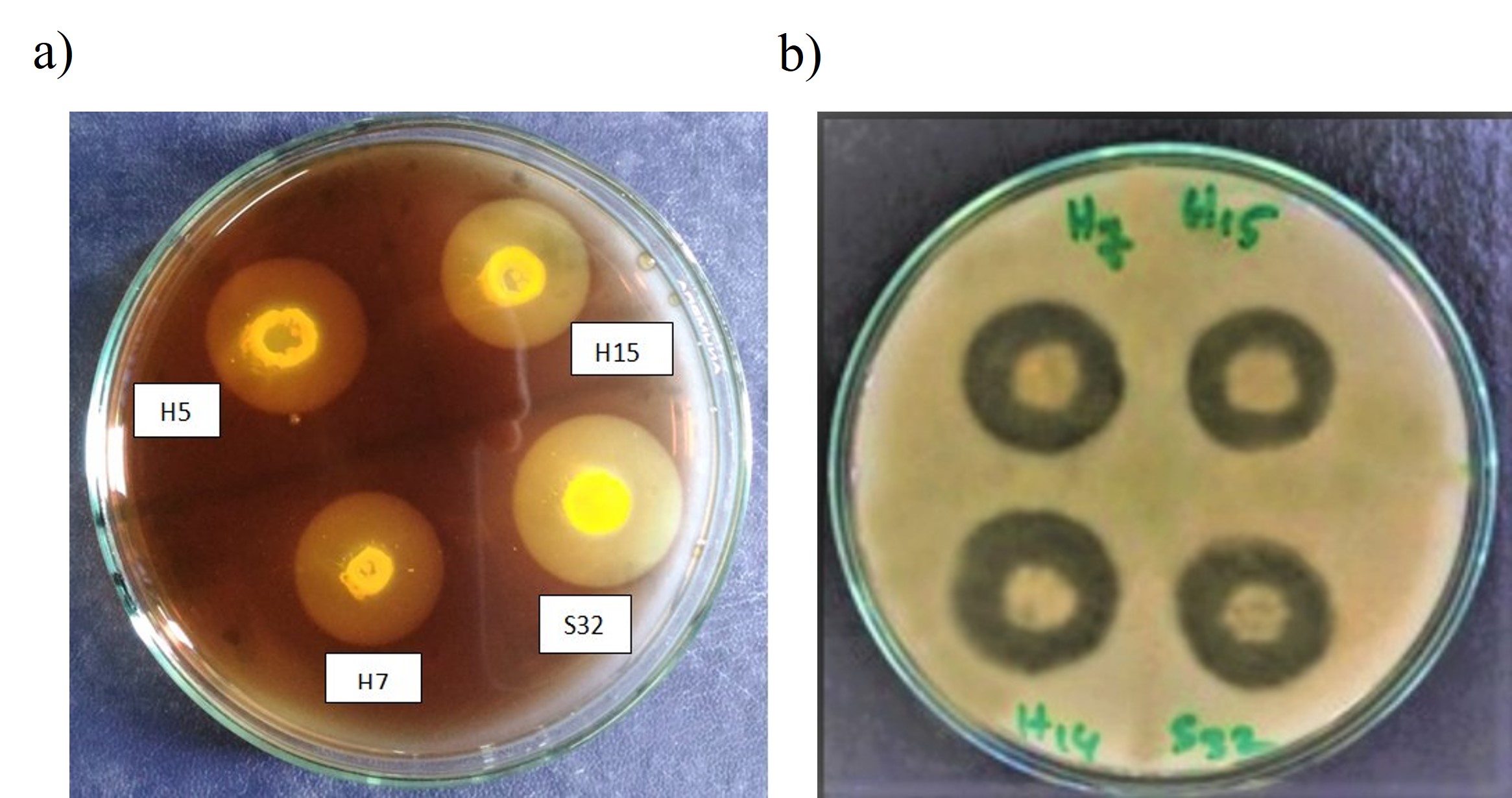
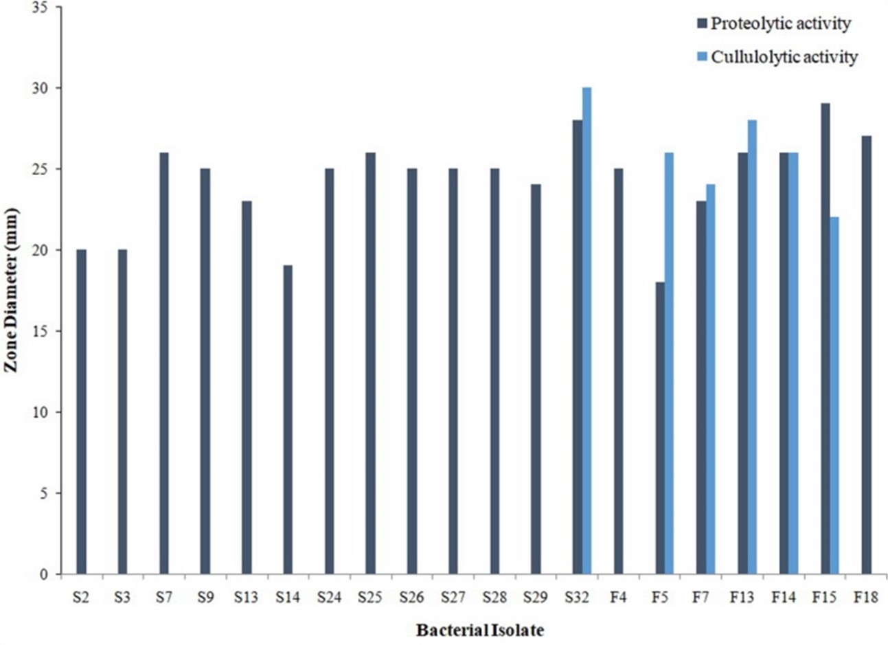
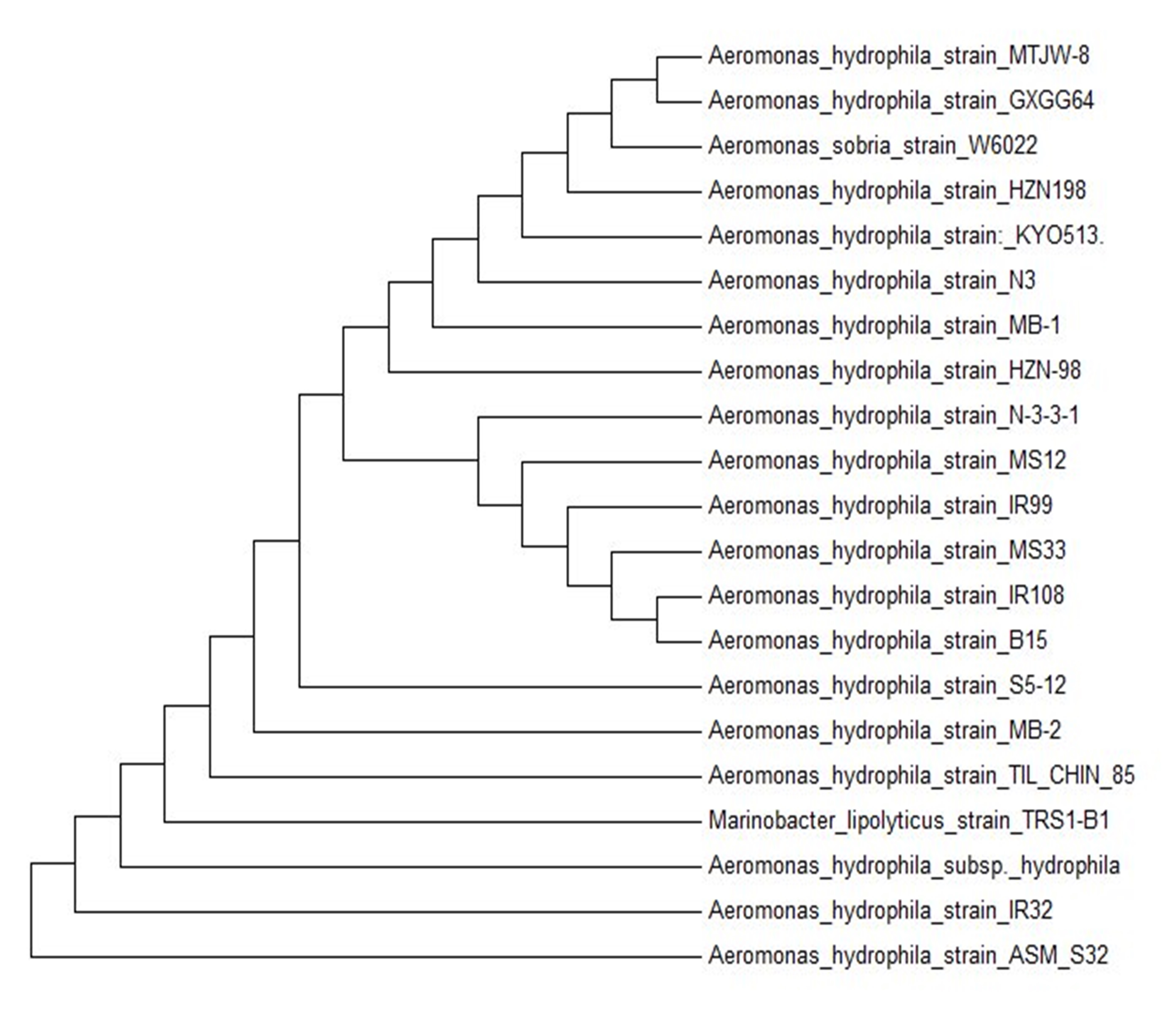
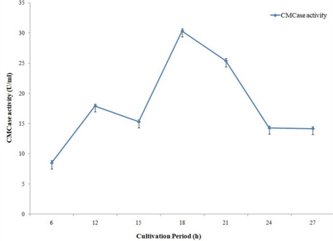
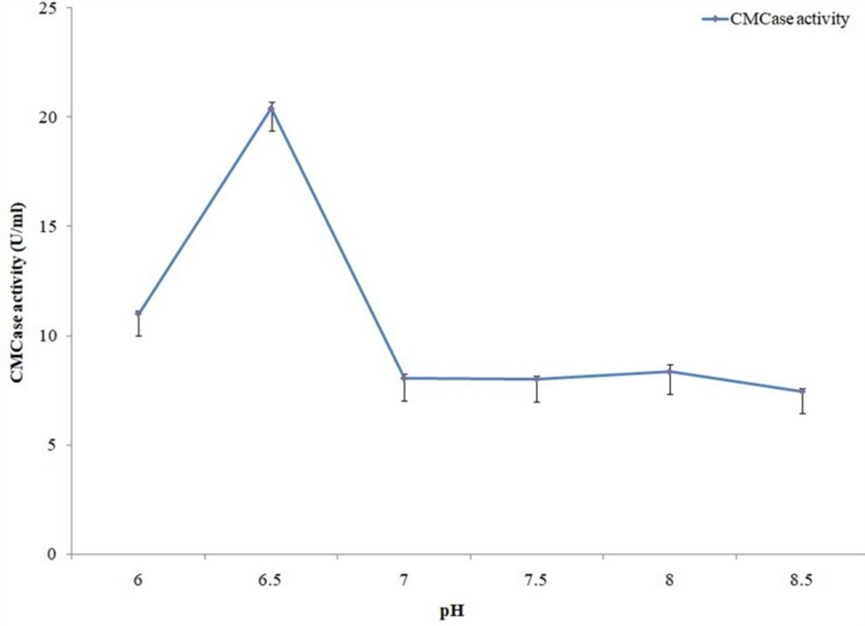
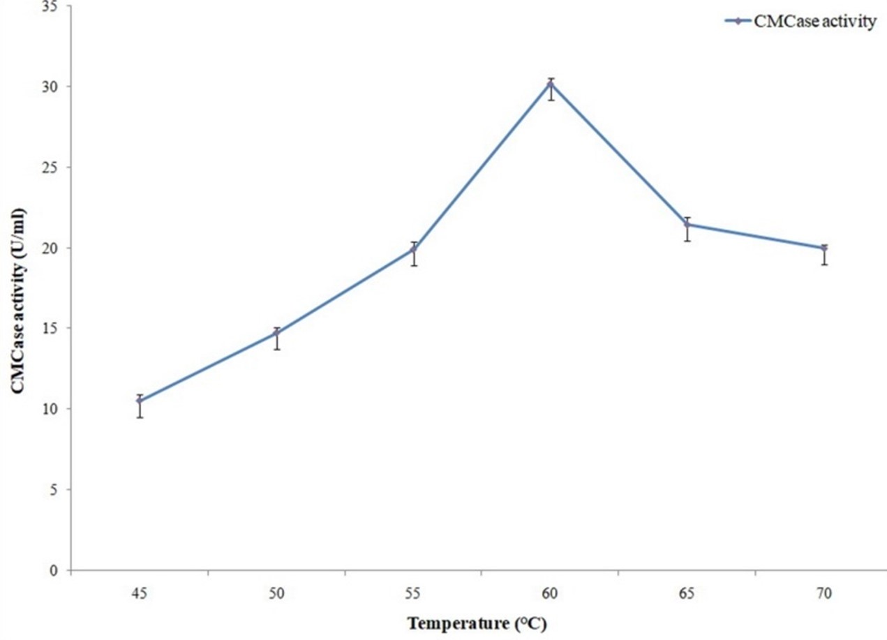
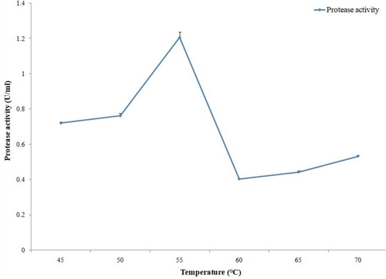
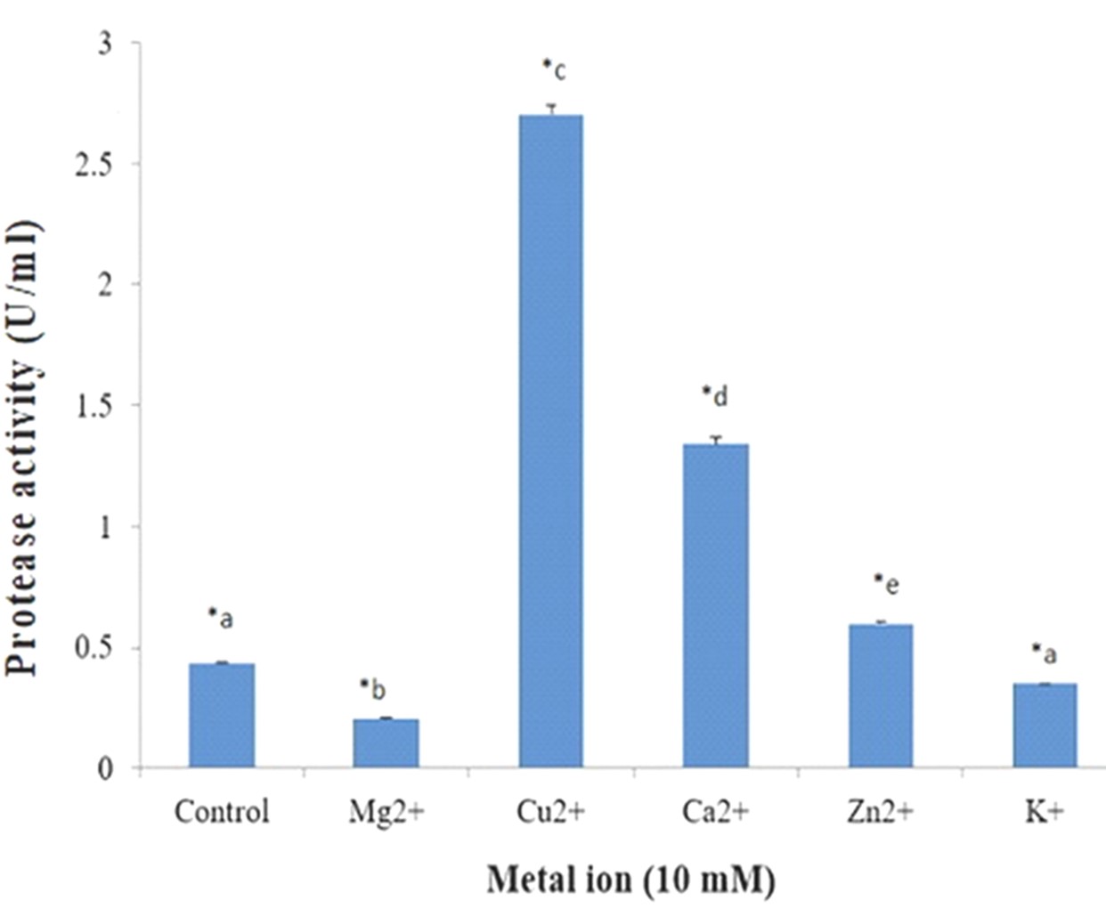
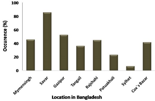
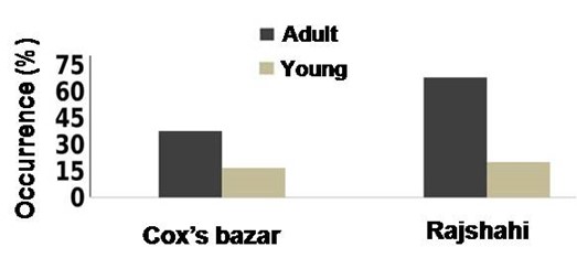
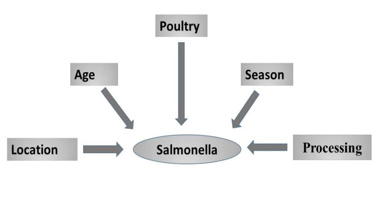
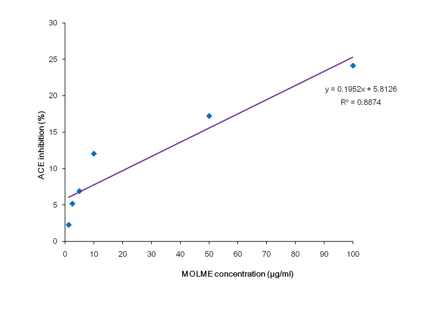
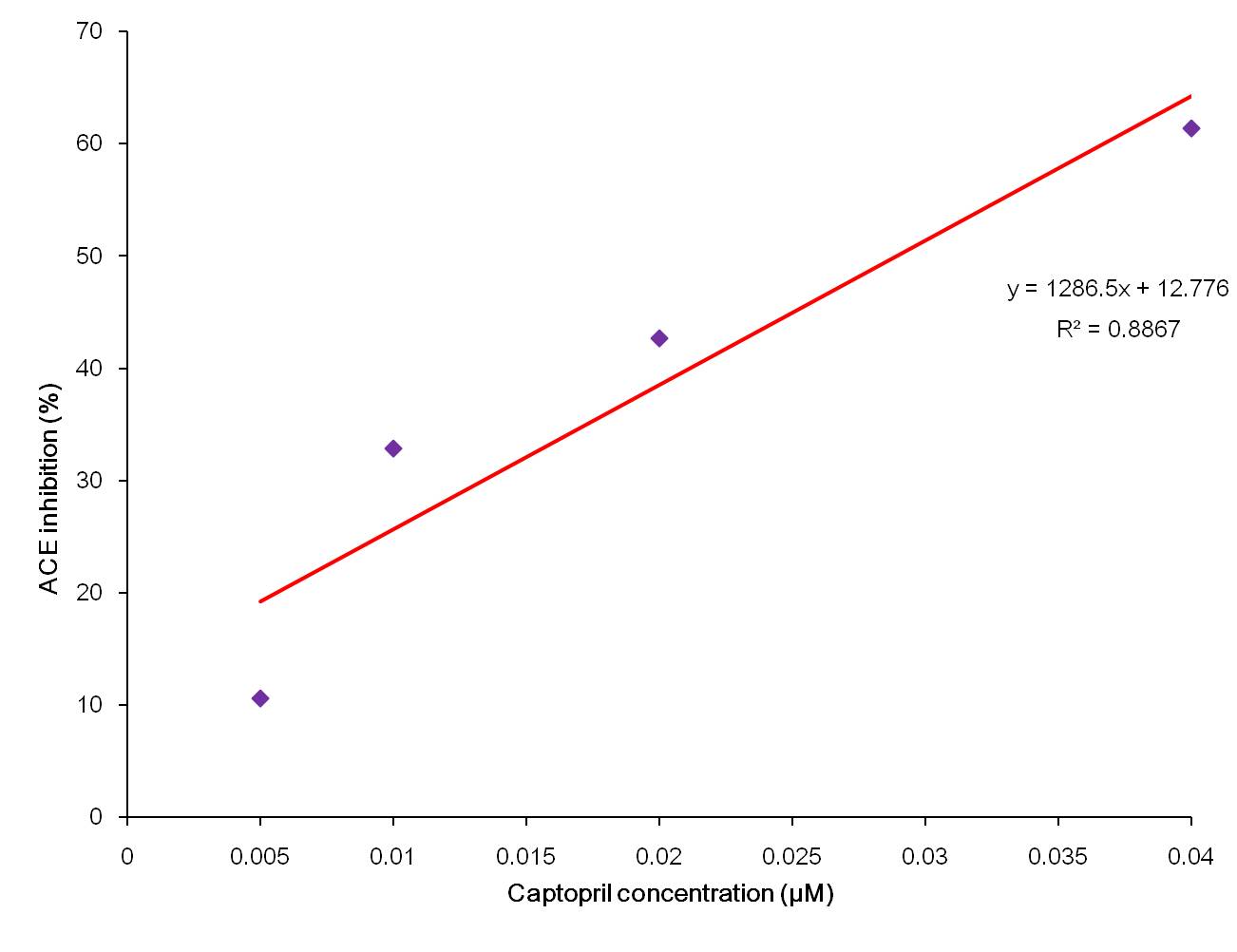
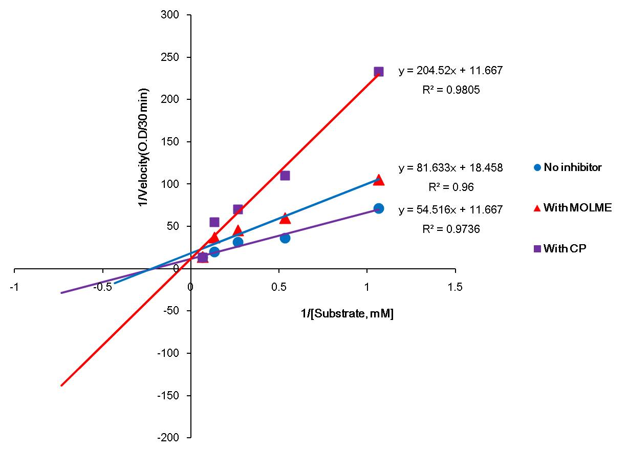
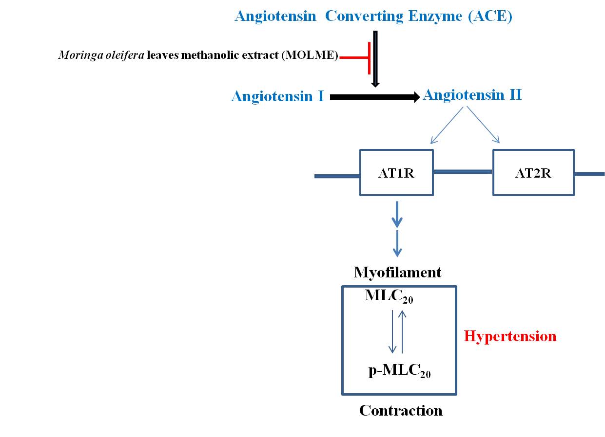