HTTP 200 OK
Allow: GET, HEAD, OPTIONS
Content-Type: application/json
Vary: Accept
{
"count": 319,
"next": "https://jabet.bsmiab.org/articles/?format=api&page=29",
"previous": "https://jabet.bsmiab.org/articles/?format=api&page=27",
"results": [
{
"id": 121,
"slug": "178-1577241188-the-medicinal-values-of-abrus-precatorius-a-review-study",
"featured": false,
"slider": false,
"issue": "Vol3 Issue2",
"type": "review_article",
"manuscript_id": "178-1577241188",
"recieved": "2019-12-25",
"revised": null,
"accepted": "2020-02-04",
"published": "2020-05-01",
"pdf_file": "https://jabet.bsmiab.org/media/pdf_file/2023/28/178-1577241188.pdf",
"title": "The medicinal values of Abrus precatorius: a review study",
"abstract": "<p> Herbal medicines are in great demand for preliminary health care due to their wide medicinal values without any side effects. Since many species are used in the system of traditional medicine, scientists have great opportunities to develop appropriate packages of practices for their multiplication and conservation. It is reported that the plant has a broad range of therapeutic potentials, like anti-bacterial, anti-fungal, anti-tumor, analgesic, anti-spasmodic, anti-diabetic, anti-serotonergic, anti-migraine, including treatment of inflammation, ulcers, wounds, throat scratches and sores. <em>Abrus precatorius </em>is the native plant of this Indian subcontinent and used in many ways in the traditional system of medicine considered as Ayurveda treatment. Ayurveda recommends the administration of the <em>Abrus precatorius</em> in disease like alopecia, edema, helminths, skin diseases, itching, urinary disorders, and anti-fertility. This review will help to explore the medicinal effects of the <em>Abrus precatorius</em> especially in the field of contraception which might be a remarkable activity in this realm. Therefore the present review is aimed to compile up the updated data and highlighting the special features on its pharmacological activities in various diseases.</p>",
"journal_reference": "J Adv Biotechnol Exp Ther. 2020; 3(2): 84-91.",
"academic_editor": "Dr. Akhi Moni, ABEx Bio-Research Center, East Azampur, Dhaka, Bangladesh.",
"cite_info": "Bhakta S, Das SK. The medicinal values of Abrus precatorius: a review study. J Adv Biotechnol Exp Ther. 2020; 3(2): 84-91.",
"keywords": [
"Medicinal values",
"pharmacological activities",
"Herbal medicine",
"Abrus precatorius"
],
"DOI": "10.5455/jabet.2020.d111",
"sections": [
{
"section_number": 1,
"section_title": "INTRODUCTION",
"body": "<p>Now a days, people are believing in the traditional medicine such as Ayurveda or others than that of the commercially available synthetic medicines. Ancient, natural health care, tribal practices, Ayurveda, sidha and unani are the part of traditional medicine [<a href=\"#r-1\">1</a>]. People of this subcontinent have been using the ayurvedic systems of medicine for many generations [<a href=\"#r-2\">2</a>]. Medicinal plants are so important for health care of human beings in respect to ancient medicine system. Most of the traditional medicines are based on herbs, which are used by almost 80% of the world’s populations. <em>Abrus precatorius</em> L. (family – leguminoseae and subfamily-Papilionaceae) is a native plant of India subcontinent and the East and West Indies [<a href=\"#r-3\">3</a>], in Hindi, it is known as Ratti or Gumchi. Plant parts such as leaf extracts is used for leucoderma, the seed having abrin is used as a purgative and abortive and the root extract used against coughs in the ayurvedic system of medicine [<a href=\"#r-4\">4</a>].<br />\r\nThe background of the discovery of many clinically useful drugs having medicinal values over the last two decades was the research of the scientists for finding out the new pharmacologically active ingredients from natural sources such as plants, animals and microbes. <em>Abrus precatorius</em> plant has been utilized as medicine from very ancient times not only in this subcontinent but also in China and other prehistoric cultures. In certain tribal regions people chew leaf of <em>Abrus precatorius</em> for the relief of the mouth ulcer. It also contains tri-terpenoid saponins and used in the treatment of inflammation, ulcers, wounds, throat scratches and sores [<a href=\"#r-5\">5</a>].</p>"
},
{
"section_number": 2,
"section_title": "CHEMICAL COMPOSITION",
"body": "<p>For the <em>Abrus precatorious</em> plant, different parts of it could be used and they have different sources of chemical constituents which have different medicinal effects on the body. Such as root, leaf and the seed of the fruit of the Abrus plant have different types of chemical constituents which help to explore the different biological effects for the treatment of different diseases. For example:<br />\r\nLeaf: Abrine, Abruslactone, Abrusoside A, B C, D, Inositol etc.<br />\r\nRoot: Abrol, Abrasine, Precasine, Precol etc.<br />\r\nSeed: Abrine, Abrin A, B, C, I, II, III, Abrus agglutinin, Saponin, Flavonoids, Abrectorin, Precatorin, Lectin, campestanol etc.</p>\r\n\r\n<div id=\"figure1\">\r\n<figure class=\"image\"><img alt=\"\" height=\"174\" src=\"/media/article_images/2024/21/21/178-1577241188-Figure1.jpg\" width=\"234\" />\r\n<figcaption><strong>Figure 1. </strong> Seed of <em>Abrus precatorius.</em></figcaption>\r\n</figure>\r\n</div>"
},
{
"section_number": 3,
"section_title": "ETHNOBOTANICAL USES",
"body": "<p><em>Abrus precatorius</em> has anti-suppurative properties. They are grinded with lime and poured on acne, sore and abscess. Decoction of leaves are taken orally for cough and flu [<a href=\"#r-6\">6, 7, 8</a>]. The roots of <em>Abrus precatorius</em> herb are useful for the treatment of jaundice and bile haemoglobinuric. Root paste is administered for the curation abdominal pains, recovery from tumors and also for inhibiting abortion. Grinded powder of roots of <em>Abrus precatorius</em> are taken with pure clarified butter thrice a day for four days to cure cough [<a href=\"#r-9\">9, 10</a>]. Root can be used as a remedy from snake bite by chewing [<a href=\"#r-11\">11</a>]. However, for using as an anti-malarial and anti-convulsant the extract of fresh root in hot water can be administered orally [<a href=\"#r-12\">12</a>], on the other hand liquid broth of dried root is taken orally for the treatment of bronchitis and hepatitis [<a href=\"#r-13\">13</a>]. Paste of leaves and seeds are applicable on head for graying of hair. Dried seeds of <em>Abrus precatorius</em> are grinded to powder and administered orally one teaspoonful once a day to cure the worm infestation for two days [<a href=\"#r-9\">9, 10</a>]. In veterinary section of medicine, dried seed powder of Abrus is used for the treatment of fractures. The brightly-colored seed of Abrus attracts the children, as a result sometimes the children at the rural villages who don’t have any knowledge about the plant and the origin of the seed they eat the seed and got attacked by the toxic effect of the seed of <em>Abrus precatorious</em> if the dose cross the safety limit. Boiled seeds are eaten in certain parts of India [<a href=\"#r-14\">14, 15</a>]. They have a weight of 1/10th of a gram which is almost uniform, hence used as weighing unit [<a href=\"#r-16\">16</a>]. Seeds also have some potential ingredients which are of good source of insecticide [<a href=\"#r-17\">17</a>] and antimicrobial [<a href=\"#r-18\">18</a>]. They are considered abortifacient [<a href=\"#r-19\">19, 20</a>], anodyne, aphrodisiac, antimicrobial, diuretic, emetic, expectorant, emollient, febrifuge, hemostat, laxative, purgative, refrigerant, sedative, vermifuge, antidote and used in various ailments to cure headache, snakebite, blennorrhagia, boil, cancer, cold, colic, conjunctivitis, convulsion, cough, diarrhea, fever, gastritis, gonorrhea, jaundice, malaria, night-blindness, ophthalmia, rheumatism, diabetes and chronic nephritis [<a href=\"#r-9\">9</a>]. Dry form of seeds are administered orally as an aphrodisiac [<a href=\"#r-21\">21, 22</a>]. Extract of seeds in hot water is taken orally for malaria [<a href=\"#r-23\">23</a>]. Dried seed powder are used by various African tribal groups as oral contraceptives [<a href=\"#r-6\">6, 7, 8</a>, <a href=\"#r-24\">24, 25, 26</a>]. Whereas, the Abrus seeds are also used to rid of tuberculosis and painful swellings [<a href=\"#r-27\">27</a>]</p>\r\n\r\n<div id=\"figure2\">\r\n<figure class=\"image\"><img alt=\"\" height=\"156\" src=\"/media/article_images/2024/21/21/178-1577241188-Figure2.jpg\" width=\"500\" />\r\n<figcaption><strong>Figure 2.</strong> Some chemical compounds for which Abrus precatorius disclaim the medicinal effects.</figcaption>\r\n</figure>\r\n</div>"
},
{
"section_number": 4,
"section_title": "ANCIENT USES",
"body": "<p>In the ayurvedic medicine leaf of <em>A. precatorius</em> has the potential as laxative, expectorant and aphrodisiac medicines. Seed is said to be purgative, emetic, tonic, antiphlogistic, aphrodisiac and antiopthalmic. For indigenous people, they are potent phytomedicines, many of them in mixtures with other plants. Their toxicity is underestimated. They are even sold via internet (Tan-Hord Exports List of herbs). In some countries of Asia beans are used as weights and jewellery is made from them by drilling. In Tanzania traditional healers claim the competence in the treatment of epilepsy. <em>A. precatorius</em> can be found between 60 plants commonly used against this illness [<a href=\"#r-28\">28</a>]. In Zimbabwe extracts of 58 plants popularly known to be effective against schistosomiasis were tested in vitro against excysted cysticercoids. Extracts of stem and root of <em>A. precatorius</em> were under the ten most effective samples [<a href=\"#r-29\">29, 30</a>]. In the Indian Central Drug Research Institute in Lucknow discussions about an antifertility program are going on. One of the plants with priority is <em>A. precatorius</em> because of its “estrogenicityî”, nor because of its lectins [<a href=\"#r-31\">31</a>]. In Germany necklaces from India were sold in the seventies, but soon warnings were propagated because of toxicity of the components. Investigations resulted that they were made with <em>A. precatorius</em> beans and warnings were propagated [<a href=\"#r-32\">32</a>]. In Christian countries the beans are used for wreaths of roses (precatory beans), for necklaces and for ornamentals together with other flowers in garlands. In China, the herb of <em>A. precatorius</em> is used as a form of folk-medicine or local medicine as a therapy of bronchitis, laryngitis and hepatitis. Because of their platelet inhibiting activity abruquinones are supposed to be the active substances [<a href=\"#r-33\">33</a>].</p>\r\n\r\n<p>Ricin originated from <em>Ricinus communis</em>, abrin originated from <em>Abrus precatorious</em> and other related toxins from different plant origins have played captivating and vital roles in the history of biomedical researches and clinical medicines. Utilization of these proteins in medical therapy since ancient times has been reviewed. Later on the proteins originated from those toxins, played important roles in the early days of immunological research. By using these toxin derived proteins of this group, some of the basic principles of immunological studies were discovered. <em>Abrus precatorius</em> was formerly used to weigh gems and precious stones. As per factual recordings the <em>Abrus precatorius</em> plant was used to weigh the famous Kohinoor Diamond as well. In a few parts of South America the seeds of this plant are used as necklaces for protecting children against illnesses particularly [<a href=\"#r-34\">34, 35</a>].<br />\r\nThis plant is native to the Himalayas in India and is also found in a few parts of Southern India. Today, the <em>Abrus precatorius</em> is also being harvested from Hawaii, southern parts of America, West Indies, as well as Africa. The seeds of this plant are small, scarlet in color with black spots around the hilum. The stout of the plant is rectangular in shape and has brownish pods that usually ripen after the winter season. The root of <em>Abrus precatorius</em> is woody and has a lot of branches [<a href=\"#r-36\">36, 37</a>].</p>\r\n\r\n<div id=\"figure3\">\r\n<figure class=\"image\"><img alt=\"\" height=\"263\" src=\"/media/article_images/2024/21/21/178-1577241188-Figure3.jpg\" width=\"500\" />\r\n<figcaption><strong>Figure 3. </strong>Schematic diagram of the sumamry of the chemical constituents and different uses of the leaf , root and seed of <em>Abrus Precatorious.</em></figcaption>\r\n</figure>\r\n</div>\r\n\r\n<div id=\"figure4\">\r\n<figure class=\"image\"><img alt=\"\" height=\"321\" src=\"/media/article_images/2024/21/21/178-1577241188-Figure4.jpg\" width=\"500\" />\r\n<figcaption><strong>Figure 4.</strong> Schematic diagram of probable mechanism of antifertility or contraceptive effects of <em>Abrus Precatorious</em>.</figcaption>\r\n</figure>\r\n</div>"
},
{
"section_number": 5,
"section_title": "ANTI-FERTILITY EFFECTS",
"body": "<p>In an antifertility program three indigenous plants (<em>Piper longum, Lawsonia inermis Abrus precatorius</em>) were studied with pregnant rats. Between these plants <em>Abrus precatorius</em> was the most effective one. A daily dose of 300mg showed a 40-60 % retardation of pregnancy of rats [<a href=\"#r-38\">38</a>]. An extract of <em>Abrus precatori</em>us seeds by using methanol, deteriorated the motility of washed human spermatozoa with an EC 50 of 2.29 mg/ml, irreversibly. The highest concentration which was tested was 20.0.mg/ml, which resulted the onset of the motility almost immediately. In contrary, this and other effects were not evident at a lower concentration than 5 mg/ml. Scientists of University of Colombo, Sri Lanka confirmed these results [<a href=\"#r-39\">39</a>]. Male albino rats treated with 50% methanol extract 250 mg/kg for 30 and 60 days became absolutely infertile. This was reversible. This treatment met the energy metabolism of the cauda epididymidis. Levels of acid succinic hydrogenase and phosphatase were significantly reduced, while protein and sialic acid appeared normal [<a href=\"#r-40\">40</a>]. The probable mechanism how the chemical constituents of <em>Abrus precatorious</em> worked as a potential for contraceptive issue is expressed as a schematic diagram in Figure 4.<br />\r\nIn a similar investigation male albino rats were treated with an ethanolic or methanolic seed extract of seeds of <em>Abrus precatorius</em> at a dose of 100 mg/kg for 60 days. The data of the experiment explained that cauda-epididymal sperm motility was significantly decreased. Although, there was no effect in the sperm concentration after 60 days of feeding with the alcoholic extract. Electron microscopic observation of morphology of sperm presented decapitation, damage of acrosome and bulging on sperm at the midpiece region, in treated rats. Energy metabolism altered due to the reduction in ATPase and succinate dehydrogenase activity for the extract allocation. Contrarily, after 60 days of administration testosterone levels in serum was noted significantly increased. Authors conclude that the decreased fertility rate was correlated with the reduction in the sperm motility, and altered morphology of the sperm in epididymis [<a href=\"#r-41\">41</a>].<br />\r\nIn testes of rats which were treated with the steroidal fraction of seeds of <em>Abrus precatorius,</em> the degenerative changes were observed, such as in case of weight of testis, sperm counting and spermatogenesis at later stages and Leydig cell counting. All of these observations are related to dose dependent reduction in enzymatic activity of hydroxysteroid dehydrogenase, glucose-6-phosphatdehydrogenase, sorbitol deydrogenase and leucin amino-peptidase.<br />\r\nThe steroidal fraction may exert their influence indirectly by a feedback reaction decreasing the production and release of testosterone, additionally [<a href=\"#r-42\">42</a>].<br />\r\nOil and crystalline steroidal fraction from the seeds of Abrus possess significant antifertility activity which has been reported by many scientists [<a href=\"#r-43\">43, 44</a>]. The leaf and root of this plant contain glycyrrhizin the active principle constituent of liquor ice. Seeds contain both water soluble –albumin and insoluble –globulin proteins. In case of Abrus, the active component ruptures histological frame due to which a large number of degenerative enzymes results in follicular atresia [<a href=\"#r-45\">45, 46</a>]. After revealing its importance as an antifertility agent, an attempt was made to investigate alteration in ovarian histo-architecture directed by Abrus for fertility regulation and control on Swiss Albino mice.<br />\r\nNormal function of male reproductive system is entirely dependent on the conventional functioning of the male reproductive organs and other accessory structures. The most important male reproductive organ is the testis, which is fundamentally responsible for the production of spermatozoa. The production of sperm occurs in the seminiferous tubules part of the testis, under the control of testosterone hormone, produced by the interstitial cells of the testis named Leydig cells. Testosterone production is directly proportional with the concentration (or activity) of leutinizing hormone (LH), in the milieu secreted by the anterior pituitary gland. Another type of hormone Follicular stimulating hormone (FSH), which is released also from the anterior pituitary, which stimulates another type of interstitial cells named the Sertoli cells of the testis, the function of whose is to give support and nourishment to developing spermatozoa. The quality and quantity of the produced spermatozoa will therefore depend on normal functioning of the structures testicles and associated reproductive hormones [<a href=\"#r-47\">47</a>].<br />\r\nOur group has worked on a combination herbal extract (<em>Ricinus communis</em>, <em>Abrus precatorius </em>and <em>Syzygium cumini</em>) which was applied on the male Swiss albino mice where we have found that the number of leydig and sertoli cells were reduced as a result the production of LH, FSH and testosterone is hindered and cause reduction in the sperm production [<a href=\"#r-48\">48</a>]. Some previous studies have mentioned that Oral administration of herbal extract over 4 weeks, caused significant (p≤0.05) effects on the levels of testosterone, LH and FSH in serum.<br />\r\nResearch conducted on the <em>Abrus precatorius</em> seeds concluded that the plant suppresses male reproductive functions reversibly [<a href=\"#r-49\">49, 50, 51</a>]. The extract affected oxidative metabolism of cauda epididymis, where levels of protein, sialic acid, acid phosphatase and succinic dehydroginase were significantly depleted. [<a href=\"#r-52\">52, 53, 54</a>] Jahan and his group detected an irreversible damage in sperm DNA integrity, thus suggested possible teratogenicity. [<a href=\"#r-55\">55, 56</a>].</p>"
},
{
"section_number": 6,
"section_title": "OTHER BIOLOGICAL EFFECTS",
"body": "<p><strong>Anti-diabetic activity</strong><br />\r\nAn ethno botanical survey of medicinal plants in five districts of Lagos State of Nigeria was conducted with the help of semi-structured questionnaire, which was reputed for the treatment of diabetes [<a href=\"#r-57\">57, 58</a>]. In the survey around 100 people responded, the respondents were from the predominantly Yoruba tribe [<a href=\"#r-59\">59</a>]. Among them mostly males (76%) were possessing knowledge regarding the traditional treatment of diabetes. About half of the respondents had experience of 20–30 years in treating diabetes by using mainly herbs (96%) unlikely the conventional treatment for diabetes. They also developed an effective and easily recognized diagnostic tool for diabetes. In the survey, fifty multi-component herbal recipes were covered which consisted of mainly liquid preparations. Those liquid form of the medications were often administered orally without any serious toxic or harmful side effects (92%). The principal antidiabetic plants [<a href=\"#r-60\">60</a>] include <em>Abrus precatorius</em>, leaf of <em>Abrus precatorius</em>, <em>Alchornea cordifoli</em>a and <em>Blighia sapida. </em>The leaves of the plants should be squeezed properly in water until the juice comes out, that prepared decoction can be used as a therapeutic approach for diabetes by using it as infusion [<a href=\"#r-61\">61</a>].</p>\r\n\r\n<p> </p>\r\n\r\n<p><strong>Anti-tumor activity</strong><br />\r\nThe ethanolic (95%) extract [<a href=\"#r-62\">62</a>] of dried leaves which was administered intra-peritoneal to mice, was observed inactive on Sarcoma 180 (ASC) AP07452. Whereas, the water extract of seeds, administered intra-peritoneal to mice was found active on Sarcoma (Yoshida solid and ASC) [<a href=\"#r-63\">63</a>]. But again when it was administered subcutaneously it was found inactive on Sarcoma (Yoshida ASC) AP01254.</p>\r\n\r\n<p> </p>\r\n\r\n<p><strong>Anti-serotonergic activity</strong><br />\r\nEthyl acetate extract of <em>Abrus precatorius</em> leaves had anti-serotonergic activity on a frog fundus strip by using sumatriptan as a standard [<a href=\"#r-64\">64</a>]. This plant also use as an abortifacient, anodyne, aphrodisiac, antimicrobial, antibacterial, diuretic, emetic, expectorant, febrifuge, hemostat, laxative, purgative, refrigerant, sedative, vermifuge [<a href=\"#r-65\">65</a>]. Studies indicated that their propensity to develop migraine61 headache. Moreover, fluctuating hormone levels indicate a migraine relation. The leaves of <em>Abrus precatorius</em> was on soxhlet extraction with ethyl acetate shown presence of alkaloids, carbohydrate, proteins, tannins, saponins and amino acids and antiserotonergic activity on frog fundus strip shown [<a href=\"#r-66\">66</a>] (Graded dose response) comparison with sumatriptan as a standard.</p>\r\n\r\n<p> </p>\r\n\r\n<p><strong>Anti-migraine activity</strong><br />\r\nAnti-migraine activity of Abrus precatorius proved by using male Wister albino rat and frog fundus muscle preparations using Sherrington rotating drum [<a href=\"#r-67\">67</a>]. Muscle contraction effect of petroleum ether and ethyl acetate crude extracts of <em>Abrus precatorius</em> performed on both muscle preparations.</p>\r\n\r\n<p> </p>\r\n\r\n<p><strong>Anti-microbial activity</strong><br />\r\nThe anti-microbial effects of <em>Abrus precatorius</em> extracts from leaves, stem and the seed oil were applied against some of the microorganisms <em>Staphylococcus aureus</em>, <em>Staphylococcus epidermidis</em>, <em>Enterococcus faecalis</em>, <em>Streptococcus anginosus</em>, <em>Bacillus subtilis</em>, <em>Corynebacterium spp,</em> <em>Escherichia coli</em>, <em>Klebsiella pneumoniae</em>, <em>Proteus mirabilis</em>, <em>Pseudomonas aeruginosa</em> and <em>Candida albicans</em> by using the agar well diffusion technique. It was observed that the extract of <em>A. precatorius</em> in aqueous, inhibited all the bacteria of test but had no effect on the fungal strain. The root extracts of the <em>Abrus precatorius L</em> also showed the anti-bacterial activity on various types of tested bacteria. Different solvent fractions or concentrations showed inhibitory activity against thirteen gram-positive and gram-negative bacteria. Through the bioautography assay it was observed that the antibacterial activity was localized to specific chromatophores in the chloroform fraction [<a href=\"#r-68\">68</a>]. Almost all the parts of the <em>Abrus precatorius </em>has the anti-bacterial activity.</p>\r\n\r\n<p> </p>\r\n\r\n<p><strong>Anti-malarial activity</strong><br />\r\nIsoflavanquinone and abruquinone was segregated from the extract of aerial parts of Abrus plant and demonstrated the anti-malarial activity. From the similar type of extract the antiplasmodial activity and cytotoxicity was also investigated during the assessment of antimalarial activity and <em>A. precatorius</em> extract also showed an IC 50 value below 20 g/ml74, which proves that the extract is quite highly potent for inhibiting some biological entities such as bacteria.</p>"
},
{
"section_number": 7,
"section_title": "SAFETY",
"body": "<p>There is an observation regarding the effect of the extract of <em>A. precatorius </em>on the hematology of the body. The study showed that the extract of the [<a href=\"#r-69\">69</a>] caused reduction in the levels of packed cell volume, concentration of Hemoglobin, Red Blood Cell (RBC) counts and also white blood cell (WBC) count. Beside that it was also demonstrated that the extract of caused decreased mean corpuscular volume and mean corpuscular haemoglobin [<a href=\"#r-70\">70, 71</a>]. Also the biochemical properties of the treated animal was observed which was administered with the extract of the<em> Abrus precatorious</em>. It was observed that the extract also resulted in increased levels of total serum protein, albumin, alanine amino transaminase, aspartate amino transferase, alkaline phosphatase and total bilirubin. Aqueous extract of <em>Abrus precatorius</em> showed that it was toxic and special care should be taken during the usage of the extract. Fatal poisoning in children has been reported after the thorough chewing of one seed. They were highly toxic and cause severe stomach cramping accompanied by nausea, severe diarrhea, cold sweats, tachycardia, coma and circulatory collapse. The loss of abrin (IP) in mice was found to be 8.34 mg/kg. Prolonged administration of abrin in mice produced initial anemia, which normalized at the end of the experiment, and an increase in white blood cell count. Intra-peritoneal injection of abrin to pregnant rats produced both maternal and fetal changes, whereas abrin given orally produced significant fetal effects. It was observed that the ethanol-water proportional (1:1) extract of the aerial parts of Abrus was much less toxic and whereas the only ethanolic (95%) extract of the dried leaves, produced a loss of 12 mg/kg body weight in the chickens where the extract was administered.</p>"
},
{
"section_number": 8,
"section_title": "FUTURE PROSPECTS",
"body": "<p>People are becoming more and more dependent on the natural sources of medicine which is increasing the use of Abrus L. like such type of medicinal plants, which has a handful use in the form of medicine. Already many researchers have worked on the different biological effects of this herbal plant for the cure of different diseases, which has proven its medicinal value. More researches should be conducted to make this plant extract commercially available in near future and make it the most reliable form of medicine for the treatment different diseases. Hopefully in the near future commercial products of <em>Abrus precatorious </em>will be found which will render no toxic or side effects to the consumers and will explore a new era of medicine.</p>"
},
{
"section_number": 9,
"section_title": "CONCLUSION",
"body": "<p>Herbal medicine is the use of plants (herbs) to treat disease and enhance wellbeing. Herbal medicine is used to treat a range of disorders including anxiety, arthritis, depression, high blood pressure, insomnia, hormonal imbalances, migraines, skin problems such as eczema and other disorders. Herbs can act on the body as powerfully as pharmaceutical drugs and need to be treated with care. Herbs are administered by a herbalist or herbal therapist.<br />\r\nWhile some people may dismiss herbal remedies as quackery, the use of botanicals is well rooted in medical practice. Ancient doctors methodically collected information about herbs and developed well-defined pharmacopoeias to treat a variety of ailments. In the recent times more than a quarter of all drugs of the commercially available synthetic medicines contain active ingredients which are derived from the plants which were available since the ancient time.<br />\r\nMany drugs are available there which have got the entrance in to the international market by the exploration of ethno-pharmacological activity, and considered as the traditional medicine. This review article exposes that <em>A. precatorius</em> is an exclusive source of many vital phytochemicals which makes this plant very unique and versatile for its large number of pharmaceutical properties i.e. antidiabetic, neuro-protective, anti-microbial, analgesic and some others. Hence comprehensive researches are in demand of the recent times with this highly medicinal quality contained plant material. It is high time to exploit the therapeutic utility of <em>Abrus precatorius </em>to combat against various diseases. It can be concluded by analyzing the above collected literature that <em>Abrus precatorius </em>is a promising candidate as a multipurpose medicinal agent because it possesses a high potential pharmacognostical and pharmacological applications.</p>"
},
{
"section_number": 10,
"section_title": "ACKNOWLEDGEMENT",
"body": "<p>The research work was supported with the grants from the Ministry of Education and Ministry of Science and Technology, Bangladesh.</p>"
},
{
"section_number": 11,
"section_title": "AUTHOR CONTRIBUTIONS",
"body": "<p>Sonali Bhakta carried out the study and wrote the initial draft of the manuscript. Shonkor Kumar Das designed and supervised the research work and revised the manuscript. The manuscript was carefully read by both the authors before the submission process.</p>"
},
{
"section_number": 12,
"section_title": "CONFLICTS OF INTEREST",
"body": "<p>The authors declare that there is no conflict of interest towards the publication of this article.</p>"
}
],
"figures": [
{
"figure": "https://jabet.bsmiab.org/media/article_images/2024/21/21/178-1577241188-Figure1.jpg",
"caption": "Figure 1. Seed of Abrus precatorius.",
"featured": false
},
{
"figure": "https://jabet.bsmiab.org/media/article_images/2024/21/21/178-1577241188-Figure2.jpg",
"caption": "Figure 2. Some chemical compounds for which Abrus precatorius disclaim the medicinal effects.",
"featured": false
},
{
"figure": "https://jabet.bsmiab.org/media/article_images/2024/21/21/178-1577241188-Figure3.jpg",
"caption": "Figure 3: Schematic diagram of the sumamry of the chemical constituents and different uses of the leaf , root and seed of Abrus Precatorious.",
"featured": false
},
{
"figure": "https://jabet.bsmiab.org/media/article_images/2024/21/21/178-1577241188-Figure4.jpg",
"caption": "Figure 4. Schematic diagram of probable mechanism of antifertility or contraceptive effects of Abrus Precatorious.",
"featured": false
}
],
"authors": [
{
"id": 490,
"affiliation": [
{
"affiliation": "Bioresearch laboratory, Department of Anatomy and Histology, Faculty of Veterinary Science, Bangladesh Agricultural University, Mymensingh-2202"
}
],
"first_name": "Sonali",
"family_name": "Bhakta",
"email": "sonali.dvm@gmail.com",
"author_order": 1,
"ORCID": null,
"corresponding": true,
"co_first_author": false,
"co_author": false,
"corresponding_author_info": "Sonali Bhakta, Department of Anatomy and Histology, Faculty of Veterinary Science, Bangladesh Agricultural University, Mymensingh-2202, Tel.:+8801717620673, Email: sonali.dvm@gmail.com",
"article": 121
},
{
"id": 491,
"affiliation": [
{
"affiliation": "Bioresearch laboratory, Department of Anatomy and Histology, Faculty of Veterinary Science, Bangladesh Agricultural University, Mymensingh-2202"
}
],
"first_name": "Shonkor Kumar",
"family_name": "Das",
"email": null,
"author_order": 2,
"ORCID": null,
"corresponding": false,
"co_first_author": false,
"co_author": false,
"corresponding_author_info": "",
"article": 121
}
],
"views": 5397,
"downloads": 267,
"references": [
{
"id": 3908,
"serial_number": 1,
"pmc": null,
"reference": "Pokharkar R, Saraswat R, Bhavare V, Kanawade M. GCMS studies of Abrus precatorius. Pharmacologyonline, 2011, 2: 1178-1189.",
"DOI": null,
"article": 121
},
{
"id": 3909,
"serial_number": 2,
"pmc": null,
"reference": "Bapat SP, Sane RT. Bio analytical studies on the process of detoxification and safety evaluation of Aconitum laciniatum and Abrus precatorius for use in ayurvedic preparation, International Journal of Pharmaceutical Sciences and Research, 2012, 3: 914-921.",
"DOI": null,
"article": 121
},
{
"id": 3910,
"serial_number": 3,
"pmc": null,
"reference": "Chadha YR. The Wealth of India, Raw Materials, CSIR Publication, New Delhi, India, 1988, pp. 18-21.",
"DOI": null,
"article": 121
},
{
"id": 3911,
"serial_number": 4,
"pmc": null,
"reference": "Nadkarni, K. M. (1976). Indian Materia Medica, vol. I, Popular Prakashan, Bombay, p 5.",
"DOI": null,
"article": 121
},
{
"id": 3912,
"serial_number": 5,
"pmc": null,
"reference": "Anant S, Maitreyi Z. Pharmacognosy, phytochemistry and pharmacology of Abrus precatorius leaf: a review. International Journal of Pharmaceutical Sciences Review and Research, 2012, 13(2): 71-76.",
"DOI": null,
"article": 121
},
{
"id": 3913,
"serial_number": 6,
"pmc": null,
"reference": "Chakre OJ. Wealth of India: Raw materials (I–X), Council of Scientific and Industrial Research, New Delhi. 1948-1976.",
"DOI": null,
"article": 121
},
{
"id": 3914,
"serial_number": 7,
"pmc": null,
"reference": "Nadkarni AK, Nadkarni KM. Indian Materia Medica, Popular Prakashan, Bombay, India. 1954; 1: 776-784.",
"DOI": null,
"article": 121
},
{
"id": 3915,
"serial_number": 8,
"pmc": null,
"reference": "Chopra RN, Nayar SL, Chopra IC. Glossary of Indian Medicinal Plants, CSIR, New Delhi. 1956.",
"DOI": null,
"article": 121
},
{
"id": 3916,
"serial_number": 9,
"pmc": null,
"reference": "Rain-tree, 2004. http://www.rain-tree.com/abrus.htm.",
"DOI": null,
"article": 121
},
{
"id": 3917,
"serial_number": 10,
"pmc": null,
"reference": "Kirtikar KR, Basu BD. Indian Medicinal Plants, International Book Distributors, Dehra Dun. 1956.",
"DOI": null,
"article": 121
},
{
"id": 3918,
"serial_number": 11,
"pmc": null,
"reference": "Watt JM, MG Breyer-Brandwijk. The medicinal and poisonous plants of Southern and Eastern Africa, 2nd Ed, E. S. Livingstone, Ltd., London. 1962.",
"DOI": null,
"article": 121
},
{
"id": 3919,
"serial_number": 12,
"pmc": null,
"reference": "Adesina SK. Studies on some plants used as anticonvulsants in Amerindian and African traditional medicine. Fitoterapia 1982; 53:147–162.",
"DOI": null,
"article": 121
},
{
"id": 3920,
"serial_number": 13,
"pmc": null,
"reference": "Chukuo S, Chen SC, Chen LH, Wu JB, Wang JP, Teng CM. Potent anti-platelet, anti-inflammatory and anti-allergic isoflavanquinones from the roots of Abrus Precatorius. Plant Medica 1995; 61 (4):307-312.",
"DOI": null,
"article": 121
},
{
"id": 3921,
"serial_number": 14,
"pmc": null,
"reference": "Rajaram N, Janardhanan K. The chemical composition and nutritional potential of the tribal pulse, Abrus Precatorius , Plant Foods Hum Nutr. 1992; 42(4):285-90.",
"DOI": null,
"article": 121
},
{
"id": 3922,
"serial_number": 15,
"pmc": null,
"reference": "Pandey VN. Leaf protein content and yield of some Indian legumes. Plant Foods for Human Nutrition 1994; 46(4):313-322.",
"DOI": null,
"article": 121
},
{
"id": 3923,
"serial_number": 16,
"pmc": null,
"reference": "Tropilab 2004. http://www.tropilab.com/companyprofile.html",
"DOI": null,
"article": 121
},
{
"id": 3924,
"serial_number": 17,
"pmc": null,
"reference": "Khanna P, Kaushik P. New sources of insecticides: Rotenoids. Proceedings of the National Academy of Sciences 59(1): 83-86.",
"DOI": null,
"article": 121
},
{
"id": 3925,
"serial_number": 18,
"pmc": null,
"reference": "Saxena AP, Vyas KM. Antimicrobial activity of seeds of some ethnomedicinal plants. J Eco and Tax Bot. 1986; 8(2): 291-300.",
"DOI": null,
"article": 121
},
{
"id": 3926,
"serial_number": 19,
"pmc": null,
"reference": "Nath D, Sethi N. Commonly used Indian abortifacient plants with special reference to their teratologic effects in rats. J Ethnopharm. 1992; 36(2):147-154.",
"DOI": null,
"article": 121
},
{
"id": 3927,
"serial_number": 20,
"pmc": null,
"reference": "Hikino H, K Aota, T Takemoto. Structure and absolute configuration of cyperotundone. Chem Pharm Bull 1966; 14: 890.",
"DOI": null,
"article": 121
},
{
"id": 3928,
"serial_number": 21,
"pmc": null,
"reference": "Elisabetsky E, W Figueiro, G Oliveria. Traditional Amazonian nerve tonics as antidepressant agents. Chaunochiton Kappleri A case study. J Herbs Spices Med Plants 1992; (1/2): 125–162.",
"DOI": null,
"article": 121
},
{
"id": 3929,
"serial_number": 22,
"pmc": null,
"reference": "Chopra RN. Indigenous drugs of India their medical and economic aspects, the art press, Calcutta, India. 1933; 550.",
"DOI": null,
"article": 121
},
{
"id": 3930,
"serial_number": 23,
"pmc": null,
"reference": "Burkhill IH. Dictionary of the economic products of the Malay peninsula. Ministry of Agriculture and Cooperatives, Kuala Lumpur, Malaysia. 1966.",
"DOI": null,
"article": 121
},
{
"id": 3931,
"serial_number": 24,
"pmc": null,
"reference": "Chopra RN. Indigenous drug of India, UN Dhar & Sons Pvt. Ltd., Calcutta.",
"DOI": null,
"article": 121
},
{
"id": 3932,
"serial_number": 25,
"pmc": null,
"reference": "Malhi BS; VP Trivedi. Vegetable antifertility drugs of India. 1958.",
"DOI": null,
"article": 121
},
{
"id": 3933,
"serial_number": 26,
"pmc": null,
"reference": "Das SK. Medicinal, Economic and useful plants of India. Bally seed store, West Bengal. 1955.",
"DOI": null,
"article": 121
},
{
"id": 3934,
"serial_number": 27,
"pmc": null,
"reference": "Arseculeratne SN, Gunatilaka AAL, Panabokke RG. Studies on medicinal plants of Sri Lanka Part 14, Toxicity of some traditional medicineal herbs. J Ethnopharmacol 1985; 13(3): 323–335.",
"DOI": null,
"article": 121
},
{
"id": 3935,
"serial_number": 28,
"pmc": null,
"reference": "Moshi MJ, Kagashe GA, Mbwambo ZH. Plants used to treat epilepsy by Tanzanian traditional healers J Ethnopharmacol, 2005; 97(2): 327-36",
"DOI": null,
"article": 121
},
{
"id": 3936,
"serial_number": 29,
"pmc": null,
"reference": "Molgaard B, Nielsen SB et al. Anthelmintic screening of Zimbabwean plants traditionally used against schistosomiasis J Ethnopharmacol. 2001; 74(3): 257-64",
"DOI": null,
"article": 121
},
{
"id": 3937,
"serial_number": 30,
"pmc": null,
"reference": "Ndamba J, Nyazema N et al. Traditional herbal remedies used for the treatment of urinary schistosomiasis in Zimbabwe J Ethnopharmacol.1994; 42(2): 125-32",
"DOI": null,
"article": 121
},
{
"id": 3938,
"serial_number": 31,
"pmc": null,
"reference": "Kamboj VP, Dhawan BN. Research on plants for fertility regulation in India J Ethnopharmacol. 1982; 6(2): 191-226.",
"DOI": null,
"article": 121
},
{
"id": 3939,
"serial_number": 32,
"pmc": null,
"reference": "Stahl E. Strychnin in indischen und afrikanischen Schmuckketten Deutsche Apoth Ztg . 1972; 117: 1107-11011.",
"DOI": null,
"article": 121
},
{
"id": 3940,
"serial_number": 33,
"pmc": null,
"reference": "[33] Kuo SC, Chen SC, Chen LH et al. Potent antiplatelet, antiin-flammatory and antiallergic isoflavanquinones from the roots of A. precatorius Planta Med. 1995; 61: 07- 12.",
"DOI": null,
"article": 121
},
{
"id": 3941,
"serial_number": 34,
"pmc": null,
"reference": "Chandraker SK, Sharma N. Abrus precatorius – an important medicinal plant: a review. IJPRBS, 2014, 3(4): 657-670.",
"DOI": null,
"article": 121
},
{
"id": 3942,
"serial_number": 35,
"pmc": null,
"reference": "Munshi SR, ShetyeTA, Nair K. Antifertility activity of three indigenous plant preparations. Planta Med, 1977, 31(l): 73-5",
"DOI": null,
"article": 121
},
{
"id": 3943,
"serial_number": 36,
"pmc": null,
"reference": "Ndamba J, Nyazema N et al. Traditional herbal remedies used for the treatment of urinary schistosomiasis in Zimbabwe. J Ethnopharmacol, 1994, 42(2): 125-32.",
"DOI": null,
"article": 121
},
{
"id": 3944,
"serial_number": 37,
"pmc": null,
"reference": "Ratnasooriya WD, Amarasekera AS et al. Sperm antimotility properties of a seed extract of Abrus precatorius J Ethopharmacol, 1999, 33(1-2): 85-90.",
"DOI": null,
"article": 121
},
{
"id": 3945,
"serial_number": 38,
"pmc": null,
"reference": "Sinha R. Post-testicular antifertility effects of Abrus precatorius seed extract in albino rats J Ethopharmacol, 1990, 28(2): 173-5.",
"DOI": null,
"article": 121
},
{
"id": 3946,
"serial_number": 39,
"pmc": null,
"reference": "Rao MV. Antifertility effects of alcoholic seed extracts of Abrus precatorius Linn. in male albino rats. Acta Eur Fertil, 1987, 18(3): 217-20",
"DOI": null,
"article": 121
},
{
"id": 3947,
"serial_number": 40,
"pmc": null,
"reference": "Sinha S, Mathur RS. Effect of steroidal fraction of seeds of Abrus precatorius Linn. on rat testis. Indian J Exp Biol, 1990, 28(8): 752-6",
"DOI": null,
"article": 121
},
{
"id": 3948,
"serial_number": 41,
"pmc": null,
"reference": "Jahan S, Rasool S, Khan MA, Ahemad M. Antifertility effects of ethanolic seed extract of Abrus precatorius L. on sperm production and DNA integrity in adult male mice. Journal of Medicinal Plant research, 2009, 3 (10): 809-814.",
"DOI": null,
"article": 121
},
{
"id": 3949,
"serial_number": 42,
"pmc": null,
"reference": "Zia-Ul-Haque A, Qazi MH, Hamdard ME. Studies on the antifertility properties of active components isolated from the seeds of Abrus precatorius Linn. Pakistan Journal of Zoology, 1983,15(2):129-139.",
"DOI": null,
"article": 121
},
{
"id": 3950,
"serial_number": 43,
"pmc": null,
"reference": "Samad F, Mukhtar A, Jan ZA, Khan ZU. Effect of alcohol extract of Ratti seeds (Abrus precatorius) on the reproduction of female rats. Journal of Mathematical Sciences 1974, 12:157.",
"DOI": null,
"article": 121
},
{
"id": 3951,
"serial_number": 44,
"pmc": null,
"reference": "Bhaduri B, Ghose CR, Bose AN, Moza BK, Basu UP. Antifertility Activity of some Medicinal Plants, Indian Journal of Experimental Biology. 1968, 6:252-253",
"DOI": null,
"article": 121
},
{
"id": 3952,
"serial_number": 45,
"pmc": null,
"reference": "Rao MV., Antifertility effects of alcoholic seeds extract of Abrus precatorius Linn. in male albino rats, Acta Europaea fertilitatis 1987;18:3:217-220.",
"DOI": null,
"article": 121
},
{
"id": 3953,
"serial_number": 46,
"pmc": null,
"reference": "Sinha R., Post-testicular antifertility effects of Abrus precatorius seed extract in albino rats, Journal of Ethnopharmacology 1990;28:2:173-181.",
"DOI": null,
"article": 121
},
{
"id": 3954,
"serial_number": 47,
"pmc": null,
"reference": "Das PC., Oral contraceptive (Longacting), Patent-Brit1445599, 1976;11.",
"DOI": null,
"article": 121
},
{
"id": 3955,
"serial_number": 48,
"pmc": null,
"reference": "Agarwal SS., Ghatak N., Arora RB., Antifertility activity of the roots of Abrus precatorius, Pharmacological Research Communications 1970;2:159164",
"DOI": null,
"article": 121
},
{
"id": 3956,
"serial_number": 49,
"pmc": null,
"reference": "Saganuwan SA, Onyeyili PA. Biochemical effects of aqueous leaf extract of Abrus precatorius (Jecquirity bean) in Swiss albino mice. Herba Rolonica. 2010; 56(3): 63-80",
"DOI": null,
"article": 121
},
{
"id": 3957,
"serial_number": 50,
"pmc": null,
"reference": "Atuboyedia WO, Jonah SA, Chinagoro TOE. Antifertility effects of aqueous extract of Ocimum gratissimum L. leaves in male mice. Journal of Medicial plants Research. 2010; 4(9): 809-816.",
"DOI": null,
"article": 121
},
{
"id": 3958,
"serial_number": 51,
"pmc": null,
"reference": "Bhakta S, Awal A, Das SK. Herbal contraceptive effect of Abrus precatorius, Ricinus communis, and Syzygium aromaticum on anatomy of the testis of male Swiss albino mice. J Adv Biotechnol Exp Ther. 2019; 2(2): 36-43.",
"DOI": null,
"article": 121
},
{
"id": 3959,
"serial_number": 52,
"pmc": null,
"reference": "Bhatt N, Chawla SL, Rao MV 2007. Contraceptive evaluation of seed extract of Abrus. Journal of herbal medicine and toxicology, 2001, 1(1): 47-50.",
"DOI": null,
"article": 121
},
{
"id": 3960,
"serial_number": 53,
"pmc": null,
"reference": "Sinha R. Post-testicular antifertility effects of Abrus precatorius seed extract in albino rats. Journal of Ethnopharmacology. 1990, 28(2): 173-81.",
"DOI": null,
"article": 121
},
{
"id": 3961,
"serial_number": 54,
"pmc": null,
"reference": "Rao MV. Antifertility effects of alcoholic seed extract of Abrus precatorius Linn. in male albino rats. Acta Euopaear Fertliitica, 1987, 8(3): 217-20.",
"DOI": null,
"article": 121
},
{
"id": 3962,
"serial_number": 55,
"pmc": null,
"reference": "Sinha S, Mathur RS. Effect of steroidal fraction of seeds of Abrus precatorius Linn. on rat testis. Indian Journal Experimental Biology, 1990, 28(8): 752-6.",
"DOI": null,
"article": 121
},
{
"id": 3963,
"serial_number": 56,
"pmc": null,
"reference": "Jahan S, Rasool S, Khan MA, Ahmad M, Zafar M, Arsahd M, Abbasi AM. Antifertiltity effect of ethanolic seed extract of Abrus precatorius L on sperm production and DNA integrity in adult male mice. Journal of Medical Plants Research, 2009, 3(10): 809-814",
"DOI": null,
"article": 121
},
{
"id": 3964,
"serial_number": 57,
"pmc": null,
"reference": "Ojewole JA. Laboratory evaluation of the hypoglycemic effect of Anacardium occidentale Linn (Anacardiaceae) stem-bark extracts in rats, Experimental Clinical Pharmacol, 2003, 25: 199–204.",
"DOI": null,
"article": 121
},
{
"id": 3965,
"serial_number": 58,
"pmc": null,
"reference": "Ojewole JAO. Antinociceptive, anti-inflammatory and antidiabetic effects of Bryophyllum pinnatum (Crassulaceae) leaf aqueous extract, J Ethnopharmacol, 2005, 99: 13–19.",
"DOI": null,
"article": 121
},
{
"id": 3966,
"serial_number": 59,
"pmc": null,
"reference": "Jouad H, Rhiouani H, El-Hilaly J, Eddouks M. Ethnobotanical survey of medicinal plants used for the treatment of diabetes, cardiac and renal diseases in the north center region of Morocco (Fez-Boulemane), J Ethnopharmacol, 2001, 77: 175–182.",
"DOI": null,
"article": 121
},
{
"id": 3967,
"serial_number": 60,
"pmc": null,
"reference": "Osadebe PO, Okide GB, Akabogu IC. Study on anti-diabetic activities of crude methanolic extracts of Loranthus micranthus (Linn.) sourced from five different host trees, J Ethnopharmacol , 2004, 95, 133–138.",
"DOI": null,
"article": 121
},
{
"id": 3968,
"serial_number": 61,
"pmc": null,
"reference": "Gbolade AA. Inventory of antidiabetic plants in selected districts of lagos state, Nigeria, J Ethno Pharm Col, 2009, 121, 137.",
"DOI": null,
"article": 121
},
{
"id": 3969,
"serial_number": 62,
"pmc": null,
"reference": "Ross IA. Medicinal Plants of the World, Chemical Constituents, Traditional and Modern Medicinal Uses, vol. 1, 2nd ed., Humana Press Inc., Totowa, NJ, pp.15-31.",
"DOI": null,
"article": 121
},
{
"id": 3970,
"serial_number": 63,
"pmc": null,
"reference": "Reddy SVV, Sirsi M. Effects of Abrus precatorius on experimental tumors, Cancer Res, 1969, 29: 1447–1451.",
"DOI": null,
"article": 121
},
{
"id": 3971,
"serial_number": 64,
"pmc": null,
"reference": "http//www.google.com, IPCS INCHEM Home, Abrus precatorius.",
"DOI": null,
"article": 121
},
{
"id": 3972,
"serial_number": 65,
"pmc": null,
"reference": "Choudhari AB, Nazim S, Khairnar AS, Gomse PV, Shaikh A. Evaluation of antiserotonergic activity of ethyl acetate extract of Abrus precatorius leaves, J Pharm Res, 2011, 4(3), 570-572.",
"DOI": null,
"article": 121
},
{
"id": 3973,
"serial_number": 66,
"pmc": null,
"reference": "Kulkarni SK. Hand book of experimental pharmacology, 3rd revised ed., Vallabh prakashan, Delhi, 2005, 85-86.",
"DOI": null,
"article": 121
},
{
"id": 3974,
"serial_number": 67,
"pmc": null,
"reference": "Khairnar AS, Parthasarthy V, Nazim S, Ahmed MH, Borase L, Chaudhari A, Gomase P. Determination of antimigraine property of leaves extracts of Abrus precatorius by serotonergic receptor agonist activity. J Pharm Res, 2011, 4(4).",
"DOI": null,
"article": 121
},
{
"id": 3975,
"serial_number": 68,
"pmc": null,
"reference": "Alli AI, Ehinmidu JO, Ibrahim YKE. Preliminary phytochemical screening and antimicrobial activities of some medicinal plants used in Ebiraland. Bayero. J Pure Appli Sci, 2011, 4(1): 10-16.",
"DOI": null,
"article": 121
},
{
"id": 3976,
"serial_number": 69,
"pmc": null,
"reference": "Bhakta S, Das SK. In praise of the medicinal plant Ricinus communis: a review. Global J Res. Med. Plants & Indigen. Med. 2015; 4(5): 95–105",
"DOI": null,
"article": 121
},
{
"id": 3977,
"serial_number": 70,
"pmc": null,
"reference": "Adedapo AA, Omoloye OA, Ohore OG. Studies on the toxicity of an aqueous extract of the leaves of Abrus precatorius in rats. Onderstepoort J Veter Res, 2007, 74: 31-36.",
"DOI": null,
"article": 121
},
{
"id": 3978,
"serial_number": 71,
"pmc": null,
"reference": "Schalm OW, Jain NC, Carroll EJ. Veterinary haematology, Philadelphia, Lea & Fabiger, 1975, pp.25.",
"DOI": null,
"article": 121
}
]
},
{
"id": 110,
"slug": "178-1575398003-presumptive-correlation-between-phenotypic-genotypic-and-symbiotic-diversities-with-antibiotic-susceptibility-traits-of-rhizobial-strains-from-plant-legumes",
"featured": false,
"slider": false,
"issue": "Vol3 Issue1",
"type": "original_article",
"manuscript_id": "178-1575398003",
"recieved": "2019-11-03",
"revised": null,
"accepted": "2019-12-31",
"published": "2020-01-19",
"pdf_file": "https://jabet.bsmiab.org/media/pdf_file/2023/34/178-1575398003.pdf",
"title": "Presumptive correlation between phenotypic, genotypic and symbiotic diversities with antibiotic susceptibility traits of rhizobial strains from plant legumes",
"abstract": "<p>This investigation was carried out to study the extremely diversified characteristics of rhizobial strains isolated from locally produced legumes of different varieties. The rhizobial isolates designated as S 1, S 2, S 3, S 4, S 5 and S 6 were purified and subjected to various morphological, cultural, antibiogram and biochemical and plasmid profiling to unmask their diversified nature to some extent. The study revealed that the Sesbania strain (S 2) possessed extraordinary properties for most other strains. Though the strain is allocated in the slow-growing group, it showed an atypical fast growth rate, and also it produced a profuse amount of exopolysaccharide (EPS)/ lipopolysaccharide (LPS). The Arachis and Vigna strains also showed a faster growth rate than usual, whereas the rate of EPS/LPS production was profuse in the cases of Vigna and Lens strains. This rate was noted low in cases of Pisum and Arachis strains, while Phaseolus produced a moderate amount of EPS/LPS. Like Sesbania, the Pisum strain also absorbed Congo red dye strongly and including Sesbania, all the other strains ware catalase-negative, which was an unusual property of rhizobial strains. Antibiogram profile of the test strains was done to retain experimental strains after plant infection test, as overlapping host range is a common phenomenon in symbiosis. The antibiogram profiles showed that more or less all the strains were resistant to Ampicillin, Bacitracin, and Amoxicillin. Phaseolus strain was intermediate to Bacitracin, and Pisum strain was susceptible to Ampicillin, which can be related to loss of the plasmid. In plasmid profiling of the strains, several numbers of plasmid DNA band was observed under UV transilluminator after gel electrophoresis. In the present study, rhizobial species from different leguminous plants were detected, and plasmids were cured of the cell using acridine orange to determine their effect on Rhizobium spp. for antibiotic resistance. The findings of the research point out the diverse nature of rhizobial isolates with specific reference to the strains infective on Sesbania aculeate, which poses problems to characterize strains infective of different legumes.</p>",
"journal_reference": "J Adv Biotechnol Exp Ther. 2020; 3(1): 59-69.",
"academic_editor": "Dr. Akhi Moni, ABEx Bio-Research Center, Azampur, Dakshinkhan, Uttara, Dhaka 1230, Bangladesh.",
"cite_info": "Shahriar A, Kobra AT, et al. Presumptive correlation between phenotypic, genotypic and symbiotic diversities with antibiotic susceptibility traits of rhizobial strains from plant legumes. J Adv Biotechnol Exp Ther. 2020; 3(1): 59-69.",
"keywords": [
"Genotypic",
"Antibiotic susceptibility",
"Rhizobial strains",
"Plasmids",
"Phenotypic"
],
"DOI": "10.5455/jabet.2020.d108",
"sections": [
{
"section_number": 1,
"section_title": "INTRODUCTION",
"body": "<p>Protein and thus amino acids are the units of life. So, the amino acids are called the “building blocks of life” [<a href=\"#r-1\">1</a>, <a href=\"#r-31\">31</a>]. Nitrogen (N) is a must component for all amino acids. Thus, all plants and animals must have nitrogen for growth. The air we breathe is 78% nitrogen gas and 21% oxygen. There is ~35,000 tons of free N above every acre of land, but this gaseous form is unavailable to plant or animal life [<a href=\"#r-2\">2</a>, <a href=\"#r-23\">23-25</a>, <a href=\"#r-32\">32</a>]. Fortunately, nature has provided us with a simple and cheap method of obtaining some of this N from the atmosphere by growing legumes. Approximately 110 million tons of N is required for the world’s annual food production but only 7 million tons are supplied by the fertilizer industry; the rest come from legumes [<a href=\"#r-25\">25</a>].<br />\r\nLegumes are plants, like peas, beans, soybean, alfalfa, clover, and aeschynomene, which have special bacteria in their rooting system and make use of gaseous nitrogen from the air [<a href=\"#r-2\">2, 3</a>, <a href=\"#r-31\">31</a>]. This phenomenon is called biological nitrogen fixation (BNF). In 1888 Hellriegelg and Wilfrath provided a major scientific contribution by establishing a rational explanation of this phenomenon. This pioneering work laid the foundation for all subsequent studies in nitrogen fixation and the legume-bacteria symbiosis [<a href=\"#r-13\">13, 14</a>,<a href=\"#r-23\"> 23-25</a>, <a href=\"#r-31\">31</a>]. The agricultural importance of nitrogen fixation is not only to provide ammonium to the crops, but the independence from using nitrogenous fertilizer also minimizes pollution of water tables, lakes, and rivers. The legume-rhizobium symbiosis and the corresponding physiological adaptations also provide a convenient model for studying aspects of plant-microbe interactions and evolution [<a href=\"#r-3\">3</a>, <a href=\"#r-23\">23-25</a>, <a href=\"#r-31\">31</a>]. Many legumes are important nutritional crops for human and animal consumption. In Bangladesh, like most other third world developing countries, malnutrition is a fact that still is resisting healthy life and improvement of lifestyle. The main food crops in Bangladesh are the non-nodulated rice and wheat. The protein content of these crops is not sufficient to improve the situation of malnutrition in this country [<a href=\"#r-9\">9, 10</a>, <a href=\"#r-23\">23-25</a>]. Hence the long-standing and ambitious goal of research in biological nitrogen fixation has been to extend the nitrogen-fixing symbiosis to non-nodulated cereal plants such as rice and wheat. In the developing countries, recent advantages in symbiotic Rhizobium-legume symbiosis at a molecular level and the discoveries of entophytic associations will help in tracking this tremendous task [<a href=\"#r-15\">15</a>, <a href=\"#r-25\">25</a>, <a href=\"#r-31\">31</a>].<br />\r\nWith the increase of intensive cultivation, our soils are becoming less fertile day by day, particularly with the depletion of nitrogen-the major limiting factor for growth. Legumes besides being protein sources, add nitrogen and other nutrients to the land [<a href=\"#r-2\">2</a>]. Grain legumes provide valuable nutritious seeds, and when effectively nodulated, can supplement nitrogen where cereals and other non-leguminous crops are grown. Previous studies have shown that the Rhizobium genus members posed an amazing diversity of nature [<a href=\"#r-16\">16</a>, <a href=\"#r-23\">23</a>]. The diversity of their ability to inhabit different environment and to form mutualistic relationships with different hosts. So it is assumable that different species of rhizobium can environmentally or genetically be modified to infect early non-leguminous crops or plants. Sesbania is such a legume [<a href=\"#r-17\">17</a>, <a href=\"#r-22\">22</a>]. Detailed knowledge of the legume-rhizobia symbiosis at a molecular level would thus help in formulating strategies for developing potential rhizobia and non-legume-rhizobia symbiosis. This will have profound implication in the agricultural system of Bangladesh since the present use of nitrogen-rich fertilizers incurs high cost both on the economy and environment [<a href=\"#r-18\">18, 19</a>].<br />\r\nAntibiotic resistance is an ancient and naturally occurring phenomenon widespread in the environment [<a href=\"#r-33\">33</a>, <a href=\"#r-35\">35</a>]. The rhizosphere contains a mixture of metabolically active microbial populations that compete in this environment in relation to size, diversity and biochemical activity [<a href=\"#r-33\">33</a>]. Production of antibiotics by some soil harbouring microorganisms mainly bacteria and fungi has been largely documented [<a href=\"#r-33\">33,36</a>]. Such organisms include Streptomyces like Streptomyces coelicolor, Micromonospora purpurea and Streptomyces griseus which produce actinorhodin, gentamycin and streptomycin, respectively [<a href=\"#r-33\">33</a>, <a href=\"#r-37\">37</a>]. Keeping these in mind the present study has been designed to understand the diversity of the strains and an attempt has been made to develop a simple, rapid, and cost-effective identification using antibiotics since the diversity and the controversial classification scheme of the strains pose a major problem [<a href=\"#r-20\">20, 21, 22</a>, <a href=\"#r-34\">34, 35</a>]. Most of the rhizobial species harbour plasmids that vary in size and number. The nodulation (nodABC) genes, the regulatory (nodD) gene and the nitrogen fixing (nif/fix) genes are located on large (usually ≥ 100 kb) symbiotic plasmids (pSym) [<a href=\"#r-31\">31</a>, <a href=\"#r-38\">38</a>]. Plasmid instability in rhizobia where genome rearrangements result in a loss or gain of symbiotic performance has been shown in a number of studies [<a href=\"#r-31\">31</a>, <a href=\"#r-39\">39</a>]. A number of workers found that treatment of rhizobia with plasmid curing agents like acridine orange, acriflavine, UV etc. decreased the symbiotic properties of these bacteria, suggesting a link between the nitrogen fixation, nodulation and plasmids [<a href=\"#r-31\">31</a>, <a href=\"#r-40\">40</a>]. The objective of this study was to explore the exciting avenues of research on causes (i.e. mutation, loss of plasmids) of rhizobial diversity. The further studies and experiments regarding this project can be determining the effects of physical and chemical mutagens on rhizobia followed by determination of plasmid transfer capabilities of the strain to other bacteria, which is of great agricultural significance, since plasmid transfer rhizobia in other related and non-related bacteria is regarded as one of the prime sources of strain diversity [<a href=\"#r-31\">31</a>]. And with successful experiments of plasmid transfer between different bacterial species, we can achieve our goal of producing more nitrogen-rich foods that we consume every day [<a href=\"#r-20\">20- 22</a>, <a href=\"#r-23\">23-25</a>, <a href=\"#r-31\">31</a>].<br />\r\nThe objective of this study was to give a comparative account on the diversified nature of the strains collected from different legumes especially the Sesbania strain with the following specific approaches by isolation, purification, and characterization of the strains obtained from different legumes. Also, the observation of the rate of extracellular polysaccharide and lipopolysaccharide production by the strains along with analysis of antibiotic susceptibility pattern for strain identification and observing the plasmid profile of the strains.</p>"
},
{
"section_number": 2,
"section_title": "MATERIALS AND METHODS",
"body": "<p><strong>Isolation of root nodule bacteria from various legume plants</strong><br />\r\n<em>Collection of nodules</em><br />\r\nSix different types of legume plants were collected from different rural parts of Bangladesh. The plant samples were carefully transported to the research laboratory following all necessary procedure to keep the plants alive. Fresh, healthy and big nodules were carefully selected from each plant for study. The selected nodules were brown, dark brown and greyish. The color and freshness of the nodules indicated that an active fixation was been established between the nodule bacteria and the legume plants.</p>\r\n\r\n<p> </p>\r\n\r\n<p><em>Surface sterilization of the nodules</em><br />\r\nCollected healthy nodules were thoroughly washed under tap water and then severed from the root using a sharp and clean cutter. Intact, undamaged nodules were then immersed in 95% ethanol for 5-1 seconds to break the surface tension, and then those were transferred to a 3% solution of H2O2 (Sigma-Aldrich, Riedstrabe, Germany) and soaked for 2-3 minutes. Nodules were then rinsed with sterile distilled water five changes using sterile forceps for transferring.</p>\r\n\r\n<p> </p>\r\n\r\n<p><em>Isolation of root nodule bacteria</em><br />\r\nThe primary step of the isolation process was to crush the sterile nodules with a blunt tipped glass rod in a large drop of sterile water in a petri dish. One loopful of nodule suspension was then streaked on Yeast Mannitol Agar (YMA) plate (Sigma-Aldrich, Riedstrabe, Germany). The same procedure was followed for every nodule sample. The designation of the isolates obtained and their respective hosts are listed in <a href=\"#Table-1\">table 1</a>.</p>\r\n\r\n<p> </p>\r\n\r\n<p><em>Culture maintenance and preservation</em><br />\r\nFor long term preservation the isolates were sub-cultured on YMA slants. After 24 hours of growth at 30 ºC sterile paraffin oil was added on the media and then stored at 4 ºC. Subcultures from these stock cultures were performed when needed [<a href=\"#r-29\">29</a>].</p>\r\n\r\n<div id=\"Table-1\">\r\n<p><a href=\"https://jabet.bsmiab.org/table/178-1575398003-table1/\">Table-1</a><strong>Table 1.</strong> List of isolates and their respective host under investigation.</p>\r\n\r\n<p> </p>\r\n</div>\r\n\r\n<p><strong>Identification of the isolates</strong><br />\r\n<em>Morphological colony characteristics of the isolates</em><br />\r\nThe colony characteristics (i.e. shape, size, color, opacity, elevation, edge, margin of the bacterial colony and their growth rate) were determined by observing the colonies on YMA plates [<a href=\"#r-29\">29</a>].</p>\r\n\r\n<p> </p>\r\n\r\n<p><strong>Cultural and metabolic characteristics</strong><br />\r\n<em>Presumptive test</em><br />\r\nStrains of rhizobia can be identified observing their growth on different solid and liquid media. The size, shape color, texture of the colonies and their ability to alter the pH of the media are generally stable characteristics and useful to determine or defining strains. Although the final decision as to whether a culture is or is not rhizobia is quite divisive due to its diversified nature and broad host range, it generally depends on plant infection tests. Other shreds of evidence can contribute at least to a presumptive decision [<a href=\"#r-29\">29, 30</a>].</p>\r\n\r\n<p> </p>\r\n\r\n<p><em>Growth on glucose peptone agar</em><br />\r\nGlucose-peptone agar media (Hi-Media, Mumbai, India) were used to differentiate rhizobia, which usually shows little or no growth on the media without altering the pH of the media. Contaminants like Agrobacteria shows massive growth media with a distinct change in pH and color of the media [<a href=\"#r-29\">29, 30</a>].</p>\r\n\r\n<p> </p>\r\n\r\n<p><em>Congo red test</em><br />\r\nThe purity of the rhizobial isolates was detected by adding Congo red (0.25 g/100 ml of EtOH; 10 ml stock/liter of YMA) (Merck KGaA, Darmstadt, Germany) in YMA media. Most rhizobia absorb the dye only weakly whereas contaminants including Agrobacteria, take it up strongly [<a href=\"#r-29\">29, 30</a>].</p>\r\n\r\n<p> </p>\r\n\r\n<p><em>Confirmatory tests</em><br />\r\nTo confirm whether the isolates were rhizobia or not, they were inoculated in different media for different physiochemical tests and then incubated depending on their growth rate at 30 ºC. The compositions of the media for each physiochemical test are described in the appendix.</p>\r\n\r\n<p> </p>\r\n\r\n<p><em>Catalase activity test</em><br />\r\nThe presence of the enzyme catalase in the rhizobial isolates was examined suspending one loopful of the organism in a drop of 3% H2O2 on a glass slide. Production of bubbles indicated a positive result or vice-versa [<a href=\"#r-29\">29, 30</a>].</p>\r\n\r\n<p> </p>\r\n\r\n<p><em>Citrate utilization test</em><br />\r\nThe ability of the isolates to utilize citrate was determined by the growth of Simmon’s Citrate Agar (SCA) (Hi-Media, Mumbai, India). A distinct colour from green to blue referred to a positive utilization of citrate by the isolates [<a href=\"#r-29\">29, 30</a>].</p>\r\n\r\n<p> </p>\r\n\r\n<p><em>Production of exopolysaccharide</em><br />\r\nThe production of exopolysaccharide by each isolate was noted. The appearance of the colonies (i.e., gummy, watery, translucent to a thick dense consistency, milky creamy appearance, opacity, presence of dark centers etc.) was observed [<a href=\"#r-29\">29, 30</a>].</p>\r\n\r\n<p> </p>\r\n\r\n<p><strong>Determination of the antibiogram profile of the test strains</strong><br />\r\nThe Resistance of the test strains to different antibiotics were determined ‘in vivo’ by using the standardized agar disk diffusion method more commonly known as ‘The Kirby-Bauer Method’ [<a href=\"#r-4\">4</a>]. A suspension of the test strain was prepared by adjusting the turbidity of the broth in phosphate buffered saline by comparing with that of McFarland 0.5 solution. With the help of a sterile glass rod a Muller-Hilton Agar plate (Sigma-Aldrich, Riedstrabe, Germany) (pH 7.0) was spread uniformly with 1ml of the strain solution. Antibiotic discs (Tetracycline, Ciprofloxacin, Bacitracin, Amoxicillin, Streptomycin, Erythromycin, Nalidixic acid, and Ampicillin) were applied aseptically on the surface of the inoculated plates at appropriate spatial arrangements by means of a sterile needle. The plates were then incubated at 30 ºC for 48-60 h. after incubation, the plates were observed for the presence of zones of inhibition and when present the diameters were measured in millimeters. The zone diameters for an individual antimicrobial agent were translated into susceptible, intermediate and resistant categories by referring to an interpreting <a href=\"#Table-5\">Table</a> (5) [<a href=\"#r-7\">7</a>].</p>\r\n\r\n<p> </p>\r\n\r\n<p><strong>Screening of Plasmids and Plasmid Size</strong><br />\r\n<em>The modified method of Birnboim and Doly (1979)</em><br />\r\nThis method for plasmid extraction was carried out according to the modified method of Birnboim and Doly, 1979 [5]. The steps are in chronological order: Fresh rhizobial culture (1 ml) was taken in Eppendorf tubes spun for 5 minutes at 13,000 rpm in a microcentrifuge. Then the supernatant was aspirated and the cells were re-suspended on solution 1 (500 µl) by re-centrifugation. The supernatant was removed and the pellet was completely re-suspended in (250 µl) of lysis buffer (L7). Then the tubes were kept at room temperature for 15 minutes.<br />\r\nAfter that, 250 µl of solution 3 was mixed gently with the sample 1 and 2, and precipitation buffer (N4) were mixed with sample 3 and 4. Then tubes were ice incubated for 30 minutes. Then the mixture was centrifuged for 5 minutes at 13,000 rpm to pellet the precipitated chromosomal DNA. The clean supernatants were transferred to fresh sterile Eppendorf tubes. Ice cold (-20 ºC) 95% ethanol (Sigma-Aldrich, Riedstrabe, Germany) (1 ml) were mixed with the sample, then kept or ice for 30 minutes, to precipitate the DNA before centrifugation for 5 minutes at 13,000 rpm. Then the supernatant was removed and the pellet was washed with 70% alcohol and dried and finally dry DNA was re-suspended in 50 µl of TE buffer.<br />\r\nThe trough, with the agarose gel was placed in a tank. The gel was submerged with the TBE buffer. 25 µl of the extracted plasmid was mixed with 5 µl gel loading buffer (blue juice) and then poured in the wells of the agarose gel. The top of the tank was closed and connected to the power source. Electrophoresis was carried out at 110 volts for 1.5 hours, until the dye reached the end of the gel [<a href=\"#r-27\">27</a>].<br />\r\nAt the end of electrophoresis, the gel was replaced in a tray where 300 ml distilled water and 20 µl ethidium bromide solution was poured previously. The tray was then placed on shaker to shake for half an hour at 35 rpm. Then the gel was distained in distilled water and observed on a UV transilluminator [<a href=\"#r-27\">27</a>].<br />\r\nThe size or molecular weight of the plasmid was determined using a marker DNA of known molecular size, which was a one kb extension ladder. The distance traveled by the test strain plasmid was compared to the distance travelled by the marker DNA, and from this comparison, the size of the test plasmid was estimated [<a href=\"#r-27\">27</a>].</p>\r\n\r\n<p> </p>\r\n\r\n<p><strong>Agarose gel electrophoresis (Agarose framing)</strong><br />\r\nAgarose gel was prepared by dissolving 1% agarose and 1% sodium dodecyl sulphate in 40 ml Tris Borate Electrophoresis (TBE) buffer (Hi-Media, Mumbai, India) by boiling it and then allowing it to cool to about 50 ºC. The melted gel was then poured in the thoroughly cleaned trough, which was assembled with a well former or comb containing 12 teeth. The comb made the slots into which the samples were loaded. The gel was then allowed to solidify at room temperature, after which the comb was slowly and carefully removed [<a href=\"#r-27\">27</a>].</p>"
},
{
"section_number": 3,
"section_title": "RESULTS",
"body": "<p><strong>Identification and characterization</strong><br />\r\n<em>Colony characteristics of the isolates</em><br />\r\nThe growth rate of Pisum sativum/ Green Pea, Vigna mungo/ Urad bean and Lens culinaris/ Tan Lentils on YMA media is 48 hours whereas Sesbania aculeata/ Dhaincha was less than 48 hours and the growth rate of Phaseolus vulgaris/ Common Bean and Arachis hypogea/ Peanut have remained 48-72 hours (<a href=\"#figure1\">Figure 1</a>). The circular milky white dense colonies with the low amount of LPS production has been observed in Pisum sativum/ Green Pea legumes and the growth rate of the isolate was same as the strain infective on Pisum spp. The Profuse amount of LPS production was observed in both Sesbania aculeata/ Dhaincha and Vigna mungo/ Urad bean but their colony characteristics are different such as circular, convex translucent colonies, smooth and glossy and circular convex, milky white colonies respectively. Interestingly, the growth rate of Sesbania aculeata/ Dhaincha was faster than other Sesbania strains whereas the Vigna mungo/Urad bean showed faster growth rate. On the other hand, the moderate amount of LPS/EPS production of Phaseolus vulgaris/Common Bean was observed with the isolate behaved accordingly as the strain infective on Phaseolus and circular, convex translucent colonies with a dark center that tends to merge whereas the circular, convex, entire colonies with profuse amount of LPS/EPS production was monitored in Lens culinaris/ Tan Lentils. But, circular, raised, convex opaque colonies with produce low amount of gum was observed in Arachis hypogea/ Peanut and the growth rate is faster than general Arachis spp. and produces less gum (<a href=\"#figure1\">Figure 1</a>).</p>\r\n\r\n<div id=\"figure1\">\r\n<figure class=\"image\"><img alt=\"\" height=\"340\" src=\"/media/article_images/2024/59/21/178-1575398003-Figure1.jpg\" width=\"500\" />\r\n<figcaption><strong>Figure 1.</strong> A: Growth of the strain on YMA medium infective on Pisum sativum; B: Growth of the strain on YMA medium infective on Sesbania aculeata; C: Growth of the strain on YMA medium infective on Vigna mungo; D: Growth of the strain on YMA medium infective on Phaseolus vulgaris; E: Growth of the strain on YMA medium infective on Lens culinaris; F: Growth of the strain on YMA medium infective on Arachis hypogea.</figcaption>\r\n</figure>\r\n</div>\r\n\r\n<p><em>Cultural, Biochemical and metabolic characters</em><br />\r\nThe presumptive test was performed for primary isolation of the test strains and to differentiate between the fast growing and slow growing strains. The results of the presumptive tests are shown below:</p>\r\n\r\n<p> </p>\r\n\r\n<p><em>Growth on glucose peptone agar</em><br />\r\nRhizobia do not grow or grow poorly on glucose peptone agar and do not alter the pH of the media but contaminants like Agrobacteria grow massively and change the pH of the media distinctly. Growth of the isolates on glucose peptone agar is summarized in <a href=\"#Table-2\">table 2</a>.</p>\r\n\r\n<div id=\"Table-2\">\r\n<p><a href=\"https://jabet.bsmiab.org/table/178-1575398003-table2/\">Table-2</a><strong>Table 2. </strong>Growth on glucose peptone agar.</p>\r\n\r\n<p> </p>\r\n</div>\r\n\r\n<p><em>Congo red test</em><br />\r\nCongo red test observed based on absorbance quality of the dye. In sample 1 and 2, both are shown as high absorbance of the dye. Contaminants can take up the dye strongly, the absorbance of the dye by Sample 1 and 2 is an unusual property. On the other hand, the low uptake of the dye Congo red is a usual characteristic feature of Rhizobium spp. in both sample 3, 4, 5 and 6 with poor absorbance of the dye (<a href=\"#figure2\">Figure 2</a>).</p>\r\n\r\n<div id=\"figure2\">\r\n<figure class=\"image\"><img alt=\"\" height=\"200\" src=\"/media/article_images/2024/59/21/178-1575398003-Figure2.jpg\" width=\"500\" />\r\n<figcaption><strong>Figure 2.</strong> Growth of test strains on Congo red dye.</figcaption>\r\n</figure>\r\n</div>\r\n\r\n<p> </p>\r\n\r\n<p><em>Confirmatory tests</em><br />\r\nBy performing different biochemical tests on different media, the purity of the test isolates was reconfirmed. The biochemical behaviour of the test strains is listed in <a href=\"#Table-3\">table 3</a>.</p>\r\n\r\n<div id=\"Table-3\">\r\n<p><a href=\"https://jabet.bsmiab.org/table/178-1575398003-table3/\">Table-3</a><strong>Table 3. </strong>Biochemical behavior of the test strains.</p>\r\n</div>\r\n\r\n<p> </p>\r\n\r\n<p><em>Production of exopolysaccharide</em><br />\r\nThe production rates of exopolysaccharide of the isolates were observed as low, moderate and profuse with highly or less inactive on plants (<a href=\"#Table-4\">Table 4</a>).</p>\r\n\r\n<div id=\"Table-4\">\r\n<p><a href=\"https://jabet.bsmiab.org/table/178-1575398003-table4/\">Table-4</a><strong>Table 4. </strong>Production of exopolysaccharide.</p>\r\n</div>\r\n\r\n<p> </p>\r\n\r\n<p><strong>Determination of antibiogram profile of the test strains</strong><br />\r\nThe zone of inhibition of S1 against Tetracycline, Ciprofloxacin, Bacitracin, Amoxicillin, Streptomycin, Erythromycin, Nalidixic acid and Ampicillin are 25 mm, 22 mm, 10 mm, 0 mm, 0 mm, 0 mm, 15 mm and 20 mm, respectively. It was showed sensitive against Tetracycline, Ciprofloxacin and Ampicillin whereas resistant against Bacitracin, Amoxicillin, Streptomycin and Erythromycin. Only Nalidixic acid was intermediate. The zone of inhibition of S2 against Tetracycline, Ciprofloxacin, Bacitracin, Amoxicillin, Streptomycin, Erythromycin, Nalidixic acid and Ampicillin are 09, 20, 0, 0, 08, 15, 26 and 0 mm respectively. It was showed sensitive against Ciprofloxacin and Nalidixic acid whereas resistant against Tetracycline, Bacitracin, Amoxicillin, Streptomycin and Ampicillin. Only Erythromycin was intermediate. The zone of inhibition of S3 against Tetracycline, Ciprofloxacin, Bacitracin, Amoxicillin, Streptomycin, Erythromycin, Nalidixic acid and Ampicillin are 32, 34, 0, 0, 0, 10, 0 and 0 mm respectively. It was showed sensitive against Tetracycline and Ciprofloxacin, whereas resistant against Bacitracin, Amoxicillin, Streptomycin, Erythromycin, Nalidixic acid and Ampicillin. The zone of inhibition of S4 against Tetracycline, Ciprofloxacin, Bacitracin, Amoxicillin, Streptomycin, Erythromycin, Nalidixic acid and Ampicillin are 24, 19, 13, 0, 15, 19, 30 and 09 mm, respectively. It was showed sensitive against Tetracycline, Ciprofloxacin, Streptomycin and Nalidixic acid whereas resistant against Amoxicillin and Ampicillin. Bacitracin and Erythromycin were intermediate. The zone of inhibition of S5 against Tetracycline, Ciprofloxacin, Bacitracin, Amoxicillin, Streptomycin, Erythromycin, Nalidixic acid and Ampicillin are 22, 20, 08, 0, 30, 14, 15 and 09 mm respectively. It was showed sensitive against Tetracycline, Ciprofloxacin and Streptomycin whereas resistant against Bacitracin, Amoxicillin and Ampicillin. Erythromycin and Nalidixic acid are intermediate. The zone of inhibition of S6 against Tetracycline, Ciprofloxacin, Bacitracin, Amoxicillin, Streptomycin, Erythromycin, Nalidixic acid and Ampicillin are 14, 0, 0, 0, 18, 32, 0 and 0 mm respectively. It was showed sensitive against Streptomycin and Erythromycin whereas resistant against Bacitracin, Amoxicillin, Streptomycin and Erythromycin. Only Nalidixic acid was intermediate. Antibiogram profile of the test strains are noted in <a href=\"#figure3\">Figure 3</a> (zone diameter) and <a href=\"#Table-5\">table 5</a> (antibiotic tolerance).</p>\r\n\r\n<div id=\"figure3\">\r\n<figure class=\"image\"><img alt=\"\" height=\"265\" src=\"/media/article_images/2024/59/21/178-1575398003-Figure3.jpg\" width=\"500\" />\r\n<figcaption><strong>Figure 3.</strong> Antibiogram profile (zone diameter in mm).</figcaption>\r\n</figure>\r\n</div>\r\n\r\n<div id=\"Table-5\">\r\n<p><a href=\"https://jabet.bsmiab.org/table/178-1575398003-table5/\">Table-5</a><strong>Table 5. </strong>Antibiogram profile (antibiotic tolerance).</p>\r\n</div>\r\n\r\n<p> </p>\r\n\r\n<p><strong>Plasmid DNA profiling</strong><br />\r\nAgarose gel electrophoresis of plasmid DNA preparations revealed that all the six (06) crystal producing isolates carried plasmids. The number of plasmid bands of the isolates ranges from 1 to 5. A common characteristic most of the isolates was the presence of a plasmid band above 564, 125 bp (<a href=\"#figure4\">Figure 4</a>).</p>\r\n\r\n<div id=\"figure4\">\r\n<figure class=\"image\"><img alt=\"\" height=\"349\" src=\"/media/article_images/2024/59/21/178-1575398003-Figure4.jpg\" width=\"500\" />\r\n<figcaption><strong>Figure 4. </strong>Photograph of the gel under UV illuminator after gel electrophoresis.</figcaption>\r\n</figure>\r\n</div>"
},
{
"section_number": 4,
"section_title": "DISCUSSION",
"body": "<p>Endless curiosity and thirst for knowledge of scientists have revealed the mystery of the relationship between plants and bacteria to such a point that made a further study on this field a must. And one of the reasons for this is that, the mutualistic relationship or the symbiosis between plants and bacteria is a fact that is keeping plants and thus animals and human being alive [<a href=\"#r-23\">23-25</a>, <a href=\"#r-27\">27</a>, <a href=\"#r-29\">29</a>, <a href=\"#r-31\">31</a>, <a href=\"#r-33\">33</a>, <a href=\"#r-40\">40</a>]. They are supplying the most necessary nitrogen to the planet. And today the vision has also been to use this miraculous relationship for human betterment. In third world countries like Bangladesh, where malnutrition is the most dangerous phenomena that keeping the population backward, further study on this field may reveal a way to remedy. The present study is expected to reveal the diversity of some very native rhizobial strains in Bangladesh, to some extent. Regarding their diversity, these strains have been investigated very poorly. But it can be hope for Bangladesh to get remedy from malnutrition of its peoples if we can reveal the mystery of symbiosis finally and use the knowledge for our purpose [<a href=\"#r-23\">23-25</a>, <a href=\"#r-27\">27</a>, <a href=\"#r-29\">29</a>, <a href=\"#r-31\">31</a>, <a href=\"#r-33\">33</a>, <a href=\"#r-40\">40</a>].<br />\r\nThe preliminary step of this experiment was to isolate and purify the rhizobial isolates collected from a variety of legumes (<a href=\"#Table-1\">Table 1</a>) from different ecological sites of Bangladesh, as the huge diversity of legumes is paralleled by a large diversity of rhizobial microsymbionts [<a href=\"#r-6\">6</a>, <a href=\"#r-23\">23-25</a>, <a href=\"#r-27\">27</a>, <a href=\"#r-29\">29</a>, <a href=\"#r-31\">31</a>, <a href=\"#r-33\">33</a>, <a href=\"#r-40\">40</a>]. The test strains were identified and classified based on their cultural properties, biochemistry etc. the strains of rhizobia were found to be diverse in several properties.<br />\r\nThe growth rate of the strains and their behaviour on different media were noted. The Sesbania (S 2), Vigna (S 3) and Arachis (S 6) strain exhibited rapid growth rate on various media, usually 48-72 hours. Normally these groups are sited in slow-growing groups in Rhizobia [<a href=\"#r-7\">7</a>, <a href=\"#r-27\">27</a>, <a href=\"#r-29\">29</a>, <a href=\"#r-31\">31</a>, <a href=\"#r-33\">33</a>, <a href=\"#r-40\">40</a>] which needs ≥ 96 hours for growth. Therefore the tentative allocation of these strains in the slow-growing Bradyrhizobium [<a href=\"#r-7\">7</a>, <a href=\"#r-27\">27</a>, <a href=\"#r-29\">29</a>, <a href=\"#r-31\">31</a>, <a href=\"#r-33\">33</a>, <a href=\"#r-40\">40</a>] group is seemingly unacceptable according to t the present study.<br />\r\nThe Congo red, Yeast Mannitol Agar (YMA), and Glucose Peptone Agar (GPA) were employed to make presumptive decisions on the recognition and classification of the test strains. All the strains, except the Pisum (S 1) and Sesbania (S 2) strains showed poor absorbance of the dye Congo red. These facts give further evidence for the purity of rhizobial isolates [<a href=\"#r-8\">8</a>, <a href=\"#r-31\">31</a>]. Poor growth on GPA can be explained as such that, rhizobia do not prefer peptone as the source of nitrogen, vitamins or growth factors of amino acids.<br />\r\nAbsence of enzymes that cause breakdown and non-utilization of citrate serves as convenient diagnostic criteria for the identification of Rhizobia [<a href=\"#r-9\">9</a>, <a href=\"#r-30\">30</a>, <a href=\"#r-31\">31</a>, <a href=\"#r-33\">33</a>, <a href=\"#r-40\">40</a>] investigated with 79 strains and reported that citrate utilization is a property exclusive to S. meliloti. In the experiment, no of the test strains showed the presence of enzymes that breakdown citrate.<br />\r\nThe catalase activity test results of the sample strains differed from that of the previously studied strains. All the test strains posed catalase-negative, which is an unnatural phenomenon.<br />\r\nThe production rate of EPS/LPS of rhizobia is an important fact that takes part in infecting plants. Also, there is an important relation between growth rate and EPS/LPS production of rhizobia. The growth rate is proportional to nitrogen fixation and EPS/LPS production determines the ability of plant infection or nodule formation of a strain. So in agronomical view, strains with high growth rate and high LPS/EPS production seek more importance [<a href=\"#r-30\">30, 31</a>]. In this experiment, it was observed that The Sesbania strain had a faster growth rate with an ability to produce a profuse amount of LPS/EPS. Which was an unusual characteristic for the strain. The result indicated that this strain might be able to infect a host and fix nitrogen faster comparing with other strains. In this experiment, the strains Pisum and Arachis produced a low amount of LPS/EPS compared to its high growth rate and that is an unusual phenomenon for them [<a href=\"#r-7\">7</a>, <a href=\"#r-31\">31</a>]. This indicated that these two strains might be able to fix nitrogen faster but will infect the host slowly. Like Sesbania, the strain Vigna also produced a high amount of EPS/LPS with a faster growth rate. Which proved its possible ability to infect a host and fix nitrogen faster. Lens, the strain was usual as its general growth rate but produced a high amount of EPS/LPS which indicated its possible ability to infect a host faster. About Phaseolus strain, it could be concluded that this strain might not be able to infect a host or fix nitrogen faster compared to other strains as its growth rate was normal and EPS/LPS producing ability was moderate. Finally, in agronomical view, it could be concluded that the Sesbania and Vigna strains are most important in the case of infecting hosts and fixing nitrogen.<br />\r\nThe organism Rhizobia is strictly related to plants. There is no evidence in past that rhizobia have somehow interacted with animal or human immune system. Past studies [<a href=\"#r-10\">10</a>, <a href=\"#r-31\">31</a>] recommended that antibiotic susceptibility/resistance pattern of rhizobia being useful as an identification tool, as this method is rather cost-effective and rapid. In this experiment, antibiogram profiling has done for a special purpose. Due to lack of time, the plant infection test could not be performed. This antibiogram profile of the test strains can be useful to retain our strains after plant infection test, as overlapping host range is a common phenomenon in symbiosis [<a href=\"#r-31\">31</a>, <a href=\"#r-33\">33</a>]. In the present study, all the strains showed almost similar susceptibility/resistant pattern against the antibiotics used. More or less all the strains were resistant to bacitracin, Ampicillin and Amoxicillin which is natural for Rhizobium.<br />\r\nThe process nitrogen fixation is a credit that goes only to the Sym plasmid. This plasmid is harbored by the organisms that can establish a symbiotic relationship with a legume. The modified method of Birnboim and Doly (1979) [<a href=\"#r-5\">5</a>, <a href=\"#r-27\">27</a>, <a href=\"#r-31\">31</a>, <a href=\"#r-40\">40</a>] was applied for the extraction of the large rhizobial plasmids. It is expected that the Sym plasmids are usually ≥100 kb in size. A review on the S. meliloti genome reveals that the Sym B plasmid of the strain is extremely large, which is 1168 kb in size [<a href=\"#r-11\">11</a>, <a href=\"#r-27\">27</a>, <a href=\"#r-40\">40</a>]. These plasmids pose a problem during extraction because they are prone to breakage. The resultant larger linear fragments are much more difficult to resolve by gel electrophoresis. Therefore, lysis of the cells with sodium dodecyl sulphate (SDS) in the agarose gel is more suitable for extracting large plasmids. In this experiment 1 kb extension ladder was used to determine plasmid size. But after the gel electrophoresis, no band was observed in the UV transilluminator. The plasmids of the strains might be lost or cured during the experiment. Or the organisms might have lost their Sym plasmids during the experiment as in experiment procedures the organisms were never exposed to such an environment where they might need that special plasmid. This kind of plasmid instability in rhizobia has been reported in several cases [<a href=\"#r-12\">12</a>, <a href=\"#r-31\">31</a>].</p>"
},
{
"section_number": 5,
"section_title": "CONCLUSIONS",
"body": "<p>The rhizobial strains present internal genomic dynamics that are continually generating subpopulations of similar but not identical organisms. Because of ever-increasing agronomic importance of rhizobia, it is indeed necessary that the causes and genetic basis of diversity of the strains and speciation of the genus be clarified. The presentation dealt with some of these interesting themes. The salient features of the present comparative study can be delineated as (i) comparison of different growth rates, biochemical and metabolic characteristics of the isolates with that of previously studied strains revealed that the strains are very much diverse, (ii) antibiogram of the wild type strains was more or less stable, and (iii) number of plasmid DNA band was observed under UV transilluminator after gel electrophoresis.<br />\r\nIn conclusion, the diversity of the rhizobial strains is high; this may be due the gain of plasmids through transformation, conjugation events, presence of mutagens or even by spontaneous gene rearrangements. The diversity of the strains thus may be a consequence of the high rate of mutation, especially in their extrachromosomal DNA. In the end, this can be concluded that the antibiotic susceptibility/resistance pattern can form a basis for a marker in selecting these strains in any future work.</p>"
},
{
"section_number": 6,
"section_title": "FUTURE PROSPECTS",
"body": "<p>In spite of the large diversity of the root nodulating bacteria, recent findings open new prospective and this will make rhizobial biology even more fascinating. The future work comprises of (i) to compare the genetic diversity of the strains for diversity analysis, (ii) study of the effect of different mutagens on the strains, (iii) transformation of different cells with the mutant strains, (iv) plant infection test for complete clarification of rhizobial species, (v) establishing protocols, like genomic fingerprinting, that provide high taxonomic resolution for rapid genotypic characterization of large collections of rhizobial isolates, (vi) plant infection tests with the wild type and mutant strains to observe and compare their nodulation and nitrogen-fixing trait, (vii) to construct a strain having desired genetic trait to increase nitrogen-fixing capability as compared to wild type strains, and (viii) finally, to construct strains capable of infecting rice and wheat crop plants and establish a symbiotic relationship that increases the nitrogen content in the crops and reduces the need for nitrogenous fertilizers.</p>"
},
{
"section_number": 7,
"section_title": "ACKNOWLEDGEMENT",
"body": "<p>We thank Stamford University Bangladesh, Dhaka-1217, Bangladesh for providing us the facilities to carry out the experiments. However, the authors received no specific funding for this work.</p>"
},
{
"section_number": 8,
"section_title": "AUTHOR CONTRIBUTIONS",
"body": "<p>This work was carried out in collaboration between all authors. All the authors have accepted responsibility for the entire content of this submitted manuscript and approved the submission. Authors AS, ATK, FYS and TBE performed experiments. AS, ATK, FYS, JM, MD, and TBE conceived the study and designed the experimental procedures. AS and TBE designed and planned the studies, supervised the experiments. TBE also acted for all correspondences. AS and TBE participated in the manuscript draft and has thoroughly checked and revised the manuscript for necessary changes in format, grammar and English standard. All authors read and approved the final version of the manuscript. All authors read and approved the manuscript.</p>"
},
{
"section_number": 9,
"section_title": "CONFLICTS OF INTEREST",
"body": "<p>All authors declare no conflict of interest.</p>"
}
],
"figures": [
{
"figure": "https://jabet.bsmiab.org/media/article_images/2024/59/21/178-1575398003-Figure1.jpg",
"caption": "Figure 1. A: Growth of the strain on YMA medium infective on Pisum sativum; B: Growth of the strain on YMA medium infective on Sesbania aculeata; C: Growth of the strain on YMA medium infective on Vigna mungo; D: Growth of the strain on YMA medium infective on Phaseolus vulgaris; E: Growth of the strain on YMA medium infective on Lens culinaris; F: Growth of the strain on YMA medium infective on Arachis hypogea.",
"featured": false
},
{
"figure": "https://jabet.bsmiab.org/media/article_images/2024/59/21/178-1575398003-Figure2.jpg",
"caption": "Figure 2. Growth of test strains on Congo red dye.",
"featured": false
},
{
"figure": "https://jabet.bsmiab.org/media/article_images/2024/59/21/178-1575398003-Figure3.jpg",
"caption": "Figure 3. Antibiogram profile (zone diameter in mm).",
"featured": false
},
{
"figure": "https://jabet.bsmiab.org/media/article_images/2024/59/21/178-1575398003-Figure4.jpg",
"caption": "Figure 4. Photograph of the gel under UV illuminator after gel electrophoresis.",
"featured": false
}
],
"authors": [
{
"id": 436,
"affiliation": [
{
"affiliation": "Department of Microbiology, Stamford University Bangladesh, 51 Siddeswari Road, Dhaka-1217, Bangladesh"
}
],
"first_name": "Asif",
"family_name": "Shahriar",
"email": null,
"author_order": 1,
"ORCID": null,
"corresponding": false,
"co_first_author": false,
"co_author": false,
"corresponding_author_info": "",
"article": 110
},
{
"id": 437,
"affiliation": [
{
"affiliation": "Department of Microbiology, Stamford University Bangladesh, 51 Siddeswari Road, Dhaka-1217, Bangladesh"
},
{
"affiliation": "National Institute of Disease of the Chest and Hospital (NIDCH), Mohakhali, Dhaka-1212, Bangladesh"
}
],
"first_name": "Anina Tul",
"family_name": "Kobra",
"email": null,
"author_order": 2,
"ORCID": null,
"corresponding": false,
"co_first_author": false,
"co_author": false,
"corresponding_author_info": "",
"article": 110
},
{
"id": 438,
"affiliation": [
{
"affiliation": "Department of Microbiology, Stamford University Bangladesh, 51 Siddeswari Road, Dhaka-1217, Bangladesh"
}
],
"first_name": "Farzana Yasmin",
"family_name": "Shomi",
"email": null,
"author_order": 3,
"ORCID": null,
"corresponding": false,
"co_first_author": false,
"co_author": false,
"corresponding_author_info": "",
"article": 110
},
{
"id": 439,
"affiliation": [
{
"affiliation": "Department of Pharmacy, BGC Trust University Bangladesh, Chandanaish, Chittagong-4381, Bangladesh"
}
],
"first_name": "Talha Bin",
"family_name": "Emran",
"email": "talhabmb@gmail.com",
"author_order": 4,
"ORCID": "https://orcid.org/0000-0003-3188-2272",
"corresponding": true,
"co_first_author": false,
"co_author": false,
"corresponding_author_info": "Talha Bin Emran PhD, Assistant Professor, Department of Pharmacy, BGC Trust University Bangladesh, Chittagong, Bangladesh. Cell: +88-01819942214, E-mail: talhabmb@gmail.com or talhabmb@bgctub.ac.bd",
"article": 110
},
{
"id": 440,
"affiliation": [
{
"affiliation": "Department of Pharmacy, BGC Trust University Bangladesh, Chandanaish, Chittagong-4381, Bangladesh"
}
],
"first_name": "Jewel",
"family_name": "Mallick",
"email": null,
"author_order": 5,
"ORCID": null,
"corresponding": false,
"co_first_author": false,
"co_author": false,
"corresponding_author_info": "",
"article": 110
},
{
"id": 441,
"affiliation": [
{
"affiliation": "Department of Pharmacy, BGC Trust University Bangladesh, Chandanaish, Chittagong-4381, Bangladesh"
}
],
"first_name": "Mycal",
"family_name": "Dutta",
"email": null,
"author_order": 6,
"ORCID": null,
"corresponding": false,
"co_first_author": false,
"co_author": false,
"corresponding_author_info": "",
"article": 110
}
],
"views": 988,
"downloads": 156,
"references": [
{
"id": 3457,
"serial_number": 1,
"pmc": null,
"reference": "Wilde SA, Valley JW, Peck WH, Graham CM. Evidence from detrital zircons for the existence of continental crust and oceans on the Earth 4.4 Gyr ago. Nature 2001; 409 (6817): 175–8.",
"DOI": null,
"article": 110
},
{
"id": 3458,
"serial_number": 2,
"pmc": null,
"reference": "Adjei MB, Quesenberry KH, Chambliss CG. Nitrogen Fixation and Inoculation of Forage Legumes. IFAS Extension. SS-AGR-56, Institute of Food and Agricultural Sciences, University of Florida 2006.",
"DOI": null,
"article": 110
},
{
"id": 3459,
"serial_number": 3,
"pmc": null,
"reference": "Provorov NA, Vorobyov NI. Population genetics of Rhizobia: construction and analysis of an “infection and release” model. J. Theor. Biol. 2000; 205: 105- 199.",
"DOI": null,
"article": 110
},
{
"id": 3460,
"serial_number": 4,
"pmc": null,
"reference": "Barry AL, Thorsberry C. Susceptibility testing: diffusion disk procedure. In: Manual of Clinical Microbiology, 3rd edn., Novick, R.(ed). VCH publishing Co., New York 1985.",
"DOI": null,
"article": 110
},
{
"id": 3461,
"serial_number": 5,
"pmc": null,
"reference": "Birnboim HC, Doly J. A rapid procedure for screening recombinant alkaline plasmid DNA. Nucl. Acid Res. 1979; 7: 1513-1523.",
"DOI": null,
"article": 110
},
{
"id": 3462,
"serial_number": 6,
"pmc": null,
"reference": "Esperanza MR and Jesils CM. Rhizobium Phylogenies and Bacterial Genetic Diversity. Critical Rev. Plant Sci. 1996; 15(2): 113-140.",
"DOI": null,
"article": 110
},
{
"id": 3463,
"serial_number": 7,
"pmc": null,
"reference": "Bergey’s Manual of Systematic Bacteriology, 9th edition, 1984.",
"DOI": null,
"article": 110
},
{
"id": 3464,
"serial_number": 8,
"pmc": null,
"reference": "P. Somasegaran; R. Woolfenden; J. Halliday. Suitability of oven‐dried root nodules for Rhizobium strain identification by immunofluorescence and agglutination. J. Appl. Bacteriol. 1983.",
"DOI": null,
"article": 110
},
{
"id": 3465,
"serial_number": 9,
"pmc": null,
"reference": "Som Prasad Paudyal and Vimal NP Gupta. Bio-chemical characterization of rhizobia isolated from root nodules of Velvet bean (Mucuna pruriens L.). Our Nature 2017; 15 (1-2): 7-12.",
"DOI": null,
"article": 110
},
{
"id": 3466,
"serial_number": 10,
"pmc": null,
"reference": "Somasegaran and H. J. Hoben. Handbook for Rhizobia: Methods in legume-Rhizobium technology 1984.",
"DOI": null,
"article": 110
},
{
"id": 3467,
"serial_number": 11,
"pmc": null,
"reference": "Galibert F, Finan TM, Long SR, Puhler A, Capela D. The composite genome of the legume symbiont Sinorhizobium meliloti. Science 2001; 27: 668-672.",
"DOI": null,
"article": 110
},
{
"id": 3468,
"serial_number": 12,
"pmc": null,
"reference": "Marta Laranjo, Ana Alexandre, Solange Oliveira. Legume growth-promoting rhizobia: An overview on the Mesorhizobium genus. Microbiol. Res. 2004; 169: 2-17.",
"DOI": null,
"article": 110
},
{
"id": 3469,
"serial_number": 13,
"pmc": null,
"reference": "Laranjoa M, Alexandrea A, Oliveira S. Legume growth-promoting rhizobia: An overview on the Mesorhizobium genus. Microbiol. Res. 2014; 169: 2–17.",
"DOI": null,
"article": 110
},
{
"id": 3470,
"serial_number": 14,
"pmc": null,
"reference": "Wdowiak-Wro´bel S; Marek-Kozaczuk M; Kalita M, Karas´ M; Wo´jcik M, Małek W. Diversity and plant growth promoting properties of rhizobia isolated from root nodules of Ononis arvensis. Antonie van Leeuwenhoek 2017; 110(8): 1087-1103.",
"DOI": null,
"article": 110
},
{
"id": 3471,
"serial_number": 15,
"pmc": null,
"reference": "Shamseldin, A., Abdelkhalek, A., and Sadowsky, M. J. Recent changes to the classification of symbiotic, nitrogen-fixing, legume-associating bacteria: a review. Symbiosis 2017; 71: 91–109.",
"DOI": null,
"article": 110
},
{
"id": 3472,
"serial_number": 16,
"pmc": null,
"reference": "Koskey G, Mburu SW, Kimiti JM, Ombori O, Maingi JM, Njeru EM. Genetic Characterization and Diversity of Rhizobium Isolated From Root Nodules of Mid-Altitude Climbing Bean (Phaseolus vulgaris L.) Varieties. Front. Microbiol. 2018 ,15;9:968.",
"DOI": null,
"article": 110
},
{
"id": 3473,
"serial_number": 17,
"pmc": null,
"reference": "Kuklinsky-Sobral, J., Araujo, W. L., Mendes, R., Geraldi, I. O., Pizzirani-Kleiner, A. A., and Azevedo, J. L. Isolation and characterization of soybean associated bacteria and their potential for plant growth promotion. Environ. Microbiol. 2004; 6: 1244–1251.",
"DOI": null,
"article": 110
},
{
"id": 3474,
"serial_number": 18,
"pmc": null,
"reference": "Rahmani, H. A., Räsänen, L. A., Afshari, M., and Lindström, K. Genetic diversity and symbiotic effectiveness of rhizobia isolated from root nodules of Phaseolus vulgaris L. grown in soils of Iran. Appl. Soil Ecol. 2011; 48: 287–293. doi: 10.1016/j.apsoil.2011.04.010.",
"DOI": null,
"article": 110
},
{
"id": 3475,
"serial_number": 19,
"pmc": null,
"reference": "Anand, A., Jaiswal, S., Dhar, B., and Vaishampayan, A. Surviving and thriving in terms of symbiotic performance of antibiotic and phage-resistant mutants of Bradyrhizobium of soybean (Glycine max (L.) Merrill). Curr. Microbiol. 2012; 65: 390–397.",
"DOI": null,
"article": 110
},
{
"id": 3476,
"serial_number": 20,
"pmc": null,
"reference": "Benjelloun I, Thami Alami I, Douira A, Udupa SM. Phenotypic and Genotypic Diversity Among Symbiotic and Non-symbiotic Bacteria Present in Chickpea Nodules in Morocco. Front. Microbiol. 2019; 18: 10:1885.",
"DOI": null,
"article": 110
},
{
"id": 3477,
"serial_number": 21,
"pmc": null,
"reference": "Sinclair, M. J., and Eaglesham, A. R. J. Intrinsic antibiotic resistance in relation to colony morphology in three populations of West African cowpea rhizobia. Soil Biol. Biochem. 1984; 16: 247–251.",
"DOI": null,
"article": 110
},
{
"id": 3478,
"serial_number": 22,
"pmc": null,
"reference": "Rai, R., Dash, P. K., Mohapatra, T., and Singh, A. Phenotypic and molecular characterization of indigenous rhizobia nodulating chickpea in India. Indian J. Exp. Biol. 2012; 50: 340–350.",
"DOI": null,
"article": 110
},
{
"id": 3479,
"serial_number": 23,
"pmc": null,
"reference": "Chen W-M, Moulin L, Bontemps C, Vandamme P, Béna G, Boivin-Masson C. Legume symbiotic nitrogen fixation by beta-proteobacteria is widespread in nature. J. Bacteriol. 2003; 185: 7266–72.",
"DOI": null,
"article": 110
},
{
"id": 3480,
"serial_number": 24,
"pmc": null,
"reference": "Al-mujahidy, S. J., Hassan, M., and Rahman, M. Isolation and characterization of Rhizobium spp. and determination of their potency for growth factor production. Int. Res. J. Biotechnol.2013; 4: 117–123. doi: 10.14303/irjob. 2013.029.",
"DOI": null,
"article": 110
},
{
"id": 3481,
"serial_number": 25,
"pmc": null,
"reference": "Dai, J., Liu, X., and Wang, Y. Genetic diversity and phylogeny of rhizobia isolated from Caragana microphylla growing in desert soil in Ningxia, China. Genet. Mol. Res. 2012; 11: 2683–2693.",
"DOI": null,
"article": 110
},
{
"id": 3482,
"serial_number": 26,
"pmc": null,
"reference": "Boakye EY, Lawson IY, Danso SKA, Offei SK. Characterization and diversity of rhizobia nodulating selected tree legumes in Ghana. Symbiosis 2016; 69: 89–99. doi:10.1007/ s13199-016-0383-1.",
"DOI": null,
"article": 110
},
{
"id": 3483,
"serial_number": 27,
"pmc": null,
"reference": "Berrada, H., Nouioui, I., Houssaini, M., Ghachtouli, N., Gtari, M., and Benbrahim, K. Phenotypic and genotypic characterizations of rhizobia isolated from root nodules of multiple legume species native of Fez, Morocco. Afr. J. Microbiol. Res. 2012; 6: 5314-5324.",
"DOI": null,
"article": 110
},
{
"id": 3484,
"serial_number": 28,
"pmc": null,
"reference": "Gnat S, Małek W, Olen´ska E, Wdowiak-Wro´bel S, Kalita M, Łotocka B, Wo´jcik M. Phylogeny of symbiotic genes and the symbiotic properties of rhizobia specific to Astragalus glycyphyllos L. PLoS ONE. 2015; 10(10):e0141504. doi:10.1371/journal.pone.0141504.",
"DOI": null,
"article": 110
},
{
"id": 3485,
"serial_number": 29,
"pmc": null,
"reference": "Grönemeyer, J. L., Kulkarni, A., Berkelmann, D., Hurek, T., and Reinhold Hurek, B. Identification and characterization of rhizobia indigenous to the Okavango region in Sub-Saharan Africa. Appl. Environ. Microbiol. 2014; 4: 1–17. doi: 10.1128/AEM.02417-14.",
"DOI": null,
"article": 110
},
{
"id": 3486,
"serial_number": 30,
"pmc": null,
"reference": "Paudyal, S., & Gupta, V. N. Bio-chemical characterization of rhizobia isolated from root nodules of Velvet bean (Mucuna pruriens L.). Our Nature, 2017, 15(1-2): 7-12.",
"DOI": null,
"article": 110
},
{
"id": 3487,
"serial_number": 31,
"pmc": null,
"reference": "Nahar M, Mahal Z, Zahid HM., Zaman K, Jahan F, Rahman M.M and Noor R. Effects of plasmid curing on Rhizobium spp. Stamford J. Microbiol. 2012; 2(1): 34-37.",
"DOI": null,
"article": 110
},
{
"id": 3488,
"serial_number": 32,
"pmc": null,
"reference": "Ondieki DK, Nyaboga EN, Wagacha JM, Mwaura FB. Morphological and Genetic Diversity of Rhizobia Nodulating Cowpea (Vigna unguiculata L.) from Agricultural Soils of Lower Eastern Kenya. Int. J. Microbiol. 2017: 8684921.",
"DOI": null,
"article": 110
},
{
"id": 3489,
"serial_number": 33,
"pmc": null,
"reference": "Naamala J, Jaiswal SK, Dakora FD. Antibiotics Resistance in Rhizobium: Type, Process, Mechanism and Benefit for Agriculture. Curr. Microbiol. 2016; 72(6): 804-816.",
"DOI": null,
"article": 110
}
]
},
{
"id": 90,
"slug": "178-1572942101-birds-from-pu-luong-nature-reserve-thanh-hoa-province-north-vietnam-an-update-on-biodiversity-and-checklist",
"featured": false,
"slider": false,
"issue": "Vol3 Issue1",
"type": "original_article",
"manuscript_id": "178-1572942101",
"recieved": "2019-10-01",
"revised": null,
"accepted": "2019-11-15",
"published": "2020-01-19",
"pdf_file": "https://jabet.bsmiab.org/media/pdf_file/2023/34/178-1572942101.pdf",
"title": "Birds from Pu Luong Nature Reserve, Thanh Hoa Province, North Vietnam: an update on biodiversity and checklist",
"abstract": "<p>Combining use of various methods of bird study from line transect observations, birds capture and release by mist-nets, collection of specimens from local people and analysis of samples kept at Pu Luong Nature Reserve (NR) office throughout the time from 2015 to 2018 and inherit the results of previous studies, we provide an updated list of 252 species of bird belonging to 58 families and 15 orders from Pu Luong NR, Thanh Hoa Province, North Vietnam.. A total of 216 species of birds were recorded during four years. In which, 74 species are reported for the first time from this nature reserve, and 41 species are first records for the avifauna of Thanh Hoa Province. Additional, the data of distribution characters of avifauna in each study site are also provided. According to the International Union for the Conservation of Nature (2019), five species were in the Near Threatened category and three species were in the vulnerable category. Compared to 6 other National Park (NP) and NR in the North Central Coast of Vietnam, the avifauna in Pu Luong NR is close to the avifauna in Pu Hu NR. There are 89 common bird species recorded in all 7 NP and NR, while there are 15 species recorded only in Pu Luong NR. Birds are considered as good bio-indicators of the degree of human disturbance to the forest ecosystem. Observing changes in bird community can assess the status of the forest in the future.</p>",
"journal_reference": "J Adv Biotechnol Exp Ther. 2020; 3(1): 29-42.",
"academic_editor": "Dr. Md Jamal Uddin, Ewha Womans University, South Korea.",
"cite_info": "Hoang HN, Nguyen SHL, et al. Birds from Pu Luong Nature Reserve, Thanh Hoa Province, North Vietnam: an update on biodiversity and checklist. J Adv Biotechnol Exp Ther. 2020; 3(1): 29-42.",
"keywords": [
"birds",
"Updated list",
"Distribution",
"Conservation",
"Pu Luong Nature Reserve"
],
"DOI": "10.5455/jabet.2020.d104",
"sections": [
{
"section_number": 1,
"section_title": "INTRODUCTION",
"body": "<p>Vietnam is one of the countries with the richest of avifauna in Southeast Asia. The total number of birds recorded from 828 in 1999 [<a href=\"#r-1\">1</a>] to 887 in 2011 [<a href=\"#r-2\">2</a>] and may be up to 943 species in 2019 [<a href=\"#r-3\">3</a>]. Vietnam is home to 19 endemic bird species and subspecies groups, and another 27 near-endemic species, the largest number of any country in mainland South-East Asia [<a href=\"#r-4\">4</a>]. The Government of Vietnam has planned a national special-use forests system to conserve and develop sustainably important natural ecosystems and endangered, precious and rare wildlife including 34 NP, 58 NR, 14 habitat and species conservation areas, 61 ecological landscape protection areas, nine forest scientific experiment sites [<a href=\"#r-5\">5</a>]. The investigation of the updated list of species in protected areas has important implications in the management, protection and sustainable development of forest resources.<br />\r\nPu Luong NR was established in 1999 to conserve typical ecosystems, flora and fauna species of lowland areas on Limestone Mountains. These are located in North Central Coast of Vietnam with a total area of 17,171.53 ha (according to the adjusted plan of 2013), the Pu Luong NR is divided into three functional subdivisions: strict protection subdivision (12,561.60 ha), forest rehabilitation subdivision (4,300.40 ha) and administrative and service subdivision (216.03 ha), other land (93 ha). With the beautifully and typically ecological landscape of high mountains ranging from 60 to 1,667m above sea level, the highest peak is Pu Luong Mountain, Pu Luong NR attracts many international tourists to visit and home stay. The diversity of habitat and topography has created a diversity of bird species here including resident and migratory birds. It is likely that Pu Luong has strong avifauna affinities with Cuc Phuong National Park, which is situated to 25km to the south-east, along the same limestone range. However, the higher elevations at Pu Luong NR and the presence of more extensive arears of evergreen forest means that Pu Luong NR can be expected to support a number of species that are not present at Cuc Phuong [<a href=\"#r-6\">6</a>]. The results of bird survey of Le and Do (1998) recorded 169 bird species in Pu Luong NR [<a href=\"#r-7\">7</a>]. The recent study of Trinh et al. (2013) in Pu Luong NR recorded 117 bird species [<a href=\"#r-8\">8</a>]. Our research for 4 years from 2015 to 2018 has continued to update and supplement bird species for this area. New records for the region, those not recorded, species and areas of conservation priority are discussed in this study.</p>"
},
{
"section_number": 2,
"section_title": "MATERIALS AND METHODS",
"body": "<p><strong>Study area</strong><br />\r\nPu Luong NR is the first point of forest ecosystems on the limestone range Pu Luong – Ngoc Son, Ngo Luong – Cuc Phuong with two parallel mountain ridges that run from north-west to south-east, and are bisected by a central valley. This valley contains several human settlements and a large area of agricultural land and hence, is not included within the nature reserve area. The coordinates of Pu Luong NR: 20<sup>0</sup>21′- 20<sup>0</sup>34’N, 105<sup>0</sup>02′- 105<sup>0</sup>20’E. The two mountain ridges in the NR have starkly contrasting landforms, based on their different substrates. The smaller, south-western ridge is made up of mostly igneous and metamorphic rocks, and consists of rounded forested hills and wide, shallow valleys. The larger, north-eastern ridge is composed of heavily dissected limestone karsts, and is a continuation of the limestone range that runs from Cuc Phuong National Park to Son La province [<a href=\"#r-6\">6</a>].<br />\r\nThe primary forest at Pu Luong NR is classified as closed evergreen tropical seasonal forest. Five major subtypes occur as a result of local variations in underlying substrate and elevation: lowland broadleaved forest on limestone (60 to 700m); lowland broadleaved forest on schist/shale and clayey sandstone (60 to 1,000m); broadleaved submontane forest on limestone (700 to 850m); and broadleaved submontane forest on basalt (1,000 to 1,650m). The NR also supports a range of secondary vegetation types, including secondary forests, bamboo, scrub and agricultural land [<a href=\"#r-9\">9</a>].<br />\r\nBird survey was conducted in eight areas along the two mountain ridges of Pu Luong NR. Characteristics of the study areas are described briefly in <a href=\"#Table-1\">Table 1</a>.</p>\r\n\r\n<div id=\"figure1\">\r\n<figure class=\"image\"><img alt=\"\" height=\"338\" src=\"/media/article_images/2024/25/21/178-1572942101-Figure1.jpg\" width=\"500\" />\r\n<figcaption><strong>Figure 1.</strong> Pu Luong Nature Reserve in the map of Northern Vietnam (<em>modified from map of Vietnam in Richard Craik & Le Quy Minh, 2018</em>) [<a href=\"#r-4\">4</a>]</figcaption>\r\n</figure>\r\n</div>\r\n\r\n<div id=\"Table-1\">\r\n<p><a href=\"https://jabet.bsmiab.org/table/178-1572942101-table1/\">Table-1</a><strong>Table 1.</strong> Brief description of habitat at each study area has been visited between 2014 and 2018 in Pu Luong Nature Reserve.</p>\r\n</div>\r\n\r\n<p> </p>\r\n\r\n<p><strong>Study methods</strong><br />\r\nFrequent site visits, observations and discussions with local people were used to determine bird species in Pu Luong NR. A variety of Vietnam and regional guide book was used in the present study to identify bird species [<a href=\"#r-10\">10, 11, 12</a>] We used mist nettings as a tool to support determining what species are present in a study area. The technique is a valuable component of species inventory because it detects more cryptic, ground-foraging, and non-singing birds than aural or visual surveys [<a href=\"#r-13\">13, 14</a>]. The identification process for avian species was performed at the site or at the Museum of Biology, Hanoi National University of Education (HNUE) for captured and dead species. Taxonomy and nomenclature primarily follow Robson (2015) [<a href=\"#r-12\">12</a>], but we have adopted more recent updates that clearly provide a more thorough perspective on nomenclature and species relationships (Birdlife International, HBW, 2016) [<a href=\"#r-15\">15</a>]. Species conservation status follows the Red List of Threatened Species (IUCN, 2019) at the global level [<a href=\"#r-16\">16</a>].</p>\r\n\r\n<div id=\"figure2\">\r\n<figure class=\"image\"><img alt=\"\" height=\"419\" src=\"/media/article_images/2024/25/21/178-1572942101-Figure2.jpg\" width=\"500\" />\r\n<figcaption><strong>Figure 2.</strong> Map showing the location of bird survey in Pu Luong Nature Reserve map (detail of each sites see the <a href=\"#Table-1\">table 1</a>). (<em>modified from map of Pu Luong Nature Reserve</em>).</figcaption>\r\n</figure>\r\n</div>"
},
{
"section_number": 3,
"section_title": "RESULTS",
"body": "<p><strong>Bird species recorded from Pu Luong Nature Reserve</strong><br />\r\nIn total, 216 bird species belonging to 135 genera, 55 families, 14 orders were recorded in Pu Luong NR over 4 years survey period (2015-2018), including 114 species were taken by photo, 43 species by observation, 31 species were captured – release by mist netting, 28 species specimens (collected from local communities and management board of Pu Luong NR). Our research results updated the birds list in Pu Luong NR to 252 bird species belonging to 58 families and 15 orders (<a href=\"#Table-2\">Table 2</a>).<br />\r\nIn order to relatively assess the animal geographic relationship between Pu Luong NR and other NR and NP in the North Central Coast, we made a comparison of bird species diversity between Pu Luong NR with Pu Hu NR [<a href=\"#r-16\">16</a>], Xuan Lien NR [<a href=\"#r-17\">17</a>], Ben En NP [<a href=\"#r-18\">18</a>], Cuc Phuong NR [<a href=\"#r-19\">19</a>], Pu Mat NP [<a href=\"#r-20\">20</a>], Pu Huong NR [<a href=\"#r-21\">21</a>]. Results of analysis and comparison are shown in <a href=\"#Table-3\">Table 3</a>. The total number of bird species recorded in 7 NP and NR in these areas is 485 species. Among them there are 89 common bird species recorded with all 7 NP and NR.</p>\r\n\r\n<p> </p>\r\n\r\n<p><strong>Status residential and distribution</strong><br />\r\nAmong of 252bird species were recorded in this study and previous study in Pu Luong NR, there are 109 species of resident birds, 38 species of winter visitor, onespecies summer visitor, threespecies of passage migrant and onespecies unknown.</p>\r\n\r\n<div id=\"Table-2\">\r\n<p><a href=\"https://jabet.bsmiab.org/table/178-1572942101-table2/\">Table-2</a><strong>Table 2.</strong> Bird species recorded in Pu Luong Nature Reserve, Thanh Hoa Province, Vietnam, 2015-2018.</p>\r\n</div>\r\n\r\n<div id=\"Table-3\">\r\n<p><a href=\"https://jabet.bsmiab.org/table/178-1572942101-table3/\">Table-3</a><strong>Table 3. </strong>Comparing the diversity of bird species composition of Pu Luong NR and other NP and NR in the North Central Coast of Vietnam.</p>\r\n</div>"
},
{
"section_number": 4,
"section_title": "DISCUSSION",
"body": "<p>A summary of bird species recorded in eight different study areas in Pu Luong NR shows that the diversity of bird species composition is markedly different. Area 6 on the land mountain range and Area3 on the limestone mountain range have the most bird species diversity. This is followed by Area 7. The Areas 4 and Area 2 on the limestone moutain range have the least recorded number of bird species (only from 27 to 38 bird species were recorded) (<a href=\"#figure3\">Figure 3</a>).</p>\r\n\r\n<p>The result has identified ninebird species are important for conservation priority in Pu Luong Nature Reserve base Red list of the International Union for the Conservation of Nature (2019) [<a href=\"#r-16\">16</a>]. Of these, fivespecies listed as Near Threatened and threespecies listed asVulnerable. In particular, two species Aceros undulatus and Sitta solangiae have just been listed in the IUCN Red List since 2018. Therefore, it can be seen that in recent years the forest has been being exploited, the situation of people trapping and hunting birds is still happening. This causes the number of species to be significantly reduced compared to before.<br />\r\nWhen comparing Pu Luong NR with 6 other NP and NR in the North Central Coast of Vietnam, it shows that Pu Luong NR has area not to large. Pu Luong NR has the highest bird species composition similarity to Pu Hu NR (<a href=\"#Table-3\">Table 3</a>). This is explained by Pu Luong NR and Pu Hu NR also located in the Northwest of Thanh Hoa Province, which are two adjacent areas with similar terrain, climate, fauna and area. There are 15 bird species recorded only in Pu Luong NR. The location at the beginning of Pu Luong – Ngoc Son, Ngo Luong – Cuc Phuong limestones mountain range has also created a unique feature for the avifauna of Pu Luong NR.<br />\r\nThe results of our study have added 74 new species to Pu Luong NR compared with previous studiesby Le& Do (1998) [<a href=\"#r-7\">7</a>] and Trinh et al. (2013) [<a href=\"#r-8\">8</a>]; 41 species are new records for Thanh Hoa Province. Also, 30 species arerecorded for the North Central Coastof Vietnam according to “Checklist of the birds of Vietnam” by Vo andNguyen (1999) [<a href=\"#r-1\">1</a>] and Nguyen and Nguyen (2011) [<a href=\"#r-2\">2</a>] (<a href=\"#Table-1\">Table 1</a>).<br />\r\nDuring the study, 40 bird species not recorded compare with the previous studies by Le and Do (1998) and Trinh et al. (2013). It is noteworthy that two large-sized birds of the horn family (Great Hornbills and Tickell’s Brown Hornbill) were previously inhabited there, which have been recorded in the past, but during the fieldwork we have not seen again. The cause may be that these species have been over-hunted, resulting in reduced numbers of bird populations. On the other hand, the living environment of these birds is the primary forest with large timber trees, which has been over-exploited, so the remaining area is very small (less than 5% of total natural forest area) and is spread in the sheer mountains that difficult to access. So, the field investigation process is very hard to detect a few of these remaining birds.<br />\r\nPu Luong NR is located at the beginning of the limestone range of Pu Luong – Ngoc Son, Ngo Luong – Cuc Phuong with the highest peak in Pu Luong (1,700m) with thelargest lowland limestone mountains forest system remaining in Northern Vietnam. Pu Luong has both tropical vegetation types and subtropical vegetation. The diversity of habitats and food sources has created a diversity of bird species. Therefore, this study has further updated 74 species of birds for the NR. The avifauna in the northeastern limestone range of the NR is characterized by a distinctive bird species. Research in four areas on this mountain range has recorded 151 birds species belonging to 46 families. The group of birds that eat insects most diverse with 131 species, the group of birds that eat plants (soft fruits, seeds, sprout) has 58 species and 23 species of raptors. The dominant species in limestone mountain ecosystems are mainly species in the family Timaliidae (13 species), Muscicapidae (12 species), Pycnonotidae (10 species). The families Picidae, Accipitridae, Nectariniidae all have 8 species. The dominant species of limestone mountain ecosystems still belong to the birds that eat insects.<br />\r\nMany bird species of large size have been easy recognized previously in Pu Luong but are no longer recorded in this study such as Polyplectron bicalcaratum, Buceros bicornis, Anorrhinus tickelli, Harpactes erythrocephalus, Cissa hypoleuca, Corvus macrorhynchus. These are the biological indicators that reflect the status quality of the forest. If the negative impacts such as illegal hunting of forest animals, illegal logging, forest land are encroached, narrowed to economic development, free grazing of cattle, making roads etc, the biodiversity will continue to decline. This will directly affect the sustainable development of the locality, especially Thanh Hoa Province, which is frequently affected by natural disasters and floods in the context of global climate change.</p>\r\n\r\n<div id=\"figure3\">\r\n<figure class=\"image\"><img alt=\"\" height=\"379\" src=\"/media/article_images/2024/25/21/178-1572942101-Figure3.jpg\" width=\"500\" />\r\n<figcaption><strong>Figure</strong> <strong>3. </strong>Diversity of bird species from areas in the Pu Luong Nature Reserve (detail of each area, see more in <a href=\"#Table-1\">Table 1</a>).</figcaption>\r\n</figure>\r\n</div>"
},
{
"section_number": 5,
"section_title": "ACKNOWLEDGEMENT",
"body": "<p>We are grateful to the directorates of the Forest Protection Department of Thanh Hoa Province and Pu Luong Nature Reserve for support of our fieldwork and issuing relevant permits. We thank Ly Ngoc Tu, Vu Thi Tham from HNUE for their assistance in the field. We thank Richard Craik and Le Quy Minh for providing the map. This research was supported by the Ministry of Education and Training (Project B2017-SPH-26) for Nguyen Lan Hung Son. We are grateful to the referee for all the comments and editing of MSS.</p>"
},
{
"section_number": 6,
"section_title": "AUTHORS CONTRIBUTIONS",
"body": "<p>Hung Ngoc Hoang, Son Hung Lan Nguyen and Cu Nguyen were involved in design of the experiments. Hung Ngoc Hoang, Son Hung Lan Nguyen contributed to perform the experiments, analyzed the data and prepared a draft of manuscript. Cu Nguyen supervised the research work. The final manuscript was carefully revised and approved by all authors.</p>"
},
{
"section_number": 7,
"section_title": "CONFLICTS OF INTEREST",
"body": "<p>The authors declare no conflict of interest.</p>"
}
],
"figures": [
{
"figure": "https://jabet.bsmiab.org/media/article_images/2024/25/21/178-1572942101-Figure1.jpg",
"caption": "Figure 1. Pu Luong Nature Reserve in the map of Northern Vietnam (modified from map of Vietnam in Richard Craik & Le Quy Minh, 2018) [4]",
"featured": false
},
{
"figure": "https://jabet.bsmiab.org/media/article_images/2024/25/21/178-1572942101-Figure2.jpg",
"caption": "Figure 2. Map showing the location of bird survey in Pu Luong Nature Reserve map (detail of each sites see the table 1). (modified from map of Pu Luong Nature Reserve).",
"featured": false
},
{
"figure": "https://jabet.bsmiab.org/media/article_images/2024/25/21/178-1572942101-Figure3.jpg",
"caption": "Figure 3. Diversity of bird species from areas in the Pu Luong Nature Reserve (detail of each area, see more in Table 1).",
"featured": false
}
],
"authors": [
{
"id": 339,
"affiliation": [
{
"affiliation": "Hong Duc University, 565 Quang Trung Street, Dong Ve Ward, Thanh Hoa City, Vietnam."
},
{
"affiliation": "Faculty of Biology, Hanoi National University of Education, 136 Xuan Thuy Road, Cau Giay District, Ha Noi, Vietnam."
}
],
"first_name": "Hung Ngoc",
"family_name": "Hoang",
"email": null,
"author_order": 1,
"ORCID": null,
"corresponding": false,
"co_first_author": false,
"co_author": false,
"corresponding_author_info": "",
"article": 90
},
{
"id": 340,
"affiliation": [
{
"affiliation": "Faculty of Biology, Hanoi National University of Education, 136 Xuan Thuy Road, Cau Giay District, Ha Noi, Vietnam."
}
],
"first_name": "Son Hung Lan",
"family_name": "Nguyen",
"email": "sonnlh@hnue.edu.vn",
"author_order": 2,
"ORCID": null,
"corresponding": true,
"co_first_author": false,
"co_author": false,
"corresponding_author_info": "Son Hung Lan Nguyen, Associate Professor, Faculty of Biology, Hanoi National University of Education, 136 Xuan Thuy Road, Cau Giay District, Ha Noi, Vietnam, Email: sonnlh@hnue.edu.vn, Tel.:+84-903212615.",
"article": 90
}
],
"views": 810,
"downloads": 136,
"references": [
{
"id": 2811,
"serial_number": 1,
"pmc": null,
"reference": "Vo Q, Nguyen C. Checklist of the Birds of Vietnamese, second printed. CRES-VNU, Agricultural Publishing House, Hanoi,",
"DOI": null,
"article": 90
},
{
"id": 2812,
"serial_number": 2,
"pmc": null,
"reference": "Nguyen SLH, Nguyen VT. Complete Checklist of the birds of Vietnam. Agricultural Publishing House, 2011.",
"DOI": null,
"article": 90
},
{
"id": 2813,
"serial_number": 3,
"pmc": null,
"reference": "Birds Checklist of Vietnam, Bird Studies Canada, Birdlife International, 2019. Downloaded on March 4, 2019 from avibase.bsc-eoc.org.",
"DOI": null,
"article": 90
},
{
"id": 2814,
"serial_number": 4,
"pmc": null,
"reference": "Craik R, Le MQ. Birds of Vietnam. Lynx and Birdlife International Field Guides,",
"DOI": null,
"article": 90
},
{
"id": 2815,
"serial_number": 5,
"pmc": null,
"reference": "The Government of the Socialist Republic of Vietnam. Decision No. 1976 QD-TTg dated October 30, 2014. Approving the Planning of the national special use forest system until 2020, with a vision to 2030, 2014.",
"DOI": null,
"article": 90
},
{
"id": 2816,
"serial_number": 6,
"pmc": null,
"reference": "Tordoff AW, Tran BQ, Nguyen TD, Le HM (eds.) (2004): Sourcebook of existing and proposed protected areas in Vietnam. Birdlife International in Indochina and Ministry of Agriculture and Rural Development, second edition, Hanoi. CD.",
"DOI": null,
"article": 90
},
{
"id": 2817,
"serial_number": 7,
"pmc": null,
"reference": "Le T T and Do T. Animal resources of Pu Luong Nature Reserve. Unpublished report to the Forest Inventory and Planning Institute, Hanoi,",
"DOI": null,
"article": 90
},
{
"id": 2818,
"serial_number": 8,
"pmc": null,
"reference": "Trinh HV. Wild animal species composition, proposed solutions for conservation and development of wild animal species in Pu Luong Nature Reserve, Thanh Hoa province. Institute of Ecology and Works Protection, Hanoi University of Sciences- VNU,",
"DOI": null,
"article": 90
},
{
"id": 2819,
"serial_number": 9,
"pmc": null,
"reference": "Averyanov, LV, Nguyen HT, Phan LK, Do DT and Regalado, JC. Preliminary botanical survey of primary vegetation at Pu Luong Nature Reserve, Thanh Hoa province. Report to the Pu Luong-Cuc Phuong Conservation Limestone Landscape Conservation Project, 2003.",
"DOI": null,
"article": 90
},
{
"id": 2820,
"serial_number": 10,
"pmc": null,
"reference": "Nguyen C, Le T T and Karen P. Birds of Vietnamese. Publisher Labour and Society, Ha Noi, 2005.",
"DOI": null,
"article": 90
},
{
"id": 2821,
"serial_number": 11,
"pmc": null,
"reference": "Le HM. Birds of Vietnam. Natural Sciences and Technology Publishing House,",
"DOI": null,
"article": 90
},
{
"id": 2822,
"serial_number": 12,
"pmc": null,
"reference": "Robson C. Birds of Southeast Asia, second edition. Christopher Helm, Bloomsbury Publishing, London,",
"DOI": null,
"article": 90
},
{
"id": 2823,
"serial_number": 13,
"pmc": null,
"reference": "Keyes BE and Grue CE. Capturing birds with mist nets: a review. North American Bird Bander. 1982; 7(1): 2-14.",
"DOI": null,
"article": 90
},
{
"id": 2824,
"serial_number": 14,
"pmc": null,
"reference": "Bibby CJ, Jones M, Marsden S. Bird Surveys (Expedition Field Techniques Series). Bird Life International, Cambridge, 2000.BirdLife International and Handbook of the Birds of the World. Bird Species Distribution Maps of the World. Version 6.0, 2016.",
"DOI": null,
"article": 90
},
{
"id": 2825,
"serial_number": 15,
"pmc": null,
"reference": "The IUCN Red list of threatened species, version 2019-2, IUCN, 2019, sources: www.iucnredlist.org.",
"DOI": null,
"article": 90
},
{
"id": 2826,
"serial_number": 16,
"pmc": null,
"reference": "Pu Hu Nature Reserve Management Board. Final Report of Investigation Project of Checklist of fauna and flora of Pu Hu Nature Reserve, Thanh Hoa province, 2013.",
"DOI": null,
"article": 90
},
{
"id": 2827,
"serial_number": 17,
"pmc": null,
"reference": "Ngo TX, Le TD. Composition of birds in Xuan Lien Nature Reserve, Thanh Hoa province. Proceeding of the 6th National Scientific Conference on Ecology and Biological Resources. Agricultural Publishing House, Hanoi, 2015; pp993-999.",
"DOI": null,
"article": 90
},
{
"id": 2828,
"serial_number": 18,
"pmc": null,
"reference": "Ben En National Park Management Board. Final Report of Investigation Project of Checklist fauna and flora of Ben En National Park, Thanh Hoa province, 2014.",
"DOI": null,
"article": 90
},
{
"id": 2829,
"serial_number": 19,
"pmc": null,
"reference": "Cu N. Bird list of Cuc Phuong National Park (typed version), 2012.",
"DOI": null,
"article": 90
},
{
"id": 2830,
"serial_number": 20,
"pmc": null,
"reference": "Ngo TX. Study on the diversity and some ecological relationships of avifauna in Pu Mat National Park, Nghe and province. PhD thesis in Biology. Institute of Ecology and Biological Resources, 2012.",
"DOI": null,
"article": 90
},
{
"id": 2831,
"serial_number": 21,
"pmc": null,
"reference": "Hoang TN. Study on avifauna of Pu Huong Nature Reserve and proposing some management and protection measures. PhD thesis on Biology. Institute of Ecology and Biological Resources, 2011.",
"DOI": null,
"article": 90
}
]
},
{
"id": 113,
"slug": "178-1575920248-antidiarrheal-cytotoxic-and-thrombolytic-activities-of-methanolic-extract-of-hedychium-coccineum-leaves",
"featured": false,
"slider": false,
"issue": "Vol3 Issue1",
"type": "original_article",
"manuscript_id": "178-1575920248",
"recieved": "2019-10-03",
"revised": null,
"accepted": "2020-01-13",
"published": "2020-01-19",
"pdf_file": "https://jabet.bsmiab.org/media/pdf_file/2023/37/178-1575920248.pdf",
"title": "Antidiarrheal, cytotoxic and thrombolytic activities of methanolic extract of Hedychium coccineum leaves",
"abstract": "<p>The study reports the <em>in vivo</em> antidiarrheal and <em>in vitro</em> cytotoxic and thrombolytic activities of methanolic extract of <em>Hedychium coccineum</em> leaves (MEHCL). The antidiarrheal activity was evaluated by castor oil-induced diarrhea, whereas the intestinal motility by charcoal marker. In addition, brine shrimp lethality bioassay and human blood clot lysis were used to evaluate the cytotoxic and thrombolytic activities, respectively. In antidiarrheal study, castor oil-induced diarrhea and gastrointestinal motility exhibited a significant dose dependent reduction in diarrhea and defecation and an extremely significant (P < 0.0001) inhibition in intestinal motility and peristalsis index by 200 and 400 mg/kg of MEHCL. The brine shrimp lethality bioassay revealed a considerable cytotoxic effect of MEHCL (LC<sub>50</sub>= 81.59 µg/mL; R² = 0.927) while in thrombolytic a significant percentage of clot lysis (17.36%, P < 0.01) demonstrated. The findings suggest that <em>H. coccineum</em> leaves could be potential sources for biological activity.</p>",
"journal_reference": "J Adv Biotechnol Exp Ther. 2020; 3(1): 77-83.",
"academic_editor": "Dr. Md Jamal Uddin, Ewha Womans University, South Korea.",
"cite_info": "Shifah F, Tareq AM, ea tl. Antidiarrheal, cytotoxic and thrombolytic activities of methanolic extract of Hedychium coccineum leaves. J Adv Biotechnol Exp Ther. 2020; 3(1): 77-83.",
"keywords": [
"Thrombolytic",
"Zingiberaceae",
"Antidiarrheal",
"Hedychium coccineum"
],
"DOI": "10.5455/jabet.2020.d110",
"sections": [
{
"section_number": 1,
"section_title": "INTRODUCTION",
"body": "<p>Diarrhea is one of the major infectious diseases in third world countries for the child [<a href=\"#r-1\">1</a>], which caused by disturbances in secretion and absorption of the intestine, causing increased volume rate of feces [<a href=\"#r-2\">2</a>]. As indicated by the World Health Organization (WHO), Bangladesh is one of the susceptible to children-diarrhea, while 17% of Bangladeshi children (<5 years) admitted in the pediatric ward [<a href=\"#r-1\">1</a>, <a href=\"#r-3\">3</a>]. Acute and chronic type diarrhea may occur where acute diarrhea caused due to epidemiological reasons such as traveling. The chronic type of diarrhea lasting more than four weeks [<a href=\"#r-4\">4, 5</a>]. Around 88% of deaths identified with diarrheal are because of insufficient sanitation and poor cleanliness while the primary causative agent for diarrhea is <em>S. flexneri, S. aureus, E. coli</em> and <em>S. typhi</em> [<a href=\"#r-6\">6</a>]. Diarrhea remains a concern in developing countries despite development in public health and economic wealth. Presently, the drugs used in the treatment of diarrhea are associated with adverse effects such as GIT disturbances, skin rash, fever, eosinophilia synonym, dry mouth, etc. [<a href=\"#r-7\">7</a>]. The World Health Organization (WHO) has presented a program to prevent diarrheal disease with traditional herbal medicines [8]. Medicinal plants exhibited antidiarrheal properties by controlling the gastrointestinal delay travel, suppress gut motility, increase water adsorption, or decrease electrolyte discharge [<a href=\"#r-9\">9</a>]. Plants offer therapeutic effects because of the presence of substances like alkaloids, tannins, and essential oils which act by producing physiological activity on the human body [<a href=\"#r-10\">10</a>]. <em>Artemia nauplii</em> or Brine shrimp used for the LC<sub>50 </sub>study of medicinal plants, which is a preliminary test for toxicity measurement [<a href=\"#r-11\">11</a>, <a href=\"#r-12\">12</a>].<br />\r\n<em>Hedychium coccineum</em> (Zingiberaceae) is commonly known as the scarlet ginger lily, which used as an ornamental plant in native Asia. It is locally known as Aichhia and Mansila [<a href=\"#r-13\">13</a>]. <em>H. coccineum </em>roots used in treating headaches and flowers pulped used in swollen body parts [<a href=\"#r-14\">14</a>]. The Indian tribal people believe that wearing the flower behind the ear could be effective against the evil eye and disease [<a href=\"#r-15\">15</a>]. It also reported being used as antipyretics and anti-inflammatory [<a href=\"#r-13\">13</a>]. According to the reports, some essential oil compounds of <em>H. coccineum </em>found in Mauritius (East Africa) in rhizome part which is 44.4% of (E)-nerolidol, 24.2% of trans-sesquisabinene hydrate , α –Terpineol (0.6 %), α -fenchyl acetate (0.2 %) β-pinene (1.8%) and 2.4% of a-pinene [<a href=\"#r-16\">16</a>]. There are no scientific report of traditional and pharmacological uses of <em>H. coccineum. </em>But a similar species named <em>Hedychium coronarium </em>available which used in various traditional uses such as inflammation, skin diseases, headache and rheumatic pain in Vietnam and China [<a href=\"#r-17\">17, 18</a>].<br />\r\nHence, the biological activity and phytochemical analysis not yet evaluated. So, the present study aimed to evaluate the <em>in vivo </em>antidiarrheal activity by Swiss albino mice and <em>in vitro </em>cytotoxicity by brine shrimp lethality assay and thrombolytic activity of methanolic extract of <em>H. coccineum </em>leaves.</p>"
},
{
"section_number": 2,
"section_title": "MATERIALS AND METHODS",
"body": "<p><strong>Chemicals and reagents</strong><br />\r\nLoperamide (Square Pharmaceuticals Ltd. Dhaka, Bangladesh), castor oil (WELL’s Health Care, Madrid, Spain), methanol (Merck, Darmstadt, Germany) procured from the cited sources. Streptokinase (Beacon Pharmaceutical Ltd, Mymensingh, Bangladesh), vincristine sulfate (Sigma-Aldrich Co.) used in this study. All drugs and chemicals were of analytical grade.</p>\r\n\r\n<p> </p>\r\n\r\n<p><strong>Collection and preparation of extract</strong><br />\r\nLeaves of <em>H. coccineum</em> collected from Fatikchari Upzilla in Chittagong (Chittagong Hill tracts Area) in February 2019 with the help of local guide Mr. Abul Kashem. The plant identified by botanist and taxonomist Dr. Shiekh Bokhtear Uddin, Professor, Department of Botany, University of Chittagong, Bangladesh. Freshly collected leaves cut into small pieces to make them suitable for grinding purposes. The leaves dried for ten days under shade and ground and finally dried in an oven at 45 °C for 24 hours. The materials were ground into a coarse powder with the help of grinder and macerated in 1.5 L methanol for 96 hours at room temperature with occasional shaking and stirring. Then the filtered through a cotton plug followed by Whatman filter paper [<a href=\"#r-19\">19</a>]. The solvent was evaporated with water both at 40°C temperature to get viscous mass. The percentage of the yield of methanol extract <em>H. coccineum </em>leaves was 4.76%.</p>\r\n\r\n<p> </p>\r\n\r\n<p><strong>Experimental animals</strong><br />\r\nMice weighing range about 28-32 gm procured from the animal house of the Department of Pharmacy, Jahangirnagar University, Savar, Dhaka, Bangladesh. All the animals familiarized themselves with the new environment for one week. During the experiment period, the animals kept in a well-ventilated animal house at 25 °C temperature. They supplied with standard pellets and fresh potable water. All the mice were kept within the cage in the animal house and maintained with a natural 12hrs light and dark cycle. The experimental animal handled according to international guidelines for the use and maintenance of experimental animals under the reference of Pharm/PND/161/31-2019 [<a href=\"#r-20\">20</a>].</p>\r\n\r\n<p> </p>\r\n\r\n<p><strong>Antidiarrheal activity (<em>In vivo</em>)</strong><br />\r\n<em>Castor oil-induced diarrhea</em><br />\r\nMice were fasted for 18 hours before the test with free access to water and divided into four groups (n=5). The mice were screened initially by giving 0.4 mL of castor oil and only those showing diarrhea selected for the experiment. The control group received vehicles only (distilled water containing 1% Tween-80), positive control received standard anti motility drug loperamide (5 mg/kg body weight) as oral suspension, test group received suspension of methanolic leaves extract of <em>H. coccineum</em> at the oral dose of 200 and 400 mg/kg body weight, respectively. After 1 hour of treatment, 0.4 mL of castor oil administered by oral gavage and placed in separate cages having adsorbent paper (blotting paper) at the bottom. The characteristics of diarrheal droppings (wet & dry faces) were noted every hour in four hours of study for each mouse. At the beginning of each hour, the old paper replaced with the new one [<a href=\"#r-21\">21</a>].<br />\r\nInhibition (%) = [(A-B)/A] × 100; where A = mean number of diarrheal feces of the control group; B = mean number of diarrheal feces of the treated group.</p>\r\n\r\n<p> </p>\r\n\r\n<p><em>Gastrointestinal motility test by charcoal marker</em><br />\r\nMice have treated same as the previously described method of Castor oil-induced diarrhea. After one hour of the oral administration, 1 mL of charcoal solution (10% charcoal, 5% gum acacia) given orally. Later, one hour’s mice sacrificed with a high dose of chloroform anesthesia. Measured the total length of the small intestine and the distance traveled by charcoal from the pylorus to cecum was measured [<a href=\"#r-22\">22</a>].<br />\r\nInhibition (%) = [(A-B)/A] × 100; where A = Distance travel by the charcoal control group (cm); B = Distance travel by the charcoal test groups group (cm).<br />\r\nPeristalsis index = (Distance travel by the charcoal meal / Total length of the small intestine) × 100</p>\r\n\r\n<p> </p>\r\n\r\n<p><em>Brine shrimp lethality bioassay (In vitro)</em><br />\r\nBrine shrimp lethality bioassay of cytotoxicity evaluated by using the using <em>Artemia salina</em> (shrimp eggs). In the artificial seawater (3.8% NaCl solution/l, w/v), the shrimp eggs hatched for 48 hours for maturing the shrimp called nauplii. The extract was dissolved in DMSO (50 µL in 5 mL solution) to prepare the test sample with artificial seawater (3.8% NaCl/L in tap water) to obtain the serially diluted concentrations of 25, 50, 100, 200 and 400 µg/mL. Vincristine sulfate used as a positive control as the preceding method in a serial concentration dilution 0.125, 0.25, 0.5, 1, 5 and 10 μg/mL. Each concentration contains ten nauplii. After 24 hours, all concentration inspected by an amplifying glass and the number of living and dead nauplii in each concentration was observed and recorded [<a href=\"#r-23\">23, 24</a>].<br />\r\n% of mortality = (N<sub>1</sub>/N<sub>0</sub>) ×100<br />\r\nWhere, N<sub>0</sub>= the number of nauplii taken; N<sub>1</sub>= the number of nauplii dead.</p>\r\n\r\n<p> </p>\r\n\r\n<p><em>Thrombolytic activity (In vitro)</em><br />\r\nThrombolytic activity test performed using the method described by Prasad et al. [<a href=\"#r-25\">25, 26</a>]. As a stock solution, lyophilized streptokinase vial (1500000 IU) mixed adequately with 5 mL (30,000 IU) sterile distilled water. This suspension used as a stock from which 100 μL (30,000 IU) used for <em>in vitro</em> thrombolysis. A 3 mL of blood withdrawn from healthy volunteers (n=5, age: 20-23 years) without a history of anticoagulant therapy or an oral contraceptive. 0.5 mL per tube blood distributed to each previously weight eppendorf tubes and incubated at 37 °C for 45 minutes to form the clot. After the formation of the blood clot, removed the serum without disturbing the clot, and each eppendorf tube reweighed for calculating the clot weight. 100 µL extract (100 mg/10 mL) added to each tube having the pre-weighed clot. In similar manner, previously suspended streptokinase (100 µL) and 100 µL distilled water were added separately to each eppendorf while the streptokinase and distilled water used as the positive and negative control group. Incubation was done for 90 minutes at 37 °C and observed clot lysis. The released fluid was removed and reweighed the tube to calculate the difference in weight after clot disruption.<br />\r\n% of clot lysis = (weight of clot after remove of fluid/clot weight) × 100</p>\r\n\r\n<p> </p>\r\n\r\n<p><strong>Statistical analysis</strong><br />\r\nThe experimental results analysed by GraphPad Prism (version 7) software. Results represented in Mean ± Standard error mean (SEM) and statistical analysis carried by unpaired t-test of one-way ANOVA where P < 0.05 considered as statistically significant.</p>"
},
{
"section_number": 3,
"section_title": "RESULTS",
"body": "<p><strong>Antidiarrheal activity</strong><br />\r\n<strong><em>Castor oil-induced diarrhea</em></strong><br />\r\nThe castor-oil induced diarrhea assay by methanolic extract of <em>H. coccineum </em>leaves (MEHCL) observed for four hours whereas a significant dose-dependent manner activity depicted (<a href=\"#Table-1\">Table 1</a>). Diarrheal episodes predominantly reduced by the positive control loperamide (5 mg/kg) in an extremely significant (P < 0.0001) manner (65.63%) while the MEHCL exhibited 42.71% and 53.12% by 200 and 400 mg/kg dose. In defecation phase, 400 mg/kg exhibited maximum inhibition (54.31%, P < 0.001) while the positive control loperamide (65.63%, P < 0.0001).</p>\r\n\r\n<div id=\"Table-1\">\r\n<p><a href=\"https://jabet.bsmiab.org/table/178-1575920248-table1/\">Table-1</a><strong>Table 1.</strong> The effect of methanolic extract of <em>H. coccineum </em>leaves on castor oil induced diarrhea in Swiss albino mice.</p>\r\n</div>\r\n\r\n<p> </p>\r\n\r\n<p><strong>Castor oil-induced intestinal motility test (Charcoal marker)</strong><br />\r\nThe intestinal motility by castor oil-induced followed by charcoal marker exhibited an extremely significant (P < 0.0001) reduction in peristalsis movement for all doses of MEHCL when compared with the negative control. A maximum percentage of inhibition (41.98%, P < 0.0001) observed by 400 mg/kg dose followed by 23.67% in 200 mg/kg while the standard drug loperamide 48.09% as shown in <a href=\"#Table-2\">Table 2</a>.</p>\r\n\r\n<div id=\"Table-2\">\r\n<p><a href=\"https://jabet.bsmiab.org/table/178-1575920248-table2/\">Table-2</a><strong>Table 2. </strong>The effect of <em>H. coccineum</em> leaves extract with reference drug Loperamide on intestinal motility in mice by using charcoal as a marker.</p>\r\n</div>\r\n\r\n<p> </p>\r\n\r\n<p><strong>Cytotoxic Assay</strong><br />\r\n<em>Brine Shrimp Lethality Bioassay</em><br />\r\nMEHCL showed different mortality rate at randomly selected concentration (25, 50, 100, 200 and 400 µg/mL). The mortality rate of brine shrimp nauplii found to be increasing with the increase of the concentration of the MEHCL. The LC<sub>50</sub> values of MEHCL (81.59 μg/mL, R<sup>2 </sup>=0.927) whereas the LC<sub>50</sub> values of Vincristine sulfate was 1.63 μg/mL (R<sup>2 </sup>= 0.8779). The results presented in <a href=\"#figure1\">Figure 1</a>.</p>\r\n\r\n<div id=\"figure1\">\r\n<figure class=\"image\"><img alt=\"\" height=\"576\" src=\"/media/article_images/2024/42/22/178-1575920248-Figure1.jpg\" width=\"500\" />\r\n<figcaption><strong>Figure 1. </strong>Cytotoxic effect of methanolic extract of <em>H. coccineum</em> leaves and positive control vincristine sulphate (VCS) on brine shrimp nauplii at different concentration.</figcaption>\r\n</figure>\r\n</div>\r\n\r\n<p><em>Thrombolytic activity</em><br />\r\nThe addition of 100 µl SK, a positive control (30,000 I.U.) along with 90 minutes incubation at 37 °C, showed 75.35% clot lysis. Clots, when treated with 100 µl sterile 0.9% normal saline as a negative control, showed only negligible clot lysis 3.78%. The MEHCL exhibited a significant percentage of clot lysis (17.36%, P< 0.01). The results presented in <a href=\"#figure2\">Figure 2</a>.</p>\r\n\r\n<div id=\"figure2\">\r\n<figure class=\"image\"><img alt=\"\" height=\"287\" src=\"/media/article_images/2024/42/22/178-1575920248-Figure2.jpg\" width=\"500\" />\r\n<figcaption><strong>Figure 2.</strong> The percentage of clot lysis of methanolic extract of <em>H. coccineum</em> leaves standard drug streptokinase (SK). Results represented in Mean ± SEM (n=5). <sup>b</sup> P < 0.01, and <sup>d</sup> P < 0.0001 are statistically significant in comparison to water (Control) followed by unpaired t-test of one-way ANOVA (GraphPad Prism 7).</figcaption>\r\n</figure>\r\n</div>"
},
{
"section_number": 4,
"section_title": "DISCUSSION",
"body": "<p>Diarrhea is generally the outcome of expanded electrolyte emission, altered intestinal motility, expanded luminal osmolarity and reduced electrolyte absorption which occurred by ricinoleic acid, an active component of castor oil [<a href=\"#r-3\">3</a>, <a href=\"#r-27\">27</a>]. The discharge of ricinoleic acid from castor oil via lipase enzyme induces irritation in the intestinal mucosa. This irritation caused secretion of prostaglandin and nitric oxide, cyclic adenosine monophosphate, platelet-activating factor and tachykinins which are inflammatory mediators. The inflammatory mediators stimulate intestinal motility as well as electrolyte and water increase. This impact could happen as an outcome of enacting the G protein-coupled prostanoid receptor (EP3) on the smooth muscle cell of the intestine by ricinoleic acid [<a href=\"#r-27\">27, 28</a>]. In our study, the MEHCL exhibited a significant reduction in the frequency of diarrhea and thus could be a potential source of phytochemicals that might inhibit the secretion of inflammatory mediators.<br />\r\nIntestinal motility test by charcoal marker used to determine the peristalsis movement of the intestine. The ricinoleic acid (bioactive components of castor oil) caused inflammation, irritation in the mucosa level of the intestine, which leads to diarrhea. Inflammation of intestine stimulates the release of prostaglandin resulting in intestinal motility as well as electrolyte and water increase [<a href=\"#r-29\">29</a>]. The α–Terpineol has significant activity in blocking the PGE<sub>2 </sub>receptor to exhibit the antidiarrheal effect [<a href=\"#r-30\">30</a>]. This plant contains α–Terpineol in its rhizome which might also present in the leaves as well. In our charcoal marker study, the MEHCL exhibited a significant inhibition in motility by inhibiting the synthesis of prostaglandin.<br />\r\nBrine shrimp lethality bioassay has widely utilized for screening of cytotoxic effects of plant extract [<a href=\"#r-31\">31</a>]. Generally, the smaller the LC<sub>50</sub>, the higher the toxicity and vice versa. The value of LC<sub>50</sub> over 1000 µg/mL considered to be non-toxic, ranging from 500 -1000 µg/mL is weakly toxic, moderately toxic for 100 – 500 µg/mL while less than 100 µg/mL is considered as highly toxic [<a href=\"#r-32\">32, 33</a>]. The a-pinene and β-pinene reported having a highly toxic cytotoxic activity [<a href=\"#r-34\">34</a>]. In our study, the MEHCL exhibited a toxic LC<sub>50</sub> (81.59 μg/mL) whereas the vincristine sulfate (1.63 μg/mL). The mortality rate of brine shrimp nauplii found to be increasing with the increase of the concentration of the MEHCL. This plant contains a-pinene and β -pinene as an essential oil in its rhizome which might also present in the leaves as well. The observed cytotoxicity through brine shrimp lethality supports the earlier study on <em>Hedychium coronarium </em>which is different plant species of the Zingiberaceae family [<a href=\"#r-35\">35</a>].<br />\r\nMost thrombolytic agents exert their beneficial effect by activating the enzyme plasminogen, which solubilizes the cross-linked fibrin mesh to restore blood flow over blocked blood vessels [<a href=\"#r-36\">36</a>]. The lysis of clots, therefore, is useful for the treatment of clot-related disorders, including myocardial infarction, thromboembolic strokes, deep vein thrombosis, and pulmonary embolism, to clear a blocked artery that prevents permanent damage to the respective tissues [<a href=\"#r-12\">12</a>, <a href=\"#r-37\">37</a>]. In our study, the MEHCL and streptokinase exhibited a significant percentage of clot lysis in comparison to negative control water. The increase in clot lysis by MEHCL compared to the controls demonstrates its potential use in clot-related disorders.</p>"
},
{
"section_number": 5,
"section_title": "CONCLUSIONS",
"body": "<p>In our present study, the methanolic extract of <em>H. coccineum</em> leaves exhibited a significant antidiarrheal and thrombolytic activity with significant cytotoxicity (<a href=\"#figure3\">Figure 3</a>). However, further advance study is required to predict the possible mechanism of <em>H. coccineum</em> leaves.</p>\r\n\r\n<div id=\"figure3\">\r\n<figure class=\"image\"><img alt=\"\" height=\"400\" src=\"/media/article_images/2024/42/22/178-1575920248-Figure3.jpg\" width=\"500\" />\r\n<figcaption><strong>Figure 3.</strong> Graphical representation of methanolic extract of H. coccineum leaves on antidiarrheal, thrombolytic and cytotoxic activity.</figcaption>\r\n</figure>\r\n</div>"
},
{
"section_number": 6,
"section_title": "ACKNOWLEDGEMENT",
"body": "<p>Authors are very much thankful to the Department of Pharmacy, International Islamic University Chittagong, Bangladesh for research facilities and other logistic supports.</p>"
},
{
"section_number": 7,
"section_title": "CONFLICTS OF INTEREST",
"body": "<p>Authors declared that they have no conflict of interest.</p>"
},
{
"section_number": 8,
"section_title": "AUTHOR CONTRIBUTIONS",
"body": "<p>FS and AMT together planned and designed the research. MAS and MNI arranged the whole facilities for the research and supervised the whole research. MAU, MAM and AU conducted the entire laboratory works with AMT and FS. FS and AMT imparted in study design and interpreted the results putting efforts on statistical analysis with MAS, MNI and TBE. FS, AMT and TBE participated in the manuscript draft and has thoroughly checked and revised the manuscript for necessary changes in format, grammar and English standard. All authors read and agreed on the final version of the manuscript.</p>"
}
],
"figures": [
{
"figure": "https://jabet.bsmiab.org/media/article_images/2024/42/22/178-1575920248-Figure1.jpg",
"caption": "Figure 1. Cytotoxic effect of methanolic extract of H. coccineum leaves and positive control vincristine sulphate (VCS) on brine shrimp nauplii at different concentration.",
"featured": false
},
{
"figure": "https://jabet.bsmiab.org/media/article_images/2024/42/22/178-1575920248-Figure2.jpg",
"caption": "Figure 2. The percentage of clot lysis of methanolic extract of H. coccineum leaves standard drug streptokinase (SK). Results represented in Mean ± SEM (n=5). b P < 0.01, and d P < 0.0001 are statistically significant in comparison to water (Control) followed by unpaired t-test of one-way ANOVA (GraphPad Prism 7).",
"featured": false
},
{
"figure": "https://jabet.bsmiab.org/media/article_images/2024/42/22/178-1575920248-Figure3.jpg",
"caption": "Figure 3. Graphical representation of methanolic extract of H. coccineum leaves on antidiarrheal, thrombolytic and cytotoxic activity.",
"featured": false
}
],
"authors": [
{
"id": 450,
"affiliation": [
{
"affiliation": "Department of Pharmacy, International Islamic University Chittagong, Kumira, Chittagong-4318, Bangladesh"
}
],
"first_name": "Fahmida",
"family_name": "Shifah",
"email": null,
"author_order": 1,
"ORCID": null,
"corresponding": false,
"co_first_author": false,
"co_author": false,
"corresponding_author_info": "",
"article": 113
},
{
"id": 451,
"affiliation": [
{
"affiliation": "Department of Pharmacy, International Islamic University Chittagong, Kumira, Chittagong-4318, Bangladesh"
}
],
"first_name": "Abu Montakim",
"family_name": "Tareq",
"email": null,
"author_order": 2,
"ORCID": null,
"corresponding": false,
"co_first_author": false,
"co_author": false,
"corresponding_author_info": "",
"article": 113
},
{
"id": 452,
"affiliation": [
{
"affiliation": "Department of Pharmacy, International Islamic University Chittagong, Kumira, Chittagong-4318, Bangladesh"
}
],
"first_name": "Mohammed Aktar",
"family_name": "Sayeed",
"email": null,
"author_order": 3,
"ORCID": null,
"corresponding": false,
"co_first_author": false,
"co_author": false,
"corresponding_author_info": "",
"article": 113
},
{
"id": 453,
"affiliation": [
{
"affiliation": "Department of Pharmacy, International Islamic University Chittagong, Kumira, Chittagong-4318, Bangladesh"
}
],
"first_name": "Mohammad Nazmul",
"family_name": "Islam",
"email": null,
"author_order": 4,
"ORCID": null,
"corresponding": false,
"co_first_author": false,
"co_author": false,
"corresponding_author_info": "",
"article": 113
},
{
"id": 454,
"affiliation": [
{
"affiliation": "Drug Discovery, GUSTO A Research Group, Chittagong-4000, Bangladesh"
},
{
"affiliation": "Department of Pharmacy, BGC Trust University Bangladesh, Chandanaish, Chittagong-4381, Bangladesh"
}
],
"first_name": "Talha Bin",
"family_name": "Emran",
"email": "talhabmb@gmail.com",
"author_order": 5,
"ORCID": "https://orcid.org/0000-0003-3188-2272",
"corresponding": true,
"co_first_author": false,
"co_author": false,
"corresponding_author_info": "Talha Bin Emran PhD, Assistant Professor, Department of Pharmacy, BGC Trust University Bangladesh, Chittagong, Bangladesh. Cell: +88-01819942214, E-mail: talhabmb@gmail.com or talhabmb@bgctub.ac.bd",
"article": 113
},
{
"id": 455,
"affiliation": [
{
"affiliation": "Department of Pharmacy, International Islamic University Chittagong, Kumira, Chittagong-4318, Bangladesh"
}
],
"first_name": "Md. Ahsan",
"family_name": "Ullah",
"email": null,
"author_order": 6,
"ORCID": null,
"corresponding": false,
"co_first_author": false,
"co_author": false,
"corresponding_author_info": "",
"article": 113
},
{
"id": 456,
"affiliation": [
{
"affiliation": "Department of Pharmacy, International Islamic University Chittagong, Kumira, Chittagong-4318, Bangladesh"
}
],
"first_name": "Muhammad Abdul",
"family_name": "Mukit",
"email": null,
"author_order": 7,
"ORCID": null,
"corresponding": false,
"co_first_author": false,
"co_author": false,
"corresponding_author_info": "",
"article": 113
},
{
"id": 457,
"affiliation": [
{
"affiliation": "Department of Pharmacy, International Islamic University Chittagong, Kumira, Chittagong-4318, Bangladesh"
}
],
"first_name": "Ahmed",
"family_name": "Ullah",
"email": null,
"author_order": 8,
"ORCID": null,
"corresponding": false,
"co_first_author": false,
"co_author": false,
"corresponding_author_info": "",
"article": 113
}
],
"views": 708,
"downloads": 105,
"references": [
{
"id": 3692,
"serial_number": 1,
"pmc": null,
"reference": "Organization WHOJWH. Geneva: Diarrheal disease Fact Sheet No. 330. 2013.",
"DOI": null,
"article": 113
},
{
"id": 3693,
"serial_number": 2,
"pmc": null,
"reference": "Araújo TS, Costa DS, Sousa NA, Souza LK, de Araújo S, Oliveira AP, Sousa FBM, Silva DA, Barbosa AL, Leite JRSA. Antidiarrheal activity of cashew GUM, a complex heteropolysaccharide extracted from exudate of Anacardium occidentale L. in rodents. J Ethnopharmcol. 2015; 174: 299-307.",
"DOI": null,
"article": 113
},
{
"id": 3694,
"serial_number": 3,
"pmc": null,
"reference": "Uddin MMN, Zahan S, Islam M, Ahmed S, Mowla TE, Sofiqur MR, Sultan R, Emran TB. Evaluation of the anti-diarrheal activity of methanol extract and its fractions of Urena sinuata L. (Borss) leaves. J Appl Pharma Sci. 2016; 6: 056-60.",
"DOI": null,
"article": 113
},
{
"id": 3695,
"serial_number": 4,
"pmc": null,
"reference": "Pawlowski SW, Warren CA, Guerrant RJG. Diagnosis and treatment of acute or persistent diarrhea. Gastroenterol. 2009; 136: 1874-86.",
"DOI": null,
"article": 113
},
{
"id": 3696,
"serial_number": 5,
"pmc": null,
"reference": "Perveen I, Hasan M, Masud M, Bhuiyan M, Rahman M. Irritable bowel syndrome in a Bangladeshi urban community: Prevalence and health care seeking pattern. Saudi J Gastroenterol. 2009; 15: 239-43.",
"DOI": null,
"article": 113
},
{
"id": 3697,
"serial_number": 6,
"pmc": null,
"reference": "Toyin YM, Khadijat OF, Saoban SS, Olakunle AT, Abraham BF, Luqman QA. Antidiarrheal activity of aqueous leaf extract of Ceratotheca sesamoides in rats. Bangladesh J Pharmacol. 2012; 7: 14-20.",
"DOI": null,
"article": 113
},
{
"id": 3698,
"serial_number": 7,
"pmc": null,
"reference": "Sarin RV, Narwal S, Bafna PA. Anti-diarrhoeal activity of aqueous extract of Ocimum kilimandscharicum. J Ethnopharmcol. 2013; 148: 223-8.",
"DOI": null,
"article": 113
},
{
"id": 3699,
"serial_number": 8,
"pmc": null,
"reference": "Atta AH, Mouneir SM. Antidiarrhoeal activity of some Egyptian medicinal plant extracts. J Ethnopharmcol. 2004; 92: 303-9.",
"DOI": null,
"article": 113
},
{
"id": 3700,
"serial_number": 9,
"pmc": null,
"reference": "de Wet H, Nkwanyana MN, van Vuuren SF. Medicinal plants used for the treatment of diarrhoea in northern Maputaland, KwaZulu-Natal Province, South Africa. J Ethnopharmcol. 2010; 130: 284-9.",
"DOI": null,
"article": 113
},
{
"id": 3701,
"serial_number": 10,
"pmc": null,
"reference": "Olowa LF, Nuñeza OM. Brine shrimp lethality assay of the ethanolic extracts of three selected species of medicinal plants from Iligan City, Philippines. Int Res J Biological Sci. 2013; 2: 74-77.",
"DOI": null,
"article": 113
},
{
"id": 3702,
"serial_number": 11,
"pmc": null,
"reference": "Krishnaraju AV, Rao TV, Sundararaju D, Vanisree M, Tsay H-S, Subbaraju GV. Assessment of bioactivity of Indian medicinal plants using brine shrimp (Artemia salina) lethality assay. Int J Res Appl Sci Eng Technol. 2005; 3: 125-34.",
"DOI": null,
"article": 113
},
{
"id": 3703,
"serial_number": 12,
"pmc": null,
"reference": "Dash R, Emran TB, Paul A, Siddique MKU, Khan MA, Rahman MG, Sarwar MS, Uddin MMN. Effects of five Bangladeshi plant extracts on in vitro thrombolysis and cytotoxicity. Pharmacognosy Res. 2016; 8: 176-80.",
"DOI": null,
"article": 113
},
{
"id": 3704,
"serial_number": 13,
"pmc": null,
"reference": "Tushar, Basak S, Sarma GC, Rangan L. Ethnomedical uses of Zingiberaceous plants of Northeast India. J Ethnopharmacol. 2010; 132: 286-96.",
"DOI": null,
"article": 113
},
{
"id": 3705,
"serial_number": 14,
"pmc": null,
"reference": "Quattrocchi U. CRC world dictionary of medicinal and poisonous plants: common names, scientific names, eponyms, synonyms, and etymology (5 Volume Set): CRC press; 2012.",
"DOI": null,
"article": 113
},
{
"id": 3706,
"serial_number": 15,
"pmc": null,
"reference": "Johnson T. CRC Ethnobotany Desk Reference. Boca Raton, Florida, USA: CRC Press Inc.; 1999.",
"DOI": null,
"article": 113
},
{
"id": 3707,
"serial_number": 16,
"pmc": null,
"reference": "Gurib-Fakim A, Maudarbaccus N, Leach D, Doimo L, Wohlmuth H. Essential oil composition of Zingiberaceae species from Mauritius. J Essent Oil Res. 2002; 14: 271-3.",
"DOI": null,
"article": 113
},
{
"id": 3708,
"serial_number": 17,
"pmc": null,
"reference": "Van Kiem P, Thuy NTK, Anh HLT, Nhiem NX, Van NJB. Chemical constituents of the rhizomes of Hedychium coronarium and their inhibitory effect on the pro-inflammatory cytokines production LPS-stimulated in bone marrow-derived dendritic cells. Bioorg Med Chem Lett. 2011; 21: 7460-5.",
"DOI": null,
"article": 113
},
{
"id": 3709,
"serial_number": 18,
"pmc": null,
"reference": "Lu Y, Zhong C, Wang L, Lu C, Li X, Wang PJ. Anti-inflammation activity and chemical composition of flower essential oil from Hedychium coronarium. Afr J Biotechnol. 2009; 8: 5373-377.",
"DOI": null,
"article": 113
},
{
"id": 3710,
"serial_number": 19,
"pmc": null,
"reference": "Vordermeier HM, Lowrie DB, Hewinson RG. Improved immunogenicity of DNA vaccination with mycobacterial HSP65 against bovine tuberculosis by protein boosting. Vet Microbiol. 2003; 93: 349-59.",
"DOI": null,
"article": 113
},
{
"id": 3711,
"serial_number": 20,
"pmc": null,
"reference": "Council NR. Guide for the care and use of laboratory animals: National Academies Press; 2010.",
"DOI": null,
"article": 113
},
{
"id": 3712,
"serial_number": 21,
"pmc": null,
"reference": "Shoba FG, Thomas MJ. Study of antidiarrhoeal activity of four medicinal plants in castor-oil induced diarrhoea. J Ethnopharmcol. 2001; 76: 73-6.",
"DOI": null,
"article": 113
},
{
"id": 3713,
"serial_number": 22,
"pmc": null,
"reference": "Mekonnen B, Asrie AB, Wubneh ZB. Antidiarrheal activity of methanolic leaf extract of Justicia schimperiana. J Evid-Based Complement Altern Med. 2018; 2018: 10.",
"DOI": null,
"article": 113
},
{
"id": 3714,
"serial_number": 23,
"pmc": null,
"reference": "Kabir H, Shah M, Hossain MM, Kabir M, Rahman M, Hasanat A, Emran TB. Phytochemical screening, antioxidant, thrombolytic, α-amylase inhibition and cytotoxic activities of ethanol extract of Steudnera colocasiifolia K. Koch leaves. J Young Pharm. 2016; 8: 391-397.",
"DOI": null,
"article": 113
},
{
"id": 3715,
"serial_number": 24,
"pmc": null,
"reference": "Asaduzzaman M, Rana M, Hasan S, Hossain M, Das NJI. Cytotoxic (brine shrimp lethality bioassay) and antioxidant investigation of Barringtonia acutangula (L.). Int J Pharma Sci Res. 2015; 6: 1179-85.",
"DOI": null,
"article": 113
},
{
"id": 3716,
"serial_number": 25,
"pmc": null,
"reference": "Prasad S, Kashyap RS, Deopujari JY, Purohit HJ, Taori GM, Daginawala HF. Development of an in vitro model to study clot lysis activity of thrombolytic drugs. Thrombosis J. 2006; 4: 14.",
"DOI": null,
"article": 113
},
{
"id": 3717,
"serial_number": 26,
"pmc": null,
"reference": "Emran TB, Rahman MA, Uddin MMN, Rahman MM, Uddin MZ, Dash R, Layzu C. Effects of organic extracts and their different fractions of five Bangladeshi plants on in vitro thrombolysis. BMC Complement Altern Med. 2015; 15: 128.",
"DOI": null,
"article": 113
},
{
"id": 3718,
"serial_number": 27,
"pmc": null,
"reference": "Degu A, Engidawork E, Shibeshi WJBc, medicine a. Evaluation of the anti-diarrheal activity of the leaf extract of Croton macrostachyus Hocsht. ex Del. (Euphorbiaceae) in mice model. BMC Complement Altern Med. 2016; 16: 379.",
"DOI": null,
"article": 113
},
{
"id": 3719,
"serial_number": 28,
"pmc": null,
"reference": "Jabri MA, Rtibi K, Ben‐Said A, Aouadhi C, Hosni K, Sakly M, Sebai H. Antidiarrhoeal, antimicrobial and antioxidant effects of myrtle berries (Myrtus communis L.) seeds extract. J Pharm Pharmacol. 2016; 68: 264-74.",
"DOI": null,
"article": 113
},
{
"id": 3720,
"serial_number": 29,
"pmc": null,
"reference": "Rahman M, Chowdhury M, Uddin A, Islam MT, Uddin ME, Sumi C. Evaluation of antidiarrheal activity of methanolic extract of Maranta arundinacea Linn. leaves. Adv Pharmacol Pharmaceutical Sci. 2015; 2015. Article ID: 257057.",
"DOI": null,
"article": 113
},
{
"id": 3721,
"serial_number": 30,
"pmc": null,
"reference": "dos Santos Negreiros P, da Costa DS, da Silva VG, de Carvalho Lima IB, Nunes DB, de Melo Sousa FB, Araújo TdSL, Medeiros JVR, dos Santos RF, Oliveira RdCM. Antidiarrheal activity of α-terpineol in mice. Biomed Pharmacother. 2019; 110: 631-40.",
"DOI": null,
"article": 113
},
{
"id": 3722,
"serial_number": 31,
"pmc": null,
"reference": "Ullah MO, Haque M, Urmi KF, Zulfiker AHM, Anita ES, Begum M, Hamid KJAP. Anti–bacterial activity and brine shrimp lethality bioassay of methanolic extracts of fourteen different edible vegetables from Bangladesh. Asian Pac J Trop Biomed. 2013; 3: 1-7.",
"DOI": null,
"article": 113
},
{
"id": 3723,
"serial_number": 32,
"pmc": null,
"reference": "Nguta J, Mbaria J, Gakuya D, Gathumbi P, Kabasa J, Kiama S. Biological screening of Kenyan medicinal plants using Artemia salina (Artemiidae). Pharmacologyonline. 2011; 2: 458-78.",
"DOI": null,
"article": 113
},
{
"id": 3724,
"serial_number": 33,
"pmc": null,
"reference": "Ahmed S, Rakib A, Islam MA, Khanam BH, Faiz FB, Paul A, Chy MNU, Bhuiya NMA, Uddin MMN, Ullah SMA, Rahman A, Emran TB. In vivo and in vitro pharmacological activities of Tacca integrifolia rhizome and investigation of possible lead compounds against breast cancer through in silico approaches. Clin Phytosci. 2019; 5: 36.",
"DOI": null,
"article": 113
},
{
"id": 3725,
"serial_number": 34,
"pmc": null,
"reference": "Ramos EHS, Moraes MM, Nerys LLdA, Nascimento SC, Militão GCG, de Figueiredo RCBQ, da Câmara CAG, Silva TG. Chemical composition, leishmanicidal and cytotoxic activities of the essential oils from Mangifera indica L. var. Rosa and Espada. BioMed Res Int. 2014; 2014: Article ID: 734946.",
"DOI": null,
"article": 113
},
{
"id": 3726,
"serial_number": 35,
"pmc": null,
"reference": "Aziz MA, Habib MR, Karim MR. Antibacterial and cytotoxic activities of Hedychium coronarium. Res J Agric Biol Sci. 2009; 5: 969-72.",
"DOI": null,
"article": 113
},
{
"id": 3727,
"serial_number": 36,
"pmc": null,
"reference": "Bhattacharya S, Ploplis VA, Castellino FJ. Bacterial plasminogen receptors utilize host plasminogen system for effective invasion and dissemination. J Biomed Biotechnol. 2012; 2012: 19.",
"DOI": null,
"article": 113
},
{
"id": 3728,
"serial_number": 37,
"pmc": null,
"reference": "Rahman MA, Sultana R, Bin Emran T, Islam MS, Rahman MA, Chakma JS, Rashid H, Hasan CMM. Effects of organic extracts of six Bangladeshi plants on in vitro thrombolysis and cytotoxicity. BMC Complement Altern Med. 2013; 13: 25.",
"DOI": null,
"article": 113
}
]
},
{
"id": 111,
"slug": "178-1576005129-seroprevalence-of-brucellosis-among-patients-attending-a-teaching-hospital-in-southern-saudi-arabia",
"featured": false,
"slider": false,
"issue": "Vol3 Issue1",
"type": "original_article",
"manuscript_id": "178-1576005129",
"recieved": "2019-11-25",
"revised": null,
"accepted": "2020-01-08",
"published": "2020-01-19",
"pdf_file": "https://jabet.bsmiab.org/media/pdf_file/2023/19/178-1576005129.pdf",
"title": "Seroprevalence of brucellosis among patients attending a teaching hospital in southern Saudi Arabia",
"abstract": "<p>Human brucellosis, also termed as Malta fever or Mediterranean fever, is prevalent globally having heavy repercussions in the form of reproductive losses and infertility, arthritis, mastitis, and severe pathologic lesions. This research aimed to analyze the seroprevalence of brucellosis in Alkharj region of Saudi Arabia and identify significant risk factors and their impact on prevalence of brucellosis in patients of the region. This research was however confined to investigating the seroprevalence of human brucellosis in such patients that complained prolonged fever. The study used a cross-sectional survey method to identify patients complaining Pyrexia of Unknown Origin (PUO) with tested and proven presence of clinical characteristics of brucellosis. The results confirmed Brucellosis in 38/278(13.6 %) patients and a strong relationship was also observed between its prevalence and the risk factors such as direct contact with animal, consumption of raw milk and animal products. A proactive approach is required to sensitize people about human brucellosis and to exercise severe discipline. The study recommends introducing awareness programs among livestock community and highlight risk factors. Serological surveillance units may also be established at all district headquarters. In order to diagnose the disease at early stages, valid and reliable serological tests should be made readily available.</p>",
"journal_reference": "J Adv Biotechnol Exp Ther. 2020; 3(1): 70-76.",
"academic_editor": "Dr. Hasan-Al-Faruque, Daegu Gyeonbuk Institute of Science and Technology, South Korea.",
"cite_info": "Qamer S, Khan MS, et al. Seroprevalence of brucellosis among patients attending a teaching hospital in southern Saudi Arabia. J Adv Biotechnol Exp Ther. 2020; 3(1): 70-76.",
"keywords": [
"Risk factors",
"Saudi Arabia",
"Human Brucellosis",
"Seroprevalence"
],
"DOI": "10.5455/jabet.2020.d109",
"sections": [
{
"section_number": 1,
"section_title": "INTRODUCTION",
"body": "<p>Human brucellosis is a commonly prevalent bacterial zoonotic disease caused by a gram-negative bacterium with a prevalence rate of 10/100,000 [<a href=\"#r-1\">1</a>] [<a href=\"#r-2\">2</a>] [<a href=\"#r-3\">3</a>]. Also known as Malta fever or Mediterranean fever, this disease is transmitted from domestic infected animals like cows, goats, dogs, camels and sheep [<a href=\"#r-4\">4</a>] and their products by direct or indirect contact, either through inhalation of infectious aerosols or ingestion of raw milk or unpasteurized dairy products or meat from an infected animal [<a href=\"#r-5\">5-7</a>]. This disease is primarily found in rural or nomadic regions where humans live in close contact with animals or natural hosts [<a href=\"#r-8\">8</a>].<br />\r\nAccording to Centers for Disease Control (CDC) [9], high risk areas for brucellosis are Mexico, Indian subcontinent, Mediterranean basin, Arabian Peninsula, Central and South America, Africa and Latin America [<a href=\"#r-10\">10</a>] [<a href=\"#r-11\">11</a>]. However, its increased occurrence has been seen in Uganda, where individual animal and herd level seroprevalence of bovine brucellosis was found to be 6% and 19% respectively [<a href=\"#r-12\">12-14</a>]. This increased prevalence is worldwide and has been hypothesized to be associated with increased global tourism and migration [<a href=\"#r-15\">15</a>].<br />\r\nWhen the disease is caused due to natural hosts, brucellosis is most commonly associated with reproductive losses and infertility, but can also cause arthritis, mastitis, and other pathologic lesions. In Saudi Arabia, its annual occurrence was estimated to 12.5/100,000 population [<a href=\"#r-16\">16</a>]. All species that can act as natural hosts for this disease are pathogenic to humans; e.g <em>Brucella abortus</em>, <em>Brucella mellitensis</em>, <em>Brucella suis</em> and <em>Brucella canis </em>species. Of all these, <em>Brucella mellitensis</em> worldwide has caused severe illness while <em>Brucella abortus</em> is least invasive and causes mild illness [<a href=\"#r-17\">17</a>]. Clinical manifestations are often nonspecific and at times misleading like fever, night sweat, anorexia, asthenia, low back pain etc, and can be mistaken for other diseases like tuberculosis, malaria, rheumatic fever, leishmanioasis and malignancy [<a href=\"#r-18\">18</a>].<br />\r\nBrucellosis can be best diagnosed by isolating patients and examining the type of bacterium that caused the disease. The isolation of Brucella especially requires high security laboratory facilities (e.g. biological containment level 3), highly trained laboratory staff and sufficient turnaround time for investigations. However, at few placed brucellosis is also diagnosed by detecting a high level of antibody in serum or another body fluid. Tests have been conducted invariably but no single test provides accurate and correct results. Hence, it is recommended to conduct the serological diagnosis by testing sera in more than one test [<a href=\"#r-19\">19</a>].<br />\r\nThe diagnosis of brucellosis also requires laboratory confirmation involving a combination of methods namely blood culture for <em>Brucellae</em> isolation cases; serological tests like Rose Bengal Plate Agglutination Test (RBPT), standard tube agglutination test (STAT), Enzyme Linked Immuno-Sorbent Assay (ELISA) and fluorescence polarization assay (FPA) among others have been applied in human brucellosis diagnosis. Nevertheless, STAT has limitations making ELISA to be most acceptable for diagnosing human brucellosis. ELISA to be more sensitive than STAT in detecting brucellosis in both acute and chronic cases while sensitivity and specificity of ELISA was reported to be 71.3% and 100% respectively [<a href=\"#r-3\">3</a>].<br />\r\nBrucellosis is also considered as the most economically significant diseases, affecting livestock population in developing countries [<a href=\"#r-1\">1</a>]. The disease is responsible for enormous economic losses in affected animals in the form of abortions, infertility and premature birth, reduced reproduction and drop in milk production. It also represents a great public health problem in endemic areas. In Brazil, the disease has estimated a loss of approximately 450 m USD [<a href=\"#r-1\">1</a>]. Alkharj is a high livestock density region where stocking, breeding and communal grazing is common and becomes major risk factor of Brucellosis. In spite of industrial development and penetration of automation in livestock merchandise like meat, poultry and milk products, Alkharj is still indulged in promotion of livestock rearing and restocking. Till date, to the best of our knowledge, no empirical study has been carried out for this region.<br />\r\nHence, keeping in mind the increased prevalence and relatively inadequate data regarding this important issue, the study was proposed with following objectives: a) to study the seroprevalence of brucellosis amongst the dwellers of Alkharj, the central region of KSA and b) to identify significant risk factors and their impact on prevalence of brucellosis in patients of Alkharj region.<br />\r\nThis research was confined to investigating the seroprevalence of human brucellosis in such patients that complained prolonged fever.</p>"
},
{
"section_number": 2,
"section_title": "MATERIALS AND METHODS",
"body": "<p><strong>Study design and population</strong><br />\r\nFor this cross sectional study, such patients were sampled that complained continually of backache, muscular stiffness, fatigue, fever, headache, joint pain, and loss of appetite, which are common symptoms of brucellosis. Suspects were identified and tested through IgG and IgM electro chemiluminescence (ECL) Cobas method at laboratory of a Teaching Hospital in Alkharj, Saudi Arabia from 1<sup>st</sup> August 2018 to 30<sup>th</sup> March 2019. After the test, a total of 278 patients of both sexes were identified and sampled for this study. Patients below 15 years and above 73 years were excluded from the study. The sample size was consistent with the recommendations made for such cross sectional surveys to be 5% desired precision and 95% confidence interval [<a href=\"#r-20\">20-21</a>].</p>\r\n\r\n<p> </p>\r\n\r\n<p><strong>Data collection</strong><br />\r\nA form was used to collect the personal details such as age, education, residence of participants, and was also used to gather information regarding risk exposure to domestic animals, consumption of raw milk, pregnancy status and like.</p>\r\n\r\n<p> </p>\r\n\r\n<p><strong>Serological examination</strong><br />\r\n5 ml of whole blood was obtained from each participant. For each sample, Serum Agglutination method, IgG and IgM electrochemiluminescence (ECL) were performed. A commercial automated cobas e411 (Roche Diagnostic GmbH, Mannheim, Germany) ECL, which was procured from IBL, Germany, was used to analyze the sera for <em>brucellosis species, </em>IgG and IgM antibodies. A positive IgG and a negative IgM were interpreted as a latent infection whereas a positive IgG and a positive IgM were taken as probability of a recent or acute infection.</p>\r\n\r\n<p> </p>\r\n\r\n<p><strong>Statistical analysis</strong><br />\r\nThe data collection for the enrolled subjects was standardized through the use of similar methodology. Protocol and procedure were used for administering a standard questionnaire. Once data was collected, it was entered into SPSS statistical software, version 24 (IBM, Chicago, Ill, USA) for analysis. Each of these datasets was first categorized in variables and then each proportion was summarized and analyzed using the Pearson’s Chi-square test in order to examine the difference among variables. The mean and standard deviation (±) was determined in the continuous variables. Also the Univariate analysis and multivariate logistic regression models were employed to identify risk factors associated with infections.<br />\r\nThe risk factors to be seen were like direct contact with animal, consumption of milk and animal product, knowledge of brucellosis and so on as stated in <a href=\"#Table-1\">Table 1</a>.</p>\r\n\r\n<p>The findings in Table 1 reveal that in the case of direct contact with animal, 166 patients (54.2%) were positive and 112 (36.6 %) were negative. Consumption of milk and milk product were positive in 116 (37.9%) patients and 162 (52.9%) were negative; and knowledge of brucellosis was positive in 68 (22.2%) patients and negative in 210(68.6%).</p>\r\n\r\n<div id=\"Table-1\">\r\n<p><a href=\"https://jabet.bsmiab.org/table/178-1576005129-table1/\">Table-1</a><strong>Table 1. </strong>Risk factors of seroprevalence.</p>\r\n\r\n<p> </p>\r\n</div>\r\n\r\n<p><strong>Ethical considerations</strong><br />\r\nThe present study and all experimental procedures were approved and performed according to the guidelines of the Ethical Committee, Prince Sattam bin Abdulaziz university, Saudi Arabia. The study was formally approved by the Ethics committee of College of Medicine, Prince Sattam Bin Abdulaziz University, The protocol and all processes were carried out in accordance with Good Clinical Practice guidelines as set by the ethical norms cited in the Declaration of Helsinki. All patients submitted a written informed consent before enrollment and before the commencement of any study related procedure. The study was formally approved by the ethics committee of College of Medicine, Prince Sattam Bin Abdulaziz University vide No PSAU/CO/RC/IRB/P/ 159.</p>"
},
{
"section_number": 3,
"section_title": "RESULTS",
"body": "<p><strong>Sampling</strong><br />\r\nThe study was continued for 9 months from 1<sup>st</sup> August 2018 to 30<sup>th</sup> March 2019, administering the test with the help of 278 blood samples obtained from patients showing signs of brucellosis and were required to be tested. <a href=\"#Table-2\">Table 2</a> illustrates that out of the 278 samples collected during the study period, IgG (32) 10.5%and IgM (6) 2.0% were positive. This is illustrative of the prevalence of the disease in the region despite all precautions and government measures taken.</p>\r\n\r\n<div id=\"Table-2\">\r\n<p><a href=\"https://jabet.bsmiab.org/table/178-1576005129-table2/\">Table-2</a><strong>Table 2.</strong> IgG and IgM prevalence.</p>\r\n</div>\r\n\r\n<p> </p>\r\n\r\n<p><strong>Demographic information</strong><br />\r\n<a href=\"#Table-3\">Table 3</a> illustrates the demographic information of the sampled respondents. Findings reveal that out of total sampled patients found positive, (n=278), the brucellosis IgG seropositive patients cases ranged between 15 and 73 years of age, having a mean age of 29.1 years. The standard deviation resulted in ±18.32 years. Out of the total sample (n=278), 187(61.1%) were male and 91(29.7%) were female, with the male to female ratio of 3.4:1. Of this sample 216 (70.6%) were Saudi Nationals and 62(20.3%) were Non-Saudis.</p>\r\n\r\n<p>Regarding the level of education, 131(42.8%) had primary education while 122(39.9%) had secondary and 25 (8.2%) had none. Regarding duration of work, below 10 years were 68 (22.2%), above 10 years were 174(56.9%) and above 20 years 35 (11.4%).</p>\r\n\r\n<div id=\"Table-3\">\r\n<p><a href=\"https://jabet.bsmiab.org/table/178-1576005129-table3/\">Table-3</a><strong>Table 3. </strong>Demographic information of the Sample respondents.</p>\r\n\r\n<p> </p>\r\n</div>\r\n\r\n<p><strong>Regression analysis</strong><br />\r\nIn this study, 38 seropositive samples identified through ECL Cobas. IgM and IgG were found positive for 32(10.5%) and 6 (2.0%) cases, respectively.<br />\r\nAdditionally, odd ratios (OR) and their confidence interval [95% CI] were also noted as illustrated in <a href=\"#Table-4\">Table 4</a> and <a href=\"#Table-5\">Table 5</a>. Factors with the p-value of less than 0.05 on multivariate logistic regression analysis were also considered having a statistically significant association with Brucellosis infection<em>.</em></p>\r\n\r\n<div id=\"Table-4\">\r\n<p><a href=\"https://jabet.bsmiab.org/table/178-1576005129-table4/\">Table-4</a><strong>Table 4.</strong> Correlation Distribution on the basis of IgG.</p>\r\n</div>\r\n\r\n<p> </p>\r\n\r\n<div id=\"Table-5\">\r\n<p><a href=\"https://jabet.bsmiab.org/table/178-1576005129-table5/\">Table-5</a><strong>Table 5.</strong> Correlation Distribution on the basis of IgM.</p>\r\n\r\n<p> </p>\r\n</div>\r\n\r\n<p><strong>Ri</strong>s<strong>k factors</strong><br />\r\nThe findings justified our purpose to study the impact of risk factors involved in the spread of the brucellosis disease The investigation of the positive cases in both categories, IgG and IgM reveal that patient in the age group (18-40), having work duration (10 to 20 years), and gender (more male than female) were mainly infected. Another important observation was that all these patients consumed raw milk and milk products. Moreover, Saudi Arabia being a warm and dry country, no significant seasonal variation was observed.</p>"
},
{
"section_number": 4,
"section_title": "DISCUSSION",
"body": "<p>The medical gazette of Saudi Arabia reports 8000+ cases annually of Brucellosis. This disease is also listed as one of those zoonotic diseases that humans have neglected (WHO), for which reason it has engrossed several regions globally and has caused extensively acute febrile illness in the Middle East regions too. The overall prevalence of human brucellosis in Alkharj has also been recorded as 13.6%, similar to the rates reported in other part of country.</p>\r\n\r\n<p>The prevalence of this disease had been in Saudi Arabia since decades; however it has increased recently. For instance, the Southwestern region reported prevalence rate of 16% with the southern region alone having 19% [<a href=\"#r-22\">22-23</a>]; the central region reported 48.5% [<a href=\"#r-24\">24-25</a>] and the least seroprevalence was 2.6% in the North Western region [<a href=\"#r-25\">25</a>]. Similar results were found in several other studies that analyzed the seroprevalence of human brucellosis in regions of Saudi Arabia. [<a href=\"#r-24\">24</a>, <a href=\"#r-26\">26-27</a>] with national average calculated as 15% [<a href=\"#r-26\">26</a>].There is seroprevalence seen in neighboring countries too; for instance, 11.4% in Sudan [<a href=\"#r-28\">28</a>], 6.26 % in Egypt [<a href=\"#r-29\">29</a>], and 6.2% in Yemen [<a href=\"#r-30\">30</a>].<br />\r\nA recent study reported a slight reduction in the occurrence of human brucellosis [<a href=\"#r-31\">31</a>]. This improvement has been accredited to the high level efforts made by public health ministry educating awareness about measures such as milk pasteurization, and livestock immunization [<a href=\"#r-32\">32</a>]. A major drawback to curtail this disease is the non-availability of a vaccine that could prevent human brucellosis. However, the public health ministry was doing a commendable job in Saudi Arabia to adopt and implement such disease control policies with the help of health staffs to conduct educational awareness program for the local community. In Alkharj too, educational and awareness program about human brucellosis and its risk factors are given priority as community service for the target population of local public and university students.<br />\r\nIt is a proven fact that brucellosis cannot be diagnosed only with the help of clinical symptoms and lab testing is a mandatory requirement through serological methods [<a href=\"#r-1\">1</a>, <a href=\"#r-3\">3</a>]. In this study, therefore, serological tests such as IgM and IgG were performed on each sample. IgM detected 96.8% of cases followed by IgG in 86.9% (Table 4 and Table 5). The IgG and IgM were detected through ECL Cobas method which is more specific and sensitive than ELISA method. However, studies have prioritized the detection of IgG antibodies more than IgM antibodies in order to diagnose brucellosis [<a href=\"#r-33\">33</a>] and obtain the current level of sensitivity and specificity. For instance, wherever there was suspicion about brucellosis, both IgG and IgM tests were carried out. A study found out the IgG and IgM sensitivities of Brucella bacterium as 91% and 100% respectively with 100% specificity in both cases [<a href=\"#r-34\">34</a>].<br />\r\nThe data of all these studies [<a href=\"#r-35\">35</a>, <a href=\"#r-22\">22</a>] reveal that seroprevalence of brucellosis was found more (<75%) in male than the female population which may be because in the Saudi regions the male are more exposed to the risk factors such as direct contact with animals, meat, and milk products. These studies also reveal that 70.6% of the infected population, with (72.2%) in the 18-40 years age group, were the Saudi natives. Again this kind of seroprevalence was attributed to the fact that the infected group has to remain more in contact with animals for the purpose of cattle breeding, farming, butchering etc. the consumption of raw milk and other dairy products was found to be the next significant cause of this seroprevalence, according to these studies.<br />\r\nThese facts and statistics are similar to our findings of the current study, where all these factors such as contact with animals and consumption of raw milk were identified as the major risk factors (p<0.001) for human brucellosis. Also these findings are consistent with that of other studies [<a href=\"#r-26\">26</a>, <a href=\"#r-29\">29</a>, <a href=\"#r-36\">36</a>].<br />\r\nLikewise, our study found out that duration of work period (10 to 20 years) proves a major factor as 82.6% of people in this work duration were found to be infected. We did not find any seasonal variation in our study which is also consistent with similar findings in other regions of Saudi Arabia [<a href=\"#r-3\">3</a>, <a href=\"#r-22\">22</a>]. Taking the larger view of this factor, no seasonal influence is generally found as cause of triggering the incidence of brucellosisis in tropical and subtropical areas. The reason could be because animal breeding happens throughout the year in these regions [<a href=\"#r-1\">1</a>, <a href=\"#r-3\">3</a>]. A major variation in our study, however, was seen in children and adolescents (<19 years) who were found to be less affected by this disease.</p>"
},
{
"section_number": 5,
"section_title": "CONCLUSIONS",
"body": "<p>This study revealed a seroprevalence rate of 13.5 % annually in the sampled region of Alkharj, Saudi Arabia. Its incidence was detected among Saudi nationals, more in male than female and among the working age group. It was also discovered that the contact with domestic animals and consumption of raw milk were found to be major risk factors and modes of transmission of this disease. In suspected cases, IgG tests were found to be more diagnostically significant. It was observed that government measures to control this infectious disease included vaccination, awareness programs about personal hygiene, farm sanitation and adoption of preventive measures to reduce its incidence.<br />\r\nBased on these findings it is recommended that more such awareness programs particularly among livestock community and in rural areas should be carried out. Efforts should be made to highlight the risk factors and methods to prevent brucellosis. The government may also initiate serological surveillance units of human brucellosis at all district headquarters of Alkharj region targeting the livestock professionals and the agropastoral communities.<br />\r\nOften clinical based diagnosis of human brucellosis is difficult due to suspected signs and symptoms. This causes further complications and delay in treatment and rehabilitation. It is also recommended that this disease should be diagnosed more accurately and at early stages to ensure quick recovery. Such valid and reliable serological tests should be readily available ensuring early diagnosis and prompt treatment.</p>"
},
{
"section_number": 6,
"section_title": "ACKNOWLEDGEMENT",
"body": "<p>This work is technically supported under the Leadership in Research Program of Deanship of Scientific Research, Prince Sattam bin Abdulaziz University, Saudi Arabia The authors also acknowledge the help and cooperation received from the university laboratory staff and nurses. All authors contributed equally to the completion of this research. No funding was received for this study.</p>"
},
{
"section_number": 7,
"section_title": "CONFLICTS OF INTEREST",
"body": "<p>The authors also declare no conflicts of interest.</p>"
}
],
"figures": [],
"authors": [
{
"id": 442,
"affiliation": [
{
"affiliation": "Department of Microbiology, College Of Medicine, Prince Sattam Bin Abdul Aziz University, Alkharj 11942, Saudi Arabia"
}
],
"first_name": "Shafqat",
"family_name": "Qamer",
"email": "dr.qamar98@gmail.com",
"author_order": 1,
"ORCID": null,
"corresponding": true,
"co_first_author": false,
"co_author": false,
"corresponding_author_info": "Shafqat Qamer, MBBS, M.Phil, Assistant Professor, Department of Microbiology, College Of Medicine, Prince Sattam Bin Abdul Aziz University, Alkharj 11942, Saudi Arabia, email: dr.qamar98@gmail.com",
"article": 111
},
{
"id": 443,
"affiliation": [
{
"affiliation": "Department of Microbiology, College Of Medicine, Prince Sattam Bin Abdul Aziz University, Alkharj 11942, Saudi Arabia"
}
],
"first_name": "Mohammed Sarosh",
"family_name": "Khan",
"email": null,
"author_order": 2,
"ORCID": null,
"corresponding": false,
"co_first_author": false,
"co_author": false,
"corresponding_author_info": "",
"article": 111
},
{
"id": 444,
"affiliation": [
{
"affiliation": "Department of Infection control, Civil Hospital Karachi, Pakistan"
}
],
"first_name": "Madiha Sattar",
"family_name": "Ansari",
"email": null,
"author_order": 3,
"ORCID": null,
"corresponding": false,
"co_first_author": false,
"co_author": false,
"corresponding_author_info": "",
"article": 111
},
{
"id": 445,
"affiliation": [
{
"affiliation": "Department of Medicine and Infectious disease, Prince Sattam Bin Abdul Aziz University, Alkharj 11942, Saudi Arabia"
}
],
"first_name": "Sana M",
"family_name": "Kamal",
"email": null,
"author_order": 4,
"ORCID": null,
"corresponding": false,
"co_first_author": false,
"co_author": false,
"corresponding_author_info": "",
"article": 111
},
{
"id": 446,
"affiliation": [
{
"affiliation": "Department of Biochemistry, College Of Medicine, Al Imam Mohammed Ibn Saud Islamic University, Riyadh, Saudi Arabia"
}
],
"first_name": "Salahuddin",
"family_name": "Khan",
"email": null,
"author_order": 5,
"ORCID": null,
"corresponding": false,
"co_first_author": false,
"co_author": false,
"corresponding_author_info": "",
"article": 111
}
],
"views": 444,
"downloads": 71,
"references": [
{
"id": 3512,
"serial_number": 1,
"pmc": null,
"reference": "Corbel M. Brucellosis in Humans and Animals: FAO, OIE, WHO, 2006. Available: http://www.who.int/csr/resources/publications/Brucellosis.pdf",
"DOI": null,
"article": 111
},
{
"id": 3513,
"serial_number": 2,
"pmc": null,
"reference": "Alrheam AI, Al Shehri ZA, Elneam AI, Cruz CP. Human Brucellosis Incidence Trends in Central Saudi Arabia (Dawadmi Governate). Int J Adv Res. 2015: 3(5):1580–6.",
"DOI": null,
"article": 111
},
{
"id": 3514,
"serial_number": 3,
"pmc": null,
"reference": "Mantur B, Parande A, Amarnath S, Patil G, Walvekar R, Desai A. ELISA versus conventional methods of diagnosing endemic brucellosis. Am J Trop Med Hyg; 2010; 83(2):314-318. doi:10.4269/ajtmh.2010.09-0790",
"DOI": null,
"article": 111
},
{
"id": 3515,
"serial_number": 4,
"pmc": null,
"reference": "Lytras T, Danis K, Dounias G. Incidence Patterns and Occupational Risk Factors of Human Brucellosis in Greece, 2004-2015. Int J Occup Environ Med 2016; 7(4):221–26.",
"DOI": null,
"article": 111
},
{
"id": 3516,
"serial_number": 5,
"pmc": null,
"reference": "Kunda J, Fitzpatrick J, French N, Kazwala R, Kambarage D, Mfinanga GS. Quantifying risk factors for human brucellosis in Rural Northern Tanzania. PLoS One 2015; 5(4): 5-7.",
"DOI": null,
"article": 111
},
{
"id": 3517,
"serial_number": 6,
"pmc": null,
"reference": "Nabukenya I, Kaddu-Mulindwa D, Nasinyama GW. Survey of Brucella infection and malaria among Abattoir workers in Kampala and Mbarara Districts, Uganda. BMC Public Health [Internet]. BMC Public Health 2013; 13(1): 901.",
"DOI": null,
"article": 111
},
{
"id": 3518,
"serial_number": 7,
"pmc": null,
"reference": "Kyebambe PS. Case Reports Acute Brucella Meningomyeloencephalo – Spondylosis in a teenage male AfrHealth Sci. 2005; 5(1):69–72.",
"DOI": null,
"article": 111
},
{
"id": 3519,
"serial_number": 8,
"pmc": null,
"reference": "Khan MY, Mah MW, Memish, ZA. Brucellosis in pregnant women. Clin Infect Dis. 2001; 32(8):1172-1177.",
"DOI": null,
"article": 111
},
{
"id": 3520,
"serial_number": 9,
"pmc": null,
"reference": "Davis, CP. Brucellosis 2010; Available at https://www.medicinenet.com/brucellosis_facts/article.htm",
"DOI": null,
"article": 111
},
{
"id": 3521,
"serial_number": 10,
"pmc": null,
"reference": "Agasthya AS, Isloor S, Krishnamsetty P (2012). Seroprevalence study of human brucellosis by conventional tests and indigenous indirect enzyme-linked immunosorbent assay Scientific World Journal: 2012; 104-239.",
"DOI": null,
"article": 111
},
{
"id": 3522,
"serial_number": 11,
"pmc": null,
"reference": "Khan, GS, A. Epidemiology and epizootology of brucellosis: A Review. Pak Vet J 2010; 27(3):145–51",
"DOI": null,
"article": 111
},
{
"id": 3523,
"serial_number": 12,
"pmc": null,
"reference": "Nasinyama G, Ssekawojwa E, Opuda J, Grimaud P, Etter, EBA. Brucella sero-prevalence and modifiable risk factors among predisposed cattle keepers and consumers of un-pasteurized milk in Mbarara and Kampala districts, Uganda Abstract: Afr Health Sci 2014; 14(4):790–796.",
"DOI": null,
"article": 111
},
{
"id": 3524,
"serial_number": 13,
"pmc": null,
"reference": "Mutanda L. Selected laboratory tests in febrile patients in Kampala, Uganda. East Afr Med J. 1998; 75(2):68–72.",
"DOI": null,
"article": 111
},
{
"id": 3525,
"serial_number": 14,
"pmc": null,
"reference": "Mugizi DR, Boqvist S, Nasinyama GW, Waiswa C, Ikwap K, Rock. Prevalence of and factors associated with Brucella sero-positivity in cattle in urban and peri-urban Gulu and Soroti towns of Uganda 2015; J Vet Med Sci. 1–8.",
"DOI": null,
"article": 111
},
{
"id": 3526,
"serial_number": 15,
"pmc": null,
"reference": "Pappas G, Panagopoulou P, Christou L, Akritidis, N. Brucella as a Biological Weapon Cell Mol Life Sci.2006; 63, 2229-2236.",
"DOI": null,
"article": 111
},
{
"id": 3527,
"serial_number": 16,
"pmc": null,
"reference": "Ali IA, Zafer SA, Ahmed IA, Charlie PC. Human brucellosis incidence trends in central Saudi Arabia (Dawadmi Governate) 2015; Int J Advance Res. 3(5):580-1586.",
"DOI": null,
"article": 111
},
{
"id": 3528,
"serial_number": 17,
"pmc": null,
"reference": "Ageely H, Bansi I, Gaffar A, Eltigani M, Yassin AO, Said B. Prevalence and Risk factors for Brucellosis in Jazan Province , Saudi Arabia. Trop J Pharm Res 2016; 15(1):189–94.",
"DOI": null,
"article": 111
},
{
"id": 3529,
"serial_number": 18,
"pmc": null,
"reference": "Cunha BA., Hage JE., Nouri Y. Recurrent Fever of Unknown Origin (FUO): Aseptic Meningitis, Hepatosplenomegaly, Pericarditis and a Double Quotidian Fever Due to Juvenile Rheumatoid Arthritis (JRA). Heart Lung. 2012; 41, 177-180.",
"DOI": null,
"article": 111
},
{
"id": 3530,
"serial_number": 19,
"pmc": null,
"reference": "Nielsen K, Yu, WL. Serological diagnosis of Brucellosis Prilozi 2010; 3(1):65-89.",
"DOI": null,
"article": 111
},
{
"id": 3531,
"serial_number": 20,
"pmc": null,
"reference": "Sauret JM, Vilissova N. Human Brucellosis J Am Board Fam Pract[Internet]. 2002; 15(5):401–6. Available from: http://www.ncbi.nlm.nih.gov/pubmed/12350062",
"DOI": null,
"article": 111
},
{
"id": 3532,
"serial_number": 21,
"pmc": null,
"reference": "Purcell, BK, Hoover DL, Friedlander AM. Chapter 9, Brucellosis. Med Asp Biol Warf 2007; 185–98 PubMed",
"DOI": null,
"article": 111
},
{
"id": 3533,
"serial_number": 22,
"pmc": null,
"reference": "Asaad AM, Alqahtani JM. Serological and molecular diagnosis of human brucellosis in Najran, Southwestern Saudi Arabia. J Infect Public Health 2012; 5(2):189-194. doi: 10.1016/j.jiph.2012.02.001",
"DOI": null,
"article": 111
},
{
"id": 3534,
"serial_number": 23,
"pmc": null,
"reference": "Alballa SR. Epidemiology of human brucellosis in southern Saudi Arabia J Trop Med Hyg 1995; 98(3):185-189.",
"DOI": null,
"article": 111
},
{
"id": 3535,
"serial_number": 24,
"pmc": null,
"reference": "Mofleh, IAA, Aska AIA, Sekait MAA, Balla SRA, Nasser ANA. Brucellosis in Saudi Arabia: Epidemiology in the Central Region 1996; 16(3):349-352.",
"DOI": null,
"article": 111
},
{
"id": 3536,
"serial_number": 25,
"pmc": null,
"reference": "Al-Sekait MA (2000) Epidemiology of brucellosis in Al medina region, Saudi Arabia. J Family Community Med. 2000; 7(1):47-53.",
"DOI": null,
"article": 111
},
{
"id": 3537,
"serial_number": 26,
"pmc": null,
"reference": "Memish Z. Brucellosis control in Saudi Arabia: prospects and challenges. J Chemother 2007; 13(Suppl 1):11-17.",
"DOI": null,
"article": 111
},
{
"id": 3538,
"serial_number": 27,
"pmc": null,
"reference": "Rahamathulla MP. Seroprevalence of Human Brucellosis in Wadi Al Dawaser region of Saudi Arabia. Pak J Med Sci. 2019; 35(1):129-135.",
"DOI": null,
"article": 111
},
{
"id": 3539,
"serial_number": 28,
"pmc": null,
"reference": "Tamador EA, Adil AR, Nageeb SS. Seroprevalence of Human Brucellosis in Kuku Dairy Scheme, Khartoum State, Sudan J Life Sci. 2014; 8:811-814. doi: 10.17265/1934-7391/2014.10.003",
"DOI": null,
"article": 111
},
{
"id": 3540,
"serial_number": 29,
"pmc": null,
"reference": "Nawal HH, Wahid A. Sero-prevalence of brucellosis in Egypt with emphasis on potential risk factors World J Med Sci. 2012; 7(2):81-86.",
"DOI": null,
"article": 111
},
{
"id": 3541,
"serial_number": 30,
"pmc": null,
"reference": "Al-Haddad AM, Al-Madhagi AK, Talab AA, Al-Shamahy HA. The prevalence of human brucellosis in three selected areas in Al-Dala’a governorate, Yemen. Faculty Sci Bull. 2013; 25:61-71.",
"DOI": null,
"article": 111
},
{
"id": 3542,
"serial_number": 31,
"pmc": null,
"reference": "Bukharie HA. Clinical features, complications and treatment outcome of Brucella infection: Ten years’ experience in an endemic area. Trop J Pharm Res. 2009; 8(4):303-310.",
"DOI": null,
"article": 111
},
{
"id": 3543,
"serial_number": 32,
"pmc": null,
"reference": "Aloufi AD, Memish ZA, Assiri AM, McNabb SJ. Trends of reported human cases of brucellosis, Kingdom of Saudi Arabia, 2004-2012. J Epidemiol Glob Health 2016; 6(1):11-18.",
"DOI": null,
"article": 111
},
{
"id": 3544,
"serial_number": 33,
"pmc": null,
"reference": "Gomez MC, Nieto JA, Rosa C, Geijo P, Escribano MA, Munoz A, Lopez C. Evaluation of seven tests for diagnosis of human brucellosis in an area where the disease is endemic. Clin Vacc Immunol 2008; 15(6):1031-1033.",
"DOI": null,
"article": 111
},
{
"id": 3545,
"serial_number": 34,
"pmc": null,
"reference": "Araj GF, Kattar MM, Fattouh LG, Bajakian KO, Kobeissi SA. Evaluation of the PANBIO Brucella immunoglobulin G (IgG) and IgM enzyme-linked immunosorbent assays for diagnosis of human brucellosis. Clin Diagn Lab Immunol 2005; 12(11):1334-1335.",
"DOI": null,
"article": 111
},
{
"id": 3546,
"serial_number": 35,
"pmc": null,
"reference": "Al-Tawfiq JA, Abukhamsin A. A 24-year study of the epidemiology of human brucellosis in a health-care system in Eastern Saudi Arabia. J Infect Public Health 2009; 2(2):81- 85.",
"DOI": null,
"article": 111
},
{
"id": 3547,
"serial_number": 36,
"pmc": null,
"reference": "Al-Sekait MA. Seroepidemiology survey of brucellosis antibodies in Saudi Arabia Ann Saudi Med 1999; 19(3):219-222.",
"DOI": null,
"article": 111
}
]
},
{
"id": 96,
"slug": "178-1573014843-ameliorating-effects-of-black-seed-oil-on-bisphenol-a-induced-abnormality-of-blood-hormone-profile-and-gonadal-histology-of-female-mice",
"featured": false,
"slider": false,
"issue": "Vol3 Issue1",
"type": "original_article",
"manuscript_id": "178-1573014843",
"recieved": "2019-10-03",
"revised": null,
"accepted": "2019-12-10",
"published": "2020-01-17",
"pdf_file": "https://jabet.bsmiab.org/media/pdf_file/2023/27/178-1573014843.pdf",
"title": "Ameliorating effects of black seed oil on bisphenol A-induced abnormality of blood, hormone profile and gonadal histology of female mice",
"abstract": "<p>Bisphenol- A (BPA) is a multi-functional chemical produced in large volumes that is extensively used primarily in the manufacture of polycarbonate plastics and epoxy resins. Black seed oil (BSO) has been employed for many century as one of the main sources of nutrition and healthcare both for humans and animals. The study was carried out to investigate the effects of BPA and BSO on blood, hormone profile, and histopathological changes of ovary in female mice. Total 15 female Swiss Albino mice, aged between 6-8 weeks with an average body weight of 27.4±1g were randomly divided into 3 groups (5 mice/group). Group A act as vehicle control. Group B was administered 50 mg BPA /kg bw daily, while group C received both BPA and BSO and dose rate was 50 mg BPA /kg/day and 1ml BSO/kg/day respectively. Results revealed that Hb concentration, TEC and PCV (p<0.01) were significantly reduced while such difference was absent in other hematological values in BPA treated group compared to control. Inclusion of BSO along with BPA prevented or improved the alterations of those values. Hormonal study showed that, serum thyroxine (T4) and Estradiol level were significantly (p<0.01) reduced by BPA and BSO counteracted the negative effects of BPA. Histopathological study of ovary demonstrated that there were degenerative changes in the lining of granulosa cells of ovarian follicles upon treatment with BPA. From the present study it could be concluded that subjection to BPA is accompanied with anemia, hormonal homeostasis disturbances and granulosa cells degeneration.</p>",
"journal_reference": "J Adv Biotechnol Exp Ther. 2020; 3(1): 43-48.",
"academic_editor": "Dr. Md Jamal Uddin, Ewha Womans University, South Korea.",
"cite_info": "Sujan KM, Hoque E, et al. Ameliorating effects of black seed oil on bisphenol A-induced abnormality of blood, hormone profile and gonadal histology of female mice. J Adv Biotechnol Exp Ther. 2020; 3(1): 43-48.",
"keywords": [
"Black Seed Oil",
"Bisphenol-A",
"And Gonadal histopathology",
"Hormone profile",
"Hematology"
],
"DOI": "10.5455/jabet.2020.d105",
"sections": [
{
"section_number": 1,
"section_title": "INTRODUCTION",
"body": "<p>There is growing concern about the impact of various endocrine disrupting chemicals (EDCs) on human and animal reproduction, that can affect the normal physiology of the endocrine and reproductive systems by inhibiting the endogenous hormone biosynthesis, metabolism, or modulating the action of hormones [<a href=\"#r-1\">1,2</a>]. Bisphenol-A (BPA), is widely known as an EDC that has been produced on large quantity and extensively utilized industrial chemical for manufacturing many products such as thermal printer paper, electronic equipment, water pipes, sports safety equipment, medical devices, plastic containers, lining of cans for food and beverages [<a href=\"#r-3\">3</a>]. Initially BPA was manufactured as a synthetic estrogen which was used to enhance the rapid growth of poultry and cattle [<a href=\"#r-4\">4</a>]. Human and animal may be exposed to BPA through ingestion, inhalation and dermal exposure. It is reported that daily exposure to BPA to a human body about 10μg/ day [<a href=\"#r-5\">5</a>]. BPA can be detected in urine, serum, placental tissue and even in the fetal liver [<a href=\"#r-6\">6</a>]. Environmental exposure to BPA is associated with many clinical manifestations in humans including immune system dysfunction [<a href=\"#r-7\">7</a>], heart failure and kidney diseases [<a href=\"#r-8\">8</a>]. BPA can suppress the normal reproductive action by inhibiting the function of hypothalamic-pituitary-gonadal axis (HPG axis) [<a href=\"#r-9\">9</a>]. BPA also causes early start of adolescence, birth defects, breast cancer, miscarriages and effects on ability to reproduce [<a href=\"#r-10\">10</a>].<br />\r\nThe use of traditional medicine increases every day and have more acceptance than prescribed drugs because it was mostly attributed to being safer than drugs, also patients believe that by using this type of medication there was no need for a physician [<a href=\"#r-11\">11</a>]. One of these alternatives medicinal plant, <em>Nigella sativa </em>commonly known as black cumin, belonging to the family Ranunculaceae and it is native plant from the Mediterranean area used for culinary and medical purposes [<a href=\"#r-12\">12</a>]. The black seed oil is reported to be beneficial for reproductive system as its seeds contain essential oils, unsaturated fatty acids, proteins, steroids, vitamins and minerals [<a href=\"#r-13\">13</a>]. The use of 1ml/kg/day of BSO restored the sexual hormones secretion that led to increase the protein synthesis, white blood cells count and decrease the serum cholesterol concentration [<a href=\"#r-14\">14</a>]. <em>Nigella sativa </em>seeds extract has role in the expulsion of fetal membrane and return of uterus to normal condition, without interfere with the progesterone hormone level during the first cycle in cows treated 100 mg/kg body weight. [<a href=\"#r-15\">15</a>]. The black seed is considered as antidiabetic, anthelmintic, anti-hyperlipidemic, analgesic, immunomodulatory, hypotensive, anti-oxidative, anti-cancer and anti-bacterial effects [<a href=\"#r-16\">16</a>].<br />\r\nSo far, in Bangladesh few studies were performed in relation to BPA and black seed oil influence on blood and reproductive organs in mice. The present research work was done with a view to fulfilling the objectives, a) to assess some selected hematological parameters (Hb, PCV, TEC and Erythrocyte indices), b) to observe the action of BPA and BSO on Thyroxine and Estradiol level, and c) to evaluate ovarian histopathology.</p>"
},
{
"section_number": 2,
"section_title": "MATERIALS AND METHODS",
"body": "<p><strong>Experimental animals</strong><br />\r\nThe mice were purchased from ICDDR’B, Dhaka and housed in a compartmentalized square wooden cages wrapped with wire mesh under controlled conditions of temperature (26-30) °C and relative humidity of 70-80% with natural day light.</p>\r\n\r\n<p> </p>\r\n\r\n<p><strong>Experimental chemicals</strong><br />\r\nBisphenol-A (BPA) was purchased from Sigma-Aldrich Company, USA. The pure black seed oil with high quality (BSO) was bought from local market in Mymensingh.</p>\r\n\r\n<p> </p>\r\n\r\n<p><strong>Ethical approval</strong><br />\r\nThe present study and all experimental procedures were approved and performed according to the guidelines for the care and use of animals as established by Animal Welfare and Experimentation Ethics Committee, Bangladesh Agricultural University, Mymensingh [AWEEC/BAU/2019(36)].</p>\r\n\r\n<p> </p>\r\n\r\n<p><strong> Experimental design</strong><br />\r\nThe experiment was conducted during the period from February to April 2018 and totals15 apparently healthy female Swiss albino mice (<em>Mus musculus</em>); aged between 6-8 weeks with an average body weight of 27.4±1g was used. At first, the mice were randomly divided into three groups, each consisted of five mice. Group A (control group) received only basal mouse pellet feed mixed with sunflower oil. Group B was administered Bisphenol-A (BPA) @ 50 mg / kg bw daily, formulated in sunflower oil (as vehicle for BPA), while group C received BPA and BSO @ 50 mg/kgbw/day and 1ml/kgbw/day, respectively.</p>\r\n\r\n<p> </p>\r\n\r\n<p><strong>Management practices</strong><br />\r\nThe diet was calculated on daily basis, diet and water were provided <em>ad libitum </em>in all groups. Feeds were kept in air tight poly packed sac to prevent spoilage. During the experimental period, effective hygienic and sanitary measures were followed and also mice cage were cleaned regularly.</p>\r\n\r\n<p> </p>\r\n\r\n<p><strong>Collection of blood and serum preparation</strong><br />\r\nAt the end of the experiment (12<sup>th</sup> week), blood samples were collected by sacrificing the mice. The mice were kept fasting overnight. Then the mice were placed one by one in an airtight container containing diethyl ether pre-soaked cotton. They were being looked over for insensibility and taken out from the airtight vessel and blood was collected directly from heart by a sterile syringe. About 1 to 1.5 ml blood was collected and transferred 0.5-0.75 ml blood into anticoagulant containing tube and the remaining half of blood was transferred to another tube without anticoagulant for serum preparation. As per conventional method serum was separated by centrifugation and stored at – 20 °C until tested.</p>\r\n\r\n<p> </p>\r\n\r\n<p><strong>Hematological parameters</strong><br />\r\nHemoglobin (Hb), packed cell volume (PCV) and Total Erythrocyte Count (TEC) were performed as per standard method [<a href=\"#r-17\">17</a>]. The RBC indices measure the size, shape, and physical characteristics of the RBCs. It provides information about the hemoglobin content and size of erythrocytes that can be determined by calculating the values obtained from TC of RBC in million/cubic mm, Hb concentration in gm% and PCV in %.</p>\r\n\r\n<p> </p>\r\n\r\n<p><strong>Hormonal assay</strong><br />\r\nThe following hormonal parameters: Serum Estradiol and Serum thyroxine (T4) were determined by using Estradiol radioimmunoassay kit and T4 radioimmunoassay kit at the Institute of Nuclear Medicine & Allied Sciences (INMAS), Mymensingh Medical College.</p>\r\n\r\n<p> </p>\r\n\r\n<p><strong>Histopathology</strong><br />\r\nThe ovary from each group of mice were collected after complete removal of blood by perfusion with phosphate buffered saline and kept in 10% neutral buffered formalin for 15 days. The well-fixed tissues were processed, sectioned and stained with Hematoxylin and Eosin (H & E) for histo-pathological study [<a href=\"#r-18\">18</a>]. The stained slides were observed under Optka Vision Lite 21 and photographs of the characteristic findings were put down. Histopathologically, the degenerative lesions of ovary in all groups of experimental mice were graded as mild (+), moderate (++) and severe (+++).</p>\r\n\r\n<p> </p>\r\n\r\n<p><strong>Statistical analysis</strong><br />\r\nAll data were placed and stored in Microsoft Excel- 2007 and imported to the software IBM SPSS Statistics 20 for analysis by one-way ANOVA followed by post-hoc Turkey’s test. Because of using multiple comparisons, the corrected p value was calculated and adjusted at 0.01 and 0.05 considered for level of significance.</p>"
},
{
"section_number": 3,
"section_title": "RESULTS",
"body": "<p><strong>Effects of BPA and BSO on hematological parameters in mice</strong><br />\r\nEffects of BPA and BSO on hematological parameters including Hb, TEC, PCV and Erythrocyte Indices (MCV, MCHC and MCH) in different groups of mice were presented in <a href=\"#Table-1\">Table 1</a>. Data revealed that Hb concentration, TEC and PCV (p<0.01) were significantly reduced while such difference was absent in MCV, MCHC and MCHC values in BPA treated group compared to control. Supplementation of BSO in BPA treated mice improved hematological parameters which was non-significant compare to control group.</p>\r\n\r\n<div id=\"Table-1\">\r\n<p><a href=\"https://jabet.bsmiab.org/table/178-1573014843-table1/\">Table-1</a><strong>Table 1.</strong> Effect of bisphenol-A (BPA) and black seed oil (BSO) treatment on hematological parameters in mice at 12th week.</p>\r\n</div>\r\n\r\n<p> </p>\r\n\r\n<p><strong>Effects of BPA and BSO on hormonal assay in mice</strong><br />\r\nEffects of BPA and BSO on serum thyroxine (T4) and estradiol concentration in different treated groups of mice were shown in <a href=\"#Table-2\">Table 2</a>. BPA treated mice showed significantly (p<0.01) decreased the level of T4 and estradiol concentration compared to the control group. Addition of BSO in BPA treated mice restored those level of T4 (p<0.01) and Estradiol (p<0.05) concentration near to normal level.</p>\r\n\r\n<div id=\"Table-2\">\r\n<p><a href=\"https://jabet.bsmiab.org/table/178-1573014843-table2/\">Table-2</a><strong>Table 2.</strong> Effect of bisphenol-A (BPA) and black seed oil (BSO) treatment on Thyroxine (T4) and Estradiol concentration in mice at 12th week.</p>\r\n\r\n<p> </p>\r\n</div>\r\n\r\n<p><strong>Effects of BPA and BSO on patho-physiological alterations in ovary</strong><br />\r\nTo see the toxic effects of BPA directly in the reproductive organs, histology of ovary had been carried out. Section of ovary of non-treated mice showed normal tissue structures and no remarkable changes were found. BPA treated group showed severed degenerative (+++) changes in the lining of granulosa cells of ovarian follicles. On the contrary, it was noticed that restoration of granulosa cells (++) after treated with BSO than the BPA treated group. (<a href=\"#figure1\">Figure 1</a>.)</p>\r\n\r\n<div id=\"figure1\">\r\n<figure class=\"image\"><img alt=\"\" height=\"209\" src=\"/media/article_images/2024/28/22/178-1573014843-Figure1.jpg\" width=\"500\" />\r\n<figcaption><strong>Figure 1. </strong>Photomicrograph (10X) of ovarian histostructure of female mice treated with bisphenol-A (BPA) and black seed oil (BSO). A. Control; B. BPA and C. BPA & BSO. [Scale bar: 5 µm].</figcaption>\r\n</figure>\r\n</div>"
},
{
"section_number": 4,
"section_title": "DISCUSSION",
"body": "<p>The study was conducted to assess the effect of BPA and BSO on hematological parameters, hormonal assay and histotexture of ovary in female Swiss albino mice. In hematological study, we observed that Hb concentration, TEC and PCV (p<0.01) were significantly reduced in BPA treated mice and the present findings are closely similar with the [<a href=\"#r-19\">19, 20</a>]. They reported that BPA induced a significant decrease in TEC, Hb concentration and PCV. The decrease in the red blood cells may indicate a disruption of erythropoiesis. BPA may interfere with the Hb synthesis or the hemolysis of RBC or react with Hb making a complex, which may affect the physiological functions of Hb [<a href=\"#r-21\">21</a>]. One study reported that, black seed oil significantly elevated Hb, TEC and PCV and decreased MCH and MCHC [<a href=\"#r-22\">22</a>]. The other study found that, BSO restoration the Hb, TEC and PCV value to normal level in ribavirin treated female albino rats [<a href=\"#r-23\">23</a>]. Black seed oil may increase the erythrocyte count and haemoglobin concentration by stimulating the bone marrow and accelerating the effect of it’s on the cellular respiratory mechanism [<a href=\"#r-24\">24</a>]. In case of hormonal assay, BPA treated mice showed significantly (p<0.01) reduced the T4 and estradiol concentration and BSO in BPA treated mice restored those values. The findings of the study are partially consistent with [<a href=\"#r-25\">25-27</a>] stated that BPA can interfere in estradiol level by agonism with estrogen nuclear receptors as well as steroid biosynthesis inhibition. BPA has a direct effect on thyroid follicle and inhibit thyroid hormones synthesis via T3 pathways arrest and suppression of thyroid hormone receptor (TR) transcription [<a href=\"#r-28\">28</a>]. BSO has a positive increased effect on estrogen concentration due to it’s content thymoquinone and unsaturated fatty acids [<a href=\"#r-29\">29</a>]. The treatment with oral administration of black seed oil increased T4 levels in rabbits [<a href=\"#r-30\">30</a>]. Nigella sativa oil used against hypothyroidism due to having its antioxidant effects and thymoquinone constituent that improved thyroid profile [<a href=\"#r-31\">31</a>].<br />\r\nConcerning histopathological study, degenerative changes were found in granulosa cells of BPA treated group and BSO restoration, these changes near to normal ovarian histotexture. One study reports that exposure of BPA reduced the granulosa cells of preantral follicles in cultured mouse [<a href=\"#r-32\">32</a>]. Other experiment showed that BPA may be the causal factor for the death of granulosa cells of ovary and antral follicles via inhibiting cell cycle and disturbances of the apoptotic pathway [<a href=\"#r-33\">33</a>].</p>"
},
{
"section_number": 5,
"section_title": "CONCLUSIONS",
"body": "<p>The findings of the present study suggested that exposure to BPA is a potential threat for development of anemia by decreasing Hb concentration, TEC and PCV level. BPA can suppress the sexual behavior and risk factor for developing hypothyroidism, which might be attributed to decrease in thyroxine (T4) and Estradiol levels and granulosa cells degeneration. On the other hand, ingestion of black seed oil may have a positive impact on hematology and reproductive function in female mice by accelerating cellular respiratory mechanisms and regulating the hypothalamo-pituitary-gonadal hormone axis respectively which are connected with the chemical composition of oil. This study seems to suggest a prophylactic activity of black seed oil in the blood and reproductive system of female mice. Further extensive research on this aspect is required to know the possible potential sources and disorders of BPA exposure and to pick up the active ingredients of black seed oil which are actually responsible for combating the BPA induced harmful effects.</p>"
},
{
"section_number": 6,
"section_title": "ACKNOWLEDGEMENT",
"body": "<p>The authors thank the authority of the Institute of Nuclear Medicine & Allied Sciences (INMAS) and Department of Pathology, Mymensingh Medical College for hormonal analysis and histo- pathological study of ovary, respectively. This research work was supported by the NST fellowship of Bangladesh for MS degree to the first author of this article (Grant No. 1217/359/2017-2018).</p>"
},
{
"section_number": 7,
"section_title": "AUTHOR CONTRIBUTIONS",
"body": "<p>MKI designed the experiment. KMS performed the experiment and wrote the draft; EH and MIH analyzed the data; MKI, MAM and AM critically revised the manuscript.</p>"
},
{
"section_number": 8,
"section_title": "CONFLICTS OF INTEREST",
"body": "<p>The author declares that no conflict of interest exists.</p>"
}
],
"figures": [
{
"figure": "https://jabet.bsmiab.org/media/article_images/2024/28/22/178-1573014843-Figure1.jpg",
"caption": "Figure 1. Photomicrograph (10X) of ovarian histostructure of female mice treated with bisphenol-A (BPA) and black seed oil (BSO). A. Control; B. BPA and C. BPA & BSO. [Scale bar: 5 µm].",
"featured": false
}
],
"authors": [
{
"id": 356,
"affiliation": [
{
"affiliation": "Department of Physiology, Faculty of Veterinary Science, Bangladesh Agricultural University, Mymensingh-2202, Bangladesh."
}
],
"first_name": "Khaled Mahmud",
"family_name": "Sujan",
"email": null,
"author_order": 1,
"ORCID": null,
"corresponding": false,
"co_first_author": false,
"co_author": false,
"corresponding_author_info": "",
"article": 96
},
{
"id": 357,
"affiliation": [
{
"affiliation": "Department of Physiology, Faculty of Veterinary Science, Bangladesh Agricultural University, Mymensingh-2202, Bangladesh."
}
],
"first_name": "Emdadul",
"family_name": "Hoque",
"email": null,
"author_order": 2,
"ORCID": null,
"corresponding": false,
"co_first_author": false,
"co_author": false,
"corresponding_author_info": "",
"article": 96
},
{
"id": 358,
"affiliation": [
{
"affiliation": "Department of Physiology, Faculty of Veterinary Science, Bangladesh Agricultural University, Mymensingh-2202, Bangladesh."
}
],
"first_name": "Md. Iqramul",
"family_name": "Haque",
"email": null,
"author_order": 3,
"ORCID": null,
"corresponding": false,
"co_first_author": false,
"co_author": false,
"corresponding_author_info": "",
"article": 96
},
{
"id": 359,
"affiliation": [
{
"affiliation": "Department of Physiology, Faculty of Veterinary Science, Bangladesh Agricultural University, Mymensingh-2202, Bangladesh."
}
],
"first_name": "Afrina",
"family_name": "Mustari",
"email": null,
"author_order": 4,
"ORCID": null,
"corresponding": false,
"co_first_author": false,
"co_author": false,
"corresponding_author_info": "",
"article": 96
},
{
"id": 360,
"affiliation": [
{
"affiliation": "Department of Physiology, Faculty of Veterinary Science, Bangladesh Agricultural University, Mymensingh-2202, Bangladesh."
}
],
"first_name": "Mohammad Alam",
"family_name": "Miah",
"email": null,
"author_order": 5,
"ORCID": null,
"corresponding": false,
"co_first_author": false,
"co_author": false,
"corresponding_author_info": "",
"article": 96
},
{
"id": 361,
"affiliation": [
{
"affiliation": "Department of Physiology, Faculty of Veterinary Science, Bangladesh Agricultural University, Mymensingh-2202, Bangladesh."
}
],
"first_name": "Md. Kamrul",
"family_name": "Islam",
"email": "k.physiol@bau.edu.bd",
"author_order": 6,
"ORCID": null,
"corresponding": true,
"co_first_author": false,
"co_author": false,
"corresponding_author_info": "Dr. Md. Kamrul Islam, Department of Physiology, Bangladesh Agricultural University, Mymensingh\u0002 2202, Bangladesh, email: k.physiol@bau.edu.bd",
"article": 96
}
],
"views": 1183,
"downloads": 214,
"references": [
{
"id": 2950,
"serial_number": 1,
"pmc": null,
"reference": "Dang VH, Choi KC, Jeung EB. Tetrabromodiphenyl ether (BDE 47) evokes estrogenicity and calbindin-D9k expression through an estrogen receptor-mediated pathway in the uterus of immature rats. Toxicol. Sci. 2007; 97:504–511.",
"DOI": null,
"article": 96
},
{
"id": 2951,
"serial_number": 2,
"pmc": null,
"reference": "Choi KC,Jeung EB. The biomarker and endocrine disruptors in mammals. J REPROD DEVELOP. 2003; 49: 337–345.",
"DOI": null,
"article": 96
},
{
"id": 2952,
"serial_number": 3,
"pmc": null,
"reference": "Geens T, Goeyens L, Covaci A. Are potential sources for human exposure to bisphenol-A overlooked? Int. J. Hyg. Environ. Health. 2011;214:339–347.",
"DOI": null,
"article": 96
},
{
"id": 2953,
"serial_number": 4,
"pmc": null,
"reference": "Singh S, Li SS. Epigenetic effects of environmental chemicals bisphenol A and phthalates. Int. J. Mol. Sci. 2012; 13(8):10143-10153.",
"DOI": null,
"article": 96
},
{
"id": 2954,
"serial_number": 5,
"pmc": null,
"reference": "Geens T, Aerts D, Berthot C, Bourguignon JP, Goeyens L, Lecomte P. A review of dietary and non-dietary exposure to bisphenol-A.Food Chem. Toxicol. 2012; 50:3725–3740.",
"DOI": null,
"article": 96
},
{
"id": 2955,
"serial_number": 6,
"pmc": null,
"reference": "Kasper-Sonnenberg M, Koch HM, Wittsiepe J, Bruning T, Wilhelm M. Phthalate metabolites and Bisphenol A in urines from German school-aged children: Results of the Duisburg Birth Cohort and Bochum Cohort Studies. Int. J. Hyg. Environ. Health. 2014; 217:830-838.",
"DOI": null,
"article": 96
},
{
"id": 2956,
"serial_number": 7,
"pmc": null,
"reference": "Holladay SD, Xiao S, Diao H, Barber J, Nagy T, Ye X,GogalJr RM. Perinatal BisphenolA exposure in C57B6/129svj male mice: Potential altered cytokine/chemokine production in adulthood. Int. J. Environ. Res. Public Health. 2010; 7:2845-2852.",
"DOI": null,
"article": 96
},
{
"id": 2957,
"serial_number": 8,
"pmc": null,
"reference": "Shankar A, Teppala S, Sabanayagam C. Bisphenol A and peripheral arterial disease: Results from the NHANES. Environ. Health Perspect. 2012; 120: 1297-1300.",
"DOI": null,
"article": 96
},
{
"id": 2958,
"serial_number": 9,
"pmc": null,
"reference": "Manfo FP, Jubendradass R, Nantia EA, Moundipa PF, Mathur PP. Adverse effects of Bisphenol A on male reproductive function. REV ENVIRON CONTAM T. 2014; 228: 57-82.",
"DOI": null,
"article": 96
},
{
"id": 2959,
"serial_number": 10,
"pmc": null,
"reference": "Al-Hiyasat AS, Darmani H, Elbetieha AM. Leached components from dental composites and their effects on fertility of female mice.Eur J Oral Sci.2004; 112:267-272.",
"DOI": null,
"article": 96
},
{
"id": 2960,
"serial_number": 11,
"pmc": null,
"reference": "Heber D. Herbal preparation for obesity: are they useful? Primary Care. 2003; 30: 441-463.",
"DOI": null,
"article": 96
},
{
"id": 2961,
"serial_number": 12,
"pmc": null,
"reference": "amal A, Arif JM, Ahmad IZ. Potential of Nigella sativa L. seed during different phases of germination on inhibition of bacterial growth. J. Biotechnol. Pharm. Res.2010; 1(1): 09-13.",
"DOI": null,
"article": 96
},
{
"id": 2962,
"serial_number": 13,
"pmc": null,
"reference": "Ali BH, Blunden G. Pharmacological and toxicological properties of Nigella sativa.Phytother. Res.2003; 17:299–305.",
"DOI": null,
"article": 96
},
{
"id": 2963,
"serial_number": 14,
"pmc": null,
"reference": "Juma FT, Abdulrahman HMA .The effects of Nigella sativa oil administration on some physiological and histological values of reproductive aspects of rats. Iraqi J. Vet.Medi.2011; 35(2): 52 – 60.",
"DOI": null,
"article": 96
},
{
"id": 2964,
"serial_number": 15,
"pmc": null,
"reference": "El-GaafarawyAM, Zaki AA, El-SedfyR,El-Khenawy KHI. Effect of feeding Nigella sativa cake on digestibility, nutritive value and reproductive performance of Frisian Iranian cows and Immuno activity of their offspring. Egyptian Journal of Nutrition Feeds.2003;6: 539-549.",
"DOI": null,
"article": 96
},
{
"id": 2965,
"serial_number": 16,
"pmc": null,
"reference": "Ayed AL, Talal Z. Long-term effects of Nigella sativa L. oil on some physiological parameters in normal and streptozotocin-induced diabetic rats. Journal of Diabetes Mellitus. 2011;1(3): 46-53.",
"DOI": null,
"article": 96
},
{
"id": 2966,
"serial_number": 17,
"pmc": null,
"reference": "Ghai CL. A Textbook of Practical Physiology. Jaypee Brothers Medical Publishers (p) Ltd: New Delhi, India, 2013.",
"DOI": null,
"article": 96
},
{
"id": 2967,
"serial_number": 18,
"pmc": null,
"reference": "Banchroft J, Stevens A, Turner D.Theory and practice of histological techniques churchillivingstone. New York, London, San Francisco, Tokyo, 1996.",
"DOI": null,
"article": 96
},
{
"id": 2968,
"serial_number": 19,
"pmc": null,
"reference": "Uluta OK, Yldz N, Durmaz E, Ahbab MA, Barlas N, Çok A. An in vivo assessment of the genotoxic potential of bisphenol -A and 4-tert-octylphenol in rats. Arch. Toxicol. 2011; 85: 995-1001.",
"DOI": null,
"article": 96
},
{
"id": 2969,
"serial_number": 20,
"pmc": null,
"reference": "Yamasaki K and Okuda H. Comparison of endocrine-mediated effects of two bisphenol – A related compounds, 2,2-bis (4-cyanatophyenyl) propane and 4,4´ cyclohexylidenebisphenol, based on subacute oral toxicity studies using rats. Toxicol. Lett. 2012; 208:162- 167.",
"DOI": null,
"article": 96
},
{
"id": 2970,
"serial_number": 21,
"pmc": null,
"reference": "Fang X, Cao S, Liu R.Interaction of Bisphenol A with bovine hemoglobin using spectroscopic and molecular modeling methods. Appl. Spectrosc.2011; 65.",
"DOI": null,
"article": 96
},
{
"id": 2971,
"serial_number": 22,
"pmc": null,
"reference": "Mohamed MA, Awad SM. Effect of Nigella sativa oil on some hematological values in aluminum-treated rats.Aust. J. Basic & Appl. Sci2008; 2(4): 1157-1164.",
"DOI": null,
"article": 96
},
{
"id": 2972,
"serial_number": 23,
"pmc": null,
"reference": "Abd-Elmonem HA. Effect of black seed oil supplementation on selected immunological, hematological and Iron status parameters in ribavirin treated female albino rats.Pak J Pharm Sci. 2018;31 (3):777-783.",
"DOI": null,
"article": 96
},
{
"id": 2973,
"serial_number": 24,
"pmc": null,
"reference": "Ebaid H, Dkhil MA, Zahran WS, El-FekiMA,Gabry MS. Role of Nigella sativa in ameliorating chloramphenicol induced tissue damage in rats. J. Med. Plant Res. 2011; 5:280-288.",
"DOI": null,
"article": 96
},
{
"id": 2974,
"serial_number": 25,
"pmc": null,
"reference": "Parhizkar S, Latiff LA, Rahman SA, Dollah MA, Parichehr H. Assessing estrogenic activity of Nigella sativa in ovariectomized rats using vaginal cornification assay. Afr. J. Pharm. Pharmacol.2011;5(2):137-142.",
"DOI": null,
"article": 96
},
{
"id": 2975,
"serial_number": 26,
"pmc": null,
"reference": "Jasima WK, Hassana MS, Keam GG. Study the effect of Nigella sativa on thyroid function and reproductive hormone of female rat. J. contemp. med. sci.2016;2(6): 67–69.",
"DOI": null,
"article": 96
},
{
"id": 2976,
"serial_number": 27,
"pmc": null,
"reference": "Caserta D, Di-Segni N, Mallozzi M, Giovanale V, Mantovani A, Marci R, Moscarini M. Bisphenol A and the female reproductive tract: an overview of recent laboratory evidence and epidemiological studies.Reprod. Biol. Endocrinol. 2014; 12:37.",
"DOI": null,
"article": 96
},
{
"id": 2977,
"serial_number": 28,
"pmc": null,
"reference": "Sheng ZG, Tang Y, Liu YX, Yuan Y, Zhao BQ, Chao XJ. Low concentrations of bisphenol A suppress thyroid hormone receptor transcription through a nongenomic mechanism. Toxicol. Appl. Pharmacol. 2012; 259(1):133–42.",
"DOI": null,
"article": 96
},
{
"id": 2978,
"serial_number": 29,
"pmc": null,
"reference": "Liu M, Xu X, Rang W, Li Y, Song Y. Influence of ovariectomy and 17-estradiol treatment on insulin sensitivity, lipid metabolism and post-ischemic cardiac function.Int. J. Cardiol.2004; 97(3): 485-493.",
"DOI": null,
"article": 96
},
{
"id": 2979,
"serial_number": 30,
"pmc": null,
"reference": "Sharif SH, Elmahdi BM, Ali Mohammed AM, Mohammed AH. The effects of Nigella sativa L. Ethanolic extract on thyroid function in normal and alloxan-induced diabetic rats. Thyroi. Res. &Prac.2012; 9:48–54.",
"DOI": null,
"article": 96
},
{
"id": 2980,
"serial_number": 31,
"pmc": null,
"reference": "Bacak GE and Avci G. Effects of thymoquinone on plasma leptin, insulin, thyroid hormones and lipid profile in rats fed a fatty diet. J. Fac. Vet. Med. KafkasUniver. 2013;19(6):1011–1016.",
"DOI": null,
"article": 96
},
{
"id": 2981,
"serial_number": 32,
"pmc": null,
"reference": "Lenie S, Cortvrindt R, Eichenlaub-Ritter U, Smitz J. Continuous exposure to bisphenol A during in vitro follicular development induces meiotic abnormalities. Mutat Res. 2008;651:71–81.",
"DOI": null,
"article": 96
},
{
"id": 2982,
"serial_number": 33,
"pmc": null,
"reference": "Peretz J, Craig ZR, Flaws JA. Bisphenol A inhibits follicle growth and induces atresia in cultured mouse antral follicles independently of the genomic estrogenic pathway. BiolReprod. 2012;87:63.",
"DOI": null,
"article": 96
}
]
},
{
"id": 97,
"slug": "178-1574149214-hypoglycemic-and-antidyslipidemic-potential-of-pleurotus-ostreatus-in-streptozotocin-induced-diabetic-rats",
"featured": false,
"slider": false,
"issue": "Vol3 Issue1",
"type": "original_article",
"manuscript_id": "178-1574149214",
"recieved": "2019-10-23",
"revised": null,
"accepted": "2019-12-11",
"published": "2020-01-16",
"pdf_file": "https://jabet.bsmiab.org/media/pdf_file/2023/14/178-1574149214.pdf",
"title": "Hypoglycemic and antidyslipidemic potential of Pleurotus ostreatus in streptozotocin-induced diabetic rats",
"abstract": "<p>Oyster Mushroom (<em>Pleurotusostreatus</em>), an edible mushroom, is traditionally being used as a curing. However, hypoglycemic as well as antidyslipidemic effects on methanolic and ethyl acetate extracts of oyster mushroom in streptozotocin-treated diabetic rat model was not investigated yet. Here, we investigated that oral administration of 200 mg/kg oyster mushroom for 30 days were estimated through evaluating the fasting blood glucose level and blood lipid profile of diabetic rat. The results from our studies explained that <em>P. ostreatus</em>oyster mushroom considerably decreased blood glucose level initiated after treatment on second week. However, oyster mushroom was significantly reduced in plasma triglycerides (TG), total cholesterol (TC), as well as low-density lipoprotein-cholesterol (LDL-C). Additionally, high density lipoprotein-cholesterol (HDL-C) was enhanced by the treatment of oyster mushroom. Therefore, the current study suggests that <em>P. ostreatus</em> has antihyperglycemic and anti-dyslipidemic effect on streptozotocin-induce diabetic rat model.</p>",
"journal_reference": "J Adv Biotechnol Exp Ther. 2020; 3(1): 49-55.",
"academic_editor": "Dr. Md Nabiul Islam, Yamaguchi University, Japan.",
"cite_info": "Karim MR, Rahman MF, et al. Hypoglycemic and antidyslipidemic potential of Pleurotus ostreatus in streptozotocin-induced diabetic rats. J Adv Biotechnol Exp Ther. 2020; 3(1): 49-55.",
"keywords": [
"Blood glucose level",
"Pleurotusostreatus",
"Antidyslipidemic",
"Lipid profile",
"Anti-hyperglycemic"
],
"DOI": "10.5455/jabet.2020.d106",
"sections": [
{
"section_number": 1,
"section_title": "INTRODUCTION",
"body": "<p>Diabetes mellitus (DM), a metabolic disease, is described as a hyperglycemia, damaged glucose, lipids, and proteins metabolism [<a href=\"#r-1\">1</a>]. Generally, insulin deficiency causes this disorder [<a href=\"#r-2\">2</a>]. Around 220 million people globally affects in this disease and this will be double in the year of 2030 [<a href=\"#r-3\">3</a>]. Oxidative stress which cause lipid peroxidation as well as tissue injury comprising nephropathy, retinopathy, in addition exhibit coronary heart disease as well [<a href=\"#r-4\">4,5</a>]. Additionally, hyperlipidemia or dyslipidemia are intricate in the improvement of cardiovascular problems with a major effect of mortality as well as morbidity [<a href=\"#r-6\">6</a>]. However, traditional food constituent that may decrease glucose absorption or appetite in hepatic gluconeogenesis, intestine, body weight, blood glucose level, in addition to stimulate glucose prompted insulin secretion from pancreatic beta-cells might demonstrated for prevention as well as control of diabetes mellitus. Thus, a diversity of plants could be used in the administration and management of DM, even though their biologically active constituents are unknown yet [<a href=\"#r-7\">7</a>].<br />\r\nTo elucidate the effect of oyster mushroom<em>, Pleurotusostreatus,</em> on hyperglycemia as well as dyslipidemia in streptozotocin (STZ)-induced diabetic rat model is investigated in this studies. As we concern that mushrooms are less calories food but highly proteins enriched, carbohydrates, fibers, and vitamins for example, Vit-B complex, Vit-C and minerals [<a href=\"#r-8\">8-12</a>]. From numerous studies it has been well known that the daily mushroom consumption is beneficial effect to health and is usually considered it as a functional foods [<a href=\"#r-13\">13-14</a>]. It has been found that mushroom comprises several bioactive compounds for example proteins, polysaccharides, fats, flavonoids, alkaloids, terpenoids, nucleic acids etc, which show numerous biological activities with antioxidant [<a href=\"#r-15\">15-17</a>], antitumor/anticancer [<a href=\"#r-18\">18</a>], antimicrobial [<a href=\"#r-19\">19</a>], immune-modulatory [<a href=\"#r-20\">20</a>], anti-inflammatory [<a href=\"#r-21\">21-22</a>], anti-atherogenic [<a href=\"#r-23\">23</a>] and hypoglycemic actions [<a href=\"#r-24\">24</a>]. For this, <em>P.ostreatus</em> is used as food and also ingredients in pharmaceuticals purposes.<br />\r\nIn the present study, we examine methanolic and ethyl acetate extracts of oyster mushroom on hypoglycemic and antidyslipidemic effects in STZ-induced diabetic rat model.</p>"
},
{
"section_number": 2,
"section_title": "MATERIALS AND METHODS",
"body": "<p><strong>Chemicals</strong><br />\r\nSTZ as well as all other bio-diagnostic kits were purchased from Human (Germany).</p>\r\n\r\n<p> </p>\r\n\r\n<p><strong>Powder and extracts of oyster mushroom collection and preparation</strong><br />\r\nFruiting bodies of mushroom were collected from Chapainawabganj Horticulture center, and brought to University of Rajshahi, Dept. of Biochemistry and Molecular Biology to screen their pharmacological efficacy. Fresh mushrooms were washed and disinfected by treating with HgCl<sub>2</sub> and washed again. The mushrooms were dried in shade under room temperature for eight to ten days, powdered with grinding machine and sieved. 100 gm of fine powder was separately dissolved in methanol (200 ml) and ethyl acetate (200 ml). The obtained extracts were filtered, concentrated and dried in a rotary flash evaporator maintained at 40°C for proper dehydration. The absolute dried extracts were preserved in separate air tight containers at a constant temperature 4˚C for further investigation.</p>\r\n\r\n<p> </p>\r\n\r\n<p><strong>Maintenance of experimental animal</strong><br />\r\nLong Evan rats were selected as experimental animal to this study. Rat’s weight 185-200 gm was selected from Animal Resource Division, ICDDR’B Mohakhali, Dhaka, Bangladesh. Animals were maintained under standard laboratory conditions according to the guidelines of OECD at ambient temperature of 22 ± 3°C, and relative humidity at 50- 60%, with dark-light cycle of 12 hrs. They were fed with diet; those were made by our research group. Thirty (30) male rats were divided randomly into five groups. Individually rat was grouped with a permanent marker for tentative experimental usage, recording weight; five cages were retained in animal house. The animals were fed on standard laboratory diet with water and kept at room temperature. Rats were acclimatized to the laboratory conditions for one week before experimental works were undertaken. Care should be taken to evade traumatic situations as well as all processes were implemented 8 and 10 am morning. To conduct this study, ethical consent was taken from the Institutional Animal, Medical Ethics, Biosafety and Biosecurity Committee (IAMEBBC) at the Institute of Biological Sciences of University of Rajshahi.</p>\r\n\r\n<p> </p>\r\n\r\n<p><strong>Preparation of standard drug solution for oral administration</strong><br />\r\nAbout 5 mg of glibenclamide was suspended uniformly in 5 ml distilled water and mixed well with a vortex mixture. The drug was not completely dissolved but dispersed in water. This dispersed drug was fed orally once daily with the help of a dropper to the experimental rats at a dose 5 mg/kg. b.wt.</p>\r\n\r\n<p> </p>\r\n\r\n<p><strong>Acute toxicity studies</strong><br />\r\nNumerous doses of mushroom extracts of 100, 200, 400, and 800 mg/kg.b.wt was orally administrated daily for 3 weeks. Rats were continuously monitored from 1 h, 6 h, 12 and 24 h up to three weeks. Toxicity of physical signs such as gasping, gross writhing, behavior, mortality, respiratory rate, and palpitation were followed by OECD guidelines [<a href=\"#r-25\">25</a>]. However, there was no mortality observed during the whole period of experiment.</p>\r\n\r\n<p> </p>\r\n\r\n<p><strong>Induction of diabetes</strong><br />\r\n5% solution of STZ (pH 4.5, 0.1 M sodium citrate buffer) was used to induce diabetes through intraperitoneally (i.p.) single dose injection in overnight fastening rats for 60 mg/kg.b.wt [20]. Diet as well as water consumptions were carefully checked daily after STZ treatment. After 10 days, glucose level was estimated from tail vein blood sample, by a Portable glucometer (Accu-Chek, Roche, Germany). Blood glucose level ≥ 8.0 mmol/L were considered with diabetes mellitus and other symptoms for example epolydipsia, polyphagia, weight loss, and polyuria were considered.</p>\r\n\r\n<p> </p>\r\n\r\n<p><strong>Experimental protocol</strong><br />\r\nExperimental rats were randomly separated into 5 groups with 6 rats in respectively group where group I is normal and group II-V were diabetic groups (<a href=\"#Table-1\">Table 1</a>). Group I (Normal) and II (diabetic control) rats received only normal diet. Group III and IV (the extracts groups) diabetic rats received a single daily dose of 200 mg/kg.b.wt of the tested extracts of <em>Pleurotusostreatus</em> initial on the 11<sup>th</sup> day. This extract of 200 mg/kg.b.wt doses was chosen from an initial small term examination in our laboratory. In Group V (glibenclamide), rats with diabetic expected a distinct daily one dose of glibenclamide of 5 mg/kg.b.wt initial on the 11<sup>th</sup> day. Glibenclamide as well as experimental extracts were applied orally for 30 days.</p>\r\n\r\n<div id=\"Table-1\">\r\n<p><a href=\"https://jabet.bsmiab.org/table/178-1574149214-table1/\">Table-1</a><strong>Table 1.</strong> Grouping of experimental rats.</p>\r\n</div>\r\n\r\n<p> </p>\r\n\r\n<p><strong>Collection of blood</strong><br />\r\nExperimental rats blood were collected from cutting the tail of its edge on 0, 7<sup>th</sup>, 15<sup>th</sup> and 21<sup>th</sup> days after treatment. After the completion of treatment schedule, rats of all groups were weighed and sacrificed by using high dose of pentobarbital anesthesia (90 mg/kg.b.wt) injection in peritoneal area. Collected blood was placed into heparinized tubes at 4°C. After clotting, the blood samples were centrifuged at 8000 rpm, 4°C for 15 minutes to separate the plasma. Then the serum was separated into another Eppendorf tubes by micropipette. Finally, the serum was stored at -80°C for biochemical analysis.</p>\r\n\r\n<p> </p>\r\n\r\n<p><strong>Plasma glucose measurements</strong><br />\r\nPlasma glucose levels were estimated via the glucose oxidase-peroxidase enzymatic colorimetric method according to the manufacture protocol (REF-1129005, protocols of LINEAR CHEMICALS, S.L.U, Spain) [<a href=\"#r-26\">26</a>]. The commercially available diagnostic reagents were brought from Human Diagnostics, Germany and the blood glucose level was estimated through the semi-auto biochemistry analyzer (Humalyzer-3000, Germany). Reduces in percentage of glucose level in experimental rat was calculated via using the following formula:<br />\r\nPercent decrease in blood glucose level= (before-after treatment) x100/ (before treatment)</p>\r\n\r\n<p> </p>\r\n\r\n<p><strong>Plasma lipids</strong> <strong>profile</strong> <strong>measurements</strong><br />\r\nTotal cholesterol (TC), triglycerides (TG) and HDL-cholesterol (HDL-C) were evaluated by colorimetric method according to the protocol (REF-1118005, REF-1155005 and REF-1133010, respectively LINEAR CHEMICALS, S.L.U, Spain) [<a href=\"#r-27\">27</a>]. Reagents were purchased from Human Diagnostics, Germany and all TC, TG, HDL-C of lipid profile were estimated through the semi-auto biochemistry analyzer (Humalyzer-3000, Germany). The decrease in percentage of TC, TG, HDL-C in experimental rat were calculated via the following formula.<br />\r\nPercent decrease in blood TC, TG, HDL-C level= (before-after treatment) x100/ (before treatment).</p>\r\n\r\n<p>Blood LDL-Cholesterol level was determined by the following formula:<br />\r\nLDL Cholesterol =Total Cholesterol – (HDL-cholesterol+ Triglyceride/5)</p>\r\n\r\n<p> </p>\r\n\r\n<p><strong>Statistical analysis</strong><br />\r\nResults were presented here as mean values as well as standard deviation (SD) and examined via with Scientific Package of Social Science (SPSS) version 17.0. Two diverse set of numbers were used for analytical and descriptive statistics. One-way analysis of variance (ANOVA) was used to test significance value of these entire experiments (p<0.01).</p>"
},
{
"section_number": 3,
"section_title": "RESULTS",
"body": "<p><strong><em>P</em></strong><strong><em>leurotusostreatus </em></strong><strong>extracts reduced plasma glucose level of </strong><strong>STZ-induced diabetic rat model</strong><br />\r\nTo investigate effects of oyster mushroom-induced plasma glucose level, here we employed to determine blood sugar level by glucose oxidase-peroxidase method [<a href=\"#r-26\">26</a>]. The blood sugar level was compared in STZ induction mediated diabetic rats (Diabetic group) with methanol as well as ethyl acetate extracts of oyster mushroom treated groups shown significant reduction of blood glucose level (<a href=\"#Table-2\">Table 2</a>). However, methanol extract treated mouse exhibited 9.8% reduction on the 30th day compared to day 0, and a 48.71% reduction compared to the control values of diabetic group (P<0.01) (<a href=\"#Table-2\">Table 2</a>). On the other hand, ethyl acetate extract significantly reduced glucose level 14.56% on 30th day as compared to day 0, ae well as 50.85% (P<0.01) compared to the control of the diabetic group (<a href=\"#Table-2\">Table 2</a>). Moreover, glibenclamide reduced glucose level on 30th day 38.56% compared to day 0, and a 69.65% compared to control values of the diabetic group (P<0.01) (Table 2). Day 0, day 7, day 15, Day 21, and day 30 of oyster mushroom supplementation groups-maintained glucose levels 9.8%-50.85% lesser than the diabetic control group, while glibenclamide sustained 38.56%-69.65% lower than diabetic control group. Taken together, these data indicated that <em>P</em><em>leurotusostreatus </em>extracts decrease glucose level in diabetic.</p>\r\n\r\n<div id=\"Table-2\">\r\n<p><a href=\"https://jabet.bsmiab.org/table/178-1574149214-table2/\">Table-2</a><strong>Table 2.</strong> Effect of Oyster mushroom <em>(Pleurotus ostreatus) </em>extracts on serum glucose level in STZ-induced diabetic rats.</p>\r\n</div>\r\n\r\n<p> </p>\r\n\r\n<p><strong>Effect of </strong><strong><em>Pleurotusostreatus </em></strong><strong>extracts on plasma lipid profile</strong><br />\r\nPlasma lipid of total cholesterol (TC), HDL-cholesterol (HDL-C), and triglycerides (TG) were measured via spectrophotometrically by commercial kits. Friedewald formula was used to measure low density lipoprotein (LDL) [<a href=\"#r-27\">27</a>]. Here, we found that diabetic rats displayed a significantly increased in blood the levels of TC, TG, as well as LDL-C. Conversely, reduction of HDL-C was significantly observed when compared to the values of normal control rats (<a href=\"#Table-3\">Table 3</a>). Moreover, Oral administration of glibenclamide significantly reduced blood levels of TC, TG and LDL-C. However, the level of HDL-C was significantly increased in glibenclamide treated mice (<a href=\"#Table-3\">Table 3</a>). The pretreatment with methanol, ethyl acetate extract and standard glibenclamide was significantly reduced the blood levels of TC (15.38%, 16.92% and 26%); TG (14.66%,17.33% and 29.33%) and LDL-C (16.6%, 17.3% and 20%) respectively. In particulate, the blood level of HDL-C was significantly augmented 15.38, 17.94% and 30.76% with methanol, ethyl acetate extract, as well as glibenclamide, respectively. These results suggested that <em>P</em><em>leurotusostreatus </em>extracts reduces TC, TG, and LDL-C level of blood lipid in diabetic.</p>\r\n\r\n<div id=\"Table-3\">\r\n<p><a href=\"https://jabet.bsmiab.org/table/178-1574149214-table3/\">Table-3</a><strong>Table 3.</strong> Effect of <em>Pleurotus ostreatus</em> extracts on total Cholesterol (C), TG, HDL-C and LDL-C level in STZ-induced diabetic rats.</p>\r\n</div>"
},
{
"section_number": 4,
"section_title": "DISCUSSION",
"body": "<p>The following investigation, we induced diabetes mellitus in rats over a lower dose of STZ administration which causes the β-cells destruction in islets of Langerhans [<a href=\"#r-28\">28-29</a>], although the some rats develop permanently diabetic condition [<a href=\"#r-30\">30</a>]. In our study, the results shows that the oral administration of methanol and ethyl acetate extracts oyster mushroom significantly decreases the elevated blood glucose level of experimental diabetic rats’ model as compare to controls groups. Even though, decrease glucose level is less than the glibenclamide standard drug treatment [<a href=\"#r-31\">31</a>]. Moreover, lipids shows a dynamic role in diabetes mellitus pathogenesis. STZ-induced diabetes model enhance glucose level is complemented through enhance in plasma cholesterol, triglycerides, LDL-C and reductions in HDL-C [<a href=\"#r-32\">32</a>], contributing to secondary complications of diabetes [<a href=\"#r-33\">33-34</a>]. Under usual conditions, insulin triggers lipase enzyme which hydrolyzes triglycerides. Insulin enhances acceptance of fatty acids into adipose tissue and rises the synthesis of triglyceride compound. Furthermore, insulin prevents lipolysis. However, concentration of serum free fatty acids is raised consequently free fatty acid in adipose tissues of insulin-deficient diabetes, in where equilibrium of free fatty acids esterification-triglyceride lipolysis cycle is evacuated in lipolysis [<a href=\"#r-35\">35</a>]. Therefore, an additional fatty acid in the plasma formed via STZ-treated diabetes encourages the change of extra fatty acids into phospholipids as well as cholesterol in liver. These two constituents accompanied by an additional triglycerides formed in the liver may be discharged into blood as a form of lipoproteins [<a href=\"#r-36\">36</a>]. As we concern that HDL is an anti-atherogenic lipoprotein that carryings cholesterol from peripheral tissues into liver, also thereby performances as a defensive factor against coronary heart disease. In our results we found that diabetic containing rats shows a significant increasing TC, TG, as well as LDL-C, whereas HDL-C is reduced as well. Additionally, oral administration of <em>Pleurotus ostreatus</em> extracts results in reducing the plasma levels of TC, TG, and LDL-C along with increasing HDL-C level in diabetic rats. These study observed the dropping in plasma lipid in <em>Pleurotus ostreatus</em> treated diabetic rats which proposes that the potential mushroom.<br />\r\nIn conclusion, these findings demonstrate that orally administrated methanol as well as ethyl acetate extracts of oyster mushroom produces significant hypoglycemic and antidyslipidemic effects which lowers glucose level and total cholesterol in diabetic rat model. This study exposes that methanol and ethyl acetate extracts of <em>Pleurotus ostreatus</em> have potent antidiabetic and antidyslipidemic effects in diabetic rats (<a href=\"#figure1\">Figure 1</a>). Therefore, additional studies are required to identify the exact chemical constituent of <em>Pleurotus ostreatus</em> responsible for these activities.</p>\r\n\r\n<div id=\"figure1\">\r\n<figure class=\"image\"><img alt=\"\" height=\"171\" src=\"/media/article_images/2024/54/22/178-1574149214-Figure1.jpg\" width=\"335\" />\r\n<figcaption><strong>Figure 1.</strong> STZ induces diabetes which was restored by oyster mushroom treatment in the model of Long Evan rats.</figcaption>\r\n</figure>\r\n</div>"
},
{
"section_number": 5,
"section_title": "ACKNOWLEDGEMENT",
"body": "<p>The authors acknowledge the cooperation received from all associate concerned with this entire research work specially Department of Biochemistry and Molecular Biology and Department of Zoology of University of Rajshahi, and Department of Biochemistry and Biotechnology, Khwaja Yunus Ali University, Sirajganj, Bangladesh.</p>"
},
{
"section_number": 6,
"section_title": "FUNDING",
"body": "<p>This research received no external funding.</p>"
},
{
"section_number": 7,
"section_title": "CONFLICTS OF INTEREST",
"body": "<p>The authors declare no conflict of interest.</p>"
}
],
"figures": [
{
"figure": "https://jabet.bsmiab.org/media/article_images/2024/54/22/178-1574149214-Figure1.jpg",
"caption": "Figure 1. STZ induces diabetes which was restored by oyster mushroom treatment in the model of Long Evan rats .",
"featured": false
}
],
"authors": [
{
"id": 362,
"affiliation": [
{
"affiliation": "Department of Biochemistry and Biotechnology, KhwajaYunus Ali University, Enayetpur, Sirajganj, Bangladesh."
}
],
"first_name": "Md. Rezaul",
"family_name": "Karim",
"email": "rezaul929@gmail.com",
"author_order": 1,
"ORCID": null,
"corresponding": true,
"co_first_author": false,
"co_author": false,
"corresponding_author_info": "Md. Rezaul Karim, email: rezaul929@gmail.com",
"article": 97
},
{
"id": 363,
"affiliation": [
{
"affiliation": "Department of Biochemistry and Biotechnology, KhwajaYunus Ali University, Enayetpur, Sirajganj, Bangladesh."
}
],
"first_name": "Md. Foyzur",
"family_name": "Rahman",
"email": null,
"author_order": 2,
"ORCID": null,
"corresponding": false,
"co_first_author": false,
"co_author": false,
"corresponding_author_info": "",
"article": 97
},
{
"id": 364,
"affiliation": [
{
"affiliation": "Department of Biochemistry and Biotechnology, KhwajaYunus Ali University, Enayetpur, Sirajganj, Bangladesh."
}
],
"first_name": "Md. Rezanur",
"family_name": "Rahman",
"email": null,
"author_order": 3,
"ORCID": null,
"corresponding": false,
"co_first_author": false,
"co_author": false,
"corresponding_author_info": "",
"article": 97
},
{
"id": 365,
"affiliation": [
{
"affiliation": "Department of Biochemistry and Biotechnology, KhwajaYunus Ali University, Enayetpur, Sirajganj, Bangladesh."
}
],
"first_name": "Zinat",
"family_name": "Tamannaa",
"email": null,
"author_order": 4,
"ORCID": null,
"corresponding": false,
"co_first_author": false,
"co_author": false,
"corresponding_author_info": "",
"article": 97
},
{
"id": 366,
"affiliation": [
{
"affiliation": "Department of Biochemistry and Molecular Biology, University of Rajshahi, Rajshahi, Bangladesh."
}
],
"first_name": "Dipayon Krisna",
"family_name": "Ghose",
"email": null,
"author_order": 5,
"ORCID": null,
"corresponding": false,
"co_first_author": false,
"co_author": false,
"corresponding_author_info": "",
"article": 97
},
{
"id": 367,
"affiliation": [
{
"affiliation": "Department of Zoology, University of Rajshahi, Rajshahi, Bangladesh."
}
],
"first_name": "Md. Nurul",
"family_name": "Islam",
"email": null,
"author_order": 6,
"ORCID": null,
"corresponding": false,
"co_first_author": false,
"co_author": false,
"corresponding_author_info": "",
"article": 97
},
{
"id": 368,
"affiliation": [
{
"affiliation": "Department of Biochemistry and Molecular Biology, University of Rajshahi, Rajshahi, Bangladesh."
}
],
"first_name": "Md. Tofazzal",
"family_name": "Hossain",
"email": null,
"author_order": 7,
"ORCID": null,
"corresponding": false,
"co_first_author": false,
"co_author": false,
"corresponding_author_info": "",
"article": 97
},
{
"id": 369,
"affiliation": [
{
"affiliation": "Department of Biotechnology and Genetic Engineering, Faculty of Biological Sciences, Islamic University, Kushtia 7003, Bangladesh."
},
{
"affiliation": "Global Biotechnology & Biomedical Research Network (GBBRN), Department of Biotechnology and Genetic Engineering, Faculty of Biological Sciences, Islamic University, Kushtia 7003, Bangladesh."
}
],
"first_name": "Rokibul",
"family_name": "Islam",
"email": null,
"author_order": 8,
"ORCID": null,
"corresponding": false,
"co_first_author": false,
"co_author": false,
"corresponding_author_info": "",
"article": 97
},
{
"id": 370,
"affiliation": [
{
"affiliation": "Global Biotechnology & Biomedical Research Network (GBBRN), Department of Biotechnology and Genetic Engineering, Faculty of Biological Sciences, Islamic University, Kushtia, 7003, Bangladesh."
}
],
"first_name": "Md. Ataur",
"family_name": "Rahman",
"email": "ataur1981rahman@hotmail.com",
"author_order": 9,
"ORCID": null,
"corresponding": true,
"co_first_author": false,
"co_author": false,
"corresponding_author_info": "Md. Ataur Rahman, email: ataur1981rahman@hotmail.com",
"article": 97
}
],
"views": 1188,
"downloads": 135,
"references": [
{
"id": 2983,
"serial_number": 1,
"pmc": null,
"reference": "Scheen AJ. Drug treatment of non-insulin-dependent diabetes mellitus in the 1990s. Drugs. 1997; 54(3):355-68.",
"DOI": null,
"article": 97
},
{
"id": 2984,
"serial_number": 2,
"pmc": null,
"reference": "Vinik AI, Vinik E. Prevention of the complications of diabetes. American Journal of Managed Care. 2003; 9(3; SUPP): S63-80.",
"DOI": null,
"article": 97
},
{
"id": 2985,
"serial_number": 3,
"pmc": null,
"reference": "World Health Organization. Prevalence of diabetes worldwide. 2016",
"DOI": null,
"article": 97
},
{
"id": 2986,
"serial_number": 4,
"pmc": null,
"reference": "Lyons TJ. Oxidized low density lipoproteins: a role in the pathogenesis of atherosclerosis in diabetes. Diabetic medicine. 1991; 8(5):411-9.",
"DOI": null,
"article": 97
},
{
"id": 2987,
"serial_number": 5,
"pmc": null,
"reference": "Wolff SP, Jiang ZY, Hunt JV. Protein glycation and oxidative stress in diabetes mellitus and ageing. Free Radical Biology and Medicine. 1991; 10(5):339-52.",
"DOI": null,
"article": 97
},
{
"id": 2988,
"serial_number": 6,
"pmc": null,
"reference": "Reasner CA. Reducing cardiovascular complications of type 2 diabetes by targeting multiple risk factors. Journal of cardiovascular pharmacology. 2008; 52(2):136-44.",
"DOI": null,
"article": 97
},
{
"id": 2989,
"serial_number": 7,
"pmc": null,
"reference": "Tamil IG, Dineshkumar B, Nandhakumar M, Senthilkumar M, Mitra A. In vitro study on α-amylase inhibitory activity of an Indian medicinal plant, Phyllanthusamarus. Indian journal of pharmacology. 2010; 42(5):280.",
"DOI": null,
"article": 97
},
{
"id": 2990,
"serial_number": 8,
"pmc": null,
"reference": "Manzi P, Gambelli L, Marconi S, Vivanti V, Pizzoferrato L. Nutrients in edible mushrooms: an inter-species comparative study. Food chemistry. 1999; 65(4):477-82.",
"DOI": null,
"article": 97
},
{
"id": 2991,
"serial_number": 9,
"pmc": null,
"reference": "Kues U, Liu Y. Fruiting body production in basidiomycete. Microbiol. Biotechnol.2000; 54(2):141–152.",
"DOI": null,
"article": 97
},
{
"id": 2992,
"serial_number": 10,
"pmc": null,
"reference": "Mattila P, Salo-Väänänen P, Könkö K, Aro H, Jalava T. Basic composition and amino acid contents of mushrooms cultivated in Finland. Journal of Agricultural and Food Chemistry. 2002; 50(22):6419-22.",
"DOI": null,
"article": 97
},
{
"id": 2993,
"serial_number": 11,
"pmc": null,
"reference": "Firenzuoli F, Gori L, Lombardo G. The medicinal mushroom Agaricusblazeimurrill: review of literature and pharmaco-toxicological problems. Evidence-Based Complementary and Alternative Medicine. 2008; 5(1):3-15.",
"DOI": null,
"article": 97
},
{
"id": 2994,
"serial_number": 12,
"pmc": null,
"reference": "Mishra S, Singh RB. Effect of mushroom on the lipid profile, lipid peroxidation and liver functions of aging Swiss albino rats. Open Nutraceuticals J. 2010; 3:248-53.",
"DOI": null,
"article": 97
},
{
"id": 2995,
"serial_number": 13,
"pmc": null,
"reference": "Lakhanpal TN, Rana M. Medicinal and nutraceutical genetic resources of mushrooms. Plant Genetic Resources. 2005; 3(2):288-303.",
"DOI": null,
"article": 97
},
{
"id": 2996,
"serial_number": 14,
"pmc": null,
"reference": "Preeti A, Pushpa S, Sakshi S, Jyoti A. Antioxidant mushrooms: a review. Int Res J Pharmacy. 2012; 3(6):65-70.",
"DOI": null,
"article": 97
},
{
"id": 2997,
"serial_number": 15,
"pmc": null,
"reference": "Peralta RM, Oliveira AL, Eler GJ, Soares AA, Bracht A. Funcional properties of edible and medicinal mushrooms. Curr. Trends Microbiol. 2008; 4:45-60.",
"DOI": null,
"article": 97
},
{
"id": 2998,
"serial_number": 16,
"pmc": null,
"reference": "Puttaraju NG, Venkateshaiah SU, Dharmesh SM, Urs SM, Somasundaram R. Antioxidant activity of indigenous edible mushrooms. Journal of agricultural and food chemistry. 2006; 54(26):9764-72.",
"DOI": null,
"article": 97
},
{
"id": 2999,
"serial_number": 17,
"pmc": null,
"reference": "Ferreira IC, Barros L, Abreu R. Antioxidants in wild mushrooms. Current Medicinal Chemistry. 2009; 16(12):1543-60.",
"DOI": null,
"article": 97
},
{
"id": 3000,
"serial_number": 18,
"pmc": null,
"reference": "Moradali MF, Mostafavi H, Ghods S, Hedjaroude GA. Immunomodulating and anticancer agents in the realm of macromycetes fungi (macrofungi). International immunopharmacology. 2007; 7(6):701-24.",
"DOI": null,
"article": 97
},
{
"id": 3001,
"serial_number": 19,
"pmc": null,
"reference": "Barros L, Baptista P, Estevinho LM, Ferreira IC. Effect of fruiting body maturity stage on chemical composition and antimicrobial activity of Lactarius sp. mushrooms. Journal of Agricultural and Food Chemistry. 2007; 55(21):8766-71.",
"DOI": null,
"article": 97
},
{
"id": 3002,
"serial_number": 20,
"pmc": null,
"reference": "Borchers AT, Keen CL, Gershwin ME. Mushrooms, tumors, and immunity: an update. Experimental Biology and Medicine. 2004; 229(5):393-406.",
"DOI": null,
"article": 97
},
{
"id": 3003,
"serial_number": 21,
"pmc": null,
"reference": "Padilha MM, Avila AA, Sousa PJ, Cardoso LG, Perazzo FF, Carvalho JC. Anti-inflammatory activity of aqueous and alkaline extracts from mushrooms (AgaricusblazeiMurill). Journal of medicinal food. 2009; 12(2):359-64.",
"DOI": null,
"article": 97
},
{
"id": 3004,
"serial_number": 22,
"pmc": null,
"reference": "Moro C, Palacios I, Lozano M, D’Arrigo M, Guillamón E, Villares A, Martínez JA, García-Lafuente A. Anti-inflammatory activity of methanolic extracts from edible mushrooms in LPS activated RAW 264.7 macrophages. Food Chemistry. 2012; 130(2):350-5.",
"DOI": null,
"article": 97
},
{
"id": 3005,
"serial_number": 23,
"pmc": null,
"reference": "Mori K, Kobayashi C, Tomita T, Inatomi S, Ikeda M. Antiatherosclerotic effect of the edible mushrooms Pleurotuseryngii (Eringi), Grifolafrondosa (Maitake), and Hypsizygusmarmoreus (Bunashimeji) in apolipoprotein E–deficient mice. Nutrition Research. 2008; 28(5):335-42.",
"DOI": null,
"article": 97
},
{
"id": 3006,
"serial_number": 24,
"pmc": null,
"reference": "Hu SH, Wang JC, Lien JL, Liaw ET, Lee MY. Antihyperglycemic effect of polysaccharide from fermented broth of Pleurotuscitrinopileatus. Applied microbiology and biotechnology. 2006; 70(1):107-13.",
"DOI": null,
"article": 97
},
{
"id": 3007,
"serial_number": 25,
"pmc": null,
"reference": "Organisation for Economic Cooperation and Development (OECD). OECD guidelines for the testing of chemicals/section 4: Health Effects Test No. 423; Acute Oral Toxicity Acute Toxic Class Method. Pandit A, Sachdeva T, Bafna P. Drug- induced hepatotoxicity 2012. A review. J. Appl.Pharm. Sci.20042: 233–243.",
"DOI": null,
"article": 97
},
{
"id": 3008,
"serial_number": 26,
"pmc": null,
"reference": "Trinder P. Determination of glucose in blood using glucose oxidase with an alternative oxygen acceptor. Annals of clinical Biochemistry. 1969; 6(1):24-7.",
"DOI": null,
"article": 97
},
{
"id": 3009,
"serial_number": 27,
"pmc": null,
"reference": "Friedewald WT, Levy RI, Fredrickson DS. Estimation of the concentration of low-density lipoprotein cholesterol in plasma, without use of the preparative ultracentrifuge. Clinical chemistry. 1972; 18(6):499-502.",
"DOI": null,
"article": 97
},
{
"id": 3010,
"serial_number": 28,
"pmc": null,
"reference": "Lowry OH, Rosebrough NJ, Farr AL, Randall RJ. Protein measurement with the Folin phenol reagent. Journal of biological chemistry. 1951; 193:265-75.",
"DOI": null,
"article": 97
},
{
"id": 3011,
"serial_number": 29,
"pmc": null,
"reference": "Fathallah N, Slim R, Larif S, Hmouda H, Salem CB. Drug-induced hyperglycaemia and diabetes. Drug safety. 2015; 38(12):1153-68.",
"DOI": null,
"article": 97
},
{
"id": 3012,
"serial_number": 30,
"pmc": null,
"reference": "Aybar MJ, Riera AN, Grau A, Sanchez SS. Hypoglycemic effect of the water extract of Smallantussonchifolius (yacon) leaves in normal and diabetic rats. Journal of ethnopharmacology. 2001; 74(2):125-32.",
"DOI": null,
"article": 97
},
{
"id": 3013,
"serial_number": 31,
"pmc": null,
"reference": "Cameron-Smith D, Habito R, Barnett M, Collier GR. Dietary guar gum improves insulin sensitivity in streptozotocin-induced diabetic rats. The Journal of nutrition. 1997; 127(2):359-64.",
"DOI": null,
"article": 97
},
{
"id": 3014,
"serial_number": 32,
"pmc": null,
"reference": "Mitra SK, Gopumadhavan S, Muralidhar TS, Anturlikar SD, Sujatha MB. Effect of D-400, a herbomineral preparation on lipid profile, glycated haemoglobin and glucose tolerance in streptozotocin induced diabetes in rats. Indian J Exp Biol. 1995; 33(10):798-800.",
"DOI": null,
"article": 97
},
{
"id": 3015,
"serial_number": 33,
"pmc": null,
"reference": "Palumbo PJ. Metformin. Effects on CardiovascularRisk Factors in Patients withNon–Insulin-Dependent Diabetes Mellitus. Journal of diabetes and its complications. 1998; 12(2):110-9.",
"DOI": null,
"article": 97
},
{
"id": 3016,
"serial_number": 34,
"pmc": null,
"reference": "Arvind K, Pradeepa R, Deepa R, Mohan V. Diabetes and coronary artery diseases. Indian J Med Res. 2002; 116:163–176.",
"DOI": null,
"article": 97
},
{
"id": 3017,
"serial_number": 35,
"pmc": null,
"reference": "Shirwaikar A, Rajendran K, Kumar CD, Bodla R. Antidiabetic activity of aqueous leaf extract of Annona squamosa in streptozotocin–nicotinamide type 2 diabetic rats. Journal of ethnopharmacology. 2004; 91(1):171-5.",
"DOI": null,
"article": 97
},
{
"id": 3018,
"serial_number": 36,
"pmc": null,
"reference": "Bopanna KN, Kannan J, Sushma G, Balaraman R, Rathod SP. Antidiabetic and antihyperlipaemic effects of neem seed kernel powder on alloxan diabetic rabbits. Indian journal of Pharmacology. 1997; 29(3):162.",
"DOI": null,
"article": 97
}
]
},
{
"id": 100,
"slug": "178-1576634495-first-two-years-journey-journal-of-advanced-biotechnology-and-experimental-therapeutics",
"featured": false,
"slider": false,
"issue": "Vol3 Issue1",
"type": "editorial_article",
"manuscript_id": "178-1576634495",
"recieved": "2019-10-23",
"revised": null,
"accepted": "2019-12-18",
"published": "2020-01-07",
"pdf_file": "https://jabet.bsmiab.org/media/pdf_file/2023/24/178-1576634495.pdf",
"title": "First two years journey: Journal of Advanced Biotechnology and Experimental Therapeutics",
"abstract": "",
"journal_reference": "J Adv Biotechnol Exp Ther. 2020; 3(1): 56-58.",
"academic_editor": "Dr. Md Jamal Uddin, Ewha Womans University, South Korea.",
"cite_info": "Rahman MS, Rahman MA. First two years journey: Journal of Advanced Biotechnology and Experimental Therapeutics. J Adv Biotechnol Exp Ther. 2020; 3(1): 56-58.",
"keywords": [],
"DOI": "10.5455/jabet.2020.d107",
"sections": [
{
"section_number": 1,
"section_title": "EDITORIAL",
"body": "<p><strong>J</strong>ournal of Advanced Biotechnology and Experimental Therapeutics (JABET) is a peer-reviewed scientific journal published by Bangladesh Society for Microbiology, Immunology, and Advanced Biotechnology (BSMIAB). From the beginning, JABET is devoted to publishing the latest advances and discoveries in translational research in the field of Biotechnology and Biomedical Sciences by providing a fast, honest, and impartial peer-review of submitted manuscripts. Papers published in JABET cover the clinical science and medical aspects involved in genetic, molecular, and cellular aspects of human, animal, environment, and plant subjects. JABET publishes full-length peer-reviewed articles reporting original research, reviews articles on the frontier topics, short communication, and editorial (invited contribution).<br />\r\nEstablishing a new journal in a traditional field, for example, Biotechnology and Biomedical Sciences is very challenging particularly if it based on a tiny society with very limited/no funding sources. Apart from these, the commitment of time, overall journal management skills, and finding the right people to bring it all together successfully are also major drawbacks. Hence, did we overcome all difficulties? Perhaps our readers and authors could answer the questions more appropriately. However, we are delighted for the first two year’s successes of JABET and present here just a few for our readers. From January 2018 to September 2019 (6 issues), the journal has received a total of 63 articles from 11 different countries (United States, United Kingdom, Canada, Japan, Korea, Turkey, Norway, India, Vietnam, Pakistan, and Bangladesh). With our limited resources and technical supports, we already published 38 articles so far. The current acceptance rate of JABET is 60 % for the submitted manuscripts (excluding some of our submitted manuscripts that are in the peer-review process), which is comparable with some prestigious mega journals, for example, Scientific Reports (56%) and PLOS ONE (~69%) [<a href=\"#r-1\">1, </a><a href=\"#r-2\">2</a>]. Typically, the manuscripts were processed from submission to publication (online first) within 6-9 weeks. In a very short period, the journal is indexed in approximately 30 national and international databases, including NCBI (NLM ID: 101745104), Google Scholar, CrossRef, CNKI scholar, and CABI abstracts, to facilitate widespread distribution of the published materials (a summary has been presented in <a href=\"#figure1\">Figure 1</a>).<br />\r\nAlmost all of our editors are a devoted bunch of professionals and practicing scientists of the relevant fields, and we take the great opportunity to thank them, in particular, our Editor-in-Chief, Professor Dr. Hunjoo Ha and Co-Editor-In-Chief, Dr. Md Jamal Uddin of Ewha Womans University, Korea for their constant support and guidance to uphold the scientific integrity and standard of the Journal. Managing a scientific journal is not possible without the help of a dedicated bunch of reviewers who particularly take part in the dynamic peer-review process. Of course, a peer-reviewed journal would not survive without the help of the reviewers. At this point, the Editors of JABET would like to take the great opportunity to thank the esteemed reviewers for their unpaid time and expertise to support JABET. We truly believe that constructive, transparent, and quality peer-review by the reviewers is the major strength of our journal. We have listed here some of our selective reviewers who evaluated over three submissions since January 2018 (Dr. Hasan Al Faruque, Dr. Anupom Mondol, Dr. Mir Rubayet Jahan) for JABET. Last but not least, JABET wouldn’t be in its current place without help from the authors and readers. Therefore, we would like to thank them all. We request our authors please submit your quality work to JABET for publication and do share/discuss the published content in your community/social media, if you find we published something interesting/worth reading.<br />\r\nFrom the beginning our major target is to publish only quality research and we are committed not to compromise with the quality for the quantity. Therefore, we have had an excellent two-years and looking forward to continue our journey in the future.</p>\r\n\r\n<div id=\"figure1\">\r\n<figure class=\"image\"><img alt=\"\" height=\"552\" src=\"/media/article_images/2024/58/23/178-1576634495-Figure1.jpg\" width=\"500\" />\r\n<figcaption><strong>Figure 1</strong>. Summary of progress by JABET in the first two years. </figcaption>\r\n</figure>\r\n</div>"
},
{
"section_number": 2,
"section_title": "ACKNOWLEDGEMENT",
"body": "<p>The authors acknowledge the cooperation related to editorial statistics received from Co-Editor-In-Chief, Dr. Md Jamal Uddin of Ewha Woman’s University, Korea.</p>"
},
{
"section_number": 3,
"section_title": "FUNDING",
"body": "<p>This research received no external funding.</p>"
},
{
"section_number": 4,
"section_title": "CONFLICTS OF INTEREST",
"body": "<p>The authors declare no conflict of interest.</p>"
}
],
"figures": [
{
"figure": "https://jabet.bsmiab.org/media/article_images/2024/58/23/178-1576634495-Figure1.jpg",
"caption": "Figure 1. Summary of progress by JABET in the first two years.",
"featured": false
}
],
"authors": [
{
"id": 426,
"affiliation": [
{
"affiliation": "Department of Animal Science and Technology and BET Research Institute, Chung-Ang University, Anseong, Gyeonggi-do 17546, Republic of Korea."
},
{
"affiliation": "ABEx Bio-Research Center, Azampur, Dakshinkhan, Uttara, Dhaka-1230, Bangladesh."
}
],
"first_name": "Md Saidur",
"family_name": "Rahman",
"email": "shohagvet@gmail.com",
"author_order": 1,
"ORCID": "https://orcid.org/0000-0003-4583-8731",
"corresponding": true,
"co_first_author": false,
"co_author": false,
"corresponding_author_info": "Md Saidur Rahman, E-mail: shohagvet@gmail.com, Fax: 82-31-670-3019, Telephone: 82-31-670-4841.",
"article": 100
},
{
"id": 427,
"affiliation": [
{
"affiliation": "ABEx Bio-Research Center, Azampur, Dakshinkhan, Uttara, Dhaka-1230, Bangladesh."
},
{
"affiliation": "Center for Neuroscience, Korea Institute of Science and Technology, Seoul 02792, Republic of Korea."
}
],
"first_name": "Md. Ataur",
"family_name": "Rahman",
"email": null,
"author_order": 2,
"ORCID": null,
"corresponding": false,
"co_first_author": false,
"co_author": false,
"corresponding_author_info": "",
"article": 100
}
],
"views": 236,
"downloads": 73,
"references": [
{
"id": 3427,
"serial_number": 1,
"pmc": null,
"reference": "Scientific Reports – Nature (About us). Web. https://www.nature.com/content/scirep-facts/index.html",
"DOI": null,
"article": 100
},
{
"id": 3428,
"serial_number": 2,
"pmc": null,
"reference": "Journal Guide whitelist, PLOS ONE, https://www.journalguide.com/journals/plos-one.",
"DOI": null,
"article": 100
}
]
},
{
"id": 94,
"slug": "age-related-development-and-histomorphological-observations-of-bursa-of-fabricius-in-sonali-chicken",
"featured": false,
"slider": false,
"issue": "Vol3 Issue1",
"type": "original_article",
"manuscript_id": "178-1572925770",
"recieved": "2019-10-13",
"revised": null,
"accepted": "2019-11-28",
"published": "2020-01-07",
"pdf_file": "https://jabet.bsmiab.org/media/pdf_file/2023/04/178-1572925770.pdf",
"title": "Age-related development and histomorphological observations of bursa of fabricius in sonali chicken",
"abstract": "<p>The aim of this research was to investigate the age related histomorphological and involutory changes of bursa of Fabricius of sonali chicken at different postnatal stages in Bangladesh. The present research was carried out on bursa of 25 healthy sonali chicken representing different stage of postnatal life; Day 1, Day 14, Day 28, Day 42 and Day 56. Sample (bursa) was collected after sacrificing the chickens by cervical subluxation method. Harris’s Haematoxylin and 1% Eosin Y stain was done to facilitate microscopic study. Grossly, the bursa was smooth, yellowish to milkish white in color. The average weight gain, length, width and thickness of bursa was developed parallel with age and found statistically significant (<em>p</em><0.001). Age dependent changes were noticed in number of follicle per plicae, length and breadth of plicae, length and breadth of follicle, thickness of cortex and breadth of medulla of follicle, height of lining epithelium and thickness of tunica muscularis and found to be developing significantly (<em>p</em><0.05) from Day 1 to Day 56. No involutory signs were found during study period. From the present study, it might be concluded that the growth and development of bursa of sonali chicken in Bangladesh was age related. The findings of this experiment would help to give an idea about the immune status of sonali chicken and provide a basis for further immunization research at different postnatal stages of development in sonali chicken.</p>",
"journal_reference": "J Adv Biotechnol Exp Ther. 2020; 3(1): 20-28.",
"academic_editor": "Dr. Md Jamal Uddin, Ewha Womans University, South Korea.",
"cite_info": "Ayman U, Alam MR, et al. Age-related development and histomorphological observations of bursa of fabricius in sonali chicken. J Adv Biotechnol Exp Ther. 2020; 3(1): 20-28.",
"keywords": [
"Sonali chicken",
"Bursa of Fabricius",
"Histomorphology",
"Postnatal stage"
],
"DOI": "10.5455/jabet.2020.d103",
"sections": [
{
"section_number": 1,
"section_title": "INTRODUCTION",
"body": "<p>In mammals, a large number of lymph nodes form a key feature of a well-developed and rather complex lymphoid system but in birds typical lymph nodes are only present in some aquatic species such as ducks, geese and swans. In other birds, only small lymphoid nodules that are associated with the walls of the lymph vessels are present [<a href=\"#r-1\">1</a>]. The avian lymphoid tissue was represented by a small group of organs and tissues consisting of thymus, spleen, bursa and cecal tonsil. An obvious characteristic of the lymphatic tissues is that they are densely packed with lymphocytes. This is because they are involved with lymphocyte production, immune responses or both of the processes occurring at the same time [<a href=\"#r-2\">2</a>]. Furthermore, lymphoid tissue can be classified into “central” and “peripheral” tissues. The central lymphoid tissue is the primary sites of development of lymphocytes which includes bone marrow, thymus and bursa in bird. The peripheral or secondary lymphoid tissues apparently depend on the primary lymphoid tissue for their origin, development and function. In birds, they include lymphoid tissue in the spleen and in the alimentary tract including the cecal tonsils.<br />\r\nThe avian immune system provides an invaluable model for studies on basic immunology. The domestic fowl had been popular with the embryologists and immunobiologists as a suitable experimental animal model due to the occurrence of unique hind gut lymphoid organ, the bursa of Fabricius that regulates humoral antibody production [<a href=\"#r-2\">2</a>]. Also the anatomical separation of primary lymphoid tissue of chicken has provided useful experimental models for study of immune system [<a href=\"#r-3\">3</a>]. The bursa of Fabricius is an immunological organ that plays a primordial role in the poultry immunity [<a href=\"#r-4\">4</a>]. The different aggressions of the environment (stress, bad hygiene, vaccination, pathologies) undergone by birds, influential on the histo-anatomical and physiological development of the bursa of Fabricius. It can, therefore, lead to an immune depression at certain birds [<a href=\"#r-5\">5, 6</a>].<br />\r\nThe bursa of Fabricius is an epithelial and lymphoid organ that is found only in birds and is generally considered to be a central lymphoid organ which is responsible for the development and differentiation of B lymphocytes [<a href=\"#r-7\">7,8</a>]. The bursa of Fabriciusis an asymmetric medial, lymphoepithelial gland unique to class aves and was located dorsal to the cloaca. It is a blind, round to oval, sac like diverticulum of the proctodeum situated on the dorsal aspect of cloaca as reported earlier [<a href=\"#r-4\">4</a>, <a href=\"#r-9\">9, 10</a>].<br />\r\nThe luminal (interior) surface of the bursa is plicated with primary and secondary plicae or folds. These plicae have hundreds of bursal follicles containing follicle-associated epithelial cells, lymphocytes, macrophages, and plasma cells. The bursa is surrounded by a thick, smooth muscle layer like other hollow organs. It is an immunological organ that plays a primordial role in the poultry immunity [<a href=\"#r-11\">11</a>].<br />\r\nThe bursa is active in young birds. It atrophies after about six months, reaches its maximum size at 8–10 weeks of age then, like the thymus, it undergoes involution. By 6–7 months most bursa are heavily involuted. In white leghorn chickens of both sexes, the involution of the bursa develops in approximately 8<sup>th</sup> weeks. In fact, it started at 20<sup>th</sup> weeks with scattered atrophic or cystic follicles, was obvious at 24<sup>th</sup> weeks, being essentially complete by 26<sup>th</sup> weeks, and only cicatrized vestiges of bursa were present at 28<sup>th</sup> weeks of age [<a href=\"#r-12\">12</a>]. The gross manifestations consisted of decreased weight, bursal atrophy, variable yellowish discoloration of the mucosa, and matting or total loss of identity of the mucosal plicae [<a href=\"#r-1\">1</a>, <a href=\"#r-13\">13</a>].<br />\r\nConcerning this immunological point of view, the histology of the bursa of the chicken is very important. Although, the development, differentiation, histological observation in native chicken [<a href=\"#r-2\">2</a>,<a href=\"#r-14\">14</a>], frequency of immunocompetent cells in the lymphoid tissues in Vencobb chicken [<a href=\"#r-15\">15</a>] and histomorphological study of the lymphoid tissues of broiler chickens [9] have already been studied, however, regarding sonali chicken, it is yet to be done.<br />\r\nIn poultry sector of Bangladesh, Sonali chicken was introduced during 1996–2000 in northern parts of country [<a href=\"#r-16\">16, 17</a>]. The Sonali chicken has better production records and higher disease resistance capability [<a href=\"#r-18\">18</a>]. The most common diseases of Sonali chicken are found to be salmonellosis, mycoplamosis, new castle disease, gumboro, coccidiosis, colibacillosis, gangrenous dermatitis, ascitis and omphalitis at the time of chicks rearing period [<a href=\"#r-19\">19, 20</a>]. For understanding the clinical course of various types of diseases in sonali chicken, a comprehensive study of the lymphoid organs is essential. Furthermore, a clear knowledge of the anatomy of lymphoid organs is a prerequisite for undertaking treatment of diseases including more intelligent planning for any kind of medical or surgical management.<br />\r\nTherefore, the present research was structured to study the age-related development and histomorphological observations of bursa of Fabricius of sonali chicken in Bangladesh.</p>"
},
{
"section_number": 2,
"section_title": "MATERIALS AND METHODS",
"body": "<p><strong>Statement of the experiment</strong><br />\r\nThe research was conducted in the post-graduate laboratory of Department of Anatomy and Histology, Faculty of Veterinary Science, Bangladesh Agricultural University, Mymensingh-2202, Bangladesh. The samples were also processed in the same laboratory.</p>\r\n\r\n<p> </p>\r\n\r\n<p><strong>Ethical approval</strong><br />\r\nThe present study and all experimental procedures were approved and performed according to the guidelines for the care and use of animals as established by Animal Welfare and Experimentation Ethics Committee, Bangladesh Agricultural University, Mymensingh, Bangladesh [AWEEC/BAU/2019(30)].</p>\r\n\r\n<p> </p>\r\n\r\n<p><strong>Rearing and care</strong><br />\r\nThe experimental sonali chickens (male chickens) were reared in Bangladesh Agricultural University (BAU) Poultry Farm, Mymensingh-2202, Bangladesh. The experimental chickens were reared in proper hygienic conditions, food and water <em>ad libitum</em>. Before collecting experimental animals, their feeding history, vaccination schedule and management practices were taken into consideration. The brooding time of chicks was 12 days. The experimental chicken was reared feeding the sonali starter and sonali grower throughout the study period. The vaccination schedule was maintained. During the whole experimental tenure the uniformity of the management practice was maintained as much as possible. Airflow/ventilation of the poultry shed was sufficient. Biosecurity of the poultry shed as well as the farm was maintained strictly. The collected chickens had neither any developmental disorder nor detectable diseases that may cause any problem in the experiment or affect the result of the experiment.</p>\r\n\r\n<p> </p>\r\n\r\n<p><strong>Developmental age groups</strong><br />\r\nTotal number of experimental chickens was twenty five (25). The chickens collected from the Bangladesh Agricultural University (BAU) poultry farm were divided into five (5) groups; Day 1, Day 14, Day 28, Day 42 and Day 56; having five (5) chickens in each groups.</p>\r\n\r\n<p> </p>\r\n\r\n<p><strong>Sample collection</strong><br />\r\nChickens from each group were sacrificed by cervical sub-luxation method. Just after sacrificing the experimental animals, the samples were immediately collected for gross and microscopic study with the help of scalpel and forceps; and individually weighed (gm) for each individual (electrical weighing measure). All kind of abnormalities were also observed. The color of the samples was recorded by eye estimation. Length, width and thickness of the bursa were measured by slide calipers. Unit of length, width and thickness measurement was millimeter (mm).</p>\r\n\r\n<p> </p>\r\n\r\n<p><strong>Tissue processing and staining</strong><br />\r\nAfter collecting gross data, the bursa was preserved in Bouin’s fluid and was processed for paraffin wax embedding. The sample was dehydrated by using ascending grade of alcohol i.e., 70%, 80%, 90%, 100% (1), 100% (2) and 100% (3) alcohol for 2 hours in each. Clearing was achieved through three changes of xylene for 2hoursin each; and infiltration with three changes of paraffin wax for 30 minutes in each. Sections were cut at 6μm thickness with a rotatory microtome. The sections were stained by Harris’s Haematoxylin and 1% Eosin Y (H and E) stain. The sections were protected by a thin cover slip attached to the slide with a mounting medium “DPX”.</p>\r\n\r\n<p> </p>\r\n\r\n<p><strong>Photography and illustration</strong><br />\r\nNecessary photography was done during gross morphological and histological investigation for better illustration of the result. The gross anatomical pictures were taken directly from the organs by using digital camera and the histological pictures were taken using Carl-Zeiss photomicroscope. The measurement of histological parameters was done using pre-calibrated ocular micrometer.</p>\r\n\r\n<p> </p>\r\n\r\n<p><strong>Statistical analysis</strong><br />\r\nAll the collected data were then analyzed using Statistical Package for the Social Sciences (SPSS; version 22.0) software and disrobe the results in graphical form. Statistical analysis was performed using one-way analysis of variance (ANOVA) followed by post hoc Duncan’s test. Results were expressed as mean ± standard error (SE). The differences were considered statistically significant when the <em>p</em> values were less than 0.05 [<a href=\"#r-21\">21, 22</a>].</p>"
},
{
"section_number": 3,
"section_title": "RESULTS",
"body": "<p><strong>Gross anatomy</strong><br />\r\nThe bursa of Fabricius of Sonali chicken was an asymmetric, lympho epithelial organ unique to class aves. The organ was a sac like diverticulum of the proctodeum situated on the dorsal wall of cloaca (<a href=\"#figure1\">Figure 1 A</a>). In the gross observation of bursa, it was found that the organ appeared as a smooth, globular to oval structure with slight anterior and posterior compression. The color of the organ was yellowish to milkish white (<a href=\"#figure1\">Figure 1 A</a>).<br />\r\nThe bursa consists of a wall surrounding a small, axial, main cavity. The central lumen of the organ was found to a great extent obscured by the presence of plicae, long mucosal folds of bursal wall, which resemble villous projections (<a href=\"#figure1\">Figure 1 B</a>). The numbers of mucosal folds were found around 15-20 throughout postnatal stages of development.</p>\r\n\r\n<div id=\"figure1\">\r\n<figure class=\"image\"><img alt=\"\" height=\"266\" src=\"/media/article_images/2024/08/22/178-1572925770-Figure1.jpg\" width=\"500\" />\r\n<figcaption><strong>Figure 1. </strong>A: Gross photographs of the bursa of Fabricius showing located on dorsal aspect of cloaca; B: central lumen (CL) obscured by plicae (P), producing from the bursal wall (W).</figcaption>\r\n</figure>\r\n</div>\r\n\r\n<p><em>Weight</em><br />\r\nThe changes of average weight of bursa during the experimental period showed that the average weight gain was significantly (<em>p</em><0.001) increasing according to age. At Day 1<sub>, </sub>the average weight was measured 0.105 ± 0.005 gm, at Day 14 was 0.205 ± 0.028 gm, at Day 28was 0.205 ± 0.054 gm, at Day 42 was 1.164 ± 0.102 gm and at Day 56 was found 1.550 ± 0.202 gm (<a href=\"#figure2\">Figure 2</a>).</p>\r\n\r\n<div id=\"figure2\">\r\n<figure class=\"image\"><img alt=\"\" height=\"441\" src=\"/media/article_images/2024/08/22/178-1572925770-Figure2.jpg\" width=\"500\" />\r\n<figcaption><strong>Figure 2.</strong> Gross anatomy (average weight; length, width and thickness) of the bursa of Fabricius among different groups, mean ± standard error. **Values with different letter (a,b,c,d,e) within the same line differ significantly (<em>p</em><0.001).</figcaption>\r\n</figure>\r\n</div>\r\n\r\n<p><em>Length, width and thickness</em><br />\r\nThe length of bursa among different groups were found 7.720 ± 0.102 mm, 8.400 ± 0.293 mm, 10.600 ± 0.443 mm, 15.220 ± 0.292 mm and 17.780 ± 0.250 mm from Day 1 to Day 56, respectively. The maximum length was obtained at Day 56.Like length, the width of bursa was found increasing throughout the study period. It was found that the width was 5.160 ± 0.093 mm at Day 1, 6.960 ± 0.151 mm at Day 14, 8.640 ± 0.181 mm at Day 28<sub>, </sub>11.580 ± 0.543 mm at Day 42and 12.720 ± 0.604 mm at Day 56. In sonali chicken the thickness of this organ was measured 2.640 ± 0.186 mm, 0.840 ± 0.470 mm, 6.300 ± 0.401 mm, 7.840 ± 0.549 mm and 9.440 ± 0.291 mm at different postnatal stages. From the statistical analysis, it was obtained that the length, width and thickness of bursa increased significantly (<em>p</em><0.001) as the age increases (<a href=\"#figure2\">Figure 2</a>).</p>\r\n\r\n<p> </p>\r\n\r\n<p><strong>Histological observations</strong><br />\r\nAt the day of hatching, the bursa of sonali chicken was well-developed. Bursa was composed of tunica mucosa, tunica muscularis and tunica serosa. Tunica mucosa was thrown into longitudinal folds (plicae) lined by pseudostratified columnar epithelium and the lumen of bursa was obliterated with these mucosal folds (plicae) of different length and thickness (<a href=\"#figure3\">Figure 3A-a</a>). The result impression suggest that the lamina propria within the plicae had lymphatic follicles of the round, oval or polyhedral shape separated by connective tissue. All the follicles had clear margin and separated from each other by connective tissue. Cortex and medulla was not well differentiated in all the lymphatic follicles. The tunica muscularis layer was thin. Amount of connective tissue was very minimal inside the core of the plicae. Within the tunica mucosa layer, intraepithelial lymphocyte was found at this stage of development (<a href=\"#figure3\">Figure 3A-b</a>).<br />\r\nAt Day 14 post hatch, the size of plicae increased along with the size and number of bursal follicle within the plicae (<a href=\"#figure3\">Figure 3B-a</a>). Each follicle had clearly defined cortex and medulla with a marked increase in follicular cell densities. The capillary layer between the cortex and medulla was distinct (<a href=\"#figure3\">Figure 3B-b</a>). At Day 28, the plicae were very tall and the size and shape were not uniform. All the lymphatic follicles were not in same size and shape. Lymphoid follicles were oval, almost round or elongated (<a href=\"#figure3\">Figure 3C-a</a>). The lymphoid follicles had clear cut margin and separated from the adjacent follicles by connective tissue fibers. With high power magnification, it was found that the capillary layer was prominent among cortex and medulla. The connective tissue core among the bursal follicles was thicker than day 1 (<a href=\"#figure3\">Figure 3C-b</a>). There were no significant variations in developmental changes observed for days 14 and 28. The changes found were age related development.</p>\r\n\r\n<p>At Day 42 there was further increase in follicle size and cell densities with the cortex still more densely populated than the medulla. All histological parameters increased than the previous groups (Figure 3D-a). Follicles separated by connective tissue fibers and space become more distinct (Figure 3D-b). At Day 56, the bursa was characterized by the presence of tall and thick plicae. Each plica consisted of large sized and polyhedral, almost square or elongated shaped follicles with dark stained cortex and lighter medulla. At this stage the density of medullary lymphocytes within some bursal follicles found to be decreased than the previous groups (Figure 3E-a). The thickness of the outer tunica muscularis was greatly increased. Undifferentiated epithelial cells at capillary layer were more distinct (<a href=\"#figure3\">Figure 3E-b</a>).</p>\r\n\r\n<div id=\"figure3\">\r\n<figure class=\"image\"><img alt=\"\" height=\"462\" src=\"/media/article_images/2024/08/22/178-1572925770-Figure3.jpg\" width=\"500\" />\r\n<figcaption><strong>Figure 3.</strong> Histological architecture of bursa among different groups of postnatal stages. A-Day 1, a:Plicae with follicles,(X10), b:IEL in lining epithelium,(X100); B-Day 14, a:Size of both plicae and follicle increased,(X10), b:CT between follicles and CL between cortex and medulla of follicle,(X40); C-Day 28, a:Plicae and follicle(X10), b:CT and CL, (X40); D-Day 42, a:Large sized follicle within plicae,(X10), b:CT,(X40); E-Day 56, a:Reduction of medullary lymphocyte,(X10), b:UEC in capillary layer,(X100). C-Cortex, M-Medulla, P-Plicae, F-Follicle, L- Lumen, IEL- Intraepithelial lymphocyte, CL-Capillary layer, CT-Connective tissue, UEC-Undifferentiated epithelial cell. H & E stain. Scale bar: 5 µm (X10), 1 µm (X40) and 0.5 µm (X100).</figcaption>\r\n</figure>\r\n</div>\r\n\r\n<p><em>Number of follicles per plicae</em><br />\r\nThe histological architecture of bursa in different groups revealed that the number of follicle per plica was found to be gradually increasing along with the increase of age (<em>p</em>>0.05). It was found 20.20 ± 2.557 at Day 1, 25.60 ± 4.082 at Day 14, 28.20 ± 3.455 at Day 28, 31.00 ± 7.314 at Day 42 and35.80 ± 6.216 at Day 56 (<a href=\"#figure4\">Figure 4</a>).</p>\r\n\r\n<p> </p>\r\n\r\n<p><em>Length and breadth of plicae</em><br />\r\nThe length of plicae at Day 1 was measured 1391.20 ± 27.383 µm, at Day 14 was 1861.20 ± 88.196 µm, at Day 28 was measured 2662.60 ± 181.065 µm, at Day 42 was 3181.60 ± 132.456 µm and at Day 56, it was increased to 3685.00 ± 160.803 µm. Similarly, the breadth of plicae also increased throughout the whole study period. The breadth of plicae was found 445.40 ± 30.016 µm, 700.00 ± 43.677 µm, 944.28 ± 69.901 µm, 1129.00 ± 63.842 µm and 1194.60 ± 33.591 µm from Day 1 to Day 56<sub>, </sub>respectively. Both the parameters were developing significantly (<em>p</em><0.001) (<a href=\"#figure4\">Figure 4</a>).</p>\r\n\r\n<div id=\"figure4\">\r\n<figure class=\"image\"><img alt=\"\" height=\"873\" src=\"/media/article_images/2024/08/22/178-1572925770-Figure4.jpg\" width=\"500\" />\r\n<figcaption><strong>Figure 4.</strong> Analysis of no. of follicles per plicae and length and breadth of plicae; mean ± standard error. **values with different letter (a,b,c,d,e) within the same line differ significantly (<em>p</em><0.001).</figcaption>\r\n</figure>\r\n</div>\r\n\r\n<p><em>Length and breadth of follicle</em><br />\r\nIn case of follicle of bursa of sonali chicken, the length was measured 185.20 ± 14.665 µm at Day 1<sub>, </sub>210.10 ± 19.212 µm at Day 14<sub>, </sub>337.10 ± 27.434 µm at Day 28<sub>, </sub>989.32 ± 39.525 µm at Day 42 and 1194.60 ± 33.591 µm at Day 56. The breadth of plicae at different stages of postnatal development was measured developing gradually: 113.00 ± 12.401 µm<sub>, </sub>199.80 ± 19.568 µm,237.60 ± 26.875 µm<sub>, </sub>314.80 ± 37.101 µm and 327.90 ± 32.311 µm. (<em>p</em><0.05) (<a href=\"#figure5\">Figure 5</a>).</p>\r\n\r\n<div id=\"figure5\">\r\n<figure class=\"image\"><img alt=\"\" height=\"866\" src=\"/media/article_images/2024/08/22/178-1572925770-Figure5.jpg\" width=\"500\" />\r\n<figcaption><strong>Figure 5.</strong> Analysis of length and breadth of follicle and thickness of cortex and breadth of medulla of follicle of bursa; mean ± standard error. **Values with different letters (a,b,c,d) within the same line differ significantly (<em>p</em><0.05).</figcaption>\r\n</figure>\r\n</div>\r\n\r\n<p><em>Thickness of cortex and breadth of medulla of follicle</em><br />\r\nThe thickness of cortex of bursal follicle gradually increased through the whole study period. The thickness at Day 1 was measured 20.06 ± 1.157 µm, at Day 14was 23.40 ± 4.298 µm, at Day 28was 52.64 ± 6.841 µm, at Day 42 was 68.50 ± 5.877 µm and at Day 56 it was increased 73.82 ± 8.146 µm. In case of breadth of medulla, they were found 83.46 ± 19.108 µm, 95.22 ± 12.954 µm, 114.10 ± 22.906 µm, 127.84 ± 9.235 µm and 143.58 ± 10.145 µm for D<sub>1, </sub>D<sub>14, </sub>D<sub>28, </sub>D<sub>42, </sub>D<sub>56, </sub>respectively. The age related development of these two parameters found to be statistically significant (<em>p</em><0.05) (<a href=\"#figure5\">Figure 5</a>).</p>\r\n\r\n<p> </p>\r\n\r\n<p><em>Height of lining epithelium and thickness of tunica muscularis</em><br />\r\nThe height of lining, pseudostratified columnar epithelium was measured 18.94 ± 6.409 µm at Day 1<sub>, </sub>21.20 ± 4.705 µm at Day 14<sub>, </sub>36.62 ± 7.289 µm at Day 28<sub>, </sub>45.24 ± 6.673 µm at Day 42and 56.32 ± 9.608 µm at Day 56. The thickness of tunica muscularis was found to be increasing gradually. It was measured 28.44 ± 2.723 µm<sub>, </sub>33.62 ± 2.471 µm<sub>, </sub>45.60 ± 7.011 µm<sub>, </sub>64.38 ± 7.833 µm and 87.74 ± 6.304 µm at postnatal stages (<a href=\"#figure6\">Figure 6</a>).</p>\r\n\r\n<div id=\"figure6\">\r\n<figure class=\"image\"><img alt=\"\" height=\"412\" src=\"/media/article_images/2024/08/22/178-1572925770-Figure6.jpg\" width=\"500\" />\r\n<figcaption><strong>Figure 6. </strong>Analysis of height of lining epithelium and thickness of tunica muscularis of bursa; mean ± standard error. **Values with different letter (a,b,c,d) within the same line differ significantly (<em>p</em><0.05).</figcaption>\r\n</figure>\r\n</div>"
},
{
"section_number": 4,
"section_title": "DISCUSSION",
"body": "<p>The current experiment explored that the bursa of Fabricius of sonali chicken appeared as a sac like diverticulum of the proctodeum situated on the dorsal wall of cloaca. The shape of the organ appeared as a smooth, globular to oval with slightly anterior and posterior compression. These findings were in agreement with the findings of [<a href=\"#r-23\">23</a>] in deshi chicken of Bangladesh, [<a href=\"#r-9\">9</a>] in broiler chicken, [<a href=\"#r-10\">10</a>] in CARI shyama and vanaraja breed of poultry, [<a href=\"#r-11\">11</a>] in broiler chicken, [<a href=\"#r-22\">22</a>] in broiler chicken of kelantan, [<a href=\"#r-4\">4</a>] in quail birds.. The color of bursa of Fabricius of sonali chicken was yellowish to milkish white and it was found similar with the finding of [<a href=\"#r-24\">24</a>] in indigenous ducklings.<br />\r\nThe average weight of bursa of sonali chicken was revealed gradually increasing with the increase of age of chicken. So, it can be said that the weight gain of bursa depends on the age. This result was similar to the findings of [<a href=\"#r-23\">23</a>] in deshi chicken. The present experiment also revealed that the length, width and thickness of bursa also increase according to age. This result was in agreement with [<a href=\"#r-23\">23</a>] and [<a href=\"#r-22\">22</a>]. It is revealed [<a href=\"#r-11\">11</a>] that the size and the weight of the Bursa of Fabricius reach their maximum between the 10th and the 11th weekend the complete regression of the Bursa appears clearly to the 27<sup>th</sup> week and remains in the fibrous state in adult broilers. Previous report [<a href=\"#r-8\">8</a>] stated that the involutory changes started at the age of two months in both the sexes in Guinea Fowl. But in bursa of Fabricius of sonali chicken no sign of involution found in the morphometric development up to day 56. Further study required to detect the age of involution in sonali chicken in Bangladesh.<br />\r\nThe microscopic observations on the general structure of bursa of Fabricius of sonali chicken were found similar to the other literatures stated before [<a href=\"#r-8\">8</a>, <a href=\"#r-25\">25</a>, <a href=\"#r-26\">26,27</a>]. Bursa of Fabricius was composed of tunica mucosa, tunica muscularis and tunica serosa. Tunica mucosa was thrown into longitudinal folds (Plicae). Each plica containing many lymphatic follicles with distinct cortex and medulla. Follicles were separated from each other by connective tissue fibers and cells. All the histological structures present at day 1 become well-developed and more prominent as age increases.<br />\r\nThe lining epithelium of mucosal folds or plicae was pseudostratified columnar epithelium which agree with [<a href=\"#r-9\">9</a>] in broiler chicken, [<a href=\"#r-27\">27</a>] in domestic fowl, [<a href=\"#r-22\">22</a>] in broiler chicken, [<a href=\"#r-28\">28</a>] in turkey, [<a href=\"#r-4\">4</a>] in quail birds, [<a href=\"#r-29\">29</a>] in kedaknath breed. But [<a href=\"#r-25\">25</a>], reported about the surface epithelium of simple columnar in chicks. This variation in lining epithelium may be due to breed variation. Intraepithelial lymphocytes were present at the lining epithelium which indicates that the bursa was functional for defense mechanism at hatching day.<br />\r\nLiterature [<a href=\"#r-8\">8</a>] said that the involutary signs appeared in bursa of Fabricius in Guinea Fowl at two months of age in both sexes and the signs reported were depletion of lymphocytic population from periphery of the cortex and medulla, separation of cortex from adjacent follicles, severe fatty changes in the sub epithelial and inter follicular connective tissue and formation of epithelial cysts, in later stages of involution, the height of the surface epithelium was reduced, with only vacuolated structures being visible in the areas of epithelium. But in current experiment up to Day 56 no significant histological involutary signs were present apart from the decrease of medullary lymphocyte in some bursal follicle.<br />\r\nBut the histological involution started at 11<sup>th</sup> week of age in broiler chicken [<a href=\"#r-11\">11</a>]. From the present study result, it can be estimated that the number of follicles per plicae, length of plicae, width of plicae, length of follicle, width of follicle, height of lining epithelium and thickness of tunica muscularis underwent age dependent changes. With the advancement of age, all this histological structures were becoming more developed and increasing in size up to Day 56.</p>"
},
{
"section_number": 5,
"section_title": "CONCLUSIONS",
"body": "<p>The growth and development of bursa revealed that histological modifications are well correlated with the morphometrical changes of bursa. In both anatomical and histological observations it was noticed, all the parameters measured were found significantly increased from day 1 to day 56. And no significant involutary signs were present. From these points, it can be concluded that the growth and development of bursa of sonali chicken was age-related. Moreover, further study required to detect the age of involution.</p>"
},
{
"section_number": 6,
"section_title": "ACKNOWLEDGEMENT",
"body": "<p>The research work was supported with the grants from the Ministry of Science and Technology (NST fellowship, 2017-18), Bangladesh. Grant number [1005/499].</p>"
},
{
"section_number": 7,
"section_title": "AUTHOR CONTRIBUTIONS",
"body": "<p>Shonkor Kumar Das designed the experiment. Ummay Ayman carried out the experiments, analyzed the data and wrote the initial draft of the manuscript. Shonkor Kumar Das and Md. Rafiqul Alam critically revised the manuscript and finalized the manuscript.</p>"
},
{
"section_number": 8,
"section_title": "CONFLICTS OF INTEREST",
"body": "<p>The author declares that no conflict of interest exists.</p>"
}
],
"figures": [
{
"figure": "https://jabet.bsmiab.org/media/article_images/2024/08/22/178-1572925770-Figure1.jpg",
"caption": "Figure 1. A: Gross photographs of the bursa of Fabricius showing located on dorsal aspect of cloaca; B: central lumen (CL) obscured by plicae (P), producing from the bursal wall (W).",
"featured": false
},
{
"figure": "https://jabet.bsmiab.org/media/article_images/2024/08/22/178-1572925770-Figure2.jpg",
"caption": "Figure 2. Gross anatomy (average weight; length, width and thickness) of the bursa of Fabricius among different groups, mean ± standard error. **Values with different letter (a,b,c,d,e) within the same line differ significantly (p<0.001).",
"featured": false
},
{
"figure": "https://jabet.bsmiab.org/media/article_images/2024/08/22/178-1572925770-Figure3.jpg",
"caption": "Figure 3. Histological architecture of bursa among different groups of postnatal stages. A-Day 1, a:Plicae with follicles,(X10), b:IEL in lining epithelium,(X100); B-Day 14, a:Size of both plicae and follicle increased,(X10), b:CT between follicles and CL between cortex and medulla of follicle,(X40); C-Day 28, a:Plicae and follicle(X10), b:CT and CL, (X40); D-Day 42, a:Large sized follicle within plicae,(X10), b:CT,(X40); E-Day 56, a:Reduction of medullary lymphocyte,(X10), b:UEC in capillary layer,(X100). C-Cortex, M-Medulla, P-Plicae, F-Follicle, L- Lumen, IEL- Intraepithelial lymphocyte, CL-Capillary layer, CT-Connective tissue, UEC-Undifferentiated epithelial cell. H & E stain. Scale bar: 5 µm (X10), 1 µm (X40) and 0.5 µm (X100).",
"featured": false
},
{
"figure": "https://jabet.bsmiab.org/media/article_images/2024/08/22/178-1572925770-Figure4.jpg",
"caption": "Figure 4. Analysis of no. of follicles per plicae and length and breadth of plicae; mean ± standard error. **values with different letter (a,b,c,d,e) within the same line differ significantly (p<0.001).",
"featured": false
},
{
"figure": "https://jabet.bsmiab.org/media/article_images/2024/08/22/178-1572925770-Figure5.jpg",
"caption": "Figure 5. Analysis of length and breadth of follicle and thickness of cortex and breadth of medulla of follicle of bursa; mean ± standard error. **Values with different letters (a,b,c,d) within the same line differ significantly (p<0.05).",
"featured": false
},
{
"figure": "https://jabet.bsmiab.org/media/article_images/2024/08/22/178-1572925770-Figure6.jpg",
"caption": "Figure 6. Analysis of height of lining epithelium and thickness of tunica muscularis of bursa; mean ± standard error. **Values with different letter (a,b,c,d) within the same line differ significantly (p<0.05).",
"featured": false
}
],
"authors": [
{
"id": 350,
"affiliation": [
{
"affiliation": "Department of Anatomy and Histology, Faculty of Veterinary Science, Bangladesh Agricultural University, Mymensingh-2202, Bangladesh"
}
],
"first_name": "Ummay",
"family_name": "Ayman",
"email": "ayman@bau.edu.bd",
"author_order": 1,
"ORCID": "https://orcid.org/0000-0001-6478-2986",
"corresponding": true,
"co_first_author": false,
"co_author": false,
"corresponding_author_info": "UmmayAyman, Lecturer, Department of Anatomy and Histology, Bangladesh Agricultural University, Mymensingh\u00022202, Bangladesh, Email: ayman@bau.edu.b",
"article": 94
},
{
"id": 351,
"affiliation": [
{
"affiliation": "Department of Surgery and Obstetrics, Faculty of Veterinary Science, Bangladesh Agricultural University, Mymensingh-2202, Bangladesh"
}
],
"first_name": "Md. Rafiqul",
"family_name": "Alam",
"email": null,
"author_order": 2,
"ORCID": null,
"corresponding": false,
"co_first_author": false,
"co_author": false,
"corresponding_author_info": "",
"article": 94
},
{
"id": 352,
"affiliation": [
{
"affiliation": "Department of Anatomy and Histology, Faculty of Veterinary Science, Bangladesh Agricultural University, Mymensingh-2202, Bangladesh"
}
],
"first_name": "Shonkor Kumar",
"family_name": "Das",
"email": null,
"author_order": 3,
"ORCID": null,
"corresponding": false,
"co_first_author": false,
"co_author": false,
"corresponding_author_info": "",
"article": 94
}
],
"views": 1219,
"downloads": 124,
"references": [
{
"id": 2921,
"serial_number": 1,
"pmc": null,
"reference": "Casteleyn C, Doom M, Lambrechts E, Van den Broeck W, Simoens P, Cornillie P. Locations of gut-associated lymphoid tissue in the 3 month-old chicken: a review. Avian Pathology. 2015; 39 (3):143-150.",
"DOI": null,
"article": 94
},
{
"id": 2922,
"serial_number": 2,
"pmc": null,
"reference": "Khalil Mohsin, Islam ZI, Khalil Mansur, Islam R. Effects of age in the structure of the bursa of Fabricius of deshi chicken of Bangladesh. Mymensingh Medical Journal. 2002; 11: 91-93.",
"DOI": null,
"article": 94
},
{
"id": 2923,
"serial_number": 3,
"pmc": null,
"reference": "Kon-Ogura T. Distribution of T-cell subsets in chicken lymphoid tissues, J. Vet. Med. Sci.1993;55 (1): 59-66.",
"DOI": null,
"article": 94
},
{
"id": 2924,
"serial_number": 4,
"pmc": null,
"reference": "Hassan S.A. Al-Tememy, Jinan S. Hussien, Bushra S. Rasool. Histological study on Bursa of Fabricius of quail birds (Coturnix Coturnix Japonica). Egypt. Poult. Sci. 2015; 31(II):613-620.",
"DOI": null,
"article": 94
},
{
"id": 2925,
"serial_number": 5,
"pmc": null,
"reference": "Manuel of Standards diagnostic tests and vaccines. IBD. 2001.www.oie/fr.",
"DOI": null,
"article": 94
},
{
"id": 2926,
"serial_number": 6,
"pmc": null,
"reference": "Denberger T.P., Eterradossi N., Toquin D., Meuleman S G. Rev. Sci. Tech. Off. Int. Epiz. 2000; 19(2): 527-543.",
"DOI": null,
"article": 94
},
{
"id": 2927,
"serial_number": 7,
"pmc": null,
"reference": "He Min, Xiaoxia Liang, Kaiyu Wang, HaiboPu, Yaodong Hu, Gang Ye, Xue Li, Ling Liu. Age-related development and histomorphological observation of bursa of Fabricius in yellow quails. Can. J. Anim. Sci.2015; 95:487-491. doi: 10.4141/CJAS-2014-095.",
"DOI": null,
"article": 94
},
{
"id": 2928,
"serial_number": 8,
"pmc": null,
"reference": "Tamilselvan, S. Jayachitra, K. Balasundaram. Age Related Histological Changes of Bursa of Fabricius in Guinea Fowl (Numidameleagris). International Journal of Livestock Research. 2017; 7 (7): 172-176.",
"DOI": null,
"article": 94
},
{
"id": 2929,
"serial_number": 9,
"pmc": null,
"reference": "Akter SH, Khan MZI, Jahan MR, Karim MR, Islam MR. Histomorphological study of the lymphoid tissues of broiler chickens. Bangl. J. Vet. Med.2006; 4(2): 87–92.",
"DOI": null,
"article": 94
},
{
"id": 2930,
"serial_number": 10,
"pmc": null,
"reference": "SP Ingole, Payal Jain, Upan Dang. Gross and Histological studies on bursa of Fabricius of Carishyama and Vanaraja breeds of poultry. Haryana Vet.2010; 49: 51-53.",
"DOI": null,
"article": 94
},
{
"id": 2931,
"serial_number": 11,
"pmc": null,
"reference": "Khenenou, M. Melizi, H. Benzaoui. Morpho-histological Study of the Bursa of Fabricius of Broiler Chickens during Post-hashing Age. World Academy of Science, Engineering and Technology International Journal of Animal and Veterinary Sciences. 2012; 6(12): 1131-1133.",
"DOI": null,
"article": 94
},
{
"id": 2932,
"serial_number": 12,
"pmc": null,
"reference": "Imre Ola ´h, Na ´ndor Nagy, Lonneke Vervelde. Structure of the Avian Lymphoid System. In: Avian immunology and Fred Davison, Bernd Kaspers, Karel A. Schat (editors). Academic press in Elseveir. 2014;13-50.",
"DOI": null,
"article": 94
},
{
"id": 2933,
"serial_number": 13,
"pmc": null,
"reference": "Ciriaco Emilia, Pablo P Pi ´N ˜Era, Bele ´N Di ´Az-Esnal, Rosaria Laura. Age-Related Changes in the Avian Primary Lymphoid Organs (Thymus and Bursa of Fabricius). MICROSC RES TECHNIQ. 2003; 62: 482–487.",
"DOI": null,
"article": 94
},
{
"id": 2934,
"serial_number": 14,
"pmc": null,
"reference": "Rahman ML, Islam MR, Asaduzzaman M, Khan MZI. Lymphoid tissues in the digestive tract of Deshi Chicken (Gallus domesticus) in Bangladesh. Pakistan Journal of Biological Science. 2003; 6: 1145-1150.",
"DOI": null,
"article": 94
},
{
"id": 2935,
"serial_number": 15,
"pmc": null,
"reference": "Karim MR, Khan MZI, Haque Z. The dynamics of immune competent cells in the major lymphoid organs and mucosa associated lymphoid tissues of chemotherapy treated chickens. Journal of the Bangladesh Society for Agricultural Science and Technology. 2005; 2: 5-8.",
"DOI": null,
"article": 94
},
{
"id": 2936,
"serial_number": 16,
"pmc": null,
"reference": "Sarkar PK, Chowdhury SD, Kabir MH, Sarker PK 2008: Comparative study on the productivity and profitability of commercial broiler, cockerel of A layer strain and cross-bred (Rir♂ × Fayoumi ♀) chicks. Bang. J. Anim. Sci. 37 (2) 89-98.",
"DOI": null,
"article": 94
},
{
"id": 2937,
"serial_number": 17,
"pmc": null,
"reference": "Saleque MA, Saha AA 2013: Production and economic performance of small scale Sonali bird farming for meat production in Bangladesh. In Proceedings of the Seminar, 8th International Poultry Show and Seminar, pp. 20–24. Dhaka, World’s Poultry Science Association, Bangladesh Branch.",
"DOI": null,
"article": 94
},
{
"id": 2938,
"serial_number": 18,
"pmc": null,
"reference": "Miah MS, Islam MA and Ali MA 2002: Growth and egg production performance of exotic pure breeds and crossbreeds chicken. Bangladesh Veterinary Journal, 19 (1) 43–47.",
"DOI": null,
"article": 94
},
{
"id": 2939,
"serial_number": 19,
"pmc": null,
"reference": "Saleque MA 2003: Pattern of Poultry Disease in Breeding Farm and Small Poultry Units in Bangladesh. 3rd International Poultry Show and Seminar, held on February 28-March. 2. p 38.",
"DOI": null,
"article": 94
},
{
"id": 2940,
"serial_number": 20,
"pmc": null,
"reference": "Hasan Md. Mahabub, Md. Shahadat Hossain, Md. Tareq Mussa, Md. Raihan Nabi, Md. Muklesur Rahman, SM Harun-ur-Rashid 2017: Pathological investigation of diseases in Sonali cross bred at Joypurhat district, Bangladesh. International Journal of Natural and Social Sciences43 (1) 11-18.",
"DOI": null,
"article": 94
},
{
"id": 2941,
"serial_number": 21,
"pmc": null,
"reference": "Casmir Onwuaso Igbokwe, Kelechi Ezenwaka. Age-related morphological changes in the thymus of indigenous Large White pig cross during fetal and postnatal development. Anatomy. 2017; 11 (1):12-20.",
"DOI": null,
"article": 94
},
{
"id": 2942,
"serial_number": 22,
"pmc": null,
"reference": "Khan M.Z.I., Masum M., Khan M.Z.I., Aziz A.R.B., Nasrin M., Siddique M.N.H., Arsad M.M. Histomorpholgy of the lymphoid tissue of broiler chicken in kelantan. Sains Malaysiana. 2014; 43: 1175-1179.",
"DOI": null,
"article": 94
},
{
"id": 2943,
"serial_number": 23,
"pmc": null,
"reference": "Mohsin Khalil. Age related histomorphological changes of lymphoid organs of deshi chicken of Bangladesh. MS thesis. Department of Anatomy and Histology. Bangladesh Agricultural University, Mymensingh-2202. 2001.",
"DOI": null,
"article": 94
},
{
"id": 2944,
"serial_number": 24,
"pmc": null,
"reference": "Sultana, M. Z. I. Khan, M.A. Wares, M.A. Masum. Histomorphological study of the major lymphoid tissues in indigenous ducklings of Bangladesh. Bangl. J. Vet. Med. 2011; 9(1): 53 – 58.",
"DOI": null,
"article": 94
},
{
"id": 2945,
"serial_number": 25,
"pmc": null,
"reference": "Purushotham G., Iyyangar M.P., Hafeezuddin ,Tandavamurty C. Age related changes in the bursa of Fabricius in deshi birds (Gallus gallus). Indian Journal of Veterinary Anatomy. 1989; 1:2932.",
"DOI": null,
"article": 94
},
{
"id": 2946,
"serial_number": 26,
"pmc": null,
"reference": "Leena, R.V. Prasad, K. Kakade, K.V. Jamuna. Age related changes in the histology of the Bursa of the domestic fowl (Gallus Domesticus). J. Vet. Anim.Sci. 2012; 43 : 45-48.",
"DOI": null,
"article": 94
},
{
"id": 2947,
"serial_number": 27,
"pmc": null,
"reference": "Chandrashekhar L., Prasad R.V., Kakade K., Jamuna K.V. Age related changes in the histology of the bursa of the domestic fowl. J. Vet. Anim. Sci.2012; 43: 45-48.",
"DOI": null,
"article": 94
},
{
"id": 2948,
"serial_number": 28,
"pmc": null,
"reference": "Nnadozie , D.N. Ezeasor , U.C. Nlebedum , E. Ikpegbu, I. Agbakwuru. Assessment of the morphological development of the Bursa of Fabricius in turkey (Meleagrisgallopavo). International Journal of Poultry Science. 2014; 13 (x): xx-xx.",
"DOI": null,
"article": 94
},
{
"id": 2949,
"serial_number": 29,
"pmc": null,
"reference": "Shailja Kanasiya, Shyamsagar Karmore, Rakesh Kumar Barhaiya, Sunil Kumar Gupta, Gaya Prasad Jatav, Rakhi Verma. Histoarchitectural Studies on Bursa of Fabricius of Kadaknath Birds. Journal of Animal Research. February 2018; 8 (1):107-110.",
"DOI": null,
"article": 94
}
]
},
{
"id": 86,
"slug": "178-1570099246-a-comparative-review-on-tlqp-21-receptors-rodent-versus-human",
"featured": false,
"slider": false,
"issue": "Vol3 Issue1",
"type": "review_article",
"manuscript_id": "178-1570099246",
"recieved": "2019-09-14",
"revised": null,
"accepted": "2019-11-02",
"published": "2020-01-06",
"pdf_file": "https://jabet.bsmiab.org/media/pdf_file/2023/20/178-1570099246.pdf",
"title": "A comparative review on TLQP-21 receptors: rodent versus human",
"abstract": "<p><strong> </strong>Growing functional information regarding bioactive TLQP-21 has led to the ubiquitous demand for identification of receptors associated to the peptide, which resulted in the first wave of murine TLQP-21 receptors, gC1qR and C3AR1. gC1qR was identified as receptor of TLQP-21 using chemical crosslinking and monomeric avidin column purification following by MS analysis. TLQP-21 responsive CHO-K1 cells were used to search for its receptor, C3AR1. Putative GPCRs, which may partake in regulating intracellular biological functions induced by TLQP-21, were indexed after the CHO-K1 cellular transcriptome was sequenced using unbiased Genome Wide Sequencing. TLQP-21 binding in the cells were found to be reduced by the gene knockdown with the siRNAs targeting C3AR1. C3AR1 antagonist, SB290157 was shown to prohibit TLQP-21 activity in CHOK1 cells. But this finding was not demonstrable in human cell line. The differences of human TLQP-21 sequence with that of murine TLQP-21 explains the poor binding of the human orthologue with its corresponding receptor. This may suggest a different set of receptors when considering human and rodent variants of TLQP-21. The identification of HSPA8 as receptor was performed using affinity based chromatography and mass spectrometry from human SHSY-5Y cells. Molecular studies <em>in silico</em> revealed that the peptide binding pocket in HSPA8 is an appropriate fit for TLQP-21 docking. Cross-linking and FACS methods presented the TLQP-21 binding to cells from the SHSY-5Y line. The establishment of HSPA8 as a putative receptor for human TLQP-21 can be exploited to explore new horizon in diagnosis and therapies for VGF related human diseases.</p>",
"journal_reference": "J Adv Biotechnol Exp Ther. 2020; 3(1): 09-19.",
"academic_editor": "Dr. Md Murshidul Ahsan, National University of Singapore, Singapore.",
"cite_info": "Akhter MS, A comparative review on TLQP-21 receptors: rodent versus human. J Adv Biotechnol Exp Ther. 2020; 3(1): 09-19.",
"keywords": [
"TLQP-21",
"VGF",
"Receptor",
"SHSY-5Y",
"HSPA8"
],
"DOI": "10.5455/jabet.2020.d102",
"sections": [
{
"section_number": 1,
"section_title": "INTRODUCTION",
"body": "<p>The term VGF (non-acronymic), derived from clonal selection in plate V of the nerve growth factor induced PC12 constructed cDNA library [<a href=\"#r-1\">1-2</a>]. It is recognized a neurosecretory protein identified as a member of the extended granin protein family being ~68 kDa in size. The protein is originally described to be gene product induced by a nerve growth factor. It is mostly synthesised conditionally within the neuroendocrine cells and neurons themselves. The VGF sequence is cleaved by endoproteolytic enzymes about the paired R–Arginine and K–Lysine residues into a number of shorter peptides. They require a stimulation provided by regulations in the secretory pathway for release <em>in vitro</em> or <em>in vivo</em>. Current data suggests peptides derived from VGF participate in intercellular communication through vesiculation and secretion. This helps to explain the large range of VGF amalgamated biological activities. Among the aforementioned short peptides, many are identified as factors in pain modulation processes and sexual behaviour as well as other functions in the body such as mood affective pathways and energy balancing <em>in vivo</em> [<a href=\"#r-3\">3-5</a>].<br />\r\nFurther studies discover one of these derived peptides, TLQP-21, to be involved in numerous physiological roles. TLQP-21 plays a key role in energy expenditure [<a href=\"#r-6\">6-8</a>], metabolic pathways [<a href=\"#r-9\">9</a>], lipolysis [<a href=\"#r-6\">6</a>], glucose-stimulated insulin secretion (GSIS) [<a href=\"#r-10\">10</a>], nociception [<a href=\"#r-11\">11-13</a>], blood pressure/hypertension regulation [<a href=\"#r-14\">14</a>], gastric contractility [<a href=\"#r-9\">9</a>, <a href=\"#r-15\">15</a>], regulating release of gastric acids [<a href=\"#r-16\">16-18</a>], reproduction [<a href=\"#r-19\">19-20</a>], stress [<a href=\"#r-21\">21-22</a>], neuroprotective agent [<a href=\"#r-23\">23</a>], anorexia [<a href=\"#r-7\">7-8</a>] (<a href=\"#figure1\">Figure 1</a>). To sum up: Out of the VGF derived peptides, the chronicle of the TLQP-21 is the most denoted to date. Even with a large augmentation of the significant ontological data, regarding TLQP-21 partaken biochemical processes, regrettably, we still very poorly understand the molecular interactions regarding TLQP-21. Identification of receptor of TLQP-21 could open new possibilities to search the molecular approaches of its physiological actions, and of pharmacological modulation thereof.</p>"
},
{
"section_number": 2,
"section_title": "HUMAN VS RODENT TLQP-21 ANALOGOUS RECEPTOR(S)",
"body": "<p>Very little knowledge regarding the molecular mechanism of TLQP-21 in physicochemical systems has been generated even after over twenty years of inundated studies of its effects. Being one of the bioactive peptides of VGF, the elucidation of the functions of the peptide is under investigation but there is lacking of the information on the receptor-dependent or receptor independent signalling pathways [<a href=\"#r-3\">3-5</a>].<br />\r\nTLQP-21 showcases stimulatory prolypolytic activities by associating to membranes in adipose tissues [<a href=\"#r-6\">6</a>] and unique binding sites of TLQP-21 were revealed on living Chinese hamster ovary (CHO) cell lines with the aid of atomic force microcopy [<a href=\"#r-24\">24</a>] but there was little information about the mechanisms of action of the peptide as well as about putative receptor that mediate the TLQP-21 effects. Complement component-3a receptor 1 (C3AR1) [<a href=\"#r-25\">25-26</a>], and the head of C1q receptor (Gc1qR) [<a href=\"#r-12\">12</a>] have been determined as possible murine TLQP-21 receptors.</p>\r\n\r\n<p> </p>\r\n\r\n<p><strong>gC1qR as rodent TLQP-21 receptor</strong><br />\r\ngC1qR or C1QBP is the globular head region of the receptor for C1q, a complement component. Also denoted as p32, p33, HABP1 and TAP, gC1qR expresses as a ubiquitous protein which conducts multiligand binding. It is found in mitochondria [<a href=\"#r-27\">27</a>], nuleaus, endoplasmic reticulum (ER) [28], cytosol [<a href=\"#r-29\">29</a>] and cell surface [<a href=\"#r-30\">30</a>]: Interacting with numerous ligands of cellular, bacterial and viral origin [<a href=\"#r-31\">31-33</a>]. It has been observed to play important role in carcinogenesis [<a href=\"http://34\">34</a>], bacterial and viral infections [<a href=\"#r-35\">35</a>], immune cell modulation and complement activation [<a href=\"#r-31\">31</a>], adipogenesis [<a href=\"#r-36\">36</a>] and pain modulation [<a href=\"#r-12\">12</a>]. Recently, it has been validated as a receptor of murine TLQP-21 highlighting its role in pain modulation [<a href=\"#r-12\">12</a>].<br />\r\nIn order to search for the receptor, Chen <em>et al</em>., 2013 [<a href=\"#r-12\">12</a>] used chemical cross-linking, affinity chromatography followed by mass spectrometry analysis: a hetero-bi-functional cross-linker, Sulfo-EMCS was conjugated with biotinylated TLQP-21 followed by crosslinking reaction between this peptide and membrane proteins obtained from whole brain and spinal cord of 6 weeks old female Wistar rat. A ~30 kDa protein was detected by Western blot, probing with HRP-conjugated Streptavidin, from the sample obtained after cross-linking reaction. Further experiment was performed by attaching the peptide to avidin monomeric column followed by incubation with three types of membrane protein fractions of post-natal day 4 rat forebrain: whole cell lysate, cytosolic and membrane & associated protein fractions. The elutants were analysed by silver-staining showing ~30 kDa protein which was identified as gC1qR after nanoLC-MS/MS analysis [<a href=\"#r-3\">3-12</a>].<br />\r\nFurther experiments showed that electroporation of siRNAs against gC1qR remarkably lowered the gC1qR level in macrophages. The siRNAs also reduced the quantity of macrophages responding to the peptide observed in live cell Ca2+ imaging. Macrophages Incubated with neutralizing gC1qR mAbs yielded a sufficient reduction of reaction with the peptide. Additionally, gC1qR antibodies were introduced to the nerve ligation site of model rats with induced partial ligation in sciatic nerves (PSNL). The experiment was conducted to apprehend the functionality of gC1qR in the pain modulation mechanisms which exhibited a drop in the punctuate mechanical threshold after antibody application. Again, the gC1qR antibody application caused delay of the inception of the hypersensitivity linked with PSNL [12]. All these results validate the gC1qR as a receptor that activates macrophages in response to TLQP-21 [<a href=\"#r-3\">3-12</a>].</p>\r\n\r\n<p> </p>\r\n\r\n<p><strong>C3AR1 as a receptor of rodent TLQP-21</strong><br />\r\nC3AR1 is a G protein-coupled receptor protein that binds both C3a (complement component-3a) and C4a (complement component-4a) though its official name implies that it only binds C3a [<a href=\"#r-37\">37</a>]. Before discovery of its role as a receptor of rodent TLQP-21, its activity was thought to be involved in innate immune system showing roles in the complement system. But its roles are not limited only to complement system, its roles also found in neurogenesis [<a href=\"#r-37\">37</a>], cancer [<a href=\"#r-38\">38</a>] and hormone release from the pituitary [<a href=\"#r-39\">39</a>]. Moreover, C3AR1 knockout mice were found to be resistant to obesity induction and high fat diet induced insulin suggesting its role in metabolism [<a href=\"#r-40\">40</a>].<br />\r\nHannedouche <em>et al</em>. 2013 [<a href=\"#r-26\">26</a>] conducted studies on signaling activity in rodent cell lines, CHO-K1 and O-342, that led to the identification of its coupled receptor, C3AR1. CHO-K1 cells responded to TLQP-21 showing increase in intracellular calcium [<a href=\"#r-24\">24</a>] though ATP priming was necessary to get a vigorous signal suggesting that the expected TLQP-21 receptor is not Gq- coupled GPCR (G-protein coupled receptor). cAMP or Inositol phosphates accumulation was not found in TLQP-21-induced signaling events in CHO-K1 cell line. Moreover, increased intracellular Ca2+ influx induced by TLQP-21 treatment was obstructed by pertussis toxin (PTX). The authors therefore concluded that it was highly likely that the G-protein mediated effects were to be G0 due to all the findings regarding PTX sensitivity and lack of cAMP modulation [<a href=\"#r-26\">26</a>].<br />\r\nSubsequently, RNA sequencing was carried on both TLQP-21 responsive, CHO-K1 and TLQP-21 non-responsive, CCL39 cell line in order to have more informations on the molecular mechanisms of TLQP-21 induced actions. A total of 21 GPCRs were identified after unbiased genome-wide sequencing of the transcriptome that were speculated to bring about TLQP-21 mediated biological activity, considering that GPCRs expression would be more in CHO-K1 than CCL39 cell line. To confirm the identity of GPCR of TLQP-21, a panel of antagonists were used to inhibit the peptide response in CHO-K1 cell line. Out of the well characterized antagonists, SB290157 was proved as the most potential antagonist which was a C3AR1 antagonist [<a href=\"#r-41\">41</a>]. In another TLQP-21 responsive cell line O-342, the antagonist SB290157 was tested to confirm that this cell line would demonstrate a homologous receptor like CHO-K1 that was further confirmed by qRT-PCR showing that O-342 cells express the C3AR1 [25-26].<br />\r\nTo further validate C3AR1 to be a TLQP-21 receptor, siRNA screening was performed. A total 63 siRNAs were tested against all the 21 genes that were confirmed by the transcriptomic analysis earlier. The only gene knockdown was found to reduce the TLQP-21 mediated response consistently was siRNAs against C3AR1. Thus, C3AR1 was confirmed as a potential receptor for TLQP-21 in rodents using defined receptor antagonist and siRNA techniques. However, this finding was not renderable to the human receptor. For example, the authors tried to show the TLQP-21 signaling in HEK293 cells expressing the human C3AR1 but failed whereas in hamster and rat C3AR1, it was successful. This might be due to the fact that human TLQP-21 is remarkably different than rodent version [<a href=\"#r-26\">26</a>] (see below, in details, about the difference between human and rodent version of the peptide). All these studies led the authors to conclude that there might be a receptor other than C3AR1 for human TLQP-21 [<a href=\"#r-4\">4-5</a>].<br />\r\nLater on, Cero <em>et al</em>., 2014 [<a href=\"#r-25\">25</a>] confirmed C3AR1 as a receptor of TLQP-21. By photo activated cross linking, the peptide was found to bind to a ~56 kDa (equivalent to molecular weight of C3AR1) protein in 3T3L1 and CHO cell lines. The same binding experiment was performed using SB290157, a C3AR1 antagonist [<a href=\"#r-41\">41</a>] and the binding was found reduced in presence of the antagonist suggesting that SB290157 competes with C3AR1 for binding with TLQP-21. Furthermore, human C3AR1 transfected HTLA cells were assessed using β-arrestin recruitment assay in which added more credence to the interaction of TLQP-21 and C3AR1 as it demonstrated that TLQP-21 acts as an agonist for the human C3AR1 [<a href=\"#r-42\">42</a>]: half maximal effective concentration (EC50) for mouse TLQP-21 was about 3 times lower than that of C3A while the EC50 for human TLQP-21 was about 22 times lower than that of C3A, suggesting that human and mouse version of the peptide show activity toward human C3AR1 with different potency [<a href=\"#r-25\">25-26</a>].<br />\r\nFurther experiments were targeted to find out the hot spots for biological function in the sequence of the peptide based on the bioassay for the peptide function of the contraction of the fundus strips in the gastrointestinal tract [<a href=\"#r-15\">15</a>]. The peptide showed ~69% contraction stimulated by acetylcholine treatment at a dose of 3 μM of the peptide. Two truncated TLQP-21 peptide fragments were used, TLQP-11 (the C-terminus sequences with the first 11 amino acids) and HFHH-10 (the N-terminus sequences with the latter 11 amino acids) showing that TLQP-11 is completely inactive whereas HFHH-10 is modestly active demonstrating 2.6 %, 62%, 75% response at a dose of the peptide 3, 6, 10 μM, respectively while TLQP-11 showed zero response even at the dose of 10 μM). This suggests that hot spots in the peptide responsible for biological activity lies in the C termini, not in the N-termini. Accordingly, Ala scanning mutagenesis in the C-terminal of the peptide was performed to observe the biological effect of the mutated peptide. It was found that this mutation had no significant effects excepts P18A mutant, P19A and R21A that showed biological activity 65%, 19%, nil, respectively. Amidation of the capping of the C-terminal resulted in the complete loss of biological activity. Notably, in this experiment human TlQP-21 showed 20% response while rodent version of the peptide showed 100% activity as expected suggesting that human peptide shows five times less biological activity to the rodent receptor compared to the rodent version of the peptide [<a href=\"#r-25\">25</a>].<br />\r\nMoreover, C3AR1 has been confirmed as a receptor of TLQP-21 pointing out the folding-upon-binding mechanism : TLQP-21 is intrinsically disordered but upon binding with its receptor C3AR1, it goes from disordered to ordered shift adopting α- helical conformations [<a href=\"#r-25\">25</a>] in consistent with a molecular dynamics study exploring that the peptide is not absolutely random coil but it also possesses stretches of distinct structural region that adopts a very much compact globular structure stabilized by π-cationic interactions and several hydrogen bonding interactions, finally validating that the peptide exhibits very flexibility in solution and that three C-terminal residues, P19-R21, and the region A7-R9 shows strong helical tendency [<a href=\"#r-43\">43</a>].<br />\r\nIn fact, according to the report from Cero <em>et al</em>., 2014 [<a href=\"#r-25\">25</a>] supplemented the finding of Hannedouche <em>et al</em>. 2013 [<a href=\"#r-26\">26</a>] that there is possibly a receptor other than C3AR1 in humans, as human TLQP-21 is different from its rodent version sequence by four amino acids [<a href=\"#r-22\">22</a>], highlighting the dissimilar biological function of human and rodent TLQP- 21[<a href=\"#r-25\">25</a>]. Cero <em>et al</em>. 2014 [<a href=\"#r-25\">25</a>] suggested that this finding might be connected with the human susceptibility to obesity due to less potency of human TLQP-21 toward receptor C3AR1 resulting in less lipolysis of adipocytes [<a href=\"#r-6\">6</a>]. This could be one of the reasons of being more susceptible to obesity by human than other species [<a href=\"#r-25\">25</a>]. All in all, considering all potential functions of TLQP-21 (as mentioned earlier), there should be another receptor in humans to which human version of the peptide will bind and potentiate 100% biological activity.<br />\r\nIt is noteworthy that almost all of these works with TLQP-21 were performed with rat/mouse TLQP-21, except, so far it has been known, very few works that were performed by Hannedouche <em>et al</em>., 2013 [<a href=\"#r-26\">26</a>] and Cero, <em>et al</em>., 2014 [<a href=\"#r-25\">25</a>] with human TLQP-21; provided that due to the difference in biological activities between human and rodent TLQP-21 [<a href=\"#r-25\">25</a>] (as discussed earlier) as well as due to the difference in respect of the sequences between human (TLQPP<u>SAL</u>RRRH<u>Y</u>HHALPP<u>S</u>R) and rodent version (TLQPP<u>ASS</u>RRRH<u>F</u>HHALPPA<u>R</u>) of the peptide, it is highly expected that there is different receptor(s) for human as mentioned by Hannedouche <em>et al</em>., 2013 [<a href=\"#r-26\">26</a>]. No doubt left, identification of the receptor of VGF-derived human peptide TLQP-21 will offer a very interesting pharmacological target to develop drugs for the treatment not only of obesity related disorders but also for improving the treatment for hypertension [<a href=\"#r-14\">14</a>], stress [<a href=\"#r-21\">21-22</a>], reproduction [<a href=\"#r-19\">19-20</a>] and diabetes [<a href=\"#r-10\">10</a>]. In addition, it should help understand the molecular mechanisms in human underlying these ailments [<a href=\"#r-4\">4-5</a>].<br />\r\nIt was proposed by Chen <em>et al</em>., 2013 [<a href=\"#r-12\">12</a>] that both receptors, gC1qR and C3AR1 functions in response to TLQP-21, either simultaneously or sequentially as it has not yet been disclosed that whether the receptors interact each other or not, in response to the peptide. However, the functional and structural similarity between gC1qR and the peptide TLQP-21 is apparently absent [<a href=\"#r-44\">44</a>]. Moreover, the possibility of the existence of the second receptor, in addition to C3AR1, in CHO or 3T3L1 cells is not supported by the recent and previous works [<a href=\"#r-6\">6</a>, <a href=\"#r-24\">24-26</a>].</p>"
},
{
"section_number": 3,
"section_title": "RECOGNITION OF HSPA8 RECEPTOR FOR HUMAN TLQP-21",
"body": "<p>71 kDa heat shock cognate protein A8, also nomenclated as HSPA8 is included in the heat shock protein (HSP70) family. It is expressed constant rate regardless of physiological demand but can be induced with exposure to stress. HSPA8 is centralized in the cytosolic and nuclear domain of the cell and performs a number of biochemical processes. It is also located in the cytoplasmic surfaces in membrane associated interactions, especially in oncocytes, pluripotent human ESCs and transformed B cells [<a href=\"#r-45\">45-48</a>]. Rat neurocerebral studies have found evidence of its presence in the form of membrane rafts [<a href=\"#r-49\">49</a>].<br />\r\nHSPA8 was recognized as a TLQP-21 receptor in human through affinity based chromatographic approaches followed by mass spectrometry. Cross-linking and FACS experiments established the TLQP-21–HSPA8 binding on SHSY-5Y cells which is in accordance with the previously confirmed profusion of HSPA8 and other HSPs on cellular surfaces observed by Pino <em>et al</em>., 2013; Shin, <em>et al</em>., 2003; Vega, <em>et al</em>., 2008; Altin and Pagler, 1995 [<a href=\"#r-50\">50-53</a>]. Molecular modeling studies predict the docking of TLQP-21 into the HSPA8 peptide binding pocket withal [<a href=\"#r-4\">4-5</a>].</p>\r\n\r\n<p> </p>\r\n\r\n<p><strong>Molecular dynamics study</strong><br />\r\nA docking and simulation study, prompted by the identification of HSPA8 as a receptor of TLQP-21, was carried out concluding that the ligand human TLQP-21 directly fits into the receptor HSPA8.<br />\r\nMolecular dynamics simulation study explored the structural dynamics of TLQP-21 bound to the HSPA8 receptor binding site. The mechanism by which ligand-peptide recognition occurs between TLQP-21 and HSPA8 was investigated by designing a protein-peptide docking model. It was apparent that TLQP-21 shows distinct affinity towards the HSPA8 receptor β-sheet region. This enables strong binding of TLQP-21 to occur inside the substrate binding site while maintaining a distinct helical conformation. Hence, proper spatial fitting of ligand to receptor can be verified via molecular docking study. Furthermore, the formation of a stable ligand-receptor complex between TLQP-21 and HSPA8 can be proposed which is strongly supported by the fact that no dissociation was noticed while performing the simulation [<a href=\"#r-4\">4-5</a>, <a href=\"#r-43\">43</a>].</p>\r\n\r\n<p> </p>\r\n\r\n<p><strong>Down regulation of HSPA8 expression via OMTR</strong><br />\r\nFACS studies, as described before, confirmed that TLQP-21 binds to HSPA8 on the surface of SHSY-5Y cells. But when OMTR, an inhibitor of the expression of HSPA8 [54], was used; binding of TLQP-21 to the surface of cells decreased [<a href=\"#r-4\">4-5</a>, <a href=\"#r-55\">55-56</a>], strongly suggesting that TLQP-21 binding was through HSPA8.<br />\r\nHSPA8 interacts with surface signaling molecules including CD14, CD40, CD91, TLR2/TLR4, CCR5, CD36, FEEL, Lox-1, SR-A, and SREC [<a href=\"#r-57\">57</a>], initiating extracellular signal transduction cascades. HSPA8 also interacts with TLR2/TLR4, induce cytokine expression, TNF-α, IL- 1β, and IL-6, and finally activates p38MAPK, NF-κB pathways [<a href=\"#r-58\">58</a>]. HSPA8 has been found to be an important signaling molecule interacting with a wide range of molecules, the molecular mechanisms involved remained largely elusive [<a href=\"#r-4\">4-5</a>].<br />\r\nThere are multiple evidences showing the ability of HSPA8 to interact with variety of ligands/binding partners of cellular, bacterial, and viral origin, illustrating their functional consequences downward, though in most cases they are still to be more clarified. Already HSPA8 has been known as a multifunctional and multiligand binding protein with multi-location. It also localizes to the cell surface for cell recognition by immune cells, even more directly performing as a receptor [<a href=\"#r-59\">59-64</a>]. For example, the first ever demonstration of HSPA8’s involvement in cell protection from complement mediated damage showed that sublytic complement activation provoked the translocation of HSPA8 from the cytosol to the exoplasmic side of human erythroleukemia cellular plasma membrane, K562. Treatment with HSPA8 inhibitor, deoxyspergualin (DSG) sensitized K562 cells to complement lysis, on the other hand, treatment with HSPA 8 inducers like ethanol, butanol and hemin, shielded the cells from complement-activated disintegration. Moreover, HSPA8 was identified by monoclonal antibodies on intact, viable cellular periphery when complement stressed. All these data validated the role of HSPA8 in cell defense against complement as well as its translocation to the plasma membrane of the cells [<a href=\"#r-4\">4</a>, <a href=\"#r-60\">60</a>].<br />\r\nThe HSPA8 proteins were also found to interact with rat CD3+, CD4-, CD8-, T-cell receptor (TCR) alphabeta-, natural killer recetor-P1- T cells [<a href=\"#r-62\">62</a>]. Moreover, the HSPA8 was found to perform directly as a receptor: Splicosomal U1-70K small nuclear ribonecleoprotein derived phosphopeptide designated as P140, exhibited participation in defensive mechanisms in lpr lupus-prone/MRL mice models. The lysosomal degradation pathway hosts a mechanism responsible for the plummet in the autoreactive T-cell differentiation, which was detected when analyzing the P140—HSPA8 interplay. This was promoted by the endogenous (auto)antigen presentation of B lymphocytes which affects the MRL/lpr APCs [<a href=\"#r-61\">61- 65</a>].<br />\r\nWith respect to the functions of membrane-associated HSPA8, HSPA8 localized on the cell surface was shown to act as a cellular receptor for HTLV-1 induced syncytium assembly [<a href=\"#r-63\">63</a>]. Also, a peptide analogue phosphorylated on Ser140 termed peptide P140 was seen to bind ‘a unique cell surface receptor’, HSPA8 [61-65] through the HSPA8 N-terminal domain [<a href=\"#r-66\">66</a>], and with the successful execution of phase II clinical trials—P140 is now considered a potential therapeutic intervention of systemic lupus erythematosus [<a href=\"#r-67\">67-68</a>]; accordingly phase III clinical trials are ongoing [<a href=\"#r-66\">66</a>].<br />\r\nHSPA8 possesses a peptide binding domain [<a href=\"#r-69\">69</a>], a feature it shares with other members of the HSP70 family although each one exhibits exclusive and diversified peptide binding features [<a href=\"#r-70\">70</a>]. When assessing a phage display peptide library, heptamers seems to have an amplifying capacity of the binding affinity of HSPA8 domain when compared with hexamers containing at least KK, KR or RR [<a href=\"#r-71\">71</a>]. Interestingly, the peptide sequence of TLQP-21 contains RR sequence at the region R9- R10- R11 [<a href=\"#r-4\">4-5</a>].<br />\r\nFurther, a circular dichroism study has shown the conformational change of HSPA8 due to binding of a decapeptide [<a href=\"#r-72\">72</a>]. HSPA8 has also been shown to bind proteins, such as the prion protein [<a href=\"#r-73\">73</a>] or listeria specific adhesion protein, for which it acts as a receptor in caco-2 cells [<a href=\"#r-74\">74</a>]. HSPA8 has been proved to react with phosphatidylserine on PC12 cellular surface [<a href=\"#r-75\">75</a>]. Binding site has also been unveiled on HSPA8 for 15-deoxyspergualin, DSG [<a href=\"#r-76\">76</a>].<br />\r\nIt is also suggested that chaperones like HSPA8 function through ‘transient exposure’ of their ‘interactive surface’ (regions of intramolecular or intermolecular contact) [<a href=\"#r-77\">77</a>] important in maintaining their interactions with other proteins [<a href=\"#r-72\">72</a>].</p>"
},
{
"section_number": 4,
"section_title": "RECEPTOR(S) FOR HUMAN VS RODENT TLQP-21",
"body": "<p>Both human and rodent version of the peptide contains basic sequence RR to which the binding affinity of the HSPA8 was found to increase considerably [<a href=\"#r-71\">71</a>]. A strong correspondence with HSPA8 binding site was found for R-10-R11 of TLQP-21, which was explored through simulating molecular dynamics of receptor bound TLQP-21 [<a href=\"#r-4\">4</a>].<br />\r\nHowever, to date most of the works focusing the characterization of TLQP-21 effects were based on rodent models, rather on human beings [<a href=\"#r-78\">78</a>]. In fact, very few studies have been carried out with TLQP-21 using human cells. It is important to note here that for the first time Stephens <em>et al</em>., 2012 [<a href=\"#r-10\">10</a>] showed the controlled TLQP-21 secretion from human islets and effect of TLQP-21 on human pancreatic islet cells was established by the same group. Later on, in 2013, Zhang <em>et al</em> [<a href=\"#r-78\">78</a>] ascertained that in human umbilical vascular endothelial cells (HUVECs) TLQP-21 was capable of stopping apoptosis when the cells were treated with high glucose concentration; additionally they validated the signaling pathway in HUVECs: TLQP-21 uplifts the synthesis of NADPH and GSH (glutathione) in response to the boost up expression of G6PD (glucose-6-phosphate dehydrogenase), finally restoring the ROS (reactive oxygen species). All these data suggest that the peptide has effects in human cells but there might be different processing mechanism than rodents leading to a different receptor recognition in humans [<a href=\"#r-26\">26</a>]. Recently, Cero <em>et al</em>., 2014 [<a href=\"#r-25\">25</a>] exhibited how an S20A substitution in human TLQP-21 enable binding with human C3AR1 – but we observe that in comparison, rodent TLQP-21 have a higher binding tendency. Moreover, rodent TLQP-21 also surpasses their human orthologues by nearly five times more exertion, considering biological activity [<a href=\"#r-26\">26</a>], upon interaction with the rodent C3AR1 receptor [<a href=\"#r-25\">25</a>].<br />\r\nAll these data support the existence of different receptors for human vs rodent TLQP-21 that will potentiate 100 % biological activity in humans. Molteni et al., 2018 [<a href=\"#r-79\">79</a>] recently provided the evidence of having different receptors for<em> TLQP-21</em> in rodent versus human cells.</p>\r\n\r\n<div id=\"figure1\">\r\n<figure class=\"image\"><img alt=\"\" height=\"588\" src=\"/media/article_images/2024/02/23/178-1570099246-Figure1.jpg\" width=\"500\" />\r\n<figcaption><strong>Figure 1</strong>. Diagrammatic representation of the effects of TLQP-21 binding to its receptors (Modified from Cero <em>et al</em>., 2014) [<a href=\"#r-25\">25</a>].</figcaption>\r\n</figure>\r\n</div>"
},
{
"section_number": 5,
"section_title": "DOWNSTREAM EFFECTS OF HUMAN TLQP-21 BINDING TO ITS RECEPTOR",
"body": "<p>Until now, what is known about the downstream effects of binding of murine TLQP-21 to its receptors: TLQP-21 was found to increase the calcium levels in macrophages in a gC1qR dependent manner in rats [<a href=\"#r-12\">12</a>]. The bioactivity of TLQP-21 was also reported to be GPCR-mediated in the CHO-K1 cell line. In CHO-K1 cells it was found that there is increase in intracellular calcium in response to TLQP-21, though to get strong signal ATP priming was required [<a href=\"#r-26\">26</a>]. However, these outcomes were not possible to translate in human suggesting that human TLQP-21 might not work very well in those systems.<br />\r\nSince it is unknown that whether similar effects, as cited by Chen <em>et al</em>., 2013 [<a href=\"#r-12\">12</a>] and Hannedouche <em>et al</em>., 2013 [<a href=\"#r-26\">26</a>], would be exerted by human TLQP-21 in the experimental system of this study, a proteomic as well as phosphoproteomic study was carried out. Of note, it is also important to validate that TLQP-21 is indeed having a biological effect in the model used in this study. Further, this is a starting point and pioneer work for future studies regarding the binding of the human version of the peptide to its receptor HSPA8.<br />\r\nTill now, HSPA8 was shown to interact with variety of ligands/binding partners with its functional consequences downward. But in most cases they are still to be more clarified. HSPA8 was reported to interact with TLR2/TLR4, induce cytokine expression, TNF-α, IL- 1β, and IL-6, initiating p38MAPK, NF-κB pathways [<a href=\"#r-58\">58</a>]. It was also found to interact with surface signaling molecules including CD14, CD40, CD91, TLR2/TLR4, CCR5, CD36, FEEL, Lox-1, SR-A, and SREC [<a href=\"#r-57\">57</a>], commencing signal transductions [<a href=\"#r-45\">45</a>]. HSPA8 found to provoke the autogenic (auto) antigen processing affecting antigen-presenting B cells, reducing autoreactive T-cell priming; via a signalling mechanism involving a lysosomal degradation pathway [<a href=\"#r-61\">61</a>].<br />\r\nBoth the proteomic and the phosphoproteomic study showed altered protein expressions. Further investigations are required to validate the result, also to confirm whether HSPA8 is involved in this altered protein expression or not. As a whole, further studies are required to clarify the overall downstream signaling pathways involved in the interaction of TLQP-21 and HSPA8. Also to be noted, the exploitation of HSPA8–TLQP-21 binding can only be achieved through extensive research to further enhance our interpretation of this ligand-receptor interplay, which is still in its early phase. In order to unravel the molecular channels associated with TLQP-21 activity in the intracellular domain, it is imperative that we establish clearer cognizance of the downstream consequences for TLQP-21 treatment of cells – apprehending the protein regulation mechanism of TLQP-21 in treated cells for qualitative and quantitative investigation [<a href=\"#r-4\">4-5</a>]. Recently, stromal interaction molecule proteins and Ca<sup>+</sup> channels were found to be involved in the intracellular pathways of <em>TLQP-21 </em>[<a href=\"#r-79\">79</a>].<em> </em>Regarding the pharmacokinetics of<em> TLQP-21, </em>Guo et al., 2018 and very recently, in 2019 Sahu et al. postulated that <em>TLQP-21 a</em>cts on adipose tissue to promote lipolysis and, therefore, potentially a future medication for obesity therapies by reducing body fat in vivo via potentiating either the<em> β-AR </em>system [<a href=\"#r-80\">80</a>]<em> </em>or adrenergic-receptor-induced lipolysis system [<a href=\"#r-81\">81</a>], respectively.</p>"
},
{
"section_number": 6,
"section_title": "FUTURE DIRECTIONS AND CONCLUSION",
"body": "<p>The major task moving forward is to elucidate the signaling pathways of the ligand TLQP-21 and its receptor HSPA8. Till to date, HSPA8 is the first putative receptor of TLQP-21 in human cells. Further advance exploration into the HSPA8-TLQP-21 interaction emphasizing, specially the downstream consequences is an essential need now. The mechanisms by which HSPA8 is released and acts later on after having bound with TLQP-21 will provide valuable information regarding different physiological processes as well as will provide viable therapeutic strategies for numbers of pathologic conditions treatment.</p>"
},
{
"section_number": 7,
"section_title": "ACKNOWLEDGEMENT",
"body": "<p>This research received no external funding. The author is grateful to Jesus Rodriguez Requena, CIMUS Biomedical Research Institute, University of Santiago de Compostela-IDIS, Santiago de Compostela-15782, Spain for his supervision and support during the work.</p>"
},
{
"section_number": 8,
"section_title": "CONFLICTS OF INTEREST",
"body": "<p>The author declares no conflict of interest.</p>"
}
],
"figures": [
{
"figure": "https://jabet.bsmiab.org/media/article_images/2024/02/23/178-1570099246-Figure1.jpg",
"caption": "Figure 1. Diagrammatic representation of the effects of TLQP-21 binding to its receptors (Modified from Cero et al., 2014) [25].",
"featured": false
}
],
"authors": [
{
"id": 327,
"affiliation": [
{
"affiliation": "Biotechnology and Genetic Engineering Discipline, Khulna University, Khulna-9208, Bangladesh."
}
],
"first_name": "Md. Shamim",
"family_name": "Akhter",
"email": "shamim11akhter@gmail.com",
"author_order": 1,
"ORCID": null,
"corresponding": true,
"co_first_author": false,
"co_author": false,
"corresponding_author_info": "Md. Shamim Akhter, Biotechnology and Genetic Engineering Discipline, Khulna University, Khulna-9208, Bangladesh, Email: shamim11akhter@gmail.com, Tel.: +88-01718482468",
"article": 86
}
],
"views": 529,
"downloads": 99,
"references": [
{
"id": 2604,
"serial_number": 1,
"pmc": null,
"reference": "Levi A, Eldridge JD, Paterson BM. Molecular cloning of a gene sequence regulated by nerve growth factor. Science 1985; 229(4711): 393-95.",
"DOI": null,
"article": 86
},
{
"id": 2605,
"serial_number": 2,
"pmc": null,
"reference": "Possenti R, Eldridge JD, Paterson BM, Grasso A, Levi A A protein induced by NGF in PC12 cells is stored in secretory vesicles and released through the regulated pathway. EMBO J.; 1989; 8(8): 2217-23.",
"DOI": null,
"article": 86
},
{
"id": 2606,
"serial_number": 3,
"pmc": null,
"reference": "Ayub M. Investigating the mechanisms of action of VGF-derived peptides in the nervous system. 2012",
"DOI": null,
"article": 86
},
{
"id": 2607,
"serial_number": 4,
"pmc": null,
"reference": "Akhter S. Isolation of VGF derived neuropeptide receptor. 2015.",
"DOI": null,
"article": 86
},
{
"id": 2608,
"serial_number": 5,
"pmc": null,
"reference": "Akhter S, Chakraborty S, Moutinho D, AÂ lvarez-Coiradas E, Rosa I, Viñuela J. The human VGF-derived bioactive peptide TLQP-21 binds heat shock 71 kDa protein 8 (HSPA8) on the surface of SHSY-5Y cells. PLoS ONE 12(9):e0185176.",
"DOI": null,
"article": 86
},
{
"id": 2609,
"serial_number": 6,
"pmc": null,
"reference": "Possenti R, Muccioli G, Petrocchi P, Cero C, Cabassi A, Vulchanova L, Riedl MS, Manieri M, Frontini A, Giordano A, Cinti S, Govoni P, Graiani G, Quaini F, Ghè C, Bresciani E, Bulgarelli I, Torsello A, Locatelli V, Sanghez V, Larsen BD, Petersen JS, Palanza P, Parmigiani S, Moles A, Levi A, Bartolomucci A. Characterization of a novel peripheral pro-lipolytic mechanism in mice: role of VGF-derived peptide TLQP-21. Biochem J. 2012; 441 (1): 511-22.",
"DOI": null,
"article": 86
},
{
"id": 2610,
"serial_number": 7,
"pmc": null,
"reference": "Jethwa PH, Warner A, Nilaweera KN, Brameld JM, Keyte JW, Carter WG, Bolton N, Bruggraber M, Morgan PJ, Barrett P.VGFderived peptide, TLQP-21, regulates food intake and body weight in Siberian hamsters. Endocrinology 2007; 148: 4044-55.",
"DOI": null,
"article": 86
},
{
"id": 2611,
"serial_number": 8,
"pmc": null,
"reference": "Bartolomucci A, Corte GL, Possenti R, Locatelli V, Rigamonti AE, Torsello A, Bresciani E, Bulgarelli I, Rizzi R, Pavone F, D’Amato FR, Severini C, Mignogna G, Giorgi A, Schinina ME, Elia G, Brancia C, Ferri GL, Conti R, Ciani B, Pascucci T, Dell’Omo G, Muller EE, Levi A, Moles A. TLQP-21, a VGF-derived peptide, increases energy expenditure and prevents the early phase of diet-induced obesity, Proc. Natl. Acad. Sci. U. S. A. 2006; 103 : 14584–89.",
"DOI": null,
"article": 86
},
{
"id": 2612,
"serial_number": 9,
"pmc": null,
"reference": "Bartolomucci A, Moles A, Levi A, Possenti R. Pathophysiological role of TLQP-21: gastrointestinal and metabolic functions. Eat Weight Disord. 2008; 13(3): e49-54. PMID: 19011364.",
"DOI": null,
"article": 86
},
{
"id": 2613,
"serial_number": 10,
"pmc": null,
"reference": "Stephens SB, Schisler JC, Hohmeier HE, An J, Sun AY, Pitt GS, Newgard CB. A VGF-derived peptide attenuates development of type 2 diabetes via enhancement of islet β-cell survival and function. Cell Metab. 2012; 16 (1): 33-43. doi: 10.1016/j.cmet.2012.05.011.",
"DOI": null,
"article": 86
},
{
"id": 2614,
"serial_number": 11,
"pmc": null,
"reference": "Fairbanks CA, Peterson CD, Speltz RH, Riedl MS, Kitto KF, Dykstra JA, Braun PD, Sadahiro M, Salton SR, Vulchanova L. The VGF-derived peptide TLQP-21 contributes to inflammatory and nerve injury induced hypersensitivity. Pain 2014; 155: 1229–37.",
"DOI": null,
"article": 86
},
{
"id": 2615,
"serial_number": 12,
"pmc": null,
"reference": "Chen YC, Pristerá A, Ayub M, Swanwick RS, Karu K, Hamada Y, Rice AS, Okuse K. Identification of a receptor for neuropeptide VGF and its role in neuropathic pain. J Biol Chem. 2013; 288 (48): 34638-46.",
"DOI": null,
"article": 86
},
{
"id": 2616,
"serial_number": 13,
"pmc": null,
"reference": "Rizzi R, Bartolomucci A, Moles A, D’Amato F, Sacerdote P, Levi A, La Corte G, Ciotti MT, Possenti R, Pavone F. The VGF-derived peptide TLQP-21: a new modulatory peptide for inflammatory pain. Neurosci Lett 2008; 441: 129–33.",
"DOI": null,
"article": 86
},
{
"id": 2617,
"serial_number": 14,
"pmc": null,
"reference": "Fargali S, Garcia AL, Sadahiro M, Jiang C, Janssen WG, Lin WJ, Cogliani V, Elste A, Mortillo S, Cero C. The granin VGF promotes genesis of secretory vesicles, and regulates circulating catecholamine levels and blood pressure. FASEB J. 2014; 28: 2120–33.",
"DOI": null,
"article": 86
},
{
"id": 2618,
"serial_number": 15,
"pmc": null,
"reference": "Severini C, La Corte G, Improta G, Broccardo M, Agostini S, Petrella C, Sibilia V, Pagani F, Guidobono F, Bulgarelli I. In vitro and in vivo pharmacological role of TLQP-21, a VGF-derived peptide, in the regulation of rat gastric motor functions. Br. J. Pharmacol. 2009; 157: 984-93.",
"DOI": null,
"article": 86
},
{
"id": 2619,
"serial_number": 16,
"pmc": null,
"reference": "Sibilia V, Pagani F, Bulgarelli I, Tulipano G, Possenti R, Guidobono F. Characterization of the mechanisms involved in the gastric antisecretory effect of TLQP-21, a VGF-derived peptide, in rats. Amino Acids 2012; 42 (4): 1261-8. doi: 10.1007/s00726-010-0818-6. Epub 2010 Dec 4.",
"DOI": null,
"article": 86
},
{
"id": 2620,
"serial_number": 17,
"pmc": null,
"reference": "Sibilia V, Pagani F, Bulgarelli I, Mrak E, Broccardo M, Improta G, Severini C, Possenti R, Guidobono F. TLQP-21, a VGF-derived peptide, prevents ethanol-induced gastric lesions: insights into its mode of action. Neuroendocrinology 2010a; 92 (3): 189-97. doi: 10.1159/000319791.",
"DOI": null,
"article": 86
},
{
"id": 2621,
"serial_number": 18,
"pmc": null,
"reference": "Sibilia V, Pagani F, Bulgarelli I, Tulipano G, Possenti R, Guidobono F. Characterization of the mechanisms involved in the gastric antisecretory effect of TLQP-21, a VGF-derived peptide, in rats. Amino Acids 2010b; 10.1007/s00726010-0818-6.",
"DOI": null,
"article": 86
},
{
"id": 2622,
"serial_number": 19,
"pmc": null,
"reference": "Aguilaera E, Pineda R, Gayta´n F, Sa´nchez-Garrido, MA Romero, M Romero-Ruiz, A Ruiz-Pino, F Tena-Sempere, M and Pinilla L. Characterization of the reproductive effects of the VGF-derived peptide TLQP-21 in female rats: in vivo and in vitro Neuroendocriology 2013; 98: 38–50.",
"DOI": null,
"article": 86
},
{
"id": 2623,
"serial_number": 20,
"pmc": null,
"reference": "Pinilla L, Pineda R, Gaytan F, Romero M, Garcia-Galiano D, Sanchez Garrido MA, Ruiz-Pino F, Tena-Sempere M, Aguilar E. Characterization of the reproductive effects of the anorexigenic VGF-derived peptide TLQP-21: in vivo and in vitro studies in male rats. Am. J. Physiol. Endocrinol. Metab. 2011; 300: 837-47.",
"DOI": null,
"article": 86
},
{
"id": 2624,
"serial_number": 21,
"pmc": null,
"reference": "Razzoli M, Bo E, Pascucci T, Pavone F, D’Amato FR, Cero C, Sanghez V, Dadomo H, Palanza P, Parmigiani S. Implication of the VGF derived peptide TLQP-21 in mouse acute and chronicstress responses. Behav. Brain Res. 2012; 229: 333–39.",
"DOI": null,
"article": 86
},
{
"id": 2625,
"serial_number": 22,
"pmc": null,
"reference": "Bartolomucci A, Possenti R, Mahata SK, Fischer-Colbrie R, Loh YP, Salton SR. The extended granin family: structure, function, and biomedical implications. Endocr Rev. 2011; 32 (6): 755-97.",
"DOI": null,
"article": 86
},
{
"id": 2626,
"serial_number": 23,
"pmc": null,
"reference": "Severini C, Ciotti MT, Biondini L, Quaresima S, Rinaldi AM, Levi A, Frank C, Possenti R.TLQP-21, a neuroendocrine VGF-derived peptide, prevents cerebellar granule cells death induced by serum and potassium deprivation. J Neurochem 2008; 104: 534-44.",
"DOI": null,
"article": 86
},
{
"id": 2627,
"serial_number": 24,
"pmc": null,
"reference": "Cassina V, Torsello A, Tempestini A, Salerno D, Brogioli D, Tamiazzo L, Bresciani E, Martinez J, Fehrentz JA, Verdié P, Omeljaniuk RJ, Possenti R, Rizzi L, Locatelli V, Mantegazza F. Biophysical characterization of a binding site for TLQP-21, a naturally occurring peptide which induces resistance to obesity. 2013. Biochim Biophys Acta. 2013; (2): 455-60. doi: 10.1016/j.bbamem.2012.10.023. Epub 2012 Oct 30.",
"DOI": null,
"article": 86
},
{
"id": 2628,
"serial_number": 25,
"pmc": null,
"reference": "Cero C, Vostrikov VV, Verardi R, Severini C, Gopinath T, Braun PD, Sassano MF, Gurney A, Roth BL, Vulchanova L, Possenti R, Veglia G, Bartolomucci A. The TLQP-21 peptide activates the G-proteincoupled receptor C3aR1 via a folding-upon-binding mechanism. Structure; 2014; 22: 1744–53.",
"DOI": null,
"article": 86
},
{
"id": 2629,
"serial_number": 26,
"pmc": null,
"reference": "Hannedouche S, Beck V, Leighton-Davies J, Beibel M, Roma G, Oakeley EJ, Lannoy V, Bernard J, Hamon J, Barbieri S, Preuss I, Lasbennes MC, Sailer AW, Suply T, Seuwen K, Parker CN, Bassilana F. The identification of the C3a Receptor (C3AR1) as the target of the VGF derived peptide TLQP-21 in rodent cells. J Biol Chem. 2013; 20; 288(38): 27434-43. doi: 10.1074/jbc.M113.497214. Epub 2013 Aug 12.",
"DOI": null,
"article": 86
},
{
"id": 2630,
"serial_number": 27,
"pmc": null,
"reference": "Dedio J, Jahnen-Dechent W, Bachmann M, Muller-Esterl W. The multiligand-binding protein gC1qR, putative C1q receptor, is a mitochondrial protein. J Immunol 1998; 160: 3534-42.",
"DOI": null,
"article": 86
},
{
"id": 2631,
"serial_number": 28,
"pmc": null,
"reference": "Petersen-Mahrt SK, Estmer C, Ohrmalm C, Matthews DA, Russell WC, Akusjarvi G. The splicing factor-associated protein, p32, regulates RNA splicing by inhibiting ASF/SF2 RNA binding and phosphorylation. The EMBO journal 1999; 18: 1014-24.",
"DOI": null,
"article": 86
},
{
"id": 2632,
"serial_number": 29,
"pmc": null,
"reference": "Peterson KL, Zhang W, Lu PD, Keilbaugh SA, Peerschke EI, Ghebrehiwet B. The C1q-binding cell membrane proteins cC1q-R and gC1q-R are released from activated cells: subcellular distribution and immunochemical characterization. Clin Immunol Immunopathol 1997; 84: 17-26.",
"DOI": null,
"article": 86
},
{
"id": 2633,
"serial_number": 30,
"pmc": null,
"reference": "Peerschke EI, Ghebrehiwet B. The contribution of gC1qR/p33 in infection and inflammation. Immunobiology 2007; 212: 333-42.",
"DOI": null,
"article": 86
},
{
"id": 2634,
"serial_number": 31,
"pmc": null,
"reference": "Peerschke EI, Yin W, Grigg SE, Ghebrehiwet B. Blood platelets activate the classical pathway of human complement. J Thromb Haemost 2006; 4: 20352042.",
"DOI": null,
"article": 86
},
{
"id": 2635,
"serial_number": 32,
"pmc": null,
"reference": "Ghebrehiwet B, Lu PD, Zhang W, Lim BL, Eggleton P, Leigh LE, Reid KB, Peerschke EI.Identification of functional domains on gC1Q-R, a cell surface protein that binds to the globular “heads” of C1Q, using monoclonal antibodies and synthetic peptides. Hybridoma 1996; 15: 333-342.",
"DOI": null,
"article": 86
},
{
"id": 2636,
"serial_number": 33,
"pmc": null,
"reference": "Xu Z, Hirasawa A, Shinoura H, Tsujimoto G. Interaction of the alpha (1B)-adrenergic receptor with gC1q-R, a multifunctional protein. J. Biol. Chem. 1999; 274: 21149- 54.",
"DOI": null,
"article": 86
},
{
"id": 2637,
"serial_number": 34,
"pmc": null,
"reference": "Ghebrehiwet B, Lim BL, Kumar R, Feng X, Peerschke EI. gC1qR/p33, a member of a new class of multifunctional and multicompartmental cellular proteins, is involved in inflammation and infection. Immunol Rev 2001; 180: 6577.",
"DOI": null,
"article": 86
},
{
"id": 2638,
"serial_number": 35,
"pmc": null,
"reference": "Ghebrehiwet B, Peerschke EI. cC1q-R (calreticulin) and gC1q-R/p33: ubiquitously expressed multi-ligand binding cellular proteins involved in inflammation and infection. 2004. Mol Immunol. 2004; 41(2-3):173-83.",
"DOI": null,
"article": 86
},
{
"id": 2639,
"serial_number": 36,
"pmc": null,
"reference": "Kim KB, Kim BW, Choo HJ, Kwon YC, Ahn BY, Choi JS, Lee JS, Ko YG. Proteome analysis of adipocyte lipid rafts reveals that gC1qR plays essential roles in adipogenesis and insulin signal transduction. Proteomics 2009; 9: 2373–82.",
"DOI": null,
"article": 86
},
{
"id": 2640,
"serial_number": 37,
"pmc": null,
"reference": "Klos A, Wende E, Wareham KJ, Monk PN. International Union of Pharmacology. LXXXVII. Complement peptide C5a, C4a, and C3a receptors. Pharmacol. Rev. 2013; 65 (1): 500–43.",
"DOI": null,
"article": 86
},
{
"id": 2641,
"serial_number": 38,
"pmc": null,
"reference": "Opstal-van Winden AW, Vermeulen RC, Peeters PH, Beijnen JH, van Gils CH. Early diagnostic protein biomarkers for breast cancer: how far have we come? Breast Cancer Res. Treat. 2012; 134: 1–12.",
"DOI": null,
"article": 86
},
{
"id": 2642,
"serial_number": 39,
"pmc": null,
"reference": "Francis K, Lewis BM, Akatsu H, Monk PN, Cain SA, Scanlon MF, Morgan BP, Ham J, Gasque P. Complement C3a receptors in the pituitary gland: a novel pathway by which an innate immune molecule releases hormones involved in the control of inflammation. FASEB J. 2003; 17: 2266–68.",
"DOI": null,
"article": 86
},
{
"id": 2643,
"serial_number": 40,
"pmc": null,
"reference": "Mamane Y, Chung Chan C, Lavallee G, Morin N, Xu LJ, Huang J, Gordon R, Thomas W, Lamb J, Schadt EE, Kennedy BP, Mancini JA. The C3a anaphylatoxin receptor is a key mediator of insulin resistance and functions by modulating adipose tissue macrophage infiltration and activation. Diabetes 2009; 58: 2006–17.",
"DOI": null,
"article": 86
},
{
"id": 2644,
"serial_number": 41,
"pmc": null,
"reference": "Ames RS, Lee D, Foley J, J Jurewicz, AJ Tornetta, MA Bautsch, W Settmacher, B Klos, A Erhard, KF Cousins, RD, Sulpizio AC, Hieble JP, McCafferty G, Ward KW, Adams JL, Bondinell WE, Underwood DC, Osborn RR, Badger AM, Sarau HM. Identification of a selective nonpeptide antagonist of the anaphylatoxin C3a receptor that demonstrates antiinflammatory activity in animal models. J Immunol. 2001; 15: 166 (10): 6341-8.",
"DOI": null,
"article": 86
},
{
"id": 2645,
"serial_number": 42,
"pmc": null,
"reference": "Lin H, Sassano MF, Roth BL, Shoichet BK. A pharmacological organization of G protein-coupled receptors. 2013.Nat Methods. 10(2): 140-6. doi: 10.1038/nmeth.2324.",
"DOI": null,
"article": 86
},
{
"id": 2646,
"serial_number": 43,
"pmc": null,
"reference": "Chakraborty S, Akhter S, Requena JR, Basu S. Probing the conformational dynamics of the bioactive peptide TLQP 21 in solution: A molecular dynamics study. Chem Biol Drug Des. 2015 86:938-44. doi: 10.1111/cbdd.12541.",
"DOI": null,
"article": 86
},
{
"id": 2647,
"serial_number": 44,
"pmc": null,
"reference": "Gaboriaud C, Juanhuix J, Gruez A, Lacroix M, Darnault C, Pignol D, Verger D, Fontecilla-Camps JC, Arlaud GJ. The crystal structure of the globular head of complement protein C1q provides a basis for its versatile recognition properties. J Biol Chem. 2003; 21 278(47): 46974-82",
"DOI": null,
"article": 86
},
{
"id": 2648,
"serial_number": 45,
"pmc": null,
"reference": "Liao Y, Tang L. The Critical Roles of HSC70 in Physiological and Pathological Processes. Curr Pharm Design. 2014; 20: 101-7.",
"DOI": null,
"article": 86
},
{
"id": 2649,
"serial_number": 46,
"pmc": null,
"reference": "Mambula SS, Calderwood SK. Heat Shock Protein 70 is secretedfrom tumor cells by a nonclassical pathway involving lysosomal endosomes. J Immunol 2006; 177: 7849-57.",
"DOI": null,
"article": 86
},
{
"id": 2650,
"serial_number": 47,
"pmc": null,
"reference": "Kettner S, Kalthoff F, Graf P, Priller E, Kricek F, Lindley I, Schweighoffer T. EWI-2/CD316 is an induciblereceptor of HSPA8 on Human Dendritic Cells. Mol Cell Biol 2007; 27: 7718-26.",
"DOI": null,
"article": 86
},
{
"id": 2651,
"serial_number": 48,
"pmc": null,
"reference": "Powers MV, Clarke PA, Workman P. Dual targeting of HSC70 and HSP72 inhibits HSP90 function and Induces tumor-specific apoptosis. Cancer Cell 2008; 14: 250-62.",
"DOI": null,
"article": 86
},
{
"id": 2652,
"serial_number": 49,
"pmc": null,
"reference": "Chen S, Bawa D, Besshoh S, Gurd JW, Brown IR. Association of heat shock proteins and neuronal membrane components with lipid rafts from the rat brain. J. Neurosci. Res. 2005; 81: 522–29.",
"DOI": null,
"article": 86
},
{
"id": 2653,
"serial_number": 50,
"pmc": null,
"reference": "Pino MF, Zhang G, Rosser CJ, Goodison S, Urquidi V. Heat shock cognate protein HSPA8 (Hsc70) regulates migration and xenograft growth of breast tumor cell lines. [Abstract]. In: Proceedings of the 104th Annual Meeting of the American Association for Cancer Research; 2013 Apr 6-10; Washington, DC. Philadelphia (PA): AACR; Cancer Res 73(8 Suppl): Abstract nr 3798.",
"DOI": null,
"article": 86
},
{
"id": 2654,
"serial_number": 51,
"pmc": null,
"reference": "Shin BK, Wang H, Marie A, Naour FL, Brichory F, Jang JH, Zhao R, Puravs E, Tra J, Michael CW, Misek DE, Hanash SM. Global profiling of the cell surface proteome of cancer cells uncovers an abundance of proteins with chaperone function. J. Biol. Chem. 2003; 278: 7607-16. Epub 2002 Dec 18.",
"DOI": null,
"article": 86
},
{
"id": 2655,
"serial_number": 52,
"pmc": null,
"reference": "Vega VL, Rodríguez-Silva M, Frey T, Gehrmann M, Diaz JC, Steinem C, Multhoff G, Arispe N, De Maio A. Hsp70 translocates into the plasma membrane after stress and is released into the extracellular environment in a membrane-associated form that activates macrophages. J. Immunol. 2008; 180: 6, 4299-4307.",
"DOI": null,
"article": 86
},
{
"id": 2656,
"serial_number": 53,
"pmc": null,
"reference": "Altin JG and Pagler EB. A one-step procedure for biotinylation and chemical cross-linking of lymphocyte surface and intracellular membrane-associated molecules. Anal Biochem. 1995; 224(1): 382-9.",
"DOI": null,
"article": 86
},
{
"id": 2657,
"serial_number": 54,
"pmc": null,
"reference": "Wang YP, Liu F, He HW, Han YX, Peng ZG, Li BW, You XF, Song DQ, Li ZR, Yu LY, Cen S, Hong B, Sun CH, Zhao LX, Kreiswirth B, Perlin D, Shao RG, Jiang JD. Heat stress cognate 70 host protein as a potential drug target against drug resistance in hepatitis B virus. Antimicrob Agents Chemother. 2010; 54(5):2070-7.",
"DOI": null,
"article": 86
},
{
"id": 2658,
"serial_number": 55,
"pmc": null,
"reference": "Akhter S, Requena JR Oxymatrine (an inhibitor of HSPA8) reduces binding of VGF derived bioactive peptide TLQP-21 to the surface of live SHSY-5Y cells. Khulna University Studies; 2017; 14 (1 & 2): 71-82.",
"DOI": null,
"article": 86
},
{
"id": 2659,
"serial_number": 56,
"pmc": null,
"reference": "Akhter S. A study on the effect of VGF derived bioactive peptide TLQP-21 on ERK/AKT signalling in SHSY-5Y cells. Proceedings of the International Conference on Bioinformatics and Biostatistics for Agriculture, Health and Environment of Department of Statistics, University of Rajshahi, Bangladesh and Bangladesh Bioinformatics and Computational Biology Association (BBCBA), 529-33.",
"DOI": null,
"article": 86
},
{
"id": 2660,
"serial_number": 57,
"pmc": null,
"reference": "Calderwood SK, Mambula SS, Gray PJ Jr, Theriault JR. Extracellular heat shock proteins in cell signaling. FEBS Lett 2007; 581: 3689-94.",
"DOI": null,
"article": 86
},
{
"id": 2661,
"serial_number": 58,
"pmc": null,
"reference": "Zou N, Ao L, Cleveland JC Jr. Critical role of extracellular heat shock cognate protein 70 in the myocardial inflammatory response and cardiac dysfunction after global ischemia-reperfusion. Am J Physiol Heart Circ Physiol 2008; 294: 2805-13.",
"DOI": null,
"article": 86
},
{
"id": 2662,
"serial_number": 59,
"pmc": null,
"reference": "Tsuboi N, Ishikawa M, Tamura Y, Takayama S, Tobioka H, Matsuura A, Hirayoshi K, Nagata K, Sato N, Kikuchi K. Monoclonal antibody specifically reacting against 73-kilodalton heat shock cognate protein: possible expression on mammalian cell surface. Hybridoma 1994; 13: 373-81.",
"DOI": null,
"article": 86
},
{
"id": 2663,
"serial_number": 60,
"pmc": null,
"reference": "Fishelson Z, Hochman I, Greene LE, Eisenberg E. Contribution of heat shock proteins to cell protection from complement-mediated lysis. Int Immunol 2001; 13: 983-91.",
"DOI": null,
"article": 86
},
{
"id": 2664,
"serial_number": 61,
"pmc": null,
"reference": "Page N, Gros F, Schall N, Décossas M, Bagnard D, Briand JP, Muller S. HSC70 blockade by the therapeutic peptide P140 affects autophagic processes and endogenous MHCII presentation in murine lupus. Ann Rheum Dis2011; 70: 837-43.",
"DOI": null,
"article": 86
},
{
"id": 2665,
"serial_number": 62,
"pmc": null,
"reference": "Kishi A, Ichinohe T, Hirai I, Kamiguchi K, Tamura Y, Kinebuchi M, Torigoe T, Ichimiya S, Kondo N, Ishitani K. The cell surface expressed HSC70-like molecule preferentially reacts with the rat T-cell receptor Vdelta6 family. Immunogenetics 2001; 53: 401-9.",
"DOI": null,
"article": 86
},
{
"id": 2666,
"serial_number": 63,
"pmc": null,
"reference": "Sagara Y, Ishida C, Inoue Y, Shiraki H, Maeda. Y.71-kilodalton heat shock cognate protein acts as a cellular receptor for syncytium formation induced by human T-cell lymphotropic virus type 1. J Virol 1998; 72: 535-41.",
"DOI": null,
"article": 86
},
{
"id": 2667,
"serial_number": 64,
"pmc": null,
"reference": "Guerrero CA, Moreno LP. Rotavirus receptor proteins Hsc70 and integrin αvβ3 are located in the lipid microdomains of animal intestinal cells. Acta Virol 2012; 56: 63-70.",
"DOI": null,
"article": 86
},
{
"id": 2668,
"serial_number": 65,
"pmc": null,
"reference": "Page N, Schall N, Strub JM, Quinternet M, Chaloin O, Décossas M, Cung MT, Dorsselaer AV, Briand JP, Muller S. The spliceosomal phosphopeptide P140 controls the lupus disease by interacting with the HSC70 protein and via a mechanism mediated by γδ T cells. PLoS ONE 4(4): 5273.",
"DOI": null,
"article": 86
},
{
"id": 2669,
"serial_number": 66,
"pmc": null,
"reference": "Stricher F, Macri C, Ruff M, Muller S.HSPA8/HSC70 chaperone protein Structure, function, and chemical targeting. Autophagy 2013; 9: (12), 1937–54.",
"DOI": null,
"article": 86
},
{
"id": 2670,
"serial_number": 67,
"pmc": null,
"reference": "Muller S, Monneaux F, Schall N, Rashkov RK, Oparanov BA, Wiesel P, Geiger JM, Zimmer R. Spliceosomal peptide P140 for immunotherapy of systemic lupus erythematosus: results of an early phase II clinical trial.Arthritis Rheum 2008; 58: 3873 – 83.",
"DOI": null,
"article": 86
},
{
"id": 2671,
"serial_number": 68,
"pmc": null,
"reference": "Zimmer R, Scherbarth HR., Rillo OL, Gomez-Reino JJ, Muller S, Lupuzor/P140 peptide in patients with systemic lupus erythematosus: a randomised, double-blind, placebo-controlled phase IIb clinical trial. Ann Rheum Dis; 2013; 72: 1830 – 5.",
"DOI": null,
"article": 86
},
{
"id": 2672,
"serial_number": 69,
"pmc": null,
"reference": "Wang TF, Chmg J-H, Wang C. Identification of the peptide binding domain of hsc70, 18-kilodalton fragment located immediately after atpase domain is sufficient for high affinity binding. J Biol Chem 1993; 268: 35, 26049-51.",
"DOI": null,
"article": 86
},
{
"id": 2673,
"serial_number": 70,
"pmc": null,
"reference": "Fourie AM, Sambrook JF, Gething MJ. Common and divergent peptide binding specificities of hsp70 molecular chaperones. J Biol Chem.1994; 2: 269(48): 30470-8.",
"DOI": null,
"article": 86
},
{
"id": 2674,
"serial_number": 71,
"pmc": null,
"reference": "Takenaka IM, Leung SM, McAndrew SJ, Brown JP, Hightower LE. Hsc70-binding peptides selected from a phage display peptide library that resemble organellar targeting sequences. J Biol Chem. 1995; 270 (34): 19839-44.",
"DOI": null,
"article": 86
},
{
"id": 2675,
"serial_number": 72,
"pmc": null,
"reference": "Park K, Flynn GC, Rothman JE, Fasman GD. Conformational change of chaperone Hsc70 upon binding to a decapeptide: A circular dichroism study. Protein Sci 1993; 2: 325-30.",
"DOI": null,
"article": 86
},
{
"id": 2676,
"serial_number": 73,
"pmc": null,
"reference": "Wilkins S, Choglay AA, Chapple JP, van der Spuy J, Rhie A, Birkett CR, Cheetham ME. The binding of the molecular chaperone Hsc70 to the prion protein PrP is modulated by pH and copper. Int J Biochem Cell Biol. 2010; 42 (7): 1226-32.",
"DOI": null,
"article": 86
},
{
"id": 2677,
"serial_number": 74,
"pmc": null,
"reference": "Wampler JL, Kim K-P, Jaradat Z, Bhunia AK. Heat shock protein 60 acts as a receptor for the listeria adhesion protein in Caco-2 cells. Infect Immun. 2004; 72(2): 931–36.",
"DOI": null,
"article": 86
},
{
"id": 2678,
"serial_number": 74,
"pmc": null,
"reference": "Arispe N, Doh M, Simakova O, Kurganov B, De Maio A. HSC70 and HSP70 interact with phosphatidyl serine on the surface of PC12 cells resulting in a decrease of viability. FASEB J. 2004; 18: 14, 1636-45.",
"DOI": null,
"article": 86
},
{
"id": 2679,
"serial_number": 76,
"pmc": null,
"reference": "Nadler SG, Dischino DD, Malacko AR, Cleaveland JS, Fujihara SM, Marquardt H. Identification of a binding site on Hsc70 for the immunosuppressant 15-deoxyspergualin. Biochem Biophys Res Commun.1998; 9: 253(1): 176-80.",
"DOI": null,
"article": 86
},
{
"id": 2680,
"serial_number": 77,
"pmc": null,
"reference": "Gatenby AA, Ellis RJ. Chaperone function: The assembly of ribulose bisphosphate carboxylase-oxygenase. Annu. Rev. Cell Biol. 1994; 6: 125-49.",
"DOI": null,
"article": 86
},
{
"id": 2681,
"serial_number": 78,
"pmc": null,
"reference": "Zhang W, Ni C, Sheng J, Hua Y, Ma J. TLQP-21 protects human umbilical vein endothelial cells against high-glucose-induced apoptosis by increasing G6PD expression. PLoS ONE 2013; 8(11): 79760.",
"DOI": null,
"article": 86
},
{
"id": 2682,
"serial_number": 79,
"pmc": null,
"reference": "Molteni L, Rizzi L, Bresciani E, Meanti R, Fehrentz J-A, Verdié P, Omeljaniuk RJ, Biagini G, Locatelli V and Torsello A. STIM Proteins and orai Ca2+ channels are involved in the intracellular pathways activated by TLQP-21 in RAW264.7 macrophages. Front. Pharmacol. 2018; 9: 1386.",
"DOI": null,
"article": 86
},
{
"id": 2683,
"serial_number": 80,
"pmc": null,
"reference": "Guo Z, Sahu BS, He R, Finan B, Cero C, Verardi R, Razzoli M, Veglia G, Di Marchi RD, Miles JM, Bartolomucci A. Clearancekinetics of the VGF-derivedneuropeptideTLQP-21. Neuropeptides 2018; 71:97- 103.",
"DOI": null,
"article": 86
},
{
"id": 2684,
"serial_number": 81,
"pmc": null,
"reference": "Sahu BS, Rodriguez P, Nguyen ME, Han R, Cero C, Razzoli M, Piaggi P, Laskowski LJ, Pavlicev M, Muglia L, Mahata SK, O’Grady S, McCorvy JD, Baier LJ, Sham YY, Bartolomucci A. Peptide/Receptor Co-evolution Explains the Lipolytic Function of the Neuropeptide TLQP-21. 2019; Cell Rep. 3; 28(10):2567-2580.e6.",
"DOI": null,
"article": 86
}
]
}
]
}
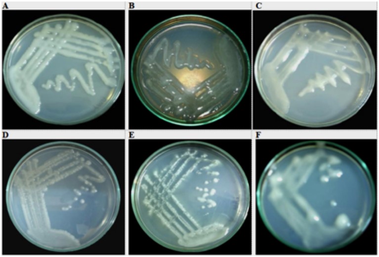
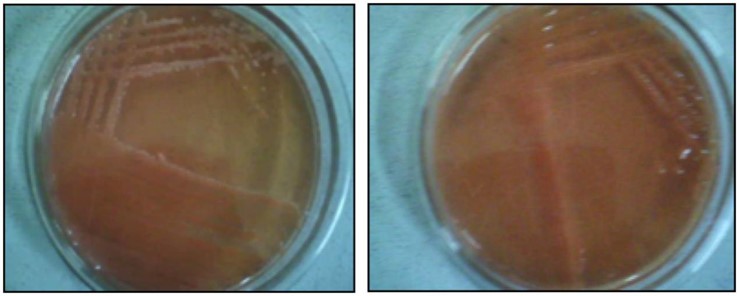
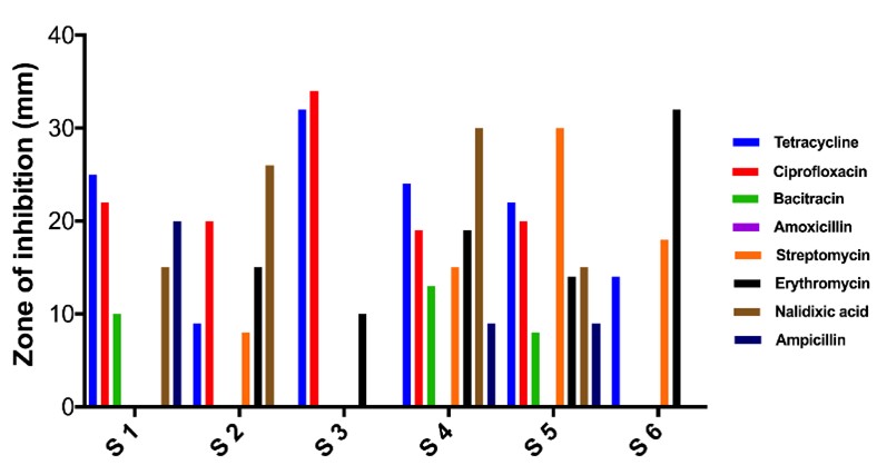
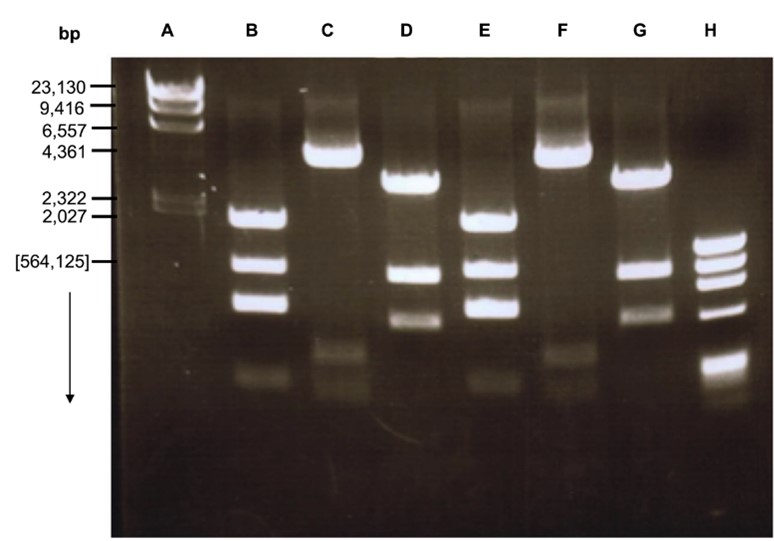
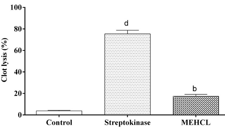
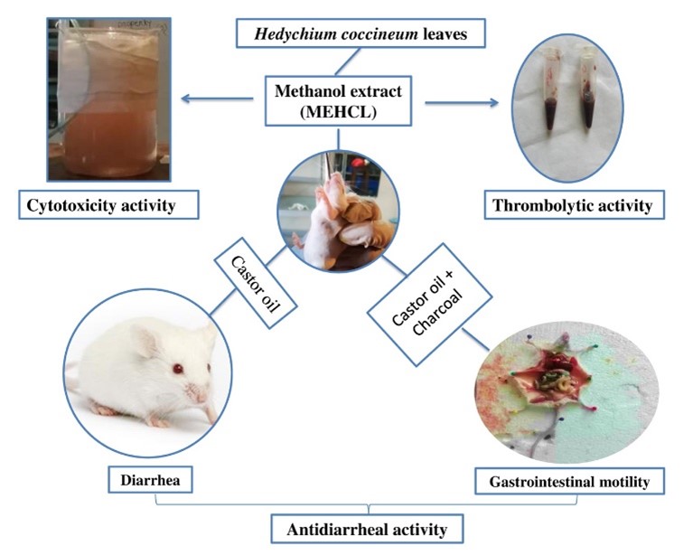
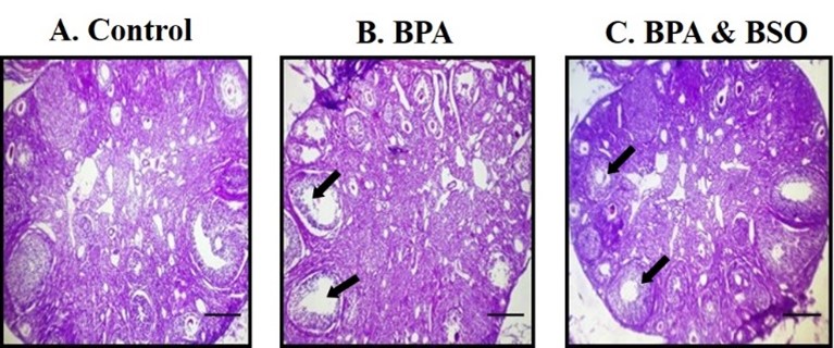
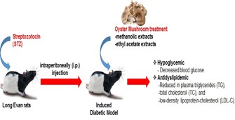
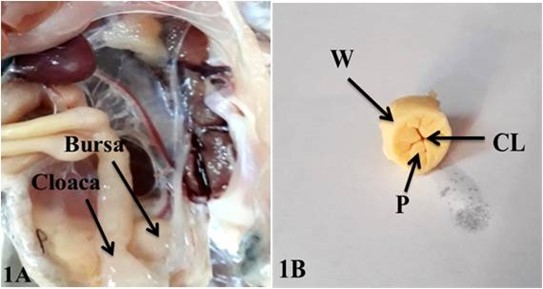
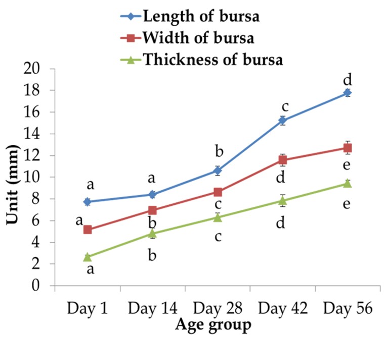
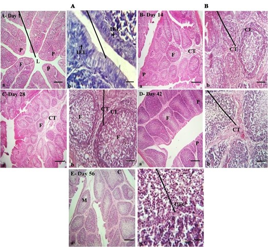