HTTP 200 OK
Allow: GET, HEAD, OPTIONS
Content-Type: application/json
Vary: Accept
{
"count": 319,
"next": "https://jabet.bsmiab.org/articles/?format=api&page=28",
"previous": "https://jabet.bsmiab.org/articles/?format=api&page=26",
"results": [
{
"id": 178,
"slug": "178-1586843165-formaldehyde-contaminated-feed-induces-histopathological-changes-in-the-testes-of-adult-pigeons-columba-livia",
"featured": false,
"slider": false,
"issue": "Vol3 Issue3",
"type": "original_article",
"manuscript_id": "178-1586843165",
"recieved": "2020-03-07",
"revised": null,
"accepted": "2020-04-23",
"published": "2020-06-01",
"pdf_file": "https://jabet.bsmiab.org/media/pdf_file/2023/27/178-1586843165.pdf",
"title": "Formaldehyde-contaminated feed induces histopathological changes in the testes of adult pigeons (Columba livia)",
"abstract": "<p>Formaldehyde (FA), a ubiquitous environmental pollutant, has long been suspected to possess reproductive toxicity. Here, we investigated the histopathological alteration of male gonads following exposure to FA-contaminated feed (40% aqueous solution of FA; 2.5 ml formalin/kg feed) in pigeons for 7 days. The mean body weights were not changed significantly in FA-contaminated feed exposed pigeons compared with control pigeons. The hemoglobin concentration was significantly decreased and serum enzyme aspartate transaminase (AST) was significantly increased in FA-exposed pigeons in comparison with control pigeons. Histologically, the structural components of the testes are the seminiferous tubules and interstitial tissues, which are surrounded by a connective tissue capsule. In control pigeons, the size and shape of seminiferous tubules were normal with a regular arrangement of spermatogenic cells. In FA-exposed pigeons, the testicular capsule was thickened and degeneration of spermatogenic cells in the seminiferous tubules was observed. The number of spermatogenic cells was significantly decreased in the seminiferous tubules of FA-exposed pigeons in comparison with control pigeons, indicating that the low exposure of FA affects the spermatogenic cells’ populations in male birds. The present results suggested that FA might cause infertility in birds.</p>",
"journal_reference": "J Adv Biotechnol Exp Ther. 2020; 3(3): 152-157.",
"academic_editor": "Dr. Md. Jamal Uddin, Ewha Womans University, South Korea.",
"cite_info": "Karim MR, Kobir A, et al. Formaldehyde-contaminated feed induces histopathological changes in the testes of adult pigeons (Columba livia). J Adv Biotechnol Exp Ther. 2020; 3(3): 152-157.",
"keywords": [
"birds",
"Histopathology",
"Formaldehyde",
"Food contaminant",
"Testes"
],
"DOI": "10.5455/jabet.2020.d120",
"sections": [
{
"section_number": 1,
"section_title": "INTRODUCTION",
"body": "<p>Contamination of foods with toxic chemicals possesses a serious threat to public health in Bangladesh due to poor health literacy and low level of awareness [<a href=\"#r-1\">1</a>]. Bangladesh is a country of the tropical region with hot and humid weather. As a result, perishable food items tend to decay quickly [<a href=\"#r-2\">2</a>]. Toxic food contaminants are now very alarming health hazards for human and animals. Food contaminants such as formaldehyde, xylene, ethane dimethane sulfonate, cleaner, toluene and methanol have a detrimental effect on testicular tissue function and structures [<a href=\"#r-3\">3, 4</a>]. Formaldehyde (FA), the recently classified carcinogen and ubiquitous environmental contaminant, is widely used in the construction, textile, furniture, resin, medical, chemical and pharmaceutical industries and FA heavily impacts the everyday consumer [<a href=\"#r-5\">5</a>]. Exposure to ubiquitous exogenous sources of FA at work, in residences, in food and medicine, possess a significant health risk [<a href=\"#r-6\">6</a>]. Formalin (37% aqueous solution of FA) is highly germicidal and used as a disinfectant in poultry and livestock industries. Food and Drug Agency in USA has approved the addition of formalin in poultry feed for keeping it salmonella free [<a href=\"#r-7\">7</a>]. FA treatment of feeds has been reported to have bactericidal effects without apparent loss of palatability or growth reduction in poultry and other food animals [<a href=\"#r-8\">8-11</a>]. However, FA has a detrimental effect on the biological systems including the reproductive system of mammals [<a href=\"#r-12\">12, 13</a>].</p>\r\n\r\n<p>FA toxicity in multiple tissues of the exposed rats and mice, in liver, lymphocytes, heart, brain, lung and gonads was reported [<a href=\"#r-13\">13-15</a>]. In rats, FA caused testicular atrophy and reduction in the testicular weight, serum testosterone level, diameter of seminiferous tubules and seminiferous epithelial height [<a href=\"#r-16\">16-19</a>]. However, comprehensive study on the histomorphological alterations of testes in FA-exposed birds has not been yet undertaken. Therefore, it is very important to know the toxic effects of FA contaminated feed on testicular tissues of pigeons.</p>"
},
{
"section_number": 2,
"section_title": "MATERIALS AND METHODS",
"body": "<p><strong>Statement of the experiment</strong><br />\r\nThe research work was conducted in the Department of Anatomy and Histology, Faculty of Veterinary Science, Bangladesh Agricultural University, Mymensingh-2202 and Village-Roholy, Saturia, Manikganj-1800, Bangladesh during the period from July, 2018 to May, 2019 to assess the effects of FA-contaminated feed exposure on testicular tissue of pigeons.</p>\r\n\r\n<p> </p>\r\n\r\n<p><strong>Ethical approval</strong><br />\r\nThe present study and all experimental protocols were approved and performed according to the guidelines for the care and use of animals as established by Animal Welfare and Experimentation Ethics Committee, Bangladesh Agricultural University, Mymensingh, Bangladesh [AWEEC/BAU/2019-40].</p>\r\n\r\n<p> </p>\r\n\r\n<p><strong>Animals and experimental procedures</strong><br />\r\nTen adult healthy male pigeons (<em>Columba livia</em>) (more than twelve month-old age, 106-146 gm; purchased from local markets at Saturia, Manikganj-1800) were housed in pigeon cages, and fed a standard diet (mustard, rice, sesame, wheat) twice daily and filtered tube-well water <em>ad libitum</em>. After one-week acclimatization, five pigeons were fed a FA-contaminated feed daily morning and fed a standard diet daily evening for 7 days. Previous studies reported that the 2.5 ml formalin/kg feed in broiler chicken and Japanese quails has no adverse effect on body weight and feed intake and suggestive as a beneficial dose [<a href=\"#r-20\">20, 21</a>]. This dose was used in the experiment. Formalin (40% aqueous solution of stock FA powder) was mixed properly with pigeon feeds (2.5 ml formalin or 1000 mg/kg feed). The other five pigeons were used as control, fed a standard diet twice daily (morning and evening) for 7 days. After 7 days of FA-exposure (in morning of the 8<sup>th</sup> day), all pigeons were euthanized under deep anesthesia using 5 ml chloroform-soaked cotton in vacuum glass chamber for 3-4 minutes and then post-mortem examination was performed.</p>\r\n\r\n<p> </p>\r\n\r\n<p><strong>Determination of hematobiochemical parameters</strong><br />\r\nAfter 7 days of FA-exposure, blood samples were collected from the wing vein of both control and FA-exposed pigeons for hematobiochemical parameters. The principles and procedures of hematobiochemical examinations were as follows:</p>\r\n\r\n<p> </p>\r\n\r\n<p><strong>Total erythrocyte count (TEC)</strong><br />\r\nTotal erythrocyte count was done following the method described by [<a href=\"#r-10\">10</a>]. Well-mixed blood sample was drawn with red blood cell diluting pipette and immediately filled with the red cell diluting fluid. The total number of RBCs was calculated as number of cells counted x 10,000 and the result was expressed in million/cubic meter of blood.</p>\r\n\r\n<p> </p>\r\n\r\n<p><strong>Hemoglobin concentration (Hb)</strong><br />\r\nWell-homogenized blood sample was drawn into the Sahli pipette and then the blood of the pipette was immediately transferred into the graduated tube containing hydrochloric acid. This blood and acid were thoroughly mixed by stirring with a glass stirrer. Water was added drop by drop to the tube containing acid hematin mixture. The solution was mixed well with a glass stirrer until the color of the mixture resembled to the standard color of the comparator. The result was read in day light by observing the height of the liquid in the tube considering the lower meniscus of the liquid column. The result was then expressed in g%.</p>\r\n\r\n<p> </p>\r\n\r\n<p><strong>Serum enzyme aspartate transaminase (AST) </strong><strong>analysis</strong><br />\r\nAt necropsy, blood was collected from the heart. Test tube containing blood was placed in slanting condition at room temperature for 30 minutes. Sera were separated from clotted blood by centrifugation at 3000 rpm for 20 minutes and again for 10 minutes. Then the supernatant was collected in eppendorf tube by micro-pipette and stored in refrigerator at -20°C until use and analyzed for aspartate transaminase (AST).</p>\r\n\r\n<p> </p>\r\n\r\n<p><strong>Postmortem examination and collection of testes for histopathology</strong><br />\r\nAfter 7 days of FA-exposure, both control and FA-exposed pigeons were weighed and then all pigeons were euthanized under deep chloroform anesthesia. The internal visceral organs including testes were visualized through ventral mid-line thoraco-abdominal opening and the gross anatomy was observed very carefully. Then, the testes were removed and their colour and gross texture were observed. The weight of testes was measured and recorded. Subsequently, testicular tissues from the left and right testes were collected and immediately fixed in 10% neutral buffered formalin (NBF) by standard method. Briefly, NBF-fixed tissues were dehydrated with ascending graded alcohols and embedded in paraffin and sectioned at 6 µm in thickness using a sliding microtome (Euromax<sup>R</sup>, Japan). The deparaffinized sections were stained with hematoxylin and eosin (HE) for histopathological examination. FA-induced changes were analyzed morphologically using light microscope (LABOMED, Labo America Inc., CA 94538). The spermatogenic cells were counted in at least 5 seminiferous tubules (clear cross sections which were sharply rounded) of each birds. The oblique or longitudinal section of seminiferous tubules were not considered for spermatogenic cell count.</p>\r\n\r\n<p> </p>\r\n\r\n<p><strong>Statistical analysis</strong><br />\r\nAll the achieved data were analyzed using student <em>t-test</em> and statistically evaluated with SPSS, version 18.0 software (IBM). The <em>p</em>-values of ≤0.05 were considered to indicate statistically significant, while <em>p-</em>values of <0.01 indicate highly significant results. All the results were expressed as mean ±SD<strong>.</strong></p>"
},
{
"section_number": 3,
"section_title": "RESULTS",
"body": "<p><strong>Effects of FA on body weight and gross morphology of testes of pigeons</strong><br />\r\nThe mean body weights of adult pigeons were not changed significantly in FA-exposed pigeons in comparison with control pigeons (<a href=\"#figure1\">Figure 1A</a>). The color, size and shape of both testes (right and left) were normal in control pigeons (<a href=\"#figure2\">Figure 2A</a>). However, in FA-exposed pigeon, the testes showed uneven shape with pin point hemorrhage on their surface (<a href=\"#figure2\">Figure 2B</a>). The weight of the testes was not change significantly in FA-exposed and control pigeons (data not shown).</p>\r\n\r\n<div id=\"figure1\">\r\n<figure class=\"image\"><img alt=\"\" height=\"369\" src=\"/media/article_images/2024/06/17/178-1586843165-Figure1.jpg\" width=\"500\" />\r\n<figcaption><strong>Figure 1.</strong> Body weight and blood parameters analysis of FA-exposed adult pigeons. A. No significant changes of body weight were seen in control and FA-exposed pigeons. B. Total erythrocyte count (TEC) showed no significant changes, but showing decreasing tendency in FA-exposed pigeons in comparison with control. C. Hemoglobin content (Hb, g%) was significantly decreased in FA-exposed pigeons compared with control pigeons. D. Serum enzyme, aspartate aminotransferase (AST) level was significantly increased in FA-exposed pigeon compared with control. Student t-test, *The <em>p</em>-values of ≤0.05 were considered to be statistically significant, **<em>p-</em>values of <0.01 indicate highly significant results. FA- Formaldehyde.</figcaption>\r\n</figure>\r\n</div>\r\n\r\n<div id=\"figure2\">\r\n<figure class=\"image\"><img alt=\"\" height=\"228\" src=\"/media/article_images/2024/06/17/178-1586843165-Figure2.jpg\" width=\"500\" />\r\n<figcaption><strong>Figure 2. </strong>Gross anatomy of testes of adult pigeons. A. Showing normal color, size and shape of the right and left testes of control pigeons. B. Normal color and size, but uneven shape and pinpoint hemorrhage was seen in the both right and left testes of FA-exposed pigeons. FA- Formaldehyde. Bar = 1 cm.</figcaption>\r\n</figure>\r\n</div>\r\n\r\n<p> </p>\r\n\r\n<p><strong>Effects of FA on hematological parameters of pigeons</strong><br />\r\nTotal erythrocyte count showed no significant change in FA-exposed pigeons in comparison with control pigeons (<a href=\"#figure1\">Figure 1B</a>). The hemoglobin concentration was significantly decreased in FA-exposed pigeons in comparison with control pigeons (<a href=\"#figure1\">Figure 1C</a>). The values of AST were significantly increased in FA-exposed pigeons in comparison with control pigeons (<a href=\"#figure1\">Figure 1D</a>), an indicative of liver injury and disturbance of body homoeostasis.</p>\r\n\r\n<p> </p>\r\n\r\n<p><strong>Effects of FA on histoarchitecture of testes of pigeons</strong><br />\r\nIn HE-stained sections, regular histological structure without abnormalities was seen in testes of control pigeons (<a href=\"#figure3\">Figures 3A</a>, <a href=\"#figure4\">4A-B</a>). The testis was surrounded by a capsule, which was composed mainly of dense collagenous fibrous connective tissue (<a href=\"#figure3\">Figure 3A</a>). The structural components of the testis were the seminiferous tubules and interstitial tissues (<a href=\"#figure4\">Figure 4A-B, F</a>). Seminiferous tubules are the structural and functional unit of testes. The sertoli cells and the spermatogenic cells lined the seminiferous tubules. The different stages of spermatogenic cells were found in several layers, namely, the spermatogonia, spermatocytes, spermatids and finally mature spermatozoa (<a href=\"#figure4\">Figure 4A-B</a>). The spermatogonia were rounded cells with rounded nuclei; and sertoli cells were tall cells with oval nuclei (<a href=\"#figure4\">Figure 4B</a>). The interstitial tissues were narrow and showed clusters of Leydig cells (<a href=\"#figure4\">Figure 4A-B, F</a>).<br />\r\nIn FA-exposed pigeons, the FA-induced testicular changes were characterized by thickened capsule and degeneration of spermatogenic cells in the seminiferous tubules (<a href=\"#figure3\">Figure 3B</a>). Irregular arrangement of spermatogenic cells in the seminiferous tubules of FA-exposed pigeons were seen (<a href=\"#figure4\">Figure 4D</a>). Lumen of the seminiferous tubules became large and number of spermatogenic cells particularly; secondary spermatocytes and spermatid were reduced (<a href=\"#figure4\">Figure 4C-D</a>). Primary spermatocytes were found separated from spermatogonia. The number of spermatogenic cells was significantly decreased in seminiferous tubules of FA-exposed pigeons in comparison with control pigeons (<a href=\"#figure4\">Figure 4E</a>). The interstitial cells or Leydig cells were localized as cord or cluster between or among the seminiferous tubules (<a href=\"#figure4\">Figure 4 A-D, F-G</a>). No changes were seen in the Leydig cells of both control and FA-exposed pigeons (<a href=\"#figure4\">Figure 4 F-G</a>).</p>\r\n\r\n<div id=\"figure3\">\r\n<figure class=\"image\"><img alt=\"\" height=\"180\" src=\"/media/article_images/2024/06/17/178-1586843165-Figure3.jpg\" width=\"500\" />\r\n<figcaption><strong>Figure 3. </strong>Histology of capsule of testes of adult pigeons. A. Showing normal histology of capsule (consisting of dense irregular connective tissues) covering the testicular parenchyma. B. The thickness of the capsule was apparently high in the testis of FA-exposed pigeon. Star indicates the degeneration of spermatogenic cells. FA- Formaldehyde. Bar = 50 µm.</figcaption>\r\n</figure>\r\n</div>\r\n\r\n<div id=\"figure4\">\r\n<figure class=\"image\"><img alt=\"\" height=\"656\" src=\"/media/article_images/2024/06/17/178-1586843165-Figure4.jpg\" width=\"500\" />\r\n<figcaption><strong>Figure 4. </strong>Histology of testes of adult pigeons. A. Showing many seminiferous tubules and interstitial tissues in testes of control pigeons. B. In higher magnification, a regular distribution of spermatogenic cells in seminiferous tubules of control pigeons. C-D. Degeneration of spermatogenic cells and large size lumen were seen in the seminiferous tubules of FA-exposed pigeons. In addition, the number of spermatogenic cells was drastically reduced in the seminiferous tubules of testis. E. The number of spermatogenic cells was significantly decreased in seminiferous tubules of FA-exposed pigeons in comparison with control pigeons. F-G. The interstitial cells or Leydig cells were seen between or among the seminiferous tubules and showing normal histology both in control and FA-exposed pigeons. Arrow indicates the interstitial cells or Leydig cells and arrowheads indicate sertoli cell. Stars indicate the degeneration areas of spermatogenic cells. SG-spermatogonia, SC-spermatocyte, ST-spermatid, SZ- spermatozoa. Student t-test, *<em>p-</em>values of <0.01 indicate significant results. FA- Formaldehyde. Bar = 50 µm.</figcaption>\r\n</figure>\r\n\r\n<p> </p>\r\n</div>"
},
{
"section_number": 4,
"section_title": "DISCUSSION",
"body": "<p>In the present study, the value of serum AST level was significantly increased and the hemoglobin concentration was significantly decreased in FA-exposed pigeons in comparison with control pigeons. The total erythrocyte count showed decreasing tendency in FA-exposed pigeons. These results suggested liver injury and imbalance of body homoeostasis caused by FA exposure. No reports are available in birds, however serum enzyme AST, ALT was significantly increased in FA-exposed mice (oral, 5mg/kg body weight) [<a href=\"#r-22\">22</a>]. FA treatment (oral, 10mg/kg body weight) resulted in significant decreases in RBCs, hemoglobin, total WBC and lymphocytes in mice [<a href=\"#r-23\">23</a>].<br />\r\nFA exposure through feed induced a significant reduction of spermatogenic cells and widening of the lumen of the seminiferous tubules in the testes of pigeons. There was no available literature on FA effect on testicular tissues of avian species. However, the present results were consistent with that of other formaldehyde exposure research on male rats and mice although the dosage, duration and route of administrations of FA were different [<a href=\"#r-13\">13</a>, <a href=\"#r-21\">21</a>, <a href=\"#r-24\">24-27</a>]. Low levels of formalin (10 ml/kg feed), when fed to Japanese quails (<em>Coturnix coturnix japonica</em>), decreased testicular weight and diameter of seminiferous tubules [<a href=\"#r-21\">21</a>]. Intraperitoneal administration of FA with dosages of 0.2, 2 and 20 mg/kg can cause degeneration and necrosis of the secondary spermatocytes, spermatogenic cells and spermatozoids [<a href=\"#r-15\">15</a>]. FA vapor (10 mg/m<sup>3</sup>) for two weeks’ exposure, caused atrophy of the seminiferous tubules, a decrease in the number of spermatogenic cells and disorganization of the seminiferous epithelial cells of male rats [<a href=\"#r-26\">26</a>]. FA-induced change in rat testes are characterized by thickened capsule, spermatogenesis arrest, a decrease in the number of spermatogenic cells and atrophy of seminiferous tubules in rats [<a href=\"#r-28\">28,29</a>], which were reproduced in the present study following FA contaminated feed exposure in testes of adult pigeons.</p>"
},
{
"section_number": 5,
"section_title": "CONCLUSIONS",
"body": "<p>The present findings revealed that the toxic feed contaminant, FA, has detrimental effects on the testes of adult pigeons. FA was found to cause degeneration of spermatogenic cells, separation of primary spermatocyte from spermatogonia and irregular arrangement of spermatogenic cells in the seminiferous tubules, these histological changes indicated that it might affect the reproduction of birds. Therefore, it is very alarming if the human being exposed to this toxic food contaminant, it may cause reproductive failure.</p>"
},
{
"section_number": 6,
"section_title": "ACKNOWLEDGEMENT",
"body": "<p>The authors thank Mr. Md. Ahasan Habib for chemical preparation and their exposure in birds through feed. This work was supported by the Ministry of Science and Technology (MoST), Government of the Peoples Republic of Bangladesh (No: 39.00.0000.009.14.004.19/ BS-74/85, 2018-19 to MRK), by the Bangladesh Agricultural University and University Grant Commission of Bangladesh (No. 2018/556/AU-GC to MRK) and by the Ministry of Education, Grant for Advance Research in Education (GARE), BANBEIS, Grant Number LS2018773 to MRK.</p>"
},
{
"section_number": 7,
"section_title": "AUTHOR CONTRIBUTIONS",
"body": "<p>The experiment was designed by MRK and MP. MRK, AK and IH undertook the experiment; MRK and IH interpreted the results putting efforts on statistical analysis with AK; MRK and IH wrote up the draft. MRK, MP and AIA checked the manuscript critically. All authors read and agreed on the final version of the manuscript.</p>"
},
{
"section_number": 8,
"section_title": "CONFLICTS OF INTEREST",
"body": "<p>Authors declared that they have no conflict of interest.Authors declared that they have no conflict of interest.</p>"
}
],
"figures": [
{
"figure": "https://jabet.bsmiab.org/media/article_images/2024/06/17/178-1586843165-Figure1.jpg",
"caption": "Figure 1. Body weight and blood parameters analysis of FA-exposed adult pigeons. A. No significant changes of body weight were seen in control and FA-exposed pigeons. B. Total erythrocyte count (TEC) showed no significant changes but showing decreasing tendency in FA-exposed pigeons in comparison with control. C. Hemoglobin content (Hb, g%) was significantly decreased in FA-exposed pigeons compared with control pigeons. D. Serum enzyme, aspartate aminotransferase (AST) level was significantly increased in FA-exposed pigeon compared with control. Student t-test, *The p-values of ≤0.05 were considered to be statistically significant, **p-values of <0.01 indicate highly significant results. FA- Formaldehyde.",
"featured": false
},
{
"figure": "https://jabet.bsmiab.org/media/article_images/2024/06/17/178-1586843165-Figure2.jpg",
"caption": "Figure 2. Gross anatomy of testes of adult pigeons. A. Showing normal color, size and shape of the right and left testes of control pigeons. B. Normal color and size, but uneven shape and pinpoint hemorrhage was seen in the both right and left testes of FA-exposed pigeons. FA- Formaldehyde. Bar = 1 cm.",
"featured": false
},
{
"figure": "https://jabet.bsmiab.org/media/article_images/2024/06/17/178-1586843165-Figure3.jpg",
"caption": "Figure 3. Histology of capsule of testes of adult pigeons. A. Showing normal histology of capsule (consisting of dense irregular connective tissues) covering the testicular parenchyma. B. The thickness of the capsule was apparently high in the testis of FA-exposed pigeon. Star indicates the degeneration of spermatogenic cells. FA- Formaldehyde. Bar = 50 µm.",
"featured": false
},
{
"figure": "https://jabet.bsmiab.org/media/article_images/2024/06/17/178-1586843165-Figure4.jpg",
"caption": "Figure 4. Histology of testes of adult pigeons. A. Showing many seminiferous tubules and interstitial tissues in testes of control pigeons. B. In higher magnification, a regular distribution of spermatogenic cells in seminiferous tubules of control pigeons. C-D. Degeneration of spermatogenic cells and large size lumen were seen in the seminiferous tubules of FA-exposed pigeons. In addition, the number of spermatogenic cells was drastically reduced in the seminiferous tubules of testis. E. The number of spermatogenic cells was significantly decreased in seminiferous tubules of FA-exposed pigeons in comparison with control pigeons. F-G. The interstitial cells or Leydig cells were seen between or among the seminiferous tubules and showing normal histology both in control and FA-exposed pigeons. Arrow indicates the interstitial cells or Leydig cells and arrowheads indicate sertoli cell. Stars indicate the degeneration areas of spermatogenic cells. SG-spermatogonia, SC-spermatocyte, ST-spermatid, SZ- spermatozoa. Student t-test, *p-values of <0.01 indicate significant results. FA- Formaldehyde. Bar = 50 µm.",
"featured": false
}
],
"authors": [
{
"id": 797,
"affiliation": [
{
"affiliation": "Department of Anatomy and Histology, Faculty of Veterinary Science, Bangladesh Agricultural University, Mymensingh-2202, Bangladesh."
}
],
"first_name": "Mohammad Rabiul",
"family_name": "Karim",
"email": "mrabiulkarim@bau.edu.bd",
"author_order": 1,
"ORCID": null,
"corresponding": true,
"co_first_author": false,
"co_author": false,
"corresponding_author_info": "Dr. Mohammad Rabiul Karim, Professor and Head, Department of Anatomy and Histology, Faculty of Veterinary Science, Bangladesh Agricultural University, Mymensingh-2202, Bangladesh, E-mail: mrabiulkarim@bau.edu.bd",
"article": 178
},
{
"id": 798,
"affiliation": [
{
"affiliation": "Department of Anatomy and Histology, Faculty of Veterinary Science, Bangladesh Agricultural University, Mymensingh-2202, Bangladesh."
}
],
"first_name": "Alamgir",
"family_name": "Kobir",
"email": null,
"author_order": 2,
"ORCID": null,
"corresponding": false,
"co_first_author": false,
"co_author": false,
"corresponding_author_info": "",
"article": 178
},
{
"id": 799,
"affiliation": [
{
"affiliation": "Department of Anatomy and Histology, Faculty of Veterinary Science, Bangladesh Agricultural University, Mymensingh-2202, Bangladesh."
}
],
"first_name": "Imam",
"family_name": "Hasan",
"email": null,
"author_order": 3,
"ORCID": null,
"corresponding": false,
"co_first_author": false,
"co_author": false,
"corresponding_author_info": "",
"article": 178
},
{
"id": 800,
"affiliation": [
{
"affiliation": "Department of Pathology, Faculty of Veterinary Science, Bangladesh Agricultural University, Mymensingh-2202, Bangladesh."
}
],
"first_name": "Munmun",
"family_name": "Pervin",
"email": null,
"author_order": 4,
"ORCID": null,
"corresponding": false,
"co_first_author": false,
"co_author": false,
"corresponding_author_info": "",
"article": 178
},
{
"id": 801,
"affiliation": [
{
"affiliation": "Department of Anatomy and Embryology, Faculty of Veterinary Medicine, Benha University, Moshtohor, Toukh 13736, Egypt."
}
],
"first_name": "Ahmed I. Abo-",
"family_name": "Ahmed",
"email": null,
"author_order": 5,
"ORCID": null,
"corresponding": false,
"co_first_author": false,
"co_author": false,
"corresponding_author_info": "",
"article": 178
}
],
"views": 634,
"downloads": 124,
"references": [
{
"id": 5939,
"serial_number": 1,
"pmc": null,
"reference": "Rahman M. Sultan Z, Rahman M, Rashid M. Food Adulteration: a serious public health concern in Bangladesh. Bangladesh Pharmaceutical Journal. 2015;18:1",
"DOI": null,
"article": 178
},
{
"id": 5940,
"serial_number": 2,
"pmc": null,
"reference": "Nunes MCN. Impact of environmental conditions on fruit and vegetable quality. Stewart Postharvest Review 2008; 4:4",
"DOI": null,
"article": 178
},
{
"id": 5941,
"serial_number": 3,
"pmc": null,
"reference": "Karimov K, Dadazhanov SH, Gil’dieva MS. Rat reproductive cells as biological indicators of the effect of environmental factors. Morfologiia (Saint Petersburg, Russia). 2003; 123(1):69-71.",
"DOI": null,
"article": 178
},
{
"id": 5942,
"serial_number": 4,
"pmc": null,
"reference": "Handagama C and Ariyaratne S. Differentiation of the adult Leydig cell population in the postnatal testis. Biology of Reproduction. 2001; 65: 660-671.",
"DOI": null,
"article": 178
},
{
"id": 5943,
"serial_number": 5,
"pmc": null,
"reference": "Formaldehyde, 2-butoxyethanol and 1-tert-butoxypropan-2-ol. Monographs on the evaluation of carcinogenic risks to humans. 88, 1- 478 (2006).",
"DOI": null,
"article": 178
},
{
"id": 5944,
"serial_number": 6,
"pmc": null,
"reference": "Tang X, Bai Y, Duong A, Smith MT, Li L, Zhang L. Formaldehyde in China: production, consumption, exposure levels, and health effects. Environment International. 2009; 35: 1210–1224.",
"DOI": null,
"article": 178
},
{
"id": 5945,
"serial_number": 7,
"pmc": null,
"reference": "Brown HR. FDA approved use of formaldehyde in poultry feed. Food Stuffs. 1996; 68. 15",
"DOI": null,
"article": 178
},
{
"id": 5946,
"serial_number": 8,
"pmc": null,
"reference": "Duncan MS and Adams AW. Effects of a chemical additive and of formaldehyde-gas fumigation on Salmonella in poultry feeds. Poultry Science. 1972; 51: 797–802.",
"DOI": null,
"article": 178
},
{
"id": 5947,
"serial_number": 9,
"pmc": null,
"reference": "Bugarski D, Handzic R, Sivcevic A, Stanic I. Effect of feeding regime and formaldehyde-treated soyabean on the productivity of dairy cows. Veterinaria Sarajevo. 1990; 39: 21–30.",
"DOI": null,
"article": 178
},
{
"id": 5948,
"serial_number": 10,
"pmc": null,
"reference": "McAllister T, Beauchemin K, McClelland L, Cheng, K. Effect of formaldehyde-treated barley or escape protein on nutrient digestibility, growth and carcass traits of feedlot lambs. Canadian Journal of Animal Science. 1992; 72: 309–316.",
"DOI": null,
"article": 178
},
{
"id": 5949,
"serial_number": 11,
"pmc": null,
"reference": "Ricke SC, Richardson K, Dittoe DK. Formaldehydes in feed and their potential interaction with the poultry gastrointestinal tract microbial community–A review. Fronters in Veterinary Science. 2019; 6: 188. doi: 3389/fvets.2019.00188",
"DOI": null,
"article": 178
},
{
"id": 5950,
"serial_number": 12,
"pmc": null,
"reference": "Duong A, Steinmaus C, McHale CM, Vaughan CP, Zhan L. Reproductive and developmental toxicity of formaldehyde: A systematic review. Mutation Research. 2011; 118-138.",
"DOI": null,
"article": 178
},
{
"id": 5951,
"serial_number": 13,
"pmc": null,
"reference": "Zhou D, Zhang J, Wang H. Assessment of the potential reproductive toxicity of long-term exposure of adult male rats to low-dose formaldehyde. Toxicology and Industrial Health. 2011; 27(7):591-5988.",
"DOI": null,
"article": 178
},
{
"id": 5952,
"serial_number": 14,
"pmc": null,
"reference": "Zahra T, Parviz T, Simin F, Mehdi T. Effect of formaldehyde injection in mice on testis function. International Journal of Pharmacology. 2007; 3(5):421-424.",
"DOI": null,
"article": 178
},
{
"id": 5953,
"serial_number": 15,
"pmc": null,
"reference": "Tang M, Xie Y, Yi Y, Wang W. Effects of formaldehyde on germ cells of male mice. Wei sheng yanjiu= Journal of hygiene research. 2003; 32(6):544-548.",
"DOI": null,
"article": 178
},
{
"id": 5954,
"serial_number": 16,
"pmc": null,
"reference": "Chung WG, Yu IJ, Park CS, Lee KH, Roh HK, Cha YN. Decreased formation of ethoxyacetic acid from ethylene glycol monoethyl ether and reduced atrophy of testes in male rats upon combined administration with toluene and xylene. Toxicology Letters. 1999; 104(1-2):143-150.",
"DOI": null,
"article": 178
},
{
"id": 5955,
"serial_number": 17,
"pmc": null,
"reference": "Lemasters GK, Olsen DM, Yiin JH, Lockey JE, Shukla R, Selevan SG, Schrader SM, Toth GP, Evenson DP, Huszar GB. Male reproductive effects of solvent and fuel exposure during aircraft maintenance. Reproductive Toxicology. 1999; 13(3):155-166.",
"DOI": null,
"article": 178
},
{
"id": 5956,
"serial_number": 18,
"pmc": null,
"reference": "Özen OA, Akpolat N, Songur A, Kuş İ, Zararsiz İ, Özaçmak VH, Sarsilmaz M. Effect of formaldehyde inhalation on Hsp70 in seminiferous tubules of rat testes: an immunohistochemical study. Toxicology and Industrial Health. 2005; 21(9):249-254.",
"DOI": null,
"article": 178
},
{
"id": 5957,
"serial_number": 19,
"pmc": null,
"reference": "Golalipour MJ, Azarhoush R, Ghafari S, Gharravi AM, Fazeli SA, Davarian A. Formaldehyde exposure induces histopathological and morphometric changes in the rat testis. Folia Morphologica. 2007; 66(3):167-171.",
"DOI": null,
"article": 178
},
{
"id": 5958,
"serial_number": 20,
"pmc": null,
"reference": "Babar AM, Khan MZ, Ahmed S, Khan A, Bachaya, Anwar MI. Toxico-pathological effects of formalin (37% formaldehyde) feeding in broiler chicks. Pakistan Veterinary Journal. 2001; 21:13-16.",
"DOI": null,
"article": 178
},
{
"id": 5959,
"serial_number": 21,
"pmc": null,
"reference": "Anwar MI, Khan MZ, Muhammad G, Bachaya A and Babar MA. Effects of dietary formalin on the health and testicular pathology of male Japanese quails (Coturnix coturnix japonica). Veterinary and Human Toxicology.2001; 43: 330–333.",
"DOI": null,
"article": 178
},
{
"id": 5960,
"serial_number": 22,
"pmc": null,
"reference": "Afrin M, Amin T, Karim, MR, Islam MR. Effects of formaldehyde intoxication on liver of Swiss albino mice. IOSR Journal of Agriculture and Veterinary Science. 2016; 9: 76-81.",
"DOI": null,
"article": 178
},
{
"id": 5961,
"serial_number": 23,
"pmc": null,
"reference": "Abd-Elhakim YM, Amany M, & Wafaa M. Hemato-immunologic impact of sub chronic exposure to melamine and/or formaldehyde in mice, Journal of Immunotoxicology. 2016; 13(5):713-722.",
"DOI": null,
"article": 178
},
{
"id": 5962,
"serial_number": 24,
"pmc": null,
"reference": "Majumder PK, Kumar VL. Inhibitory effects of formaldehyde on the reproductive system of male rats. Indian Journal of Physiology and Pharmacology. 1995; 39(1):80-82.",
"DOI": null,
"article": 178
},
{
"id": 5963,
"serial_number": 25,
"pmc": null,
"reference": "Shah BM, Vachharajani KD, Chinoy NJ and Roy Chowdhary A. Formaldehyde-induced changes in testicular tissues of rats. Journal of Reproductive Biology and Comparative Endocrinology.1992; 7: 42–52.",
"DOI": null,
"article": 178
},
{
"id": 5964,
"serial_number": 26,
"pmc": null,
"reference": "Zhou DX, Qiu SD, Wang ZY and Zhang J. Effect of tail-suspension on the reproduction of adult male rats. Zhonghua Nan KeXueZaZhi. 2006a; 12: 326–329.",
"DOI": null,
"article": 178
},
{
"id": 5965,
"serial_number": 27,
"pmc": null,
"reference": "Zhou DX, Qiu SD, Zhang J, Tian H and Wang HX. The protective effect of vitamin E against oxidative damage caused by formaldehyde in the testes of adult rats. Asian Journal of Androoglogy.2006b; 8: 584–588.",
"DOI": null,
"article": 178
},
{
"id": 5966,
"serial_number": 28,
"pmc": null,
"reference": "Hegazy AA, Elsayed NE, Ahmad MM, Omar NM. Effect of formaldehyde on rat testis structure. Academia Anatmica International. 2017; 3(2):15-23.",
"DOI": null,
"article": 178
},
{
"id": 5967,
"serial_number": 29,
"pmc": null,
"reference": "Chowdhury AR, Gautam AK, Patel KG, Trivedi HS. Steroidogenic inhibition in testicular tissue of formaldehyde exposed rats. Indian Journal of Physiology and Pharmacology. 1992; 36: 162.",
"DOI": null,
"article": 178
}
]
},
{
"id": 187,
"slug": "178-1585833379-culture-positivism-exploitation-through-automated-fluorescent-sensor-technology-from-patients-with-blood-stream-infections",
"featured": false,
"slider": false,
"issue": "Vol3 Issue3",
"type": "original_article",
"manuscript_id": "178-1585833379",
"recieved": "2020-01-17",
"revised": null,
"accepted": "2020-04-06",
"published": "2020-06-01",
"pdf_file": "https://jabet.bsmiab.org/media/pdf_file/2023/29/178-1585833379.pdf",
"title": "Culture positivism exploitation through automated fluorescent-sensor technology from patients with blood stream infections",
"abstract": "<p>This study tracks and analyses the culture results of 3615 blood samples received in Popular Diagnostic Centre, Dhanmondi, Dhaka 1205, throughout twelve month from suspected patients with blood stream infections. The samples were prepared by exploitation machine-controlled Fluorescent-Sensor Technology by BACTEC 9120® Culture System (Becton Dickinson and Company, Sparks, USA) choosing a 5 days incubation protocol. A total of 668/3615(18.50%) BACTEC 9120® system positive samples were then sub-cultured in 7% sheep blood agar, MacConkey agar, and chocolate agar plates. A sum of 346/668 (51.8%) were infective organisms and 312/668 (46.70%) positive vial cultures were contaminants. False positivism rate was 1.5% (10/668). The mean detection time for the clinical vital isolates was 17.7 h and for all the isolates was 38.1 h. Microorganisms characterization and antibiotic sensitivity testing was done using typical ways. The majorities (342, 98.84%) of those were Gram-negative microorganisms and solely 4 (1.16%) isolates were Gram-positive pathogens. Clinically vital pathogens recouped on day one, two and three were 91.0%, 7.5% and 1.5% severally. Most of the bacterial isolates were found extremely susceptible to a number of antibiotics along with ceftriaxone (95%), ceftazidine (95%), and cefepime (96%), whereas moderately sensitive to ciprofloxacin (81%), levofloxacin (89%) and chloramphenicol (81%); whereas all bacterial isolates were found to be resistant of nalidic acid (100%). Since all our cultures were positive at intervals the primary 72 h, our data supported 5 days incubation system for the recuperation of medically vital microorganisms in BACTEC 9120® Culture System.</p>",
"journal_reference": "J Adv Biotechnol Exp Ther. 2020; 3(3): 165-170.",
"academic_editor": "Dr. Hasan-Al-Faruque, Daegu Gyeonbuk Institute of Science and Technology, South Korea.",
"cite_info": "Abedin MZ, Jarin L, et al. Culture positivism exploitation through automated fluorescent-sensor technology from patients with blood stream infections. J Adv Biotechnol Exp Ther. 2020; 3(3): 165-170.",
"keywords": [
"BACTEC 9120® system",
"Culture positivism",
"Antibiotic sensitivity profile",
"Blood stream infections"
],
"DOI": "10.5455/jabet.2020.d122",
"sections": [
{
"section_number": 1,
"section_title": "INTRODUCTION",
"body": "<p>Blood stream infection is a danger to each organ inside the body and might have genuine quick results, together with stun, various organ disappointments, dispersed intravascular coagulation and passing (demise frequency at 20% to 50%). Therefore, identification and detection on time of microbial pathogens in blood is one of the vital significant elements of the biological laboratory [<a href=\"#r-1\">1</a>]. As of late, several propelled strategies like as super molecule probes and polymerase chain reaction (PCR) are created for the assignment of blood contaminations; anyway blood culture despite everything remains the foremost sensible and solid methodology [<a href=\"#r-2\">2</a>].<br />\r\nTypical blood culture techniques include naked eye assessment of blood culture vials once every twenty-four-hour period for the proof of development for 48 hours and then visually impaired sub-culture on the next day on solid culture media. Negative containers of the culture are more re-brooded for 5 to 7 days before coverage. In recent years, sensational development has been occurred in blood culture procedures, culture media, and in the frameworks. The greater part of the mechanically propelled blood culture systems are completely machine driven constantly observed blood culture frameworks [<a href=\"#r-3\">3</a>]. Every 8-10 minutes vials are screened by these frameworks and final out calculations supported by evaluations of changes related with being development. Presently, 4 frameworks are accessible: Becton Dickinson Microbiology systems, Sparks, Md. (BACTEC®), Trek Diagnostic systems Inc., Organon Teknika, Durham, N.C. (BacT/Alert®), Westlake, Ohio (ESP®), and bioMerieux, Inc. Hazelwood, Mo. (Vital) [<a href=\"#r-1\">1</a>]. All of those systems, there’s none of significant distinction in the performances and everyone is extremely strong. Detection of growth is the principal differentiation exists within these systems [<a href=\"#r-3\">3</a>]. For 5-7 days incubation period is programmed by the applicant to incubate samples vials in the systems.<br />\r\nIn this research the main point of analysis was to see the range of microscopic bacteria confined from blood samples, their opportunity to identification by BACTEC 9120®, antibiotic sensitivity- resistance profile and to investigate the information to make our mind up that incubation protocol much be a lot of appropriate by machine-controlled BACTEC 9120® culture system. In our study as suggested by the manufacturer, we introduced a five day convention of incubation as there’s absence of printed data relating to the ideal duration of incubation for the system from this apart of the nation.</p>"
},
{
"section_number": 2,
"section_title": "MATERIALS AND METHODS",
"body": "<p><strong>Materials</strong><br />\r\nAll materials and chemicals were maintained in analytical grade in this research project.</p>\r\n\r\n<p> </p>\r\n\r\n<p><strong>Bloodstream sample collection</strong><br />\r\nIn this examination, we directed our study since December 2010 to November 2011 at Popular Diagnostic Centre Ltd, Dhaka 1205. The machine-controlled incessantly monitored blood culture system utilized is BACTEC 9120®. During this study, a complete of 3615 blood samples from individuals of suspected septicemia was received. Blood samples were collected by antiseptic method.</p>\r\n\r\n<p> </p>\r\n\r\n<p><strong>Blood culture through BACTEC 9120® method</strong><br />\r\nA 1-5 milliliter amount of blood sample was inoculated into BACTEC Peds Plus/F for kids and 8-10 milliliter into BACTEC Aerobic/F culture vials for adults. Only aerobic blood cultures were done in this study. According to the maker’s directions, culture bottles were stacked into the BACTEC 9120® system with inoculated blood samples. Five days incubation time was maintained in the complete examination. Every culture vial contained advanced Soybean-Casein Digest broth with CO<sub>2</sub> and resin (nonionic adsorbing resin and cationic exchange resin) to neutralize an enormous kind of antibacterial agents. At the bottom level, each vial has a synthetic sensing element which might observe rising in carbon dioxide (CO<sub>2</sub>) created by development organisms. This sensing element was checked by the machine each 10 minutes for a rise in its fluorescence units that is corresponding to the measure of CO2 created. A positive perusing demonstrates the hypothetical nearness of feasible microorganisms within the vials. At whatever point there was a signal of microorganism development, the location time was reported by BACTEC 9120® system programming bundle. Days were determined as full 24 h time frames. For instance, disconnects were recognized at 24, 48, and 72 h thought of as distinguished on the very first moment, two and three severally.</p>\r\n\r\n<p> </p>\r\n\r\n<p><strong>Bacterial subculture</strong><br />\r\nAll machine signal positive case bottles were sub-cultured on MacConkey agar, blood agar, and chocolate agar media. Also we conducted Gram staining from all of the machine signal positive cases and then the primary results were shared with medical practitioner. Standard biochemical methods were used for identification from sub-culture growth. In this work, negative culture bottles weren’t analyzed for sub-culture because it has been demonstrated to be gratuitous [<a href=\"#r-4\">4, 5</a>].</p>\r\n\r\n<p> </p>\r\n\r\n<p><strong>Bacterial identification and standardization</strong><br />\r\nMicroscopic organisms bacteria were described and known exploitation customary strategies [<a href=\"#r-6\">6</a>] as antecedently represented [<a href=\"#r-7\">7, 8</a>]. Stocks of isolates were prepared by suspending a loop full of each bacterial growth in 10 milliliter nutrient broth. When incubation at 37<sup>0</sup>C for 12 h, the turbidity was acclimated to be outwardly practically identical with a 0.5 McFarland’s standard.</p>\r\n\r\n<p> </p>\r\n\r\n<p><strong>Antibiotic sensitivity testing</strong><br />\r\nBacterial antibiotic sensitivity of the pure growth to entirely unexpected antibiotics has made up our minds by the Kirby- Bauer disc diffusion method and supported the rules of the Clinical and Laboratory Standards Institute (CLSI) [<a href=\"#r-9\">9</a>]. The subsequent medical drugs were used that contained agents: Ceftriaxone (30 µg), Ceftazidine (30 µg), Cefepime (30 µg), Ciprofloxacin (5 µg), Levofloxacin (30 µg), Chloramphenicol (25 µg), Nalidic acid (30 µg). Muella-Hinton agar culture plates were inoculated with bacteria from the stock arrangement effectively acclimated to the 0.5 MacFarland’s turbidity standard. The antibiotic discs were henceforth fastidiously superimposed on the agar and incubated at 37° C for 24-48 h.</p>"
},
{
"section_number": 3,
"section_title": "RESULTS",
"body": "<p><strong>Bacterial growth based on fluorescent-sensor technology</strong><br />\r\nA total of 3615 specimens were gotten for culture over a time of one year. Over the course of our study, 668 (18.5%) culture vials were hailed positive by BACTEC 9120® and pure growth of bacteria were segregated from 346 positive culture vials. There have been 207 males and 139 females. The male feminine magnitude relation was 1.4:1. Pure organisms were recovered from 346 (100%) culture vials and no double organism was found from positive culture bottles. An aggregate of ten positive cases vials (1.5%) were taken as false positive, as they indicated no life form on Gram staining and no development on sub-culture <strong>(</strong><a href=\"#Table-1\">Table 1</a>).<br />\r\nInfectious agents recouped from positive signal cultural bottle and opportunity to detections is appeared within the <a href=\"#Table-2\">Table 2</a>.</p>\r\n\r\n<div id=\"Table-1\">\r\n<p><a href=\"https://jabet.bsmiab.org/table/178-1585833379-table1/\">Table-1</a><strong>Table 1. </strong>Analysis results from both BACTEC 9120® culture system & conventional culture methods.</p>\r\n</div>\r\n\r\n<div id=\"Table-2\">\r\n<p><a href=\"https://jabet.bsmiab.org/table/178-1585833379-table2/\">Table-2</a><strong>Table 2. </strong>Ideal time to identification of bacterial isolates by BACTEC 9120® culture system.</p>\r\n\r\n<p> </p>\r\n</div>\r\n\r\n<p><strong>Sub-culture of clinically vital microorganisms</strong><br />\r\nA total of 668 positive vials were sub-cultured, out of that 346 (51.8%) were clinically vital pathogens and 312 (46.7%) were contaminants. False positivism rate was 1.5% (10/668). The segregation ratio was 1.16% (4) for Gram-positive and 98.84 %( 342) for Gram-negative bacteria. In this examination, Gram-negative bacteria were foremost pathogens which were under the family of Enterobacteriaceae. In our study a total of 315 (91.0%) Positive cases cultures turned on day one; 26 (7.5%) extra isolates were recovered on day two. Just five isolates recognized on day three were <em>Salmonella typhi </em>however when 96 h and one hundred twenty (120) h there was no any single significant isolate during this study (<a href=\"#figure1\">Figure 1</a>). At five (5) days interval incubation period, 312 (46.7%) positive signal vial cultures were contaminants detected and extremely few amounts (1.5%) were showed false positive signals that each culturally and Gram-stain was negative</p>\r\n\r\n<div id=\"figure1\">\r\n<figure class=\"image\"><img alt=\"\" height=\"392\" src=\"/media/article_images/2024/42/17/178-1585833379-Figure1.jpg\" width=\"500\" />\r\n<figcaption><strong>Figure 1. </strong>Total range of clinically vital microorganisms and detection time.</figcaption>\r\n</figure>\r\n\r\n<p> </p>\r\n</div>\r\n\r\n<p><strong>Susceptibility of culturing microorganisms</strong><br />\r\nAntibiotic susceptibility tests by Kirbey-Bauer disc diffusion method are shown in<a href=\"#figure2\"> figure 2</a>. A considerable range of Gram-negative isolates were sensitive to first-line agents, i.e. Ceftriaxone, Ceftazidime, Ciprofloxacin, Chloramphenicol, Cefepime, Levofloxacin and Nalidicacid.</p>\r\n\r\n<div id=\"figure2\">\r\n<figure class=\"image\"><img alt=\"\" height=\"270\" src=\"/media/article_images/2024/42/17/178-1585833379-Figure2.jpg\" width=\"500\" />\r\n<figcaption><strong>Figure 2. </strong>Anti-biogram sensitivity and resistant pattern of blood culture isolates<strong> (</strong>AML=Amoxycillin, AZM= Azithromycin, CRO=Ceftriaxone, CAZ=Ceftazidime, CIP=Ciprofloxacin, C=Chloramphenicol, FEP=Cefepime, LEV=Levofloxacin, and NA= Nalidic acid).</figcaption>\r\n</figure>\r\n</div>"
},
{
"section_number": 4,
"section_title": "DISCUSSION",
"body": "<p>Most of continuous observance machine-controlled blood culture systems are typically three to five days incubation period, suggested once they were initial introduced in Bangladesh. However, 5 days incubation duration is longer for detailing negative results and extra equipment would be needed to suit the accumulated and the accrued range of vials, thus what is more we have a tendency to follow a multi-day brooding convention in our research facility. In this investigation, we recovered 315 (91.0%) clinically vital microorganism isolates at interval the primary twenty four hours of incubation and 31(9.0 %) by the remainder of four days of incubation. Comparable investigations were performed with alternative machine-controlled culture frameworks for deciding the timeframe needed for these frameworks. There are 4 days blood culture positivism rumored by Nita Pal <em>et al</em> [<a href=\"#r-10\">10</a>] was 97.61%, Reisner, <em>et al</em>.[<a href=\"#r-3\">3</a>] was 97.35% and Baka, <em>et al</em>.[<a href=\"#r-11\">11</a>] 98.5%, while Kara, <em>et al</em>.,[<a href=\"#r-12\">12</a>] rumored an occasional culture positivism of 77%. Durmaz, <em>et al</em>., recuperate a large portion of the bacteria at intervals of 5 days [<a href=\"#r-2\">2</a>]. A few agents have reputed at intervals of 3 days in 96-98% positivism cases [<a href=\"#r-5\">5</a>, <a href=\"#r-13\">13-17</a>].<br />\r\nIn this analysis, medically essential Gram-negative and Gram-positive bacteria were 98.84% and 1.16% respectively, comparable isolation rates were accounted by Durmaz, <em>et al</em>., [<a href=\"#r-2\">2</a>] rumored a lot of Gram-negative bacteria. In our investigation, Gram-negative bacteria were the foremost pathogenic bacteria under the family of <em>Enterobacteriaceae</em> that corresponds with different examinations. The general defilement rate of blood culture was 8.63 %( 312/3615), that is somewhat higher contrasted with elective investigations [<a href=\"#r-10\">10</a>, <a href=\"#r-18\">18, 19</a>]. This might result to the way that in our examination, nursing employees were blame for getting blood for culture rather than explicitly prepared phlebotomists. At whatever point prepared phlebotomists are used in such settings, a decreased defilement may be accomplished as found by Weinbaum <em>et al.</em> [<a href=\"#r-20\">20</a>].<br />\r\nOur investigation had 2 attainable impediments. Right off the bat, no consideration was taken the amount of blood sample inoculated in every vial to search out the outcomes with in progress routine day by day follow in this establishment. Besides, we couldn’t consider previously used antibiotics. The BACTEC 9120® blood culture media utilized antibiotic neutralize agents. Blood specimens of patient getting antibiotics didn’t differ from the pre-antibiotic specimens for the opportunity to identify the microorganisms was appeared by Kara<em>, et al</em>.,[<a href=\"#r-12\">12</a>]<br />\r\nFinally, our information bolster a 5-days incubation procedure for recuperation of routine microscopic organisms with the BACTEC 9120® culture system with in general un-wellness prevalence of 9.57% and negative predictive value and positive predictive value of 100% and 51.80% respectively. This is often virtually similar observations by alternative investigators [<a href=\"#r-3\">3-5</a>, <a href=\"#r-11\">11-14</a>]. All isolates were found extremely susceptible to a number of antibiotics including ceftriaxone (95%), ceftazidine (95%), and cefepime (96%), while moderately sensitive to ciprofloxacin (87%), levofloxacin (89%) and chloramphenicol (81%); while all of bacterial isolates found to be resistant in Nalidic acid (100%).</p>"
},
{
"section_number": 5,
"section_title": "CONCLUSIONS",
"body": "<p>In conclusion, the most of pathogens were recuperated at intervals of three (3) days. There’s no infective being isolated from the remainder of two (2) days. Even if there’s not ample printed information relating to the optimum incubation time for this technique, the data in our examination prescribe is that a rebate of the 5-day timeframe that was commonly applied with the BACTEC 9120® framework to three (3) days is feasible, that successively renders the BACTEC 9120® system a simper device.</p>"
},
{
"section_number": 6,
"section_title": "ACKNOWLEDGEMENT",
"body": "<p>We are grateful to the managing director of the Popular Diagnostic centre Limited to conduct this study. We thank Mr. Md. Nurul Islam Tutul Lab In charge of the Department of Microbiology for assisting in laboratory work.</p>"
},
{
"section_number": 7,
"section_title": "FUNDING",
"body": "<p>There is no external funding received</p>"
},
{
"section_number": 8,
"section_title": "CONFLICTS OF INTEREST",
"body": "<p>Authors declared that they have no conflict of interest.</p>"
}
],
"figures": [
{
"figure": "https://jabet.bsmiab.org/media/article_images/2024/42/17/178-1585833379-Figure1.jpg",
"caption": "Figure 1. Total range of clinically vital microorganisms and detection time.",
"featured": false
},
{
"figure": "https://jabet.bsmiab.org/media/article_images/2024/42/17/178-1585833379-Figure2.jpg",
"caption": "Figure 2. Anti-biogram sensitivity and resistant pattern of blood culture isolates (AML=Amoxycillin, AZM= Azithromycin, CRO=Ceftriaxone, CAZ=Ceftazidime, CIP=Ciprofloxacin, C=Chloramphenicol, FEP=Cefepime, LEV=Levofloxacin, and NA= Nalidic acid).",
"featured": false
}
],
"authors": [
{
"id": 812,
"affiliation": [
{
"affiliation": "Department of Microbiology, School of Biomedical Science, Khwaja Yunus Ali University, Bangladesh."
}
],
"first_name": "Mohammad Zakerin",
"family_name": "Abedin",
"email": null,
"author_order": 1,
"ORCID": null,
"corresponding": false,
"co_first_author": false,
"co_author": false,
"corresponding_author_info": "",
"article": 187
},
{
"id": 813,
"affiliation": [
{
"affiliation": "Department of Microbiology, LabAid Medical Centre Gulshan Ltd, Bangladesh."
}
],
"first_name": "Laila",
"family_name": "Jarin",
"email": null,
"author_order": 2,
"ORCID": null,
"corresponding": false,
"co_first_author": false,
"co_author": false,
"corresponding_author_info": "",
"article": 187
},
{
"id": 814,
"affiliation": [
{
"affiliation": "Global Biotechnology & Biomedical Research Network (GBBRN), Department of Biotechnology and Genetic Engineering, Faculty of Biological Sciences, Islamic University, Kushtia, 7003, Bangladesh."
},
{
"affiliation": "Center for Neuroscience, Brain Science Institute, Korea Institute of Science and Technology (KIST), Seoul, 02792, South Korea."
}
],
"first_name": "Md. Ataur",
"family_name": "Rahman",
"email": "ataur1981rahman@hotmail.com",
"author_order": 3,
"ORCID": null,
"corresponding": true,
"co_first_author": false,
"co_author": false,
"corresponding_author_info": "Md. Ataur Rahman, Email: ataur1981rahman@hotmail.com",
"article": 187
},
{
"id": 815,
"affiliation": [
{
"affiliation": "Global Biotechnology & Biomedical Research Network (GBBRN), Department of Biotechnology and Genetic Engineering, Faculty of Biological Sciences, Islamic University, Kushtia, 7003, Bangladesh."
},
{
"affiliation": "Department of Biotechnology and Genetic Engineering, Faculty of Biological Sciences, Islamic University, Kushtia 7003, Bangladesh."
}
],
"first_name": "Rokibul",
"family_name": "Islam",
"email": "rakibbgeiu@yahoo.com",
"author_order": 4,
"ORCID": null,
"corresponding": true,
"co_first_author": false,
"co_author": false,
"corresponding_author_info": "Rokibul Islam, Email: rakibbgeiu@yahoo.com",
"article": 187
}
],
"views": 190,
"downloads": 137,
"references": [
{
"id": 6270,
"serial_number": 1,
"pmc": null,
"reference": "Forbes BA, Sahm DF, Weissfeld AS. Bailey and Scott’s Diagnostic Microbiology. 11th ed. St. Luise Mosby; 2002.",
"DOI": null,
"article": 187
},
{
"id": 6271,
"serial_number": 2,
"pmc": null,
"reference": "Durmaz G, Tercan US, Aydinli A, Kiremitci A, Kiraz N, Akgun Y. Optimum detection times for bacteria and yeast species with the BACTEC 9120 aerobic blood culture system: Evaluation for a 5-year period in a Turkish University Hospital. J Clin Microbiol 2003; 41:819-21.",
"DOI": null,
"article": 187
},
{
"id": 6272,
"serial_number": 3,
"pmc": null,
"reference": "Reisner BS, Woods GL. Times to detection of bacteria and yeast in 9240 Blood culture bottles. J Clin Microbiol 1999; 37: 2024-6.",
"DOI": null,
"article": 187
},
{
"id": 6273,
"serial_number": 4,
"pmc": null,
"reference": "Doern GV, Brueggemann AB, Dunne WM, Jenkins SG, Halstead DC, McLaughlin JC. Four-day incubation period for blood culture bottles processed with the Difco ESP blood culture system. J Clin Microbiol 1997; 35: 1290-2.",
"DOI": null,
"article": 187
},
{
"id": 6274,
"serial_number": 5,
"pmc": null,
"reference": "Hardy DJ, Hulbert BB, Migneault PC. Time to detection of positive BacT/ Alert cultures and lack of need for routine subculture of 5- to 7- day negative cultures. J Clin Microbiol 1992; 30: 2743-5.",
"DOI": null,
"article": 187
},
{
"id": 6275,
"serial_number": 6,
"pmc": null,
"reference": "Barrow GI, Feltham RKA. Cowan and Steel’s Manual for the Identification of Medical Bacteria. 3rd edition, Cambridge University Press 1993; Pp331.",
"DOI": null,
"article": 187
},
{
"id": 6276,
"serial_number": 7,
"pmc": null,
"reference": "Ehwarieme DA, Egbule OS, Okonjo NP. Antibotic resistance profile of soil-borne enteric bacteria isolated from parts of Delta State, Nigeria. Journal of Applied Sciences 2010; 13(1): 8949-8958.",
"DOI": null,
"article": 187
},
{
"id": 6277,
"serial_number": 8,
"pmc": null,
"reference": "Enabulele OI, Ehwarieme DA, Aluyi HSA. Resistance pattern of Salmonella isolates from food, animal and human sources, to common antimicrobial agents. Global Journal of Pure and Applied Sciences 2008; 14(2): 179-182.",
"DOI": null,
"article": 187
},
{
"id": 6278,
"serial_number": 9,
"pmc": null,
"reference": "Wayne, PA. Clinical and Laboratory Standards Institute. Performance Standards for Antimicrobial Susceptibility Testing. 17th International Supplement. CLSI M100- S17 2001.",
"DOI": null,
"article": 187
},
{
"id": 6279,
"serial_number": 10,
"pmc": null,
"reference": "Pal N, Sharma R, Rishi S, Vyas L. Optimum Time to Detection of Bacteria and Yeast Species with BACTEC 9120 Culture System from Blood and Sterile Body Fluids. J. of Laboratory Physicians 2009; 1(2): 69.",
"DOI": null,
"article": 187
},
{
"id": 6280,
"serial_number": 11,
"pmc": null,
"reference": "Baka S, Logginidis I, Efstratiou V, Panagiotopoulou E, Kaparos G, Gerolymatos K. Time to positivity of BACTEC blood culture bottles, abstr. No. 1733_441. In Abstracts of the 17th European Congress of Clinical Microbiology and Infectious Diseases ICC, Munich, Germany, 2007.",
"DOI": null,
"article": 187
},
{
"id": 6281,
"serial_number": 12,
"pmc": null,
"reference": "Kara A, Kanra G, Cengiz AB, Apis M, Gür D. Pediatric blood culture: Time to positivity. Turk J Pediatr 2004; 46: 251-5.",
"DOI": null,
"article": 187
},
{
"id": 6282,
"serial_number": 13,
"pmc": null,
"reference": "Bourbeau PP, Pohlman JK. Three days of incubation may be sufficient for routine blood cultures with BacT/Alert FAN blood culture bottles. J Clin Microbiol 2001; 39: 2079-82.",
"DOI": null,
"article": 187
},
{
"id": 6283,
"serial_number": 14,
"pmc": null,
"reference": "Bourbeau PP, Foltzer M. Routine incubation of BacT/Alert FA and FN blood culture bottles for more than 3 days may not be necessary. J Clin Microbiol 2005; 43: 2506-9.",
"DOI": null,
"article": 187
},
{
"id": 6284,
"serial_number": 15,
"pmc": null,
"reference": "McGowan KL, Foster JA, Coffin SE. Outpatient pediatric blood cultures: Time to positivity. Pediatrics 2000; 106: 251-5.",
"DOI": null,
"article": 187
},
{
"id": 6285,
"serial_number": 16,
"pmc": null,
"reference": "Endimiani A, Tamborini A, Luzzaro F, Lombardi G, Toniolo A. Epidemiology of bloodstream infections and time to detection of positive blood culture: An evaluation of the automated BacT/Alert and BACTEC 9240 systems. New Microbiol 2002; 25:9-16.",
"DOI": null,
"article": 187
},
{
"id": 6286,
"serial_number": 17,
"pmc": null,
"reference": "Johnson AS, Touchie C, Haldane DJ, Forward KR. Four-day incubation for detection of bacteremia using BACTEC 9240. Diagn Microbiol Infect Dis 2000; 38: 195-9.",
"DOI": null,
"article": 187
},
{
"id": 6287,
"serial_number": 18,
"pmc": null,
"reference": "Smith JA, Bryce EA, Ngui-Yen JH, Roberts FJ. Comparison of BACTEC 9240 and BacT/Alert blood culture system in an adult hospital. J Clin Microbiol 1995; 33: 1905-8.",
"DOI": null,
"article": 187
},
{
"id": 6288,
"serial_number": 19,
"pmc": null,
"reference": "Nolte FS, Williams JM, Jerris RC, Morello JA, Leitch CD, Matushek S. Multicenter clinical evaluation of a continuous monitoring blood culture system using fluorescent-sensor technology (BACTEC 9240). J Clin Microbiol 1993; 31: 552-7.",
"DOI": null,
"article": 187
},
{
"id": 6289,
"serial_number": 20,
"pmc": null,
"reference": "Weinbaum FI, Lavie S, Danek M, Sixsmith D, Heinrich GF, Mills SS. Doing it right the first time: Quality improvement and the contaminated blood culture. J Clin Microbiol 1997; 35: 563-5.",
"DOI": null,
"article": 187
}
]
},
{
"id": 146,
"slug": "178-1581536838-possible-neuropharmacological-effects-of-apis-cerana-indica-beehive-in-the-swiss-albino-mice",
"featured": false,
"slider": false,
"issue": "Vol3 Issue2",
"type": "original_article",
"manuscript_id": "178-1581536838",
"recieved": "2020-01-30",
"revised": null,
"accepted": "2020-03-17",
"published": "2020-05-05",
"pdf_file": "https://jabet.bsmiab.org/media/pdf_file/2023/33/178-1581536838.pdf",
"title": "Possible neuropharmacological effects of Apis cerana indica beehive in the Swiss Albino mice",
"abstract": "<p>The water-soluble extract is a gummy semi-solid content of <em>Apis cerana</em> <em>indica </em>beehive (WSE-BH). The present study reports the neuropharmacological effects of beehive derived from <em>Apis cerana</em> <em>indica</em>. The neuropharmacological results evaluated by modified open field, hole cross, elevated plus maze and hole board (OF-HC-EPM-HBT) test by Swiss Albino mice of both sexes after single oral administration where parameters for sedative and anxiolytic activity was square movements, hole crossing, time spent in open arm and head dipping which is the unpunished or unlearned response. A time-dependent manner activity observed by WSE-BH (200 and 400 mg/kg) and diazepam (1 mg/kg) against negative control normal saline. At low dose (200 mg/kg), the OF and HC possess significant reducing effects in time dependence manner while EPM and HBT exhibited significant anxiolytic activity avoiding sedation, whereas at 400 mg/kg exhibited an irregular effect. The current results were suggesting that WSE-BH might a good source of anxiolytic and sedative effects at low dose concentration.</p>",
"journal_reference": "J Adv Biotechnol Exp Ther. 2020; 3(2): 128-134.",
"academic_editor": "Dr. Md. Abdul Hannan, Dongguk University, South Korea.",
"cite_info": "Tareq AM, Sohel M, et al. Possible neuropharmacological effects of Apis cerana indica beehive in the Swiss Albino mice. J Adv Biotechnol Exp Ther. 2020; 3(2): 128-134.",
"keywords": [
"Anxiolytic",
"Apis cerana indica",
"Beehive",
"Neuropharmacological effects",
"Sedative"
],
"DOI": "10.5455/jabet.2020.d117",
"sections": [
{
"section_number": 1,
"section_title": "INTRODUCTION",
"body": "<p>Four hundred fifty million people suffered from mental disorders, with 121 million in depression [<a href=\"#r-1\">1</a>]. Anxiety is a common health disorder in the world that causes a problem in the health care system [<a href=\"#r-2\">2</a>] and moderates quality of life [<a href=\"#r-3\">3</a>]. Anxiety is a profoundly predominant mental and physiological state described by psychomotor pressure, perceptive hyperactivity, and cautiousness disorders and causing one-eighth of the absolute populace of the world and turned into a significant territory of research enthusiasm for psychopharmacology [<a href=\"#r-4\">4, 5</a>]. Synthetic anxiolytic drugs such as benzodiazepines are the most common medications for anxiety disorders. Unfortunately, they have a few adverse effects, for example, tolerance and physical dependency, amnesia, loss of sexual drive, weakness, gastrointestinal (GI) effects and body weight changes, sedation, and relaxation of muscle, which lead patients to look for alternative treatments [<a href=\"#r-6\">6</a>].<br />\r\nApitherapy (Apisis a Latin word that implies honey bee) is the act of utilizing honey bee items, for example, honey, propolis, jelly, pollen and venom of bee for disease or treatment proposes. It mentioned as “the science and art of the utilization of bee items, to maintain health care” [<a href=\"#r-7\">7, 8</a>]. Bee items are parts of traditional medicine. Bee items have been found to show antioxidants, anti-inflammatory, and antimicrobial activities. It has been additionally demonstrated that characteristics of bee items hinder tumor cell development and metastasis and induce apoptosis of malignancy cells. Henceforth, these bioactive natural items may demonstrate to be helpful in malignant growth treatment [<a href=\"#r-9\">9</a>]. In recent years, interest has increased in bee products for medical purposes.<br />\r\nIn our study, the beehive of <em>Apis cerana</em> <em>indica</em> belongs to <em>Apidae</em> Family and genus <em>Apis </em>has been used. According to <em>Qur’an</em>, honey is a medicine [<a href=\"#r-10\">10</a>]. Pharmacologically, it has been used as antimicrobial [<a href=\"#r-11\">11</a>], GIT diseases [<a href=\"#r-12\">12, 13</a>], diabetes [<a href=\"#r-14\">14</a>], anti-inflammatory and immunomodulatory [<a href=\"#r-15\">15, 16</a>], antioxidant activities [<a href=\"#r-17\">17</a>] and cardiovascular diseases [<a href=\"#r-18\">18</a>]. The <em>A. cerana </em>reported<em> </em>containing fructose (37.27 – 40.51 %) along with glucose (35.12 – 38.04 %) while reducing sugar 73 % [<a href=\"#r-19\">19</a>]. The genus <em>Apis </em>reported to have copper, zinc, iron which are essential for brain functions [<a href=\"#r-20\">20</a>].<br />\r\nOur present study design to evaluate the neuropharmacological activity (anxiolytic and sedative) of a water-soluble extract of the beehive by using a modified open filed and hole crossed for sedative activity and elevated plus maze and hole board test for anxiolytic activity whereas the assessment is the unpunished or unlearned response while acute toxicity also studied.</p>"
},
{
"section_number": 2,
"section_title": "MATERIALS AND METHODS",
"body": "<p><strong>Collection and preparation of the extract</strong><br />\r\nApproximately 1kg of beehive collected from hilly area of Bandarban of Chittagong division, Bangladesh in August 2019. The beehive was identified by co-author Mohammad Sohel and supervisor by A.S.M. Ali Reza (Assistant Professor) Department of Pharmacy, International Islamic University Chittagong, Kumira-4318, Chittagong, Bangladesh.<br />\r\nThe collected beehive dried and ground by mechanical drier (NOWAKE, Japan). A total of 620 g powder was found. From that, only (80 g) was soaked in 150 ml water for ten days at room temperature with irregular shaking. Filtration followed by Whatman filter paper No. 1. The evaporation was done by a water bath at 65 °C to get a gummy semi-solid water-soluble extract of the beehive (12.15 g of WSE-BH). The percentages of the yield of a water-soluble extract of beehive 15.19 %. The extract preserved in an amber glass vial at refrigerator (4 °C) until further used.</p>\r\n\r\n<p> </p>\r\n\r\n<p><strong>Chemicals</strong><br />\r\nDiazepam (10 mg/2mL) purchased from Opsonin Pharma Limited, Dhaka, Bangladesh and NaCl, (Merck, Mumbai, India.) were used in this study.</p>\r\n\r\n<p> </p>\r\n\r\n<p><strong>Experimental animals</strong><br />\r\nSwiss albino mice weighing about 27-32 g of both sexes used in this experiment were procured from the International Islamic University Chittagong, Kumira-4318, Chittagong, Bangladesh. All the mice were habituated in the animal house under room temperature (25 ± 2 °C) with proper food and water supply maintenance.</p>\r\n\r\n<p> </p>\r\n\r\n<p><strong>Experimental design (OF-HC-EPM- HBT)</strong><br />\r\nFour separately groups (n=5) were formed. The Negative control group received normal saline (0.9 % NaCl) at 10 mL/kg, whereas the positive control diazepam received 1 mg/kg intraperitoneally. The test group received 200 and 400 mg/kg dose in accordance with their body weight by oral gavage.<br />\r\nThe sedative and anxiolytic activity of beehive evaluated by the previously described method with few modifications [<a href=\"#r-21\">21, 22</a>]. Open field (OF), hole-cross (HC), and elevated plus maze (EPM), hole-board test (HBT) was sequentially presented to evaluate sedative and anxiolytic activity by single dosing. After administration, mice placed in OF for 0-3 minutes, at HC for 4-6 minutes, at EPM for 7-9 minutes, and at HBT for 10-12 minutes. The animals allowed calming for 18 minutes before the next series starting. The series sequentially followed for 30, 60, 90, 120 minutes, respectively. The study approved by the Institutional Animal Ethical Committee, Department of Pharmacy, International Islamic University Chittagong, Bangladesh according to governmental guidelines under the reference Pharm/P&D/147/13-19.</p>\r\n\r\n<p> </p>\r\n\r\n<p><strong>Open field test</strong><br />\r\nThe sedative-anxiolytic activity of WSE-BH evaluated as behavioral parameters such as the number of square movements by the previously described method [<a href=\"#r-23\">23</a>]. The open field devices a square box (60 × 60 × 60 cm) with 25 square of equal dimension (5 × 5 cm) marked as black and white.</p>\r\n\r\n<p> </p>\r\n\r\n<p><strong>Hole-cross test</strong><br />\r\nThe test performed in accordance with the previously described method with modification [<a href=\"#r-24\">24</a>]. Hole cross test devices were having a size of 30 × 20 × 14 cm with a fixed partition in the middle, whereas a 3cm in diameter hole was made at a 7 cm height.</p>\r\n\r\n<p> </p>\r\n\r\n<p><strong>Elevated plus maze test</strong><br />\r\nThe elevated plus maze (EPM) consisted of two opened arms (35 × 5 cm) crossed with two closed arms (35 × 20 cm). The arms were connected together with a central square (5 × 5 cm). The apparatus was elevated to a height of 25 cm in a dimly illuminated room. The percentage of Time spent in open arms recorded for anxiolytic properties [<a href=\"#r-25\">25</a>].</p>\r\n\r\n<p> </p>\r\n\r\n<p><strong>Hole-board test</strong><br />\r\nThe study was conducted using a wooden board measuring 20 cm × 40 cm with 16 equally spaced holes. The HBT based on head dipping, which measures anxiety behavior with exploratory activity [<a href=\"#r-26\">26</a>].</p>\r\n\r\n<p> </p>\r\n\r\n<p><strong>Statistical analysis</strong><br />\r\nThe result characterized in mean ± SEM (n=5). The statistical analysis followed by unpaired t-test of GraphPad Prism (ver 7) in comparison with negative control (normal saline 0.9 % NaCl) where P < 0.05 considered as statistically significant.</p>"
},
{
"section_number": 3,
"section_title": "RESULTS",
"body": "<p><strong>WSE-BH extract decreases the number of movements in a dose-dependent manner</strong><br />\r\nThe sedative activity of WSE-BH summarized in <a href=\"#figure1\">Figure 1</a>. The number of movements high at 200 mg/kg with significant (P < 0.05) time-dependency while 400 mg/kg showed an irregular decreased with the time. The positive control diazepam (standard) showed a sharp decreased from 0 to 120 minutes. The number of movements at 90 minutes for 200 mg/kg (18.00 ± 7.21, P < 0.05), and diazepam (22.00 ± 1.73, P < 0.05) in comparison to negative control. The last observation (120 minutes) exhibited 23.00 ± 3.06 (P < 0.05), 12.33 ± 3.38 (P < 0.01) movements by 200 mg/kg and diazepam (1mg/kg), respectively.</p>\r\n\r\n<div id=\"figure1\">\r\n<figure class=\"image\"><img alt=\"\" height=\"271\" src=\"/media/article_images/2024/17/17/178-1581536838-Figure1.jpg\" width=\"500\" />\r\n<figcaption><strong>Figure 1.</strong> Effects of water soluble extract of beehive on open filed test by mice in different time intervals. The result characterized in mean ± SEM (n=5). <sup>a</sup> P < 0.05, <sup>b </sup>P < 0.01 and <sup>d</sup> P < 0.0001. The statistical analysis followed by unpaired t-test of GraphPad Prism (ver 7) in comparison with negative control (normal saline 0.9 % NaCl). WSE-BH – water soluble extract of beehive.</figcaption>\r\n</figure>\r\n\r\n<p> </p>\r\n</div>\r\n\r\n<p><strong>WSE-BH extract reduces the number of hole cross in a time-dependent manner</strong><br />\r\nThe hole-cross test for sedative activity of WSE-BH depicted in <a href=\"#figure2\">Figure 2</a>. The dose of 200 and 400 mg/kg and positive control diazepam (1 mg/kg) reduced the number of hole cross in a time dependence manner significantly (P < 0.05) in 0, 30, and 90 minutes. The number of hole-cross at first observation (0 minutes) for 200 mg/kg (5.33 ± 2.53, P < 0.05), 400 mg/kg (5.00 ± 1.15, P < 0.001) and diazepam (15.33 ± 0.88, P < 0.01) in comparison to negative control. The second observation (30 minutes) exhibited 4.33 ± 1.20 (P < 0.05), 2.67 ± 0.33 (P < 0.001) hole cross by 200 mg/kg and 400 mg/kg, respectively.</p>\r\n\r\n<div id=\"figure2\">\r\n<figure class=\"image\"><img alt=\"\" height=\"285\" src=\"/media/article_images/2024/17/17/178-1581536838-Figure2.jpg\" width=\"500\" />\r\n<figcaption><strong>Figure 2.</strong> Effects of water-soluble extract of beehive on hole cross test by mice in different time intervals. The result characterized in mean ± SEM (n=5). a P < 0.05, and c P < 0.001. The statistical analysis followed by unpaired t-test of GraphPad Prism (ver 7) in comparison with negative control (normal saline 0.9 % NaCl). WSE-BH – water soluble extract of beehive.</figcaption>\r\n</figure>\r\n\r\n<p> </p>\r\n</div>\r\n\r\n<p><strong>WSE-BH extract reduces the time spent in open arm</strong><br />\r\nIn an elevated plus-maze (EPM) test, a significant (P <0.0001) percentage of time spent observed by diazepam (74.97 ± 7.96 %) compared to the negative control group at second observation. A dose-dependent manner anxiolytic activity observed at 0 to 60 minutes while at 60 minutes 200 mg/kg exhibited (23.53 ± 3.89 %) and 400 mg/kg (12.71 ± 2.42 %, P < 0.05) in open arm time spent compared to negative control (<a href=\"#figure3\">Figure 3</a>).</p>\r\n\r\n<div id=\"figure3\">\r\n<figure class=\"image\"><img alt=\"\" height=\"298\" src=\"/media/article_images/2024/17/17/178-1581536838-Figure3.jpg\" width=\"500\" />\r\n<figcaption><strong>Figure 3. </strong>Effects of water soluble extract of beehive on percentage of time spent in open arm on elevated plus maze test by mice in different time intervals. The result characterized in mean ± SEM (n=5). <sup>a</sup> P < 0.05, <sup>b</sup> P < 0.01, and <sup>d</sup> P < 0.0001. The statistical analysis followed by unpaired t-test of GraphPad Prism (ver 7) in comparison with negative control (normal saline 0.9 % NaCl). WSE-BH – water soluble extract of beehive.</figcaption>\r\n</figure>\r\n\r\n<p> </p>\r\n</div>\r\n\r\n<p><strong>WSE-BH extract maximizes the number of head dipping in a time-dependent manner</strong><br />\r\nThe result of hole-board test depicted in<a href=\"#figure4\"> Figure 4</a>. A dose dependent manner significant number of head dipping observed by 200 mg/kg at 0 to 120 minutes observation while dose dependency of 400 mg/kg exhibited from 30 to 120 minutes. In second observation (30 minutes)<strong>, </strong>the number of head dipping was maximum whereas the diazepam exhibited (35.33 ± 2.91, P < 0.01) and the 400 mg/kg (31.00 ± 0.58, P < 0.01).</p>\r\n\r\n<div id=\"figure4\">\r\n<figure class=\"image\"><img alt=\"\" height=\"306\" src=\"/media/article_images/2024/17/17/178-1581536838-Figure4.jpg\" width=\"500\" />\r\n<figcaption><strong>Figure 4. </strong>Effects of water soluble extract of beehive on number of head dipping on hole board test by mice in different time intervals. The result characterized in mean ± SEM (n=5). <sup>a</sup> P < 0.05, <sup>b</sup> P < 0.01, and <sup>d</sup> P < 0.0001. The statistical analysis followed by unpaired t-test of GraphPad Prism (ver 7) in comparison with negative control (normal saline 0.9 % NaCl). WSE-BH – water soluble extract of beehive.</figcaption>\r\n</figure>\r\n</div>"
},
{
"section_number": 4,
"section_title": "DISCUSSION",
"body": "<p>Anxiety and stress-related conditions are extreme mental conditions that affect daily works performance. The perception of Charles Darwin that animals and humans individually share the same mechanism in expressing the emotion (mainly rodents). The advancement of animal models of anxiety and stress has recognized the pharmacological action and potential clinical impacts of a few medications. The open filed, hole cross, elevated plus maze and hole board test assessed for unlearned/unpunished responses whereas the model used for neurological conditions. Accumulation of these tools allowed us to study the sedative activity by open filed, hole board test and anxiolytic activity by elevated plus maze and hole-board test [<a href=\"#r-27\">27-29</a>].<br />\r\nThe administration of doses for animal model evaluated by acute toxicity study, whereas the WSE-BH exhibited a safe dose up to 4000 mg/kg. Hence a depression mode observed at the first 6 hours observation. The depression of mice in the first 6 hours might be because of the sugar level in the honey of<strong> </strong><em>A. cerana, </em>which is fructose (37.27 – 40.51 %) along with glucose (35.12 – 38.04 %) [<a href=\"#r-19\">19</a>] as it might also be available at the beehive. Sugar (specifically fructose) consumption might increase inflammation in humans [30] and also the risk of depression [<a href=\"#r-31\">31</a>]. So, in our study, we choose a lower dose of 200 and 400 mg/kg for evaluation of the neuropharmacological activity.<br />\r\nDiazepam, among the most well-known Benzodiazepines, acts by allosteric modulators of the γ-aminobutyric acid type A (GABA A) receptor complex. It links to the alpha-gamma subunit interface and proliferation of neuronal chloride-ion influx, which hyperpolarizes postsynaptic membranes [<a href=\"#r-32\">32</a>]. The potentiation’s of GABA A receptor at α2/α3 subunit in the limbic system, thalamus, hypothalamus, and ganglion produce reducing or calming effects which facilities anxiolytic activity [<a href=\"#r-22\">22</a>]. The calming or reducing effects observed at WESE-BH by experimental models at a low dose of diazepam (1 mg/kg) followed by open field and hole-cross test at 200 mg/kg where a significant (P < 0.05) result observed in time dependence manner while at high dose 400 mg/kg exhibited an irregular movement and crossing with time. Similar to open field and hole cross test, the EPM for anxiolytic activity exhibited significant activity at a lower dose while the hole board test exhibited the maximum number of significant (P < 0.01) head dipping at second observation (30 minutes) by 400 mg/kg. In contrast, the 200 mg/kg observed similar activity at first (0 minutes) and fifth observation (P < 0.05). An earlier study by bee honey demonstrated the anxiolytic and locomotion or sedative activity, whereas a similar activity was observed a low dose concentration by reducing the anxiety [<a href=\"#r-33\">33-50</a>].</p>\r\n\r\n<div id=\"figure5\">\r\n<figure class=\"image\"><img alt=\"\" height=\"260\" src=\"/media/article_images/2024/17/17/178-1581536838-Figure5.jpg\" width=\"500\" />\r\n<figcaption><strong>Figure 5.</strong> Graphical representation of neuropharmacological effects of water-soluble extract (WSE) of beehive.</figcaption>\r\n</figure>\r\n</div>"
},
{
"section_number": 5,
"section_title": "CONCLUSIONS",
"body": "<p>Results are suggesting that Beehive of <em>A. cerana </em>could be a potential source of anxiolytic and sedative agents at a lower dose (Figure 5). Further study required to predict the possible mechanism of water-soluble extract of the beehive.</p>"
},
{
"section_number": 6,
"section_title": "ACKNOWLEDGEMENT",
"body": "<p>Authors are very much thankful to the Department of Pharmacy, International Islamic University Chittagong, Bangladesh for research facilities and other logistic supports. This work is conducted with the individual funding of all authors.</p>"
},
{
"section_number": 7,
"section_title": "AUTHOR CONTRIBUTIONS",
"body": "<p>AMT planned and designed the research. ASMAR and MSN arranged the whole facilities for the research and supervised the whole research. MS collected the beehive and prepared the extract with ASMAR. MU, MHM, MH and FBK conducted the entire laboratory works with AMT. AMT, MSN, ASMAR and TBE imparted in study design and interpreted the results putting efforts on statistical analysis and also participated in the manuscript draft and has thoroughly checked and revised the manuscript for necessary changes in format, grammar and English standard. All authors read and agreed on the final version of the manuscript.</p>"
},
{
"section_number": 8,
"section_title": "CONFLICTS OF INTEREST",
"body": "<p>Authors declared that they have no conflict of interest.</p>"
}
],
"figures": [
{
"figure": "https://jabet.bsmiab.org/media/article_images/2024/17/17/178-1581536838-Figure1.jpg",
"caption": "Figure 1. Effects of water soluble extract of beehive on open filed test by mice in different time intervals. The result characterized in mean ± SEM (n=5). a P < 0.05, b P < 0.01 and d P < 0.0001. The statistical analysis followed by unpaired t-test of GraphPad Prism (ver 7) in comparison with negative control (normal saline 0.9 % NaCl). WSE-BH - water soluble extract of beehive.",
"featured": false
},
{
"figure": "https://jabet.bsmiab.org/media/article_images/2024/17/17/178-1581536838-Figure2.jpg",
"caption": "Figure 2. Effects of water-soluble extract of beehive on hole cross test by mice in different time intervals. The result characterized in mean ± SEM (n=5). a P < 0.05, and c P < 0.001. The statistical analysis followed by unpaired t-test of GraphPad Prism (ver 7) in comparison with negative control (normal saline 0.9 % NaCl). WSE-BH - water soluble extract of beehive.",
"featured": false
},
{
"figure": "https://jabet.bsmiab.org/media/article_images/2024/17/17/178-1581536838-Figure3.jpg",
"caption": "Figure 3. Effects of water soluble extract of beehive on percentage of time spent in open arm on elevated plus maze test by mice in different time intervals. The result characterized in mean ± SEM (n=5). a P < 0.05, b P < 0.01, and d P < 0.0001. The statistical analysis followed by unpaired t-test of GraphPad Prism (ver 7) in comparison with negative control (normal saline 0.9 % NaCl). WSE-BH - water soluble extract of beehive.",
"featured": false
},
{
"figure": "https://jabet.bsmiab.org/media/article_images/2024/17/17/178-1581536838-Figure4.jpg",
"caption": "Figure 4. Effects of water soluble extract of beehive on number of head dipping on hole board test by mice in different time intervals. The result characterized in mean ± SEM (n=5). a P < 0.05, b P < 0.01, and d P < 0.0001. The statistical analysis followed by unpaired t-test of GraphPad Prism (ver 7) in comparison with negative control (normal saline 0.9 % NaCl). WSE-BH - water soluble extract of beehive.",
"featured": false
},
{
"figure": "https://jabet.bsmiab.org/media/article_images/2024/17/17/178-1581536838-Figure5.jpg",
"caption": "Figure 5. Graphical representation of neuropharmacological effects of water-soluble extract (WSE) of beehive.",
"featured": false
}
],
"authors": [
{
"id": 611,
"affiliation": [
{
"affiliation": "Department of Pharmacy, International Islamic University Chittagong, Kumira, Chittagong-4318, Bangladesh"
}
],
"first_name": "Abu Montakim",
"family_name": "Tareq",
"email": null,
"author_order": 1,
"ORCID": null,
"corresponding": false,
"co_first_author": false,
"co_author": false,
"corresponding_author_info": "",
"article": 146
},
{
"id": 612,
"affiliation": [
{
"affiliation": "Department of Pharmacy, International Islamic University Chittagong, Kumira, Chittagong-4318, Bangladesh"
}
],
"first_name": "Mohammad",
"family_name": "Sohel",
"email": null,
"author_order": 2,
"ORCID": null,
"corresponding": false,
"co_first_author": false,
"co_author": false,
"corresponding_author_info": "",
"article": 146
},
{
"id": 613,
"affiliation": [
{
"affiliation": "Department of Pharmacy, International Islamic University Chittagong, Kumira, Chittagong-4318, Bangladesh"
}
],
"first_name": "Main",
"family_name": "Uddin",
"email": null,
"author_order": 3,
"ORCID": null,
"corresponding": false,
"co_first_author": false,
"co_author": false,
"corresponding_author_info": "",
"article": 146
},
{
"id": 614,
"affiliation": [
{
"affiliation": "Department of Pharmacy, International Islamic University Chittagong, Kumira, Chittagong-4318, Bangladesh"
}
],
"first_name": "Md. Hasan",
"family_name": "Mahmud",
"email": null,
"author_order": 4,
"ORCID": null,
"corresponding": false,
"co_first_author": false,
"co_author": false,
"corresponding_author_info": "",
"article": 146
},
{
"id": 615,
"affiliation": [
{
"affiliation": "Department of Pharmacy, International Islamic University Chittagong, Kumira, Chittagong-4318, Bangladesh"
},
{
"affiliation": "Drug Discovery, GUSTO A Research Group, Chittagong-4000, Bangladesh"
}
],
"first_name": "Muminul",
"family_name": "Hoque",
"email": null,
"author_order": 5,
"ORCID": null,
"corresponding": false,
"co_first_author": false,
"co_author": false,
"corresponding_author_info": "",
"article": 146
},
{
"id": 616,
"affiliation": [
{
"affiliation": "Department of Pharmacy, International Islamic University Chittagong, Kumira, Chittagong-4318, Bangladesh"
}
],
"first_name": "A.S.M. Ali",
"family_name": "Reza",
"email": null,
"author_order": 6,
"ORCID": null,
"corresponding": false,
"co_first_author": false,
"co_author": false,
"corresponding_author_info": "",
"article": 146
},
{
"id": 617,
"affiliation": [
{
"affiliation": "Department of Pharmacy, International Islamic University Chittagong, Kumira, Chittagong-4318, Bangladesh"
}
],
"first_name": "Mst. Samima",
"family_name": "Nasrin",
"email": null,
"author_order": 7,
"ORCID": null,
"corresponding": false,
"co_first_author": false,
"co_author": false,
"corresponding_author_info": "",
"article": 146
},
{
"id": 618,
"affiliation": [
{
"affiliation": "Department of Pharmacy, International Islamic University Chittagong, Kumira, Chittagong-4318, Bangladesh"
}
],
"first_name": "Faisal Bin",
"family_name": "Kader",
"email": null,
"author_order": 8,
"ORCID": null,
"corresponding": false,
"co_first_author": false,
"co_author": false,
"corresponding_author_info": "",
"article": 146
},
{
"id": 619,
"affiliation": [
{
"affiliation": "Department of Pharmacy, International Islamic University Chittagong, Kumira, Chittagong-4318, Bangladesh"
},
{
"affiliation": "Drug Discovery, GUSTO A Research Group, Chittagong-4000, Bangladesh"
}
],
"first_name": "Faisal Bin",
"family_name": "Kader",
"email": null,
"author_order": 8,
"ORCID": null,
"corresponding": false,
"co_first_author": false,
"co_author": false,
"corresponding_author_info": "",
"article": 146
},
{
"id": 620,
"affiliation": [
{
"affiliation": "Drug Discovery, GUSTO A Research Group, Chittagong-4000, Bangladesh"
},
{
"affiliation": "Department of Pharmacy, BGC Trust University Bangladesh, Chandanaish, Chittagong-4381, Bangladesh"
}
],
"first_name": "Talha Bin",
"family_name": "Emran",
"email": "talhabmb@gmail.com",
"author_order": 9,
"ORCID": null,
"corresponding": true,
"co_first_author": false,
"co_author": false,
"corresponding_author_info": "Talha Bin Emran PhD, Assistant Professor, Department of Pharmacy, BGC Trust University Bangladesh, Chittagong, Bangladesh. Cell: +88-01819942214, E-mail: talhabmb@gmail.com or talhabmb@bgctub.ac.bd",
"article": 146
}
],
"views": 464,
"downloads": 96,
"references": [
{
"id": 4792,
"serial_number": 1,
"pmc": null,
"reference": "Huerta-Reyes M, Herrera-Ruiz M, Gonzalez-Cortazar M, Zamilpa A, Leon E, Reyes-Chilpa R, Aguilar-Rojas A, Tortoriello J. Neuropharmacological in vivo effects and phytochemical profile of the extract from the aerial parts of Heteropterys brachiata (L.) DC. (Malpighiaceae). J Ethnopharmacol. 2013;146:311-7.",
"DOI": null,
"article": 146
},
{
"id": 4793,
"serial_number": 2,
"pmc": null,
"reference": "Campos AC, Fogaca MV, Aguiar DC, Guimaraes FS. Animal models of anxiety disorders and stress. Braz J Psychiatry. 2013;35 Suppl 2:S101-11.",
"DOI": null,
"article": 146
},
{
"id": 4794,
"serial_number": 3,
"pmc": null,
"reference": "Sareen J, Jacobi F, Cox BJ, Belik SL, Clara I, Stein MB. Disability and poor quality of life associated with comorbid anxiety disorders and physical conditions. Arch Intern Med. 2006;166:2109-16.",
"DOI": null,
"article": 146
},
{
"id": 4795,
"serial_number": 4,
"pmc": null,
"reference": "Mansouri MT, Soltani M, Naghizadeh B, Farbood Y, Mashak A, Sarkaki A. A possible mechanism for the anxiolytic-like effect of gallic acid in the rat elevated plus maze. Pharmacol Biochem Behav. 2014;117:40-6.",
"DOI": null,
"article": 146
},
{
"id": 4796,
"serial_number": 5,
"pmc": null,
"reference": "Uddin MZ, Rana MS, Hossain S, Ferdous S, Dutta E, Dutta M, Emran TB. In vivo neuroprotective, antinociceptive, anti-inflammatory potential in Swiss albino mice and in vitro antioxidant and clot lysis activities of fractionated Holigrana longifolia leaves extract. J Complement Integr Med. 2019.",
"DOI": null,
"article": 146
},
{
"id": 4797,
"serial_number": 6,
"pmc": null,
"reference": "Moreno LC, da Silva Oliveira GZ, Cavalcanti IM, Santos-Magalhaes NS, Rolim HM, de Freitas RM. Development and evaluation of liposomal formulation containing nimodipine on anxiolytic activity in mice. Pharmacol Biochem Behav. 2014;116:64-8.",
"DOI": null,
"article": 146
},
{
"id": 4798,
"serial_number": 7,
"pmc": null,
"reference": "Cherbuliez T. Apitherapy–the use of honeybee products. Biotherapy-History, Principles and Practice: Springer; 2013. p. 113-46.",
"DOI": null,
"article": 146
},
{
"id": 4799,
"serial_number": 8,
"pmc": null,
"reference": "Baltuskevicius A. Bee Products for Human Health. Monograph, Kaunas. 2003.",
"DOI": null,
"article": 146
},
{
"id": 4800,
"serial_number": 9,
"pmc": null,
"reference": "Sforcin J, Bankova V, Kuropatnicki AK. Medical Benefits of Honeybee Products. Evidence-Based Complemet Altern Med. 2017;2017:2.",
"DOI": null,
"article": 146
},
{
"id": 4801,
"serial_number": 10,
"pmc": null,
"reference": "Yusuf’Ali A. An Nahl, Al-Quran Chapter 16 (The Bee) quoted from” The Holy Qur’an: Original Arabic Text with English Translation & Selected Commentaries”. Saba Islamic Media Retrieved. 2013;20.",
"DOI": null,
"article": 146
},
{
"id": 4802,
"serial_number": 11,
"pmc": null,
"reference": "Dustmann JH. Antibacterial effect of honey. Apiacta. 1979;14:7-11.",
"DOI": null,
"article": 146
},
{
"id": 4803,
"serial_number": 12,
"pmc": null,
"reference": "Bansal V, Medhi B, Pandhi P. Honey–a remedy rediscovered and its therapeutic utility. Kathmandu Univ Med J (KUMJ). 2005;3:305-9.",
"DOI": null,
"article": 146
},
{
"id": 4804,
"serial_number": 13,
"pmc": null,
"reference": "Haffejee IE, Moosa A. Honey in the treatment of infantile gastroenteritis. Br Med J (Clin Res Ed). 1985;290:1866-7.",
"DOI": null,
"article": 146
},
{
"id": 4805,
"serial_number": 14,
"pmc": null,
"reference": "Al-Waili NS, Haq A. Effect of honey on antibody production against thymus-dependent and thymus-independent antigens in primary and secondary immune responses. J Med Food. 2004;7:491-4.",
"DOI": null,
"article": 146
},
{
"id": 4806,
"serial_number": 15,
"pmc": null,
"reference": "Markelov V, Trushin M. Bee venom therapy and low dose naltrexone for treatment of multiple sclerosis. Nepal J Neurosci. 2006;3:71-7.",
"DOI": null,
"article": 146
},
{
"id": 4807,
"serial_number": 16,
"pmc": null,
"reference": "Al-Waili NS. Effects of daily consumption of honey solution on hematological indices and blood levels of minerals and enzymes in normal individuals. J Med Food. 2003;6:135-40.",
"DOI": null,
"article": 146
},
{
"id": 4808,
"serial_number": 17,
"pmc": null,
"reference": "Blasa M, Candiracci M, Accorsi A, Piacentini MP, Piatti E. Honey flavonoids as protection agents against oxidative damage to human red blood cells. Food Chem. 2007;104:1635-40.",
"DOI": null,
"article": 146
},
{
"id": 4809,
"serial_number": 18,
"pmc": null,
"reference": "Bogdanov S, Jurendic T, Sieber R, Gallmann P. Honey for nutrition and health: a review. J Am Coll Nutr. 2008;27:677-89.",
"DOI": null,
"article": 146
},
{
"id": 4810,
"serial_number": 19,
"pmc": null,
"reference": "Gairola A, Tiwari P, Tiwari J. Physico-chemical properties of Apis cerana-indica F. honey from Uttarkashi district of Uttarakhand, India. J Global Biosci. 2013;2:20-5.",
"DOI": null,
"article": 146
},
{
"id": 4811,
"serial_number": 20,
"pmc": null,
"reference": "Rao PV, Krishnan KT, Salleh N, Gan SH. Biological and therapeutic effects of honey produced by honey bees and stingless bees: a comparative review. Revista Brasileira de Farmacognosia. 2016;26:657-64.",
"DOI": null,
"article": 146
},
{
"id": 4812,
"serial_number": 21,
"pmc": null,
"reference": "Faisal M, Hossain AI, Rahman S, Jahan R, Rahmatullah M. A preliminary report on oral glucose tolerance and antinociceptive activity tests conducted with methanol extract of Xanthosoma violaceum aerial parts. BMC Complement Altern Med. 2014;14:335.",
"DOI": null,
"article": 146
},
{
"id": 4813,
"serial_number": 22,
"pmc": null,
"reference": "Billah MM, Rayhan MA, Yousuf SA, Nawrin K, Rayhan J, Khengari EM. A Novel Integrated (OF-HC-EPM) Approach to Study Anxiety Related Depressive Behavior in Mice Model: A Comparison of Neuro Standards. Adv Pharmacol Pharm. 2019;7:39-48.",
"DOI": null,
"article": 146
},
{
"id": 4814,
"serial_number": 23,
"pmc": null,
"reference": "Saleem A, Hidayat MT, Jais AM, Fakurazi S, Moklas M, Sulaiman M, Amom Z. Antidepressant-like effect of aqueous extract of Channa striatus fillet in mice models of depression. Eur Rev Med Pharmacol Sci. 2011;15:795-802.",
"DOI": null,
"article": 146
},
{
"id": 4815,
"serial_number": 24,
"pmc": null,
"reference": "Subhan N, Alam MA, Ahmed F, Shahid IJ, Nahar L, Sarker SD. Bioactivity of Excoecaria agallocha. Revista Brasileira de Farmacognosia. 2008;18:521-6.",
"DOI": null,
"article": 146
},
{
"id": 4816,
"serial_number": 25,
"pmc": null,
"reference": "Thippeswamy B, Mishra B, Veerapur V, Gupta G. Anxiolytic activity of Nymphaea alba Linn. in mice as experimental models of anxiety. Indian J Pharmacol. 2011;43:50.",
"DOI": null,
"article": 146
},
{
"id": 4817,
"serial_number": 26,
"pmc": null,
"reference": "Sonavane GS, Sarveiya VP, Kasture VS, Kasture SB. Anxiogenic activity of Myristica fragrans seeds. Pharmacol Biochem Behav. 2002;71:239-44.",
"DOI": null,
"article": 146
},
{
"id": 4818,
"serial_number": 27,
"pmc": null,
"reference": "Campos AC, Fogaca MV, Aguiar DC, Guimaraes FS. Animal models of anxiety disorders and stress. Braz J Psychiatry. 2013;35:S101-S11.",
"DOI": null,
"article": 146
},
{
"id": 4819,
"serial_number": 28,
"pmc": null,
"reference": "Al Mahmud Z, Emran TB, Qais N, Bachar SC, Sarker M, Uddin MMN. Evaluation of analgesic, anti-inflammatory, thrombolytic and hepatoprotective activities of roots of Premna esculenta (Roxb). J Basic Clin Physiol Pharmacol. 2016;27:63-70.",
"DOI": null,
"article": 146
},
{
"id": 4820,
"serial_number": 29,
"pmc": null,
"reference": "Emran T, Rahman M. Sedative, anxiolytic and analgesic effects of Urena sinuata leaf extract in animal models. Int Food Res J. 2014;21.",
"DOI": null,
"article": 146
},
{
"id": 4821,
"serial_number": 30,
"pmc": null,
"reference": "Della Corte KW, Perrar I, Penczynski KJ, Schwingshackl L, Herder C, Buyken AE. Effect of Dietary Sugar Intake on Biomarkers of Subclinical Inflammation: A Systematic Review and Meta-Analysis of Intervention Studies. Nutrients. 2018;10:606.",
"DOI": null,
"article": 146
},
{
"id": 4822,
"serial_number": 31,
"pmc": null,
"reference": "Sen S, Duman R, Sanacora G. Serum brain-derived neurotrophic factor, depression, and antidepressant medications: meta-analyses and implications. Biol Psychiatry. 2008;64:527-32.",
"DOI": null,
"article": 146
},
{
"id": 4823,
"serial_number": 32,
"pmc": null,
"reference": "Calcaterra NE, Barrow JC. Classics in chemical neuroscience: diazepam (valium). ACS Chem Neurosci. 2014;5:253-60.",
"DOI": null,
"article": 146
},
{
"id": 4824,
"serial_number": 33,
"pmc": null,
"reference": "Oyekunle O, Akanmu M, Ogundeji T. Evaluation of anxiolytic and novelty induced behaviours following bee-honey consumption in rats. J Neurosci Behav Health. 2010;2:38-43.",
"DOI": null,
"article": 146
},
{
"id": 4825,
"serial_number": 34,
"pmc": null,
"reference": "Akanmu MA, Olowookere TA, Atunwa SA, Ibrahim BO, Lamidi OF, Adams PA, Ajimuda BO, Adeyemo LE. Neuropharmacological effects of Nigerian honey in mice. Afr J Tradit Complement Altern Med. 2011;8:230-49.",
"DOI": null,
"article": 146
},
{
"id": 4826,
"serial_number": 35,
"pmc": null,
"reference": "Rakib A, Ahmed S, Islam MA, Haye A, Uddin SMN, Uddin MMN, Hossain MK, Paul A, Emran, TB. Antipyretic and hepatoprotective potential of Tinospora crispa and investigation of possible lead compounds through in silico Food Sci Nutrition. 2020;8(1):547-556.",
"DOI": null,
"article": 146
},
{
"id": 4827,
"serial_number": 36,
"pmc": null,
"reference": "Shifah F, Tareq AM, Sayeed MA, Islam MN, Emran TB, Ullah MA, Mukit MA, Ullah M. Antidiarrheal, cytotoxic and thrombolytic activities of methanolic extract of Hedychium coccineum J Adv Biotechnol Exp Therapeutics. 2020;3(1):77-83.",
"DOI": null,
"article": 146
},
{
"id": 4828,
"serial_number": 37,
"pmc": null,
"reference": "Dutta T, Paul A, Majumder M, Sultan RA and Emran TB. Pharmacological evidence for the use of Cissus assamica as a medicinal plant in the management of pain and pyrexia. Biochem Biophy Rep. 2019;21:1-8.",
"DOI": null,
"article": 146
},
{
"id": 4829,
"serial_number": 38,
"pmc": null,
"reference": "Ahmed S, Rakib A, Islam MA, Khanam BH, Faiz FB, Paul A, Chy MNU, Bhuiya NMMA, Uddin MMN, Ullah SMA, Rahman MA and Emran TB. In vivo and in vitro pharmacological activities of Tacca integrifolia rhizome and investigation of possible lead compounds against breast cancer through in silico approaches. Clin Phytosci. 2019;5:36.",
"DOI": null,
"article": 146
},
{
"id": 4830,
"serial_number": 39,
"pmc": null,
"reference": "Emran TB, Dash R, Uddin MMN, Rahman MA. Sedative, anxiolytic, antinociceptive, anti-inflammatory and antipyretic effects of a chloroform extract from the leaves of Urena sinuata (Borss) L. in rodents. J Appl Life Sci Int. 2018;16(3): 1-19.",
"DOI": null,
"article": 146
},
{
"id": 4831,
"serial_number": 40,
"pmc": null,
"reference": "Al Mahmud Z, Qais N, Bachar SC, Hasan CM, Emran TB and Uddin MMN (2017). Phytochemical investigations and antioxidant potential of leaf of Leea macrophylla (Roxb.). BMC Res Notes. 2017;10: 245.",
"DOI": null,
"article": 146
},
{
"id": 4832,
"serial_number": 41,
"pmc": null,
"reference": "Dash R, Ahsan MT, Hosen SMZ, Rahman MG, Emran TB and Uddin MMN. Evolution of selective COX-2 inhibitor from Alangium salvifolium: an in silico J Appl Pharm Sci. 2015;5(4):89-93.",
"DOI": null,
"article": 146
},
{
"id": 4833,
"serial_number": 42,
"pmc": null,
"reference": "Kabir MSH, Hossain MM, Kabir MI, Rahman MM, Hasanat A, Emran TB and Rahman MA. Phytochemical screening, antioxidant, thrombolytic, α-amylase inhibition and cytotoxic activities of ethanol extract of Steudnera colocasiifolia Koch leaves. J Young Pharmacists. 2016;8(4):391-397.",
"DOI": null,
"article": 146
},
{
"id": 4834,
"serial_number": 43,
"pmc": null,
"reference": "Uddin MMN, Zahan S, Islam MA, Ahmed S, Tajbiha-E-Mowla, Rahman MS, Sultan RA and Emran TB. Evaluation of the anti-diarrheal activity of methanol extract and its fractions of Urena sinuata (Borss) leaves. J Appl Pharm Sci. 2016;6(12):056-060.",
"DOI": null,
"article": 146
},
{
"id": 4835,
"serial_number": 44,
"pmc": null,
"reference": "Emran TB, Rahman MA, Uddin MMN, Rahman MM, Uddin MZ, Dash R and Layzu C. Effects of organic extracts and their different fractions of five Bangladeshi plants on in vitro BMC Complement Altern Med. 2015;15:128.",
"DOI": null,
"article": 146
},
{
"id": 4836,
"serial_number": 45,
"pmc": null,
"reference": "Emran TB, Rahman MA, Uddin MMN, Dash R, Hossen MF, Mohiuddin M and Alam MR. Molecular docking and inhibition studies on the interactions of Bacopa monnieri’s potent phytochemicals against Staphylococcus aureus. DARU J Pharm Sci. 2015;23:26.",
"DOI": null,
"article": 146
},
{
"id": 4837,
"serial_number": 46,
"pmc": null,
"reference": "Momin MAM, Bellah SMF, Rahman SM, Rahman AA, Murshid GMM and Emran TB. Phytopharmacological evaluation of ethanol extract of Sida cordifolia roots. Asian Pac J Trop Biomed. 2014;4(1):18-24.",
"DOI": null,
"article": 146
},
{
"id": 4838,
"serial_number": 47,
"pmc": null,
"reference": "Biswas FB, Roy TG, Rahman MA, Emran TB. An in vitro antibacterial and antifungal effects of cadmium(II) complexes of hexamethyltetraazacyclotetradecadiene and isomers of its saturated analogue. Asian Pac J Trop Biomed. 2014;7(Suppl. 2):S534-S539.",
"DOI": null,
"article": 146
},
{
"id": 4839,
"serial_number": 48,
"pmc": null,
"reference": "Rahman MA, Sultana R, Emran TB, Islam MS, Rahman MA, Chakma JS, Rashid HU and Hasan CMM. Effects of organic extracts of six Bangladeshi plants on in vitro thrombolysis and cytotoxicity. BMC Complement Altern Med. 2013;13(25):1-7.",
"DOI": null,
"article": 146
},
{
"id": 4840,
"serial_number": 49,
"pmc": null,
"reference": "Rahman MA Emran TB and Islam MS. Antioxidative, antimicrobial and cytotoxic effects of the phenolics of Leea indica leaf extract. Saudi J Biol Sci. 2013;20:213-225.",
"DOI": null,
"article": 146
},
{
"id": 4841,
"serial_number": 50,
"pmc": null,
"reference": "Dash R, Emran TB, Paul A, Siddique MK, Khan MA, Rahman MG and Uddin MMN. Effects of five Bangladeshi plant extracts on in vitro thrombolysis and cytotoxicity. Pharmacognosy Res. 2016;8(3):176-180.",
"DOI": null,
"article": 146
}
]
},
{
"id": 176,
"slug": "178-1584771498-developing-pseudomonas-aeruginosa-mutants-with-hyper-proteolytic-activity-through-uv-mutagenesis-and-characterization-for-optimized-production",
"featured": false,
"slider": false,
"issue": "Vol3 Issue2",
"type": "original_article",
"manuscript_id": "178-1584771498",
"recieved": "2020-02-09",
"revised": null,
"accepted": "2020-04-15",
"published": "2020-05-05",
"pdf_file": "https://jabet.bsmiab.org/media/pdf_file/2023/17/178-1584771498.pdf",
"title": "Developing Pseudomonas aeruginosa mutants with hyper-proteolytic activity through UV mutagenesis and characterization for optimized production",
"abstract": "<p>Over 60% of the global productions of industrial enzymes are proteolytic enzymes in which about 35% are alkaline proteases. The current microbial sources are unable to reach industrial demands of alkaline protease which led scientists to search new sources with enhanced enzyme activity. Therefore, we applied UV irradiation to develop a <em>Pseudomonas</em> <em>aeruginosa</em> mutant as a new source of protease overproduction, followed by cultural and nutritional optimizations. The mutagenesis was carried out by exposing parent strains to UV radiation (30w, 2537 Å) at 25 ºC with a different time interval. The protease activity was estimated as relative protease activity and standard protease assay (OD<sub>660</sub>). Among all, mutant strain <em>P. aeruginosa</em>–M25 (PA-M25) exhibited 75.47% increased protease activity over the parent strain in submerged fermentation. It showed 612.84±2.50 U/ml of alkaline protease production compared to 349.26±2.57 U/ml by wild-type strain (significant at <em>P</em>≤0.005). Besides, the effects of nutritional factors on the protease production by PA-M25 were also studied. We found the optimal alkaline protease production in the medium (adjusted to pH 9.0) supplemented with 1% (w/v) glucose as carbon source, 0.5% (w/v) casein as nitrogen source and 2% (w/v) NaCl when incubated at 35 ºC for 48 h without agitation. We believe that the mutant PA-M25 could be a potential candidate to meet the growing protease demands. However, further assessments regarding the characterization of the protease enzyme, as well as the industrial fitness of the mutant, are warranted.</p>",
"journal_reference": "J Adv Biotechnol Exp Ther. 2020; 3(2): 135-142.",
"academic_editor": "Dr. Md Nabiul Islam, Yamaguchi University School of Medicine, Japan.",
"cite_info": "Rakib K, Nain Z, et al. Developing Pseudomonas aeruginosa mutants with hyper-proteolytic activity through UV mutagenesis and characterization for optimized production. J Adv Biotechnol Exp Ther. 2020; 3(2): 135-142.",
"keywords": [
"Mutagenesis",
"Alkaline protease",
"UV irradiation",
"Pseudomonas aeruginosa"
],
"DOI": "10.5455/jabet.2020.d118",
"sections": [
{
"section_number": 1,
"section_title": "INTRODUCTION",
"body": "<p>Proteases (EC 3.4.21) are ubiquitous in biological systems and are well-known for their wide range of applications in food, detergent, and biomedical industries. Globally, proteases alone contribute ~65% market share among all industrial enzymes [<a href=\"#r-1\">1, 2</a>] about 40% of which are microbial proteases [<a href=\"#r-3\">3</a>]. Microbial proteases can catalyze the hydrolysis of proteins to release amino acids and serve many industrial applications. For instance, alkaline proteases mostly used in detergent formulations, which constitute 30% of the world’s total enzyme production, due to their stability and high activity at the higher pH range [<a href=\"#r-4\">4</a>]. Laundry detergents usually maintain a pH range between 9-12, therefore, alkaline proteases are necessary for the efficient breakdown of stained proteins [<a href=\"#r-5\">5,6</a>]. In addition, alkaline proteases are used in numerous other industries (<em>e.g.,</em> foods, pharmaceuticals, peptide synthesis industries, etc.) as well as in degradation of proteinaceous waste and extraction of silver from used X-ray film [<a href=\"#r-7\">7–9</a>]. Therefore, there is a pressing need for new sources of alkaline proteases with improved specificity and stability to meet growing industrial demand [<a href=\"#r-10\">10</a>].<br />\r\nThe production of alkaline protease by different strains of <em>Pseudomonas aeruginosa </em>is well documented [<a href=\"#r-11\">11</a>]. However, their native protease activity is insufficient to serve industrial purposes [<a href=\"#r-12\">12</a>]. In addition, proteases are prone to inactivation by autolysis due to thermal and operational conditions [<a href=\"#r-13\">13</a>]. Therefore, improvement of protease yielding strains and optimization of cultural parameters are necessary for the enhancement of protease production. Strain improvement by UV exposure is very useful for higher protease yield [<a href=\"#r-14\">14</a>]. Moreover, UV irradiation as a means of random mutagenesis [<a href=\"#r-15\">15–17</a>] and cultural optimization [<a href=\"#r-18\">18–20</a>] for enhancement of protease activity are previously reported. Furthermore, optimization of cultural parameters (<em>e.g., </em>temperature, pH, incubation period, media supplements, etc.) is being used in the industrial process for making enzyme production higher, cost-effective and economically viable [<a href=\"#r-12\">12</a>]. Studies on alkaline protease production by <em>Bacillus spp. </em>[<a href=\"#r-18\">18</a>, <a href=\"#r-21\">21, 22</a>] and <em>Pseudomonas spp. </em>[<a href=\"#r-23\">23–26</a>] have been reported previously. However, reports on the enhancement of alkaline protease production by <em>P. aeruginosa</em> through UV irradiation and cultural optimization are very few.<br />\r\nThe current study was intended to evaluate <em>P. aeruginosa</em> for alkaline protease activity <em>in vitro</em> and generation of potential mutants with hyper-protease activity through UV-induced mutagenesis. In addition, cultural and nutritional optimization of the potential mutant for enhanced protease yield were also studied.</p>"
},
{
"section_number": 2,
"section_title": "MATERIALS AND METHODS",
"body": "<p><strong>Bacterial strains and culture conditions</strong><br />\r\nThe wild-type <em>P. aeruginosa</em> MZ2F and <em>P. aeruginosa</em> MZ4A strains were obtained from our previous study [<a href=\"#r-27\">27</a>] at the Laboratory of Microbiology, Department of Biotechnology and Genetic Engineering, Islamic University, Bangladesh, in which these bacteria were isolated and identified using the standard protocol. The bacteria were revived from glycerol stock (-80 ºC) by culturing on nutrient agar (NA) plates for 24 h at 37 ºC. The NA plates were maintained at 4 ºC for further investigations.</p>\r\n\r\n<p> </p>\r\n\r\n<p><strong><em>In vitro</em></strong><strong> determination of protease activity</strong><br />\r\nQualitative protease activity was determined according to Latorre et al. (2016) with slight modifications [<a href=\"#r-28\">28</a>]. Briefly, selected strains were grown in nutrient broth (NB) at 37 °C for 24 h. To estimate protease activity, 10 μl of each bacterial suspension (0.5 McFarland, 1.5×10<sup>8 </sup>CFU/mL) were placed in the center of the skim milk (SM) agar plate (pH adjusted to 8.0) and incubated for 24 h at 37°C. After incubation, the plates were evaluated by measuring the diameters of the clearance zone and bacterial colony. The relative proteolytic activity (RPA) was determined by using the formula:<br />\r\nBased on RPA value, parent strains were categorized into excellent (RPA > 5.0), good (2.0 ≤ RPA ≥ 5.0), or poor (RPA ≤ 2.0) protease producers.</p>\r\n\r\n<p> </p>\r\n\r\n<p><strong>Quantitative assay of protease activity</strong><br />\r\nThe protease activity was estimated by the method described by Beg et al. (2002) with minor modifications [<a href=\"#r-29\">29</a>]. Briefly, following incubation, the bacterial broth was centrifuged at 7000 rpm for 15 min at 4ºC to obtain the cell-free supernatant (CFS). Then 0.5 mL of CFS was added to 1 mL of 1% (w/v) casein solution in the glycine-NaOH buffer of pH 10.0 and incubated for 15 min at 37 ºC. The reaction was discontinued by adding 4 mL of 5% (v/v) trichloroacetic acid (TCA) kept at 25 ºC for 30 minutes. The supernatant was examined by measuring optical density at 660 nm (OD<sub>660</sub>) using a spectrophotometer (UV mini-1240, Shimadzu, Japan). The liberated tyrosine content of the supernatant was compared with the standard curve of tyrosine. A single unit (U) of protease activity was calculated as the amount of enzyme that released 1 µg tyrosine per min at 37 ºC under assay conditions.</p>\r\n\r\n<p> </p>\r\n\r\n<p><strong>Mutagenesis and selection of potential mutants</strong><br />\r\nThe mutagenesis greatly depends on the time and intensity of UV exposure as well as the physiology of bacterial cells [<a href=\"#r-30\">30</a>]. Therefore, wild-type strains were exposed to UV irradiation for different length of period [<a href=\"#r-17\">17</a>]. In brief, bacterial cells were grown in NB medium for 24 h at 37 ºC with shaking. The culture broth was centrifuged at 10,000 rpm for 10 min at 4ºC and precipitated pellets were suspended with 0.9% NaCl. Then, 2 ml of bacterial suspension was placed onto glass Petri dishes (15 cm diameter) and treated with UV radiation (30w, 2537 Å) illuminating at 45º angle at room temperature for different time interval. An untreated plate was considered as a control plate. A portion of treated bacterial strains (100 µL) was spread on SM agar plates and incubated at 37 ºC for 24 h. Colonies developed after incubation were counted and potential mutants were selected based on their protease activity in submerged culture.</p>\r\n\r\n<p> </p>\r\n\r\n<p><strong>Fermentation for protease production</strong><br />\r\nSubmerged culture for protease production was performed as described by Singh and Bajaj (2015) [<a href=\"#r-31\">31</a>]. Briefly, wild and mutant strains (0.5 McFarland, 1.5×10<sup>8</sup> CFU/ml) were cultured in media containing (%, w/v) 1.0 glucose, 0.5 peptone, 0.5 yeast extract, 0.4 K<sub>2</sub>HPO<sub>4</sub>, 0.1 Na<sub>2</sub>HPO<sub>4</sub>, 0.01 MgCl<sub>2</sub>, and 0.6 Na<sub>2</sub>CO<sub>3</sub> at pH 9.0. The fermentation was conducted at 37 ºC for 48 h. After incubation, the culture broth was withdrawn and centrifuged, and the supernatant (crude enzyme) was assayed for protease activity as described earlier.</p>\r\n\r\n<p> </p>\r\n\r\n<p><strong>Optimization of physical parameters</strong><br />\r\nThe influence of temperature, pH of the medium, and incubation period on alkaline protease production by the selected strain was investigated at varying temperature (<em>viz., </em>30, 35, 40, 45, and 50 °C), initial pH (<em>viz.,</em> 6, 7, 8, 9, 10, and 11), incubation period (<em>viz.,</em>12, 24, 36, 48, 60, and 72 h) while keeping the other parameters constant. The bacterial inoculum (0.5 McFarland, 1.5×10<sup>8</sup> CFU/ml) was added into the media containing 0.5% yeast extract and 1% glucose as nitrogen and carbon sources, respectively. Finally, the pH of the broth was fixed with 1N NaOH/HCl using a pH meter.</p>\r\n\r\n<p> </p>\r\n\r\n<p><strong>Optimization of medium supplements</strong><br />\r\nDifferent types of carbon sources (1%, w/v) and nitrogen sources (0.5%, w/v) were added as media supplements to evaluate their effect on protease production. The carbon sources used were sucrose, glucose, lactose, and starch while peptone, beef extract, casein, and yeast extract were used as nitrogen sources. Medium supplemented with 1.5%, 2%, 2.5%, and 3% of NaCl (w/v) was used to evaluate the influence of salt concentration on alkaline protease production. Protease yield was carried out individually after incubation at 37 ºC for 48 h.</p>\r\n\r\n<p> </p>\r\n\r\n<p><strong>Statistical analyses</strong><br />\r\nAll experiments were conducted in triplicate and data were presented as means ± standard deviations (mean ± SD). Standard deviations, one way ANOVA, and two-tailed t-tests were calculated with Microsoft Excel 2013. Protease activity was considered significant when <em>p</em>≤0.05, <em>p</em>≤0.01, and <em>p</em>≤0.005. Graphical representations were prepared by Origin Pro v8.0 (Origin Lab Corporation, Massachusetts, USA).</p>"
},
{
"section_number": 3,
"section_title": "RESULTS",
"body": "<p><strong>Inherent protease activity of wild-type strains</strong><br />\r\nThe RPA has been used to estimate the degree of protease activity. In this study, the <em>P. aeruginosa</em> strain MZ2F (PA-MZ2F) showed an RPA of 2.53±0.04 (significant at <em>P≤</em>0.05) while <em>P. aeruginosa</em> strain MZ4A (PA-MZ4A) provided RPA of 2.12±0.12 (significant at <em>P≤</em>0.01). Despite the considerable differences in their RPA values, both strain PA-MZ2F and PA-MZ4A showed good RPA profiles <em>in vitro</em> as shown in <a href=\"#Table-1\">Table 1</a>.</p>\r\n\r\n<div id=\"Table-1\">\r\n<p><a href=\"https://jabet.bsmiab.org/table/178-1584771498-table1/\">Table-1</a><strong>Table 1. </strong>Relative proteolytic activity (RPA) of the wild-type bacterial strains.</p>\r\n\r\n<p> </p>\r\n</div>\r\n\r\n<p><strong>Selection of UV-induced hyper-proteolytic mutants</strong><br />\r\nUV exposure was applied to both wild-type strains and the survival data obtained after successful UV treatment are presented in <a href=\"#Table-2\">Table 2</a>. However, no viable bacterial cells were found after 35 min of UV irradiation. <a href=\"#Table-3\">Table 3</a> shows that most of the mutants from different periods of UV exposure showed no significant difference in protease production than that of the wild strains. Interestingly, UV exposure (2537 Å) for 25 min resulted in a strain mutated from wild strain <em>P. aeruginosa</em> MZ2F (designated as PA-M25) showed higher protease production. In submerged culture, the mutant strain PA-M25 exhibited 612.84±2.50 U/ml of protease as compared to 349.26±2.57 U/ml of the parent strain (<a href=\"#Table-3\">Table 3</a>). Thus, mutant PA-M25 provided almost 2-fold more protease yield than the parent strain, hence, considered as the potential hyper-proteolytic mutant.</p>\r\n\r\n<div id=\"Table-2\">\r\n<p><a href=\"https://jabet.bsmiab.org/table/178-1584771498-table2/\">Table-2</a><strong>Table 2.</strong> Number of viable colonies after a different length of UV exposure.</p>\r\n</div>\r\n\r\n<div id=\"Table-3\">\r\n<p><a href=\"https://jabet.bsmiab.org/table/178-1584771498-table3/\">Table-3</a><strong>Table 3. </strong>UV-induced protease activity by mutant and parent strains of <em>P. aeruginosa.</em></p>\r\n\r\n<p> </p>\r\n</div>\r\n\r\n<p><strong>Effect of physical parameters in protease production</strong><br />\r\nThe production of microbial enzymes is known to be affected by the physical parameters which directly involved in microbial growth. <a href=\"#figure1\">Figure 1</a> shows how the temperature, incubation period, and pH influence the protease production by mutant PA-M25. In this study, for instance, mutant PA-M25 showed the highest protease activity at 35 °C which is 571.06±5.63 U/ml while the wild-type strain showed 397.89±5.16 U/ml. The incubation at temperatures other than 35 ºC was found to decrease the protease production (<a href=\"#figure1\">Figure 1a</a>).<br />\r\nIn addition to temperature, the pH of the media was found to affect protease activity. The mutant PA-M25 showed maximum protease yield of 605.12±1.98 U/ml at pH 9.0 while wild-type strain exhibited 342.97±0.19 U/ml of protease with pH 10.0 (<a href=\"#figure1\">Figure 1b</a>). We also observed stimulation of enzyme production at alkaline pH followed by a dramatic decrease after pH 9.0. The enzyme production also varies with incubation time [<a href=\"#r-25\">25</a>]. We found an incubation period of 48 h to be the best for protease production for both wild-type strain (362.87±4.09 U/mL) and mutant strain (594.09±5.59 U/ml). However, the production of protease was decreased after 48 h of incubation (<a href=\"#figure1\">Figure 1c</a>).</p>\r\n\r\n<div id=\"figure1\">\r\n<figure class=\"image\"><img alt=\"\" height=\"443\" src=\"/media/article_images/2024/40/17/178-1584771498-Figure1.jpg\" width=\"500\" />\r\n<figcaption><strong>Figure 1.</strong> Effect of physical parameters on alkaline protease production by the wild and mutant strain of <em>P. aeruginosa</em> MZ2F: (a) incubation temperature, (b) pH of medium, and (c) incubation period. The number of asterisk sign indicates the level of significant protease activity as <em>p</em>≤0.05 (*), <em>p</em>≤0.01 (**), and <em>p</em>≤0.005 (***) while ‘ns’ denotes non-significant.</figcaption>\r\n</figure>\r\n\r\n<p> </p>\r\n</div>\r\n\r\n<p><strong>Effect of media composition on protease production</strong><br />\r\nThe effects of carbon source, nitrogen source and salinity of the media in protease activity are provided in <a href=\"#figure2\">Figure 2</a> The highest protease production by wild and mutant strain was achieved with medium containing 1% glucose and 1% lactose, respectively (<a href=\"#figure2\">Figure 2a</a>). With the best carbon supplements, mutant strain PA-M25 gave 615.56±2.79 U/ml of protease production while wild-type strain providing 405.02±1.85 U/ml. Therefore, mutant PA-M25, grown in 1% glucose, provided 1.5-fold more protease production over the wild-type strain. On the other hand, mutant PA-M25 provided the highest protease yield (618.99±1.78 U/ml) with 0.5% casein supplement followed by yeast extract, beef extract, and peptone.<br />\r\nIn contrast, maximum protease activity (365.95±3.18 U/ml) by wild-type strain was found with 0.5% yeast extract (<a href=\"#figure2\">Figure 2b</a>). Therefore, casein was evident to be the best nitrogen source for PA-M25 which resulted in 1.7 times more protease activity. Furthermore, 2% NaCl concentration was the best salt concentration for both wild and mutant strains providing 356.37±0.74 U/ml and 573.16±2.83 U/ml of protease production, respectively (<a href=\"#figure2\">Figure 2c</a>). Therefore, mutant PA-M25 showed more than 60% increased protease yield with 2% NaCl than the parent strain.</p>\r\n\r\n<div id=\"figure2\">\r\n<figure class=\"image\"><img alt=\"\" height=\"418\" src=\"/media/article_images/2024/40/17/178-1584771498-Figure2.jpg\" width=\"500\" />\r\n<figcaption><strong>Figure 2.</strong> Effect of medium ingredients on alkaline protease production by the wild and mutant strain of <em>P. aeruginosa</em> MZ2F: (a) carbon sources, (b) nitrogen sources, and (c) salt concentration. The number of asterisk sign indicates the level of significant protease activity as <em>p</em>≤0.05 (*), <em>p</em>≤0.01 (**), and <em>p</em>≤0.005 (***) while ‘ns’ denotes non-significant.</figcaption>\r\n</figure>\r\n</div>"
},
{
"section_number": 4,
"section_title": "DISCUSSION",
"body": "<p>Due to the rapid growth and cultivation with limited resources and ease of genetic manipulation, bacteria-derived alkaline proteases are preferred in industrial applications [<a href=\"#r-32\">32</a>]. In this study, <em>P.</em> <em>aeruginosa</em> strains which showed good RPA profiles were subjected to UV exposure. We found significantly increased protease activity (<em>p</em>≤0.005) in the medium exposed to UV radiation for 25 min by one of the strains. The potential mutant PA-M25 showed 75.47% (~1.75-fold) increased protease production. In a previous study, 10 min of UV radiation generated a mutant from <em>Pseudomonas</em> <em>sp. </em>RAJR 044 which gave 2.5 fold increased protease activity (99.78 U/mL) over the parent strain (40.01 U/mL) when culturing at 35ºC for 48 h [<a href=\"#r-33\">33</a>]. UV-induced enhancement of protease production was also studied for other bacteria as well [<a href=\"#r-17\">17</a>,<a href=\"#r-34\">34</a>]. For instance, Sher et al. (2012) and Wang et al. (2016) found <em>Bacillus subtilis</em> mutants which exhibited 2- and 2.5-fold higher extracellular caseinolytic activity compared to the wild strains, respectively. It is suggested that the degree of enzyme production might increase due to the mutation/damage in the genes reside in bacterial plasmids that might influence the chromosomal genes for alkaline protease production [<a href=\"#r-35\">35</a>].<br />\r\nBacterial metabolism is affected by the physical parameters of the culture environment. For example, the temperature can influence the secretion of the extracellular enzyme by changing the physical properties of the cell membrane [<a href=\"#r-36\">36</a>]. Therefore, protease production by our mutant PA-M25 is well comparable with several other studies where the bacteria like <em>P. aeruginosa</em> [<a href=\"#r-25\">25,26</a>,<a href=\"#r-37\">37</a>], <em>P. fluorescens</em> [<a href=\"#r-24\">24</a>], <em>B. subtilis</em> [<a href=\"#r-21\">21</a>], <em>Vibrio alginolyticus</em> [<a href=\"#r-38\">38</a>] preferred 35-37 ºC for the highest level of proteolytic activity. In one study, however, <em>B. subtilis</em> showed a high level of protease activity at 45 ºC after 36 h of fermentation with continuous agitation [<a href=\"#r-18\">18</a>]. The pH of the media also known to affect the enzymatic processes and nutrient transportation across the cell membrane [<a href=\"#r-39\">39</a>]. In concurrence to our result, <em>Pseudomonas spp.</em> exhibited the highest protease production at pH 9.0 in earlier studies [<a href=\"#r-24\">24</a>,<a href=\"#r-40\">40</a>]. Similarly, Ash and Ramteke (2018) observed the highest productivity at pH 9.0 using medium supplemented with 1% glucose, 1% yeast extract when cultivated for 36 h at 37 °C. However, pH requirements may vary with bacterial species. For example, maximum protease activity is also found at pH 10.0 by <em>B. subtilis</em> [<a href=\"#r-18\">18</a>]. The hyper-protease activity at alkaline medium provided cue about the alkali nature of the protease. In our study, both mutant PA-M25 and respective parent strain preferred an incubation period of 48 h for maximum yields (<em>P</em>≤0.05). The obtained results coincide with other studies in which protease activity was found maximum after 48 h for <em>P. aeruginosa</em> [<a href=\"#r-25\">25</a>,<a href=\"#r-41\">41</a>]. In another study, <em>P. aeruginosa</em> showed maximum protease activity at pH 9.5, temperature 37 ºC and 48 h of incubation time [<a href=\"#r-42\">42</a>]. Similarly, <em>B. subtilis</em> showed maximum protease yield for an incubation period of 48 h using beef extract [<a href=\"#r-42\">42, 43</a>]. However, protease production from <em>Pseudomonas spp.</em> was found to be maximum at 24 h and decreased afterward unlike the present finding [<a href=\"#r-24\">24</a>,<a href=\"#r-33\">33</a>].<br />\r\nThe second most significant parameter regarding protease production is media composition. The mutant PA-M25 provided 1.5 fold more protease production (<em>P≤</em>0.005<em>)</em> over the wild strain with 1% glucose as the carbon source. A similar result was also observed in another study [<a href=\"#r-26\">26</a>]. Additionally, 1% glucose was also suggested as an optimal carbon source for <em>P. aeruginosa</em> in an RSM-based analysis [<a href=\"#r-43\">43</a>]. However, a study demonstrated that both growth and protease production were suppressed when glucose present in the medium [<a href=\"#r-44\">44</a>]. Moreover, increased yields of alkaline proteases were also reported by several studies with other sugar materials including maltose, sucrose, and fructose [<a href=\"#r-25\">25</a>,<a href=\"#r-43\">43</a>,<a href=\"#r-45\">45</a>]. For nitrogen sources, our finding agrees with the findings of other workers as well [<a href=\"#r-46\">46,47</a>]. However, nitrogen sources other than casein have also been reported to give maximum protease yield [<a href=\"#r-24\">24</a>,<a href=\"#r-25\">25</a>,<a href=\"#r-44\">44</a>]. For instance, Patil and Chaudhari (2011) observed higher protease activity (>300.0 U/ml) by <em>P. aeruginosa</em> with beef extract/peptone/yeast extract than with casein (269.3 U/ml) as nitrogen sources when fermentation carried out for 48 h at 40 ºC and pH 9.0 [<a href=\"#r-44\">44</a>]. The osmotic shock (dehydration and/or rehydration) due to NaCl might lead to modifications of the phospholipid structure of the cell membrane that can cause cell death [<a href=\"#r-48\">48</a>]. In our study, 2% of NaCl provided 60% more protease yield than that of wild strain (<em>P≤</em>0.01) which is in line with the findings of other studies [<a href=\"#r-2\">2</a>,<a href=\"#r-38\">38</a>]. Therefore, mutant PA-M25 can be used in industrial applications dealing with high salinity or osmotic pressures. However, we observed a significant reduction (<em>P≤</em>0.05) in protease activity beyond this concentration. This could be due to cell membrane disruption caused by increased salt concentration [<a href=\"#r-2\">2</a>].</p>"
},
{
"section_number": 5,
"section_title": "CONCLUSIONS",
"body": "<p>A high protease yielding mutant PA-M25 was generated from wild-type strain <em>P. aeruginosa </em>MZ2F using UV mutagenesis. The mutant PA-M25 exhibited a significant 75.47% (~1.75 fold) more protease yield compared to the parent strain. In addition, no significant damages occurred due to UV irradiation since the morphological features were consistent in both wild and mutant strains. Protease production by PA-M25 reached the maximum level when cultured for 48 h at 35 ºC with medium supplemented with 1% glucose, 0.5% casein and 2% NaCl. Therefore, the mutant PA-M25 is halotolerant and alkaliphilic in nature. These results of the present study demonstrate that strains with higher enzyme activity could be produced with UV mutagenesis. However, further research is needed to characterize the PA-M25 derived alkaline protease and its scale-up fitness before using the mutant in practical applications on a large-scale production.</p>"
},
{
"section_number": 6,
"section_title": "ACKNOWLEDGEMENTS",
"body": "<p>This work is financially (partial) supported by Bangladesh University Grant Commission (UGC) research fund fiscal year (2018-19) for experimental expenditure and special thanks to the Department of Biotechnology and Genetic Engineering, Islamic University, Kushtia, Bangladesh for providing laboratory facilities.</p>"
},
{
"section_number": 7,
"section_title": "CONFLICTS OF INTEREST",
"body": "<p>Authors declared that they have no conflict of interest.</p>"
},
{
"section_number": 8,
"section_title": "AUTHOR CONTRIBUTIONS",
"body": "<p>KR and MMK designed the study; KR, ZN, and BA executed the experimental works; KR and ZN analyzed the data; ZN wrote the manuscript; MMK critically reviewed the manuscript and supervised the whole work. All authors approved the final version of the manuscript.</p>"
}
],
"figures": [
{
"figure": "https://jabet.bsmiab.org/media/article_images/2024/40/17/178-1584771498-Figure1.jpg",
"caption": "Figure 1. Effect of physical parameters on alkaline protease production by the wild and mutant strain of P. aeruginosa MZ2F: (a) incubation temperature, (b) pH of medium, and (c) incubation period. The number of asterisk sign indicates the level of significant protease activity as p≤0.05 (*), p≤0.01 (**), and p≤0.005 (***) while ‘ns’ denotes non-significant.",
"featured": false
},
{
"figure": "https://jabet.bsmiab.org/media/article_images/2024/40/17/178-1584771498-Figure2.jpg",
"caption": "Figure 2. Effect of medium ingredients on alkaline protease production by the wild and mutant strain of P. aeruginosa MZ2F: (a) carbon sources, (b) nitrogen sources, and (c) salt concentration. The number of asterisk sign indicates the level of significant protease activity as p≤0.05 (*), p≤0.01 (**), and p≤0.005 (***) while ‘ns’ denotes non-significant.",
"featured": false
}
],
"authors": [
{
"id": 750,
"affiliation": [
{
"affiliation": "Department of Biotechnology and Genetic Engineering, Faculty of Biological Sciences, Islamic University, Kushtia-7003, Bangladesh."
}
],
"first_name": "Kariul",
"family_name": "Rakib",
"email": null,
"author_order": 1,
"ORCID": null,
"corresponding": false,
"co_first_author": true,
"co_author": false,
"corresponding_author_info": "",
"article": 176
},
{
"id": 751,
"affiliation": [
{
"affiliation": "Department of Biotechnology and Genetic Engineering, Faculty of Biological Sciences, Islamic University, Kushtia-7003, Bangladesh."
},
{
"affiliation": "Department of Genetic Engineering and Biotechnology, Faculty of Sciences and Engineering, East West University, Dhaka-1212, Bangladesh."
}
],
"first_name": "Zulkar",
"family_name": "Nain",
"email": null,
"author_order": 2,
"ORCID": null,
"corresponding": false,
"co_first_author": true,
"co_author": false,
"corresponding_author_info": "",
"article": 176
},
{
"id": 752,
"affiliation": [
{
"affiliation": "Department of Biotechnology and Genetic Engineering, Faculty of Biological Sciences, Islamic University, Kushtia-7003, Bangladesh."
}
],
"first_name": "Bulbul",
"family_name": "Ahammed",
"email": null,
"author_order": 3,
"ORCID": null,
"corresponding": false,
"co_first_author": false,
"co_author": false,
"corresponding_author_info": "",
"article": 176
},
{
"id": 753,
"affiliation": [
{
"affiliation": "Department of Biotechnology and Genetic Engineering, Faculty of Biological Sciences, Islamic University, Kushtia-7003, Bangladesh."
}
],
"first_name": "Mohammad Minnatul",
"family_name": "Karim",
"email": "mkmicro.du@gmail.com",
"author_order": 4,
"ORCID": null,
"corresponding": true,
"co_first_author": false,
"co_author": false,
"corresponding_author_info": "Mohammad Minnatul Karim, Department of Biotechnology and Genetic Engineering, Faculty of Biological Sciences, Islamic University, Kushtia-7003, Bangladesh; Email: mkmicro.du@gmail.com; Tel.: +880 1620 191993",
"article": 176
}
],
"views": 361,
"downloads": 87,
"references": [
{
"id": 5878,
"serial_number": 1,
"pmc": null,
"reference": "Bouacem K, Bouanane-Darenfed A, Laribi-Habchi H, Elhoul M Ben, Hmida-Sayari A, Hacene H, et al. Biochemical characterization of a detergent-stable serine alkaline protease from Caldicoprobacter guelmensis. International Journal of Biological Macromolecules. 2015; 81:299–307.",
"DOI": null,
"article": 176
},
{
"id": 5879,
"serial_number": 2,
"pmc": null,
"reference": "Ibrahim ASS, Al-Salamah AA, Elbadawi YB, El-Tayeb MA, Ibrahim SSS. Production of extracellular alkaline protease by new halotolerant alkaliphilic Bacillus sp. NPST-AK15 isolated from hyper saline soda lakes. Electronic Journal of Biotechnology. 2015; 18:236–243.",
"DOI": null,
"article": 176
},
{
"id": 5880,
"serial_number": 3,
"pmc": null,
"reference": "Raval VH, Pillai S, Rawal CM, Singh SP. Biochemical and structural characterization of a detergent-stable serine alkaline protease from seawater haloalkaliphilic bacteria. Process Biochemistry. 2014; 49(6):955–962.",
"DOI": null,
"article": 176
},
{
"id": 5881,
"serial_number": 4,
"pmc": null,
"reference": "Guleria S, Walia A, Chauhan A, Shirkot CK. Purification and characterization of detergent stable alkaline protease from Bacillus amyloliquefaciens SP1 isolated from apple rhizosphere. Journal of Basic Microbiology. 2016; 56(2):138–152.",
"DOI": null,
"article": 176
},
{
"id": 5882,
"serial_number": 5,
"pmc": null,
"reference": "Haddar A, Agrebi R, Bougatef A, Hmidet N, Sellami-Kamoun A, Nasri M. Two detergent stable alkaline serine-proteases from Bacillus mojavensis A21: Purification, characterization and potential application as a laundry detergent additive. Bioresource Technology. 2009; 100(3):3366–3373.",
"DOI": null,
"article": 176
},
{
"id": 5883,
"serial_number": 6,
"pmc": null,
"reference": "Jain D, Pancha I, Mishra SK, Shrivastav A, Mishra S. Purification and characterization of haloalkaline thermoactive, solvent stable and SDS-induced protease from Bacillus sp.: A potential additive for laundry detergents. Bioresource Technology. 2012; 115:228–236.",
"DOI": null,
"article": 176
},
{
"id": 5884,
"serial_number": 7,
"pmc": null,
"reference": "Anwar A, Saleemuddin M. Alkaline proteases: A review. Bioresource Technology. 1998; 64(3):175–183.",
"DOI": null,
"article": 176
},
{
"id": 5885,
"serial_number": 8,
"pmc": null,
"reference": "Sathish KR, Ananthan G, Selva PA. Optimization of medium composition for alkaline protease production by Marinobacter sp. GA CAS9 using response surface methodology – A statistical approach. Biocatalysis and Agricultural Biotechnology. 2014; 3(2):191–197.",
"DOI": null,
"article": 176
},
{
"id": 5886,
"serial_number": 9,
"pmc": null,
"reference": "Shah K, Mody K, Keshri J, Jha B. Purification and characterization of a solvent, detergent and oxidizing agent tolerant protease from Bacillus cereus isolated from the Gulf of Khambhat. Journal of Molecular Catalysis B: Enzymatic. 2010; 67(1-2):85–91.",
"DOI": null,
"article": 176
},
{
"id": 5887,
"serial_number": 10,
"pmc": null,
"reference": "Vijayaraghavan P, Vincent SGP. Cow dung as a novel, inexpensive substrate for the production of a halo-tolerant alkaline protease by Halomonas sp. PV1 for eco-friendly applications. Biochemical Engineering Journal. 2012; 69:57–60.",
"DOI": null,
"article": 176
},
{
"id": 5888,
"serial_number": 11,
"pmc": null,
"reference": "Andrejko M, Zdybicka-Barabas A, Janczarek M, Cytryńska M. Three Pseudomonas aeruginosa strains with different protease profiles. Acta Biochimica Polonica. 2013; 60(1):83–90.",
"DOI": null,
"article": 176
},
{
"id": 5889,
"serial_number": 12,
"pmc": null,
"reference": "Baweja M, Tiwari R, Singh PK, Nain L, Shukla P. An Alkaline Protease from Bacillus pumilus MP 27: Functional Analysis of Its Binding Model toward Its Applications As Detergent Additive. Frontiers in Microbiology. 2016; 7:1195.",
"DOI": null,
"article": 176
},
{
"id": 5890,
"serial_number": 13,
"pmc": null,
"reference": "Anwar A, Saleemuddin M. Alkaline proteases: A review. Bioresource Technology. 1998; 64(3):175–183.",
"DOI": null,
"article": 176
},
{
"id": 5891,
"serial_number": 14,
"pmc": null,
"reference": "Beg QK, Saxena R, Gupta R. De-repression and subsequent induction of protease synthesis by Bacillus mojavensis under fed-batch operations. Process Biochemistry. 2002; 37(10):1103–1109.",
"DOI": null,
"article": 176
},
{
"id": 5892,
"serial_number": 15,
"pmc": null,
"reference": "Szekeres A, Kredics L, Antal Z, Kevei F, Manczinger L. Isolation and characterization of protease overproducing mutants of Trichoderma harzianum. FEMS Microbiology Letter. 2004; 233(2):215–222.",
"DOI": null,
"article": 176
},
{
"id": 5893,
"serial_number": 16,
"pmc": null,
"reference": "Wang HY, Liu DM, Liu Y, Cheng CF, Ma QY, Huang Q, et al. Screening and mutagenesis of a novel Bacillus pumilus strain producing alkaline protease for dehairing. Letters in Applied Microbiology. 2007; 44(1):1–6.",
"DOI": null,
"article": 176
},
{
"id": 5894,
"serial_number": 17,
"pmc": null,
"reference": "Sher M, Nadeem M, Syed Q, Irfan M, Baig S. Protease Production from UV Mutated Bacillus subtilis. Bangladesh Journal of Scientific and Industrial Research. 2012; 47(1):69–76.",
"DOI": null,
"article": 176
},
{
"id": 5895,
"serial_number": 18,
"pmc": null,
"reference": "Pant G, Prakash A, Pavani JVP, Bera S, Deviram GVNS, Kumar A, et al. Production, optimization and partial purification of protease from Bacillus subtilis. Journal of Taibah University for Science. 2015; 9(1):50–55.",
"DOI": null,
"article": 176
},
{
"id": 5896,
"serial_number": 19,
"pmc": null,
"reference": "Singh S, Bajaj BK. Medium Optimization for Enhanced Production of Protease with Industrially Desirable Attributes from Bacillus subtilis K-1. Chemical Engineering Communications. 2015; 202(8):1051–60.",
"DOI": null,
"article": 176
},
{
"id": 5897,
"serial_number": 20,
"pmc": null,
"reference": "Sharma A, Gangwar M, Kumar D, Nath G. Phytochemical characterization, antimicrobial activity and reducing potential of seed oil, latex, machine oil and presscake of Jatropha curcas. Avicenna Journal of Phytomedicine. 2016; 6(4):366–375.",
"DOI": null,
"article": 176
},
{
"id": 5898,
"serial_number": 21,
"pmc": null,
"reference": "Imtiaz S, Mukhtar H. Production of alkaline protease by Bacillus subtilis using solid state fermentation. African Journal of Microbiology Research. 2013; 7(16):1558–1568.",
"DOI": null,
"article": 176
},
{
"id": 5899,
"serial_number": 22,
"pmc": null,
"reference": "Sharma KM, Kumar R, Panwar S, Kumar A. Microbial alkaline proteases: Optimization of production parameters and their properties. Journal of Genetic Engineering and Biotechnology. 2017; 15(1):115–126.",
"DOI": null,
"article": 176
},
{
"id": 5900,
"serial_number": 23,
"pmc": null,
"reference": "Izrael-Živković L, Gojgić-Cvijović G, Karadžić I. Isolation and partial characterization of protease from Pseudomonas aeruginosa ATCC 27853. Journal of Serberian Chemical Society. 2010; 75(8): 1041–1052.",
"DOI": null,
"article": 176
},
{
"id": 5901,
"serial_number": 24,
"pmc": null,
"reference": "Kalaiarasi K, Sunitha PU. Optimization of alkaline protease production from Pseudomonas fluorescens isolated from meat waste contaminated soil. African Journal of Biotechnology. 2009; 8:7035–7041.",
"DOI": null,
"article": 176
},
{
"id": 5902,
"serial_number": 25,
"pmc": null,
"reference": "Raj A, Khess N, Pujari N, Bhattacharya S, Das A, Rajan SS. Enhancement of protease production by Pseudomonas aeruginosa isolated from dairy effluent sludge and determination of its fibrinolytic potential. Asian Pacific Journal of Tropical Biomedicine. 2012; 2(3):S1845–S1851.",
"DOI": null,
"article": 176
},
{
"id": 5903,
"serial_number": 26,
"pmc": null,
"reference": "Ash K, Ramteke PW. Optimization of Extracellular Alkaline Protease Production From Pseudomonas aeruginosa Isolated from Soil Samples. International Journal of Agriculture, Environment and Biotechnology. 2018; 11(1):187–194.",
"DOI": null,
"article": 176
},
{
"id": 5904,
"serial_number": 27,
"pmc": null,
"reference": "Nain Z, Islam MA, Karim MM. Antibiotic resistance profiling and molecular phylogeny of biofilm forming bacteria from clinical and non-clinical environment in southern part of Bangladesh. International Journal of Enteric Pathogens. 2019; 7(2):37-43.",
"DOI": null,
"article": 176
},
{
"id": 5905,
"serial_number": 28,
"pmc": null,
"reference": "Latorre JD, Hernandez-Velasco X, Wolfenden RE, Vicente JL, Wolfenden AD, Menconi A, et al. Evaluation and Selection of Bacillus Species Based on Enzyme Production, Antimicrobial Activity, and Biofilm Synthesis as Direct-Fed Microbial Candidates for Poultry. Frontiers in Veterinary Science. 2016; 3:95.",
"DOI": null,
"article": 176
},
{
"id": 5906,
"serial_number": 29,
"pmc": null,
"reference": "Beg QK, Saxena RK, Gupta R. De-repression and subsequent induction of protease synthesis by Bacillus mojavensis under fedbatch operations. Process Biochemistry. 2002; 37:1103-1109.",
"DOI": null,
"article": 176
},
{
"id": 5907,
"serial_number": 30,
"pmc": null,
"reference": "Kostyleva EV, Sereda AS, Velikoretskaya IA, Nefedova LI, Sharikov AY, Tsurikova NV, et al. A New Bacillus licheniformis Mutant Strain Producing Serine Protease Efficient for Hvdrolysis of Soy Meal Proteins. Mikrobiologiia. 2016; 85(4):436–445.",
"DOI": null,
"article": 176
},
{
"id": 5908,
"serial_number": 31,
"pmc": null,
"reference": "Bajaj BK, Singh NP. Production of xylanase from an alkali tolerant Streptomyces sp. 7b under solid-state fermentation, its purification, and characterization. Applied Biochemistry and Biotechnology. 2010; 162: 1804–1818.",
"DOI": null,
"article": 176
},
{
"id": 5909,
"serial_number": 32,
"pmc": null,
"reference": "Najafi MF, Deobagkar D, Deobagkar D. Potential application of protease isolated from Pseudomonas aeruginosa Electronic Journal of Biotechnology. 2005; 8(2).",
"DOI": null,
"article": 176
},
{
"id": 5910,
"serial_number": 33,
"pmc": null,
"reference": "Dutta JR, Banerjee R. Isolation and Characterization of a Newly Isolated Pseudomonas Mutant for Protease Production. Brazilian Archives of Biology and Technology. 2006; 49(1):37–47.",
"DOI": null,
"article": 176
},
{
"id": 5911,
"serial_number": 34,
"pmc": null,
"reference": "Wang XC, Zhao HY, Liu G, Cheng XJ, Feng H. Improving production of extracellular proteases by random mutagenesis and biochemical characterization of a serine protease in Bacillus subtilis S1-4. Genetics and Molecular Research. 2016; 15(2).",
"DOI": null,
"article": 176
},
{
"id": 5912,
"serial_number": 35,
"pmc": null,
"reference": "Solaiman EAM, Wafaa K, Hegazy MEM. Induction of overproducing alkaline protease Bacillus mutants through UV irradiation. Arab Journal of Biotechnology. 2005; 8(1):49–60.",
"DOI": null,
"article": 176
},
{
"id": 5913,
"serial_number": 36,
"pmc": null,
"reference": "Abusham RA, Rahman RNZR, Salleh AB, Basri M. Optimization of physical factors affecting the production of thermo-stable organic solvent-tolerant protease from a newly isolated halo tolerant Bacillus subtilis strain Rand. Microbial Cell Factories. 2009; 8:20.",
"DOI": null,
"article": 176
},
{
"id": 5914,
"serial_number": 37,
"pmc": null,
"reference": "Zambare V, Nilegaonkar S, Kanekar P. A novel extracellular protease from Pseudomonas aeruginosa MCM B-327: enzyme production and its partial characterization. New Biotechnology. 2011; 28(2):173–181.",
"DOI": null,
"article": 176
},
{
"id": 5915,
"serial_number": 38,
"pmc": null,
"reference": "Shanthakumari AR, Nagalakshmi R, Ramesh S. Scaleup and media optimization of protease by Vibrio alginolyticus. Journal of Ecobiotechnology. 2010; 2(2):17–25.",
"DOI": null,
"article": 176
},
{
"id": 5916,
"serial_number": 39,
"pmc": null,
"reference": "Moon SH, Parulekar SJ. A parametric study ot protease production in batch and fed-batch cultures of Bacillus firmus. Biotechnology and Bioengineering. 1991; 37(5):467–483.",
"DOI": null,
"article": 176
},
{
"id": 5917,
"serial_number": 40,
"pmc": null,
"reference": "Ariole CN, Ilega E. Alkaline protease production by Pseudomonas aeruginosa isolated from the gut of Pila ovata. Journal of Global Science. 2013; 2(5):126-131.",
"DOI": null,
"article": 176
},
{
"id": 5918,
"serial_number": 41,
"pmc": null,
"reference": "Vasantha ST, Subramanian AT. Optimization of cultural conditions for the production of an extracellular protease by Pseudomonas International Current Pharmaceutical Journal. 2012; 2(1):1–6.",
"DOI": null,
"article": 176
},
{
"id": 5919,
"serial_number": 42,
"pmc": null,
"reference": "Shivakumar S. Co-production of alkaline protease and amylase of Bacillus sp Y in solid state cultivations. Research Journal of Biotechnology. 2012; 7(2):32–37.",
"DOI": null,
"article": 176
},
{
"id": 5920,
"serial_number": 43,
"pmc": null,
"reference": "Nejad ZG, Yaghmaei S, Hosseini RH. Production of Extracellular Protease and Determination of Optimal Condition by Bacillus licheniformis. International Journal of Engineering – Transactions B: Applications. 2009; 22(3):221–228.",
"DOI": null,
"article": 176
},
{
"id": 5921,
"serial_number": 44,
"pmc": null,
"reference": "Patil U, Chaudhari A. Optimal production of alkaline protease from solvent-tolerant alkalophilic Pseudomonas aeruginosa MTCC 7926. Indian Journal of Biotechnology. 2011; 10:329-339.",
"DOI": null,
"article": 176
},
{
"id": 5922,
"serial_number": 45,
"pmc": null,
"reference": "Sevinc N, Demirkan ES. Production of Protease by Bacillus sp. N40 Isolated from Soil and Its Enzymatic Properties. Journal of Biological and Environmental Science. 2011; 5(14):95-103.",
"DOI": null,
"article": 176
},
{
"id": 5923,
"serial_number": 46,
"pmc": null,
"reference": "Ahmad V, Kamal A, Ahmad K, Khan MS. Protease characteristics of bacteriocin producing Lysinibacilli, isolated from fruits and vegetable waste. Bioinformation. 2014; 10(1):13–18.",
"DOI": null,
"article": 176
},
{
"id": 5924,
"serial_number": 47,
"pmc": null,
"reference": "Sharma AK, Sharma V, Saxena J, Yadav B, Alam A, Prakash A. Optimization of protease production from bacteria isolated from soil. Applied Research Journal. 2015; 1(7):388–394.",
"DOI": null,
"article": 176
},
{
"id": 5925,
"serial_number": 48,
"pmc": null,
"reference": "Sochocka M, Boratyński J. Osmoregulation – an important parameter of bacterial growth. Postepy Higieny I Medycyny Doswiadczalnej. 2011; 65:714–724.",
"DOI": null,
"article": 176
}
]
},
{
"id": 177,
"slug": "178-1585668883-identifying-and-locating-superior-gluteal-artery-perforator-on-vietnamese-adults-a-study-for-clinical-applications",
"featured": false,
"slider": false,
"issue": "Vol3 Issue2",
"type": "original_article",
"manuscript_id": "178-1585668883",
"recieved": "2020-02-02",
"revised": null,
"accepted": "2020-04-27",
"published": "2020-05-05",
"pdf_file": "https://jabet.bsmiab.org/media/pdf_file/2023/50/178-1585668883.pdf",
"title": "Identifying and locating superior gluteal artery perforator on Vietnamese adults – A study for clinical applications",
"abstract": "<p>The superior gluteal artery (SGA) perforator flap (SGAP) used to cover lumbosacral soft tissue defects, in order to take advantage of the SGAP flap, all surgeons are required to have substantial knowledge of its cutaneous distribution of perforator. This study describes the anatomical characteristics of the perforators of SGA on Vietnamese adults; and locates the perforator of SGA on the gluteal region. We carried out on 32 Vietnamese adult cadavers at Department of Anatomy of Ho Chi Minh University of Medicine and Pharmacy, from Jan 2017 to June 2019. On average, SGA gives off 4-5 perforators, while none was found on the deep branch. The average length of perforator from the skin to the dissecting point is short compared to that of from skin to origin diameter at origin is quite large (1mm). The chance of finding 3-5 perforators in the upper triangle is 75% and 87% on the right and left side, respectively. Also, the probability of finding a single perforator in the upper triangle is 100% based on the x-axis, most perforators are found in segment 2/5 to segment 3/5 with the center being the midpoint between two segments; based on the y-axis, most perforators are found in segment 1.5/5 to segment 3.5/5 with the center being the midpoint between the two.</p>",
"journal_reference": "J Adv Biotechnol Exp Ther. 2020; 3(2): 143-151.",
"academic_editor": "Dr. Md. Niamul Haque,Korea University, South Korea.",
"cite_info": "Khoa TD, Son TT, et al. Identifying and locating superior gluteal artery perforator on Vietnamese adults – A study for clinical applications. J Adv Biotechnol Exp Ther. 2020; 3(2): 143-151.",
"keywords": [
"perforator",
"Superficial branch",
"Deep branch",
"Superior gluteal artery"
],
"DOI": "10.5455/jabet.2020.d119",
"sections": [
{
"section_number": 1,
"section_title": "INTRODUCTION",
"body": "<p>Nowadays, there are many prevention modalities used to counteract the debilitating outcomes of pressure ulcers as well as definitive treatments to reconstruct the lumbosacral soft tissue defect using septocutaneous flap, gluteus maximus myocutaneous flap or free flap [<a href=\"#r-1\">1</a>] [<a href=\"#r-2\">2, 3</a>]. When a free flap is used, the microsurgical reconstructive material must meet specific anatomic, functional and cosmetic requirements of the donor site as well as those of recipient site, in which tissue amount and quality, consistency and compatibility of flap pedicle, etc. Pedicled flap and free flap are two ways that a SGAP flap can be used to reconstruct soft tissue defects. In 1993, Koshima et al was the first to use SGAP flap as an advancement flap or a rotation flap to cover lumbosacral defects in 8 patients whose great outcomes could be attributed to its great rotational arc and adequate flap thickness [<a href=\"#r-4\">4, 5</a>]. Surgeons need to have a profound understanding of the anatomy of the pedicle supplying the flap, especially its course and cutaneous visualization of the SGAP. The perforators can be found in two ways: (1) a line is drawn from the posterior superior iliac spine (PSIS) to the coccyx (C), and another from the PSIS to the apex of the greater trochanter. At the midpoint of the line connecting the PSIS and coccyx, draw another line from the cranial edge of the greater trochanter then mark a point in the medial third of the line connecting the PSIS and greater trochanter as this is where the SGA exits the pelvis via the suprapiriform foramen. SGAPs are mostly found in the middle third region of the line connecting the PSIS and greater trochanter; and (2) using Doppler ultrasonography to mark the cutaneous insertion of the perforator based on the spectrum in the aforementioned triangular region, which is used to locate the perforator [<a href=\"#r-5\">5</a>] [<a href=\"#r-2\">2, 3</a>]. However, in Vietnam, no author has ever investigated the cutaneous insertion pattern of the SGAP in Vietnamese adults while foreign counterparts had long studied its anatomy [<a href=\"#r-6\">6</a>]. Therefore, identifying the origin, course, perforator type and locating the cutaneous insertion of perforator have crucial impacts on flap elevation surgery used to cover soft tissue defects or microsurgical free flap reconstruction [<a href=\"#r-1\">1-3</a>].<br />\r\nWith these necessities in mind, we initiated this research to investigate the anatomical characteristics of SGAP and to find the cutaneous insertion pattern of SGAP in the buttocks of Vietnamese adults. There were two main objectives in this study: (1) Describe the anatomical characteristics of the perforators of SGA on Vietnamese adults, and (2) Locate the perforator of SGA on the gluteal region.</p>"
},
{
"section_number": 2,
"section_title": "MATERIALS AND METHODS",
"body": "<p><strong>Materials</strong><br />\r\nDissections were performed on the gluteal region of 32 formalin-preserved Vietnamese adult cadavers, including both male and female, at the Department of Anatomy of Ho Chi Minh University of Medicine and Pharmacy from Jan 2017 to Jun 2019. Therefore:<br />\r\nWe obtained a convenience sample of readily available cadavers with the following inclusion criterion:<br />\r\n(1) Vietnamese adult cadaver, 18 years old and above.<br />\r\n(2) Intact gluteal region with no past surgical history<br />\r\n(3) No visible deformities, tumors or anatomical abnormalities in the gluteal region<br />\r\nExclusion criterion: exclude specimens whose gluteal region are deformed or previously operated on.</p>\r\n\r\n<p> </p>\r\n\r\n<p><strong>Equipment</strong><br />\r\nStandard classical dissection kit, vernier caliper, compass, 4x magnifying loupe. Sony Alpha 7 mark II full-frame mirrorless camera with Sony Zeiss FE 24-70mm f/4 lens equipped.</p>\r\n\r\n<p> </p>\r\n\r\n<p><strong>Protocols</strong><br />\r\nGluteal dissection was performed as following: made an incision along the line connecting the two anterior superior iliac spine (ASIS, x-axis), then made another incision along the intergluteal cleft (y-axis) so that it’s perpendicular to the x-axis and meet the x-axis at a point called the origin (point O); made another incision along the gluteal sulcus then make another one from the ASIS to the gluteal sulcus so that it’s parallel to the y-axis. The origin (O) is the point where the intergluteal cleft (y-axis) intersects the dorsally projected interconnecting ASIS line<strong> (</strong>x-axis). Next, we took these measurements: the distance between the uppermost point of the intergluteal cleft and point O, the distance between point O and ASIS, intergluteal cleft length, and the distance between interconnecting ASIS line and left and right gluteal sulcus (<a href=\"#figure1\">Figure 1A-B</a>)<br />\r\nThe dissection continued along the vessels’ interseptal course to enter the plane underneath the gluteus maximus and medius muscles in order to trace back to the origin of the SGA. After having dissected the gluteus maximus and medius muscles, we reached the gluteal deep layer and then identified the piriformis muscle as well as the superior and inferior gluteal neurovascular bundles. Next, dissected the origin of SGA and its superficial and deep branches. From the superficial branches, we continued following the musculocutaneous and septocutaneous perforators. On the other hand, we did not document any perforators branching off the SGA’s deep branch, and only branches supplying the muscles were recorded.<br />\r\nWe documented the number of superficial branches, branches that supply the muscles, musculocutaneous and septocutaneous branches. In addition, the origin of each perforator, the length from the overlying skin to the dissecting point, the length from the overlying skin to the SGA origin, the cutaneous diameter as well as its diameter at origin were all identified; musculocutaneous and septocutaneous types were recorded; the perpendicular or oblique course was also noted. Additionally, we obtained the location and coordinates of each perforator, together with the following parameters: the distance between the greater trochanter and PSIS, the distance between the greater trochanter and coccyx, the distance between PSIS and coccyx, the distance between the greater trochanter and the midpoint of the line connecting PSIS and coccyx. Based on this triangle, we determined the coordinates and location of each perforator (x, y), and whether the perforator lies in the upper or lower triangle.</p>\r\n\r\n<div id=\"figure1\">\r\n<figure class=\"image\"><img alt=\"\" height=\"688\" src=\"/media/article_images/2024/54/17/178-1585668883-Figure1.jpg\" width=\"500\" />\r\n<figcaption><strong>Figure 1.</strong> Landmarks and gluteal coordinate system A. Landmarks and gluteal coordinate system; B. Measure the coordinates where the SGA sends the perforators to enter the overlying skin. (Source: specimen R.476).<br />\r\nWhile dissecting the skin from the subcutaneous fat, we noticed that the gluteal subcutaneous fat is quite thick. Continued dissecting the gluteal subcutaneous fat carefully and watch out for exit points of perforators. Then, dissected along the lateral border of the gluteus maximus muscle while paying attention to the septocutaneous perforators of the superficial branch of the SGA that could run through the interseptal plane between the gluteus maximus and medius muscles.</figcaption>\r\n</figure>\r\n\r\n<p> </p>\r\n</div>\r\n\r\n<p><strong>Data collection</strong><br />\r\nOrigin of perforators, musculocutaneous or septocutaneous types were all collected. In addition, we determined whether the perforator’s course was perpendicular or oblique. Furthermore, the distance between the uppermost point of the intergluteal cleft to point O, the distance between point O and ASIS, the length of intergluteal cleft, the distance between the ASIS line to the bilateral gluteal sulci were obtained. Additionally, we took the measurements of the distance between the greater trochanter and the PSIS, the distance between the greater trochanter and coccyx, the distance between PSIS and coccyx, the distance between the greater trochanter and midpoint of the line connecting PSIS and coccyx. All of the above measurements were used to locate the perforators by means of the Cartesian coordinate system. In addition, we also documented whether the perforator lies in the upper or lower triangle.</p>\r\n\r\n<p> </p>\r\n\r\n<p><strong>Ethical approval</strong><br />\r\nAll procedures performed in studies involving human participants were in accordance with the ethical standards of institutional and/or national research committee and with the 1964 Helsinki declaration and its later amendments or comparable ethical standards. This study was approved by certificate number 166B/BVCR-HĐĐĐ 10/9/2015. The source of cadavers belongs to the Department of Anatomy of Ho Chi Minh University of Medicine and Pharmacy.</p>\r\n\r\n<p> </p>\r\n\r\n<p><strong>Statistical analysis</strong><br />\r\nQualitative variables were described, and quantitative variables were measured. Percentages and mean values of variables were analysed using chi-squared test when comparing the percentage of each variable and paired t-test when comparing average values between each side. Statistical analyses were performed using SPSS/PC 21.0.</p>"
},
{
"section_number": 3,
"section_title": "RESULTS",
"body": "<p><strong>Origin, number, type, course and dimensions of perforators</strong><br />\r\nThe superficial branch of the SGA gives off 3 to 6 perforators, and the ratio of 5 to 6 perforators is quite high, about 62.5% on the right and 43.8% on the left (<a href=\"#Table-1\">Table 1</a>). The percentage of muscular perforators from the right superficial branch and left superficial branch accounts for 70% and septal perforator 30% on both sides (<a href=\"#figure2\">Figure 2A-B</a>).<br />\r\nOn average, the superficial branch of the SGA gives off 4.6±1.1 and 4.4±0.9 perforators on the right and left side respectively (no significant difference between right- and left-sided distribution of perforators was found (p=0.32). Besides, we were not able to identify any perforators coming off the deep branch. The chance of perforators coming off the superficial branch is described below (<a href=\"#Table-1\">Table 1</a>). The superficial branch gives rise to 3-6 perforators and none was identified coming off the deep branch.<br />\r\nOur observations revealed that 100% of the perforators arising from the superficial branch will reach the skin in an oblique orientation. We found that the length of the perforator from skin to the dissection point is short (5.5-7.5mm) compared to that of from skin to SGA’s origin (4-9cm). We also observed that the diameter at the origin of the perforator is quite large (over 1mm) compared to that of at the terminal end at the cutis (less than 0.5mm). The dimensions between left-sided and right-sided perforators aren’t found to be statistically significant for p ≥ 0.05. We classified the diameter at the origin of the perforator arising from the superficial branch over 1mm which accounts for 50% and from 0.5-1mm which accounts for 95% on both sides; no significant difference between the diameter at origin of perforator on both sides (p≥0.05 ) (<a href=\"#Table-2\">Table 2</a>)</p>\r\n\r\n<div id=\"figure2\">\r\n<figure class=\"image\"><img alt=\"\" height=\"776\" src=\"/media/article_images/2024/54/17/178-1585668883-Figure2.jpg\" width=\"500\" />\r\n<figcaption><strong>Figure 2.</strong> Muscular and septal perforator. A. Course of muscular perforator from the superficial branch of the right SGA; B. Interseptal course of septal perforator from the superficial branch of SGA between the gluteus maximus and medius muscles<strong> (</strong>Source: 2A- specimen T.566, 2B- specimen H. 546).</figcaption>\r\n</figure>\r\n</div>\r\n\r\n<div id=\"Table-1\">\r\n<p><a href=\"https://jabet.bsmiab.org/table/178-1585668883-table1/\">Table-1</a><strong>Table 1. </strong>Chance of perforators coming off the superficial branch.</p>\r\n</div>\r\n\r\n<div id=\"Table-2\">\r\n<p><a href=\"https://jabet.bsmiab.org/table/178-1585668883-table2/\">Table-2</a><strong>Table 2. </strong>Classification of diameter at origin of perforators rising from superficial branches.</p>\r\n</div>\r\n\r\n<p> </p>\r\n\r\n<p><strong>Cutaneous projection of the perforators on the buttocks</strong><br />\r\nOn average, the distance from the greater trochanter to the PSIS is 15cm, while the distance from the greater trochanter to the midpoint of a line connecting the PSIS and coccyx is 13.5cm. We also found that the distance from the midpoint of the line between the PSIS and coccyx to the PSIS is approximately 6.5cm (<a href=\"#Table-3\">Table 3</a>). The triangles’ dimensions between the left and right side is not shown to be statistically significant (p ≥0.05).<br />\r\nAdditionally, the chance of finding 3-5 perforators found in the upper triangle is 75% and 87% on the right and left side respectively. Furthermore, the probability of finding a single perforator in the upper triangle is 100% (<a href=\"#figure3\">Figure 3A</a>). No significant difference was identified between the left and right-sided probability of finding perforators in the upper triangle (p ≥0.05) (<a href=\"#Table-4\">Table 4</a>). However, we only identified perforators in the lower triangle in 5/16 specimens and most specimens were shown to only have a single perforator which comes from the SGA’s superficial branch. The majority of the perforators are located in the upper triangle on both the right and left side, while the chance of finding perforators rising from the SGA’s superficial branch is negligible.<br />\r\nThere were no significant differences between the left and right-sided coordinates of perforators (p≥0.05). Based on the coordinates, we are able to calculate the ratio of coordinates of the SGAP and map the frequencies of perforator segment in the gluteal region.<br />\r\nOn the basis of ratio of coordinates in <a href=\"#Table-6\">Table 6</a> above, we were able to determine the location of perforators by dividing x- and y- axes into 5 equal line segments, each of which is 3.5cm long (<a href=\"#figure3\">Figure 3B</a>):<br />\r\n(1) X-axis (from point O to bilateral ASIS): we divided the x-axis into 5 equal segments (with segment no. 1 lies nearest to point O, and segment no. 5 lies farthest from point O), the regions where most perforators are mostly found are from segment no. 2/5 to segment no. 3/5 with the center being the midpoint between both segments.<br />\r\n(2) Y-axis (from point O to the endpoint of the intergluteal cleft): we also divided the y-axis into 5 equal segments (with segment no 1 lies nearest to point O, and segment no. 5 lies farthest from point O), the regions where most perforators are mostly found are from segment 1.5/5 to segment 3.5/5 with the center being the midpoint between both segments.<br />\r\n(3) The region that perforators are mostly concentrated is a rectangle that has a length of 7 cm (2 segments) and a width of 3.5 cm (1 segment); its center is the intersection of two lines, each of which bisects the x-axis and y-axis; and this rectangle lies within the upper triangle which occupies more area than that of the lower one.</p>\r\n\r\n<div id=\"figure3\">\r\n<figure class=\"image\"><img alt=\"\" height=\"806\" src=\"/media/article_images/2024/54/17/178-1585668883-Figure3.jpg\" width=\"500\" />\r\n<figcaption><strong>Figure 3. </strong>Locating perforators entering the skin.<strong> </strong>A. Upper triangle has 3 perforators<em>, </em>B. Identifying perforators based on 5 equal segments (Source: 3A- specimen T. 589; 3B- specimen R. 476).</figcaption>\r\n</figure>\r\n</div>\r\n\r\n<div id=\"Table-3\">\r\n<p><a href=\"https://jabet.bsmiab.org/table/178-1585668883-table3/\">Table-3</a><strong>Table 3. </strong>Dimensions of gluteal triangles used to locate perforators.</p>\r\n</div>\r\n\r\n<div id=\"Table-4\">\r\n<p><a href=\"https://jabet.bsmiab.org/table/178-1585668883-table4/\">Table-4</a><strong>Table 4. </strong>Number of perforators in superficial triangle.</p>\r\n</div>\r\n\r\n<div id=\"Table-5\">\r\n<p><a href=\"https://jabet.bsmiab.org/table/178-1585668883-table5/\">Table-5</a><strong>Table 5.</strong> Coordinates of perforators.</p>\r\n</div>\r\n\r\n<div id=\"Table-6\">\r\n<p><a href=\"https://jabet.bsmiab.org/table/178-1585668883-table6/\">Table-6</a><strong>Table 6. </strong>Ratio of coordinates of SGAP.</p>\r\n\r\n<p> </p>\r\n</div>"
},
{
"section_number": 4,
"section_title": "DISCUSSION",
"body": "<p>During the dissection, we documented that approximately 4 to 5 perforators were arising from superficial branch of SGA with the chance of finding 5-6 perforators being 62.5% and 43.8% on the right and left side respectively, which was quite high. No perforators can be found arising from the deep branch, this can be explained by (1) the fact that the dissection becomes increasingly difficult to maintain the vessel integrity due to the perforator’s small size and deep course, and (2) the fact that the deep branch that runs between the gluteus medius and minimus muscles only supplies the muscles around it and therefore does not give rise to any perforator. This concurs well with a study of Park et al [<a href=\"#r-2\">2</a>] in Korean adults which stated that 4-5 perforators originate from the SGA. Cadaveric dissection studies showed that the number and location of perforators arising from SGA vary greatly, with an average amount of 3-5 large perforators arising from the SGA (amount of perforators can fluctuate from 1-7 vessels) [<a href=\"#r-1\">1</a>]. Our findings, which corroborate with previous results by Ahmadzadeh et al [<a href=\"#r-1\">1</a>], show that the upper gluteal region is supplied by 5±2 perforators which stem from the SGA. However, a domestic study of Vu Quang Vinh and Tran Van Anh (2011) [<a href=\"#r-6\">6</a>] showed that the amount of perforators supplying the SGAP flap to repair lumbosacral defects is usually 2-3 perforators.<br />\r\nThe percentages of muscular perforators from the superficial branch between both sides are similar, in which the muscular and septal type account for 70% and 30% respectively. We also found that 100% of perforators will reach the skin in an oblique orientation. In addition, our findings revealed that both musculocutaneous and septocutaneous will certainly have an intramuscular or intermuscular septal course before terminating at the skin. As a result of its long oblique course, the pedicle length will be also longer. However, when using the SGAP flap to cover lumbosacral defects, since the flap design usually lies next to the defect, dissection of perforator flap pedicle to rotate the flap to cover the defect does not necessitate long perforator flap pedicle and therefore stopping the dissection at the gluteus maximus is enough to cover the defect. This finding is consistent with the date reviewed by Hashimoto I. et al [<a href=\"#r-7\">7</a>] which revealed that the SGA gives rise to a vertical arrangement of perforators that go straight into the superficial tissue above the muscles. The vertical arrangement of perforators is preferred due to its resultant post-dissection pedicle length. Hashimoto also showed that, according to his expertise, he could pedicle the SGAP flap on a single perforator without having any risk of flap necrosis. Our results are similar to Granzow J.W et al.’s [<a href=\"#r-8\">8</a>] findings in which they found perforators supplying the medial portion of the buttocks have short intramuscular lengths while perforators supplying the lateral portion of the buttocks will have an oblique course through the muscle. Therefore, pedicles based on perforators from the lateral aspects of the skin paddle tend to be longer than those based on more medial perforators. In contrary to earlier findings by Hashimoto and Granzow, Vasilee J.V. et al.’s studies [<a href=\"#r-7\">7-9</a>] showed that perforators course through the gluteus maximus and medius muscles at a variety of angles and distances. The perforators usually go through the gluteus maximus muscle at a more acute angle which can result in a shorter skin incision as well as create a SGAP flap having a shorter pedicle length (6-8cm).<br />\r\nWe also agree with Tuinder S.’s findings [<a href=\"#r-4\">4</a>], which stated that the perforator course is a secondary factor influencing the choice of perforator. If both vessels have the same dimensions and subcutaneous branching characteristics, whichever perforator is easier to dissect or less traumatizing to muscles will be chosen. Instead of the pedicle which has a sufficient length to cover the defect, single perforator-based flap with a direct course into muscles will be chosen because it operates easier and faster with less damaging effects to gluteus maximus muscle, although hypothetically septal perforators are more advantageous thanks to its muscle-sparing effect on the gluteus maximus muscle when flap harvesting [<a href=\"#r-1\">1</a>]. According to our study, when it comes to choosing or disregarding a perforator when dissecting, we can always rely on Doppler ultrasonography to choose whichever one that has a larger spectrum<strong>.</strong><br />\r\nAccording to Sung K.W.’s study [<a href=\"#r-10\">10</a>], ascending and transverse branches of the superficial branch of the superior gluteal artery are difficult to dissect because they don’t follow along the muscle fibers’ direction. The main descending branch has a longer pedicle and a parallel course to muscle fiber, which is completely consistent with our findings. However, dissecting this branch is an extremely demanding and dangerous task due to the gluteus maximus thickness at the insertion point of the perforator as well as that of tensor fasciae latae.<br />\r\nAverage perforator internal diameter is 1-1.5mm, in which vessels that have 1mm diameter account for 50% and those that have 0.5-1mm diameter account for 95% on both sides. The length of perforator from skin to the dissection point is quite short (5-8mm) compared to that of from the skin to its origin (4-9cm). This finding is mostly consistent with a previous study by Lin et al [<a href=\"#r-11\">11</a>], which showed that perforators have 1-1.5mm diameter and 3-8cm in length. Ahmadzadeh et al.’s [<a href=\"#r-1\">1</a>] observations showed that perforator have diameter of 0.6-1.0mm and average pedicle length from deep fascia of 2.3±1.1mm. Another study by Tansatit showed that perforators have an external diameter of 0.5 to 1.7mm (mean value is 1.2mm) and pedicle length from skin to main artery of 3-11.5cm (mean value is 5.9 cm) [<a href=\"#r-5\">5</a>].<br />\r\nAccording to Vasile J.V et al [<a href=\"#r-9\">9</a>], the most mandatory factor for an optimum perforator at the base of the pedicle is the size of the perforator, its pedicle length, the position where the pedicle enters the flap, and the subcutaneous branching patterns of vessels. The larger the vessel is, the more sufficient the pedicle will be for flap transplant; the center position of the vessel on the pedicle and the branching characteristics for graft perfusion are preferred. Based on their findings, we believe that SGAP diameter and length are strong points when using this kind of flap and this is also consistent with others’ findings.<br />\r\nThe chance of finding 3-5 perforators in the upper triangle is 75% on the right side and 87% on the left side; a single perforator can be found 100% of the time in the upper triangle. However, perforators in the lower triangle are only present in 5/16 cadavers; most of the time only a single perforator is found and also belongs to the superficial branch of the SGA. In general, the majority of perforators are mostly located within the upper triangle on both sides while the chance of finding a perforator in the lower triangle is small, and therefore, negligible. The prime cause for this result is the fact that we divide the large triangle into 2 smaller and equal ones by a line connecting the greater trochanter to the midpoint between PSIS and coccyx. Therefore, when we look at the distribution, the perforators tend to aggregate in the upper triangle and sometimes 1-2 perforators are found asymptotically with the lower side of the upper triangle. This finding isn’t clinically relevant, but can act as a warning to surgeons when they use ultrasonography preoperatively to look for perforators in the upper triangle and they should also focus on the asymptotic lower side.<br />\r\nThis result is also consistent with the localization method of Hallock G.G [<a href=\"#r-12\">12</a>] in which they locate the SGAP by drawing a line from the PSIS to coccyx and another from the PSIS to the greater trochanter. At the midpoint of the line between PSIS and coccyx, another line is drawn from the cranial edge of the greater trochaner that will be equivalent to the course of the piriformis muscle. After that, mark a point at the medial third of a line between PSIS and the greater trochanter. This point is where the SGA exits the pelvis through the suprapiriform foramen. The main perforator of the SGA is found at the transverse and distal portion of this exit point and is also found superior to the piriformis muscle. In recent studies, Ahmadzadeh et al.’s study showed that they mostly found the SGA in the two third of a line between the PSIS and the greater trochanter. Also, another study by Tansatit T. (2008) [<a href=\"#r-5\">5</a>] revealed that the perforators are arranged in a line along the upper and lateral free border of the gluteus maximus muscle and this line is parallel inferiorly by a line marked by connecting the PSIS and greater trochanter. However, in our study, we only find a linear distribution of perforators in 1 specimen.<br />\r\nNevertheless, during our dissection and flap harvest, we realize that locating 2 landmarks which are the greater trochanter of the femur and the PSIS can be relatively difficult in obese patients whose buttocks can contain a lot of fat. Our findings are consistent with that of Kim et al [<a href=\"#r-13\">13</a>] in which they documented superior gluteal flap adjacent in the superomedial region has the least thickness while the thickest is the superolateral region, whose area can be 3.24 times larger than that of the former. The flap tended to be thicker in the direction of the thinnest superomedial area to the inferomedial area than in the superior area. Therefore we chose our point of origin to be the midpoint of the dorsally projected interconnecting ASIS line (ASIS is an easy-to-identify landmark on both thin and obese patients) (x-axis) and from point O a line is drawn along the intergluteal cleft to the coccyx (y-axis). Then, we tried to locate the perforators’ coordinates by mapping this coordinate system on the gluteal region. Our results showed that the region at which most perforators can be found along the segment 2/5 to segment 3/5 with the center being the midpoint of the two segments (about 3.5cm) on the x-axis and segment 1.5/5 to segment 3.5/5 with the center being the midpoint of the two segments (about 7cm). Based on this result, the majority of perforators can be found within a rectangle with a length of 7cm (two segments) and a width of 3.5cm (1 segment) with its center being the intersection between two perpendicular bisectors of the x- and y- axis on both sides respectively. This rectangle lies within the upper triangle whose area occupied more area than the lower one. Our findings revealed that a perforator distribution pattern is easily applicable in the clinical context because it lies within a rectangle (which can be divided into 5 equal segments) and it also supplements and substantiates previous concepts by other authors. A previous study by Hallock G.G [<a href=\"#r-12\">12</a>] showed that in order to identify SGAP, draw a line connecting the PSIS and the coccyx, and a line from PSIS to the apex of the greater trochanter. Then connect the midpoint of the line connecting the PSIS and coccyx to the point at the cranial edge of the greater trochanter. This line is equivalent to the course of the piriformis muscle. Next, mark a point at the medial third of the line connecting the PSIS to the greater trochanter. This point is where the SGAP exits the pelvis through the suprapiriform foramen. The main perforator of the SGA is found at the transverse and distal portion of this exit point and cranially to the piriformis muscle. In recent studies, Ahmadzadeh et al hypothesized that they mostly found the SGA in the middle third of the line connecting the PSIS to the great trochanter. A study by Gagnon A.R. et al. [<a href=\"#r-3\">3</a>] used a similar coordinate system similar to that of our method, in which they draw a rectangle based on the PSIS-greater trochanter axis in order to locate the perforators, while our method uses a vertical coordinate system. As a result, our method has a better chance of finding the SGAP.</p>"
},
{
"section_number": 5,
"section_title": "CONCLUSION",
"body": "<p>The superficial branch of the SGA usually gives off 5 cutaneous perforators in which 5-6 perforators account for 62.5% and 43.8% on the right and left side respectively. No perforators were found branching off the deep branch of the SGA. The perforator length from skin to the dissection point is short compared to the length from skin to its origin. Vessels with diameter at origin from 0.5-1mm account for 95% on both sides.<br />\r\nChance of finding 3-5 perforators in the upper triangle accounts for 75% and 87% on the right and left side respectively and the probability of finding a single perforator in the upper triangle is 100%. Using a two-dimensional system (x-axis, y-axis). We’re able to visualize a rectangle containing the SGAP perforators with a length of 7cm (2 segments) and a width of 3.5cm (1 segment) with its center being the intersection of two bisectors of the x- and y-axis; this rectangle is found within the upper triangle whose area occupied more area than the lower one.</p>"
},
{
"section_number": 6,
"section_title": "FUNDING",
"body": "<p>No funding was received for this study</p>"
},
{
"section_number": 7,
"section_title": "ACKNOWLEDGEMENT",
"body": "<p>We would like to thank Nghia Pham (10900 Euclid Ave., Cleveland, OH, 44106, Department of Biomedical Engineering, Case Western Reserve University) for critical reading and checking to improve the manuscript.</p>"
},
{
"section_number": 8,
"section_title": "CONFLICTS OF INTEREST",
"body": "<p>Authors declared that they have no conflict of interest.</p>"
}
],
"figures": [
{
"figure": "https://jabet.bsmiab.org/media/article_images/2024/54/17/178-1585668883-Figure1.jpg",
"caption": "Figure 1. Landmarks and gluteal coordinate system A. Landmarks and gluteal coordinate system; B. Measure the coordinates where the SGA sends the perforators to enter the overlying skin. (Source: specimen R.476).",
"featured": false
},
{
"figure": "https://jabet.bsmiab.org/media/article_images/2024/54/17/178-1585668883-Figure2.jpg",
"caption": "Figure 2. Muscular and septal perforator. A. Course of muscular perforator from the superficial branch of the right SGA; B. Interseptal course of septal perforator from the superficial branch of SGA between the gluteus maximus and medius muscles (Source: 2A- specimen T.566, 2B- specimen H. 546).",
"featured": false
},
{
"figure": "https://jabet.bsmiab.org/media/article_images/2024/54/17/178-1585668883-Figure3.jpg",
"caption": "Figure 3. Locating perforators entering the skin. A. Upper triangle has 3 perforators, B. Identifying perforators based on 5 equal segments (Source: 3A- specimen T. 589; 3B- specimen R. 476).",
"featured": false
}
],
"authors": [
{
"id": 754,
"affiliation": [
{
"affiliation": "Department of Anatomy, Pham Ngoc Thach University of Medicine (PNTU), Ho Chi Minh City, Vietnam"
}
],
"first_name": "Tran Dang",
"family_name": "Khoa",
"email": "khoatrandr@gmail.com",
"author_order": 1,
"ORCID": null,
"corresponding": true,
"co_first_author": false,
"co_author": false,
"corresponding_author_info": "Tran Dang Khoa, Department of Anatomy, Pham Ngoc Thach University of Medicine (PNTU), Ho Chi Minh City, Vietnam, E-mail: khoatrandr@gmail.com",
"article": 177
},
{
"id": 755,
"affiliation": [
{
"affiliation": "Department of Reconstructive and Plastic Surgery, Hanoi University of Medicine, Hanoi, Vietnam"
}
],
"first_name": "Tran Thiet",
"family_name": "Son",
"email": null,
"author_order": 2,
"ORCID": null,
"corresponding": false,
"co_first_author": false,
"co_author": false,
"corresponding_author_info": "",
"article": 177
},
{
"id": 756,
"affiliation": [
{
"affiliation": "Department of Burn of Cho Ray Hospital, Ho Chi Minh City, Vietnam"
}
],
"first_name": "Le Nguyen Dien",
"family_name": "Minh",
"email": null,
"author_order": 3,
"ORCID": null,
"corresponding": false,
"co_first_author": false,
"co_author": false,
"corresponding_author_info": "",
"article": 177
},
{
"id": 757,
"affiliation": [
{
"affiliation": "Thanh Van Plastic Hospital, Ho Chi Minh City, Vietnam"
}
],
"first_name": "Phan Tran Thanh",
"family_name": "Thao",
"email": null,
"author_order": 4,
"ORCID": null,
"corresponding": false,
"co_first_author": false,
"co_author": false,
"corresponding_author_info": "",
"article": 177
}
],
"views": 591,
"downloads": 114,
"references": [
{
"id": 5926,
"serial_number": 1,
"pmc": null,
"reference": "Ahmadzadeh R, Bergeron L, Tang M, Morris SF. The Superior and Inferior Gluteal Artery Perforator Flaps. Plastic and Reconstructive Surgery. 2007;120:1551-1556.",
"DOI": null,
"article": 177
},
{
"id": 5927,
"serial_number": 2,
"pmc": null,
"reference": "Park HJ, Son KM, Choi WY, Cheon JS. Clinical Efficacy of Gluteal Artery Perforator Flaps for Various Lumbosacral Defects. JKSM. 2016;25:49-55.",
"DOI": null,
"article": 177
},
{
"id": 5928,
"serial_number": 3,
"pmc": null,
"reference": "Gagnon AR, Blondeel PN. Superior Gluteal Artery Perforator Flap. Semin Plast Surg. 2006;20:79-88.",
"DOI": null,
"article": 177
},
{
"id": 5929,
"serial_number": 4,
"pmc": null,
"reference": "Stefania Tuinder, Rene Van Der Hulst, Marc Lobbes, Lataster BVaA. Septocutaneous Gluteal Artery Perforator (Sc-GAP) Flap for Breast Reconstruction: How We Do It. Breast Reconstruction – Current Perspectives and State of the Art Techniques: IntechOpen; 2013. p. 135-160.",
"DOI": null,
"article": 177
},
{
"id": 5930,
"serial_number": 5,
"pmc": null,
"reference": "Tansatit T, Chokrungyaranont P, Sanguansit P, Wanidchaphloi S. Anatomical study of the superior gluteal artery perforator (S-GAP) for free flap harvesting. Journal of the Medical Association of Thailand = Chotmaihet thangphaet. 2008;91:1244-1249.",
"DOI": null,
"article": 177
},
{
"id": 5931,
"serial_number": 6,
"pmc": null,
"reference": "Vinh V.Q, T.V. A. Applied research of supeior gluteal artery perforator flap in treantment for sacral pressure ulcers (Nghiên cứu ứng dụng vạt da nhánh xuyên động mạch mông trên trong điều trị loét vùng cùng cụt do tì đè). Journal of Disaster Medicine and Burn injuries 2011;2:208-214.",
"DOI": null,
"article": 177
},
{
"id": 5932,
"serial_number": 7,
"pmc": null,
"reference": "Hashimoto I, Abe Y, Ishida S, Kashiwagi K, Mineda K, Yamashita Y, et al. Development of Skin Flaps for Reconstructive Surgery: Random Pattern Flap to Perforator Flap. The Journal of Medical Investigation. 2016;63:159-162.",
"DOI": null,
"article": 177
},
{
"id": 5933,
"serial_number": 8,
"pmc": null,
"reference": "Granzow JW, Levine JL, Chiu ES, Allen RJ. Breast reconstruction with gluteal artery perforator flaps. Journal of Plastic, Reconstructive & Aesthetic Surgery. 2006;59:614-621.",
"DOI": null,
"article": 177
},
{
"id": 5934,
"serial_number": 9,
"pmc": null,
"reference": "Vasile JV, Newman T, Rusch DG, Greenspun DT, Allen RJ, Prince M, et al. Anatomic Imaging of Gluteal Perforator Flaps without Ionizing Radiation: Seeing Is Believing with Magnetic Resonance Angiography. J reconstr Microsurg. 2010;26:45-57.",
"DOI": null,
"article": 177
},
{
"id": 5935,
"serial_number": 10,
"pmc": null,
"reference": "Sung KW, Lee WJ, Yun IS, Lee DW. Reconstruction of Large Defects in the Perineal Area Using Multiple Perforator Flaps. Arch Plast Surg. 2016;43:446-450.",
"DOI": null,
"article": 177
},
{
"id": 5936,
"serial_number": 11,
"pmc": null,
"reference": "Lin C-T, Chang S-C, Chen S-G, Tzeng Y-S. Modification of the superior gluteal artery perforator flap for reconstruction of sacral sores. Journal of Plastic, Reconstructive & Aesthetic Surgery. 2014;67:526-532.",
"DOI": null,
"article": 177
},
{
"id": 5937,
"serial_number": 12,
"pmc": null,
"reference": "Geoffrey H. A Primer of Schematics to Facilitate the Design of the Preferred Muscle Perforator Flaps. Plastic and reconstructive surgery. 2009;123:1107-1115.",
"DOI": null,
"article": 177
},
{
"id": 5938,
"serial_number": 13,
"pmc": null,
"reference": "Kim DY, Choi HN, Park JH, Kim SR, Kim H, Han YS. Regional Analysis of Soft Tissue Thickness on Korean Buttocks and Application to Fasciocutaneous Flap Design. Arch Plast Surg. 2014;41:133-139.",
"DOI": null,
"article": 177
}
]
},
{
"id": 144,
"slug": "178-1582512014-biodiversity-and-composition-of-the-herpetofauna-from-the-tien-hai-wetland-nature-reserve-north-vietnam",
"featured": false,
"slider": false,
"issue": "Vol3 Issue2",
"type": "original_article",
"manuscript_id": "178-1582512014",
"recieved": "2020-01-01",
"revised": null,
"accepted": "2020-04-01",
"published": "2020-05-03",
"pdf_file": "https://jabet.bsmiab.org/media/pdf_file/2023/13/178-1582512014.pdf",
"title": "Biodiversity and composition of the herpetofauna from the Tien Hai Wetland Nature Reserve, North Vietnam",
"abstract": "<p>Based on the novel data collected during the field surveys in 2019, we here in provided a checklist of eight species of amphibians belonging to seven genera (five families, one order) and nine species of reptiles belonging to eight genera (seven families, two orders) from Tien Hai Wetland Nature Reserve in Thai Binh Province. The species <em>Hemidactylus stejnegeri</em> is reported for the first time from Thai Binh Province. In terms of distribution pattern most of recorded species were found in the canal and aquaculture pond habitat (nine species of amphibians and reptiles, 52.94% of the total number of species). The diversity of amphibian and reptile species recorded from Tien Hai Wetland Nature Reserve is lower than the other wetland areas in North Vietnam. In this nature reserve, the number of species change according to salinity of amphibians is higher than of reptiles.</p>",
"journal_reference": "J Adv Biotechnol Exp Ther. 2020; 3(2): 116-121.",
"academic_editor": "Dr. Shahed Uddin Ahmed Shazib, University of Ulsan, South Korea.",
"cite_info": "Le DT, Lo NT, et al. Biodiversity and composition of the herpetofauna from the Tien Hai Wetland Nature Reserve, North Vietnam. J Adv Biotechnol Exp Ther. 2020; 3(2): 116-121.",
"keywords": [
"Distribution",
"Amphibians",
"Salinity",
"Checklist",
"Reptiles",
"Tien Hai"
],
"DOI": "10.5455/jabet.2020.d115",
"sections": [
{
"section_number": 1,
"section_title": "INTRODUCTION",
"body": "<p>Mangroves form unique ecological environments which provide an appropriate habitat for a rich assemblage of species. A large amount of organisms like insects, reptiles, amphibians, birds and mammals thrive in this habitat and contribute with its lifestyle to its unique character [<a href=\"#r-1\">1</a>]. In accord to this, Guebas et al. (2005) states that mangrove ecosystems are among the most productive and biologically complex ecosystems on the planet [<a href=\"#r-2\">2</a>].<br />\r\nTien Hai Wetland Nature Reserve (WNR) was established in 2014 by the People’s Committee of Thai Binh an area of 12,500 ha. The major habitat of this area is mangrove forest, consists wetland [<a href=\"#r-3\">3</a>]. However, nothing is known about biodiversity of the Tien Hai Nature Reserve, including the amphibian and reptile fauna. In this paper, we herein provide the first list of amphibians and reptiles from the conservation area and new record of additional species of the Thai Binh Province’ herpetofauna.</p>"
},
{
"section_number": 2,
"section_title": "MATERIALS AND METHODS",
"body": "<p><strong>Sampling</strong><br />\r\nField surveys (Figure 1 and 2) were conducted in the following areas of Tien Hai WNR, Thai Binh Province, North Vietnam on 3-8 March, 2019 and 4-9 August, 2019 by Le DT, Lo NT, Do YT, and Tran HN. Sampling locations are along puddles, small ponds, swamps, rotting carpets in casuarina and pulse forests around residential areas. The coordinates of the study sites are determined by Garmin 60CX GPS satellite positioning machine. The study area was subdivided into four zones, Zone 1 (House garden and rice field habitat), Zone 2 (Canal and aquaculture pond habitat), Zone 3 (Mangrove forest habitat), Zone 4 (Habitat casuarina forest habitat). Each month of March and August, a herpetofaunal survey at each breeding site were conducted. Each survey method was performed once during each month at all five sites TH 1-5 (TH1: N20°18.872’ E106°35.622’; TH2: N20°18.988′ E106°35.770′; TH3: N20°17.192’ E106°34.956’; TH4: N20°17.198’ E106°34.991’; TH5: N20°18.342’ E106°35.605’).<br />\r\nWater temperature (°C), salinity (psu) and turbidity (NTU) were measured at each station during the sampling period using a Water Quality Checker (WQC-22A, TOA DDK).Specimens were collected from 19:00 to 24:00. In addition, specimens can be active during the day so they can be collected from 9:00 am to 16:00 pm, mainly collected by hand. Night surveys within the study sites were conducted using boots, hand lamps and powerful torches to avoid dangerous snakes. After taking photographs specimens were euthanized in a closed vessel with a piece of cotton wool containing ethyl acetate, fixed in 85% ethanol and subsequently stored in 70% ethanol [<a href=\"#r-4\">4</a>]. Specimens were deposited in the Museum of Biology, Hanoi National University of Education (HNUE), Hanoi, Vietnam.</p>\r\n\r\n<div id=\"figure1\">\r\n<figure class=\"image\"><img alt=\"\" height=\"330\" src=\"/media/article_images/2024/08/17/178-1582512014-Figure1.jpg\" width=\"500\" />\r\n<figcaption><strong>Figure 1. </strong>Map showing the geographical location of Tien Hai Wetland Nature Reserve in the North Vietnam.</figcaption>\r\n</figure>\r\n</div>\r\n\r\n<div id=\"figure2\">\r\n<figure class=\"image\"><img alt=\"\" height=\"256\" src=\"/media/article_images/2024/08/17/178-1582512014-Figure2.jpg\" width=\"500\" />\r\n<figcaption><strong>Figure 2. </strong>The picture depicts a detailed view of the Tien Hai Wetland Nature Reserve with numbered collection localities.</figcaption>\r\n</figure>\r\n\r\n<p> </p>\r\n</div>\r\n\r\n<p><strong>Checklist</strong><br />\r\nIdentification of amphibians and reptiles according to Taylor (1962); Smith (1935, 1943); Nguyen (2007); and updated documents. Compare the morphology of collected specimens with identified specimens stored in Biological Museum (Hanoi National University of Education) [<a href=\"#r-5\">5-10</a>]. Checklist, scientific name and English name of species according to documents of Nguyen et al. (2009), Frost (2019), and Uetz et al. (2019) [<a href=\"#r-11\">11-13</a>].</p>\r\n\r\n<p> </p>\r\n\r\n<p><strong>Statistical analysis</strong><br />\r\nThe data was analyzed using Microsoft excel software 2007 and PAST as described by Hammer et al, 2001 [<a href=\"#r-13\">13</a>].</p>"
},
{
"section_number": 3,
"section_title": "RESULTS",
"body": "<p><strong>Species composition</strong><br />\r\nBased on our field surveys in Tien Hai WNR in 2019, identified a total of 17 species, including eight amphibian species and nine reptile species of which 11 species have samples and six species recorded through observation.<br />\r\nResults show that this area has a low diversity of amphibians and reptiles. Because the majority of the surveyed area is arable land (aquaculture ponds, rice fields, ponds) frequently affected by human activities. Brackish water environment is not a good environment for many amphibians and reptiles, especially amphibians that mainly breathe through the skin. Because the survey site is small, the survey time is not long, so there are undetected species.</p>\r\n\r\n<div id=\"figure3\">\r\n<figure class=\"image\"><img alt=\"\" height=\"426\" src=\"/media/article_images/2024/08/17/178-1582512014-Figure3.jpg\" width=\"500\" />\r\n<figcaption><strong>Figure 3.</strong> (A) <em>Duttaphrynus melanostictus</em>, (B) <em>Microhyla fissipes, </em>(C) <em>Ferervarya </em><em>l</em><em>imnocharis,</em> (D) <em>Hoplobatrachus rugolosus, </em>(E) <em>Sylvirana guentheri</em>, and (F) <em>Polypedates mutus</em>.</figcaption>\r\n</figure>\r\n</div>\r\n\r\n<div id=\"figure4\">\r\n<figure class=\"image\"><img alt=\"\" height=\"566\" src=\"/media/article_images/2024/08/17/178-1582512014-Figure4.jpg\" width=\"500\" />\r\n<figcaption><strong>Figure 4. </strong>(A) <em>Hemidactylus frenatus,</em> (B) <em>Hemidactylus stejnegeri</em>, (C) <em>Hypsiscopus plumbea</em>, (D) <em>Fowlea flavipunctatus</em>, and (E) <em>Lepidochelys olivacea.</em></figcaption>\r\n</figure>\r\n</div>\r\n\r\n<div id=\"Table-1\">\r\n<p><a href=\"https://jabet.bsmiab.org/table/178-1582512014-table1/\">Table-1</a><strong>Table 1.</strong> List of amphibia and reptilia species recorded from Tien Hai Wetland Nature Reserve.</p>\r\n\r\n<p> </p>\r\n</div>\r\n\r\n<p><strong>Comparison</strong><br />\r\nComparing the herpetofauna of Tien Hai WNR with mangrove areas in northern Vietnam, including Xuan Thuy National Park (Le et al., 2004), Bai Tu Long National Park (Gowor et al, 2016), Bach Long Vy Island (Bui et al., 2013) , Cat Ba National Park (Nguyen et al., 2011), there was a remarkable difference in species number of amphibians and reptiles between studied areas [<a href=\"#r-14\">14-17</a>]. In which, the total number of amphibian and reptile species was highest in Cat Ba National Park (23 amphibian species and 40 reptile species), following up by Xuan Thuy National Park (13 amphibian species and 24 reptile species), Bai Tu Long National Park (eight amphibian species and 21 reptile species) and Bach Long Vy Island (5 amphibian species and 15 reptile species). Tien Hai WNR has lowest amphibian and reptile species diversity (eight amphibian species and nine reptile species).</p>\r\n\r\n<p> </p>\r\n\r\n<p><strong>Habitat distribution</strong><br />\r\nBased on habitat fragmentation of Ngo and Hoang (2002) [<a href=\"#r-12\">12</a>], mangrove habitat division of Phan et al. (2004) [<a href=\"#r-19\">19</a>] and based on natural characteristics of Tien Hai WNR, four main habitats in the study area were divided: House garden and rice field habitat, canal aquaculture pond habitat for seafood ponds, mangrove forest habitat, habitat casuarina forest habitat.<br />\r\nMangrove forest habitat (image of <a href=\"#figure5\">Figure 5A</a>): Consisting of mangrove forests in conservation areas, affected by high tide. Three species have been identified, accounting for 17.65% of the total species (<a href=\"#figure6\">Figure 6</a>).<br />\r\n<em>Canal and aquaculture pond habitat (image of <a href=\"#figure5\">Figure 5B</a>)</em>: Habitats include large and small canals and aquaculture ponds in the reserve, areas with tide and current. Nine species have been identified, accounting for 53.94% of the total species (Figure 6).<br />\r\n<em>Casuarina forest</em> <em>habitat </em><em>(image of <a href=\"#figure5\">Figure </a></em><em><a href=\"#figure5\">5C</a>)</em><em>:</em> This habitat includes litter, trail in casuarina forest, scrub. Nine species have been identified, accounting for 53.94% of the total species of the system (<a href=\"#figure6\">Figure 6</a>).<br />\r\n<em>House garden and rice field habitat:</em> Including areas surrounding residential areas, vegetable gardens, fields and habitats greatly influenced by daily human activities and production activities. Six species have been identified, accounting for 35.29% of the total species (<a href=\"#figure6\">Figure 6</a>).</p>\r\n\r\n<div id=\"figure5\">\r\n<figure class=\"image\"><img alt=\"\" height=\"423\" src=\"/media/article_images/2024/08/17/178-1582512014-Figure5.jpg\" width=\"500\" />\r\n<figcaption><strong>Figure 5. </strong>Habitats from Tien Hai Wetland Nature Reserve: A) Mangrove forest; B) Canal and aquaculture pond; C) Casuarina forest.</figcaption>\r\n</figure>\r\n</div>\r\n\r\n<div id=\"figure6\">\r\n<figure class=\"image\"><img alt=\"\" height=\"272\" src=\"/media/article_images/2024/08/17/178-1582512014-Figure6.jpg\" width=\"500\" />\r\n<figcaption><strong>Figure 6. </strong>Species diversity of amphibians and reptiles associated with habitat types in Tien Hai Wetland Nature Reserve, Thai Binh Province.</figcaption>\r\n</figure>\r\n</div>\r\n\r\n<p> </p>\r\n\r\n<p><strong>Salinity distribution</strong><br />\r\nIt is generally understood that amphibians breed and associate with freshwater habitats such as ponds, lakes and other small water bodies. Many scientific studies have demonstrated that amphibians are particularly vulnerable to saline conditions at embryo, larval and adult stages, for example, research by Hopkins et al. (2015) [<a href=\"#r-20\">20</a>]. In this study we conducted field survey to collect samples and measure salinity at 5 locations TH1, TH2, TH3, TH4, and TH5 as shown in<a href=\"#figure2\"> Figure 2</a>. Salinity measurements are conducted in two seasons, the dry season in March 2019 and the rainy season in August 2019.<br />\r\nBased on the results of the field survey, the distribution of some amphibians and reptiles adapted to saline soils in the WNR are as follows: <em>Duttaphrynus melanostictus, Ferervarya cancrivora, F. limnocharis, Sylvirana guentheri</em>,<em> Hemidactylus frenatus, H</em><em>.</em><em> stejnegeri</em>,<em> Eutropis longicaudata</em> are present in pale brackish to marine brackish, salinity ranges from 2.7-20.3, salinity from 2.7-13.8 in the rainy season and from 13.6 to 20.3 in the dry season<em>. Microhyla fissipes, Hoplobatrachus rugolosus</em>, <em>Polypedates mutus</em> are present in waters with salinity of 2.7 to 20.3, salinity of 2.7 to 4 in the rainy season and from 20 to 20.3 in the dry season. <em>Bungarus fasciatus, Naja atra</em> are present in waters with salinity from 2.7 to 20.3, salinity from 2.7 to 13.8 in the rainy season and from 11 to 20.3 in the dry season. <em>Fowlea flavipunctatus</em> is present in waters with salinity of 4 to 20.3, salinity from 4 to 11.4 in the rainy season and from 11 to 20.3 in the dry season. <em>Indotyphlops braminus, Enhydris plumbea</em> are present in saline areas of 4 in the rainy season and 20.3 in the dry season. <em>Lepidochelys olivacea</em> is present in waters with a salinity of 11.<br />\r\nResults showed that most species in Tien Hai WNR were able to tolerate salinity from 2.7 to 13.8 in the rainy season and salinity from 11 to 20.3 in the dry season.<br />\r\nThe number of reptile species varies according to salinity at TH1, TH2, TH3, TH4, TH5. The salinity level is relatively stable at TH1 and TH2 sites in the rainy and dry seasons, due to the location close to the sea, the number of amphibian reptiles is low. Through field survey, there was no appearance of amphibians at TH1 and TH2 sites, the number of reptiles was low compared to TH3, TH4 and TH5. The salinity and water content of the Th4 and TH5 are significantly different in the dry and rainy seasons due to the location adjacent to the Ba Lat estuary in the rainy season, freshwater from upstream reduces salinity, in the dry season the amount of water decreases salinity increases. The TH4 and TH5 sites have the highest number of amphibians and reptiles due to their low salinity and are farther from the sea than TH1 and TH2. At the TH3 site the number of reptiles is moderate.<br />\r\nThe results showed that the increase in salinity decreased the number of reptile and amphibians<br />\r\nThrough <a href=\"#figure7\">Figure 7</a> shows that the number change according to salinity of amphibians is higher than that of reptiles. The number of amphibians is more volatile because amphibians are well known as osmotically sensitive organisms due to their highly permeable skin and eggs, complex life cycles and salt water tends to disrupt the ionic and water exchange across permeable membranes. Reptiles are less affected by salinity, so the fluctuations are low.</p>\r\n\r\n<div id=\"figure7\">\r\n<figure class=\"image\"><img alt=\"\" height=\"301\" src=\"/media/article_images/2024/08/17/178-1582512014-Figure7.jpg\" width=\"500\" />\r\n<figcaption><strong>Figure 7. </strong>Relationship between number of amphibia, reptile species and survey areas in Tien Hai Wetland Nature Reserve, Thai Binh Province.</figcaption>\r\n</figure>\r\n</div>"
},
{
"section_number": 4,
"section_title": "DISCUSSION",
"body": "<p>Vietnam is one of the most well-known countries in the world in terms of amphibians and reptiles diversity with a total of about 780 recognized species [<a href=\"#r-1\">1</a>,<a href=\"#r-13\">13</a>]. However, there are many areas with poorly studied such as high mountains and mangrove areas [<a href=\"#r-2\">2</a>,<a href=\"#r-17\">17</a>]. The checklist of eight species of amphibians belonging to seven genera (five families, one order) and nine species of reptiles belonging to eight genera (seven families, two orders) from Tien Hai Wetland Nature Reserve in Thai Binh Province. This result is low compared to the wetland areas in northern Vietnam. Future herpetological surveys in Tien Hai Wetland Nature Reserve are urgently needed and likely to reveal the existence of further species. If time, team size and preparation are sufficient should be applied to detect more secretive species.<br />\r\nIn terms of species richness, the Zone 4 casuarina forest and the Zone 2 canal and aquaculture pond habour the highest number of recorded species (nine species, accounting for 53.94% of total species of the WNR), follow by the Zone 1 house garden and rice field (six species, 35.29% of total species of the WNR), the Zone 3 mangrove forest (three species, 17.65% of the total species of the WNR). The number change according to salinity of amphibians is higher than of reptiles.</p>"
},
{
"section_number": 5,
"section_title": "ACKNOWLEDGEMENT",
"body": "<p>We are grateful to the directorates of the Tien Hai Wetland Nature Reserve for support of our field work and issuing relevant permits. We thank N.H. Nguyen (IEBR) and T.M. Doan (HNUE) for their assistance in the field. We thank E. Sterling (New York, NY, USA) and K. Koy (Berkeley, CA, USA) for providing the map of Vietnam. This study is financially supported by a project of Ministry of Education and Training, Vietnam, code B2019-SPH-05.</p>"
},
{
"section_number": 6,
"section_title": "AUTHOR CONTRIBUTIONS",
"body": "<p>Le DT, Lo NT, Tran HN, and Do YT designed the experiment, Lo NT and Tran HN performed experiments, Le DT and Do YT analyzed the data. Le DT, Lo TN, Tran HN, and Do YT drafted the manuscript and critically revised the manuscript.</p>"
},
{
"section_number": 7,
"section_title": "CONFLICTS OF INTEREST",
"body": "<p>Authors declared that they have no conflict of interest.</p>"
}
],
"figures": [
{
"figure": "https://jabet.bsmiab.org/media/article_images/2024/08/17/178-1582512014-Figure1.jpg",
"caption": "Figure 1. Map showing the geographical location of Tien Hai Wetland Nature Reserve in the North Vietnam.",
"featured": false
},
{
"figure": "https://jabet.bsmiab.org/media/article_images/2024/08/17/178-1582512014-Figure2.jpg",
"caption": "Figure 2. The picture depicts a detailed view of the Tien Hai Wetland Nature Reserve with numbered collection localities.",
"featured": false
},
{
"figure": "https://jabet.bsmiab.org/media/article_images/2024/08/17/178-1582512014-Figure3.jpg",
"caption": "Figure 3. (A) Duttaphrynus melanostictus, (B) Microhyla fissipes, (C) Ferervarya limnocharis, (D) Hoplobatrachus rugolosus, (E) Sylvirana guentheri, and (F) Polypedates mutus.",
"featured": false
},
{
"figure": "https://jabet.bsmiab.org/media/article_images/2024/08/17/178-1582512014-Figure4.jpg",
"caption": "Figure 4. (A) Hemidactylus frenatus, (B) Hemidactylus stejnegeri, (C) Hypsiscopus plumbea, (D) Fowlea flavipunctatus, and (E) Lepidochelys olivacea.",
"featured": false
},
{
"figure": "https://jabet.bsmiab.org/media/article_images/2024/08/17/178-1582512014-Figure5.jpg",
"caption": "Figure 5. Habitats from Tien Hai Wetland Nature Reserve: A) Mangrove forest; B) Canal and aquaculture pond; C) Casuarina forest.",
"featured": false
},
{
"figure": "https://jabet.bsmiab.org/media/article_images/2024/08/17/178-1582512014-Figure6.jpg",
"caption": "Figure 6. Species diversity of amphibians and reptiles associated with habitat types in Tien Hai Wetland Nature Reserve, Thai Binh Province.",
"featured": false
},
{
"figure": "https://jabet.bsmiab.org/media/article_images/2024/08/17/178-1582512014-Figure7.jpg",
"caption": "Figure 7. Relationship between number of amphibia, reptile species and survey areas in Tien Hai Wetland Nature Reserve, Thai Binh Province.",
"featured": false
}
],
"authors": [
{
"id": 600,
"affiliation": [
{
"affiliation": "Faculty of Biology, Hanoi National University of Education, Hanoi, Vietnam"
}
],
"first_name": "Dzung Trung",
"family_name": "Le",
"email": "letrungdung_sp@hnue.edu.vn",
"author_order": 1,
"ORCID": "https://orcid.org/0000-0002-5925-7797",
"corresponding": true,
"co_first_author": false,
"co_author": false,
"corresponding_author_info": "Le Trung Dung, Faculty of Biology, Hanoi National University of Education, Hanoi, Vietnam, E-mail: letrungdung_sp@hnue.edu.vn",
"article": 144
},
{
"id": 601,
"affiliation": [
{
"affiliation": "Faculty of Biology, Hanoi National University of Education, Hanoi, Vietnam"
}
],
"first_name": "Ngam Thi",
"family_name": "Lo",
"email": null,
"author_order": 2,
"ORCID": null,
"corresponding": false,
"co_first_author": false,
"co_author": false,
"corresponding_author_info": "",
"article": 144
},
{
"id": 602,
"affiliation": [
{
"affiliation": "Faculty of Biology, Hanoi National University of Education, Hanoi, Vietnam"
}
],
"first_name": "Hai Nam",
"family_name": "Tran",
"email": null,
"author_order": 3,
"ORCID": null,
"corresponding": false,
"co_first_author": false,
"co_author": false,
"corresponding_author_info": "",
"article": 144
},
{
"id": 603,
"affiliation": [
{
"affiliation": "Faculty of Biology, Hanoi National University of Education, Hanoi, Vietnam"
}
],
"first_name": "Yen Thi",
"family_name": "Do",
"email": null,
"author_order": 4,
"ORCID": null,
"corresponding": false,
"co_first_author": false,
"co_author": false,
"corresponding_author_info": "",
"article": 144
}
],
"views": 677,
"downloads": 158,
"references": [
{
"id": 4755,
"serial_number": 1,
"pmc": null,
"reference": "Kathiresan K, Bingham BL. Biology of mangroves and mangrove ecosystems. Advances in Marine Biology. 2001; 40: 25-81.",
"DOI": null,
"article": 144
},
{
"id": 4756,
"serial_number": 2,
"pmc": null,
"reference": "Guebas FD, Javatissa LP, Nitto DD, Bosire JO, Seen DL, Koedam N. How effective were mangroves as a defense against the recent tsunami?. Current Biology. 2005; 15: 443-",
"DOI": null,
"article": 144
},
{
"id": 4757,
"serial_number": 3,
"pmc": null,
"reference": "Nguyen HT. Sustainable development of Tien Hai Natural Reserve. Environmental magazine. 2017; 10.",
"DOI": null,
"article": 144
},
{
"id": 4758,
"serial_number": 4,
"pmc": null,
"reference": "Simmons JE. Herpetological collecting and collections management. Revised edition. Society for the Study of Amphibians and Reptiles, Herpetological Circular. 2002; 31: 1-",
"DOI": null,
"article": 144
},
{
"id": 4759,
"serial_number": 5,
"pmc": null,
"reference": "Taylor EH. The amphibian fauna of Thailand. University of Kansas Science Bulletin. 1962; 43: 265-599.",
"DOI": null,
"article": 144
},
{
"id": 4760,
"serial_number": 6,
"pmc": null,
"reference": "Smith MA. The fauna of British India including Ceylon and Burma. Reptilia and Amphibia. Vol. II. Sauria, Taylor and Francis, London, 1935.",
"DOI": null,
"article": 144
},
{
"id": 4761,
"serial_number": 7,
"pmc": null,
"reference": "Smith MA. The fauna of British India, Ceylon and Burma, including the whole of the Indo – Chinese Subregion. Reptilia and Amphibia.Vol. III. Serpentes, Taylor and Francis (London), 1943.",
"DOI": null,
"article": 144
},
{
"id": 4762,
"serial_number": 8,
"pmc": null,
"reference": "Nguyen SV. Fauna of Vietnam: Serpentes. Science and Technics Publishing House, Hanoi, 2007.",
"DOI": null,
"article": 144
},
{
"id": 4763,
"serial_number": 9,
"pmc": null,
"reference": "Nguyen SV, Ho CT, Nguyen TQ. Herpetopauna of Vietnam. Edition Chimaira, Frankfurt am Main, 2009.",
"DOI": null,
"article": 144
},
{
"id": 4764,
"serial_number": 10,
"pmc": null,
"reference": "Frost DR. Amphibian species of the World: an online reference, Version 6.0, Electronic Database accessible athttp://research.amnh.org/herpetology/amphibia/index.html. American Museum of Natural History, New York, USA. Last accessed in October 2019, 2019.",
"DOI": null,
"article": 144
},
{
"id": 4765,
"serial_number": 11,
"pmc": null,
"reference": "Uetz P, Freed T, Hošek J. The Reptile database, Available from http://www.reptile-database.org (accessed October 2019), 2019.",
"DOI": null,
"article": 144
},
{
"id": 4766,
"serial_number": 12,
"pmc": null,
"reference": "Ngo CD, Hoang NT. The distribution of amphibians and reptiles in An Giang and Dong Thap lands. Proceedings in the 2nd National Scientific Workshop Amphibians and Reptile in Viet nam. Vinh University Publishing House. 2012; 90-97.",
"DOI": null,
"article": 144
},
{
"id": 4767,
"serial_number": 13,
"pmc": null,
"reference": "Hammer Ø, Harper DAT, Ryan PD. PAST: Paleontological statistics software package for education and data analysis. Palaeontologia Electronica. 2001; 4(1): 9 pp.",
"DOI": null,
"article": 144
},
{
"id": 4768,
"serial_number": 14,
"pmc": null,
"reference": "Le NN, Tran GH. Amphibians and reptiles in the coastal areas of Nam Dinh and Thai Binh Province. Mangrove ecosystem in the red river coastal zone. Agricultural Publishing House, Hanoi. 2004.",
"DOI": null,
"article": 144
},
{
"id": 4769,
"serial_number": 15,
"pmc": null,
"reference": "Nguyen TQ, Stenke R., Nguyen HX, Ziegler T. The terrestrial reptile fauna of the biosphere reserve Cat Ba Archipelago, Hai Phong, Vietnam. Tropical vertebrates in a changing world. 2011.",
"DOI": null,
"article": 144
},
{
"id": 4770,
"serial_number": 16,
"pmc": null,
"reference": "Bui QD, Le CX, Ha QQ. Species diversity of mammal, bird, reptile and amphibian fauna in Bach Long Vy Island, Hai Phong City. The 5th National Scientific Conference on Ecology and Biological resources. 2013.",
"DOI": null,
"article": 144
},
{
"id": 4771,
"serial_number": 17,
"pmc": null,
"reference": "Gawor A, Pham CT, Nguyen TQ, Nguyen TT, Schmitz A, Ziegler T. The herpetofauna of the Bai Tu Long National Park, northeastern Vietnam. Salamandra. 2016; 52(1): 23-41.",
"DOI": null,
"article": 144
},
{
"id": 4772,
"serial_number": 18,
"pmc": null,
"reference": "Hayek LC. Analysis of Amphibian Biodiversity Data. In W.R. Heyek et al. (Eds.), Measuring and Monitoring Biological Diversity, Standard Methods for Amphibians (207-246). Washington and London: Smithsonian Institution Press. 1994.",
"DOI": null,
"article": 144
},
{
"id": 4773,
"serial_number": 19,
"pmc": null,
"reference": "Phan HN, Vu HT, Nguyen TH. Some natural features of the mud flats in the coastal estuarine mangrove area of Thai Binh and Nam Dinh provinces. Mangrove ecosystem in the Red River Coastal Zone: Biodiversity, Ecology, Scio-economics, Management and Education. Agricultural Publishing House, Hanoi. 2004; 6-8.",
"DOI": null,
"article": 144
},
{
"id": 4774,
"serial_number": 20,
"pmc": null,
"reference": "Hopkins GR, Brodie ED, Jr. Occurrence of amphibians in saline habitats. a review and evolutionary perspective. Herpetological Monographs. 2015; 29: 1-27.",
"DOI": null,
"article": 144
}
]
},
{
"id": 145,
"slug": "178-1580307084-evidence-of-health-complications-caused-by-mosquito-coil-smoke-inhalation-in-mouse-model",
"featured": false,
"slider": false,
"issue": "Vol3 Issue2",
"type": "original_article",
"manuscript_id": "178-1580307084",
"recieved": "2020-02-09",
"revised": null,
"accepted": "2020-04-03",
"published": "2020-05-03",
"pdf_file": "https://jabet.bsmiab.org/media/pdf_file/2023/54/178-1580307084.pdf",
"title": "Evidence of health complications caused by mosquito coil smoke inhalation in mouse model",
"abstract": "<p>Direct exposure to mosquito coil smoke (MCS) is a regular event in the entire life of most tropical country peoples, although it has countable significant health implications. However, noxious effect of mosquito coil smoke exposure on living being remains elusive. Herein, to address the mosquito coil smoke implication on health, Swiss albino mice were directly used to detect the health indicator alteration upon MCS administration as experimental model. In total 45 albino mice were grouped and exposed with MCS in two different time period viz, short time for 20 days and long time for 40 days. At the end of exposed time period, blood samples were collected and various health indicators analyzed. We revealed that biochemical indicators like serum aspartate aminotransferase (AST), alkaline phosphatase (ALP), alanine aminotransferase (ALT), bilirubin, urea, glucose, cholesterol, and triacylglycerol (TG) as well as hematological indicators like red blood cell (RBC), hemoglobin (Hb) and leukocyte (white blood cell) were significantly augmented in the exposed mice as compared to controls. In contrast, serum levels of total protein and albumin were remarkably decreased while serum creatinine and globulin level remained relatively unchanged. Furthermore, the microscopic analysis showed that the histopathological lesions were present on the lung tissue of exposed mice resulting, the alveolar septa and bronchiolar epithelial thickening, emphysema, and atelectasis of lung tissue as well as increased alveolar macrophages. Despite of, liver histopathology of the exposed mice showed same findings with a little deviation. Collectively, our findings propose that inhaling mosquito coil smoke induce significant health hazards on mice model followed by modification of vital health indicators and histological tissue-alignment alterations of lung and liver tissues.</p>",
"journal_reference": "J Adv Biotechnol Exp Ther. 2020; 3(2): 122-127.",
"academic_editor": "Dr. Md Nabiul Islam, Yamaguchi University School of Medicine, Japan.",
"cite_info": "Karim MR, Ghose DK, et al. Evidence of health complications caused by mosquito coil smoke inhalation in mouse model. J Adv Biotechnol Exp Ther. 2020; 3(2): 122-127.",
"keywords": [
"Histopathological lesions",
"Emphysema",
"Mosquito coil smoke",
"Hematological indicators"
],
"DOI": "10.5455/jabet.2020.d116",
"sections": [
{
"section_number": 1,
"section_title": "INTRODUCTION",
"body": "<p>Mosquitoes are a group of very familiar insects that grow and pass their early stages of lives in a variety of aquatic habitats: permanent and transient. Permanent habitats are river, ponds, lakes etc. and transient habitats include hollow trees, bromeliad tanks etc. [<a href=\"#r-1\">1</a>]. Several lines of evidence established that mosquitoes carried out as main vectors of some catastrophic mammalian and animal diseases such as malaria, dengue, filariasis, chikungunya and West Nile virus, and which have made us to approve several methods to regulate their reproduction around our habitats. Among the insecticides that are used to regulate mosquito population, four major types- namely sprayers, mosquito coils, liquid vaporizers and sprayer units have the annual worldwide consumption of billions of units [<a href=\"#r-2\">2</a>]. Because of their cheapness and readily availability, mosquito coils are the preferred anti-mosquito materials in many developing countries like Bangladesh. Mosquito coils are burned, which emit smoke containing one or more insecticides as a regular practice in indoors to repel mosquito in maximum households of those countries [<a href=\"#r-3\">3</a>]. The most familiar effective ingredients are various pyrethrins, elucidating for about 0.3 – 0.4% of the coil’s mass [<a href=\"#r-4\">4</a>], which are very potent against maximum genera of mosquitoes counting Aedes as well as Anopheles. [<a href=\"#r-5\">5</a>]. Toxic substances such as aldehydes, cyanide, sulphates, cyanohydrins and polyaromatic hydrocarbons (PAH) such as acenaphthene, phenanthrene, benzo(a)pyrene etc. are generated from the combustion of the remnants (organic fillers, binders, dyes etc.) of mosquito coil [<a href=\"#r-6\">6</a>]. Evidence shows that the effect of burning 75 to 137 cigarettes is equivalent to the effect of burning a single mosquito coil on health. Despite of it’s also noticed single MC emitting formaldehyde as equivalent to 51 cigarettes [<a href=\"#r-7\">7</a>]. Insecticides evaporate with smoke prevent mosquitos entering the room [<a href=\"#r-4\">4</a>]. That’s why, mosquito coils are widely used overnight in living rooms where augmented exposure may occur and people are often inhaled to those toxic chemicals [<a href=\"#r-8\">8</a>]. Epidemiologic studies show that, children, who are exposing a long term of mosquito coil, have respiratory problems like asthma and persistent wheeze [<a href=\"#r-9\">9</a>]. In addition, it has been reported that coil smoke has mutagenic effect that cause chromosomal aberrations in metaphases and exposed rats and mice have a significantly higher incidence of chromosomal mutation [<a href=\"#r-10\">10</a>]. Toxicological studies in rat models by using mosquito coils have reveled central declination of the epithelial metaplasia and morphologic dissimilarities of the alveolar macrophages [<a href=\"#r-11\">11</a>]. However, the most menacing point is that a large number of unconscious people use mosquito coil as a daily basis event almost all over the year in their closed sitting rooms and bedrooms to get rid of mosquito and mosquito borne diseases. Therefore, this study was conducted to evaluate the toxic effect of mosquito coil smoke on human health indirectly using mice model.</p>\r\n\r\n<p>Here, we found that serum aspartate aminotransferase (AST), alkaline phosphatase (ALP), alanine aminotransferase (ALT), cholesterol; triacylglycerol (TG) and glucose level were significantly augmented in mice models. Along with this red blood cell (RBC), hemoglobin (Hb), and leukocyte (white blood cell) levels were also significantly accelerated, whereas total protein and albumin levels in serum were remarkably decreased. Despite of it, serum creatinine and globulin level was remained relatively unchanged. Thus, the harmful effects of Mosquito coil elucidated on mice model in this study.</p>"
},
{
"section_number": 2,
"section_title": "MATERIALS AND METHODS",
"body": "<p><strong>Experimental animals</strong><br />\r\nSwiss albino male mice were selected as experimental animal to carry out this study. Mice, weighing about 20-25 g were collected from the “Animal Resource Division” of ICCDR’B, Mohakhali, Dhaka, Bangladesh. The experiment was conducted in a room of size 17.5 m3 (3.5m × 2.5m × 2m). A total of forty five (45) male mice weighing about 20-25 g were used. Two weeks acclimation period of mice were followed and personally investigated by color tattoo indication and weighed. The mice were maintained in cages at room temperature of 22 ± 3°C, corresponding moisture 50-60% with a ½ day light: ½ day dark cycle manner. They had free access to drinking water and provided ideal laboratory diet. Along with the ideal laboratory animal ethical guidelines, this study was maintained at the laboratory of animal house, department of Biochemistry & Molecular Biology, University of Rajshahi, Bangladesh.</p>\r\n\r\n<p> </p>\r\n\r\n<p><strong>Test compounds</strong><br />\r\nMosquito coils were collected locally from several outlets situated within Rajshahi, Bangladesh. The brand mosquito coils commercially purchased for the experiment consist of pyrethroids (d-trans-allethrin). The used non brand mosquito coil was measured 13 cm diameter and 30 g weight.</p>\r\n\r\n<p> </p>\r\n\r\n<p><strong>Treatment schedule</strong><br />\r\nThe total 45 Swiss albino male mice were grouped into three groups in random manner and each group consists of fifteen mice. Group I counted as control and without inhalation to mosquito coil smoke; Group II was exposed with mosquito coil smoke for short time for 20 days, while Group III was exposed with mosquito coil smoke for long time for 40 days time period. The study was accomplished by igniting one mosquito coil per day for 8 hours (8 p.m. to 4 a.m.).</p>\r\n\r\n<p> </p>\r\n\r\n<p><strong>Blood sample collection for serum biochemistry and hematological study</strong><br />\r\nAfter the completion of treatment schedule, mice of all groups were anaesthetized by using highest dose of pentobarbital anesthesia (90 mg/kg) injection in peritoneal region and then weighed and sacrificed. The blood was collected into cold heparinized tube from mice abdominal aorta and placed the tubes at 4°C. Then, the blood samples were centrifuged at 8000 rpm, at 4°C for 15 minutes and the blood plasma were separated within 30 minutes of blood collection. After that, the serum was separated into another eppendorf tubes by micropipette. Finally the collected serum was kept at -80°C for serum biochemistry results analysis. For hematological study, blood was peaked from each group of mice of indicated time period by tail puncture.</p>\r\n\r\n<p> </p>\r\n\r\n<p><strong>H&E staining of mice liver and lung tissue</strong><br />\r\nThe liver and lung were collected from each of three mices of every experimental group of indicated time period after dissected. These two organs were rinsed in 1X PBS for 10 min two times to discard any blood and debris affix on the outer surface of organs. Then sliced tissues were occupied with 10% formalin for 3 days. Then the tissues were dried out and placed in ethanol in ascending order as well as fixed with paraffin and cleaned in xylene. Before performing with H&E staining, the tissue slide was de-paraffinized and rehydrated. Tissues slide was placed in a rack and performed de-paraffinization with xylene 2 min 3 times. Then slide was washed with ethanol 100% (2X3 min), 90% (2X3 min) and step wise 80%, 70%, and finally rehydrated 30 min with cold water, subsequently the slide was washed with 1X PBS for 10 min two times. Finally for staining, slides were stained with haematoxylin and eosin. Then tissue containing glass slides were cleaned, dried and mounting with a drop of Canada balsam with the help of cover slip. And then the prepared slide was observed under microscope.</p>\r\n\r\n<p> </p>\r\n\r\n<p><strong>Statistical analysis</strong><br />\r\nStatistical analysis and tabulated presentation of all data were performed using the Statistical Package software for Social Sciences, SPSS (version 21.0). In this study, the data are presented as the mean values and standard deviation (SD) of 3 independent experiments. Statistical analysis of significance was based on Student’s t-test, in which *p˂ 0.05 and **p˂ 0.01.</p>"
},
{
"section_number": 3,
"section_title": "RESULTS",
"body": "<p><strong>Effect of mosquito coil exposed to mice body weight</strong><br />\r\nThe harmful consequences of mosquito coil smoke inhalation on mean body weight of treated ant non-treated mice are recorded in <a href=\"#Table-1\">Table 1</a>. The percentage of mean body weight changes were reduced in both groups exposed to MCS for indicated time period as compared to control group. Although, short time exposed (group II) mice mean body weight slightly reduced but long time exposed mice significantly decreased their body weight as compared with both control and group II mice. So, it clearly indicates that long time mosquito coil inhalation may have negative health implications due to decreases body weight of exposed mice.</p>\r\n\r\n<div id=\"Table-1\">\r\n<p><a href=\"https://jabet.bsmiab.org/table/178-1580307084-table1/\">Table-1</a><strong>Table 1. </strong> Consequence of mosquito coil exposed on mice mean body weight.</p>\r\n\r\n<p> </p>\r\n</div>\r\n\r\n<p><strong>Effect of mosquito coil smoke inhalation on serum biochemistry and hematological indicators</strong><br />\r\nThe health implication effect of mosquito coil smoke inhalation on serum biochemistry and hematological indicators of treated ant non-treated mice are clearly tabulated in <a href=\"#Table-2\">Table 2</a>. The alteration of serum biochemistry and hematological indicators due to MCS inhalation are represented here as Mean ± SD. Biochemistry results of blood, indicate that significant alterations in MCS inhaled groups as compared with control groups in almost all health parameters such as liver function, kidney function and full blood count respectively. Comparing to controls liver function indicator of serums ALT, AST, ALP and bilirubin were significantly (*p<0.05) augmented in long term (treatment 40 days) coil smoke exposed mice, except serum ALP also significantly increased at short term treatment group (20 days). Otherwise, number of total protein levels and albumin content in serum were significantly (*p<0.05) decreased only at long time treated mice groups (40 days) but it was maintained the opposite trend at short time treatment mice groups. In case of serum globulin no major changes reported but changing trend was decreasing in both short and long time treatment period. In addition, kidney function indicator urea content in blood increased only in long time induced mice group. However, serum creatinine was maintained increasing trend without significant. Here, we observed that lipid profile and blood sugar parameters activity significantly (**p<0.01 for total cholesterol and *p<0.05) augmented in long time MCS inhaled mice group. Anyway, all indicators followed by increasing trends without significant in short time exposed groups. Farther more, we found that full blood count (FBC) parameters (WBC, RBC and Hemoglobin) activity augmented dramatically in both short time and long-time inhaled mice groups as compared with control. Trends pattern for p values shows that the activity of serum ALT, AST, ALP, bilirubin, urea, T. cholesterol, TG and hematology indicators were augmented when the MCS inhalation duration were prolonged (40 days), on the other hand total protein, serum albumin and globulin concentration were abated as a regular changeable. These results suggest that blood biochemistry parameters were changed with MCS inhalation indicated time manner.</p>\r\n\r\n<div id=\"Table-2\">\r\n<p><a href=\"https://jabet.bsmiab.org/table/178-1580307084-table2/\">Table-2</a><strong>Table 2. </strong>Mosquito coil smoke inhalation effect on serum biochemistry and hematological indicators.</p>\r\n\r\n<p> </p>\r\n</div>\r\n\r\n<p><strong>Effect of mosquito coil smoke inhalation on lung tissue histology</strong><br />\r\nIn control group (untreated), the cross section of lung tissue displayed normal histological structures and condensed organization with thin alveolar duct, alveolar sac, pulmonary artery and inter-alveolar septa (<a href=\"#figure1\">Figure 1a</a>).<br />\r\nConversely, mosquito coil smoke inhaled both mice groups histopathological lesions were noticed on the lung tissues including thickening of the alveolar septa, bronchiolar epithelial thickening, and emphysema, atelectasis of lung tissue and enlargement of alveolar macrophages (<a href=\"#figure1\">Figure 1b & c</a>). This result shows that different extent of histo-tissue arrangemental alterations in a time dependent manner at inhaled mice group’s as compared with control.</p>\r\n\r\n<div id=\"figure1\">\r\n<figure class=\"image\"><img alt=\"\" height=\"218\" src=\"/media/article_images/2024/52/17/178-1580307084-Figure1.jpg\" width=\"500\" />\r\n<figcaption><strong>Figure 1. </strong>Microscopic views of lung tissue of experimental mice. (a) Lung tissue slide was prepared and visualized by microscope using H&E staining from Group-I (Control) mice. (b) and (c) Lung tissue slide of mosquito coil smoke exposed mice (Group-II for 20 days and Group-III for 40 days) were visualized by microscope using H&E staining. (Microscopic view, X400).</figcaption>\r\n</figure>\r\n\r\n<p> </p>\r\n</div>\r\n\r\n<p><strong>Effect of mosquito coil smoke inhalation on liver tissue histology</strong><br />\r\nNoxious effect of mosquito coil smoke (MCS) on liver tissue in treated or non-treated mice groups are depicted in <a href=\"#figure2\">Figure 2</a>. In control group (untreated), the cross section of liver tissue shows regular and well characterized hepatocytes and central vein with compact architectural configuration (<a href=\"#figure2\">Figure 2a</a>). Differently, apoptotic and necrotic hepatocytes in the sinusoidal space and along with in the site of central vain were observed at the short-term inhaled mice groups of liver section (<a href=\"#figure2\">Figure 2b</a>). Similarly, we found liver histopathology of the long term exposed mice group same degraded hepatocytes, expansion of central vein and distention of sinusoid with a little deviation with treated group-II mice. Taken together, these results suggests that toxicological effects of mosquito coil smoke have a great implications on health condition of MCS inhaled mice groups.</p>\r\n\r\n<div id=\"figure2\">\r\n<figure class=\"image\"><img alt=\"\" height=\"218\" src=\"/media/article_images/2024/52/17/178-1580307084-Figure2.jpg\" width=\"500\" />\r\n<figcaption><strong>Figure 2.</strong> Microscopic views of liver tissue of experimental mice. (a) Liver tissue slide was prepared and visualized by microscope using H&E staining from Group-I (Control) mice. (b) and (c) Liver tissue slide of mosquito coil smoke exposed mice (Group-II for 20 days and Group-III for 40 days) were visualized by microscope using H & E staining. (Microscopic view, X400).</figcaption>\r\n</figure>\r\n</div>"
},
{
"section_number": 4,
"section_title": "DISCUSSION",
"body": "<p>This research was planned to clarify the noxious effects of Mosquito coil inhalation on mice model. Not enough the body weight effect upon mosquito coil exposed was noticed. Declined mean body weight trend was evidenced in rat model upon MCS inhalation, although it was no significant but similar body weight measurement trend followed by our observations [<a href=\"#r-2\">2</a>].<br />\r\nThe experimental results (<a href=\"#Table-2\">Table 2</a>) reveal that the mosquito coil smoke elevated the level liver function enzymes in blood which indicates the hepatic damage or dysfunction. Similar result was found in a study reported by Abubakar <em>et al</em>. [<a href=\"#r-12\">12</a>]. The increment of these enzyme levels is not due to elevated rate of biosynthesis, rather the crackage of these enzymes (<a href=\"#figure2\">Figure 2</a>) from cytosolic damaged hepatic cells into the blood stream [<a href=\"#r-13\">13</a>]. Elevated serum bilirubin explores the possibility of the impairment of the biliary excretory system has been previously confirmed by several studies [<a href=\"#r-14\">14</a>].<br />\r\nThe blood concentration of excretory constituents (urea and creatinine) is an important marker in assessing the functional capacity of the kidney [<a href=\"#r-15\">15</a>]. The present study represents the higher serum urea level in mice which were directly inhaled mosquito coil smoke compared to control. This may be caused by the excessive catabolism of blood protein and tissue protein. But kidney dysfunction was not confirmed due to the lack of significant change in creatinine level. Increased level of serum urea is also related to the hepatotoxic effect of chlorine which is present as an inert ingredient of mosquito coil [<a href=\"#r-16\">16</a>]. Reduced serum contents of total protein like albumin and globulin were also found in this study in coil smoke exposed mice. This may due to excess breakdown of these proteins or reduced protein biosynthetic activity of liver due to direct coil smoke exposure. Hematological study characterizes the elevated WBC and it may be the result of immune response against toxic smoke. Mosquito coil smoke is responsible for the elevation of Hb level [<a href=\"#r-17\">17</a>]. This is due to physiological response of the body to low levels of oxygen intake while inhaling smoke.<br />\r\nNo histological alteration was observed in the lungs and livers of the mice referred as control. Conversely, histopathological lesions were noticed on the lung tissue of coil smoke inhaled mice including fibrous thickening of the alveolar septa which is called pulmonary fibrosis, thickening of bronchiolar consequently results rigid lungs (<a href=\"#figure1\">Figure 1</a>). As a result normal respiratory function is impeded. Another degenerative event named emphysema and atelectasis was observed in treatment group. Recruitment of polymorphonuclear leukocytes and monocytes in the respiratory tract, due to of epithelial injury caused by toxic substance of coil smoke, may be the reason of emphysema.<br />\r\nThe deformation of normal liver tissue architecture, reduced intercellular space, inflammation and absence of the usual lobular arrangement became noticeable from the histological analysis of the liver of mosquito coil smoke exposed mice. These may be due to the exposure of various toxic substances dispersed through coil smoke.</p>"
},
{
"section_number": 5,
"section_title": "CONCLUSIONS",
"body": "<p>Our work unveils that mosquito coil smoke exposure has adverse effects on lung, liver and kidney of Swiss albino mice. Biochemical assay revealed significant elevation of the liver function enzyme activity, serum level of bilirubin, urea, glucose, cholesterol, triglyceride and the reduction of the serum content of total protein like albumin and globulin level in the treatment groups comparing to the normal. The hematological analyses demonstrated the increment of the total WBC count and hemoglobin content in the smoke exposed groups. Microscopic alterations of lung and liver at tissue level were also found from the histological study. On the other hand, there was no significant change on serum creatinine level and total red blood cell (RBC) count. All these findings suggest that inhaling mosquito coil (local brand) smoke causes toxic effects on experimental mice models in time dependent manner. But further investigation is required to study the mechanism of its toxicity and the reversibility or irreversibility of these harmful effects on experimental model system.</p>"
},
{
"section_number": 6,
"section_title": "AUTHOR CONTRIBUTIONS",
"body": "<p>This work was a collaboration among all of the authors. DKG performed all experiments. MFR proposed the original idea and reviewed the scientific contents described in the manuscript. All authors read and approved the final submitted version of the manuscript. MRK and MTH outlines and wrote the draft of the manuscript. MAR submitted the manuscript. RI reviewed the manuscript. All authors read and approved the final submitted version of the manuscript.</p>"
},
{
"section_number": 7,
"section_title": "FUNDING",
"body": "<p>The authors received no financial support for the research, authorship, and publication of this manuscript.</p>"
},
{
"section_number": 8,
"section_title": "CONFLICTS OF INTEREST",
"body": "<p>Authors declared that they have no conflict of interest.</p>"
}
],
"figures": [
{
"figure": "https://jabet.bsmiab.org/media/article_images/2024/52/17/178-1580307084-Figure1.jpg",
"caption": "Figure 1. Microscopic views of lung tissue of experimental mice. (a) Lung tissue slide was prepared and visualized by microscope using H&E staining from Group-I (Control) mice. (b) and (c) Lung tissue slide of mosquito coil smoke exposed mice (Group-II for 20 days and Group-III for 40 days) were visualized by microscope using H&E staining. (Microscopic view, X400).",
"featured": false
},
{
"figure": "https://jabet.bsmiab.org/media/article_images/2024/52/17/178-1580307084-Figure2.jpg",
"caption": "Figure 2. Microscopic views of liver tissue of experimental mice. (a) Liver tissue slide was prepared and visualized by microscope using H&E staining from Group-I (Control) mice. (b) and (c) Liver tissue slide of mosquito coil smoke exposed mice (Group-II for 20 days and Group-III for 40 days) were visualized by microscope using H & E staining. (Microscopic view, X400).",
"featured": false
}
],
"authors": [
{
"id": 604,
"affiliation": [
{
"affiliation": "Department of Biochemistry and Molecular Biology, University of Rajshahi, Rajshahi, Bangladesh."
},
{
"affiliation": "Department of Biochemistry and Biotechnology, Khwaja Yunus Ali University, Enayetpur, Sirajganj, Bangladesh."
}
],
"first_name": "Md. Rezaul",
"family_name": "Karim",
"email": null,
"author_order": 1,
"ORCID": null,
"corresponding": false,
"co_first_author": true,
"co_author": false,
"corresponding_author_info": "",
"article": 145
},
{
"id": 605,
"affiliation": [
{
"affiliation": "Department of Biochemistry and Molecular Biology, University of Rajshahi, Rajshahi, Bangladesh."
}
],
"first_name": "Dipayon Krisna",
"family_name": "Ghose",
"email": null,
"author_order": 2,
"ORCID": null,
"corresponding": false,
"co_first_author": true,
"co_author": false,
"corresponding_author_info": "",
"article": 145
},
{
"id": 606,
"affiliation": [
{
"affiliation": "Department of Biochemistry and Biotechnology, Khwaja Yunus Ali University, Enayetpur, Sirajganj, Bangladesh."
}
],
"first_name": "Md Foyzur",
"family_name": "Rahman",
"email": "rahman.foyzur5239@gmail.com",
"author_order": 3,
"ORCID": null,
"corresponding": true,
"co_first_author": false,
"co_author": false,
"corresponding_author_info": "Md Foyzur Rahman, E-mail: rahman.foyzur5239@gmail.com",
"article": 145
},
{
"id": 607,
"affiliation": [
{
"affiliation": "Department of Biochemistry and Molecular Biology, University of Rajshahi, Rajshahi, Bangladesh."
}
],
"first_name": "Md. Tofazzal",
"family_name": "Hossain",
"email": null,
"author_order": 4,
"ORCID": null,
"corresponding": false,
"co_first_author": false,
"co_author": false,
"corresponding_author_info": "",
"article": 145
},
{
"id": 608,
"affiliation": [
{
"affiliation": "Department of Biochemistry and Biotechnology, Khwaja Yunus Ali University, Enayetpur, Sirajganj, Bangladesh."
}
],
"first_name": "Md. Rezanur",
"family_name": "Rahman",
"email": null,
"author_order": 5,
"ORCID": null,
"corresponding": false,
"co_first_author": false,
"co_author": false,
"corresponding_author_info": "",
"article": 145
},
{
"id": 609,
"affiliation": [
{
"affiliation": "Global Biotechnology & Biomedical Research Network (GBBRN), Department of Biotechnology and Genetic Engineering, Faculty of Biological Sciences, Islamic University, Kushtia, 7003, Bangladesh."
}
],
"first_name": "Md. Ataur",
"family_name": "Rahman",
"email": null,
"author_order": 6,
"ORCID": null,
"corresponding": false,
"co_first_author": false,
"co_author": false,
"corresponding_author_info": "",
"article": 145
},
{
"id": 610,
"affiliation": [
{
"affiliation": "Global Biotechnology & Biomedical Research Network (GBBRN), Department of Biotechnology and Genetic Engineering, Faculty of Biological Sciences, Islamic University, Kushtia, 7003, Bangladesh."
},
{
"affiliation": "Department of Biotechnology and Genetic Engineering, Faculty of Biological Sciences, Islamic University, Kushtia 7003, Bangladesh."
}
],
"first_name": "Rokibul",
"family_name": "Islam",
"email": "rakibbgeiu@yahoo.com",
"author_order": 7,
"ORCID": null,
"corresponding": true,
"co_first_author": false,
"co_author": false,
"corresponding_author_info": "Rokibul Islam, E-mail: rakibbgeiu@yahoo.com",
"article": 145
}
],
"views": 1979,
"downloads": 215,
"references": [
{
"id": 4775,
"serial_number": 1,
"pmc": null,
"reference": "Santos CF, Silva AC, Rodrigues RA, Sanndy J, Jesus RD, Borges MAZ. Inventory of mosquitoes (diptera: culicidae) in conservation units in Brazilian tropical dry forests. Rev Inst Med Trop Sao Paulo 2015; 57(3): 227-232.",
"DOI": null,
"article": 145
},
{
"id": 4776,
"serial_number": 2,
"pmc": null,
"reference": "Garba SH, Shehu MM, Adelaiye AB. Toxicological effects of inhaled mosquito coil smoke on the rat spleen: a haematological and histological study. J Med Sci 2007; 7(1): 94-99.",
"DOI": null,
"article": 145
},
{
"id": 4777,
"serial_number": 3,
"pmc": null,
"reference": "Agarwal A, Yadav AK, Singh R. Ameliorating effects of garlic oil against mosquito coil smoke induced histopathological changes in rat kidney. Int J Pharm Bio Sci. 2013; 4(2): 1112 – 1116.",
"DOI": null,
"article": 145
},
{
"id": 4778,
"serial_number": 4,
"pmc": null,
"reference": "Lukwa N, Chandiwana SK. Efficacy of mosquito coils containing 0.3% and 0.4% pyrethrins against An.gambiae sensu lato mosquitoes. Afr J Med 1998; 44(4): 104-107.",
"DOI": null,
"article": 145
},
{
"id": 4779,
"serial_number": 5,
"pmc": null,
"reference": "Krieger RI, Dinoff TM, Zhang X. Octachlorodipropyl ether (S-2) mosquito coils are in adequately studied for residential use in Asia and illegal in the United States. Env Heal Pers. 2003; 111: 1439-1442.",
"DOI": null,
"article": 145
},
{
"id": 4780,
"serial_number": 6,
"pmc": null,
"reference": "Chang J, Lin J. Aliphatic aldehydes and allethrin in mosquito coil smoke. Chemosphere 1998; 36(3): 617–624.",
"DOI": null,
"article": 145
},
{
"id": 4781,
"serial_number": 7,
"pmc": null,
"reference": "Chen SC, Wong RH, Shiu LJ, Chiou MC, Lee H. Exposure to mosquito coil smoke may be a risk factor for lung cancer in Taiwan. J Epidemiol 2008; 18: 19-25.",
"DOI": null,
"article": 145
},
{
"id": 4782,
"serial_number": 8,
"pmc": null,
"reference": "Garba SH, Adelaiye AB, Mshelia LY. Histopathological and biochemical changes in the rat’s kidney following exposure to a pyrethroid based mosquito coil. J App Sci Res. 2007; 3(12): 304-310.",
"DOI": null,
"article": 145
},
{
"id": 4783,
"serial_number": 9,
"pmc": null,
"reference": "Koo LC, Ho JHC. Mosquito coil smoke and respiratory health among Hong Kong Chinese epidemiological studies. Indoor Environ. 1994; 3: 304-310.",
"DOI": null,
"article": 145
},
{
"id": 4784,
"serial_number": 10,
"pmc": null,
"reference": "Idowu ET, Aimufua OJ, Ejovwoke Y, Akinsanya B, Otubanjo OA. Toxicological effects of prolonged and intense use of mosquito coil emission in rats and its implications on malaria control. Rev Biol Trop. 2013; 61(3): 1463-1473.",
"DOI": null,
"article": 145
},
{
"id": 4785,
"serial_number": 11,
"pmc": null,
"reference": "Liu WK, Sun SE. Ultrastructural changes of tracheal epithelium and alveolar macrophages of rats exposed to mosquito-coil smoke. Toxicol. Lett. 1988; 41: 145-157.",
"DOI": null,
"article": 145
},
{
"id": 4786,
"serial_number": 12,
"pmc": null,
"reference": "Abubakar M, Hassan L. Toxicological effects of some mosquito coils brands in experimental rats. Int J Toxicol. 2006; 4(1): 1-4.",
"DOI": null,
"article": 145
},
{
"id": 4787,
"serial_number": 13,
"pmc": null,
"reference": "Gaw A, Cowan RA, Reilly JO, Murphy MJ, Srivastava R. Clinical Biochemistry: An illustrated colour text; Edinburgh, UK, 1998; p120-123: ISBN 978-0-7020-5179-1.",
"DOI": null,
"article": 145
},
{
"id": 4788,
"serial_number": 14,
"pmc": null,
"reference": "Narvarr CM, Montilla PM, Martin A, Jimenez J, Utrilla PM. Free radicals scavenger and antihepatotoxic activity of Rosmarinus. Plant Med. 1993; 59: 312-314.",
"DOI": null,
"article": 145
},
{
"id": 4789,
"serial_number": 15,
"pmc": null,
"reference": "Panda NC. Kidney In: Textoobk of Biochemistry and Human Biology, 2nd ed.; 1999; p296-290.",
"DOI": null,
"article": 145
},
{
"id": 4790,
"serial_number": 16,
"pmc": null,
"reference": "Halliwell B, Gutteridge JMC. Free Radical in Biology and Medicine: Oxidative Stress, 3rd ed.; Oxford Science Pub: 1999; p105-245.",
"DOI": null,
"article": 145
},
{
"id": 4791,
"serial_number": 17,
"pmc": null,
"reference": "Shah BK, Nepal AK, Agrawal M, Sinha AK. The effects of cigarette smoking on hemoglobin levels compared between smokers and non-smokers. Sunsari Technical College J. 2012; 1(1): 42-44.",
"DOI": null,
"article": 145
}
]
},
{
"id": 124,
"slug": "178-1575350410-a-general-introduction-of-propagation-and-micropropagation-techniques-of-macadamia-nut-macadamia-spp",
"featured": false,
"slider": false,
"issue": "Vol3 Issue2",
"type": "review_article",
"manuscript_id": "178-1575350410",
"recieved": "2020-01-01",
"revised": null,
"accepted": "2020-02-27",
"published": "2020-05-02",
"pdf_file": "https://jabet.bsmiab.org/media/pdf_file/2023/29/178-1575350410.pdf",
"title": "A general introduction of propagation and micropropagation techniques of Macadamia Nut (Macadamia spp.)",
"abstract": "<p>Macadamia is categorized into a group of minor crop but can exhibit wide climatic adaptability. It has quick expanding industry demands for its high economic and nutritional values. Graftings, cuttings and micropropagations are the three major propagation methods of macadamia that can be utilized in a combination manner as seedlings with successful graftage can be encouraged to elongate and provide more scion woods for further multiplication such as supporting materials for cuttings. Cuttings can be sprouted to provide explant materials for micropropagation. Therefore, micropropagations are being used for commercial production of macadamia. Micropropagations are comparatively easy and convenient methods that can faster the introduction of any novel varieties into a new region, production of biotic stress tolerant plants, production of plants with greater uniformity and superior branching. Hence, this crop plant might hold an example for the other plants in the next few decades regarding much better application of advance propagation technologies and other genomic approaches.</p>",
"journal_reference": "J Adv Biotechnol Exp Ther. 2020; 3(2): 102-108.",
"academic_editor": "Dr. Akhi Moni, ABEx Bio-Research Center, East Azampur, Dhaka, Bangladesh.",
"cite_info": "Rahman MS. A general introduction of propagation and micropropagation techniques of Macadamia Nut (Macadamia spp.). J Adv Biotechnol Exp Ther. 2020; 3(2): 102-108.",
"keywords": [
"Propagation",
"Micropropagation",
"Macadamia Nut"
],
"DOI": "10.5455/jabet.2020.d113",
"sections": [
{
"section_number": 1,
"section_title": "INTRODUCTION",
"body": "<p>Four species of macadamia such as <em>M. ternifolia, M. tetraphylla</em>, <em>M. integrifolia and M. hildebrandi </em>belong to the tree family proteaceae are native to Australia and other ten species are assumed across the world [<a href=\"#r-1\">1</a>],[<a href=\"#r-2\">2</a>]. These trees favor warm region with high humidity and rainfall and growing in tropical climate regions but cannot tolerate any sort of chilling temperature. Despite, macadamia, an Australian originated plants but primarily it is commercialized in Hawaii and their germplasm enhancement contributes a major role for the chronological development of macadamia in several countries [<a href=\"#r-1\">1</a>],[<a href=\"#r-3\">3–8</a>]. In general, Hawaiian cultivars are more responsible for much production in the current world [<a href=\"#r-1\">1</a>],[<a href=\"#r-3\">3</a>],[<a href=\"#r-9\">9–11</a>]. Commercial macadamia cultivars are recommended by the growing conditions so that it has been distributed in Hawaii [<a href=\"#r-4\">4</a>],[<a href=\"#r-8\">8</a>],[<a href=\"#r-12\">12</a>], Australia [<a href=\"#r-12\">12</a>], South Africa [<a href=\"#r-12\">12</a>] and California [<a href=\"#r-12\">12</a>]. Production of highly valued macadamia nut is massive in Australia [<a href=\"#r-1\">1</a>] and day by day, it is emerging the international commercial food crop in Australian flora [<a href=\"#r-4\">4</a>]. Most of the commercial farms of macadamia, propagation is usually used for multiplication and improvement of macadamia nuts. Grafting is one of the most important propagation method of macadamia because of the better quality seedling rootstocks and also ease of production particularly in Australia [<a href=\"#r-9\">9</a>] and many other macadamia cultivated countries as well [<a href=\"#r-1\">1</a>],[<a href=\"#r-3\">3</a>],[<a href=\"#r-5\">5</a>],[<a href=\"#r-9\">9</a>]. Hidden Valley Plantations is a well-recognized Australian private macadamia breeding program which produces a series of broad genetic base macadamia cultivars (<em>M. integrifolia </em>× <em>M. tetraphylla</em>) comparing with Hawaiian cultivars. Ultimately, those are selected on the basis of yields, tree traits and ease of propagation by cuttings [<a href=\"#r-2\">2</a>]. It has been noticed that macadamia genotypes mostly <em>M. tetraphylla </em>is uncomplicated to grow from cuttings rather than <em>M. integrifolia</em> [<a href=\"#r-2\">2</a>],[<a href=\"#r-9\">9</a>], but it has not been explicitly evaluated. Micropropagation is another means of propagation technique of macadamia that can be utilized to get rapid and vigor production of macadamia.</p>"
},
{
"section_number": 2,
"section_title": "CYTOGENETICS",
"body": "<p>Survey reports of macadamia species have been accomplished and found that all macadamia species are diploid in nature having a total of 14 haploid chromosome number [<a href=\"#r-12\">12–14</a>]. No disruptions are found in normal chromosome pairing or disjunction during <em>M. tetraphylla</em> and <em>M. integrifolia</em> hybridization are accomplished, and their F<sub>1</sub> progeny remains n ¼ 14 chromosome number [<a href=\"#r-1\">1</a>],[<a href=\"#r-13\">13</a>]. Five other genera of macadamia’s subfamily Grevilleoideae of Proteaceae family have been surveyed and reported that they have relatively small chromosomes size [<a href=\"#r-1\">1</a>],[<a href=\"#r-15\">15</a>]. The evidence of the number of isozyme loci of macadamia has been sorted out by Peace <em>et al., </em>[<a href=\"#r-3\">3</a>] and Aradhya <em>et al., </em>[<a href=\"#r-3\">3</a>]<em>,</em>[<a href=\"#r-16\">16</a>] and they reveal that it is a diploid species rather than the other ancient tetraploid origins. Stace <em>et al., </em>[<a href=\"#r-15\">15</a>] also suggests that “if paleo-polyploidy has occurred in ancestral proteaceae then molecular genetics investigation of genera (macadamia) may reveal (extensive) gene silencing, which would have happened through the process of diploidization” [<a href=\"#r-1\">1</a>].</p>"
},
{
"section_number": 3,
"section_title": "GROWTH AND DEVELOPMENT OF MACADAMIA",
"body": "<p>It has been reported that four weeks to five or eight months are somewhere required for the germination of macadamia nuts after sowing of seeds. Seed dormancy actually causes the variation of seed germination among the different macadamia cultivars [<a href=\"#r-1\">1</a>],[<a href=\"#r-19\">19</a>]. A 15-year-old macadamia tree produces around 10,000 racemes during the flowering season [<a href=\"#r-20\">20</a>],[<a href=\"#r-21\">21</a>]<strong>. </strong>Pendulant type racemes bear a couple of hundred flowers (hermaphroditic) that look like perfect white to cream color at each whorl on the rachis. Each flower also consists an ovary having two ovules and a style covered with an extremely small stigmatic surface [<a href=\"#r-12\">12</a>],[<a href=\"#r-16\">16</a>],[<a href=\"#r-20\">20</a>],[<a href=\"#r-21\">21</a>]. In Hawaii, blooming of macadamia flowers usually occurs from November to May but in Australia it takes places from August to September which is also more numerous than in Hawaii [<a href=\"#r-4\">4</a>],[<a href=\"#r-5\">5</a>],[<a href=\"#r-12\">12</a>]. It perhaps due to the fertilization of ovules in Hawaiian conditions starts in between 48 to 72 h after pollen germination [<a href=\"#r-12\">12</a>],[<a href=\"#r-20\">20</a>],[<a href=\"#r-21\">21</a>]. It indicates the environmental factors involved in flower development. Macadamia is strongly mixed of out crossing, and cross-pollinated species that lead to increases in yield, and quality and size of kernels [<a href=\"#r-20\">20</a>],[<a href=\"#r-21\">21</a>]. Scions on rootstocks of most of the plant species have great qualitative and quantitative impact on crop yields controlling through nutrient accumulation and genetic variation among the seedlings [<a href=\"#r-3\">3</a>], particularly in apple [<a href=\"#r-17\">17</a>]. Macadamia also exhibits a small quantitative effects on strong rootstock [<a href=\"#r-1\">1</a>],[<a href=\"#r-4\">4</a>] but in case of yield, experimentally no significant difference has been found in five Hawaiian <em>M. integrifolia</em> cultivars using <em>M. tetraphylla</em> seedling rootstocks or cuttings of own-roots [<a href=\"#r-1\">1</a>],[<a href=\"#r-18\">18</a>].</p>"
},
{
"section_number": 4,
"section_title": "PROPAGATION",
"body": "<p>Macadamia is usually propagated by graftings and infrequently using own rooted cuttings or clonal rootstocks [<a href=\"#r-1\">1</a>],[<a href=\"#r-3\">3</a>],[<a href=\"#r-9\">9</a>]. The success of graftings of macadamia entirely depends upon the age of the scions. The scion of several years old branches typically performs more success rate than younger branches [<a href=\"#r-22\">22</a>]. Clonal propagation of rootstock significantly guides for an uniform orchards because it can control the genetic variation on the selection of cultivars [<a href=\"#r-1\">1</a>],[<a href=\"#r-17\">17</a>]. Splice, side wedge and approach grafts are the most commonly exploited graft techniques for young trees. On the other hand, cleft and bark grafts are more practiced for top working of aged trees [<a href=\"#r-12\">12</a>],[<a href=\"#r-19\">19</a>],[<a href=\"#r-23\">23</a>]. There is the evidence on the seedling rootstocks influencing in tree nutrition which eventually assists the variation of tree size, shape, vigor, nutrient contents, and productivity of the grafted orchards of macadamia [<a href=\"#r-12\">12</a>]. Seedling rootstocks of macadamia habitually prefers their superior quality root systems and ease of production [<a href=\"#r-2\">2</a>]. It has been observed that <em>M. tetraphylla </em>seedlings have a preference of more potential rootstocks to <em>M. integrifolia </em>orchards [<a href=\"#r-12\">12</a>]. Cuttings and air layering are also another two propagating tools of macadamia, but in this manner trees become weak rooted that consequently vulnerable to strong wind-flow. Rooting of cuttings in macadamia is also influenced with various factors such as indolebutyric acid and bottom heat treatment [<a href=\"#r-12\">12</a>],[<a href=\"#r-24\">24</a>], season, cultivars, and carbohydrate reserves [<a href=\"#r-16\">16</a>]<strong>. </strong>Alternatively<strong>, </strong>air layered trees reduce 2.5 to 3 years the juvenile period for the first fruiting of young trees [<a href=\"#r-12\">12</a>]. Thus, air layering techniques have been successfully practiced in many other countries across the world.<br />\r\nDevelopment of grafting of commercial macadamias (<em>M. integrifolia </em>and <em>M. tetraphylla</em>) are very time consuming process and also too expensive but in Australia they are commonly put up for sale as well [<a href=\"#r-2\">2</a>]. Usually 12 to 18 months are required for the first flowering and at all around the cost is 14-29 AUD per tree from the beginning of the process until plantation [9]. Alternatively, cuttings are the most competent and reasonably cost-effective practices by which producing of tree plants through rapid clonal multiplication can be achieved [<a href=\"#r-2\">2</a>]. Clonal macadamia trees have been accomplished by semi-hardwood cuttings under a mist system with a wide range of success rates[<a href=\"#r-2\">2</a>],[<a href=\"#r-9\">9</a>],[<a href=\"#r-25\">25</a>]. It is thus the narrow genetic bases of Hawaiian <em>M. integrifolia</em> cultivars are relatively complicated to utilize on Hawaiian industry [<a href=\"#r-2\">2</a>],[<a href=\"#r-4\">4</a>],[<a href=\"#r-7\">7</a>],[<a href=\"#r-9\">9</a>],[<a href=\"#r-26\">26</a>].<br />\r\nBeaumont is an important hybrid of <em>M. integrifolia </em>× <em>M. tetraphylla</em> cultivars in South Africa. Potential yields and ease of propagation (cuttings) attributes of beaumont [<a href=\"#r-2\">2</a>],[<a href=\"#r-9\">9</a>] makes itself as the standard rootstocks in South African industry but Hawaiian cultivars perform very ordinary attitude [<a href=\"#r-2\">2</a>]. Numerous advantages of cuttings over grafted stocks have been recognized by the most of the macadamia industries. They suggest that cuttings mediated tree plants usually provide inferior root systems rather than trees coming from seedlings or grafted stock [<a href=\"#r-2\">2</a>],[<a href=\"#r-9\">9</a>]. Various well adapted modified methods of cuttings are commonly being practiced in macadamia using around 3-5 mm in diameter and 15 to 20 cm long propagule tips [<a href=\"#r-2\">2</a>],[<a href=\"#r-12\">12</a>]. The tips are then dipped into rooting hormone before planting [<a href=\"#r-2\">2</a>]. Cuttings are usually planted in the same collection day to prevent drying out [<a href=\"#r-9\">9</a>]. No significant differences in tree quality can be found from cuttings when are practiced through good quality root systems and well cultural managements in the field intended for the first one and half year [<a href=\"#r-2\">2</a>]. All cuttings are clones having several very important features such as no genetic variation, absence of rootstock suckers, lack of graft incompatibility between rootstock and scion but they are very common when plants are grown from seedling rootstocks [<a href=\"#r-2\">2</a>],[<a href=\"#r-9\">9</a>]. The macadamia industry didn’t suggest yet any rootstock breeding program. On the other hand, well-established rootstock breeding programs are very common for the deciduous fruit trees. Screenings of potential candidate rootstocks are commenced into current breeding research for further development of breeding program in near future [<a href=\"#r-2\">2</a>],[<a href=\"#r-27\">27</a>]. Cuttings of <em>M</em>. <em>jansenii</em>, <em>M. tetraphylla</em>, and hybrids between<em> M. integrifolia </em>and<em> M</em>. <em>tetraphylla </em>are the good examples of superior rooting ability [<a href=\"#r-2\">2</a>],[<a href=\"#r-9\">9</a>]. The number of selection traits containing rooting ability, rootstock-scion compatibility, disease resistance, productivity and tree size are being investigated. Therefore, the breeders will have potential scopes to incorporate those selection attributes into broad genetic base of macadamia cultivars as well [<a href=\"#r-2\">2</a>],[<a href=\"#r-9\">9</a>].</p>"
},
{
"section_number": 5,
"section_title": "MICROPROPAGATION",
"body": "<p>Tissue cultures are now widely used techniques for mass multiplication of various nut crops such as chestnut [<a href=\"#r-16\">16</a>],[<a href=\"#r-28\">28</a>], cashew nut [<a href=\"#r-16\">16</a>],[<a href=\"#r-28\">28</a>] walnuts [<a href=\"#r-16\">16</a>],[<a href=\"#r-29\">29</a>] and macadamia nut [<a href=\"#r-30\">30</a>],[<a href=\"#r-31\">31</a>]. Tissue culture techniques (<a href=\"#figure1\">Figure 1</a>) for clonal propagation of macadamia have been improved since last decades [<a href=\"#r-30\">30–32</a>] but there is lack of information regarding genetic variation of productive micropropagation [<a href=\"#r-4\">4</a>]. It has been succeeded in regeneration of shoots from <em>Macadamia integrifolia </em>[<a href=\"#r-33\">33</a>] and <em>M. tetraphylla </em>[<a href=\"#r-30\">30</a>]. However, hybrids (<em>M. integrifolia</em> x <em>M. tetraphylla</em>) and different cultivars of macadamia are available but no efficient tissue culture techniques are established yet [<a href=\"#r-25\">25</a>]. Generally, the degree of success of tissue culture systems and their commercial viability depend upon the characteristics of explants such as genotype, source or type of mother plants and history [<a href=\"#r-30\">30</a>],[<a href=\"#r-34\">34–37</a>].<br />\r\nVarious genotypes of different explants alters their response in successful tissue culture systems through the balance of their endogenous hormones [<a href=\"#r-16\">16</a>],[<a href=\"#r-36\">36</a>],[<a href=\"#r-38\">38</a>],[<a href=\"#r-39\">39</a>]. Explants like nodal segments of <em>M. tetraphylla</em> are commonly being utilized in a successful tissue culture systems of crop plants [<a href=\"#r-30\">30</a>],[<a href=\"#r-31\">31</a>]. The readily available axillary buds in nodal segments may be required to trigger the bud break of leaf and cotyledonary tissue otherwise proliferation of adventitious buds [<a href=\"#r-16\">16</a>],[<a href=\"#r-40\">40</a>] and somatic embryos [<a href=\"#r-41\">41</a>] would be achieved before any shoot regeneration [<a href=\"#r-42\">42</a>]. The explants from <em>M. tetraphylla</em> response better performance comparing with <em>M. integrifolia</em> in <em>in vitro</em> regeneration which signify genotypic differences among them [<a href=\"#r-16\">16</a>],[<a href=\"#r-31\">31</a>]. It has been reported in <em>in vitro</em> regeneration systems that oxidation of polyphenols from explants exhibits high level of variations of some selected woody plants [<a href=\"#r-30\">30</a>],[<a href=\"#r-43\">43–46</a>]. Phenolic exudation of young explants other than the mature trees into the culture medium is a common problem in tissue culture of woody species [<a href=\"#r-45\">45</a>],[<a href=\"#r-46\">46</a>]. Phenolic exudation also accumulates in macadamia shoots varying with age. Shoot tip necrosis of macadamia has also been observed which can be influenced by the relative humidity in the culture vessels [<a href=\"#r-30\">30</a>],[<a href=\"#r-31\">31</a>],[<a href=\"#r-42\">42</a>].</p>"
},
{
"section_number": 6,
"section_title": "SUB-CULTURING AND ROOTING",
"body": "<p>Generally, physiological juvenility of the explants is one of the factors to the ability of vegetatively propagated woody plants [<a href=\"#r-6\">6</a>],[<a href=\"#r-47\">47</a>],[<a href=\"#r-48\">48</a>]. A mature woody plant is often lack of juvenility so that it is the cause of difficulty in rooting of <em>in vitro</em> shoots [49]. <em>In vivo</em> rooting of macadamia cuttings also observes after six to nine months on moist sand without using rooting hormones because macadamia requires a long period <em>in vitro </em>condition to initiate rooting and shooting [<a href=\"#r-33\">33</a>],[<a href=\"#r-50\">50</a>]. Nodal segments are the most popular and suitable explants of shoot regeneration of macadamia [<a href=\"#r-30\">30</a>]. Beside this, both grafted seedlings and matured field growing trees of macadamia also can be utilized as source of explant materials [<a href=\"#r-42\">42</a>].<br />\r\nShoot regeneration from cotyledonary explants is often possible because of their production of green embryogenic calli, roots and shoot primordia followed sub-culture on medium supplemented with different auxin and cytokinin group of plant hormones [<a href=\"#r-42\">42</a>]. On the other hand<em>, </em>somatic embryos of <em>M. tetraphylla </em>has also capability to regenerate shoots [<a href=\"#r-30\">30</a>] and this method also can be applied to explore the improvement of shoot regeneration from the hybrids of macadamia (<em>M. integrifolia</em> x <em>M. tetraphylla</em>) and their cultivars [<a href=\"#r-42\">42</a>],[<a href=\"#r-51\">51</a>]. In addition, approaches such as use of epicormic shoots sprouting from the tree trucks [<a href=\"#r-52\">52</a>], serial graftings [<a href=\"#r-30\">30</a>],[<a href=\"#r-53\">53</a>] also suggest for tissue rejuvenation which ultimately overcome the rooting of <em>in vitro</em> shoots of woody plants [<a href=\"#r-42\">42</a>]. Micropropagation has ability to multiply thousands of elite clonal material within a relatively shorter time which is very uncommon over other propagation methods. Development of any competent protocols for micropropagation of macadamia are directly associated with farm cost as propagules of macadamia should be provided to farmers at a more reasonable price [<a href=\"#r-54\">54</a>]. <em>In vitro</em> storage [<a href=\"#r-55\">55</a>] or cryopreservation [<a href=\"#r-6\">6</a>],[<a href=\"#r-43\">43</a>] can be followed to conserve elite macadamia varieties for future breeding through slow growth having cheaper cost compared to field and on-farm collections [<a href=\"#r-4\">4</a>],[<a href=\"#r-56\">56</a>]. Meristem culture has tendency to regenerate virus-free plants [<a href=\"#r-57\">57</a>] while the endophytes of tissues in <em>in vitro </em>may also be induced resistance of the resultant macadamia trees to other disease phytotoxins [<a href=\"#r-30\">30</a>],[<a href=\"#r-58\">58</a>] along with somaclonal variation [<a href=\"#r-59\">59</a>].</p>\r\n\r\n<div id=\"figure1\">\r\n<figure class=\"image\"><img alt=\"\" height=\"394\" src=\"/media/article_images/2024/02/21/178-1575350410-Figure1.jpg\" width=\"500\" />\r\n<figcaption><strong>Figure 1</strong><strong>. </strong>Schematic diagram of different propagation techniques of macadamia nut.</figcaption>\r\n</figure>\r\n</div>"
},
{
"section_number": 7,
"section_title": "GENETIC VARIATIONS IN TISSUE CULTURE",
"body": "<p>Various genetic variation in germinability (degree of germination, rate of dormancy) [<a href=\"#r-1\">1</a>],[<a href=\"#r-19\">19</a>],[<a href=\"#r-59\">59</a>] has been identified in several macadamia cultivars depending upon the thickness of the nut’s shell [<a href=\"#r-1\">1</a>],[<a href=\"#r-3\">3</a>],[<a href=\"#r-18\">18</a>],[<a href=\"#r-26\">26</a>],[<a href=\"#r-60\">60</a>]. Several studies reveal the significant differences among the macadamia cultivars on the subject of rooting and the success of cuttings [<a href=\"#r-1\">1</a>]. Cultivars of rooting response and food storage (stem carbohydrate levels) of the mother plant are variable with each other, and there, no correlation has been found in both the germinability of seeds and the average strike success of cuttings from a cultivar [<a href=\"#r-1\">1</a>]. Generally, it is bit complicated to find out any support for the hypothesis of “Hawaiian-derived cultivars are relatively more difficult to root than Australian selections” [<a href=\"#r-1\">1</a>],[<a href=\"#r-9\">9</a>]. Genetic differences also promote the variation in nursery growth that has been explained in several studies [<a href=\"#r-1\">1</a>]. Cuttings from more vigorous and robust cultivars in the nursery generally tend to be wide rate of success [<a href=\"#r-1\">1</a>], on the other hand, less vigorous cuttings usually lead poor rooting systems which are apparently less satisfactory for the further steps of any breeding programs [<a href=\"#r-1\">1</a>],[<a href=\"#r-16\">16</a>],[<a href=\"#r-61\">61</a>].</p>"
},
{
"section_number": 8,
"section_title": "PROSPECTS AND FUTURE DIRECTIONS",
"body": "<p>Macadamia is classified into a group of highly valued nut tree crop and it is also prized for carrying their richly flavored nuts, sweet and soft flesh. From graftings and cuttings, the two major means of propagating materials of macadamia having narrow genetic base, this tree is preferably propagated by grafting because it requires only 12 to 18 months for the first flowering and producing commercial quantities of seeds. On the other hand, cuttings are also practiced for a while just because of its rapid clonal multiplication but not commercial purposes because it produces inferior rooting systems. Cuttings can be sprouted of hundreds of explant materials for micropropagation utilization. Micropropagation or <em>in vitro</em> regeneration is another prominent propagating tool of macadamia that can facilitate quicker multiplication of new rootstock and scion varieties with wider genetic base. The readily available auxiliary buds in nodal segments are commonly utilized as explants in a successful micropropagation of macadamia because it generates phenolic exudation on culture media which are very essential elements to make genetic variation of macadamia. Once this macadamia plant can be established from either of those propagating systems, the certain potentiality of this plant is able to continue its fruit bearing tendency over next 100 years. Hence, the three suitable propagating tools can be used in combination order to initiate several hundred plants from a desirable single mother plant. Beside this, the bright future research of many other relevant fruits and nut species such as almonds, chestnuts, hazelnuts, pecans, pistachios, walnuts etc. can be guided with this study to improve them in aspects of agronomic or commercial attributes.</p>"
}
],
"figures": [
{
"figure": "https://jabet.bsmiab.org/media/article_images/2024/02/21/178-1575350410-Figure1.jpg",
"caption": "Figure 1. Schematic diagram of different propagation techniques of macadamia nut.",
"featured": false
}
],
"authors": [
{
"id": 499,
"affiliation": [
{
"affiliation": "Department of Biotechnology, Faculty of Agriculture, Patuakhali Science and Technology University, Patuakhali -8602, Bangladesh."
}
],
"first_name": "Md Shafikur",
"family_name": "Rahman",
"email": "shafikjss@gmail.com",
"author_order": 1,
"ORCID": null,
"corresponding": true,
"co_first_author": false,
"co_author": false,
"corresponding_author_info": "Md Shafikur Rahman, Department of Biotechnology, Faculty of Agriculture, Patuakhali Science and Technology University, Patuakhali -8602, Bangladesh. Tel: +8801717469112, Email: shafikjss@gmail.com",
"article": 124
}
],
"views": 1723,
"downloads": 187,
"references": [
{
"id": 4053,
"serial_number": 1,
"pmc": null,
"reference": "Hardner CM, Peace C, Lowe AJ, Neal J, Pisanu P, Powell M. Schmidt A, Spain C, Williams K. Genetic Resources and Domestication of Macadamia. In Horticultural Reviews, 2009; 35: 1–125.",
"DOI": null,
"article": 124
},
{
"id": 4054,
"serial_number": 2,
"pmc": null,
"reference": "Russell DM, Neal JM, Mayer R, Bell D, Topp BL. Variation of cutting rooting ability in cultivated and wild species of Macadamia. Acta Horticulturae, 2016; 1109: 197–202.",
"DOI": null,
"article": 124
},
{
"id": 4055,
"serial_number": 3,
"pmc": null,
"reference": "Alam M, Neal J, O’Connor K, Kilian A, Topp B. Ultra-high-throughput DArTseq-based silicoDArT and SNP markers for genomic studies in macadamia. PLoS ONE, 2018; 13(8).",
"DOI": null,
"article": 124
},
{
"id": 4056,
"serial_number": 4,
"pmc": null,
"reference": "O’Connor K, Hayes B, Topp B. Prospects for increasing yield in macadamia using component traits and genomics. Tree Genetics and Genomes. 2018; 14 (7).",
"DOI": null,
"article": 124
},
{
"id": 4057,
"serial_number": 5,
"pmc": null,
"reference": "Huett DO. Macadamia physiology review: A canopy light response study and literature review. Australian Journal of Agricultural Research. 2004; 55(6) 609-624.",
"DOI": null,
"article": 124
},
{
"id": 4058,
"serial_number": 6,
"pmc": null,
"reference": "Pavlov A and Bley T (Eds.). Bioprocessing of Plant In Vitro Systems, Springer reference live, 2018.",
"DOI": null,
"article": 124
},
{
"id": 4059,
"serial_number": 7,
"pmc": null,
"reference": "Knight A. A guide to poisonous house and garden plants. Teton NewMedia, 2007; page 324.",
"DOI": null,
"article": 124
},
{
"id": 4060,
"serial_number": 8,
"pmc": null,
"reference": "Mihail JD, Champaco ER. Diseases of Amaranthus spp. caused by Pythium aphanidermatum and Macrophomina phaseolina. Canadian Journal of Botany, 1993; 71(9): 1219–1223.",
"DOI": null,
"article": 124
},
{
"id": 4061,
"serial_number": 9,
"pmc": null,
"reference": "Alam MM, Wilkie J, Topp BL. Early growth and graft success in macadamia seedling and cutting rootstocks. Acta Horticulturae, 2018;1205: 637–643.",
"DOI": null,
"article": 124
},
{
"id": 4062,
"serial_number": 10,
"pmc": null,
"reference": "West J. Optimising adaptation decisions in macadamia production using contingent claim valuation. Australian Journal of Agricultural and Resource Economics, 2018; 62(4), 527–547.",
"DOI": null,
"article": 124
},
{
"id": 4063,
"serial_number": 11,
"pmc": null,
"reference": "Hardner C. Macadamia domestication in Hawai‘i. Genetic Resources and Crop Evolution, 2016; 63(8): 1411–1430.",
"DOI": null,
"article": 124
},
{
"id": 4064,
"serial_number": 12,
"pmc": null,
"reference": "Nagao MA, Hirae HH. Macadamia: Cultivation and physiology. Critical Reviews in Plant Sciences, (1992); 10(5): 441–470.",
"DOI": null,
"article": 124
},
{
"id": 4065,
"serial_number": 13,
"pmc": null,
"reference": "Peace C, Ming R, Schmidt A, Manners J, Vithanage V. Genomics of Macadamia, a Recently Domesticated Tree Nut Crop. In Genomics of Tropical Crop Plants. 2008; 313–332.",
"DOI": null,
"article": 124
},
{
"id": 4066,
"serial_number": 14,
"pmc": null,
"reference": "Liu H, Yan G, Shan F, Sedgley R. Karyotypes in Leucadendron (Proteaceae): Evidence of the primitiveness of the genus. Botanical Journal of the Linnean Society, 2006; 151(3), 387–394.",
"DOI": null,
"article": 124
},
{
"id": 4067,
"serial_number": 15,
"pmc": null,
"reference": "Stevens PF. An end to all things? Plants and their names. Australian Systematic Botany,",
"DOI": null,
"article": 124
},
{
"id": 4068,
"serial_number": 16,
"pmc": null,
"reference": "Loyola-vargas VM, Ochoa-Alejo N (Eds.). Plant Cell Culture Protocols- Methods in Molecular Biology, Springer Science and Business Media, LLC, Humana Press, New York, NY, 2018; 1815.",
"DOI": null,
"article": 124
},
{
"id": 4069,
"serial_number": 17,
"pmc": null,
"reference": "Badenes ML, Byrne DH. Fruit breeding, 2012; 1–875.",
"DOI": null,
"article": 124
},
{
"id": 4070,
"serial_number": 18,
"pmc": null,
"reference": "O’Connor K, Kilian A, Hayes B, Hardner C, Nock C, Baten, A. et al. Population structure, genetic diversity and linkage disequilibrium in a macadamia breeding population using SNP and silicoDArT markers. Tree Genetics and Genomes, (2019); 15(2).",
"DOI": null,
"article": 124
},
{
"id": 4071,
"serial_number": 19,
"pmc": null,
"reference": "Neal JM, Hardner CM, Gross CL. Population demography and fecundity do not decline with habitat fragmentation in the rainforest tree Macadamia integrifolia (Proteaceae). Biological Conservation, 2010; 143(11): 2591–2600.",
"DOI": null,
"article": 124
},
{
"id": 4072,
"serial_number": 20,
"pmc": null,
"reference": "Endress PK, Igersheim A. Gynoecium diversity and systematics of the basal eudicots. Botanical Journal of the Linnean Society, 1999;130(4), 305–393.",
"DOI": null,
"article": 124
},
{
"id": 4073,
"serial_number": 21,
"pmc": null,
"reference": "Wallace HM, Vithanage V, Exley EM. The effect of supplementary pollination on nut set of Macadamia (Proteaceae). Annals of Botany, 1996; 78(6): 765–773.",
"DOI": null,
"article": 124
},
{
"id": 4074,
"serial_number": 22,
"pmc": null,
"reference": "Fukunaga ET. Grafting and topworking the macadamia, University of Hawaii Agricultural Extension. Circ., 1951; 58.Fukunaga ET. Grafting and topworking the macadamia, University of Hawaii Agricultural Extension. Circ., 1951; 58.",
"DOI": null,
"article": 124
},
{
"id": 4075,
"serial_number": 23,
"pmc": null,
"reference": "Hobson L. Vegetative propagation of Macadamia nuts. S Afr Citrus J., 1971.",
"DOI": null,
"article": 124
},
{
"id": 4077,
"serial_number": 24,
"pmc": null,
"reference": "Kaur S. Evaluation of different doses of indole-3-butyric acid (IBA) on the rooting, survival and vegetative growth performance of hardwood cuttings of Flordaguard peach (Prunus persica L. Batch). Journal of Applied and Natural Science, 2017; 9(1): 173–180.",
"DOI": null,
"article": 124
},
{
"id": 4078,
"serial_number": 25,
"pmc": null,
"reference": "Alam MM, Howell E, Hardner CM, Topp BL. Variation in precocity in a macadamia breeding population. Acta Horticulturae, 2018; 1205: 645–651.",
"DOI": null,
"article": 124
},
{
"id": 4079,
"serial_number": 26,
"pmc": null,
"reference": "Langdon KS, King GJ, Nock CJ. DNA paternity testing indicates unexpectedly high levels of self-fertilisation in macadamia. Tree Genetics and Genomes, 2019; 15(2).",
"DOI": null,
"article": 124
},
{
"id": 4080,
"serial_number": 27,
"pmc": null,
"reference": "Wang SY, Chang HN, Lin KT, Lo CP, Yang NS, Shyur LF. Antioxidant properties and phytochemical characteristics of extracts from Lactuca indica. Journal of Agricultural and Food Chemistry, 2003; 51(5): 1506–1512.",
"DOI": null,
"article": 124
},
{
"id": 4081,
"serial_number": 28,
"pmc": null,
"reference": "Gaidamashvili M, Khurtsidze E, Khechoshvili V. Conservation of six threatened tree species of Georgia by in vitro propagation. Acta Horticulturae, 2017; 1187: 189–198.",
"DOI": null,
"article": 124
},
{
"id": 4082,
"serial_number": 29,
"pmc": null,
"reference": "Licea-Moreno RJ, Contreras A, Morales AV, Urban I, Daquinta M, Gomez L. Improved walnut mass micropropagation through the combined use of phloroglucinol and FeEDDHA. Plant Cell, Tissue and Organ Culture, 2015; 123(1): 143–154.",
"DOI": null,
"article": 124
},
{
"id": 4083,
"serial_number": 30,
"pmc": null,
"reference": "Ahmad N, Faisal M. Thidiazuron: From urea derivative to plant growth regulator. Thidiazuron: From Urea Derivative to Plant Growth Regulator, 2018; 1–491.",
"DOI": null,
"article": 124
},
{
"id": 4084,
"serial_number": 31,
"pmc": null,
"reference": "Mulwa RMS, Bhalla PL. Assessment of clonal stability of in vitro regenerated shoots of Macadamia tetraphylla by RAPD analysis. Australian Journal of Agricultural Research, 2007; 58(3): 253–257.",
"DOI": null,
"article": 124
},
{
"id": 4085,
"serial_number": 32,
"pmc": null,
"reference": "Jain A, Apparanda C, Bhalla PL. Evaluation of genetic diversity and genome fingerprinting of Pandorea (Bignoniaceae) by RAPD and inter-SSR PCR. In Genome, 1999; 42: 714–719.",
"DOI": null,
"article": 124
},
{
"id": 4086,
"serial_number": 33,
"pmc": null,
"reference": "Aboubacar K, Sidikou DSR. In vitro regeneration of Neocarya macrophylla (Sabine) Prance, wild fruit of Niger. African Journal of Biotechnology, 2018; 17(33): 1007–1014.",
"DOI": null,
"article": 124
},
{
"id": 4087,
"serial_number": 34,
"pmc": null,
"reference": "Xie X, Agüero CB, Wang Y, Walker MA. Genetic transformation of grape varieties and rootstocks via organogenesis. Plant Cell, Tissue and Organ Culture, 2016; 126(3): 541–552.",
"DOI": null,
"article": 124
},
{
"id": 4088,
"serial_number": 35,
"pmc": null,
"reference": "Sugiharto B. Chapter 8: Biotechnology of drought-tolerant sugarcane. In: Oliveira AD (ed) Sugarcane Technology and Research. IntechOpen, Florida, USA, 2018;139-165.",
"DOI": null,
"article": 124
},
{
"id": 4105,
"serial_number": 36,
"pmc": null,
"reference": "Varshney A, Anis M. Trees: Propagation and conservation: Biotechnological approaches for propagation of a multipurpose tree, Balanites aegyptiaca Del. Trees: Propagation and Conservation: Biotechnological Approaches for Propagation of a Multipurpose Tree, Balanites Aegyptiaca Del. 2014; 1–116.",
"DOI": null,
"article": 124
},
{
"id": 4106,
"serial_number": 37,
"pmc": null,
"reference": "Sayed A, Radah AM. Molecular and Protein Analysis for the Identification of Salt Tolerance of the In-vitro Propagated Lemon Genotypes Propagated Lemon Genotypes = التحلیل الجزیئي والبروتیني لتحمل الملوحة في أصناف اللیمون الناتجة من الإكثار المعملي الدقیق “, King Abdulaziz University: Scientific Publishing Centre, 2017.",
"DOI": null,
"article": 124
},
{
"id": 4107,
"serial_number": 38,
"pmc": null,
"reference": "Gubiš J, Lajchová Z, Klčová L, Jureková Z. Influence of growth regulators on plant regeneration in tomato. Horticultural Science, 2018; 32(3): 118–122.",
"DOI": null,
"article": 124
},
{
"id": 4108,
"serial_number": 39,
"pmc": null,
"reference": "El‐Esawi MA. Introductory Chapter: Hormonal Regulation in Plant Development and Stress Tolerance. In Phytohormones – Signaling Mechanisms and Crosstalk in Plant Development and Stress Responses, InTech.",
"DOI": null,
"article": 124
},
{
"id": 4109,
"serial_number": 40,
"pmc": null,
"reference": "Correia S, Cunha AE, Salgueiro L, Canhoto JM. Somatic embryogenesis in tamarillo (Cyphomandra betacea): Approaches to increase efficiency of embryo formation and plant development. Plant Cell, Tissue and Organ Culture, 2012; 109(1): 143–152",
"DOI": null,
"article": 124
},
{
"id": 4110,
"serial_number": 41,
"pmc": null,
"reference": "Amer A, Mohamed G, Pantaleo V, Leonetti P, Hanafy MS. In vitro regeneration through organogenesis in Egyptian chickpea. Plant Biosystems, 2019; 153(6): 835–842.",
"DOI": null,
"article": 124
},
{
"id": 4111,
"serial_number": 42,
"pmc": null,
"reference": "Naing AH, Il Park K, Chung MY, Lim KB, Kim CK. Optimization of factors affecting efficient shoot regeneration in chrysanthemum cv. Shinma. Revista Brasileira de Botanica, 2016; 39(4): 975–984.",
"DOI": null,
"article": 124
},
{
"id": 4112,
"serial_number": 43,
"pmc": null,
"reference": "Pua EC, Davey MR. (Eds.). Transgenic Crops V. Biotechnology in Agriculture and Forestry, Springer, 2007.",
"DOI": null,
"article": 124
},
{
"id": 4113,
"serial_number": 44,
"pmc": null,
"reference": "Ng TLM, Karim R, Tan YS, Teh HF, Danial AD, Ho LS, Harikrishna, JA. Amino acid and secondary metabolite production in embryogenic and non-embryogenic callus of fingerroot ginger (Boesenbergia rotunda). PLoS ONE, 2016; 11(6).",
"DOI": null,
"article": 124
},
{
"id": 4114,
"serial_number": 45,
"pmc": null,
"reference": "Ghadirzadeh-Khorzoghi E, Jahanbakhshian-Davaran Z, Seyedi SM. Direct somatic embryogenesis of drought resistance pistachio (Pistacia vera) and expression analysis of somatic embryogenesis-related genes. South African Journal of Botany, 2019; 121: 558–567.",
"DOI": null,
"article": 124
},
{
"id": 4115,
"serial_number": 46,
"pmc": null,
"reference": "Carra A, Catalano C, Badalamenti O, Carimi F, Pasta S, Motisi A, Garfì G. Overcoming sexual sterility in conservation of endangered species: the prominent role of biotechnology in the multiplication of Zelkova sicula (Ulmaceae), a relict tree at the brink of extinction. Plant Cell, Tissue and Organ Culture, 2019; 137(1): 139–148.",
"DOI": null,
"article": 124
},
{
"id": 4116,
"serial_number": 47,
"pmc": null,
"reference": "Tuskan GA, Mewalal R, Gunter LE, Palla KJ, Carter K, Jacobson DA. Muchero W. Defining the genetic components of callus formation: A GWAS approach. PLoS ONE, 2018; 13(8).",
"DOI": null,
"article": 124
},
{
"id": 4117,
"serial_number": 48,
"pmc": null,
"reference": "Vasava D, Kher MM, Nataraj M, Teixeira da Silva JA. Bael tree (Aegle marmelos (L.) Corrêa): importance, biology, propagation, and future perspectives. Trees – Structure and Function. Springer Verlag, 2018.",
"DOI": null,
"article": 124
},
{
"id": 4118,
"serial_number": 49,
"pmc": null,
"reference": "Ashraf M, Ahmad MSA, Öztürk M, Aksoy A. Crop production for agricultural improvement. Crop Production for Agricultural Improvement, 2012; 9789400741164:1–796.",
"DOI": null,
"article": 124
},
{
"id": 4119,
"serial_number": 50,
"pmc": null,
"reference": "Nitish (Ed.) Biotechnological Approaches for Medicinal and Aromatic Plants. Springer Singapore, 2018.",
"DOI": null,
"article": 124
},
{
"id": 4120,
"serial_number": 51,
"pmc": null,
"reference": "Akinsanmi OA, Neal J, Drenth A, Topp B. Characterization of accessions and species of Macadamia to stem infection by Phytophthora cinnamomi. Plant Pathology, 2017; 66(2): 186–193.",
"DOI": null,
"article": 124
},
{
"id": 4121,
"serial_number": 52,
"pmc": null,
"reference": "Pereira MJ. Germplasm selection and breeding by in vitro culture of wild grown azorean blueberry (Vaccinium cylindraceum) at São miguel island. Acta Horticulturae, 2014; 1017, 169–176.",
"DOI": null,
"article": 124
},
{
"id": 4122,
"serial_number": 53,
"pmc": null,
"reference": "Hussain G, Wani MS, Mir MA, Rather ZA, Bhat KM. Micrografting for fruit crop improvement. African Journal of Biotechnology, 2014; 13(25): 2474–2483.",
"DOI": null,
"article": 124
},
{
"id": 4123,
"serial_number": 54,
"pmc": null,
"reference": "Lokhande VH, Nikam TD, Ghane SG, Suprasanna P. In vitro culture, plant regeneration and clonal behaviour of Sesuvium portulacastrum (L.) L.: A prospective halophyte. Physiology and Molecular Biology of Plants, 2010; 16(2): 187–193.",
"DOI": null,
"article": 124
},
{
"id": 4124,
"serial_number": 55,
"pmc": null,
"reference": "Robledo-Paz A, Manuel H. Biotechnological Tools for Garlic Propagation and Improvement. In Innovations in Biotechnology. InTech, 2012.",
"DOI": null,
"article": 124
},
{
"id": 4125,
"serial_number": 56,
"pmc": null,
"reference": "Alansi S, Al-Qurainy F, Nadeem M, Khan S, Tarroum M, Alshameri A, Gaafar ARZ. Cryopreservation: A tool to conserve date palm in Saudi Arabia. Saudi Journal of Biological Sciences, 2019; 26 (7): 1896-1902.",
"DOI": null,
"article": 124
},
{
"id": 4126,
"serial_number": 57,
"pmc": null,
"reference": "Carr MKV. The water relations and irrigation requirements of macadamia (Macadamia SPP.): A review. Experimental Agriculture, 2013; 49(1), 74–90.",
"DOI": null,
"article": 124
},
{
"id": 4127,
"serial_number": 58,
"pmc": null,
"reference": "Helander M, Ahlholm J, Sieber TN, Hinneri S, Saikkonen K. Fragmented environment affects birch leaf endophytes. New Phytologist, 2007; 175(3): 547–553.",
"DOI": null,
"article": 124
},
{
"id": 4128,
"serial_number": 59,
"pmc": null,
"reference": "Steinmacher DA, Krohn NG, Dantas ACM, Stefenon VM, Clement CR, Guerra MP. Somatic embryogenesis in peach palm using the thin cell layer technique: Induction, morpho-histological aspects and AFLP analysis of somaclonal variation. Annals of Botany, 2007; 100(4), 699–709.",
"DOI": null,
"article": 124
},
{
"id": 4129,
"serial_number": 60,
"pmc": null,
"reference": "Wallace HM, Walton DA. Macadamia (Macadamia integrifolia, Macadamia tetraphylla and hybrids). In Postharvest Biology and Technology of Tropical and Subtropical Fruits: Cocona to Mango. Elsevier Ltd., 2011; 450–473.",
"DOI": null,
"article": 124
},
{
"id": 4130,
"serial_number": 61,
"pmc": null,
"reference": "McFadyen L, Robertson D, Sedgley M, Kristiansen P, Olesen T. Effects of girdling on fruit abscission, yield and shoot growth in macadamia. Scientia Horticulturae, 2013; 164: 172–177.",
"DOI": null,
"article": 124
}
]
},
{
"id": 128,
"slug": "178-1579125981-phytochemical-screening-and-in-vitro-pharmacological-activities-of-methanolic-leaves-extract-of-caryota-mitis",
"featured": false,
"slider": false,
"issue": "Vol3 Issue2",
"type": "original_article",
"manuscript_id": "178-1579125981",
"recieved": "2020-01-04",
"revised": null,
"accepted": "2020-02-29",
"published": "2020-05-02",
"pdf_file": "https://jabet.bsmiab.org/media/pdf_file/2023/00/178-1579125981.pdf",
"title": "Phytochemical screening and in vitro pharmacological activities of methanolic leaves extract of Caryota mitis",
"abstract": "<p>The present study design to assess the qualitative phytochemical constituents responsible for anti-inflammatory, thrombolytic and cytotoxic activity of methanol extract of <em>Caryota mitis </em>leaves. The freshly collected<em> Caryota mitis </em>leaves extracted with methanol (MECM) to evaluate the secondary plant metabolites along with anti-inflammatory activity by protein denaturation assay. Thrombolytic and cytotoxic activity investigated using human blood clot lysis and brine shrimp lethality bioassay, respectively. The MECM exhibited several secondary plant metabolites named alkaloid, glycoside, protein, flavonoid, reducing sugar, saponins, and phenolic compound. The MECM extract (62.5-1000 μg/mL) exhibited an extremely significant (P < 0.0001) 6.06-45.45 % inhibition of protein denaturation against positive control diclofenac sodium (43.11-85.48%). In thrombolytic, the MECM exhibited 24.29 % significant (P < 0.0001) protection against blood clotting, whereas the positive control streptokinase (75.35%). In the cytotoxic study, a significant (P < 0.05) percentage of mortality exhibited while 70% mortality of Brine shrimp after 24 h at a concentration of 1000 μg/mL with an LC<sub>50</sub> value of 550.57 μg/mL whereas the positive control vincristine sulfate (1.63 μg/mL). The current results suggesting MECM has promising anti-inflammatory, thrombolytic activities with weak cytotoxicity.</p>",
"journal_reference": "J Adv Biotechnol Exp Ther. 2020; 3(2): 109-115.",
"academic_editor": "Dr. Md. Abdul Hannan, Dongguk University, South Korea.",
"cite_info": "Tona MR, Tareq AM, et al. Phytochemical screening and in vitro pharmacological activities of methanolic leaves extract of Caryota mitis. J Adv Biotechnol Exp Ther. 2020; 3(2): 109 115.",
"keywords": [
"Cytotoxicity",
"Anti-inflammatory",
"Thrombolytic",
"Caryota mitis"
],
"DOI": "10.5455/jabet.2020.d114",
"sections": [
{
"section_number": 1,
"section_title": "INTRODUCTION",
"body": "<p>Although the role of inflammation in the pathophysiology of arterial thrombosis has revealed in the last decade, the relationship between them is rarely known. Recently, inflammation has acknowledged as a potential system through which distinctive risk factors trigger thrombus formation in veins. A conventional activation pathway followed for inflammation and coagulation, whereas the compositions of thrombus in vein cause inflammation in the vessel wall. Activation of endothelial cells, platelets, and leucocytes leads to the development of thrombus, with the initiation of inflammation and development of microparticles that trigger the coagulation system through the enlistment of a tissue factor. In this manner, the critical start of venous thrombus development is most presumably vein wall inflammation [<a href=\"#r-1\">1</a>]. C-receptive protein (CRP) is an acute stage reactant plasma protein that is available in plasma of healthy humans, whereas a significant plasma concentration increases at acute and chronic inflammation [<a href=\"#r-2\">2</a>]. According to several reports, the CRP concentration independently associated with myocardial infarction [<a href=\"#r-3\">3</a>]. Plasma CRP is inadequately associated with atherosclerosis [<a href=\"#r-4\">4</a>]. According to the report, the CRP plays a vital role in the human body by regulating the blood function, platelets, the fibrinolytic system, etc. CRP dependent on inflammation and thrombosis is functioning in two directions due to activation of the blood clotting system (platelets), which regulates CRP structure and genetic function [<a href=\"#r-5\">5, 6</a>].<br />\r\nThe brine shrimp lethality bioassay for cytotoxicity is quick (24 h), basic (e.g., no aseptic strategies are required), easy to understand, reasonable, and requires limited quantities of test material [<a href=\"#r-7\">7</a>]. The bioassay has a correlation with cytotoxic effect in solid tumors of humans and with pesticide activity [<a href=\"#r-7\">7, 8</a>]. This test was recommended by (Michael <em>et al.</em> 1956) and changed by others. Since its recommendation, this <em>in vivo</em> lethality test has progressively utilized for giving a screening for the specific advanced bioassays once the active compounds have been isolated [<a href=\"#r-9\">9</a>].<br />\r\nTraditional medicine has been utilized by most of the population for an extended period. The World Health Organization (WHO) stated that an expected 80 % of the people in a developing country relies upon medicinal plants for their essential therapeutic services [<a href=\"#r-10\">10</a>]. Caryota mitis belongs to the Arecaceae family, which locally known as fishtail palm. These palm fruits and roots reported to use in folk medicine in Bangladesh and India as constipation, to stop vomiting, stomach aches, piles and arthritis [<a href=\"#r-11\">11, 12</a>]. According to the literature review, the <em>C. mitis </em>exhibited antimicrobial and antioxidant activity [<a href=\"#r-13\">13, 14</a>] and there is no report of thrombolytic, anti-inflammatory and cytotoxic activity. So, the present study proves the thrombolytic, anti-inflammatory and cytotoxic activity of methanolic extract of <em>C. mitis </em>leaves.</p>"
},
{
"section_number": 2,
"section_title": "MATERIALS AND METHODS",
"body": "<p><strong>Chemicals</strong><br />\r\nThe chemicals used in the experiment include methanol, sodium citrate, dextrose, citric acid, hydrochloric acid, sodium chloride, albumin, diclofenac sodium from Merck (Mumbai, India) through Taj Scientific Ltd. Bangladesh. UV visible spectrophotometer (Shimadzu, Japan) used to measure the absorbance. Lyophilized streptokinase vial (1500000 IU) obtained from Square Pharmaceuticals Ltd, Bangladesh, and Vincristine sulfate (1 mg/vial) was procured from Beacon Pharmaceuticals Ltd. Bangladesh.</p>\r\n\r\n<p> </p>\r\n\r\n<p><strong>Identification and preparation of plant extract</strong><br />\r\nLeaves of <em>C. mitis</em> were gathered from Sitakunda, Chattogram and marking off done by Dr. Shaikh Bokhtear Uddin, Professor, Department of Botany, University of Chittagong, Bangladesh. 5 kg of leaves collected and separated all unwanted plant parts. The collected leaves dried for 15 days and dried into a coarse powder. For extraction, 500 gm of the powder submerged in 2.5 ml methanol and vigorously shaken for ten days. The plant extract filtrated by using Whatman filter paper (#1) followed by evaporation in a water bath (40 ºC). A greenish color of semisolid acquired with a percentage of 7.64 yields of methanolic extract of <em>C. mitis </em>leaves.</p>\r\n\r\n<p> </p>\r\n\r\n<p><strong>Phytochemical screening</strong><br />\r\nThe methanol extract of <em>C. mitis</em> leaves qualitative phytochemical screening was carried out by standard method to evaluate the alkaloid, carbohydrate, glycoside, protein, oxalate, cholesterol, steroid, reducing sugar, tannin, flavonoid, saponins and phenolic compound [<a href=\"#r-15\">15</a>].</p>\r\n\r\n<p> </p>\r\n\r\n<p><strong>Inhibition of protein denaturation</strong><br />\r\nThe anti-inflammatory activity of methanolic extract of <em>C. mitis </em>(MECM) evaluated by albumin denaturation technique followed the previously described method with minor modifications [<a href=\"#r-16\">16, 17</a>]. The test solution (0.5 mL) included test samples (0.05 mL) and 5% w/v aqueous solution of albumin (0.45 mL). The test control solution (0.5 mL) included of 0.45 mL of albumin solution (5% w/v aqueous solution) and 0.05 mL of distilled water. 5% albumin solution prepared by using, 5 gm albumin in 100 ml distilled water (w/v solution). Different concentrations (62.5, 125, 250, 500 and 1000 μg/mL) MECM and diclofenac sodium were taken. All solutions were adjusted to pH 6.3 using 1N HCl. The samples were incubated at 37 °C for 20 min and afterward kept at 57 °C for 30 min. After cooling, 2.5 mL of phosphate buffer was added and the turbidity measured at 416 nm in a UV-visible spectrophotometer. The control represents all the ingredients except the extract or standard solution. The percentage of inhibition of protein denaturation was calculated as follows:<br />\r\nPercentage inhibition of protein= [(A<sub>c</sub>– A<sub>s</sub>)/ A<sub>c</sub>] ×100<br />\r\nWhere A<sub>c</sub>=absorbance of control and A<sub>s</sub>=absorbance of sample.</p>\r\n\r\n<p> </p>\r\n\r\n<p><strong>Thrombolytic activity</strong><br />\r\nThrombolytic activity test was implemented by Prasad et al. [<a href=\"#r-18\">18, 19</a>]. As a stock solution, lyophilized streptokinase vial (1500000 IU) mixed adequately with 5 mL sterile distilled water from which appropriate dilution made. Form healthy volunteers (n=5), 5mL of blood withdrawn by syringe and distributed 0.5 mL per pre-weighted eppendorf tube. At 37 °C for 45 minutes, the eppendorf tubes incubated for the formation of the clot. If any serum formed in the eppendorf tube along with clot removed the serum without disrupting the clot and again reweighed the eppendorf tube for calculating the clot weight where 100 µL plants extract of <em>C. mitis</em> (10 mg/mL) added. 100 µL streptokinase (positive control) and 100 µL distilled water (negative control) were added, respectively and incubated at 37 °C for 90 minutes. After incubation, observed clot lysis and released fluid was removed and reweighed the tube to calculate the difference in weight after clot disruption.<br />\r\n% of clot lysis = (weight of clot after remove of fluid/clot weight) × 100</p>\r\n\r\n<p> </p>\r\n\r\n<p><strong>Brine shrimp lethality bioassay</strong><br />\r\nThe cytotoxicity activity evaluated by brine shrimp lethality bioassay by using <em>Artemia salina</em> organisms (shrimp eggs). In the artificial seawater (3.8% NaCl solution), the shrimp eggs hatched for 48 hours for maturing the shrimp called nauplii. The extract serially diluted by using artificial seawater (3.8% NaCl in water) at concentrations of 31.25, 62.5, 125, 250, 500, and 1000 µg/mL. Vincristine sulfate used as a positive control, whereas a serial concentration dilution 0.25, 0.5, 1, 5, and 10 μg/mL used. Ten of the nauplii separately distributed in the premeasured concentration and observed for 24 hours. After 24 hours, count the number of living nauplii in each concentration and recorded [<a href=\"#r-20\">20, 21</a>].<br />\r\n% of mortality = (N<sub>0</sub>-N<sub>1</sub>/N<sub>0</sub>) ×100<br />\r\nWhere, N<sub>0</sub>= the number of nauplii taken; N<sub>1</sub>= the number of nauplii alive.</p>\r\n\r\n<p> </p>\r\n\r\n<p><strong>Statistical analysis</strong><br />\r\nResults represented as the mean ± SEM (standard error mean). The intergroup comparison was performed using the GraphPad Prism 6 through t-test of one-way ANOVA. P-value (<0.0001) was considered statistically significant.</p>"
},
{
"section_number": 3,
"section_title": "RESULTS",
"body": "<p><strong>Phytochemical analysis revealed the presence of secondary metabolites</strong><br />\r\nThe qualitative phytochemical analysis revealed several secondary metabolites such as alkaloid, glycoside, protein, flavonoid, reducing sugar, saponins, and phenolic compound. The presence of carbohydrates also exhibited in the Molish test, but the Benedict test not shown carbohydrates (<a href=\"#Table-1\">Table 1</a>).</p>\r\n\r\n<div id=\"Table-1\">\r\n<p><a href=\"https://jabet.bsmiab.org/table/178-1579125981-table1/\">Table-1</a><strong>Table 1.</strong> Result of phytochemical screening of methanolic extract of <em>C. mitis</em> leaves.</p>\r\n\r\n<p> </p>\r\n</div>\r\n\r\n<p><strong>Effect of the extract on protein denaturation in vitro</strong><br />\r\nThe anti-inflammatory activity evaluated by the protein denaturation method, which summarized in <a href=\"#figure1\">Figure 1</a>. The result exhibited an extremely (P < 0.0001) significant<em> </em>inhibition of protein denaturation of <em>C. mitis</em> <em>vs. </em>positive control<em> </em>Diclofenac-Na. The anti-inflammatory activities showed a dose-dependent manner activity. The MECM showed 6.06 ± 0.29, 9.09 ± 0.47, 19.69 ± 0.39, 31.81 ± 1.16 and 45.45 ± 1.73 % inhibition of protein denaturation at 62.5, 125, 250 500 and 1000 μg/mL, respectively, whereas the positive control diclofenac-Na showed maximum 85.48 ± 0.52 % inhibition at a dose of 1000 μg/mL.</p>\r\n\r\n<div id=\"figure1\">\r\n<figure class=\"image\"><img alt=\"\" height=\"423\" src=\"/media/article_images/2024/10/21/178-1579125981-Figure1.jpg\" width=\"500\" />\r\n<figcaption><strong>Figure 1.</strong> Percentage of inhibition of protein denaturation of anti-inflammatory activity by <em>C. mitis </em>leaves at different concentration. Results were represented as the mean ± SEM. (n = 3).<sup> * </sup>P < 0.0001 was statistically significant in comparison to Diclofenac Na followed by t-test of one-way ANOVA (GraphPad Prism 6). MECM= Methanolic extract of <em>C. mitis.</em></figcaption>\r\n</figure>\r\n\r\n<p> </p>\r\n</div>\r\n\r\n<p><strong>Effect of the extract on clot lysis activity</strong><br />\r\nThe results of the clot lysis activity of extracts summarized in <a href=\"#figure2\">Figure 2</a>. The positive control Streptokinase showed an extremely significant (P < 0.0001) percentage of clot lysis 75.35 ± 3.38, whereas the negative control water exhibited negligible clot lysis (3.78 ± 0.53 %). After the treatment of clots with 100 µL MECM, a significant (P < 0.0001) 24.29 ± 1.99 % clot lysis observed.</p>\r\n\r\n<div id=\"figure2\">\r\n<figure class=\"image\"><img alt=\"\" height=\"304\" src=\"/media/article_images/2024/10/21/178-1579125981-Figure2.jpg\" width=\"500\" />\r\n<figcaption><strong>Figure 2. </strong>Percentage of clot lysis of human blood by <em>C. mitis </em>leaves and positive control streptokinase. Results were represented as the mean ± SEM. (n = 5). * P < 0.0001 was statistically significant in comparison to negative control water followed by t-test of one-way ANOVA (GraphPad Prism 6). MECM= Methanolic extract of <em>C. mitis.</em></figcaption>\r\n</figure>\r\n\r\n<p> </p>\r\n</div>\r\n\r\n<p><strong>Effect of the extract on the mortality of brine shrimp</strong><br />\r\nBrine shrimp lethality bioassay followed to evaluate the cytotoxicity, which results is summarized in <a href=\"#figure3\">Figure 3</a>. The percent of mortality is significantly (P <0.05),<strong> </strong>decreasing with the lowering of concentration in both treatments. The LC<sub>50</sub> values of MECM and vincristine sulfate (VCS) were 550.57 µg/mL and 1.63 µg/mL, respectively. MECM is weakly toxic because it exhibited the LC<sub>50</sub> between 500-1000 μg/mL.</p>\r\n\r\n<div id=\"figure3\">\r\n<figure class=\"image\"><img alt=\"\" height=\"346\" src=\"/media/article_images/2024/10/21/178-1579125981-Figure3.jpg\" width=\"500\" />\r\n<figcaption><strong>Figure 3. </strong>Percentage of mortality of brine shrimp of <em>C. mitis</em> leaves and positive control at different concentrations. MECM= Methanolic extract of <em>C. mitis</em>, VCS= Vincristine sulfate.</figcaption>\r\n</figure>\r\n</div>"
},
{
"section_number": 4,
"section_title": "DISCUSSION",
"body": "<p>Medicinal plants are a potential source of bioactive compounds and have been utilized as a crude material or as pure compounds for treating different diseases [<a href=\"#r-22\">22</a>]. The methanolic extract revealed the presence of alkaloid, glycoside, protein, flavonoid, reducing sugar, saponins, carbohydrates and phenolic compound. They may help to quantitative estimation and in finding the pharmacologically active substance by qualitative phytochemical tests, which are vital and helpful in discovering chemical constituents in the plant material.<br />\r\nInflammation is a complex biological reaction of vascular tissue to harmful stimuli, pathogens, pain, warmness and swelling [<a href=\"#r-23\">23</a>]. The prolongation of inflammation leads to atherosclerosis, fever and heart problems [<a href=\"#r-24\">24, 25</a>]. Inflammation is well documented due to the denaturation of protein [<a href=\"#r-26\">26</a>]. According to the literature report, denaturation of protein leads to rheumatoid arthritis because of the production of auto-antigens in arthritic diseases [<a href=\"#r-27\">27</a>]. The Mechanism of denaturation is intricate in the modification of electric force, hydrogen, hydrophobic and disulfide bonds. Anti-inflammatory drugs reported having a dose-dependent ability to inhibit the protein denaturation [<a href=\"#r-28\">28</a>]. From the results, the methanolic extract of <em>C. mitis</em> leaves capable of inhibiting protein denaturation, whereas the maximum 45.45 % inhibition exhibited at 1000 µg/mL while the positive control 85.48 %. The extract revealed a dose-dependent manner effect. This effect may be due to the presence of secondary metabolites such as alkaloid and flavonoid [29] and also validated the traditional use of <em>C. mitis</em> in folk medicine as arthritis [<a href=\"#r-11\">11, 12</a>].<br />\r\nThrombogenicity of the atherosclerotic plaque is resolved by the immobility of a fibrous cap and substance of tissue factor in its center, which initiates the coagulation when exposed to blood flow. These components connect with one another and with the blood vessel and under biological circumstances, the blood flow to tissues is unimpaired by coagulating or clotting [<a href=\"#r-30\">30</a>]. Tissue plasminogen activator (tPA), streptokinase (SK) and urokinase-type plasminogen activator (uPA) are thrombolytic drugs available in the market which act by dissolving the blood clot formation by converting plasminogen to plasmin and increased clot lysis [<a href=\"#r-31\">31</a>]. Our result suggests that the methanolic extract of <em>C. mitis </em>exhibited an extremely significant (P < 0.0001) clot lysis (24.29 %) compared to negative control water (3.78 %), whereas the positive control streptokinase (75.35 %). This effect may be due to the presence of flavonoids which affect embolus and cardiovascular disease by interfering with platelet activation [<a href=\"#r-32\">32</a>] or due to the presence of phenolic compounds [<a href=\"#r-31\">31</a>].<br />\r\nThe brine shrimp test characterizes as fast, economical and basic bioassay for testing the plant extract lethality which connects the cytotoxicity and antitumor properties. Yet, a typical biological reaction isn’t because of one component instead because of several mixtures of bioactive plant components. Hence, the screening of plant extract is necessary to evaluate the biological activity. The brine shrimp lethality bioassay has demonstrated to be a helpful framework for checking the biological reaction of natural plant products [<a href=\"#r-33\">33</a>]. The lethality of plant extracts directly proportional to the concentration with an LC<sub>50 </sub>of 550.57 μg/mL. This effect may be due to the presence of alkaloids indicates cytotoxic properties [<a href=\"#r-34\">34-48</a>].</p>"
},
{
"section_number": 5,
"section_title": "CONCLUSIONS",
"body": "<p>The present study suggests that the methanolic leaves extract of <em>C. mitis </em>exhibited secondary metabolites, which also exert weak toxicity. The methanolic extract of <em>C. mitis </em>leaves have shown an extremely<em> </em>significant concentration-dependent anti-inflammatory effect on bovine serum albumin. Moreover, the extract demonstrated a significant thrombolytic activity on human blood clot lysis. The present study of <em>C. mitis</em> concludes as the potential source for the potent bioactive substances, especially anti-inflammatory and clot lysis activities as summarized (Figure 4). Further advance study required to identify the mechanism for pharmacological properties.</p>\r\n\r\n<div id=\"figure4\">\r\n<figure class=\"image\"><img alt=\"\" height=\"345\" src=\"/media/article_images/2024/10/21/178-1579125981-Figure4.jpg\" width=\"500\" />\r\n<figcaption><strong>Figure 4. </strong>Graphical representation of pharmacological activities of methanolic leaves extract of <em>C. mitis</em> through in vitro approach.</figcaption>\r\n</figure>\r\n</div>"
},
{
"section_number": 6,
"section_title": "ACKNOWLEDGEMENT",
"body": "<p>Authors are very much thankful to the Department of Pharmacy, International Islamic University Chittagong, Bangladesh for research facilities and other logistic supports.</p>"
},
{
"section_number": 7,
"section_title": "FUNDING",
"body": "<p>This work is conducted with the individual funding of all authors.</p>"
},
{
"section_number": 8,
"section_title": "AUTHOR CONTRIBUTIONS",
"body": "<p>MRT and AMT together planned and designed the research. MAS arranged the whole facilities for the research and supervised the whole research. IJ, SAS, MHM and MS conducted the entire laboratory works with MRT and AMT. AMT and TBE imparted in study design and interpreted the results putting efforts on statistical analysis and also participated in the manuscript draft and has thoroughly checked and revised the manuscript for necessary changes in format, grammar and English standard. All authors read and agreed on the final version of the manuscript.</p>"
},
{
"section_number": 9,
"section_title": "CONFLICTS OF INTEREST",
"body": "<p>Authors declared that they have no conflict of interest.</p>"
}
],
"figures": [
{
"figure": "https://jabet.bsmiab.org/media/article_images/2024/10/21/178-1579125981-Figure1.jpg",
"caption": "Figure 1. Percentage of inhibition of protein denaturation of anti-inflammatory activity by C. mitis leaves at different concentration. Results were represented as the mean ± SEM. (n = 3). * P < 0.0001 was statistically significant in comparison to Diclofenac Na followed by t-test of one-way ANOVA (GraphPad Prism 6). MECM= Methanolic extract of C. mitis.",
"featured": false
},
{
"figure": "https://jabet.bsmiab.org/media/article_images/2024/10/21/178-1579125981-Figure2.jpg",
"caption": "Figure 2. Percentage of clot lysis of human blood by C. mitis leaves and positive control streptokinase. Results were represented as the mean ± SEM. (n = 5). * P < 0.0001 was statistically significant in comparison to negative control water followed by t-test of one-way ANOVA (GraphPad Prism 6). MECM= Methanolic extract of C. mitis.",
"featured": false
},
{
"figure": "https://jabet.bsmiab.org/media/article_images/2024/10/21/178-1579125981-Figure3.jpg",
"caption": "Figure 3. Percentage of mortality of brine shrimp of C. mitis leaves and positive control at different concentrations. MECM= Methanolic extract of C. mitis, VCS= Vincristine sulfate.",
"featured": false
},
{
"figure": "https://jabet.bsmiab.org/media/article_images/2024/10/21/178-1579125981-Figure4.jpg",
"caption": "Figure 4. Graphical representation of pharmacological activities of methanolic leaves extract of C. mitis through in vitro approach.",
"featured": false
}
],
"authors": [
{
"id": 509,
"affiliation": [
{
"affiliation": "Department of Pharmacy, International Islamic University Chittagong, Kumira, Chittagong-4318, Bangladesh"
}
],
"first_name": "Marzia Rahman",
"family_name": "Tona",
"email": null,
"author_order": 1,
"ORCID": null,
"corresponding": false,
"co_first_author": true,
"co_author": false,
"corresponding_author_info": "",
"article": 128
},
{
"id": 510,
"affiliation": [
{
"affiliation": "Department of Pharmacy, International Islamic University Chittagong, Kumira, Chittagong-4318, Bangladesh"
}
],
"first_name": "Abu Montakim",
"family_name": "Tareq",
"email": null,
"author_order": 2,
"ORCID": null,
"corresponding": false,
"co_first_author": true,
"co_author": false,
"corresponding_author_info": "",
"article": 128
},
{
"id": 521,
"affiliation": [
{
"affiliation": "Department of Pharmacy, International Islamic University Chittagong, Kumira, Chittagong-4318, Bangladesh"
}
],
"first_name": "Mohammed Aktar",
"family_name": "Sayeed",
"email": null,
"author_order": 3,
"ORCID": null,
"corresponding": false,
"co_first_author": false,
"co_author": false,
"corresponding_author_info": "",
"article": 128
},
{
"id": 522,
"affiliation": [
{
"affiliation": "Department of Pharmacy, International Islamic University Chittagong, Kumira, Chittagong-4318, Bangladesh"
}
],
"first_name": "Md. Hasan",
"family_name": "Mahmud",
"email": null,
"author_order": 4,
"ORCID": null,
"corresponding": false,
"co_first_author": false,
"co_author": false,
"corresponding_author_info": "",
"article": 128
},
{
"id": 523,
"affiliation": [
{
"affiliation": "Department of Pharmacy, International Islamic University Chittagong, Kumira, Chittagong-4318, Bangladesh"
}
],
"first_name": "Israt",
"family_name": "Jahan",
"email": null,
"author_order": 5,
"ORCID": null,
"corresponding": false,
"co_first_author": false,
"co_author": false,
"corresponding_author_info": "",
"article": 128
},
{
"id": 524,
"affiliation": [
{
"affiliation": "Department of Pharmacy, International Islamic University Chittagong, Kumira, Chittagong-4318, Bangladesh"
}
],
"first_name": "Shahenur Alam",
"family_name": "Sakib",
"email": null,
"author_order": 6,
"ORCID": null,
"corresponding": false,
"co_first_author": false,
"co_author": false,
"corresponding_author_info": "",
"article": 128
},
{
"id": 525,
"affiliation": [
{
"affiliation": "Department of Pharmacy, BGC Trust University Bangladesh, Chandanaish, Chittagong-4381, Bangladesh"
}
],
"first_name": "Mutakabrun",
"family_name": "Shima",
"email": null,
"author_order": 7,
"ORCID": null,
"corresponding": false,
"co_first_author": false,
"co_author": false,
"corresponding_author_info": "",
"article": 128
},
{
"id": 526,
"affiliation": [
{
"affiliation": "Drug Discovery, GUSTO A Research Group, Chittagong-4000, Bangladesh"
},
{
"affiliation": "Department of Pharmacy, BGC Trust University Bangladesh, Chandanaish, Chittagong-4381, Bangladesh"
}
],
"first_name": "Talha Bin",
"family_name": "Emran",
"email": "talhabmb@gmail.com",
"author_order": 8,
"ORCID": null,
"corresponding": true,
"co_first_author": false,
"co_author": false,
"corresponding_author_info": "Talha Bin Emran PhD, Assistant Professor, Department of Pharmacy, BGC Trust University Bangladesh, Chittagong, Bangladesh. Cell: +88-01819942214, E-mail: talhabmb@gmail.com or talhabmb@bgctub.ac.bd",
"article": 128
}
],
"views": 884,
"downloads": 147,
"references": [
{
"id": 4220,
"serial_number": 1,
"pmc": null,
"reference": "Poredos P, Jezovnik MK. The role of inflammation in venous thromboembolism and the link between arterial and venous thrombosis. Int Angiol. 2007;26:306-11.",
"DOI": null,
"article": 128
},
{
"id": 4221,
"serial_number": 2,
"pmc": null,
"reference": "Pepys MB, Hirschfield GM. C-reactive protein: a critical update. J Clin Invest. 2003;111:1805-12.",
"DOI": null,
"article": 128
},
{
"id": 4222,
"serial_number": 3,
"pmc": null,
"reference": "Ridker PM. High-sensitivity C-reactive protein and cardiovascular risk: rationale for screening and primary prevention. Am J Cardiol. 2003;92:17k-22k.",
"DOI": null,
"article": 128
},
{
"id": 4223,
"serial_number": 4,
"pmc": null,
"reference": "Zebrack JS, Muhlestein JB, Horne BD, Anderson JL. C-reactive protein and angiographic coronary artery disease: independent and additive predictors of risk in subjects with angina. J Am Coll Cardiol. 2002;39:632-7.",
"DOI": null,
"article": 128
},
{
"id": 4224,
"serial_number": 5,
"pmc": null,
"reference": "Albert MA, Danielson E, Rifai N, Ridker PM. Effect of statin therapy on C-reactive protein levels: the pravastatin inflammation/CRP evaluation (PRINCE): a randomized trial and cohort study. Jama. 2001;286:64-70.",
"DOI": null,
"article": 128
},
{
"id": 4225,
"serial_number": 6,
"pmc": null,
"reference": "Pepys MB, Hirschfield GM, Tennent GA, Gallimore JR, Kahan MC, Bellotti V, Hawkins PN, Myers RM, Smith MD, Polara A, Cobb AJ, Ley SV, Aquilina JA, Robinson CV, Sharif I, Gray GA, Sabin CA, Jenvey MC, Kolstoe SE, Thompson D, Wood SP. Targeting C-reactive protein for the treatment of cardiovascular disease. Nature. 2006;440:1217-21.",
"DOI": null,
"article": 128
},
{
"id": 4226,
"serial_number": 7,
"pmc": null,
"reference": "Colegate SM, Molyneux RJ. Bioactive natural products: detection, isolation, and structural determination: CRC press; 2007.",
"DOI": null,
"article": 128
},
{
"id": 4227,
"serial_number": 8,
"pmc": null,
"reference": "Mclaughlin JL, Rogers LL, Anderson JE. The use of biological assays to evaluate botanicals. Drug Information J. 1998;32:513-24.",
"DOI": null,
"article": 128
},
{
"id": 4228,
"serial_number": 9,
"pmc": null,
"reference": "Apu A, Muhit M, Tareq S, Pathan A, Jamaluddin A, Ahmed M. Antimicrobial activity and brine shrimp lethality bioassay of the leaves extract of Dillenia indica J Young Pharm. 2010;2:50-3.",
"DOI": null,
"article": 128
},
{
"id": 4229,
"serial_number": 10,
"pmc": null,
"reference": "Palhares RM, Gonçalves Drummond M, Dos Santos Alves Figueiredo Brasil B, Pereira Cosenza G, das Graças Lins Brandão M, Oliveira G. Medicinal plants recommended by the world health organization: DNA barcode identification associated with chemical analyses guarantees their quality. PLoS One. 2015;10:e0127866-e.",
"DOI": null,
"article": 128
},
{
"id": 4230,
"serial_number": 11,
"pmc": null,
"reference": "Mollik AH, Hassan AI, Paul TK, Sintaha M, Khaleque HN, Noor FA, Nahar A, Seraj S, Jahan R, Chowdhury MH. A survey of medicinal plant usage by folk medicinal practitioners in two villages by the Rupsha River in Bagerhat district, Bangladesh. Am-Eura J Sustainable Agricult. 2010:349-57.",
"DOI": null,
"article": 128
},
{
"id": 4231,
"serial_number": 12,
"pmc": null,
"reference": "Sharief M. Plants folk medicine of Negrito tribes of Bay Islands. 2007.",
"DOI": null,
"article": 128
},
{
"id": 4232,
"serial_number": 13,
"pmc": null,
"reference": "Abdelhakim IA, El-Mokhtar MA, El-Baky AMA, Bishay DW. Chemical constituents and antimicrobial activity of the leaves of Caryota mitis (Arecaceae). J Med Plants. 2017;5:250-5.",
"DOI": null,
"article": 128
},
{
"id": 4244,
"serial_number": 14,
"pmc": null,
"reference": "Abdelhakim IA, Abdel-baky AM, Bishay DW. In vitro evaluation of antioxidant activity of Caryota mitis Leaves extracts. J Pharmacognosy Phytochem. 2017;6:2559-62.",
"DOI": null,
"article": 128
},
{
"id": 4245,
"serial_number": 15,
"pmc": null,
"reference": "Auwal MS, Saka S, Mairiga IA, Sanda KA, Shuaibu A, Ibrahim A. Preliminary phytochemical and elemental analysis of aqueous and fractionated pod extracts of Acacia nilotica (Thorn mimosa). Veterinary Res Forum. 2014;5:95-100.",
"DOI": null,
"article": 128
},
{
"id": 4246,
"serial_number": 16,
"pmc": null,
"reference": "Juvekar A, Sakat S, Wankhede S, Juvekar M, N. Gambhire M. In vitro antioxidant and anti-inflammatory activity of methanol extract of Oxalis corniculat a Linn Planta Med. 2009;75(9):12-19.",
"DOI": null,
"article": 128
},
{
"id": 4247,
"serial_number": 17,
"pmc": null,
"reference": "Uddin MJ, Ansari P, Rahman MM, Mamun A, Islam M, Hazrat M, Reza ASMA. Anti-inflammatory, anti-diarrheal, thrombolytic and cytotoxic activities of an ornamental medicinal plant: Persicaria orientalis. J Basic Clin Physiol Pharmacol. 2016;28.",
"DOI": null,
"article": 128
},
{
"id": 4248,
"serial_number": 18,
"pmc": null,
"reference": "Prasad S, Kashyap RS, Deopujari JY, Purohit HJ, Taori GM, Daginawala HF. Development of an in vitro model to study clot lysis activity of thrombolytic drugs. Thrombosis J. 2006;4:14.",
"DOI": null,
"article": 128
},
{
"id": 4249,
"serial_number": 19,
"pmc": null,
"reference": "Rahman MA, Sultana R, Emran TB, Islam MS, Rahman MA, Chakma JS, Rashid H-u, Hasan CMM. Effects of organic extracts of six Bangladeshi plants on in vitro thrombolysis and cytotoxicity. BMC Complement Altern Med. 2013;13:25.",
"DOI": null,
"article": 128
},
{
"id": 4250,
"serial_number": 20,
"pmc": null,
"reference": "Hossain S, Kader G, Nikkon F, Yeasmin T. Cytotoxicity of the rhizome of medicinal plants. Asian Pacific J Trop Biomed. 2012;2:125-7.",
"DOI": null,
"article": 128
},
{
"id": 4251,
"serial_number": 21,
"pmc": null,
"reference": "Dash R, Emran TB, Paul A, Siddique MKU, Khan MA, Rahman MG, Sarwar MS, Nasir Uddin MM. Effects of five Bangladeshi plant extracts on in vitro thrombolysis and cytotoxicity. Pharmacognosy Res. 2016;8:176-80.",
"DOI": null,
"article": 128
},
{
"id": 4252,
"serial_number": 22,
"pmc": null,
"reference": "Dar SA, Yousuf A, Ganai FA, Sharma P, Kumar N, Singh R. Bioassay guided isolation and identification of anti-inflammatory and anti-microbial compounds from Urtica dioica(Urticaceae) leaves. African J Biotechnol. 2012;11:12910-20.",
"DOI": null,
"article": 128
},
{
"id": 4253,
"serial_number": 23,
"pmc": null,
"reference": "Sherwood ER, Toliver-Kinsky T. Mechanisms of the inflammatory response. Best Practice & Res Clin Anaesthesiol. 2004;18:385-405.",
"DOI": null,
"article": 128
},
{
"id": 4254,
"serial_number": 24,
"pmc": null,
"reference": "Libby P. Role of inflammation in atherosclerosis associated with rheumatoid arthritis. The Am J Med. 2008;121:S21-S31.",
"DOI": null,
"article": 128
},
{
"id": 4255,
"serial_number": 25,
"pmc": null,
"reference": "Black PH, Garbutt LD. Stress, inflammation and cardiovascular disease. J Psychosomatic Res. 2002;52:1-23.",
"DOI": null,
"article": 128
},
{
"id": 4256,
"serial_number": 26,
"pmc": null,
"reference": "Umapathy E, Ndebia E, Meeme A, Adam B, Menziwa P, Nkeh-Chungag B, Iputo JJ. An experimental evaluation of Albuca setosa aqueous extract on membrane stabilization, protein denaturation and white blood cell migration during acute inflammation. J Med Plant Res. 2010;4:789-95.",
"DOI": null,
"article": 128
},
{
"id": 4257,
"serial_number": 27,
"pmc": null,
"reference": "Dey P, Chatterjee P, Chandra S, Bhattacharya S. Comparative in vitro evaluation of anti-inflammatory effects of aerial parts and roots from Mikania scandens. J Adv Pharm Educ Res. 2011;1:271-7.",
"DOI": null,
"article": 128
},
{
"id": 4258,
"serial_number": 28,
"pmc": null,
"reference": "Modi CM, Bhatt PR, Pandya KB, Patel HB, Patel UD. Comparative Evaluation of in vitro Anti-Inflammatory activity of different extracts of selected edicinal plants from Saurashtra Region, Gujarat, India. 2019;8:1686-98.",
"DOI": null,
"article": 128
},
{
"id": 4259,
"serial_number": 29,
"pmc": null,
"reference": "Shravan K, Kishore G, Siva K, Sindhu P. In vitro anti-inflammatory and anti-arthritic activity of leaves of Physalis angulata Int J Pharm Ind Res. 2011;1:211-3.",
"DOI": null,
"article": 128
},
{
"id": 4260,
"serial_number": 30,
"pmc": null,
"reference": "Ananyeva NM, Kouiavskaia DV, Shima M, Saenko EL. Intrinsic pathway of blood coagulation contributes to thrombogenicity of atherosclerotic plaque. Blood. 2002;99:4475-85.",
"DOI": null,
"article": 128
},
{
"id": 4261,
"serial_number": 31,
"pmc": null,
"reference": "Naderi GA, Asgary S, Jafarian A, Askari N, Behagh A, Aghdam RH. Fibrinolytic effects of Ginkgo biloba Exp Clin Cardiol. 2005;10:85-7.",
"DOI": null,
"article": 128
},
{
"id": 4262,
"serial_number": 32,
"pmc": null,
"reference": "Dwivedi S. Terminalia arjuna Wight & Arn.—a useful drug for cardiovascular disorders. J Ethnopharmacol. 2007;114:114-29.",
"DOI": null,
"article": 128
},
{
"id": 4263,
"serial_number": 33,
"pmc": null,
"reference": "Chanda S, Baravalia Y. Brine shrimp cytotoxicity of Caesalpinia pulcherrima aerial parts, antimicrobial activity and characterisation of isolated active fractions. Nat Prod Res. 2011;25:1955-64.",
"DOI": null,
"article": 128
},
{
"id": 4264,
"serial_number": 34,
"pmc": null,
"reference": "Wiart C. Medicinal plants of Asia and the Pacific: CRC Press; 2006.",
"DOI": null,
"article": 128
},
{
"id": 4265,
"serial_number": 35,
"pmc": null,
"reference": "Rakib A, Ahmed S, Islam MA, Haye A, Uddin SMN, Uddin MMN, Hossain MK, Paul A, Emran, TB. Antipyretic and hepatoprotective potential of Tinospora crispa and investigation of possible lead compounds through in silico Food Sci Nutrition. 2020;8(1):547-556.",
"DOI": null,
"article": 128
},
{
"id": 4266,
"serial_number": 36,
"pmc": null,
"reference": "Shifah F, Tareq AM, Sayeed MA, Islam MN, Emran TB, Ullah MA, Mukit MA, Ullah M. Antidiarrheal, cytotoxic and thrombolytic activities of methanolic extract of Hedychium coccineum J Adv Biotechnol Exp Therapeutics. 2020;3(1):77-83.",
"DOI": null,
"article": 128
},
{
"id": 4267,
"serial_number": 37,
"pmc": null,
"reference": "Dutta T, Paul A, Majumder M, Sultan RA and Emran TB. Pharmacological evidence for the use of Cissus assamica as a medicinal plant in the management of pain and pyrexia. Biochem Biophy Rep. 2019;21:1-8.",
"DOI": null,
"article": 128
},
{
"id": 4268,
"serial_number": 38,
"pmc": null,
"reference": "Ahmed S, Rakib A, Islam MA, Khanam BH, Faiz FB, Paul A, Chy MNU, Bhuiya NMMA, Uddin MMN, Ullah SMA, Rahman MA and Emran TB. In vivo and in vitro pharmacological activities of Tacca integrifolia rhizome and investigation of possible lead compounds against breast cancer through in silico approaches. Clin Phytosci. 2019;5:36.",
"DOI": null,
"article": 128
},
{
"id": 4269,
"serial_number": 39,
"pmc": null,
"reference": "Emran TB, Dash R, Uddin MMN, Rahman MA. Sedative, anxiolytic, antinociceptive, anti-inflammatory and antipyretic effects of a chloroform extract from the leaves of Urena sinuata (Borss) L. in rodents. J Appl Life Sci Int. 2018;16(3): 1-19.",
"DOI": null,
"article": 128
},
{
"id": 4270,
"serial_number": 40,
"pmc": null,
"reference": "Al Mahmud Z, Qais N, Bachar SC, Hasan CM, Emran TB and Uddin MMN (2017). Phytochemical investigations and antioxidant potential of leaf of Leea macrophylla (Roxb.). BMC Res Notes. 2017;10: 245.",
"DOI": null,
"article": 128
},
{
"id": 4271,
"serial_number": 41,
"pmc": null,
"reference": "Dash R, Ahsan MT, Hosen SMZ, Rahman MG, Emran TB and Uddin MMN. Evolution of selective COX-2 inhibitor from Alangium salvifolium: an in silico J Appl Pharm Sci. 2015;5(4):89-93.",
"DOI": null,
"article": 128
},
{
"id": 4272,
"serial_number": 42,
"pmc": null,
"reference": "Kabir MSH, Hossain MM, Kabir MI, Rahman MM, Hasanat A, Emran TB and Rahman MA. Phytochemical screening, antioxidant, thrombolytic, α-amylase inhibition and cytotoxic activities of ethanol extract of Steudnera colocasiifolia Koch leaves. J Young Pharmacists. 2016;8(4):391-397.",
"DOI": null,
"article": 128
},
{
"id": 4273,
"serial_number": 43,
"pmc": null,
"reference": "Uddin MMN, Zahan S, Islam MA, Ahmed S, Tajbiha-E-Mowla, Rahman MS, Sultan RA and Emran TB. Evaluation of the anti-diarrheal activity of methanol extract and its fractions of Urena sinuata (Borss) leaves. J Appl Pharm Sci. 2016;6(12):056-060.",
"DOI": null,
"article": 128
},
{
"id": 4274,
"serial_number": 44,
"pmc": null,
"reference": "Emran TB, Rahman MA, Uddin MMN, Rahman MM, Uddin MZ, Dash R and Layzu C. Effects of organic extracts and their different fractions of five Bangladeshi plants on in vitro BMC Complement Altern Med. 2015;15:128.",
"DOI": null,
"article": 128
},
{
"id": 4275,
"serial_number": 45,
"pmc": null,
"reference": "Emran TB, Rahman MA, Uddin MMN, Dash R, Hossen MF, Mohiuddin M and Alam MR. Molecular docking and inhibition studies on the interactions of Bacopa monnieri’s potent phytochemicals against Staphylococcus aureus. DARU J Pharm Sci. 2015;23:26.",
"DOI": null,
"article": 128
},
{
"id": 4276,
"serial_number": 46,
"pmc": null,
"reference": "Momin MAM, Bellah SMF, Rahman SM, Rahman AA, Murshid GMM and Emran TB. Phytopharmacological evaluation of ethanol extract of Sida cordifolia roots. Asian Pac J Trop Biomed. 2014;4(1):18-24.",
"DOI": null,
"article": 128
},
{
"id": 4277,
"serial_number": 47,
"pmc": null,
"reference": "Biswas FB, Roy TG, Rahman MA, Emran TB. An in vitro antibacterial and antifungal effects of cadmium(II) complexes of hexamethyltetraazacyclotetradecadiene and isomers of its saturated analogue. Asian Pac J Trop Biomed. 2014;7(Suppl. 2):S534-S539.",
"DOI": null,
"article": 128
},
{
"id": 4278,
"serial_number": 48,
"pmc": null,
"reference": "Rahman MA Emran TB and Islam MS. Antioxidative, antimicrobial and cytotoxic effects of the phenolics of Leea indica leaf extract. Saudi J Biol Sci. 2013;20:213-225.",
"DOI": null,
"article": 128
}
]
},
{
"id": 123,
"slug": "178-1577511776-towards-the-antimicrobial-therapeutic-and-invasive-properties-of-mikania-micrantha-knuth-a-brief-overview",
"featured": false,
"slider": false,
"issue": "Vol3 Issue2",
"type": "review_article",
"manuscript_id": "178-1577511776",
"recieved": "2019-12-21",
"revised": null,
"accepted": "2020-02-03",
"published": "2020-05-01",
"pdf_file": "https://jabet.bsmiab.org/media/pdf_file/2023/36/178-1577511776.pdf",
"title": "Towards the antimicrobial, therapeutic and invasive properties of Mikania micrantha Knuth: a brief overview",
"abstract": "<p>Plant-derivatives impose a huge momentum in the field of medical science lately due to their wide-spectrum therapeutic attributes. Owing to the emerging drug resistance and hazardous side-effect of synthetic drugs, phytochemicals are now coming into play as a source of new and effective therapeutics. <em>Mikania micrantha</em> is a medicinal plant commonly found in tropical Asian countries including Bangladesh. The pharmacological significances of this plant were reported earlier which include a diverse range of antimicrobial and therapeutic potencies. However, the rapid-growing nature and covering surrounding flora reckoned <em>M. micrantha</em> as one of the world’s most invasive weeds. Therefore, it is essential to understand if the therapeutic essence of <em>M. micrantha</em> outweighs its invasiveness. In this brief review, we tried to explore the biological activities of <em>M. micrantha</em>. The future perspectives regarding the management of its invasiveness were also highlighted in this limited scope.</p>",
"journal_reference": "J Adv Biotechnol Exp Ther. 2020; 3(2): 92-101.",
"academic_editor": "Dr. Hasan-Al-Faruque, Daegu Gyeonbuk Institute of Science and Technology, South Korea.",
"cite_info": "Sheam MM, Haque Z, et al. Towards the antimicrobial, therapeutic and invasive properties of Mikania micrantha Knuth: a brief overview. J Adv Biotechnol Exp Ther. 2020; 3(2): 92-101.",
"keywords": [
"phytochemicals",
"Antimicrobial",
"Invasive species",
"Mikania micrantha",
"Medicinal plants"
],
"DOI": "10.5455/jabet.2020.d112",
"sections": [
{
"section_number": 1,
"section_title": "INTRODUCTION",
"body": "<p><em>Mikania micrantha</em> Knuth is a fast-growing tropical herb, also known as mile-a-minute or bitter vine, which belongs to the <em>Asteraceae </em>family [<a href=\"#r-1\">1</a>]. It is native to the tropical zones of Central and South America but is now widely distributed in Southeast Asia, Pacific Islands, South China, etc. [<a href=\"#r-1\">1,2</a>]. Traditionally, it has been used as folk medicine in many areas around the world. For example, a poultice made from the leaves of <em>M. micrantha</em> is used to treat venomous biting of insects [<a href=\"#r-3\">3,4</a>] and the leaf juice is used to reduce skin rashes and itches [<a href=\"#r-5\">5</a>]. In Jamaica, its most popular uses are for wound dressings and promote the healing of sores as folk medicine [<a href=\"#r-6\">6</a>]. Furthermore, it is used to mitigate stomach ache, jaundice, fever, rheumatism, cold, and respiratory diseases [<a href=\"#r-4\">4</a>]. Modern pharmacological studies provide scientific evidence that bitter vine possesses outstanding therapeutic potencies, <em>i.e.,</em> antimicrobial, anti-inflammatory, cytotoxic, anticancer, antidiabetic, antioxidant, and wound healing activities [<a href=\"#r-4\">4,7</a>]. Therefore, <em>M. micrantha</em> has gained the attention of natural product chemists because of its numerous biological potencies. Due to its fast-growing nature and invading surrounding flora, however, <em>M. micrantha</em> has been recorded as one of the 100 worst invasive alien species in the world [<a href=\"#r-8\">8</a>], and the second most serious weed in South Pacific regions [<a href=\"#r-9\">9</a>].<br />\r\nNatural products have played an important role in the discovery of drugs and therapeutics. Phytochemicals are small molecules with diverse chemical profiles and more “drug-like” than synthetic compounds, hence, they are considered as good candidates for the development of drug leads [<a href=\"#r-10\">10</a>]. In recent decades, the genus <em>Mikania</em> under the <em>Asteraceae</em> family has been extensively studied due to their diverse chemical compositions [<a href=\"#r-11\">11,12</a>]. For instance, <em>M. micrantha</em> has been reported to contain several classes of bioactive chemical substances, <em>i.e.,</em> terpenoids (sesquiterpene lactones and diterpenes), polyphenols and flavonoids [<a href=\"#r-12\">12</a>]. Likewise, the presence of various terpene-derivatives, especially mikanolide and miscandenin, is responsible for the antibacterial and analgesic activities of the plant [<a href=\"#r-13\">13,14</a>]. The recent emergence of multidrug-resistance in pathogenic bacteria poses a new threat to our current therapeutic advances, thereby, pressing urges to find new antimicrobial agents [<a href=\"#r-15\">15</a>].<br />\r\nAn adequate amount of research has been done on the biological properties of <em>M. micrantha</em> while a few review works were conducted either on its medicinal or invasive activities. To the best of our knowledge, however, no article encompasses both sides of this plant with prospective control management. In this review, therefore, we aimed to explore and summarize both therapeutic and invasive attributes of <em>M. micrantha</em> plant. <a href=\"#figure1\">Figure 1</a> shows the major biological attributes reported previously. The biological activities and their mechanism of actions were reviewed based on the existing pieces of literature.</p>\r\n\r\n<div id=\"figure1\">\r\n<figure class=\"image\"><img alt=\"\" height=\"529\" src=\"/media/article_images/2024/31/21/178-1577511776-Figure1.jpg\" width=\"500\" />\r\n<figcaption><strong>Figure 1.</strong> Pharmacological significances of <em>Mikania micrantha</em> plant. The central <em>M. micrantha</em> photograph was collected from National Inventory of Natural Heritage database (Captured by Cesar Delnatte).</figcaption>\r\n</figure>\r\n</div>"
},
{
"section_number": 2,
"section_title": "ANTIMICROBIAL ACTIVITIES",
"body": "<p><strong>Antibacterial properties</strong><br />\r\nAntibiotics have been using since 1940s to treat bacterial infections [<a href=\"#r-16\">16</a>]. Due to the selective pressure involved, the bacteria are prone to develop resistance against antibiotics which is now considered as a major global health concern [<a href=\"#r-15\">15</a>]. Most of the common bacterial pathogens have now become resistant to almost all classes of antibiotics in empirical use [<a href=\"#r-17\">17</a>]. In addition, the discovery of new antibiotics is getting rare. For instance, only one antibiotic named daptomycin was discovered in the last 50 years [<a href=\"#r-18\">18</a>]. Therefore, there is a pressing need to find an alternative therapeutics. Plant-derived phytochemicals could be useful in such scenarios because of their antimicrobial potencies, non-toxic nature, and bioavailability [<a href=\"#r-5\">5</a>]. Plants belong to <em>Mikania</em> genus have been reported to possess antibacterial activity against a large number of bacteria [<a href=\"#r-12\">12</a>].<br />\r\nIn a study, methanolic extract (at a concentration of 200 mg/ml) of the <em>M. micrantha</em> was able to potentially inhibit the growth of six bacterial strains where antibacterial activity against <em>Bacillus cereus</em> was equivalent to the antibiotic ciprofloxacin [<a href=\"#r-19\">19</a>]. Also, leaf and flower extracts showed moderate inhibitory activity against the growth of <em>B. cereus, Escherichia coli, Shigella sonnii</em> and <em>Streptococcus pyogenes</em> [<a href=\"#r-13\">13,14</a>,<a href=\"#r-20\">20</a>]. Furthermore, various extracts of <em>M. micrantha</em> containing tannins, flavonoids and polyphenols exhibited potential antibacterial activities against multidrug-resistant pathogenic bacteria <em>i.e.,</em> <em>Pseudomonas aeruginosa, Salmonella typhii, Staphylococcus aureus, Staphylococcus epidermidis, Bacillus subtilis, E. coli</em>, and <em>Streptococcus pneumoniae </em>[<a href=\"#r-13\">13</a>,<a href=\"#r-21\">21,22</a>]. This is plausible because polyphenols have toxicity towards microbial enzyme while structural features of flavonoids may help to gain entry into the bacterial cell which eventually leads to multiple component inactivation [<a href=\"#r-23\">23-24</a>]. Likewise, sesquiterpene lactones (SLs) from different plants provides antimicrobial activity against a wide range of microorganisms including bacteria, virus, and fungi [<a href=\"#r-25\">25</a>]. For instance, <em>M. micrantha</em> derived sesquiterpene lactones showed significant inhibitory activity against <em>S. aureus, B. subtilis, Micrococcus luteus, B. cereus, Ralstonia solanacearum, Xanthomonas oryzae</em> pv. oryzae, <em>Xanthomonas campestris</em> pv. vesicatoria, and <em>X. campestris</em> pv. citri [<a href=\"#r-5\">5</a>]. Therefore, <em>M. micrantha</em> represents a potential source of novel antibacterial phytochemicals that needs further investigation <em>in vivo</em>. A summary of the antibacterial activity by different parts of <em>M. micrantha</em> is provided in Table 1.</p>\r\n\r\n<p> </p>\r\n\r\n<p><strong>Antifungal attributes</strong><br />\r\nApproximately a billion people around the world have skin, nail, and hair infections caused by pathogenic fungi. Although most of them are curable, the mortality rate of fungal diseases is similar to tuberculosis [<a href=\"#r-26\">26</a>]. In addition, plant pathogenic fungi living in or on plant tissues have an enormous impact on agriculture that imposes a major challenge to overcome [<a href=\"#r-27\">27</a>]. In response to fungal attack, the plant synthesizes various metabolites as a part of their defense mechanism [<a href=\"#r-28\">28</a>]. Table 1 includes a list of antifungal activities by different parts of <em>M. micrantha</em>. The leaf extracts of <em>M. micrantha</em> have an inhibitory effect against the germination of the spore by several fungal species such as <em>Exserohilum turcicum, Colletotrichum lagenarium, Pseudoperonospora cubensis, </em>and <em>Botrytis cinerea </em>[<a href=\"#r-5\">5</a>]. A study revealed that the presence of glycosides and quinones is responsible for the antifungal activity against the<em> Fusarium moniliforme, Fusarium eridiforme, Fusarium proliferatum,</em> and <em>Sclerotium rolfsii </em>[<a href=\"#r-29\">29</a>]. Quinone targets the cell-wall polypeptides, membrane-bound enzymes leading to the inactivation of protein synthesis and cellular function [<a href=\"#r-30\">30</a>]. Furthermore, ethyl acetate extracts of <em>M. micrantha</em> leaves, flowers and stems were able to completely inhibit the growth of several phytopathogenic fungi, <em>i.e.,</em> <em>Epidermophyton floccosum</em> var. nigricans, <em>Microsporum gypseum, Microsporum canis, </em>and <em>Trichophyton rubrum</em> while petroleum ether extract provided significant inhibition [<a href=\"#r-2\">2</a>].</p>\r\n\r\n<p> </p>\r\n\r\n<p><strong>Antiparasitic activities</strong><br />\r\nMore than a quarter of the world population is infected with soil-borne helminths like hookworm and <em>Ascaris</em> species – several million people are affected by intestinal protozoal diseases, <em>i.e.,</em> amebiasis and giardiasis [<a href=\"#r-31\">31,32</a>]. In addition, parasites often cause the death of wildlife as a consequence of the altered behavior of their hosts [<a href=\"#r-33\">33</a>]. For centuries, natural derivatives have been used for the treatment of parasitic diseases. Both <em>in vivo </em>and <em>in vitro</em> experiments suggest that the plant-derived lactones, alkaloids, and tannins have antiparasitic activity [<a href=\"#r-34\">34</a>]. For example, sesquiterpene lactones of <em>M. micrantha</em> have been reported to exhibit significant antiprotozoal activity against <em>Trypanosoma cruzi</em> and <em>Leishmania braziliensis</em> [<a href=\"#r-35\">35</a>]. Laura <em>et al.,</em> 2017 also reported that α and β-unsaturated lactone groups in the sesquiterpene lactone are the major determinants of antiprotozoal activity. Likewise, alkaloids, flavonoids, saponins, phenolic compounds present in the methanolic extract of <em>M. micrantha</em> have a mild antihelmintic activity that causes paralysis and death of adult earthworm <em>Pheretima posthuma </em>in a dose-dependent manner [<a href=\"#r-36\">36</a>]. Some notable antiparasitic activities of <em>M. micrantha</em> are provided in <a href=\"#Table-1\">Table 1</a>.</p>\r\n\r\n<div id=\"Table-1\">\r\n<p><a href=\"https://jabet.bsmiab.org/table/178-1577511776-table1/\">Table-1</a><strong>Table 1.</strong> Previously reported biological attributes of the different part of <em>Mikania micrantha</em> plant.</p>\r\n</div>"
},
{
"section_number": 3,
"section_title": "THERAPEUTIC ATTRIBUTES",
"body": "<p><strong>Anti-inflammatory attributes</strong><br />\r\nThere are many side effects associated with the administration of non-steroidal anti-inflammatory drugs (NSAIDs) including the risk of gastrointestinal (GI) and cardiovascular complications [<a href=\"#r-37\">37</a>]. Medicinal plants that’s possess anti-inflammatory potency with little or no side effects have been using by Africans for centuries [<a href=\"#r-38\">38</a>]. For example, ethanolic extracts of <em>M. micrantha</em> leaves were found to have anti-inflammatory activity in a dose-dependent manner. Deori <em>et al.,</em> 2017 reported that at a certain dose it can reach up to the effect provided by aspirin at the dose level of 100 mg/kg. Moreover, the study speculated a significant weight reduction in rat adjuvant arthritis at a dose of 200 mg/kg and 400 mg/kg [<a href=\"#r-39\">39</a>]. In another study, ethanolic extracts of <em>M. micrantha</em> showed higher anti-inflammatory activity than <em>M. scandens </em>but lower than that of aspirin which was estimated based on the ability to inhibit hypotonic solution and heat-induced hemolysis of human erythrocytes <em>in vitro </em>[<a href=\"#r-14\">14</a>]. Furthermore, micrantholides from <em>M. micrantha</em> has exhibited better anti-inflammatory effect in tetradecanoylphorbol acetate (TPA) induced mouse ear edema (<a href=\"#Table-1\">Table 1</a>) [<a href=\"#r-3\">3</a>]. Moreover, hexane and ethyl acetate extracts of <em>M. micrantha</em> stems and leaves also provided a significant reduction of inflammation <em>in vivo</em> (<a href=\"#Table-1\">Table 1</a>) [<a href=\"#r-21\">21</a>]. Therefore, further investigations are necessary for the identification of relevant bioactive chemicals alternative to NSAIDs.</p>\r\n\r\n<p> </p>\r\n\r\n<p><strong>Cytotoxic and anticancer potencies</strong><br />\r\nCancer is the second major global disease accounting for ⁓13% of global death each year [<a href=\"#r-40\">40</a>]. Today’s cancer treatments are mostly chemotherapeutics and have plenty of side effects. Therefore, natural derivatives with less toxicity could be a better replacement for those synthetic drugs. Many plants in the <em>Asteraceae</em> family are found to have cytotoxic effects against cancer cells [<a href=\"#r-41\">41</a>]. Likewise, various extracts and phytochemicals from <em>M. micrantha</em> are reported to have cytotoxic and anticancer potentials (<a href=\"#Table-1\">Table 1</a>). For instance, <em>M. micrantha</em> derived flavonoids have shown a dose-dependent anticancer effect against Ehrlich ascites carcinoma cells in Swiss albino mice [<a href=\"#r-42\">42</a>]. Also, an aqueous extract obtained from <em>M. micrantha</em> showed <em>in vitro</em> anticancer activity against human cancer cell lines, K562 and HeLa [<a href=\"#r-43\">43</a>]. Dou <em>et al.,</em> 2013 also reported <em>in vivo</em> growth inhibition of murine Sarcoma 180 cells (S180). In their study, the growth inhibition of S180 occurred via antiproliferation, apoptosis, and cell cycle arrest. Lack of cellular apoptosis causes malignancy, hence, restoring the cellular regulation on programmed cell death could reduce cancer growth. In addition, plenty of studies suggested that using apoptosis in cancer treatments is feasible [<a href=\"#r-44\">44</a>]. Among other phytochemicals, sesquiterpene lactones (SLs) are known to exhibit major antiproliferative effects [<a href=\"#r-25\">25</a>]. Nine SLs isolated from the aerial parts of <em>M. micrantha</em> provided cytotoxic activities on three cancer cell lines due to their specific structural features [<a href=\"#r-3\">3</a>]. These structural features may cause specific damage to the oncogenic marker included in the tumor cells. These studies concluded that <em>M. micrantha</em> may contain phytochemicals with anticancer potency. However, further investigation of their mechanism of action is warranted to extrapolate their potentiality in clinical practice.</p>\r\n\r\n<p> </p>\r\n\r\n<p><strong>Antidiabetic characteristics</strong><br />\r\nAccording to the International Diabetes Federation (IDF), diabetes affects about 285 million people across the world [<a href=\"#r-45\">45</a>]. Most commonly used oral and injectable antidiabetic agents have different side effects like nausea, vomiting, diarrhea, cardiovascular complications, etc. [<a href=\"#r-46\">46</a>]. Therefore, natural derivatives with fewer side effects and efficacy can be used as an alternative antidiabetic agent. Many plants under the <em>Asteraceae</em> family have been reported to provide antidiabetic activity [<a href=\"#r-47\">47</a>]. For example, extracts of <em>M. micrantha</em> is reported to have a hypoglycemic effect (<a href=\"#Table-1\">Table 1</a>) [<a href=\"#r-48\">48</a>]. The blood glucose level of alloxan-induced diabetic rats was significantly reduced (<em>p</em><0.05) when treated with ethanolic extract of <em>M. micrantha</em>, probably due to the stimulation of insulin-producing β-cells of the pancreas [<a href=\"#r-49\">49</a>]. Furthermore, Nurdiana <em>et al., </em>2013 reported a 72% reduction in blood glucose level which is higher than metformin, a first-line type-2 diabetes medicine [<a href=\"#r-48\">48</a>]. They suggested insulin-mimetic activity could be associated with this process.</p>\r\n\r\n<p> </p>\r\n\r\n<p><strong>Antioxidant properties</strong><br />\r\nAerobic metabolic processes within the cell produce reactive oxygen species (ROS) which leads to cell damage and even cell death [<a href=\"#r-50\">50</a>]. Antioxidants reduce the oxidative stress in cells by quenching those free radicals [<a href=\"#r-51\">51</a>]. A large number of plant-derived compounds have been reported to possess free radical scavenging activity [<a href=\"#r-52\">52</a>]. The methanolic extract of <em>M. micrantha</em> leaves showed significant antioxidant activity in ferric-reducing antioxidant potential (FRAP) assay (<a href=\"#Table-1\">Table 1</a>). In FRAP assay, the antioxidant phytochemical reduces iron from ferric (Fe<sup>3+</sup>) to ferrous (Fe<sup>2+</sup>) state [<a href=\"#r-53\">53</a>]. In addition, phenolic compounds from <em>M. micrantha</em> exhibit more potent 2,2-diphenyl-1-picrylhydrazyl (2,2-DPPH) radical scavenging activity than L-ascorbic acid (<a href=\"#Table-1\">Table 1</a>) [<a href=\"#r-54\">54</a>]. These phenolics act as chain-breaking antioxidants by shifting its hydrogen (H) atom as proton from hydroxyl (OH) group to the chain carrying ROO• radicals [<a href=\"#r-55\">55</a>]. Furthermore, methanolic extract of <em>M. micrantha</em> showed prominent antioxidant activity on 1,1-diphenyl-2-picrylhydrazyl (1,1-DPPH) which may be due to the presence of the phenolic compounds in the extract [<a href=\"#r-36\">36</a>].</p>\r\n\r\n<p> </p>\r\n\r\n<p><strong>Wound healing aspects</strong><br />\r\nMedicinal plants are reported to promote wound healing by boosting blood clotting and fighting infection with lower side effects [<a href=\"#r-56\">56</a>]. Traditionally, herbal medicine used in India and China for wound dressing and healing of sores by our ancestors because of its fascinating healing power. The ethanolic extract of <em>M. micrantha</em> showed a significant acceleration in wound healing on fibroblast cells even in a lower dose (<a href=\"#Table-1\">Table 1</a>) [<a href=\"#r-57\">57</a>]. Histological studies suggest that the ethanolic extract of <em>M. micrantha</em> improves the healing process on diabetic wounds in rats by increasing granulation tissue and collagen deposition [<a href=\"#r-48\">48</a>]. For wound healing, collagen protein acts as a scaffold in connective tissue and deposition of collagen results in increased tensile strength of the wound site [<a href=\"#r-58\">58</a>]. Moreover, an ointment made from <em>M. micrantha</em> showed a moderate rate of wound healing in male Wistar albino rats probably by enhancing collagen concentration [<a href=\"#r-59\">59</a>].</p>"
},
{
"section_number": 4,
"section_title": "INVASIVE CHARACTERISTICS",
"body": "<p>Biological invasion has become one of the most critical environmental problems in the 21<sup>st </sup>century which leads to world-wide extensive economic losses [<a href=\"#r-60\">60</a>]. <em>M. micrantha</em> has been listed among the top 100 worst invasive alien species [<a href=\"#r-8\">8</a>]. Rhizosphere soil of <em>M. micrantha</em> causes a significant decrease (⁓21%) of shoot length of <em>Panicum antidotale</em>. Further, leaf leachate of genus <em>Mikania</em> contributes to the decreased rice seed germination in non-sterile soil [<a href=\"#r-61\">61</a>]. In a study, <em>M. micrantha</em> is negatively correlated with <em>Ageratum conyzoides, Bidens pilosa, Borreria latifolia, Digitaria sanguinalis, </em>and <em>Galinsoga parviflora</em> [<a href=\"#r-62\">62</a>]. Moreover, the leaf extract of <em>M. micrantha</em> inhibited the germination of <em>Raphanus sativus</em> and <em>Oryza sativa</em> due to the presence of allelochemicals in the extract [<a href=\"#r-63\">63</a>].<br />\r\nIn Kolkata, the state capital of West Bengal, a total of 86 sites were surveyed to identify the presence of this exotic alien plant. The survey revealed that 13 sites were highly infested with <em>M. micrantha</em> while moderate infestation was found at 39 sites [<a href=\"#r-64\">64</a>]. The rapid spreading of <em>M. micrantha</em> is responsible for yield loss by several cash crops, <em>i.e.,</em> sugarcane, lemon, orange, and banana [<a href=\"#r-65\">65</a>]. Due to its involvement with tea, rubber, and other crop infestation, <em>Mikania</em> has been declared a class-I weed in Queensland (Australia) under the Rural Land Protection Act. Moreover, it has been ranked amongst the top three worst weed plant by the Secretariat of the Pacific Community [<a href=\"#r-66\">66</a>].</p>"
},
{
"section_number": 5,
"section_title": "BIOLOGICAL CONTROL",
"body": "<p>Despite the potential applicability, the invasiveness of <em>M. micrantha</em> should be controlled as a part of crop management as well as to protect the biological diversity. There are two major weed control strategies – chemical and mechanical approaches [<a href=\"#r-67\">67</a>]. For example, chemical herbicides including atrazine, bensulfuron-methyl, and prometryne have significant bioactivity and selectivity to the germination and seedling of <em>M. micrantha</em> plant [<a href=\"#r-65\">65</a>]. Furthermore, herbicides such as 2,4-dichlorophenoxyacetic acid, sulfometuron-methyl, and glyphosate are widely used in farming systems and forest systems to control <em>M. micrantha</em> infestation [<a href=\"#r-68\">68</a>]. On the other hand, the mechanical method is also used to control various weeds including <em>M. micrantha</em>. For instance, a study suggests a manual cutting strategy before flowering in Nepal which results in a 91% mortality of <em>M. micrantha</em> [<a href=\"#r-69\">69</a>]. However, mechanical methods cause soil disturbance which may consequently lead to erosion. Likewise, chemical-based herbicides can compromise the soil fertility and environment that pose serious threats to humans and wildlife. Moreover, certain weeds are prone to develop resistance against these chemicals [<a href=\"#r-68\">68</a>].<br />\r\nTherefore, biological control could be more beneficial over the conventional methods due to several reasons – permanent control of weeds, host-specific control, cost-effectiveness and low health risk [<a href=\"#r-70\">70,71</a>]. The term ‘biological control’ refers to the control of weeds by introducing natural antagonistic pathogens. Interestingly, several arthropods are known to interfere with the growth of <em>M. micrantha </em>plant. For example, <em>Acalitus sp.</em> causes shortened internodes with reduced flowering and <em>Liothrips mikaniae</em> causes small to moderate lesions on <em>M. micrantha</em> leaves [<a href=\"#r-72\">72</a>]. Furthermore, co-cultivation of sweet potato (<em>Ipomoea batatas</em>) and <em>M. micrantha</em> results in the reduction of shoot length and increased rate of <em>M. micrantha</em> inhibition [<a href=\"#r-62\">62</a>]. The rust fungus <em>Puccinia spegazzinii </em>reduced <em>M. micrantha</em> population by ⁓50% through selective growth suppression without affecting the other plants [<a href=\"#r-73\">73</a>]. These antagonistic pathogens can be used to control the overgrowth of <em>M. micrantha</em>. In Papua New Guinea, for instance, <em>P. spegazzinii</em> was released in 15 provinces as a specific control strategy against <em>M. micrantha</em> infestation to compensate for the economic loss due to reduced yields and high weeding cost [<a href=\"#r-74\">74</a>]. However, more understanding of <em>M. micrantha</em> genetics is needed for fine-tuning the population-dependent effective management approaches [<a href=\"#r-68\">68</a>]. Thus, biological control could play a large role in mitigation and adaptation strategies used to maintain biological diversity as well as human well-being by protecting food and fiber resources.</p>"
},
{
"section_number": 6,
"section_title": "CONCLUSION",
"body": "<p>In a nutshell, <em>Mikania micrantha</em> is the reservoir of numerous compounds with pharmacological value in spite of the invasiveness. Based on the previous studies, these bioactive chemicals has been found to hold promising therapeutic value regarding their use in various pathological conditions including pathogen inflicted diseases, malignancies, diabetes, tissue inflammation, and severe wounds. However, further investigations are warranted to shed light on the molecular mechanisms behind their biological actions that are essential in functional drug development. Importantly, considering the rapid and ubiquitous growth of bitter vine, it could also be used in the development of safe and cost-effective medical treatments for developing countries like Bangladesh. For this purpose, the extensive loss due to its invasive nature should be minimized through eco-friendly biological control and novel genetic engineering approaches.</p>"
},
{
"section_number": 7,
"section_title": "AUTHOR CONTRIBUTIONS",
"body": "<p>ZN conceived the idea; ZN, MMS, and ZH wrote the draft manuscript; ZN critically revised and finalized the manuscript; ZN supervised the whole work. All authors approved the final version of the manuscript.</p>"
},
{
"section_number": 8,
"section_title": "CONFLICTS OF INTEREST",
"body": "<p>The authors declare no conflict of interest.</p>"
}
],
"figures": [
{
"figure": "https://jabet.bsmiab.org/media/article_images/2024/31/21/178-1577511776-Figure1.jpg",
"caption": "Figure 1. Pharmacological significances of Mikania micrantha plant. The central M. micrantha photograph was collected from National Inventory of Natural Heritage database (Captured by Cesar Delnatte).",
"featured": false
}
],
"authors": [
{
"id": 496,
"affiliation": [
{
"affiliation": "Department of Biotechnology and Genetic Engineering, Faculty of Biological Sciences, Islamic University, Kushtia-7003, Bangladesh"
}
],
"first_name": "Md Moinuddin",
"family_name": "Sheam",
"email": null,
"author_order": 1,
"ORCID": null,
"corresponding": false,
"co_first_author": false,
"co_author": false,
"corresponding_author_info": "",
"article": 123
},
{
"id": 497,
"affiliation": [
{
"affiliation": "Department of Biotechnology and Genetic Engineering, Faculty of Biological Sciences, Islamic University, Kushtia-7003, Bangladesh"
}
],
"first_name": "Zahurul",
"family_name": "Haque",
"email": null,
"author_order": 2,
"ORCID": null,
"corresponding": false,
"co_first_author": false,
"co_author": false,
"corresponding_author_info": "",
"article": 123
},
{
"id": 498,
"affiliation": [
{
"affiliation": "Department of Biotechnology and Genetic Engineering, Faculty of Biological Sciences, Islamic University, Kushtia-7003, Bangladesh"
}
],
"first_name": "Zulkar",
"family_name": "Nain",
"email": "znain.bd@gmail.com",
"author_order": 3,
"ORCID": null,
"corresponding": true,
"co_first_author": false,
"co_author": false,
"corresponding_author_info": "Zulkar Nain, Department of Biotechnology and Genetic Engineering, Faculty of Biological Sciences, Islamic University, Kushtia-7003, Bangladesh; Email: znain.bd@gmail.com; Tel.: +880 1710 849539",
"article": 123
}
],
"views": 3000,
"downloads": 463,
"references": [
{
"id": 3979,
"serial_number": 1,
"pmc": null,
"reference": "Day MD, Clements DR, Gile C, et al. Biology and Impacts of Pacific Islands Invasive Species. 13. Mikania micrantha Kunth (Asteraceae). Pacific Sci 2016; 70(3): 257-85. doi:10.2984/70.3.1",
"DOI": null,
"article": 123
},
{
"id": 3980,
"serial_number": 2,
"pmc": null,
"reference": "Jyothilakshmi M, Jyothis M, Latha MS. Antidermatophytic Activity of Mikania micrantha Kunth: An Invasive Weed. Pharmacognosy Res 2015; 7(Suppl 1):S20-5. doi:10.4103/0974-8490.157994",
"DOI": null,
"article": 123
},
{
"id": 3981,
"serial_number": 3,
"pmc": null,
"reference": "Ríos VE, León A, Chávez MI, Torres Y, Ramírez-Apan MT, Toscano RA et al. Sesquiterpene lactones from Mikania micrantha and Mikania cordifolia and their cytotoxic and anti-inflammatory evaluation. Fitoterapia 2014; 94:155-63. doi:10.1016/j.fitote.2014.02.006",
"DOI": null,
"article": 123
},
{
"id": 3982,
"serial_number": 4,
"pmc": null,
"reference": "Ishak AH, Shafie NH, Norhaizan ME, Bahari H. Nutritional, Phytochemical and Pharmacological Properties of Mikania micrantha Pertanika J Sch Res Rev 2016; 2(3):123-32.",
"DOI": null,
"article": 123
},
{
"id": 3983,
"serial_number": 5,
"pmc": null,
"reference": "Li Y, Li J, Li Y, Wang X, Cao A. Antimicrobial Constituents of the Leaves of Mikania micrantha B. K. PLoS One 2013; 8(10):e76725. doi:10.1371/journal.pone.0076725",
"DOI": null,
"article": 123
},
{
"id": 3984,
"serial_number": 6,
"pmc": null,
"reference": "Bakir M, Facey PC, Hassan I, Mulder WH, Porter RB. Mikanolide from Jamaican Mikania micrantha. Acta Cryst 2004; C60(11): 798-800. doi:10.1107/S0108270104017809",
"DOI": null,
"article": 123
},
{
"id": 3985,
"serial_number": 7,
"pmc": null,
"reference": "Juliani HR. Physical and Chemical properties, Composition, and Biological Activity of Essential Oils of Philippine Medicinal Plants Recommended Citation. J Med Act Plants 2017; 5(2):28-35. doi:10.7275/R5P84933",
"DOI": null,
"article": 123
},
{
"id": 3986,
"serial_number": 8,
"pmc": null,
"reference": "Clout M, De Poorter M. International Initiatives Against Invasive Alien Species. Weed Technol 2005; 19(3): 523-7. doi:10.1614/WT-04-126.1",
"DOI": null,
"article": 123
},
{
"id": 3987,
"serial_number": 9,
"pmc": null,
"reference": "Khadka A. Assessment of the perceived effects and management challenges of Mikania micrantha invasion in Chitwan National Park buffer zone community forest, Nepal. Heliyon 2017; 3(4):e00289. doi:10.1016/j.heliyon.2017.e00289",
"DOI": null,
"article": 123
},
{
"id": 3988,
"serial_number": 10,
"pmc": null,
"reference": "Laurella LC, Frank FM, Sarquiz A, Alonso MR, Giberti G, Cavallaro L et al. In vitro evaluation of antiprotozoal and antiviral activities of extracts from Argentinean Mikania species. Sci World J 2012; 2012:121253. doi: 10.1100/2012/121253",
"DOI": null,
"article": 123
},
{
"id": 3989,
"serial_number": 11,
"pmc": null,
"reference": "Reidel RVB, Nardoni S, Mancianti F, Anedda C, El Gendy AEG, Omer EA et al. Chemical composition and antifungal activity of essential oils from four Asteraceae plants grown in Egypt. Zeitschrift für Naturforsch C 2018; 73(7-8):313-8. doi:10.1515/znc-2017-0219",
"DOI": null,
"article": 123
},
{
"id": 3990,
"serial_number": 12,
"pmc": null,
"reference": "Rufatto LC, Gower A, Schwambach J, Moura S. Genus Mikania: chemical composition and phytotherapeutical activity. Rev Bras Farmacogn 2012; 22(6):1384-403. doi:10.1590/S0102-695X2012005000099",
"DOI": null,
"article": 123
},
{
"id": 3991,
"serial_number": 13,
"pmc": null,
"reference": "Matawali A, Ping L, Siew H, Gansau J. Antibacterial and Phytochemical Investigations of Mikania micranthaB.K. (Asteraceae) From Sabah, Malaysia. Trans Sci Technol 2016; 3(2):244-50.",
"DOI": null,
"article": 123
},
{
"id": 3992,
"serial_number": 14,
"pmc": null,
"reference": "Khatun R, Nasrin L, Roy S, Tantry MA, Abdur Rahman MA. Comparative antimicrobial evaluation of available Mikania species in Bangladesh. Int J Plant Res 2017; 7(2):36-8.",
"DOI": null,
"article": 123
},
{
"id": 3993,
"serial_number": 15,
"pmc": null,
"reference": "Nain Z, Islam MA, Minnatul Karim M. Antibiotic Resistance Profiling and Molecular Phylogeny of Biofilm Forming Bacteria From Clinical and Non-clinical Environment in Southern Part of Bangladesh. Int J Enteric Pathog 2019; 7(2):37-43. doi:10.15171/ijep.2019.10",
"DOI": null,
"article": 123
},
{
"id": 3994,
"serial_number": 16,
"pmc": null,
"reference": "Ventola CL. The antibiotic resistance crisis: part 1: causes and threats. P&T 2015; 40(4):277-83. PMID: 25859123.",
"DOI": null,
"article": 123
},
{
"id": 3995,
"serial_number": 17,
"pmc": null,
"reference": "Cole ST. Who will develop new antibacterial agents? Phil Trans R Soc B 2014; 369(1645):20130430. doi:10.1098/rstb.2013.0430",
"DOI": null,
"article": 123
},
{
"id": 3996,
"serial_number": 18,
"pmc": null,
"reference": "Lewis K. Antibiotics: Recover the lost art of drug discovery. Nature 2012; 485:439-40. doi:10.1038/485439a",
"DOI": null,
"article": 123
},
{
"id": 3997,
"serial_number": 19,
"pmc": null,
"reference": "Borkataky M, Bhushan Kakoty B, Saikia L. Antimicrobial activity and phytochemical screening of some common weeds of asteraceae family. Int J Pharm Sci Rev Res 2013; 23(1):116-20.",
"DOI": null,
"article": 123
},
{
"id": 3998,
"serial_number": 20,
"pmc": null,
"reference": "Jyothilakshmi M, Jyothis M, Sankunni Latha M. Mikania micrantha – a Natural Remedy to Skin Infections. Int J Curr Microbiol Appl Sci 2016; 5(2):742-5. doi:10.20546/ijcmas.2016.502.083",
"DOI": null,
"article": 123
},
{
"id": 3999,
"serial_number": 21,
"pmc": null,
"reference": "Pérez-Amador MC, Muñoz Ocotero V, Ibarra Balcazar R, García Jiménez F. Phytochemical and pharmacological studies on Mikania micranthaB.K. (Asteraceae). Phyton 2010; 79:77-80.",
"DOI": null,
"article": 123
},
{
"id": 4000,
"serial_number": 22,
"pmc": null,
"reference": "Harahap NI, Nainggolan M, Harahap U. Formulation And Evaluation Of Herbal Antibacterial Gel Containing Ethanolic Extract Of Mikania micrantha Kunth Leaves. Asian J Pharm Clin Res 2018; 11(3):429. doi:10.22159/ajpcr.2018.v11i3.22211",
"DOI": null,
"article": 123
},
{
"id": 4001,
"serial_number": 23,
"pmc": null,
"reference": "Cowan MM. Plant Products as Antimicrobial agents. Clin Microbiol Rev 1999; 12(4):564-82. doi: 10.1128/CMR.12.4.564",
"DOI": null,
"article": 123
},
{
"id": 4002,
"serial_number": 24,
"pmc": null,
"reference": "Cushnie TPT, Lamb AJ. Antimicrobial activity of flavonoids. Int J Antimicrob Agents 2005; 26(5):343-56. doi:10.1016/j.ijantimicag.2005.09.002",
"DOI": null,
"article": 123
},
{
"id": 4003,
"serial_number": 25,
"pmc": null,
"reference": "Matejić J, Šarac Z, Ranđelović V. Pharmacological Activity of Sesquiterpene Lactones. Biotechnol Biotechnol Equip 2010; 24(sup1):95-100. doi:10.1080/13102818.2010.10817819",
"DOI": null,
"article": 123
},
{
"id": 4004,
"serial_number": 26,
"pmc": null,
"reference": "Bongomin F, Gago S, Oladele R, Denning D. Global and Multi-National Prevalence of Fungal Diseases—Estimate Precision. J Fungi 2017; 3(4):E57. doi:10.3390/jof3040057",
"DOI": null,
"article": 123
},
{
"id": 4005,
"serial_number": 27,
"pmc": null,
"reference": "Doehlemann G, Ökmen B, Zhu W, Sharon A. Plant Pathogenic Fungi. Microbiol. Spectr 2016; 5(1):1-23. doi:10.1128/microbiolspec.FUNK-0023-2016",
"DOI": null,
"article": 123
},
{
"id": 4006,
"serial_number": 28,
"pmc": null,
"reference": "Chitarra GS. Germination inhibitors of fungal spores: identification and mode of action. PhD Thesis 2003. Web: https://edepot.wur.nl/121471",
"DOI": null,
"article": 123
},
{
"id": 4007,
"serial_number": 29,
"pmc": null,
"reference": "Baral B, Bhattarai N, Vaidya GS. Pharmacological and Antagonistic Potentials of Mikania micrantha. Nepal J Sci Technol 2012; 12:75-84. doi:10.3126/njst.v12i0.6483",
"DOI": null,
"article": 123
},
{
"id": 4008,
"serial_number": 30,
"pmc": null,
"reference": "Omojate GC, Enwa FO, Jewo AO, Eze CO. Mechanisms of Antimicrobial Actions of Phytochemicals against Enteric Pathogens – A Review. J Pharm Chem Biol Sci 2014; 2(2):77-85.",
"DOI": null,
"article": 123
},
{
"id": 4009,
"serial_number": 31,
"pmc": null,
"reference": "Omojate GC, Enwa FO, Jewo AO, Eze CO. Mechanisms of Antimicrobial Actions of Phytochemicals against Enteric Pathogens – A Review. J Pharm Chem Biol Sci 2014; 2(2):77-85.",
"DOI": null,
"article": 123
},
{
"id": 4010,
"serial_number": 32,
"pmc": null,
"reference": "Chacon-Cruz E. Intestinal Protozoal Diseases. eMedicine J 2003; 999282. Web: https://emedicine.medscape.com/article/999282",
"DOI": null,
"article": 123
},
{
"id": 4011,
"serial_number": 33,
"pmc": null,
"reference": "Borgsteede FHM. The effect of parasites on wildlife. Vet Q 1996; 18(sup3):138-40. doi:10.1080/01652176.1996.9694717",
"DOI": null,
"article": 123
},
{
"id": 4012,
"serial_number": 34,
"pmc": null,
"reference": "Athanasiadou S, Githiori J, Kyriazakis I. Medicinal plants for helminth parasite control: facts and fiction. Animal 2007; 1(9):1392-400. doi:10.1017/S1751731107000730",
"DOI": null,
"article": 123
},
{
"id": 4013,
"serial_number": 35,
"pmc": null,
"reference": "Laurella LC, Cerny N, Bivona AE, et al. Assessment of sesquiterpene lactones isolated from Mikania plants species for their potential efficacy against Trypanosoma cruzi and Leishmania sp. PLoS Negl Trop Dis 2017; 11(9):e0005929. doi:10.1371/journal.pntd.0005929",
"DOI": null,
"article": 123
},
{
"id": 4014,
"serial_number": 36,
"pmc": null,
"reference": "Dev UK, Hossain MT, Islam MZ. Phytochemical Investigation, Antioxidant Activity and Anthelmintic Activity of Mikania micrantha World J Pharm Res 2015; 4(5):121-30.",
"DOI": null,
"article": 123
},
{
"id": 4015,
"serial_number": 37,
"pmc": null,
"reference": "Sostres C, Gargallo CJ, Arroyo MT, Lanas A. Adverse effects of non-steroidal anti-inflammatory drugs (NSAIDs, aspirin and coxibs) on upper gastrointestinal tract. Best Pract Res Clin Gastroenterol 2010; 24(2):121-32. doi:10.1016/j.bpg.2009.11.005",
"DOI": null,
"article": 123
},
{
"id": 4016,
"serial_number": 38,
"pmc": null,
"reference": "Oguntibeju O. Medicinal plants with anti-inflammatory activities from selected countries and regions of Africa. J Inflamm Res 2018; 11:307-17. doi:10.2147/JIR.S167789",
"DOI": null,
"article": 123
},
{
"id": 4017,
"serial_number": 39,
"pmc": null,
"reference": "Deori C, Dutta G, Das S, Phukan D, Gogoi G. To Evaluate The Anti-Inflammatory Activity Of Ethanolic Extract Of Leaves Of Mikania micrantha On Experimental Animal Models. J Evol Med Dent Sci 2017; 6(50):3818-21. doi:10.14260/Jemds/2017/825",
"DOI": null,
"article": 123
},
{
"id": 4018,
"serial_number": 40,
"pmc": null,
"reference": "Aliomrani M, Jafarian A, Zolfaghari B. Phytochemical Screening and Cytotoxic Evaluation of Euphorbia turcomanica on Hela and HT-29 Tumor Cell Lines. Adv Biomed Res 2017; 6(1):68. doi:10.4103/2277-9175.192734",
"DOI": null,
"article": 123
},
{
"id": 4019,
"serial_number": 41,
"pmc": null,
"reference": "Uzun K, İkiz P, Daşkın R, Gürbüz P, Yalçın FN. Cytotoxic Potentials of Some Asteraceae Plants from Turkey on HeLa Cell Line. Proceedings 2017; 1(10):1068. doi:10.3390/proceedings1101068",
"DOI": null,
"article": 123
},
{
"id": 4020,
"serial_number": 42,
"pmc": null,
"reference": "Debaprotim D, Suvakanta D, Jashabir C. Evaluation of Anticancer Activity of Mikania micrantha Kunth (Asteraceae) Against Ehrlich Ascites Carcinoma in Swiss Albino Mice. Int J Pharm Res Allied Sci 2014; 3(2):9-18.",
"DOI": null,
"article": 123
},
{
"id": 4021,
"serial_number": 43,
"pmc": null,
"reference": "Dou X, Zhang Y, Sun N, Wu Y, Li L. The anti-tumor activity of Mikania micrantha aqueous extract in vitro and in vivo. Cytotechnology 2014; 66(1):107-17. doi:10.1007/s10616-013-9543-9",
"DOI": null,
"article": 123
},
{
"id": 4022,
"serial_number": 44,
"pmc": null,
"reference": "Wong RSY. Apoptosis in cancer: From pathogenesis to treatment. J Exp Clin Cancer Res 2011; 30(1): 87. doi:10.1186/1756-9966-30-87",
"DOI": null,
"article": 123
},
{
"id": 4023,
"serial_number": 45,
"pmc": null,
"reference": "Hu FB. Globalization of Diabetes: The role of diet, lifestyle, and genes. Diabetes Care 2011; 34(6):1249-57. doi:10.2337/dc11-0442",
"DOI": null,
"article": 123
},
{
"id": 4024,
"serial_number": 46,
"pmc": null,
"reference": "Marín-Peñalver JJ, Martín-Timón I, Sevillano-Collantes C, Cañizo-Gómez FJ del. Update on the treatment of type 2 diabetes mellitus. World J Diabetes 2016; 7(17):354-95. doi:10.4239/wjd.v7.i17.354",
"DOI": null,
"article": 123
},
{
"id": 4025,
"serial_number": 47,
"pmc": null,
"reference": "Sidhu MC, Sharma T. A Database of Antidiabetic Plant Species of Family Asteraceae, Euphorbiaceae, Fabaceae, Lamiaceae and Moraceae. Int J Herb Med 2013; 1(2):187-99.",
"DOI": null,
"article": 123
},
{
"id": 4026,
"serial_number": 48,
"pmc": null,
"reference": "Samsulrizal N, Suhaimi NA, Sopian NFA, Hatta SKM, Jamil NM. Hypoglycaemic, Antioxidant and Wound Healing Activities of Mikania micrantha Leaves Extract in Normal and Alloxan-Induced Diabetic Rats. Sci Lett 2013; 7(2):6-10.",
"DOI": null,
"article": 123
},
{
"id": 4027,
"serial_number": 49,
"pmc": null,
"reference": "Nurhayati WHW, Arlizan NT, Nurdiana S. Effect of Mikania micrantha leaf extract on the level of blood glucose and hepatic glycogen in the normal and alloxan-induced diabetic rats. Nat Prod 2013; 9(10):398-402.",
"DOI": null,
"article": 123
},
{
"id": 4028,
"serial_number": 50,
"pmc": null,
"reference": "Sharma P, Jha AB, Dubey RS, Pessarakli M. Reactive Oxygen Species, Oxidative Damage, and Antioxidative Defense Mechanism in Plants under Stressful Conditions. J Bot 2012; 2012:1-26. doi:10.1155/2012/217037",
"DOI": null,
"article": 123
},
{
"id": 4029,
"serial_number": 51,
"pmc": null,
"reference": "Krishnaiah D, Sarbatly R, Nithyanandam R. A review of the antioxidant potential of medicinal plant species. Food Bioprod Process 2011; 89(3):217-33. doi:10.1016/j.fbp.2010.04.008",
"DOI": null,
"article": 123
},
{
"id": 4030,
"serial_number": 52,
"pmc": null,
"reference": "Gupta V, Sharma S. Plants as natural antioxidants. Nat Prod Radiance 2006; 5(4):326-34.",
"DOI": null,
"article": 123
},
{
"id": 4031,
"serial_number": 53,
"pmc": null,
"reference": "Lallianchhunga MC, Ali MA, Lalchhandama C, Lalmuanthanga C, Devi LI. Antioxidant Activity of Methanolic Extract of Mikania micrantha World J Pharm Res 2016; 5(4):879-86.",
"DOI": null,
"article": 123
},
{
"id": 4032,
"serial_number": 54,
"pmc": null,
"reference": "Dong LM, Jia XC, Luo QW, Zhang Q, Luo B, Liu WB et al. Phenolics from Mikania micrantha and Their Antioxidant Activity. Molecules 2017; 22(7): E1140. doi:10.3390/molecules22071140",
"DOI": null,
"article": 123
},
{
"id": 4033,
"serial_number": 55,
"pmc": null,
"reference": "Foti MC. Antioxidant properties of phenols. J Pharm Pharmacol 2007; 59(12):1673-85. doi:10.1211/jpp.59.12.0010",
"DOI": null,
"article": 123
},
{
"id": 4034,
"serial_number": 56,
"pmc": null,
"reference": "Raina R, Prawez S, Verma PK, Pankaj NK. Medicinal Plants and their Role in Wound Healing. Vet Scan 2008; 3(1):1-7.",
"DOI": null,
"article": 123
},
{
"id": 4035,
"serial_number": 57,
"pmc": null,
"reference": "Sarimah MN, Mizaton HH. The Wound Healing Activity Of Mikania micrantha Ehanolic Leaf Extract. J Fundam Appl Sci 2018; 10(6):425-37.",
"DOI": null,
"article": 123
},
{
"id": 4036,
"serial_number": 58,
"pmc": null,
"reference": "Rangaraj A, Harding K, Leaper D. Role of collagen in wound management. Wounds UK 2011; 7(2):54-63.",
"DOI": null,
"article": 123
},
{
"id": 4037,
"serial_number": 59,
"pmc": null,
"reference": "Sahu DS, Upadhyaya SN, Bora M, Puia L, Chowdhary S, Rao MM. Acute Dermal Toxicity And Wound Healing Activity Of Mikania micrantha Ointment In Rats. Indian J Appl Res 2019; 9(1):4-6. doi:10.36106/ijar",
"DOI": null,
"article": 123
},
{
"id": 4038,
"serial_number": 60,
"pmc": null,
"reference": "Qiu L, Zhang D, Huang H, Xiong Q, Zhang G. BP Neural Network Based Prediction of Potential Mikania micrantha Distribution in Guangzhou City. For Res 2018; 7(1):216. doi:10.4172/2168-9776.1000216",
"DOI": null,
"article": 123
},
{
"id": 4039,
"serial_number": 61,
"pmc": null,
"reference": "Kaur R, Malhotra S, Inderjit. Effects of invasion of Mikania micrantha on germination of rice seedlings, plant richness, chemical properties and respiration of soil. Biol Fertil Soils 2012; 48(4):481-8. doi:10.1007/s00374-011-0645-2",
"DOI": null,
"article": 123
},
{
"id": 4040,
"serial_number": 62,
"pmc": null,
"reference": "Shen S, Xu G, Clemen DR, Jin G, Liu S, Zhang F et al. Effects of Invasive Plant Mikania micrantha on Plant Community and Diversity in Farming Systems. Asian J Plant Sci 2015; 14(1):27-33. doi:10.3923/ajps.2015.27.33",
"DOI": null,
"article": 123
},
{
"id": 4041,
"serial_number": 63,
"pmc": null,
"reference": "Sahu A, Devkota A. Allelopathic Effects of Aqueous Extract of Leaves of Mikania micranthaB.K. on Seed Germination and Seedling Growht of Oryza sativa L. and Raphanus sativus L. Sci World 2013; 11(11):91-3. doi:10.3126/sw.v11i11.8559",
"DOI": null,
"article": 123
},
{
"id": 4042,
"serial_number": 64,
"pmc": null,
"reference": "Banerjee AK, Dewanji A. Mikania micranthaB.K.–a potential and economical threat to global biodiversity with special emphasis on Indian context. Conf Pap 2012. doi:10.13140/2.1.4486.9763",
"DOI": null,
"article": 123
},
{
"id": 4043,
"serial_number": 65,
"pmc": null,
"reference": "Shen S, Xu G, Zhang F, Jin G, Liu SML, Chen AYZ. Harmful effects and chemical control study of Mikania micranthaB.K in Yunnan, Southwest China. African J Agric Res 2013; 8(44):5554-61. doi:10.5897/AJAR2013.7688",
"DOI": null,
"article": 123
},
{
"id": 4044,
"serial_number": 66,
"pmc": null,
"reference": "Devi M. Invasive Species Mikania micrantha an Environmental Threat, Its Control With Reference To Eri Silk Worm. Int J Adv Sci Eng Technol 2017; 5(3):7-10.",
"DOI": null,
"article": 123
},
{
"id": 4045,
"serial_number": 67,
"pmc": null,
"reference": "Melander B, Rasmussen IA, Bàrberi P. Integrating Physical and Cultural Methods of Weed Control: Examples from European Research. Weed Sci 2005; 53(3):369-81. doi: 10.1614/WS-04-136R",
"DOI": null,
"article": 123
},
{
"id": 4046,
"serial_number": 68,
"pmc": null,
"reference": "Clements DR, Day MD, Oeggerli V, Shen SC, Weston LA, Xu GF et al. Site‐specific management is crucial to managing Mikania micrantha. Weed Res 2019; 59(3):155-69. doi:10.1111/wre.12359",
"DOI": null,
"article": 123
},
{
"id": 4047,
"serial_number": 69,
"pmc": null,
"reference": "Rai RK, Sandilya M, Subedi R. Controlling Mikania micrantha HBK: How effective manual cutting is? J Ecol Environ 2012; 35(3):235-42. doi:10.5141/JEFB.2012.029",
"DOI": null,
"article": 123
},
{
"id": 4048,
"serial_number": 70,
"pmc": null,
"reference": "Culliney TW. Benefits of Classical Biological Control for Managing Invasive Plants. CRC Crit Rev Plant Sci 2005; 24(2):131-50. doi:10.1080/07352680590961649",
"DOI": null,
"article": 123
},
{
"id": 4049,
"serial_number": 71,
"pmc": null,
"reference": "Islam MA, Nain Z, Alam MK, Banu NA, Islam MR. In vitro study of biocontrol potential of rhizospheric Pseudomonas aeruginosa against Fusarium oxysporum sp. cucumerinum. Egypt J Biol Pest Control 2018; 28(1):90. doi:10.1186/s41938-018-0097-1",
"DOI": null,
"article": 123
},
{
"id": 4050,
"serial_number": 72,
"pmc": null,
"reference": "Cock MJW. Potential biological control agents for Mikania micrantha HBK from the Neotropical region. Trop Pest Manag 1982; 28(3):242-54. doi:10.1080/09670878209370717",
"DOI": null,
"article": 123
},
{
"id": 4051,
"serial_number": 73,
"pmc": null,
"reference": "Day MD, Kawi AP, Fidelis J, Tunabuna A, Orapa W, Swamy B et al. Biology, Field Release and Monitoring of the Rust Fungus Puccinia spegazzinii ( Pucciniales : Pucciniaceae ), a Biological Control Agent of Mikania micrantha (Asteraceae) in Papua New Guinea and Fiji. XIII Int Symp Biol Control Weeds 2011; S5:211-7.",
"DOI": null,
"article": 123
},
{
"id": 4052,
"serial_number": 74,
"pmc": null,
"reference": "Matawali A, Chin LP, Eng HS, Boon LH, Gansau JA. In-vitro evaluation of anti-kinase, anti-phosphatase and cytotoxic activities of Mikania micranthaB.K. (asteraceae) from Malaysia. J Chem Pharm Sci 2016; 9 (2): 696-701.",
"DOI": null,
"article": 123
}
]
}
]
}
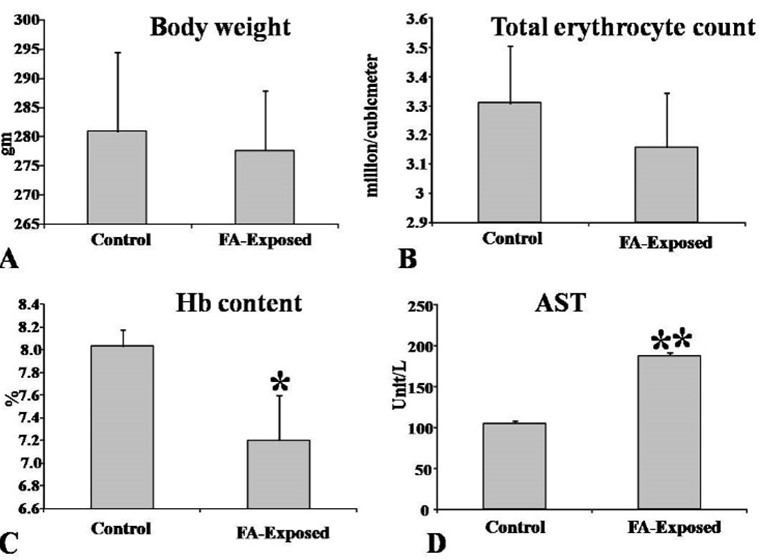
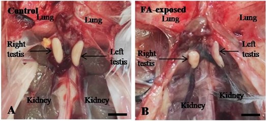
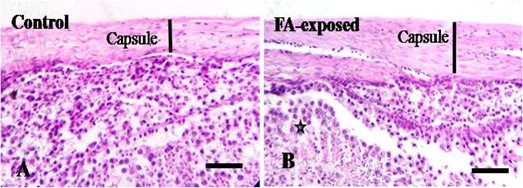
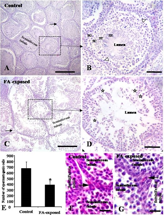
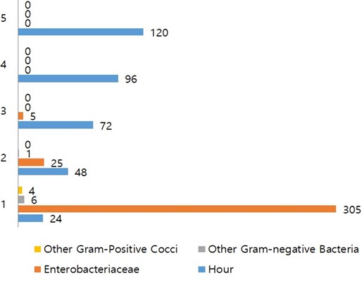
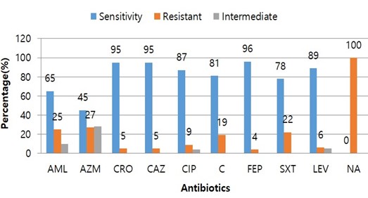
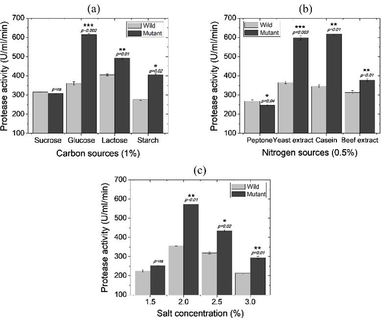