HTTP 200 OK
Allow: GET, HEAD, OPTIONS
Content-Type: application/json
Vary: Accept
{
"count": 319,
"next": "https://jabet.bsmiab.org/articles/?format=api&page=26",
"previous": "https://jabet.bsmiab.org/articles/?format=api&page=24",
"results": [
{
"id": 210,
"slug": "178-1593625242-tissue-culture-of-phalaenopsis-present-status-and-future-prospects",
"featured": false,
"slider": false,
"issue": "Vol3 Issue3",
"type": "review_article",
"manuscript_id": "178-1593625242",
"recieved": "2020-06-17",
"revised": null,
"accepted": "2020-08-04",
"published": "2020-08-16",
"pdf_file": "https://jabet.bsmiab.org/media/pdf_file/2023/51/178-1593625242.pdf",
"title": "Tissue culture of Phalaenopsis: present status and future prospects",
"abstract": "<p><em>Phalaenopsis </em>one of the popular cut-flower among the orchid species. The improvement/multiplication of this orchid is very difficult through conventional breeding due to delay flowering and uneven flower characteristics. Therefore, tissue culture techniques have been extensively used for improvement of <em>Phalaenopsis </em>by inducing and selecting somaclonal variants. However, it is difficult to get stable regenerations techniques of <em>Phalaenopsis </em>due to production of phenolic compounds, arising somaclonal variation in the culture and less recovery in the field of the regenerated plantlets. Improved and modified tissue culture techniques providing regeneration from various vegetative parts of plant are needed for industrialization and <em>ex situ</em> conservation of this valuable orchid. In this paper we have reviewed various <em>in vitro</em> propagation methods of <em>Phalaenopsis </em>culture which will be helpful for commercialization of this valuable orchid.</p>",
"journal_reference": "J Adv Biotechnol Exp Ther. 2020; 3(3): 273-285.",
"academic_editor": "Dr. Akhi Moni, ABEx Bio-Research Center, Bangladesh.",
"cite_info": "Khatun K, Nath UK, et al. Tissue culture of Phalaenopsis: present status and future prospects. J Adv Biotechnol Exp Ther. 2020; 3(3): 273-285.",
"keywords": [
"Phalaenopsis",
"Organ culture",
"Somatic embryogenesis"
],
"DOI": "10.5455/jabet.2020.d135",
"sections": [
{
"section_number": 1,
"section_title": "INTRODUCTION",
"body": "<p>Orchids considered as the most popular ornamental crop species in the world due to their unique use as cut flower and pot plants. Their ubiquitous beauty fascinated people since ancient times. Orchids are widely grown as ornamental cut flowers because of their exotic beauty and long shelf life [<a href=\"#r-1\">1</a>]. Orchid cultivation is one of the most economically global trade nursery industries constituting a multi-billion dollar exchange among different countries [<a href=\"https://www.bsmiab.org/jabet/178-1593625242-tissue-culture-of-phalaenopsis-present-status-and-future-prospects/#_ENREF_2\">2</a><a href=\"#r-2\">, 3</a>]. In contrast to world run-up, Bangladesh is in initial stage of orchid cultivation and starting orchid production commercially just few years ago along with the development of floriculture. In Bangladesh, BRAC, Proshika and Wonderland Toys etc. NGO’s are commercially planting orchids in a large scale. Orchids are bisexual plants and produced fruits after pollination and fertilization. They are normally produced large numbers of capsule, that are highly fragile and possess virtually no stored food material or endosperm [<a href=\"#r-4\">4</a>].<br />\r\n<em>Phalaenopsis</em> (moth orchids) is one of the most popular among orchid species because of their specially beautiful and long-lasting flowers, and can cultivate quite easily in the artificial conditions [<a href=\"#r-5\">5-7</a>]. The nomenclature of <em>Phalaenopsis </em>derived from Greek words <em>phalaina</em>, meaning moth, and <em>opsis</em>, meaning look-alike and the name describes the flowers apparently look like to flying moth [<a href=\"#r-8\">8</a>]. In international flower market, these orchids have high economic value as cut flower. Today, <em>Phalaenopsis </em>are the most widely grown orchids. Statistical data from Netherlands show that <em>Phalaenopsis </em>market prospects increased from 5% to 66% in the year 1983 to 1994%, respectively [<a href=\"#r-9\">9</a>]. As a monopodial plant <em>Phalaenopsis </em>are traditionally propagated by the cutting or division of off-shoots, however, these methods results low multiplication rate and hamper the growth of the mother plant, making them ineffective for large scale production. Therefore, their vegetative propagation is difficult and seedling characteristics are not uniform. It needs at least 3 years for flowering under greenhouse condition which is one of the vital problems in commercial production of <em>Phalaenopsis</em>. Therefore, tissue culture may be an efficient and alternative tool for propagation of this orchid species [<a href=\"#r-10\">10</a>]. Thereafter, scientists in different corner in the world are trying their level best for commercial <em>Phalaenopsis </em>production through tissue culture technique (<a href=\"#figure1\">Figure 1</a>). Inflorescence nodes of <em>Phalaenopsis </em>were induced to form plantlet in aseptic seed germination media by which laid the landmark of <em>Phalaenopsis </em>tissue culture [<a href=\"#r-11\">11</a>]. Based on the findings of Rotor (1950), <em>in vitro Phalaenopsis </em>propagation and multiplication protocols have been developed by many researchers [<a href=\"#r-12\">12</a>]. In this paper, we have tried to review the explant-based <em>Phalaenopsis </em>tissue culture starting from the pioneering works of Rotor (1950) to date and also the future perspectives of the tissue culture techniques for improvement of <em>Phalaenopsis </em>as well as <em>in vitro</em> conservation of the varietal purity of this species.</p>\r\n\r\n<div id=\"figure1\">\r\n<figure class=\"image\"><img alt=\"\" height=\"293\" src=\"/media/article_images/2024/34/08/178-1593625242-Figure1.jpg\" width=\"500\" />\r\n<figcaption><strong>Figure 1.</strong> Schematic diagram of <em>Phalaenopsis</em> propagation method: 1) <em>Phalaenopsis</em> propagation can be done by cutting which may result delay flowering and uneven flower characteristics. 2) Propagation can also be done by tissue culture. Tissue culture of <em>Phalaenopsis</em> is done by using different method e.g. organogenesis and somatic embryogenesis.</figcaption>\r\n</figure>\r\n</div>"
},
{
"section_number": 2,
"section_title": "THE TRADE IN PHALAENOPSIS AND ITS CONTRIBUTION IN ECONOMY",
"body": "<p><em>Phalaenopsis</em> are the second most important orchid marketed as both cut and potted flower. It is one of the most popular and economically important orchid genera at commercial scale production [<a href=\"#r-13\">13</a>]. “Orchid growing has not fully achieved the transition from a hobby to an industry” stated by James Shoemaker in 1957. However, today, orchid growing is an international business and more than just an industry. <em>Phalaenopsis </em>are about 75% of all orchids sold [<a href=\"#r-14\">14</a>]. Many countries like Germany, Netherlands, United States, China, Japan and Taiwan commercially grown the <em>Phalaenopsis </em>in large scale. Currently <em>Phalaenopsis </em>young plants production may have extended more than 300 million per year over the world [<a href=\"#r-14\">14</a>]. Germany, Japan, United States, Netherlands and Taiwan commercially grown <em>Phalaenopsis </em>and Taiwan ranks tops in the world production [<a href=\"#r-15\">15</a>]. In Taiwan <em>Phalaenopsis </em>export value increased from $8 million to $13 million in 2005 to 2006 [<a href=\"#r-16\">16</a>], where worldwide turnover of Taiwanese <em>Phalaenopsis </em>raised from $27.5 million to $35.4 million from 2005 to 2006 [<a href=\"#r-17\">17</a>]. Bangladesh is in very beginning stage of <em>Phalaenopsis </em>production. The Government of Bangladesh is now giving concern to meet up the local demand and can be participated in export market of <em>Phalaenopsis</em>.</p>"
},
{
"section_number": 3,
"section_title": "PHALAENOPSIS REGENERATION THROUGH ORGANOGENESIS",
"body": "<p>Orchid could be propagated rapidly via protocorm like body (PLB) formation from explants rather than direct regeneration. However, recently PLB formation is considered as limited condition due to identify some crucial deleterious factors of orchid tissue culture. Therefore, scientists are seeking alternatives of the PLB formation in commercial orchid production. However, plant regeneration through the formation of PLB has been still practiced for mass propagation of monopodial orchid <em>Phalaenopsis</em>. Rotor, 1950 pioneered of vegetative propagation of <em>Phaleonopsis </em>gave the first documented report on micropropagation of <em>Phaleonopsis</em> using flower stalk cutting as explant [<a href=\"#r-11\">11</a>]. Several reports have provided the indications regarding the flower-stalk cuttings would be very promising approach for clonal propagation of <em>Phalaenopsis </em>[<a href=\"#r-11\">11</a>, <a href=\"#r-18\">18-27</a>]. Micropropagation of <em>Phalaenopsis </em>using flower stalk cuttings is the most widely used technique for mass propagation since the explants can be collected without damaging the mother plant [<a href=\"#r-1\">1</a>]. In contrast , Tanaka <em>et al.</em> (1988) claimed that the flower stalk cuttings could not be used for large scale clonal propagation since the propagation rate is very low [<a href=\"#r-28\">28</a>]. They suggested that the large scale propagation and multiplication of <em>Phalaenopsis </em>would be possible through the formation of PLB [<a href=\"#r-22\">22</a>, <a href=\"#r-23\">23</a>, <a href=\"#r-29\">29-34</a>]. Murdad <em>et al.</em> 2006 reported protocorm is the unique structure for <em>Phalaenopsis </em>production and observed multiplication capability of trimmed and untrimmed protocorms using coconut water and activated charcoal on XER medium contain 20 gl<sup>-1</sup> fructose [<a href=\"#r-35\">35</a>]. Though Murdad <em>et al.</em> 2006 did not suggest that this protocol could be used for mass clonal propagation; they did stated that trimmed protocorm obtained from germinated seed is much better than untrimed one and trimmed protocorm cultured on coconut water and activated charcoal could be used for high frequency multiplication of <em>Phalaenopsis gigantae. </em>Chen <em>et al.</em> 2000 developed a reliable <em>Phalaenopsis </em>regeneration protocol using seed derived protocoms [<a href=\"#r-36\">36</a>]. They used seed of <em>Phalaenopsis nebula</em> for the formation of protocorm in ½ MS basal medium and found that seed derived protocorm performed better for callus induction and subsequent plant regeneration from the induced callus. Whereas Tanaka and Sakanishi, 1985 recommended efficient seed germination of <em>Phalaenopsis </em>in MS medium through PLB formation using different leaf segment (distal, middle and proximal) from <em>in vivo</em> grown mature plant [<a href=\"#r-37\">37</a>]. Park et al. 2002 reported an efficient <em>in vitro Phalaenopsis </em>regeneration protocol through PLB formation using flower stalk nodes derived leaf segments and recommended that modified hyponex medium is suitable for optimal number of PLBs [<a href=\"#r-10\">10</a>]. They optimized the growth regulators combination, to obtain the highest regeneration of PLBs on½ MS medium with BA (88.8 µM) and NAA (5.4 µM) and for the first time they used the raft culture along with solid and liquid culture for proliferation of PLBs. Gonokbari, 2007gave an account for <em>Phalaenopsis </em>regeneration via protocorm formation through thin cell layer culture [<a href=\"#r-38\">38</a>].They used ½ MS media with 2.0 mgl<sup>-1</sup> BAP + 0.5 mgl<sup>-1 </sup>NAA along with coconut water and activated charcoal for PLB formation and used L-glutamine instead of plant hormone for shoot development from PLB. By using this method, they obtained large number of plantlet within a short period. Whereas, Tanaka, 1977 and Tanaka and Sakanishi, 1980 used both solid and liquid VW media with 20% coconut water for the proliferation of PLBs [<a href=\"#r-39\">39</a>, <a href=\"#r-40\">40</a>]. MS medium used by Hass-Von, 1983 for proliferation and differentiation of PLBs. PLBs derived from ½ MS medium were cultured on solidified Hyponex medium (1 gl<sup>-1 </sup>6.5 N- 4.5 P- 19 N + 1 gl<sup>-1</sup>20 N – 20 P – 20 K + 2 gl<sup>-1 </sup>peptone + 0.05% activated charcoal + 30 gl<sup>-1</sup>sucrose) for plantlets development [<a href=\"#r-41\">41</a>]. They found that use of simple Hyponex medium during the proliferation and conversion of PLBs into plantlets was always advantageous. Among different liquid media VW liquid medium was effective for PLB multiplication [<a href=\"#r-42\">42</a>]. Though Park <em>et al.</em> 2002 did not suggest any selective method that could be used on commercial scale vegetative propagation of orchid. Tokuhara and Mii, 1993 used New Dogashima Medium (NDM) instead of ½ MS medium for PLB formation containing 0.1 mgl<sup>-1</sup> naphthaleneacetic acid (NAA) and 1mgl<sup>-1</sup> 6- benzylaminopurine (BAP) suggesting that their method could be used for vegetative propagation of <em>Phalaenopsis</em> and <em>doritaenopsis</em> on a commercial scale [<a href=\"#r-43\">43</a>]. TDZ and auxins combination in culture medium found to be best for the induction of callus and PLBs from leaf of <em>Phalaenopsis </em>[<a href=\"#r-44\">44</a>]. Maximum seed germination was observed in VW medium containing coconut water, with 1mgl<sup>-1</sup> BAP and 2 mgl<sup>-1</sup> kinetin. Callus and PLB were induced from the leaf of germinated plantlets on NDM medium containing TDZ, BAP and combination of TDZ and NAA. They found that TDZ in combination with NAA produce good quality and higher quantity PLBs than TDZ alone. Their result is contradictory with the findings of Soe et al. 2014. They found that PGR free MS medium was efficient for PLB formation [<a href=\"#r-45\">45</a>]. In the propagation of <em>Phalaenopsis </em>Dora and <em>doritaenopsis</em> from inflorescence axis section, TDZ alone found to be more effective [<a href=\"#r-46\">46</a>]. Arditti and Ernst, 1993 used modified MS medium with NAA and BA, Young et al. 2000 used MS medium with NAA and BA for PLB induction from leaf explants but these medium did not give any good result in case of <em>Phalaenopsis gigantae </em>so they used NDM medium for PLB induction [<a href=\"#r-12\">12</a>]. They harvested plantlet after culturing the PLB and callus in hormone free NDM medium. Homma and Asahira, 1985 used inter-nodal section of flower stalk as explants to regenerate shoot of <em>Phalaenopsis </em>through PLB formation and PLB were produced from basal end of explants which touch the media [<a href=\"#r-34\">34</a>]. Intermodal section of flower stalk was better than using flower stalk node and leaf culture in terms of duration of PLB formation and rate of contamination [<a href=\"#r-34\">34</a>]. Among the different parts (tip, middle and basal) of PLB, the basal parts showed highest PLB formation in the PGR free medium [<a href=\"#r-35\">45</a>]. Kobayashi <em>et al.</em> 1993 cultured protoplast derived from callus of lateral bud on flower stalks for regeneration of shoot and established a plant regeneration system from protoplasts culture in <em>Phalaenopsis </em>[<a href=\"#r-47\">47</a>]<em>.</em> They pre-cultured the bud on P basal medium without coconut water and sucrose for 30 days for callus formation. The protoplasts were isolated from callus enzymetically and then cultured in the medium supplemented with 0.05-1.0 mgl<sup>-1</sup> 2,4-D and 10% cw. They found that 2,4-D was more important than CW for colony formation. Then the protoplast derived PLBs were placed on P basal regeneration medium (10% CW, 3% gelrite) for shoot regeneration. Tokuhara and Mii, 1993 developed an efficient PLB formation and subsequently plantlet regeneration method from PLB using shoot tip of flower stalk bud through cell suspension culture by selecting suitable carbohydrate source and concentration. They found that glucose produce the highest PLB than other carbohydrate sources used and lactose was not suitable for cell proliferation or PLB formation. Among the carbon sources, sorbitol was most suitable for plantlet initiation and development from PLB on <em>Phalaenopsis </em>regeneration [<a href=\"#r-48\">48</a>]. They cultured lateral bud from young flower stalk in new <em>Phalaenopsis </em>medium (NP) with 10gl<sup>-1</sup> sorbitol for callus induction. The PLBs were than cultured on NP medium supplanted with 20 gl<sup>-1</sup> sucrose, 20 gl<sup>-1</sup> maltose and 10gl<sup>-1</sup> sorbitol and found that sucrose containing medium showed some necrosis while maltose and sorbitol medium have no necrosis and plantlet production was higher in sorbitol medium than sucrose and maltose medium.<br />\r\nMany researchers try to get regeneration without formation of PLB. Myint et al. 2001 have developed a rapid <em>Phalaenopsis </em>propagation technique through PLB (protocorm like body) formation using leaf as explant [<a href=\"#r-49\">49</a>]. Plantlet was successfully regenerated via the adventitious bud without PLB formation from vegetative bud of flower stalk for avoiding somaclonal variation [<a href=\"#r-50\">50</a>].They used Vacin and Went medium with 15% coconut water along with different concentration of TDZ and BAP and found that TDZ was more effective than BAP in stimulating the axillary buds for induction of shoots. Rotor, 1950 initiated <em>in vitro</em> <em>Phalaenopsis</em> cultures using flower stalks without disturbing the whole plant [<a href=\"#r-11\">11</a>]. This technique found to be used extensively for mass propagation of <em>Phalaenopsis</em>. Dormant buds of the inflorescence were most advantageous among the other explants for <em>in vitro</em> propagation of <em>Phalaenopsis</em>; where Indole Acetyl amino Acids (IAA) used for the propagation of plantlets [<a href=\"#r-33\">33</a>]. Flower stalk buds were cultured and achieved reproductive shoots from upper node and vegetative shoots from lower node [<a href=\"#r-19\">19</a>, <a href=\"#r-22\">22</a>, <a href=\"#r-51\">51</a>]. Effect of bud position, temperature and BAP on the growth mode of bud studied in <em>P. amabilis</em> [<a href=\"#r-51\">51</a>]. Lin, 1986 reported the influence of developmental stage and age of flower stalk on plantlet regeneration in <em>Phalaenopsis </em>and <em>doritaenopsis</em> and marked that the flower stalk with first flower and intermodal section near the tip of stalk have the highest regeneration capacity [<a href=\"#r-52\">52</a>]. Kosir <em>et al.</em> 2004 used nodes with dormant bud of flower stalks for rapid shoot regeneration of <em>Phalaenopsis </em>[<a href=\"#r-53\">53</a>]. They used six media with little difference in composition and found that none of the media was appropriate for mass generation and their result was contradictory with Arditii and Ernst (1993). They suggested that media with higher BAP and lower nitrogen content would be suitable in tissue culture and later for <em>in vivo</em> flower induction. BA was mandatory for floral bud induction where high nitrogen concentration inhibits the development of floral buds and shortening nine months growth period of <em>Phalaenopsis </em>for flowering [<a href=\"#r-54\">54</a>]. Flower stalk culture is most frequently used as explants but it takes a long time to come out plantlets, leaf culture also need more time to produce protocorm and frequent release of phenolic compound is also a major problem. Plantlet regeneration through PLB formation is not easily reproducible [<a href=\"#r-55\">55</a>]. Alternatively elongated stem node was used as explants for regeneration of plantlets by Duan et al. 1996 [<a href=\"#r-55\">55</a>]. The elongated stems were cut into 4 section as top, second, third and basal node and placed on Hyponex medium supplemented with various concentration of BA. Shoots and multiple adventitious buds were produced after 70 days of culture and the highest number of shoots was obtained from third nodes and second nodes.<br />\r\n<em>Phalaenopsis</em> propagation through PLB formation was efficient but in many cases occurrence of somaclonal variation is a major problem for large scale production of plantlets. Chen et al. 1998 found considerable variation in flower colour and shape of <em>Phalaenopsis </em>“True Lady- B79-19” regenerated through tissue culture [<a href=\"#r-56\">56</a>]. Tokuhara and Mii, 1998 also found somaclonal variation in flower and inflorescence axis of the micropropagated plants derived from flower stalk bud via protocorm like body formation of <em>Phalaenopsis </em>[<a href=\"#r-57\">57</a>]. The variation ranges from 0 to 100% but maximum cultivars showed less than 10% somaclonal variation. Release of high phenolic compounds is another major problems of tissue culture of <em>Phalaenopsis </em>which is toxic for <em>in vitro</em> growing plantlets [<a href=\"#r-58\">58</a>]. Use of bioreactor systems: continuous immersion system (air-lift type) and temporally immersion system could solve this problem. Temporary immersion bioreactor with activated charcoal filter attached was most suitable for multiplication of PLBs which was effective to remove phenolics. 83% PLBs regenerated into plantlets in 8 weeks and fresh weight of the plantlets and rooting percentage was also very high of the regenerated plantlets [<a href=\"#r-58\">58</a>].</p>"
},
{
"section_number": 4,
"section_title": "PHALAENOPSIS REGENERATION THROUGH SOMATIC EMBRYOGENESIS",
"body": "<p>Somatic embryogenesis has often been considered efficient techniques for plant regeneration and for obtaining transgenic plant. Currently somatic embryogenesis protocols have been successful studied in <em>Phalaenopsis</em> [<a href=\"#r-59\">59-62</a>]. Successful regeneration of <em>Phalaenopsis </em>through somatic embryogenesis depends on many factors like source of explant, nutrient composition, the growth hormones and part of the explant taken, explant orientation etc. Kuo <em>et al.</em> 2005 reported plant regeneration using leaf explants through direct somatic embryogenesis after 20-30 days of culture on half-strength MS medium supplemented with BA and TDZ [<a href=\"#r-61\">61</a>]. The frequency of embryogenesis is affected by explant orientation usually adaxial surfaces near wounded regions gave the highest embryogenic competency compared to other regions of explants though the authors have not clarify the reason for this. However Gow <em>et al.</em> 2008 found that the cut ends of leaf had the highest embryogenic competence than adaxial and abaxial sides in <em>Phalaenopsis</em><em> amabilis</em> and <em>Phalaenopsis</em><em> nebula </em>[<a href=\"#r-63\">63</a>]. Cytokinin is effective for the somatic embryo induction. BA and TDZ has been reported to promoted embryogenesis mostly from the epidermal cell layers [<a href=\"#r-61\">61</a>]. TDZ has also been reported to promote direct embryo formation from the epidermal cells and secondary embryogenesis from the leaf explants of <em>Phalaenopsis amabilis </em>[<a href=\"#r-62\">62</a>] (<a href=\"#figure2\">Figure 2</a>). Whereas NAA, 2,4-D highly retarded the somatic embryo formation from leaf explants of <em>Phalaenopsis </em>[<a href=\"#r-61\">61</a>, <a href=\"#r-62\">62</a>].Concentrations of different plant growth regulators had effect on somatic embryogenesis from leaf explant of <em>Phalaenopsis </em>[<a href=\"#r-64\">64</a>]<em>.</em> N6-benzyl adenine (6-BA) had better performance than adenine sulphate (Ad) in embryoid induction [<a href=\"#r-64\">64</a>]. They reported that upper epidermis and single cell of mesophyll were the starting source of somatic embryos origination. Chen and Chang, 2004 reported TDZ promoted the formation of embryo from protocorm like bodies derived from seed; whereas NAA retarded the embryo formation of <em>Phalaenopsis amabilis</em> var. <em>Formosa </em>[<a href=\"#r-65\">65</a>]<em>.</em> When protocorms derived from seed were cultured on ½MS medium without plant growth regulators except TDZ, 100% of the protocorms were produced embryos from the posterior regions. Regeneration of plantlets thorough somatic embryogenesis has also been achieved by Samson et al. 1998 [<a href=\"#r-66\">66</a>]. They had used internodal flower stalk segment with an axillary bud to develop protocorms. They cultured the nodal cutting on Vacin-Went medium to develop vegetative shoots which were cultured on solid New Dogashima Medium (NDM1) supplemented with NAA and 4,4,4 tri-fluro-isopentenyl-adenine for the initiation of protocorm regeneration and histological study permit these protocorms as somatic embryo. They have recommended their methods for commercial propagation of <em>Phalaenopsis.</em> In the similar way, Tokuhara and Mii, 2001 developed a method for embryogenic calli; subsequently, plantlets from the calli using flower stalk bud by changing the sucrose concentration in NDM medium following liquid cell suspension culture [<a href=\"#r-60\">60</a>]. Although Sajise and Sagawa, 1991 were first reported on embryogenic calli formation but they did not give any clear-cut protocol for callus induction [<a href=\"#r-67\">67</a>]. However, Tokuhara and Mii, 2001 found that high sucrose concentration in media inhibit initial callus induction, but high sucrose plays vital role of callus proliferation, whenever callus being established in media [<a href=\"#r-60\">60</a>]. Their proposed method could be efficiently utilized for the microprogation of <em>Phalaenopsis </em>despite about 10% somaclonal variations. Embryogenic cell suspension culture for the regeneration of plantlets from protoplast of <em>Phalaenopsis wataboushi</em> were followed using ½ NDM medium containing 0.06M sucrose, 0.44M sorbitol and 0.1g/l glutamine [<a href=\"#r-68\">68</a>]. They established a plant regeneration protocol from protoplast without any plant growth regulators and coconut water as supplement. They used shoot tip for the induction of embryogenic calli following the protocol of Tokuhara and Mii, 2001, whereas one year old cells of suspension culture was used to isolate protoplast. As a carbohydrate source sorbitol (10gl<sup>-1</sup> sorbitol in hormone free NDM medium with 0.3% gellun gum) considered most suitable for the regeneration of plantlets from PLBs and sucrose was most suitable for shoot development [<a href=\"#r-68\">68</a>]. Ishii <em>et al.</em> 1998 has been reported that sucrose was effective for callus induction but somatic embryos were formed upon subculture in medium without sucrose indicated that the growth of monopodial orchid <em>Phalaenopsis </em>influenced by the sugar in medium [<a href=\"#r-59\">59</a>]. <a href=\"https://www.bsmiab.org/jabet/wp-content/uploads/sites/2/2020/08/178-1593625242.pdf\">Table 1</a> represents the brief scenario of tissue culture of Phalaenopsis.</p>\r\n\r\n<div id=\"figure2\">\r\n<figure class=\"image\"><img alt=\"\" height=\"574\" src=\"/media/article_images/2024/34/08/178-1593625242-Figure2.jpg\" width=\"500\" />\r\n<figcaption><strong>Figure 2. </strong>Direct somatic embryogenesis and Plant regeneration of <em>Phalaenopsis amabilis</em> using leaf explants : (A) somatic embryos after 20 days of culture (bar = 700 mm); (B) enlarged and elongated embryos after 30 days of culture (bar = 750 mm); (C) green embryos under light and young somatic protocorm after 45 days of culture (bar = 850 mm); (D) somatic embryos developed from leaf-derived nodular masses (bar = 950 mm); (E) shoots and some formed secondary embryos from the developed embryos (bar = 1.2 mm); (F) leaf-derived embryos formed shoots (bar = 2 mm);(G) a plantlet from the leaf derived embryos (bar = 7.2 mm) [<a href=\"#r-62\">62</a>].</figcaption>\r\n</figure>\r\n</div>\r\n\r\n<div id=\"Table-1\">\r\n<p><a href=\"https://jabet.bsmiab.org/table/178-1593625242-table1/\">Table-1</a><strong>Table 1. </strong>Brief scenario of the success in micropropagation of Phalaenopsis orchid species using different explants and media by different researchers. </p>\r\n</div>"
},
{
"section_number": 5,
"section_title": "CONCLUSION AND FUTURE PROSPECTS OF PHALAENOPSISIS",
"body": "<p><em>Phalaenopsis</em>is one of the most popular orchid and has immense economic value as ornamental cut flower. To date, the seed derived propagation of <em>Phalaenopsis</em> is very rapid and easy approach. Therefore, their uniform flower characteristics are one of the important criteria for commercialization. This could only possible by following tissue culture techniques despite very little somaclonal variation up to 10% within acceptable limit. Meanwhile, tissue culture method need less time to develop and maintain varietal purity compare to conventional breeding. In this paper we have tried to collect together most of the commercially important <em>in vitro</em> propagation of <em>Phalaenopsis </em>using different explants and different growth condition which will be helpful for rapid clonal propagation, industrial exploitation and also improvising the currently available method for <em>in vitro </em>mass propagation of this valuable orchid. The commercial demand of <em>Phalaenopsis</em> has been increasing day by day and <em>Phalaenopsis</em> production is now international in scope. Based upon the advanced tissue culture techniques new types might be developed with a compact growth habit, variegated foliage and uniform flower characteristics. Therefore, the <em>Phalaenopsis </em>trade might be increased both in volume and value throughout the world and possible to earn lots of foreign exchange by exporting the orchids.</p>"
},
{
"section_number": 6,
"section_title": "ACKNOWLEDGEMENT",
"body": "<p>The authors acknowledge Department of Biotechnology, Patuakhali Science and Technology University, Bangladesh. The authors thankful to Dr. Chung, Mi-Young for her valuable efforts concerning this research.</p>"
},
{
"section_number": 7,
"section_title": "AUTHOR CONTRIBUTIONS",
"body": "<p>Dr. Khadiza Khatun drafted the manuscript and revised the final draft. Dr. Ujjal Kumar nath revised the initial manuscript. Md. Shafikur Rahman helped to write the manuscript.</p>"
},
{
"section_number": 8,
"section_title": "CONFLICTS OF INTEREST",
"body": "<p>Authors declared that they have no conflict of interest.</p>"
}
],
"figures": [
{
"figure": "https://jabet.bsmiab.org/media/article_images/2024/34/08/178-1593625242-Figure1.jpg",
"caption": "Figure 1. Schematic diagram of Phalaenopsis propagation method: 1) Phalaenopsis propagation can be done by cutting which may result delay flowering and uneven flower characteristics. 2) Propagation can also be done by tissue culture. Tissue culture of Phalaenopsis is done by using different method e.g. organogenesis and somatic embryogenesis.",
"featured": false
},
{
"figure": "https://jabet.bsmiab.org/media/article_images/2024/34/08/178-1593625242-Figure2.jpg",
"caption": "Figure 2. Direct somatic embryogenesis and Plant regeneration of Phalaenopsis amabilis using leaf explants : (A) somatic embryos after 20 days of culture (bar = 700 mm); (B) enlarged and elongated embryos after 30 days of culture (bar = 750 mm); (C) green embryos under light and young somatic protocorm after 45 days of culture (bar = 850 mm); (D) somatic embryos developed from leaf-derived nodular masses (bar = 950 mm); (E) shoots and some formed secondary embryos from the developed embryos (bar = 1.2 mm); (F) leaf-derived embryos formed shoots (bar = 2 mm);(G) a plantlet from the leaf derived embryos (bar = 7.2 mm) [62].",
"featured": false
}
],
"authors": [
{
"id": 933,
"affiliation": [
{
"affiliation": "Department of Biotechnology, Patuakhali Science and Technology University, Dumki, Patuakhali, Bangladesh"
}
],
"first_name": "Khadiza",
"family_name": "Khatun",
"email": "kkr@pstu.ac.bd",
"author_order": 1,
"ORCID": null,
"corresponding": true,
"co_first_author": false,
"co_author": false,
"corresponding_author_info": "Dr. Khadiza Khatun, Department of Biotechnology, Patuakhali Science and Technology University, Dumki, Patuakhali, Bangladesh, Phone: +8801763405303, Email: kkr@pstu.ac.bd",
"article": 210
},
{
"id": 934,
"affiliation": [
{
"affiliation": "Bangladesh Argicultural University, Mymensingh, Bangladesh"
}
],
"first_name": "Ujjal Kumar",
"family_name": "Nath",
"email": null,
"author_order": 2,
"ORCID": null,
"corresponding": false,
"co_first_author": false,
"co_author": false,
"corresponding_author_info": "",
"article": 210
},
{
"id": 935,
"affiliation": [
{
"affiliation": "Department of Biotechnology, Patuakhali Science and Technology University, Dumki, Patuakhali, Bangladesh"
}
],
"first_name": "Md. Shafikur",
"family_name": "Rahman",
"email": null,
"author_order": 3,
"ORCID": null,
"corresponding": false,
"co_first_author": false,
"co_author": false,
"corresponding_author_info": "",
"article": 210
}
],
"views": 3541,
"downloads": 289,
"references": [
{
"id": 7073,
"serial_number": 1,
"pmc": null,
"reference": "Chugh S, Guha S, Rao IU. Micropropagation of orchids: a review on the potential of different explants. Scientia Horticulturae, 2009; 122(4):507-520.",
"DOI": null,
"article": 210
},
{
"id": 7074,
"serial_number": 2,
"pmc": null,
"reference": "Hew C. Orchid cut-flower production in Singapore and neighboring ASEAN countries. American Orchid Society bulletin (USA), 1989; 887-897.",
"DOI": null,
"article": 210
},
{
"id": 7075,
"serial_number": 3,
"pmc": null,
"reference": "Alam M, Rashid M, Hossain M, Salam M, Rouf M. In vitro seed propagation of Dendrobium (Dendrobium transparens) orchid as influenced by different media. International Journal of Biotechnology (Pakistan), 2002; 1:111-115.",
"DOI": null,
"article": 210
},
{
"id": 7076,
"serial_number": 4,
"pmc": null,
"reference": "Mitra G. Studies on seeds, shoot-tips & stem-discs of an orchid grown in aseptic culture. Indian Journal of Experimental Biology, 1971; 79-85.",
"DOI": null,
"article": 210
},
{
"id": 7077,
"serial_number": 5,
"pmc": null,
"reference": "Guo WJ, Lin YZ, Lee N. Photosynthetic light requirements and effects of low irradiance and daylength on Phalaenopsis amabilis. Journal of the American Society for Horticultural Science, 2012; 137(6):465-472.",
"DOI": null,
"article": 210
},
{
"id": 7078,
"serial_number": 6,
"pmc": null,
"reference": "Lin MJ, Hsu BD. Photosynthetic plasticity of Phalaenopsis in response to different light environments. Journal of Plant Physiology, 2004; 161(11):1259-1268.",
"DOI": null,
"article": 210
},
{
"id": 7079,
"serial_number": 7,
"pmc": null,
"reference": "Liu YC, Tseng KM, Chen CC, Tsai YT, Liu CH, Chen WH, Wang HL. Warm-night temperature delays spike emergence and alters carbon pool metabolism in the stem and leaves of Phalaenopsis aphroide. Scientia Horticulturae, 2013; 161:198-203.",
"DOI": null,
"article": 210
},
{
"id": 7080,
"serial_number": 8,
"pmc": null,
"reference": "Mayr H. Orchid names and their meanings: Lubrecht & Cramer Limited, 1998.",
"DOI": null,
"article": 210
},
{
"id": 7081,
"serial_number": 9,
"pmc": null,
"reference": "Griesbach R. Potted Phalaenopsis orchid production: history, present status, and challenges for the future. HortTechnology, 2000; 10(3):429-429.",
"DOI": null,
"article": 210
},
{
"id": 7082,
"serial_number": 10,
"pmc": null,
"reference": "Park SY, Murthy HN, Paek KY. Rapid propagation of Phalaenopsis from floral stalk-derived leaves. In Vitro Cellular & Developmental Biology-Plant, 2002; 38(2):168-172.",
"DOI": null,
"article": 210
},
{
"id": 7083,
"serial_number": 11,
"pmc": null,
"reference": "Rotor JrG. A method of vegetative propagation of Phalaenopsis species and hybrids. In: Proceedings American Society for Horticultural Science, 1950.",
"DOI": null,
"article": 210
},
{
"id": 7084,
"serial_number": 12,
"pmc": null,
"reference": "Arditti J, Ernst R. Micropropagation of orchids. John Willey & Sons. Inc, NY, 1993.",
"DOI": null,
"article": 210
},
{
"id": 7085,
"serial_number": 13,
"pmc": null,
"reference": "Minh TN, Tuyen PT, Khang DT, Quan NV, Ha PTT, Quan NT, Andriana Y, Fan X, Van TM, Khanh TD. Potential use of plant waste from the moth orchid (Phalaenopsis Sogo Yukidian “V3”) as an antioxidant source. Foods, 2017; 6(10):85.",
"DOI": null,
"article": 210
},
{
"id": 7086,
"serial_number": 14,
"pmc": null,
"reference": "Griesbach R. Development of Phalaenopsis orchids for the mass-market. Trends in new crops and new uses ASHS Press, Alexandria, VA, 2002; 458-465.",
"DOI": null,
"article": 210
},
{
"id": 7087,
"serial_number": 15,
"pmc": null,
"reference": "Khoddamzadeh AA, Sinniah U, Kadir M, Kadzimin S, Mahmood M, Sreeramanan S. In vitro induction and proliferation of protocorm-like bodies (PLBs) from leaf segments of Phalaenopsis bellina (Rchb. f.) Christenson. Plant Growth Regulation, 2011; 65(2):381.",
"DOI": null,
"article": 210
},
{
"id": 7088,
"serial_number": 16,
"pmc": null,
"reference": "De L, Khan A, Kumar R, Medhi R. Orchid farming—a remunerative approach for farmers livelihood. International Journal of Science and Research, 2014; 3:468-471.",
"DOI": null,
"article": 210
},
{
"id": 7089,
"serial_number": 17,
"pmc": null,
"reference": "De LC, Pathak P, Rao A, Rajeevan P. 2 Global Orchid Industry. In: Commercial Orchids. Sciendo Migration, 2014; 13-19.",
"DOI": null,
"article": 210
},
{
"id": 7090,
"serial_number": 18,
"pmc": null,
"reference": "Sagawa Y, Niimoto D. Vegetative propagation of. In.: Phalaenopsis, 1960.",
"DOI": null,
"article": 210
},
{
"id": 7091,
"serial_number": 19,
"pmc": null,
"reference": "Urata U. The use of Ito-type vials for vegetative propagation of Phalaenopsis. American Orchid Society Bulletin, 1965; 34:410-413.",
"DOI": null,
"article": 210
},
{
"id": 7092,
"serial_number": 20,
"pmc": null,
"reference": "Scully R. Stem propagation of Phalaenopsis. American Orchid Society Bulletin, 1966; 35:40-42.",
"DOI": null,
"article": 210
},
{
"id": 7093,
"serial_number": 21,
"pmc": null,
"reference": "Intuwong O. Vegetative propagation of Phalaenopsis by flower stalk cuttings. Hawaii Orchid J, 1973; 1:13-18.",
"DOI": null,
"article": 210
},
{
"id": 7094,
"serial_number": 22,
"pmc": null,
"reference": "Koch L. Untersuchungen zur vegetativen Vermehrung bei Phalaenopsis in vitro. Technische Universität Hannover, 1974.",
"DOI": null,
"article": 210
},
{
"id": 7095,
"serial_number": 23,
"pmc": null,
"reference": "Koch L. Erbgleiche Vermehrung von Phalaenopsis in vitro. Gartenwelt, 1974; 74:482-484.",
"DOI": null,
"article": 210
},
{
"id": 7096,
"serial_number": 24,
"pmc": null,
"reference": "Reisinger DM. Clonal propagation of Phalaenopsis by means of flower-stalk node culture. American Orchid Society Bulletin, 1976; 45:45-52.",
"DOI": null,
"article": 210
},
{
"id": 7097,
"serial_number": 25,
"pmc": null,
"reference": "Zhongming Z, Linong L, Xiaona Y, Wangqiang Z, Wei L. Culture of flower-stalk buds a method for vegetative propagation of Phalaenopsis, 1977.",
"DOI": null,
"article": 210
},
{
"id": 7098,
"serial_number": 26,
"pmc": null,
"reference": "Flamee M, Boesman G. Clonal multiplication of Phalaenopsis-hybrids by means of sections of the flower stalk [ornamental plants]. Mededelingen van de Faculteit Landbouwwetenschappen Rijksuniversiteit Gent, 1977; 42: 1865-1868",
"DOI": null,
"article": 210
},
{
"id": 7099,
"serial_number": 27,
"pmc": null,
"reference": "Bouriquet R, Broly H, Legrand B. Clonal propagation of Phalaenopsis (Orchidaceae) by in vitro culture. Praeger, New York, 1982; 35-38.",
"DOI": null,
"article": 210
},
{
"id": 7100,
"serial_number": 28,
"pmc": null,
"reference": "Tanaka M, Kumura M, Goi M. Optimal conditions for shoot production from Phalaenopsis flower-stalk cuttings cultured in vitro. Scientia Horticulturae, 1988; 35(1-2):117-126.",
"DOI": null,
"article": 210
},
{
"id": 7101,
"serial_number": 29,
"pmc": null,
"reference": "Intuwong O, Sagawa Y. Clonal propagation of Phalaenopsis by shoot tip culture. American Orchid Society Bulletin,",
"DOI": null,
"article": 210
},
{
"id": 7102,
"serial_number": 30,
"pmc": null,
"reference": "Pieper W, Zimmer K. Clonal propagation of Phalaenopsis in vitro. In: Symposium on Production of Potted Plants and Cut Flowers, 1976; 64: 21-24.",
"DOI": null,
"article": 210
},
{
"id": 7103,
"serial_number": 31,
"pmc": null,
"reference": "Lay FFM. Studies on the tissue culture of orchids. I. Clonal propagation of Phalaenopsis by lateral buds from flower stems. Orchid Review,",
"DOI": null,
"article": 210
},
{
"id": 7104,
"serial_number": 32,
"pmc": null,
"reference": "Zimmer K, Pieper W. Clonal propagation of Phalaenopsis by excised buds. Orchid Review, 1978.",
"DOI": null,
"article": 210
},
{
"id": 7105,
"serial_number": 33,
"pmc": null,
"reference": "Griesbach RJ. The use of indoleacetylamino acids in the in vitro propagation of Phalaenopsis Scientia Horticulturae, 1983; 19(3-4):363-366.",
"DOI": null,
"article": 210
},
{
"id": 7106,
"serial_number": 34,
"pmc": null,
"reference": "Homma Y, Asahira T. New means of Phalaenopsis propagation with internodal sections of flower stalk. Journal of the Japanese Society for Horticultural Science, 1985; 54(3):379-387.",
"DOI": null,
"article": 210
},
{
"id": 7107,
"serial_number": 35,
"pmc": null,
"reference": "Murdad R, Hwa KS, Seng CK, Latip MA, Aziz ZA, Ripin R. High frequency multiplication of Phalaenopsis gigantea using trimmed bases protocorms technique. Scientia Horticulturae, 2006; 111(1):73-79.",
"DOI": null,
"article": 210
},
{
"id": 7108,
"serial_number": 36,
"pmc": null,
"reference": "Chen YC, Chang C, Chang WC. A reliable protocol for plant regeneration from callus culture of Phalaenopsis. In Vitro Cellular & Developmental Biology-Plant, 2000; 36(5):420-423.",
"DOI": null,
"article": 210
},
{
"id": 7109,
"serial_number": 37,
"pmc": null,
"reference": "Tanaka M, Sakanishi Y. Regenerative capacity of in vitro cultured leaf segments excised from mature Phalaenopsis Bulletin of the University of Osaka Prefecture Ser B, Agriculture and biology, 1985; (37):1-4.",
"DOI": null,
"article": 210
},
{
"id": 7110,
"serial_number": 38,
"pmc": null,
"reference": "Gonokbari S. Efficient micropropagation of Phalaenopsis amabilis (L.) Bl. cv.‘Cool Breeze’using inflorescence axis thin sections as explants. Propagation of Ornamental Plants, 2007; 7(1):9-15.",
"DOI": null,
"article": 210
},
{
"id": 7111,
"serial_number": 39,
"pmc": null,
"reference": "Tanaka M. Clonal propagation of Phalaenopsis by leaf tissue culture. American Orchid Soceity Bulletin, 1977; 46:733-737.",
"DOI": null,
"article": 210
},
{
"id": 7112,
"serial_number": 40,
"pmc": null,
"reference": "Tanaka M, Sakanishi Y. Clonal propagation of Phalaenopsis through tissue culture. In: Proc 9th World Orchid Conference, Bangkok, 1980; 215-221.",
"DOI": null,
"article": 210
},
{
"id": 7113,
"serial_number": 41,
"pmc": null,
"reference": "Hass-Von Schmude N. Klonale Massenvemehrung von Phalaenopsis. Die Orchidee, 1983; 34:242-248.",
"DOI": null,
"article": 210
},
{
"id": 7114,
"serial_number": 42,
"pmc": null,
"reference": "Park YS, Kakuta S, Kano A, Okabe M. Efficient propagation of protocorm-like bodies of Phalaenopsis in liquid medium. Plant Cell, Tissue and Organ Culture, 1996; 45(1):79-85.",
"DOI": null,
"article": 210
},
{
"id": 7115,
"serial_number": 43,
"pmc": null,
"reference": "Tokuhara K, Mii M. Micropropagation of Phalaenopsis and Doritaenopsis by culturing shoot tips of flower stalk buds. Plant Cell Reports, 1993; 13(1):7-11.",
"DOI": null,
"article": 210
},
{
"id": 7116,
"serial_number": 44,
"pmc": null,
"reference": "Niknejad A, Kadir M, Kadzimin S. In vitro plant regeneration from protocorms-like bodies (PLBs) and callus of Phalaenopsis gigantea (Epidendroideae: Orchidaceae). African Journal of Biotechnology, 2011; 10(56):11808-11816.",
"DOI": null,
"article": 210
},
{
"id": 7117,
"serial_number": 45,
"pmc": null,
"reference": "Soe KW, Myint KT, Naing AH, Kim CK. Optimization of efficient protocorm-like body (PLB) formation of Phalaenopsis and Dendrobium Current Research on Agriculture and Life Sciences, 2014; 32(4):179-183.",
"DOI": null,
"article": 210
},
{
"id": 7118,
"serial_number": 46,
"pmc": null,
"reference": "Ernst R. Effects of thidiazuron on in vitro propagation of Phalaenopsis and Doritaenopsis (Orchidaceae). Plant Cell, Tissue and Organ Culture, 1994; 39(3):273-275.",
"DOI": null,
"article": 210
},
{
"id": 7119,
"serial_number": 47,
"pmc": null,
"reference": "Kobayashi S, Kameya T, Ichihashi S. Plant regeneration from protoplasts derived from callus of Phalaenopsis. Plant tissue culture letters, 1993; 10(3):267-270.",
"DOI": null,
"article": 210
},
{
"id": 7120,
"serial_number": 48,
"pmc": null,
"reference": "Islam MO, Ichihashi S, Matsui S. Control of growth and development of protocorm like body derived from callus by carbon sources in Phalaenopsis. Plant Biotechnology, 1998; 15(4):183-187.",
"DOI": null,
"article": 210
},
{
"id": 7121,
"serial_number": 49,
"pmc": null,
"reference": "Myint KT, Chung MY, Chung JD, Kim CK. Propagation via in vitro culture of leaf tissue of Phalaenopsis Horticulture Environment and Biotechnology, 2001; 42(1):1-5.",
"DOI": null,
"article": 210
},
{
"id": 7122,
"serial_number": 50,
"pmc": null,
"reference": "Chen Y, Piluek C. Effects of thidiazuron and N 6-benzylaminopurine on shoot regeneration of Phalaenopsis. Plant Growth Regulation, 1995; 16(1):99-101.",
"DOI": null,
"article": 210
},
{
"id": 7123,
"serial_number": 51,
"pmc": null,
"reference": "Tanaka M, Sakanishi Y. Factors affecting the growth of in vitro cultured lateral buds from Phalaenopsis flower stalks. Scientia Horticulturae, 1978; 8(2):169-178.",
"DOI": null,
"article": 210
},
{
"id": 7124,
"serial_number": 52,
"pmc": null,
"reference": "Lin CC. In vitro culture of flower stalk internodes of Phalaenopsis and Doritaenopsis. Lindleyana, 1986, 1:158-163.",
"DOI": null,
"article": 210
},
{
"id": 7125,
"serial_number": 53,
"pmc": null,
"reference": "Košir P, Škof S, Luthar Z. Direct shoot regeneration from nodes of Phalaenopsis Acta agriculturae slovenica, 2004; 83(2):233-242.",
"DOI": null,
"article": 210
},
{
"id": 7126,
"serial_number": 54,
"pmc": null,
"reference": "Duan JX, Yazawa S. Floral induction and development in Phalaenopsis in vitro. Plant Cell, Tissue and Organ Culture, 1995; 43(1):71-74.",
"DOI": null,
"article": 210
},
{
"id": 7127,
"serial_number": 55,
"pmc": null,
"reference": "Duan JX, Chen H, Yazawa S. In vitro propagation of Phalaenopsis via culture of cytokinin-induced nodes. Journal of Plant Growth Regulation, 1996; 15(3):133-137.",
"DOI": null,
"article": 210
},
{
"id": 7128,
"serial_number": 56,
"pmc": null,
"reference": "Chen W, Chen T, Fu Y, Hsieh R, Chen W. Studies on somaclonal variation in Plant Cell Reports, 1998; 18(1-2):7-13.",
"DOI": null,
"article": 210
},
{
"id": 7129,
"serial_number": 57,
"pmc": null,
"reference": "Tokuhara K, Mii M. Somaclonal variations in flower and inflorescence axis in micropropagated plants through flower stalk bud culture of Phalaenopsis and Doritaenopsis. Plant Biotechnology, 1998; 15(1):23-28.",
"DOI": null,
"article": 210
},
{
"id": 7130,
"serial_number": 58,
"pmc": null,
"reference": "Young PS, Murthy H, Yoeup PK. Mass multiplication of protocorm-like bodies using bioreactor system and subsequent plant regeneration in Phalaenopsis. Plant Cell, Tissue and Organ Culture, 2000; 63(1):67-72.",
"DOI": null,
"article": 210
},
{
"id": 7131,
"serial_number": 59,
"pmc": null,
"reference": "Ishii Y, Takamura T, Goi M, Tanaka M. Callus induction and somatic embryogenesis of Plant Cell Reports, 1998; 17(6-7):446-450.",
"DOI": null,
"article": 210
},
{
"id": 7132,
"serial_number": 60,
"pmc": null,
"reference": "Tokuhara K, Mii M. Induction of embryogenic callus and cell suspension culture from shoot tips excised from flower stalk buds of Phalaenopsis (Orchidaceae). In Vitro Cellular & Developmental Biology-Plant, 2001; 37(4):457-461.",
"DOI": null,
"article": 210
},
{
"id": 7133,
"serial_number": 61,
"pmc": null,
"reference": "Kuo HL, Chen JT, Chang WC. Efficient plant regeneration through direct somatic embryogenesis from leaf explants of Phalaenopsis ‘Little Steve’. In Vitro Cellular & Developmental Biology-Plant, 2005; 41(4):453.",
"DOI": null,
"article": 210
},
{
"id": 7134,
"serial_number": 62,
"pmc": null,
"reference": "Chen J, Chang WC. Direct somatic embryogenesis and plant regeneration from leaf explants of Phalaenopsis amabilis. Biologia Plantarum, 2006; 50(2):169-173.",
"DOI": null,
"article": 210
},
{
"id": 7135,
"serial_number": 63,
"pmc": null,
"reference": "Gow WP, Chen JT, Chang WC. Influence of growth regulators on direct embryo formation from leaf explants of Phalaenopsis Acta Physiologiae Plantarum, 2008; 30(4):507.",
"DOI": null,
"article": 210
},
{
"id": 7136,
"serial_number": 64,
"pmc": null,
"reference": "Cui G, Hou X, Zhang Z, Zhang C, Hu N, LIU YC. Efficient somatic embryogenesis from leaf explants of Phalaenopsis in vitro culture and histological observations. Acta Hortic Sin, 2007; 34(2):431-436.",
"DOI": null,
"article": 210
},
{
"id": 7137,
"serial_number": 65,
"pmc": null,
"reference": "Chen JT, Chang WC. Induction of repetitive embryogenesis from seed-derived protocorms of Phalaenopsis amabilis var. formosa Shimadzu. In Vitro Cellular & Developmental Biology-Plant, 2004; 40(3):290.",
"DOI": null,
"article": 210
},
{
"id": 7138,
"serial_number": 66,
"pmc": null,
"reference": "Samson I, Hamama L, Letouzé R. The role of new synthetic cytokinins in the improvement of mass propagation of Phalaenopsis via protocorms regeneration. In: XIX International Symposium on Improvement of Ornamental Plants,1998; 508: 265-266.",
"DOI": null,
"article": 210
},
{
"id": 7139,
"serial_number": 67,
"pmc": null,
"reference": "Sajise JU, Sagawa Y. Regeneration of plantlets from callus and protoplasts of Phalaenopsis Malayan Orchid Bulletin, 1991; 5:23-28.",
"DOI": null,
"article": 210
},
{
"id": 7140,
"serial_number": 68,
"pmc": null,
"reference": "Shrestha B, Tokuhara K, Mii M. Plant regeneration from cell suspension-derived protoplasts of Phalaenopsis. Plant Cell Reports, 2007; 26(6):719.",
"DOI": null,
"article": 210
},
{
"id": 7141,
"serial_number": 69,
"pmc": null,
"reference": "Kubota K, Shimizu A, Kunitomo Y, Fujiki T. The study of clonal propagation methods in Phalaenopsis and related genus. Bulletin of the Yamanashi Agricultural Research Center (Japan),",
"DOI": null,
"article": 210
},
{
"id": 7142,
"serial_number": 70,
"pmc": null,
"reference": "Tokuhara K, Mii M. Highly-efficient somatic embryogenesis from cell suspension cultures of Phalaenopsis orchids by adjusting carbohydrate sources. In Vitro Cellular & Developmental Biology-Plant, 2003; 39(6):635.",
"DOI": null,
"article": 210
},
{
"id": 7143,
"serial_number": 71,
"pmc": null,
"reference": "Ling A, Yap C, Shaib JM, Vilasini P. Induction and morphogenesis of Phalaenopsis Journal of Tropical Agriculture and Food Science, 2007; 35(1):147.",
"DOI": null,
"article": 210
},
{
"id": 7144,
"serial_number": 72,
"pmc": null,
"reference": "Gow WP, Chen JT, Chang WC. Effects of genotype, light regime, explant position and orientation on direct somatic embryogenesis from leaf explants of Phalaenopsis Acta Physiologiae Plantarum, 2009; 31(2):363.",
"DOI": null,
"article": 210
},
{
"id": 7145,
"serial_number": 73,
"pmc": null,
"reference": "hepsithar C, Thongpukdee A, Kukieatdetsakul K. Enhancement of organic supplements and local fertilisers in culture medium on growth and development of Phalaenopsis ‘Silky Moon’protocorm. African Journal of Biotechnology, 2009; 8(18).",
"DOI": null,
"article": 210
},
{
"id": 7146,
"serial_number": 74,
"pmc": null,
"reference": "Myint KT, Kim CK, Chung MY, Park JS, Lim KB, Chung JD. Cyclic Micropropagation of Phalaenopsis using thin cross section of floral stalk-derived leaf. 원예과학기술지, 2009; 27(1):150-155.",
"DOI": null,
"article": 210
},
{
"id": 7147,
"serial_number": 75,
"pmc": null,
"reference": "Sinha P, Jahan M. Clonal propagation of Phalaenopsis amabilis (L.) BL. cv.’Golden Horizon’through In vitro culture of leaf segments. Bangladesh Journal of Scientific and Industrial Research, 2011; 46(2):163-168.",
"DOI": null,
"article": 210
},
{
"id": 7148,
"serial_number": 76,
"pmc": null,
"reference": "Rittirat S, Kongruk S, Te-chato S. Induction of protocorm-like bodies (PLBs) and plantlet regeneration from wounded protocorms of Phalaenopsis cornu-cervi (Breda) Blume & Rchb. f. Journal of Agricural Technology, 2012; 8:2397-2407.",
"DOI": null,
"article": 210
},
{
"id": 7149,
"serial_number": 77,
"pmc": null,
"reference": "Balilashaki K, Naderi R, Kalantari S, Soorni A. Micropropagation of Phalaenopsis amabilis Cool “Breeze” with using of flower stalk nodes and leaves of sterile obtained from node cultures. International Journal of Farming and Allied Sciences, 2014; 3(7):823-829.",
"DOI": null,
"article": 210
},
{
"id": 7150,
"serial_number": 78,
"pmc": null,
"reference": "Feng JH, Chen JT. A novel in vitro protocol for inducing direct somatic embryogenesis in Phalaenopsis aphrodite without taking explants. The Scientific World Journal, 2014; 2014.",
"DOI": null,
"article": 210
}
]
},
{
"id": 209,
"slug": "178-1593501943-molecular-identification-of-four-medicinal-plants-using-dna-barcoding-approach-from-chittagong-bangladesh",
"featured": false,
"slider": false,
"issue": "Vol3 Issue3",
"type": "original_article",
"manuscript_id": "178-1593501943",
"recieved": "2020-06-12",
"revised": null,
"accepted": "2020-08-02",
"published": "2020-08-11",
"pdf_file": "https://jabet.bsmiab.org/media/pdf_file/2023/23/178-1593501943.pdf",
"title": "Molecular identification of four medicinal plants using DNA barcoding approach from Chittagong, Bangladesh",
"abstract": "<p>Accurate identification of important plants is essential for their safety, efficacy and herbal remedies. The study was aimed to identify 4 locally available medicinal plants using DNA barcoding approach. Genomic DNA was extracted from plant samples followed by their amplification by the conventional PCR approach. Short sequence diversity of standardized specific coding gene regions of <em>matK</em> gene of plastid genome was used to compare and differentiate the plant species. Subsequently, all the samples were purified and sequenced successfully. A phylogenetic tree was constructed to assess their cross-species relationship. All the samples showed a high similarity rate with their homologs after blasting them in NCBI database. The phylogenetic study showed a distinguished relationship with each other. All the result indicates that DNA barcoding approach could be successfully used for reliable identification of medicinal plants and <em>matK</em> gene is a good candidate for this approach.</p>",
"journal_reference": "J Adv Biotechnol Exp Ther. 2020; 3(3): 268-272.",
"academic_editor": "Dr. Shahed Uddin Ahmed Shazib, Smith College & University, Massachusetts, USA.",
"cite_info": "Amin S, Ghosh S, et al. Molecular identification of four medicinal plants using DNA barcoding approach from Chittagong, Bangladesh. J Adv Biotechnol Exp Ther. 2020; 3(3): 268-272.",
"keywords": [
"Phylogeny",
"Medicinal plant",
"DNA barcode",
"MatK gene",
"Species identification"
],
"DOI": "10.5455/jabet.2020.d134",
"sections": [
{
"section_number": 1,
"section_title": "INTRODUCTION",
"body": "<p>Reliable identification of any species is crucial for monitoring large-scale bio-diversity and conservation [<a href=\"#r-1\">1</a>]. DNA-barcode is a short DNA-sequence that identifies a species, by comparing the sequence of an unknown specimen to barcodes in a sequence database of known species [<a href=\"#r-2\">2</a>]. The purpose of DNA barcoding is to authenticate known species by matching their sequences to the assembled reference libraries of barcode sequences as well as to facilitate the discovery of novel species. Since mid-Nineties, DNA technology has been widely used for the identification of medicinal plants [<a href=\"#r-3\">3</a>]. Currently, four standard DNA barcodes (rbcL, matK, trnH–psbA and ITS) have been extensively used in the identification of plant species in DNA barcoding method. Similar to their morphological, ecological and behavioral differences show differences in their DNA sequences [<a href=\"#r-4\">4</a>]. It is generally accepted that <em>rbcL</em> and <em>matK</em> gene fragment can be used as standard barcodes with ITS gene fragment and <em>trnH– psbA </em>gene fragment [<a href=\"#r-5\">5, 6</a>].<br />\r\nIn recent years the matK coding region is one of the most advanced regions in chloroplasts that shows a high level of species discrimination among angiosperms [<a href=\"#r-7\">7, 8</a>]. The <em>matK </em>region is commonly used to identify medicinal materials of different geographical origins for its high variability properties.<br />\r\nDrug discovery from medicinal plants is very promising and about 60% of the antitumor and anticancer drugs have been derived from natural products [<a href=\"#r-5\">5</a>, <a href=\"#r-9\">9</a>]. The identification of medicinal plants by using molecular markers has recently been demonstrated on a large scale [<a href=\"#r-7\">7</a>]. It is possible to derive adulterants from herbal medicinal materials from closely related plant species. The correct identification of medicinal plants is very crucial for their safe and proper use in novel drug discovery [<a href=\"#r-10\">10</a>].<br />\r\nThus, the study was designed to identify local medicinally important plants using a growingly popular DNA barcode method.</p>"
},
{
"section_number": 2,
"section_title": "MATERIALS AND METHODS",
"body": "<p><strong>Collection of plant</strong><br />\r\nA total of four different plant specimens known as <em>Azadirachta indica,</em> <em>Justicia adhatoda</em>, <em>Calotropis procera</em> and <em>Mikania scandens</em> were collected during their reproductive growth stage from Panchlaish area of Chittagong in Bangladesh (22°22′N 91°49.5′E).</p>\r\n\r\n<p> </p>\r\n\r\n<p><strong>Tissue sample preparation</strong><br />\r\nYoung and healthy leaves from the selected plant specimens were used for the preparation of samples. The samples were prepared with the proper sample ID after collection (Table 1). Leaves were properly cleaned and kept at room temperature in sealed plastic packs [<a href=\"#r-11\">11</a>]. The specimens were later used for DNA extraction at molecular biology lab.<br />\r\n<a href=\"https://www.bsmiab.org/jabet/wp-content/uploads/sites/2/2020/08/178-1593501943.pdf\">Table 1.</a> Accession number of submitted sequences in NCBI/GeneBank.</p>\r\n\r\n<p> </p>\r\n\r\n<p><strong>DNA extraction</strong><br />\r\nDNA was extracted from a small amount of leaf tissue sample using the CTAB (cetyl trimethylammonium bromide) extraction protocol by incubating it at 65˚C for 2 hours [<a href=\"#r-12\">12</a>]. An equal volume of chloroform and isoamyl alcohol were used during extraction and it was centrifuged at 10,000 rpm for 10 minutes. The aqueous layer was transferred to another microcentrifuge tube and added 2 volumes of cold absolute ethanol. After keeping it at -20<sup>o</sup>C for 2-3 hours it was spun at 10,000 rpm for 10 minutes at 4<sup>0</sup> C. Finally, the pellet was washed with 80% ethanol followed by centrifugation at 10,000 rpm for 2 minutes. The pellet was then collected and dried and added with 20µl nuclease-free water.</p>\r\n\r\n<p> </p>\r\n\r\n<p><strong>PCR and sequencing</strong><br />\r\nThe primer used for amplification of <em>matK </em>gene was as follows: 3F-KIM (5ʹ-CGTACAGTACTTTTGTGTTTACGAG-3ʹ) and 1R-KIM (5ʹ ACCCAGTCCATCTGGAAATCTTGGTTC-3ʹ)[13]. The <em>matK</em> gene fragment was amplified following the plant protocol of CCDB [<a href=\"#r-7\">7</a>, <a href=\"#r-14\">14</a>]. Using thermal cycler (2720 Thermal cycler, Applied Biosystems, USA), PCR was carried out with the above primer. The PCR mixture contained 2 μl plant genomic DNA, 5 μl Green Master mix, 2X (Go Taq® G2 Hot Start Version, Cat No. M7424, USA), 1 μl each of forward and reverse primers (10 pmol) and 1μl nuclease-free water. PCR in a reaction mixture of 10 μl was prepared with the PCR thermal profile as 95°C for 2 min, 95 °C for 30 sec; 50 °C for 1min 30 sec; 72 °C for 40 sec for 45 cycles and a final extension at 72 °C for 5 min. [<a href=\"#r-15\">15</a>]. According to CCDB(Canadian Centre for DNA Barcoding) protocol, diluted PCR replicons used directly for sequencing [<a href=\"#r-16\">16</a>]. The purified PCR products were sequenced bi-directionally using automated DNA sequencer (ABI 3730 xl, Hitachi Ltd., USA) by Bioneer Sequencing Service (South Korea) where the same primers were used as like as PCR.</p>\r\n\r\n<p> </p>\r\n\r\n<p><strong>Sequence editing, alignment and phylogeny analysis</strong><br />\r\nOnce the sequences were available, Bioedit software was used to align, compare and refine the sequences. Multiple sequence alignment was done by using MEGA [<a href=\"#r-17\">17</a>]. A phylogenetic tree was constructed using MEGA7 tools to understand their evolutionary relationship (Figure 2). The sequences were submitted at the NCBI (National Center for Biotechnology Information) database using BankIt submission tool.</p>"
},
{
"section_number": 3,
"section_title": "RESULTS",
"body": "<p><strong>PCR amplification</strong><br />\r\nAmplification of the matK DNA barcode regions of 4 samples with universal primers showed 100% PCR performance. All the samples showed a clear band at 800 bp length (<a href=\"#figure3\">Figure 3</a>). It indicates that <em>matK</em> region of all the samples was amplified.</p>\r\n\r\n<div id=\"figure1\">\r\n<figure class=\"image\"><img alt=\"\" height=\"420\" src=\"/media/article_images/2024/47/08/178-1593501943-Figure1.jpg\" width=\"500\" />\r\n<figcaption><strong>Figure 1.</strong> Physical appearance of nine medicinal plants used in this study. a) Azadirachta indica, b) Justicia adhatoda, c) Calotropis procera, d) Mikania scandens.</figcaption>\r\n</figure>\r\n\r\n<p> </p>\r\n</div>\r\n\r\n<p><strong>Sequencing success</strong><br />\r\nThe <em>matK</em> sequences from reverse primer showed good quality sequences at the beginning and poor-quality sequence at the end. That means <em>matK</em> reverse sequence chromatograms from reverse primer had sharp peaks at the beginning and considered reverse sequences for barcoding analysis. In this study, reverse sequences of each sample were bi-directionally sequenced and annotated with Sequence Scanner v1.0 and Bioedit. The sequences originated from this study were submitted at the NCBI database with the accession numbers MK204499, MK204500, MK204501, and MK204502 (<a href=\"#Table-1\">Table 1</a>).</p>\r\n\r\n<div id=\"figure2\">\r\n<figure class=\"image\"><img alt=\"\" height=\"121\" src=\"/media/article_images/2024/47/08/178-1593501943-Figure2.jpg\" width=\"500\" />\r\n<figcaption><strong>Figure 2. </strong>Evolutionary relationships of taxa.</figcaption>\r\n</figure>\r\n</div>\r\n\r\n<div id=\"Table-1\">\r\n<p><a href=\"https://jabet.bsmiab.org/table/178-1593501943-table1/\">Table-1</a><strong>Table 1</strong>. Accession number of submitted sequences in NCBI/GeneBank.</p>\r\n\r\n<p> </p>\r\n</div>\r\n\r\n<p><strong>Phylogenetic analysis and BLAST matching</strong><br />\r\nThe comparative study of the molecular sequence data is important for the reconstruction of plant evolutionary history. In this regard, a Neighbor-Joining (NJ) phylogenetic tree was constructed by MEGA7 (<a href=\"#figure2\">Figure 2</a>). The analysis involved 4 nucleotide sequences. From the phylogenetic analysis, we have found that the NJ tree had 3 branches where <em>Azadirachta indica</em> and <em>Calotropis procera </em>clustering with <em>Justicia adhatoda</em>. In other branches <em>Mikania scandens</em> and <em>Justicia adhatoda</em> did not show any clustering with other species. The cladding developed in the trees was mainly a multi-plant mixture. The interaction and differences between plants and the big and small divisions are clear between the plant species.<br />\r\nTo find out sequence similarities and their taxonomic confirmation, BLASTn tool was used from the NCBI database. From the result, <em>matK</em> sequence of sample <em>Azadirachta indica</em> showed 99% similarity with <em>Azadirachta indica</em> (Accession: EF489115). <em>Justicia adhatoda</em> showed 94% identity with <em>Justicia campylostemon</em> database (NCBI) sequence from Southern Africa (Accession: JX518170). <em>Calotropis procera</em> showed 88% nearest similarity with <em>Calotropis procera</em> database (NCBI) sequence (Accession: KT344854) and <em>Mikania scandens</em> showed 98% nearest similarity with <em>Dyscritothamnus mirandae</em> database (NCBI) sequence (Accession: AY215786). This percentage of identity from the Blast result confirmed the taxonomic identity of all the 4 samples.<br />\r\nAll the results of analyses strongly suggest that 4 different medicinal plant species from the same Kingdom (Plantae) have <em>matK </em>gene in their chloroplast genome and <em>matK</em> can be used as a common barcoding primer for these 4 different medicinal plants. It also indicates that all the 4 medicinal plants were successfully identified by DNA barcoding method.</p>\r\n\r\n<div id=\"figure3\">\r\n<figure class=\"image\"><img alt=\"\" height=\"318\" src=\"/media/article_images/2024/47/08/178-1593501943-Figure3.jpg\" width=\"500\" />\r\n<figcaption><strong>Figure 3. </strong>Partially amplified <em>matK</em> sequences by one pair of barcode primer by PCR on 1% agarose gel. Lane 1: 100 bp DNA ladder; Lane 2:<em> matK</em> genome of <em>Azadirachta indica</em>; Lane 3: <em>matK</em> genome of <em>Mikania scandens</em>; Lane 4<em>: matK</em> genome of <em>Calotropis procera.</em></figcaption>\r\n</figure>\r\n</div>"
},
{
"section_number": 4,
"section_title": "DISCUSSION",
"body": "<p>Traditional taxonomic methods to identify medicinal plants and their fruits or leaves to treat various diseases are quite useful, but the herbal industry suffers from the adulteration and reliable identification of medicinal herbs. This is mostly due to the existence of closely related species and the lack of proper taxonomic identification. DNA barcoding is considered a genetic and bioinformatics method to classify and recognize plant species at the level of molecular taxonomy. For this study we selected 4 locally available plants. Though the selected plants are very familiar and widely used in Bangladesh, but DNA barcoding method may help to distinguish local species from other globally available species. It’s also may use to see their evolutionary relationship. Above all, this study may show a way for the identification of other locally important plants using DNA barcoding approach which are unknown.</p>\r\n\r\n<p>The <em>matK</em> gene is one of the most quickly developing plastid coding regions and has been extensively studied [<a href=\"#r-18\">18, 19</a>]. Chloroplast’s <em>matK</em> gene is highly conserved in plants. In this study, <em>matK </em>gene was studied to identify these local medicinal plant species. In this regard, all the four extracted DNA from the samples were performed PCR for amplification. In <a href=\"#figure3\">Figure 3</a>, all 4 nucleotides showing a clear band at 800 bp length that indicates that all the samples are successfully amplified. After successfully sequencing, to analyze their evolutionary relationship a neighbor-joining tree was constructed (<a href=\"#figure2\">Figure 2</a>). The evolutionary history was derived by the Neighbor-Joining method [<a href=\"#r-20\">20</a>]. The standard bootstrap tree derived from 1000 replicates [<a href=\"#r-21\">21</a>]. The branch length of the tree was 0. 67193986. Branches referring to partitions replicated in fewer than 50 percent of bootstrap replicates is collapsed. Next to the divisions, the number of duplicate trees is shown in which the associated taxa clustered together in the bootstrap analysis (1000) replicate [<a href=\"#r-21\">21</a>]. Using the Maximum Composite Likelihood method, the evolutionary distances were estimated and are in the units of the number of base substitutions per site [<a href=\"#r-22\">22</a>]. 4 nucleotide sequences were involved in the study. The inclusive Codon positions were 1st+2nd+3rd+Noncoding. All positions which contain gaps and missing data were removed. The final dataset had a total of 383 positions. This whole phylogenetic analysis was conducted in MEGA7 [<a href=\"#r-23\">23</a>]. From the phylogenetic tree, it is clearly seen that only <em>Azadirachta indica</em> and <em>Calotropis procera</em> clustering with <em>Justicia adhatoda</em>. Interestingly, <em>Justicia adhatoda</em> and <em>Mikania scandens </em>did not clustered with others. So, the result showed that<em> matK</em> could efficiently distinguish all the plants.<br />\r\nBlastn tool was used to find the nearest similarity with the same or different species. From the Blastn result, it showed a high rate of similarity with its homologous sequence (<a href=\"#Table-2\">Table 2</a>). The highest similarity was found in <em>Azadirachta indica</em> (99%) and the lowest one was <em>Calotropis procera</em> that showed 88% of similarity with its homologs. This Blastn result confirmed the taxonomical accuracy of all the samples.</p>\r\n\r\n<div id=\"Table-2\">\r\n<p><a href=\"https://jabet.bsmiab.org/table/178-1593501943-table2/\">Table-2</a><strong>Table 2.</strong> BLAST output for <em>matk</em> gene of selected medicinal plants.</p>\r\n</div>"
},
{
"section_number": 5,
"section_title": "CONCLUSIONS",
"body": "<p>Based on sequence analysis and blast result, it indicates that the DNA barcoding approach is a good application for the identification of these locally available medicinal plant species. Moreover, species identification in this study will help to develop further experiments with other important medicinal plants inside Bangladesh.</p>"
},
{
"section_number": 6,
"section_title": "ACKNOWLEDGEMENT",
"body": "<p>We would like to show our gratitude to Dr. Nurul Absar, Head of the Department of Biochemistry and Biotechnology, University of Science and Technology Chittagong (USTC) for sharing his pearls of wisdom and support us during this research.</p>"
},
{
"section_number": 7,
"section_title": "AUTHOR CONTRIBUTIONS",
"body": "<p>All authors contributed to the study conception and design. Material preparation, data collection and analysis were performed by Sabrina Amin, Srabasti Ghosh, Baishakhi Biswas, Md. Arifuzzaman, Md. Abul Kalam Azad, and AMAM Zonaed Siddiki. The first draft of the manuscript was written by [Md. Abul Kalam Azad] and all authors commented on previous versions of the manuscript. All authors read and approved the final manuscript.</p>"
},
{
"section_number": 8,
"section_title": "FUNDING",
"body": "<p>This research received no external funding.</p>"
},
{
"section_number": 9,
"section_title": "CONFLICTS OF INTEREST",
"body": "<p>Authors declared that they have no conflict of interest.</p>"
}
],
"figures": [
{
"figure": "https://jabet.bsmiab.org/media/article_images/2024/47/08/178-1593501943-Figure1.jpg",
"caption": "Figure 1. Physical appearance of nine medicinal plants used in this study. a) Azadirachta indica, b) Justicia adhatoda, c) Calotropis procera, d) Mikania scandens.",
"featured": false
},
{
"figure": "https://jabet.bsmiab.org/media/article_images/2024/47/08/178-1593501943-Figure2.jpg",
"caption": "Figure 2. Evolutionary relationships of taxa.",
"featured": false
},
{
"figure": "https://jabet.bsmiab.org/media/article_images/2024/47/08/178-1593501943-Figure3.jpg",
"caption": "Figure 3. Partially amplified matK sequences by one pair of barcode primer by PCR on 1% agarose gel. Lane 1: 100 bp DNA ladder; Lane 2: matK genome of Azadirachta indica; Lane 3: matK genome of Mikania scandens; Lane 4: matK genome of Calotropis procera.",
"featured": false
}
],
"authors": [
{
"id": 927,
"affiliation": [
{
"affiliation": "Department of Biochemistry and Biotechnology, Faculty of Basic Medical and Pharmaceutical Sciences, University of Science and Technology Chittagong (USTC), Foy’s Lake, Khulshi-4202, Chittagong, Bangladesh."
}
],
"first_name": "Sabrina",
"family_name": "Amin",
"email": null,
"author_order": 1,
"ORCID": null,
"corresponding": false,
"co_first_author": false,
"co_author": false,
"corresponding_author_info": "",
"article": 209
},
{
"id": 928,
"affiliation": [
{
"affiliation": "Department of Biochemistry and Biotechnology, Faculty of Basic Medical and Pharmaceutical Sciences, University of Science and Technology Chittagong (USTC), Foy’s Lake, Khulshi-4202, Chittagong, Bangladesh."
}
],
"first_name": "Srabasti",
"family_name": "Ghosh",
"email": null,
"author_order": 2,
"ORCID": null,
"corresponding": false,
"co_first_author": false,
"co_author": false,
"corresponding_author_info": "",
"article": 209
},
{
"id": 929,
"affiliation": [
{
"affiliation": "Department of Biochemistry and Biotechnology, Faculty of Basic Medical and Pharmaceutical Sciences, University of Science and Technology Chittagong (USTC), Foy’s Lake, Khulshi-4202, Chittagong, Bangladesh."
}
],
"first_name": "Baishakhi",
"family_name": "Biswas",
"email": null,
"author_order": 3,
"ORCID": null,
"corresponding": false,
"co_first_author": false,
"co_author": false,
"corresponding_author_info": "",
"article": 209
},
{
"id": 930,
"affiliation": [
{
"affiliation": "Department of Biochemistry and Biotechnology, Faculty of Basic Medical and Pharmaceutical Sciences, University of Science and Technology Chittagong (USTC), Foy’s Lake, Khulshi-4202, Chittagong, Bangladesh."
}
],
"first_name": "Md.",
"family_name": "Arifuzzaman",
"email": null,
"author_order": 4,
"ORCID": null,
"corresponding": false,
"co_first_author": false,
"co_author": false,
"corresponding_author_info": "",
"article": 209
},
{
"id": 931,
"affiliation": [
{
"affiliation": "Department of Biochemistry and Biotechnology, Faculty of Basic Medical and Pharmaceutical Sciences, University of Science and Technology Chittagong (USTC), Foy’s Lake, Khulshi-4202, Chittagong, Bangladesh."
}
],
"first_name": "Md. Abul Kalam",
"family_name": "Azad",
"email": "akazadbtech@gmail.com",
"author_order": 5,
"ORCID": null,
"corresponding": true,
"co_first_author": false,
"co_author": false,
"corresponding_author_info": "Md. Abul Kalam Azad, Department of Biochemistry and Biotechnology, Faculty of Basic Medical and Pharmaceutical Sciences, University of Science and Technology Chittagong (USTC), Foy’s Lake, Khulshi-4202, Chittagong, Bangladesh. Phone: +8801836930655, Email: akazadbtech@gmail.com",
"article": 209
},
{
"id": 932,
"affiliation": [
{
"affiliation": "Genomics Research Group, Chittagong Veterinary and Animal Science University, Khulshi -4225, Chittagong, Bangladesh."
},
{
"affiliation": "Nextgen Informatics Pvt Ltd."
}
],
"first_name": "AMAM Zonaed",
"family_name": "Siddiki",
"email": null,
"author_order": 6,
"ORCID": null,
"corresponding": false,
"co_first_author": false,
"co_author": false,
"corresponding_author_info": "",
"article": 209
}
],
"views": 2051,
"downloads": 245,
"references": [
{
"id": 7050,
"serial_number": 1,
"pmc": null,
"reference": "DeSalle R, Amato GJNRG. The expansion of conservation genetics. 2004;5:702-12.",
"DOI": null,
"article": 209
},
{
"id": 7051,
"serial_number": 2,
"pmc": null,
"reference": "Stoeckle MJB. Taxonomy, DNA, and the bar code of life. 2003;53:796-7.",
"DOI": null,
"article": 209
},
{
"id": 7052,
"serial_number": 3,
"pmc": null,
"reference": "Li M, Cao H, BUT PPH, SHAW PCJJoS, Evolution. Identification of herbal medicinal materials using DNA barcodes. 2011;49:271-83.",
"DOI": null,
"article": 209
},
{
"id": 7053,
"serial_number": 4,
"pmc": null,
"reference": "Wilson JJ, Sing KW, Lee PS, Wee AKJCB. Application of DNA barcodes in wildlife conservation in Tropical East Asia. 2016;30:982-9.",
"DOI": null,
"article": 209
},
{
"id": 7054,
"serial_number": 5,
"pmc": null,
"reference": "Group CPW, Hollingsworth PM, Forrest LL, Spouge JL, Hajibabaei M, Ratnasingham S, et al. A DNA barcode for land plants. 2009;106:12794-7.",
"DOI": null,
"article": 209
},
{
"id": 7055,
"serial_number": 6,
"pmc": null,
"reference": "Kress WJ, Wurdack KJ, Zimmer EA, Weigt LA, Janzen DHJPotNAoS. Use of DNA barcodes to identify flowering plants. 2005;102:8369-74.",
"DOI": null,
"article": 209
},
{
"id": 7056,
"serial_number": 7,
"pmc": null,
"reference": "Fazekas AJ, Burgess KS, Kesanakurti PR, Graham SW, Newmaster SG, Husband BC, et al. Multiple multilocus DNA barcodes from the plastid genome discriminate plant species equally well. 2008;3.",
"DOI": null,
"article": 209
},
{
"id": 7057,
"serial_number": 8,
"pmc": null,
"reference": "Lahaye R, Van der Bank M, Bogarin D, Warner J, Pupulin F, Gigot G, et al. DNA barcoding the floras of biodiversity hotspots. 2008;105:2923-8.",
"DOI": null,
"article": 209
},
{
"id": 7058,
"serial_number": 9,
"pmc": null,
"reference": "Cao H, Liu Y, Cai J, Wang Z, Xu LJZyxzz. Correlative analysis between geographical distribution and nucleotide sequence of chloroplast matK gene of Cnidium monnieri fruit in China. 2001;36:373-6.",
"DOI": null,
"article": 209
},
{
"id": 7059,
"serial_number": 10,
"pmc": null,
"reference": "Chen F, Chan H-YE, Wong K-L, Wang J, Yu M-T, But PP-H, et al. Authentication of Saussurea lappa, an endangered medicinal material, by ITS DNA and 5S rRNA sequencing. 2008;74:889-92.",
"DOI": null,
"article": 209
},
{
"id": 7060,
"serial_number": 11,
"pmc": null,
"reference": "Pathak MR, Mohamed AA, Farooq MJAJoPS. DNA Barcoding and Identification of Medicinal Plants in the Kingdom of Bahrain. 2018;9:2757-74.",
"DOI": null,
"article": 209
},
{
"id": 7061,
"serial_number": 12,
"pmc": null,
"reference": "Gurib-Fakim AJMaoM. Medicinal plants: traditions of yesterday and drugs of tomorrow. 2006;27:1-93.",
"DOI": null,
"article": 209
},
{
"id": 7062,
"serial_number": 13,
"pmc": null,
"reference": "Costion C, Ford A, Cross H, Crayn D, Harrington M, Lowe AJPo. Plant DNA barcodes can accurately estimate species richness in poorly known floras. 2011;6:e26841.",
"DOI": null,
"article": 209
},
{
"id": 7063,
"serial_number": 14,
"pmc": null,
"reference": "Kuzmina M, Ivanova N. PCR Amplification for Plants and Fungi. CCDB Protocols. 2016.",
"DOI": null,
"article": 209
},
{
"id": 7064,
"serial_number": 15,
"pmc": null,
"reference": "Ivanova NV, Kuzmina MLJMer. Protocols for dry DNA storage and shipment at room temperature. 2013;13:890-8.",
"DOI": null,
"article": 209
},
{
"id": 7065,
"serial_number": 16,
"pmc": null,
"reference": "Gonzalez MA, Baraloto C, Engel J, Mori SA, Pétronelli P, Riéra B, et al. Identification of Amazonian trees with DNA barcodes. 2009;4.",
"DOI": null,
"article": 209
},
{
"id": 7066,
"serial_number": 17,
"pmc": null,
"reference": "Edgar RCJNar. MUSCLE: multiple sequence alignment with high accuracy and high throughput. 2004;32:1792-7.",
"DOI": null,
"article": 209
},
{
"id": 7067,
"serial_number": 18,
"pmc": null,
"reference": "Asahina H, Shinozaki J, Masuda K, Morimitsu Y, Satake MJJonm. Identification of medicinal Dendrobium species by phylogenetic analyses using matK and rbcL sequences. 2010;64:133-8.",
"DOI": null,
"article": 209
},
{
"id": 7068,
"serial_number": 19,
"pmc": null,
"reference": "Mahadani P, Sharma GD, Ghosh SKJPM. Identification of ethnomedicinal plants (Rauvolfioideae: Apocynaceae) through DNA barcoding from northeast India. 2013;9:255.",
"DOI": null,
"article": 209
},
{
"id": 7069,
"serial_number": 20,
"pmc": null,
"reference": "Saitou N, Nei MJMb, evolution. The neighbor-joining method: a new method for reconstructing phylogenetic trees. 1987;4:406-25.",
"DOI": null,
"article": 209
},
{
"id": 7070,
"serial_number": 21,
"pmc": null,
"reference": "Dopazo JJJome. Estimating errors and confidence intervals for branch lengths in phylogenetic trees by a bootstrap approach. 1994;38:300-4.",
"DOI": null,
"article": 209
},
{
"id": 7071,
"serial_number": 22,
"pmc": null,
"reference": "Tamura K, Nei M, Kumar SJPotNAoS. Prospects for inferring very large phylogenies by using the neighbor-joining method. 2004;101:11030-5.",
"DOI": null,
"article": 209
},
{
"id": 7072,
"serial_number": 23,
"pmc": null,
"reference": "Kumar S, Stecher G, Tamura KJMb, evolution. MEGA7: molecular evolutionary genetics analysis version 7.0 for bigger datasets. 2016;33:1870-4.",
"DOI": null,
"article": 209
}
]
},
{
"id": 83,
"slug": "178-1594961238-japans-public-health-and-culture-and-the-ongoing-fight-against-covid-19",
"featured": false,
"slider": false,
"issue": "Special Issue",
"type": "review_article",
"manuscript_id": "178-1594961238",
"recieved": "2020-07-02",
"revised": null,
"accepted": "2020-08-04",
"published": "2020-08-10",
"pdf_file": "https://jabet.bsmiab.org/media/pdf_file/2023/53/178-1594961238.pdf",
"title": "Japan’s public health and culture, and the ongoing fight against COVID-19",
"abstract": "<p>The rising number of new cases or new waves of COVID-19 infections threaten to overwhelm or collapse the health care system in many countries. The pandemic is likely to make the human sufferings worse. However, unlike many other developed countries, the situation in Japan with respect to COVID-19 remains relatively very good, despite the existence of a number of unfavorable factors that could make the country more vulnerable to COVID-19. Therefore, it seems important to understand and clarify the factors that have helped hold down the number of COVID-19 cases and deaths in Japan. The purpose of this narrative review was to provide some insights into the public health system and cultural factors that might have significantly contributed to the success of Japan in the fight against COVID-19. In light of the findings and discussion of this review, we suggest that Japan has achieved a remarkable success against COVID-19, by virtue of its strong and effective public health system, and also high standards of ingrained hygiene practices that are deeply influenced by local culture.</p>",
"journal_reference": "J Adv Biotechnol Exp Ther. 2020; 3(4): 42-48.",
"academic_editor": "Dinh-Toi Chu, PhD; \r\nHanoi National University of Education, Vietnam.",
"cite_info": "Mahbub MH, Khan M, et al. Japan’s public health and culture, and the ongoing fight against COVID-19. J Adv Biotechnol Exp Ther. 2020; 3(4): 42-48.",
"keywords": [
"COVID-19",
"Hygiene",
"Public health",
"Japan"
],
"DOI": "10.5455/jabet.2020.d155",
"sections": [
{
"section_number": 1,
"section_title": "INTRODUCTION",
"body": "<p>During the last 20 years, we have faced six infectious epidemics―SARS and MERS, avian flu and swine flu, Ebola and Zika virus. Now we are facing the crisis of COVID-19 outbreak, the rapid spread of which has caused an unparalleled health and economic crises globally. COVID-19, the disease that is caused by the novel coronavirus (2019-nCoV), emerged in Wuhan (the epicenter of the outbreak) last December. Subsequently, WHO Director General declared it as a Public Health Emergency of International Concern (PHEIC) on 30 January 2020 [<a href=\"#r-1\">1</a>]. COVID-19 continues to pose dramatic and serious threats to public physical and mental health and well-being. As of 3rd August 2020, there are about 18,221,215 reported confirmed cases of COVID-19 and 692,384 deaths worldwide [<a href=\"#r-2\">2</a>], and the numbers are increasing every day. The number of new infections or the new wave of infections threatens to collapse the health care system in many countries of the world. The pandemic is also taking a catastrophic toll on the global economy and likely to make the human sufferings worse.<br />\r\nJapan, a country with strong trade and tourism ties with China and very close to it, was among the first countries in the world to be affected by the novel coronavirus after China [<a href=\"#r-3\">3</a>]. Furthermore, a number of unfavorable factors made Japan a very fertile field for the potential spread of the novel coronavirus: </p>\r\n\r\n<ul>\r\n\t<li>Japan ranked first on the list of popular overseas destinations for Chinese tourists in 2019. In January 2020, Japan received an estimated total of 925,000 visitors from China [<a href=\"#r-4\">4</a>]. This probably caused more exposure of Japanese people to tourists and travelers from Wuhan.</li>\r\n\t<li>Japan, a country with very densely populated cities, has a super-aged society with the world’s highest proportion of population aged 65 years or over. Rising burden of chronic diseases and various comorbidities associated with aging and declining immunity, pose an increased risk of getting severely ill from COVID-19 and with dramatically higher case fatalities associated with the latter [<a href=\"#r-5\">5</a>].</li>\r\n\t<li>Japan has no national public health agency comparable to the Centers for Disease Control and Prevention (CDC) in USA, China and South Korea. CDC provides expert and critical decision-making advice to the government during a health crisis and also provides necessary important and urgent public guidance with the accurate and up-to-date information. Furthermore, like the National Library of Medicine in USA, a national online platform on life science and biomedical research is absent in Japan. Such databases can provide the citizens with easily obtainable up-to-date, evidence-based and accurate information in the native language. On the other hand, unlike other developed countries, the people in Japan make relatively little use of digital tools [<a href=\"#r-6\">6</a>]. Until recently, no high-tech application was available in Japan to stem the spread of the coronavirus. Only very recently, the government of Japan launched a coronavirus contact-tracing free smartphone application [<a href=\"#r-7\">7</a>]. Furthermore, Japan has a shortage of doctors specializing in infectious diseases. According to the National Institute of Public Health, 33% of Class 1 hospitals are lacking in infectious disease specialists [<a href=\"#r-8\">8</a>]. Moreover, during the recent outbreak, Japan faced a dilemma initially over the lack of adequately prepared infectious disease departments and a stockpile of personal protective equipment (PPE) [<a href=\"#r-9\">9</a>]. All these raise the question on whether Japan has the proper scientific infrastructure to respond to a viral outbreak.</li>\r\n\t<li>In order to monitor and control the COVID-19 pandemic, WHO made a strong recommendation to perform more tests for the potential coronavirus cases. In contrast, Japan’s testing rate for coronavirus remains the lowest among the developed countries [<a href=\"#r-10\">10</a>]. As Japanese government’s coronavirus response was more or less sluggish with reluctance to carry out extensive testing for the new coronavirus, this was probably caused by the consideration of adverse impacts on a slow-growth economy associated with the postponement or cancellation of Tokyo 2020 Summer Olympics.</li>\r\n\t<li>Also, compared to other countries, Japan’s initial response was slow to block or limit arrivals of travelers from China and other regions, hit especially hard by the COVID-19 outbreaks. The Chinese President Xi Jinping’s state visit to Japan, originally planned for early April, probably also played a role in such a decision regarding the entry of those travelers to Japan.</li>\r\n\t<li>To curb the spread of the new coronavirus, Japan did not introduce any lock-down as a measure of social distancing, as seen in many other countries. On the other hand, the state of emergency in Japan was imposed with polite requests to the citizens – strictly voluntary and not as a legal obligation.</li>\r\n\t<li>In March, Japanese people could not refrain from going outside for enjoying ‘hanami’, when cherry blossoms reached full bloom. It caused the chances of interpersonal contacts and the spread of the coronavirus higher.</li>\r\n\t<li>The general health literacy of Japanese is lower than their European counterparts, which indicates their difficulties in acquiring and processing healthcare information and making sound health decisions [<a href=\"#r-11\">11</a>].</li>\r\n</ul>\r\n\r\n<p>Considering all the issues and factors mentioned above, Japan was expected to have a coronavirus explosion with a high number of infections and coronavirus deaths. In contrast to the gloomy forecasts by different experts, politicians and various media, the situation in Japan with respect to the new coronavirus remains relatively very good! Japan has been able to keep the deaths from COVID-19 low compared with most of the other coronavirus-stricken countries. As we can see in the following figure, the death rates are surprisingly lower in Japan (8 deaths per 1 million population) in comparison to some of the hardest hit countries, such as Belgium (849 per million), UK (680 per million), Spain (608 per million), Italy (582 per million), Sweden (568 per million), USA (478 per million) and, France (464 per million) [<a href=\"#r-12\">12</a>] (<a href=\"#figure1\">Figure 1</a>). It should be noted here that a few other countries have much lower death rates than that of Japan like Thailand (0.8 per million), Sri Lanka (0.5 per million), Taiwan (0.3 per million), Vietnam (0.06 per million), and Mongolia with zero deaths from COVID-19 [<a href=\"#r-12\">12</a>]. However, like the latter countries, Japan did not impose harsh measures like closure of borders, complete lockdowns, and large-scale testing or strict implementation of quarantines. Also, those countries do not have a rapidly ageing population like that in Japan (proportion of population aged 65 years or over: ranging between 4.2% in Mongolia to 14.9% in Taiwan versus >28% in Japan) [<a href=\"#r-13\">13, 14</a>]. Considering such facts and other above-mentioned factors that made Japan more vulnerable to COVID-19, it is obvious that Japan has been able to keep the deaths from COVID-19 low compared with most of the other coronavirus-stricken countries. In Japan, the state of emergency has been completely lifted on May 25, 2020; life is quickly returning to normal with some precautions.</p>\r\n\r\n<p>Therefore, it seems important to understand and clarify the factors that have made the prevention and mitigation of the current coronavirus outbreak possible for the Japanese people. Also, understanding Japan’s experiences in the control of the COVID-19 outbreak might provide important lessons for the global health community, especially for the countries struggling to deal with the ongoing pandemic and might help to better prepare for future epidemics or pandemics of novel infections. Currently, there is no vaccine against COVID-19 and/or proven effective or specific treatments for it. Also, there is a lack of clear understanding on the characteristics of the novel coronavirus and environmental factors influencing the virus persistence in the environment. In light of these facts, in this review, we provided some insights into the public health system and also cultural factors that might have helped Japan to hold down the number of COVID-19 cases and deaths</p>\r\n\r\n<div id=\"figure1\">\r\n<figure class=\"image\"><img alt=\"\" height=\"254\" src=\"/media/article_images/2024/04/08/178-1594961238-Figure1.jpg\" width=\"339\" />\r\n<figcaption><strong>Figure 1. </strong>Number of deaths from COVID-19 in selected countries (per million people; as of 3 August 2020; according to data website Worldometer).</figcaption>\r\n</figure>\r\n</div>"
},
{
"section_number": 2,
"section_title": "JAPAN’S RESPONSE TO COVID-19 AND ITS PUBLIC HEALTH",
"body": "<p>Japan did not heed to the advice of the World Health Organization (WHO) to perform mass testing to detect the coronavirus; instead, limited coronavirus tests were conducted in Japan for which the government drew widespread criticism. Over the past few months, there is a gradual increase in coronavirus testing; but Japan still lags far behind other developed countries [<a href=\"#r-12\">12</a>] (<a href=\"#figure2\">Figure 2</a>). However, from the public health point of view, there was an important reason for Japan’s relatively restrained approach on coronavirus testing. The existing Infectious Disease Law in Japan mandates hospitalization of each patient infected with a Category-II disease (like COVID-19), irrespective of the severity of the disease. The public health authorities worried that potential confirmation of a large number of coronavirus cases could cause the admissions of those even with mild symptoms or without symptoms, accordance to the law, which in turn would lead to the overflows of the specialized hospital wards and overwhelm the health care system. All these would make it difficult to treat the critically ill patients or those with urgent needs requiring intensive care. Therefore, Japan prioritized the public health perspective focusing on the prevention of severe cases, rather than the diagnosis and admission and treatment of each individual patient and conducted a limited number of tests for the coronavirus. Thus, overburdening of the hospitals was avoided and collapse of the health care system was prevented [<a href=\"#r-15\">15</a>]. However, in recent days, there is an increase in testing capacities, and a modified strategy of pro-active testing has been suggested for this purpose [<a href=\"#r-16\">16</a>]. This strategy includes suitable testing guidelines for each of the following 3 categories of people: (1) Category 1- People with symptoms (should be tested immediately); (2) Category 2 ― People without symptoms, who have been assessed with a high pre-test risk of infection, and/or who work in high-risk areas (should be tested in a more pro-active manner); (3) Category 3 ― People without symptoms, who have been assessed with a low pre-test risk of infection, and who work in low-risk areas (further consensus is necessary regarding the testing strategy for this group). Now the situation of COVID-19 is continuously being monitored and the appropriate information is constantly being updated.</p>\r\n\r\n<div id=\"figure2\">\r\n<figure class=\"image\"><img alt=\"\" height=\"343\" src=\"/media/article_images/2024/04/08/178-1594961238-Figure2.jpg\" width=\"500\" />\r\n<figcaption><strong>Figure 2. </strong>Number of tests for COVID-19 in selected countries (per million people; as of 3 August 2020; according to data website Worldometer).</figcaption>\r\n</figure>\r\n</div>\r\n\r\n<div id=\"figure3\">\r\n<figure class=\"image\"><img alt=\"\" height=\"288\" src=\"/media/article_images/2024/04/08/178-1594961238-Figure3.jpg\" width=\"500\" />\r\n<figcaption><strong>Figure 3. </strong>Key components of the national campaign against COVID-19 – Avoid the “Three Cs”! – launched by Ministry of Health, Labour and Welfare Health, Japan [<a href=\"#r-16\">16</a>].</figcaption>\r\n</figure>\r\n\r\n<p> </p>\r\n</div>\r\n\r\n<p>In Japan, the public health authorities introduced a special social distancing campaign ― known as “Three Cs” (closed spaces, crowded places, close-contact settings; <a href=\"#figure3\">Figure 3</a>) ― with an easily understandable message to the public to prevent the occurrence of coronavirus clusters [<a href=\"#r-17\">17</a>]. Also, the Japanese people have higher public health consciousness. Altogether, the “Three Cs” campaign appears to be very effective in avoiding high-risk environments by the public, instead of keeping away from each other entirely (e.g. lock-downs) [<a href=\"#r-8\">18</a>].<br />\r\nAt an early stage of the COVID-19 outbreak, Japanese public health experts identified the characteristic mode of transmission as human-to-human transmission clusters and suggested for prevention of formation of such clusters, which might have helped to suppress the virus transmission in Japan [<a href=\"#r-19\">19, 20</a>]. Furthermore, this cluster-based approach uses thorough and retrospective tracing of the contacts to patients with COVID-19, which helps to suppress the further spread of the infection.<br />\r\nJapan has established a strong public health care system since the end of World War II. In Japan, the National Health Insurance Law was implemented in 1938, and the universal health insurance coverage was achieved in 1961 [<a href=\"#r-21\">21</a>]. The national health insurance system provides easy access to medical care and enables anyone to receive the high-quality medical care anywhere anytime. Moreover, to identify and report any new infections to the health ministry, a nationwide network of public health centers (hokenjo in Japanese) was established by the government of Japan in 1950s [<a href=\"#r-21\">21</a>]. This excellent system of public health centers are local government authorities and responsible for the public health of the country [<a href=\"#r-22\">22</a>]. In 2018, the centers employed more than half of 50,000 public health nurses, who are experienced in common infectious diseases [<a href=\"#r-23\">23</a>]. Currently, these centers are providing consultations for COVID-19. Also, they are engaged in transport of samples to the test centres and safe transport of confirmed positive cases to the designated hospitals, and contact tracing for COVID-19. For the latter purpose, contact tracing methods for tuberculosis practiced by the public health centres are being used. All these are obviously playing important roles in the fight against the ongoing pandemic.<br />\r\nIt has been observed that older age, obesity, cardiovascular disease, diabetes, hypertension, chronic respiratory disease, cancer, etc., are associated with an increased risk of death from COVID-19 [<a href=\"#r-24\">24, 25</a>]. A recent report by the United States Centers for Disease Control and Prevention (US CDC) revealed that hospitalizations were six times higher and deaths, 12 times higher among those with three common underlying health conditions (cardiovascular disease, diabetes, and chronic lung disease), compared with otherwise healthy individuals [<a href=\"#r-26\">26</a>]. In Japan, the national government launched a strategy for the prevention and control of hypertension and stroke in 1969 and introduced population-wide health education for reduction of dietary salt intake [<a href=\"#r-21\">21</a>]. Moreover, the robust public health system in Japan focuses on disease prevention strategies with improvements in healthy lifestyles (e.g. diet and nutrition, exercise, reduction in smoking and alcohol consumption) and preventing onset of life-style related diseases like hypertension, diabetes, cardiovascular disease (CVD) and cancer [<a href=\"#r-27\">27</a>]. Moreover, a variety of public health programs are being organized in order to keep the elderly healthy and active as long as possible [<a href=\"#r-28\">28</a>]. In Japan, the prevalence of overweight and obesity, risk factors for CVD and diabetes, are 21.4% and 4.1%, remarkably lower than the OECD averages of 34.6% and 18.0% respectively; alcohol consumption is well below the OECD average [<a href=\"#r-8\">8</a>, <a href=\"#r-29\">29</a>]. Now Japan has the lowest rates of obesity, CVD mortality, and the lowest admissions for asthma and chronic obstructive pulmonary disease (COPD) in the OECD [<a href=\"#r-8\">8</a>, <a href=\"#r-29\">29</a>]. It is noteworthy here that among the OECD countries, Japan has the most extensive range of health check-ups and screenings for the entire population [<a href=\"#r-8\">8</a>]. Therefore, it can be assumed that a relatively lower prevalence of comorbidities such as hypertension, diabetes, cardiovascular diseases, lung diseases, etc., among the elderly in Japan has partly played a role in a lower death rate due to COVID-19.<br />\r\nConsidering all the above-mentioned facts, it is reasonable to assume that a strong and effective public health system has demonstrated its strength and helped Japan in the fight COVID-19. The role of the public health system and strategies in Japan against COVID-19 has been summarized and presented schematically in<a href=\"#figure4\"> figure 4</a>.</p>\r\n\r\n<div id=\"figure4\">\r\n<figure class=\"image\"><img alt=\"\" height=\"306\" src=\"/media/article_images/2024/04/08/178-1594961238-Figure4.jpg\" width=\"500\" />\r\n<figcaption><strong>Figure 4. </strong>Schematic diagram showing the main factors underlying the relatively low number of COVID-19 cases and associated deaths in Japan.</figcaption>\r\n</figure>\r\n</div>"
},
{
"section_number": 3,
"section_title": "JAPANESE CULTURE AND ITS IMPACTS ON THE SPREAD OF COVID-19",
"body": "<p>In recent months, there is a lot of discussion on the possibility of regional differences in genetic susceptibility and/or immunity to COVID-19, possible past exposure to other strains of coronavirus, and the existence of some unknown so-called “Factor X” that could probably explain the mortality differences between Japan and other countries. However, these are just speculations and not proven. Rather, from the experiences of the successful countries in the fight against COVID-19, it is clear that dramatic reduction in the transmission of the virus has played the pivotal role. And here comes something special about Japan ― it’s unique culture (<a href=\"#figure4\">Figure 4</a>)!<br />\r\nResponses to epidemics or pandemics seem to be closely related to local cultural values as self-care, self-sacrifice and taking care of others – all can influence individual and collective roles in the fight against an epidemic [<a href=\"#r-30\">30</a>]. Courtesy (acting out of genuine concern for others), obligation (placing others needs before those of oneself) and shame (for not complying with group or societal norms) are three dominant motivating factors for the Japanese, which play determining roles in their cultural behaviors [<a href=\"#r-31\">31</a>]. They also have strong moral values and higher sense of solidarity and conformity to overcome adversity [<a href=\"#r-32\">32</a>]. On the other hand, East Asia has the history of sufferings from various infections such as malaria, leprosy, typhus, and tuberculosis. To prevent the spread of such diseases, culture-gene coevolution of collectivism of the host populations has occurred [<a href=\"#r-33\">33, 34</a>]. Such genetic polymorphisms helped to adapt to the principle of preferring groups rather than individuals. The tradition of collectivism, in contrast to European individualism, focuses on group coordination, harmony and obedience to the rules of the group, and also avoidance of contacts with the outsiders. All these have been reflected in the fact that unlike the United States and many other countries of the world, Japan’s government did not feel the need to impose any lockdowns or fines for breaking any coronavirus rules under the state of emergency. Instead, across Japan, the public responded more or less positively to the polite request of the government to suspend nonessential businesses and stay at home as much as possible, in order to prevent the spread of COVID-19.<br />\r\nHygiene practices are closely tied to Japanese culture. In Japan, there is widespread use of face masks to defend against pollen allergies and prevent transmission of seasonal influenza. Besides these, Japanese people also wear face masks as a form of social responsibility, to prevent the potential spread of infections to others. A number of recent studies have suggested that wearing face masks in public might help in mitigating the spread of COVID-19 [<a href=\"#r-35\">35-37</a>]. According to the estimates of that study, as many as 230,000–450,000 COVID-19 cases have been possibly averted by 22 May 2020, in the United States only, due to the policy mandating public or community use of face masks. According to the expert panel of Japan government on COVID-19, mask-wearing ― anathema to many in the United States ― might be a reason why Japan has avoided the heavy coronavirus death tolls seen in many other countries in the world [<a href=\"#r-38\">38</a>].<br />\r\nJapan has a widespread reputation for cleanliness in everyday life [<a href=\"#r-39\">39</a>]. As already known, one can get COVID-19 by touching a surface or object containing the virus and then touching own mouth, nose, or possibly eyes [<a href=\"#r-40\">40</a>]. In contrast, ingrained hygiene practices in everyday life, such as frequently washing hands with soap or cleaning hands with hand sanitizers, changing clothes upon returning home, leaving the outdoor shoes outside and using indoor slippers, proper home ventilation etc., have played important roles in fewer severe cases and fatalities of COVID-19 in Japan.<br />\r\nBehavioral adaptation like reduction of interpersonal contacts has been a part of human responses to infectious disease for centuries [<a href=\"#r-41\">41</a>]. Japanese have a low-contact culture with the custom of greetings that involve bowing instead of shaking hands or hugging or kissing, and less interaction with strangers, which has also helped to minimize the spread of the novel coronavirus. Prime Minister of Japan, Shinzo Abe probably spoke of such “Japan Model” of success against the coronavirus, while announcing the lifting of the state of emergency late last May [<a href=\"#r-42\">42</a>]. For long-term fight against the coronavirus, the government is promoting the ‘new lifestyle’ (to follow the three Cs and hygienic practices, work remotely as much as possible, exercise at home, refrain from speaking on public transportation etc.), which might help to prevent the spread of the infections while maintaining the socio-economic activities in a sustainable manner [<a href=\"#r-43\">43</a>].</p>"
},
{
"section_number": 4,
"section_title": "MAIN RECOMMENDATIONS",
"body": "<p>Currently, there is no proven medicine to successfully treat COVID-19 or no vaccine to prevent the infection with the virus. At this point, for the prevention or control of infectious disease threats like COVID-19: 1) a robust, efficient and effective national public health system is crucial; 2) proper risk communication by the government customized to reach the target population and cooperation between people is essential; 3) high standards of personal and public hygiene practices and habits should be integrated into our lifestyles.</p>"
},
{
"section_number": 5,
"section_title": "CONCLUSIONS",
"body": "<p>Japan has achieved a remarkable success in the fight against COVID-19, probably by virtue of its strong and effective public health system, and also high standards of ingrained hygiene practices that are deeply influenced by local culture. In contrast, it seems reasonable to postulate that all these do not exclude the probability of resurgence of COVID-19 cases as the country lifted the state of emergency and is returning to normal with an increase in economic and social activities. However, we suggest that the experiences learned from this success in Japan in controlling COVID-19 hold important lessons for the countries <em>continuing</em><em> </em>to<em> struggle</em> with the ongoing pandemic, and also for improved preparedness and responses towards the future outbreaks of unpredictable new infectious diseases, which would also be without readily available medications and/or vaccinations.</p>"
},
{
"section_number": 6,
"section_title": "ACKNOWLEDGEMENT",
"body": "<p>None.</p>"
},
{
"section_number": 7,
"section_title": "AUTHOR CONTRIBUTIONS",
"body": "<p>Conceptualization: MM, KM and TT; methodology: NY, RH and NH; original draft preparation: MM and KM; review and editing: NY, RH and NH; visualization: KM; supervision: TT. All authors read and approved the final version of the manuscript.</p>"
},
{
"section_number": 8,
"section_title": "CONFLICTS OF INTEREST",
"body": "<p>The authors declare no conflict of interest.</p>"
}
],
"figures": [
{
"figure": "https://jabet.bsmiab.org/media/article_images/2024/04/08/178-1594961238-Figure1.jpg",
"caption": "Figure 1. Number of deaths from COVID-19 in selected countries (per million people; as of 3 August 2020; according to data website Worldometer).",
"featured": false
},
{
"figure": "https://jabet.bsmiab.org/media/article_images/2024/04/08/178-1594961238-Figure2.jpg",
"caption": "Figure 2. Number of tests for COVID-19 in selected countries (per million people; as of 3 August 2020; according to data website Worldometer).",
"featured": false
},
{
"figure": "https://jabet.bsmiab.org/media/article_images/2024/04/08/178-1594961238-Figure3.jpg",
"caption": "Figure 3. Key components of the national campaign against COVID-19 – Avoid the “Three Cs”! – launched by Ministry of Health, Labour and Welfare Health, Japan [16].",
"featured": false
},
{
"figure": "https://jabet.bsmiab.org/media/article_images/2024/04/08/178-1594961238-Figure4.jpg",
"caption": "Figure 4. Schematic diagram showing the main factors underlying the relatively low number of COVID-19 cases and associated deaths in Japan.",
"featured": false
}
],
"authors": [
{
"id": 311,
"affiliation": [
{
"affiliation": "Department of Public Health and Preventive Medicine, Yamaguchi University Graduate School of Medicine, Ube, Yamaguchi 755-8505, Japan"
}
],
"first_name": "MH",
"family_name": "Mahbub",
"email": "hossain@yamaguchi-u.ac.jp",
"author_order": 1,
"ORCID": null,
"corresponding": true,
"co_first_author": false,
"co_author": false,
"corresponding_author_info": "MH Mahbub, Department of Public Health and Preventive Medicine, Yamaguchi University Graduate School of Medicine, Japan. Email: hossain@yamaguchi-u.ac.jp",
"article": 83
},
{
"id": 312,
"affiliation": [
{
"affiliation": "Digital Medical Systems Limited, Dhaka, 1205, Bangladesh."
}
],
"first_name": "Mohammadullah",
"family_name": "Khan",
"email": null,
"author_order": 2,
"ORCID": null,
"corresponding": false,
"co_first_author": false,
"co_author": false,
"corresponding_author_info": "",
"article": 83
},
{
"id": 313,
"affiliation": [
{
"affiliation": "Department of Public Health and Preventive Medicine, Yamaguchi University Graduate School of Medicine, Ube, Yamaguchi 755-8505,\r\nJapan"
}
],
"first_name": "Natsu",
"family_name": "Yamaguchi",
"email": null,
"author_order": 3,
"ORCID": null,
"corresponding": false,
"co_first_author": false,
"co_author": false,
"corresponding_author_info": "",
"article": 83
},
{
"id": 314,
"affiliation": [
{
"affiliation": "Department of Public Health and Preventive Medicine, Yamaguchi University Graduate School of Medicine, Ube, Yamaguchi 755-8505, Japan"
}
],
"first_name": "Ryosuke",
"family_name": "Hase",
"email": null,
"author_order": 4,
"ORCID": null,
"corresponding": false,
"co_first_author": false,
"co_author": false,
"corresponding_author_info": "",
"article": 83
},
{
"id": 315,
"affiliation": [
{
"affiliation": "Digital Medical Systems Limited, Dhaka, 1205, Bangladesh."
}
],
"first_name": "Noriaki",
"family_name": "Harada",
"email": null,
"author_order": 5,
"ORCID": null,
"corresponding": false,
"co_first_author": false,
"co_author": false,
"corresponding_author_info": "",
"article": 83
},
{
"id": 316,
"affiliation": [
{
"affiliation": "Department of Public Health and Preventive Medicine, Yamaguchi University Graduate School of Medicine, Ube, Yamaguchi 755-8505, Japan"
}
],
"first_name": "Tsuyoshi",
"family_name": "Tanabe",
"email": null,
"author_order": 6,
"ORCID": null,
"corresponding": false,
"co_first_author": false,
"co_author": false,
"corresponding_author_info": "",
"article": 83
}
],
"views": 1772,
"downloads": 156,
"references": [
{
"id": 2459,
"serial_number": 1,
"pmc": null,
"reference": "World Health Organization/WHO (2020). Novel coronavirus (2019-nCoV). Situation Report – 11. Posted on 31 January 2020, available at https://www.who.int/docs/default-source/coronaviruse/situation-reports/20200131-sitrep-11-ncov.pdf?sfvrsn=de7c0f7_4 (accessed on 20 June 2020).",
"DOI": null,
"article": 83
},
{
"id": 2460,
"serial_number": 2,
"pmc": null,
"reference": "Worldmeter (2020). COVID-19 coronavirus pandemic. Posted on 16 January 2020, available at https://www.worldometers.info/coronavirus/?utm_campaign=homeAdvegas1?%20 (accessed on 3 August 2020).",
"DOI": null,
"article": 83
},
{
"id": 2461,
"serial_number": 3,
"pmc": null,
"reference": "World Health Organization/WHO (2020). Novel Coronavirus – Japan (ex-China). Posted on 31 January 2020, available at https://www.who.int/csr/don/16-january-2020-novel-coronavirus-japan-ex-china/en/ (accessed on 20 June 2020).",
"DOI": null,
"article": 83
},
{
"id": 2462,
"serial_number": 4,
"pmc": null,
"reference": "Japan data (2020). Foreign Visitors to Japan Continue Downward Trend in January. Posted on 21 February 2020, available at https://www.nippon.com/en/japan-data/h00656/foreign-visitors-to-japan-continue-downward-trend-in-january.html (accessed on 24 June 2020).",
"DOI": null,
"article": 83
},
{
"id": 2463,
"serial_number": 5,
"pmc": null,
"reference": "Centers for Disease Control and Prevention/CDC (2020). Coronavirus Disease 2019 (COVID-19). Older Adults. Posted on 25June 2020, available at https://www.cdc.gov/coronavirus/2019-ncov/need-extra-precautions/older-adults.html (accessed on 5 July 2020).",
"DOI": null,
"article": 83
},
{
"id": 2464,
"serial_number": 6,
"pmc": null,
"reference": "Institut Montaigne (2020). Fighting the Coronavirus Pandemic, East Asian Responses – Japan: Self-Restraint, Path Dependence and the Shadow of the Olympics. Posted on 3 April 2020, available at https://www.institutmontaigne.org/en/blog/japan-self-restraint-path-dependence-and-shadow-olympics (accessed on 25 June 2020).",
"DOI": null,
"article": 83
},
{
"id": 2465,
"serial_number": 7,
"pmc": null,
"reference": "The Japan Times (2020). Japan’s coronavirus contact-tracing app launched amid privacy concerns. Posted on 19 June 2020, available at https://www.japantimes.co.jp/news/2020/06/19/national/japan-contact-tracing-app-launched/ (accessed on 26 June 2020).",
"DOI": null,
"article": 83
},
{
"id": 2466,
"serial_number": 8,
"pmc": null,
"reference": "Organization for Economic Co-operation and Development/OECD (2019). OECD Reviews of Public Health: Japan: A Healthier Tomorrow. OECD Publishing: Paris, France. https://doi.org/10.1787/9789264311602-en.",
"DOI": null,
"article": 83
},
{
"id": 2467,
"serial_number": 9,
"pmc": null,
"reference": "Nikkei Asian Review (2020). Japan’s coronavirus response is flawed – but it works. Posted on 14 May 2020, available at https://asia.nikkei.com/Opinion/Japan-s-coronavirus-response-is-flawed-but-it-works (accessed on 26 June 2020).",
"DOI": null,
"article": 83
},
{
"id": 2468,
"serial_number": 10,
"pmc": null,
"reference": "The Japan Times (2020). Will Japan’s virus testing policy be enough to flatten the curve? Posted on 16 March 2020, available at https://www.japantimes.co.jp/news/2020/03/16/national/japan-coronavirus-policy-flatten-curve/ (accessed on 27 June 2020).",
"DOI": null,
"article": 83
},
{
"id": 2469,
"serial_number": 11,
"pmc": null,
"reference": "Nakayama K, Osaka W, Togari T, Ishikawa H, Yonekura Y, Sekido A, et al. Comprehensive health literacy in Japan is lower than in Europe: a validated Japanese-language assessment of health literacy. BMC Public Health. 2015; 15: 505.",
"DOI": null,
"article": 83
},
{
"id": 2470,
"serial_number": 12,
"pmc": null,
"reference": "Worldmeter (2020). Reported Cases and Deaths by Country, Territory, or Conveyance. Posted on 8 July 2020, available at https://www.worldometers.info/coronavirus/#countries (accessed on 3 August 2020).",
"DOI": null,
"article": 83
},
{
"id": 2471,
"serial_number": 13,
"pmc": null,
"reference": "The World Bank (2020). Population ages 65 and above (% of total population). Available at https://data.worldbank.org/indicator/SP.POP.65UP.TO.ZS (accessed on 9 July 2020).",
"DOI": null,
"article": 83
},
{
"id": 2472,
"serial_number": 14,
"pmc": null,
"reference": "China Daily (2019). Taiwan’s aging population continues to rise. Posted on 9 September 2019, available at https://www.chinadaily.com.cn/a/201909/09/WS5d761e83a310cf3e3556a92f.html (accessed on 30 June 2020).",
"DOI": null,
"article": 83
},
{
"id": 2473,
"serial_number": 15,
"pmc": null,
"reference": "Reuters (2020). Japan defends cautious approach on coronavirus testing amid concerns. Posted on 26 February 2020, available at https://www.reuters.com/article/us-china-health-japan-tests/japan-defends-cautious-approach-on-coronavirus-testing-amid-concerns-idUSKCN20K1IM (accessed on 27 June 2020).",
"DOI": null,
"article": 83
},
{
"id": 2474,
"serial_number": 16,
"pmc": null,
"reference": "Ministry of Health, Labour and Welfare Health, Japan (2020). The new phase of COVID-19 response in Japan. Available at https://www.mhlw.go.jp/content/10900000/000648616.pdf (accessed on 21 July 2020).",
"DOI": null,
"article": 83
},
{
"id": 2475,
"serial_number": 17,
"pmc": null,
"reference": "Ministry of Health, Labour and Welfare Health, Japan (2020). “Avoid the three Cs”! Available at https://www.mhlw.go.jp/content/10900000/000615287.pdf (accessed on 27 June 2020).",
"DOI": null,
"article": 83
},
{
"id": 2476,
"serial_number": 18,
"pmc": null,
"reference": "Business Insider (2020). Japan avoided a lockdown by telling everyone to steer clear of the 3 C’s. Here’s what that means. Posted on 28 May 2020, available at https://www.businessinsider.com/how-japan-tackled-coronavirus-without-a-lockdown-2020-5 (accessed on 27 June 2020).",
"DOI": null,
"article": 83
},
{
"id": 2477,
"serial_number": 19,
"pmc": null,
"reference": "Furuse Y, Sando E, Tsuchiya N, Miyahara R, Yasuda I, Ko YK et al. Clusters of Coronavirus Disease in Communities, Japan, January-April 2020. Emerg Infect Dis. 2020;26(9). doi: 10.3201/eid2609.202272 (online ahead of print).",
"DOI": null,
"article": 83
},
{
"id": 2478,
"serial_number": 20,
"pmc": null,
"reference": "Ministry of Foreign Affairs, Japan (2020). Japan’s COVID-19 Response. Available at https://www.mofa.go.jp/files/100061341.pdf (accessed on 30 June 2020).",
"DOI": null,
"article": 83
},
{
"id": 2479,
"serial_number": 21,
"pmc": null,
"reference": "Japan International Cooperation Agency/JICA. Japan’s experiences in public health and medical systems: Towards improving public health and medical systems in developing countries. Research Group, Institute for International Cooperation (JICA): Tokyo, Japan, 2005.",
"DOI": null,
"article": 83
},
{
"id": 2480,
"serial_number": 22,
"pmc": null,
"reference": "Katsuda N, Hirosawa T, Reyer JA, Hamajima N. Roles of public health centers ( hokenjo) in tuberculosis control in Japan. Nagoya J Med Sci. 2015); 77: 19-28.",
"DOI": null,
"article": 83
},
{
"id": 2481,
"serial_number": 23,
"pmc": null,
"reference": "Bloomberg (2020). Did Japan Just Beat the Virus Without Lockdowns or Mass Testing? Posted on 23 May 2020, available at https://www.bloomberg.com/news/articles/2020-05-22/did-japan-just-beat-the-virus-without-lockdowns-or-mass-testing (accessed on 30 June 2020).",
"DOI": null,
"article": 83
},
{
"id": 2482,
"serial_number": 24,
"pmc": null,
"reference": "Wu Z, McGoogan JM. Characteristics of and important lessons from the coronavirus disease 2019 (COVID-19) outbreak in China: summary of a report of 72314 cases from the Chinese Center for Disease Control and Prevention. JAMA. 2020; 323: 1239– doi:10.1001/jama.2020.2648.",
"DOI": null,
"article": 83
},
{
"id": 2483,
"serial_number": 25,
"pmc": null,
"reference": "Hu Y, Sun J, Dai Z, Deng H, Li X, Huang Q, Wu Y, Sun L, Xu Y Prevalence and severity of corona virus disease 2019 (COVID-19): A systematic review and meta-analysis. J Clin Virol. 2020; 127: 104371. doi: 10.1016/j.jcv.2020.104371.",
"DOI": null,
"article": 83
},
{
"id": 2484,
"serial_number": 26,
"pmc": null,
"reference": "Stokes EK, Zambrano LD, Anderson KN, Marder EP, Raz KM, El Burai Felix S, et al. Coronavirus Disease 2019 Case Surveillance — United States, January 22–May 30, 2020. MMWR Morb Mortal Wkly Rep. 2020; 69: 759-765. doi: http://dx.doi.org/10.15585/mmwr.mm6924e2.",
"DOI": null,
"article": 83
},
{
"id": 2485,
"serial_number": 27,
"pmc": null,
"reference": "Udagawa K, Miyoshi M, Yoshiike N. Mid-term evaluation of “Health Japan 21”: focus area for the nutrition and diet. Asia Pac J Clin Nutr. 2008; 2: 445.",
"DOI": null,
"article": 83
},
{
"id": 2486,
"serial_number": 28,
"pmc": null,
"reference": "Matsuda S. The health and social system for the aged in Japan. Aging Clin Exp Res. 2002; 14: 265-270. doi: 10.1007/BF03324449.",
"DOI": null,
"article": 83
},
{
"id": 2487,
"serial_number": 29,
"pmc": null,
"reference": "Organisation for Economic Co-operation and Development (2015). Cardiovascular Disease and Diabetes: Policies for Better Health and Quality of Care. Available at https://www.oecd.org/japan/Cardiovascular-Disease-and-Diabetes-Policies-for-Better-Health-and-Quality-of-Care-Japan.pdf (accessed on 27 June 2020).",
"DOI": null,
"article": 83
},
{
"id": 2488,
"serial_number": 30,
"pmc": null,
"reference": "Tangwa GB, Abayomi A, Ujewe SJ, Munung NS (2019). Socio-cultural Dimensions of Emerging Infectious Diseases in Africa: An Indigenous Response to Deadly Epidemics. Springer International Publishing, Cham, Switzerland.",
"DOI": null,
"article": 83
},
{
"id": 2489,
"serial_number": 31,
"pmc": null,
"reference": "The Japan Times (2018). Japan has a reputation for cleanliness — just don’t look in our closets. Posted on 25 November 2018, available at https://www.japantimes.co.jp/community/2018/11/25/our-lives/japan-reputation-cleanliness-just-dont-look-closets/ (accessed on 30 June 2020).",
"DOI": null,
"article": 83
},
{
"id": 2490,
"serial_number": 32,
"pmc": null,
"reference": "Japan Forward (2020). The Power of the Japanese in Avoiding a Coronavirus Explosion. Posted on 19 May 2020, available at https://japan-forward.com/speaking-out-the-power-of-the-japanese-in-avoiding-a-coronavirus-explosion/ (accessed on 30 June 2020).",
"DOI": null,
"article": 83
},
{
"id": 2491,
"serial_number": 33,
"pmc": null,
"reference": "Chiao JY, Blizinsky KD. Culture-gene coevolution of individualism-collectivism and the serotonin transporter gene. Proc Biol Sci. 2010; 277: 529-537. doi:10.1098/rspb.2009.1650.",
"DOI": null,
"article": 83
},
{
"id": 2492,
"serial_number": 34,
"pmc": null,
"reference": "Fincher CL, Thornhill R, Murray DR, Schaller M. Pathogen prevalence predicts human cross-cultural variability in individualism/collectivism. Proc Biol Sci. 2008; 275: 1279-1285. doi:10.1098/rspb.2008.0094.",
"DOI": null,
"article": 83
},
{
"id": 2493,
"serial_number": 35,
"pmc": null,
"reference": "Lyu W, Wehby GL. Community use of face masks and COVID-19: Evidence from a natural experiment of state mandates in the US. Health Aff (Millwood). 2020. doi: 10.1377/hlthaff.2020.00818 (e-pub ahead of print).",
"DOI": null,
"article": 83
},
{
"id": 2494,
"serial_number": 36,
"pmc": null,
"reference": "Leung NHL, Chu DKW, Shiu EYC, Chan KH, McDevitt JJ, Hau BJP, et al. Respiratory virus shedding in exhaled breath and efficacy of face masks. Nat Med. 2020; 26: 676-680. doi: 10.1038/s41591-020-0843-2.",
"DOI": null,
"article": 83
},
{
"id": 2495,
"serial_number": 37,
"pmc": null,
"reference": "Prather KA,Wang CC, Schooley RT.Reducing transmission of SARS-CoV-2. 2020; 368:1422-1424.DOI: 10.1126/science.abc6197.",
"DOI": null,
"article": 83
},
{
"id": 2496,
"serial_number": 38,
"pmc": null,
"reference": "The Japan Times (2020). Masks helped keep Japan’s COVID-19 death toll low, says expert panel. Posted on 28 May 2020, available at https://www.japantimes.co.jp/news/2020/05/28/national/science-health/masks-helped-fight-coronavirus/ (accessed on 30 June 2020).",
"DOI": null,
"article": 83
},
{
"id": 2497,
"serial_number": 39,
"pmc": null,
"reference": "British Broadcasting Corporation/BBC (2019). What Japan can teach us about cleanliness.Posted on 7 October 2019, available at http://www.bbc.com/travel/story/20191006-what-japan-can-teach-us-about-cleanliness (accessed on 30 June 2020).",
"DOI": null,
"article": 83
},
{
"id": 2498,
"serial_number": 40,
"pmc": null,
"reference": "Centers for Disease Control and Prevention/CDC (2020). Coronavirus Disease 2019 (COVID-19). How COVID-19 Spreads. Posted on 16 June 2020, available at https://www.cdc.gov/coronavirus/2019-ncov/prevent-getting-sick/how-covid-spreads.html (accessed on 5 July 2020).",
"DOI": null,
"article": 83
},
{
"id": 2499,
"serial_number": 41,
"pmc": null,
"reference": "Fenichel E. Economic considerations for social distancing and behavioral based policies during an epidemic. J Health Econ. 2013; 32: 440.",
"DOI": null,
"article": 83
},
{
"id": 2500,
"serial_number": 42,
"pmc": null,
"reference": "British Broadcasting Corporation/BBC (2020). Coronavirus: Japan’s mysteriously low virus death rate. Posted on 4 July 2020, available at https://www.bbc.com/news/world-asia-53188847 (accessed on 8 July 2020).",
"DOI": null,
"article": 83
},
{
"id": 2501,
"serial_number": 43,
"pmc": null,
"reference": "Ministry of Health, Labour and Welfare Health, Japan (2020). Basic policies for novel coronavirus disease control by the Government of Japan (summary). Link: https://www.mhlw.go.jp/content/10900000/000634753.pdf (accessed on 21 July 2020).",
"DOI": null,
"article": 83
}
]
},
{
"id": 208,
"slug": "178-1592098521-prophylactic-effect-of-vitamin-e-and-coriander-coriandrum-sativum-seed-extract-against-lead-toxicity-in-liver-of-swiss-albino-mice",
"featured": false,
"slider": false,
"issue": "Vol3 Issue3",
"type": "original_article",
"manuscript_id": "178-1592098521",
"recieved": "2020-06-17",
"revised": null,
"accepted": "2020-08-02",
"published": "2020-08-10",
"pdf_file": "https://jabet.bsmiab.org/media/pdf_file/2023/29/178-1592098521.pdf",
"title": "Prophylactic effect of vitamin E and coriander (Coriandrum sativum) seed extract against lead toxicity in liver of Swiss albino mice",
"abstract": "<p>Lead is the most abundant toxic metal in the environment that causes serious health hazards in animals and humans. In this study, the prophylactic effects of vitamin E and coriander (<em>Coriandrum sativum </em>L.) seed extract on lead-induced liver damage in Swiss albino mice were investigated by gross and histological studies. At first, mice were intoxicated with a particular dose of lead acetate for 42 days. After intoxication, mice were divided into three groups for treatment purpose. The first and second groups of mice were treated with vitamin E and coriander extract respectively. Both vitamin E and coriander extract were given to the third group of mice. Treatment was done for 42 days. In this study, lead acetate was found to cause nodular lesion and congestion in the central vein in liver. Vitamin E was found effective in the treatment of congestion in the central vein of liver but ineffective in the treatment of the nodular lesion. The nodular lesion was found in liver even after vitamin E treatment. The liver was found normal in the coriander extract-treated group as well as combined vitamin E and coriander extract-treated group. There was no nodular lesion as well as congestion in the central vein of liver in these two groups. The present findings revealed that lead has detrimental effects on the liver of mice. Treatment with coriander extract was found more effective than the treatment with vitamin E in lead intoxicated mice. The combined action of vitamin E and coriander extract is more effective than their individual action. The present investigation may serve as baseline data about the adverse effects of lead toxicity and efficacy of vitamin E and coriander extract against lead toxicity.</p>",
"journal_reference": "J Adv Biotechnol Exp Ther. 2020; 3(3): 263-267.",
"academic_editor": "Dr. Md. Abdul Hannan, Dongguk University, South Korea.",
"cite_info": "Jahid MA, Khan MZI, et al. Prophylactic effect of vitamin E and coriander (Coriandrum sativum) seed extract against lead toxicity in liver of Swiss albino mice. J Adv Biotechnol Exp Ther. 2020; 3(3): 263-267.",
"keywords": [
"Mice",
"Vitamin E",
"coriander",
"lead",
"Hepatotoxicity"
],
"DOI": "10.5455/jabet.2020.d133",
"sections": [
{
"section_number": 1,
"section_title": "INTRODUCTION",
"body": "<p>Lead is believed to be the most available occupational and environmental toxicant that has serious potential health hazards to animals and humans. Livestock is affected by lead toxicity in Bangladesh [<a href=\"#r-1\">1</a>]. Soft tissues like liver, kidney, brain, ovary, and testis are affected seriously. Lead has a wide range of toxic biochemical and histological impacts when it deposits in these soft organs. In our previous study, we found lead acetate causes morphological deformities of seminiferous tubules and irregular arrangement of spermatogenic cells in the seminiferous tubules of mice testis [<a href=\"#r-2\">2</a>]. Lead, as a toxicant, is very harmful to the liver and its associated functions. The liver, responsible for maintaining the body’s metabolic homeostasis has been considered as the target organ for the toxic effects of lead [<a href=\"#r-3\">3</a>]. Considerable alterations induced by lead intoxication were seen in the nuclei of the hepatocytes [<a href=\"#r-4\">4, 5</a>]. Nuclear polymorphism is seen in hepatic dysplasia and carcinomatous lesion due to lead toxicity [<a href=\"#r-6\">6</a>].<br />\r\nMany antioxidants including vitamin C [<a href=\"#r-7\">7</a>] and vitamin E [<a href=\"#r-8\">8</a>] have been used to prevent the lead toxicity and to reduce oxidative stress in tissues. Vitamins are ideal antioxidants to increase tissue protection from oxidative stress due to their easy, effective, and safe dietary administration in a large range of concentrations [<a href=\"#r-9\">9</a>]. Vitamin E exerts its action as a free radical scavenger, scavenging superoxide, hydrogen peroxide, and hydroxyl radicals [<a href=\"#r-10\">10</a>]. Vitamin E is believed to be an ideal antioxidant to increase tissue protection from oxidative stress [<a href=\"#r-11\">11</a>].<br />\r\nThe coriander seed is one of the most popular spices in the world. Coriander is also popular for its antioxidant properties. Properties of coriander as an antioxidant could be directly linked to both the scavenging function against reactive oxygen species and the elevation of antioxidant makeup. Coriander helps to remove harmful mineral residue such as lead and mercury from the body through the faces and urine. It also increases chloramphenicol acetyltransferase and salicylate dioxygenase functions, and glutathione content and decreases lipid peroxidation level in lead-induced mice tissues. It has been reported that the activities of antioxidant enzymes (glutathione peroxidase, catalase) increased, and the formation of lipid peroxides reduced in rats treated by coriander extracts [<a href=\"#r-12\">12</a>].<br />\r\nThe present study was designed to investigate the effects of lead toxicity on the liver of Swiss albino mice and possible prophylactic effects produced by vitamin E and extract of coriander seeds supplementation.</p>"
},
{
"section_number": 2,
"section_title": "MATERIALS AND METHODS",
"body": "<p><strong>Animals</strong><br />\r\nThe study was conducted in the Department of Anatomy and Histology, Faculty of Veterinary Science, Bangladesh Agricultural University, Mymensingh-2202. The experimental Swiss albino mice (male) were collected from Department of Pharmacy, Jahangirnagar University, Dhaka. Collected mice were 6 weeks of age and about 25-28 grams at the time of collection. All mice were raised under confinement as an intensive system. Mice were kept in cages at room temperature. Water and feed were supplied<em> ad libitum </em>to the mice<em>. </em>All experimental protocols were approved by the Animal Welfare and Ethical Committee, Faculty of Veterinary Science, Bangladesh Agricultural University; Order no. AWEEC/BAU/2019 (4), Date: 12.03.2019.<strong> </strong></p>\r\n\r\n<p> </p>\r\n\r\n<p><strong>Chemicals</strong><br />\r\nLead (II) acetate trihydrate and Vitamin E were purchased from Merck (Darmstadt, Germany). Aqueous coriander extract was prepared in the Department of Pharmacology, Faculty of Veterinary Science, Bangladesh Agricultural University, Mymensingh. Coriander extract was prepared according to the procedure of previous research [<a href=\"#r-13\">13</a>].</p>\r\n\r\n<p> </p>\r\n\r\n<p><strong>Treatment</strong><br />\r\nMice were divided into different groups according to the experimental design. At first, there were two groups- Group A: Control group (10 mice) and Group B: Lead intoxicated group (25 mice). Only feed and normal water were given to the control group. The lead intoxicated group was treated with 60 mg lead acetate per kg body weight every day orally for 6 weeks. After six weeks samples were collected from 5 mice of the control group and 5 mice of the intoxicated group. Remaining 5 mice of the control group were kept as a control for the next 6 weeks. Five mice of the intoxicated group were further intoxicated for the next 6 weeks. The other 15 mice of the intoxicated group were divided into three groups (C, D, and E) each having 5 mice. Group C was treated with 150 mg vitamin E (diluted in soya oil) per kg body weight every day orally for 6 weeks. Group D was treated with 300 mg coriander extract (diluted in distilled water) per kg body weight every day orally for 6 weeks. Group E was treated with both vitamin E (150 mg per kg body weight) and coriander extract (300 mg per kg body weight) in every day orally for 6 weeks. The doses of lead acetate, vitamin E, and coriander seed extract were selected on the basis of previous studies [<a href=\"#r-13\">13, 14</a>]. After completion of the experiment, the liver was collected from all the mice of different groups.</p>\r\n\r\n<p> </p>\r\n\r\n<p><strong>Gross and histology</strong><br />\r\nIn the gross study, parameters such as color, weight, and length were taken into consideration. All kinds of abnormalities were also observed. The color of liver was compared with the liver of control group by eye observation. Weight was measured in gram by electronic balance. The length of liver of different groups was measured by a graded scale. The unit of length measurement was millimeter.<br />\r\nAfter gross observation, samples were preserved in 10% formalin and Bouin’s fluid. After proper fixation, samples were processed for histological study. H & E staining protocol was applied. A detailed histological study was done using a light microscope.</p>\r\n\r\n<p> </p>\r\n\r\n<p><strong>Photomicrographs</strong><br />\r\nPhotographs for the present study were taken according to a previous study which was performed in the same laboratory [<a href=\"#r-15\">15</a>]. Necessary photomicrographs were taken with Olympus BX 51 photographic light microscope and placed for better illustration of the result.</p>\r\n\r\n<p> </p>\r\n\r\n<p><strong>Data analysis</strong><br />\r\nAll the collected data were then analyzed using Statistical Package for the Social Sciences (SPSS; version 22.0) software and disrobe the results in tabular form. The chi-squared test was used for the analytical assessment. The differences were considered statistically significant when the p values were less than 0.05.</p>"
},
{
"section_number": 3,
"section_title": "RESULTS",
"body": "<p>Prophylactic effects of vitamin E and coriander (<em>Coriandrum sativum </em>L.) seed extract on lead-induced liver damage in Swiss albino mice were investigated in this study. Results of different parameters studied in the experiment have been presented under two subheadings. The studied results have been presented in different figures for better illustration.</p>\r\n\r\n<p> </p>\r\n\r\n<p><strong>Gross observation</strong><br />\r\nThe liver of control group was reddish (<a href=\"#figure1\">Figure 1A</a>). The liver was also reddish in the intoxicated group (<a href=\"#figure1\">Figure 1B</a>) and treatment groups (<a href=\"#figure1\">Figure 1 C-E</a>). But nodular lesion was found in the intoxicated group (<a href=\"#figure1\">Figure 1B</a>). The nodular lesion was also found in the vitamin E treated group (<a href=\"#figure1\">Figure 1C</a>). In case of the coriander extract-treated group, the nodular lesion was not found (<a href=\"#figure1\">Figure 1D</a>). On the other hand, the appearance of liver was found normal in combined vitamin E and coriander extract-treated group (<a href=\"#figure1\">Figure 1E</a>). The nodular lesion was not observed in this group.<br />\r\nThe mean weights of liver in the control group, intoxicated group, vitamin E-treated group, coriander extract-treated group, and combined vitamin E and coriander extract-treated group were 2.69 ± 0.10, 2.21 ± 0.06, 2.32 ± 0.12, 2.36 ± 0.17 and 2.35 ± 0.15 g, respectively (<a href=\"#figure2\">Figure 2A</a>).<br />\r\nThe mean lengths of liver in the control group, intoxicated group, vitamin E-treated group, coriander extract-treated group, and combined vitamin E and coriander extract-treated group were 30.43 ± 0.88, 27.14 ± 0.46, 30.50 ± 0.59, 27.29 ± 1.19 and 29.29 ± 0.81 mm, respectively (<a href=\"#figure2\">Figure 2 B</a>).</p>\r\n\r\n<div id=\"figure1\">\r\n<figure class=\"image\"><img alt=\"\" height=\"365\" src=\"/media/article_images/2024/59/08/178-1592098521-Figure1.jpg\" width=\"500\" />\r\n<figcaption><strong>Figure 1. </strong>Gross observation of liver in mice. Normal appearance of liver was found in the control group (A). Nodular lesion (white arrow) was found in the lead intoxicated group (B). Nodular lesion (white arrow) was also found in the vitamin E treated group (C). Normal appearance of liver in coriander extract-treated group (D), and vitamin E and coriander extract (combined) treated group (E). Scale bar = 1 cm.</figcaption>\r\n</figure>\r\n</div>\r\n\r\n<div id=\"figure2\">\r\n<figure class=\"image\"><img alt=\"\" height=\"324\" src=\"/media/article_images/2024/59/08/178-1592098521-Figure2.jpg\" width=\"500\" />\r\n<figcaption><strong>Figure 2. </strong>Weight (A) and length (B) of liver of mice in different groups (Mean ± standard error).</figcaption>\r\n</figure>\r\n</div>\r\n\r\n<p> </p>\r\n\r\n<p><strong>Histological observation</strong><br />\r\nIn the present study, the liver was found with normal histological architecture in the control group (<a href=\"#figure3\">Figure 3A</a>). In the lead intoxicated group, congestion in central vein and nodule with fibrous covering were found in some sections (<a href=\"#figure3\">Figure 3 B-C</a>). The appearance of liver was found normal in vitamin E treated group, coriander extract-treated group, and combined vitamin E and coriander extract-treated group. Congestion in the central vein and nodular lesion were not observed in these groups (<a href=\"#figure3\">Figure 3 D-F</a>).</p>\r\n\r\n<div id=\"figure3\">\r\n<figure class=\"image\"><img alt=\"\" height=\"272\" src=\"/media/article_images/2024/59/08/178-1592098521-Figure3.jpg\" width=\"500\" />\r\n<figcaption><strong>Figure 3.</strong> (A-F): Histological observation of liver in mice (H&E). Normal appearance of liver was found in the control group (A, 10X and 40X). In the lead intoxicated group, congestion in the central vein (white arrow) was found in some sections (B, 10X and 40X). In addition, nodular lesion (blue arrow) was also found in some sections of this group (C, 10X and 40X). Appearance of liver was found normal in vitamin E treated group (D, 10X and 40X), coriander extract-treated group (E, 10X and 40X), and vitamin E and coriander extract (combined) treated group (F, 10X and 40X). CV= Central vein. Scale bar: 5 μm (10X) and 1 μm (40X).</figcaption>\r\n</figure>\r\n</div>"
},
{
"section_number": 4,
"section_title": "DISCUSSION",
"body": "<p>In the present study, the prophylactic effects of vitamin E and coriander (<em>Coriandrum sativum </em>L<em>.</em>) extract on lead-induced hepatic damage in Swiss albino mice were investigated in detail by gross and histological studies.<br />\r\nLivers of all groups were found reddish. The nodular lesion was found in the lead intoxicated group of mice. Regarding nodular lesion, literature is not available. However, some researchers reported regarding carcinomatous lesion which is partially consistent with my findings [<a href=\"#r-6\">6</a>]. They reported that nuclear polymorphism is seen in hepatic dysplasia and carcinomatous lesion due to lead toxicity. The actual mechanism of nodule formation is unknown. But this may be due to increased cellular activity and nuclear interruption in the mechanism of lead detoxification.<br />\r\nVitamin E was found unable to reduce the nodular lesions in the liver as the nodular lesion was also found in the vitamin E-treated group in the present study. It indicates that the properties of vitamin E as an antioxidant are not effective to reduce the nodular lesion, although some investigators described the role of vitamin E as an antioxidant [<a href=\"#r-9\">9</a>]. The nodular lesion was not observed in the coriander extract-treated group in the present study. It indicates that the properties of coriander extract as an antioxidant can prevent the formation of nodular lesion in the liver. This report is supported by the findings of previous researchers [<a href=\"#r-13\">13</a>]. They suggested that aqueous and ethanolic extracts of <em>Coriandrum sativum </em>can prevent or slow down the oxidative damage induced by lead in mice. The nodular lesion was not observed in vitamin E and coriander extract-treated group. This may be due to the fact of the combined actions of vitamin E and coriander extract.<br />\r\nThe mean length and mean weight of liver in the lead intoxicated group were reduced in comparison to the control group. This is partially supported by the other research findings which found decrease growth rate in rats when fed with lead [<a href=\"#r-16\">16</a>]. More probably, decreased weight of liver is due to reduced body weight.<br />\r\nIn the lead intoxicated group, congestion was found in the central vein in some of the sections in the present study. In addition, nodule with fibrous covering was found in this group. Present findings are partially consistent with the previous report of carcinomatous lesion in which lead has been classified as a possible human carcinogen on the basis of sufficient evidence for carcinogenicity in experimental animals but inadequate evidence for carcinogenicity in humans [<a href=\"#r-17\">17</a>].<br />\r\nNormal histology of liver was found in vitamin E treated group, coriander extract-treated group, and vitamin E and coriander extract (combined) treated group in the present study. There was no congestion in the central vein of liver in these groups. This may be due to the antioxidative actions of vitamin E and coriander extract. This is supported by the findings of other researchers. It has been reported that vitamin E is an ideal antioxidant to increase tissue protection from oxidative stress [<a href=\"#r-11\">11</a>]. It has also been reported that vitamin E has an antioxidant function and other functions include enzymatic activities, gene expression, and neurological function [<a href=\"#r-18\">18</a>].</p>"
},
{
"section_number": 5,
"section_title": "CONCLUSIONS",
"body": "<p>Lead is the most abundant toxic metal in the environment. The present findings revealed that lead has detrimental effects on the liver of mice. Lead was found to cause nodular lesion and congestion in the central vein in liver of mice. Vitamin E showed its antioxidative effects in the present study. Vitamin E was found effective against congestion in the central vein of liver but was ineffective to prevent the formation of noduar lesion in the liver. Treatment with coriander extract was found more effective than treatment with vitamin E in lead intoxicated mice. The gross and microscopic architecture of liver was found normal in this group. The gross and microscopic architecture of liver was also found normal in vitamin E and coriander extract (combined) treated group. The present investigation may serve as baseline data about the harmful effects of lead toxicity and efficacy of vitamin E and coriander extract against lead toxicity. Further research needs to be carried out to isolate and purify the active principle involved in the antioxidant activity of coriander seed.</p>"
},
{
"section_number": 6,
"section_title": "ACKNOWLEDGEMENT",
"body": "<p>The authors would like to express their gratitude to the Ministry of Science and Technology (MoST), Bangladesh for providing National Science and Technology (NST) fellowship for the year 2017-18 to conduct the research works (Grant number-1520/722) and sincerely acknowledge the support of the Department of Pharmacology, Bangladesh Agricultural University.</p>"
},
{
"section_number": 7,
"section_title": "AUTHOR CONTRIBUTIONS",
"body": "<p>MRI designed the experiment. MAJ performed the experiments, analyzed the data and wrote the draft. MRI and MZIK critically revised the manuscript.</p>"
},
{
"section_number": 8,
"section_title": "CONFLICTS OF INTEREST",
"body": "<p>Authors declared that they have no conflict of interest.</p>"
}
],
"figures": [
{
"figure": "https://jabet.bsmiab.org/media/article_images/2024/59/08/178-1592098521-Figure1.jpg",
"caption": "Figure 1. Gross observation of liver in mice. Normal appearance of liver was found in the control group (A). Nodular lesion (white arrow) was found in the lead intoxicated group (B). Nodular lesion (white arrow) was also found in the vitamin E treated group (C). Normal appearance of liver in coriander extract-treated group (D), and vitamin E and coriander extract (combined) treated group (E). Scale bar = 1 cm.",
"featured": false
},
{
"figure": "https://jabet.bsmiab.org/media/article_images/2024/59/08/178-1592098521-Figure2.jpg",
"caption": "Figure 2. Weight (A) and length (B) of liver of mice in different groups (Mean ± standard error).",
"featured": false
},
{
"figure": "https://jabet.bsmiab.org/media/article_images/2024/59/08/178-1592098521-Figure3.jpg",
"caption": "Figure 3. (A-F): Histological observation of liver in mice (H&E). Normal appearance of liver was found in the control group (A, 10X and 40X). In the lead intoxicated group, congestion in the central vein (white arrow) was found in some sections (B, 10X and 40X). In addition, nodular lesion (blue arrow) was also found in some sections of this group (C, 10X and 40X). Appearance of liver was found normal in vitamin E treated group (D, 10X and 40X), coriander extract-treated group (E, 10X and 40X), and vitamin E and coriander extract (combined) treated group (F, 10X and 40X). CV= Central vein. Scale bar: 5 μm (10X) and 1 μm (40X).",
"featured": false
}
],
"authors": [
{
"id": 924,
"affiliation": [
{
"affiliation": "Department of Anatomy & Histology, Faculty of Animal Science and Veterinary Medicine, Patuakhali Science and Technology University, Khanpura, Babugonj, Barishal-8210, Bangladesh."
}
],
"first_name": "Md. Anwar",
"family_name": "Jahid",
"email": null,
"author_order": 1,
"ORCID": null,
"corresponding": false,
"co_first_author": false,
"co_author": false,
"corresponding_author_info": "",
"article": 208
},
{
"id": 925,
"affiliation": [
{
"affiliation": "Department of Anatomy & Histology, Bangladesh Agricultural University, Mymensingh-2202, Bangladesh."
}
],
"first_name": "Mohammad Zahirul Islam",
"family_name": "Khan",
"email": null,
"author_order": 2,
"ORCID": null,
"corresponding": false,
"co_first_author": false,
"co_author": false,
"corresponding_author_info": "",
"article": 208
},
{
"id": 926,
"affiliation": [
{
"affiliation": "Department of Anatomy & Histology, Bangladesh Agricultural University, Mymensingh-2202, Bangladesh."
}
],
"first_name": "Mohammad Rafiqul",
"family_name": "Islam",
"email": "rafiqul.islam@bau.edu.bd",
"author_order": 3,
"ORCID": null,
"corresponding": true,
"co_first_author": false,
"co_author": false,
"corresponding_author_info": "Dr. Mohammad Rafiqul Islam, Department of Anatomy & Histology, Bangladesh Agricultural University, Mymensingh-2202, Bangladesh, E-mail: rafiqul.islam@bau.edu.bd",
"article": 208
}
],
"views": 812,
"downloads": 131,
"references": [
{
"id": 7032,
"serial_number": 1,
"pmc": null,
"reference": "Forsyth JE, Weaver KL, Maher K, Islam MS, Raqib R, Rahman M, Fendorf S, Luby SP. Sources of blood lead exposure in rural Bangladesh. Environmental Science & Technology. 2019; 53(19): 11429-11436.",
"DOI": null,
"article": 208
},
{
"id": 7033,
"serial_number": 2,
"pmc": null,
"reference": "Islam MR, Jahid MA, Khan MZI, Islam MN. Prophylactic effects of vitamin E and coriander (Coriandrum sativum) extract on lead-induced testicular damage in Swiss albino mice. J Adv Biotechnol Exp Ther. 2019; 2(1):31-35.",
"DOI": null,
"article": 208
},
{
"id": 7034,
"serial_number": 3,
"pmc": null,
"reference": "Patra RC, Swarup D, Dwivedi SK. Antioxidant effects of alpha-tocopherol, ascorbic acid and L-methionine on lead induced oxidative stress to the liver, kidney and brain in rats. Toxicology. May 11, 2001; 162(2):81-88.",
"DOI": null,
"article": 208
},
{
"id": 7035,
"serial_number": 4,
"pmc": null,
"reference": "Piasek M, Kostial K, Bunarevic A. The effect of lead exposure on pathohistological changes in the liver and kidney in relation to age in rats. Hig. Rada.Toksikol. 1989; 40(1):15-21.",
"DOI": null,
"article": 208
},
{
"id": 7036,
"serial_number": 5,
"pmc": null,
"reference": "Jarrar BM, Mahmoud ZN. Histochemical demonstration of changes in the activity of hepatic phosphatases induced by experimental lead poisoning in male white rats (Rattu snorvegicus). Toxicol. Ind. Health. 1999; 15:1-9.",
"DOI": null,
"article": 208
},
{
"id": 7037,
"serial_number": 6,
"pmc": null,
"reference": "Zusman I, Kozlenko M, Zimber A. Nuclear polymorphism and nuclear size in precarcinomatous and carcinomatous lesions in rat colon and liver. Cytometry. 1991; 12(4): 302-307.",
"DOI": null,
"article": 208
},
{
"id": 7038,
"serial_number": 7,
"pmc": null,
"reference": "Hsu PC, MY Liu, CC Hsu, LY Chen, YL Guo. Effects of vitamin E and/or C on reactive oxygen species-related lead toxicity in the rat sperm. Toxicology. 1998; 128:169-179.",
"DOI": null,
"article": 208
},
{
"id": 7039,
"serial_number": 8,
"pmc": null,
"reference": "Patra RC, Rautray AK, Swarup D. Oxidative stress in lead and cadmium toxicity and its amelioration. Veterinary Medicine International. 2011; 1-9.",
"DOI": null,
"article": 208
},
{
"id": 7040,
"serial_number": 9,
"pmc": null,
"reference": "Janisch KM, Milde J, Schempp H, Elstner E. Vitamin C,Vitamin E and flavonoids. Dev. Dphthal. mol. 2005; 38:59-69.",
"DOI": null,
"article": 208
},
{
"id": 7041,
"serial_number": 10,
"pmc": null,
"reference": "Kartikeya M, Agarwal A, Sharma R. Oxidative stress and male infertility. Indian J. Med. Res. 2009; 129:357-367.",
"DOI": null,
"article": 208
},
{
"id": 7042,
"serial_number": 11,
"pmc": null,
"reference": "Flora G, Gupta D, Tiwari A. Toxicity of lead review with recent updates. Inter. Toxicol. 2012; 5(2):47- 58.",
"DOI": null,
"article": 208
},
{
"id": 7043,
"serial_number": 12,
"pmc": null,
"reference": "Chithra V, Leelamma S. Coriandrum sativum changes the levels of lipid peroxidase and activity of antioxidant enzymes in experimental animals. Ind J Biochem Biophys. 1999; 36:59-61.",
"DOI": null,
"article": 208
},
{
"id": 7044,
"serial_number": 13,
"pmc": null,
"reference": "Kansal L, Sharma A, Lodi S. Remedial effect of Coriandrum sativum (coriander) extracts on lead induced oxidative damage in soft tissues of Swiss albino mice. Int J Pharm Pharm Sci. 2012; 4 (3):729-736.",
"DOI": null,
"article": 208
},
{
"id": 7045,
"serial_number": 14,
"pmc": null,
"reference": "El-Tohami AA, Ali EK. Effect of vitamin E supplementation on testicular tissues of mice exposed to sub-chronic lead intoxication. Trends in Medical Research. 2014; 9: 33-43.",
"DOI": null,
"article": 208
},
{
"id": 7046,
"serial_number": 15,
"pmc": null,
"reference": "Jannat N, Sultana N, Jahan MR, Islam MR. Long term administration of gentamicin affects hemato-biochemical parameters and liver architecture of Swiss Albino Mice. J Adv Biotechnol Exp Ther. 2018; 1 (2):29-35.",
"DOI": null,
"article": 208
},
{
"id": 7047,
"serial_number": 16,
"pmc": null,
"reference": "Nabil IM, Eweis EA, el-Beltaqi HS, Abdel-Mobdy YE. Effect of lead acetate toxicity on experimental male albino rat. Asian Pacific Journal of Tropical Biomedicine. 2012; 41-46.",
"DOI": null,
"article": 208
},
{
"id": 7048,
"serial_number": 17,
"pmc": null,
"reference": "IARC (International Agency for Research on Cancer). Lead and compounds, inorganic. IARC Monographs. Lyon. 1987; 23(7).",
"DOI": null,
"article": 208
},
{
"id": 7049,
"serial_number": 18,
"pmc": null,
"reference": "Songthaveesin C, Saikhun J, Kitiyananta Y, Pavasuthipaisit K. Radioprotective effect of vitamin E on spermatogenesis in mice exposed to gamma-irradiation: a flow cytometric study. Asian J. Androl. 2004; 6:331-336.",
"DOI": null,
"article": 208
}
]
},
{
"id": 82,
"slug": "178-1594050559-health-service-facilities-are-positively-linked-with-outcome-of-covid-19-patients-in-majority-of-the-countries-the-global-situation",
"featured": false,
"slider": false,
"issue": "Special Issue",
"type": "review_article",
"manuscript_id": "178-1594050559",
"recieved": "2020-07-01",
"revised": null,
"accepted": "2020-08-02",
"published": "2020-08-09",
"pdf_file": "https://jabet.bsmiab.org/media/pdf_file/2023/21/178-1594050559.pdf",
"title": "Health service facilities are positively linked with outcome of COVID-19 patients in majority of the countries: The global situation",
"abstract": "<p>Since emergence, coronavirus disease-19 (COVID-19) has extensively spread to >210 countries. Till date, no specific drug or vaccine has been developed against this deadly disease. Thus, all affected countries have been struggling to manage their COVID-19 patients. Here, we aimed to report impact of health service facilities on outcome of COVID-19 patients. As of June 28, 2020, the highest number of COVID-19 patients was reported in USA, which sum up to 2,617,847 with 4.9% death. Likewise, Spain, Italy, UK and France were greatly affected by COVID-19 with 10 to 18% death. Notably, Germany has been affected by COVID-19 in similar to UK and France with a total of 194,771 confirmed cases; however, recovery rate is very high (91.3%) with only 4.6% death. Alternatively, India and Bangladesh reported positive cases of 548,817 and 137,787, respectively. The comparison of health service facilities among different countries shows that Germany and also Russia have the highest number of doctors, hospital beds, ICU and ventilators in proportion to their people, which might contribute to restrict death rate only 1.4 to 4.6% with excellent recovery. USA has better health system with compared to that in China, India and Bangladesh; however, the recovery rate is 41.3 %, because the country is dealing with large number of patients. The limited health service facilities in Bangladesh might result in relatively lower recovery rate (40.4%) of COVID-19 patients. Thus, health service facilities of the nations are likely to be associated with successful management of COVID-19 patients.</p>",
"journal_reference": "J Adv Biotechnol Exp Ther. 2020; 3(4): 36-41.",
"academic_editor": "Dinh-Toi Chu, PhD\r\nHanoi National University of Education, Vietnam.",
"cite_info": "Islam MT, Talukder AK, et al. Health service facilities are positively linked with outcome of COVID-19 patients in majority of the countries: The global situation. J Adv Biotechnol Exp Ther. 2020; 3(4): 36-41.",
"keywords": [
"COVID-19",
"Recovery",
"Health service facilities",
"Management",
"Death"
],
"DOI": "10.5455/jabet.2020.d154",
"sections": [
{
"section_number": 1,
"section_title": "INTRODUCTION",
"body": "<p>A number of pneumonia cases of unknown reason in people, who have relation with the Huanan Seafood Wholesale Market, have been reported in Wuhan City of Hubei Province, China on December 31, 2019 [<a href=\"#r-1\">1-5</a>]. The infected patients showed some non-specific symptoms including fever, cough, dyspnea, myalgia or fatigue, headache, hemoptysis, diarrhoea and acute respiratory distress syndrome (ARDS) [<a href=\"#r-5\">5, 6</a>]. The Chinese health authorities have reported the causal agent as a novel coronavirus by next generation sequencing [<a href=\"#r-3\">3</a>]. On January 7, 2020, the Chinese Centre for Disease Control and Prevention (CCDC) has detected the causative agent from throat swab samples and named Severe Acute Respiratory Syndrome Coronavirus-2 (SARS-CoV-2) [<a href=\"#r-7\">7</a>]. On the other side, world health organization (WHO) named the disease, caused by this novel SARS-CoV-2, as coronavirus disease-19 (COVID-19) [<a href=\"#r-7\">7</a>]. Within the next six months, the infection has spread out to >210 countries including North America, South America, Europe, Asia and Africa. As of June 28, 2020, the disease has affected at least 10169,380 people and resulted in total 502,802 deaths globally [<a href=\"#r-8\">8</a>]. Similar to developing countries, all the developed countries of the world have also failed to manage this highly contagious COVID-19, even if they have the modern health facilities and sophisticated equipment. In fact, all affected countries of the world have been struggling to deliver necessary support and medical treatment to their COVID-19 patients. The effective therapeutic drugs or vaccines against this deadly disease have not yet been identified, which has made the situation more difficult in treating COVID-19 patients. However, timely supportive and symptomatic therapy to COVID-19 patients may contribute to save the life of patients, which heavily relies on the health service facilities of the country. The influence of health service facilities of a country on management of COVID-19 patients remains to be elucidated. We, therefore, aimed to report the influence of health service facilities on management and consequence of COVID-19 patients.</p>"
},
{
"section_number": 2,
"section_title": "GLOBAL DISTRIBUTION OF COVID-19 PATIENTS",
"body": "<p>The WHO has declared this local ‘Chinese outbreak of COVID-19’ a Public Health Emergency of International Concern on January 30, 2020. According to WHO, countries with vulnerable health systems remain at higher risk for COVID-19. The emergency committee of WHO has announced that the spread of COVID-19 could be discontinued by traces, early detection, isolation, and prompt treatment [<a href=\"#r-7\">7</a>]. <strong> </strong>Till June 28, 2020, a total of 83,500 cases of COVID-19 were recorded in China with 4,634 deaths (5.6%). As of June 28, 2020, the maximum number of confirmed cases of COVID-19 was reported in USA, which accounts for 2617,847 with 128,243 deaths (4.9%), while 41.3% patients were recovered and rest are currently under treatment. Likewise, Spain, Italy, UK and France have also been highly affected by COVID-19 with higher death rate of 10 to 18% (<a href=\"#Table-1\">Table 1</a>). It is important to note that Germany has been affected by COVID-19 in similar to Spain, Italy, UK and France with a total of 194,771 confirmed cases; however, recovery rate is very high (91.3%) with only 4.6% death rate. On the other hand, the developing countries, Bangladesh and India reported 137,787 and 548,817 positive cases of COVID-19. It has to be mentioned that the smaller number of people are tested in these two countries compared to other countries. The death rate of COVID-19 patients is only 1.3% in Bangladesh; however, the most important point is that recovery rate of COVID-19 patients is comparatively low (40.4%) among the severely affected countries (<a href=\"#Table-1\">Table 1</a>) [<a href=\"#r-8\">8</a>]. Of note, the social media claimed that many patients have been died regularly after showing symptoms of COVID-19 in Bangladesh, which are not officially reported [<a href=\"#r-9\">9</a>].<br />\r\nThe recovery and death (mortality) rate are often used to express the ability of the healthcare system of a country against severe outbreak of a disease [<a href=\"#r-10\">10</a>]. Germany recorded the highest recovery rate (91.3%) with relatively lower mortality rate (4.6%) among the countries, where more than 100,000 people are COVID-19 positive. On the other hand, Russia reported comparatively lower mortality rate (1.4%) of COVID-19 cases with better recovery rate (62.9%). These findings indicated the potential ability of country’s own health service system for managing COVID-19 patients [<a href=\"#r-8\">8</a>].</p>\r\n\r\n<div id=\"Table-1\">\r\n<p><a href=\"https://jabet.bsmiab.org/table/178-1594050559-table1/\">Table-1</a><strong>Table 1.</strong> Distribution of COVID-19 patients with recovery and death rate in different countries (as of June 28, 2020) [8].</p>\r\n</div>"
},
{
"section_number": 3,
"section_title": "IMPACT OF HEALTH SERVICE FACILITIES ON MANAGEMENT OF COVID-19 PATIENTS",
"body": "<p>The pandemic COVID-19 has already overwhelmed the health care systems of most of the countries in the world. This deadly viral disease, COVID-19 has challenged the ability of the healthcare system of all countries to treat the infected patients. In this critical situation, almost all countries are demanding to (i) increase number of healthcare service providers (doctors, nurses, technicians and pathologists etc.), which is extremely required to perform test and care for the infected patients, as well as (ii) to increase number of health support equipment and accessories such as hospital beds, ICU and mechanical ventilators required for critically ill patients [<a href=\"#r-11\">11</a>].<br />\r\nUSA has 26 doctors per 10,000 people. Russia and Germany have the highest number of doctors, 43.8 and 43 per 10,000 people, while Brazil, Italy, Spain, UK and China have 21.8, 41, 39, 28 and 20 doctors per 10,000 people, respectively (<a href=\"#figure1\">Figure 1</a>) [<a href=\"#r-10\">10, 12</a>]. Furthermore, USA has 11.7 nurses per 1,000 people, whereas Germany has the highest number of nurses that is 12.9 per 1,000 people. Bangladesh has the lowest number of doctors (5.3 per 10,000 people) and nurses (0.3 per 1,000 people) in comparison to other countries presented in <a href=\"#figure1\">Figure 1</a> [<a href=\"#r-10\">10</a>, <a href=\"#r-13\">13, 14</a>].</p>\r\n\r\n<div id=\"figure1\">\r\n<figure class=\"image\"><img alt=\"\" height=\"254\" src=\"/media/article_images/2024/16/08/178-1594050559-Figure1.jpg\" width=\"500\" />\r\n<figcaption><strong>Figure 1. </strong>Health service management facilities in different countries of the world. The data show that Germany has very improved health care facilities for their people among the developed countries. On the other side, the developing countries such as Bangladesh and India have relatively poor health management services.</figcaption>\r\n</figure>\r\n\r\n<p> </p>\r\n</div>\r\n\r\n<p>The number of acute hospital beds in Germany and Russia is 8 per 1,000 people. In contrast, USA has 2.77 acute hospital beds per 1,000 people. Brazil, Spain, Italy, UK and China have 2.3, 2.4, 3.18, 2.54 and 4.34 hospital beds per 1,000 people, respectively (Figure 1) [<a href=\"#r-10\">10</a>, <a href=\"#r-15\">15</a>]. In Germany, the number of intensive care unit (ICU) beds is 33.9 per 100,000 people, which is far above in comparison to other countries (<a href=\"#figure1\">Figure 1</a>). USA has 25.8 ICU beds per 100,000 people. On the other hand, Brazil, Spain, Italy, UK, France and China have only 13, 9.7, 8.6, 10.5, 16.3 and 3.6 ICU beds per 100,000 people, respectively [<a href=\"#r-10\">10</a>, <a href=\"#r-15\">15, 16</a>]. Bangladesh has only 0.87 hospital beds per 1,000 people and 0.72 ICU beds per 100,000 people [<a href=\"#r-16\">16</a>]. In a similar trend, Germany and Russia have the highest proportion of ventilators in comparison to other counties, which is 31.2 and 27.3 per 100,000 people, respectively, whereas USA and Italy have 5 per 100,000 people. Brazil, UK, France and India have 25, 4.3, 3.3 and 3.3 ventilators per 100,000 people, respectively [<a href=\"#r-10\">10</a>, <a href=\"#r-16\">16, 17</a>]. Bangladesh has only 1.1 ventilators per 100,000 people (<a href=\"#figure1\">Figure 1</a>) [<a href=\"#r-16\">16</a>, <a href=\"#r-18\">18</a>].<br />\r\nThe above-mentioned comparison of health service facilities among different countries including USA clearly reveals that Germany has the highest number of doctors, nurses, hospital beds, ICU and ventilators (<a href=\"#figure1\">Figure 1</a>). The health management facilities are extremely low in Bangladesh with compared to other developed countries in the world due to limited number of doctors, nurses, ICU and ventilators in the hospitals.<br />\r\nCOVID-19 has been a serious pandemic disease, therefore, almost every country in the world are struggling to treat their patients of this deadly disease. There is no effective drug to treat COVID-19 patients. Thus, to fight against this highly contagious disease, it is highly important to follow test, trace and treatment policies. An essential approach to handle this disease is to diagnose it at an earliest stage. In this way, it could be ensured easily to immediate tracing, isolation of the patient and quarantine of the person(s), who have the chances to come in contact with COVID-19 patients. In our previous study, we showed that when more people are tested, possibility for identification of for COVID-19 positive cases is increased [<a href=\"#r-19\">19</a>]. Therefore, it is crucial to increase the number of tests as much as possible for suspected and asymptomatic people as soon as possible. Then, all identified and suspected cases must be quarantined, and treatment should be provided accordingly. USA is the most affected country in the world; however, they remain in the list of the lower testing countries (97,097/million people) compared to Russia, UK and Spain. Of note, Russia, UK and Spain have tested 130,504, 135,455 and 110,426 samples per million people, respectively [<a href=\"#r-8\">8</a>].<br />\r\nThe better health service facilities of Germany and Russia might help to restrict the death rate despite of having a higher number of confirmed cases similar to UK and France. USA has much better health care system with compared to that in China and developing countries like Bangladesh and India; however, the country has been mostly affected by COVID-19 and recovery rate is lower and death rate is higher than India and China. Obviously, it cannot be ignored that the country is fighting for management of highest number of COVID-19 patients in the world, which could be a major obstacle for providing treatment to a large number of people at a time. Nevertheless, failure in taking right decision at the right time and unawareness about COVID-19 has led to the large number of people affected by this pandemic disease in USA. The USA government has ignored the devastating nature of this disease, failed to increase public awareness at the earlier stage and did not enforce total lockdown timely [<a href=\"#r-20\">20</a>]. Furthermore, a larger number of adults in USA have chronic disease burden (28%) and obesity problem (40%) in comparison to other countries [<a href=\"#r-21\">21</a>], which might worsen the situation in the country. Similarly, in Italy and Spain, a greater percentage of population is more than 65 years old (22.7% in Italy and 19.3% in Spain) [<a href=\"#r-13\">13</a>] that might result in higher number of deaths in those countries. Russia recorded the world’s third-highest number of COVID-19 cases but has registered about 10 times fewer deaths than UK, France, Italy and Spain. This lower number of deaths might be attributed to the lower number of elder people in Russia (14.6%) compared to Italy (22.7%), Spain (19.3%) and USA (16.0%), who are especially vulnerable to the virus. Again, Russia is one of the highest coronavirus testing country in the world. The high rate of testing has helped the authorities to identify and isolate more COVID-19 positive patients [<a href=\"#r-22\">22</a>]. Of note, the success to control COVID-19 depends on an uncompromising approach involving robust testing, contact tracing and mandatory quarantine of infected persons. Some developed countries have failed to follow this approach at the earlier stage of disease including USA and Italy similar to developing countries like Bangladesh and India. In some countries including China, a large number of asymptomatic or mild cases were tested, that’s why recovery rate might be higher.<br />\r\nOverall, Germany has been successful in managing COVID-19 patients as 91.3% patients have been recovered with only 4.6% death [<a href=\"#r-8\">8</a>]. It can be attributed partly to the early and robust testing of a large number of populations nationwide and rigorous contact tracing [<a href=\"#r-23\">23</a>], and very strong health management facilities in the country. In many countries, only high-risk patients and critically ill patients are being tested. Thus, asymptomatic patients increase the chances of community transmission in those countries. Germany is operating the rapid and vigorous testing by use of distributed network of testing through individual hospitals, clinics and laboratories, instead of depending on the test from a centralized government resource, as the condition in many countries like USA and UK [<a href=\"#r-24\">24</a>]. However, the death rate of COVID-19 patients in Germany is little bit higher in comparison to that in Russia, Bangladesh, and India. Germany has the large number of elderly people (21.4%) compared to Russia (14.6%) and Bangladesh (8.0%) [<a href=\"#r-13\">13</a>, <a href=\"#r-22\">22</a>, <a href=\"#r-25\">25</a>]. Moreover, a larger number of adults in Germany have obesity problem (23.6%) [<a href=\"#r-21\">21</a>], which might be responsible for a relatively higher death rate in the country. In Bangladesh, only high risk and critical patients were tested, which might attribute to detection of small number of people positive to COVID-19 (<a href=\"#Table-1\">Table 1</a>). The country has relatively very limited health service facilities with compared to other developed countries that might be one of the possible reasons for comparatively lower recovery rate (40.4%) of COVID-19 patients in Bangladesh [<a href=\"#r-26\">26</a>]. However, it should be noted that a large number of population of Bangladesh are suffering from many non-communicable diseases like diabetes, cardiovascular diseases, hypertension, stroke, malnutrition, chronic respiratory diseases and cancer, and communicable diseases including tuberculosis (TB), tetanus, malaria, measles, rubella, and leprosy etc. [<a href=\"#r-27\">27</a>]. This might be attributed partly to the slowing of the recovery rate of COVID-19 patients in Bangladesh.<br />\r\nOn the other hand, Bangladesh also encountered lower death rate in comparison to many developed countries with better health service facilities. Several factors might contribute to the lower death rate. Bangladesh is one of the densely populated countries of the world and people get infections by various microorganisms during their lifetime. Therefore, people in Bangladesh acquire heterologous immunity from natural infections [<a href=\"#r-28\">28</a>]. The lower death rate of COVID-19 in Bangladesh could be related to the childhood acquired immunity against a wide range of organisms [<a href=\"#r-28\">28</a>]. It has been reported that countries with high BCG vaccine coverage had lower mortality rate due to incidence of COVID-19, suggesting some protective mechanisms are available in TB endemic areas [<a href=\"#r-29\">29</a>]. Bangladesh introduced BCG mass immunization program in 1985 [<a href=\"#r-30\">30</a>]. Again, the number of young people is higher than elderly people in Bangladesh. The median age of Bangladeshi people is 27.90 years [<a href=\"#r-31\">31</a>], and only 8% of total population is more than 60 years old in Bangladesh [<a href=\"#r-25\">25</a>]. Furthermore, the obesity problem is very low (3.6%) in Bangladesh which is linked to many non-communicable diseases [<a href=\"#r-32\">32</a>]. In addition, food habit of Bangladeshi people might help to boost the immune systems. Seasonal fruits, vegetables and spices like turmeric, garlic, onion, ginger, cloves, cinnamon, cardamom, and black pepper could also play a vital role in decreasing the death rate due to COVID-19 in Bangladesh [<a href=\"#r-28\">28</a>]. Hot temperature might improve COVID-19 scenario in Bangladesh. Surface stability and viability of SARS-CoV was reported to be decreased at higher temperature and higher humidity [<a href=\"#r-33\">33</a>]. It has been also reported that cold and dry condition increases the stability of the virus and thus promote the spread of the virus. Mortality rate due to respiratory diseases increased at low temperature [<a href=\"#r-34\">34</a>]. Exposure to sunlight during summer season helps to produce vitamin-D and to enhance immunity which is important to fight against COVID-19 in Bangladesh [<a href=\"#r-35\">35</a>]. Robust studies are needed to clarify the role of temperature on the progression of this disease.</p>\r\n\r\n<div id=\"figure2\">\r\n<figure class=\"image\"><img alt=\"\" height=\"276\" src=\"/media/article_images/2024/16/08/178-1594050559-Figure2.jpg\" width=\"500\" />\r\n<figcaption><strong>Figure 2.</strong> Association of health service facilities with outcome of COVID-19 patients. In majority of cases, improved health service facilities are likely to enhance recovery and reduce death rate of COVID-19 patients. However, other factors (?) might also be involved on outcome of COVID-19 patients that may include age, food habit and immune status of the patient.</figcaption>\r\n</figure>\r\n</div>"
},
{
"section_number": 4,
"section_title": "CONCLUSION",
"body": "<p>The highly contagious and deadly COVID-19 adversely affects the global human health in the last six months after its emergence in China. To combat this situation, testing, contact tracing and screening, quarantine and isolation and treatment of infected patients are the major steps for prevention and control of COVID-19. Implementation of above-mentioned steps in the country mainly depends on its own health service facilities available. The data presented in this report showed that Germany has the highest number of doctors, nurses, hospital beds, ICU and ventilators in proportion to their people, which might contribute to outstanding recovery of their COVID-19 patients. USA has better health system with compared to that in China, India and Bangladesh; however, the country is struggling to manage the highest number of COVID-19 patients. The limited health care facilities in Bangladesh might result in lower recovery of COVID-19 patients. These results suggest that the health service facilities of the nations are likely to be associated with management and consequence of COVID-19 patients (<a href=\"#figure2\">Figure 2</a>). Thus, COVID-19 affected countries should take necessary initiatives for improving the existing health service facilities via increasing doctors and nurses and importing ICU and ventilators in the hospital. It should be noted that other factors might also be involved on outcome of COVID-19 patients that may include age, food habit and immune status of the patient. Therefore, a further investigation with comprehensive statistical analysis is required to relate health service facilities of the countries including other possible contributing factors of patients themselves with outcome of COVID-19 patients.</p>"
},
{
"section_number": 5,
"section_title": "ACKNOWLEDGEMENT",
"body": "<p>None.</p>"
},
{
"section_number": 6,
"section_title": "AUTHOR CONTRIBUTIONS",
"body": "<p>MTI conceived the idea, collected all information and drafted the first version of the manuscript, AKT participated in the idea development, analyzed data and edited the manuscript, MB and MAHNAK edited the manuscript.</p>"
},
{
"section_number": 7,
"section_title": "CONFLICTS OF INTEREST",
"body": "<p>The authors declare no conflicts of interest.</p>"
}
],
"figures": [
{
"figure": "https://jabet.bsmiab.org/media/article_images/2024/16/08/178-1594050559-Figure1.jpg",
"caption": "Figure 1. Health service management facilities in different countries of the world. The data show that Germany has very improved health care facilities for their people among the developed countries. On the other side, the developing countries such as Bangladesh and India have relatively poor health management services.",
"featured": false
},
{
"figure": "https://jabet.bsmiab.org/media/article_images/2024/16/08/178-1594050559-Figure2.jpg",
"caption": "Figure 2. Association of health service facilities with outcome of COVID-19 patients. In majority of cases, improved health service facilities are likely to enhance recovery and reduce death rate of COVID-19 patients. However, other factors (?) might also be involved on outcome of COVID-19 patients that may include age, food habit and immune status of the patient.",
"featured": false
}
],
"authors": [
{
"id": 307,
"affiliation": [
{
"affiliation": "Department of Pathobiology, Faculty of Veterinary Medicine and Animal Science, Bangabandhu Sheikh Mujibur Rahman Agricultural University, Gazipur 1706, Bangladesh"
}
],
"first_name": "Md. Taimur",
"family_name": "Islam",
"email": "taimurpbl@bsmrau.edu.bd",
"author_order": 1,
"ORCID": null,
"corresponding": true,
"co_first_author": false,
"co_author": false,
"corresponding_author_info": "Md. Taimur Islam, PhD;\r\nDepartment of Pathobiology, Faculty of Veterinary Medicine and Animal Science, Bangabandhu Sheikh Mujibur Rahman Agricultural University, Gazipur 1706, Bangladesh\r\nE-mail: taimurpbl@bsmrau.edu.bd",
"article": 82
},
{
"id": 308,
"affiliation": [
{
"affiliation": "Department of Gynecology, Obstetrics and Reproductive Health, Faculty of Veterinary Medicine and Animal Science, Bangabandhu Sheikh Mujibur Rahman Agricultural University, Gazipur 1706, Bangladesh"
}
],
"first_name": "Anup Kumar",
"family_name": "Talukder",
"email": "anupbau@bsmrau.edu.bd",
"author_order": 2,
"ORCID": null,
"corresponding": true,
"co_first_author": false,
"co_author": false,
"corresponding_author_info": "Anup Kumar Talukder, PhD;\r\nDepartment of Gynecology, Obstetrics and Reproductive Health, Faculty of Veterinary Medicine and Animal Science, Bangabandhu Sheikh Mujibur Rahman Agricultural University, Gazipur 1706, Bangladesh, \r\nE-mail: anupbau@bsmrau.edu.bd",
"article": 82
},
{
"id": 309,
"affiliation": [
{
"affiliation": "Department of Biochemistry and Molecular Biology, Bangabandhu Sheikh Mujibur Rahman Agricultural University, Gazipur 1706, Bangladesh"
}
],
"first_name": "Muhammad",
"family_name": "Badruzzaman",
"email": null,
"author_order": 3,
"ORCID": null,
"corresponding": false,
"co_first_author": false,
"co_author": false,
"corresponding_author_info": "",
"article": 82
},
{
"id": 310,
"affiliation": [
{
"affiliation": "Department of Pathology, Bangladesh Agricultural University, Mymensingh 2202, Bangladesh"
}
],
"first_name": "Md. Abu Hadi Noor Ali",
"family_name": "Khan",
"email": null,
"author_order": 4,
"ORCID": null,
"corresponding": false,
"co_first_author": false,
"co_author": false,
"corresponding_author_info": "",
"article": 82
}
],
"views": 552,
"downloads": 152,
"references": [
{
"id": 2334,
"serial_number": 1,
"pmc": null,
"reference": "Zhu N, Zhang D, Wang W, Li X, Yang B, Song J, et al. A novel coronavirus from patients with pneumonia in China, 2019. New Engl J Med. 2020; 382:727-733.",
"DOI": null,
"article": 82
},
{
"id": 2335,
"serial_number": 2,
"pmc": null,
"reference": "Wang C, Horby PW, Hayden FG, Gao GF. A novel coronavirus outbreak of global health concern. Lancet. 2020; 395:470-473.",
"DOI": null,
"article": 82
},
{
"id": 2336,
"serial_number": 3,
"pmc": null,
"reference": "Lu H, Stratton CW, Tang YW. Outbreak of Pneumonia of Unknown Etiology in Wuhan China: the Mystery and the Miracle. J Med Virol. 2020; 92:401-402.",
"DOI": null,
"article": 82
},
{
"id": 2337,
"serial_number": 4,
"pmc": null,
"reference": "Chen N, Zhou M, Dong X, Qu J, Gong F, Han Y, et al. Epidemiological and clinical characteristics of 99 cases of 2019 novel coronavirus pneumonia in Wuhan, China: a descriptive study. Lancet. 2020; 395:507-513.",
"DOI": null,
"article": 82
},
{
"id": 2338,
"serial_number": 5,
"pmc": null,
"reference": "Huang C, Wang Y, Li X, Ren L, Zhao J, Hu Y, et al. Clinical features of patients infected with 2019 novel coronavirus in Wuhan, China. Lancet. 2020; 395:497-506.",
"DOI": null,
"article": 82
},
{
"id": 2339,
"serial_number": 6,
"pmc": null,
"reference": "Jiang F, Deng L, Zhang L, Cai Y, Cheung CW, Xia Z. Review of the clinical characteristics of coronavirus disease 2019 (COVID-19). J Gen Intern Med. 1-5.",
"DOI": null,
"article": 82
},
{
"id": 2340,
"serial_number": 7,
"pmc": null,
"reference": "Sohrabi C, Alsafi Z, O’Neill N, Khan M, Kerwan A, Al-Jabir A, et al. World Health Organization declares global emergency: A review of the 2019 novel coronavirus (COVID-19). Inter J Surg. 2020; 76:71–76.",
"DOI": null,
"article": 82
},
{
"id": 2341,
"serial_number": 8,
"pmc": null,
"reference": "Worldometer (2020). COVID-19 Coronavirus pandemic. https://www.worldometers.info/coronavirus/",
"DOI": null,
"article": 82
},
{
"id": 2342,
"serial_number": 9,
"pmc": null,
"reference": "Akhter F (2020). COVID-19 and healthcare denial. https://www.newagebd.net/article/103590/covid-19-and-healthcare-denial",
"DOI": null,
"article": 82
},
{
"id": 2343,
"serial_number": 10,
"pmc": null,
"reference": "Tikkanen R, Aboulafia GN, Williams RD (2020). How the U.S. Compares to Other Countries in Responding to COVID-19: Populations at Risk, Health System Capacity, and Affordability of Care. https://www.commonwealthfund.org/blog/2020/how-us-compares-other-countries-responding-covid-19-populations-risk-health-system?fbclid=IwAR2MOL7ctDHkqndO0LPhf08VK_p08W0pwf2f7yjJL2DnbUrZT_K2tS7Nrr0",
"DOI": null,
"article": 82
},
{
"id": 2344,
"serial_number": 11,
"pmc": null,
"reference": "Mina FB, Billah M, Rahman MS, Das S, Karmakar S, Faruk M, et al. COVID-19: transmission, diagnosis, policy intervention, and potential broader perspective on the rapidly evolving situation in Bangladesh. J Adv Biotechnol Exp Ther. 2020; 3:18-29.",
"DOI": null,
"article": 82
},
{
"id": 2345,
"serial_number": 12,
"pmc": null,
"reference": "Statistica (2020). Number of physicians per 1,000 inhabitants in Brazil between 2012 and 2018. https://www.statista.com/statistics/787553/number-physicians-inhabitants-brazil/",
"DOI": null,
"article": 82
},
{
"id": 2346,
"serial_number": 13,
"pmc": null,
"reference": "Kamal R, Kurani N, McDermott D, Cox C (2020). How prepared is the US to respond to COVID-19 relative to other countries? https://www.healthsystemtracker.org/chart-collection/how-prepared-is-the-us-to-respond-to-covid-19-relative-to-other-countries/?fbclid=IwAR0qmlI232YhXG7dozRXMTNe0AvlisBF-SFh5IWb2Oyse-rosTVHLj4fRzI#item-start",
"DOI": null,
"article": 82
},
{
"id": 2347,
"serial_number": 14,
"pmc": null,
"reference": "Alam A (2020). Patient, doctors, nurses ratio: Bangladesh lags far behind its neighbours. https://www.dhakatribune.com/health/2019/07/21/patient-doctors-nurses-ratio-bangladesh-lags-far-behind-its-neighbours",
"DOI": null,
"article": 82
},
{
"id": 2348,
"serial_number": 15,
"pmc": null,
"reference": "OECD (2014). OECD Health Statistics 2014 How does Brazil compare? http://www.oecd.org/els/health-systems/Briefing-Note-BRAZIL-2014.pdf",
"DOI": null,
"article": 82
},
{
"id": 2349,
"serial_number": 16,
"pmc": null,
"reference": "Wikipedia (2020). List of countries by hospital beds. https://en.wikipedia.org/wiki/List_of_countries_by_hospital_beds",
"DOI": null,
"article": 82
},
{
"id": 2350,
"serial_number": 17,
"pmc": null,
"reference": "Rhodes A. Moreno RP. Intensive care provision: a global problem. Rev Bras Ter Intensiva. 2012; 24:322-325.",
"DOI": null,
"article": 82
},
{
"id": 2351,
"serial_number": 18,
"pmc": null,
"reference": "The Daily Star (2020). Bangladesh urgently needs to secure ventilators for Covid-19 patients. https://www.thedailystar.net/editorial/news/bangladesh-urgently-needs-secure-ventilators-covid-19-patients-1891039",
"DOI": null,
"article": 82
},
{
"id": 2352,
"serial_number": 19,
"pmc": null,
"reference": "Islam MT, Talukder AK, Siddiqui MN, Islam T. Tackling the Pandemic COVID-19: The Bangladesh Perspective. 2020. doi:10.20944/preprints202004.0384.v1",
"DOI": null,
"article": 82
},
{
"id": 2353,
"serial_number": 20,
"pmc": null,
"reference": "Haseltine WA (2020). Why America Is Losing To COVID-19. https://theaseanpost.com/article/why-america-losing-covid-19",
"DOI": null,
"article": 82
},
{
"id": 2354,
"serial_number": 21,
"pmc": null,
"reference": "Tikkanen R, Abrams MK (2020). U.S. Health Care from a Global Perspective, 2019: Higher Spending, Worse Outcomes? https://www.commonwealthfund.org/publications/issue-briefs/2020/jan/us-health-care-global-perspective-2019",
"DOI": null,
"article": 82
},
{
"id": 2355,
"serial_number": 22,
"pmc": null,
"reference": "ET HealthWorld (2020). Why is Russia’s coronavirus death rate so low? https://health.economictimes.indiatimes.com/news/industry/why-is-russias-coronavirus-death-rate-so-low/75748618",
"DOI": null,
"article": 82
},
{
"id": 2356,
"serial_number": 23,
"pmc": null,
"reference": "Stafford N. Covid-19: Why Germany’s case fatality rate seems so low. BMJ. 2020;369:m1395",
"DOI": null,
"article": 82
},
{
"id": 2357,
"serial_number": 24,
"pmc": null,
"reference": "The Conservation (2020). Coronavirus: why is Germany’s fatality rate so low? https://theconversation.com/coronavirus-why-is-germanys-fatality-rate-so-low-135496",
"DOI": null,
"article": 82
},
{
"id": 2358,
"serial_number": 25,
"pmc": null,
"reference": "Ageingasia (2020). Ageing population in Bangladesh. https://ageingasia.org/ageing-population-bangladesh/#:~:text=As%20of%202019%2C%20over%2013,will%20be%20a%20senior%20citizen.",
"DOI": null,
"article": 82
},
{
"id": 2359,
"serial_number": 26,
"pmc": null,
"reference": "Sheam MM, Syed SB, Barman SK, Hasan MR, Kumar D. COVID-19: The catastrophe of our time. J Adv Biotechnol Exp Ther. 2020; 3:1-3.",
"DOI": null,
"article": 82
},
{
"id": 2360,
"serial_number": 27,
"pmc": null,
"reference": "Muhammad F, Chowdhury M, Arifuzzaman M, Chowdhury AA. Public health problems in Bangladesh: Issues and challenges. South East Asia J Pub Healt. 2016; 6:11-16. doi:10.3329/seajph.v6i2.31830",
"DOI": null,
"article": 82
},
{
"id": 2361,
"serial_number": 28,
"pmc": null,
"reference": "Ahmed A, Rahman MM. COVID-19 trend in Bangladesh: deviation from epidemiological model and critical analysis of the possible factors. medRxiv. 2020. doi:10.1101/2020.05.31.20118745",
"DOI": null,
"article": 82
},
{
"id": 2362,
"serial_number": 29,
"pmc": null,
"reference": "Madan M, Pahuja S, Mohan A, Pandey RM, Madan K, Hadda V, et al. TB infection and BCG vaccination: Are we protected from COVID-19? Pub Healt. 2020; 185:91-92.",
"DOI": null,
"article": 82
},
{
"id": 2363,
"serial_number": 30,
"pmc": null,
"reference": "Banglapedia (2014). Vaccination. http://en.banglapedia.org/index.php?title=Vaccination&fbclid=IwAR31KJ9AI8Zfnc6AyZs99BMp96GoOESpJuFIjnkRr3vKsPjCPtkyQ_-u7fA",
"DOI": null,
"article": 82
},
{
"id": 2364,
"serial_number": 31,
"pmc": null,
"reference": "CIA (2020). Country comparison-median age. https://www.cia.gov/library/publications/resources/the-world-factbook/fields/343rank.html",
"DOI": null,
"article": 82
},
{
"id": 2365,
"serial_number": 32,
"pmc": null,
"reference": "WHO (2017). Prevalence of obesity among adults, BMI ≥ 30, age-standardized: Estimates by country. https://apps.who.int/gho/data/node.main.A900A?lang=en",
"DOI": null,
"article": 82
},
{
"id": 2366,
"serial_number": 33,
"pmc": null,
"reference": "Chan KH, Peiris JM, Lam SY, Poon LL, Yuen KY, Seto WH. The effects of temperature and relative humidity on the viability of the SARS coronavirus. Adv in virol. 2011;734690.",
"DOI": null,
"article": 82
},
{
"id": 2367,
"serial_number": 34,
"pmc": null,
"reference": "Ghalhari GF, Mayvaneh F. Effect of air temperature and universal thermal climate index on respiratory diseases mortality in Mashhad, Iran. Arch Iran med. 2016; 19:9.",
"DOI": null,
"article": 82
},
{
"id": 2368,
"serial_number": 35,
"pmc": null,
"reference": "Grant WB, Lahore H, McDonnell SL, Baggerly CA, French CB, Aliano JL, et al. Evidence that vitamin D supplementation could reduce risk of influenza and COVID-19 infections and deaths. Nutrients. 2020; 12:988.",
"DOI": null,
"article": 82
}
]
},
{
"id": 205,
"slug": "178-1594026646-isolation-and-characterization-of-bacteria-from-two-soil-samples-and-their-effect-on-wheat-triticum-aestivum-l-growth-promotion",
"featured": false,
"slider": false,
"issue": "Vol3 Issue3",
"type": "original_article",
"manuscript_id": "178-1594026646",
"recieved": "2020-06-07",
"revised": null,
"accepted": "2020-07-24",
"published": "2020-07-28",
"pdf_file": "https://jabet.bsmiab.org/media/pdf_file/2023/30/178-1594026646.pdf",
"title": "Isolation and characterization of bacteria from two soil samples and their effect on wheat (Triticum aestivum L.) growth promotion",
"abstract": "<p>Since ancient times, soil bacteria play an important role on crop growth and yield by genetic transformation naturally. But the continuous use of chemical fertilizers reduces their number and proper environment for multiplication. Seed treatment with beneficial bacteria provides nutrients for the growth of crop plants. Thus, soil bacteria were isolated, their growth characteristics and effect on wheat growth were observed. The maximum growth of Isolate A and Isolate B was observed at pH 5.5, 7.0 and 33°C, 35°C respectively. Morphological characteristics indicated that Isolate A and Isolate B were gram-positive. But both bacteria were non-motile. In Biochemical test, both of them showed positive result in the methyl red test, catalase test, urea test, starch hydrolysis test, and negative in TSI (Triple Sugar Iron) test, mannitol salt test. Isolate B showed positive result in BSA, MacConkey test and EMB (Eosin Methylene Blue) test and Isolate A showed negative result in BSA (Bismuth Sulphite Agar), MacConkey test and EMB test. Besides, both of the bacteria were multi-drug resistance showing resistance to penicillin, amoxicillin, ampicillin cefuroxime, and ceftazidime.16S rRNA gene sequencing identified the isolate A and Isolate B as <em>Bacillus thuringiensis</em> and <em>Bacillus anthracis</em>. After 6 hours of wheat seed treatment germination percentage, fresh root and shoot weight, root and shoot dry weight, relative water content of both root and shoot, and plant growth was enhanced by <em>Bacillus thuringiensis</em> and <em>Bacillus anthracis</em>. <em>Bacillus anthracis</em> was more capable than <em>bacillus thuringiensis</em> for increasing germination rates, both root and shoot growth of wheat. It indicated that <em>Bacillus anthracis</em> and <em>Bacillus thuringiensis</em> mediated growth improvement of wheat is possibly originated in roots.</p>",
"journal_reference": "J Adv Biotechnol Exp Ther. 2020; 3(3): 254-262.",
"academic_editor": "Dr. Hasan-Al-Faruque, Daegu Gyeongbuk Institute of Science & Technology, South Korea.",
"cite_info": "Paul GK, Mahmud S, et al. Isolation and characterization of bacteria from two soil samples and their effect on wheat (Triticum aestivum L.) growth promotion. J Adv Biotechnol Exp Ther. 2020; 3(3): 254-262.",
"keywords": [
"Characterization",
"Growth parameters",
"Soil microbes",
"Wheat"
],
"DOI": "10.5455/jabet.2020.d132",
"sections": [
{
"section_number": 1,
"section_title": "INTRODUCTION",
"body": "<p>Soil microorganisms can fix nitrogen, multiply, and release oxygen into the atmosphere and affect soil structure and fertility [<a href=\"#r-1\">1, 2, 3</a>]. These microbes have different characteristics and their advantageous function in soil. Soil bacteria have been used for decades for crop production [<a href=\"#r-4\">4</a>]. Nowadays, in an integrated plant nutrient management system, microbiological approaches have become more popular for crop improvement and yield rather than chemical fertilizers. Use of plant growth promoting rhizobacteria (PGPR) has played a crucial role in crop production, in particular, developing sustainable systems in the plant ecosystem [<a href=\"#r-5\">5, 6</a>]. Symbiotic and non-symbiotic bacteria are now being used worldwide for the enhancement of plant productivity [<a href=\"#r-7\">7, 8</a>]. Besides this, non-symbiotic nitrogen-fixing Bacillus sp. is also being used to inoculate large areas of cultivable land around the world for enhancing plant productivity [<a href=\"#r-9\">9</a>].<em> Bacillus</em> and <em>Paenibacillus</em> are phosphate-solubilizing bacteria and were applied to soils for enhancing the phosphorus status of plants [<a href=\"#r-10\">10</a>]. Phosphorus is essential for the vigor of all plants and processes from the beginning of seedling growth. <em>Bacillus</em> bacterial species perform many important ecosystem services in the soil including improved soil structure and soil aggregation, recycling of soil nutrients, and water recycling. PGPR have been playing a progressive role in the development of sustainable agricultural systems [<a href=\"#r-11\">11</a>]. Generally, PGPR function in three different ways: particular compounds that are synthesized for the plants [<a href=\"#r-12\">12, 13</a>], facilitating the uptake of some crucial nutrients from the soil [<a href=\"#r-14\">14, 15</a>], and prohibiting the plants from diseases [<a href=\"#r-16\">16-19</a>]. The PGPR mediated plant growth and yield improvement of many crops are not fully understood [<a href=\"#r-20\">20</a>]. Phosphorus (P), an important macronutrient plays a crucial role in plant growth and development [<a href=\"#r-21\">21</a>]. Organic and inorganic phosphates are found in soils as macronutrients. Both organic and inorganic phosphorus induce PGPR for increasing plant yields [<a href=\"#r-22\">22, 23</a>]. Some reports showed microbial phosphorus release from organic P sources [<a href=\"#r-24\">24</a>]. Bacteria strains such as <em>Pseudomonas, Bacillus, Rhizobium, Burkholderia, Achromobacter, Agrobacterium, Micrococcus, Aerobacter, Flavobacterium,</em> and <em>Erwinia </em>can solubilize insoluble inorganic phosphate compounds [<a href=\"#r-25\">25</a>]. But <em>Pseudomonas,</em> <em>Bacillus</em>, and <em>Rhizobium</em> are the most powerful phosphate solubilizers [<a href=\"#r-26\">26, 27</a>]. <em>Bacillus</em> bacterial genera holds potential for developing biofertilizer and biocontrol agents and continued research with these genera will make <em>Bacillus</em> as a potential PGPR and reveal a new era of achieving sustainable crop yield in agriculture. Thus, the present study was designed to isolate and characterize of <em>Bacillus </em>spp. from flooded and unflooded soil and to observe their effect on wheat seeds for plant-growth promoting morphological traits.</p>"
},
{
"section_number": 2,
"section_title": "MATERIALS AND METHODS",
"body": "<p><strong>Sample collection</strong><br />\r\nTwo soil samples were collected from the different land (Flooded and unflooded) of Bogura district. One of the lands is flooded every year but the other land is never flooded. Samples were aseptically collected in sterile plastic container and transported to the Microbiology Laboratory, Department of Genetic Engineering and Biotechnology, University of Rajshahi, Rajshahi, Bangladesh. Then samples were stored in ice for 16 hours until subsequent analysis in the laboratory. The wheat (BARI gom-33) was collected from regional wheat and maize research center, Shyampur, Rajshahi.</p>\r\n\r\n<p> </p>\r\n\r\n<p><strong>Chemicals</strong><br />\r\nPeptone, yeast extract, bacteriological agar, sodium chloride, ethanol, methanol, sodium hydroxide (NaOH), hydrochloric acid (HCl), crystal violet, and grams iodine were obtained from bioWORLD, USA. Basal salt, mannitol salt, macConkey agar, urea agar, starch agar, TSI agar, simmons citrate agar, EMB agar were purchased from Merck, Germany. All other chemicals and solvents were in analytical grade.</p>\r\n\r\n<p> </p>\r\n\r\n<p><strong>Preparation of mixed bacterial culture</strong><br />\r\nTwo (2) gm of each soil sample was mixed with 100 ml distilled water in a beaker and filtered through the whatman filter paper. After filtration 100µl solution was added in Luria-Bertani (LB) liquid medium and incubated for 16-18 hours at 37°C temperature to prepare bacterial mixed culture.</p>\r\n\r\n<p> </p>\r\n\r\n<p><strong>Isolation of pure bacterial culture</strong><br />\r\nAfter serial dilution, from each tube 100μl of diluted samples were transferred into nutrient agar plates and incubated at 37°C for 24hours. Then single colony was selected and streaked several times for pure bacterial colony. Pure single colony was transferred into LB liquid medium for store and further use.</p>\r\n\r\n<p> </p>\r\n\r\n<p><strong>Morphological and biochemical test</strong><br />\r\nMorphological and biochemical tests were used for specific identification of bacteria. Isolated bacteria were characterized by several morphological and biochemical tests such as gram staining, motility, catalase, methyl red, MacConkey, Mannitol, Urea Hydrolysis, Starch Hydrolysis, Triple Sugar Iron (TSI), Citrate, Bismuth Sulfite Agar (BSA), and Eosin Methylene Blue (EMB) Agar test.</p>\r\n\r\n<p> </p>\r\n\r\n<p><strong>Role of pH and temperature on bacterial growth</strong><br />\r\nTo observe the effect of pH on bacterial growth, the culture medium was adjusted to pH ranging from 3.0 to 8.0 with 0.5 intervals. For temperature effect data were recorded at 25°C, 30°C, 33°C, 35°C, 40°C and 45°C. Bacterial cell density was determined by measuring optical density at 600 nm with a UV-Vis spectrophotometer (Analytic Gena, Germany).</p>\r\n\r\n<p> </p>\r\n\r\n<p><strong>Antibiotic sensitivity test of isolated bacteria</strong><br />\r\nDifferent antibiotics like Penicillin, Amoxicillin, Gentamycin, Tetracycline, Ciprofloxacin, Cefuroxime, Cefixime, Ampicillin, Erythromycin, Erythromycin, Kanamycin, Ceftazidime, and Doxycycline were used for antibiotic sensitivity test. Antibiotic discs were placed carefully on the respective plates and incubated overnight at 37<sup>0</sup>C. After overnight incubation the zone was observed on the plate and measured with the help of mm scale. Gentamycin was used as a control.</p>\r\n\r\n<p> </p>\r\n\r\n<p><strong>Molecular methods for species identification</strong><br />\r\nThe 16S rRNA gene was sequenced from Invent technology and compared with other sequences from the gene bank database using Basic Local Alignment Search Tool (BLAST) available from the website <a href=\"http://www.ncbi.nlm.nih.gov/Blast\">www.ncbi.nlm.nih.gov/Blast</a> to identify bacteria [<a href=\"#r-28\">28</a>].</p>\r\n\r\n<p> </p>\r\n\r\n<p><strong>Seed treatment</strong><br />\r\nFresh bacterial culture was prepared 1 day before seeds treatment. Then seeds were washed thoroughly and immersed in distilled water for 30 min. After immersion they were sterilized with 70% (v/v) ethanol for 3 min and properly washed with distilled water. Then seeds were transferred into fresh bacteria culture and were shook in the shaker for 0.5 h, 1h, 2h, 4h, 6h, 8h and 10h at 37°C. After shaking 30 seeds were placed into each 90 mm petri dish (with three replications) which containing 2 layers moistened of tissue papers at the bottom for germination. The petri dishes were taken on room temperature for germination. The treated and control seeds were watered every day for germination.</p>\r\n\r\n<p> </p>\r\n\r\n<p><strong>Germination percentage analysis</strong><br />\r\nThe germination percentage was recorded after 7 days of treatment. The germination percentage was calculated by using the following equation:<br />\r\nGermination percentage = (Number of seeds germinated / total number of seeds inoculated) * 100</p>\r\n\r\n<p> </p>\r\n\r\n<p><strong>Plant growth analysis</strong><br />\r\nPlant growth was measured after 7 days of planting in petri dishes. Root and shoot length were measured in cm and number of roots were counted and compared with the control.</p>\r\n\r\n<p> </p>\r\n\r\n<p><strong>Determination of shoot and root dry weight</strong><br />\r\nTo determine shoot and root dry weight, 10 days germinated plants were harvested from the petri dish. Roots and shoots were separated from each other. Roots were washed with distilled water to remove the adherent tissue paper and kept in the dryer incubator at 70<sup>0</sup> C for 3 days, the dry weight of shoot and root were measured by using electrical balance.</p>\r\n\r\n<p> </p>\r\n\r\n<p><strong>Relative water content (RWC) in root and shoot</strong><br />\r\nThe relative water content (RWC) of both roots and shoots for each treatment was calculated according to the formula of Weatherly [<a href=\"#r-29\">29</a>].<br />\r\nRWC = [(FW-DW)/(TW-DW)] × 100.<br />\r\nWhere, FW= Fresh weight of shoot/root<br />\r\nDW= Dry weight of shoot/root<br />\r\nTW= Turgid weight of shoot/root</p>\r\n\r\n<p> </p>\r\n\r\n<p><strong>Statistical analysis</strong><br />\r\nAll experiments were carried out in triplicates and the results are presented as the mean of three independent observations. Significance of each group data was analyzed statistically at P ≤ 0.05 by ANOVA one-way followed by Duncan’s Multiple Range Test (DMRT) in SPSS Statistics 20 software. Graphs were prepared using GraphPad Prism 8.0 (GraphPad Software, San Diego, CA, USA).</p>"
},
{
"section_number": 3,
"section_title": "RESULTS",
"body": "<p><strong>Pure bacterial colony isolation</strong><br />\r\nIsolate A (flooded) and Isolate B (unflooded) were isolated from mixed bacterial culture by using the streak plate method and serial dilution (<a href=\"#figure1\">Figure 1</a>). Colonies were selected according to their morphological characters and nature.</p>\r\n\r\n<div id=\"figure1\">\r\n<figure class=\"image\"><img alt=\"\" height=\"245\" src=\"/media/article_images/2024/13/08/178-1594026646-Figure1.jpg\" width=\"500\" />\r\n<figcaption><strong>Figure 1. </strong>Streaking and pure LB agar plate of Isolate A and Isolate B bacterial strain.</figcaption>\r\n</figure>\r\n\r\n<p> </p>\r\n</div>\r\n\r\n<p><strong>Morphological and biochemical characteristics</strong><br />\r\nThe morphological characteristics of isolated bacteria are shown in <a href=\"#figure1\">Figure 1</a> and <a href=\"#Table-1\">Table 1</a>. Both Isolates were almost rod-<em>shaped</em><em>,</em> gram-positive, and yellowish. Biochemical test results are also shown in<a href=\"#Table-1\"> Table 1</a> which indicated that the Isolate-A was positive for Methyl Red test, Catalase test, Urea Hydrolysis test, Starch Hydrolysis test and negative for Motility test, MacConkey test, Mannitol salt test, Simmons’ Citrate test, Bismuth Sulfate Agar (BSA) test, Eosin Methylene Blue (EMB) agar test. Besides this, Isolate B was positive for Methyl Red test, Catalase test, MacConkey test, Urea Hydrolysis test, Starch Hydrolysis test, Simmons’ Citrate test and negative for Motility test and Mannitol salt test.</p>\r\n\r\n<div id=\"Table-1\">\r\n<p><a href=\"https://jabet.bsmiab.org/table/178-1594026646-table1/\">Table-1</a><strong>Table 1. </strong>The results of morphological and biochemical test of Isolate A and Isolate B.</p>\r\n\r\n<p> </p>\r\n</div>\r\n\r\n<p><strong>Antibiotic sensitivity test</strong><br />\r\nThe results (<a href=\"#figure2\">Figure 2</a>) showed that, Isolate A had no intermediate resistance but was susceptible to gentamycin, tetracycline, ciprofloxacin, erythromycin, kanamycin, and doxycycline and resistant to penicillin, ampicillin, amoxicillin, cefuroxime, cefixime and ceftazidime (<a href=\"#Table-2\">Table 2</a>). On the other hand, Isolate B was intermediate resistant to tetracycline and susceptible to gentamycin, ciprofloxacin, cefixime, erythromycin, kanamycin, doxycycline and resistant to penicillin, amoxicillin, ampicillin, cefuroxime, and ceftazidime. So, Isolate A and isolate B did not show similar characteristics in antibiotic sensitivity tests.</p>\r\n\r\n<div id=\"figure2\">\r\n<figure class=\"image\"><img alt=\"\" height=\"490\" src=\"/media/article_images/2024/13/08/178-1594026646-Figure2.jpg\" width=\"500\" />\r\n<figcaption><strong>Figure 2.</strong> Antibiotic sensitivity test result of Isolate A and isolate B.</figcaption>\r\n</figure>\r\n</div>\r\n\r\n<div id=\"Table-2\">\r\n<p><a href=\"https://jabet.bsmiab.org/table/178-1594026646-table2/\">Table-2</a><strong>Table 2. </strong>Antibiotic sensitivity test for detection of the resistance pattern of isolated bacteria.</p>\r\n\r\n<p> </p>\r\n</div>\r\n\r\n<p><strong>Role of pH and temperature on bacterial growth</strong><br />\r\nThe optimal growth conditions of Isolate A and Isolate B were determined with different pH ranging from 3.0 to 8.0. Isolate A and Isolate B showed maximum growth at pH 5.5 and 6.5 respectively (<a href=\"#figure3\">Figure 3A</a>). The temperature effect on bacterial growth was also measured in various temperatures ranging from 25 to 45°C with an interval of 5.0. Isolate A and Isolate B showed their maximum growth at 33°C and 35°C respectively (<a href=\"#figure3\">Figure 3B</a>).</p>\r\n\r\n<div id=\"figure3\">\r\n<figure class=\"image\"><img alt=\"\" height=\"198\" src=\"/media/article_images/2024/13/08/178-1594026646-Figure3.jpg\" width=\"500\" />\r\n<figcaption><strong>Figure 3. </strong>Effect of pH and temperature on the growth of Isolate A and Isolate B bacteria after 24 hours of incubation. Error bars presented mean ± standard deviation of triplicates of three.</figcaption>\r\n</figure>\r\n</div>\r\n\r\n<div id=\"figure4\">\r\n<p> </p>\r\n</div>\r\n\r\n<p><strong>Species identification</strong><br />\r\nFrom the 16S rRNA gene sequence comparison, Isolate A showed 99.51% similarity with <em>Bacillus thuringiensis </em>and Isolate B showed 99.04% similarity with <em>Bacillus anthracis</em>. Thus, Isolate A and Isolate B were confirmed as <em>Bacillus thuringiensis </em>and <em>Bacillus anthracis,</em> respectively.</p>\r\n\r\n<p> </p>\r\n\r\n<p><strong>Germination percentage</strong><br />\r\nAmong different treatment duration, seed germination percentage was increased due to treatment for 6 hours (<a href=\"#figure4\">Figure 4</a>) with both the bacterial strains in comparison to control (<a href=\"#figure4\">Figure 4</a>). After 7 days, <em>Bacillus thuringiensis </em>treated seed germination rate was 77.78% whereas <em>Bacillus anthracis</em> treated seed germination rate was 85.56%. Between these two bacteria, <em>Bacillus anthracis</em> showed better results in seed germination (<a href=\"#figure4\">Figure 4</a>)</p>\r\n\r\n<div id=\"figure4\">\r\n<figure class=\"image\"><img alt=\"\" height=\"241\" src=\"/media/article_images/2024/13/08/178-1594026646-Figure4.jpg\" width=\"500\" />\r\n<figcaption><strong>Figure 4.</strong> Germination percentage of both treated and untreated wheat seeds in different time duration. Different letters indicate significant differences between mean ±SD of treatments (n=3) at p<0.05 significance level.</figcaption>\r\n</figure>\r\n</div>\r\n\r\n<p> </p>\r\n\r\n<p><strong>Morphological and physiological characteristics of wheat seedlings</strong><br />\r\n<em>Bacillus thuringiensis </em>and<em> Bacillus anthracis </em>treatment caused a remarkable increase in root length, shoot length, both root and shoot dry weight in wheat seedlings compared to controls. Significant changes were observed in shoot length, root length, fresh shoot weight and fresh root weight in wheat seedlings compared to controls due to seed treatment with the <em>B. anthracis </em>but in case of <em>B. thuringiensis</em> non-significant changes were observed in the above mentioned characters (<a href=\"#figure5\">Figure 5</a>)<br />\r\nHowever, shoot dry weight significantly increased in seedlings treated with B. anthracis. Whilst other characteristics such as root dry weight, relative water content in shoot and root showed no significant changes due to treatment with both the bacteria, but relative water content in root was remarkably decreased due to the treatment with <em>B. thuringiensis</em> (<a href=\"#figure5\">Figure 5</a>). In all the parameters, <em>Bacillus anthracis </em>showed more significant results than control and <em>Bacillus thuringiensis</em>.</p>\r\n\r\n<div id=\"figure5\">\r\n<figure class=\"image\"><img alt=\"\" height=\"331\" src=\"/media/article_images/2024/13/08/178-1594026646-Figure5.jpg\" width=\"500\" />\r\n<figcaption><strong>Figure 5. </strong>Morphological characteristics of wheat seedlings treated with <em>Bacillus thuringiensis</em> and <em>Bacillus anthracis</em>. Shoot length of wheat seedlings (A), Root length of wheat seedlings (B), Fresh shoot weight (C), Fresh root weight (D), Shoot dry weight (E), Root dry weight (F), Relative water content in shoot (G) and Relative water content in root (H). Different letters indicate significant differences between mean ±SD of treatments (n=3) at p<0.05 significance level.</figcaption>\r\n</figure>\r\n</div>"
},
{
"section_number": 4,
"section_title": "DISCUSSION",
"body": "<p>Improvement of crop plants is highly desirable to fulfill the demand of the vast population. Beneficial bacterial treatment of seeds has been proven to be efficient in crops species whilst induced mechanism of PGPR for germination and growth remains to be uncertain.<br />\r\nThis study reveals new insights into the role of seed treatment technology for triggering the germination and growth of wheat plants. Application of beneficial bacteria for seed treatment to increase yield and reduce use of pesticide and chemical fertilizer, which is harmful to both humans and environments. So, it is very attractive, and benefits could be considerable. The commercial use of beneficial bacteria as a common agricultural practice will depend on such aspects as cost-benefit ratios, wide-spread applicability of specific strains. Bacillus thuringiensis and Bacillus anthracis preferred ecological niche is also home to various other types of soil micro-organisms due to its rich nutrient availability.<br />\r\nIn this study we also optimized and characterized the primarily isolated strains [<a href=\"#r-30\">30</a>]. They showed maximal growth at different pH and temperature. However, isolate-A revealed maximum growth at pH 5.5 in 33ºC and isolate B at pH 6.5 in 35ºC after 24 hours incubation. Many bacteria are resistant to some of common antibiotics and these antibiotics can’t kill the bacteria. So, antibiotics sensitivity analysis is a useful tool to help quickly determine if bacteria are resistant to certain drugs. Examples of antibiotic-resistant infections include: a persistent sore throat, a recurring urinary tract infection (UTI) and an unresponsive case of pneumonia [<a href=\"#r-31\">31</a>]. Isolate A was susceptible to gentamycin, tetracycline, ciprofloxacin, erythromycin, kanamycin, and doxycycline and resistant to penicillin, ampicillin, amoxicillin, cefuroxime, cefixime and ceftazidime. On the other hand, Isolate B was intermediate resistant to tetracycline and susceptible to gentamycin, ciprofloxacin, cefixime, erythromycin, kanamycin, doxycycline and resistant to penicillin, amoxicillin, ampicillin, cefuroxime, and ceftazidime<br />\r\nAfter isolation, two <em>Bacillus </em>spp<em>.</em> were used for seed treatment and their effect on seed germination, root-shoot growth, fresh root-shoot weight, root-shoot dry weight, relative water content of root-shoot of wheat were measured. After 7 days, control wheat variety showed 68.89% germination with an average of 19.73 cm shoot and 7.87 cm root length whereas seeds treated with <em>Bacillus thuringiensi</em><em>s</em> showed maximum 77.78% germination with 3.4 cm shoot and 8.93 cm root length but 85.56% germination with 28.07 cm shoot and 10.27 cm length was achieved by <em>Bacillus anthracis</em> seed treatment. Plant growth promoting rhizobacteria (PGPR) can improve the extent of plant growth directly or indirectly. A study reported that<em> Pseudomonas</em><em>, Azospirillum, Azotobacter, Klebsiella, Bacillus, </em>and<em> Serratia</em> could increase plant growth [<a href=\"#r-32\">32</a>]. Both bacteria have the ability to increase the growth of wheat plant but overall <em>Bacillus anthracis</em> showed the best results compared to <em>Bacillus thuringiensis</em> on root-shoot growth. <em>Bacillus megaterium</em> strain (RmBm31) that possesses a wide range of genomic features linked to plant growth promotion. [<a href=\"#r-33\">33</a>]. It acts as a PGPR with biological promotion of different characteristics of plant growth [<a href=\"#r-34\">34</a>]. Many <em>Bacillus </em>species are well-known plant-growth promoters, capable of promoting plant nutrient uptake, controlling phytopathogens, and producing phytohormones [<a href=\"#r-35\">35</a>]. All these enhanced the plant growth as a result of their ability to fix nitrogen. Other mechanism may be attributed to growth promotion by plant growth promoting hormones production and other PGR activities [<a href=\"#r-36\">36</a>].<br />\r\nThe RWC showed 86.83% in shoot and 97.38% in root treated with <em>Bacillus anthracis</em>, whereas seeds treated with <em>Bacillus thuringiensis</em> showed a maximum of 90.26% in shoot and 66.43% in root RWC but control showed 85.04% in shoot and 96.63% in root RWC. In this case, seed treated with <em>Bacillus anthracis </em>increased both root and shoot relative water content. Water status in leaf is related to several leaf physiological variables, such as leaf turgor, growth, stomatal conductance, photosynthesis and respiration [<a href=\"#r-37\">37</a>]. Water content is used to quantify the water presence in shoot and root tissues. So, shoot and root water content is a useful indicator of plant water balance. Potential water provides energetic status of shoot and root [<a href=\"#r-38\">38</a>].<br />\r\nThough <em>Bacillus anthracis</em> is considered as an obligate agent that cause anthrax in humans, livestock and wildlife, it also may promote plant growth when inoculated into carcass site soil [<a href=\"#r-39\">39</a>]. <em>Bacillus anthracis</em> can interact with plants (<em>Enneapogon desvauxii</em>) and promote anthrax transmission [<a href=\"#r-39\">39</a>]. So, for considering this bacterium as wheat growth promoting purpose, more studies should be carried out about its transmission through this crop and subsequent effects on yield and other quantitative traits of wheat (<a href=\"#figure6\">Figure 6</a>).</p>\r\n\r\n<div id=\"figure6\">\r\n<figure class=\"image\"><img alt=\"\" height=\"292\" src=\"/media/article_images/2024/13/08/178-1594026646-Figure6.jpg\" width=\"500\" />\r\n<figcaption><strong>Figure 6. </strong>Schematic diagram of summary.</figcaption>\r\n</figure>\r\n</div>"
},
{
"section_number": 5,
"section_title": "CONCLUSION",
"body": "<p>Our findings collectively point out that inoculation of wheat seed with both <em>Bacillus thuringiensis</em> and <em>Bacillus anthracis</em> for 6 hours could be the way for rapid enhancement of plant morphological characteristics. Thus, both the bacteria can be used for wheat growth promotion at the field level.</p>"
},
{
"section_number": 6,
"section_title": "ACKNOWLEDGEMENT",
"body": "<p>We acknowledge Regional Wheat Research Center (Bangladesh Agricultural Research Institute, Shyampur Rajshahi-6212) for supplying wheat seed. The authors received no funding from an external source.</p>"
},
{
"section_number": 7,
"section_title": "AUTHOR CONTRIBUTIONS",
"body": "<p>GKP and MAS conceived the idea. GKP performed all experiments. GKP and SM prepared manuscript. MAS supervised the research and revised the manuscript. SZ, MSU and MAS arranged the whole facilities for the research. KN, TJ, MLM and MNH helped for performing the experiments. All of the authors read and approved the manuscript.</p>"
},
{
"section_number": 8,
"section_title": "CONFLICTS OF INTEREST",
"body": "<p>The author (s) declared no potential conflicts of interest with respect to the research, authorship and publication of this article.</p>"
}
],
"figures": [
{
"figure": "https://jabet.bsmiab.org/media/article_images/2024/13/08/178-1594026646-Figure1.jpg",
"caption": "Figure 1. Streaking and pure LB agar plate of Isolate A and Isolate B bacterial strain.",
"featured": false
},
{
"figure": "https://jabet.bsmiab.org/media/article_images/2024/13/08/178-1594026646-Figure2.jpg",
"caption": "Figure 2. Antibiotic sensitivity test result of Isolate A and isolate B.",
"featured": false
},
{
"figure": "https://jabet.bsmiab.org/media/article_images/2024/13/08/178-1594026646-Figure3.jpg",
"caption": "Figure 3. Effect of pH and temperature on the growth of Isolate A and Isolate B bacteria after 24 hours of incubation. Error bars presented mean ± standard deviation of triplicates of three independent experiments.",
"featured": false
},
{
"figure": "https://jabet.bsmiab.org/media/article_images/2024/13/08/178-1594026646-Figure4.jpg",
"caption": "Figure 4. Germination percentage of both treated and untreated wheat seeds in different time duration. Different letters indicate significant differences between mean ±SD of treatments (n=3) at p<0.05 significance level.",
"featured": false
},
{
"figure": "https://jabet.bsmiab.org/media/article_images/2024/13/08/178-1594026646-Figure5.jpg",
"caption": "Figure 5. Morphological characteristics of wheat seedlings treated with Bacillus thuringiensis and Bacillus anthracis. Shoot length of wheat seedlings (A), Root length of wheat seedlings (B), Fresh shoot weight (C), Fresh root weight (D), Shoot dry weight (E), Root dry weight (F), Relative water content in shoot (G) and Relative water content in root (H). Different letters indicate significant differences between mean ±SD of treatments (n=3) at p<0.05 significance level.",
"featured": false
},
{
"figure": "https://jabet.bsmiab.org/media/article_images/2024/13/08/178-1594026646-Figure6.jpg",
"caption": "Figure 6. Schematic diagram of summary.",
"featured": false
}
],
"authors": [
{
"id": 905,
"affiliation": [
{
"affiliation": "Microbiology Laboratory, Department of Genetic Engineering and Biotechnology, University of Rajshahi, Rajshahi-6205, Bangladesh."
}
],
"first_name": "Gobindo Kumar",
"family_name": "Paul",
"email": null,
"author_order": 1,
"ORCID": null,
"corresponding": false,
"co_first_author": false,
"co_author": false,
"corresponding_author_info": "",
"article": 205
},
{
"id": 906,
"affiliation": [
{
"affiliation": "Microbiology Laboratory, Department of Genetic Engineering and Biotechnology, University of Rajshahi, Rajshahi-6205, Bangladesh."
}
],
"first_name": "Shafi",
"family_name": "Mahmud",
"email": null,
"author_order": 2,
"ORCID": null,
"corresponding": false,
"co_first_author": false,
"co_author": false,
"corresponding_author_info": "",
"article": 205
},
{
"id": 907,
"affiliation": [
{
"affiliation": "Microbiology Laboratory, Department of Genetic Engineering and Biotechnology, University of Rajshahi, Rajshahi-6205, Bangladesh."
}
],
"first_name": "Kamrun",
"family_name": "Naher",
"email": null,
"author_order": 3,
"ORCID": null,
"corresponding": false,
"co_first_author": false,
"co_author": false,
"corresponding_author_info": "",
"article": 205
},
{
"id": 908,
"affiliation": [
{
"affiliation": "Microbiology Laboratory, Department of Genetic Engineering and Biotechnology, University of Rajshahi, Rajshahi-6205, Bangladesh."
}
],
"first_name": "Tabassum",
"family_name": "Jabin",
"email": null,
"author_order": 4,
"ORCID": null,
"corresponding": false,
"co_first_author": false,
"co_author": false,
"corresponding_author_info": "",
"article": 205
},
{
"id": 909,
"affiliation": [
{
"affiliation": "Microbiology Laboratory, Department of Genetic Engineering and Biotechnology, University of Rajshahi, Rajshahi-6205, Bangladesh."
}
],
"first_name": "Md. Liton",
"family_name": "Mahmud",
"email": null,
"author_order": 5,
"ORCID": null,
"corresponding": false,
"co_first_author": false,
"co_author": false,
"corresponding_author_info": "",
"article": 205
},
{
"id": 910,
"affiliation": [
{
"affiliation": "Microbiology Laboratory, Department of Genetic Engineering and Biotechnology, University of Rajshahi, Rajshahi-6205, Bangladesh."
}
],
"first_name": "Md. Nazmul",
"family_name": "Haque",
"email": null,
"author_order": 6,
"ORCID": null,
"corresponding": false,
"co_first_author": false,
"co_author": false,
"corresponding_author_info": "",
"article": 205
},
{
"id": 911,
"affiliation": [
{
"affiliation": "Microbiology Laboratory, Department of Genetic Engineering and Biotechnology, University of Rajshahi, Rajshahi-6205, Bangladesh."
}
],
"first_name": "Md. Salah",
"family_name": "Uddin",
"email": null,
"author_order": 7,
"ORCID": null,
"corresponding": false,
"co_first_author": false,
"co_author": false,
"corresponding_author_info": "",
"article": 205
},
{
"id": 912,
"affiliation": [
{
"affiliation": "Microbiology Laboratory, Department of Genetic Engineering and Biotechnology, University of Rajshahi, Rajshahi-6205, Bangladesh."
}
],
"first_name": "Shahriar",
"family_name": "Zaman",
"email": null,
"author_order": 8,
"ORCID": null,
"corresponding": false,
"co_first_author": false,
"co_author": false,
"corresponding_author_info": "",
"article": 205
},
{
"id": 913,
"affiliation": [
{
"affiliation": "Microbiology Laboratory, Department of Genetic Engineering and Biotechnology, University of Rajshahi, Rajshahi-6205, Bangladesh."
}
],
"first_name": "Md. Abu",
"family_name": "Saleh",
"email": "saleh@ru.ac.bd",
"author_order": 9,
"ORCID": null,
"corresponding": true,
"co_first_author": false,
"co_author": false,
"corresponding_author_info": "Dr. Md. Abu Saleh, Department of Genetic Engineering and Biotechnology, University of Rajshahi, Rajshahi-6205, Bangladesh, E-mail: saleh@ru.ac.bd",
"article": 205
}
],
"views": 1636,
"downloads": 183,
"references": [
{
"id": 6876,
"serial_number": 1,
"pmc": null,
"reference": "Farquhar J, Bao H, Thiemens M. Atmospheric influence of Earth’s earliest sulfur cycle. sci. 2000; 289(5480): 756-758.",
"DOI": null,
"article": 205
},
{
"id": 6877,
"serial_number": 2,
"pmc": null,
"reference": "Canfield DE. Oxygen: a four billion years history. Princeton University Press. 2014.",
"DOI": null,
"article": 205
},
{
"id": 6878,
"serial_number": 3,
"pmc": null,
"reference": "Falkowski PG. Life’s engines: how microbes made Earth habitable. Princeton University Press 2015; Vol. 24.",
"DOI": null,
"article": 205
},
{
"id": 6879,
"serial_number": 4,
"pmc": null,
"reference": "Hayat R, Ali S, Amara U, Khalid R, Ahmed I. Soil beneficial bacteria and their role in plant growth promotion: a review. Ann Microbiol. 2010; 60(4): 579-598.",
"DOI": null,
"article": 205
},
{
"id": 6880,
"serial_number": 5,
"pmc": null,
"reference": "Sturz AV, Nowak J. Endophytic communities of rhizobacteria and the strategies required to create yield enhancing associations with crops. APPL SOIL ECOL 2000; 15(2): 183-190.",
"DOI": null,
"article": 205
},
{
"id": 6881,
"serial_number": 6,
"pmc": null,
"reference": "Shoebitz M, Ribaudo CM, Pardo MA, Cantore ML, Ciampi L, et al. Plant growth promoting properties of a strain of Enterobacter ludwigii isolated from Lolium perenne rhizosphere. Soil Biol Biochem 2009; 41(9): 1768-1774.",
"DOI": null,
"article": 205
},
{
"id": 6882,
"serial_number": 7,
"pmc": null,
"reference": "Burd GI, Dixon DG, Glick BR. Plant growth-promoting bacteria that decrease heavy metal toxicity in plants. Can J Microbiol. 2000; 46(3): 237-245.",
"DOI": null,
"article": 205
},
{
"id": 6883,
"serial_number": 8,
"pmc": null,
"reference": "Cocking EC. Endophytic colonization of plant roots by nitrogen-fixing bacteria. Plant Soil 2003; 252(1): 169-175.",
"DOI": null,
"article": 205
},
{
"id": 6884,
"serial_number": 9,
"pmc": null,
"reference": "Ma Y, Oliveira RS, Wu L, Luo Y, Rajkumar M, Rocha I, Freitas H. Inoculation with metal-mobilizing plant-growth-promoting rhizobacterium Bacillus sp. SC2b and its role in rhizoremediation. J Toxicol Environ. 2015; 78(13-14): 931-944.",
"DOI": null,
"article": 205
},
{
"id": 6885,
"serial_number": 10,
"pmc": null,
"reference": "Awasthi R, Tewari R, Nayyar H. Synergy between plants and P-solubilizing microbes in soils: effects on growth and physiology of crops. Int Res J Microbiol 2011; 2(12): 484-503.",
"DOI": null,
"article": 205
},
{
"id": 6886,
"serial_number": 11,
"pmc": null,
"reference": "Gouda S, Kerry RG, Das G, Paramithiotis S, Shin HS, et al. Revitalization of plant growth promoting rhizobacteria for sustainable development in agriculture. Microbiol Res 2018; 206: 131-140.",
"DOI": null,
"article": 205
},
{
"id": 6887,
"serial_number": 12,
"pmc": null,
"reference": "Dobbelaere S, Vanderleyden J, Okon Y. Plant growth-promoting effects of diazotrophs in the rhizosphere. CRIT REV PLANT SCI 2003; 22(2):107-149.",
"DOI": null,
"article": 205
},
{
"id": 6888,
"serial_number": 13,
"pmc": null,
"reference": "Zahir ZA, Arshad M, Frankenberger WT. Plant growth promoting rhizobacteria: applications and perspectives in agriculture. Adv Agron. 2004; 81(1): 98-169.",
"DOI": null,
"article": 205
},
{
"id": 6889,
"serial_number": 14,
"pmc": null,
"reference": "Li L, Tilman D, Lambers H, Zhang FS. Plant diversity and overyielding: insights from belowground facilitation of intercropping in agriculture. New Phytol.2014; 203(1): 63-69.",
"DOI": null,
"article": 205
},
{
"id": 6890,
"serial_number": 15,
"pmc": null,
"reference": "Cakmakçi R, Dönmez F, Aydın A, Şahin F. Growth promotion of plants by plant growth-promoting rhizobacteria under greenhouse and two different field soil conditions. Soil Biol Biochem. 2006; 38(6): 1482-1487.",
"DOI": null,
"article": 205
},
{
"id": 6891,
"serial_number": 16,
"pmc": null,
"reference": "Guo JH, Qi HY, Guo YH, Ge HL, Gong LY, et al. Biocontrol of tomato wilt by plant growth-promoting rhizobacteria. Biol Control 2004; 29(1): 66-72.",
"DOI": null,
"article": 205
},
{
"id": 6892,
"serial_number": 17,
"pmc": null,
"reference": "Jetiyanon K, Kloepper JW. Mixtures of plant growth-promoting rhizobacteria for induction of systemic resistance against multiple plant diseases. Biol Control 2002; 24(3): 285-291.",
"DOI": null,
"article": 205
},
{
"id": 6893,
"serial_number": 18,
"pmc": null,
"reference": "Raj SN, Deepak SA, Basavaraju P, Shetty HS, Reddy MS, et al. Comparative performance of formulations of plant growth promoting rhizobacteria in growth promotion and suppression of downy mildew in pearl millet. J Crop Prot 2003; 22(4): 579-588.",
"DOI": null,
"article": 205
},
{
"id": 6894,
"serial_number": 19,
"pmc": null,
"reference": "Saravanakumar D, Lavanya N, Muthumeena B, Raguchander T, Suresh S, et al. Pseudomonas fluorescens enhances resistance and natural enemy population in rice plants against leaf folder pest. J Appl Entomol 2008; 132(6): 469-479.",
"DOI": null,
"article": 205
},
{
"id": 6895,
"serial_number": 20,
"pmc": null,
"reference": "Bhattacharyya PN, Jha DK. Plant growth-promoting rhizobacteria (PGPR): emergence in agriculture. WORLD J MICROB BIOT 2012; 28(4): 1327-1350.",
"DOI": null,
"article": 205
},
{
"id": 6896,
"serial_number": 21,
"pmc": null,
"reference": "Mmbaga GW, Mtei KM, Ndakidemi PA. Extrapolations on the use of rhizobium inoculants supplemented with phosphorus (P) and potassium (K) on growth and nutrition of legumes. Int J Agric Sci. 2014; 5(12): 1207.",
"DOI": null,
"article": 205
},
{
"id": 6897,
"serial_number": 22,
"pmc": null,
"reference": "Babalola OO. Beneficial bacteria of agricultural importance. Biotechnol Lett. 2010; 32(11): 1559-70.",
"DOI": null,
"article": 205
},
{
"id": 6898,
"serial_number": 23,
"pmc": null,
"reference": "Rodríguez H, Fraga R, Gonzalez T, Bashan Y. Genetics of phosphate solubilization and its potential applications for improving plant growth-promoting bacteria. Plant Soil 2006; 287(1-2): 15-21.",
"DOI": null,
"article": 205
},
{
"id": 6899,
"serial_number": 24,
"pmc": null,
"reference": "Jalali M, Ranjbar F. Rates of decomposition and phosphorus release from organic residues related to residue composition. J PLANT NUTR SOIL SC 2009; 172(3): 353-359.",
"DOI": null,
"article": 205
},
{
"id": 6900,
"serial_number": 25,
"pmc": null,
"reference": "Ngoma L, Esau B, Babalola OO. Isolation and characterization of beneficial indigenous endophytic bacteria for plant growth promoting activity in Molelwane Farm, Mafikeng, South Africa. Afr J Biotechnol. 2013; 12(26): 4105-4114.",
"DOI": null,
"article": 205
},
{
"id": 6901,
"serial_number": 26,
"pmc": null,
"reference": "Banerjee S, Palit R, Sengupta C, Standing D. Stress induced phosphate solubilization by ‘Arthrobacter’ Sp. And ‘Bacillus’ sp. isolated from tomato rhizosphere. Aust J Crop Sci. 2010; 4(6): 378.",
"DOI": null,
"article": 205
},
{
"id": 6902,
"serial_number": 27,
"pmc": null,
"reference": "Wakelin SA, Warren RA, Harvey PR, Ryder MH. Phosphate solubilization by Penicillium spp. closely associated with wheat roots. Biol Fertil. 2004; 40(1): 36-43.",
"DOI": null,
"article": 205
},
{
"id": 6903,
"serial_number": 28,
"pmc": null,
"reference": "Stach JE, Bathe S, Clapp JP, Burns RG. PCR-SSCP comparison of 16S rDNA sequence diversity in soil DNA obtained using different isolation and purification methods. FEMS Microbiol Ecol. 2001; 36(2-3): 139-151.",
"DOI": null,
"article": 205
},
{
"id": 6904,
"serial_number": 29,
"pmc": null,
"reference": "Weatherly PE. Studies in the water relations of cotton. I. The field measurement of water deficit in leaves. New Phytol. 1950; 49: 81-97",
"DOI": null,
"article": 205
},
{
"id": 6905,
"serial_number": 30,
"pmc": null,
"reference": "Sharmin S, Hossain MT, Anwar MN. Isolation and characterization of a protease producing bacteria Bacillus amovivorus and optimization of some factors of culture conditions for protease production. J Biol Sci. 2005; 5(3): 358-362.",
"DOI": null,
"article": 205
},
{
"id": 6906,
"serial_number": 31,
"pmc": null,
"reference": "Hudzicki J. 2009. Kirby-Bauer disk diffusion susceptibility test protocol.",
"DOI": null,
"article": 205
},
{
"id": 6907,
"serial_number": 32,
"pmc": null,
"reference": "Etminani F, Harighi B. Isolation and identification of endophytic bacteria with plant growth promoting activity and bio control potential from wild pistachio trees. Plant Pathol J. 2018; 34(3): 208.",
"DOI": null,
"article": 205
},
{
"id": 6908,
"serial_number": 33,
"pmc": null,
"reference": "Dahmani MA, Desrut A, Moumen B, Verdon J, Mermouri L, et al. Unearthing the Plant Growth Promoting Traits of Bacillus megaterium RmBm31, an Endophytic Bacterium Isolated From Root Nodules of Retama monosperma. Front. Plant Sci. 2020; 11:124.",
"DOI": null,
"article": 205
},
{
"id": 6909,
"serial_number": 34,
"pmc": null,
"reference": "Sansinenea E. Bacillus spp.: As Plant Growth Promoting Bacteria. Springer Nature Singapore Pte Ltd. 2019; pp. 225-237",
"DOI": null,
"article": 205
},
{
"id": 6910,
"serial_number": 35,
"pmc": null,
"reference": "Tiwari S, Prasad V, Lata C. Bacillus: Plant Growth Promoting Bacteria for Sustainable Agriculture and Environment. Elsevier 2019; Pp.43-55",
"DOI": null,
"article": 205
},
{
"id": 6911,
"serial_number": 36,
"pmc": null,
"reference": "Kumar A, Kumar A, Devi S, Patil S, Payal C, et al. Isolation, screening and characterization of bacteria from rhizospheric soils for different plant growth promotion (PGP) activities: an in vitro study. Recent res sci. 2012; 4(1): 01-05.",
"DOI": null,
"article": 205
},
{
"id": 6912,
"serial_number": 37,
"pmc": null,
"reference": "Galmés J, Flexas J, Savé R, Medrano H. Water relations and stomatal characteristics of Mediterranean plants with different growth forms and leaf habits: responses to water stress and recovery. Plant Soil 2007; 290(1-2): 139-155.",
"DOI": null,
"article": 205
},
{
"id": 6913,
"serial_number": 38,
"pmc": null,
"reference": "Jones HG. Monitoring plant and soil water status: established and novel methods revisited and their relevance to studies of drought tolerance. J Exp Bot. 2007; 58(2): 119-130.",
"DOI": null,
"article": 205
},
{
"id": 6914,
"serial_number": 39,
"pmc": null,
"reference": "Ganz HH, Turner WC, Brodie EL, Kusters M, Shi Y, et al. Interactions between Bacillus anthracis and Plants May Promote Anthrax Transmission. PLoS Negl Trop Dis. 2014; 8(6): e2903.",
"DOI": null,
"article": 205
}
]
},
{
"id": 204,
"slug": "178-1593668249-tyrosine-phosphorylation-related-to-thymic-involution-induced-by-diet-restriction-in-comparison-with-aging",
"featured": false,
"slider": false,
"issue": "Vol3 Issue3",
"type": "original_article",
"manuscript_id": "178-1593668249",
"recieved": "2020-06-12",
"revised": null,
"accepted": "2020-07-17",
"published": "2020-07-21",
"pdf_file": "https://jabet.bsmiab.org/media/pdf_file/2023/04/178-1593668249.pdf",
"title": "Tyrosine phosphorylation related to thymic involution induced by diet restriction in comparison with aging",
"abstract": "<p>Tyrosine phosphorylation signaling is known to be essential for the proliferation and differentiation of cells. Protein tyrosine kinases (PTKs) are present mainly in the lymphoid tissues including the thymus. The objective of the study was to investigate the expression of PTKs associated with stress-related thymic involution due to diet restriction compared with that due to aging. p56<sup>lck</sup> and p59<sup>f</sup><sup>yn</sup> belong to the Src family membrane-associated PTKs. We found that diet-restricted rats had significantly lower thymus weights, and the expression of p59<sup>fyn</sup> was significantly decreased compared with the control group. In contrast, the expression of p56<sup>lck </sup>was not significantly different. We also found that aging-related thymic involution was not affected by the expression of those kinases. We confirmed that the mechanisms of diet-restricted thymic involution were different from those of aging-related thymic involution. It might be used as an index of chronic stress due to diet restriction in cases of child abuse or neglect.</p>",
"journal_reference": "J Adv Biotechnol Exp Ther. 2020; 3(3): 248-253.",
"academic_editor": "Dr. Md. Jamal Uddin, Ewha Womans University, South Korea.",
"cite_info": "Takase I. Tyrosine phosphorylation related to thymic involution induced by diet restriction in comparison with aging. J Adv Biotechnol Exp Ther. 2020; 3(3): 248-253.",
"keywords": [
"Thymic involution",
"P59fyn",
"Diet restriction",
"Protein tyrosine kinases",
"Child abuse"
],
"DOI": "10.5455/jabet.2020.d131",
"sections": [
{
"section_number": 1,
"section_title": "INTRODUCTION",
"body": "<p>The reported cases of child abuse or neglect have increased every year since the Japanese law on prevention of child abuse was legislated in 2000. Reported deaths associated with child abuse or neglect have also increased. However, it is difficult for forensic pathologists and physicians to form judgments in those cases. Previous investigators have mentioned the mechanisms of injuries and the circumstances of abused children [<a href=\"#r-1\">1, 2</a>] after Kempe et al. first reported the ‘battered-child syndrome’ [<a href=\"#r-3\">3</a>]. In the field of legal medicine, stress due to abuse or neglect was found to have led to thymic involution [<a href=\"#r-4\">4, 5</a>] and the underlying mechanisms at the molecular level have also been reported [<a href=\"#r-6\">6, 7</a>]. However, the difference between the mechanisms of stress-related thymic involution and those of aging-related thymic involution remains unknown.<br />\r\nThe sensitivity of the thymus to stress is well known [<a href=\"#r-8\">8</a>]. It has been reported that not only physically abused children but also neglected ones had shown marked involution of the thymus and that this was one of the indices of child abuse or neglect [<a href=\"#r-4\">4, 6</a>]. Nishio and his group found that tyrosine-phosphorylated proteins had reduced remarkably in involuted thymi of stressed rats and that they could be molecular markers for thymic involution [<a href=\"#r-9\">9</a>].<br />\r\nProtein tyrosine kinases (PTKs) are present mainly in lymphoid tissues including the thymus. Moreover, they are known to be essential for the proliferation and differentiation of cells. p56<sup>lck </sup>and p59<sup>fyn </sup>belong to the Src family membrane-associated PTKs [<a href=\"#r-10\">10</a>]. p56<sup>lck</sup> is principally expressed in all T lymphocytes and is involved in signal transduction for the development of T cells in the thymus and for positive selection [<a href=\"#r-11\">11-13</a>]. p59<sup>fyn</sup> is universally expressed but is expressed highly in the brain and the thymus, and it has been shown to play a critical role in T-cell receptor signaling of mature thymocytes [<a href=\"#r-14\">14, 15</a>]. Additionally, Nishio suggested that p59<sup> fyn</sup> is involved in fasting-induced thymic involution [<a href=\"#r-16\">16</a>].<br />\r\nIn the present study, we compared the mechanisms of stress-related thymic involution with those of aging-related thymic involution using anti-p56<sup>lck </sup>and anti-p59<sup>fyn </sup>antibodies.</p>"
},
{
"section_number": 2,
"section_title": "MATERIALS AND METHODS",
"body": "<p><strong>Chemicals and reagents</strong><br />\r\nThe homogenizing buffer contained 20 mM Tris-HCl (pH 7.4), 1% Nonidet P-40, 2 mM ethylenediamine-N,N,N’,N’-tetra-acetic acid (EDTA), 2 mM ethylene glycol bis-N,N,N’,N’-tetraacetic acid (EGTA), 2 mM sodium orthovanadate, 0.3 mg/ml benzamidine, 10 μg/ml leupeptin and 10 μg/ml aminoethyl benzenesulfonyl fluoride (AEBSF). Bicinchoninic acid protein assay reagent was purchased from Pierce, USA. Prestained Broad Range SDS-PAGE Standards for molecular weight estimation purchased from BIO-RAD, USA was used. The antibodies of clone 4G10 (1:200 dilution, Catalog # 05-321, RRID: AB_309678, Upstate Biotechnology, Inc., USA), p56<sup>lck </sup>(1:100 dilution, Catalog # sc-433, RRID:AB_627880, Santa Cruz Biotechnology, Inc., USA) and p59<sup>fyn</sup> (1:100 dilution, Catalog # sc-16, RRID:AB_631528, Santa Cruz Biotechnology, Inc., USA) were used. The HRP-conjugated secondary antibody was purchased from DAKO A/S, Denmark.</p>\r\n\r\n<p> </p>\r\n\r\n<p><strong>Animal study</strong><br />\r\n<em>Diet restriction model</em><br />\r\nEight 4-week-old adolescent male Sprague-Dawley rats were used. The rats were housed in a stainless mesh cage and acclimatized to the environmental conditions for 3 days. They were divided into two groups: control rats (n=4) and stressed rats (n=4). Food (standard rat rearing pellets) and water were provided ad libitum to the control group. The stressed group had access to food every other day for two weeks with ad libitum access to water.</p>\r\n\r\n<p> </p>\r\n\r\n<p><em>Aging model</em><br />\r\nFour 3-week-old, three 8-week-old and three aged male Sprague-Dawley rats were used. Each group was housed in a stainless mesh cage for 3 days as acclimation period. Food and water were provided ad libitum to all groups.</p>\r\n\r\n<p> </p>\r\n\r\n<p><strong>Animal ethics</strong><br />\r\nAll experimental procedures were performed in accordance with the Guide for the Care and Use of Laboratory Animals of the Animal Research Laboratory, Osaka Medical College, Japan and approved by Osaka Medical College Animal Care and Use Committee (Protocol #:2019-110).</p>\r\n\r\n<p> </p>\r\n\r\n<p><strong>Western blot analysis</strong><br />\r\nAll rats were sacrificed and their thymi were excised. Immediately after the excision, they were weighed and homogenized in ten volumes of homogenizing buffer for 40 sec using a polytron homogenizer. The homogenate was centrifuged at 15,000 g for 30 min. The supernatant was collected, and protein concentrations were determined by bicinchoninic acid protein assay reagent. The same amount of protein was subjected to 10% SDS-polyacrylamide gel electrophoresis with Prestained Broad Range SDS-PAGE Standards for molecular weight estimation. They were then transferred to Polyvinylidene difluoride (PVDF) membranes (Millipore 0.45 μm) in a Bio-Rad apparatus at 100 V for 1 h. The membranes were blocked for 30 min at room temperature with 5% milk in TBS. The blots were then incubated overnight at room temperature with the mouse monoclonal anti-phosphotyrosine antibody (clone 4G10), the mouse monoclonal anti-p56<sup>lck </sup>antibody and the rabbit polyclonal anti-p59<sup>fyn</sup> antibody followed by incubation for 1 h with a secondary antibody (horseradish peroxidase-conjugated). Immunoreactive bands were visualized using a SuperSignal CL-HRP substrate system (Pierce). We replicated this experiment three times.</p>\r\n\r\n<p> </p>\r\n\r\n<p><strong>Statistical analysis</strong><br />\r\nStatistical significance of the differences between the groups was determined by Mann-Whitney’s U test. Differences were considered significant at P<0.05. All statistical analyses were conducted using IBM SPSS Statistics (IBM Japan, Ltd. Tokyo, Japan).</p>"
},
{
"section_number": 3,
"section_title": "RESULTS",
"body": "<p><strong>The effects of stress due to diet restriction</strong><br />\r\nThe stressed group had significantly lower body weights, lower thymus weights and thymus/body weight ratios than the control group (<a href=\"#figure1\">Figure 1</a>). Furthermore, Western blot analysis revealed that the expression of tyrosine-phosphorylated proteins (4G10) and p59<sup>fyn </sup>were significantly decreased in the stress group compared with the control group, although the expression of T lymphocyte p56<sup>lck </sup>was not significantly different (<a href=\"#figure2\">Figure 2</a>).<br />\r\nThe decrease in expression of 4G10 reflected the decrease in overall tyrosine phosphorylation signaling, that is, stress due to diet restriction lowered the levels of overall tyrosine phosphorylation signaling in the thymi. Of tyrosine-phosphorylated proteins, p59<sup>fyn </sup>was significantly affected by stress due to diet restriction remarkably different from p56<sup>lck</sup>.</p>\r\n\r\n<div id=\"figure1\">\r\n<figure class=\"image\"><img alt=\"\" height=\"454\" src=\"/media/article_images/2024/31/14/178-1593668249-Figure1.jpg\" width=\"500\" />\r\n<figcaption><strong>Figure 1. </strong>The differences of body weights, thymus weights and thymus/body weight ratios between the control group and the stressed group in the diet restriction model compared to each control set as 100% (***p<0.001, **p<0.01).</figcaption>\r\n</figure>\r\n</div>\r\n\r\n<div id=\"figure2\">\r\n<figure class=\"image\"><img alt=\"\" height=\"280\" src=\"/media/article_images/2024/31/14/178-1593668249-Figure2.jpg\" width=\"500\" />\r\n<figcaption><strong>Figure 2. </strong>Representative Western blots of 4G10 (A), p59<sup>fyn </sup>(B) and p56<sup>lck </sup>(C) in the diet restriction model. Numerical digits at the bottom of each figure showed the intensity of each band compared to that of each control group set as 100 using Image J (NIH, USA).</figcaption>\r\n</figure>\r\n\r\n<p> </p>\r\n</div>\r\n\r\n<p><strong>The changes of thymus/body weight ratios in the aging model</strong><br />\r\nThymus/body weight ratios were decreased with age (<a href=\"#figure3\">Figure 3</a>, p<0.01). The phenomenon called ‘physiologic thymic involution (or atrophy)’ was demonstrated. Usually this phenomenon is noticeable after puberty in humans followed by gradual replacement of the parenchymal tissue by fat, which leads to reduction in thymic size. This aging-related thymic involution in our rat model was not associated with the changes of expression of tyrosine-phosphorylated proteins (4G10), p59<sup>fyn</sup> and p56<sup>lck</sup> (<a href=\"#figure4\">Figure 4</a>).</p>\r\n\r\n<p> </p>\r\n\r\n<p><strong>A comparison between the diet restriction model and the aging model</strong><br />\r\nThese results showed that the mechanisms of thymic involution due to diet restriction stress is evidently different from those by physiological changes.</p>\r\n\r\n<div id=\"figure3\">\r\n<figure class=\"image\"><img alt=\"\" height=\"494\" src=\"/media/article_images/2024/31/14/178-1593668249-Figure3.jpg\" width=\"422\" />\r\n<figcaption><strong>Figure 3. </strong>The changes of thymus/body weight ratios of the 3-week-old group, the 8-week-old group and the aged group in the aging model (***p<0.001, **p<0.01) demonstrated the phenomenon called ‘physiologic thymic involution (or atrophy)’.</figcaption>\r\n</figure>\r\n</div>\r\n\r\n<div id=\"figure4\">\r\n<figure class=\"image\"><img alt=\"\" height=\"256\" src=\"/media/article_images/2024/31/14/178-1593668249-Figure4.jpg\" width=\"500\" />\r\n<figcaption><strong>Figure 4. </strong>Representative Western blots of 4G10 (A), p59<sup>fyn</sup> (B) and p56<sup>lck </sup>(C) of the 3-week-old group, the 8-week-old group and the aged group in the aging model.</figcaption>\r\n</figure>\r\n\r\n<p> </p>\r\n</div>"
},
{
"section_number": 4,
"section_title": "DISCUSSION",
"body": "<p>The major finding of this paper is that thymic involution induced by stress due to diet restriction is accompanied by a decrease of p59<sup>fyn</sup>. Furthermore, our results confirmed the difference between the mechanisms of stress-related thymic involution due to diet restriction and those of aging-related thymic involution at the molecular level. The enzymatic activities of p56<sup>lck</sup> and p59<sup>fyn</sup> are regulated by protein tyrosine phosphatases, CD45. When antigens are recognized, CD45 dephosphorylates p56<sup>lck </sup>and p59<sup>fyn</sup> on tyrosine residues 505 and 528, respectively. Then, p56<sup>lck</sup> and p59f<sup>yn</sup> are autophosphorylated on tyrosine residues 394 and 417 in the catalytic kinase domain, respectively. Active p56<sup>lck</sup> and p59<sup>fyn</sup> phosphorylate the CD3ζ chain which permits association of ZAP-70 with the TCR/CD3 complex. As a result, antigen-induced signaling is transduced [<a href=\"#r-10\">10</a>]. Although we also investigated the expression of ZAP-70 (the rabbit polyclonal anti-ZAP-70 antibody, Santa Cruz Biotechnology, USA) by Western blotting in the diet restriction model, no significant differences were detected (data not shown). This indicates that the signal transduction pathways mediated by molecules other than ZAP-70 might be involved in stress-related thymic involution.<br />\r\nFurthermore, the kinase activities of Src family kinases such as p56<sup>lck</sup> and p59<sup>fyn</sup> are repressed by phosphorylation of one of cytoplasmic protein tyrosine kinases, p50<sup>csk</sup> which is present at the highest level in lymphoid tissues [<a href=\"#r-17\">17</a>]. We performed Western blotting analysis using the anti-p50<sup>csk</sup> antibody in the diet restriction model. However, a significant difference between the stressed group and the control group was not detected (data not shown).<br />\r\nTo identify the signal transduction pathway in stress-related thymic involution, further immunological studies, including identifying the downstream targets of fyn, such as either PI(3)K or ADAP that lead to cytotoxicity or cytokine production [<a href=\"#r-18\">18</a>], will be needed. Tanegashima et al. found that although the relative number of CD4+CD8+ double-positive thymocytes were markedly decreased and the CD4+CD8− and CD4−CD8+ subsets were relatively increased in neglected children, marked alteration of subpopulations was not observed in the physiologically involuted thymus caused by aging [<a href=\"#r-7\">7</a>]. Moreover, in p56<sup>lck</sup>-deficient mice, the absolute numbers of single-positive and CD4+CD8+ double-positive thymocytes were substantially reduced compared with normal littermates [<a href=\"#r-10\">10</a>].<br />\r\nIn our study, although the expression of p56<sup>lck</sup> in the stressed group was not different from the control group, the expression of p59<sup>fyn</sup> in the stressed group was significantly decreased. These results suggest that p59<sup>fyn</sup> might contribute to the alteration of subpopulations in stress-related thymic involution. In the thymus, the capacity of leptin to reduce the rate of apoptosis and a tight connection between thymic function in malnutrition and leptin activity have been demonstrated [<a href=\"#r-19\">19, 20</a>]. Leptin also might have played an important role in our diet restriction model.<br />\r\nExpression of those tyrosine-phosphorylated proteins in thymi of abused or neglected children will need to be examined. This method could contribute to the evaluation of the degree and duration of abuse or neglect. Moreover, analyzing the tyrosine- phosphorylated proteins in human peripheral T lymphocytes might assist physicians in judging whether a child has been abused or neglected for a long period.</p>"
},
{
"section_number": 5,
"section_title": "ACKNOWLEDGEMENTS",
"body": "<p>I would thank Prof. Hajime Nishio (Hyogo College of Medicine, Hyogo, Japan) for technical direction, Dr. Hiroko Kishi (Yamaguchi University Graduate School of Medicine, Yamaguchi, Japan) for technical advice, Dr. Masayuki Tokunaga (Yamaguchi University Graduate School of Medicine, Yamaguchi, Japan) for technical support and Prof. Koichi Suzuki (Osaka Medical College, Osaka, Japan) for his sound advice.</p>"
},
{
"section_number": 6,
"section_title": "CONFLICTS OF INTEREST",
"body": "<p>The author declares no conflict of interest.</p>"
},
{
"section_number": 7,
"section_title": "AUTHORS CONTRIBUTION",
"body": "<p>Izumi Takase performed the experiments, analyzed the data and wrote the manuscript.</p>"
}
],
"figures": [
{
"figure": "https://jabet.bsmiab.org/media/article_images/2024/31/14/178-1593668249-Figure1.jpg",
"caption": "Figure 1. The differences of body weights, thymus weights and thymus/body weight ratios between the control group and the stressed group in the diet restriction model compared to each control set as 100% (***p<0.001, **p<0.01).",
"featured": false
},
{
"figure": "https://jabet.bsmiab.org/media/article_images/2024/31/14/178-1593668249-Figure2.jpg",
"caption": "Figure 2. Representative Western blots of 4G10 (A), p59fyn (B) and p56lck (C) in the diet restriction model. Numerical digits at the bottom of each figure showed the intensity of each band compared to that of each control group set as 100 using Image J (NIH, USA).",
"featured": false
},
{
"figure": "https://jabet.bsmiab.org/media/article_images/2024/31/14/178-1593668249-Figure3.jpg",
"caption": "Figure 3. The changes of thymus/body weight ratios of the 3-week-old group, the 8-week-old group and the aged group in the aging model (***p<0.001, **p<0.01) demonstrated the phenomenon called ‘physiologic thymic involution (or atrophy)’.",
"featured": false
},
{
"figure": "https://jabet.bsmiab.org/media/article_images/2024/31/14/178-1593668249-Figure4.jpg",
"caption": "Figure 4. Representative Western blots of 4G10 (A), p59fyn (B) and p56lck (C) of the 3-week-old group, the 8-week-old group and the aged group in the aging model.",
"featured": false
}
],
"authors": [
{
"id": 904,
"affiliation": [
{
"affiliation": "Department of Legal Medicine, Yamaguchi University Graduate School of Medicine, 1-1-1 Minami-Kogushi, Ube City, Yamaguchi 755-8505, Japan"
}
],
"first_name": "Izumi",
"family_name": "Takase",
"email": "takase@yamaguchi-u.ac.jp",
"author_order": 1,
"ORCID": null,
"corresponding": true,
"co_first_author": false,
"co_author": false,
"corresponding_author_info": "Izumi Takase, 1Department of Legal Medicine, Yamaguchi University Graduate School of Medicine, 1-1-1 Minami\u0002Kogushi, Ube City, Yamaguchi 755-8505, Japan, e-mail: takase@yamaguchi-u.ac.jp",
"article": 204
}
],
"views": 172,
"downloads": 109,
"references": [
{
"id": 6856,
"serial_number": 1,
"pmc": null,
"reference": "Halsey NA, Frentz JM, Tucker TW, Sproles T, Redding J, Daum Recurrent nosocomial polymicrobial sepsis secondary to child abuse. Lancet. 1996; 2: 558-560.",
"DOI": null,
"article": 204
},
{
"id": 6857,
"serial_number": 2,
"pmc": null,
"reference": "Zohar Y, Avidan G, Shvili Y, et al. Otolaryngologic cases of Munchausen’s syndrome. Laryngoscope. 1987; 97: 201-203.",
"DOI": null,
"article": 204
},
{
"id": 6858,
"serial_number": 3,
"pmc": null,
"reference": "Kempe CH, Silverman FN, Steele BF, Droegemueller W, Silver HK. The battered-child syndrome. J Am Med Assoc. 1962; 181: 17-24.",
"DOI": null,
"article": 204
},
{
"id": 6859,
"serial_number": 4,
"pmc": null,
"reference": "Fukunaga T. Discussion of “a case of panhypogammagloblinemia masquerading As child abuse” with regard to the involution of thymus. J Forensic Sci. 1990; 35: 232-234.",
"DOI": null,
"article": 204
},
{
"id": 6860,
"serial_number": 5,
"pmc": null,
"reference": "Ohshima T, Nakaya T, Saito K, Maeda H, Nagano Child neglect followed by marked thymic involution and fatal systemic pseudomonas infection. Int J Legal Med. 1991; 104: 167-171.",
"DOI": null,
"article": 204
},
{
"id": 6861,
"serial_number": 6,
"pmc": null,
"reference": "Fukunaga T, Mizoi Y, Yamashita A, Yamada M, Yamamoto Y, Tatsuno Y, et al. Thymus of abused/neglected children. Forensic Sci Int. 1992; 53: 69-79.",
"DOI": null,
"article": 204
},
{
"id": 6862,
"serial_number": 7,
"pmc": null,
"reference": "Tanegashima A, Yamamoto H, Yada I, Fukunaga Estimation of stress in child neglect from thymic involution. Forensic Sci Int. 1999; 101: 55-63.",
"DOI": null,
"article": 204
},
{
"id": 6863,
"serial_number": 8,
"pmc": null,
"reference": "Selye H. In: The physiology and pathology of exposure to stress. Acta Inc: Montreal, 1950, pp 452-488.",
"DOI": null,
"article": 204
},
{
"id": 6864,
"serial_number": 9,
"pmc": null,
"reference": "Nishio H, Matsui K, Tsuji H, Tamura A, Suzuki Immunohistochemical study of tyrosine phosphorylation signaling in the involuted thymus. Forensic Sci Int. 2000; 110: 189-198.",
"DOI": null,
"article": 204
},
{
"id": 6865,
"serial_number": 10,
"pmc": null,
"reference": "Penninger JM, Wallace VA, Kishihara K, Mak The role of p56lck and p59fyn tyrosine kinases and CD45 protein tyrosine phosphatase in T-cell development and clonal selection. Immunol Rev. 1993; 135: 183-214.",
"DOI": null,
"article": 204
},
{
"id": 6866,
"serial_number": 11,
"pmc": null,
"reference": "Wallace VA, Kawai K, Levelt CN, Kishihara K, Molina T, TimmsE, et al. T lymphocyte development in p56lck deficient mice: allelic exclusion of the TcRβ locus is incomplete but thymocyte development is not restored by TcRβ or TcR αβ transgenes. Eur J Immunol. 1995; 25: 1312-1318.",
"DOI": null,
"article": 204
},
{
"id": 6867,
"serial_number": 12,
"pmc": null,
"reference": "Sohn SJ, Forbush KA, Pan XC, Perlmutter Activated p56lck directs maturation of both CD4 and CD8 single-positive thymocytes. J Immunol. 2001; 166: 2209-2217.",
"DOI": null,
"article": 204
},
{
"id": 6868,
"serial_number": 13,
"pmc": null,
"reference": "van Oers NSC, Killeen N, Weiss A. Lck regulates the tyrosine phosphorylation of the T cell receptor subunits and ZAP-70 in murine thymocytes. J Exp Med. 1996; 183: 1053-1062.",
"DOI": null,
"article": 204
},
{
"id": 6869,
"serial_number": 14,
"pmc": null,
"reference": "Campion AL, Lucas B, Dautigny N, Léaument S, Vasseur F, Pénit C. Quantitative and qualitative adjustment of thymic T cell production by clonal expansion of premigrant thymocytes. J Immunol. 2002; 168: 1664-1671.",
"DOI": null,
"article": 204
},
{
"id": 6870,
"serial_number": 15,
"pmc": null,
"reference": "Utting O, The S-J and The H-S. T cells expressing receptors of different affinity for antigen ligands reveal a unique role for p56fyn in T cell development and optimal stimulation of T cells by antigen. J Immunol. 1998; 160: 5410-5419.",
"DOI": null,
"article": 204
},
{
"id": 6871,
"serial_number": 16,
"pmc": null,
"reference": "Nishio H, Takase I, Fukunishi S, Takagi T, Tamura A, Miyazaki T, et al. Evidence for involvement of p59 fyn in fasting-induced thymic involution. Scand J Immunol. 2005; 62: 103-107.",
"DOI": null,
"article": 204
},
{
"id": 6872,
"serial_number": 17,
"pmc": null,
"reference": "Okada M, Nada S, Yamanashi Y, Yamamoto T, Nakagawa CSK: a protein-tyrosine kinase involved in regulation of src family kinases. J Biol Chem. 1991; 266: 24249-24252.",
"DOI": null,
"article": 204
},
{
"id": 6873,
"serial_number": 18,
"pmc": null,
"reference": "Gerbec ZJ, Thakar MS and Malarkannan S. The Fyn–ADAP axis: cytotoxicity versus cytokine production in killer cells. Front Immunol. 6:472.doi:10.3389/fimmu.",
"DOI": null,
"article": 204
},
{
"id": 6874,
"serial_number": 19,
"pmc": null,
"reference": "Girasol A, Albuquerque GG, Mansour E, Araújo EP, Degasperi G, RG Denis, et al. Fyn mediates leptin actions in the thymus of rodents. PLoS One. 2009; 4(11): e7707. doi:10.1371/journal.pone.0007707.",
"DOI": null,
"article": 204
},
{
"id": 6875,
"serial_number": 20,
"pmc": null,
"reference": "Abe S, Saito T, Sato T, Suzuki K. Stressors increase leptin receptor-expressing thymic epithelial cells in the infant/child thymus. Int J Legal Med. 2018; 132(6): 1665-1670. doi: 10.1007/s00414-018-1793-9.",
"DOI": null,
"article": 204
}
]
},
{
"id": 13,
"slug": "178-1594965742-current-knowledge-on-mechanisms-involved-in-sars-cov-2-infection-and-kidney-diseases",
"featured": false,
"slider": false,
"issue": "Special Issue",
"type": "review_article",
"manuscript_id": "178-1594965742",
"recieved": "2020-06-07",
"revised": null,
"accepted": "2020-07-18",
"published": "2020-07-19",
"pdf_file": "https://jabet.bsmiab.org/media/pdf_file/2023/43/178-1594965742.pdf",
"title": "Current knowledge on mechanisms involved in SARS-CoV-2 infection and kidney diseases",
"abstract": "<p>Severe acute respiratory syndrome coronavirus 2 (SARS-CoV-2) is cause of a global pandemic which is demolishing global health and economy. SARS-CoV-2 infected patients are hospitalized with pneumonia where almost 20-30% of patients are led to kidney failure. The entry of SARS-CoV-2 into the systemic circulation leads to acute kidney injury (AKI) which may develop chronic kidney disease (CKD). In addition, patients who are diagnosed with AKI or CKD are at major risk of SARS-CoV-2 infection. Although a significant number of compounds have been proposed and the existing drugs have also been tested for repurposing, no specific therapy has been approved yet. SARS-CoV-2 invades human cells binding to the receptor of angiotensin-converting enzyme 2 (ACE2) via the receptor-binding domain. Cells that express ACE2 are susceptible to SARS-CoV-2 infection and the proportion of ACE2-positive cells in kidney proximal tubule is approximately 4%, indicating that SARS-CoV-2 might damage the kidney tubules leading to fatal kidney injury. Therefore, a better understanding of the potential mechanisms involved in SARS-CoV-2 infection-mediated kidney disease may unveil a novel therapeutic strategy against kidney diseases during COVID-19.</p>",
"journal_reference": "J Adv Biotechnol Exp Ther. 2020; 3(4): 30-35",
"academic_editor": "Dinh-Toi Chu, PhD \r\nHanoi National University of Education, Vietnam.",
"cite_info": "Rahman MH, Zahan MS, Hasib TA, etal. Current knowledge on mechanisms involved in SARS-CoV-2 infection and kidney diseases. J Adv Biotechnol Exp Ther. 2020 Dec; 3(4): 30-35.",
"keywords": [
"Acute kidney injury",
"mechanisms",
"SARS-CoV-2",
"COVID-19",
"chronic kidney disease"
],
"DOI": "10.5455/jabet.2020.d153",
"sections": [
{
"section_number": 1,
"section_title": "INTRODUCTION",
"body": "<p>Corona virus disease 2019 (COVID-19) pandemic is a serious concern worldwide [<a href=\"#r-1\"><span style=\"color:#e74c3c\">1</span></a>]. On December 30, 2019, concentrated pneumonia cases were reported in the Hubei province of China, which were found to be linked with a seafood market in Wuhan. Chinese health authorities and the centers for disease control and prevention (CDC) announced that a novel coronavirus is causing pneumonia and international committee on taxonomy of viruses (ICTV) announced “severe acute respiratory syndrome coronavirus 2 (SARS-CoV-2)” as the name of the new virus on 11 February 2020 and at the same time WHO also announced the name of the disease caused by the virus as coronavirus disease 19 (COVID-19) [<a href=\"#r-2\">2</a>]. SARS-CoV-2 is a novel enveloped RNA beta coronavirus that causes pneumonia with fever, cough, and dyspnea [<a href=\"#r-3\">3</a>]. In some cases, patients express non-respiratory symptoms and multiorgan failure such as kidney failure which indicates that SARS-CoV-2 might invade other organs such as heart, kidney, bladder, and esophagus [<a href=\"#r-4\">4</a>]. In addition to respiratory organs, the expression of angiotensin-converting enzyme 2 (ACE2) protein has also been noticed in human kidneys [<a href=\"#r-5\">5</a>]. The virus enters into a cell by its surface spike protein which binds to ACE2 receptor via its receptor-binding domain (RBD). SARS-CoV is proteolytically activated by human proteases but SARS-CoV-2 is preactivated by protease furin that reduces depending on human proteases for activation [<a href=\"#r-6\">6</a>]. There is a pandemic spread of COVID-19 and the increased COVID-19 morbidity and mortality in patients with kidney disease [<a href=\"#r-7\">7</a>, <a href=\"#r-8\">8</a>]. Although a significant number of compounds have been proposed and the existing drugs have also been tested for repurposing, no specific therapy has been approved yet. Therefore, it is imperative to understand the knowledge on the potential mechanisms involved in SARS-CoV-2 infection-mediated kidney disease may unveil a novel therapeutic strategy against kidney diseases during COVID-19.</p>\r\n\r\n<p> </p>"
},
{
"section_number": 2,
"section_title": "SARS-CoV-2 INFECTION AND KIDNEY DISEASES",
"body": "<p>Like SARS-CoV-1 and MERS viruses, SARS-CoV-2 infections were expected to exhibit pneumonia-like syndromes such as diffused alveolar damage and acute respiratory failure [<a href=\"#r-9\">9</a>]. Multiple organ like kidneys, gastrointestinal tract, liver has been reported during SARS-CoV-1 infection in 2003 [<a href=\"#r-10\">10</a>] and recently in patients infected with SARS-CoV-2 [<a href=\"#r-4\">4</a>, <a href=\"#r-11\">11</a>].<br />\r\nThe heterogeneous disorders that affect the structure and function of the kidney are generally termed as chronic kidney disease (CKD). In CKD, kidney damage happens slowly and can’t filtrate blood the way they should do. Kidney damage (i.e, increase in albuminuria) or reduction of kidney function (ie, GFR< 60 mL/min per 1.73 m²) for 3 months or more, irrespective of clinical diagnosis may lead to development of CKD [<a href=\"#r-12\">12</a>]. CKD is associated with an increased risk of both inpatient and outpatient pneumonia [<a href=\"#r-13\">13</a>] where mortality rate in CKD patients appears to be 14–16 times higher [<a href=\"#r-14\">14</a>]. Though only 0.7% of patients of 1099 cases had chronic kidney failure in Wuhan, China [<a href=\"#r-15\">15</a>], during the early stages of the pandemic found that 2–4% of COVID-19 patients had chronic kidney failure [<a href=\"#r-16\">16</a>]. Recent studies suggest that acute kidney injury (AKI) can contribute to the progression of CKD and end-stage renal disease (ESRD) [<a href=\"#r-17\">17</a>, <a href=\"#r-18\">18</a>]. Endothelial injury, glomerular hypertension, interstitial fibrosis are the main causes of CKD associated with AKI. AKI is a severe symptom of the COVID-19 [<a href=\"#r-4\">4</a>]. Among the AKI patients with SARS-COV-2 infection, 9% of cases are non-severe and 66% of cases are severe and among them, 49% patients died of due to severe AKI [<a href=\"#r-7\">7</a>]. During AKI, the glomerular filtration rate (GFR) is suddenly reduced with retaining nitrogenous wastes. It also interrupts extracellular fluid volume, electrolytes, and homeostasis in the body [<a href=\"#r-4\">4</a>]. COVID-19 patients who developed AKI had 5.3 mortality [<a href=\"#r-19\">19</a>]. Intensive care unit (ICU) patients had increased creatinine (15.8% vs 4.1%) and BUN (26.3% vs 5.7%) [<a href=\"#r-8\">8</a>]. In addition, autopsy studies suggest that patients with COVID-19 are consistent with direct viral infection of the kidney [<a href=\"#r-20\">20</a>, <a href=\"#r-21\">21</a>].<br />\r\nDialysis is required for both the AKI and CKD patients. Twenty-five percent of all CKD patients have a prior history of AKI [<a href=\"#r-22\">22</a>]. Dialysis patients are more susceptible to upper respiratory tract infections as well as have a higher complication rate of these diseases [<a href=\"#r-23\">23</a>]. Both AKI and CKD causes uremia, elevated the blood urea level, which is associated with inflammation and reduction of immune cell function [<a href=\"#r-24\">24</a>]. This phenomenon can explain the susceptibility of CKD patients to SARS-CoV-2. As the number of COVID 19 patients increased, medical facilities and/or dialysis facilities for CKD patients became deficient. This might worsen the uremic condition and contribute to compromising the immunity of CKD patients leaving them more vulnerable to the infection. In addition, the most commonly found condition in CKD patients with SARS-COV-2 infection is type-2 diabetes and hypertension [<a href=\"#r-25\">25</a>, <a href=\"#r-26\">26</a>]. Considering the seriousness of association of SARS-CoV-2 infection in kidney disease, it is necessary to find out the exact mechanisms involved in the pathogenesis to develop the potential therapy for the patients.</p>"
},
{
"section_number": 3,
"section_title": "MECHANISM OF SARS-COV-2 IN KIDNEY DISEASES",
"body": "<p>ACE2 expressing cells are the key targets for SARS-CoV-2 infections as they can allow virus entry, multiplication, spread, and pathogenesis [<a href=\"#r-27\">27</a>]. On account of SARS-COV-2 infection, human kidney is a specific target for the virus since ACE2 expression is much higher in kidneys than the lungs [<a href=\"#r-28\">28</a>]. Moreover, SARS-COV-2 has a great affinity for ACE2, consequently kidney may act as a reservoir for the virus [<a href=\"#r-29\">29</a>]. As a matter of fact, the precise mechanism of SARS-CoV-2 to AKI or CKD is not well understood. Studies of patients with COVID-19 show that the direct viral infection of the kidney and expression of molecules that mediate viral entry may be cell type-specific [<a href=\"#r-21\">21</a>]. In addition, AKI pathogenesis is multifaceted and associated mostly with kidney tubular cell injury [<a href=\"#r-30\">30</a>-<a href=\"#r-33\">33</a>]. Since the exact mechanism of kidney involvement in SARS-CoV-2 infection is unknown, here we have summarized the knowledge on mechanisms as below (<a href=\"https://jabet.bsmiab.org/media/article_images/2023/05/02/image-003.jpg\">Figure 1</a>).<br />\r\nRecently, single-cell RNA-seq (scRNA-seq) was performed on the urinary system, specifically kidney and bladder [<a href=\"#r-4\">4</a>]. Single-cell RNA-seq data demonstrate high expression of ACE2 in kidney proximal tubule and the bladder urothelial cells. The number of ACE2-positive cells in kidney proximal tubule and bladder urothelial cells was 4% and 2.4%, respectively. Therefore, the kidney should be considered as a high risk for potential COVID-19 infection since SARS-CoV-2 can invade non-respiratory organs [<a href=\"#r-4\">4</a>]. In addition, corona virus (CoV)-infection increases inflammatory response by depletion of angiotensin (Ang) 1–7 and Ang 1–9 levels when ACE2 can cause kidney damage along with other organs [<a href=\"#r-9\">9</a>]. Since glomeruli microscopy imaging showed normal morphologies in COVID-19 patients, it is postulated that kidney injury may be due to accumulation of immune complex of viral antigen or virus-induced effectors of immune system [<a href=\"#r-34\">34</a>]. Proinflammatory cells like CD4<sup>+</sup> T cells, CD56<sup>+</sup> and CD68<sup>+</sup> macrophages can cause direct cell infiltration and cell injury subsequently SARS-CoV-2 enter into proximal tubular cells [<a href=\"#r-35\">35</a>]. SARS-CoV-2 can be detected in distal convoluted renal tubules and proximal straight tubular cells [<a href=\"#r-5\">5</a>]. In addition, inflammatory cells such as CD68<sup>+</sup> macrophages, CD4<sup>+</sup>T cells, CD56<sup>+ </sup>natural killer (NK) cells, and CD8<sup>+</sup>T cells were observed in the tubulointerstitium of the SARS-COV-2 infected tissue and hyperactivation of these inflammatory cells may enhance fibrosis, cause epithelial cell necrosis and bring about microvascular change [<a href=\"#r-5\">5</a>, <a href=\"#r-36\">36</a>]. Further, the significant presence of viral RNA in urine was reported [<a href=\"#r-37\">37</a>]. These observations suggest that SARS-CoV-2 infection-mediated inflammatory signals may lead to development of fibrosis in kidney disease.<br />\r\nThe activation of C5b-9 complex, known as membrane attack complex observed in tubules or glomeruli of COVID-19 patients which causes renal parenchymal cells to release proinflammatory cytokines, ROS and profibrotic factors inducing acute tubular necrosis leading to kidney damage [<a href=\"#r-38\">38</a>]. This study suggests that the development of AKI in SARS-COV-2 infection occurs also through C5b-9 expression and deposition since it is absent in normal kidney tissue [<a href=\"#r-5\">5</a>]. In addition, cytokines release might exert indirect effects on renal tissue, such as hypoxia, shock, and rhabdomyolysis [<a href=\"#r-16\">16</a>]. Insufficient blood flow-induces AKI increases HIF-1 and ROS leading to mitochondrial dysfunction [<a href=\"#r-39\">39</a>]. In addition, a heatmap shows that SARS-CoV-2 infection downregulates mitochondrial processes such as electron transport chain and respiration [<a href=\"#r-40\">40</a>].<br />\r\nPatients with severe SARS-CoV-2 infection requiring intensive care may suffer from cytokine storm syndrome (CSS) [<a href=\"#r-41\">41</a>]. Cytokine storms can be caused due to excess production of immune cells [<a href=\"#r-42\">42</a>] and mediators of pro-inflammatory and inflammatory cytokines [<a href=\"#r-9\">9</a>, <a href=\"#r-41\">41</a>, <a href=\"#r-43\">43</a>]. Critical autoimmune inflammation and life-threatening edema were manifested in SARS-CoV-2 and SARS-CoV infected patients [<a href=\"#r-41\">41</a>]. TH17 type responses, a marker of severe SARS-CoV-2 infection are induced by several cytokines, such as IL-17, IL-22, and TNFα [<a href=\"#r-44\">44</a>]. A previous study demonstrates that cytokine release syndrome (CRS) or cytokine storm in the kidney induces AKI in SARS-COV-infected patients instead of active viral replication [<a href=\"#r-45\">45</a>]. All of these occurred in AKI,<strong> s</strong>ubsequently develops kidney inflammatory response, microcirculatory dysfunction, mitochondrial injury, cell necrosis, and fibrosis leading to CKD or kidney damage [<a href=\"#r-30\">30</a>, <a href=\"#r-31\">31</a>]. The synergistic effect of all of these factors may be connected to the increased incidence of AKI in COVID-19 patients along with state of dehydration, toxic tubular damage, and drug-induced nephrotoxicity [<a href=\"#r-46\">46</a>].<br />\r\nAKI is directly linked to the progression of CKD and studies proved that AKI is also a cause of CKD [<a href=\"#r-18\">18</a>]. Since COVID-19 is a recent epidemic and development of CKD required a period of months to a year, information from further studies are needed to understand the exact mechanisms in development of CKD in SARS-CoV-2 infected patients.</p>\r\n\r\n<figure class=\"image\"><img alt=\"\" height=\"300\" src=\"/media/ck_uploads/2023/03/02/image-003.jpg\" width=\"500\" />\r\n<figcaption><strong>Figure 1. </strong>Possible mechanisms involved in the SARS-CoV-2 in kidney diseases. After binding spike protein of SARS-COV-2 with ACE2 receptor of the proximal tubule, it directly enters in the cells. Consequently, the release/activation of AKI inducing factors such as inflammatory cytokines, activation of C5b-9 complex, and induction of hypoxia (ROS, HIF-1) which may lead to mitochondrial injury, cell deaths, and loss of brush border. Along with these, the system inflammatory responses may induce tubular epithelial injury and fibrosis that ultimately causes CKD.</figcaption>\r\n</figure>"
},
{
"section_number": 4,
"section_title": "FUTURE DIRECTION AND CONCLUSION",
"body": "<p>SARS-CoV-2 infection has a fast expansion rate and can cause death to patients. COVID-19 patients who developed AKI or CKD are experiencing higher mortality. People of all ages are at risk of the SARS-CoV-2 virus infection. The virus can be exposed of by not only patients with clinical symptoms but also by them who are not showing any active clinical symptoms. There are no specific treatments or vaccines for this virus because the virus mutations could make them impractical. But by taking some effective steps [<a href=\"#r-47\">47</a>-<a href=\"#r-51\">51</a>], we can reduce the rate of death cases. SARS-CoV-2 infected patients who need dialysis should be instructed properly because of the inflexibility of diagnosis, and immunosuppression as well as use of antiviral drugs. Further, the medical workers should pay additional care to the dialysis patients infected by SARS-CoV-2 [<a href=\"#r-52\">52</a>]. Since existence of SARS-CoV-2 in urine was revealed, urine tests might be potential diagnostic option for COVID-19, especially in developing countries.<br />\r\nIn conclusion, the release/activation of AKI inducing factors such as mitochondrial injury, cell deaths, and loss of brush border in addition with the system inflammatory responses may induce tubular epithelial injury and fibrosis that ultimately may cause CKD during SARS-CoV-2 infection. Considering seriousness of kidney injury in SARS-CoV-2 infected patients, enormous effort must be paid by researchers to better understand the mechanism of action to identify new strategies to improve the therapeutic options for treating kidney disease in SARS-CoV-2 infected patients.</p>"
},
{
"section_number": 5,
"section_title": "ACKNOWLEDGMENTS",
"body": "<p>This work acknowledges RP-Grant 2020 of Ewha Womans University, and the National Research Foundation (NRF grant No. 2020R1I1A1A01072879), Republic of Korea.</p>"
},
{
"section_number": 6,
"section_title": "CONFLICT OF INTEREST",
"body": "<p>No conflict of interest from authors regarding the publication of this manuscript.</p>"
},
{
"section_number": 7,
"section_title": "AUTHOR CONTRIBUTIONS",
"body": "<p>This work is a collaboration among all the authors. MJU designed outlines and drafted the manuscript. MHR, SJ, TAH, KAA, MK and MSO wrote the initial draft of the manuscript. MJU and AM reviewed the scientific contents described in the manuscript. All authors read and approved the final submitted version of the manuscript.</p>"
}
],
"figures": [
{
"figure": "https://jabet.bsmiab.org/media/article_images/2023/05/02/image-003.jpg",
"caption": "Figure 1. Possible mechanisms involved in the SARS-CoV-2 in kidney diseases. After binding spike protein of SARS-COV-2 with ACE2 receptor of the proximal tubule, it directly enters in the cells. Consequently, the release/activation of AKI inducing factors such as inflammatory cytokines, activation of C5b-9 complex, and induction of hypoxia (ROS, HIF-1) which may lead to mitochondrial injury, cell deaths, and loss of brush border. Along with these, the system inflammatory responses may induce tubular epithelial injury and fibrosis that ultimately causes CKD.",
"featured": true
}
],
"authors": [
{
"id": 4,
"affiliation": [
{
"affiliation": "ABEx Bio-Research Center, Azampur, Dakkhinkhan, Dhaka-1230, Bangladesh"
}
],
"first_name": "MD. Hasanur",
"family_name": "Rahman",
"email": "hasanurrahman.bge@gmail.com",
"author_order": 1,
"ORCID": "https://orcid.org/0000-0001-9238-3149",
"corresponding": false,
"co_first_author": false,
"co_author": false,
"corresponding_author_info": "",
"article": 13
},
{
"id": 5,
"affiliation": [
{
"affiliation": "ABEx Bio-Research Center, Azampur, Dakkhinkhan, Dhaka-1230, Bangladesh"
}
],
"first_name": "Md. Sarwar",
"family_name": "Zahan",
"email": null,
"author_order": 2,
"ORCID": "https://orcid.org/0000-0001-5768-4317",
"corresponding": false,
"co_first_author": false,
"co_author": false,
"corresponding_author_info": "",
"article": 13
},
{
"id": 6,
"affiliation": [
{
"affiliation": "ABEx Bio-Research Center, Azampur, Dakkhinkhan, Dhaka-1230, Bangladesh"
}
],
"first_name": "Tasnin Al",
"family_name": "Hasib",
"email": null,
"author_order": 3,
"ORCID": null,
"corresponding": false,
"co_first_author": false,
"co_author": false,
"corresponding_author_info": "",
"article": 13
},
{
"id": 7,
"affiliation": [
{
"affiliation": "ABEx Bio-Research Center, Azampur, Dakkhinkhan, Dhaka-1230, Bangladesh"
}
],
"first_name": "Kazi Ahsan",
"family_name": "Ahmed",
"email": null,
"author_order": 4,
"ORCID": "https://orcid.org/0000-0002-4831-7758",
"corresponding": false,
"co_first_author": false,
"co_author": false,
"corresponding_author_info": "",
"article": 13
},
{
"id": 8,
"affiliation": [
{
"affiliation": "ABEx Bio-Research Center, Azampur, Dakkhinkhan, Dhaka-1230, Bangladesh"
}
],
"first_name": "Mushira",
"family_name": "Khanam",
"email": null,
"author_order": 4,
"ORCID": null,
"corresponding": false,
"co_first_author": false,
"co_author": false,
"corresponding_author_info": "",
"article": 13
},
{
"id": 10,
"affiliation": [
{
"affiliation": "ABEx Bio-Research Center, Azampur, Dakkhinkhan, Dhaka-1230, Bangladesh"
}
],
"first_name": "Akhi",
"family_name": "Moni",
"email": null,
"author_order": 6,
"ORCID": "http://orcid.org/0000-0002-5812-7755",
"corresponding": false,
"co_first_author": false,
"co_author": false,
"corresponding_author_info": "",
"article": 13
},
{
"id": 9,
"affiliation": [
{
"affiliation": "Department of Genetic Engineering and Biotechnology, Shahjalal University of Science & Technology, Bangladesh"
}
],
"first_name": "MD. Sadman",
"family_name": "Omit",
"email": null,
"author_order": 6,
"ORCID": null,
"corresponding": false,
"co_first_author": false,
"co_author": false,
"corresponding_author_info": "",
"article": 13
},
{
"id": 11,
"affiliation": [
{
"affiliation": "ABEx Bio-Research Center, Azampur, Dakkhinkhan, Dhaka-1230, Bangladesh"
},
{
"affiliation": "Graduate School of Pharmaceutical Sciences, College of Pharmacy, Ewha Womans University, Seoul, Korea"
}
],
"first_name": "Md Jamal",
"family_name": "Uddin",
"email": "hasan800920@gmail.com",
"author_order": 7,
"ORCID": "https://orcid.org/0000-0003-2911-3255",
"corresponding": true,
"co_first_author": false,
"co_author": false,
"corresponding_author_info": "Md Jamal Uddin, PhD \r\nGraduate School of Pharmaceutical\r\nSciences, College of Pharmacy, Ewha\r\nWomans University, Seoul 03760,\r\nRepublic of Korea,",
"article": 13
}
],
"views": 1849,
"downloads": 204,
"references": [
{
"id": 11,
"serial_number": 1,
"pmc": "27465939",
"reference": "Sheam M, Syed S, Barman S, Hasan M, Paul D, Hasan R, et al. COVID-19: The catastrophe of our time. J Adv Biotechnol Exp Ther. 2020;3:1-13.",
"DOI": null,
"article": 13
},
{
"id": 12,
"serial_number": 2,
"pmc": null,
"reference": "Xu X, Chen P, Wang J, Feng J, Zhou H, Li X, et al. Evolution of the novel coronavirus from the ongoing Wuhan outbreak and modeling of its spike protein for risk of human transmission. Sci China Life Sci. 2020;63:457-60.",
"DOI": null,
"article": 13
},
{
"id": 13,
"serial_number": 3,
"pmc": null,
"reference": "Merad M, Martin JC. Pathological inflammation in patients with COVID-19: a key role for monocytes and macrophages. Nat Rev Immunol. 2020;20:355-62.",
"DOI": null,
"article": 13
},
{
"id": 14,
"serial_number": 4,
"pmc": null,
"reference": "Zou X, Chen K, Zou J, Han P, Hao J, Han Z. Single-cell RNA-seq data analysis on the receptor ACE2 expression reveals the potential risk of different human organs vulnerable to 2019-nCoV infection. Front Med. 2020;14:185-92.",
"DOI": null,
"article": 13
},
{
"id": 15,
"serial_number": 5,
"pmc": null,
"reference": "Diao B, Wang C, Wang R, Feng Z, Tan Y, Wang H, et al. Human kidney is a target for novel severe acute respiratory syndrome coronavirus 2 (SARS-CoV-2) infection. Preprint from medRxiv. 2020.",
"DOI": null,
"article": 13
},
{
"id": 16,
"serial_number": 6,
"pmc": null,
"reference": "Shang J, Wan Y, Luo C, Ye G, Geng Q, Auerbach A, et al. Cell entry mechanisms of SARS-CoV-2. Proc Natl Acad Sci U S A. 2020;117:11727-34.",
"DOI": null,
"article": 13
},
{
"id": 17,
"serial_number": 7,
"pmc": null,
"reference": "Durvasula R, Wellington T, McNamara E, Watnick S. COVID-19 and Kidney Failure in the Acute Care Setting: Our Experience From Seattle. Am J Kidney Dis. 2020;76:4-6.",
"DOI": null,
"article": 13
},
{
"id": 18,
"serial_number": 8,
"pmc": null,
"reference": "Cao M, Zhang D, Wang Y, Lu Y, Peng L. Clinical features of patients infected with the 2019 novel coronavirus (COVID-19) in Shanghai, China. medRxiv In press 101101/2020030420030395. 2020.",
"DOI": null,
"article": 13
},
{
"id": 19,
"serial_number": 9,
"pmc": null,
"reference": "Huang C, Wang Y, Li X, Ren L, Zhao J, Hu Y, et al. Clinical features of patients infected with 2019 novel coronavirus in Wuhan, China. Lancet. 2020;395:497-506.",
"DOI": null,
"article": 13
},
{
"id": 1232,
"serial_number": 10,
"pmc": null,
"reference": "Tsang KW, Ho PL, Ooi GC, Yee WK, Wang T, Chan-Yeung M, et al. A cluster of cases of severe acute respiratory syndrome in Hong Kong. N Engl J Med. 2003;348:1977-85.",
"DOI": null,
"article": 13
},
{
"id": 1233,
"serial_number": 11,
"pmc": null,
"reference": "Chen N, Zhou M, Dong X, Qu J, Gong F, Han Y, et al. Epidemiological and clinical characteristics of 99 cases of 2019 novel coronavirus pneumonia in Wuhan, China: a descriptive study. Lancet. 2020;395:507-13.",
"DOI": null,
"article": 13
},
{
"id": 1234,
"serial_number": 12,
"pmc": null,
"reference": "Levey AS, Coresh J. Chronic kidney disease. Lancet. 2012;379:165-80.",
"DOI": null,
"article": 13
},
{
"id": 1235,
"serial_number": 13,
"pmc": null,
"reference": "Chou CY, Wang SM, Liang CC, Chang CT, Liu JH, Wang IK, et al. Risk of pneumonia among patients with chronic kidney disease in outpatient and inpatient settings: a nationwide population-based study. Medicine (Baltimore). 2014;93:e174.",
"DOI": null,
"article": 13
},
{
"id": 1236,
"serial_number": 14,
"pmc": null,
"reference": "Sarnak MJ, Jaber BL. Pulmonary infectious mortality among patients with end-stage renal disease. Chest. 2001;120:1883-7.",
"DOI": null,
"article": 13
},
{
"id": 1237,
"serial_number": 15,
"pmc": null,
"reference": "Guan WJ, Ni ZY, Hu Y, Liang WH, Ou CQ, He JX, et al. Clinical Characteristics of Coronavirus Disease 2019 in China. N Engl J Med. 2020;382:1708-20.",
"DOI": null,
"article": 13
},
{
"id": 1238,
"serial_number": 16,
"pmc": null,
"reference": "Cheng Y, Luo R, Wang K, Zhang M, Wang Z, Dong L, et al. Kidney disease is associated with in-hospital death of patients with COVID-19. Kidney Int. 2020;97:829-38.",
"DOI": null,
"article": 13
},
{
"id": 1239,
"serial_number": 17,
"pmc": null,
"reference": "He L, Wei Q, Liu J, Yi M, Liu Y, Liu H, et al. AKI on CKD: heightened injury, suppressed repair, and the underlying mechanisms. Kidney Int. 2017;92:1071-83.",
"DOI": null,
"article": 13
},
{
"id": 1240,
"serial_number": 18,
"pmc": null,
"reference": "Chawla LS, Eggers PW, Star RA, Kimmel PL. Acute kidney injury and chronic kidney disease as interconnected syndromes. N Engl J Med. 2014;371:58-66.",
"DOI": null,
"article": 13
},
{
"id": 1241,
"serial_number": 19,
"pmc": null,
"reference": "Anti-2019-nCoV Volunteers, Zhen L, Ming W, Jiwei Y, Yan J. Caution on kidney dysfunctions of 2019-nCoV patients. . MedRxiv In press 2020. https://doiorg/101101/2020020820021212.",
"DOI": null,
"article": 13
},
{
"id": 1242,
"serial_number": 20,
"pmc": null,
"reference": "Su H, Yang M, Wan C, Yi LX, Tang F, Zhu HY, et al. Renal histopathological analysis of 26 postmortem findings of patients with COVID-19 in China. Kidney Int. 2020;98:219-27.",
"DOI": null,
"article": 13
},
{
"id": 1243,
"serial_number": 21,
"pmc": null,
"reference": "Farkash EA, Wilson AM, Jentzen JM. Ultrastructural Evidence for Direct Renal Infection with SARS-CoV-2. J Am Soc Nephrol. 2020;ASN.2020040432.",
"DOI": null,
"article": 13
},
{
"id": 1244,
"serial_number": 22,
"pmc": null,
"reference": "Ferenbach DA, Bonventre JV. Acute kidney injury and chronic kidney disease: From the laboratory to the clinic. Nephrol Ther. 2016;12 Suppl 1:S41-8.",
"DOI": null,
"article": 13
},
{
"id": 1245,
"serial_number": 23,
"pmc": null,
"reference": "Cohen-Hagai K, Rozenberg I, Korzets Z, Zitman-Gal T, Einbinder Y, Benchetrit S. Upper Respiratory Tract Infection among Dialysis Patients. Isr Med Assoc J. 2016;18:557-60.",
"DOI": null,
"article": 13
},
{
"id": 1246,
"serial_number": 24,
"pmc": null,
"reference": "Betjes MG. Immune cell dysfunction and inflammation in end-stage renal disease. Nat Rev Nephrol. 2013;9:255-65.",
"DOI": null,
"article": 13
},
{
"id": 1247,
"serial_number": 25,
"pmc": null,
"reference": "Soler MJ, Barrios C, Oliva R, Batlle D. Pharmacologic modulation of ACE2 expression. Curr Hypertens Rep. 2008;10:410-4.",
"DOI": null,
"article": 13
},
{
"id": 1248,
"serial_number": 26,
"pmc": null,
"reference": "Fang L, Karakiulakis G, Roth M. Are patients with hypertension and diabetes mellitus at increased risk for COVID-19 infection? Lancet Respir Med. 2020;8:e21.",
"DOI": null,
"article": 13
},
{
"id": 1249,
"serial_number": 27,
"pmc": null,
"reference": "Zhou P, Yang XL, Wang XG, Hu B, Zhang L, Zhang W, et al. A pneumonia outbreak associated with a new coronavirus of probable bat origin. Nature. 2020;579:270-3.",
"DOI": null,
"article": 13
},
{
"id": 1250,
"serial_number": 28,
"pmc": null,
"reference": "Fagerberg L, Hallstrom BM, Oksvold P, Kampf C, Djureinovic D, Odeberg J, et al. Analysis of the human tissue-specific expression by genome-wide integration of transcriptomics and antibody-based proteomics. Mol Cell Proteomics. 2014;13:397-406.",
"DOI": null,
"article": 13
},
{
"id": 1251,
"serial_number": 29,
"pmc": null,
"reference": "Perico L, Benigni A, Remuzzi G. Should COVID-19 Concern Nephrologists? Why and to What Extent? The Emerging Impasse of Angiotensin Blockade. Nephron. 2020;144:213-21.",
"DOI": null,
"article": 13
},
{
"id": 1252,
"serial_number": 30,
"pmc": null,
"reference": "Doi K. Role of kidney injury in sepsis. J Intensive Care. 2016;4:17.",
"DOI": null,
"article": 13
},
{
"id": 1253,
"serial_number": 31,
"pmc": null,
"reference": "Togel F, Westenfelder C. Recent advances in the understanding of acute kidney injury. F1000Prime Rep. 2014;6:83.",
"DOI": null,
"article": 13
},
{
"id": 1254,
"serial_number": 32,
"pmc": null,
"reference": "Uddin MJ, Dorotea D, Pak ES, Ha H. Fyn Kinase: A Potential Therapeutic Target in Acute Kidney Injury. Biomol Ther (Seoul). 2020;28:213-21.",
"DOI": null,
"article": 13
},
{
"id": 1255,
"serial_number": 33,
"pmc": null,
"reference": "Uddin MJ, Pak ES, Ha H. Carbon monoxide releasing molecule-2 protects mice against acute kidney injury through inhibition of ER stress. Korean J Physiol Pharmacol. 2018;22:567-75.",
"DOI": null,
"article": 13
},
{
"id": 1256,
"serial_number": 34,
"pmc": null,
"reference": "Chu KH, Tsang WK, Tang CS, Lam MF, Lai FM, To KF, et al. Acute renal impairment in coronavirus-associated severe acute respiratory syndrome. Kidney Int. 2005;67:698-705.",
"DOI": null,
"article": 13
},
{
"id": 1257,
"serial_number": 35,
"pmc": null,
"reference": "Khouchlaa A, Bouyahya A. COVID-19 nephropathy: probable mechanisms of kidney failure. J Nephropathol. 2020;9:e35.",
"DOI": null,
"article": 13
},
{
"id": 1258,
"serial_number": 36,
"pmc": null,
"reference": "Saffarzadeh M, Juenemann C, Queisser MA, Lochnit G, Barreto G, Galuska SP, et al. Neutrophil extracellular traps directly induce epithelial and endothelial cell death: a predominant role of histones. PLoS One. 2012;7:e32366.",
"DOI": null,
"article": 13
},
{
"id": 1259,
"serial_number": 37,
"pmc": null,
"reference": "Wang L, Li X, Chen H, Yan S, Li D, Li Y, et al. Coronavirus Disease 19 Infection Does Not Result in Acute Kidney Injury: An Analysis of 116 Hospitalized Patients from Wuhan, China. Am J Nephrol. 2020;51:343-8.",
"DOI": null,
"article": 13
},
{
"id": 1260,
"serial_number": 38,
"pmc": null,
"reference": "Rodriguez E, Gimeno J, Arias-Cabrales C, Barrios C, Redondo-Pachon D, Soler MJ, et al. Membrane Attack Complex and Factor H in Humans with Acute Kidney Injury. Kidney Blood Press Res. 2018;43:1655-65.",
"DOI": null,
"article": 13
},
{
"id": 1261,
"serial_number": 39,
"pmc": null,
"reference": "Pialoux V, Mounier R. Hypoxia-induced oxidative stress in health disorders. Oxid Med Cell Longev. 2012;2012:940121.",
"DOI": null,
"article": 13
},
{
"id": 1262,
"serial_number": 40,
"pmc": null,
"reference": "Singh K, Chen YC, Judy JT, Seifuddin F, Tunc I, Pirooznia M. Network Analysis and Transcriptome Profiling Identify Autophagic and Mitochondrial Dysfunctions in SARS-CoV-2 Infection. bioRxiv. 2020.",
"DOI": null,
"article": 13
},
{
"id": 1263,
"serial_number": 41,
"pmc": null,
"reference": "Mehta P, McAuley DF, Brown M, Sanchez E, Tattersall RS, Manson JJ, et al. COVID-19: consider cytokine storm syndromes and immunosuppression. Lancet. 2020;395:1033-4.",
"DOI": null,
"article": 13
},
{
"id": 1264,
"serial_number": 42,
"pmc": null,
"reference": "Channappanavar R, Perlman S. Pathogenic human coronavirus infections: causes and consequences of cytokine storm and immunopathology. Semin Immunopathol. 2017;39:529-39.",
"DOI": null,
"article": 13
},
{
"id": 1265,
"serial_number": 43,
"pmc": null,
"reference": "Li X, Geng M, Peng Y, Meng L, Lu S. Molecular immune pathogenesis and diagnosis of COVID-19. J Pharm Anal. 2020;10:102-8.",
"DOI": null,
"article": 13
},
{
"id": 1266,
"serial_number": 44,
"pmc": null,
"reference": "Wu D, Yang XO. TH17 responses in cytokine storm of COVID-19: An emerging target of JAK2 inhibitor Fedratinib. J Microbiol Immunol Infect. 2020;53:368-70.",
"DOI": null,
"article": 13
},
{
"id": 1267,
"serial_number": 45,
"pmc": null,
"reference": "Tisoncik JR, Korth MJ, Simmons CP, Farrar J, Martin TR, Katze MG. Into the eye of the cytokine storm. Microbiol Mol Biol Rev. 2012;76:16-32.",
"DOI": null,
"article": 13
},
{
"id": 1268,
"serial_number": 46,
"pmc": null,
"reference": "Rothan HA, Byrareddy SN. The epidemiology and pathogenesis of coronavirus disease (COVID-19) outbreak. J Autoimmun. 2020;109:102433.",
"DOI": null,
"article": 13
},
{
"id": 1269,
"serial_number": 47,
"pmc": null,
"reference": "Hannan MA, Rahman MA, Rahman MS, Sohag AAM, Das R, Hossian KS, et al. Fasting-mediated priming of host defense against SARS-CoV-2 infection: implication of autophagy and immune response. Immunol Lett. 2020;226:38-45.",
"DOI": null,
"article": 13
},
{
"id": 1270,
"serial_number": 48,
"pmc": null,
"reference": "Islam MN, Hossain MK, Sarker PP, Ferdous J, Hannan MA, Rahman MM, et al. Revisiting pharmacological potentials of Nigella sativa seed: a promising option for COVID-19 prevention and cure. Osfpreprints. 2020.",
"DOI": null,
"article": 13
},
{
"id": 1271,
"serial_number": 49,
"pmc": null,
"reference": "Hossain K, Hossain M, Moni A, Rahman M, Rahman U, Alam M, et al. Prospects of honey in fighting against COVID-19: pharmacological insights and therapeutic promises. OSFpreprints, DOI: 1031219/osfio/w3hqu. 2020.",
"DOI": null,
"article": 13
},
{
"id": 1272,
"serial_number": 50,
"pmc": null,
"reference": "Hannan MA, Islam MN, Uddin MJ. Self-confidence as an immune-modifying psychotherapeutic intervention for COVID-19 patients and understanding of its connection to CNS-endocrine-immune axis. J Adv Biotechnol Exp Ther. 2020;3:14-7.",
"DOI": null,
"article": 13
},
{
"id": 1273,
"serial_number": 51,
"pmc": null,
"reference": "Farjana M, Moni A, Sohag A, Hasan A, Hannan M, Hossain M, et al. Repositioning vitamin C as a promising option for alleviating the complications associated with COVID-19. OSFpreprints, DOI:1031219/osfio/qamsw. 2020.",
"DOI": null,
"article": 13
},
{
"id": 1274,
"serial_number": 52,
"pmc": null,
"reference": "Xiao Y, Qian K, Luo Y, Chen S, Lu M, Wang G, et al. Severe acute respiratory syndrome coronavirus 2 infection in renal failure patients: a potential covert source of infection. Eur Urol. 2020;S0302-2838:30200-1.",
"DOI": null,
"article": 13
}
]
},
{
"id": 203,
"slug": "178-1591686107-dietary-acidifier-and-lysozyme-improve-growth-performances-and-hemato-biochemical-profile-in-broiler-chicken",
"featured": false,
"slider": false,
"issue": "Vol3 Issue3",
"type": "original_article",
"manuscript_id": "178-1591686107",
"recieved": "2020-05-19",
"revised": null,
"accepted": "2020-07-09",
"published": "2020-07-12",
"pdf_file": "https://jabet.bsmiab.org/media/pdf_file/2023/29/178-1591686107.pdf",
"title": "Dietary acidifier and lysozyme improve growth performances and hemato-biochemical profile in broiler chicken",
"abstract": "<p><strong> </strong>Acidifier and lysozyme may have a significant impact on growth performance and body defense in broilers. The experiment was conducted to evaluate the effects of dietary acidifier and lysozyme on physiognomies (live body weight and body weight gain), feed conversion ratio (FCR) and selected hemato-biochemical parameters in broiler chicken. A total of 60, day old Lohman broiler chicks were collected and reared up to 28 days with timely vaccination. At day 14, the chicks were randomly divided into four equal groups: A, B, C and D (n=15). Group A was considered as non-treated control; group B was supplemented with acidifier, group C with lysozyme and group D with both acidifier and lysozyme through drinking water. Results showed that acidifier, lysozyme and combined groups had significantly higher live body weight and lower feed conversion ratio, FCR (p<0.05) compared to the control group. Total erythrocyte count (TEC), hemoglobin concentration and packed cell volume (PCV) were significantly higher (p<0.05) especially in lysozyme supplemented group. Serum lipid profile (total cholesterol, triglyceride and LDL cholesterol) were significantly decreased in the acidifier supplemented group, whereas HDL cholesterol was increased (p<0.05). Lysozyme supplemented birds showed an increased level of total cholesterol and significant (p<0.05) decrease in triglyceride and LDL level. Combined supplementation showed almost similar results as lysozyme. Serum alanine transaminase (ALT), aspartate transaminase (AST) and creatinine concentration didn’t differ significantly upon supplementations. It is concluded that acidifier and lysozyme could be used in broiler feed for better growth performance and hemato-biochemical profile.</p>",
"journal_reference": "J Adv Biotechnol Exp Ther. 2020; 3(3): 241-247.",
"academic_editor": "Dr. Md Nabiul Islam, Yamaguchi University, Japan.",
"cite_info": "Khalil KKI, Islam MA, et al. Dietary acidifier and lysozyme improve growth performances and hemato-biochemical profile in broiler chicken. J Adv Biotechnol Exp Ther. 2020; 3(3): 241-247.",
"keywords": [
"Broilers",
"Growth performance",
"lysozymes",
"Acidifiers",
"Hemato-biochemical parameters"
],
"DOI": "10.5455/jabet.2020.d130",
"sections": [
{
"section_number": 1,
"section_title": "INTRODUCTION",
"body": "<p>The broiler industry is becoming a prominent field day by day due to its low cost of startup and quicker return of the investment. Although poultry farming has become popular day by day, there are some constraints which cannot be overlooked. Newer approaches are followed with different considerations, such as the use of antibiotics as a growth promoter, use of probiotics, prebiotics and synbiotics, use of phytobiotics or enzymes or other feed additives to increase the profitability from broiler farming. Acidifiers are being used in poultry nutrition in different forms and combinations. Besides, one strategy for replacing antibiotics in animal diets is to employ antibacterial molecules normally found along the digestive tract such as Lactoferrin and lysozyme which are present in mucosal secretions and in milk where they provide defense against bacteria along epithelial surfaces, and to use the exogenous lysozyme for preventive purpose in chicken is thought to be safer than antibiotics [<a href=\"#r-1\">1, 2</a>]. The evaluation and significance of undertaking any feeding protocol for broiler chicken should be done beforehand from the physiological point of view to assure the actual surety of their effectiveness.<br />\r\nAcidifiers are organic acids which reduce pH value of food, therefore act as conserving agents. It prevents microbial contamination of food and this effect is also exhibited in digestive tract of poultry [<a href=\"#r-3\">3</a>]. Organic acids contributed tremendously to the profitability in the poultry husbandry and providing healthy and nutritious poultry products to the people [<a href=\"#r-4\">4</a>]. Feed acidifiers inhibit the growth of pathogenic intestinal microflora and results in better growth and performance of the chicken [<a href=\"#r-3\">3</a>]. They also act as mold inhibitors. Acidifiers improve protein and energy digestibility by decreasing microbial competition with the host for nutrients and endogenous nitrogen losses by lowering the incidence of subclinical infections and secretion of immune mediators. Acidifiers involve in reducing production of ammonia and other growth-depressing microbial metabolites. In presence of organic acids, pH of digesta decreases, increase pancreatic secretion and also have an effect on intestinal mucosa [<a href=\"#r-5\">5</a>]. Acidifiers have benefits related to uncontrolled variables such as the buffering capacity of dietary ingredients, presence of other antimicrobial compounds, cleanliness of the production environment, and heterogeneity of gut microbiota. Microbes compete with the host for nutrients and nutrient absorption increases by reducing the microbial population with the help of acidifier, so the digestion of protein and energy improve [<a href=\"#r-6\">6</a>]. Various acidifier preparations are available in the market at present and their indiscriminate uses are in practice without much scientific information. However, information on the use of acidifiers, their levels in broiler diets in our country condition and its effects on growth performance and hematology is yet to be studied further.<br />\r\nAgain, lysozyme is a common enzyme that is commercially obtained from avian egg white and is widespread in many tissues and secretions of animals [<a href=\"#r-7\">7</a>]. Chemically it is an antimicrobial enzyme that takes part in the innate immune system. Lysozyme is a glycoside hydrolase that catalyzes 1,4-beta-linkages between N-acetylmuramic acid and N-acetyl-D-glucosamine residues in peptidoglycan of gram-positive bacterial cell wall [<a href=\"#r-8\">8</a>]. Some studies have reported significant function of lysozyme in various organisms as a defender against bacteria [<a href=\"#r-9\">9</a>]. So, levels of lysozyme are most effective, as well as determining which critical periods of the broiler growth cycle lysozyme may have the greatest impact on growth performance and microbial populations of the gastrointestinal tract (GIT) of broiler chickens. Research regarding the use of lysozyme as an alternative to antibiotics for poultry is limited. Therefore, the usefulness of supplementation of acidifier and lysozyme in addition to commercial feed, with revealing their effects on hemato-biochemical parameters in broiler, is yet to be investigated. The research work was aimed to know the effects of dietary acidifier, lysozyme and their combined supplementation on growth performance, hemato-biochemical profile and on liver and kidney function test.</p>"
},
{
"section_number": 2,
"section_title": "MATERIALS AND METHODS",
"body": "<p><strong>Statement of the experiment </strong><br />\r\nThe research was conducted at the experimental shed of the Department of Physiology, Faculty of Veterinary Science of Bangladesh Agricultural University, Mymensingh during the period from 1st November 2018 to 28th November 2018. A total of sixty healthy day-old Lohman broiler chicks were purchased from Agro-Industrial Trust Hatchery Ltd, Digharkanda, Mymensingh.</p>\r\n\r\n<p> </p>\r\n\r\n<p><strong>Ethical approval </strong><br />\r\nThe present research work and all experimental protocols were approved and performed according to the guidelines for the care and use of animals as established by Animal Welfare and Experimentation Ethics Committee, Bangladesh Agricultural University, Mymensingh, Bangladesh [AWEEC/BAU/2020-15].</p>\r\n\r\n<p> </p>\r\n\r\n<p><strong>Experimental design</strong><br />\r\nThe experiment was conducted in a completely randomized design. On the 14th day of age, broiler chicks were randomly divided into the four equal groups; each group contained 15 birds receiving the following treatment, i) Group A – untreated control and received non-medicated water, ii) Groups B – acidifier – 1 ml/L through drinking water, iii) Group C – lysozyme at the rate of – 1 g/4 L drinking water, and iv) Group D- both acidifier and lysozyme. Initial body weight of each bird was recorded and kept them separately. Bodyweight was recorded at seven days interval up to the end of the experimental period and the birds were sacrificed to collect a blood sample for hematological studies (TEC, Hb and PCV) and serum sample for biochemical studies (Lipid profile, AST, ALT and creatinine). All parameters were analyzed and compared with each other.</p>\r\n\r\n<p> </p>\r\n\r\n<p><strong>Management of the experimental birds</strong><br />\r\nBroiler starter, grower pellet and broiler finisher feeds were obtained from ACI Godrej Agrovet Feeds Ltd, Dhaka, Bangladesh. From 12 to 23 days grower feed and 23 to 30 days finisher feed were supplied to the birds twice in a day. Acidifier used in this study was ‘Fra® AC 34’ marketed by ACI Animal Health Ltd, Bangladesh. It is a well-balanced mixture of Monoglycerides of propionic acid (Monopropionin), Monoglycerides of butyric acid (Monobutyrin) and essential oils. As a lysozyme preparation ‘AciGuard WS’ was used, which was also supplied by ACI Animal Health Limited, Bangladesh. Feed supplementations were prepared on a daily basis. Birds received their freshly prepared daily medication in the morning hour of each day. The concentration of medications in the water, to give the required dose per kilogram of body weight was calculated by determining the water consumption and body weight of each bird on the day of medication. Managements and rearing of birds were strictly followed according to the standard broiler farming system. Birds were vaccinated against common infectious diseases as per manufacturers’ instructions. Strict biosecurity measures were taken during the experimental periods.</p>\r\n\r\n<p> </p>\r\n\r\n<p><strong>Measurements of body weight and feed conversion ratio (FCR)</strong><br />\r\nThe bodyweight of each bird was measured in a weekly basis at 14th, 21th and 28th days of age using digital balance and total body weight gain were calculated (body weight gain= final body weight – initial body weight). FCR was determined by the formula: total feed consumed by birds, divided by total body weight gain. Feed consumption is the amount of feed consumed by the birds in a period of time. Feed intake was calculated as the difference between the amount of feed supplied to the birds and the amount of feed that remained at the end of each feeding period. At the end of the week, the total amount of feed was calculated by the sum of several days’ consumed feed.</p>\r\n\r\n<p> </p>\r\n\r\n<p><strong>Blood collection</strong><br />\r\nAfter completion of the experimental period, blood samples were collected at slaughter. About 10 mL of blood was collected from each bird. Half of the blood was kept in a sterile test tube containing anticoagulant (3.8% Trisodium citrate solution) at a ratio of 1:10 and remaining half of the blood was taken in another sterile test tube without any anticoagulant for preparing serum [<a href=\"#r-10\">10</a>].</p>\r\n\r\n<p> </p>\r\n\r\n<p><strong>Preparation of serum</strong><br />\r\nTest tubes containing blood without anticoagulant were kept in a slanting position at room temperature. These samples were refrigerated overnight at 4°C. The separation of serum from the clotted blood was achieved following centrifugation at 1000 rpm for 15 min. These cell-free serum samples were preserved at –20°C for further biochemical analysis.</p>\r\n\r\n<p> </p>\r\n\r\n<p><strong>Hemato-biochemical analysis</strong><br />\r\nThe selected hematological parameters (Hb, TEC and PCV) were performed within two hours of the collection according to the standard procedure [<a href=\"#r-10\">10</a>]. The biochemical tests were performed in collaboration with Professor Muhammad Hossain Central Laboratory, Bangladesh Agricultural University, Mymensingh. The serum total cholesterol, triglycerides, high density lipoproteins (HDL), creatinine, alanine aminotransferase (ALT) and aspartate aminotransferase (AST) were performed according to standard method [<a href=\"#r-11\">11</a>] using a UV spectrophotometer T 80, PG instruments, Great Britain. Specific reagents from High Technology Incorporation (HTI), USA were used for each test.</p>\r\n\r\n<p> </p>\r\n\r\n<p><strong>Statistical analysis</strong><br />\r\nThe data obtained in the laboratory was stored in Microsoft Excel-2013 and imported to the software Graph Pad Prism 8 for analysis. Results are expressed as the mean +SEM. One-way analysis of variance (ANOVA) was used for data analysis [<a href=\"#r-12\">12</a>]. Significant differences between the groups were detected in the ANOVA using Tukey’s multiple comparison test. P<0.05 was considered as statistically significant.</p>"
},
{
"section_number": 3,
"section_title": "RESULTS",
"body": "<p><strong>Effects on the physiognomies (body weight, body weight gain and FCR) of broilers</strong><br />\r\nOn the 14<sup>th</sup> day of age, the mean body weights of different groups of broiler birds prior to treatment were more or less similar and not statistically significant (p>0.05). On the 21<sup>st</sup> day of age, the highest body weight was recorded in group C (lysozyme) and the lowest body weight was in group A (control). All the data were statistically significant (p<0.05) (<a href=\"#Table-1\">Table 1</a>). Again, on the 28<sup>th</sup> day of age, the highest body weight was recorded in group C (lysozyme) followed by group D and group B and the lowest body weight was in group A (control). The body weight increased slowly in the control group A in respective days of the experiment but much rise in body weight was noticed in the treated groups (B, C and D) (<a href=\"#Table-1\">Table 1</a>). It was highest in group C in comparison with control group A. Although body weight on 1<sup>st</sup> day of the experiment (14<sup>th</sup> day) was more or less similar, a distinct fluctuation was observed with the advancement of age among different groups (<a href=\"#Table-1\">Table 1</a>). In parallel, there was a significant increase of body weight gain and % body weight gain among all treated groups compared to the control group (p<0.05) and maximum growth was observed in lysozyme treated group (<a href=\"#figure1\">Figure 1</a>). The highest FCR was recorded in group A (control) and decreased FCR was found in group B, C and D (<a href=\"#figure1\">Figure 1</a>).</p>\r\n\r\n<div id=\"figure1\">\r\n<figure class=\"image\"><img alt=\"\" height=\"386\" src=\"/media/article_images/2024/52/14/178-1591686107-Figure1.jpg\" width=\"500\" />\r\n<figcaption><strong>Figure 1. </strong>Effects of dietary acidifier and lysozyme on physiognomies (mean ± SEM) in broiler chicken at 28<sup>th</sup> day of age. Broilers birds were reared with supplemented acidifier, lysozyme and both (details in materials and methods). A, body weight; B, body weight gain; C, percentage of body weight gain; D, feed conversion ratio (FCR). Different letters: a,b and c over a bar graph denote significant difference at p<0.05.</figcaption>\r\n</figure>\r\n</div>\r\n\r\n<div id=\"Table-1\">\r\n<p><a href=\"https://jabet.bsmiab.org/table/178-1591686107-table1/\">Table-1</a><strong>Table 1. </strong>Effects of dietary acidifier and lysozyme on live body weight (g) in broiler chicken at different time intervals.</p>\r\n\r\n<p> </p>\r\n</div>\r\n\r\n<p><strong>Effects on hematological parameters</strong><br />\r\nThe mean values of hematological parameters (Hb, TEC, PCV and erythrocyte indices) in different treatment groups of broilers are presented in <a href=\"#Table-2\">table 2</a>. The birds fed with the diet containing lysozyme had higher Hb, TEC and PCV values than birds fed with acidifier, combined and control groups. The data were statistically significant between lysozyme versus other groups. In case of erythrocyte indices including mean corpuscular volume (MCV), mean corpuscular haemoglobin concentration (MCHC) and mean corpuscular haemoglobin (MCH) didn’t differ significantly (p>0.05) between the control and treatment groups and among the treatment groups.</p>\r\n\r\n<div id=\"figure2\">\r\n<figure class=\"image\"><img alt=\"\" height=\"430\" src=\"/media/article_images/2024/52/14/178-1591686107-Figure2.jpg\" width=\"500\" />\r\n<figcaption><strong>Figure 2. </strong>Effects of dietary acidifier and lysozyme on lipid profile (mean ± SEM) in broiler chicken at 28<sup>th</sup> day of age. Broilers birds were reared with supplemented acidifier, lysozyme and both (see Materials and Methods for details). Blood samples were collected, and sera were separated and analysed for lipid profile. A, TC, total cholesterol; B, TG, triglycerides; C, LDL-c, low density lipoprotein cholesterol and D, HDL-c, high density lipoprotein cholesterol. Different letters: a,b, and c over a bar graph denote significant difference among the group at p<0.05 level.</figcaption>\r\n</figure>\r\n</div>\r\n\r\n<div id=\"Table-2\">\r\n<p><a href=\"https://jabet.bsmiab.org/table/178-1591686107-table2/\">Table-2</a><strong>Table 2.</strong> Effects of dietary acidifier and lysozyme on hematological parameters in broiler chicken.</p>\r\n\r\n<p> </p>\r\n</div>\r\n\r\n<p><strong>Effects on biochemical parameters</strong><br />\r\nThe result showed that dietary acidifier with drinking water decreased the serum total cholesterol and LDL cholesterol level and improved HDL-cholesterol without altering the triglyceride values when compared to control (<a href=\"#figure2\">Figure 2</a>). Birds of lysozyme treated, and combined group had lower triglyceride levels and higher serum total cholesterol and HDL cholesterol values without significant variation of LDL-cholesterol values. The mean values of liver and kidney function tests (ALT, AST and creatinine) in broilers supplemented with acidifier and lysozyme were slightly higher, but the values were within normal ranges and statistically non-significant (<a href=\"#figure3\">Figure 3</a>).</p>\r\n\r\n<div id=\"figure3\">\r\n<figure class=\"image\"><img alt=\"\" height=\"145\" src=\"/media/article_images/2024/52/14/178-1591686107-Figure3.jpg\" width=\"500\" />\r\n<figcaption><strong>Figure 3. </strong>Effects of dietary acidifier and lysozyme on ALT, AST and creatinine (mean ± SEM) in broiler chicken of the experimental groups. Broilers birds were reared with supplemented acidifier, lysozyme and both (see Materials and Methods for details). Blood samples were collected, and sera were separated and analysed. A, AST, aspartate amino transferase; B, ALT, alanine amino tnasferase ; and C, creatinine. ns – Not significant (p>0.05).</figcaption>\r\n</figure>\r\n</div>"
},
{
"section_number": 4,
"section_title": "DISCUSSION",
"body": "<p>The experiment was conducted to study the effects of dietary acidifier and lysozyme on growth performance, hematological parameters (TEC, Hb, PCV, MCV, MCHC and MCH) and biochemical parameters (Total cholesterol, triglyceride, HDL-c, LDL-c, AST, ALT and creatinine) in broiler chicken. The increased rate of body weight gain was found in the treated group of broilers which might be due to an increased feed utilization, digestion, absorption and metabolism of supplied feed nutrients due to addition of acidifier and lysozyme. The result of the present study is similar with the findings of previous reports even though the doses, duration and route of administration were varied [<a href=\"#r-13\">13</a>, <a href=\"#r-14\">14</a>, <a href=\"#r-15\">15</a>, <a href=\"#r-16\">16</a>], which stated that mean body weight and daily live weight gain were higher (p<0.05) in the chickens fed with acidified ration. Previously it was found that addition of transgenic rice that expresses lysozyme improved feed efficiency and the birds had increased villi height in the small intestine compared to birds fed with conventional rice [<a href=\"#r-17\">17</a>]. There was a significant increase of body weight gain and % body weight gain among all treated groups compared with the control group (p<0.05) and maximum growth was observed in lysozyme treated group (<a href=\"#figure1\">figure 1</a>). This may be due to decreased nutrient utilization by gut microflora, which is decreased in number by the action of lysozyme. This fact is supported by previous reports [<a href=\"#r-18\">18</a>] that, weight gain by birds fed the 50 ppm lysozyme were greater (p<0.05) as it caused a negative effect on the growth of intestinal microbiota of broiler chickens from the cage trial. Supplementation of acidifier with drinking water was also found to be useful for growth in broiler rearing. This result is in line with the previous findings [<a href=\"#r-19\">19</a>] that dietary inclusion of citric acid and avilamycin showed the highest carcass weight in broilers which was significantly (p<0.05) higher than control group whereas, carcass weight of citric acid treated group was higher than control and avilamycin groups (p<0.05). The present investigation showed that acidifier and lysozyme both have a significant (p<0.01) lowering effect on FCR in broiler chicks and especially lysozyme treated birds achieved minimum FCR than other groups. Similar decrease FCR was reported by other investigators [<a href=\"#r-20\">20</a>, <a href=\"#r-21\">21</a>, <a href=\"#r-22\">22</a>].<br />\r\nThe present study showed that dietary acidifier and lysozyme had positive effects on hematological parameters in broiler chicken. Hematological indices including WBC, RBC and hematocrit (Hct) were significantly increased in juvenile rainbow trout fish fed on varying levels of dietary lysozyme [<a href=\"#r-23\">23</a>]. Improvement of erythrocyte count and the increasing of the hemoglobin concentration after the administration of lysozyme may be explained by an increased number of cells in the bone marrow. Lysozymes are associated with the development of monocyte-macrophage system and immunoglobulin. The acidifier supplementation improves blood cell producing organs (increased tibia weight and relative liver weight) in broilers [<a href=\"#r-24\">24</a>]. But, the minimum hematological value in combined supplementation group may be due to the decreased lysozyme activity by relatively low pH and salts of acidifier. These findings are similar with previous findings [<a href=\"#r-15\">15</a>, <a href=\"#r-24\">24</a>]. The present experiment revealed that lysozyme supplementation increased total cholesterol level, but insignificantly decreased in triglyceride and LDL-cholesterol level. On the other hand, acidifier supplementation decreased the total cholesterol and LDL-cholesterol. This may be due to the better utilization of supplied feed nutrients as a result of the decreased loss of nutrients by microbes and an increased anabolic activity occurring from increased villi height in ileum. Again, the combination of acidifier and lysozyme showed more or less similar activity like lysozyme alone, with slight decreased rate which may be due to some inhibitory effects of lysozyme at lower pH resulted by acidifier and environmental condition. It was previously reported that significant reduction in serum level of cholesterol, total lipid or low-density lipoprotein (LDL) was achieved due to dietary acidification. Acidifier stimulated immune response and reduced gut pH to make balanced gut microflora [<a href=\"#r-25\">25</a>]. But, another report [<a href=\"#r-26\">26</a>] differed that blood total protein and cholesterol values were not affected in the treatment groups of broilers by the use of acidified ration. In case of enzymes involved in liver and kidney function test (AST, ALT and creatinine), the results suggested that the use of acidifier, lysozyme or both might not influence the liver and kidney function of birds. This result coincides with the findings [<a href=\"#r-15\">15</a>, <a href=\"#r-21\">21</a>] but differed the findings [<a href=\"#r-24\">24</a>], which showed that there was increased serum AST and ALT activities concentration upon acidifier supplementation.</p>\r\n\r\n<div id=\"figure4\">\r\n<figure class=\"image\"><img alt=\"\" height=\"390\" src=\"/media/article_images/2024/52/14/178-1591686107-Figure4.jpg\" width=\"500\" />\r\n<figcaption><strong>Figure 4. </strong>Schematic view of the experiment revealing the promoting effects of dietary acidifier and lysozymes in broiler chickens.</figcaption>\r\n</figure>\r\n</div>"
},
{
"section_number": 5,
"section_title": "CONCLUSION",
"body": "<p>It could be concluded from this research finding that the use of acidifiers, and lysozyme as feed supplement significantly (p<0.05) increased weight gain, feed conversion ratio (FCR) and certain hematological parameters (Hb, TEC and PCV) probably due to their positive effects on feed utilization by increasing digestion, absorption and metabolism (Figure 4). But, when both are used simultaneously, the effect of lysozyme is reduced by lowered pH of acidifier. Although the cholesterol level increases in lysozyme supplementation, LDL-cholesterol is not increased significantly. So dietary acidifier and lysozymes can be used as an alternative to antibiotic growth promoter, individually for maximum broiler production. The broiler chickens supplemented with acidifier and lysozyme are safe and economic compared to those supplemented with antibiotic growth promoter. Humans can consume safer animal protein to boost up the body defense system. Further study is needed to investigate the molecular mechanism of action of acidifier and lysozyme as well as their tolerance level.</p>"
},
{
"section_number": 6,
"section_title": "ACKNOWLEDGEMENT",
"body": "<p>This research work was supported by the NST fellowship of Bangladesh for MS degree to the first author of this article (Grant No. 1259/234/2017- 2018).</p>"
},
{
"section_number": 7,
"section_title": "AUTHOR CONTRIBUTIONS",
"body": "<p>MAM designed the experiment, analyzed data and wrote the final draft of the manuscript. KKIK performed the experiment, analyzed data and wrote the draft of the manuscript; MAI, performed the experiment. KMJ, AM and NA critically revised the manuscript.</p>"
},
{
"section_number": 8,
"section_title": "CONFLICTS OF INTEREST",
"body": "<p>The author declares no conflict of interest.</p>"
}
],
"figures": [
{
"figure": "https://jabet.bsmiab.org/media/article_images/2024/52/14/178-1591686107-Figure1.jpg",
"caption": "Figure 1. Effects of dietary acidifier and lysozyme on physiognomies (mean ± SEM) in broiler chicken at 28th day of age. Broilers birds were reared with supplemented acidifier, lysozyme and both (details in materials and methods). A, body weight; B, body weight gain; C, percentage of body weight gain; D, feed conversion ratio (FCR). Different letters: a,b and c over a bar graph denote significant difference at p<0.05.",
"featured": false
},
{
"figure": "https://jabet.bsmiab.org/media/article_images/2024/52/14/178-1591686107-Figure2.jpg",
"caption": "Figure 2. Effects of dietary acidifier and lysozyme on lipid profile (mean ± SEM) in broiler chicken at 28th day of age. Broilers birds were reared with supplemented acidifier, lysozyme and both (see Materials and Methods for details). Blood samples were collected, and sera were separated and analysed for lipid profile. A, TC, total cholesterol; B, TG, triglycerides; C, LDL-c, low density lipoprotein cholesterol and D, HDL-c, high density lipoprotein cholesterol. Different letters: a,b, and cover a bar graph denote significant difference among the group at p<0.05 level.",
"featured": false
},
{
"figure": "https://jabet.bsmiab.org/media/article_images/2024/52/14/178-1591686107-Figure3.jpg",
"caption": "Figure 3. Effects of dietary acidifier and lysozyme on ALT, AST and creatinine (mean ± SEM) in broiler chicken of the experimental groups. Broilers birds were reared with supplemented acidifier, lysozyme and both (see Materials and Methods for details). Blood samples were collected, and sera were separated and analyzed. A, AST, aspartate amino transferase; B, ALT, alanine amino transferase; and C, creatinine. ns - Not significant (p>0.05)",
"featured": false
},
{
"figure": "https://jabet.bsmiab.org/media/article_images/2024/52/14/178-1591686107-Figure4.jpg",
"caption": "Figure 4. Schematic view of the experiment revealing promoting effects of dietary acidifier and lysozymes in broiler chickens.",
"featured": false
}
],
"authors": [
{
"id": 898,
"affiliation": [
{
"affiliation": "Department of Physiology, Bangladesh Agricultural University, Mymensingh-2202."
}
],
"first_name": "Kazi Khalid Ibne",
"family_name": "Khalil",
"email": null,
"author_order": 1,
"ORCID": null,
"corresponding": false,
"co_first_author": false,
"co_author": false,
"corresponding_author_info": "",
"article": 203
},
{
"id": 899,
"affiliation": [
{
"affiliation": "Department of Physiology, Bangladesh Agricultural University, Mymensingh-2202."
}
],
"first_name": "Md. Atiqul",
"family_name": "Islam",
"email": null,
"author_order": 2,
"ORCID": null,
"corresponding": false,
"co_first_author": false,
"co_author": false,
"corresponding_author_info": "",
"article": 203
},
{
"id": 900,
"affiliation": [
{
"affiliation": "Department of Physiology, Bangladesh Agricultural University, Mymensingh-2202."
}
],
"first_name": "Khaled Mahmud",
"family_name": "Sujan",
"email": null,
"author_order": 3,
"ORCID": null,
"corresponding": false,
"co_first_author": false,
"co_author": false,
"corresponding_author_info": "",
"article": 203
},
{
"id": 901,
"affiliation": [
{
"affiliation": "Department of Physiology, Bangladesh Agricultural University, Mymensingh-2202."
}
],
"first_name": "Afrina",
"family_name": "Mustari",
"email": null,
"author_order": 4,
"ORCID": null,
"corresponding": false,
"co_first_author": false,
"co_author": false,
"corresponding_author_info": "",
"article": 203
},
{
"id": 902,
"affiliation": [
{
"affiliation": "Department of Physiology, Bangladesh Agricultural University, Mymensingh-2202."
}
],
"first_name": "Nazim",
"family_name": "Ahmad",
"email": null,
"author_order": 5,
"ORCID": null,
"corresponding": false,
"co_first_author": false,
"co_author": false,
"corresponding_author_info": "",
"article": 203
},
{
"id": 903,
"affiliation": [
{
"affiliation": "Department of Physiology, Bangladesh Agricultural University, Mymensingh-2202."
}
],
"first_name": "Mohammad Alam",
"family_name": "Miah",
"email": "mam74@bau.edu.bd",
"author_order": 6,
"ORCID": null,
"corresponding": true,
"co_first_author": false,
"co_author": false,
"corresponding_author_info": "Dr. Mohammad Alam Miah, Department of Physiology, Bangladesh Agricultural University, Mymensingh-2202. Email: mam74@bau.edu.bd",
"article": 203
}
],
"views": 916,
"downloads": 171,
"references": [
{
"id": 6830,
"serial_number": 1,
"pmc": null,
"reference": "Gong M, Anderson D, Rathgeber B, MacIsaac J. The effect of dietarylysozyme with EDTA on growth performance and intestinal microbiota of broiler chickens in each period of the growth cycle. Journal of Applied Poultry Research. 2017; 26:1–8.",
"DOI": null,
"article": 203
},
{
"id": 6831,
"serial_number": 2,
"pmc": null,
"reference": "Abdel-Latif MA, El-Far H, Elbestawy AR, Ghanem R, Mousa SA, Abd El-Hamid HS. Exogenous dietary lysozyme improves the growth performance and gut microbiota in broiler chickens targeting the antioxidant and non-specific immunity mRNA expression. PLoS One. 2017; 12(10):e0185153",
"DOI": null,
"article": 203
},
{
"id": 6832,
"serial_number": 3,
"pmc": null,
"reference": "Ding X, Yang CW, Yang ZB, Yang WR, Jiang SZ and YiWen Effects of feed acidifiers on growth performance, caecum microflora and nutrient and energy utilisation in broilers. European Poultry Science.2017; 81:1-13.",
"DOI": null,
"article": 203
},
{
"id": 6833,
"serial_number": 4,
"pmc": null,
"reference": "Patten JD and Waldroup PW. The use of organic acids in broiler diets. Poultry Science. 1988; 67 (8): 1178-82.",
"DOI": null,
"article": 203
},
{
"id": 6834,
"serial_number": 5,
"pmc": null,
"reference": "Pearlin BV, Muthuvel S, Govidasamy P, Vilivan M, Alagawany M, Farag MR et al. Role of acidifiers in livestock nutrition and health: A review. Journal of Animal Physiology and Animal Nutrition. 2020; 104:558–569.",
"DOI": null,
"article": 203
},
{
"id": 6835,
"serial_number": 6,
"pmc": null,
"reference": "Dibner JJ and Buttin P. Use of organic acids as a model to study the impact of gut microflora on nutrition and metabolism. Poultry Science Association, Inc.2002; 11:453–463.",
"DOI": null,
"article": 203
},
{
"id": 6836,
"serial_number": 7,
"pmc": null,
"reference": "Lee M, Kovacs-Nolan J, Yang C, Archbold T, Fan MZ, Mine Y. Hen egg lysozyme attenuates inflammation and modulates local gene expression in a porcine model of dextran sodium sulfate (DSS)-induces colitis. Journal Agricultural Food Chemistry. 2009; 57:2233–2240.",
"DOI": null,
"article": 203
},
{
"id": 6837,
"serial_number": 8,
"pmc": null,
"reference": "Xia Y, Kong J, Zhang G, Zhang X, Seviour R, Kong Y. Effects of dietary supplementation with lysozyme on the structure and function of the cecal microbiota in broiler chickens. PLoS Oone. 2019; 14(6): e0216748.",
"DOI": null,
"article": 203
},
{
"id": 6838,
"serial_number": 9,
"pmc": null,
"reference": "Ibrahim HR, Higashiguchi S, Koketsu M, Juneja LR, Kim M, Yamanoto T, et al. Partially unfolded lysozyme at neutral pH agglutinates and kills Gram-negative and Gram-positive bacteria through membrane damage mechanism. Journal of Agricultural Food Chemistry. 1996; 44:3799–3806.",
"DOI": null,
"article": 203
},
{
"id": 6839,
"serial_number": 10,
"pmc": null,
"reference": "Salahuddin M, Miah MA and Ahmad N. Effects of protein and vitamin ade on growth performance and haemato-biochemical profile in broiler. Bangladesh Journal of Veterinary Med 2012; 10 (1&2): 9–14.",
"DOI": null,
"article": 203
},
{
"id": 6840,
"serial_number": 11,
"pmc": null,
"reference": "Haque MI, Ahmad N and Miah MA. Comparative analysis of body weight and serum biochemistry in broilers supplemented with some selected probiotics and antibiotic growth promoters. Journal of Advanced Veterinary and Animal Research. 20117; 4(3):288-294.",
"DOI": null,
"article": 203
},
{
"id": 6841,
"serial_number": 12,
"pmc": null,
"reference": "Steel and Torrie. Principles and procedures of statistics. 1980; 2nd. Edn. Mc Grow Hill, New York.",
"DOI": null,
"article": 203
},
{
"id": 6842,
"serial_number": 13,
"pmc": null,
"reference": "He S, Ding J, Xiong Y, Liu D, Dai Sand Hu. Effects of dietary fumaric acid on growth performance, meat quality, nutrient composition and oxidative status of breast muscle in broilers under chronic heat stress. European Poultry Science. 2020; 84:1-11.",
"DOI": null,
"article": 203
},
{
"id": 6843,
"serial_number": 14,
"pmc": null,
"reference": "Khooshechin F, Hosseini SM and Nourmohammadi R. Effect of Dietary Acidification in Broiler Chickens: 1. Growth Performance and Nutrients Ileal Digestibility. Italian Journal of Animal Science. 2015 14:3, 3885,423-427.",
"DOI": null,
"article": 203
},
{
"id": 6844,
"serial_number": 15,
"pmc": null,
"reference": "Kamal AM and Ragaa NM. Effect of dietary supplementation of organic acids on performance and serum biochemistry of broiler chicken. Nature and Science. 2014; 12(2):38-44.",
"DOI": null,
"article": 203
},
{
"id": 6845,
"serial_number": 16,
"pmc": null,
"reference": "Deng J, Bi B, An Q, Kong L, Wang Q, Tao L, Zhang. Effect of dietary inclusion of lysozyme on growth performance and plasma biochemical parameters of rainbow trout (Oncorhynchus mykiss ). Aquaculture Nutrition.2012; 18(3)17:332-339.",
"DOI": null,
"article": 203
},
{
"id": 6846,
"serial_number": 17,
"pmc": null,
"reference": "Humphrey BD, Huang N and Klasing KC. Rice expressing lactoferrin and lysozyme has antibiotic-like properties when fed to chicks. The Journal of Nutrition. 2002; 132: 1214-1218.",
"DOI": null,
"article": 203
},
{
"id": 6847,
"serial_number": 18,
"pmc": null,
"reference": "Liu D, Guo Y, Wang Z and Yuan J. Exogenous lysozyme influence Clostridium perfringens colonization and intestinal barrier function in broiler chickens. Avian Pathology. 2010; 39: 17-24.",
"DOI": null,
"article": 203
},
{
"id": 6848,
"serial_number": 19,
"pmc": null,
"reference": "Chowdhury R, Islam KM, Khan MJ, Karim MR, Haque MN, Khatun M, Pesti GM. Effect of citric acid, avilamycin, and their combination on the performance, tibia ash, and immune status of broilers. Poultry science.2009; 1616-22.",
"DOI": null,
"article": 203
},
{
"id": 6849,
"serial_number": 20,
"pmc": null,
"reference": "Hasan AM. Effect of utilization organic acid supplement on broiler (ROS-308) feeding at pre-starter and starter period breeding on basic performance parameters. International Journal of Advanced Research on Biological Science. 2006; 3(6): 76-81.",
"DOI": null,
"article": 203
},
{
"id": 6850,
"serial_number": 21,
"pmc": null,
"reference": "Adil S, Banday T, Bhat GA, Mir MS, Rehman M. Effect of dietary supplementation of organic acids on performance, intestinal histomorphology, and serum biochemistry of broiler chicken. Veterinary Medicine International.2010; 479485: 1-7.",
"DOI": null,
"article": 203
},
{
"id": 6851,
"serial_number": 22,
"pmc": null,
"reference": "Macisaac JL and Anderson DM. Evaluating the efficacy of lysozyme as a feed supplement for heavy hen turkeys. Canadian Journal of Animal Science.2008; 88: 167.",
"DOI": null,
"article": 203
},
{
"id": 6852,
"serial_number": 23,
"pmc": null,
"reference": "Shakoori M, Hoseinifar SH, Paknejad H, Jafari V, Safari R. The effects of dietary lysozyme on growth performance and haematological indices of rainbow trout (Oncorhynchus mykiss) fingerling. International Journal of Aquatic Biology (Iran). 2018; 6 (1): 31-36.",
"DOI": null,
"article": 203
},
{
"id": 6853,
"serial_number": 24,
"pmc": null,
"reference": "Viveros A, Brenes A, Arija I, Centens C, Pizarro M and Bravo C. The effect of citric acid and microbial phytase on mineral utilization in broiler chicks. Animal Feed Science and Technology. 2002; 110: 201-219.",
"DOI": null,
"article": 203
},
{
"id": 6854,
"serial_number": 25,
"pmc": null,
"reference": "Engberg RM, Hedemann MS, Leser TD and Jensen BB. Effect of zinc bacitracin and salinomycin on intestinal microflora and performance of broilers. Poultry Science. 2000; 79(9): 1301-1309.",
"DOI": null,
"article": 203
},
{
"id": 6855,
"serial_number": 26,
"pmc": null,
"reference": "Bayram I, Midilli M, Tuncer SD and Kilinc C. The effects of supplementation of enzyme and probiotic to the rations on some blood parameters of broilers. Indian Veterinary Journal. 2004; 81: 1213-1216.",
"DOI": null,
"article": 203
}
]
},
{
"id": 73,
"slug": "178-1592312166-covid-19-transmission-diagnosis-policy-intervention-and-potential-broader-perspective-on-the-rapidly-evolving-situation-in-bangladesh",
"featured": false,
"slider": false,
"issue": "Special Issue",
"type": "review_article",
"manuscript_id": "178-1592312166",
"recieved": "2020-05-17",
"revised": null,
"accepted": "2020-07-03",
"published": "2020-07-07",
"pdf_file": "https://jabet.bsmiab.org/media/pdf_file/2023/23/178-1592312166.pdf",
"title": "COVID-19: transmission, diagnosis, policy intervention, and potential broader perspective on the rapidly evolving situation in Bangladesh",
"abstract": "<p>Following the first outbreak of COVID-19 in China, various continents became serious and aware to combat against it, though degraded dramatically preventing it, due to its unique transmission strategy. On March 8, 2020, Bangladesh confirmed its first cases of COVID-19 with three people being infected and the first death was reported on March 18, 2020, until June 29, 2020, the total number of infected people and deaths reached to 141,801 and 1783, respectively. Bangladesh has strengthened its efforts to improve the health care system’s ability, including COVID-19 diagnosis to prevent the crisis, following discovery of the first 100 reported cases of COVID-19 at the start of April. Though, the government of Bangladesh had put in place preventive measures, the country has no remarkable legislative structures for combating COVID-19 in which Bangladesh, the South Asian low-income economic country, is under very precarious conditions and is at the forefront of the threat of disease that can spread to over the 160 million people. The aim of this article is to describe the current Bangladesh situation as well as the consequences in the country due to COVID-19 and to describe how people are confronted with this pandemic.</p>",
"journal_reference": "J Adv Biotechnol Exp Ther. 2020; 3(4): 18-29.",
"academic_editor": "Dr. Md. Masudur Rahman, Sylhet Agricultural University, Bangladesh.",
"cite_info": "Mina FB, Billah M, et al. COVID-19: transmission, diagnosis, policy intervention, and potential broader perspective on the rapidly evolving situation in Bangladesh. J Adv Biotechnol Exp Ther. 2020; 3(4): 18-29.",
"keywords": [
"COVID-19",
"Bangladesh",
"Diagnosis",
"transmission",
"Quarantine",
"Policy intervention"
],
"DOI": "10.5455/jabet.2020.d152",
"sections": [
{
"section_number": 1,
"section_title": "INTRODUCTION",
"body": "<p>On December 30, 2019, a series of viral pneumonia case of coronavirus infection was reported in relation to the Huanan Seafood Wholesale Market (South China Seafood City Food Market) in Wuhan, Hubei province, China. A week later, on January 7, 2020, the virus was isolated from the infected patients by Chinese Centre for Disease Control and Prevention (CCDC), which was initially referred to as novel coronavirus 2019 (2019-nCoV) because of being the new viral strain among their belonging family but given the official name ‘COVID-19’ by World Health Organization (WHO) on February 12, 2020 [<a href=\"#r-1\">1</a>]. On January 20, China reported death of three COVID-19 patients and more than 200 infections, with cases also reported outside Hubei province including in the capital Beijing, Shanghai and Shenzhen. On January 21, 2020, WHO announced that COVID-19 could be transmitted by human-to-human transmission, urging the public to take measures to protect themselves from COVID-19. Elsewhere, a Chinese infectious disease specialist reported human-to-human transmission to state broadcaster CCTV, raising fears of a major outbreak as millions traveled for the holiday of the Lunar New Year [<a href=\"#r-2\">2</a>]. The ongoing pandemic of COVID-19 confirmed to have spread in Italy on January 31, 2020, when two tourists of China were tested positive for the virus in Rome. On February 21, 2020, 16 cases were confirmed in Lombardy, with 60 additional cases and first death was found on 22 February [<a href=\"#r-3\">3</a>]. Nepal, which reported its first instance of COVID-19, on January 24, 2020, in a man who was from China on January 5, 2020, was the first country to register a confirmed case in South Asia. From June 29, 2020 on, in every country of South Asia, one or more cases of COVID-19 were reported. On March 2020, it was confirmed that the pandemic COVID-19 had spread to Bangladesh. The Institute of Epidemiology, Disease Control and Research (IEDCR) reported the first three known cases on March 8, 2020 [<a href=\"#r-4\">4</a>]. As of June 29, 2020, the COVID-19 had affected 213 countries and territories worldwide, with more than 10 million confirmed cases leading to more than 501,562 deaths and 5.6 million recoveries [<a href=\"#r-5\">5</a>]. In a nation such as Bangladesh, in order to assemble resources and respond to those on time, early strategic planning and groundwork for evolving and setting up problems are crucial. In this review, we highlighted the transmission of COVID-19, vulnerable people for COVID-19, diagnosis, active steps taken by government officials, and possible wider consequences of COVID-19 outbreak till June 29, 2020; hoping to provide a guide for future studies and help in the prevention and control of COVID-19 pandemic in Bangladesh (<a href=\"#figure1\">Figure 1</a>).</p>\r\n\r\n<div id=\"figure1\">\r\n<figure class=\"image\"><img alt=\"\" height=\"445\" src=\"/media/article_images/2024/31/08/178-1592312166-Figure1.jpg\" width=\"500\" />\r\n<figcaption><strong>Figure 1. </strong>A schematic diagram representing the rapidly evolving COVID-19 situation in Bangladesh.</figcaption>\r\n</figure>\r\n</div>"
},
{
"section_number": 2,
"section_title": "TRANSMISSION OF COVID-19",
"body": "<p>Bangladesh has confirmed its first cases of the COVID-19 as three persons were infected with the virus, including two members of one family, two of the people being infected are expatriates who have returned from Italy and the other one is a member of their family, which the Institute of Epidemiology, Disease Control and Research (IEDCR) reported to the media on March 8, 2020 [<a href=\"#r-6\">6</a>]. The COVID-19 detection system in Bangladesh involved instability and lack of coordination, and a solution had not been found in almost last four months of the COVID-19 outbreak that had spread from Wuhan in China [<a href=\"#r-7\">7</a>]. While the government of Bangladesh has taken measures to identify people infected with coronavirus, various departments, including those at various airports, are in complete clutter setting. Just three thermal scanners were mounted in Dhaka and one at Sylhet and Chattogram airports each to screen a large number of inbound passengers. It was reported that the UNICEF official’s arrival at the airport, her temperature was tested by a hand thermometer and no different queue maintained for female passengers [<a href=\"#r-8\">8</a>]. A further three COVID-19 patients were identified in Bangladesh on March 16, 2020, including two females. Five more cases of COVID-19 had been identified as of March 19 and all were members of one Italy returnee’s kin. There were three more cases reported on 20 March, one female and two males, among whom one was a Bangladeshi, who came back from Italy and another patient was kept in the Intensive Care Unit (ICU). At a press briefing, the health minister of Bangladesh told that China had offered assistance by providing masks, test kits and other required materials to encounter the ongoing pandemic of COVID-19. Seven new cases were reported on March 22, 2020, among which four are abroad returnees and one of them was contaminated by the other infected person coming into contact [<a href=\"#r-9\">9</a>]. Until the end of March, infections remained low but steeply increased in April. In both the number of cases detected and the number of deaths, the country had the highest number of positive tests to date on April 6, 2020, with 35 new detected cases and 3 new deaths, raising the number from 88 to 123 and from 9 to 12, respectively. New cases in Bangladesh grew by 1,155% in the week ending on April 11, 2020, with 186% being the highest in Asia, ahead of India, Indonesia, Thailand and Sri Lanka [<a href=\"#r-6\">6</a>]. The total number of people infected in Bangladesh reached 10,143 and the total number of deaths was increased to 182, with five deaths across the nation on April 11, 2020 [<a href=\"#r-10\">10</a>]. COVID-19 cases reached 10,000 marks in Bangladesh on May 4, 2020, and a record 688 infected individuals were found in one day. Bangladesh hit a record of 1,202 new affected cases, crossing 20,000 marks on May 15, 2020, while the total number of cases of coronavirus in Bangladesh was reported 20065, with a total of 298 deaths. Bangladesh crossed 40000 marks on May 28, 2020, with 2,029 new cases being reported on a single day, and till then in Bangladesh, there were a total of 40,321 COVID-19 cases, with a total of 559 deaths. Bangladesh crossed 60000 marks on June 05, 2020, recording 2,828 new cases of COVID-19 in one single day, leading the total number of cases in 60,391 with 30 more deaths, increasing the death toll to 811, having 643 recovered patients and a total of 12,804 recoveries in Bangladesh. Bangladesh reached a reported 80,000 marks on June 12, 2020 with 3,471 new cases of COVID-19 in one day and an overall figure of 81,523 cases of Bangladesh with 46 more cases of dead, and a total death count of 1,095 with 502 new recoveries, having the total number of 17,249 recoveries. On June 13, 2020, in Bangladesh, the number of cases was higher than the number of cases in China, the country where the outbreak began.<br />\r\nWith a record of 3,803 new cases affected, Bangladesh crossed 100,000 marks in a single day on June 18, 2020. As of June 29, 2020, a record of 4,014 new cases had been registered, leading the total number of COVID-19 cases to 141,801. With 45 more death cases, the total death toll stood up to 1,783, with a total of 57,780 recoveries around the nation [<a href=\"#r-6\">6</a>] (<a href=\"#figure2\">Figure 2</a>).</p>\r\n\r\n<div id=\"figure2\">\r\n<figure class=\"image\"><img alt=\"\" height=\"237\" src=\"/media/article_images/2024/31/08/178-1592312166-Figure2.jpg\" width=\"500\" />\r\n<figcaption><strong>Figure 2. </strong>The rapid rise in the number of confirmed cases; from March 08, 2020 to June 29, 2020; Bangladesh. Data Source: DGHS [<a href=\"#r-10\">10</a>].</figcaption>\r\n</figure>\r\n</div>"
},
{
"section_number": 3,
"section_title": "VULNERABLE PEOPLE FOR COVID-19",
"body": "<p>Bangladesh reported the first death of the COVID-19 on March 18, 2020, the victim being a man in his early seventies, amid growing public concern about the government’s preparations to control the spread of the virus [<a href=\"#r-11\">11</a>]. The death came 11 days after the first case of coronavirus had been confirmed by the country. It was reported by IEDCR that the man had underlying health problems and was contaminated with one of his relatives who had returned from Italy. The first person who died from the COVID-19 in Bangladesh was at the ICU in a private hospital in Dhaka and had several pre-existing health complications, such as Chronic Obstructive Pulmonary Disease (COPD), hypertension, heart disease, had a stent implanted in his heart and also diabetes [<a href=\"#r-8\">8</a>]. On March 21, 2020, Bangladesh announced the second death, a 70-year-old man, from COVID-19 in the country. It was reported that few members of the victim’s family had returned from Italy and Japan and was diagnosed with comorbidity, had been receiving treatment at a hospital in Mirpur until he died from COVID-19 [<a href=\"#r-10\">10</a>]. An older woman in her early sixties with signs of COVID-19 died in Bangladesh as the 3rd death of COVID-19 on March 23, 2020.On April 20, 2020, Bangladesh reported ten more deaths due to COVID-19, bringing a three-digit mark on its death toll, with a number of 101, a total of COVID-19 deaths in the country, warned by the experts that in view of the growing number of death cases a visible scenario could be the tip of an iceberg [<a href=\"#r-10\">10</a>]. On May 25, 2020, Bangladesh reached a distant landmark of 501 deaths from COVID-19. A total of 53 deaths in one day from COVID-19 was witnessed in Bangladesh on June 16, 2020, were the greatest number of deaths a day recorded. Deaths due to COVID-19 reached the country’s bleak 1000 threshold on June 10, 2020.The death toll reached 1,502 on June 22, 2020 with 38 more deaths registered on that day, and as of June 29, 2020, the death toll increased to 1,787 individuals due to COVID-19 in Bangladesh [<a href=\"#r-5\">5, 6</a>]. The elderly people and individuals with pre-existing medical conditions tend to be more vulnerable to the virus and become seriously ill [<a href=\"#r-9\">9</a>].<br />\r\nAs of June 29, 2020, the highest death rate of 30.1% is registered of the people who died of the disease to date were among the age group of 61-70 years. Followed by 24.9% death rate among the 51-60 age group, 16% among the 71-80 age group, 11.6% among the 41-50 age group, 7.5% among the 31-40 age group, 5.1% death rate recorded among the age group of people who were above 80 years, 3% among the 21-30 age group, 1.1% and 0.5% among the age group of 11-20 and 0-10 respectively [<a href=\"#r-12\">12</a>]. With age, immune systems are weakening, leaving the older individuals at a significantly higher risk of developing serious complications from a respiratory disease, stated by the officials of public health. Of the total number of people infected with COVID-19 till June 29, 2020, the infection rate is high among the people of 31-40 and 21-30 age groups at 27.6% and 23.1% respectively, while the 3rd highest rate of infection is 18.5% among 41-50 age groups of people. The 4th highest infection rate is registered as 13.7% among the 51-60 age group of people, while the infection rate remained 6.4% as the 5th highest in 61-70 age group, followed by 5.6% among 11-20 age group. The infection rate is 2.6%, 2.1% and 0.7% among the 0-10, 71-80 and above 80 age groups respectively [<a href=\"#r-12\">12</a>]. As of June 29, 2020, among the total number of confirmed cases, 69% infected individuals were registered as men and the rest 31% as women, while 80% death cases reported were men and 20% were women [<a href=\"#r-12\">12</a>] (<a href=\"#figure3\">Figure 3</a>).<br />\r\nIt was stated by Dr. Li Zhang of the Chinese Centre for Disease Control and Prevention (CCDC) that men may have been more likely to be exposed to the virus at the start of the epidemic, for social or cultural reasons where women’s reduced vulnerability to viral infections could be due to the defense of X chromosomes and sex hormones which play a major role in immunity [<a href=\"#r-13\">13</a>].<br />\r\nBy June 29, 2020, 100% (141,801/141,801) of the cases were subject to confirmed COVID-19 regional spread; of which 70.6% (21,079) of the confirmed cases were registered from Dhaka division, followed by 14.1% (4,731) from Chattogram division, 3.7% (656) from Rajshahi division, 2.9% (505) from Khulna division, 2.8% (663) from Sylhet division, 2.2% (992) from Mymensingh division, 1.9% (857) from Rangpur division, and 1.8% (231) from Barisal division, demonstrating ‘Dhaka’ as the most vulnerable division for COVID-19 [<a href=\"#r-6\">6</a>, <a href=\"#r-12\">12</a>].<br />\r\nThe doubling time of the confirmed cases can be used for concluding the rapid spread in Bangladesh of the COVID-19 pandemic. Parameters are represented to us to see that in various divisions in Bangladesh, the number of reported cases increased quickly. As of June 29, 2020, the time in which the cases are doubling in Dhaka division is 7 days, and in Chattogram division is 7.5 days, while in Khulna, Sylhet, Rajshahi division is 8 days, and the case doubling time is 9.5 days in Mymensingh, Rangpur and Barisal division [<a href=\"#r-12\">12</a>] (<a href=\"#figure4\">Figure 4</a>).</p>\r\n\r\n<div id=\"figure3\">\r\n<figure class=\"image\"><img alt=\"\" height=\"178\" src=\"/media/article_images/2024/31/08/178-1592312166-Figure3.jpg\" width=\"500\" />\r\n<figcaption><strong>Figure 3. </strong>Infection and death rates in various age groups and gender; June 29, 2020; Bangladesh. Source:WHO-Bangladesh situation report-19 [<a href=\"#r-12\">12</a>]; Permission ID: 350039.</figcaption>\r\n</figure>\r\n</div>\r\n\r\n<div id=\"figure4\">\r\n<figure class=\"image\"><img alt=\"\" height=\"227\" src=\"/media/article_images/2024/31/08/178-1592312166-Figure4.jpg\" width=\"500\" />\r\n<figcaption><strong>Figure 4.</strong>The case-doubling period of COVID-19 confirmed cases in all divisions around the country; from the day on which 10 cases were reported, June 29, 2020, Bangladesh. Source: WHO-Bangladesh situation report-19 [<a href=\"#r-12\">12</a>]; Permission ID: 350039.</figcaption>\r\n</figure>\r\n</div>"
},
{
"section_number": 4,
"section_title": "DIAGNOSIS IN BANGLADESH",
"body": "<p>COVID-19 is being diagnosed by next generation sequencing, real-time RT-PCR, cell culture, and electron microscopy in human clinical specimens all over the world [<a href=\"#r-13\">13</a>]. There are currently several NAAT (Nucleic Acid Amplification Test) multiplex tests available commercially to identify pathogenic species in respiratory specimens in clinical virology laboratories [<a href=\"#r-14\">14</a>-<a href=\"#r-16\">16</a>]. The preferred method for the diagnosis of COVID-19 is the real-time reverse transcription-polymerase chain reaction (RT-PCR), which uses a nasopharyngeal swab or sputum sample sequencing [<a href=\"#r-14\">14</a>].<br />\r\nExperts criticized the lack of tests carried out in this country with more than 160 million inhabitants. Newspaper and social media continued reporting further deaths of COVID-19 symptoms than the deaths reported due to COVID-19 [<a href=\"#r-7\">7</a>]. Certain deaths were treated in COVID-19 isolation centers, while some were denied treatment in the districts, but no tests were carried out to confirm infection. Earlier, only the Institute of Epidemiology, Disease Control and Research (IEDCR) had operated COVID-19 testing in Bangladesh [<a href=\"#r-10\">10</a>]. It was stated by Dr. ASM Alamgir, principal scientific officer of IEDCR that nasal and throat swab samples were taken from the patient at first, sample collectors were then sent from Dhaka who are experienced in collecting respiratory samples, and then the samples were processed via real-time RT-PCR, a laboratory technique for in vitro qualitative detection of COVID-19, which were only available at the IEDCR in Bangladesh [<a href=\"#r-6\">6</a>]. The US Centers for Disease Control and Prevention suggests using a laboratory of BSL 3 (Biosafety Level) when dealing with live viruses. In Bangladesh, two BSL3 laboratories are labeled, one at ICDDR,B and the other at IEDCR [<a href=\"#r-7\">7</a>]. COVID-19 testing was centralized for a long time only in the IEDCR, in the capital Dhaka, although symptom patients were reported all over the country [<a href=\"#r-7\">7</a>]. The Directorate General of Health Services (DGHS), previously, had not allowed private laboratories to conduct COVID-19 tests because they might be more interested in doing business than assisting in the crisis [<a href=\"#r-10\">10</a>]. At first, COVID-19 research facilities were in Dhaka alone, and there was no intention to extend them outside of capital due to lack of qualified staff. The former regional adviser of WHO, Muzaherul Huq, by the end of March, advised to equip the large hospitals and institutions in the country, such as – Bangabandhu Sheikh Mujib Medical University Hospital, Combined Military Hospital, ICDDR,B, Dhaka Medical College Hospital, Chattogram Medical College Hospital, Khulna Medical College Hospital, Rajshahi Medical College Hospital, Barishal Medical College Hospital, Bogura Medical College Hospital and Rangpur Medical College Hospital to test COVID-19 in Bangladesh [<a href=\"#r-5\">5</a>, <a href=\"#r-10\">10</a>].<br />\r\nIn Bangladesh, the number of COVID-19 testing laboratories have only risen over time and approximately, 4,410 tests per million people have been carried out till June 29, 2020, that are still not enough. By June 29, 2020, a total of 751,034 COVID-19 tests were conducted by 67 laboratories, with a total positive rate of 22.5% in Bangladesh (36 laboratories in Dhaka and 32 laboratories in other divisions of the country) Laboratories inside Dhaka city tested 63.1% of all the samples of COVID-19, whereas, 37.9% of the samples were tested from laboratories outside Dhaka [<a href=\"#r-12\">12</a>] (<a href=\"#figure5\">Figure 5</a>).<br />\r\nGonoshasthaya Kendra, a Bangladeshi private hospital, had reported to develop Rapid Dot Blot, by a technical team of Gonoshasthaya-RNA Biotech Limited, a low-priced testing kit that could analyze samples to detect Covid-19 as quickly as in 15 minutes on March 18, 2020, at a cost of Tk 2 crore. The kit developed by Gonoshasthaya-RNA Biotech Limited in Bangladesh was similar to one developed by scientists in China in January 2020, as the COVID-19 outbreak escalated in Hubei province in China. The kit uses the Rapid Dot Blot technique to identify positive cases of COVID-19 within 15 minutes and searches for antibodies that are produced in the body, in response to infection with the virus and would approximately cost less than $3 dollar. As the rapid test is based on the presence of appropriate antibodies in the blood, factors such as duration of the test, existing infections, a person’s immune system, cross-reaction with certain antigens may lead to false results. RT-PCR, under normal circumstances, is the only ‘gold standard’ test for COVID-19 detection but the current situation is far from normal, and so, countries like Bangladesh suffer from intense COVID-19 test kit crisis, a rapid dot blot test could be given a fair chance [<a href=\"#r-10\">10</a>].<br />\r\nIn Bangladesh, On May 27, 2020, The Designated Reference Institute for Chemical Measurements (DRICM) developed viral transport media or VTM kits and reported that the chemical materials used in VTM kits are easy to collect, transport and preserve. They had supplied 5,000 kits to the Directorate General of Health Services or DGHS. The collection includes pipeline, nasal and nasopharyngeal swabs and oropharyngeal swabs. They also possess a tongue holder for easy oropharyngeal swab collections and can store samples for three days at a temperature of 4 degrees Celsius. The kits are hoped to reduce the risk of infection and contamination of samples among health workers. Such kits could also save the money used for importing swabs and tubes from Bangladesh [<a href=\"#r-10\">10</a>].</p>\r\n\r\n<div id=\"figure5\">\r\n<figure class=\"image\"><img alt=\"\" height=\"154\" src=\"/media/article_images/2024/31/08/178-1592312166-Figure5.jpg\" width=\"342\" />\r\n<figcaption><strong>Figure 5.</strong> The cumulative number of weekly COVID-19 conducted tests and positive rates; from March 08, 2020 to June 29, Bangladesh. Source: WHO-Bangladesh situation report-19 [<a href=\"#r-12\">12</a>]; Permission ID: 350039.</figcaption>\r\n</figure>\r\n</div>"
},
{
"section_number": 5,
"section_title": "ACTIVE STEPS BY GOVERNMENT",
"body": "<p>After three confirmed COVID-19 cases diagnosed in Bangladesh, the government had put in place preventive measures to combat the spread of the deadly COVID-19 strain that has shaken the world. On March 9, 2020, Prime Minister Sheikh Hasina urged the people of Bangladesh to avoid mass gathering to protect themselves from COVID-19 and instructed them not to panic [<a href=\"#r-11\">11</a>]. Following the first discovery of cases of COVID-19 in Bangladesh, the year-long programs taken to mark the centenary of the birth of Father of the Nation Bangabandhu Sheikh Mujibur Rahman during the ‘Mujib Borsho’ had been rearranged in public health considerations [<a href=\"#r-8\">8</a>, <a href=\"#r-11\">11</a>]. The March 17 grand rally – Mujib Borsho’s flagship event – at National Parade Ground was postponed hours after the country had announced the first cases of COVID-19 on March 8, 2020, and the inauguration program had taken place in a scaled-down manner [<a href=\"#r-17\">7-8</a>]. On the occasion of the centenary of birth of Bangabandhu Sheikh Mujibur Rahman, the T20I series between World XI and Asia XI was postponed as a precautionary measure to avoid spread of COVID-19. The Bangladesh Ministry of Education had declared to shut down all educational institutions until 31 March, and the Dhaka University from March 18 to March 28, on March 16, 2020 to avoid spreading of COVID-19 [<a href=\"#r-8\">8</a>]. Following India’s decision to suspend all visas, with the exception of a selected few groups, the current cross-border passenger train and bus services in Indo-Bangla remained suspended from March 15 to April 15, 2020 [<a href=\"#r-8\">8</a>]. Bangladesh suspended flights to all European countries except the UK on March 14, upon returning from Italy and Germany, two more people tested positive for COVID-19 [<a href=\"#r-8\">8</a>, <a href=\"#r-17\">17</a>]. The government of Bangladesh locked down vulnerable Shibchar upazila in Madaripur district on March 19, 2020, as three more COVID-19 patients were found there. On March 21, 2020, authorities locked down Mirpur Darussalam’s Tolarbagh neighborhood in the capital, hours after COVID-19 second death was reported from the city, with four new cases of COVID-19 found on March 21, 2020 [<a href=\"#r-8\">8</a>]. The government of Bangladesh had commanded to close all bars, hotels, restaurants and clubs until March 31, 2020, expecting to curb the COVID-19 outbreak [<a href=\"#r-11\">11</a>]. Government agreed on March 21, 2020, for the cancellation of all major events on March 26, 2020, to commemorate the Independence Day of Bangladesh. To avoid COVID-19 from spreading, the government declared a 10-point guideline, including a general holiday for both public and private offices from March 26 to April 04, 2020, and the armed forces were deployed to all districts from March 24, 2020, to support the civil administration until further instruction on March 23, 2020 [<a href=\"#r-8\">8</a>, <a href=\"#r-11\">11</a>, <a href=\"#r-17\">17</a>]. For students from class six to ten, the Government has taken a special step to offer video lessons. This initiative, ‘My School at My Home,’ enables students to take subject-specific video lessons in Sangsad TV at home [<a href=\"#r-10\">10</a>, <a href=\"#r-18\">18</a>].<br />\r\nA joint effort by five organizations to manufacture four lakh equipment was made earlier for medical doctors and nurses about the lack of personal protective equipment (PPE) in Bangladesh. The government had supplied over 2.5 million personal protective equipment (PPE) to protect caregivers and doctors. However, by the end of June 29, 2020, there are 1,235,772 PPE in stock. The government also gave doctors and nurses special training in fighting this pandemic. The teaching has now been provided by over 3.7 thousand physicians, 1.5 thousand nurses and healthcare technicians from over 500 schools. Furthermore, 10,812 physicians have completed COVID-19 online courses till the start of June 2020 [<a href=\"#r-5\">5</a>, <a href=\"#r-6\">6</a>, <a href=\"#r-10\">10</a>].<br />\r\nThe DGHS reports that, from June 29, 2020 onwards, 629 centers across 64 districts, with 31,991 individuals capable of receiving the existing institutional quarantine capacity in the nation. A total of 18,864 people placed in quarantine facilities, 14,302 (75.8%) of whom were released. As of June 29, 2020, in total 25,838 individuals had been isolated in specified health facilities in the country, with a release of 10,896 (36%), and total of 12,464 (57.8%) still remaining in isolation facilities [<a href=\"#r-10\">10</a>, <a href=\"#r-12\">12</a>]. On April 24, 2020, the highest number (6,547) of persons were reported on quarantine facilities and the figure has currently been reduced to 4,562.In total, 342,460 people were placed under home quarantine in every region from March 17 to June 29, 2020, with a release of 82.4% (282,063/342,460) to date. There are presently 21.4% (60,397 individuals) in home quarantine (<a href=\"#figure6\">Figure 6</a>).<br />\r\nApparently, COVID-19 had spread to the community transmission level in Bangladesh, reported by IEDCR. The authorities kept sticking to their argument that there was no community transmission in the region, but experts said that, too few studies had been done to reach that conclusion and recommended that the Government had to carry out more studies in all regions of the country to determine the transmission standard, and when the number of COVID-19 tests were increased, the verdict came true. The supplementary table below shows the key steps that the government has taken from January through June to counter against COVID-19 evolving situation in Bangladesh (<a href=\"#Table-1\">Table 1</a>).</p>\r\n\r\n<div id=\"figure6\">\r\n<figure class=\"image\"><img alt=\"\" height=\"240\" src=\"/media/article_images/2024/31/08/178-1592312166-Figure6.jpg\" width=\"500\" />\r\n<figcaption><strong>Figure 6. </strong>The number of people in home quarantine and released from quarantine; March 16, 2020 to June 29, 2020, Bangladesh. Source: WHO-Bangladesh situation report-19 [<a href=\"#r-12\">12</a>]; Permission ID: 350039.</figcaption>\r\n</figure>\r\n</div>\r\n\r\n<div id=\"Table-1\">\r\n<p><a href=\"https://jabet.bsmiab.org/table/178-1592312166-table1/\">Table-1</a><strong>Table 1. </strong>The key steps that the government has taken from January through June to counter against COVID-19 evolving situation in Bangladesh</p>\r\n</div>"
},
{
"section_number": 6,
"section_title": "CONSEQUENCES OF COVID-19 OUTBREAK",
"body": "<p>With the COVID-19 pandemic unfolding in Bangladesh, there is increasing concern among people as public health experts had said the government lacked clarity in portraying the actual scenario. The former vice-chancellor of Bangabandhu Sheikh Mujib Medical University, Professor Md. Nazrul Islam, stated that the government had resorted to a ‘bureaucratic trickery’ to hide the realistic situation of COVID-19 in Bangladesh. Transparency and adequate preparedness help to keep citizens and the various authorities aware and make informed choices, but the urge to hide information or behave like the proverbial ostrich could only make things worse in this COVID-19 pandemic [<a href=\"#r-10\">10</a>].<br />\r\nRelevant age group data on percentage of cases needing hospitalization, critical care, and infection fatality ratio, we can estimate these rates in Bangladesh based on population age structure [<a href=\"#r-19\">19</a>]. It is estimated that if one percent of the total population is infected with COVID-19, nearly 1.7 million people will be infected in Bangladesh, including physicians and healthcare staffs and scientists warned that, it has the potential to double within seven days as this strain of COVID-19 would inevitably fail and overpower the potential of the existing healthcare system in Bangladesh [<a href=\"#r-20\">20-21</a>]. COVID-19 has brought a dramatic downturn in the world’s overall lifestyle and economy in which Bangladesh has also become a victim as the rapid spread of the virus and its lethal impact made it clear that it has the capability to destroy the economy [<a href=\"#r-21\">21-22</a>]. Asian Development Bank (ADB) estimates that Bangladesh might lose up to $3,021 billion if the current global COVID-19 epidemic turns to the worst and expects the loss would be $8.0 million in the best case and $16 million, or 0.01 per cent of GDP if it ends moderately. In the sectoral scenarios in particular, the largest GDP losses and job cuts in the business sector, including the banking, industrial and public services industries, would be $1.14 billion and 2,00,106 people respectively, followed by agriculture ($637 million, 4.58 million people), tourism ($510 million, 50,000 people), construction and utilities ($400 million, 1.18 million people) and transport service ($334 million, 67,000 people) in Bangladesh [<a href=\"#r-21\">21-23</a>].<br />\r\nWith the economy of Bangladesh so much dependent on China, it is apprehended that the COVID-19 outbreak may have serious consequences for Bangladesh. UNCTAD has estimated that if Bangladesh’s intermediate input imports from China decrease by 2 percent due to COVID-19, it will cost Bangladesh $16 million and probably more [19]. Leather industry will lose $15 million, The textile and apparel sector will experience $1 million loss in Bangladesh. The ADB has predicted that in the worst-case scenario, Bangladesh will lose around 1.1% of its GDP when the outbreak lasts for at least six months. It implies that COVID-19 will take $3.02 billion off the economy of Bangladesh. Furthermore, it is also apprehended that a global economic downturn could lead to 4 million (approximately) job losses [<a href=\"#r-23\">23</a>]. COVID-19 pandemic is taking a heavy toll on Bangladesh, which is the second of clothing worldwide as the textile industry is increasingly losing orders and millions of jobs are at stake [<a href=\"#r-22\">22</a>]. On a different note, driven by the COVID-19, the global shutdown has shuttered factories and limited travel, slashing lethal emissions from greenhouse gases that heat the atmosphere. It was stated by François Gemenne, Director of the Hugo Observatory, which studies the correlations between changes in the climate, human migration and politics, that the lockdown will save more lives from pollution mitigation than the threatened COVID-19 itself [<a href=\"#r-24\">24</a>].</p>\r\n\r\n<table>\r\n\t<tbody>\r\n\t\t<tr>\r\n\t\t</tr>\r\n\t</tbody>\r\n</table>"
},
{
"section_number": 7,
"section_title": "CONCLUSION",
"body": "<p>Due to social structure, economic capacity and resources, the response of different countries varied. That’s why, due to its political structures, the UK strategy to combat the COVID-19 may not be the same as China. As the number of infections has increased exponentially in the following days, Bangladesh started to face crucial circumstances. The current healthcare infrastructure in Bangladesh was however also claimed not to be very strong under the Guidelines of the WHO, and, in the case of community spread of COVID-19, the government of Bangladesh may face difficulties in managing spread in the wake of a changing situation. Bangladesh alone, without the necessary support from society and private sectors, would be an unlikely challenge for the government to combat the menace Indeed, government, non-government, independent, private, philanthropic individuals, industrialists, celebrities, sportsmen, volunteers and ordinary people are all supposed to work together in the fight against the fearful COVID-19. In addition, along with the state, citizens must preserve social isolation, their personal awareness, their basic hygiene, their self-quarantine condition and respect national and WHO laws.</p>"
},
{
"section_number": 8,
"section_title": "ACKNOWLEDGEMENT",
"body": "<p>None</p>"
},
{
"section_number": 9,
"section_title": "AUTHOR CONTRIBUTIONS",
"body": "<p>MB conceived and developed the concept of the study. The conception and design of this research were made by MB, FBM, MSR, SD, SK, MFH and UKA. FBM wrote the draft of the manuscript. MB & SK analyzed the data. The review of the manuscript was performed by MFH, UKA, FBM, and MB. All authors read and revised the article, and MB approved the final manuscript and hence worked as a corresponding author.</p>"
},
{
"section_number": 10,
"section_title": "CONFLICT OF INTEREST",
"body": "<p>The authors do not declare any conflict of interest.</p>"
}
],
"figures": [
{
"figure": "https://jabet.bsmiab.org/media/article_images/2024/31/08/178-1592312166-Figure1.jpg",
"caption": "Figure 1. A schematic diagram representing the rapidly evolving COVID-19 situation in Bangladesh.",
"featured": false
},
{
"figure": "https://jabet.bsmiab.org/media/article_images/2024/31/08/178-1592312166-Figure2.jpg",
"caption": "Figure 2. The rapid rise in the number of confirmed cases; from March 08, 2020 to June 29, 2020; Bangladesh. Data Source: DGHS [10].",
"featured": false
},
{
"figure": "https://jabet.bsmiab.org/media/article_images/2024/31/08/178-1592312166-Figure3.jpg",
"caption": "Figure 3. Infection and death rates in various age groups and gender; June 29, 2020; Bangladesh. Source: WHO-Bangladesh situation report-19 [12]; Permission ID: 350039.",
"featured": false
},
{
"figure": "https://jabet.bsmiab.org/media/article_images/2024/31/08/178-1592312166-Figure4.jpg",
"caption": "Figure 4. The case-doubling period of COVID-19 confirmed cases in all divisions around the country; from the day on which 10 cases were reported, June 29, 2020, Bangladesh. Source: WHO-Bangladesh situation report-19 [12]; Permission ID: 350039.",
"featured": false
},
{
"figure": "https://jabet.bsmiab.org/media/article_images/2024/31/08/178-1592312166-Figure5.jpg",
"caption": "Figure 5. The cumulative number of weekly COVID-19 conducted tests and positive rates; from March 08, 2020 to June 29, Bangladesh. Source: WHO-Bangladesh situation report-19 [12]; Permission ID: 350039",
"featured": false
},
{
"figure": "https://jabet.bsmiab.org/media/article_images/2024/31/08/178-1592312166-Figure6.jpg",
"caption": "Figure 6: The number of people in home quarantine and released from quarantine; March 16, 2020 to June 29, 2020, Bangladesh. Source: WHO-Bangladesh situation report-19 [12]; Permission ID: 350039.",
"featured": false
}
],
"authors": [
{
"id": 249,
"affiliation": [
{
"affiliation": "Department of Genetic Engineering and Biotechnology, University of Rajshahi, Rajshahi-6205, Bangladesh"
}
],
"first_name": "Fahmida Begum",
"family_name": "Mina",
"email": null,
"author_order": 1,
"ORCID": null,
"corresponding": false,
"co_first_author": false,
"co_author": false,
"corresponding_author_info": "",
"article": 73
},
{
"id": 250,
"affiliation": [
{
"affiliation": "Professor Joarder DNA & Chromosome Research Laboratory, University of Rajshahi, Rajshahi-6205, Bangladesh"
}
],
"first_name": "Mutasim",
"family_name": "Billah",
"email": "mutasimbillahshazu@gmail.com",
"author_order": 2,
"ORCID": null,
"corresponding": true,
"co_first_author": false,
"co_author": false,
"corresponding_author_info": "Mutasim Billah, Professor Joarder DNA & Chromosome Research Laboratory, University of Rajshahi, Rajshahi-6205, Bangladesh, E-mail: mutasimbillahshazu@gmail.com",
"article": 73
},
{
"id": 251,
"affiliation": [
{
"affiliation": "Department of Genetic Engineering and Biotechnology, University of Rajshahi, Rajshahi-6205, Bangladesh"
}
],
"first_name": "Md. Siddikur",
"family_name": "Rahman",
"email": null,
"author_order": 3,
"ORCID": null,
"corresponding": false,
"co_first_author": false,
"co_author": false,
"corresponding_author_info": "",
"article": 73
},
{
"id": 252,
"affiliation": [
{
"affiliation": "Department of Genetic Engineering and Biotechnology, University of Rajshahi, Rajshahi-6205, Bangladesh"
}
],
"first_name": "Sabuj",
"family_name": "Das",
"email": null,
"author_order": 4,
"ORCID": null,
"corresponding": false,
"co_first_author": false,
"co_author": false,
"corresponding_author_info": "",
"article": 73
},
{
"id": 253,
"affiliation": [
{
"affiliation": "Molecular Biology and Protein Science Laboratory, University of Rajshahi, Rajshahi-6205, Bangladesh"
}
],
"first_name": "Sumon",
"family_name": "Karmakar",
"email": null,
"author_order": 5,
"ORCID": null,
"corresponding": false,
"co_first_author": false,
"co_author": false,
"corresponding_author_info": "",
"article": 73
},
{
"id": 254,
"affiliation": [
{
"affiliation": "Professor Joarder DNA & Chromosome Research Laboratory, University of Rajshahi, Rajshahi-6205, Bangladesh"
}
],
"first_name": "Md. Faruk",
"family_name": "Hasan",
"email": null,
"author_order": 6,
"ORCID": null,
"corresponding": false,
"co_first_author": false,
"co_author": false,
"corresponding_author_info": "",
"article": 73
},
{
"id": 255,
"affiliation": [
{
"affiliation": "Professor Joarder DNA & Chromosome Research Laboratory, University of Rajshahi, Rajshahi-6205, Bangladesh"
}
],
"first_name": "Uzzal Kumar",
"family_name": "Acharjee",
"email": null,
"author_order": 7,
"ORCID": null,
"corresponding": false,
"co_first_author": false,
"co_author": false,
"corresponding_author_info": "",
"article": 73
}
],
"views": 751,
"downloads": 135,
"references": [
{
"id": 1996,
"serial_number": 1,
"pmc": null,
"reference": "Li G, Fan Y, Lai Y, Han T, Lai Z, Zhou P, et al. Coronavirus infections and immune responses. J Med Virol. 2020;92(4):424-432. doi:10.1002/jmv.25685",
"DOI": null,
"article": 73
},
{
"id": 1997,
"serial_number": 2,
"pmc": null,
"reference": "Shereen MA, Khan S, Kazmi A, Bashir N, Siddique R. COVID-19 infection: Origin, transmission, and characteristics of human coronaviruses. J Adv Res. 2020;24:91-98. doi:10.1016/j.jare.2020.03.005",
"DOI": null,
"article": 73
},
{
"id": 1998,
"serial_number": 3,
"pmc": null,
"reference": "Di Lorenzo G, Di Trolio R. Coronavirus Disease (COVID-19) in Italy: Analysis of Risk Factors and Proposed Remedial Measures. Front Med (Lausanne). 2020;7:140. doi:10.3389/fmed.2020.00140",
"DOI": null,
"article": 73
},
{
"id": 1999,
"serial_number": 4,
"pmc": null,
"reference": "Bhutta ZA, Basnyat B, Saha S, Laxminarayan R. Covid-19 risks and response in South Asia. BMJ. 2020;368:m1190. doi:10.1136/bmj.m1190",
"DOI": null,
"article": 73
},
{
"id": 2000,
"serial_number": 5,
"pmc": null,
"reference": "WHO (2020). The Global Health Observatory data repository. https://apps.who.int/gho/data/node.main.HWFGRP_0020.",
"DOI": null,
"article": 73
},
{
"id": 2001,
"serial_number": 6,
"pmc": null,
"reference": "IEDCR (2020). COVID-19 Status Report Bangladesh. https://www.iedcr.gov.bd/",
"DOI": null,
"article": 73
},
{
"id": 2002,
"serial_number": 7,
"pmc": null,
"reference": "Huq S, Biswas RK. COVID-19 in Bangladesh: Data deficiency to delayed decision. J Glob Health. 2020;10(1):010342. doi:10.7189/jogh.10.010342",
"DOI": null,
"article": 73
},
{
"id": 2003,
"serial_number": 8,
"pmc": null,
"reference": "Islam MT, Talukder KA, Siddiqui NM, Islam T. Tackling the Pandemic COVID-19: The Bangladesh Perspective. 2020. doi:10.20944/preprints202004.0384.v1",
"DOI": null,
"article": 73
},
{
"id": 2004,
"serial_number": 9,
"pmc": null,
"reference": "Jahan Y, Rahman S, Rahman A. COVID-19: A case report from Bangladesh perspective. Respir Med Case Rep. 2020;30:101068. doi:10.1016/j.rmcr.2020.101068",
"DOI": null,
"article": 73
},
{
"id": 2005,
"serial_number": 10,
"pmc": null,
"reference": "DGHS (2020). Situation analysis of novel COVID-19. https://corona.gov.bd/press-release.",
"DOI": null,
"article": 73
},
{
"id": 2006,
"serial_number": 11,
"pmc": null,
"reference": "Siddika A, Islam DM. COVID-19 and Bangladesh: A study of the public perception on the measures taken by the government. 2020. doi:10.13140/RG.2.2.30042.49608",
"DOI": null,
"article": 73
},
{
"id": 2007,
"serial_number": 12,
"pmc": null,
"reference": "WHO (2020). Coronavirus disease 2019 (COVID-2019) Bangladesh Situation Report-18. https://www.who.int/bangladesh/emergencies/coronavirus-disease-(covid-19)",
"DOI": null,
"article": 73
},
{
"id": 2008,
"serial_number": 13,
"pmc": null,
"reference": "Zhu N, Zhang D, Wang W, Li X, Yang B, Song J, et al. A Novel Coronavirus from Patients with Pneumonia in China, 2019. N Engl J Med. 2020;382(8):727-733. doi:10.1056/NEJMoa2001017",
"DOI": null,
"article": 73
},
{
"id": 2009,
"serial_number": 14,
"pmc": null,
"reference": "Beckmann C, Hirsch HH. Comparing Luminex NxTAG-Respiratory Pathogen Panel and RespiFinder-22 for multiplex detection of respiratory pathogens. J Med Virol. 2016;88(8):1319-1324. doi:10.1002/jmv.24492",
"DOI": null,
"article": 73
},
{
"id": 2010,
"serial_number": 15,
"pmc": null,
"reference": "Huang HS, Tsai CL, Chang J, Hsu TC, Lin S, Lee CC. Multiplex PCR system for the rapid diagnosis of respiratory virus infection: systematic review and meta-analysis. Clin Microbiol Infect. 2018;24(10):1055-1063. doi:10.1016/j.cmi.2017.11.018",
"DOI": null,
"article": 73
},
{
"id": 2011,
"serial_number": 16,
"pmc": null,
"reference": "Babady NE, England MR, Smith JLK, He T, Wijetunge SD, Tang WY, et al. Multicenter evaluation of the ePlex respiratory pathogen panel for the detection of viral and bacterial respiratory tract pathogens in nasopharyngeal swabs. J Clin Microbiol. 2018;56(2):e01658-17. doi:10.1128/JCM.01658-17",
"DOI": null,
"article": 73
},
{
"id": 2012,
"serial_number": 17,
"pmc": null,
"reference": "Anwar S, Nasrullah M, Hosen MJ. COVID-19 and Bangladesh: Challenges and How to Address Them. Front Public Health. 2020;8:154. doi:10.3389/fpubh.2020.00154",
"DOI": null,
"article": 73
},
{
"id": 2013,
"serial_number": 18,
"pmc": null,
"reference": "Shammi M, Bodrud-Doza M, Towfiqul Islam ARM, Rahman MM. COVID-19 pandemic, socioeconomic crisis and human stress in resource-limited settings: A case from Bangladesh. Heliyon. 2020;6(5):e04063. doi:10.1016/j.heliyon.2020.e04063",
"DOI": null,
"article": 73
},
{
"id": 2014,
"serial_number": 19,
"pmc": null,
"reference": "Islam MS, Rahman KM, Sun Y, Qureshi OM, Abdi I, Chughtai AA, et al. Current knowledge of COVID-19 and infection prevention and control strategies in healthcare settings: A global analysis. Infect Control Hosp Epidemiol. 2020;1-11. doi:10.1017/ice.2020.237",
"DOI": null,
"article": 73
},
{
"id": 2015,
"serial_number": 20,
"pmc": null,
"reference": "Imai N, Dorigatti I, Cori A, Donnelly C, Riley, Ferguson MN. Report 2: Estimating the potential total number of novel Coronavirus cases in Wuhan City, China. (2020). doi.10.25561/77150",
"DOI": null,
"article": 73
},
{
"id": 2016,
"serial_number": 21,
"pmc": null,
"reference": "Amit S. Long read: coronavirus and the Bangladesh economy: navigating the global COVID-19 shutdown. South Asia@ LSE. 2020 http://eprints.lse.ac.uk/104465/",
"DOI": null,
"article": 73
},
{
"id": 2017,
"serial_number": 22,
"pmc": null,
"reference": "Kar SK, Arafat SMY, Sharma P, Dixit A, Marthoenis M, Kabir R. COVID-19 pandemic and addiction: Current problems and future concerns. Asian J Psychiatr. 2020;51:102064. doi:10.1016/j.ajp.2020.102064",
"DOI": null,
"article": 73
},
{
"id": 2018,
"serial_number": 23,
"pmc": null,
"reference": "Bhuiyan AKMI, Sakib N, Pakpour AH, Griffiths MD, Mamun MA. COVID-19-Related Suicides in Bangladesh Due to Lockdown and Economic Factors: Case Study Evidence from Media Reports. Int J Ment Health Addict. 2020;1-6. doi:10.1007/s11469-020-00307-y",
"DOI": null,
"article": 73
},
{
"id": 2019,
"serial_number": 24,
"pmc": null,
"reference": "Qu G, Li X, Hu L, Jiang G. An Imperative Need for Research on the Role of Environmental Factors in Transmission of Novel Coronavirus (COVID-19). Environ Sci Technol. 2020;54(7):3730-3732. doi:10.1021/acs.est.0c01102.",
"DOI": null,
"article": 73
}
]
}
]
}
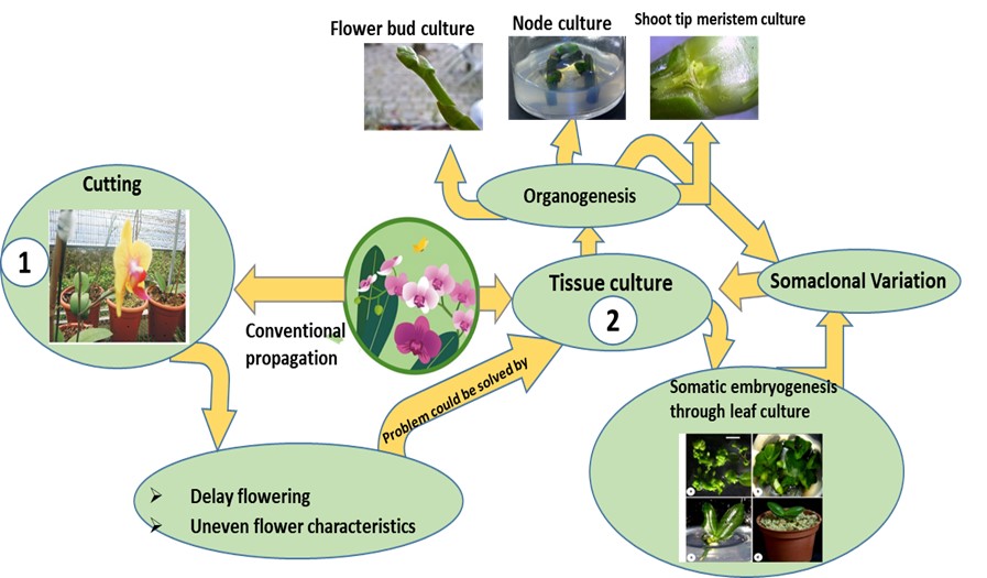
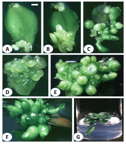
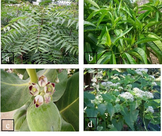
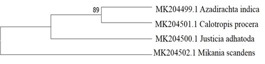
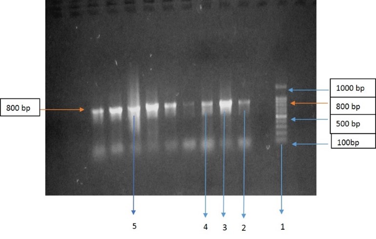
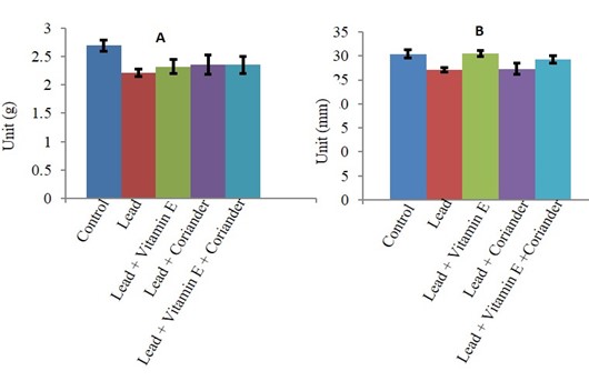
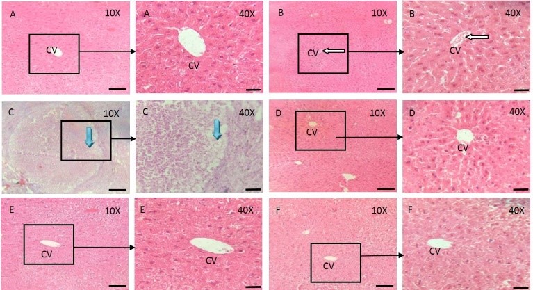
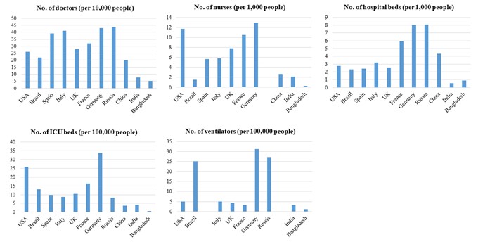
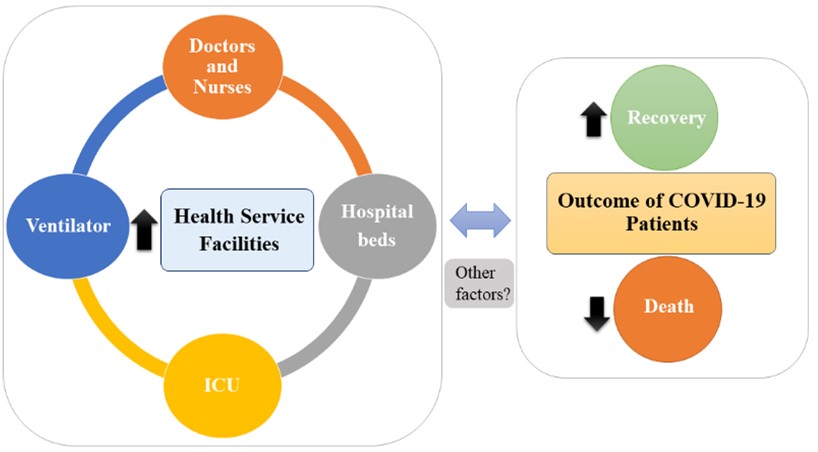
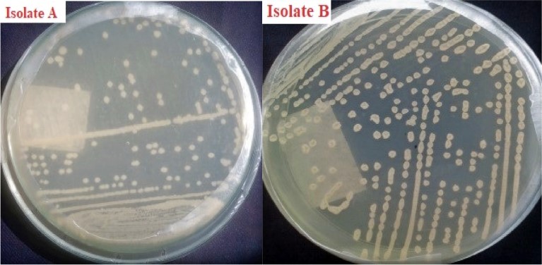
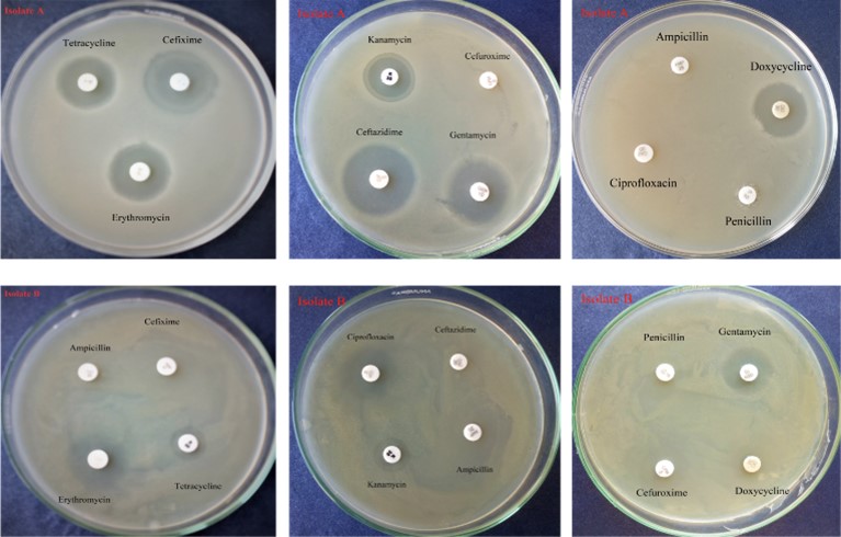
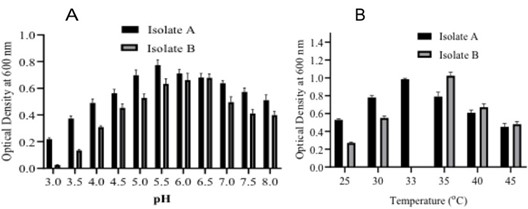
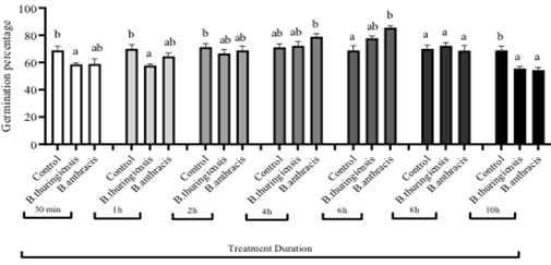
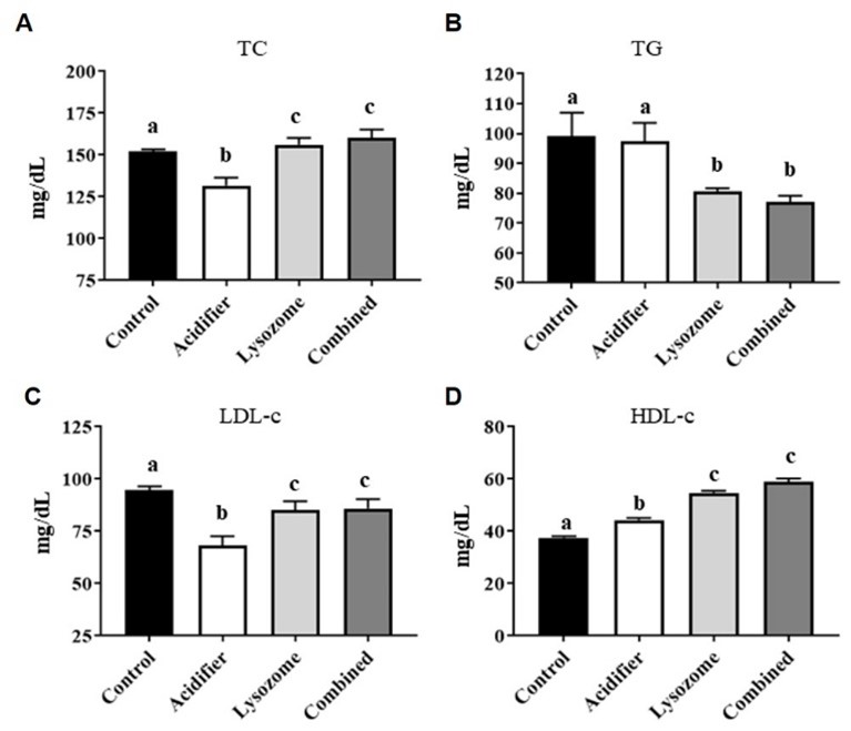
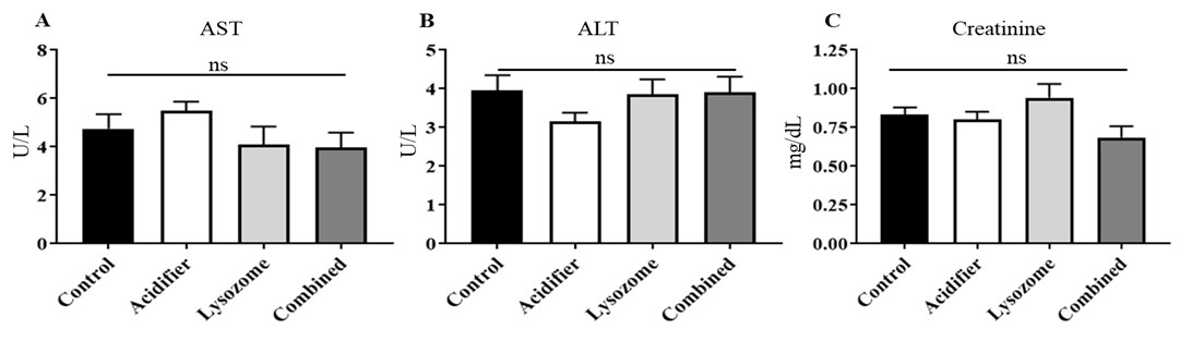
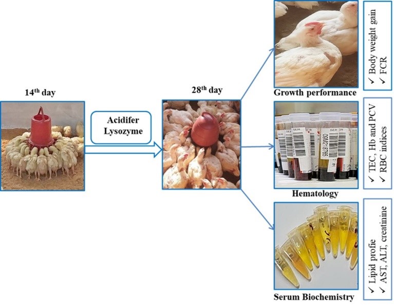
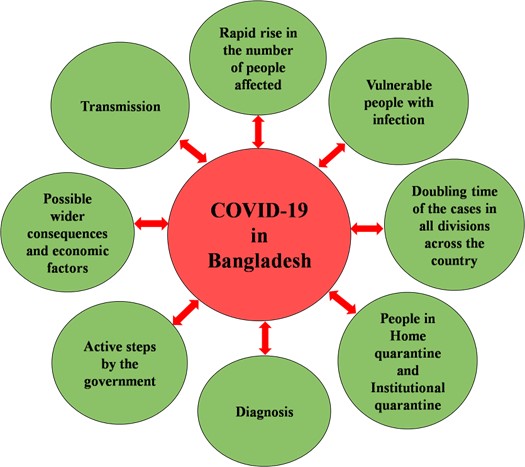
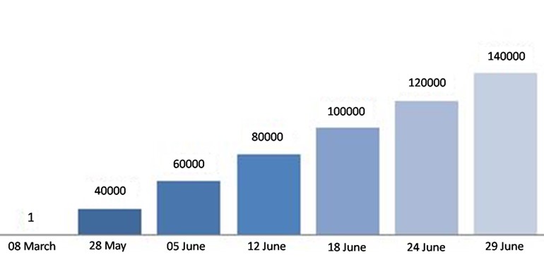
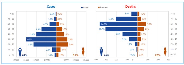
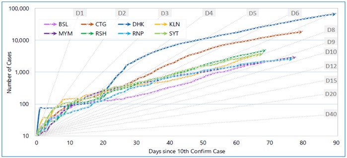
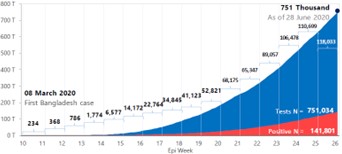