HTTP 200 OK
Allow: GET, HEAD, OPTIONS
Content-Type: application/json
Vary: Accept
{
"count": 319,
"next": "https://jabet.bsmiab.org/articles/?format=api&page=24",
"previous": "https://jabet.bsmiab.org/articles/?format=api&page=22",
"results": [
{
"id": 153,
"slug": "178-1612493625-follow-up-of-bacterial-and-physicochemical-quality-of-water-during-live-transportation-of-climbing-perch-anabas-testudineus-in-bangladesh",
"featured": false,
"slider": false,
"issue": "Vol4 Issue2",
"type": "original_article",
"manuscript_id": "178-1612493625",
"recieved": "2021-01-24",
"revised": null,
"accepted": "2021-03-05",
"published": "2021-03-13",
"pdf_file": "https://jabet.bsmiab.org/media/pdf_file/2023/11/178-1612493625.pdf",
"title": "Follow-up of bacterial and physicochemical quality of water during live transportation of Climbing perch (Anabas testudineus) in Bangladesh",
"abstract": "<p>This study was conducted to evaluate the changes in viable count of bacteria and physicochemical parameters, and their correlations in the changing pattern during live transportation of Climbing perch, (<em>Anabas testudineus</em>). Investigations were conducted in three live fish supply channels of Bangladesh from Mymensingh to Dhaka (channel 1), Mymensingh to Sylhet (channel 2), and from Mymensingh to Rajshahi (channel 3). It took nearly 6 h in channel 1, 8 h in channel 2, and 8 h in channel 3 for the live fish to reach the unloading points of destination. Viable count of bacteria and physicochemical parameters, such as temperature, dissolved oxygen (DO), pH, and ammonia were recorded from 0 hr with 2 h interval during transportation. The viable count of bacteria in water increased several folds from the initial values and showed a regrowth within the system. In all the supply channels, water temperature and pH were more or less stable with the headway of the transportation, but remarkable differences were observed in the concentrations of DO and ammonia. Although the initial (0 hr) DO level was varying among the supply channels, a decreasing trend was observed at the end in every channel. On the other hand, ammonia concentration was gradually increased during the transportation process. Thus, a gradual decline in water quality (decreased DO level, increased ammonia concentration, and higher bacterial regrowth) may negatively affect the survival and quality of live fish during transportation.</p>",
"journal_reference": "J Adv Biotechnol Exp Ther. 2021; 4(2): 149-160.",
"academic_editor": "Md. Niamul Haque, PhD; Incheon National University, South Korea",
"cite_info": "Hossain MM, Bhuiyan ANMRK, et al. Follow-up of bacterial and physicochemical quality of water during live transportation of Climbing perch (Anabas testudineus) in Bangladesh. J Adv Biotechnol Exp Ther. 2021; 4(2): 149-160.",
"keywords": [
"Bacterial viable count",
"Climbing perch",
"Physicochemical parameters",
"Live fish transportation"
],
"DOI": "10.5455/jabet.2021.d115",
"sections": [
{
"section_number": 1,
"section_title": "INTRODUCTION",
"body": "<p>Fish transportation is a global necessity, related to human feeding [<a href=\"#r-1\">1</a>] and transportation of live fish is a common practice among aquaculture facilities. The most obvious respect in which the road transport of live fish differs from that of terrestrial livestock is the requirement to provide a life-support system for the duration of the process [<a href=\"#r-2\">2</a>]. Handling and the physical disturbances associated with loading, transport, and discharge have the potential to cause stress in live fish [<a href=\"#r-2\">2</a>]. Climbing perch (<em>Anabas testudineus</em>), commonly known as Koi in Bangladesh, is a freshwater fish found in small rivers, canals, and swamps [<a href=\"#r-3\">3</a>]. It is one of the most delicious fish with high market demand in Bangladesh and thus commercial Koi fish farming in the pond is becoming very popular [<a href=\"#r-4\">4</a>]. Due to good market prices and consumer demands, Koi fish needs to transport from the production site to retail markets in the live condition. The transportation of fishes is influenced by many factors, including the fish species and its physical condition, loading density, physicochemical parameters, the duration of transport, etc. [<a href=\"#r-5\">5</a>]. In Bangladesh, live fish is transported usually by two systems viz., “open truck system” and “tank system” with or without aeration. In the “open truck system,” the truck bed is usually covered with a plastic/ tarpaulin so that it can hold water and here the water volume varies with the carrying capacity of the truck. In the case of the “tank system”, low-cost plastic tanks with a capacity of around 1 m<sup>3</sup> are used, which are sometimes reinforced by an iron frame [<a href=\"#r-6\">6</a>]. Typically, in Bangladesh mainly Taki, Pangus, Koi, Shing, Magur fish are transported by truck as live form by plastic drum from the production site. Each drum contains 35-40 kg of fish with water [<a href=\"#r-7\">7</a>].<br />\r\nWater that is chemically and physically suitable for normal fish at the beginning of the transport operation can easily become contaminated with the organic toxin, resulting from the bacterial breakdown of fecal waste, that the carrying medium becomes harmful to the fish<br />\r\n[<a href=\"#r-8\">8</a>]. Water quality is a crucial factor in the transportation of live fish and can be affected by the condition and/or deterioration of the holding systems [<a href=\"#r-9\">9</a>]. The number (or weight) of fish that can be successfully transported depends on water quality, the duration of the transport, water temperature, fish size, and the species [<a href=\"#r-10\">10</a>]. Maintenance of water quality parameters like dissolved oxygen (DO), pH, temperature, carbon dioxide, ammonia at an optimal level is essential in the successful transportation of live fish [9]. The major water quality effects experienced by fish during live transportation were low DO levels due to oxygen consumption by respiration, accumulation of carbon dioxide from respiration, depression of pH caused by carbon dioxide accumulation, and increased ammonia levels resulting from ammonia excretion [<a href=\"#r-11\">11</a>]. Long-transport episodes lead to more extreme values of pH and ammonia in the water [<a href=\"#r-12\">12</a>]. Microorganisms introduced into a live fish distribution system, through the raw water, will change density and diversity due to the selective pressures of the system. The goal here is to better integrate these two approaches, relating physicochemical consequences to water quality deterioration and microbial changes especially considering oxygen deprivation and enrichment of ammonia after transport. Treatment of raw water with different chemicals will result in a decrease in microbial load, with many live fish distribution systems later experiencing an increase in bacterial numbers with a distance away from the point of treatment. This increase has been termed regrowth and is recognized as a major problem within many live fish distribution systems [<a href=\"#r-13\">13</a>].<br />\r\nThe regrowth of bacteria within any fish distribution system is of concern to fish traders for a variety of reasons. Many microorganisms found in live fish distribution systems also can behave as opportunistic pathogens [<a href=\"#r-14\">14, 15</a>]. Poor water quality causes burn of the slime coat or stress the fish making it more susceptible to bacterial infection [<a href=\"#r-16\">16</a>]. The slime produced by the fish is another substrate for bacterial growth that consumes O<sub>2</sub> and releases toxic metabolites resulting in a decrease in the water oxygen content [<a href=\"#r-17\">17</a>]. During the transportation of fish in live conditions, adverse changes of physicochemical parameters and bacterial loads in water are frequent which influence much in successful transportation. Fish is affected by bacteria and the degradation of fish is accelerated by microorganisms associated with aquatic environments as well as contaminants during post-harvest handling [<a href=\"#r-18\">18</a>]. Changes in physicochemical parameters and viable bacterial count and the correlation of different factors with viable bacterial count during live transportation of <em>A</em>.<em> testudineus</em> are not well understood in Bangladesh. We hypothesized that the bacterial count will be increased with the distance and duration of the transportation, and the DO level will be reduced and reach a critical level; the ammonia concentration will be increased with time which will ultimately result in bacterial regrowth to occur. We also hypothesized that the bacterial viable count will be correlated with the concentration of ammonia and the DO level in the water. Thus, the objective of this study is to verify these hypotheses. Three supply channels of live <em>A</em>.<em> testudineus</em> transportation were selected and sub-samples were collected to follow the regrowth of bacteria and changes of physicochemical parameters from the beginning (at 0 h of loading) of the supply channel to the end/ final destination (at point of unloading).</p>"
},
{
"section_number": 2,
"section_title": "MATERIALS AND METHODS",
"body": "<p><strong>Supply channels details</strong><br />\r\nThe study was conducted in three different <em>A</em>.<em> testudineus</em> supply channels of Bangladesh. The loading/stating point was a major fish producing division, Mymensingh (Muktagacha, 24°44′01″<em>N</em> 90°20′36″<em>E</em>) and unloading point was three important divisions such as Dhaka (Showarighat, 23°42′25″N 90°24′34″E; channel 1), Sylhet (Poschim Kazir Bazar, 24°.88′80″N 91°.86′40″E; channel 2) and Rajshahi (Shaheb Bazar, 24°21’53″N 88°35’66″E; channel 3) respectively (<a href=\"#figure1\">Figure 1</a>). The samples were collected from July to September 2019.</p>\r\n\r\n<div id=\"figure1\">\r\n<figure class=\"image\"><img alt=\"\" height=\"571\" src=\"/media/article_images/2024/37/05/178-1612493625-Figure1.jpg\" width=\"500\" />\r\n<figcaption><strong>Figure 1.</strong> Map showing location of sampling supply channels of live transportation of Climbing perch (<em>Anabas testudineus</em>) in Bangladesh (channel 1: Muktagacha, Mymensingh to Showarighat, Dhaka; channel 2: Muktagacha, Mymensingh to Poschim kazir Bazar, Sylhet; channel 3: Muktagacha, Mymensingh to Saheb Bazar, Rajshahi). Images were extracted from DIVA-GIS using Geographical Information System (GIS). The map was developed by using ArcMap version 10.7.</figcaption>\r\n</figure>\r\n\r\n<p> </p>\r\n</div>\r\n\r\n<p><strong>Sampling techniques</strong><br />\r\nThoroughly washed and sterilized sampling materials like beakers, volumetric flasks, tips, plastic containers, physiological saline, test tube, L-shaped glass rod, micropipette, hand gloves, cotton, etc. were carried within the iceboxes and traveled by the same carrying vehicle<br />\r\n(commercial truck) to carry out works during transportation. Previously prepared agar plates and necessary reagents for physicochemical parameters were also carried. For the viable count of bacteria and physicochemical parameters, water samples were collected randomly in triplicate from different sites of the plastic barrels in previously sterile plastic bottles (250 ml) from 0 hr and at every 2 hrs interval throughout the transportation. In this experiment after the collection of water from the plastic barrels, subsequent analyses and plating for viable counting were carried out immediately.</p>\r\n\r\n<p> </p>\r\n\r\n<p><strong>Determination of physicochemical parameters</strong><br />\r\nWater temperature was recorded using a glass thermometer (Saraan Scientific Industries, Haryana, India) by sticking the bulb end into the water, waiting for a few minutes until the liquid in the glass stops moving. Finally, the temperature was measured as °C.<br />\r\nThe DO was measured as mg/L by using Biosol dissolved oxygen kit (Biosol AE DO8 Dissolved Oxygen Kit, A.A. Biotech Private Limited, Chennai, India). The DO test bottle was filled with sample water till it overflew and then stoppered the bottle and ensured that no air bubbles were trapped inside. Then, 10 drops of DO-1 followed by 10 drops of DO-2 were added and waited for a minute. Brown precipitation was formed and started to settling. The bottle was kept in a safe place for a minimum of 20 min. Then 10-20 drops of DO-3 were added and shook the bottle till the precipitate dissolved. In the test jar, 10 ml of sample was taken, 4 drops of DO-4 were added and mixed well, and then DO-5 was added drop-wise until the blue colors disappeared. At last dissolved oxygen was calculated by using the following formulae: Dissolved oxygen (mg/l) = 0.65× (No. of drops of DO-5).<br />\r\nWater pH was measured by using a pH test kit (Charoen Pokphand Feed Company, Thailand). A color cell was washed with the sample water. Then 5 ml of sample was taken in the color cell. Two drops of pH reagent were added and mixed well. Lastly, pH was determined by comparing it with the standard color gradation.<br />\r\nThe ammonia concentration (mg/l) was measured by using an ammonia test kit (Charoen Pokphand Feed Company, Thailand). At first, the measuring vessel of the kit was rinsed with the test sample and filled to the 5 ml mark. Two drops of the reagent AMMONIA-1 were added and mixed. Two drops of reagent AMMONIA-2 were added and mixed. Then two drops of reagent AMMONIA-3 were added and mixed thoroughly. At last four drops of the reagent AMMONIA-4 were added and mixed. After 20 minutes, the color on the top view of the measuring vessel was compared with the color band.</p>\r\n\r\n<p> </p>\r\n\r\n<p><strong>Determination of viable count of bacteria</strong><br />\r\nAfter the collection of the water sample, 1 ml was transferred with a micropipette to a test tube containing 9 ml of physiological saline (0.85% w/v NaCl). The test tube was shaken thoroughly by a vortex mixture to get 10<sup>-1</sup> dilution of the original sample solution. Using a similar process several dilutions of 10<sup>-2</sup>, 10<sup>-3 </sup>were made. Aliquots of 0.1 ml of the above dilutions were pipetted out and transferred aseptically to the previously prepared plate count agar plates (HiMedia Laboratories Private Limited, Kolkata, West Bengal, India) with maintaining replication of each, by raising the upper lids sufficient enough to admit the tip of the pipette. The samples pipetted were spread by L­-shaped glass rods throughout the surface of the media until the samples were dried out. The plates were then wrapped by aluminum foil and kept in the zipper bag, where the temperature was around 28°C. All these activities were carried out aseptically in the cabinet of the truck and then plates were brought to the Fisheries Microbiology Laboratory, Dept. of Fisheries Technology, Bangladesh Agricultural University and put in the incubator at 30°C. After 48 h of incubation, the developed colonies were counted using the Stuart Scientific colony counter (SC6PLUS-UK). The plates having a number of colonies in between 30 to 300 were considered for calculation as colony forming unit (cfu) per ml of samples using the following formulae: cfu/ml = no. of colonies on petridish × 10 × dilution factor<br />\r\nAfter calculation, the mean logarithmic values (log10 cfu/ml) were obtained for every subsample of water.</p>\r\n\r\n<p> </p>\r\n\r\n<p><strong>Statistical analysis</strong><br />\r\nAll the collected data for both physicochemical parameters and bacterial viable count were incorporated and analyzed using Microsoft Excel 2010. Statistical analysis was performed by SPSS software (SPSS-24.0) and Pearson’s correlation was done to evaluate the significant relationship between physicochemical parameters and bacterial viable count at 5% level (p<0.05).</p>"
},
{
"section_number": 3,
"section_title": "RESULTS",
"body": "<p><strong>Length and duration of the supply channels</strong><br />\r\nThe distances between starting to the end of the supply channels are about 140 km, 305 km, and 230 km, respectively in the case of channel 1,<br />\r\nchannel 2, and channel 3. But the time duration was not completely relevant to the distance that was about 6 h, 8 h, and 8 h for channel 1, channel 2, and channel 3, respectively due to traffic jams on the road.</p>\r\n\r\n<p> </p>\r\n\r\n<p><strong>Fish harvesting and preparation for transportation</strong><br />\r\nAt the fish loading point (commercial climbing perch farm), the capturing of fish was undertaken during the late afternoon for transportation the following night. The fishes are usually harvested using a large seine net. After catching, the fishes were kept in “hapas” (net made rectangular barriers) setting at the corner of the fishponds for acclamation usually for about 2 h before transportation.<br />\r\nFor transportation of fish in the live condition, a commercial truck Tata LPT-709 (5029.2 mm × 2286 mm × 2341 mm) maximum carrying capacity of 5.17 ton from (Tata Motors Limited, Mumbai, India) and to provide a life-support system (water) for the duration of transport plastic barrel (1000 L) was used. Plastic barrels were filled with 500 L of groundwater from the commercial water supplier before 3-4 h of transportation and carried on the truck bed to the targeted loading point. For road transportation, each barrel was filled with 35 kg of fish approximately (on average 3 fishes per kg weight) and a maximum of 40 barrels was placed on the truck in every channel. Any kind of water additives, aeration facilities, and water exchange not practiced throughout the transportation in all supply channels.</p>\r\n\r\n<p> </p>\r\n\r\n<p><strong>Physicochemical parameters</strong><br />\r\nPhysicochemical parameters as water temperature (°C), pH, ammonia (mg/l), and dissolved oxygen (mg/l) were measured from 0 h just before loading of the fish up to reach the desired unloading point from all the three supply channels every 2 h interval. It was found that the water temperature changed from 29-31 °C in channel 1, while 30-29 °C in channel 2 and 31-30 °C in channel 3. Dissolved oxygen (DO) decreased from 6.25 to 2.05 mg/l in channel 1, while 7.35 to 1.95 mg/l in channel 2, and 6.75 to 1.65 mg/l in channel 3. The pH values of the water in all the supply channels were found to decrease slightly, reduced from 8.0 to 7.0 in channel 1, from 8.2 to 6.7 in channel 2, and from 7.6 to 7.0 in channel 3. The ammonia level increased from 0.1 to 5.0 mg/l in channel 1, while 0.5 to 5.0 mg/l in channel 2, and 0.2 to 3.0 mg/l in channel 3, respectively (<a href=\"#figure2\">Figure 2</a>).</p>\r\n\r\n<div id=\"figure2\">\r\n<figure class=\"image\"><img alt=\"\" height=\"259\" src=\"/media/article_images/2024/37/05/178-1612493625-Figure2.jpg\" width=\"500\" />\r\n<figcaption><strong>Figure 2. </strong>Changes of physicochemical parameters along the fish supply channels of live transportation of Climbing perch (<em>Anabas testudineus</em>) in Bangladesh. There were no statistically significant differences in the parameters from the studied supply channles (p= 0.97, two-factor ANOVA without replication).</figcaption>\r\n</figure>\r\n\r\n<p> </p>\r\n</div>\r\n\r\n<p><strong>Viable count of bacteria</strong><br />\r\nIn this study, the averages of the viable count of bacterial in water changed from about 0.64×10<sup>4 </sup>cfu/ml at 0 h to 16.32×10<sup>4 </sup>cfu/ml at 2 h, 28.85×10<sup>4 </sup>cfu/ml at 4 h, and to 31.01×10<sup>4 </sup>cfu/ml at 6 h (before unloading) of transportation in channel 1. While in channel 2, the averages of the viable count were about 0.56×10<sup>4 </sup>cfu/ml at 0 h, about 18.27×10<sup>4 </sup>cfu/ml at 2 h, about 24.55×10<sup>4 </sup>cfu/ml at 4 h to about 38.52×10<sup>4 </sup>cfu/ml at 6 h, and 47.52×10<sup>4 </sup>cfu/ml at 8 h of transportation. In the case of channel 3, the averages of the viable counts of bacteria changed from about 0.69×10<sup>4 </sup>cfu/ml at 0 h to about 16.98×10<sup>4 </sup>cfu/ml at 2 hrs, about 23.07×10<sup>4 </sup>cfu/ml at 4 h to about 29.52×10<sup>4 </sup>cfu/ml at 6 hrs, and finally about 41.01×10<sup>4 </sup>cfu/ml at 8 h of transportation (<a href=\"#figure3\">Figure 3</a>).</p>\r\n\r\n<div id=\"figure3\">\r\n<figure class=\"image\"><img alt=\"\" height=\"299\" src=\"/media/article_images/2024/37/05/178-1612493625-Figure3.jpg\" width=\"500\" />\r\n<figcaption><strong>Figure 3. </strong>Changes in viable cell count of bacteria in water along the fish supply channels of live transportation of Climbing perch (<em>Anabas testudineus</em>) in Bangladesh. There were no statistically significant differences in the viable counts from the studied supply channels (<em>p</em>= 0.4, two-factor ANOVA without replication).</figcaption>\r\n</figure>\r\n\r\n<p> </p>\r\n</div>\r\n\r\n<p><strong>Relationship between viable count and physicochemical parameters</strong><br />\r\nThroughout the study, it was observed that the DO level decreased while NH<sub>3</sub> concentration increased gradually. The viable count also increased gradually from the loading to the unloading points in all the three supply channels (<a href=\"#figure4\">Figure 4</a>, <a href=\"#figure5\">5</a>, and <a href=\"#figure6\">6</a>). Pearson correlation analysis between the viable count of bacteria and physicochemical parameters was carried out and the relationship was considered for significance at <em>p</em> < 0.05. In the case of channel 1, between viable count and ammonia, a moderate positive correlation (r= 0.689) was observed, while between viable count and DO level, a strong negative correlation (r= -0.981) was assessed. In the case of channel 2 and channel 3, Pearson correlation analyses between viable count and ammonia showed a significant positive correlation (r= 0.879 and r= 0.916, respectively for channel 2 and channel 3). Similar to channel 1, a strong negative correlation (r= -0.994, and r= -0.984, respectively for channel 2 and channel 3) was assessed between viable count and dissolved oxygen level in channel 2 and channel 3. Moderate to strong negative correlations (r= -0.814, r= -0.838, and r= -0.909 in channel 1, channel 2, and channel 3, respectively) were observed between ammonia concentration and DO level in all the studied channels (<a href=\"#Table-1\">Table 1</a>).</p>\r\n\r\n<div id=\"figure4\">\r\n<figure class=\"image\"><img alt=\"\" height=\"284\" src=\"/media/article_images/2024/37/05/178-1612493625-Figure4.jpg\" width=\"500\" />\r\n<figcaption><strong>Figure 4. </strong>Correlation between (A) viable cell count of bacteria and ammonia, (B) viable cell count of bacteria and dissolved oxygen in supply channel 1 during live transportation of Climbing perch (<em>Anabas testudineus</em>) in Bangladesh.</figcaption>\r\n</figure>\r\n</div>\r\n\r\n<div id=\"figure5\">\r\n<figure class=\"image\"><img alt=\"\" height=\"270\" src=\"/media/article_images/2024/37/05/178-1612493625-Figure5.jpg\" width=\"500\" />\r\n<figcaption><strong>Figure 5. </strong>Correlation between (A) viable cell count of bacteria and ammonia, (B) viable cell count of bacteria and dissolved oxygen in supply channel 2 during live transportation of Climbing Perch (<em>Anabas testudineus</em>) in Bangladesh.</figcaption>\r\n</figure>\r\n</div>\r\n\r\n<div id=\"figure6\">\r\n<figure class=\"image\"><img alt=\"\" height=\"264\" src=\"/media/article_images/2024/37/05/178-1612493625-Figure6.jpg\" width=\"500\" />\r\n<figcaption><strong>Figure 6. </strong>Correlation between (A) viable cell count of bacteria and ammonia, (B) viable cell count of bacteria and dissolved oxygen in supply channel 3 during live transportation of Climbing perch (<em>Anabas testudineus</em>) in Bangladesh.</figcaption>\r\n</figure>\r\n\r\n<p> </p>\r\n</div>\r\n\r\n<div id=\"Table-1\">\r\n<p><a href=\"https://jabet.bsmiab.org/table/178-1612493625-table1/\">Table-1</a><strong>Table 1. </strong>Correlations between viable count of bacteria and physicochemical parameters (DO level and ammonia concentration) in different supply channels during live transportation of Climbing perch (<em>Anabas testudineus</em>) in Bangladesh.</p>\r\n</div>"
},
{
"section_number": 4,
"section_title": "DISCUSSION",
"body": "<p>This study was conducted to assess the changing pattern of bacterial count and water quality parameters during live transportation of climbing perch, <em>A. testudineus</em>. We hypothesized that the viable count of bacteria will be increased with the distance and duration of the transportation, and the DO level will be reduced and reach a critical level; the ammonia concentration would be increased with time which will ultimately result in bacterial regrowth. We also hypothesized that the bacterial viable count will be correlated with the concentration of ammonia and the DO level in the water.<br />\r\nResults of this study indicated the occurrence of bacterial regrowth within the live <em>A. testudineus </em>transportation system with relation to the changes of physicochemical parameters and they are significantly correlated. The greatest challenge with any live fish transportation is to keep the water quality parameter near the optimum range to minimize stress and mortality since a remarkable percentage of fish die before reaching the desired market. Among the physicochemical parameters assessed in this study, temperature and pH are the most important that control behavioral characteristics of organisms, the solubility of gases and salts in water as well as chemical and physical characteristics of water [<a href=\"#r-19\">19</a>]. The results of water temperature were almost stable at around 30 °C in all the three supply channels (<a href=\"#figure2\">Figure 2</a>) which is comparable with the findings of [<a href=\"#r-20\">20, 21</a>] as they were observed temperature fluctuation during transportation of fish in live conditions was negligible.<br />\r\nThe water pH is an important water quality parameter for aquatic organisms to survive since most of the chemical reactions in the aquatic environment are controlled by any change in its value. A decreasing trend of water pH during fish transportation was reported by [<a href=\"#r-12\">12</a>, <a href=\"#r-22\">22</a>]. In the current study, pH values were decreased from the initial values with time intervals in all of the three studied supply channels, and the averages were from 8.0 to 7.0 (<a href=\"#figure2\">Figure 2</a>). However, the pH range was within the optimum level as it was reported that pH levels 6.90 to 7.43 are tolerable to climbing perch [<a href=\"#r-23\">23</a>].<br />\r\nIn this study, the DO was found to decline sharply from the initial loading to unloading period and reached a critical in all three supply channels (<a href=\"#figure2\">Figure 2</a>). The fish transporters usually fill the plastic barrels with groundwater several hours before loading the fish and carried to the loading point by truck. In the meantime on the road, due to the interaction of air and water on the truck, the DO levels may increase slightly. As a result, we found an average DO level of about 6.70 mg/l initially in all three supply channels. In live fish transportation, the initial 30–60 min in the transport container is critical [<a href=\"#r-10\">10</a>] and a drop from 20 to <5 mg/l DO during this time was noticed by [<a href=\"#r-24\">24</a>]. A similar condition was also observed here may be due to the increased activity by the fish during the initial tank loading process. At the later stage of transportation, the DO level continued to decrease and reached a critical level (2.05 mg/l, 1.95 mg/l, and 1.65 mg/l in channel 1, channel 2, and channel 3, respectively).<br />\r\nDO is a limiting factor at very high loading densities over an extended period. The decomposition and oxidation of organic matter reduce the solubility of oxygen in the water [<a href=\"#r-25\">25</a>].In this study, although the DO level reduced to the critical limit, most of the transported fish was survived may be due to the following reasons. Firstly, the interaction between water and air added oxygen to the water during the running of the transport, especially at the surface. However, at the same time the DO was utilized by the fishes. Secondly, <em>A. testudineus </em>possesses <em>accessory</em><em> </em>air-<em>breathing organs</em> and <em>can withstand</em> harsh environmental conditions such as <em>low DO,</em> a wide range of temperature, and other <em>poor</em> water conditions.<br />\r\nThere was a noticeable increase in concentrations of ammonia (almost 5 mg/l) was found in all the studied channels with the progress of the transportation period (Figure 2). Ammonia accumulates in water during live fish transportation, mostly due to the catabolism of protein and excretion by the fishes. Besides, bacterial decomposition of organic matters such as uneaten feed, fecal materials, dead algae, and aquatic plants also enhances ammonia accumulation [<a href=\"#r-19\">19</a>, <a href=\"#r-26\">26</a>]. Higher levels of NH<sub>4</sub><sup>+</sup> concentration are associated with low dissolved oxygen levels may lead to the death of fish [<a href=\"#r-27\">27</a>]. Also, high NH<sub>4</sub><sup>+</sup> concentrations can strongly influence dissolved oxygen levels because around 4.3 mg oxygen is necessary to oxidize 1.0 mg NH<sub>4</sub><sup>+</sup> [<a href=\"#r-26\">27, 28</a>]. Both in nature and under typical culture conditions, excreted CO<sub>2</sub> and NH<sub>3</sub> tend not to accumulate because of their high solubility and the potential for extensive dissolution. In contrast, during live transportation of fish, significant accumulation of these potentially toxic waste products in the fixed volume of water available is possible. Fortunately, during dissolution, the majority of NH<sub>3</sub> is converted to the nontoxic ammonium ion (NH<sub>4</sub>+) [<a href=\"#r-2\">2</a>].<br />\r\nAmmonia is toxic, and therefore, air-breathing fishes have special adaptations to defend against ammonia toxicity [<a href=\"#r-29\">29, 30</a>]. Although the ammonia level was higher, the fishes were survived throughout the transportation period in all the supply channels.<br />\r\nIn the present study, bacterial viable count in water increased several folds from the initial values and found positively related to the duration and distance of transport (Figure 3). Viable counts were also negatively correlated with the DO level and positively correlated with the ammonia concentration of the water during live transportation of climbing perch in all the supply channels (<a href=\"#Table-1\">Table 1</a> and <a href=\"#figure4\">Figure 4</a>, <a href=\"#figure5\">5</a>, <a href=\"#figure6\">6</a>).<br />\r\nAlthough groundwater is used for the transportation of the fishes, bacterial numbers may be increased in the water as fish itself is a source of bacteria (it is well known that fish can harbor around 10<sup>5</sup> cfu/inch<sup>2</sup> bacteria in the skin, and 10<sup>5</sup> cfu/g in gill and intestine) that transferred a wide range of bacteria in water [<a href=\"#r-31\">31</a>]. Further increase in bacterial number is due to their regrowth by utilization excretory organic matters of the transport water. The temperature of the water was almost around 30 °C in all the supply channels, which facilitated the metabolic activities of microbes and hence increased growth [<a href=\"#r-32\">32</a>]. It was reported that there were usually negative correlations between viable bacterial count and dissolved oxygen [<a href=\"#r-33\">33, 34</a>] and positive correlations between viable bacterial count and ammonia [<a href=\"#r-34\">34, 35</a>] in water. So in this study, the higher the temperature, the higher was the growth rate of bacteria, and the decomposition of fecal organic matters resulted in the critical declined in DO level in the water during transportation. Thus, the viable count of bacteria was negatively correlated with the DO level and positively correlated with the ammonia concentration in the water during live transportation of climbing perch. The findings showed a gradual decline in water quality (decreased DO level, increased ammonia concentration, and higher bacterial growth) that may negatively affect the survival and quality of live fish during transportation.</p>"
},
{
"section_number": 5,
"section_title": "ACKNOWLEDGMENTS",
"body": "<p>The authors are grateful to the Bangladesh Agricultural University Research System (BAURES) for funding the research and also to the Department of Fisheries Technology, Bangladesh Agricultural University, for their technical supports.<strong> </strong></p>"
},
{
"section_number": 6,
"section_title": "AUTHOR CONTRIBUTIONS",
"body": "<p>The work was designed by MNH, MNU, and MIH. MMH, ANMRKB conducted the experiments and collected data. MAH also aided in data collection. MNH, MNU, and MIH supervised the whole work. The first draft of this manuscript was prepared by MMH. MNH critically checked, improved the manuscript and finally approved it for publication.</p>"
},
{
"section_number": 7,
"section_title": "CONFLICTS OF INTEREST",
"body": "<p>There is no conflict of interest among the authors.</p>"
}
],
"figures": [
{
"figure": "https://jabet.bsmiab.org/media/article_images/2024/37/05/178-1612493625-Figure1.jpg",
"caption": "Figure 1. Map showing location of sampling supply channels of live transportation of Climbing perch (Anabas testudineus) in Bangladesh (channel 1: Muktagacha, Mymensingh to Showarighat, Dhaka; channel 2: Muktagacha, Mymensingh to Poschim kazir Bazar, Sylhet; channel 3: Muktagacha, Mymensingh to Saheb Bazar, Rajshahi). Images were extracted from DIVA-GIS using Geographical Information System (GIS). The map was developed by using ArcMap version 10.7.",
"featured": false
},
{
"figure": "https://jabet.bsmiab.org/media/article_images/2024/37/05/178-1612493625-Figure2.jpg",
"caption": "Figure 2. Changes of physicochemical parameters along the fish supply channels of live transportation of Climbing perch (Anabas testudineus) in Bangladesh. There were no statistically significant differences in the parameters from the studied supply channles (p= 0.97, two-factor ANOVA without replication).",
"featured": false
},
{
"figure": "https://jabet.bsmiab.org/media/article_images/2024/37/05/178-1612493625-Figure3.jpg",
"caption": "Figure 3. Changes in viable cell count of bacteria in water along the fish supply channels of live transportation of Climbing perch (Anabas testudineus) in Bangladesh. There were no statistically significant differences in the viable counts from the studied supply channels (p= 0.4, two-factor ANOVA without replication).",
"featured": false
},
{
"figure": "https://jabet.bsmiab.org/media/article_images/2024/37/05/178-1612493625-Figure4.jpg",
"caption": "Figure 4. Correlation between (A) viable cell count of bacteria and ammonia, (B) viable cell count of bacteria and dissolved oxygen in supply channel 1 during live transportation of Climbing perch (Anabas testudineus) in Bangladesh.",
"featured": false
},
{
"figure": "https://jabet.bsmiab.org/media/article_images/2024/37/05/178-1612493625-Figure5.jpg",
"caption": "Figure 5. Correlation between (A) viable cell count of bacteria and ammonia, (B) viable cell count of bacteria and dissolved oxygen in supply channel 2 during live transportation of Climbing Perch (Anabas testudineus) in Bangladesh.",
"featured": false
},
{
"figure": "https://jabet.bsmiab.org/media/article_images/2024/37/05/178-1612493625-Figure6.jpg",
"caption": "Figure 6. Correlation between (A) viable cell count of bacteria and ammonia, (B) viable cell count of bacteria and dissolved oxygen in supply channel 3 during live transportation of Climbing perch (Anabas testudineus) in Bangladesh.",
"featured": false
}
],
"authors": [
{
"id": 641,
"affiliation": [
{
"affiliation": "Department of Fisheries Technology, Bangladesh Agricultural University, Mymensingh-2202, Bangladesh"
}
],
"first_name": "Md. Mubarack",
"family_name": "Hossain",
"email": null,
"author_order": 1,
"ORCID": null,
"corresponding": false,
"co_first_author": false,
"co_author": false,
"corresponding_author_info": "",
"article": 153
},
{
"id": 645,
"affiliation": [
{
"affiliation": "Department of Fisheries Technology, Bangladesh Agricultural University, Mymensingh-2202, Bangladesh"
}
],
"first_name": "A N M Rezvi Kaysar",
"family_name": "Bhuiyan",
"email": null,
"author_order": 2,
"ORCID": null,
"corresponding": false,
"co_first_author": false,
"co_author": false,
"corresponding_author_info": "",
"article": 153
},
{
"id": 646,
"affiliation": [
{
"affiliation": "Technical-Fisher, Biswas Agro Fisheries and Hatchery Ltd., Mahbub Group of Industries, Trisal, Mymensingh, Bangladesh"
}
],
"first_name": "Md. Anwar",
"family_name": "Hossain",
"email": null,
"author_order": 3,
"ORCID": null,
"corresponding": false,
"co_first_author": false,
"co_author": false,
"corresponding_author_info": "",
"article": 153
},
{
"id": 647,
"affiliation": [
{
"affiliation": "Department of Fisheries Technology, Bangladesh Agricultural University, Mymensingh-2202, Bangladesh"
}
],
"first_name": "Md. Naim",
"family_name": "Uddin",
"email": null,
"author_order": 4,
"ORCID": null,
"corresponding": false,
"co_first_author": false,
"co_author": false,
"corresponding_author_info": "",
"article": 153
},
{
"id": 648,
"affiliation": [
{
"affiliation": "Department of Fisheries Technology, Bangladesh Agricultural University, Mymensingh-2202, Bangladesh"
}
],
"first_name": "Md. Ismail",
"family_name": "Hossain",
"email": null,
"author_order": 5,
"ORCID": null,
"corresponding": false,
"co_first_author": false,
"co_author": false,
"corresponding_author_info": "",
"article": 153
},
{
"id": 649,
"affiliation": [
{
"affiliation": "Department of Fisheries Technology, Bangladesh Agricultural University, Mymensingh-2202, Bangladesh"
},
{
"affiliation": "Atmosphere and Ocean Research Institute, The University of Tokyo, Chiba 277-8564, Japan"
}
],
"first_name": "Md. Nurul",
"family_name": "Haider",
"email": "raselmnh@bau.edu.bd",
"author_order": 6,
"ORCID": null,
"corresponding": true,
"co_first_author": false,
"co_author": false,
"corresponding_author_info": "Md. Nurul Haider, PhD; Department of Fisheries Technology, Bangladesh Agricultural University, Mymensingh-2202, Bangladesh, Email: raselmnh@bau.edu.bd",
"article": 153
}
],
"views": 1100,
"downloads": 83,
"references": [
{
"id": 5046,
"serial_number": 1,
"pmc": null,
"reference": "Marcalo A, Pousao P, Mateus L, Duarte JH, Stratoudakis Y. Sardine early survival, physical condition and stress after introduction to captivity. J Fish Biol 2008;72:103–20.",
"DOI": null,
"article": 153
},
{
"id": 5047,
"serial_number": 2,
"pmc": null,
"reference": "King HR. Fish transport in the aquaculture sector: An overview of the road transport of Atlantic salmon in Tasmania. J Vet Behav Clin Appl Res 2009;4:163–8.",
"DOI": null,
"article": 153
},
{
"id": 5048,
"serial_number": 3,
"pmc": null,
"reference": "Mijkherjee M, Praharaj A, Das S. Conservation of endangered fish stocks through artificial propagation and larval rearing technique in West Bengal, India. Aquac Asia 2002;7:8–11.",
"DOI": null,
"article": 153
},
{
"id": 5049,
"serial_number": 4,
"pmc": null,
"reference": "Hossain MA, Sultana Z, Kibria ASM, Azimuddin KM. Optimum dietary protein requirement of a Thai strain of climbing perch (Anabas testudineus) (Bloch, 1792) fry. Turk J Fish Aquat Sci 2012a, 12: 217-224.",
"DOI": null,
"article": 153
},
{
"id": 5050,
"serial_number": 5,
"pmc": null,
"reference": "Carneiro PC, Kaiseler PH, Swarofsky EA, Baldisserotto B. Transport of jundia Rhamdia quelen juveniles at different loading densities: Water quality and blood parameters. Neotrop Ichthyol 2009;7:283–8.",
"DOI": null,
"article": 153
},
{
"id": 5051,
"serial_number": 6,
"pmc": null,
"reference": "Rajts F, Shelley CC. Guide to improving live fish transportation with special attention to the COVID-19 pandemic in Bangladesh and other tropical developing countries 2020.",
"DOI": null,
"article": 153
},
{
"id": 5052,
"serial_number": 7,
"pmc": null,
"reference": "Alam MJ, Yasmin R, Rahman A, Nahar N, Pinky NI, Hasan M. A study on fish marketing system in Swarighat, Dhaka, Bangladesh. Nature and science 2010; 8(12):96-103.",
"DOI": null,
"article": 153
},
{
"id": 5053,
"serial_number": 8,
"pmc": null,
"reference": "Washburn GN. Device for the transportation and storage of live fish. United States patent US 2,652,807. 1953 Sep 22.",
"DOI": null,
"article": 153
},
{
"id": 5054,
"serial_number": 9,
"pmc": null,
"reference": "Metar S, Chogale N, Shinde K, Satam SB. Transportation of live marine ornamental fish. Adv Agric Res Technol 2018.",
"DOI": null,
"article": 153
},
{
"id": 5055,
"serial_number": 10,
"pmc": null,
"reference": "Conte FS. Stress and the welfare of cultured fish. Appl Anim Behav Sci 2004;86:205–23.",
"DOI": null,
"article": 153
},
{
"id": 5056,
"serial_number": 11,
"pmc": null,
"reference": "Paterson BD, Rimmer MA, Meikle GM, Semmens GL. Physiological responses of the Asian sea bass, (Lates calcarifer) to water quality deterioration during simulated live transport: Acidosis, red-cell swelling, and levels of ions and ammonia in the plasma. Aquaculture 2003;218:717–28.",
"DOI": null,
"article": 153
},
{
"id": 5057,
"serial_number": 12,
"pmc": null,
"reference": "Treasurer JW. Changes in pH during transport of juvenile cod (Gadus morhua) L. and stabilisation using buffering agents. Aquaculture 2012;330–333:92–9.",
"DOI": null,
"article": 153
},
{
"id": 5058,
"serial_number": 13,
"pmc": null,
"reference": "Edge JC, Finch PE. Observations on bacterial aftergrowth in water supply distribution systems: implications for disinfection strategies. Water Environ J 1987;1:104–10.",
"DOI": null,
"article": 153
},
{
"id": 5059,
"serial_number": 14,
"pmc": null,
"reference": "Burke V, Robinson J, Cooper M, Beaman J, Partridge K, Peterson D, Gracey M. Biotyping and virulence factors in clinical and environmental isolates of Aeromonas species. Appl Environ Microbiol 1984;47:1146–9.",
"DOI": null,
"article": 153
},
{
"id": 5060,
"serial_number": 15,
"pmc": null,
"reference": "Burke V, Robinson J, Gracey M, Peterson D, Meyer N, Haley V. Isolation of Aeromonas spp. from an unchlorinated domestic water supply. Appl Environ Microbiol 1984;48:367–70.",
"DOI": null,
"article": 153
},
{
"id": 5061,
"serial_number": 16,
"pmc": null,
"reference": "Wendelaar SE. The stress response in fish. Physiol Rev 1997;77:591–625.",
"DOI": null,
"article": 153
},
{
"id": 5062,
"serial_number": 17,
"pmc": null,
"reference": "Berka R. The transport of live fish. A review. EIFAC Technical Paper 1986; 48, 52 pp.",
"DOI": null,
"article": 153
},
{
"id": 5063,
"serial_number": 18,
"pmc": null,
"reference": "Getu A, Misganaw K. Post-harvesting and major related problems of fish production. Fish Aquac J 2015;06.",
"DOI": null,
"article": 153
},
{
"id": 5064,
"serial_number": 19,
"pmc": null,
"reference": "Engdaw F. Physioco-chemical parameters and bacteriological qualities of water samples from wastewater treatment pond, University of Gonder, Ethiopia. Inter J Pharma Res 2014; pp.192-197.",
"DOI": null,
"article": 153
},
{
"id": 5065,
"serial_number": 20,
"pmc": null,
"reference": "Gomes LC, Araujo CA, Chippari AR, Roubach R. Transportation of juvenile tambaqui (Colossoma macropomum) in a closed system. Brazilian J Biol 2006;66:493–502.",
"DOI": null,
"article": 153
},
{
"id": 5066,
"serial_number": 21,
"pmc": null,
"reference": "Cerreta AJ, Lewbart GA, Harrison TM. A novel approach to ornamental fish transportation for the aquarium hobbyist. J Surv Fish Sci 2020;7:105–12.",
"DOI": null,
"article": 153
},
{
"id": 5067,
"serial_number": 22,
"pmc": null,
"reference": "Wang W, Zhang Y, Liu Y, Adanyi N, Zhang X. Effects of waterless live transportation on survivability, physiological responses and flesh quality in Chinese farmed sturgeon (Acipenser schrenckii). Aquacultur 2020; 518:734834.",
"DOI": null,
"article": 153
},
{
"id": 5068,
"serial_number": 23,
"pmc": null,
"reference": "Ahmed GU, Rahman MM, Alam MN, Alam MB, Sarker B. Impact of stocking density on growth and production performance of Vietnamese koi (Anabas testudineus) in semi-intensive culture system at Muktaghasa region of Mymensingh district. Res Agric Livest Fish 2015;2:335–41.",
"DOI": null,
"article": 153
},
{
"id": 5069,
"serial_number": 24,
"pmc": null,
"reference": "Fries JN, Berkhouse CS, Morrow JC, Carmichae GJ. Evaluation of an aeration system in a loaded fish-hauling tank. Progress Fish-Culturist 1993;55:187–90.",
"DOI": null,
"article": 153
},
{
"id": 5070,
"serial_number": 25,
"pmc": null,
"reference": "Khalil MA, Beltagy EA, Fattah El, El-Naga EHA, Elshenawy MA, Kelany MS. Seasonal bacteriological and physico-chemical analysis of Lake Timsah, Ismailia, Egypt. Life Sci J 2014;11:9–17.",
"DOI": null,
"article": 153
},
{
"id": 5071,
"serial_number": 26,
"pmc": null,
"reference": "Durborow RM, David MC, Martin WB. Ammonia in fish ponds. J Fish Res Board of Canada 1997: 2379-2383.",
"DOI": null,
"article": 153
},
{
"id": 5072,
"serial_number": 27,
"pmc": null,
"reference": "Golombieski JI, Silva LF, Baldisserotto B, Silva JS. Transport of silver catfish\r\n(Rhamdia quelen) fingerlings at different times, load densities, and temperatures. Aquaculture 2003; 216:95–102.",
"DOI": null,
"article": 153
},
{
"id": 5073,
"serial_number": 28,
"pmc": null,
"reference": "Esteves FA. Fundamentos de limnologia. Interciencia/FINEP. Rio de Janeiro, 1988: 575 pp.",
"DOI": null,
"article": 153
},
{
"id": 5074,
"serial_number": 29,
"pmc": null,
"reference": "Tay YL, Loong AM, Hiong KC, Lee SJ, Tng YYM, Wee NLJ. Active ammonia transport and excretory nitrogen metabolism in the climbing perch (Anabas testudineus) during 4 days of emersion or 10 minutes of forced exercise on land. J Exp Biol 2006;209:4475–89.",
"DOI": null,
"article": 153
},
{
"id": 5075,
"serial_number": 30,
"pmc": null,
"reference": "Randall DJ, Ip YK. Ammonia as a respiratory gas in water and air-breathing fishes. Respir Physiol Neurobiol 2006;154:216–25.",
"DOI": null,
"article": 153
},
{
"id": 5076,
"serial_number": 31,
"pmc": null,
"reference": "Mayer BK, Ward DR. Microbiology of finfish and finfish processing. Microbiol Mar Food Prod 1991:3–17.",
"DOI": null,
"article": 153
},
{
"id": 5077,
"serial_number": 32,
"pmc": null,
"reference": "LeChevallier MW. Coliform regrowth in drinking water. A review. J / Am water work assoc 1990;82:74–86.",
"DOI": null,
"article": 153
},
{
"id": 5078,
"serial_number": 33,
"pmc": null,
"reference": "Garcia OF, Gomez E, Santos VE, Merchuk JC. Oxygen uptake rate in microbial processes: An overview. Biochem Eng J 2010;49:289–307.",
"DOI": null,
"article": 153
},
{
"id": 5079,
"serial_number": 34,
"pmc": null,
"reference": "Power KN, Nagy LA. Relationship between bacterial regrowth and some physical and chemical parameters within Sydney’s drinking water distribution system. Water Res 1999;33:741–50.",
"DOI": null,
"article": 153
},
{
"id": 5080,
"serial_number": 35,
"pmc": null,
"reference": "Tsushima I, Kindaichi T, Okabe S. Quantification of anaerobic ammonium-oxidizing bacteria in enrichment cultures by real-time PCR. Water Res 2007;41:785–94.",
"DOI": null,
"article": 153
}
]
},
{
"id": 152,
"slug": "178-1611554456-potential-roles-of-vitamin-d-in-the-treatment-of-covid-19-patient-and-improving-maternal-and-child-health-during-pandemic",
"featured": false,
"slider": false,
"issue": "Vol4 Issue2",
"type": "review_article",
"manuscript_id": "178-1611554456",
"recieved": "2021-01-17",
"revised": null,
"accepted": "2021-02-25",
"published": "2021-03-04",
"pdf_file": "https://jabet.bsmiab.org/media/pdf_file/2023/19/178-1611554456.pdf",
"title": "Potential roles of vitamin D in the treatment of COVID-19 patient and improving maternal and child health during pandemic",
"abstract": "<p>The coronavirus disease 2019 (COVID-19) pandemic is supposed to cause Vitamin D deficiency in many people by a direct effect of home quarantine in the affected countries. Generally, vitamin D provides the human body with significant health benefits including bone development, specific gene regulation, and protection against different diseases. However, deficiency of the optimal amount of vitamin D inside the human body may result in susceptibility to multiple infectious diseases. Therefore, with vitamin D levels gravely decreased by reduced movement and activity, a number of possible negative outcomes are expected in COVID-19 patients, pregnant women, and children during this ongoing pandemic. Vitamin D has a direct inhibitory effect on post-infection through a number of mechanisms that promise to make vitamin D a future adjunctive therapy for COVID-19 treatment. Besides, clinical evidence also supports its role in preventing pregnancy complications and improving pregnancy outcomes. Consistent with the manifold role of vitamin D, an increasing number of studies suggest its role in improving the mental health of children who have been adversely affected throughout this pandemic. This review article discusses the potential roles of vitamin D on COVID-19 patients, pregnant women, and children focusing its scope to become a supplementary candidate for these vulnerable groups to combat the ongoing pandemic.</p>",
"journal_reference": "J Adv Biotechnol Exp Ther. 2021; 4(2): 133-148.",
"academic_editor": "Md. Masudur Rahman, PhD; Sylhet Agricultural University, Bangladesh",
"cite_info": "Ahmed N, Araf Y, et al. Potential roles of vitamin D in the treatment of COVID-19 patient and improving maternal and child health during pandemic. J Adv Biotechnol Exp Ther. 2021; 4(2): 133-148.",
"keywords": [
"COVID-19",
"Pandemic",
"Pregnancy",
"Deficiency",
"Vitamin D",
"Child health"
],
"DOI": "10.5455/jabet.2021.d114",
"sections": [
{
"section_number": 1,
"section_title": "INTRODUCTION",
"body": "<p>The world is now in the merciless clutch of Severe Acute Respiratory Syndrome Coronavirus 2 (SARS-CoV-2) outbreak, which is causing exceedingly higher number of deaths of people than any other coronavirus outbreak that the world has witnessed before. SARS-CoV-2 is a novel enveloped RNA beta coronavirus with 79% genetic similarity with SARS-CoV and 50% with MERS-CoV and causes similar clinical manifestations such as pneumonia, fever, dyspnea and acute respiratory distress syndrome (ARDS) as Severe Acute Respiratory syndrome (SARS) and Middle East Respiratory Syndrome (MERS) [<a href=\"#r-1\">1</a>]. The virus has spread so rapidly in almost every country and territories of the world from its origin in Wuhan, Hubei Province, People’s Republic of China, that it has caused more than 2.3 million deaths along with 107 million confirmed infected cases during the time of writing [<a href=\"#r-2\">2</a>].<br />\r\nBeing highly transmissible, this contagious virus presents an alarming threat to the people of affected countries. As a result, the infected countries have employed emergency measures like social distancing and lockdown to reduce the spread of the virus which has forced almost two-third of the global people into quarantine. These complementary restrictive measures have dramatically changed the daily routines of people in affected countries which has again raised the concern of alteration of their normal metabolic pattern. For example, vitamin D is primarily produced inside human body upon sunlight exposure. However, due to staying at home for months and consequently having a reduced sunlight exposure, the drop in serum vitamin D level in people who are experiencing the prolonged lockdown or other movement restrictions during this pandemic is expected to be analogous to that seen in winter.<br />\r\nVitamin D plays significant roles in maintaining body health in different ways such as by preventing cardiovascular diseases, cognitive deterioration, Type 1 and Type 2 diabetes, by improving the development of autoimmune diseases as well as by participating in immune regulations [<a href=\"#r-3\">3,4</a>]. Likewise, low levels of vitamin D has been linked to hypertension, cognitive impairment, glucose intolerance, increased autoimmune and infection rate [<a href=\"#r-3\">3,4</a>]. Vitamin D has been observed to limit rhinovirus replication, attenuate Respiratory Syncytial virus (RSV) and lessen the risk of developing influenza, advocating its role against respiratory RNA viruses [<a href=\"#r-5\">5</a>, <a href=\"#r-24\">24</a>]. Regarding COVID-19 susceptibility and maternal health, the immunomodulatory effects of vitamin D are well acknowledged [<a href=\"#r-6\">6,7</a>]. Moreover, low levels of vitamin D have been linked with many pregnancy complications in different studies [<a href=\"#r-7\">7</a>]. Therefore, any imbalance in vitamin D profile caused by prolonged lockdown in COVID-19 patients and pregnant women may lead to undesirable consequences. Additionally, months of social isolation and restricted out-of-home activities have left many children struggling with their mental health. Children are constantly exposed to COVID-19 updates and have to battle with their hurled-up emotions over a number of reasons like loss of a close one. Consequently, there is a sharp incline in mental health issues among children and adolescents following the COVID-19 outbreak [<a href=\"#r-8\">8</a>]. Different studies have also suggested the potential roles of vitamin D in improving mental health [<a href=\"#r-9\">9</a>]. With only a few globally approved, licensed vaccines and specific antiviral drugs, this pandemic calls for effective adjunctive therapy. As the virus spreads in new geographical areas and a potential vaccine being available to the mass population is both time consuming and costly, cost-effective supplementary therapeutic candidates such as vitamin C and honey have already been recognized for their pharmacological effects against SARS-CoV-2 [<a href=\"#r-10\">10,11</a>]. Recent epidemiological data on COVID-19 and a century worth of research behind vitamin D suggest that it could be a potential, complementary therapeutic agent in mitigating COVID-19 [<a href=\"#r-24\">24</a>,<a href=\"#r-26\">26</a>,<a href=\"#r-27\">27</a>,<a href=\"#r-30\">30</a>].<br />\r\nHere, we sought to explore the possible roles of vitamin D in the treatment of COVID-19 patient and improving maternal health. Herein, we also discuss the beneficial aspects of vitamin D on children’s mental health which is at risk or adversely affected by this continuing global crisis.</p>"
},
{
"section_number": 2,
"section_title": "GENERAL ROLES OF VITAMIN D IN HUMAN",
"body": "<p>Vitamin D is classically recognized for its role in calcium absorption and in bone mineralization. Deficiency of vitamin D is known to cause myopathy, osteoporosis, osteomalacia, sarcopenia, rickets, juvenile as well as rheumatoid arthritis [<a href=\"#r-12\">12,13</a>,<a href=\"#r-3\">3</a>].<br />\r\nThe largest organ, skin, is the site of synthesis of vitamin D. Ultraviolet B (UVB) rays of wavelength 290 to 315 nm drive the cutaneous synthesis of pre-vitamin D3 from 7-dehydrocholesterol (7-DHC) (<a href=\"#figure1\">Figure 1</a>). Vitamin D3 formed from thermal isomerization of pre-vitamin D3 is hydroxylated once by 25-hydroxylase in the liver to form the relatively inactive metabolite, 25-hydroxyvitamin D (calcifediol). The second hydroxylation is mediated by 1α-hydroxylase in the kidney to form the active hormone, 1,25-dihydroxyvitamin D (calcitriol) [<a href=\"#r-5\">5</a>]. While cutaneous synthesis accounts for most of the vitamin D, diet provides only a minor portion of the required amounts of vitamin D in the human body. Moreover, except for a few dietary sources such as fatty fish, egg yolk and mushrooms, most of the common foods do not contain vitamin D. Afterwards, the vitamin D metabolites bind to its primary transport protein called Vitamin D binding protein (DBP) present in the blood plasma for systemic transport to the target organs. Ultimately, all biological actions of the hormonal form of vitamin D (calcitriol) in the target cells are mediated by a transcription factor called Vitamin D receptors (VDRs) present on the nuclei of most cells. Binding of the ligand calcitriol to VDR on the nucleus modulates cell-specific gene expression. Most cells in the body, including skin, heart, stomach, brain, pancreas and activated B and T cells have nuclear VDR [<a href=\"#r-14\">14</a>]. Upon binding of Calcitriol with VDRs, the retinoic receptor of the heterodimer VDR subsequently binds to the Vitamin D response element (VDRE) in the promoter of vitamin D regulated genes [<a href=\"#r-5\">5</a>]. This modulates 3–5% of the human genome, enabling vitamin D to exhibit pleiotropic effects [<a href=\"#r-15\">15</a>].<br />\r\nDespite utilizing the largest organ with large surface area for UVB exposure, vitamin D synthesis is strictly dependent on both the amount of UVB reaching the dermis and the amount of 7-DHC present in the skin. This is because dietary vitamin D intake does not suffice the recommended optimal vitamin D levels as does synthesis of endogenous vitamin D. Epidermal concentration of 7-DHC declines with age, placing elderly population under an increased risk of developing vitamin D deficiency [<a href=\"#r-16\">16</a>]. Co-incidentally, the severity of COVID-19 also increases with aging. The amount of UVB penetrating the dermis is hindered by the type of clothing, sunscreen, season, altitude and type of day-to-day activity, time spent away from windows, or outside as well. As a result, being house bound for long durations greatly suppresses cutaneous synthesis of vitamin D.</p>\r\n\r\n<p>The presence of VDR in most cells of the body indicates that the extra skeletal effects stretch far beyond calcium absorption and bone health. Among many pathways that calcitriol regulates, a significant number is devoted to antineoplastic actions and thus plays a crucial role in preventing cancer progression [<a href=\"#r-15\">15</a>]. Besides these, different studies have revealed the association of vitamin D deficiency with variety of diseases including cardiovascular diseases, type 1 and type 2 diabetes mellitus and multiple sclerosis [<a href=\"#r-3\">3</a>].<br />\r\nAdditionally, vitamin D plays crucial roles for different groups of people like children, adolescents and pregnant women who are the center of attention during the ongoing pandemic. Expression of VDR by almost all cells and the enzyme that converts calcifediol to calcitriol by some immune cells indicate vitamin D’s role in modulating both the innate and adaptive immune system [<a href=\"#r-4\">4</a>]. This key enzyme and VDR are also expressed by the cells present in placenta reflecting its potential roles during pregnancy [<a href=\"#r-17\">17</a>]. Vitamin D, being able to bind to VDR in neuronal and glial cells, highlights its neurosteroid function in the central nervous system and hence in mental wellbeing [<a href=\"#r-18\">18</a>]. Above all, the multiple roles of vitamin D in human body seem to be seemingly important with other dietary metabolites in order to maintain good body health. And therefore, the lack of synthesis of vitamin D due to reduced activity during this lockdown may leave one individual unhealthy and increase the risk of disease. With the general roles of vitamin D being described so far, the later part of this article sheds light on the necessity of vitamin D supplementation for regulating the immune system and for the mental wellbeing of children and women during pregnancy.<br />\r\nThe half-life of circulating calcitriol is only 12 hours whereas it is about 2 weeks for 25(OH) D (Calcifediol) [<a href=\"#r-19\">19</a>]. Therefore, serum 25(OH) D level instead of calcitriol is a reliable indicator of blood vitamin D levels. And thus, serum level of 25(OH) D above 30 ng/ml in serum is considered as ideal whereas less than 15 ng/ml is an indicator of vitamin D deficiency [<a href=\"#r-4\">4</a>]. As long as blood level of 25(OH) D does not exceed 150 ng/ml, vitamin D intoxication is very unlikely to occur. Since blood level of 25(OH) D increases only by 1ng/ml for every 100 International Units (IU) vitamin D ingested, the daily recommended vitamin D intake for infants should be at least 400 IU, for children at least 600 IU according to both Endocrine Society and Institutes of Medicine and between 1500–2000 IUs for adults according to Endocrine Society [<a href=\"#r-20\">20,21</a>]. Considering the limited amount of sunlight exposure in this pandemic, these should be the minimum doses of every age range.</p>\r\n\r\n<div id=\"figure1\">\r\n<figure class=\"image\"><img alt=\"\" height=\"393\" src=\"/media/article_images/2024/54/05/178-1611554456-Figure1.jpg\" width=\"500\" />\r\n<figcaption><strong>Figure 1. </strong>The metabolic pathway of Vitamin D. In the skin, 7- dehydrocholesterol absorbs UVB and is converted to pre-vitamin D3. Pre-vitamin D3 then rapidly transforms into vitamin D3. Dietary vitamin D can be in the form of vitamin D2 (animal source) and vitamin D3 (plant source). The vitamin D is hydroxylated once in the liver and a second time in the kidney. The active metabolite, calcitriol, then travels in blood with the help of vitamin D binding protein to target cells. UVB: Ultraviolet B, VDR: vitamin D Receptor, CYP2R1: Cytochrome P450 Family 2 Subfamily R Member 1, CYP27B1: Cytochrome P450 Family 27 Subfamily B Member 1. The figure was created in BioRender.com and imported under the terms of premium subscription.</figcaption>\r\n</figure>\r\n</div>"
},
{
"section_number": 3,
"section_title": "POTENTIAL ROLES OF VITAMIN D ON COVID-19 PATIENTS",
"body": "<p>In light of different epidemiological findings on COVID-19, a number of studies have discovered the correlations between the disease susceptibility and vitamin D levels. Assessing the prevalence of COVID-19 cases worldwide, it is revealed that people in countries with high latitude and colder temperatures and with low means of vitamin D are most affected [<a href=\"#r-22\">22</a>]. In fact, the major coronavirus epidemics, SARS and COVID-19, all thrived in colder temperatures and disproportionately affected the people in these regions compared to those in comparatively warm and hot atmosphere [<a href=\"#r-23\">23</a>].<br />\r\nA number of observational and clinical trials have shown that vitamin D supplementation could reduce the risk of respiratory diseases like influenza and direct correlations of low vitamin D levels and COVID-19 severity exist [<a href=\"#r-24\">24</a>]. COVID-19 and influenza are both caused by respiratory tract viruses and both were very likely to reach their peaks in winter. Promising clinical trials with vitamin D supplements against influenza could also be extended to COVID-19. Moreover, people with tuberculosis share similar symptoms with COVID-19 patients. Cod liver oil, a rich source of vitamin D was used unknowingly as a remedy for Tuberculosis for its remarkable health-restoring ability [<a href=\"#r-25\">25</a>]. Higher levels of calcitriol have shown to be effective in alleviating severe pneumonia [<a href=\"#r-26\">26</a>]. Low 25(OH) D levels have also been related to high COVID-19 case-fatality rates, while increasing serum 25(OH) D levels have shown to abate the disease severity [<a href=\"#r-27\">27</a>] indicating that vitamin D might play pivotal roles in reducing COVID-19 severity and death rates as well. Additionally, high doses of vitamin D3 on mechanically ventilated intensive care unit (ICU) patients have helped them get discharged from the hospital considerably earlier than planned in one study and have shown to increase mRNA expression of the Human Cationic Antimicrobial Protein (hCAP18) in critically ill, ventilated patients in another study, suggesting the effective role of vitamin D in post SARS-CoV-2 infection treatment [<a href=\"#r-28\">28,29</a>]. Thus, suppressing severe pneumonia and disease severity could save patients on the brink of death. Moreover, reduced hospital stays and expedited recovery time is not only a relief for the patient but also allows new patients to be admitted. This is particularly essential in this COVID-19 crisis as hospitals are brimming with patients and the healthcare system is facing challenges.<br />\r\nBesides the classical roles of vitamin D in maintaining homeostasis of calcium and bone metabolism, the immunomodulatory effects of vitamin D have become clearer in last few decades. Hence, the deficiency of vitamin D can increase the susceptibility of a number of infections. For example, it is known to modulate 3 pathways during episodes of common cold i.e., physical barrier, cellular natural immunity and adaptive immunity (<a href=\"#figure2\">Figure 2</a>) [<a href=\"#r-30\">30</a>].</p>\r\n\r\n<p>Calcitriol upregulates occludin, E-cadherin, vinculin and promotes translocation of “zonula occludens” that are essential for maintaining tight junction between epithelial cells [<a href=\"#r-31\">31</a>]. It has also shown to contribute to epithelial barrier function by increasing trans epithelial resistance. As epithelial tissue is the entry site of SARS-CoV-2, barrier integrity is crucial which is oftentimes disrupted upon viral infection [<a href=\"#r-32\">32</a>].<br />\r\nVDR, the receptor of calcitriol, is expressed in many immune cells of myeloid and lymphoid lineage, suggesting its role as an immunomodulator both in autocrine and paracrine manner [<a href=\"#r-33\">33</a>,<a href=\"#r-25\">25</a>]. Following infection, macrophages use Toll-like receptors (TLR) to recognize foreign antigens. Binding of TLR causes enhanced expression of both VDR and the 1-α-hydroxylase, the enzyme that synthesizes calcitriol from serum 25-hydroxyvitamin D [<a href=\"#r-34\">34</a>]. Metabolically active calcitriol binds to VDR which then recognizes and binds to VDREs in the promoters of genes that code for antimicrobial peptide (AMP) [<a href=\"#r-35\">35</a>]. As a result, enhanced transcription of the human cathelicidin antimicrobial peptide (<em>camp</em>) and defensin β2 (<em>defB2</em>) occur. Defensins and cathelicidins are body’s host defense peptides which act against viruses. Multi antiviral mechanisms of defensins target viral envelopes, capsids and inhibit viral replication, while cathelicidin have shown to inactivate virus and reduce influenza A viral replication in infected mice [<a href=\"#r-36\">36,37</a>]. Moreover, vitamin D also improves innate immunity by acting as a potent inhibitor of dendritic cell differentiation and pro-inflammatory cytokine secretion from macrophages to prevent uncontrolled inflammation [<a href=\"#r-38\">38</a>]. This is essential because infection by SARS-CoV-2 triggers a T helper1 (Th1) cell response leading to a dysfunctional immune response referred to as “cytokine storm” which is injurious to lung health [<a href=\"#r-39\">39</a>]. In fact, COVID-19 severity is fueled by this pro-inflammatory “cytokine storm” in the lung lining leading to severe pneumonia and COVID-19 associated Acute Respiratory Distress Syndrome (ARDS).</p>\r\n\r\n<p>As a modulator of adaptive immunity, vitamin D inhibits production of inflammatory cytokines and enhances cytokine production by the T helper type 2 (Th2) cells and thus causing the shift from Th1 to Th2 response [<a href=\"#r-25\">25</a>,<a href=\"#r-38\">38</a>]. It also suppresses the inflammatory Th17 response and induces Treg response and help preventing further havoc in the cytokine storm [<a href=\"#r-25\">25</a>,<a href=\"#r-40\">40</a>].<br />\r\nVitamin D may also protect against COVID-19 severity by increasing the expression of angiotensin-converting enzyme 2 (ACE2) by the lung cells [41]. SARS-CoV-2 uses ACE2 receptor for entry into cells, a receptor that is found in most body tissues but with more prominent expression in respiratory, oral epithelial and alveolar cells [<a href=\"#r-41\">41</a>]. Binding of the virus with ACE2 causes down regulation of the ACE2 receptors. As a result, less ACE2 remains that can degrade Angiotensin II known to increase blood pressure, cause ARDS and even myocarditis [<a href=\"#r-41\">41</a>]. To prevent these adverse effects and multiorgan failure from COVID-19, the therapeutic approach of vitamin D is that it increases ACE-2 that then breaks down angiotensin II (Ang II) and suppresses ACE and renin expression that are essential for Ang II formation<br />\r\n(<a href=\"#figure3\">Figure 3</a>) [<a href=\"#r-41\">41,42</a>].<br />\r\nFurthermore, an in vitro study demonstrated calcitriol’s effectiveness in reducing SARS-CoV-2 viral load in African green monkey Vero E6 cells [<a href=\"#r-43\">43</a>]. The same study also reported that post-infection treatment of human nasal epithelial cells, the primary target of SARS-CoV-2, with calcitriol proved to be more beneficial than most other prophylactic compounds. This study showed a direct inhibitory effect of vitamin D against SARS-CoV-2. Thereby, with only limited and nonspecific current therapeutic agents, vitamin D may be an effective prophylactic combination of vitamin D3 and primaquine significantly reduces lung inflammation and alleviates inflammatory cytokines in pneumonia than primaquine alone, proposing its potential in treating pneumonia associated with COVID-19 [<a href=\"#r-44\">44</a>]. Patients with ARDS often have damaged alveoli as the cells undergo apoptosis upon infection, greatly reducing their oxygen saturation. One study showed that vitamin D deficient mice had greater bronchoalveolar lavage fluid (BALF) cellular inflammation and hypoxia [<a href=\"#r-45\">45</a>] suggesting that vitamin D could have positive impacts on severe ARDS patients. Overall, a notable number of in vitro and in-vivo studies are emerging that relate and show how vitamin D reduces post-infection viral load in infected cells and COVID-19 associated lung damage and examine more effectively than other prophylactic candidates. Although no animal studies have been conducted till now to directly state that vitamin D could prevent the infection of COVID-19 in the first place, epidemiological trends of countries with high mean vitamin D levels and their corresponding low number of cases and the immunomodulatory effects of vitamin D can be considerable factors for its protective role.</p>\r\n\r\n<div id=\"figure2\">\r\n<figure class=\"image\"><img alt=\"\" height=\"329\" src=\"/media/article_images/2024/54/05/178-1611554456-Figure2.jpg\" width=\"500\" />\r\n<figcaption><strong>Figure 2. </strong>Immunomodulatory roles of vitamin D. Vitamin D improves barrier function by promoting tighter junctions and maintaining barrier function in the epithelium. It also enhances expression of Antimicrobial peptides and inhibits dendritic cells and macrophages from causing excessive inflammation. Again, Vitamin D downregulates Th1 and Th17 responses as well as upregulates Th2 and Treg responses to abate the cytokine storm. AMP: antimicrobial peptide, Th: helper T cell, Treg: regulatory T cell, TNFα: tumor necrosis factor, IFN-γ: interferon γ, IL: interleukin. The figure was created in BioRender.com and imported under the terms of premium subscription.</figcaption>\r\n</figure>\r\n</div>\r\n\r\n<div id=\"figure3\">\r\n<figure class=\"image\"><img alt=\"\" height=\"583\" src=\"/media/article_images/2024/54/05/178-1611554456-Figure3.jpg\" width=\"500\" />\r\n<figcaption><strong>Figure 3. </strong>Vitamin D and Renin-Angiotensin System. The proposed therapeutic effect of Vitamin D as a negative regulator of the renin-angiotensin system that could reduce lung injury following SARS-CoV-2 infection. SARS-CoV-2 binds to ACE2 and causes a downregulation of ACE2. This increases Angiotensin II concentration and eventually leads to acute lung injury. Vitamin D, on the contrary, increases ACE2 concentration and thus lowers Angiotensin II concentration. ACE: angiotensin converting enzyme. The figure was created in BioRender.com and imported under the terms of premium subscription.</figcaption>\r\n</figure>\r\n</div>"
},
{
"section_number": 4,
"section_title": "ESSENTIAL ROLES OF VITAMIN D ON PREGNANT WOMEN AND INFANTS",
"body": "<p>Natural disasters and viral outbreaks affect the world in a similar fashion and thus most of them involve a common vulnerable group. For example, the pregnant women were hardest hit during the Spanish flu, the deadliest outbreak of the world so far but the reason behind it still remains a subject of debate [<a href=\"#r-46\">46</a>]. The outbreak of Severe Acute Respiratory Coronavirus (SARS-CoV) was also linked to many pregnancy complications [<a href=\"#r-47\">47</a>] and hence since most about the COVID-19 is still unknown therefore, the impact of the SARS-CoV-2 on pregnant woman cannot be entirely ignored. As a result, the pregnant women still remain another center of great attention and care during COVID-19 pandemic. Different studies have suggested that the influences of vitamin D in the maintenance of good maternal health during pregnancy and healthy infants travel beyond intestinal calcium absorption and bone metabolism. However, maternal 25-hydroxyvitamin D is significantly lower in black and Hispanic women than in white women as melanin competes with 7-DHC in absorbing UVB [<a href=\"#r-48\">48,49</a>]. Although skin pigmentation and season are important determinants of vitamin D synthesis, it has been seen 84% of pregnant women in India had vitamin D insufficiency and 95.7% of neonates had hypovitaminosis D which is much unexpected in a tropical country with abundant sun exposure [<a href=\"#r-50\">50</a>]. It can thus be reasoned that vitamin D insufficiency might be quite common during pregnancy all around the world [<a href=\"#r-51\">51</a>].<br />\r\nSpending months inside a home in quarantine further diminishes serum 25(OH) D levels, the demand for which escalates as increasing levels of calcitriol is made throughout pregnancy [<a href=\"#r-52\">52</a>]. This increased synthesis of calcitriol during pregnancy is not only to increase calcium absorption for fetal growth but also to provide the required amount of vitamin D for the fetus (Figure 4). Again, the fetal vitamin D levels are so strongly associated with maternal levels that vitamin D supplementation of pregnant women prevents neonatal vitamin D deficiency as evidenced by cord blood vitamin D level measurement [<a href=\"#r-53\">53</a>]. Both placental and decidual tissues are able to synthesize VDR and 1α-hydroxylase [<a href=\"#r-54\">54</a>]. The expression of these vitamin D signaling components increases during the first trimester and thus again suggests the increased synthesis of calcitriol and its potential roles in fetal development [<a href=\"#r-54\">54</a>].<br />\r\nCalcium is indispensable for fetal bone mineralization and accounts for 30g of calcium in the fetal skeleton at term, most of which is derived from maternal nutrition [<a href=\"#r-55\">55</a>]. If maternal intestinal calcium absorption is restricted, the fetus might derive its necessary calcium from maternal bone resorption resulting in maternal bone loss [<a href=\"#r-56\">56</a>]. To prevent bone loss of the mother and to ensure proper skeletal mineralization of the fetus, the classical role of calcium absorption of vitamin D increases to double maternal intestinal calcium uptake (<a href=\"#figure4\">Figure </a>4) [<a href=\"#r-57\">57</a>]. While some animal models demonstrate the lack of function of the components of vitamin D signaling in active transport of calcium and phosphorus across the placenta [<a href=\"#r-58\">58</a>,<a href=\"#r-59\">59</a>], another study suggests the possible role of VDR in placenta involved in transplacental calcium transfer as hypothesized from positive correlation between placental VDR and fetal femur length [<a href=\"#r-60\">60</a>].<br />\r\nMoreover, maternal vitamin D deficiency can result in enamel defects, congenital rickets as well as craniotabes in neonates [<a href=\"#r-61\">61-63</a>]. In addition to that, vitamin D has also been shown to be associated with fertility in mice [<a href=\"#r-64\">64</a>] and in the regulation of HOXA10, a gene involved in embryogenesis and implantation [<a href=\"#r-65\">65,66</a>]. Tolerance of the semi-allogeneic fetus possessing half of the genes from the father- throughout pregnancy might require the suppression of the adaptive immune system and enhanced stimulation of the innate immune system to compensate for the compromised immunity [<a href=\"#r-54\">54</a>,<a href=\"#r-67\">67</a>]. The immunomodulatory and immunosuppressive effects of vitamin D explained earlier could suggest a role in implantation tolerance. Calcitriol also induces the differentiation of endometrial cells into decidual cells, synthesis and secretion of human placental lactogen and regulates human chorionic gonadotropin, progesterone and estradiol secretion in trophoblasts, all of which are essential during pregnancy and any dysregulation may give rise to a number of complications [<a href=\"#r-68\">68-70]</a>.<br />\r\nBesides, vitamin D deficiency is correlated with a number of different adverse pregnancy outcomes. Pre-eclampsia and eclampsia are directly related to 10% to 15% of maternal deaths, with early onset preeclampsia increasing risk of maternal mortality by 20 folds [<a href=\"#r-71\">71,72</a>]. Vitamin D deficiency is a significant risk factor of severe and mild forms of preeclampsia in pregnant women [<a href=\"#r-73\">73-76</a>]. In fact, a 10 ng/mL increase in 25-hydroxyvitamin D in pregnant women has shown to cause a 63% decrease in risk of early onset of severe preeclampsia [<a href=\"#r-76\">76</a>]. Vitamin D is also associated with an increased risk of low birth weight, Small for Gestational Age (SGA) babies and preterm birth [<a href=\"#r-77\">77-81</a>]. Again, supplementation of vitamin D has been shown to reduce the risk of gestational diabetes [<a href=\"#r-82\">82</a>]. Moreover, optimal level of vitamin D during gestation is crucial for prenatal brain development and proper alveolarization. Mice progeny born to mothers with low 25(OH)D levels have unusual brain sizes and shapes due to uncontrolled neuronal proliferation (<a href=\"#figure4\">Figure 4</a>) [<a href=\"#r-83\">83</a>]. This backs up vitamin D’s role in maintaining an orderly brain development, morphology and cellular proliferation in embryos.</p>\r\n\r\n<p>Maternal vitamin D also regulates proteins involved in surfactant synthesis and alveolar inflation in the fetus, a deficiency of which leads to low lung volume and stiffness in mice [<a href=\"#r-84\">84</a>]. Lower surfactant levels and increased collagen deposition during alveolar development as shown in this study suggests its importance in pregnancy to prevent babies born with impaired lung function and incidents like this during respiratory disease pandemic could be fatal. This is particularly important as hospital transmission poses a risk factor to newborn babies, putting them in an increased risk of respiratory morbidity and debilitation if they are born with impaired lungs. Study has also linked low vitamin D status to increased risk in acute lower respiratory infection in neonates [<a href=\"#r-85\">85</a>]. Complication due to hypovitaminosis D during pregnancy does not end but even extends to health problems later in life of child. Vitamin D insufficiency of the mother during pregnancy does not only affect bone development of the child during childhood but also increases risk of developing type 1 diabetes and islet autoimmunity [<a href=\"#r-86\">86-90</a>]. Language impairments in the child and schizophrenia have also been positively associated with maternal and neonatal vitamin D status [<a href=\"#r-91\">91-93</a>].<br />\r\nAlthough little is known about the effects of COVID-19 in pregnant women, lessons learned from pregnancy complications of SARS-infected mothers indicate the emergency implication of necessary measures to improve maternal health in this pandemic. Miscarriage, preterm delivery and intrauterine growth restriction was seen among 12 patients who contracted SARS-CoV [<a href=\"#r-47\">47</a>]. Mouse hepatitis virus, a species of coronavirus, has shown to be transmitted in utero from the infected mother mice to the mice fetus [<a href=\"#r-94\">94</a>]. In humans, placental infection with SARS-CoV-2, miscarriage, maternal vascular malperfusion has been observed in pregnant women with COVID-19 [<a href=\"#r-95\">95-97</a>].<br />\r\nVitamin D deficiency is widely prevalent in pregnant women despite taking prenatal vitamins [<a href=\"#r-98\">98</a>]. This, combined with only minimal exposure to sunlight during quarantine is supposed to greatly deplete serum 25(OH) D levels in pregnant women. In this case,120,000 IU doses given at 20, 24, 28, and 32 weeks of gestation will be sufficient and safe for pregnant women with inadequate sun exposure [<a href=\"#r-98\">98</a>]. Existing literature provide various implications of vitamin D in preventing pregnancy complications and improving postnatal outcomes. Taken together, the supplementation of vitamin D could be an alternative remedy to ensure good maternal and child health during this pandemic. However, little is known if maternal vitamin D has genetic or epigenetic effects on the developing fetus which offers new areas to be addressed to more effectively demonstrate the pivotal roles of vitamin D on pregnant woman, fetus and infant.</p>\r\n\r\n<div id=\"figure4\">\r\n<figure class=\"image\"><img alt=\"\" height=\"358\" src=\"/media/article_images/2024/01/05/178-1611554456-Figure4.jpg\" width=\"500\" />\r\n<figcaption><strong>Figure 4. </strong>Possible roles of vitamin D regarding pregnancy as determined from clinical outcomes and mouse models: in maternal health, maternal-fetal interface, in the fetus and later life of the child. hPL: human placental lactogen, hCG: human chorionic gonadotropin, SGA: small for gestational age, ALRI: acute lower respiratory infection. The figure was created in BioRender.com and imported under the terms of premium subscription.</figcaption>\r\n</figure>\r\n</div>"
},
{
"section_number": 5,
"section_title": "VITAMIN D SUPPLEMENTATION IN IMPROVING CHILDREN’S MENTAL HEALTH DURING COVID-19 PANDEMIC",
"body": "<p>To prevent the transmission of the highly contagious COVID-19, government of nearly all countries implemented disease containment measures. Along with wearing masks, washing hands, maintaining social distance and avoiding public gatherings, the government has also ordered emergency school closure and home quarantine. All previous viral outbreaks of the world were reported to have psychological impacts on affected people i.e., post-outbreak depressive symptoms following 2003’s SARS outbreak, high level of concern, worry and anxiety due to swine flu (H1N1) outbreak, post-traumatic stress disorder (PTSD) and anxiety-depression following the Ebola outbreak [<a href=\"#r-99\">99-102</a>]. Although these quarantine policies are commendable to bring down COVID-19 infection rates, lack of social contact, and separation from school friends further threaten the mental health of children. Not only do these outbreaks cause panic and worry, but restrictive measures such as quarantine and isolation have caused PTSD in 30% children, boredom, isolation frustration, loneliness and depression in people who had been quarantined [<a href=\"#r-103\">103-106</a>].<br />\r\nMany education institutions have shifted to online education providing remote and pre-recorded classes. Lack of in-person contact with peers, teachers and friends, separation from an infected family member, grief of a lost family member, fear of their parents losing their job or suffering financial crisis, exposure to endless reports of deaths is likely to worsen the mental health of a child. COVID-19 trauma might have similar psychological effects as childhood emotional abuse in children Childhood trauma and emotional abuse lead to heightened stress in later life [<a href=\"#r-107\">107</a>]. Many parents and teachers have reported seeing children become withdrawn and depressed especially due to at home classes [<a href=\"#r-108\">108</a>]. Their inability to focus and learn are signs of depression that could prevail even later in life.<br />\r\nBesides quarantine, vitamin D status is also associated with psychological disorders. Evidence suggests that low vitamin D status is associated with depression and depressive disorder, attention deficit hyperactivity disorder (ADHD) in children, PTSD, low mood and impairment of cognitive performance [<a href=\"#r-109\">109-113</a>]. Moreover, Vitamin D supplementation has been shown to lower the depressive symptoms in people with depressive symptoms [<a href=\"#r-114\">114,115</a>]. COVID-19 trauma and vitamin D deficiency due to quarantine could have integrated negative effects on the mental health of children. Additionally, high pro-inflammatory cytokines, as in the cytokine storm induced by SARS-CoV-2, are involved in the pathogenesis of severe post-traumatic psychiatric symptoms [<a href=\"#r-116\">116</a>]. Prolonged imbalance of pro- and anti-inflammatory cytokines could have potential negative impacts in physical health. Suppressing CD4+ T cell cytokines and pro-inflammatory response mentioned previously, vitamin D aids in rectifying the imbalance.<br />\r\nThe presence of VDR and 1α-hydroxylase (CYP27B1) in both neurons and glial cells highlights neurosteroid function of vitamin D in the central nervous system and hence in mental wellbeing [<a href=\"#r-117\">117</a>]. Calcitriol also might have an anti-neuroinflammatory activity as seen by its potential of reducing the production of inflammatory molecules in neuron-glia cultures [<a href=\"#r-118\">118</a>]. It has also shown its anti-oxidative properties against reactive oxygen species to protect dopaminergic neurons [<a href=\"#r-119\">119</a>]. Moreover, in vitro studies have revealed its role in the synthesis of nerve growth factor (NGF) and regulation of neurotrophin (NT) [<a href=\"#r-120\">120-122</a>]. VDR is not only expressed in the brain micro vascular endothelial cells of the blood brain barrier [<a href=\"#r-123\">123</a>] but is extensively distributed in many regions of the brain, namely in the cingulate cortex, limbic system, hypothalamus, cerebellum, hippocampus, and cerebral cortex [<a href=\"#r-124\">124,125</a>], with many of the regions showed co-expression of both VDR and 1α-hydroxylase [<a href=\"#r-117\">117</a>]. VDR mutant mouse models exhibit anxiety-like behavior and vitamin D deficient mice have depression-like behavior, both of which have risen during the past couple of months in children [<a href=\"#r-126\">126,127</a>]. This demonstrates how vitamin D could be a sole factor behind anxiety and depression.<br />\r\nAlthough many factors are thought to interplay in the pathogenesis of depression and have yet to be fully understood, advances have been made that have found potential links in the etiology of depression. Dysregulation of neuronal calcium signaling is linked to major psychiatric and neuronal diseases [<a href=\"#r-128\">128,129</a>]. Vitamin D maintains neuronal calcium homeostasis by controlling the expression of genes involved in neuronal calcium signaling [<a href=\"#r-130\">130</a>]. The long-discussed neurotransmitter, serotonin for its role against depression [<a href=\"#r-131\">131</a>] is up regulated by vitamin D via inducing tryptophan hydroxylase 2 (TPH2), the gene responsible for synthesizing serotonin [<a href=\"#r-132\">132,133</a>]. It has not only proved to enhance serotonin as well as repress serotonin reuptake in in-vitro serotonergic neuronal cells, exhibiting its antidepressant properties [<a href=\"#r-134\">134</a>]. Thus, the ability of vitamin D in neuroplasticity and potentiation opens new insight in its role as alternative Selective Serotonin Reuptake Inhibitor (SSRI) antidepressants. Another neurotransmitter, dopamine, is also well known for its role against the physiology of depression [<a href=\"#r-135\">135-137</a>]. Calcitriol has shown increased dopaminergic neuron differentiation and the production of dopamine [<a href=\"#r-138\">138</a>]. It also stimulates the expression of the antioxidant, glutathione and inhibits the expression of gamma-glutamyl transpeptidase, key enzyme of glutathione metabolism to prevent glutathione depletion and thus has a protective role against neurodegeneration [<a href=\"#r-129\">129</a>,<a href=\"#r-140\">140</a>].<br />\r\nMitochondrial dysfunction, such as impaired oxidative phosphorylation and membrane polarity are linked to the onset of depression, whereas vitamin D has shown to increase oxidative function [<a href=\"#r-141\">141,142</a>]. By reduction of Ca2+ level in the brain, increasing glutathione, inhibiting the toxicity of reactive oxygen species, inducing nerve growth factors, vitamin D exhibits its neuroprotective effect (<a href=\"#figure5\">Figure 5</a>) [<a href=\"#r-140\">140</a>].</p>\r\n\r\n<p>Childhood and adolescence are considered as the window of opportunity for cognitive development and prolonged home quarantine and other restrictive measures may retard the developmental process. Consequently, these may give rise to many short terms and long-term psychological effects. Therefore, preventing negative psychological outcomes of COVID-19 trauma is crucial to prevent predisposed children from developing depression and other disorders. Children dealing with forgetfulness, distraction and restlessness, the prominent signs of stress and anxiety could see changes in their mood and mental health by raising their 25(OH) D levels as shown in different studies. However, lack of direct clinical trials and human studies on exploring the roles of vitamin D on children mental health may require further interventions to establish vitamin D as protective candidate for children suffering from psychological disorders.</p>\r\n\r\n<div id=\"figure5\">\r\n<figure class=\"image\"><img alt=\"\" height=\"373\" src=\"/media/article_images/2024/02/05/178-1611554456-Figure5.jpg\" width=\"500\" />\r\n<figcaption><strong>Figure 5. </strong>Possible roles of Vitamin D: protective role against depression and neuroprotection. By controlling neuronal calcium signaling, sustaining serotonin levels and enhancing dopamine production, vitamin D protects against major psychiatric diseases and depression, respectively. By reducing inflammatory molecules, increasing NGF and increasing oxidative function, vitamin D aids to maintain healthy neurones and prevent mitochondrial dysfunction respectively. NGF: nerve growth factor, TPH2: tryptophan hydroxylase 2, NT: neurotrophin. The figure was created in BioRender.com and imported under the terms of premium subscription.</figcaption>\r\n</figure>\r\n</div>"
},
{
"section_number": 6,
"section_title": "FUTURE PERSPECTIVES",
"body": "<p>Above all, the vitamin D status seems to be an individual risk factor for everyone as evidenced in different epidemiological and observational studies discussed earlier. But there is still lack of enough clinical experiments that can significantly draw the functional roles of vitamin D in preventing COVID-19 severity and reducing mortality. However, depending on the general roles of vitamin D and its function in maintaining good lung health and preventing infection of respiratory tract diseases, people can maintain balanced serum vitamin D level during this pandemic. Hence, it should be advisable for everyone to intake vitamin D supplements or consume fatty fish, or food fortified with vitamin D. Loading doses of 200,000-300,000 IU for vitamin D repletion and subsequent smaller doses according to age, gender and lifestyle is recommended to maintain vitamin D above 30ng/ml [<a href=\"#r-32\">32</a>].<br />\r\nThe effect of vitamin D on the cardiovascular and respiratory health in pregnant women suffering from respiratory diseases still remains poorly understood. The lack of studies of mental disorder in children should also be briefly mentioned. On the contrary, the importance of vitamin D in the nervous system is underappreciated, till now. The role of vitamin D in mental disorders is only extensively studied in older adults. Yet again, association between vitamin D deficiency and mental health is largely unexplored in children. More animal models and clinical trials should be conducted so the results can be extrapolated to children and pregnant women as well. Nonetheless, based on the extant literature, this article suggests that vitamin D could be a potential candidate for COVID-19 prophylaxis and complementary treatment. However, more animal studies and clinical trials are needed to study the preventive effects of vitamin D against COVID-19. Moreover, it also appears to be a promising agent for combatting pregnancy comorbidities and ameliorating built-up anxiety and depression in children during hard times like the ongoing pandemic which again requires further investigations to confirm its effectiveness.</p>"
},
{
"section_number": 7,
"section_title": "CONCLUSION",
"body": "<p>Although the confirmatory and clinical data to postulate the direct impact of vitamin D on COVID-19 patients, pregnant woman and children’s mental health are scarce, given the strong biological evidence, it seems logical to take vitamin D supplementation especially by the people in the regions where its deficiency predominates. Again, the safeness of its use makes it indispensable to advocate in order to maintain a sound body health. Therefore, uncertain dietary intake of vitamin D to prevent COVID-19 severity should benefit the consumers a much by the time the true correlation establishes. Moreover, the observational and epidemiological studies involving COVID-19 and vitamin D suggest immediate further investigations on vitamin D and its roles in respiratory tract diseases not only for combating COVID-19 but also for any other forthcoming severe respiratory virus outbreak.</p>"
},
{
"section_number": 8,
"section_title": "ACKNOWLEDGEMENT",
"body": "<p>Authors are thankful to the members of Community of Biotechnology and Swift Integrity Computational Lab, Dhaka, Bangladesh for the supports during the preparation of the manuscript.</p>"
},
{
"section_number": 9,
"section_title": "AUTHOR CONTRIBUTIONS",
"body": "<p>YA conceived the study. MAU designed the study and refined the outline. NA conducted the complementary literature searches and reviews. NA and YA wrote the initial draft. MAU and NA edited and revised the final draft.</p>"
},
{
"section_number": 10,
"section_title": "CONFLICTS OF INTEREST",
"body": "<p>There is no conflict of interest among the authors.</p>"
}
],
"figures": [
{
"figure": "https://jabet.bsmiab.org/media/article_images/2024/54/05/178-1611554456-Figure1.jpg",
"caption": "Figure 1. The metabolic pathway of Vitamin D. In the skin, 7- dehydrocholesterol absorbs UVB and is converted to pre-vitamin D3. Pre-vitamin D3 then rapidly transforms into vitamin D3. Dietary vitamin D can be in the form of vitamin D2 (animal source) and vitamin D3 (plant source). The vitamin D is hydroxylated once in the liver and a second time in the kidney. The active metabolite, calcitriol, then travels in blood with the help of vitamin D binding protein to target cells. UVB: Ultraviolet B, VDR: vitamin D Receptor, CYP2R1: Cytochrome P450 Family 2 Subfamily R Member 1, CYP27B1: Cytochrome P450 Family 27 Subfamily B Member 1. The figure was created in BioRender.com and imported under the terms of premium subscription.",
"featured": false
},
{
"figure": "https://jabet.bsmiab.org/media/article_images/2024/54/05/178-1611554456-Figure2.jpg",
"caption": "Figure 2. Immunomodulatory roles of vitamin D. Vitamin D improves barrier function by promoting tighter junctions and maintaining barrier function in the epithelium. It also enhances expression of Antimicrobial peptides and inhibits dendritic cells and macrophages from causing excessive inflammation. Again, Vitamin D downregulates Th1 and Th17 responses as well as upregulates Th2 and Treg responses to abate the cytokine storm. AMP: antimicrobial peptide, Th: helper T cell, Treg: regulatory T cell, TNFα: tumor necrosis factor, IFN-γ: interferon γ, IL: interleukin. The figure was created in BioRender.com and imported under the terms of premium subscription.",
"featured": false
},
{
"figure": "https://jabet.bsmiab.org/media/article_images/2024/54/05/178-1611554456-Figure3.jpg",
"caption": "Figure 3. Vitamin D and Renin-Angiotensin System. The proposed therapeutic effect of Vitamin D as a negative regulator of the renin-angiotensin system that could reduce lung injury following SARS-CoV-2 infection. SARS-CoV-2 binds to ACE2 and causes a downregulation of ACE2. This increases Angiotensin II concentration and eventually leads to acute lung injury. Vitamin D, on the contrary, increases ACE2 concentration and thus lowers Angiotensin II concentration. ACE: angiotensin converting enzyme. The figure was created in BioRender.com and imported under the terms of premium subscription.",
"featured": false
},
{
"figure": "https://jabet.bsmiab.org/media/article_images/2024/01/05/178-1611554456-Figure4.jpg",
"caption": "Figure 4. Possible roles of vitamin D regarding pregnancy as determined from clinical outcomes and mouse models: in maternal health, maternal-fetal interface, in the fetus and later life of the child. hPL: human placental lactogen, hCG: human chorionic gonadotropin, SGA: small for gestational age, ALRI: acute lower respiratory infection. The figure was created in BioRender.com and imported under the terms of premium subscription.",
"featured": false
},
{
"figure": "https://jabet.bsmiab.org/media/article_images/2024/02/05/178-1611554456-Figure5.jpg",
"caption": "Figure 5. Possible roles of Vitamin D: protective role against depression and neuroprotection. By controlling neuronal calcium signaling, sustaining serotonin levels and enhancing dopamine production, vitamin D protects against major psychiatric diseases and depression, respectively. By reducing inflammatory molecules, increasing NGF and increasing oxidative function, vitamin D aids to maintain healthy neurones and prevent mitochondrial dysfunction respectively. NGF: nerve growth factor, TPH2: tryptophan hydroxylase 2, NT: neurotrophin. The figure was created in BioRender.com and imported under the terms of premium subscription.",
"featured": false
}
],
"authors": [
{
"id": 638,
"affiliation": [
{
"affiliation": "Biotechnology Program, Department of Mathematics and Natural Sciences, BRAC University, Dhaka, Bangladesh."
}
],
"first_name": "Nafisa",
"family_name": "Ahmed",
"email": null,
"author_order": 1,
"ORCID": null,
"corresponding": false,
"co_first_author": false,
"co_author": false,
"corresponding_author_info": "",
"article": 152
},
{
"id": 639,
"affiliation": [
{
"affiliation": "Department of Genetic Engineering and Biotechnology, School of Life Sciences, Shahjalal University of Science and Technology, Sylhet,Bangladesh"
}
],
"first_name": "Yusha",
"family_name": "Araf",
"email": null,
"author_order": 2,
"ORCID": null,
"corresponding": false,
"co_first_author": false,
"co_author": false,
"corresponding_author_info": "",
"article": 152
},
{
"id": 640,
"affiliation": [
{
"affiliation": "Department of Biotechnology and Genetic Engineering, Faculty of Biological Sciences, Jahangirnagar University, Dhaka, Bangladesh"
}
],
"first_name": "Md. Asad",
"family_name": "Ullah",
"email": "ullah1194@gmail.com",
"author_order": 3,
"ORCID": null,
"corresponding": true,
"co_first_author": false,
"co_author": false,
"corresponding_author_info": "Md. Asad Ullah, Department of Biotechnology and Genetic Engineering, Faculty of Biological Sciences, Jahangirnagar University, Dhaka, Bangladesh, Email: ullah1194@gmail.com",
"article": 152
}
],
"views": 2016,
"downloads": 163,
"references": [
{
"id": 4986,
"serial_number": 1,
"pmc": null,
"reference": "Rahman MH, Zahan MS, Al Hasib T, Ahmed KA, Khanam M, Omit MS, Moni A, Uddin MJ. Current knowledge on mechanisms involved in SARS-CoV-2 infection and kidney diseases. J Adv Biotechnol Exp Ther. 2020;3: 30-35.",
"DOI": null,
"article": 152
},
{
"id": 8502,
"serial_number": 2,
"pmc": null,
"reference": "Coronavirus Update (Live): 107,982,232 Cases and 2,368,183 Deaths from COVID-19 Virus Pandemic – Worldometer [Internet]. Worldometers.info. 2021 [cited 11 February 2021]. Available from: https://www.worldometers.info/coronavirus/?utm_campaign=homeAdUOA?Si",
"DOI": null,
"article": 152
},
{
"id": 8503,
"serial_number": 3,
"pmc": null,
"reference": "Wimalawansa SJ. Non-musculoskeletal benefits of vitamin D. J Steroid Biochem Mol Biol. 2018;175:60-81.",
"DOI": null,
"article": 152
},
{
"id": 8504,
"serial_number": 4,
"pmc": null,
"reference": "Baeke F, Takiishi T, Korf H, Gysemans C, Mathieu C. Vitamin D: modulator of the immune system. Curr Opin Pharmacol. 2010;10(4):482-96.",
"DOI": null,
"article": 152
},
{
"id": 8505,
"serial_number": 5,
"pmc": null,
"reference": "Lee C. Controversial Effects of Vitamin D and Related Genes on Viral Infections, Pathogenesis, and Treatment Outcomes. Nutrients. 2020;12(4).",
"DOI": null,
"article": 152
},
{
"id": 8506,
"serial_number": 6,
"pmc": null,
"reference": "Vyas N, Kurian SJ, Bagchi D, Manu MK, Saravu K, Unnikrishnan MK, et al. Vitamin D in Prevention and Treatment of COVID-19: Current Perspective and Future Prospects. J Am Coll Nutr. 2020:1-14.",
"DOI": null,
"article": 152
},
{
"id": 8507,
"serial_number": 7,
"pmc": null,
"reference": "Barrett H, McElduff A. Vitamin D and pregnancy: An old problem revisited. Best Pract Res Clin Endocrinol Metab. 2010;24(4):527-39.",
"DOI": null,
"article": 152
},
{
"id": 8508,
"serial_number": 8,
"pmc": null,
"reference": "The Coronavirus Seems to Spare Most Kids From Illness, but Its Effect on Their Mental Health Is Deepening [Internet]. Time. 2021 [cited 11 February 2021]. Available from: https://time.com/5870478/children-mental-health-coronavirus/",
"DOI": null,
"article": 152
},
{
"id": 8509,
"serial_number": 9,
"pmc": null,
"reference": "Focker M, Antel J, Ring S, Hahn D, Kanal O, Ozturk D, et al. Vitamin D and mental health in children and adolescents. Eur Child Adolesc Psychiatry. 2017;26(9):1043-66.",
"DOI": null,
"article": 152
},
{
"id": 8510,
"serial_number": 10,
"pmc": null,
"reference": "Farjana M, Moni A, Sohag AA, Hasan A, Hannan MA, Hossain MG, Uddin MJ. Repositioning vitamin C as a promising option to alleviate complications associated with COVID-19. Infect Chemother. 2020;52(4):461.",
"DOI": null,
"article": 152
},
{
"id": 8511,
"serial_number": 11,
"pmc": null,
"reference": "Hossain KS, Hossain MG, Moni A, Rahman MM, Rahman UH, Alam M, et al. Prospects of honey in fighting against COVID-19: pharmacological insights and therapeutic promises. Heliyon. 2020;6(12):e05798.",
"DOI": null,
"article": 152
},
{
"id": 8512,
"serial_number": 12,
"pmc": null,
"reference": "Yuen AW, Jablonski NG. Vitamin D: in the evolution of human skin colour. Med Hypotheses. 2010;74(1):39-44.",
"DOI": null,
"article": 152
},
{
"id": 8513,
"serial_number": 13,
"pmc": null,
"reference": "Sengler C, Zink J, Klotsche J, Niewerth M, Liedmann I, Horneff G, Kessel C, Ganser G, Thon A, Haas JP, Hospach A. Vitamin D deficiency is associated with higher disease activity and the risk for uveitis in juvenile idiopathic arthritis-data from a German inception cohort. Arthritis Res Ther. 2018;20(1):276.",
"DOI": null,
"article": 152
},
{
"id": 8514,
"serial_number": 14,
"pmc": null,
"reference": "Holick MF. Vitamin D: importance in the prevention of cancers, type 1 diabetes, heart disease, and osteoporosis. Am J Clin Nutr. 2004;79(3):362-71.",
"DOI": null,
"article": 152
},
{
"id": 8515,
"serial_number": 15,
"pmc": null,
"reference": "Feldman D, Krishnan AV, Swami S, Giovannucci E, Feldman BJ. The role of vitamin D in reducing cancer risk and progression. Nat Rev Cancer. 2014;14(5):342-57.",
"DOI": null,
"article": 152
},
{
"id": 8516,
"serial_number": 16,
"pmc": null,
"reference": "Chen TC, Lu Z, Holick MF. Photobiology of vitamin D. InVitamin D 2010 (pp. 35-60). Humana press.",
"DOI": null,
"article": 152
},
{
"id": 8517,
"serial_number": 17,
"pmc": null,
"reference": "Zehnder D, Bland R, Williams MC, McNinch RW, Howie AJ, Stewart PM, et al. Extrarenal expression of 25-hydroxyvitamin d(3)-1 alpha-hydroxylase. J Clin Endocrinol Metab. 2001;86(2):888-94.",
"DOI": null,
"article": 152
},
{
"id": 8518,
"serial_number": 18,
"pmc": null,
"reference": "Anjum I, Jaffery SS, Fayyaz M, Samoo Z, Anjum S. The Role of Vitamin D in Brain Health: A Mini Literature Review. Cureus. 2018;10(7):e2960.",
"DOI": null,
"article": 152
},
{
"id": 8519,
"serial_number": 19,
"pmc": null,
"reference": "Vieth R. Vitamin D supplementation: cholecalciferol, calcifediol, and calcitriol. Eur J Clin Nutr. 2020;74(11):1493-7.",
"DOI": null,
"article": 152
},
{
"id": 8520,
"serial_number": 20,
"pmc": null,
"reference": "Holick MF. Vitamin D and health: evolution, biologic functions, and recommended dietary intakes for vitamin D. InVitamin D 2010 (pp. 3-33). Humana Press.",
"DOI": null,
"article": 152
},
{
"id": 8521,
"serial_number": 21,
"pmc": null,
"reference": "Vieth R, Holick MF. The IOM—endocrine society controversy on recommended vitamin D targets: in support of the endocrine society position. InVitamin D 2018 Jan 1 (pp. 1091-1107). Academic Press.",
"DOI": null,
"article": 152
},
{
"id": 8522,
"serial_number": 22,
"pmc": null,
"reference": "Ilie PC, Stefanescu S, Smith L. The role of vitamin D in the prevention of coronavirus disease 2019 infection and mortality. Aging Clin Exp Res. 2020;32(7):1195-8.",
"DOI": null,
"article": 152
},
{
"id": 8523,
"serial_number": 23,
"pmc": null,
"reference": "Chan KH, Peiris JS, Lam SY, Poon LL, Yuen KY, Seto WH. The Effects of Temperature and Relative Humidity on the Viability of the SARS Coronavirus. Adv Virol. 2011;2011:734690.",
"DOI": null,
"article": 152
},
{
"id": 8524,
"serial_number": 24,
"pmc": null,
"reference": "Grant WB, Lahore H, McDonnell SL, Baggerly CA, French CB, Aliano JL, Bhattoa HP. Evidence that vitamin D supplementation could reduce risk of influenza and COVID-19 infections and deaths. Nutrients. 2020;12(4):988.",
"DOI": null,
"article": 152
},
{
"id": 8525,
"serial_number": 25,
"pmc": null,
"reference": "Aranow C. Vitamin D and the immune system. J Investig Med. 2011;59(6):881-6.",
"DOI": null,
"article": 152
},
{
"id": 8526,
"serial_number": 26,
"pmc": null,
"reference": "Pletz MW, Terkamp C, Schumacher U, Rohde G, Schutte H, Welte T, et al. Vitamin D deficiency in community-acquired pneumonia: low levels of 1,25(OH)2 D are associated with disease severity. Respir Res. 2014;15:53.",
"DOI": null,
"article": 152
},
{
"id": 8527,
"serial_number": 27,
"pmc": null,
"reference": "Alipio M. Vitamin D Supplementation Could Possibly Improve Clinical Outcomes of Patients Infected with Coronavirus-2019 (COVID-19). Available at SSRN 3571484. 2020.",
"DOI": null,
"article": 152
},
{
"id": 8528,
"serial_number": 28,
"pmc": null,
"reference": "Han JE, Jones JL, Tangpricha V, Brown MA, Brown LAS, Hao L, et al. High Dose Vitamin D Administration in Ventilated Intensive Care Unit Patients: A Pilot Double Blind Randomized Controlled Trial. J Clin Transl Endocrinol. 2016;4:59-65.",
"DOI": null,
"article": 152
},
{
"id": 8529,
"serial_number": 29,
"pmc": null,
"reference": "Han JE, Alvarez JA, Jones JL, Tangpricha V, Brown MA, Hao L, et al. Impact of high-dose vitamin D3 on plasma free 25-hydroxyvitamin D concentrations and antimicrobial peptides in critically ill mechanically ventilated adults. Nutrition. 2017;38:102-8.",
"DOI": null,
"article": 152
},
{
"id": 8530,
"serial_number": 30,
"pmc": null,
"reference": "Rondanelli M, Miccono A, Lamburghini S, Avanzato I, Riva A, Allegrini P, et al. Self-Care for Common Colds: The Pivotal Role of Vitamin D, Vitamin C, Zinc, and Echinacea in Three Main Immune Interactive Clusters (Physical Barriers, Innate and Adaptive Immunity) Involved during an Episode of Common Colds-Practical Advice on Dosages and on the Time to Take These Nutrients/Botanicals in order to Prevent or Treat Common Colds. Evid Based Complement Alternat Med. 2018;2018:5813095.",
"DOI": null,
"article": 152
},
{
"id": 8531,
"serial_number": 31,
"pmc": null,
"reference": "Zhang YG, Wu S, Sun J. Vitamin D, Vitamin D Receptor, and Tissue Barriers. Tissue Barriers. 2013;1(1).",
"DOI": null,
"article": 152
},
{
"id": 8532,
"serial_number": 32,
"pmc": null,
"reference": "Short KR, Kasper J, van der Aa S, Andeweg AC, Zaaraoui-Boutahar F, Goeijenbier M, et al. Influenza virus damages the alveolar barrier by disrupting epithelial cell tight junctions. Eur Respir J. 2016;47(3):954-66.",
"DOI": null,
"article": 152
},
{
"id": 8533,
"serial_number": 33,
"pmc": null,
"reference": "Di Rosa M, Malaguarnera M, Nicoletti F, Malaguarnera L. Vitamin D3: a helpful immuno-modulator. Immunology. 2011;134(2):123-39.",
"DOI": null,
"article": 152
},
{
"id": 8534,
"serial_number": 34,
"pmc": null,
"reference": "Liu PT, Stenger S, Li H, Wenzel L, Tan BH, Krutzik SR, et al. Toll-like receptor triggering of a vitamin D-mediated human antimicrobial response. Science. 2006;311(5768):1770-3.",
"DOI": null,
"article": 152
},
{
"id": 8535,
"serial_number": 35,
"pmc": null,
"reference": "Wang TT, Nestel FP, Bourdeau V, Nagai Y, Wang Q, Liao J, et al. Cutting edge: 1,25-dihydroxyvitamin D3 is a direct inducer of antimicrobial peptide gene expression. J Immunol. 2004;173(5):2909-12.",
"DOI": null,
"article": 152
},
{
"id": 8536,
"serial_number": 36,
"pmc": null,
"reference": "Wilson SS, Wiens ME, Smith JG. Antiviral mechanisms of human defensins. J Mol Biol. 2013;425(24):4965-80.",
"DOI": null,
"article": 152
},
{
"id": 8537,
"serial_number": 37,
"pmc": null,
"reference": "Barlow PG, Svoboda P, Mackellar A, Nash AA, York IA, Pohl J, et al. Antiviral activity and increased host defense against influenza infection elicited by the human cathelicidin LL-37. PLoS One. 2011;6(10):e25333.",
"DOI": null,
"article": 152
},
{
"id": 8538,
"serial_number": 38,
"pmc": null,
"reference": "Yin K, Agrawal DK. Vitamin D and inflammatory diseases. J Inflamm Res. 2014;7:69-87.",
"DOI": null,
"article": 152
},
{
"id": 8539,
"serial_number": 39,
"pmc": null,
"reference": "Coperchini F, Chiovato L, Croce L, Magri F, Rotondi M. The cytokine storm in COVID-19: An overview of the involvement of the chemokine/chemokine-receptor system. Cytokine Growth Factor Rev. 2020;53:25-32.",
"DOI": null,
"article": 152
},
{
"id": 8540,
"serial_number": 40,
"pmc": null,
"reference": "Chang SH, Chung Y, Dong C. Vitamin D suppresses Th17 cytokine production by inducing C/EBP homologous protein (CHOP) expression. J Biol Chem. 2010;285(50):38751-5.",
"DOI": null,
"article": 152
},
{
"id": 8541,
"serial_number": 41,
"pmc": null,
"reference": "Malek Mahdavi A. A brief review of interplay between vitamin D and angiotensin‐converting enzyme 2: Implications for a potential treatment for COVID‐19. Rev Med Virol. 2020;30(5):e2119.",
"DOI": null,
"article": 152
},
{
"id": 8542,
"serial_number": 42,
"pmc": null,
"reference": "Li YC, Kong J, Wei M, Chen ZF, Liu SQ, Cao LP. 1, 25-Dihydroxyvitamin D 3 is a negative endocrine regulator of the renin-angiotensin system. J Clin Invest. 2002;110(2):229-38.",
"DOI": null,
"article": 152
},
{
"id": 8543,
"serial_number": 43,
"pmc": null,
"reference": "Mok CK, Ng YL, Ahidjo BA, Lee RC, Loe MW, Liu J, Tan KS, Kaur P, Chng WJ, Wong JE, Hao EW. Calcitriol, the active form of vitamin D, is a promising candidate for COVID-19 prophylaxis. bioRxiv. 2020.",
"DOI": null,
"article": 152
},
{
"id": 8544,
"serial_number": 44,
"pmc": null,
"reference": "Lei GS, Zhang C, Cheng BH, Lee CH. Mechanisms of action of vitamin D as supplemental therapy for Pneumocystis pneumonia. Antimicrob Agents and Chemother. 2017;61(10).",
"DOI": null,
"article": 152
},
{
"id": 8545,
"serial_number": 45,
"pmc": null,
"reference": "Dancer RC, Parekh D, Lax S, D’Souza V, Zheng S, Bassford CR, et al. Vitamin D deficiency contributes directly to the acute respiratory distress syndrome (ARDS). Thorax. 2015;70(7):617-24.",
"DOI": null,
"article": 152
},
{
"id": 8546,
"serial_number": 46,
"pmc": null,
"reference": "Gottfredsson M. [The Spanish flu in Iceland 1918. Lessons in medicine and history]. Laeknabladid. 2008;94(11):737-45.",
"DOI": null,
"article": 152
},
{
"id": 8547,
"serial_number": 47,
"pmc": null,
"reference": "Wong SF, Chow KM, Leung TN, Ng WF, Ng TK, Shek CC, et al. Pregnancy and perinatal outcomes of women with severe acute respiratory syndrome. Am J Obstet Gynecol. 2004;191(1):292-7.",
"DOI": null,
"article": 152
},
{
"id": 8548,
"serial_number": 48,
"pmc": null,
"reference": "Johnson DD, Wagner CL, Hulsey TC, McNeil RB, Ebeling M, Hollis BW. Vitamin D deficiency and insufficiency is common during pregnancy. Am J Perinatol. 2011;28(01):007-12.",
"DOI": null,
"article": 152
},
{
"id": 8549,
"serial_number": 49,
"pmc": null,
"reference": "Gokhale S, Bhaduri A. Provitamin D3 modulation through prebiotics supplementation: simulation based assessment. Sci Rep. 2019;9(1):19267.",
"DOI": null,
"article": 152
},
{
"id": 8550,
"serial_number": 50,
"pmc": null,
"reference": "Sachan A, Gupta R, Das V, Agarwal A, Awasthi PK, Bhatia V. High prevalence of vitamin D deficiency among pregnant women and their newborns in northern India. Am J Clin Nutr. 2005;81(5):1060-4.",
"DOI": null,
"article": 152
},
{
"id": 8551,
"serial_number": 51,
"pmc": null,
"reference": "Agudelo-Zapata Y, Maldonado-Acosta LM, Sandoval-Alzate HF, Poveda NE, Garcés MF, Cortés-Vásquez JA, Linares-Vaca AF, Mancera-Rodríguez CA, Perea-Ariza SA, Ramírez-Iriarte KY, Castro-Saldarriaga CA. Serum 25-hydroxyvitamin D levels throughout pregnancy: a longitudinal study in healthy and preeclamptic pregnant women. Endocr Connect. 2018;7(5):698-707.",
"DOI": null,
"article": 152
},
{
"id": 8552,
"serial_number": 52,
"pmc": null,
"reference": "Barrera D, Diaz L, Noyola-Martinez N, Halhali A. Vitamin D and Inflammatory Cytokines in Healthy and Preeclamptic Pregnancies. Nutrients. 2015;7(8):6465-90.",
"DOI": null,
"article": 152
},
{
"id": 8553,
"serial_number": 53,
"pmc": null,
"reference": "Rodda CP, Benson JE, Vincent AJ, Whitehead CL, Polykov A, Vollenhoven B. Maternal vitamin D supplementation during pregnancy prevents vitamin D deficiency in the newborn: an open-label randomized controlled trial. Clin Endocrinol (Oxf). 2015;83(3):363-8.",
"DOI": null,
"article": 152
},
{
"id": 8554,
"serial_number": 54,
"pmc": null,
"reference": "Evans KN, Bulmer JN, Kilby MD, Hewison M. Vitamin D and placental-decidual function. J Soc Gynecol Investig. 2004;11(5):263-71.",
"DOI": null,
"article": 152
},
{
"id": 8555,
"serial_number": 55,
"pmc": null,
"reference": "Mahadevan S, Kumaravel V, Bharath R. Calcium and bone disorders in pregnancy. Indian J Endocrinol Metab. 2012;16(3):358-63.",
"DOI": null,
"article": 152
},
{
"id": 8556,
"serial_number": 56,
"pmc": null,
"reference": "Kovacs CS. Calcium and bone metabolism during pregnancy and lactation. J Mammary Gland Biol Neoplasia. 2005;10(2):105-18.",
"DOI": null,
"article": 152
},
{
"id": 8557,
"serial_number": 57,
"pmc": null,
"reference": "Kirby BJ, Ma Y, Martin HM, Buckle Favaro KL, Karaplis AC, Kovacs CS. Upregulation of calcitriol during pregnancy and skeletal recovery after lactation do not require parathyroid hormone. J Bone Miner Res. 2013;28(9):1987-2000.",
"DOI": null,
"article": 152
},
{
"id": 8558,
"serial_number": 58,
"pmc": null,
"reference": "Kovacs CS, Woodland ML, Fudge NJ, Friel JK. The vitamin D receptor is not required for fetal mineral homeostasis or for the regulation of placental calcium transfer in mice. Am J Physiol Endocrinol Metab. 2005;289(1):E133-44.",
"DOI": null,
"article": 152
},
{
"id": 8559,
"serial_number": 59,
"pmc": null,
"reference": "Brommage R, DeLuca HF. Placental transport of calcium and phosphorus is not regulated by vitamin D. Am J Physiol. 1984;246(4 Pt 2):F526-9.",
"DOI": null,
"article": 152
},
{
"id": 8560,
"serial_number": 60,
"pmc": null,
"reference": "Young BE, Cooper EM, McIntyre AW, Kent T, Witter F, Harris ZL, O’Brien KO. Placental vitamin D receptor (VDR) expression is related to neonatal vitamin D status, placental calcium transfer, and fetal bone length in pregnant adolescents. The FASEB J. 2014;28(5):2029-37.",
"DOI": null,
"article": 152
},
{
"id": 8561,
"serial_number": 61,
"pmc": null,
"reference": "Misra M, Pacaud D, Petryk A, Collett-Solberg PF, Kappy M, Drug, et al. Vitamin D deficiency in children and its management: review of current knowledge and recommendations. Pediatrics. 2008;122(2):398-417.",
"DOI": null,
"article": 152
},
{
"id": 8562,
"serial_number": 62,
"pmc": null,
"reference": "Paterson CR, Ayoub D. Congenital rickets due to vitamin D deficiency in the mothers. Clin Nutr. 2015;34(5):793-8.",
"DOI": null,
"article": 152
},
{
"id": 8563,
"serial_number": 63,
"pmc": null,
"reference": "Yorifuji J, Yorifuji T, Tachibana K, Nagai S, Kawai M, Momoi T, et al. Craniotabes in normal newborns: the earliest sign of subclinical vitamin D deficiency. J Clin Endocrinol Metab. 2008;93(5):1784-8.",
"DOI": null,
"article": 152
},
{
"id": 8564,
"serial_number": 64,
"pmc": null,
"reference": "Bouillon R, Carmeliet G, Verlinden L, van Etten E, Verstuyf A, Luderer HF, et al. Vitamin D and human health: lessons from vitamin D receptor null mice. Endocr Rev. 2008;29(6):726-76.",
"DOI": null,
"article": 152
},
{
"id": 8565,
"serial_number": 65,
"pmc": null,
"reference": "Du H, Daftary GS, Lalwani SI, Taylor HS. Direct regulation of HOXA10 by 1,25-(OH)2D3 in human myelomonocytic cells and human endometrial stromal cells. Mol Endocrinol. 2005;19(9):2222-33.",
"DOI": null,
"article": 152
},
{
"id": 8566,
"serial_number": 66,
"pmc": null,
"reference": "Zanatta A, Rocha AM, Carvalho FM, Pereira RM, Taylor HS, Motta EL, et al. The role of the Hoxa10/HOXA10 gene in the etiology of endometriosis and its related infertility: a review. J Assist Reprod Genet. 2010;27(12):701-10.",
"DOI": null,
"article": 152
},
{
"id": 8567,
"serial_number": 67,
"pmc": null,
"reference": "Trowsdale J, Betz AG. Mother’s little helpers: mechanisms of maternal-fetal tolerance. Nat Immunol. 2006;7(3):241-6.",
"DOI": null,
"article": 152
},
{
"id": 8568,
"serial_number": 68,
"pmc": null,
"reference": "Lerchbaum E, Obermayer-Pietsch B. Vitamin D and fertility: a systematic review. Eur J Endocrinol. 2012;166(5):765-78.",
"DOI": null,
"article": 152
},
{
"id": 8569,
"serial_number": 69,
"pmc": null,
"reference": "Barrera D, Avila E, Hernandez G, Mendez I, Gonzalez L, Halhali A, et al. Calcitriol affects hCG gene transcription in cultured human syncytiotrophoblasts. Reprod Biol Endocrinol. 2008;6:3.",
"DOI": null,
"article": 152
},
{
"id": 8570,
"serial_number": 70,
"pmc": null,
"reference": "Barrera D, Avila E, Hernandez G, Halhali A, Biruete B, Larrea F, et al. Estradiol and progesterone synthesis in human placenta is stimulated by calcitriol. J Steroid Biochem Mol Biol. 2007;103(3-5):529-32.",
"DOI": null,
"article": 152
},
{
"id": 8571,
"serial_number": 71,
"pmc": null,
"reference": "Duley L. The global impact of pre-eclampsia and eclampsia. Semin Perinatol. 2009;33(3):130-7.",
"DOI": null,
"article": 152
},
{
"id": 8572,
"serial_number": 72,
"pmc": null,
"reference": "Von Dadelszen P, Magee LA, Roberts JM. Subclassification of preeclampsia. Hypertens Pregnancy. 2003;22(2):143-8.",
"DOI": null,
"article": 152
},
{
"id": 8573,
"serial_number": 73,
"pmc": null,
"reference": "Zhao X, Fang R, Yu R, Chen D, Zhao J, Xiao J. Maternal Vitamin D Status in the Late Second Trimester and the Risk of Severe Preeclampsia in Southeastern China. Nutrients. 2017;9(2).",
"DOI": null,
"article": 152
},
{
"id": 8574,
"serial_number": 74,
"pmc": null,
"reference": "Benachi A, Baptiste A, Taieb J, Tsatsaris V, Guibourdenche J, Senat MV, Haidar H, Jani J, Guizani M, Jouannic JM, Haguet MC. Relationship between vitamin D status in pregnancy and the risk for preeclampsia: A nested case-control study. Clin Nutr. 2020;39(2):440-6.",
"DOI": null,
"article": 152
},
{
"id": 8575,
"serial_number": 75,
"pmc": null,
"reference": "Robinson CJ, Alanis MC, Wagner CL, Hollis BW, Johnson DD. Plasma 25-hydroxyvitamin D levels in early-onset severe preeclampsia. Am J Obstet Gynecol. 2010;203(4):366 e1-6.",
"DOI": null,
"article": 152
},
{
"id": 8576,
"serial_number": 76,
"pmc": null,
"reference": "Baca KM, Simhan HN, Platt RW, Bodnar LM. Low maternal 25-hydroxyvitamin D concentration increases the risk of severe and mild preeclampsia. Ann Epidemiol. 2016;26(12):853-7 e1.",
"DOI": null,
"article": 152
},
{
"id": 8577,
"serial_number": 77,
"pmc": null,
"reference": "Song SJ, Si S, Liu J, Chen X, Zhou L, Jia G, et al. Vitamin D status in Chinese pregnant women and their newborns in Beijing and their relationships to birth size. Public Health Nutr. 2013;16(4):687-92.",
"DOI": null,
"article": 152
},
{
"id": 8578,
"serial_number": 78,
"pmc": null,
"reference": "Chen YH, Fu L, Hao JH, Yu Z, Zhu P, Wang H, et al. Maternal vitamin D deficiency during pregnancy elevates the risks of small for gestational age and low birth weight infants in Chinese population. J Clin Endocrinol Metab. 2015;100(5):1912-9.",
"DOI": null,
"article": 152
},
{
"id": 8579,
"serial_number": 79,
"pmc": null,
"reference": "Chen Y, Zhu B, Wu X, Li S, Tao F. Association between maternal vitamin D deficiency and small for gestational age: evidence from a meta-analysis of prospective cohort studies. BMJ Open. 2017;7(8):e016404.",
"DOI": null,
"article": 152
},
{
"id": 8580,
"serial_number": 80,
"pmc": null,
"reference": "Bodnar LM, Platt RW, Simhan HN. Early-pregnancy vitamin D deficiency and risk of preterm birth subtypes. Obstet Gynecol. 2015;125(2):439-47.",
"DOI": null,
"article": 152
},
{
"id": 8581,
"serial_number": 81,
"pmc": null,
"reference": "Qin LL, Lu FG, Yang SH, Xu HL, Luo BA. Does maternal vitamin D deficiency increase the risk of preterm birth: a meta-analysis of observational studies. Nutrients. 2016;8(5):301.",
"DOI": null,
"article": 152
},
{
"id": 8582,
"serial_number": 82,
"pmc": null,
"reference": "Zhang Y, Gong Y, Xue H, Xiong J, Cheng G. Vitamin D and gestational diabetes mellitus: a systematic review based on data free of Hawthorne effect. BJOG. 2018;125(7):784-93.",
"DOI": null,
"article": 152
},
{
"id": 8583,
"serial_number": 83,
"pmc": null,
"reference": "McGrath JJ, Feron FP, Burne TH, Mackay-Sim A, Eyles DW. Vitamin D3-implications for brain development. J Steroid Biochem Mol Biol. 2004;89-90(1-5):557-60.",
"DOI": null,
"article": 152
},
{
"id": 8584,
"serial_number": 84,
"pmc": null,
"reference": "Chen L, Wilson R, Bennett E, Zosky GR. Identification of vitamin D sensitive pathways during lung development. Respir Res. 2016;17:47.",
"DOI": null,
"article": 152
},
{
"id": 8585,
"serial_number": 85,
"pmc": null,
"reference": "Karatekin G, Kaya A, Salihoglu O, Balci H, Nuhoglu A. Association of subclinical vitamin D deficiency in newborns with acute lower respiratory infection and their mothers. Eur J Clin Nutr. 2009;63(4):473-7.",
"DOI": null,
"article": 152
},
{
"id": 8586,
"serial_number": 86,
"pmc": null,
"reference": "Zhu K, Whitehouse AJ, Hart PH, Kusel M, Mountain J, Lye S, et al. Maternal vitamin D status during pregnancy and bone mass in offspring at 20 years of age: a prospective cohort study. J Bone Miner Res. 2014;29(5):1088-95.",
"DOI": null,
"article": 152
},
{
"id": 8587,
"serial_number": 87,
"pmc": null,
"reference": "Javaid MK, Crozier SR, Harvey NC, Gale CR, Dennison EM, Boucher BJ, et al. Maternal vitamin D status during pregnancy and childhood bone mass at age 9 years: a longitudinal study. Lancet. 2006;367(9504):36-43.",
"DOI": null,
"article": 152
},
{
"id": 8588,
"serial_number": 88,
"pmc": null,
"reference": "Sorensen IM, Joner G, Jenum PA, Eskild A, Torjesen PA, Stene LC. Maternal serum levels of 25-hydroxy-vitamin D during pregnancy and risk of type 1 diabetes in the offspring. Diabetes. 2012;61(1):175-8.",
"DOI": null,
"article": 152
},
{
"id": 8589,
"serial_number": 89,
"pmc": null,
"reference": "Stene LC, Ulriksen J, Magnus P, Joner G. Use of cod liver oil during pregnancy associated with lower risk of Type I diabetes in the offspring. Diabetologia. 2000;43(9):1093-8.",
"DOI": null,
"article": 152
},
{
"id": 8590,
"serial_number": 90,
"pmc": null,
"reference": "Fronczak CM, Baron AE, Chase HP, Ross C, Brady HL, Hoffman M, et al. In utero dietary exposures and risk of islet autoimmunity in children. Diabetes Care. 2003;26(12):3237-42.",
"DOI": null,
"article": 152
},
{
"id": 8591,
"serial_number": 91,
"pmc": null,
"reference": "Whitehouse AJ, Holt BJ, Serralha M, Holt PG, Kusel MM, Hart PH. Maternal serum vitamin D levels during pregnancy and offspring neurocognitive development. Pediatrics. 2012;129(3):485-93.",
"DOI": null,
"article": 152
},
{
"id": 8592,
"serial_number": 92,
"pmc": null,
"reference": "Eyles DW, Trzaskowski M, Vinkhuyzen AA, Mattheisen M, Meier S, Gooch H, Anggono V, Cui X, Tan MC, Burne TH, Jang SE. The association between neonatal vitamin D status and risk of schizophrenia. Sci Rep. 2018;8(1):1-8.",
"DOI": null,
"article": 152
},
{
"id": 8593,
"serial_number": 93,
"pmc": null,
"reference": "Kinney DK, Teixeira P, Hsu D, Napoleon SC, Crowley DJ, Miller A, et al. Relation of schizophrenia prevalence to latitude, climate, fish consumption, infant mortality, and skin color: a role for prenatal vitamin d deficiency and infections? Schizophr Bull. 2009;35(3):582-95.",
"DOI": null,
"article": 152
},
{
"id": 8594,
"serial_number": 94,
"pmc": null,
"reference": "Baergen RN, Heller DS. Placental Pathology in Covid-19 Positive Mothers: Preliminary Findings. Pediatr Dev Pathol. 2020;23(3):177-80.",
"DOI": null,
"article": 152
},
{
"id": 8595,
"serial_number": 95,
"pmc": null,
"reference": "Hosier H, Farhadian SF, Morotti RA, Deshmukh U, Lu-Culligan A, Campbell KH, et al. SARS-CoV-2 infection of the placenta. J Clin Invest. 2020;130(9):4947-53.",
"DOI": null,
"article": 152
},
{
"id": 8596,
"serial_number": 96,
"pmc": null,
"reference": "Baud D, Greub G, Favre G, Gengler C, Jaton K, Dubruc E, et al. Second-Trimester Miscarriage in a Pregnant Woman With SARS-CoV-2 Infection. JAMA. 2020;323(21):2198-200.",
"DOI": null,
"article": 152
},
{
"id": 8597,
"serial_number": 97,
"pmc": null,
"reference": "Shanes ED, Mithal LB, Otero S, Azad HA, Miller ES, Goldstein JA. Placental Pathology in COVID-19. Am J Clin Pathol. 2020;154(1):23-32.",
"DOI": null,
"article": 152
},
{
"id": 8598,
"serial_number": 98,
"pmc": null,
"reference": "Sablok A, Batra A, Thariani K, Batra A, Bharti R, Aggarwal AR, et al. Supplementation of vitamin D in pregnancy and its correlation with feto-maternal outcome. Clin Endocrinol (Oxf). 2015;83(4):536-41.",
"DOI": null,
"article": 152
},
{
"id": 8599,
"serial_number": 99,
"pmc": null,
"reference": "Liu X, Kakade M, Fuller CJ, Fan B, Fang Y, Kong J, et al. Depression after exposure to stressful events: lessons learned from the severe acute respiratory syndrome epidemic. Compr Psychiatry. 2012;53(1):15-23.",
"DOI": null,
"article": 152
},
{
"id": 8600,
"serial_number": 100,
"pmc": null,
"reference": "Jones JH, Salathe M. Early assessment of anxiety and behavioral response to novel swine-origin influenza A(H1N1). PLoS One. 2009;4(12):e8032.",
"DOI": null,
"article": 152
},
{
"id": 8601,
"serial_number": 101,
"pmc": null,
"reference": "Goodwin R, Gaines SO, Jr., Myers L, Neto F. Initial psychological responses to swine flu. Int J Behav Med. 2011;18(2):88-92.",
"DOI": null,
"article": 152
},
{
"id": 8602,
"serial_number": 102,
"pmc": null,
"reference": "Jalloh MF, Li W, Bunnell RE, Ethier KA, O’Leary A, Hageman KM, Sengeh P, Jalloh MB, Morgan O, Hersey S, Marston BJ. Impact of Ebola experiences and risk perceptions on mental health in Sierra Leone, July 2015. BMJ Glob Health. 2018;3(2):e000471.",
"DOI": null,
"article": 152
},
{
"id": 8603,
"serial_number": 103,
"pmc": null,
"reference": "Sprang G, Silman M. Posttraumatic stress disorder in parents and youth after health-related disasters. Disaster Med Public Health Prep. 2013;7(1):105-10.",
"DOI": null,
"article": 152
},
{
"id": 8604,
"serial_number": 104,
"pmc": null,
"reference": "Reynolds DL, Garay JR, Deamond SL, Moran MK, Gold W, Styra R. Understanding, compliance and psychological impact of the SARS quarantine experience. Epidemiol Infect. 2008;136(7):997-1007.",
"DOI": null,
"article": 152
},
{
"id": 8605,
"serial_number": 105,
"pmc": null,
"reference": "Yoon MK, Kim SY, Ko HS, Lee MS. System effectiveness of detection, brief intervention and refer to treatment for the people with post-traumatic emotional distress by MERS: a case report of community-based proactive intervention in South Korea. Int J Ment Health Syst. 2016;10(1):51.",
"DOI": null,
"article": 152
},
{
"id": 8606,
"serial_number": 106,
"pmc": null,
"reference": "Hawryluck L, Gold WL, Robinson S, Pogorski S, Galea S, Styra R. SARS control and psychological effects of quarantine, Toronto, Canada. Emerg Infect Dis. 2004;10(7):1206-12.",
"DOI": null,
"article": 152
},
{
"id": 8607,
"serial_number": 107,
"pmc": null,
"reference": "Muller N, Krause D, Barth R, Myint AM, Weidinger E, Stettinger W, et al. Childhood Adversity and Current Stress are related to Pro- and Anti-inflammatory Cytokines in Major Depression. J Affect Disord. 2019;253:270-6.",
"DOI": null,
"article": 152
},
{
"id": 8608,
"serial_number": 108,
"pmc": null,
"reference": "Bracho-Sanchez D. The full toll of Covid-19 on children’s mental health won’t be known for years [Internet]. CNN. 2021 [cited 12 February 2021]. Available from: https://edition.cnn.com/2020/05/28/health/coronavirus-mental-health-children/index.html",
"DOI": null,
"article": 152
},
{
"id": 8609,
"serial_number": 109,
"pmc": null,
"reference": "Ganji V, Milone C, Cody MM, McCarty F, Wang YT. Serum vitamin D concentrations are related to depression in young adult US population: the Third National Health and Nutrition Examination Survey. Int Arch Med. 2010;3:29.",
"DOI": null,
"article": 152
},
{
"id": 8610,
"serial_number": 110,
"pmc": null,
"reference": "Jaddou HY, Batieha AM, Khader YS, Kanaan SH, El-Khateeb MS, Ajlouni KM. Depression is associated with low levels of 25-hydroxyvitamin D among Jordanian adults: results from a national population survey. Eur Arch Psychiatry Clin Neurosci. 2012;262(4):321-7.",
"DOI": null,
"article": 152
},
{
"id": 8611,
"serial_number": 111,
"pmc": null,
"reference": "Milaneschi Y, Hoogendijk W, Lips PT, Heijboer AC, Schoevers R, Van Hemert AM, Beekman AT, Smit JH, Penninx BW. The association between low vitamin D and depressive disorders. Mol Psychiatry. 2014;19(4):444-51.",
"DOI": null,
"article": 152
},
{
"id": 8612,
"serial_number": 112,
"pmc": null,
"reference": "Terock J, Hannemann A, Van der Auwera S, Janowitz D, Spitzer C, Bonk S, Völzke H, Grabe HJ. Posttraumatic stress disorder is associated with reduced vitamin D levels and functional polymorphisms of the vitamin D binding-protein in a population-based sample. Prog Neuro-Psychopharmacol Biol Psychiatry. 2020;96:109760.",
"DOI": null,
"article": 152
},
{
"id": 8613,
"serial_number": 113,
"pmc": null,
"reference": "Wilkins CH, Sheline YI, Roe CM, Birge SJ, Morris JC. Vitamin D deficiency is associated with low mood and worse cognitive performance in older adults. Am J Geriatr Psychiatry. 2006;14(12):1032-40.",
"DOI": null,
"article": 152
},
{
"id": 8614,
"serial_number": 114,
"pmc": null,
"reference": "Högberg G, Gustafsson SA, Hällström T, Gustafsson T, Klawitter B, Petersson M. Depressed adolescents in a case‐series were low in vitamin D and depression was ameliorated by vitamin D supplementation. Acta Paediatr. 2012;101(7):779-83.",
"DOI": null,
"article": 152
},
{
"id": 8615,
"serial_number": 115,
"pmc": null,
"reference": "Jorde R, Sneve M, Figenschau Y, Svartberg J, Waterloo K. Effects of vitamin D supplementation on symptoms of depression in overweight and obese subjects: randomized double blind trial. J Intern Med. 2008;264(6):599-609.",
"DOI": null,
"article": 152
},
{
"id": 8616,
"serial_number": 116,
"pmc": null,
"reference": "Gola H, Engler H, Sommershof A, Adenauer H, Kolassa S, Schedlowski M, Groettrup M, Elbert T, Kolassa IT. Posttraumatic stress disorder is associated with an enhanced spontaneous production of pro-inflammatory cytokines by peripheral blood mononuclear cells. BMC Psychiatry. 2013;13(1):40.",
"DOI": null,
"article": 152
},
{
"id": 8617,
"serial_number": 117,
"pmc": null,
"reference": "Eyles DW, Smith S, Kinobe R, Hewison M, McGrath JJ. Distribution of the vitamin D receptor and 1 alpha-hydroxylase in human brain. J Chem Neuroanat. 2005;29(1):21-30.",
"DOI": null,
"article": 152
},
{
"id": 8618,
"serial_number": 118,
"pmc": null,
"reference": "Huang YN, Ho YJ, Lai CC, Chiu CT, Wang JY. 1,25-Dihydroxyvitamin D3 attenuates endotoxin-induced production of inflammatory mediators by inhibiting MAPK activation in primary cortical neuron-glia cultures. J Neuroinflammation. 2015;12:147.",
"DOI": null,
"article": 152
},
{
"id": 8619,
"serial_number": 119,
"pmc": null,
"reference": "Ibi M, Sawada H, Nakanishi M, Kume T, Katsuki H, Kaneko S, et al. Protective effects of 1 alpha,25-(OH)(2)D(3) against the neurotoxicity of glutamate and reactive oxygen species in mesencephalic culture. Neuropharmacology. 2001;40(6):761-71.",
"DOI": null,
"article": 152
},
{
"id": 8620,
"serial_number": 120,
"pmc": null,
"reference": "Brown J, Bianco JI, McGrath JJ, Eyles DW. 1,25-dihydroxyvitamin D3 induces nerve growth factor, promotes neurite outgrowth and inhibits mitosis in embryonic rat hippocampal neurons. Neurosci Lett. 2003;343(2):139-43.",
"DOI": null,
"article": 152
},
{
"id": 8621,
"serial_number": 121,
"pmc": null,
"reference": "Garcion E, Wion-Barbot N, Montero-Menei CN, Berger F, Wion D. New clues about vitamin D functions in the nervous system. Trends Endocrinol Metab. 2002;13(3):100-5.",
"DOI": null,
"article": 152
},
{
"id": 8622,
"serial_number": 122,
"pmc": null,
"reference": "Groves NJ, McGrath JJ, Burne TH. Vitamin D as a neurosteroid affecting the developing and adult brain. Annu Rev Nutr. 2014;34:117-41.",
"DOI": null,
"article": 152
},
{
"id": 8623,
"serial_number": 123,
"pmc": null,
"reference": "Takahashi S, Maeda T, Sano Y, Nishihara H, Takeshita Y, Shimizu F, Kanda T. Active form of vitamin D directly protects the blood–brain barrier in multiple sclerosis. Clin Exp Neuroimmunol. 2017;8(3):244-54.",
"DOI": null,
"article": 152
},
{
"id": 8624,
"serial_number": 124,
"pmc": null,
"reference": "Eyles DW, Smith S, Kinobe R, Hewison M, McGrath JJ. Distribution of the vitamin D receptor and 1 alpha-hydroxylase in human brain. J Chem Neuroanat. 2005;29(1):21-30.",
"DOI": null,
"article": 152
},
{
"id": 8625,
"serial_number": 125,
"pmc": null,
"reference": "Prufer K, Veenstra TD, Jirikowski GF, Kumar R. Distribution of 1,25-dihydroxyvitamin D3 receptor immunoreactivity in the rat brain and spinal cord. J Chem Neuroanat. 1999;16(2):135-45.",
"DOI": null,
"article": 152
},
{
"id": 8626,
"serial_number": 126,
"pmc": null,
"reference": "Lardner AL. Vitamin D and hippocampal development-the story so far. Front Mol Neurosci. 2015;8:58.",
"DOI": null,
"article": 152
},
{
"id": 8627,
"serial_number": 127,
"pmc": null,
"reference": "Groves NJ, Kesby JP, Eyles DW, McGrath JJ, Mackay-Sim A, Burne TH. Adult vitamin D deficiency leads to behavioural and brain neurochemical alterations in C57BL/6J and BALB/c mice. Behav Brain Res. 2013;241:120-31.",
"DOI": null,
"article": 152
},
{
"id": 8628,
"serial_number": 128,
"pmc": null,
"reference": "Berridge MJ. Calcium signalling and psychiatric disease: bipolar disorder and schizophrenia. Cell Tissue Res. 2014;357(2):477-92.",
"DOI": null,
"article": 152
},
{
"id": 8629,
"serial_number": 129,
"pmc": null,
"reference": "Berridge MJ. Dysregulation of neural calcium signaling in Alzheimer disease, bipolar disorder and schizophrenia. Prion. 2013;7(1):2-13.",
"DOI": null,
"article": 152
},
{
"id": 8630,
"serial_number": 130,
"pmc": null,
"reference": "Berridge MJ. Vitamin D and Depression: Cellular and Regulatory Mechanisms. Pharmacol Rev. 2017;69(2):80-92.",
"DOI": null,
"article": 152
},
{
"id": 8631,
"serial_number": 131,
"pmc": null,
"reference": "Nautiyal KM, Hen R. Serotonin receptors in depression: from A to B. F1000Res. 2017;6:123.",
"DOI": null,
"article": 152
},
{
"id": 8632,
"serial_number": 132,
"pmc": null,
"reference": "Patrick RP, Ames BN. Vitamin D hormone regulates serotonin synthesis. Part 1: relevance for autism. FASEB J. 2014;28(6):2398-413.",
"DOI": null,
"article": 152
},
{
"id": 8633,
"serial_number": 133,
"pmc": null,
"reference": "Patrick RP, Ames BN. Vitamin D and the omega-3 fatty acids control serotonin synthesis and action, part 2: relevance for ADHD, bipolar disorder, schizophrenia, and impulsive behavior. FASEB J. 2015;29(6):2207-22.",
"DOI": null,
"article": 152
},
{
"id": 8634,
"serial_number": 134,
"pmc": null,
"reference": "Sabir MS, Haussler MR, Mallick S, Kaneko I, Lucas DA, Haussler CA, et al. Optimal vitamin D spurs serotonin: 1,25-dihydroxyvitamin D represses serotonin reuptake transport (SERT) and degradation (MAO-A) gene expression in cultured rat serotonergic neuronal cell lines. Genes Nutr. 2018;13:19.",
"DOI": null,
"article": 152
},
{
"id": 8635,
"serial_number": 135,
"pmc": null,
"reference": "Dunlop BW, Nemeroff CB. The role of dopamine in the pathophysiology of depression. Arch Gen Psychiatry. 2007;64(3):327-37.",
"DOI": null,
"article": 152
},
{
"id": 8636,
"serial_number": 136,
"pmc": null,
"reference": "Kapur S, Mann JJ. Role of the dopaminergic system in depression. Biol Psychiatry. 1992;32(1):1-17.",
"DOI": null,
"article": 152
},
{
"id": 8637,
"serial_number": 137,
"pmc": null,
"reference": "Brown AS, Gershon S. Dopamine and depression. J Neural Transm Gen Sect. 1993;91(2-3):75-109.",
"DOI": null,
"article": 152
},
{
"id": 8638,
"serial_number": 138,
"pmc": null,
"reference": "Pertile RA, Cui X, Eyles DW. Vitamin D signaling and the differentiation of developing dopamine systems. Neuroscience. 2016;333:193-203.",
"DOI": null,
"article": 152
},
{
"id": 8639,
"serial_number": 139,
"pmc": null,
"reference": "Jain SK, Micinski D. Vitamin D upregulates glutamate cysteine ligase and glutathione reductase, and GSH formation, and decreases ROS and MCP-1 and IL-8 secretion in high-glucose exposed U937 monocytes. Biochem Biophys Res Commun. 2013;437(1):7-11.",
"DOI": null,
"article": 152
},
{
"id": 8640,
"serial_number": 140,
"pmc": null,
"reference": "Kalueff AV, Eremin KO, Tuohimaa P. Mechanisms of neuroprotective action of vitamin D(3). Biochemistry (Mosc). 2004;69(7):738-41.",
"DOI": null,
"article": 152
},
{
"id": 8641,
"serial_number": 141,
"pmc": null,
"reference": "Allen J, Romay-Tallon R, Brymer KJ, Caruncho HJ, Kalynchuk LE. Mitochondria and mood: mitochondrial dysfunction as a key player in the manifestation of depression. Front Neurosci. 2018;12: 386.",
"DOI": null,
"article": 152
},
{
"id": 8642,
"serial_number": 142,
"pmc": null,
"reference": "Sinha A, Hollingsworth KG, Ball S, Cheetham T. Improving the vitamin D status of vitamin D deficient adults is associated with improved mitochondrial oxidative function in skeletal muscle. J Clin Endocrinol Metab. 2013;98(3): E509-13.",
"DOI": null,
"article": 152
}
]
},
{
"id": 151,
"slug": "178-1609794640-hematological-leukocytes-ratio-indices-predictors-of-acute-purulent-fecal-peritonitis-in-nonlinear-laboratory-rats",
"featured": false,
"slider": false,
"issue": "Vol4 Issue2",
"type": "original_article",
"manuscript_id": "178-1609794640",
"recieved": "2021-01-02",
"revised": null,
"accepted": "2021-02-03",
"published": "2021-02-10",
"pdf_file": "https://jabet.bsmiab.org/media/pdf_file/2023/43/178-1609794640.pdf",
"title": "Hematological leukocytes ratio indices: predictors of acute purulent fecal peritonitis in nonlinear laboratory rats",
"abstract": "<p>The use of hematological leukocytes ratio indices (HLRI), which show the interrelations between different blood cells, for evaluation the organism state of laboratory rats is questionable. The aim is to investigate the informativity of HLRI in laboratory rats on the model of acute infectious process. The study performed on white nonlinear (7–8 months) male laboratory rats: control (n = 10) and experimental groups (n = 10), which simulated acute purulent fecal peritonitis. Analyzed the number of leukocytes, blood leukocyte formula, phagocytic activity of neutrophils, HLRI. Acute infectious process is accompanied by leukocytosis, neutrophilia with leukocyte blood formula shift to the left and inhibition of neutrophils absorption capacity. A set of changes of HLRI in experimental group animals reflects the predominance of cellular immunity over the humoral, prevalence of granulocytes and their immature forms, increased activity of the inflammatory process, predominance of the microphage system, intense non-specific immunity, disturbance of immune system functional state with a shift of balance towards monokines, formation of mainly delayed-type hypersensitivity reactions, decreased function of affector cells and predominance of effector part of immunological process, the infectious nature of intoxication. Analysis with receiver operating characteristic (ROC) curves displayed that 8 from 16 applied HLRI, such as blood leukocyte shift index, neutrophil to lymphocyte, segmented neutrophil to lymphocyte, banded neutrophil to lymphocyte, eosinophil to lymphocyte, monocyte to lymphocyte, neutrophil to monocyte and segmented neutrophil to monocyte count ratio indices have very high levels of sensitivity and specificity and can be used as diagnostic markers for acute purulent fecal peritonitis.</p>",
"journal_reference": "J Adv Biotechnol Exp Ther. 2021; 4(2): 120-132.",
"academic_editor": "Md Jamal Uddin, PhD; Ewha Womans University, Seoul, South Korea",
"cite_info": "Lytvynenko RO, Makyeyeva VL. Hematological leukocytes ratio indices: predictors of acute purulent fecal peritonitis in nonlinear laboratory rats. J Adv Biotechnol Exp Ther. 2021; 4(2): 120-132.",
"keywords": [
"Phagocytic activity",
"Laboratory rats",
"Fecal peritonitis",
"Leukocytes"
],
"DOI": "10.5455/jabet.2021.d113",
"sections": [
{
"section_number": 1,
"section_title": "INTRODUCTION",
"body": "<p>Acute purulent fecal peritonitis is one of the common and severe diseases of abdominal surgery, because most acute surgical diseases and injuries of the abdominal organs are complicated by peritonitis and require an immediate surgical intervention [<a href=\"#r-1\">1, 2, 3</a>]. The cause of acute peritonitis is predominantly autoinfection, less often – an exogenous infection [4]. Although modern medicine is highly developed, there is still high mortality rate for peritonitis [<a href=\"#r-1\">1, 2, 3</a>, <a href=\"#r-5\">5</a>], including in elderly patients [<a href=\"#r-6\">6</a>]. The main cause of thanatogenesis is the septicemia and infectious-toxiс shock, which contributes to the development of multiple organ failure [<a href=\"#r-2\">2, 4</a>]. Peritonitis is also a frequent pathology of veterinary medicine [<a href=\"#r-7\">7, 8</a>], which indicates research relevance.<br />\r\nDespite the growing number of laboratory capabilities, the search for available and simple blood tests, associated with systemic inflammation that allow monitoring the state of immune system at the time of examination and predicting the prognosis of disease remains relevant [<a href=\"#r-9\">9</a>]. Practically all changes in mammalian organism are largely related with quantitative and qualitative changes in blood leukocyte formula that depends on nature, strength of external influences and organism reactivity. Different types of leukocytes perform certain functions, so the study of their ratio, content of young, immature and pathological forms is a valuable diagnostic feature [<a href=\"#r-10\">10</a>].<br />\r\nConsequently, usage of some leukocyte counts and their ratio as a marker of patients’ outcome is an easy, cheap and fast method of laboratory diagnostics, so it can be used in daily clinical practice, especially in developing countries [<a href=\"#r-11\">11</a>]. However, it remains not clear enough whether it is advisable to use hematological leukocytes ratio indices (HLRI) for evaluation of organism state of sexually mature laboratory rats, which, unlike humans, have a lymphoid blood profile. So, the aim of the study is to investigate the basic parameters of blood leukocytes and informativity of HLRI in nonlinear sexually mature laboratory rats on the model of acute infectious process – diffused purulent fecal peritonitis.</p>"
},
{
"section_number": 2,
"section_title": "MATERIALS AND METHODS",
"body": "<p><strong>Animals</strong><br />\r\nThe study performed in accordance with the ethical standards on 20 white nonlinear, sexually mature (7–8 months) male laboratory rats weighing 180–220 g, which have passed the quarantine and had no external manifestations of the disease. The rats were housed in cages under standard laboratory conditions: fixed temperature and humidity and with natural and artificial lighting (from 8:00 to 5:00 h). All the animals had access to fresh water, and they were fed with balanced food. Animals were randomly divided into control/intact (n = 10) and experimental groups (n = 10). Manipulations with animals were performed in compliance with regulated norms and rules for the treatment of laboratory animals: principles of bioethics, legislation and requirements in accordance with the provisions of the European Convention for the Protection of Vertebrate Animals Used for Research and Scientific Purposes (Strasbourg, France, 1986), The Law of Ukraine “On protection of animals from cruel treatment”. All procedures performed were approved by the Bioethics committee of Zaporizhzhia National University Biology faculty.</p>\r\n\r\n<p> </p>\r\n\r\n<p><strong>Experimental conditions</strong><br />\r\nSimulated an acute diffuse purulent fecal peritonitis with 10% fresh filtered autogenic fecal mixture in a dose of 0.5 ml per 100 g of body weight which injected into the abdominal cavity and taken out animals from experiment on day 3 after injection [<a href=\"#r-12\">12</a>]. Fecal mixture was prepared from 2 g of feces received for each rat individually, which was dissolved in 20 ml of 0.9% saline solution, homogenized, filtered through gauze and used it no later than 20 minutes after preparation.</p>\r\n\r\n<p> </p>\r\n\r\n<p><strong>Collection of blood samples and </strong><strong>laboratory analysis</strong><br />\r\nThe animals decapitated under ethereal anesthesia (Sorbpolimer-Analitic Ltd., Kyiv, Ukraine), the arteriovenous blood was collected, stabilized with heparin (20 mg/ml, Spofa, Prague, Czech Republic) and immediately analyzed. The research carried out at the same time of day. The number of leukocytes analyzed in Goryaev’s chamber (MICROmed TM, Poltava, Ukraine) [<a href=\"#r-13\">13</a>]. In order to perform this, 0.38 ml of 3% acetic acid solution (System Optimum Ltd., Lviv, Ukraine) was added to the well of the serological plate and 0.02 ml of test blood was added. The mixture was mixed thoroughly with a pipette and left for 1-2 minutes till complete lysis of erythrocytes, after which it was again thoroughly mixed and injected at Goryaevʼs chamber. It was left in a horizontal position for 1 minute with subsequent counting of leukocytes in 100 large squares and further multiplication of the obtained number by 50. Blood smears stained by Pappenheim using a solution of eosin methylene blue dye according to May-Grünwald (Biomed Ltd., Shostka, Ukraine) and 10% solution of Romanowsky-Giemza dye (Biomed Ltd., Shostka, Ukraine). Blood leukocyte formula was evaluated according to corresponding morphological signs of leukocytes by Pappenheim staining (not only typical signs were taken into account, but the features of cytoplasmic membrane, signs of cell nucleus activation, presence of atypical nucleus, signs of apoptosis such as pyknosis, karyorrhexis and karyolysis, the presence of cytoplasmic vacuolation, immature and/or toxic granules, Döhle bodies and others) [<a href=\"#r-10\">10</a>, <a href=\"#r-14\">14</a>] and further calculation of relative and absolute ratio of different leukocytes’ types: eosinophils, basophils, neutrophils, monocytes and lymphocytes (the percentage of pathological forms in each cell type was not calculated separately). Cytological preparations were analyzed using a Primo Star iLED microscope with an AxioCam ERc5s camera (Carl Zeiss, Goettingen, Germany).<br />\r\nPhagocytic activity of neutrophils (PAN) evaluated in test with <em>Saccharomyces cerevisiae </em>Meyen ex EC Hansen, 1883 by standard method: 0.05 ml of whole blood stabilized with heparin and 0.05 ml of 1% yeast suspension were introduced into the well of the serological plate for serological tests, the suspension was prepared on RPMI-1640 culture medium (PanEco, Moscow, Russian Federation); thoroughly mixed by pipetting. Incubated in a thermostat for 30 minutes at a temperature of 37˚C, with periodic shaking every 10 minutes, after which smears were prepared; fixed and stained by Pappenheim. The following indicators of PAN were studied: phagocytic index (PI) as percentage of neutrophils that are involved in phagocytosis; phagocytic number (PN) as average number of microorganisms absorbed by one neutrophil; number of active phagocytes (NAP, ×10<sup>9</sup>/L) as absolute number of phagocytotic neutrophils in 1 L of blood, which was determined based on the absolute number of neutrophils (N) in the blood smear and the percentage of phagocytosis (PI):</p>\r\n\r\n<table style=\"width:100%\">\r\n\t<tbody>\r\n\t\t<tr>\r\n\t\t\t<td>NAP = (PI / 100 </td>\r\n\t\t\t<td> (1)</td>\r\n\t\t</tr>\r\n\t</tbody>\r\n</table>\r\n\r\n<p>phagocytic blood volume (PBV, ×10<sup>9</sup>/L) as count of microbes that can absorb phagocytes of 1 L of blood [15]:</p>\r\n\r\n<table style=\"width:100%\">\r\n\t<tbody>\r\n\t\t<tr>\r\n\t\t\t<td>PBV = PN × N.</td>\r\n\t\t\t<td>(2)</td>\r\n\t\t</tr>\r\n\t</tbody>\r\n</table>\r\n\r\n<p>The following HLRI were determined based on blood leukocyte formula:</p>\r\n\r\n<ol>\r\n\t<li>Leukocyte index (LI), or adaptation index, or stress index, or total index of nonspecific reactivity Garkavi <em>et al</em>., or coefficient of resistance:</li>\r\n</ol>\r\n\r\n<table style=\"width:100%\">\r\n\t<tbody>\r\n\t\t<tr>\r\n\t\t\t<td>LI = LYM / sN.</td>\r\n\t\t\t<td>(3)</td>\r\n\t\t</tr>\r\n\t</tbody>\r\n</table>\r\n\r\n<ol start=\"2\">\r\n\t<li>Lymphocytic index (LYMI):</li>\r\n</ol>\r\n\r\n<table style=\"width:100%\">\r\n\t<tbody>\r\n\t\t<tr>\r\n\t\t\t<td>LYMI = LYM / (sN + bN + Ml + metaMl).</td>\r\n\t\t\t<td> (4)</td>\r\n\t\t</tr>\r\n\t</tbody>\r\n</table>\r\n\r\n<ol start=\"3\">\r\n\t<li>Blood leukocyte shift index (BLSI), or blood cell index, or granulocyte-agranulocytic index:</li>\r\n</ol>\r\n\r\n<table style=\"width:100%\">\r\n\t<tbody>\r\n\t\t<tr>\r\n\t\t\t<td>BLSI = (E + B + sN + bN + Ml + metaMl) / (LYM + Mono).</td>\r\n\t\t\t<td> (5)</td>\r\n\t\t</tr>\r\n\t</tbody>\r\n</table>\r\n\r\n<ol start=\"4\">\r\n\t<li>Lymphocyte to granulocyte index (LGI):</li>\r\n</ol>\r\n\r\n<table style=\"width:100%\">\r\n\t<tbody>\r\n\t\t<tr>\r\n\t\t\t<td>LGI = 10 × LYM / (Ml + metaMl + bN + sN + E + B).</td>\r\n\t\t\t<td>(6)</td>\r\n\t\t</tr>\r\n\t</tbody>\r\n</table>\r\n\r\n<ol start=\"5\">\r\n\t<li>Neutrophil to lymphocyte count ratio (NLCR) or Krebs index:</li>\r\n</ol>\r\n\r\n<table style=\"width:100%\">\r\n\t<tbody>\r\n\t\t<tr>\r\n\t\t\t<td>NLCR = (bN + sN) / LYM.</td>\r\n\t\t\t<td>(7)</td>\r\n\t\t</tr>\r\n\t</tbody>\r\n</table>\r\n\r\n<ol start=\"6\">\r\n\t<li>Lymphocyte to eosinophil count ratio (LECR):</li>\r\n</ol>\r\n\r\n<table style=\"width:100%\">\r\n\t<tbody>\r\n\t\t<tr>\r\n\t\t\t<td>LECR = LYM / Е.</td>\r\n\t\t\t<td>(8)</td>\r\n\t\t</tr>\r\n\t</tbody>\r\n</table>\r\n\r\n<ol start=\"7\">\r\n\t<li>Neutrophil to monocyte count ratio (NMCR):</li>\r\n</ol>\r\n\r\n<table style=\"width:100%\">\r\n\t<tbody>\r\n\t\t<tr>\r\n\t\t\t<td>NMCR = (bN + sN) / Mono.</td>\r\n\t\t\t<td>(9)</td>\r\n\t\t</tr>\r\n\t</tbody>\r\n</table>\r\n\r\n<ol start=\"8\">\r\n\t<li>Lymphocyte to monocyte count ratio (LMCR):</li>\r\n</ol>\r\n\r\n<table style=\"width:100%\">\r\n\t<tbody>\r\n\t\t<tr>\r\n\t\t\t<td>LMCR = LYM / Mono.</td>\r\n\t\t\t<td>(10)</td>\r\n\t\t</tr>\r\n\t</tbody>\r\n</table>\r\n\r\n<ol start=\"9\">\r\n\t<li>Segmented neutrophil to lymphocyte count ratio (sNLCR):</li>\r\n</ol>\r\n\r\n<table style=\"width:100%\">\r\n\t<tbody>\r\n\t\t<tr>\r\n\t\t\t<td>sNLCR = sN / LYM.</td>\r\n\t\t\t<td>(11)</td>\r\n\t\t</tr>\r\n\t</tbody>\r\n</table>\r\n\r\n<ol start=\"10\">\r\n\t<li>Immunoreactivity index (ІRІ):</li>\r\n</ol>\r\n\r\n<table style=\"width:100%\">\r\n\t<tbody>\r\n\t\t<tr>\r\n\t\t\t<td>ІRІ = (LYM + Е) / Моno.</td>\r\n\t\t\t<td>(12)</td>\r\n\t\t</tr>\r\n\t</tbody>\r\n</table>\r\n\r\n<ol start=\"11\">\r\n\t<li>Eosinophil to lymphocyte count ratio (ЕLCR):</li>\r\n</ol>\r\n\r\n<table style=\"width:100%\">\r\n\t<tbody>\r\n\t\t<tr>\r\n\t\t\t<td>ЕLCR = Е / LYM.</td>\r\n\t\t\t<td>(13)</td>\r\n\t\t</tr>\r\n\t</tbody>\r\n</table>\r\n\r\n<ol start=\"12\">\r\n\t<li>Monocyte to lymphocyte count ratio (MLCR):</li>\r\n</ol>\r\n\r\n<table style=\"width:100%\">\r\n\t<tbody>\r\n\t\t<tr>\r\n\t\t\t<td>MLCR = Моno / LYM.</td>\r\n\t\t\t<td>(14)</td>\r\n\t\t</tr>\r\n\t</tbody>\r\n</table>\r\n\r\n<ol start=\"13\">\r\n\t<li>Segmented neutrophil to monocyte count ratio (sNMCR):</li>\r\n</ol>\r\n\r\n<table style=\"width:100%\">\r\n\t<tbody>\r\n\t\t<tr>\r\n\t\t\t<td>sNMCR = sN / Моno.</td>\r\n\t\t\t<td>(15)</td>\r\n\t\t</tr>\r\n\t</tbody>\r\n</table>\r\n\r\n<ol start=\"14\">\r\n\t<li>Banded neutrophil to lymphocyte count ratio (bNLCR):</li>\r\n</ol>\r\n\r\n<table style=\"width:100%\">\r\n\t<tbody>\r\n\t\t<tr>\r\n\t\t\t<td>bNLCR = bN / LYM.</td>\r\n\t\t\t<td>(16)</td>\r\n\t\t</tr>\r\n\t</tbody>\r\n</table>\r\n\r\n<ol start=\"15\">\r\n\t<li>Segmented neutrophil to banded neutrophil count ratio (sNbNCR):</li>\r\n</ol>\r\n\r\n<table style=\"width:100%\">\r\n\t<tbody>\r\n\t\t<tr>\r\n\t\t\t<td>sNbNCR = sN / bN.</td>\r\n\t\t\t<td>(17)</td>\r\n\t\t</tr>\r\n\t</tbody>\r\n</table>\r\n\r\n<ol start=\"16\">\r\n\t<li>Modified leukocyte index of intoxication (МLІІ):</li>\r\n</ol>\r\n\r\n<table style=\"width:100%\">\r\n\t<tbody>\r\n\t\t<tr>\r\n\t\t\t<td>МLІІ = WBC count / (WBC count – LYM count),</td>\r\n\t\t\t<td>(18)</td>\r\n\t\t</tr>\r\n\t</tbody>\r\n</table>\r\n\r\n<p>Where all indicators are measured in ×10<sup>9</sup>/L.<br />\r\nThe abbreviation in the presented formulas: WBC – white blood cell; Ml – myelocytes; metaMl – metamyelocytes; bN – banded neutrophils; sN – segmented neutrophils; LYM – lymphocytes; Mono – monocytes; E – eosinophils; B – basophils in the leukocyte formula of peripheral blood [<a href=\"#r-16\">16, 17, 18</a>].</p>\r\n\r\n<p> </p>\r\n\r\n<p><strong>Statistical analysis</strong><br />\r\nStatistical analysis and presentation of experimental results were performed using IBM SPSS Statistics version 20 (IBM corp., Armonk, NY, USA). Normality of quantitative indicators’ distribution was checked by Kolmogorov-Smirnov single-sample test. Two-sample Student t-tests for independent samples with normal distribution was used. The values in the tables are presented in form ± m, where is the arithmetic mean, <em>m</em> is the standard error of the mean. <em>P</em>-values less than 0.05 (*<em>P </em>< 0.05) were considered statistically significant [<a href=\"#r-19\">19</a>].<br />\r\nReceiver operating characteristic (ROC) curves were constructed to evaluate the sensitivity and specificity of HLRI in predicting acute purulent fecal peritonitis. ROC curves displayed sensitivity versus specificity such that the area under the curve (AUC) varied from 0.5 to 1.0, with higher values indicating increased discriminatory ability. Confidence intervals on the AUC were calculated using nonparametric assumptions [<a href=\"#r-20\">20</a>].</p>"
},
{
"section_number": 3,
"section_title": "RESULTS",
"body": "<p><strong>Disease symptoms</strong><br />\r\nFecal peritonitis modeled in laboratory rats for shifting leukocyte indices due to activation of immunogenesis during acute infection. Observation of animals’ behavior, which simulated fecal peritonitis, showed that in rats already on day 3 after injection of 10% filtered fecal suspension, a severe clinical picture of acute purulent peritonitis observed (without death of animals). The rats were adynamic, lethargic with lacklustre eyes, the animals refused water and food. Also ruffled fur, rapid breathing, shortness of breath, bloating, lack of stool were observed.</p>\r\n\r\n<p> </p>\r\n\r\n<p><strong>Laboratory findings of blood cells</strong><br />\r\nAcute purulent peritonitis is one of the manifestations of the acute inflammatory reaction of whole organism, therefore, corresponding changes are also observed in peripheral blood (<a href=\"#Table-1\">Table 1</a>, <a href=\"#Table-2\">Table 2</a>). The volume of arteriovenous blood obtained after decapitation of animals in the group of healthy (control) laboratory rats was 26.95 ± 1.361 ml/kg, and in the experimental group with fecal peritonitis – 15.83 ± 1.016 ml/kg, which is lower than the control one by 41.26%.<br />\r\nAcute diffuse purulent fecal peritonitis was accompanied by leukocytosis (an increase in leukocytes count by 1.85 times compared with the control group at <em>P </em>< 0.05 level, <a href=\"#Table-1\">Table 1</a>). Neutrophilia was observed with a shift to the left: an increase in relative and absolute neutrophils count by 2.5 and 4.67 times (<em>P </em>< 0.05); an increase in absolute banded neutrophils count by 9.25 times and an increase in absolute segmented neutrophils count by 4.26 times (<em>P </em>< 0.05); a twofold decrease in relative lymphocytes (<em>P </em>< 0.05) count was also found too in leukocyte blood formula.<br />\r\nMorphological features of laboratory rats’ blood cells stained by Pappenheim are presented in <a href=\"#figure1\">Figures 1</a> and <a href=\"#figure2\">2</a>. There are rouleaux of erythrocytes, echinocytes, polychromatophiles and rarely orthochromatic erythroblasts in blood smears during fecal peritonitis (<a href=\"#figure1\">Figure 1</a> G, J, P, S; Figure 2 N, S).<br />\r\nIn blood smears of rats with fecal peritonitis there are large lymphocytes with signs of activation, which have rounded or bean-shaped nuclei, loose chromatin, where nucleoli can be distinguished, and basophilic cytoplasm (<a href=\"#figure1\">Figure 1</a> C, D, E, G) in addition to typical small and medium lymphocytes with a dense compact nucleus, narrow rim of basophilic cytoplasm, fine azurophilic granules (<a href=\"#figure1\">Figure 1 </a>A, B, D, F). There are reactive and atypical lymphocytes with different morphological forms of the nucleus, in particular, nuclei with notches, bi-nucleated lymphocytes and ones with signs of apoptosis, in particular with cytoplasmic blebbing (<a href=\"#figure1\">Figure 1</a> H-K).<br />\r\nMonocytes in most common morphology are with bean-shaped, horseshoe-shaped or irregularly shaped nucleus and gray-blue cytoplasm (<a href=\"#figure1\">Figure 1</a> L-O), there is vacuolation of the cytoplasm in blood smears during fecal peritonitis (<a href=\"#figure1\">Figure 1</a> M, N). In laboratory rats, both banded and segmented eosinophils are normally found, but more often band-shaped eosinophils (<a href=\"#figure1\">Figure 1</a> Q-T). In fecal peritonitis, eosinophils with cytoplasmic degranulation are found (<a href=\"#figure1\">Figure 1 T</a>).<br />\r\nLaboratory rats are normally characterized by band-shaped, mostly a ring-shaped nucleus (<a href=\"#figure2\">Figure 2</a> F-L) and segmented neutrophils (<a href=\"#figure2\">Figure 2</a> M, N). With fecal peritonitis, the number of immature forms of neutrophils increases rare myelocytes, which are characterized by immature granulation (<a href=\"#figure2\">Figure 2</a> A, B, C, E), there are young forms of neutrophils (<a href=\"#figure2\">Figure 2 D</a>), the number of banded neutrophils increases. Döhle bodies (<a href=\"#figure2\">Figure 2</a> G, H) are found in individual neutrophils, which cannot always be distinguished by light microscopy. Among neutrophils there are segmental cells with toxic granularity in the cytoplasm (<a href=\"#figure2\">Figure 2</a> P, Q), neutrophils with cytoplasmic vacuolation (<a href=\"#figure2\">Figure 2</a> O, R), nuclear karyorrhexis (signs of nuclear fragmentation are observed) and slight eosinophilia of the cytoplasm (<a href=\"#figure2\">Figure 2</a> S, T).</p>\r\n\r\n<div id=\"figure1\">\r\n<figure class=\"image\"><img alt=\"\" height=\"459\" src=\"/media/article_images/2024/49/06/178-1609794640-Figure1.jpg\" width=\"500\" />\r\n<figcaption><strong>Figure 1. </strong>Morphology of blood cells of healthy laboratory rats and during fecal peritonitis: (A) small lymphocyte; (B) middle lymphocyte; (C) large lymphocyte with a synthetically active nucleus; (D) broad plasma lymphocyte with vacuolation of the cytoplasm (top), middle lymphocyte (bottom); (E) lymphocyte with synthetically active nucleus and basophilic cytoplasm, nucleus with indentation; (F) middle lymphocyte with cytoplasmic basophilia and azurophilic granules; (G) large lymphocyte with cytoplasmic basophilia, erythrocytes with spines (echinocytes); (H) atypical bi-nucleated lymphocyte; (I) atypical lymphocyte with cytoplasmic blebbing; (J) atypical lymphocyte, on the right rouleaux of erythrocytes; (K) atypical reactive lymphocyte; (L) monocyte with a bean-shaped nucleus; (M) monocyte with bean-shaped nucleus and vacuolation of cytoplasm; (N) monocyte with atypical (irregularly shaped) nucleus and vacuolation of cytoplasm (left), segmented neutrophil (right); (O) monocyte with an atypical nucleus (top), lymphocyte (bottom); (P) orthochromatic erythroblast; (Q, R) banded eosinophil; (S) banded eosinophil, echinocytes; (T) degranulation of the cytoplasm of banded eosinophil (×1000, Pappenheim stain).</figcaption>\r\n</figure>\r\n</div>\r\n\r\n<div id=\"figure2\">\r\n<figure class=\"image\"><img alt=\"\" height=\"460\" src=\"/media/article_images/2024/49/06/178-1609794640-Figure2.jpg\" width=\"500\" />\r\n<figcaption><strong>Figure 2. </strong>Morphology of blood neutrophils in healthy laboratory rats and in fecal peritonitis: (A, B, C) myelocyte; (D) young neutrophil; (E) myelocyte (top), segmented neutrophil (bottom); (F-L) banded neutrophil; (G, H) Döhle bodies in the cytoplasm can be differentiated near the cell membrane; (M) segmented neutrophil; (N) segmented neutrophil (right), lymphocyte (left), echinocyte, rouleaux of erythrocytes; (O) vacuolation of the cytoplasm of banded neutrophil; (P, Q) toxic granularity of neutrophil cytoplasm; (R) segmented neutrophil (left), vacuolation of the cytoplasm of banded neutrophil (right); (S) signs of karyorrhexis of the segmented neutrophil nucleus, rouleaux of erythrocytes, echinocytes are found; (T) signs of karyorrhexis of the segmented neutrophil nucleus, slight eosinophilia of the cytoplasm (×1000, Pappenheim stain).</figcaption>\r\n</figure>\r\n</div>\r\n\r\n<div id=\"Table-1\">\r\n<p><a href=\"https://jabet.bsmiab.org/table/178-1609794640-table1/\">Table-1</a><strong>Table 1. </strong>Some leukocyte blood indices of intact young, mature laboratory rats (control group) and rats with purulent fecal peritonitis (experimental group), х ̅ ± m.</p>\r\n\r\n<p> </p>\r\n</div>\r\n\r\n<div id=\"Table-2\">\r\n<p><a href=\"https://jabet.bsmiab.org/table/178-1609794640-table2/\">Table-2</a><strong>Table 2. </strong>Hematological leukocytes ratio indices of intact young, mature laboratory rats (control group) and rats with purulent fecal peritonitis (experimental group), х ̅ ± m.</p>\r\n</div>\r\n\r\n<p> </p>\r\n\r\n<p><strong>Phagocytic activity of blood neutrophils</strong><br />\r\nEvaluation of PAN (<a href=\"#Table-1\">Table 1</a>) in young sexually mature rats with fecal peritonitis compared to control group detected inhibition of neutrophils absorption capacity (reduction of PI in 1,56 and PN in 1,41 times at <em>P </em>< 0.05) and an increase of PBV in 3.29 and NAP in 2.97 times (<em>P </em>< 0.05) due to an increase in neutrophils count.</p>\r\n\r\n<p> </p>\r\n\r\n<p><strong>Hematological leukocytes ratio indices</strong><br />\r\nA comprehensive assessment of experimental animals’ organism status carried out using some HLRI (<a href=\"#Table-2\">Table 2</a>) represented by intoxication indices (BLSI, МLІІ), indices of nonspecific reactivity (LI, LYMI, ІRІ, NLCR, NMCR, LECR, ELCR, LMCR, sNLCR, MLCR, sNMCR, bNLCR, sNbNCR) and inflammation activity indices (LGI). A statistically significant (<em>P </em>< 0.05) increase, among the analyzed indices in laboratory rats with acute diffuse purulent fecal peritonitis, compared with the control group, observed in BLSI of 4.63, NLCR of 5.21, NMCR of 1.90, sNLCR of 4.87, ELCR of 2.61, MLCR of 2.55, sNMCR of 1.74, bNLCR of 13.0 times and decrease in LI of 4.68, LGI of 4.86, LYMI of 5.12, LECR of 2.80, LMCR of 2.68, ІRІ of 2.53, sNbNCR of 2.72 and МLІІ of 2.24 times were detected (<a href=\"#Table-2\">Table 2</a>).<br />\r\nROC curves of the 16 HLRI for differentiating acute diffuse purulent fecal peritonitis from healthy state are presented in <a href=\"#figure3\">Figure 3</a>. The highest AUC was 1.00 for the following indices: BLSI, NLCR, sNLCR, bNLCR. The AUC of 0.98 (confidence interval = 0.93 to 1.00) was determined in ELCR and MLCR. NMCR and sNMCR had the AUC of 0.96 (confidence interval = 0.88 to 1.00) and 0.95 (confidence interval = 0.86 to 1.00), respectively. All remaining indices (LI, LGI, LYMI, LECR, LMCR, ІRІ, sNbNCR and МLІІ) showed very low levels of specificity and sensitivity.</p>\r\n\r\n<div id=\"figure3\">\r\n<figure class=\"image\"><img alt=\"\" height=\"662\" src=\"/media/article_images/2024/49/06/178-1609794640-Figure3.jpg\" width=\"500\" />\r\n<figcaption><strong>Figure 3. </strong>Receiver operating characteristic curves of HLRI for differentiating acute purulent fecal peritonitis from healthy state. The value of AUC ranges from 0 to 1. An AUC value of 0.50 indicates that HLRI did not perform better than random markers for differentiating acute purulent fecal peritonitis from healthy state, whereas a value of 1.0 indicates that HLRI can be used as predictors of acute purulent fecal peritonitis in nonlinear laboratory rats.</figcaption>\r\n</figure>\r\n</div>"
},
{
"section_number": 4,
"section_title": "DISCUSSION",
"body": "<p>The identified physiological symptoms and laboratory indicators of blood confirm the formation of acute fecal peritonitis in the experimental group of animals. The decrease in the volume of arteriovenous blood obtained after decapitation in experimental animals, compared with the control, indicates the presence of a deficit in the volume of circulating blood [<a href=\"#r-4\">4</a>]. According to the recent published data, peritonitis also causes an imbalance of electrolytes in the blood [<a href=\"#r-21\">21, 22</a>]. According to Mokhber Dezfouli <em>et al.</em> (2012) the concentration of phosphorus increases and the concentration of calcium, magnesium, sodium, potassium and chlorides decreases in animals with peritonitis [<a href=\"#r-21\">21</a>]. During cecal ligation and puncture (CLP) induced sepsis model occurs, multiorgan failure syndrome, metabolic acidosis, electrolyte imbalance and death [<a href=\"#r-22\">22</a>].<br />\r\nThe indices of leukocytes count and leukocyte blood formula in healthy young adult laboratory rats corresponded to the normal values for this age. Thus, the normal leukocyte blood formula of nonlinear (outbred) rats has a lymphoid profile: the prevalence of lymphocytes over neutrophils, which is consistent with the literature data [<a href=\"#r-23\">23</a>]. Acute infectious process in experimental group of animals is accompanied by leukocytosis with corresponding changes in leukocyte blood formula (neutrophilia with leukocyte blood formula shift to the left, relative lymphopenia) and inhibition of neutrophils absorption capacity. Such changes could be caused by mixed aerobic-anaerobic infections [<a href=\"#r-4\">4</a>, <a href=\"#r-6\">6</a>]. It is well known that count and functional activity changes of blood leukocytes have phase character and reflects the reactions of the entire blood system during inflammation: events in the inflammation focus, bone marrow and peripheral blood, the ratio between leukocyte emigration to the site of inflammation and their entry from the bone marrow into the blood, inflammation persistence [<a href=\"#r-24\">24</a>]. An increase in banded neutrophils count apparently indicates an intensification of compensatory mechanisms responsible for inactivation of toxins. It is known that segmentation of neutrophils’ nucleus occurs during maturation, so an increase in the number of ring-shaped nuclei in laboratory rats indicates accelerated granulocytopoiesis [<a href=\"#r-10\">10</a>]. However, young forms of neutrophils are not able to fully perform their functions: they migrate from vessels more slowly because they have fewer chemotaxis receptors, produce less adhesins, their nuclei are more rigid, such cells contain fewer specific granules [<a href=\"#r-25\">25</a>]. Therefore, the predominance of immature forms of neutrophils contributes to a decrease in the efficiency of phagocytosis and chronization of inflammation [<a href=\"#r-15\">15</a>]. A significant decrease in lymphocytes’ count is probably a manifestation of a stress-induced situation and indicates a suppression of immune system functioning and their deposit in the focus of infectious process (in parenchymal organs, where the infectious source accumulates). It is noteworthy that a decrease in PAN indicators is an unfavorable prognostic factor that indicates existence of serious infections and decompensation of anti-infective protection [<a href=\"#r-15\">15</a>].<br />\r\nWe found not only quantitative but also morphological changes in blood cells in laboratory rats with fecal peritonitis, compared with controls. When analyzing the morphology of erythrocytes in acute diffuse purulent fecal peritonitis, rouleaux, echinocytes are identified, erythroblasts are rarely found in the blood smear (<a href=\"#figure1\">Figure 1</a>, <a href=\"#figure2\">2</a>). It is shown that such changes, namely: the presence of echinocytes, erythrocyte aggregates and abnormal erythrocytes from spherostomatocytes to spheroechinocytes, where the erythrocyte membranes are damaged are characteristic of sepsis [<a href=\"#r-26\">26</a>].<br />\r\nLeukocytes also differed by morphological features compared to control, there were blast-transformed and atypical/reactive lymphocytes, vacuolation of monocyte cytoplasm, degranulation of eosinophils, immature forms of neutrophils (myelocytes, young neutrophils), Döhle bodies, signs of neutrophil nucleus karyorrhexis, etc. (<a href=\"#figure1\">Figure 1</a>, <a href=\"#figure2\">2</a>). Such changes indicate the presence of a pathological process (for example, sepsis), which result from the activation of increased cytokines during bacterial infection [<a href=\"#r-14\">14</a>, <a href=\"#r-27\">27</a>].<br />\r\nThe revealed changes in HLRI in acute infectious process can be explained by the following arguments. LI reflects the relationship between humoral and cellular components of immune system [<a href=\"#r-18\">18</a>]. A decrease in LI during inflammation is a negative factor, since in this case formed a tendency of incomplete immune response [<a href=\"#r-16\">16</a>]. According to Garkavi <em>et al.</em> (1990), the LI is a universal indicator characterizing the type of adaptation reaction to any external intervention in the body. Thus, the well-known “stressful” type of adaptation reaction, where a decrease in LI (less than 0.25 in humans) is present. It reflected relative and/or absolute lymphopenia [<a href=\"#r-28\">28</a>].<br />\r\nA decrease in LYMI that characterizes the lymphocyte to neutrophil count ratio and reflects the relationship between the humoral and cellular components of immune system [<a href=\"#r-18\">18</a>], in laboratory rats with purulent fecal peritonitis indicates predominance of cellular immunity over humoral. As known, IL-1, IL-2, IL-4, IL-6, IL-8, IL-10, IL-12, MCP-1, macrophage migration inhibition factor (MIF), LIF, G-CSF, IFNɣ and TNF-α are involved in the pathogenesis of sepsis and septic shock, which represent an intense systemic inflammatory response syndrome (SIRS). The evolution of SIRS may have resulted from an imbalance in the endogenous production of cytokines. The production of proinflammatory cytokines at the infection site is important to the recruitment and activation of leukocytes, which mediate local host defenses. However, high levels of the same proinflammatory cytokines in the circulation result in systemic inflammatory response syndrome [<a href=\"#r-29\">29</a>]. IL-1, IL-2, IL -6, IL-10, and TNF-α cytokines are shown to regulate early responses in inflammation linked sepsis [<a href=\"#r-30\">30, 31, 32</a>]. The levels of these cytokines later decrease, but they remain high. A similar dynamic trend is observed in the plasma chemokines, including MIP-1a, MIP-1b, and MIP-2 [<a href=\"#r-31\">31</a>]. Some researchers believe that anti-inflammatory factors (such as IL-4, IL-10, IL-1 receptor antagonist (IL-1RA), and others) are representing a compensatory, anti-inflammatory response, although this is debatable [<a href=\"#r-33\">33</a>].<br />\r\nBLSI reflects the ratio of granulocytopoietic cells to agranulocytes and it is a marker of the body’s reactivity in acute inflammatory process. Its increase indicates the presence of an active inflammatory process and immune reactivity disorders [<a href=\"#r-18\">18</a>]. Increased BLSI indicates the predominance of granulocytes role at this stage of immune response with some delay of lymphocyte-monocyte cells part, which is the central to implementation of immune response to infectious agents. Such a lag in response to entry of the microorganism by monocytes and lymphocytes leads to delay of phagocytosis completion stage and to late activation of lymphocytes, as an effector component of immune response. In this case, conditions for an extra damaging effect on the tissue due to active granulocytes degranulation are created in the inflammation center. Prognostically can expect a protracted illness [<a href=\"#r-16\">16</a>]. A simultaneous increase in BLSI and/or a decrease in LI in acute inflammation are unfavorable changes in the overall reactivity, which may show an insufficient resource of macroorganism adaptation mechanisms. LI considers more informative, since lymphocytes are the main part of adaptation reactions. BLSI considers characterizing the adequacy and timeliness of the immune response of blood cells, and the LI reflects its balance [<a href=\"#r-16\">16</a>].<br />\r\nLGI is a marker of the degree of leukocyte formula shift, which allows identifying the predominant component of intoxication (autoimmune or infectious) [<a href=\"#r-18\">18</a>]. The decrease of this index in experimental group of animals also indicates the predominance of granulocytes’ role at the time of examination and indicates the infectious nature of intoxication.<br />\r\nNLCR is a simplified biomarker of intoxication, which reflects the ratio of cells of non-specific and specific protection [<a href=\"#r-18\">18</a>]. Some studies have reported that an increased NLCR is associated with inflammatory conditions [<a href=\"#r-34\">34</a>] and might be used as an indicator of the subclinical inflammation [<a href=\"#r-35\">35</a>]. Systemic inflammation leads to neutrophilia and lymphopenia that result in increased NLCR [<a href=\"#r-36\">36</a>]. If we consider that neutrophils have a short lifespan and die during phagocytosis, this limits the duration of non-specific immune protection [<a href=\"#r-24\">24</a>]. So, an increase of NLCR in rats with purulent fecal peritonitis indicates the predominance of non-specific defense cells. This is also confirmed by the increase of sNLCR [<a href=\"#r-18\">18</a>]. Generally, a higher NLCR is associated with poor prognosis [<a href=\"#r-37\">37</a>] and high mortality [<a href=\"#r-9\">9</a>]. It has been established that increased NLCR is a sign of infection and could predict the diagnosis and severity of sepsis. Absolute eosinophil count and NLCR came out as better independent biomarker of sepsis compared to C-reactive protein in critically ill patients with infection [<a href=\"#r-38\">38</a>]. It was investigated that lymphocytopenia and NLCR are better predictors of bacteremia than such routine parameters as C-reactive protein, leukocytes and neutrophils count [<a href=\"#r-20\">20</a>]. Also reported that NLCR is a more reliable marker than C-reactive protein and erythrocyte sedimentation rate for evaluating activity of rheumatoid arthritis [<a href=\"#r-39\">39</a>].<br />\r\nbNLCR is used to assess the state of non-specific resistance of the organism [<a href=\"#r-18\">18</a>]. In experimental group of animals bNLCR is increased significantly, that indicates predominance of immature forms of granulocytes.<br />\r\nNMCR allows to check the ratio of components of microphages and macrophages systems [<a href=\"#r-18\">18</a>]; its increase in experimental group of animals indicates predominance of the microphagal system activity over the macrophages. sNMCR also increased.<br />\r\nLECR reflects the ratio of processes of immediate and delayed type hypersensitivity [<a href=\"#r-18\">18</a>]. The detected decrease in this index in laboratory rats with acute purulent fecal peritonitis shows the formation of hypersensitivity reactions predominantly of delayed type due to changes in leukocyte blood formula (decreasing lymphocytes and increasing eosinophils count). This is also confirmed by increase in ELCR, which is a marker of allergization degree.<br />\r\nAn association among infection and lymphocyte and monocyte counts was found, as well as specific associations between these two counts [<a href=\"#r-20\">20</a>]. LMCR together with NLCR can be used as factors of the prognosis determination of patients in various clinical situations. However, many differences exist in these markers depending on race, sex, and age [<a href=\"#r-9\">9</a>]. LMCR reflects the relationship between the affector and effector components of immunological process [<a href=\"#r-18\">18</a>]; its decrease in experimental group of animals compared to control group indicates the predominance of effector component of immunological process and the suppression function of affector cells of immunity. LMCR have been reported to measure the degree of systemic inflammation and indicate prognosis in critically ill patients [<a href=\"#r-40\">40</a>].<br />\r\nIncreasing MLCR [<a href=\"#r-18\">18</a>] in experimental group of animals is characteristic for the initiation phases of SIRS and immune toxicosis, immune distress. Its reduction to subnormal levels can mean both elimination of the source of aggression and failure of phagocytic mononuclear cells system or “immune paralysis” [<a href=\"#r-41\">41</a>].<br />\r\nІRІ in general clinical practice is a marker of hyperergic reactivity; it is the ratio of blood cells that synthesize cytokines. In this case, the relative content of cytokine-producing cells reflects a shift in balance towards lymphokines or monokines [<a href=\"#r-42\">42</a>]. Under the action of IL-5 and IL-13, that produced by T-lymphocyte-helper type 2 [<a href=\"#r-43\">43</a>], eosinophils can produce other cytokines (for example, IL-1, IL-3, IL-5, IL-6, IL-8, colony-stimulating factors, adhesion molecules, etc.), which affects the overall cytokine profile and the spectrum of secondary inflammatory mediators [<a href=\"#r-44\">44</a>]. Sukalo <em>et al. </em>(2013) established that ІRІ increases with hyperergic reaction [<a href=\"#r-45\">45</a>]. The decrease of ІRІ in experimental group of laboratory rats indicates an imbalance in functional state of immune system with a shift towards monokines due to decrease of lymphokines’ producers count. It is known that a decrease in IRI is due to a decrease in the relative lymphocytes count and an increase in monocytes count, indicating a lack of inflammatory blockers, and so also a detoxification part in the spectrum of mediators and means an unfavorable dynamic of immune reactions [<a href=\"#r-46\">46</a>]. Furthermore, the hypoergic variant of sepsis, which is characterized by a sharp decrease in IRI, reflects a deficiency of cytokines of lymphocytic origin and limited adaptation reserves, while the hyperergic variant (a significant increase in IRI) indicates cytokine hyperproduction and mediators’ imbalance (“mediator storm”) [<a href=\"#r-42\">42</a>].<br />\r\nRevealed a decrease of sNbNCR in experimental group of animals indicates the predominance of immature granulocytes forms at the time of examination. Decrease in МLІІ [<a href=\"#r-18\">18</a>] indicates existence of infectious intoxication.<br />\r\nThus, а set of changes of analyzed HLRI in laboratory rats with acute diffuse purulent fecal peritonitis, compared with the control group, reflects next immune system disorders: the predominance of cellular immunity over the humoral (decrease in LI and LYMI), granulocytes prevail and increased activity of the inflammatory process (increase in BLSI and sNLCR), predominance of the microphage system over the macrophage system (increase in NLCR, NMCR, sNMCR), intense non-specific immunity (increase in NLCR and MLCR, decrease in LGI and LMCR), granulocytes and their immature forms prevail (increase in bNLCR, decrease in sNbNCR), the infectious nature of intoxication (decrease in LGI and МLІІ), disturbance of immune system functional state with a shift of balance towards monokines by reducing lymphokines’ producers count (decrease in ІRІ), formation of hypersensitivity reactions mainly of delayed-type (decrease in LECR, increase in ELCR), decrease function of affector cells and predominance of effector part of immunological process (decrease in LMCR, increase in MLCR).<br />\r\nAUC for BLSI, NLCR, sNLCR, bNLCR, ELCR, MLCR, NMCR and sNMCR is in range 0.95-1.00 means that these indices have very high levels of sensitivity and specificity and can be used as diagnostic markers for acute purulent fecal peritonitis.<br />\r\nProspects for further research are to clarify HLRI informativity in comparison with other methods for assessing reactivity in laboratory rats of different ages in normal and pathological conditions. It is interesting also to analyze correlation of HLRI with laboratory parameters of blood and urine.</p>"
},
{
"section_number": 5,
"section_title": "CONCLUSIONS",
"body": "<p>To sum up, acute infectious process in experimental group of animals is accompanies by leukocytosis with corresponding changes in leukocyte blood formula (neutrophilia with leukocyte blood formula shift to the left, relative lymphopenia) and inhibition of neutrophils absorption capacity. A set of changes of analyzed HLRI in laboratory rats with acute diffuse purulent fecal peritonitis reflects such immune system disorders: cellular immunity prevails over the humoral, increased activity of the inflammatory process, microphages system prevail, intense non-specific immunity, granulocytes and their immature forms prevail, the infectious nature of intoxication, disturbance of immune system functional state with a shift of balance towards monokines, formation of hypersensitivity reactions mainly of delayed-type, predominance of effector part of immunological process. Analysis of ROC showed that BLSI, NLCR, sNLCR, bNLCR, ELCR, MLCR, NMCR and sNMCR can be used as predictors of acute purulent fecal peritonitis in nonlinear laboratory rats.<br />\r\nThus, HLRI give additional information about the state of macroorganism at the time of examination without using additional laboratory tests and can be an informative indicator for early diagnosis, assessment of disease dynamics and the effectiveness of treatment of acute infectious diseases in humans and animals.</p>"
},
{
"section_number": 6,
"section_title": "ACKNOWLEDGEMENT",
"body": "<p>The authors received no funding from external sources.</p>"
},
{
"section_number": 7,
"section_title": "AUTHOR CONTRIBUTIONS",
"body": "<p>This work is a collaboration among all the authors. ROL and LVM were involved in conception and design of the experiments. ROL contributed to perform the experiments. Both ROL and LVM analyzed and interpreted data, made the illustration. ROL contributed to drafting the article. LVM contributed to revising it critically for important intellectual content, revised the manuscript for necessary changes in grammar and English standard. ROL and LVM made the final approval of the version to be published.</p>"
},
{
"section_number": 8,
"section_title": "CONFLICTS OF INTEREST",
"body": "<p>There is no conflict of interest among the authors.</p>"
}
],
"figures": [
{
"figure": "https://jabet.bsmiab.org/media/article_images/2024/49/06/178-1609794640-Figure1.jpg",
"caption": "Figure 1. Morphology of blood cells of healthy laboratory rats and during fecal peritonitis: (A) small lymphocyte; (B) middle lymphocyte; (C) large lymphocyte with a synthetically active nucleus; (D) broad plasma lymphocyte with vacuolation of the cytoplasm (top), middle lymphocyte (bottom); (E) lymphocyte with synthetically active nucleus and basophilic cytoplasm, nucleus with indentation; (F) middle lymphocyte with cytoplasmic basophilia and azurophilic granules; (G) large lymphocyte with cytoplasmic basophilia, erythrocytes with spines (echinocytes); (H) atypical bi-nucleated lymphocyte; (I) atypical lymphocyte with cytoplasmic blebbing; (J) atypical lymphocyte, on the right rouleaux of erythrocytes; (K) atypical reactive lymphocyte; (L) monocyte with a bean-shaped nucleus; (M) monocyte with bean-shaped nucleus and vacuolation of cytoplasm; (N) monocyte with atypical (irregularly shaped) nucleus and vacuolation of cytoplasm (left), segmented neutrophil (right); (O) monocyte with an atypical nucleus (top), lymphocyte (bottom); (P) orthochromatic erythroblast; (Q, R) banded eosinophil; (S) banded eosinophil, echinocytes; (T) degranulation of the cytoplasm of banded eosinophil (×1000, Pappenheim stain).",
"featured": false
},
{
"figure": "https://jabet.bsmiab.org/media/article_images/2024/49/06/178-1609794640-Figure2.jpg",
"caption": "Figure 2. Morphology of blood neutrophils in healthy laboratory rats and in fecal peritonitis: (A, B, C) myelocyte; (D) young neutrophil; (E) myelocyte (top), segmented neutrophil (bottom); (F-L) banded neutrophil; (G, H) Döhle bodies in the cytoplasm can be differentiated near the cell membrane; (M) segmented neutrophil; (N) segmented neutrophil (right), lymphocyte (left), echinocyte, rouleaux of erythrocytes; (O) vacuolation of the cytoplasm of banded neutrophil; (P, Q) toxic granularity of neutrophil cytoplasm; (R) segmented neutrophil (left), vacuolation of the cytoplasm of banded neutrophil (right); (S) signs of karyorrhexis of the segmented neutrophil nucleus, rouleaux of erythrocytes, echinocytes are found; (T) signs of karyorrhexis of the segmented neutrophil nucleus, slight eosinophilia of the cytoplasm (×1000, Pappenheim stain).",
"featured": false
},
{
"figure": "https://jabet.bsmiab.org/media/article_images/2024/49/06/178-1609794640-Figure3.jpg",
"caption": "Figure 3. Receiver operating characteristic curves of HLRI for differentiating acute purulent fecal peritonitis from healthy state. The value of AUC ranges from 0 to 1. An AUC value of 0.50 indicates that HLRI did not perform better than random markers for differentiating acute purulent fecal peritonitis from healthy state, whereas a value of 1.0 indicates that HLRI can be used as predictors of acute purulent fecal peritonitis in nonlinear laboratory rats.",
"featured": false
}
],
"authors": [
{
"id": 633,
"affiliation": [
{
"affiliation": "Department of Physiology, Immunology and Biochemistry with the Course of Civil Defense and Medicine, Faculty of Biology, Zaporizhzhia National University, Zaporizhzhia, 69600, Ukraine"
}
],
"first_name": "Raisa Oleksandrivna",
"family_name": "Lytvynenko",
"email": "r_litvinenko@ukr.net",
"author_order": 1,
"ORCID": null,
"corresponding": true,
"co_first_author": false,
"co_author": false,
"corresponding_author_info": "Raisa Oleksandrivna Lytvynenko, Department of Physiology, Immunology and Biochemistry with the Course of Civil Defense and Medicine, Faculty of Biology, Zaporizhzhia National University, Zaporizhzhia, 69600, Ukraine, Email: r_litvinenko@ukr.net",
"article": 151
},
{
"id": 634,
"affiliation": [
{
"affiliation": "Department of Histology, Cytology and Embryology, Medical Faculty No. 2, Zaporizhzhia State Medical University, Zaporizhzhia, 69035, Ukraine"
}
],
"first_name": "Lyudmyla Valeriyivna",
"family_name": "Makyeyeva",
"email": null,
"author_order": 2,
"ORCID": null,
"corresponding": false,
"co_first_author": false,
"co_author": false,
"corresponding_author_info": "",
"article": 151
}
],
"views": 1269,
"downloads": 98,
"references": [
{
"id": 4934,
"serial_number": 1,
"pmc": null,
"reference": "Ross JT, Matthay MA, Harris HW. Secondary peritonitis: principles of diagnosis and intervention. BMJ. 2018; 361: k1407. doi: 10.1136/bmj.k1407.",
"DOI": null,
"article": 151
},
{
"id": 4935,
"serial_number": 2,
"pmc": null,
"reference": "Styazhkina SN, Ovechkina IA, Shakirova LCh, Habibullina GF. The problem of generalized peritonitis in modern abdominal surgery. Sinergiya Nauk / Synergy of Science. 2017; 11: 561-566.",
"DOI": null,
"article": 151
},
{
"id": 4936,
"serial_number": 3,
"pmc": null,
"reference": "Bensignor T, Lefevre JH, Creavin B, Chafai N, Lescot T, Hor T, et al. Postoperative peritonitis after digestive tract surgery: surgical management and risk factors for morbidity and mortality, a cohort of 191 patients. World J Surg. 2018; 42(11): 3589–3598.",
"DOI": null,
"article": 151
},
{
"id": 4937,
"serial_number": 4,
"pmc": null,
"reference": "Krivoruchko IA, Lesovoy VN. (eds). Urgentnaya abdominalnaya hirurgiya [Urgent abdominal surgery]. Schedraya Usadba Plyus: Harkov, Ukraine, 2015. (in Russian).",
"DOI": null,
"article": 151
},
{
"id": 4938,
"serial_number": 5,
"pmc": null,
"reference": "Facciorusso A, Antonino M, Orsitto E, Sacco R. Primary and secondary prophylaxis of spontaneous bacterial peritonitis: current state of the art. Expert Rev Gastroenterol Hepatol. 2019; 13(8): 751-759.",
"DOI": null,
"article": 151
},
{
"id": 4939,
"serial_number": 6,
"pmc": null,
"reference": "Mascena GV, Figueiredo Filho CA, Lima Júnior M, Oliveira T, Gadelha D, Melo M, et al. Fecal peritonitis in aging rat model. Therapeutic response to different antibiotic strategies. Acta Cir Bras. 2018; 33(5): 446–453.",
"DOI": null,
"article": 151
},
{
"id": 4940,
"serial_number": 7,
"pmc": null,
"reference": "Kennedy MA. Feline infectious peritonitis: update on pathogenesis, diagnostics, and treatment. Vet Clin North Am Small Anim Pract. 2020; 50(5): 1001-1011.",
"DOI": null,
"article": 151
},
{
"id": 4941,
"serial_number": 8,
"pmc": null,
"reference": "Miesner MD, Reppert EJ. Diagnosis and treatment of hardware disease. Vet Clin North Am Food Anim Pract. 2017; 33(3): 513–523.",
"DOI": null,
"article": 151
},
{
"id": 4942,
"serial_number": 9,
"pmc": null,
"reference": "Lee JS, Kim NY, Na SH, Youn YH, Shin CS. Reference values of neutrophil-lymphocyte ratio, lymphocyte-monocyte ratio, platelet-lymphocyte ratio, and mean platelet volume in healthy adults in South Korea. Medicine (Baltimore). 2018; 97(26): e11138.",
"DOI": null,
"article": 151
},
{
"id": 4943,
"serial_number": 10,
"pmc": null,
"reference": "Campbell TW. Exotic Animal hematology and cytology (4th ed.). Wiley Blackwell: Ames, Iowa, 2015.",
"DOI": null,
"article": 151
},
{
"id": 4944,
"serial_number": 11,
"pmc": null,
"reference": "Terradas R, Grau S, Blanch J, Riu M, Saballs P, Castells X, et al. Eosinophil count and neutrophil-lymphocyte count ratio as prognostic markers in patients with bacteremia: a retrospective cohort study. PloS One. 2012; 7(8): e42860.",
"DOI": null,
"article": 151
},
{
"id": 4945,
"serial_number": 12,
"pmc": null,
"reference": "Lazarenko VA, Lipatov VA, Blinkov YuYu, Skorikov DV. Experimental model of diffuse fecal peritonitis. Kurskij Nauchno-Prakticheskij Vestnik “Chelovek i ego Zdorov’e” / Kursk Scientific and Practical Bulletin “Man and His Health”. 2008; 4: 128-132.",
"DOI": null,
"article": 151
},
{
"id": 4946,
"serial_number": 13,
"pmc": null,
"reference": "Gordeev AA, Chetverin AB. Methods for screening live cells. Biochemistry (Moscow). 2018; 83(1): S81-S102.",
"DOI": null,
"article": 151
},
{
"id": 4947,
"serial_number": 14,
"pmc": null,
"reference": "Theml H, Diem H, Haferlach T. Color atlas of hematology. Practical microscopic and clinical diagnosis (2nd ed.). Thieme: Stuttgart–New York, USA, 2004.",
"DOI": null,
"article": 151
},
{
"id": 4948,
"serial_number": 15,
"pmc": null,
"reference": "Nazarenko GI, Kishkun AA. Klinicheskaya otsenka rezultatov laboratornykh issledovaniy [Clinical assessment of laboratory test results]. Meditsina: Moscow, Russia, 2000. (in Russian).",
"DOI": null,
"article": 151
},
{
"id": 4949,
"serial_number": 16,
"pmc": null,
"reference": "Sakovich AR. Haematological leucocytes indexes in cases of acute purulent sinusitis. Medicinskij Zhurnal / Med J. 2012; 4: 88-91.",
"DOI": null,
"article": 151
},
{
"id": 4950,
"serial_number": 17,
"pmc": null,
"reference": "Abramovich ML, Ploskireva AA. Characteristics of hematological indices in acute respiratory diseases in children of different age groups. Lechaschiy Vrach. 2015; 11: 59-64.",
"DOI": null,
"article": 151
},
{
"id": 4951,
"serial_number": 18,
"pmc": null,
"reference": "Hodlevskyi AI, Savoliuk SI. Diahnostyka ta monitorynh endotoksykozu u khirurhichnykh khvorykh [Diagnosis and monitoring of endotoxicosis in surgical patients]. Nova Knyha: Vinnytsia, Ukraine, 2015.",
"DOI": null,
"article": 151
},
{
"id": 4952,
"serial_number": 19,
"pmc": null,
"reference": "Dunn OJ, Clark VA. Basic statistics: a primer for the biomedical sciences (4th ed.). John Wiley & Sons: New York, USA, 2009.",
"DOI": null,
"article": 151
},
{
"id": 4953,
"serial_number": 20,
"pmc": null,
"reference": "de Jager CP, van Wijk PT, Mathoera RB, de Jongh-Leuvenink J, van der Poll T, Wever PC. Lymphocytopenia and neutrophil-lymphocyte count ratio predict bacteremia better than conventional infection markers in an emergency care unit. Crit Care. 2010; 14(5): R192.",
"DOI": null,
"article": 151
},
{
"id": 4954,
"serial_number": 21,
"pmc": null,
"reference": "Mokhber Dezfouli MR, Lotfollahzadeh S, Sadeghian S, Kojouri GA, Eftekhari Z, Khadivar F, et al. Blood electrolytes changes in peritonitis of cattle. Comp Clin Path. 2012; 21(6): 1445–1449.",
"DOI": null,
"article": 151
},
{
"id": 4955,
"serial_number": 22,
"pmc": null,
"reference": "Liao MH, Chen SJ, Tsao CM, Shih CC, Wu CC. Possible biomarkers of early mortality in peritonitis-induced sepsis rats. J Surg Res. 2013; 183(1): 362–370.",
"DOI": null,
"article": 151
},
{
"id": 4956,
"serial_number": 23,
"pmc": null,
"reference": "Bailly Y, Duprat P. Normal blood cell values, rat. In: Jones TC, Ward JM, Mohr U, Hunt RD. (eds). Hemopoietic system. Monographs on pathology of laboratory animals. Springer: Berlin, Heidelberg, 1990, pp. 27-38.",
"DOI": null,
"article": 151
},
{
"id": 4957,
"serial_number": 24,
"pmc": null,
"reference": "Serhan CN, Ward PA, Gilroy DW. (eds). Fundamentals of inflammation. Cambridge University Press: New York, USA, 2010.",
"DOI": null,
"article": 151
},
{
"id": 4958,
"serial_number": 25,
"pmc": null,
"reference": "Abdulkhaleq LA, Assi MA, Abdullah R, Zamri-Saad M, Taufiq-Yap YH, Hezmee M. The crucial roles of inflammatory mediators in inflammation: a review. Vet World. 2018; 11(5): 627–635.",
"DOI": null,
"article": 151
},
{
"id": 4959,
"serial_number": 26,
"pmc": null,
"reference": "Bateman RM, Sharpe MD, Singer M, Ellis CG. The effect of sepsis on the erythrocyte. Int J Mol Sci. 2017; 18(9): 1932.",
"DOI": null,
"article": 151
},
{
"id": 4960,
"serial_number": 27,
"pmc": null,
"reference": "Segev G, Klement E, Aroch I. Toxic neutrophils in cats: clinical and clinicopathologic features, and disease prevalence and outcome – a retrospective case control study. J Vet Intern Med. 2006; 20(1): 20–31.",
"DOI": null,
"article": 151
},
{
"id": 4961,
"serial_number": 28,
"pmc": null,
"reference": "Garkavi LKh, Kvakina EB, Ukolova MA. Adaptatsionnyye reaktsii i rezistentnost organizma [Adaptation reactions and organism resistance] (3rd ed.). Rostov-na-Donu, Russia, 1990. (in Russian).",
"DOI": null,
"article": 151
},
{
"id": 4962,
"serial_number": 29,
"pmc": null,
"reference": "Benjamim CF, Silva JS, Fortes ZB, Oliveira MA, Ferreira SH, Cunha FQ. Inhibition of leukocyte rolling by nitric oxide during sepsis leads to reduced migration of active microbicidal neutrophils. Infect Immun. 2002; 70(7): 3602–3610.",
"DOI": null,
"article": 151
},
{
"id": 4963,
"serial_number": 30,
"pmc": null,
"reference": "Li JL, Li G, Jing XZ, Li YF, Ye QY, Jia HH, et al. Assessment of clinical sepsis-associated biomarkers in a septic mouse model. J Int Med Res. 2018; 46(6): 2410–2422.",
"DOI": null,
"article": 151
},
{
"id": 4964,
"serial_number": 31,
"pmc": null,
"reference": "Bhardwaj M, Rutuja P, Mani S, Malarvizhi R, Vasanthi HR. Refinement of LPS induced sepsis in SD rats to mimic human sepsis. Biomed Pharmacol J. 2020; 13(1): 335–346.",
"DOI": null,
"article": 151
},
{
"id": 4965,
"serial_number": 32,
"pmc": null,
"reference": "Skirecki T, Drechsler S, Hoser G, Jafarmadar M, Siennicka K, Pojda Z, et al. The fluctuations of leukocytes and circulating cytokines in septic humanized mice vary with outcome. Front Immunol. 2019; 10: 1427.",
"DOI": null,
"article": 151
},
{
"id": 4966,
"serial_number": 33,
"pmc": null,
"reference": "Assinger A, Schrottmaier WC, Salzmann M, Rayes J. Platelets in sepsis: an update on experimental models and clinical data. Front Immunol. 2019; 10: 1687.",
"DOI": null,
"article": 151
},
{
"id": 4967,
"serial_number": 34,
"pmc": null,
"reference": "Delgado-Miguel C, Muñoz-Serrano AJ, Núñez V, Estefanía K, Velayos M, Miguel-Ferrero M, et al. Neutropthil-to-lymphocyte ratio as a predictor of postsurgical intraabdominal abscess in children operated for acute appendicitis. Front Pediatr. 2019; 7: 424.",
"DOI": null,
"article": 151
},
{
"id": 4968,
"serial_number": 35,
"pmc": null,
"reference": "Ahsen A, Ulu MS, Yuksel S, Demir K, Uysal M, Erdogan M, et al. As a new inflammatory marker for familial Mediterranean fever: neutrophil-to-lymphocyte ratio. Inflammation. 2013; 36(6): 1357–1362.",
"DOI": null,
"article": 151
},
{
"id": 4969,
"serial_number": 36,
"pmc": null,
"reference": "Zahorec R. Ratio of neutrophil to lymphocyte counts – rapid and simple parameter of systemic inflammation and stress in critically ill. Bratisl Lek Listy. 2001; 102(1): 5–14.",
"DOI": null,
"article": 151
},
{
"id": 4970,
"serial_number": 37,
"pmc": null,
"reference": "Motomura T, Shirabe K, Mano Y, Muto J, Toshima T, Umemoto Y, et al. Neutrophil-lymphocyte ratio reflects hepatocellular carcinoma recurrence after liver transplantation via inflammatory microenvironment. J Hepatol. 2013; 58(1): 58–64.",
"DOI": null,
"article": 151
},
{
"id": 4971,
"serial_number": 38,
"pmc": null,
"reference": "Khajuria R, Jamwal V, Gupta AK, Gupta A. Evaluation of eosinophil count and neutrophil-lymphocyte count ratio versus C-reactive protein levels in patients with sepsis. Int J Res Med Sci. 2017; 5(11): 4754-4760.",
"DOI": null,
"article": 151
},
{
"id": 4972,
"serial_number": 39,
"pmc": null,
"reference": "Ghang B, Kwon O, Hong S, Lee CK, Yoo B, Kim YG. Neutrophil-to-lymphocyte ratio is a reliable marker of treatment response in rheumatoid arthritis patients during tocilizumab therapy. Mod Rheumatol. 2017; 27(3): 405–410.",
"DOI": null,
"article": 151
},
{
"id": 4973,
"serial_number": 40,
"pmc": null,
"reference": "Li S, Liu K, Zhang R, Gao Y, Fang H, Liu X, et al. Lower lymphocyte to monocyte ratio is a potential predictor of poor outcome in patients with cerebral venous sinus thrombosis. Stroke Vasc Neurol. 2018; 4(3): 148–153.",
"DOI": null,
"article": 151
},
{
"id": 4974,
"serial_number": 41,
"pmc": null,
"reference": "Cherniy VI, Nesterenko AN. Narusheniya immuniteta pri kriticheskikh sostoyaniyakh: osobennosti diagnostiki [Immunity disturbances in critical conditions: diagnostic features]. Vnutrennyaya Medicina / Internal Medicine. 2007; 2(2).",
"DOI": null,
"article": 151
},
{
"id": 4975,
"serial_number": 42,
"pmc": null,
"reference": "Shabalov NP. Neonatologiya [Neonatology], vol. 2 (3rd ed.). MEDpress-inform: Moscow, Russia, 2004. (in Russian).",
"DOI": null,
"article": 151
},
{
"id": 4976,
"serial_number": 43,
"pmc": null,
"reference": "Fort MM, Cheung J, Yen D, Li J, Zurawski SM, Lo S, et al. IL-25 induces IL-4, IL-5, and IL-13 and Th2-associated pathologies in vivo. Immunity. 2001; 15(6): 985–995.",
"DOI": null,
"article": 151
},
{
"id": 4977,
"serial_number": 44,
"pmc": null,
"reference": "Weller PF, Lim K. Human eosinophil-lymphocyte interactions. Mem Inst Oswaldo Cruz. 1997; 92(2): 173–182.",
"DOI": null,
"article": 151
},
{
"id": 4978,
"serial_number": 45,
"pmc": null,
"reference": "Sukalo AV, Elinevskaya GF, Prilutskaya VA. Allergiya u novorozhdennykh detey [Allergy in newborn children]. Belorusskaya Nauka: Minsk, Belarus, 2013. (in Russian).",
"DOI": null,
"article": 151
},
{
"id": 4979,
"serial_number": 46,
"pmc": null,
"reference": "Pakhrova OA, Krishtop VV, Kurchaninova MG, Rumyantseva TA. Changes of blood leukocyte indices under acute experimental cerebral hypoxia in rats with different levels of stress resistance. Modern Problems of Science and Education. 2016; 6.",
"DOI": null,
"article": 151
}
]
},
{
"id": 149,
"slug": "178-1601578466-antagonistic-activity-of-exiguobacterium-indicum-lis01-isolated-from-sediment",
"featured": false,
"slider": false,
"issue": "Vol4 Issue2",
"type": "original_article",
"manuscript_id": "178-1601578466",
"recieved": "2020-10-02",
"revised": null,
"accepted": "2020-12-25",
"published": "2021-02-07",
"pdf_file": "https://jabet.bsmiab.org/media/pdf_file/2023/07/178-1601578466.pdf",
"title": "Antagonistic activity of Exiguobacterium indicum LIS01 isolated from sediment",
"abstract": "<p>The bacterial resistance over antibiotics has gained significant concern worldwide for the last few decades. This study aimed to isolate and characterize antibiotic producing bacteria from different water sediment sources in Bangladesh. A total of 9 samples were collected from three different sediment sources from Gopalganj, Bangladesh. Bacterial isolates grown in nutrient agar with cycloheximide were tested for antagonistic activity following disc-diffusion assay. Based on preliminary careening, three isolates namely LIS01, LIS02 and LIS03 showed antibacterial activity against six clinical isolates: <em>E. coli, Salmonella </em>sp.,<em> Shigella </em>sp.,<em> Klebsiella pneumoniae, Streptococcus </em>sp., and <em>Staphylococcus </em>sp. The isolate LIS01 with the highest (P<0.05) zone of inhibition was characterized using 16S rRNA gene sequencing. According to phylogenetic analysis, the isolate was identified as <em>Exiguobacterium indicum</em> with more than 99.7% sequence similarity to previously characterized strains. The results of our study are very promising considering the antibiotic resistant bacteria and could be helpful in treating diseases associated with drug failure caused.</p>",
"journal_reference": "J Adv Biotechnol Exp Ther. 2021; 4(2): 114-119.",
"academic_editor": "Mahmudul Hasan Sikder, PhD; Bangladesh Agricultural University, Bangladesh",
"cite_info": "Lisa AK, Alam MS, et al. Antagonistic activity of Exiguobacterium indicum LIS01 isolated from sediment. J Adv Biotechnol Exp Ther. 2021; 4(2): 114-119.",
"keywords": [
"Antagonistic bacteria",
"16S rRNA sequencing",
"Drug resistance",
"Phylogenetic analysis"
],
"DOI": "10.5455/jabet.2021.d112",
"sections": [
{
"section_number": 1,
"section_title": "INTRODUCTION",
"body": "<p>The emergence of drug resistance in clinical isolates causes severe health alarm globally. In developing countries with poor health and hygiene, the problem becomes worse day by day [<a href=\"#r-1\">1,2</a>]. In addition, environmental pollution, mostly by industrial and medical waste generates super bags through horizontal transfer of antibiotic resistant genes from pathogenic microbes into non-pathogenic isolates [<a href=\"#r-3\">3,4</a>]. This cycle continues and gives rise to numerous superbugs, making the treatment process difficult and complicated. In Bangladesh, most recent studies revealed alarming drug resistant patterns in clinical isolates where bacteria gained complete resistance against third generation antibiotics (ceftriaxone, cephalosporin and ciprofloxacin) [<a href=\"#r-4\">4–8</a>]. Considering the side effects, environmental impacts, and sustainability, finding an alternative source of antibiotics has still remained the utmost priority in biological science and natural product research.<br />\r\nEnvironment is considered as the natural reservoir of a vast range of antagonistic bacteria with therapeutic applications [<a href=\"#r-9\">9–11</a>]. Among the environmental bacteria, <em>Streptomyces</em>, <em>Bacillus</em> and <em>Pseudomonas</em> species are the most important bacteria reported to produce broad spectrum antibiotics having agricultural and medical applications [<a href=\"#r-4\">4</a>,<a href=\"#r-12\">12–14</a>]. Although previous studies demonstrated the antagonistic activity of these isolates against drug resistant clinical isolates [<a href=\"#r-15\">15–20</a>], however re-emergence of virulent strains and ever changing counter mechanisms of bacteria against drugs compelling researchers to find new antagonistic bacteria with potent anti-microbial activity. Bangladesh has a diverse land area, ocean, rivers, hills and mangrove forest that are reported as the most potent sources of antagonistic bacteria [<a href=\"#r-21\">21–23</a>]. Especially the soil and sediment bacteria due to their diversity and distribution produce a wide range of bioactive metabolites and antimicrobial proteins [<a href=\"#r-24\">24–26</a>]. In this backdrop, the present research aimed to characterize new, safe, eco-friendly, and potent antagonistic bacteria from the river sediment of Bangladesh and evaluate the effects against a broad range of clinical pathogens.</p>"
},
{
"section_number": 2,
"section_title": "MATERIALS AND METHODS",
"body": "<p><strong>Collection of bacterial isolates</strong><br />\r\nBacterial samples were collected from three different places of Gopalganj (23.0130° N, 89.8224° E), Bangladesh. The sampling points are Madhumoti river (latitude: 22° 52′ 47″ N, longitude: 89° 54′ 45″ E), Chandar Beel (23.2632° N, 89.9066° E<strong>) </strong>and Borni baor (22.9602° N, 89.8523° E). A total of 9 sediment samples (n = 3) were collected from at least 15 cm of depth with a sterile inoculating spoon during March and April 2019, placed in sterile plastic zip-lock bag and transported immediately (< 1 h) to the Biotechnology and Genetic Engineering-laboratory of the Bangabandhu Sheikh Mujibur Rahman Science and Technology University, Bangladesh, by maintaining cold chain with gel ice. Then, the samples were air dried according to methods described by Hossain and Rahman [<a href=\"#r-23\">23</a>].</p>\r\n\r\n<p> </p>\r\n\r\n<p><strong>Culture conditions and biochemical characterization</strong><br />\r\nFor the isolation of bacteria, 10- fold serial dilution method was performed for 9 samples with phosphate buffered saline (PBS, pH 7.2). Sample from each dilution (10<sup>4</sup>-10<sup>7</sup>) was then streaked on a nutrient agar (NA) plate amended with cycloheximide (100 mg/ml) to prevent fungal growth and incubated at 37°C for 24 h [<a href=\"#r-23\">23</a>]. Isolated colonies were then sub-cultured into NA plate and incubated overnight at 37°C [<a href=\"#r-27\">27</a>]. Primary biochemical characterization was performed according to Bergey’s manual for the identification of bacteria [<a href=\"#r-28\">28</a>]. Further biochemical characterization was performed following methods for identification of <em>Bacillus</em> and <em>Exiguobacterium </em>species [<a href=\"#r-29\">29,30</a>].</p>\r\n\r\n<p> </p>\r\n\r\n<p><strong>Evaluation of antagonistic activity</strong><br />\r\nThe antagonistic activity of bacterial isolates was measured by cross streak method described by Ran et al., 2012 [<a href=\"#r-31\">31</a>]. A pure culture of each isolate was first grown on the nutrient broth (NB) and incubated for 24 h at 37°C. From the NB broth, 50 ml of culture was poured in 6 mm circular Whatman’s filter paper, dried at room temperature, followed by disc-preparation using a punching machine [<a href=\"#r-26\">26</a>]. Six clinical isolates namely <em>E. coli, Salmonella sp., Shigella sp., Klebsiella pneumoniae, Streptococcus sp.</em><em>,</em> and <em>Staphylococcus sp. collected from </em>Gopalganj medical college and hospital were used to evaluate the antagonistic activity of cultured isolates. The disc-diffusion assay was performed following methods described by Ran et al., 2020. These clinical isolates were grown on nutrient broth (NA) culture and incubated overnight at 37 °C. Then, 50 ml of bacterial culture was grown in the nutrient agar (NA) by spread plate method [<a href=\"#r-26\">26</a>]. Then previously prepared filter paper discs of bacterial isolates were carefully placed onto the culture plate skeptically under the biological safety cabinet. In the present study, we analyzed 12 h, 18 h, and 24 h of bacterial culture for the inhibition assay. After 24 h of incubation, the zone of inhibition was measured according to the method described earlier [<a href=\"#r-26\">26</a>]. Each experiment was performed as triplicate.</p>\r\n\r\n<p> </p>\r\n\r\n<p><strong>Characterization of potential antagonistic bacteria</strong><br />\r\nThe isolate with highest antagonistic activity was selected for further study. It was sub-cultured again on nutrient broth for molecular characterization using 16S rRNA sequencing. The overnight broth culture was used for bacterial genomic DNA extraction using Genomic DNA Mini Prep Kit (Bio Basic Inc., Ontario, Canada) according to manufacturer’s instructions. The extracted DNA was checked for quality and quantity in 1% agarose gel using lambda (k) DNA marker and Nanodrop spectrophotometer (Thermo Fisher Scientific, Waltham, USA) as a ratio of DNA-protein absorbance. The DNA was then stored at -20°C until further use.</p>\r\n\r\n<p> </p>\r\n\r\n<p><strong>PCR amplification of bacteria</strong><br />\r\nThe PCR was performed in 25 µl master reaction mixtures containing template 1.5 µl DNA (genomic DNA of bacteria), 2.5 µl of 25 mM MgCl<sub>2</sub>, 2.5µl of 10x colorless reaction buffer, 1.0 µl concentration of deoxynucleotide triphosphate (dNTP), 1 µl of each of universal forward (27 F, 5′- AGA GTT TGA TCM TGG CTC AG -3′) and reverse primer (1391 R, 5′- GGT TAC CTT GTT ACG ACT T -3′) for bacteria, 0.5µl Taq DNA polymerase and 15 µL of nuclease-free water. A total of 35 cycles of amplification reactions were carried out in a MultiGene gradient thermal cycler (Labnet International Inc., USA) under following conditions; an initial denaturation step at 95°C for 5 min followed by a second denaturation step at 95°C for 40 sec, annealing for 1.0 min at 55°C, an elongation at 72°C for 2.0 min, and a final extension step of 72°C for 10 min [<a href=\"#r-32\">32</a>].</p>\r\n\r\n<p> </p>\r\n\r\n<p><strong>Sequencing and analysis of 16s rDNA</strong><br />\r\nThe PCR product was purified using the PureLink PCR purification kit (ThermoFisher Scientific, USA) according to the manufacturer’s guidelines. The purified PCR product was then sent to Invent Technologies Limited, Dhaka, for 16s rDNA sequencing. The sequences were then edited and assembled in Geneious software (vR11.1) [<a href=\"#r-33\">33</a>] and before further phylogenetic analysis using MEGA 7.0 [<a href=\"#r-34\">34</a>]. After primary nucleotide BLAST (BLASTn) in NCBI, multiple sequence alignment was performed as ClustalW in MEGA 7.0. The phylogenetic tree was constructed using neighbour-joining method with 1000 bootstrap replicates, taking <em>Streptomyces</em> sp. as the out-group strain. The nucleotide sequence of the bacteria is currently available at NCBI GenBank database under the accession number of MT742616.1.</p>\r\n\r\n<p> </p>\r\n\r\n<p><strong>Statistical analysis</strong><br />\r\nOne-way ANOVA with Tukey’s post hoc test was applied to compare the mean values of antagonistic activity for three isolates (LIS01, LIS02 and LIS03). Alpha level of 0.05 was considered as statistically significant.</p>"
},
{
"section_number": 3,
"section_title": "RESULTS",
"body": "<p><strong>Isolation of bacteria</strong><br />\r\nBased on morphological and biochemical tests (<a href=\"#Table-1\">Table 1</a>), three isolates primarily identified as Bacilli. However, isolate LIS01 showed differences in biochemical characteristics compared to LIS02 and LIS03, indicating a different bacteria strain.</p>\r\n\r\n<div id=\"Table-1\">\r\n<p><a href=\"https://jabet.bsmiab.org/table/178-1601578466-table1/\">Table-1</a><strong>Table 1</strong>. Biochemical characterization of isolates.</p>\r\n\r\n<p> </p>\r\n</div>\r\n\r\n<p><strong>Antagonistic activity of <em>E. indicum</em> LSA01</strong><br />\r\nIn primary screening, the isolate <em>E. indicum</em> LSA01 showed significantly higher (P<0.05) antagonistic activity compared to other two isolates (LIS02 and LIS03) (<a href=\"#figure1\">Figure 1</a>). The <em>E. indicum</em> LSA01 showed potential inhibitory effects against all the six tested clinical isolates in the <em>in vitro</em> plate assay. Study results revealed that the overnight culture (24 h) was most effective (P<0.05) against tested isolates, followed by 18 h and 12 h, respectively. The highest zone of inhibition with <em>E. indicum</em> LAS01 was 16.3 mm for <em>E. coli</em>, followed by 15.3 mm for <em>Shigella sp., 12.3 mm for </em><em>Salmonella sp., and Streptococcus sp.</em><em>,</em> and 11.7 mm for <em>Staphylococcus sp., and 10.7 mm for K. pneumoniae, respectively </em>(<a href=\"#Table-2\">Table 2</a>).</p>\r\n\r\n<div id=\"figure1\">\r\n<figure class=\"image\"><img alt=\"\" height=\"333\" src=\"/media/article_images/2024/08/06/178-1601578466-Figure1.jpg\" width=\"500\" />\r\n<figcaption><strong>Figure 1. </strong>Inhibitory activity of present study isolates (LIS01, LIS02 and LIS03) against six clinical bacteria. Bar with different superscripts indicates significantly different mean values at 0.05 level of significance.Caption</figcaption>\r\n</figure>\r\n\r\n<p> </p>\r\n</div>\r\n\r\n<div id=\"Table-2\">\r\n<p><a href=\"https://jabet.bsmiab.org/table/178-1601578466-table2/\">Table-2</a><strong>Table 2. </strong>Antagonistic activity (mean ± SE) of <em>E. indicum</em> LSA01 against six clinical isolates. </p>\r\n\r\n<p> </p>\r\n</div>\r\n\r\n<p><strong>Identification of bacteria and phylogenetic analysis</strong><br />\r\nThe modified tests for the classification of <em>Exiguobacterium</em> genus [<a href=\"#r-29\">29</a>] confirmed the highest antibiotic producing isolate LIS01 as <em>E. indicum</em>. The NCBI nucleotide BLAST (BLASTn) and phylogenetic tree analysis using <em>16S</em> rRNA data confirmed the species and relationship of LIS01 to previously identified <em>E. indicum</em> (<a href=\"#figure2\">Figure 2</a>). Neighbour-joining tree revealed close clustering of <em>E. indicum</em> LSA01 with other previously identified <em>E. indicum</em> including strain isolated from lake, river, and marine water in India. The study isolates also upheld close relationship to <em>Bacillus</em> species, however distant to <em>Streptomyces</em> species, as shown in phylogenetic tree (<a href=\"#figure2\">Figure 2</a>).</p>\r\n\r\n<div id=\"figure2\">\r\n<figure class=\"image\"><img alt=\"\" height=\"141\" src=\"/media/article_images/2024/08/06/178-1601578466-Figure2.jpg\" width=\"335\" />\r\n<figcaption><strong>Figure 2. </strong>Phylogenetic tree showing the position and relationship of present study <em>E. indicum</em> LIS01 isolate to other strains characterized earlier. The tree was constructed using neighbour-joining method in MEGA 7. <em>Streptomyces </em>sp., and <em>Pseudomonas </em>sp., were considered as out group bacteria in the tree.</figcaption>\r\n</figure>\r\n</div>"
},
{
"section_number": 4,
"section_title": "DISCUSSION",
"body": "<p>Emergence of antibiotic resistant bacteria compelled scientists to find new and effective drugs to prevent and treat many diseases of human. Compared to other sources, biocontrol agents are effective, safe and eco-friendly, and therefore used in novel drug developments since the discovery of penicillin [<a href=\"#r-35\">35</a>]. Nature represents the most significant resource for the biologically active compounds that can be used as antibiotics to fight against bacterial diseases [<a href=\"#r-36\">36</a>]. In this study, we have identified an <em>Exiguobacterium</em> species that found to have potent inhibitory activity against six MDR clinical isolates. The isolate was confirmed as <em>E. indicum</em> by biochemical and 16S rRNA based sequencing. The 16S rRNA method has been considered as gold standard for the identification of bacterial species [<a href=\"#r-37\">37,38</a>]. Considering the drug resistant patterns in clinical isolates, the <em>E. indicum</em> species could be used as potential antibiotic candidate for treating diseases associated with these six bacteria.<br />\r\nThe isolates were screened for antibiotics production through disc diffusion in the <em>in vitro</em> plate assay. This is an agreement with some previous literatures that used the same methods to screen prospective antibiotic producing isolates [<a href=\"#r-26\">26</a>,<a href=\"#r-39\">39,40</a>]. In present study, <em>E. indicum</em> strongly inhibit <em>E. coli </em>and <em>Shigella</em> spp., indicating its higher efficacies against food-borne pathogens. However, the activity was significantly lower for <em>Staphylococcus</em> and <em>Klebsiella</em>, two most drug resistant clinical bacteria characterized from urinary tract infected (UTI) patients in Bangladesh [<a href=\"#r-41\">41–43</a>]. These results are inconsistent with some previous studies where the higher inhibitory activity of <em>Streptomyces</em> and <em>Bacillus</em> species was recorded against <em>E. coli</em> compared to <em>Streptococcus</em>, <em>Staphylococcus</em> and <em>Klebsiella</em> species [<a href=\"#r-17\">17</a>,<a href=\"#r-44\">44,45</a>]. In recent studies, complete drug resistant <em>Staphylococcus</em> and <em>Klebsiella</em> isolates have been identified from patients with UTI revealing the severity of drug incompetence in Bangladesh [<a href=\"#r-41\">41</a>].<br />\r\nThe phylogenetic data of <em>E. indicum </em>revealed a close relationship of present study isolate with other antagonistic <em>Exiguobacterium</em> species isolated from soil, water and sediments [<a href=\"#r-41\">41</a>,<a href=\"#r-46\">46-47</a>]. Out of these close neighbours, <em>E. indicum</em> POL5 had highest similarity score to present study isolate that is reported as potential inhibitor of pathogenic bacteria. To compare with other potential antagonistic strains, we found that present study isolate was distinctly different from <em>Bacillus</em> sp. BC01 and <em>Streptomyces </em>species. Therefore, it is quite clear that, present study bacteria are a new strain of <em>E. indicum</em> with promising antagonistic activity against clinical isolates.<br />\r\nAfter investigating the results, the present study strain could be used as potential antibiotic candidate after purification and necessary strain development processes. However, considering the limited resources and financial constraints, those steps were beyond our study objectives. In addition, the number of clinical isolates (six species) tested is not big enough to make a conclusion about the broad-spectrum antagonistic activity of present study bacteria. Therefore, inhibitory activity should be measured with big numbers and greater volume of clinical samples. Further genome sequencing and functional annotations are required to identify the candidate genes associated with antagonistic activity.</p>"
},
{
"section_number": 5,
"section_title": "ACKNOWLEDGEMENT",
"body": "<p>None.</p>"
},
{
"section_number": 6,
"section_title": "AUTHOR CONTRIBUTIONS",
"body": "<p>AKL designed the study, performed laboratory assays, analyzed the data, and prepared the draft manuscript. MSA and FHM collected samples and performed inhibitory assays. AAJ analyzed the data and reviewed the manuscript. All authors approved the final manuscript.</p>"
},
{
"section_number": 7,
"section_title": "CONFLICTS OF INTEREST",
"body": "<p>There is no conflict of interest among the authors.</p>"
}
],
"figures": [
{
"figure": "https://jabet.bsmiab.org/media/article_images/2024/08/06/178-1601578466-Figure1.jpg",
"caption": "Figure 1. Inhibitory activity of present study isolates (LIS01, LIS02 and LIS03) against six clinical bacteria. Bar with different superscripts indicates significantly different mean values at 0.05 level of significance.",
"featured": false
},
{
"figure": "https://jabet.bsmiab.org/media/article_images/2024/08/06/178-1601578466-Figure2.jpg",
"caption": "Figure 2. Phylogenetic tree showing the position and relationship of present study E. indicum LIS01 isolate to other strains characterized earlier. The tree was constructed using neighbour-joining method in MEGA 7. Streptomyces sp., and Pseudomonas sp., were considered as out group bacteria in the tree.",
"featured": false
}
],
"authors": [
{
"id": 629,
"affiliation": [
{
"affiliation": "Department of Biotechnology and Genetic Engineering, Bangabandhu Sheikh Mujibur Rahman Science and Technology University, Gopalgonj8100, Bangladesh"
}
],
"first_name": "Asura Khanam",
"family_name": "Lisa",
"email": "asura@bsmrstu.edu.bd",
"author_order": 1,
"ORCID": null,
"corresponding": true,
"co_first_author": false,
"co_author": false,
"corresponding_author_info": "Asura Khanam Lisa, Department of Biotechnology and Genetic Engineering, Bangabandhu Sheikh Mujibur Rahman Science and Technology University, Gopalganj-8100, Bangladesh, Email: asura@bsmrstu.edu.bd",
"article": 149
},
{
"id": 630,
"affiliation": [
{
"affiliation": "Department of Genetic Engineering and Biotechnology, University of Rajshahi, Rajshahi-6205, Bangladesh"
}
],
"first_name": "Most. Sumya",
"family_name": "Alam",
"email": null,
"author_order": 2,
"ORCID": null,
"corresponding": false,
"co_first_author": false,
"co_author": false,
"corresponding_author_info": "",
"article": 149
},
{
"id": 631,
"affiliation": [
{
"affiliation": "Department of Biotechnology and Genetic Engineering, Bangabandhu Sheikh Mujibur Rahman Science and Technology University, Gopalgonj8100, Bangladesh"
}
],
"first_name": "Abdullah-Al",
"family_name": "Jubayer",
"email": null,
"author_order": 3,
"ORCID": null,
"corresponding": false,
"co_first_author": false,
"co_author": false,
"corresponding_author_info": "",
"article": 149
},
{
"id": 632,
"affiliation": [
{
"affiliation": "School of Public Health, Independent University, Bangladesh"
},
{
"affiliation": "Department of Pediatrics, Anwer Khan Modern Medical College Hospital, Dhaka, Bangladesh"
}
],
"first_name": "Fabia Hannan",
"family_name": "Mone",
"email": null,
"author_order": 4,
"ORCID": null,
"corresponding": false,
"co_first_author": false,
"co_author": false,
"corresponding_author_info": "",
"article": 149
}
],
"views": 670,
"downloads": 112,
"references": [
{
"id": 4887,
"serial_number": 1,
"pmc": null,
"reference": "World Health Organization. WHO global strategy for containment of antimicrobial resistance, World Health Organisatin. WHO Glob Strateg Contain Antimicrob Resist. 2001;1–105.",
"DOI": null,
"article": 149
},
{
"id": 4888,
"serial_number": 2,
"pmc": null,
"reference": "Ayukekbong JA, Ntemgwa M, Atabe AN. The threat of antimicrobial resistance in developing countries: Causes and control strategies. Antimicrob Resist Infect Control. 2017;6(1):1–8.",
"DOI": null,
"article": 149
},
{
"id": 4889,
"serial_number": 3,
"pmc": null,
"reference": "Chowdhury FFK, Acharjee M, Noor R. Maintenance of Environmental Sustainability Through Microbiological Study of Pharmaceutical Solid Wastes. Clean – Soil, Air, Water. 2016;44(3):309–316.",
"DOI": null,
"article": 149
},
{
"id": 4890,
"serial_number": 4,
"pmc": null,
"reference": "Moges F, Endris M, Belyhun Y, Worku W. Isolation and characterization of multiple drug resistance bacterial pathogens from waste water in hospital and non-hospital environments, Northwest Ethiopia. BMC Res Notes. 2014;7(1):1–6.",
"DOI": null,
"article": 149
},
{
"id": 4891,
"serial_number": 5,
"pmc": null,
"reference": "Shamsuzzaman A, Paul SK, Mahmud MC, Musa A, Hossain MA. Emerging antimicrobial resistance amongst common bacterial pathogens in Mymensingh Medical College Hospital. Bangladesh J Med Microbiol. 2016;1(1):4–9.",
"DOI": null,
"article": 149
},
{
"id": 4892,
"serial_number": 6,
"pmc": null,
"reference": "Rashid A, Chowdhury A, Rahman SH, Begum SA, Muazzam N. Infections by Pseudomonas aeruginosa and antibiotic resistance pattern of the isolates from Dhaka Medical College Hospital. Bangladesh J Med Microbiol. 2016;1(2):48–51.",
"DOI": null,
"article": 149
},
{
"id": 4893,
"serial_number": 7,
"pmc": null,
"reference": "Siddiqua M, Alam AN, Akter S, Ferdousi RS. Antibiotic resistance pattern of bacteria causing urinary tract infection in a private medical college hospital, Dhaka. Bangladesh J of Med Sci. 2015;16(01):42–47.",
"DOI": null,
"article": 149
},
{
"id": 4894,
"serial_number": 8,
"pmc": null,
"reference": "Ahmed I, Rabbi MB, Sultana S. Antibiotic resistance in Bangladesh: A systematic review. Int J Infect Dis. 2019;80(January):54–61.",
"DOI": null,
"article": 149
},
{
"id": 4895,
"serial_number": 9,
"pmc": null,
"reference": "Cycoń M, Mrozik A, Piotrowska-Seget Z. Antibiotics in the soil environment—degradation and their impact on microbial activity and diversity. Front Microbiol. 2019;10:Article 338.",
"DOI": null,
"article": 149
},
{
"id": 4896,
"serial_number": 10,
"pmc": null,
"reference": "Feichtmayer J, Deng L, Griebler C. Antagonistic microbial interactions: Contributions and potential applications for controlling pathogens in the aquatic systems. Front Microbiol. 2017;8(11):1–14.",
"DOI": null,
"article": 149
},
{
"id": 4897,
"serial_number": 11,
"pmc": null,
"reference": "Hatosy SM, Martiny AC. The ocean as a global reservoir of antibiotic resistance genes. Appl Environ Microbiol. 2015;81(21):7593–7599.",
"DOI": null,
"article": 149
},
{
"id": 4898,
"serial_number": 12,
"pmc": null,
"reference": "Naturally Occurring Antimicrobial Drugs : Antibiotics. 2020;13.3B: 1–2.",
"DOI": null,
"article": 149
},
{
"id": 4899,
"serial_number": 13,
"pmc": null,
"reference": "Rafiq A, Khan SA, Akbar A, Shafi M, Ali I, Ur Rehman F, et al. Isolation And identification of antibiotic producing microorganisms from soil. Int J Pharm Sci Res. 2015;9(3):1002–1011.",
"DOI": null,
"article": 149
},
{
"id": 4900,
"serial_number": 14,
"pmc": null,
"reference": "Cesa-Luna C, Baez A, Quintero-Hernández V, De La Cruz-Enríquez J, Castañeda-Antonio MD, Muñoz-Rojas J. The importance of antimicrobial compounds produced by beneficial bacteria on the biocontrol of phytopathogens. Acta Biol Colomb. 2020;25(1):140–154.",
"DOI": null,
"article": 149
},
{
"id": 4901,
"serial_number": 15,
"pmc": null,
"reference": "Hasan FB, Reza M, Masud HA Al, Uddin MK, Uddin MS. Preliminary characterization and inhibitory activity of bacteriocin like substances from Lactobacillus casei against multi-drug resistant bacteria. Bangladesh J Microbiol. 2019;36(1):1–6.",
"DOI": null,
"article": 149
},
{
"id": 4902,
"serial_number": 16,
"pmc": null,
"reference": "Lagadinou M, Onisor MO, Rigas A, Musetescu DV, Gkentzi D, Assimakopoulos SF, et al. Antimicrobial properties on non-antibiotic drugs in the era of increased bacterial resistance. Antibiotics. 2020;9(3):1–12.",
"DOI": null,
"article": 149
},
{
"id": 4903,
"serial_number": 17,
"pmc": null,
"reference": "Monte J, Abreu AC, Borges A, Simões LC, Simões M. Antimicrobial activity of selected phytochemicals against Escherichia coli and Staphylococcus aureus and their biofilms. Pathogens. 2014;3(2):473–498.",
"DOI": null,
"article": 149
},
{
"id": 4904,
"serial_number": 18,
"pmc": null,
"reference": "Bantawa K, Sah SN, Subba Limbu D, Subba P, Ghimire A. Antibiotic resistance patterns of Staphylococcus aureus, Escherichia coli, Salmonella, Shigella and Vibrio isolated from chicken, pork, buffalo and goat meat in eastern Nepal. BMC Res Notes. 2019;12(1):1–6.",
"DOI": null,
"article": 149
},
{
"id": 4905,
"serial_number": 19,
"pmc": null,
"reference": "Chattopadhyay MK, Chakraborty R, Grossart HP, Reddy GS, Jagannadham M V. Antibiotic resistance of bacteria. Biomed Res Int. 2015;2015.",
"DOI": null,
"article": 149
},
{
"id": 4906,
"serial_number": 20,
"pmc": null,
"reference": "Hemashenpagam N, Saranya S. Antagonistic activity and antibiotic sensitivity of Lactic acid bacteria from fermented dairy products. Adv Appl Sci Res. 2011;2(4):528–534.",
"DOI": null,
"article": 149
},
{
"id": 4907,
"serial_number": 21,
"pmc": null,
"reference": "Rahman MM, Ali ME, Khan AA, Akanda AM, Uddin MK, Hashim U, et al. Isolation, characterization, and identification of biological control agent for potato soft rot in Bangladesh. Sci World J. 2012;2012.",
"DOI": null,
"article": 149
},
{
"id": 4908,
"serial_number": 22,
"pmc": null,
"reference": "Rahman MM, Khan AA, Ali ME. Screening of antagonistic bacteria against bacterial wilt of tomato, eggplant and potato in Bangladesh. Int J Agric Biol. 2013;15(5):973–977.",
"DOI": null,
"article": 149
},
{
"id": 4909,
"serial_number": 23,
"pmc": null,
"reference": "Hossain MN, Rahman MM. Antagonistic activity of antibiotic producing Streptomyces sp. against fish and human pathogenic bacteria. Brazilian Arch Biol Technol. 2014;57(2):233–237.",
"DOI": null,
"article": 149
},
{
"id": 4910,
"serial_number": 24,
"pmc": null,
"reference": "Mandal C, Tabassum T, Shuvo M, Habib A. Biochemical and molecular identification of antibiotic-producing bacteria from waste dumpsite soil. J Adv Biotechnol Exp Ther. 2019;2(3):120.",
"DOI": null,
"article": 149
},
{
"id": 4911,
"serial_number": 25,
"pmc": null,
"reference": "Gebreyohannes G, Moges F, Sahile S, Raja N. Isolation and characterization of potential antibiotic producing actinomycetes from water and sediments of Lake Tana, Ethiopia. Asian Pac J Trop Biomed. 2013;3(6):426–435.",
"DOI": null,
"article": 149
},
{
"id": 4912,
"serial_number": 26,
"pmc": null,
"reference": "Foysal MJ, Lisa AK. Isolation and characterization of Bacillus sp. strain BC01 from soil displaying potent antagonistic activity against plant and fish pathogenic fungi and bacteria. J Genet Eng Biotechnol. 2018;16(2):387–392.",
"DOI": null,
"article": 149
},
{
"id": 4913,
"serial_number": 27,
"pmc": null,
"reference": "Cazorla FM, Romero D, Pérez-García A, Lugtenberg BJJ, Vicente A De, Bloemberg G. Isolation and characterization of antagonistic Bacillus subtilis strains from the avocado rhizoplane displaying biocontrol activity. J Appl Microbiol. 2007;103(5):1950–1959.",
"DOI": null,
"article": 149
},
{
"id": 4914,
"serial_number": 28,
"pmc": null,
"reference": "Krieg NR. Identification of Procaryotes. Bergey’s Manual® Syst Bacteriol. 2001;33–38.",
"DOI": null,
"article": 149
},
{
"id": 4915,
"serial_number": 29,
"pmc": null,
"reference": "Chaturvedi P, Shivaji S. Exiguobacterium indicum sp. nov., a psychrophilic bacterium from the Hamta glacier of the Himalayan mountain ranges of India. Int J Syst Evol Microbiol. 2006;56(12):2765–2770.",
"DOI": null,
"article": 149
},
{
"id": 4916,
"serial_number": 30,
"pmc": null,
"reference": "Wulff EG, Mguni CM, Fels J, Hockenhull J. Biochemical and molecular characterization of Bacillus amyloliquefaciens, B . subtilis and B . pumilus isolates with distinct antagonistic potential against Xanthomonas campestris pv . campestris. 2002;574–584.",
"DOI": null,
"article": 149
},
{
"id": 4917,
"serial_number": 31,
"pmc": null,
"reference": "Ran C, Carrias A, Williams MA, Capps N, Dan BCT, Newton JC, et al. Identification of Bacillus strains for biological control of catfish pathogens. PLoS One. 2012;7(9).",
"DOI": null,
"article": 149
},
{
"id": 4918,
"serial_number": 32,
"pmc": null,
"reference": "Frank JA, Reich CI, Sharma S, Weisbaum JS, Wilson BA, Olsen GJ. Critical evaluation of two primers commonly used for amplification of bacterial 16S rRNA genes. Appl Environ Microbiol. 2008;74(8):2461–2470.",
"DOI": null,
"article": 149
},
{
"id": 4919,
"serial_number": 33,
"pmc": null,
"reference": "Kearse M, Moir R, Wilson A, Stones-Havas S, Cheung M, Sturrock S, et al. Geneious Basic: An integrated and extendable desktop software platform for the organization and analysis of sequence data. Bioinformatics. 2012;28(12):1647–1649.",
"DOI": null,
"article": 149
},
{
"id": 4920,
"serial_number": 34,
"pmc": null,
"reference": "Kumar S, Stecher G, Tamura K. MEGA7: Molecular Evolutionary Genetics Analysis version 0 for bigger datasets. Mol Biol Evol. 2016;33(7):1870–1874.",
"DOI": null,
"article": 149
},
{
"id": 4921,
"serial_number": 35,
"pmc": null,
"reference": "Barratt BIP, Moran VC, Bigler F, van Lenteren JC. The status of biological control and recommendations for improving uptake for the future. BioControl. 2018;63(1):155–167.",
"DOI": null,
"article": 149
},
{
"id": 4922,
"serial_number": 36,
"pmc": null,
"reference": "Bérdy J. Thoughts and facts about antibiotics: Where we are now and where we are heading. J Antibiot (Tokyo). 2012;65(8):385–395.",
"DOI": null,
"article": 149
},
{
"id": 4923,
"serial_number": 37,
"pmc": null,
"reference": "Janda JM, Abbott SL. 16S rRNA gene sequencing for bacterial identification in the diagnostic laboratory: Pluses, perils, and pitfalls. J Clin Microbiol. 2007;45(9):2761–2764.",
"DOI": null,
"article": 149
},
{
"id": 4924,
"serial_number": 38,
"pmc": null,
"reference": "Clarridge Jill E. Impact of 16S rRNA gene sequence analysis for identification of bacteria on clinical microbiology and infectious diseases. Clin Microbiol Rev. 2004;17(4):840–862.",
"DOI": null,
"article": 149
},
{
"id": 4925,
"serial_number": 39,
"pmc": null,
"reference": "Kaur S, Kaur J, Pankaj PP. Isolation and characterization of antibiotic producing microorganisms from soil samples of certain area of Punjab region of India. Int J Pharm Clin Res. 2014;6(4):312–315.",
"DOI": null,
"article": 149
},
{
"id": 4926,
"serial_number": 40,
"pmc": null,
"reference": "Moore T. Antagonistic activity of Bacillus bacteria against food-borne pathogens. J Probiotics Heal. 2013;01(03):1–6.",
"DOI": null,
"article": 149
},
{
"id": 4927,
"serial_number": 41,
"pmc": null,
"reference": "Mahmudunnabi G, Majlish ANK, Momtaz F, Foysal MJ, Rahman MM, Islam K. Molecular detection and PCR-RFLP analysis using Pst1 and Alu1 of multidrug resistant Klebsiella pneumoniae causing urinary tract infection in women in the eastern part of Bangladesh. J Genet Eng Biotechnol. 2018;16(1):77–82.",
"DOI": null,
"article": 149
},
{
"id": 4928,
"serial_number": 42,
"pmc": null,
"reference": "Majlish ANK, Momtaz F, Foysal MJ, Islam K, Alam MJ, Prodhan MSH. Characterization of multi-drug resistant Klebsiella pneumoniae isolates from urinary tract infected-women in Sylhet city, Bangladesh. Malays J Microbiol. 2019;15(6):455–462.",
"DOI": null,
"article": 149
},
{
"id": 4929,
"serial_number": 43,
"pmc": null,
"reference": "Sharmin L, Akter S. Bacterial aetiology and antibiotic resistance pattern of community-acquired urinary tract infections in children in a tertiary care hospital in Bangladesh. J Enam Med Coll. 2017;7(3):134–139.",
"DOI": null,
"article": 149
},
{
"id": 4930,
"serial_number": 44,
"pmc": null,
"reference": "Kadhum HA, Hasan TH. The study of Bacillus subtils antimicrobial activity on some of the pathological isolates. Int J Drug Deliv Technol. 2019;9(2):193–196.",
"DOI": null,
"article": 149
},
{
"id": 4931,
"serial_number": 45,
"pmc": null,
"reference": "Hasan N, Yang H. Factors affecting the composition of the gut microbiota, and its modulation. PeerJ. 2019;2019(8):1–31.",
"DOI": null,
"article": 149
},
{
"id": 4932,
"serial_number": 46,
"pmc": null,
"reference": "Kasana RC, Pandey CB. Exiguobacterium: an overview of a versatile genus with potential in industry and agriculture. Crit Rev Biotechnol. 2018;38(1):141–156.",
"DOI": null,
"article": 149
},
{
"id": 4933,
"serial_number": 47,
"pmc": null,
"reference": "Selvakumar G, Joshi P, Nazim S, Mishra PK, Kundu S, Gupta HS. Exiguobacterium acetylicum strain 1P (MTCC 8707) a novel bacterial antagonist from the North Western Indian Himalayas. World J Microbiol Biotechnol. 2009;25(1):131–137.",
"DOI": null,
"article": 149
}
]
},
{
"id": 143,
"slug": "178-1607862738-challenges-in-medical-waste-management-amid-covid-19-pandemic-in-a-megacity-dhaka",
"featured": false,
"slider": false,
"issue": "Vol4 Issue1",
"type": "short_communication",
"manuscript_id": "178-1607862738",
"recieved": "2020-12-02",
"revised": null,
"accepted": "2021-01-11",
"published": "2021-01-14",
"pdf_file": "https://jabet.bsmiab.org/media/pdf_file/2023/18/178-1607862738.pdf",
"title": "Challenges in medical waste management amid COVID-19 pandemic in a megacity Dhaka",
"abstract": "<p>The COVID-19 pandemic has altered global waste generation dynamics, which is a challenging task for poor countries having inefficient waste management system. On an average, 6,180 tons of medical waste (MW) during this COVID-19 pandemic is generated per month in the Dhaka city. This voluminous amount of MW generated in the Dhaka city is remained poorly managed, and thus, posing a serious threat to public health and environment. To protect any risk of spread of SARS-CoV-2 through MW, a concerted and prompt effort from municipal authorities, hospital administration, and concerned non-government organization (NGOs) is needed to adopt new ways of state-of-the-art, safe and cost-effective MW management system for the Dhaka city. Furthermore, research should be directed to find out to find out other potential sources (e.g., inanimate objects or aquatic bodies) of SARS-CoV-2 infections to track it’s spatial and temporal dynamics, and also to get early warning in case of future outbreaks.</p>",
"journal_reference": "J Adv Biotechnol Exp Ther. 2021; 4(1): 106-113.",
"academic_editor": "Md. Masudur Rahman, PhD; Sylhet Agricultural University, Bangladesh",
"cite_info": "Faisal GM, Hoque MN, et al. Challenges in medical waste management amid COVID-19 pandemic in a megacity Dhaka. J Adv Biotechnol Exp Ther. 2021; 4(1): 106-113.",
"keywords": [
"SARS-CoV-2",
"Management",
"Dhaka",
"Medical Waste",
"Strategies"
],
"DOI": "10.5455/jabet.2021.d111",
"sections": [
{
"section_number": 1,
"section_title": "INTRODUCTION",
"body": "<p>The pandemic COVID-19 is a highly transmissible and pathogenic viral disease caused by a novel coronavirus, the SARS-CoV-2 [<a href=\"#r-1\">1-3</a>]. The ending of this deadly pandemic is unknown, and no reliable therapeutic or preventive medicines are available. Use masks and other personal protection equipment are considered major plausible strategies to protect people from this viral disease [<a href=\"#r-4\">4</a>]. Management of COVID-19 related medical waste (MW) is a new challenge for developing countries where the waste management system is inadequate. The Wuhan city of China experienced a more than the five-fold increase of MW generation immediately after COVID-19 emergence [<a href=\"#r-5\">5</a>]. Different megacities, including Manila, Kuala Lumpur, Hanoi, Bangkok, and some United Kingdom cities, experienced similar increases, producing 154 to 280 tons more MW per day than before the pandemic [<a href=\"#r-6\">6-8</a>]. If not properly managed, the waste generated from health care activities can affect the global environment and the community health of humans, domestic and wild animals [<a href=\"#r-9\">9</a>]. It is estimated that about 5.2 million people including 4 million children die every year due to MW related diseases [<a href=\"#r-10\">10</a>]. Recent World Health Organization (WHO) report states that about 25.0% diseases in developing countries are due to improper waste management, leading to environmental pollution and ultimately to diseases [<a href=\"#r-11\">11</a>]. Improper management of waste associated with COVID-19 disease poses a threat to the spread of this highly contagious disease [<a href=\"#r-12\">12</a>].<br />\r\nDhaka, the capital city of Bangladesh is the most densely megacity (28,410 people living per square kilometer) in the world. People in Bangladesh (163 million people in 147.5 sq. km) have passed a tough time along with the whole world after COVID-19 hits the country in early March 2020. Dhaka city is the hot spot of COVID-19 infection in Bangladesh. The rate of COVID-19 infection in Dhaka (4,857 per million) is 4-fold higher than the average rate of Bangladesh (1,922 per million) [<a href=\"#r-13\">13</a>]. The COVID-19 associated hazardous MW has created a major havoc in the densely populated Dhaka city. The MW generated from the SARS-CoV-2 infected households and quarantine facilities could represent another potential route for the spread of the SARS-CoV-2 [<a href=\"#r-14\">14</a>]. We are perturbed by the blatant disregard for proper disposal of MW when SARS-CoV-2 infections are increasing along with the number of deaths per day [<a href=\"#r-15\">15</a>]. Peoples in Dhaka city are quite aware of their health, and to protect from COVID-19 disease, more than 50% and 30% people of the Dhaka city wear masks and gloves, respectively. The capital city Dhaka is now the hot spot of COVID-19 disease and more than 50.0% dwellers of this city have started to use protective equipment to protect themselves from the SARS-CoV-2 infection. Thus, MW from households and healthcare facilities are tremendously increasing. In Bangladesh, there are around 654 government hospitals and 5055 private hospitals and clinics with 141,903 beds in total, along with an additional 9061 diagnostic center beds, all of which lead to the generation of huge amounts of MW. The average MW generation rate is 1·63–1·99 kg per bed per day in Dhaka [<a href=\"#r-16\">16</a>]. On an average, 206 tons of MW per day is generated in the Dhaka city alone [<a href=\"#r-16\">16</a>], which is indisputably increasing with the increasing rate of SARS-CoV-2 infections (<a href=\"#figure1\">Figure 1</a>).</p>\r\n\r\n<div id=\"figure1\">\r\n<figure class=\"image\"><img alt=\"\" height=\"211\" src=\"/media/article_images/2023/02/28/178-1607862738-Figure1.jpg\" width=\"500\" />\r\n<figcaption><strong>Figure 1. </strong>Spilling over of medical waste (MW) in the megacity of Dhaka, Bangladesh. (A) The biohazard bag containing COVID-19 waste (such as masks, gloves, head cover and personal protective equipment) along with other MW is thrown outside a dedicated COVID-19 hospital in Dhaka. (B) COVID-19 related waste mixed with general medical waste left in neighboring place of a hospital in Dhaka [<a href=\"#r-37\">37</a>].</figcaption>\r\n</figure>\r\n\r\n<p> </p>\r\n</div>\r\n\r\n<p>According to Environment and Social Development Organization, 14,165 tons of wastes from single-use plastic was generated in 26 March- 25 April 2020, roughly the first month of COVID-19 infection in Bangladesh [<a href=\"#r-16\">16</a>]. The maximum amount of waste (5,877 tons) was generated from used hand gloves including 3,039 tons from plastic gloves, and 2,838 tons from surgical gloves. In addition, 5,796, 1,592, and 900-tons MW was generated from polythene shopping bags, surgical masks, and used hand sanitizer containers [<a href=\"#r-17\">17</a>]. This voluminous amount of MW generated in the country remained poorly managed, and thus, posing a potential environmental threat, creating a prolonged and unwanted public health hazard, and be a potential source of re-emerging infection (<a href=\"#figure2\">Figure 2</a>).<br />\r\nMost of the hospitals dispose their COVID-19 related MW by mixing them with general waste without sterilization. Moreover, the untrained, unprotected, and unaware cleaners collect the MW, and disposed them in unauthorized places without any separation or proper treatment [<a href=\"#r-16\">16</a>]. Furthermore, owing to lack of the established protected areas for the disposal of MW, the wastes are disposed of in canals or open dumping zone ultimately polluting the environment and contaminate the food chain. In addition to MW, SARS-CoV-2 has been detected in excreta (feces and urine) of infected people, and therefore, wastewater and sewage sludge from infected area can contain SARS-CoV-2 RNA [18, 19]. The SARS-CoV-2 can also survive on plastic and inanimate objects for up to 2-3 days and cause extensive environmental contamination by the confirmed patient [<a href=\"#r-20\">20</a>]. In Dhaka city, all sewage and domestic wastewater are finally discharged to the river Buriganga through various open canals (<a href=\"#figure2\">Figure 2</a>). It poses an additional risk of spread of COVID-19 to Dhaka city.<br />\r\nThere are around 5,000 slums in the megacity Dhaka where some of four million slum dwells [<a href=\"#r-21\">21</a>]. During COVID-19 pandemic, more than half of the slum households experiencing illnesses such as fever, cold and other respiratory illnesses [<a href=\"#r-22\">22</a>]. The house-hold waste generated from these slums are not disposed of properly, kept here and there (Figure 2). Therefore, fear of COVID-19 is gearing up among the displaced people in these slums [<a href=\"#r-23\">23, 24</a>].<br />\r\nDuring the lockdown period, the number of operational waste collectors reduced by almost 50.0% in Dhaka [<a href=\"#r-6\">6</a>]. Though, some non-government organizations (NGOs) namely PRISM Bangladesh Foundation, Nobo Waste Management, Chattogram Seba Songstha and Prodipon etc. have been working as third party by making contracts with the hospitals, but these capacities do not even meet the demands of healthy waste disposal mechanism.</p>\r\n\r\n<div id=\"figure2\">\r\n<figure class=\"image\"><img alt=\"\" height=\"488\" src=\"/media/article_images/2023/02/28/178-1607862738-Figure2.jpg\" width=\"500\" />\r\n<figcaption><strong>Figure 2.</strong> Present scenario of spilling over of medical waste (MW) in the megacity of Dhaka, Bangladesh. The biohazard bag containing COVID-19 waste (such as masks, gloves, head cover and personal protective equipment) along with other MW is thrown here and there, which sometimes mixed with household wastes, and left in open dumping places. The generated MW sometimes discharged to the river Buriganga through various open canals mixed with general waste, and thus creating a public health risk [<a href=\"#r-37\">37, 38</a>].</figcaption>\r\n</figure>\r\n\r\n<p> </p>\r\n</div>"
},
{
"section_number": 2,
"section_title": "REVIEW RATIONALE AND METHODOLOGY",
"body": "<p>Till now, thousands of reports on the etiology, origin, genome evolution, molecular diagnosis and vaccine and/or therapeutics of COVID-19 disease have been published. However, literature survey and a comprehensive review on the generation of MW associated with COVID-19, its impact on public health, collection, labelling, disinfection/decontamination and safe disposal are lacking. Therefore, we conducted a rigorous literature survey on the generation, collection, disinfection/decontamination of MW in the megacity of Dhaka, Bangladesh and suggested five key strategic points for its safe disposal. The concept and evidence of public health hazard of MW, and a rationale of this comprehensive review are described in the introduction section. Later sections of this short review were arranged coherently from the literature available in the PubMed central, Google Scholar, ResearchGate, bioRxiv, MedRxiv, Preprints archives, Asian Development Bank (ADB), World Health<br />\r\nOrganization (WHO) and online news portals. The literature search was done through screening of titles, abstracts and full articles for eligibility. Proposed strategic points for MW disposal in Dhaka city amid COVID-19 has been represented in <a href=\"#figure3\">Figure 3</a> which could be used for other megacities of the developing countries of the world.</p>\r\n\r\n<div id=\"figure3\">\r\n<figure class=\"image\"><img alt=\"\" height=\"293\" src=\"/media/article_images/2023/02/28/178-1607862738-Figure3.jpg\" width=\"500\" />\r\n<figcaption><strong>Figure 3. </strong>Proposed strategies for medical waste (MW) management in the megacity of Dhaka. Images were collected from the open data source [<a href=\"#r-36\">36-38</a>].</figcaption>\r\n</figure>\r\n\r\n<p> </p>\r\n</div>"
},
{
"section_number": 3,
"section_title": "GLOBAL SCENARIO OF WASTE MANAGEMENT AMID COVID-19",
"body": "<p>Different countries have adopted different strategies to manage medical wastes generated from healthcare facility and household/quarantine facilities amid this COVID-19 pandemic (<a href=\"#Table-1\">Table 1</a>) [<a href=\"#r-25\">25</a>]. Many studies have focused on medical waste management in countries such as Jordan [<a href=\"#r-26\">26</a>], Iran, Egypt, Mauritius [<a href=\"#r-27\">27</a>], Turkey [<a href=\"#r-28\">28</a>], Brazil [<a href=\"#r-29\">29</a>], Mongolia, the United States of America, the United Kingdom, and India [<a href=\"#r-30\">30</a>]. In many developed countries, specific rules and regulations have been implemented for hospital waste management systems and thus, these systems are more effective than those in many developing countries. In many developing countries such as Iran and India, there exists inadequate and insufficient waste treatment facilities as well as protective measures and efficient training of personnel [<a href=\"#r-31\">31</a>].<br />\r\nNorway for instance, allows a temporary change in landfill permit and grants permit to carry waste elsewhere to cope with the medical waste surge. A current debate dealing with this unexpected crisis is to have onsite, mobile or off-site treatment [<a href=\"#r-31\">31</a>]. In China onsite and mobile treatment is considered preferably due to its flexibility in responding to shifting demands. Due to the overwhelming surge in daily waste (i.e., over 240 metric tonnes) and increasing levels of hospital medical waste by sixfold, it is reported that the influx of COVID-19 patients led to the construction of waste plants and deployment of 46 mobile waste treatment facilities in China [<a href=\"#r-32\">32</a>]. In Barcelona, medical waste such as overall, face masks and gloves increased by 350% generating about 1,200 tonnes of medical waste compared to the usual waste of ~ 275 tonnes [<a href=\"#r-33\">33</a>]. Due to the overwhelming tonnes of waste generated during the lockdown, the Irish government announced a million euros funding ring-fenced to tackle the level of illegal dumping attributed to the COVID-19 crisis [<a href=\"#r-34\">34</a>].</p>\r\n\r\n<div id=\"Table-1\">\r\n<p><strong><a href=\"https://jabet.bsmiab.org/table/178-1607862738-table1/\">Table-1</a> Table 1. </strong>Medical waste management during COVID-19 in different countries [25].</p>\r\n</div>"
},
{
"section_number": 4,
"section_title": "SUGGESTED MEDICAL WASTE (MW) MANAGEMENT STRATEGIES",
"body": "<p>The COVID-19 pandemic has led to an unexpected collapse of waste management chains [<a href=\"#r-35\">35</a>]. Safe and healthy management of MW is crucial to successfully thwarting the disease. Considering the increasing rate of new COVID-19 related MW, we suggest five points strategies as illustrated in Figure 3 for managing the vast amount of MW in Dhaka. First, awareness campaign among the community people including slum dwellers regarding potential hazards of COVID-19-associated MW disposal, and train volunteers, workers, healthcare providers and professionals involved in MW management. Second, urgently issue guidelines for the safe and healthy MW disposal by the government to ensure public health safety. For example, vehicles that carry the MW from healthcare facilities should have a non-absorbent, sealed load area capable of being locked, disinfected, and separate from the driver’s cabin. Third, government should provide yellow medical bags and collection services for proper- (a) wrapping and storing, (b) collection and transportation, and (c) refining and removal of the MW through safe disposal mechanism [<a href=\"#r-6\">6</a>]. Fourth, reschedule municipal solid waste collection frequency according to workforce availability and reallocate available assets for infectious MW management is needed. Fifth, routine treatment of sewage drains or disinfection of the aquatic or inanimate bodies from where the virus can spread should be carried out following the WHO recommended guidelines [<a href=\"#r-36\">36</a>].<br />\r\nThese recommendations could be suggested and/or implemented in any other megacity of the developing countries where the MW management systems are very poor, particularly amid COVID-19 pandemic.</p>"
},
{
"section_number": 5,
"section_title": "CONCLUSIONS AND PERSPECTIVES",
"body": "<p>In this COVID-19 pandemic situation, safe disposal of MW is now a legal requirement in Bangladesh but lacks in practice. This report discusses current challenges associated MW management strategies in relation to the international guidelines and proposes some strategies to overcome the problems during COVID-19 pandemic. In Dhaka, MW is mainly collected, transported, and managed by municipal agency, hospital authorities, and NGOs. However, the capacities of these stakeholders are not sufficient to comply with the present challenges associated with waste collection, transportation, and environment-safe waste disposal mechanism. A concerted and prompt effort from hospital administration, municipal authorities and other NGO’s is needed to adopt new ways of state-of-the-art, safe and cost-effective MW management system in Dhaka city. Future research should be directed towards the application of the waste-based epidemiology approach, and to find out other potential sources (e.g., inanimate objects or aquatic bodies) of SARS-CoV-2 infections to track the spatial and temporal dynamics of this pandemic, and also to get early warning in case of future outbreaks.</p>"
},
{
"section_number": 6,
"section_title": "ACKNOWLEDGEMENT",
"body": "<p>The authors wish to thank the on-line and print media of Bangladesh to bring the concerning issue of medical waste in Bangladesh with data available.</p>"
},
{
"section_number": 7,
"section_title": "AUTHOR CONTRIBUTIONS",
"body": "<p>Conceptualization, and drafted the manuscript: GMF and MNH, drafted the manuscript and prepared figures: MSR, and conceptualization, and critically edited the manuscript: MTI.</p>"
},
{
"section_number": 8,
"section_title": "CONFLICTS OF INTEREST",
"body": "<p>There is no conflict of interest among the authors.</p>"
}
],
"figures": [
{
"figure": "https://jabet.bsmiab.org/media/article_images/2023/02/28/178-1607862738-Figure1.jpg",
"caption": "Figure 1. Spilling over of medical waste (MW) in the megacity of Dhaka, Bangladesh. (A) The biohazard bag containing COVID-19 waste (such as masks, gloves, head cover and personal protective equipment) along with other MW is thrown outside a dedicated COVID-19 hospital in Dhaka. (B) COVID-19 related waste mixed with general medical waste left in neighboring place of a hospital in Dhaka [37].",
"featured": false
},
{
"figure": "https://jabet.bsmiab.org/media/article_images/2023/02/28/178-1607862738-Figure2.jpg",
"caption": "Figure 2. Present scenario of spilling over of medical waste (MW) in the megacity of Dhaka, Bangladesh. The biohazard bag containing COVID-19 waste (such as masks, gloves, head cover and personal protective equipment) along with other MW is thrown here and there, which sometimes mixed with household wastes, and left in open dumping places. The generated MW sometimes discharged to the river Buriganga through various open canals mixed with general waste, and thus creating a public health risk [37, 38].",
"featured": false
},
{
"figure": "https://jabet.bsmiab.org/media/article_images/2023/02/28/178-1607862738-Figure3.jpg",
"caption": "Figure 3. Proposed strategies for medical waste (MW) management in the megacity of Dhaka. Images were collected from the open data source [36-38].",
"featured": false
}
],
"authors": [
{
"id": 596,
"affiliation": [
{
"affiliation": "Faculty of Veterinary Medicine and Animal Sciences, Bangabandhu Sheikh Mujibur Rahman Agricultural University, Gazipur-1706, Bangladesh"
}
],
"first_name": "Golam Mahbub",
"family_name": "Faisal",
"email": null,
"author_order": 1,
"ORCID": null,
"corresponding": false,
"co_first_author": true,
"co_author": false,
"corresponding_author_info": "",
"article": 143
},
{
"id": 597,
"affiliation": [
{
"affiliation": "Faculty of Veterinary Medicine and Animal Sciences, Bangabandhu Sheikh Mujibur Rahman Agricultural University, Gazipur-1706, Bangladesh"
}
],
"first_name": "M. Nazmul",
"family_name": "Hoque",
"email": null,
"author_order": 2,
"ORCID": null,
"corresponding": false,
"co_first_author": true,
"co_author": false,
"corresponding_author_info": "",
"article": 143
},
{
"id": 598,
"affiliation": [
{
"affiliation": "Department of Microbiology, University of Dhaka, Dhaka-1000, Bangladesh"
}
],
"first_name": "M. Shaminur",
"family_name": "Rahman",
"email": null,
"author_order": 3,
"ORCID": null,
"corresponding": false,
"co_first_author": false,
"co_author": false,
"corresponding_author_info": "",
"article": 143
},
{
"id": 599,
"affiliation": [
{
"affiliation": "Institute of Biotechnology and Genetic Engineering (IBGE), Bangabandhu Sheikh Mujibur Rahman Agricultural University, Gazipur-1706, Bangladesh"
}
],
"first_name": "Tofazzal",
"family_name": "Islam",
"email": "tofazzalislam@yahoo.com",
"author_order": 4,
"ORCID": "https://orcid.org/0000-0002-7613-0261",
"corresponding": true,
"co_first_author": false,
"co_author": false,
"corresponding_author_info": "Tofazzal Islam, PhD; Institute of Biotechnology and Genetic Engineering (IBGE), Bangabandhu Sheikh Mujibur Rahman Agricultural University, Gazipur-1706, Bangladesh, Email: tofazzalislam@yahoo.com",
"article": 143
}
],
"views": 1534,
"downloads": 180,
"references": [
{
"id": 4754,
"serial_number": 1,
"pmc": null,
"reference": "Hoque MN, Chaudhury A, Akanda MAM, Hossain MA, Islam MT. Genomic diversity and evolution, diagnosis, prevention, and therapeutics of the pandemic COVID-19 disease. PeerJ, 2020, 8, p.e9689.",
"DOI": null,
"article": 143
},
{
"id": 8261,
"serial_number": 2,
"pmc": null,
"reference": "Rahman MS, Hoque MN, Islam MR, Akter S, Rubayet-Ul-Alam AS, Siddique MA, Saha O, Rahaman MM, Sultana M, Hossain MA. Epitope-based chimeric peptide vaccine design against S, M and E proteins of SARS-CoV-2 etiologic agent of global pandemic COVID-19: an in silico approach. PeerJ, 2020, 8, pe9572.",
"DOI": null,
"article": 143
},
{
"id": 8262,
"serial_number": 3,
"pmc": null,
"reference": "Islam MR, Hoque MN, Rahman MS, Alam ARU, Akther M, Puspo JA, Akter S, Sultana M, Crandall KA, Hossain MA. Genome-wide analysis of SARS-CoV-2 virus strains circulating worldwide implicates heterogeneity. Sci. Rep., 2020, 10(1), 1-9.",
"DOI": null,
"article": 143
},
{
"id": 8263,
"serial_number": 4,
"pmc": null,
"reference": "Islam MT, Talukder AK, Siddiqui MN, Islam T. Tackling the Pandemic COVID-19: The Bangladesh Perspective. Preprints, 2020, 2020040384 (doi: 10.20944/preprints202004.0384.v1).",
"DOI": null,
"article": 143
},
{
"id": 8264,
"serial_number": 5,
"pmc": null,
"reference": "Si ZH, Li Y. Medical waste treatment in Wuhan from emergency to stability. Xin Hua Net., 2020, pp.2020-04.",
"DOI": null,
"article": 143
},
{
"id": 8265,
"serial_number": 6,
"pmc": null,
"reference": "Asian Development Bank (ADB). Managing infectious medical waste during the COVID-19 pandemic, 2020. https://www.adb.org/publications/managing-medical-waste-covid19 (Accessed 23 July, 2020).",
"DOI": null,
"article": 143
},
{
"id": 8266,
"serial_number": 7,
"pmc": null,
"reference": "Association of Directors of Environment, Economy, Planning, and Transport, “COVID 19—waste survey results w/c 27 April” 2020.",
"DOI": null,
"article": 143
},
{
"id": 8267,
"serial_number": 8,
"pmc": null,
"reference": "Roberts K. P. et al. “Rubbish is piling up and recycling has stalled—waste systems must adapt.” The Conversation, 2020.",
"DOI": null,
"article": 143
},
{
"id": 8268,
"serial_number": 9,
"pmc": null,
"reference": "Stuart K, Stewart AG, Wilkinson E, Dobson AE, Edwin MR. A whole systems approach to hospital waste management in rural Uganda. Front. Pub. Health, 2020, 7, p.136.",
"DOI": null,
"article": 143
},
{
"id": 8269,
"serial_number": 10,
"pmc": null,
"reference": "The Daily Star. For effective management of medical waste. 05 September, 2020. https://www.thedailystar.net/law-our-rights/law-watch/news/effective-management-medical-waste-1875757 (Accessed 05 September, 2020).",
"DOI": null,
"article": 143
},
{
"id": 8270,
"serial_number": 11,
"pmc": null,
"reference": "Trivedi M, Mathur M, Johri P, Singh A, Tiwari, R.K.. Waste management: A paradigm shift. In Environ. Concerns Sustainable Develop., 2020, pp. 337-363, Springer, Singapore.",
"DOI": null,
"article": 143
},
{
"id": 8271,
"serial_number": 12,
"pmc": null,
"reference": "Das A, Garg R, Ojha B, Banerjee T. Biomedical Waste Management: The Challenge amidst COVID-19 Pandemic. J. Laboratory Physicians, 2020, 12(2), p.161.",
"DOI": null,
"article": 143
},
{
"id": 8272,
"serial_number": 13,
"pmc": null,
"reference": "COVID-19, World Health Organization, Bangladesh. https://www.who.int/docs/default-source/searo/bangladesh/covid-19-who-bangladesh-situation-reports/who-covid-19-update-28-20200907.pdf?sfvrsn=108ef905_2.",
"DOI": null,
"article": 143
},
{
"id": 8273,
"serial_number": 14,
"pmc": null,
"reference": "Di Maria F, Beccaloni E, Bonadonna L, Cini C, Confalonieri E, La Rosa G et al.. Minimization of spreading of SARS-CoV-2 via household waste produced by subjects affected by COVID-19 or in quarantine. Sci. Total Environ., 2020, p.140803.",
"DOI": null,
"article": 143
},
{
"id": 8274,
"serial_number": 15,
"pmc": null,
"reference": "Uddin MN, Islam MR, Yesmin K. Knowledge on hospital waste management among senior staff nurses working in a selected medical college hospital of Bangladesh. J. Waste Management, 2014, ID 573069.",
"DOI": null,
"article": 143
},
{
"id": 8275,
"serial_number": 16,
"pmc": null,
"reference": "ahman MM, Bodrud-Doza M, Griffiths MD, Mamun MA. Biomedical waste amid COVID-19: perspectives from Bangladesh. The Lancet. Global Health, 2020,",
"DOI": null,
"article": 143
},
{
"id": 8276,
"serial_number": 17,
"pmc": null,
"reference": "The New Nation. Managing Solid Waste In Covid Time. 17 July, 2020. http://m.thedailynewnation.com/news/258077/complexity (Accessed 23 July, 2020).",
"DOI": null,
"article": 143
},
{
"id": 8277,
"serial_number": 18,
"pmc": null,
"reference": "Collivignarelli MC, Collivignarelli C, Miino MC, Abbà A, Pedrazzani R, Bertanza G. SARS-CoV-2 in sewer systems and connected facilities. Process Safety and Environmental Protection, 2020, 143, 196-203.",
"DOI": null,
"article": 143
},
{
"id": 8278,
"serial_number": 19,
"pmc": null,
"reference": "Núñez-Delgado A (2020). What do we know about the SARS-CoV-2 coronavirus in the environment?. Sci. Total Environ., 2020, 727, 138647.",
"DOI": null,
"article": 143
},
{
"id": 8279,
"serial_number": 20,
"pmc": null,
"reference": "Ong SWX, Tan YK, Chia PY, Lee TH, Ng OT, Wong MSY, Marimuthu K. Air, surface environmental, and personal protective equipment contamination by severe acute respiratory syndrome coronavirus 2 (SARS-CoV-2) from a symptomatic patient. The JAMA., 2020, 323(16),1610-1612.",
"DOI": null,
"article": 143
},
{
"id": 8280,
"serial_number": 21,
"pmc": null,
"reference": "Mallick B. Bangladesh slums and Coronavirus concerns. The Third Pole.net. Retrieved from https://www.thethirdpole.net/2020/02/11/bangladesh-concerns-coronavirus, 2020 (Accessed 12 September, 2020).",
"DOI": null,
"article": 143
},
{
"id": 8281,
"serial_number": 22,
"pmc": null,
"reference": "The Daily Star. Dhaka Slums: Where COVID is curiously quiet. https://www.thedailystar.net/frontpage/news/dhaka-slums-where-covid-curiously-quiet-1936293, 2020, (Accessed 12 September, 2020).",
"DOI": null,
"article": 143
},
{
"id": 8282,
"serial_number": 23,
"pmc": null,
"reference": "Latif MB, Irin A, Ferdaus J. Socio-economic and health status of slum dwellers of the Kalyanpur slum in Dhaka city. Bangladesh J. Sci. Res., 2020, 29(1), 73-83.",
"DOI": null,
"article": 143
},
{
"id": 8283,
"serial_number": 24,
"pmc": null,
"reference": "Arias-Granada Y, Haque SS, Joseph G, Yanez-Pagans M. Water and sanitation in Dhaka slums: access, quality, and informality in service provision. The World Bank, 2020.",
"DOI": null,
"article": 143
},
{
"id": 8284,
"serial_number": 25,
"pmc": null,
"reference": "ISWA, WASTE MANAGEMENT DURING THE COVID-19 PANDEMIC ISWA’s Recommendations, no. April. 2020.",
"DOI": null,
"article": 143
},
{
"id": 8285,
"serial_number": 26,
"pmc": null,
"reference": "Abdulla F., Abu Qdais H., Rabi A. “Site investigation on medical waste management practices in northern Jordan,” Waste Management, 2008, 28(2), 450–458, doi: 10.1016/j.wasman.2007.02.035.",
"DOI": null,
"article": 143
},
{
"id": 8286,
"serial_number": 27,
"pmc": null,
"reference": "Mohee R. “Medical wastes characterization in healthcare institutions in Mauritius,” Waste Management, 2005, 25(6), 575–581, doi: 10.1016/j.wasman.2004.10.003.",
"DOI": null,
"article": 143
},
{
"id": 8287,
"serial_number": 28,
"pmc": null,
"reference": "Birpinar M. E, Bilgili M. S., Erdoǧan T., “Medical waste management in Turkey: A case study of Istanbul,” Waste Management, 2009, 29(1), 445–448, doi: 10.1016/j.wasman.2008.03.015.",
"DOI": null,
"article": 143
},
{
"id": 8288,
"serial_number": 29,
"pmc": null,
"reference": "Da Silva C. E., Hoppe A. E., Ravanello M. M., Mello N. “Medical wastes management in the south of Brazil,” Waste Management, 2005, 25(6), 600–605, doi: 10.1016/j.wasman.2004.03.002.",
"DOI": null,
"article": 143
},
{
"id": 8289,
"serial_number": 30,
"pmc": null,
"reference": "Patil AD., Shekdar AV. “Health-care waste management in India,” Journal of Environmental Management, 2001, 63(2), 211–220, doi: 10.1006/jema.2001.0453.",
"DOI": null,
"article": 143
},
{
"id": 8290,
"serial_number": 31,
"pmc": null,
"reference": "Ugom M. “Managing Medical Wastes During the COVID-19 Pandemic in Nigeria,” International Journal of Waste Resources, 2020, 1–7, doi: 10.35248/2252-5211.20.10.386.",
"DOI": null,
"article": 143
},
{
"id": 8291,
"serial_number": 32,
"pmc": null,
"reference": "Calma J. “The COVID-19 pandemic is generating tons of medical waste – The Verge.” https://www.theverge.com/2020/3/26/21194647/the-covid-19-pandemic-is-generating-tons-of-medical-waste (accessed Dec. 28, 2020).",
"DOI": null,
"article": 143
},
{
"id": 8292,
"serial_number": 33,
"pmc": null,
"reference": "ACR+, “Municipal waste management and covid-19.” https://www.acrplus.org/en/municipal-waste-management-covid-19#data_catalonia (accessed Dec. 28, 2020).",
"DOI": null,
"article": 143
},
{
"id": 8293,
"serial_number": 34,
"pmc": null,
"reference": "arkodie S.A., Owusu P.A. “Impact of COVID-19 pandemic on waste management,” Environmental Developmental Sustainability, 2020, doi: 10.1007/s10668-020-00956-y.",
"DOI": null,
"article": 143
},
{
"id": 8294,
"serial_number": 35,
"pmc": null,
"reference": "You S, Sonne C, Ok YS. COVID-19’s unsustainable waste management. Science, 2020, 368(6498), p.1438.",
"DOI": null,
"article": 143
},
{
"id": 8295,
"serial_number": 36,
"pmc": null,
"reference": "World Health Organization (WHO). Water, sanitation, hygiene, and waste management for SARS-CoV-2, the virus that causes COVID-19: interim guidance, 29 July 2020 (No. WHO/COVID-19/IPC_WASH/2020.4) (Accessed 12 September, 2020).",
"DOI": null,
"article": 143
},
{
"id": 8296,
"serial_number": 37,
"pmc": null,
"reference": "United News of Bangladesh; https://unb.com.bd/category/Special/amid-covid-19-pandemic-biomedical-waste-turning-more-hazardous/49165.",
"DOI": null,
"article": 143
},
{
"id": 8297,
"serial_number": 38,
"pmc": null,
"reference": "https://www.google.com/search?q=google&source=lmns&bih=625&biw=1349&hl=en&sa=X&ved=2ahUKEwiH_qOQwpHuAhUCh-YKHRDhBuIQ_AUoAHoECAEQAA.",
"DOI": null,
"article": 143
}
]
},
{
"id": 142,
"slug": "178-1603105853-preliminary-analysis-of-phytochemicals-and-in-vitro-free-radical-scavenging-activity-of-dhanwantaram-kashayam",
"featured": false,
"slider": false,
"issue": "Vol4 Issue1",
"type": "original_article",
"manuscript_id": "178-1603105853",
"recieved": "2020-10-19",
"revised": null,
"accepted": "2021-01-07",
"published": "2021-01-10",
"pdf_file": "https://jabet.bsmiab.org/media/pdf_file/2023/03/178-1603105853.pdf",
"title": "Preliminary analysis of phytochemicals and in vitro free radical scavenging activity of Dhanwantaram Kashayam",
"abstract": "<p><em>Dhanwantaram Kashayam</em> (DK) is a polyherbal decoction used in Ayurveda for the postnatal care of mothers and for treating gynaecological diseases. It is also used as a growth stimulant in children as well as a regenerative medicine. Present study was to assess the various phytochemicals present in DK and to elucidate the role of DK as an effective antioxidant and free radical scavenger. Phytochemicals such as total phenolic content, total flavonoids and total tannins were assessed using standard biochemical methods. This study also investigated it’s <em>in vitro</em> antioxidant activity by ferric reducing antioxidant power (FRAP) assay and the free radical scavenging activity by assessing the scavenging activities on 2’2-Diphenyl-1-Picryl Hydrazine (DPPH), 2,2′-azino-bis (3-ethylbenzothiazoline-6-sulfonic acid (ABTS), phosphomolybdenum, hydrogen peroxide, nitric oxide and the hydroxyl radical. Phytochemical assay reported fairly high levels of tannins 0.22 ± 0.015mg/g, total flavonoids 1.23 ± 0.043 mg/g and total poly phenolic content 10.05 ± 0.94 mg/g. DK was found to have very good antioxidant activity and it scavenged the different free radicals in a dose dependent manner with low IC<sub>50</sub> values (DPPH: 2.08±0.051, ABTS:38.46±2.75, phosphomolybdenum 50.4±2.63, H<sub>2</sub>O<sub>2</sub>: 57.9±3.15, NO: 57.25±3.7, all expressed in µg/ml. Values were significant with p<0.05). Results of this study clearly revealed that the DK is rich in phytochemicals and is a good source of natural antioxidants and is an efficient scavenger of peroxide radicals. This supports the use of DK in Ayurveda as a regenerative medicine, but further studies are needed to correlate the <em>in vitro</em> observations with its pharmacological effects <em>in vivo</em>.</p>",
"journal_reference": "J Adv Biotechnol Exp Ther. 2021; 4(1): 95-105.",
"academic_editor": "Md Jamal Uddin, PhD; Ewha Womans University, South Korea",
"cite_info": "Renganathan S, Pillai RG.Preliminary analysis of phytochemicals and in vitro free radical scavenging activity of Dhanwantaram Kashayam. J Adv Biotechnol Exp Ther. 2021; 4(1): 95-105.",
"keywords": [
"DPPH",
"Phytochemical",
"FRAP",
"Ayurveda",
"Dhanwantaram Kashayam",
"Total Phenolic content"
],
"DOI": "10.5455/jabet.2021.d110",
"sections": [
{
"section_number": 1,
"section_title": "INTRODUCTION",
"body": "<p>Plants synthesize different phytochemicals [<a href=\"#r-1\">1, 2</a>] which are good antioxidants help in the fight against damages caused by reactive oxygen species (ROS) in animals [<a href=\"#r-3\">3, 4</a>]. The vitamins A, C, E, and different phenolic compounds such as flavonoids, tannins, and lignins are the major phytochemicals with significant antioxidant capacity [<a href=\"#r-3\">3</a>]. Other phytochemicals such as beta carotene, ascorbic acid and different phenolic compounds found in plants were found to have anti-inflammatory activity [<a href=\"#r-5\">5</a>], and prevent or ameliorate symptoms of degenerative diseases like diabetes, Alzheimer’s [<a href=\"#r-6\">6</a>], Parkinson’s [<a href=\"#r-7\">7</a>], and certain cancers [<a href=\"#r-8\">8, 9</a>].<br />\r\nOxidative stress is reported to be the major cause of the pathogenesis of chronic diseases like diabetes. Reactive oxygen species (ROS) and free radicals play major roles in the progression and development of diseases like cancer, asthma and diabetes [<a href=\"#r-10\">10</a>]<strong>.</strong> Phytochemicals are found to protect our body by lessening oxidative stress as well as by removing ROS [<a href=\"#r-11\">11</a>]. <em>Dhanwantaram Kashayam</em> (DK) is a poly herbal formulation in the form of a decoction and the herbs used for its preparation are having regeneration property [<a href=\"#r-12\">12</a>]. DK has excellent antioxidant properties as well [<a href=\"#r-13\">13</a>].<br />\r\nDrugs of plant origin from Ayurveda have already proved to be excellent leads for drug development [<a href=\"#r-14\">14</a>]. DK is used in Ayurveda for many ailments and improving general health without any noticeable side effects, but as in the case of many other ayurvedic drugs, no objective, verifiable data exist to support many such claims [<a href=\"#r-15\">15</a>]. As <em>DK</em><em> </em>is well known as a rejuvenating drug as well as having a suggested ability of preventing tissue regeneration in Ayurveda, in the present study, we investigated on the phytochemical components with antioxidant capacity of DK. We mainly focused on the possible phytochemical components like total tannins, total flavonoids and total phenolic content. I<em>n vitro</em> antioxidant power as well as free radical scavenging activity through the assays like ferric reducing antioxidant power (FRAP) and 2’2-diphenyl-1-picryl hydrazine (DPPH) scavenging activity, 2,2′-azino-bis(3-ethylbenzothiazoline-6-sulfonic acid) (ABTS) radical scavenging assay, phosphomolybdenum assay, hydroxyl radical scavenging activity, hydrogen peroxide-scavenging activity and nitric oxide scavenging activity of DK were also studied to delineate its mode of action.<br />\r\nThe observations in this study provides information about the phytochemical composition as well as the antioxidant power of DK. This information throws light into the possible mode of action and provide experimental support for its use in Ayurvedic system of medicine in managing various diseases initiated by oxidative stress. Unravelling pharmacological information of Ayurvedic drugs in contemporary scientific language would help in expanding its acceptance worldwide in the management of various disorders. This will also help in the development of new cost-effective drugs from natural resources.</p>"
},
{
"section_number": 2,
"section_title": "MATERIALS AND METHODS",
"body": "<p><strong>Chemicals</strong><br />\r\nAll biochemicals used for the study were obtained from Sigma-Aldrich, Bangalore, India and Sisco Research Laboratories (Mumbai, India). Folin–Ciocalteu’s reagent, methanol, ethanol, sulfuric acid, sodium carbonate, sodium nitrite, sodium hydroxide, sodium acetate, acetic acid, aluminium chloride were purchased from Loba Chemie Pvt Ltd, Mumbai, India and iron (III) chloride, 1,1-diphenyl-2-picrylhydrazyl (DPPH), 2,2′- azinobis-(3-ethylben-zothiazoline-6-sulfonicacid) (ABTS), 2,4,6-tris(1-pyridyl)-5-triazine (TPTZ), were purchased from Sigma– Aldrich. Gallic acid was from HiMedia Laboratories, Mumbai, India. All chemicals and reagents used in the study were analytical grade.</p>\r\n\r\n<p> </p>\r\n\r\n<p><strong>DK collection</strong><br />\r\nDK (Batch No. 518524) used in this study was purchased from ‘Kottakkal Arya Vaidya Sala’ (Kottakkal, Kerala, India) in the form of decoction.</p>\r\n\r\n<p> </p>\r\n\r\n<p><strong>Estimation of total tannins</strong><br />\r\nThe total tannin content was determined using the method of Broadhurst and Jones, 1978 [<a href=\"#r-16\">16</a>]. Different concentrations of tannic acid (0.5-2.5ml) were taken in test tubes labelled as standard. 0.5ml of DK was taken in test tube marked as test and the volume of all the tubes were made up to 3ml with methanol. 5 ml of HCI- Vanillin regent (equal mixture of 4% HCl in methanol and 2% vanillin in methanol) was added to all the tubes. Tubes were incubated at 30<sup>0</sup>C in water bath for 30 minutes and cooled in room temperature. Absorbance was measured at 495 nm against methanol blank using a UV-Vis spectrophotometer (Shimadzu, UV-1800, Japan). The total tannin content was expressed in terms of tannic acid equivalent (mg TAE/ml).</p>\r\n\r\n<p> </p>\r\n\r\n<p><strong>Total flavonoid content</strong><br />\r\nTotal flavonoid content was estimated using the method of Barros <em>et al.</em> 2010 [<a href=\"#r-17\">17</a>]. 500 μl of DK was taken in a test tube and to this added 2ml of deionized water and 150 μl of sodium nitrite solution (5% w/v). After that, the mixture was incubated for 6 min at room temperature. After incubation, 150 μl of aluminium nitrate solution (10% w/v) was added and allowed to stand for another 6 min. Then added 3 ml of NaOH solution (4% w/v) and made up the volume to 6ml with deionized water. The reaction mixture was kept in dark at room temperature for 15 min. The intensity of the pink colour developed indicated the concentration of the flavonoid content in the DK. The intensity of the developed pink colour was measured at 510 nm using a spectrophotometer. Total flavonoid content was calculated with the help of a standard curve with catechin and the flavonoid content was expressed as mg catechin equivalents (CE)/g.</p>\r\n\r\n<p> </p>\r\n\r\n<p><strong>Total phenolic content</strong><br />\r\nTotal Phenolic Content was estimated according to the method of Kosar <em>et al.</em> [<a href=\"#r-18\">18</a>]. 50 μl of DK and 2 ml of distilled H<sub>2</sub>O were in a test tube and added 250 μl of undiluted Folin-Ciocalteu reagent. 750 μl sodium carbonate (Na<sub>2</sub>CO<sub>3</sub>, (20% w/v) was added to the mixture to neutralise it. The volume was made up to 5 ml with deionised H<sub>2</sub>O. The mixture was incubated for two hours in the dark and at room temperature with continuous shaking. The absorbance of the developed colour was measured at 765 nm in a spectrophotometer. The total phenolic contents were determined from the linear equation of a standard curve prepared using Gallic acid as standard. The levels of total phenolic compounds were expressed as mg/g gallic acid equivalent (GAE) of DK.</p>\r\n\r\n<p> </p>\r\n\r\n<p><strong>Ferric reducing antioxidant power (FRAP) of DK</strong><br />\r\nFerric reducing antioxidant power of DK was assayed according to the method of Vijayalakshmi and Ruckmani [<a href=\"#r-19\">19</a>]. Different volumes of the DK (10-50µl) were taken in different test tubes and the volumes were made up to 50µl with distilled water. 2.5ml of 0.2M sodium phosphate buffer (pH 6.6) and 2.5ml of 1% potassium ferricyanide [K<sub>3</sub>Fe(CN)<sub>6</sub>] solution was added to this. Mixed well using a vortex machine and incubated for 20min at 50<sup>o</sup>C which reduced the ferricyanide into ferrocyanide. 2.5ml of 10% trichloroacetic acid to the mixture to stop the reaction and centrifuged at 3,000 rpm for 10min. 2.5ml of the supernatant was mixed with equal volume of deionized water and 0.5ml of 0.1% ferric chloride. The colour intensity was measured at 700nm using a UV-visible spectrophotometer (Shimadzu, UV-1800, Japan). Ascorbic acid (AA) was used as a reference standard and the calibration curve was prepared by plotting the absorbance against the concentrations of FeSO<sub>4</sub> used to reduce specific concentrations of AA. The reducing powers of the samples were calculated using the standard curve of the reference standard and expressed as mg/ml.</p>\r\n\r\n<p> </p>\r\n\r\n<p><strong>Scavenging power of DK on DPPH</strong><br />\r\nThe DPPH scavenging activity was measured by the method of Blois [<a href=\"#r-20\">20</a>]. Different volumes of DK were taken in different test tubes and the difference in volume was compensated by adding distilled water. Control was prepared without DK, but volume compensated by distilled water. To this 1ml of 0.1 mM DPPH Solution was added and made up to a final volume of 4ml with 95% ethanol. The mixture was vigorously shaken and incubated for 30 minutes in the dark at room temperature. The colour of the reaction mixture changed from purple to yellow causing a decrease in absorbance at wavelength 517 nm in proportion to the DPPH scavenging power of the DK. The reduction in the color intensity of the solution by free radicals (DPPH) was measured at 517nm against ethanol blank using a spectrophotometer.<br />\r\nAscorbic acid concentrations varying between 0-12 μg/ml (0, 2, 4, 6, 8, 10 and 12) prepared from a stock solution containing 100µg ascorbic acid in one ml of ethanol were used for making standard curve for antioxidant activity assay.<br />\r\nThe capability of DK to reduce DPPH was determined by using the following equation and expressed as % of inhibition:</p>\r\n\r\n<p><img alt=\"\" height=\"43\" src=\"https://jabet.bsmiab.org/media/ck_uploads/2024/02/01/image-20240201195017-1.png\" width=\"399\" /></p>\r\n\r\n<p> </p>\r\n\r\n<p><strong>Effect of DK in scavenging ABTS radical</strong><br />\r\nFree radical scavenging activity of DK was determined by ABTS cation radical scavenging assay [<a href=\"#r-21\">21</a>]. ABTS·<sup>+</sup> cation radical was produced by adding 7.4 mM ABTS (2, 20-azinobis-(3-ethylbenzothiazoneline-6-sulphonic acid) in water with 2.45 mM potassium per sulphate (1:1). The mixture was prepared 12-16 h before use and stored at room temperature in dark. The solution was diluted with 98% of ethanol to obtain an absorbance of 0.7 -1.5 at 734 nm. Different volumes (10-100 μl) of DK were taken in different test tubes containing the above reagent, and mixed thoroughly. The difference in volume of DK added was compensated by adding 98% ethanol. Kept at room temperature for 90 min during which the colour change from slightly yellow to an intensely turquoise (<em>blue</em>/green) colour with an absorbance at 405 nm. The OD of the developed colour was examined at 405 nm using a UV–vis spectrophotometer. OD of the reagent without adding DK, but 100 μl 98% ethanol was taken as the initial absorbance. Likewise, antioxidant capacity of trolox was also determined. The capability to scavenge the ABTS radical cation was calculated using the following equation;</p>\r\n\r\n<p style=\"text-align:justify\"><img alt=\"\" height=\"32\" src=\"https://jabet.bsmiab.org/media/ck_uploads/2024/02/01/image-20240201195214-2.png\" width=\"362\" /></p>\r\n\r\n<p> Where, Ab1: the absorbance of the control (ABTS solution without DK/trolox), and Ab2: the absorbance in the presence of DK or trolox.</p>\r\n\r\n<p> </p>\r\n\r\n<p><strong>Reducing power of DK on phosphomolybdenum</strong><br />\r\nPhosphomolybdenum method of Prieto et al. was employed to measure the antioxidant activity of DK [<a href=\"#r-22\">22</a>]. Different volumes of DK were taken in different test tubes and the volume was made to 100μl using distilled water. 1 ml of reagent solution (0.6 M sulphuric acid, 28 mM sodium phosphate and 4 mM ammonium molybdate) was added to these test tubes. The tubes were capped and incubated in a water bath at 95°C for 90 min. After cooling to room temperature, the absorbance of the mixture was measured at 765 nm against a blank. Percent inhibition was calculated by using the formula and IC<sub>50</sub> was calculated. </p>\r\n\r\n<p style=\"text-align:justify\"><img alt=\"\" height=\"37\" src=\"https://jabet.bsmiab.org/media/ck_uploads/2024/02/01/image-20240201195324-3.png\" width=\"284\" /></p>\r\n\r\n<p> </p>\r\n\r\n<p><strong>Effect of DK on hydroxyl radicals</strong><br />\r\nThe deoxyribose method of Kunchandy and Rao [<a href=\"#r-23\">23</a>] was used to assess the hydroxyl radicals scavenging activity of DK. The competition between deoxyribose and DK for hydroxyl radicals generated from the Fe3+/ascorbate/ EDTA/ H<sub>2</sub>O<sub>2</sub> system (Fenton reaction) was assessed in this method. The hydroxyl radicals attack deoxyribose and form thiobarbituric acid reactive substance (TBARS) as final product. Various volumes of DK (10- 100 μl) was taken in different test tubes and the volumes were made up to 100 μl with distilled H<sub>2</sub>O. 900 μl of the reagent containing deoxy ribose (28mM), FeCl<sub>3</sub> (0.1mM), EDTA (0.1mM), H<sub>2</sub>O<sub>2</sub> (1mM), ascorbic acid (0.1mM), KH<sub>2</sub>PO<sub>4</sub>-KOH buffer (20mM, pH 7.4) was added to each test tube with DK. The mixture was incubated for 1hr at 37°C. After incubation 0.5 ml of this reaction mixture was added to 1 ml of 2.8% TCA. Colour was developed by adding 1 ml of 1% aqueous thiobarbituric acid (TBA) and incubating the mixture at 90°C for 15 min. The mixture was cooled and the absorbance was measured at 532 nm against a blank containing all reagents except DK. Mannitol, a classical OH<sup>. </sup>scavenger was used as a positive control. Percentage of inhibition was evaluated by comparing with the test and blank solutions.<br />\r\nThe ability to scavenge the hydroxyl radical was calculated using the equation;</p>\r\n\r\n<p><img alt=\"\" height=\"32\" src=\"https://jabet.bsmiab.org/media/ck_uploads/2024/02/01/image-20240201195404-4.png\" width=\"415\" /></p>\r\n\r\n<p> </p>\r\n\r\n<p><strong>Ability of DK to scavenge hydrogen peroxide</strong><br />\r\nThe ability of DK to scavenge hydrogen peroxide was determined by the method of Ruch <em>et al. </em>[<a href=\"#r-24\">24</a>]. Different concentrations of DK (10, 20, 40, 60, 80, and 100 μg) were taken in test tubes and the volumes were made up to 0.4 mL with 50 mM phosphate buffer (pH 7.4). 0.6 ml of hydrogen peroxide solution (2 mM hydrogen peroxide in 50 mM phosphate buffer- pH 7.4) was added to each test tube and vortexes. After 10min the absorbance of the remaining hydrogen peroxide was measured at 230 nm against a blank containing all reagents except hydrogen peroxide.<br />\r\nHydrogen peroxide scavenging ability was calculated by the equation: </p>\r\n\r\n<p style=\"text-align:justify\"><img alt=\"\" height=\"33\" src=\"https://jabet.bsmiab.org/media/ck_uploads/2024/02/01/image-20240201195457-5.png\" width=\"408\" /></p>\r\n\r\n<p> </p>\r\n\r\n<p><strong>Nitric oxide scavenging activity of DK</strong><br />\r\nNO scavenging activity was measured by the method of Marcocci <em>et al.</em> [<a href=\"#r-25\">25</a>] with minor modifications. The scavenging activity of DK was tested against the nitric oxide generated from sodium nitroprusside. Varying volumes (10-100 µl) of DK were taken in different test tubes and the difference in the volume used was compensated by the addition of phosphate buffer. 1ml of Sodium nitroprusside (10 mM) in PBS (0.25 M, pH 7.4) was added to each test tube and incubated at 25ºC for 5 h. A mixture containing all reagents except DK, but with 100 µl of buffer was used as control. After 5 h 0.5 ml of Griess reagent (equal volumes of 1% sulphanilamide in 20% glacial acetic acid and 0.1% (w/v) naphthyl-ethylenediamine dihydrochloride) was added. The absorbance of the pink coloured mixture was measured at 546 nm. Ascorbic acid was used as standard. % NO scavenging activity was determined by the equation:</p>\r\n\r\n<p style=\"text-align:justify\"><img alt=\"\" height=\"32\" src=\"https://jabet.bsmiab.org/media/ck_uploads/2024/02/01/image-20240201195539-6.png\" width=\"232\" /></p>\r\n\r\n<p> Where, A<sub>0 </sub>and A<sub>1: </sub>Absorbance of mixture before and after reaction with Griess reagent.</p>\r\n\r\n<p> </p>\r\n\r\n<p><strong>Calculation of IC<sub>50 </sub>values</strong><br />\r\nA scatter graph was plotted in excel with concentration on X axis and % activity on Y axis. Equation for slope was generated from the excel software. IC50 value was calculated using the equation, where Y=50, M and C values were obtained from the equation itself.</p>\r\n\r\n<p> </p>\r\n\r\n<p><strong>Statistical analysis</strong><br />\r\nAnalysis was carried out in triplicate and data are represented as Mean ± SD (Standard Deviation) of six parallel measurements. Results were analyzed by using the SPSS -21 version software for windows. Statistical significance was calculated by using one – way ANOVA (analysis of variance). Differences with p-values equal to or less than 0.05 were considered as statically significant.</p>"
},
{
"section_number": 3,
"section_title": "RESULTS",
"body": "<p><strong>Phytochemicals in DK</strong><br />\r\nSignificant levels of tannins, phenolics and flavonoids were present in DK. Total tannin content was found to be 0.22 ± 0.015 mg TAE/g and total flavonoid content of DK was 1.23 ± 0.043mg CE/g. The results also showed that DK has high total phenolic content (10.05 ± 0.94 mg GAE/g).</p>\r\n\r\n<p> </p>\r\n\r\n<p><strong>FRAP activity of DK</strong><br />\r\nQuantitative measurement of the antioxidant potential of DK was measured by using FRAP assay. Blue coloured Fe<sup>+2</sup>-tripyridyltriazine was formed from colourless Fe<sup>+3</sup> by the action of electron donating antioxidant (DK). The intensity of the blue colour developed was measured by assessing the change in absorbance at 593 nm. The FRAP value of 1 ml of DK was 36.29 ± 0.90µg/ml. This FRAP value signifies the reducing power of DK and the ability to act as an efficient antioxidant.</p>\r\n\r\n<p> </p>\r\n\r\n<p><strong>DPPH radical scavenging activity of DK</strong><br />\r\nDK demonstrated strong percentage inhibition and DPPH radical scavenging activity (<a href=\"#figure1\">Figure 1</a>), but not as strong as ascorbic acid. IC<sub>50</sub> value of DK was 2.08 ± 0.11 µg/ml which was found to be double of ascorbic acid which was 1.03 ± 0.051 µg/ml. The result from the antioxidant assay showed that DK can scavenge the radical to a great extent.</p>\r\n\r\n<div id=\"figure1\">\r\n<figure class=\"image\"><img alt=\"\" height=\"379\" src=\"/media/article_images/2023/13/28/178-1603105853-Figure1.jpg\" width=\"482\" />\r\n<figcaption><strong>Figure 1. </strong>DPPH scavenging activity of <em>Dhanwantaram Kashayam</em> (DK) expressed as µg/ml. Concentration was calculated using ascorbic acid as reference standard. Values are expressed as mean ± SD of six parallel estimations.</figcaption>\r\n</figure>\r\n\r\n<p> </p>\r\n</div>\r\n\r\n<p><strong>ABTS<sup>.+</sup> radical scavenging activity of DK</strong><br />\r\nThe capacity of DK to scavenge the ABTS<strong><sup>.+</sup></strong> radical cation was assessed and the antioxidant capacities were expressed by IC<sub>50</sub> value which indicated the concentration of DK needed to scavenge 50% of ABTS<strong><sup>.+</sup></strong> radical. As shown in <a href=\"#figure2\">Figure 2</a>, the IC<sub>50</sub> value of the ABTS<sup>• +</sup> radical scavenging activity was 38.46 ± 2.75µg/ml. Comparing these values with standard (Trolox IC<sub>50</sub> = 66.66 ± 1.51 µg/ml), it is obvious that DK is more effective in scavenging the ABTS<strong><sup>.+</sup></strong> radical cation than trolox.</p>\r\n\r\n<div id=\"figure2\">\r\n<figure class=\"image\"><img alt=\"\" height=\"354\" src=\"/media/article_images/2023/13/28/178-1603105853-Figure2.jpg\" width=\"478\" />\r\n<figcaption><strong>Figure 2. </strong>ABTS scavenging activity of DK expressed as µg/ml and Concentration was calculated using Trolox as reference standard. Values are expressed as mean ± SD of six parallel estimations.</figcaption>\r\n</figure>\r\n\r\n<p> </p>\r\n</div>\r\n\r\n<p><strong>Reduction of phosphomolybdenum by</strong> <strong>DK</strong><br />\r\nAntioxidant potency of DK was also assayed by the formation of green phosphomolybdenum complex which revealed strong effects of DK on reducing Molybdenum radical with an IC<sub>50</sub> value of 50.4 ± 2.63μg/ml.</p>\r\n\r\n<p> </p>\r\n\r\n<p><strong>Hydroxyl radical scavenging activity of</strong> <strong>DK</strong><br />\r\nHydroxyl radical is the most reactive among the oxygen radicals, which are reported to induce severe damage to biomolecules such as DNA, proteins and lipids and also causes lipid peroxidation which is the root cause of many tissue damages, cancer and cell death. Hence removal of this free radical is important in protecting life. Hydroxyl radical scavenging assay also showed DK as having notable effect. The results showed concentration dependent inhibition of DK against hydroxyl radical-induced degradation of deoxyribose in <a href=\"#figure3\">Figure 3</a>.</p>\r\n\r\n<div id=\"figure3\">\r\n<figure class=\"image\"><img alt=\"\" height=\"381\" src=\"/media/article_images/2023/13/28/178-1603105853-Figure3.jpg\" width=\"482\" />\r\n<figcaption><strong>Figure 3. </strong>Hydroxyl radical scavenging activity of DK expressed as µg/ml. Concentration was calculated using ascorbic acid as reference standard. Values are expressed as mean ± SD of six rats.</figcaption>\r\n</figure>\r\n\r\n<p> </p>\r\n</div>\r\n\r\n<p><strong>Hydrogen peroxide</strong> <strong>scavenging activity of</strong> <strong>DK</strong><br />\r\nHydrogen peroxide is nonreactive and unstable, but at high concentrations it is toxic to living cells. Certain cellular metabolic processes change H<sub>2</sub>O<sub>2</sub> into hydroxyl radicals; which comes under the harmful group free radicals. Effective removal of H<sub>2</sub>O<sub>2 </sub>is important to maintain cellular health and normal functions. Therefore, the H<sub>2</sub>O<sub>2</sub> scavenging effects of DK was evaluated and he results are shown in <a href=\"#figure4\">Figure 4</a>. DK was found to have strong scavenging capacity with an IC<sub>50</sub> of 57.9 ± 3.15 μg/ml, but was higher than standard ascorbic acid which was 35.8 ± 2.76 μg/ml. The H<sub>2</sub>O<sub>2</sub> scavenging ability of DK was dose dependent which increased with increasing dose.</p>\r\n\r\n<div id=\"figure4\">\r\n<figure class=\"image\"><img alt=\"\" height=\"381\" src=\"/media/article_images/2023/13/28/178-1603105853-Figure4.jpg\" width=\"486\" />\r\n<figcaption><strong>Figure 4. </strong>Hydrogen peroxide scavenging activity of DK expressed as µg/ml. Concentration was calculated using ascorbic acid as reference standard. Values are expressed as mean ± SD of six rats.</figcaption>\r\n</figure>\r\n\r\n<p> </p>\r\n</div>\r\n\r\n<p><strong>Nitric-oxide radical scavenging activity of</strong> <strong>DK</strong><br />\r\nNitric oxide (NO) is a physiologically important chemical produced in the body and regulate various physiological processes. NO scavenging capacity of DK was determined by the decrease in the absorbance at 546 nm which was caused by the antioxidants in DK. Optical density of NO was changed with increasing concentrations of DK. This OD change was in proportion to the amount of nitric oxide scavenged and the same was monitored. Dose-dependent increase in nitric-oxide radical scavenging activity was observed at all the studied concentrations of DK with an EC<sub>50</sub> value of 57.25 ± 3.7 μg/ml, exhibited in <a href=\"#figure5\">Figure 5</a>. The result shows that DK is a potent scavenger of NO radicals.</p>\r\n\r\n<div id=\"figure5\">\r\n<figure class=\"image\"><img alt=\"\" height=\"403\" src=\"/media/article_images/2023/13/28/178-1603105853-Figure5.jpg\" width=\"485\" />\r\n<figcaption><strong>Figure 5. </strong>Nitric Oxide scavenging activity of DK expressed as µg/ml. Concentration was calculated using ascorbic acid as reference standard. Values are expressed as mean ± SD of six rats.</figcaption>\r\n</figure>\r\n\r\n<p> </p>\r\n</div>"
},
{
"section_number": 4,
"section_title": "DISCUSSION",
"body": "<p>Phytochemicals are secondary metabolites produced by plants which protect plants from diseases. This phytochemicals are widely used for the treatment of numerous diseases like cancer and diabetes as they were found to prevent or delay symptoms of diseases due to antioxidant stress [<a href=\"#r-26\">26</a>]. The present study determined the concentration of phytochemicals such as total tannins, total flavonoid and total phenolics in DK. Total tannins content was reasonably high in the DK (0.22 ± 0.015 mg TAE/ml). Besides, tannins act as potential drugs for the treatment of type II diabetes as it have the ability to enhance glucose uptake and inhibit adipogenesis [<a href=\"#r-27\">27</a>]. The free radical scavenging ability as well as the ability to activate antioxidant enzymes of tannins is well reported. Various mechanisms including decrease in intestinal absorption of glucose [<a href=\"#r-28\">28</a>], reduction in food intake [<a href=\"#r-29\">29</a>], induction of β cell regeneration [<a href=\"#r-30\">30</a>], enhancing insulin activity [<a href=\"#r-31\">31</a>] are suggested as the main mechanisms behind the hypoglycemic effect of tannins. DK is used in Ayurveda to treat diabetes and we could suggest the involvement of tannins in this beneficial effect as tannin was reported to have anti-hyperglycemic activity in diabetic rats [<a href=\"#r-32\">32</a>].<br />\r\nFlavonoids are large family of phytochemical compounds that are further divided into several subclasses namely anthocyanidins, flavanols, flavanones, flavonols and isoflavones [<a href=\"#r-33\">33</a>]. Flavonoids are a group of hydroxylated phenolic substances known to act as potent free radical scavengers and have the ability to chelate metals [<a href=\"#r-34\">34</a>]. In biological system it also reduces 𝛼-tocopherol radicals [<a href=\"#r-35\">35</a>], and inhibits oxidases [<a href=\"#r-36\">36</a>]. Bahadoran <em>et al</em>. in 2013 reported reduce risk of diabetes through consumption of flavonoids or flavonoid-rich foods [<a href=\"#r-37\">37</a>]. Our present study revealed the richness of DK in flavonoids that are water-soluble polyphenolic molecules having antioxidant, free radical scavenging, and antimicrobial activities, [<a href=\"#r-38\">38</a>] and this explain the beneficial effects of DK in various disease conditions and as a rejuvenating tonic.<br />\r\nThe beneficial role of flavonoids in the positive modulation of carbohydrate and lipid metabolism is established <em>in vitro</em> animal models and human studies. The important role of flavonoids in attenuating hyperglycemia, insulin resistance, dyslipidemia, and adipose tissue metabolism are well demonstrated in these models. Alleviation of oxidative stress and stress-sensitive signaling pathways by flavonoids is well reported [<a href=\"#r-37\">37,39,40</a>]. The ability of phenolic compounds to inhibit the absorption of amylase makes it useful in the treatment of diseases affecting carbohydrate absorption, such as diabetes [<a href=\"#r-41\">41</a>].<br />\r\nMultiple phenolic hydroxyl groups in flavonoids makes them strong antioxidants which can effectively scavenge the reactive oxygen species [<a href=\"#r-42\">42</a>]. The present study revealed the significant concentration of poly phenolic compounds in DK which bestow high antioxidant ability to DK. Our findings provide a good pharmacological logic for the therapeutic efficiency of DK in nerve disorders and as a growth stimulant [<a href=\"#r-43\">43</a>]. High total polyphenol content increases the antioxidant activity as there is a linear correlation between phenolics content and antioxidant activity [<a href=\"#r-44\">44</a>]. Plant derived drugs such as DK having free radical scavenging and antioxidant capacity [<a href=\"#r-45\">45</a>] are used in many diseases like diabetes, cancer, inflammation and cardiovascular disease [<a href=\"#r-46\">46</a>].<br />\r\nPhytochemicals increase antioxidant enzymes like catalase, GPx and GRd, which regulate blood glucose level and increase the insulin production in our body [<a href=\"#r-47\">47</a>]. Plenty of plants are used in Ayurveda to treat diabetes mellitus and DK is the versatile product of 40 herbal ingredients. These plants containing natural antioxidants especially tannins and flavonoids have the ability to maintain pancreatic β-cells performance and decrease the glucose level in the blood [<a href=\"#r-48\">48</a>]. Flavonoids, tannins, phenolic compounds, and alkaloids are the most common herbal active ingredients used for treating diabetes [<a href=\"#r-49\">49</a>].<br />\r\nFRAP measure the ability of a compound to reduce ferric ion (Fe<sup>3</sup>+) to ferrous (Fe<sup>2+</sup>) ion and is a unique method for assessing antioxidant power [<a href=\"#r-50\">50</a>]. In biological system, the reducing activity of an antioxidant against the oxidative effects of reactive oxygen species can be measured by FRAP assay. In this assay the Fe<sup>2+</sup> produced is determined by measuring the absorbance maximum at 593 nm [<a href=\"#r-50\">50</a>]. Increasing absorbance indicates increased production of Fe<sup>2+ </sup>which indicates increase in the reductive ability of antioxidants in the DK. AS previously reported; most herbal ingredients of DK have strong antioxidant activities [<a href=\"#r-13\">13</a>]. In the present study the reducing power of DK was estimated to be 36.29 ± 0.90µg/ml and this excellent antioxidant power of DK might be one of the reasons for the benefits offered by DK.<br />\r\nDPPH radical scavenging activity is one of the commonly used methods for determining the antioxidant activity of plant extracts. In the present study, the antioxidant activity of the DK extract was analyzed by using this method and IC<sub>50</sub> value of the DK extract was found to be 2.04 ± 0.11µg/ml. The addition of various concentration of DK (1 -15 µg/ml) caused the ethanolic solution of DPPH turns from deep violet color to light yellow color with increasing concentration of DK increasing the colour change. The main principle of this assay was reduction of the DPPH by antioxidants. Phytochemical analysis of DK extract had already shown that DK was rich with phenols, flavonoids and tannins. Presence of these components may be the reason for the reduction DPPH [<a href=\"#r-51\">51</a>], and lower IC<sub>50</sub> value of DK. Previous studies [<a href=\"#r-38\">38</a>] had suggested that plant extracts or drugs that contain flavonoids, and polyphenols donate hydrogen atom to free radicals and thereby neutralize it.<br />\r\nThe potential of DK to scavenge free radical was also assessed by ABTS radical inhibition assay, and DK was found to have an IC<sub>50</sub> value of 38.46 ± 2.75  μg/ml, showing a strong activity. At concentrations higher than 80µg/ml, the ABTS scavenging activity of DK did not increase proportionately with the concentration. Suspecting precipitation of DK at higher concentration, the mixture was centrifuged at 5000rpm which resulted in a thin film of brown substance sedimenting at the bottom of the test tube. Oversaturation might have resulted in the precipitation of part of the DK which made it unavailable for scavenging ABTS. The antioxidant activities measured by ABTS or DPPH assay could be correlated to the concentration and chemical structures of the antioxidants present [<a href=\"#r-52\">52</a>]. DK was found to have a reasonably good concentration of Total tannins, a high molecular weight phenolic compound. They are found to have high ability to quench free radicals [<a href=\"#r-53\">53</a>]. Phytochemical components of DK justify the good ABTS scavenging activity of DK.<br />\r\nAntioxidant activity of materials could possibly be attributed to numerous mechanisms and binding of transition metal ion catalyst is one of these mechanisms. Molybdenum is a transition metal and the phosphomolybdate assay is another routine test used to evaluate the total antioxidant capacity [<a href=\"#r-54\">54</a>]. We observed an IC<sub>50</sub> value of 50.4 ± 2.63 μg/ml which is indicative of significant antioxidant power of the drug. The strong antioxidant activity of DK revealed by this assay might be due to the presence of phenolics compounds present [<a href=\"#r-55\">55</a>].<br />\r\nThe results of this study also revealed the strong scavenging activity of DK on hydroxyl radical, hydrogen peroxide and nitric oxide which might have resulted by the presence of bioactive flavonoids. H<sub>2</sub>O<sub>2</sub> is not very reactive, but is important to the body due to its ability to penetrate into biological membranes. Its decomposition may produce hydroxyl radicals (OH<sup>•</sup>) [<a href=\"#r-56\">56</a>] which can initiate lipid peroxidation and cause damage to cell membrane and DNA. The results of this study showed that the DK had potent H<sub>2</sub>O<sub>2</sub> scavenging activity. Scavenging of H<sub>2</sub>O<sub>2</sub> by DK could be attributed to the richness of DK in antioxidant compounds. The antioxidant components of DK could donate electron and may thus neutralize H<sub>2</sub>O<sub>2</sub> to water. The DK was found to scavenge the H<sub>2</sub>O<sub>2</sub> radicals with an excellent inhibition percentage and its ability was comparable to ascorbic acid and has 61% ability in comparison to ascorbic acid.<br />\r\nHydroxyl radicals, an active oxygen species cause lipid peroxidation and are responsible for different damages in the body. DK was able to scavenge the hydroxyl radicals and the low IC<sub>50</sub> value indicates high hydroxyl radical scavenging activity of DK. We have already reported the ability of DK to suppress lipid peroxidation in diabetic rats [<a href=\"#r-57\">57</a>] and the hydroxyl radical scavenging ability of DK revealed in this study provides a possible explanation to the reduction in lipid peroxidation in DK fed rats. NO is very reactive and alter the structure and function of cellular components. In this assay linear time-dependent nitrite production occurs from sodium nitroprusside and got reduced by the antioxidants in DK. Antioxidant principles in the DK compete with oxygen and react with nitric oxide which inhibit the generation of nitrite. DK is rich in flavonoids and phenolic compounds and their nitric oxide scavenging activity are well reported [<a href=\"#r-58\">58</a>].<br />\r\nIn the DPPH, ABTS<sup>+</sup> and phosphomolybdenum assay methods, hydrogen and electron transfer occur from antioxidants to DPPH<sup>−</sup>, ABTS<sup>+</sup> and Mo (VI) complex, but in these methods the transfers occur at different redox potentials and also depend on the structure of the antioxidant. This difference can be used to identify the presence of different antioxidants to some extent. DPPH<sup>−</sup> and ABTS<sup>+</sup> scavenging assays detect antioxidants such as flavonoids and polyphenols, whereas the phosphomolybdenum method usually detects antioxidants such as ascorbic acid, some phenolics, a-tocopherol, and carotenoids [<a href=\"#r-55\">55</a>]. DK answered all these assays and is an indication of the presence of different antioxidants which establish its strong antioxidant activity.</p>"
},
{
"section_number": 5,
"section_title": "CONCLUSION",
"body": "<p>Present study revealed the richness of DK with total tannins, total flavonoids and total phenolic content. DK is also found to have strong antioxidant and free radical scavenging activity. The strong antioxidant and free radical scavenging activity of DK suggested by the presence of these phytochemicals may be the reason for its beneficial impact in biological system as reported in Ayurvedic system of medicine.</p>"
},
{
"section_number": 6,
"section_title": "ACKNOWLEDGEMENTS",
"body": "<p>No animals were used in this study. S Renganathan acknowledges the financial assistance in the form of Research fellowship from the University of Calicut.</p>"
},
{
"section_number": 7,
"section_title": "AUTHOR CONTRIBUTIONS",
"body": "<p>Conceptualization: S Renganathan and RG Pillai; Methodology: S Renganathan and RG Pillai; Investigation: S Renganathan and RG Pillai; Formal analysis: S Renganathan and RG Pillai; Sample collection: S Renganathan and RG Pillai; Resources: S Renganathan and RG Pillai; Writing, review and editing: S Renganathan and RG Pillai. All authors have read and agreed to the published version of the manuscript.</p>"
},
{
"section_number": 8,
"section_title": "CONFLICTS OF INTEREST",
"body": "<p>There is no conflict of interest among the authors.</p>"
}
],
"figures": [
{
"figure": "https://jabet.bsmiab.org/media/article_images/2023/13/28/178-1603105853-Figure1.jpg",
"caption": "",
"featured": false
},
{
"figure": "https://jabet.bsmiab.org/media/article_images/2023/13/28/178-1603105853-Figure2.jpg",
"caption": "",
"featured": false
},
{
"figure": "https://jabet.bsmiab.org/media/article_images/2023/13/28/178-1603105853-Figure3.jpg",
"caption": "",
"featured": false
},
{
"figure": "https://jabet.bsmiab.org/media/article_images/2023/13/28/178-1603105853-Figure4.jpg",
"caption": "",
"featured": false
},
{
"figure": "https://jabet.bsmiab.org/media/article_images/2023/13/28/178-1603105853-Figure5.jpg",
"caption": "",
"featured": false
}
],
"authors": [
{
"id": 594,
"affiliation": [
{
"affiliation": "Department of Life Sciences, University of Calicut, Calicut University PO, 673635, India"
}
],
"first_name": "Smitha",
"family_name": "Renganathan",
"email": null,
"author_order": 1,
"ORCID": null,
"corresponding": false,
"co_first_author": false,
"co_author": false,
"corresponding_author_info": "",
"article": 142
},
{
"id": 595,
"affiliation": [
{
"affiliation": "Department of Life Sciences, University of Calicut, Calicut University PO, 673635, India"
}
],
"first_name": "Radhakrishna Gopala",
"family_name": "Pillai",
"email": "pillai_radhakrishna@hotmail.com",
"author_order": 2,
"ORCID": null,
"corresponding": true,
"co_first_author": false,
"co_author": false,
"corresponding_author_info": "Radhakrishna G Pillai, PhD; Department of Life Sciences, University of Calicut, India, Email: pillai_radhakrishna@hotmail.com",
"article": 142
}
],
"views": 1421,
"downloads": 154,
"references": [
{
"id": 4753,
"serial_number": 1,
"pmc": null,
"reference": "Gibson EL, Wardel J, Watts CJ. Fruit and Vegetable Consumption, Nutritional Knowledge and Beliefs in Mothers and Children. Appetite. 1998;31(2):205-228.",
"DOI": null,
"article": 142
},
{
"id": 8298,
"serial_number": 2,
"pmc": null,
"reference": "Mathai K. Nutrition in the Adult Years. In Krause’s Food, Nutrition, and Diet Therapy (10thedn)., ed. L.K. Mahan and S. Escott-Stump. American Cancer Society: 2000;271:274-275.",
"DOI": null,
"article": 142
},
{
"id": 8299,
"serial_number": 3,
"pmc": null,
"reference": "Suffredini IB, Sader HS, Gonçalves AG, Reis AO, Gales AC, Varella, AD, et al. Screening of antibacterial extracts from plants native to the brazilian amazon rain forest and atlantic forest. Braz J Med Biol Res. 2004;37(3):379–384.",
"DOI": null,
"article": 142
},
{
"id": 8300,
"serial_number": 4,
"pmc": null,
"reference": "Boots AW, Haenen GR, Bast A. Health effects of quercetin: From antioxidant to nutraceutical. Eur J Pharmacol. 2008;585(2-3):325–337.",
"DOI": null,
"article": 142
},
{
"id": 8301,
"serial_number": 5,
"pmc": null,
"reference": "Pillai RG. Curcumin prolongs graft survival in mice models of corneal allografts by suppressing the expression of inflammatory factors. JETIR. 2019;6(6):272-277.",
"DOI": null,
"article": 142
},
{
"id": 8302,
"serial_number": 6,
"pmc": null,
"reference": "Silveira AC, Dias JP, Santos VM, Oliveira PF, Alves MG, Rato L, Silva BM. The Action of Polyphenols in Diabetes Mellitus and Alzheimer’s Disease: A Common Agent for Overlapping Pathologies. Curr Neuropharmacol. 2019;17(7):590–613.",
"DOI": null,
"article": 142
},
{
"id": 8303,
"serial_number": 7,
"pmc": null,
"reference": "Kujawska M, Jodynis-Liebert J. Polyphenols in Parkinson’s Disease: A Systematic Review of In Vivo Studies. Nutrients. 2018;10(5):642. doi: 10.3390/nu10050642.",
"DOI": null,
"article": 142
},
{
"id": 8304,
"serial_number": 8,
"pmc": null,
"reference": "Duthie SJ, Ma A, Ross MA, Collins AR. Antioxidant supplementation decreases oxidative DNA damage in human lymphocytes. Cancer Res. 1996;56(6):1291–1295.",
"DOI": null,
"article": 142
},
{
"id": 8305,
"serial_number": 9,
"pmc": null,
"reference": "Pillai RG. Antiproliferative actions of Ziziphus jujube fruit extract is mediated through alterations in Bcl2-Bax ration and through the activation of caspases. Chem Biol Lett. 2020;7(1):41-46.",
"DOI": null,
"article": 142
},
{
"id": 8306,
"serial_number": 10,
"pmc": null,
"reference": "Phaniendra A, Jestadi DB. Free radicals: properties, sources, targets, and their implication in various diseases. IJCB. 2015;30(1):11-26.",
"DOI": null,
"article": 142
},
{
"id": 8307,
"serial_number": 11,
"pmc": null,
"reference": "Ngoci SN, Mwendia CM, Mwaniki CG. Phytochemical and cytotoxicity testing of Indigofera lupatana Baker F. J Anim Plant Sci. 2011;11:1364-1373.",
"DOI": null,
"article": 142
},
{
"id": 8308,
"serial_number": 12,
"pmc": null,
"reference": "Joshi KS, Bhonde R. Insights from Ayurveda for translational stem cell research. J Ayurveda Integr Med. 2014;5(1):4-10.",
"DOI": null,
"article": 142
},
{
"id": 8309,
"serial_number": 13,
"pmc": null,
"reference": "Sruthi CV, Sindhu A. A Comparison of the antioxidant property of five Ayurvedic formulations commonly used in the management of vatavyadhis. J Ayurveda Integr Med 2012;3(1):29-32.",
"DOI": null,
"article": 142
},
{
"id": 8310,
"serial_number": 14,
"pmc": null,
"reference": "Rayudu V, Raju AB. Effect of Triphala on dextran sulphate sodium-induced colitis in rats. Ayu. 2014;35(3):333-338.",
"DOI": null,
"article": 142
},
{
"id": 8311,
"serial_number": 15,
"pmc": null,
"reference": "Panigrahi B, Sharma S, Sitapara B, De S, Nariya M. Safety profile of Ayurvedic poly-herbomineral formulation – Bacnil capsule in albino rats. Ayu. 2019;40(3):185-191.",
"DOI": null,
"article": 142
},
{
"id": 8312,
"serial_number": 16,
"pmc": null,
"reference": "Broadhurst RB, Jones WT. Analysis of Condensed Tannins Using Acidified Vanillin. J Sci Food Agric. 1978;29(9):788-794.",
"DOI": null,
"article": 142
},
{
"id": 8313,
"serial_number": 17,
"pmc": null,
"reference": "Barros L, Carvalho AM, Morais JS, Ferreira ICFR. Strawberry-tree, blackthorn and rose fruits: Detailed characterisation in nutrients and phytochemicals with antioxidant properties. Food Chem. 2010;120(1):247–254.",
"DOI": null,
"article": 142
},
{
"id": 8314,
"serial_number": 18,
"pmc": null,
"reference": "Koşar M, Göger F, Başer KHC. In vitro antioxidant properties and phenolic composition of Salvia virgata Jacq. from Turkey. J Agri Food Chem 2008;56(7):2369–2374.",
"DOI": null,
"article": 142
},
{
"id": 8315,
"serial_number": 19,
"pmc": null,
"reference": "Vijayalakshmi M, Ruckmani K. Ferric reducing anti-oxidant power assay in plant extract. Bangladesh J Pharmacol. 2016;11(3):570-572.",
"DOI": null,
"article": 142
},
{
"id": 8316,
"serial_number": 20,
"pmc": null,
"reference": "Blois MS. Antioxidant determinations by the use of a stable free radical. Nature. 1958;181(4):1199-1200.",
"DOI": null,
"article": 142
},
{
"id": 8317,
"serial_number": 21,
"pmc": null,
"reference": "Re R, Pellegrini N, Proteggente A, Yang M, Rice-Evans C. Antioxidant activity applying an improved ABTS radical cationdecolorization assay. Free Radic Biol Med. 1999;26(9-10):1231-1237.",
"DOI": null,
"article": 142
},
{
"id": 8318,
"serial_number": 22,
"pmc": null,
"reference": "Prieto P, Pineda M, Aguilar M. Spectrophotometric quantitation of antioxidant capacity through the formation of a phosphomolybdenum complex: specific application to the determination of vitamin E. Anal Biochem. 1999;269(2):337-341.",
"DOI": null,
"article": 142
},
{
"id": 8319,
"serial_number": 23,
"pmc": null,
"reference": "Kunchandy E, Rao MNA. Oxygen radical scavenging activity of curcumin. Int Journal of pharmaceut. 1990;58(3):237-240.",
"DOI": null,
"article": 142
},
{
"id": 8320,
"serial_number": 24,
"pmc": null,
"reference": "Ruch RJ, Cheng SJ, Klaunig JE. Prevention of cytotoxicity and inhibition of intercellular communication by antioxidant catechins isolated from Chinese green tea. Carcinogenesis. 1989;10(6):1003-1008.",
"DOI": null,
"article": 142
},
{
"id": 8321,
"serial_number": 25,
"pmc": null,
"reference": "Marcocci L, Maguire JJ, Droy-Leffix MT, Packer L. The nitric oxide scavenging property of Ginkgo biloba extract. Biochemical and Biophysical Research Communications. 1994;201(6):748-755.",
"DOI": null,
"article": 142
},
{
"id": 8322,
"serial_number": 26,
"pmc": null,
"reference": "Saxena M, Saxena J, Nema R, Singh D, Gupta A. Phytochemistry of Medicinal Plants. J Pharmacogn Phytochem. 2013;1(6):168-182.",
"DOI": null,
"article": 142
},
{
"id": 8323,
"serial_number": 27,
"pmc": null,
"reference": "Kumari M, Jain S. Tannins: An Antinutrient with Positive Effect to Manage Diabetes. Res J Recent Sci. 2012;1(12):1-8.",
"DOI": null,
"article": 142
},
{
"id": 8324,
"serial_number": 28,
"pmc": null,
"reference": "Shimizu M, Kobayashi Y, Suzuki M, Satsu H, Miyamoto Y. Regulation of intestinal glucose transport by tea catechins. Biofactors. 2000;13(1-4):61-65.",
"DOI": null,
"article": 142
},
{
"id": 8325,
"serial_number": 29,
"pmc": null,
"reference": "Kao YH, Hiipakka RA, Liao S. Modulation of endocrine systems and food intake by green tea epigallocatechin gallate. Endocrinology. 2000;141(3):980-987.",
"DOI": null,
"article": 142
},
{
"id": 8326,
"serial_number": 30,
"pmc": null,
"reference": "Kim MJ, Ryu GR, Chung JS, Sim SS, Min DS, Rhie DJ, et al. Protective effects of epicatechin against the toxic effects of streptozotocin on rat pancreatic islets: in vivo and in vitro. Pancreas. 2003;26(3):292-299.",
"DOI": null,
"article": 142
},
{
"id": 8327,
"serial_number": 31,
"pmc": null,
"reference": "Anderson RA, Polansky MM. Tea enhances insulin activity. J Agric Food Chem 2002;50(24):7182-7186.",
"DOI": null,
"article": 142
},
{
"id": 8328,
"serial_number": 32,
"pmc": null,
"reference": "Pinent M, Blay M, Blade MC, Salvado MJ, Arola L, Ardevol A. Grape seed derived procyanidins have an antihyperglycemic effect in streptozotocin-induced diabetic rats and insulinomimetic activity in insulinsensitive cell lines. Endocrinology. 2004;145(11): 4985-4990.",
"DOI": null,
"article": 142
},
{
"id": 8329,
"serial_number": 33,
"pmc": null,
"reference": "Testa R, Bonfigli AR, Genovese S, Nigris VD, Ceriello A. The Possible Role of Flavonoids in the Prevention of Diabetic Complications. Nutrients. 2016; 8(5):310.",
"DOI": null,
"article": 142
},
{
"id": 8330,
"serial_number": 34,
"pmc": null,
"reference": "Arts IC, Hollman PC. Polyphenols and disease risk in epidemiologic studies. Am J Clin Nutr 2005;81(1):317S–325S.",
"DOI": null,
"article": 142
},
{
"id": 8331,
"serial_number": 35,
"pmc": null,
"reference": "Hirano R, Sasamoto W, Matsumoto A, Itakura H, Igarashi O, Kondo K. Antioxidant ability of various flavonoids against DPPH radicals and LDL oxidation. J Nutr Sci Vitaminol 2001;47(5):357–362.",
"DOI": null,
"article": 142
},
{
"id": 8332,
"serial_number": 36,
"pmc": null,
"reference": "Lima CC, Lemos RP, Conserva LM. Dilleniaceae family: an overview of its ethnomedicinal uses, biological and phytochemical profile. J Pharmacogn Phytochem. 2014;3(2):181–04.",
"DOI": null,
"article": 142
},
{
"id": 8333,
"serial_number": 37,
"pmc": null,
"reference": "Bahadoran Z, Mirmiran P, Azizi F. Dietary polyphenols as potential nutraceuticals in management of diabetes: A review. J Diabetes Metab Disord. 2013;12(1):43.",
"DOI": null,
"article": 142
},
{
"id": 8334,
"serial_number": 38,
"pmc": null,
"reference": "Uddin MS, Hossain MS, Mamun AA, Tewari D, Asduzzaman M, Islam MS, et al. Phytochemical analysis and antioxidant profile of methanolic extract of seed, pulp and peel of Baccaurea ramiflora Lour. Asian Pacific Journal of Tropical Medicine. 2018;11(7):443-450.",
"DOI": null,
"article": 142
},
{
"id": 8335,
"serial_number": 39,
"pmc": null,
"reference": "Johnston K, Sharp P, Clifford M, Morgan L. Dietary polyphenols decrease glucose uptake by human intestinal Caco-2 cells. FEBS Lett. 2005;579(7):1653–1657.",
"DOI": null,
"article": 142
},
{
"id": 8336,
"serial_number": 40,
"pmc": null,
"reference": "Jung UJ, Lee MK, Jeong KS, Choi MS. The hypoglycemic effects of hesperidin and naringin are partly mediated by hepaticglucose-regulating enzymes in C57BL/KsJ-db/db mice. J Nutr. 2004;134(10):2499–2503.",
"DOI": null,
"article": 142
},
{
"id": 8337,
"serial_number": 41,
"pmc": null,
"reference": "Sales PM, Souza PM, Simeoni LA, Magalhães PO, Silveira D. α-Amylase Inhibitors: A Review of Raw Material and Isolated Compounds from Plant Source. J Pharm Pharm Sci. 2012;15(1):141–183.",
"DOI": null,
"article": 142
},
{
"id": 8338,
"serial_number": 42,
"pmc": null,
"reference": "Cao G, Sofic E, Prior RL. Antioxidant and prooxidant behavior of flavonoids: structure-activity relationships. Free Radic Biol Med. 1997;22(5):749-760.",
"DOI": null,
"article": 142
},
{
"id": 8339,
"serial_number": 43,
"pmc": null,
"reference": "Renganathan S, Srivastava AS, Pillai RG. Dhanwantharam kashayam, an AyurvedicPolyherbal Formulation, Reduces Oxidative Radicals and Reverts Lipids Profile towards Normal in Diabetic Rats. Biochem Biophys Rep. 2020; 22. doi:10.1016/j.bbrep.2020.100755.",
"DOI": null,
"article": 142
},
{
"id": 8340,
"serial_number": 44,
"pmc": null,
"reference": "Khan RA, Khan MR, Sahreen S, Ahmed M. Assessment of flavonoids contents and in vitro antioxidant activity of Launaea procumbens, Chem Cent J. 2012;6:43. doi: 10.1186/1752-153X-6-43.",
"DOI": null,
"article": 142
},
{
"id": 8341,
"serial_number": 45,
"pmc": null,
"reference": "Al-Snafi AE. Medicinal plants with antioxidant and free radical scavenging effects (part 2): plant based review. IOSR Journal Of Pharmacy. 2016;6(7):62-82.",
"DOI": null,
"article": 142
},
{
"id": 8342,
"serial_number": 46,
"pmc": null,
"reference": "Khan A, Suleman M, Baqi A, Samiullah, Ayub M. Phytochemicals and their role in curing fatal diseases: A Review. Pure Appl Biol. 2019;8(1):343-354.",
"DOI": null,
"article": 142
},
{
"id": 8343,
"serial_number": 47,
"pmc": null,
"reference": "Rizvi SI, Mishra N. Traditional Indian medicines used for the management of diabetes mellitus. J Diabetes Res. 2013.",
"DOI": null,
"article": 142
},
{
"id": 8344,
"serial_number": 48,
"pmc": null,
"reference": "Kooti W, Farokhipour M, Asadzadeh Z, Larky DA, Samani MA. The role of medicinal plants in the treatment of diabetes: a systematic review. Electronic Physician. 2016;8(1):1832-1842.",
"DOI": null,
"article": 142
},
{
"id": 8345,
"serial_number": 49,
"pmc": null,
"reference": "Rashid AM, Hossain MS, Hassan NB, Dash KKM, Sapon A, Sen MK. A review on medicinal plants with antidiabetic activity. J Pharmacogn Phytochem. 2014;3(4):149-159.",
"DOI": null,
"article": 142
},
{
"id": 8346,
"serial_number": 50,
"pmc": null,
"reference": "Benzie IFF, Strain JJ. The ferric reducing ability of plasma (FRAP) as a measure of “antioxidant power”: the FRAP assay. Anal Biochem 1996;239(1):70-76.",
"DOI": null,
"article": 142
},
{
"id": 8347,
"serial_number": 51,
"pmc": null,
"reference": "Boly R, Lamkami T, Lompo M, Dubois J, Guissou IP. DPPH Free Radical Scavenging Activity of Two Extracts from Agelanthus dodoneifolius (Loranthaceae) Leaves. IJTPR. 2016;8(1):29-34.",
"DOI": null,
"article": 142
},
{
"id": 8348,
"serial_number": 52,
"pmc": null,
"reference": "Oszmianski J, Wolniak M, Wojdylo A, Wawer I. Comparative study of polyphenolic content and antiradical activity of cloudy and clear apple juices. J Sci Food Agric. 2007;87(4):573–579.",
"DOI": null,
"article": 142
},
{
"id": 8349,
"serial_number": 53,
"pmc": null,
"reference": "Hagerman AE, Riedl KM, Jones GA, Sovik KN, Ritchard NT, Hartzfeld PW, Riechel TL. High Molecular Weight Plant Polyphenolics (Tannins) as Biological Antioxidants. J Agric Food Chem. 1998;46(5):1887-1892.",
"DOI": null,
"article": 142
},
{
"id": 8350,
"serial_number": 54,
"pmc": null,
"reference": "Prieto P, Pineda M, Aguilar M. Spectrophotometric quantitation of antioxidant capacity through the formation of a phosphomolybdenum complex: specific application to the determination of vitamin E. Analytical Biochemistry. 1999;269(2):337–341.",
"DOI": null,
"article": 142
},
{
"id": 8351,
"serial_number": 55,
"pmc": null,
"reference": "Pourreza N. Phenolic Compounds as Potential Antioxidant. Jundishapur J Nat Pharm Prod. 2013;8(4):149–150.",
"DOI": null,
"article": 142
},
{
"id": 8352,
"serial_number": 56,
"pmc": null,
"reference": "Sasikumar V, Kalaisezhiyen P. Evaluation of Free Radical Scavenging Activity of Various Leaf Extracts from Kedrostis foetidissima (Jacq.) Cogn. Biochemistry & Analytical Biochemistry, 2014, 3:2 DOI: 10.4172/2161-1009.1000150.",
"DOI": null,
"article": 142
},
{
"id": 8353,
"serial_number": 57,
"pmc": null,
"reference": "nganathan S, Pillai RG. Antioxidant activities of Dhanwantaram Kashayam- an ayurvedic polyherbal formulation alleviates diabetic complications in rats, J Diabetes and Metabolic Disorders, 2020, DOI 10.1007/s40200-020-00655-5",
"DOI": null,
"article": 142
},
{
"id": 8354,
"serial_number": 58,
"pmc": null,
"reference": "Parul R, Kundu SK and Saha P, Nitric Oxide Scavenging Activity Of Methanol Extracts Of Three Bangladeshi Medicinal Plants, The Pharma Innovation Journal, 2013; 1 (12): 83-90.",
"DOI": null,
"article": 142
}
]
},
{
"id": 141,
"slug": "178-1603396401-assessing-drug-repurposing-option-for-emerging-viral-diseases-concerns-solutions-and-challenges-for-forthcoming-viral-battles",
"featured": false,
"slider": false,
"issue": "Vol4 Issue1",
"type": "review_article",
"manuscript_id": "178-1603396401",
"recieved": "2020-10-22",
"revised": null,
"accepted": "2020-12-09",
"published": "2020-12-14",
"pdf_file": "https://jabet.bsmiab.org/media/pdf_file/2023/24/178-1603396401.pdf",
"title": "Assessing drug repurposing option for emerging viral diseases: concerns, solutions, and challenges for forthcoming viral battles",
"abstract": "<p>Since the beginning of time, microorganisms have been in existence. With time, new pathogens have emerged as a result of complex interplay of anthropogenic and natural factors like, human migration, shifts in weather pattern, genetic shuffling of the organisms themselves and more which have been discussed in detail. This review article focuses solely on emerging and re-emerging viruses: Chikungunya, Coronavirus, Dengue, Ebola, Hepatitis C, Herpes Simplex Virus (HSV), Human Immunodeficiency Virus (HIV), Human Papilloma Virus (HPV), Influenza and Zika; and the latest progresses made in finding effective antiviral drugs via drug repurposing as we know this approach outplays de novo production significantly with respect to time and money. In a time where new diseases are being reported once every year, drug repurposing will certainly come in handy in developing antiviral therapeutics promptly. Moreover, the article also discusses major challenges in drug repurposing from finding a patent, dealing with all relevant governing frameworks, to careful and safe handling of viruses, these are some challenges faced by drug repurposing, to name a few. Additionally, the study elaborates on the mechanisms of actions of these drugs as well as the targets whilst including recent and well-known incidences of deadly, viral outbreaks.</p>",
"journal_reference": "J Adv Biotechnol Exp Ther. 2021; 4(1): 74-94.",
"academic_editor": "Md. Abdul Hannan, PhD; Dongguk University, South Korea",
"cite_info": "Sarwar SB, Khondokar F, et al. Assessing drug repurposing option for emerging viral diseases: concerns, solutions, and challenges for forthcoming viral battles. J Adv Biotechnol Exp Ther. 2021; 4(1): 74-94.",
"keywords": [
"Challenges",
"Viral diseases",
"Emerging diseases",
"Drug repurposing",
"Epidemics"
],
"DOI": "10.5455/jabet.2021.d109",
"sections": [
{
"section_number": 1,
"section_title": "INTRODUCTION",
"body": "<p>Emerging and re-emerging diseases are a major public health challenge. The current rate at which new diseases are being reported is one per year [<a href=\"#r-1\">1</a>]. In the past 3 decades, minimum 33 new pathogens, one of which is HIV, have been identified to be emerging [<a href=\"#r-1\">1</a>]. Research and drug development have brought about effective therapies for a number of infectious diseases, yet there still many exists without a proper drug or vaccine in the 20<sup>th</sup> century. Over the years, there has been numerous outbreaks worldwide costing lives in thousands and even millions. Table 1 illustrates outbreaks and targeted areas for the year of 2020 (at the time of writing) and 2019. To contain outbreaks and prevent transmissions, the lack of data on novel pathogens has frequently come in the way of action. Most of these viruses had been previously unrecognized. Hence, there is no effective, adequate treatment till date for the majority which calls for an immediate need to produce an antiviral cure. No emergence can be taken lightly; the ongoing pandemic caused by SARS-CoV-2 previously had its relative strains responsible for SARS and MERS epidemics worldwide and both were identified with mild respiratory conditions [<a href=\"#r-2\">2</a>]. With the unpredictable nature of emergence, urgent need for working drugs during a health crisis cannot be met with the pace of traditional drug-making process. Having repurposed drugs ready in an instance of a viral outbreak can further enable widespread control.<br />\r\nDrug repurposing or repositioning (DR) is currently a highly coveted mode to develop antiviral drugs against new or re-emerging diseases. Recycled drugs with antiviral properties have seen a surge in attention due to dearth of suitable treatment for emerging viruses, in the last decade. Also known as drug re-profiling, DR is a promising approach where existing, known drugs/compounds are reworked on to provide new medical indications by seeking different targets for intervention or novel molecular pathways. Potential drug candidates are determined by screening (approved and developmental) libraries of small molecule drugs and other bioactive compounds and undergo a number of computational and experimental techniques [<a href=\"#r-3\">3</a>]. Moreover, there are already thousands of drugs that have successfully cleared clinical trials but nonetheless, failed to efficiently treat; this miscellany of compounds is another source of antiviral [<a href=\"#r-4\">4</a>].<br />\r\nIn comparison to the de novo drug development process, repurposed drugs have an undeniable economic upper hand: prompt clinical trials with the likelihood of bypassing phase I trials as the drug has undergone prior testing warranting safety and success [<a href=\"#r-3\">3,4,5</a>]. From the discovery of the compound to the final approval for use on humans, it takes 12 to 16 years on average [<a href=\"#r-5\">5</a>]. The discovery alone stretches for 3-6 years following which preclinical studies are implemented to assess the efficiency and safety of the molecule. This takes about 3 more years. If the results are optimistic, clinical trials (phase I, phase II, phase III) in humans begin and is continued for roughly 5 years. Finally, the molecule is expected to fulfill all required conditions set by an appropriate agency. It is estimated approximately 5% of all the candidates pass the investigation [<a href=\"#r-4\">4</a>]. Adding onto the tedious procedure is the cost in billions of dollars. Besides, drugs that are recycled are undertaken for compassionate use, especially if the viral disease is untreatable or in the case of a pandemic [<a href=\"#r-3\">3</a>]. In a period of 6 years (2012-2017), only twelve new antiviral drugs have been approved for use by the FDA in USA [<a href=\"#r-14\">14</a>]. The de novo process is excruciatingly long and resource-consuming [<a href=\"#r-6\">6</a>].<br />\r\nIn this review, the global impact of emerging and re-emerging viral diseases are discussed as well as the progresses made in concocting recycled drugs for these diseases. The article also elaborates on the mechanisms of drug repurposing and the functions and target of drugs.</p>"
},
{
"section_number": 2,
"section_title": "EMERGING AND RE-EMERGING DISEASES",
"body": "<p>Emerging diseases are infections that have been recently found in populations. Or infections whose incidence or geographic range is pacing fast and has the potential to increase in near future. Examples of emerging viruses are chikungunya virus (CHIKV), dengue virus (DENV), Ebola (EBOV), hantavirus, HIV, SARS, Zika virus (ZIKV) (Table 1). Such an infection can be caused if the agent has been previously undetected or simply, unknown. It is also possible for known agents to play a role in an emerging disease about which there has been no prior knowledge or conclusive evidence. Diseases caused by these known agents in new populations or geographic locations too are emerging. On the other hand, diseases with recent incident cases after a significantly long period of decline are known as re-emerging diseases. Due to high error rates of replication polymerases, RNA viruses adapt quickly to changing conditions and exploit host organisms. Thus, it is no surprise that most of the emerging or re-emerging viruses are indeed RNA viruses [<a href=\"#r-7\">7</a>].</p>"
},
{
"section_number": 3,
"section_title": "FACTORS LEADING TO EMERGENCE",
"body": "<p>Disease emergence is a result of a sophisticated interlinked network of factors and influences. These determinants are detailed below.</p>\r\n\r\n<p> </p>\r\n\r\n<p><strong>Genetic variation</strong><br />\r\nGenetic changes such as mutation, recombination and re-assortment are common adaptive mechanisms viruses use to evolve [<a href=\"#r-7\">7</a>,<a href=\"#r-18\">18</a>]. In fact, viruses have an intrinsic tendency for such genetic changes to generate progenies much phenotypically diverse than the parent [<a href=\"#r-19\">19</a>]. The mutation rate in RNA viruses is between 10<sup>-4</sup>-10<sup>-6 </sup>bp per generation [<a href=\"#r-20\">20</a>]. These mutations, in structural or non-structural proteins, can be detrimental or advantageous to the virus, in the event of which it will aid the virus in adapting to a new host or a changing environment. This means that their target host range expands and enables non-human viruses to gain precedence over humans [<a href=\"#r-18\">18</a>].<br />\r\nNew strains of influenza virus responsible for the pandemics in 1957, 1968 and 2009 were a consequence of gene shuffling via re-assortment [<a href=\"#r-21\">21</a>]. This is possible when two different strains of a virus invade the same host causing a new progeny to appear. This new strain has mixed genome derived from the two and can efficiently invade hosts that lack immunity to the novel pathogen [<a href=\"#r-18\">18</a>].<br />\r\nRecombination occurs with two similar strains of virus in the same host to form a new strain after a genetic exchange [<a href=\"#r-18\">18</a>]. Instances of infection by novel viruses have great potential to cause havoc among human and non-human populations alike. Novelty can not only be achieved by any of these methods but by a combination of any two or all three. This is simply a strategy for survival when the odds against hosts fare low.</p>\r\n\r\n<div id=\"Table-1\">\r\n<p><a href=\"https://jabet.bsmiab.org/table/178-1603396401-table1/\">Table-1</a><strong>Table 1. </strong>Viral pandemics and epidemics in the past centuries.</p>\r\n\r\n<p> </p>\r\n</div>\r\n\r\n<p><strong>Environmental changes</strong><br />\r\nAnthropological changes, for example, deforestation and subsequent disruption of habitats have led to migration of animals for food and shelter, thus bringing them closer to human contact. In other words, diseases that were limited to these animals are now being able to be transferred to humans [<a href=\"#r-1\">1</a>].</p>\r\n\r\n<p> </p>\r\n\r\n<p><strong>Climate change</strong><br />\r\nClimate change has long been known to cause indirect and even unprecedented damages to the earth and its living beings, macro- or microscopic. Thus, extremities in weather pattern like, excessive rainfall, heat waves etc. can once again lead to removal of animal species from their niches [<a href=\"#r-1\">1</a>].</p>\r\n\r\n<p> </p>\r\n\r\n<p><strong>Human behavioral pattern</strong><br />\r\nIn the past 150 years, human population has dramatically risen coupled with significant shrinkage in time taken to travel around the globe [<a href=\"#r-22\">22</a>].<br />\r\nHumans are widely changing in terms of their demographics. Since the rise in urbanization, human movement has hardly ever been stagnant. The global travel system is ever running, transporting people from one region to another, from one country to another, from one continent to another. Lack of precaution and prior knowledge easily cause transmission from one host to another.</p>\r\n\r\n<p> </p>\r\n\r\n<p><strong>Bioterrorism</strong><br />\r\nAn aberration from the natural mode of emergence, bioweapons can alone potentially score a massive death toll. This is because the technology and equipment necessary to build biological weapons, or in other words, grow killer pathogens, is rife and has multidimensional, legitimate uses that do not pose any need of secrecy [<a href=\"#r-23\">23</a>]. Additionally, few other factors like poverty and social disparity, war and consequential famine, shortfall of political will have also been taken into account for the role they play in emergence [<a href=\"#r-24\">24</a>].</p>\r\n\r\n<p> </p>\r\n\r\n<p><strong>Abattoirs</strong><br />\r\nThe meat industry has been criticized for its malpractices in the abattoirs or slaughterhouses. These practices include a range of human, animal and environmental issues. Apart from this, workers in these places tend to work in close proximity of one another and are not educated on how various slaughterhouse-related diseases spread.<br />\r\nThere have been several studies conducted in abattoirs that have come to similar conclusions. Such as the one in Kumasi, where 75% of all arriving cattle have an unknown health status [<a href=\"#r-25\">25</a>]. The survey explored the practices and facilities at the slaughterhouse as well as the knowledge and attitude of butchers. Common practices included inadequate protection, ingestion activities at lair age and killing sites and unsanitary conditions in general. These butchers also thought they were only at risk during the time of cutting and very reliant on self-medication.<br />\r\nLack of hygiene and inadequate facilities increase the chances of meat contamination and occupational hazard to butchers. As a consequence of not having sufficient knowledge on disease transmission, diseases can easily be introduced to mass public. Another study found from studying 142 abattoirs in Kenya that only 31% of the workers had knowledge of zoonotic disease [<a href=\"#r-26\">26</a>]. Even though the state of slaughterhouses varies from country to country, it is important that the workers be trained in this aspect and that sanitation be of top priority in these places.</p>\r\n\r\n<div id=\"figure1\">\r\n<figure class=\"image\"><img alt=\"\" height=\"267\" src=\"/media/article_images/2024/49/03/178-1603396401-Figure1.jpg\" width=\"323\" />\r\n<figcaption><strong>Figure 1.</strong> Factors of disease emergence and connected events.</figcaption>\r\n</figure>\r\n</div>"
},
{
"section_number": 4,
"section_title": "MAJOR DRUG TARGETS AGAINST VIRUSES",
"body": "<p>There are many drug targets in viruses, both structural and non-structural, which form the principle behind developing antiviral drugs (<a href=\"#Table-2\">Table 2</a>). Knowing and understanding the target of drugs is one of the most important steps in developing antiviral drugs. This allows for better understanding of how the drug may act on the target [<a href=\"#r-27\">27</a>].<br />\r\nMany antiviral agents target certain features of viruses to combat diseases caused by them. Not only structural but also the non-structural parts of viruses have been the target of antiviral drugs for decades and continue to be so for modern day therapeutics (<a href=\"#Table-1\">Table 1</a>). Some of the best possible drug targets are viral enzymes like DNA and RNA polymerases and helicase, and certain structural proteins like channel proteins and envelop proteins, as these are involved in maintaining important steps of the life cycle of many viruses.<br />\r\nThe M2 channel protein of influenza viruses are often a major target as they serve as ion channels, allowing hydrogen ions to enter inside the viral envelop, which is a very crucial step for viral uncoating. After attachment and membrane fusion the virus enters the host cytoplasm while it is inside an endosome formed from the host plasma membrane. The acidic environment of the endosome opens up M2 channel proteins on the viral envelop, allowing hydrogen ions to enter inside. The drugs, Amantadine and Rimantadine, target this M2 protein by binding to one of its pockets which hinders its usual function and inhibits hydrogen ions from entering inside the viral envelop, stopping viral uncoating and releasing its ribonucleoprotein complex into host cell cytoplasm [<a href=\"#r-28\">28,29</a>].<br />\r\nThe neuraminidase enzyme of the influenza virus is also a propitious drug target of antiviral drugs (<a href=\"#Table-2\">Table 2</a>). This enzyme is required to digest the hemagglutinin receptors on the surface of infected cells that are holding the virus and infected cell together which is needed so that the virus can infect new cells. It also digests neuraminic acid in respiratory mucus, helping in faster spread of virus. The drugs, Zanamivir, Oseltamivir, Peramivir and Laninamivir, bind to the active site on the viral neuraminidase, and inhibits its function. As a result, the virus cannot infect new cells [<a href=\"#r-30\">30,31</a>].<br />\r\nRNA viruses use their RNA polymerase for replication and translation of mRNA transcripts to produce more virus particles, and thus this viral enzyme is seen as a promising target of antiviral drugs. Favipiravir (T-705) is an antiviral drug that targets the viral RNA polymerase and stops amplification of viral genome of influenza virus [<a href=\"#r-32\">32,33</a>]. The envelop protein Hemagglutinin (HA) is yet another target for anti-influenza drugs (<a href=\"#Table-2\">Table 2</a>). This protein is complementary to the receptors on host cell membrane and is thus required by virus to attach to host cell. The compounds, MBX2329 and MBX2546, which seem to be very promising antiviral drugs, are complementary to the viral HA and bind to them to prevent viral HA from binding to its complementary receptors on cell surface of the host, thereby inhibiting the virus from attaching and to host cell [<a href=\"#r-34\">34</a>]. Another drug used in China and Russia named Arbidol, intercalates into the membrane lipids of the virus and interacts with HA and stabilizes it, and consequently prevents the virus from fusing with host cell membrane [<a href=\"#r-35\">35</a>].<br />\r\nSome drugs aim at viral nucleoproteins as these binds to viral RNA and prevent it from digestion by cellular RNase. Nucleozin, another drug, causes viral nucleoproteins to aggregate, preventing them from binding with viral RNA, causing viral RNA transcription to stop [<a href=\"#r-36\">36-38</a>].<br />\r\nViral DNA polymerases are also a common target for many antiviral drugs because these enzymes are responsible for viral genome multiplication. Acyclovir is used as a drug to treat Herpes Simplex Virus (HSV) infection. Acyclovir is converted to its monophosphate derivative via action of viral thymidine kinase. Consecutive diphosphorylation and triphosphorylation converts it into acyclovir triphosphate which is a competitor for the substrate of the viral DNA polymerase, deoxyguanosine triphosphate. The 3′-hydroxyl group is absent in acyclovir triphosphate, which is required to elongate the DNA chain. This terminates the synthesis of viral DNA [<a href=\"#r-39\">39,40</a>].<br />\r\nOther viral enzymes, like helicase and primase, are targeted by drugs because their actions initiate viral DNA replication. Pritelivir is another drug used to combat HSV which targets the helicase-primase complex of the virus and thereby stopping viral genome replication (T<a href=\"#Table-2\">able 2</a>) [<a href=\"#r-41\">41,42</a>].<br />\r\nViral proteases serve as excellent antiviral targets as they have very important roles to play in viral life cycle. They cleave specific peptide bonds in viral polyprotein precursors so that the cleaved proteins can now carry out their functions as they are set free from the polyprotein complex [<a href=\"#r-43\">43</a>]. Viral protease inhibitors work by selectively binding to viral proteases and preventing the proteolytic cleavage of viral protein precursors. Atazanavir is a protease inhibitor that is used to treat HIV patients [<a href=\"#r-44\">44</a>].<br />\r\nThese were some of the major targets aimed by antiviral drugs in attempt to alleviate different viral infections. Most of the attempts to develop antiviral drugs are made focusing on the effective blocking of one or more of these targets of a particular virus. Although, effective antiviral drugs might not be available for every virus but, different laboratory and clinical experiments of recent time have shown positive effects of different non-specific drugs and other chemicals on the major targets of a range of lethal viruses. Thus, these antiviral targets provide the blueprints for non-specific drugs to be aimed against with a view to repurposing of existing counter measures.</p>\r\n\r\n<div id=\"Table-2\">\r\n<p><a href=\"https://jabet.bsmiab.org/table/178-1603396401-table2/\">Table-2</a><strong>Table 2. </strong>Major drug targets aimed to design antiviral drugs in different viruses.</p>\r\n\r\n<p> </p>\r\n</div>"
},
{
"section_number": 5,
"section_title": "DRUG REPURPOSING FOR EMERGING VIRAL DISEASES",
"body": "<p>Drug repurposing, or drug repositioning as many may term it, is the use of an already existing and approved drug to treat some other disease which is not the drug’s initial target disease. It differs from de novo drug design and synthesis because unlike de novo drug design, drug repurposing utilizes already-existing drugs instead of having to design a completely new one from scratch. This method has many advantages over conventional de novo drug synthesis. First of all, the chances of disapproving a drug for safety reasons are very low as it has already gone through early-stage trials and have been deemed safe for use in human body. Secondly, the time requirement for overall drug development is less as a lot of important steps like preclinical testing, evaluation of safety and, in some cases, developing the formulation, already have been completed. Thirdly, any type of research with the drug is easier as information related to it is already available [<a href=\"#r-45\">45,46</a>].<br />\r\nIt has been estimated that it costs approximately US$1 billion and takes roughly about 10 years to bring a drug from the laboratory to market. However, when the costs of failed projects are taken into account, the real cost rises to about US$2.6 billion [<a href=\"#r-47\">47</a>]. Repurposing drugs save a lot of money and time and thus this field of research is getting more and more attention. The FDA’s 505(b)(2) new drug application pathway allows the approval of a new drug when a product contains similar active ingredients to a previously approved drug. In 2016 the number of 505(b)(2) drug approvals reached a 13-year high of 48 [<a href=\"#r-48\">48</a>].<br />\r\nIn the past, there have been examples of drug repurposing to treat many diseases. Different drugs originally used to treat kidney diseases, glaucoma, epilepsy, urinary-tract infections, etc. have been repurposed to treat malaria [<a href=\"#r-49\">49</a>]. Drugs used to locally treat cutaneous metastases of breast cancer were repositioned for oral treatment for leishmaniasis [<a href=\"#r-50\">50</a>]. Repurposing drugs for viral diseases have become something of utmost importance in recent times as such diseases continue to be some of the deadliest in the world. Past pandemics and endemics caused by viruses have claimed thousands of lives and there are always chances of more such breakouts in the near future.<br />\r\nCreating more options to fight the deadliest viruses is of top priority and thus repurposing drugs to use them as antiviral agents is gaining more popularity and importance. Different experimental and clinical tests have shown that drugs that were used as antibacterial agents could be used for combatting Zika virus [<a href=\"#r-51\">51</a>] and those used for influenza viruses and coronaviruses had been proven to successfully work against Ebola virus [<a href=\"#r-52\">52,53</a>]. Moreover, anti-parasitic drugs are being repurposed to treat influenza viruses [<a href=\"#r-54\">54</a>].<br />\r\nTaking a look at the recent pandemic caused by SARS-CoV-2, many drugs have shown positive results when repurposed to treat COVID-19. Remdesivir, a drug previously used to treat other viral infections like Ebola and MERS-CoV, has been found to produce some desirable effects against SARS-CoV-2 in clinical tests. In other clinical tests, drugs like Hydroxychloroquine and Chloroquine, which were used as remedy for malaria for decades, have also been found to be effective against COVID-19 by reducing the viral load. Among the ten major virus outbreaks in the last 50 years, some of the viruses had very high fatality rates, e.g., Marburg virus (1967) had a fatality rate of 80%, Nipah virus (1998) had 77.6 % and Hendra virus (1994) had a fatality rate of 57% [<a href=\"#r-55\">55</a>].<br />\r\nWith their super-fast pace of mutation, viruses may lead to worldwide pandemics and as viral diseases are mostly communicable, designing antiviral drugs is something of the highest concern. Thus, antiviral drug repositioning has become an important weapon in combating some of the most dangerous and deadliest viruses.</p>"
},
{
"section_number": 6,
"section_title": "REDISCOVERING THERAPEUTIC FUNCTIONS OF EXISTING DRUGS AGAINST EMERGING VIRAL DISEASES",
"body": "<p><strong>Chikungunya virus</strong><br />\r\nTransferred by mosquitoes <em>Aedes Aegypti </em>and <em>Aedes Albopictus, </em>Chikungunya Virus (CHIKV) is an Alphavirus that belongs to the <em>Togaviridae</em> spectrum [<a href=\"#r-56\">56</a>]. Originating from African countries and slowly marking its territory in India and Southeast Asia, CHIKV was seen to have spread to U.S. territories by the means of tourists and travelers. Reported cases of chikungunya virus infection had spiked up to over 2 million within the past few years. The maximum number of Chikungunya cases was reported in 2006 as well as approximately 2944 deaths reported during August-November 2006 epidemic in Ahmedabad, India [<a href=\"#r-57\">57</a>]. During the most recent outbreak of this virus in 2016, the mortality rate was 47.9 deaths / 100,000 populations in Pernambuco, Brazil. Many other deaths from various countries including Puerto Rico, have been reported over years, during the same months: August- November 2014 and 2016, this was further validated through research that Chikungunya was in fact the disease responsible for these deaths [<a href=\"#r-58\">58</a>].<br />\r\nCHIKV infection is explicitly observed as high fever and sharp joint pains in humans and it can be severe involving encephalitis in a few cases, especially in neonates. The virus can cause arthritis. It infects connective tissues between the femur and tibia as well. However, symptoms are seen to resolve within 10 days, but in some cases, recurrent joint pain is seen to persist, causing permanent damage and hardship for the rest of the patients’ lives [<a href=\"#r-59\">59</a>]. For reasons stated above and more, Chikungunya posed a grave threat to humanity over the years.<br />\r\nChikungunya virus was found to possess a spherical envelope with an icosahedral capsule which has a width of 65nm. The positive stranded RNA is assembled of approximately 12000 base pairs. There are two open reading frames (ORFs). The 5’ ORF codes the 5 nonstructural proteins, NSP1-4. The 3’ ORF is translated from the 26 sRNA which synthesizes the proteins: C, E1, E2, E3, 6K [<a href=\"#r-60\">60</a>].<br />\r\nUp until now, no licensed vaccine is commercially available against CHIKV and instead therapeutic agents that have shown antiviral activity towards CHIKV are being tested and further examined. Many specific and non-specific drugs have been said to possess anti-virulence against this virus which are expected to be used to treat the CHIKV infection.</p>\r\n\r\n<p> </p>\r\n\r\n<p><em>Adopting use of Sofosbuvir to restrict viral replication</em><br />\r\nSofosbuvir is an antiviral drug which was initially allowed in treating HCV and seen to have antiviral activity against flaviviruses [<a href=\"#r-61\">61</a>]. Further studies showed that Sofosbuvir was successful in inhibiting CHIKV growth in different cell lines including Human Hepatoma cells (Table 3) [<a href=\"#r-62\">62</a>]. Under experimental conditions, infected neonatal mice were subjected to a reference dose of 20mg/kg/day of Sofosbuvir [<a href=\"#r-63\">63</a>] for preclinical trial. Results showed it was effective (EC50 = 17 ± 5 μM) and that viral replication had reduced as well as inflammation and tissue damage. However, phase II of clinical trials was not yet done, so effectiveness of Sofosbuvir in humans could not be guaranteed [<a href=\"#r-64\">64</a>] and given these findings, further research and other interventions might be required here to determine whether it has the potential to act as an antiviral against CHIKV in humans.</p>\r\n\r\n<p> </p>\r\n\r\n<p><em>Use of Mycophenolic Acids against CHIKV</em><br />\r\nMycophenolic acids may have been previously used as anti-proliferative, but studies show that at a concentration of 10μM, it is able to obstruct 99.9% replication of viruses that were tested [<a href=\"#r-65\">65</a>]. Hence MA can be counted as a prospective remedy against CHIKV, however, until it’s activity against CHIKV is explored, effectiveness of this drug is difficult to determine.</p>\r\n\r\n<p> </p>\r\n\r\n<p><em>Miltefosine for treating CHIKV infection</em><br />\r\nMiltefosine is an FDA approved, antineoplastic compound, used against visceral leishmaniasis, was seen to have antiviral activity against CHIKV. When cells in a culture medium were experimentally infected with CHIKV, miltefosine was then administered at a dosage of 8.1–16.3 μg/mL, which helped the cells to survive the infection (Table 3) [<a href=\"#r-66\">66</a>]. Similar to Sofosbuvir, further clinical trials are required to validate its effectiveness in humans.</p>\r\n\r\n<p> </p>\r\n\r\n<p><em>Curcumin forbids surface protein binding</em><br />\r\nCurcumin is a naturally occurring component of Turmeric, which possesses antiviral activity against viral infections, cancers and has been administered to fight against Dengue Virus (DENV) [<a href=\"#r-67\">67</a>], Herpes Simplex [68] and Human Immunodeficiency Virus (HIV) [<a href=\"#r-69\">69</a>] in various laboratory experiments. Curcumin aids in reducing viral replication by disabling attachment of virus at the cell surface, it does not directly degrade viral RNA. This ability of Curcumin suggests the potential use of its derivatives for antiviral development. Other studies have shown Curcumin to have little to zero side effects when administered at a regular dosage, thus making it less harmful to be considered as a potential antiviral agent [<a href=\"#r-70\">70</a>].</p>\r\n\r\n<p> </p>\r\n\r\n<p><em>Commonly Used DENV drug, Ribavirin for CHIKV treatment</em><br />\r\nRibavirin is a medication adopted for inhibiting DENV [<a href=\"#r-70\">71</a>] which also displays anti CHIKV activity with a 1:1 ratio in combination with Doxycycline. An EC50 value of 4.52 ± 1.42 μM was recorded when this combination was tested on mice for its potential anti-virulence against CHIKV [<a href=\"#r-72\">72</a>]. Other drugs such as Chloroquine had an EC50= 17.2 in an <em>in vitro</em> cell culture [<a href=\"#r-73\">73</a>] but was seen ineffective during human trials. Arbidol was also seen to inhibit CHIV but EC50 value was less than 10 μg/mL <em>in Vero </em>cells [<a href=\"#r-74\">74</a>] and so was considered a weak agent. Although these drugs portray antiviral activity against CHIKV, further research proved Sofosbuvir to be more effective than drugs such as Arbidol, Chloroquine etc.</p>\r\n\r\n<p> </p>\r\n\r\n<p><strong>Influenza virus</strong><br />\r\nInfluenza Virus (IFV) is a virus surrounded by an envelope, with a negative sense RNA and is a member of the <em>Orthomyxoviridae</em> family. Influenza virus consists of two types of surface proteins, Hemagglutinin (H) and Neuraminidase (N). There are four categories of Influenza virus (IFV A-D) where Influenza A and B are responsible for the seasonal flu and epidemic, worldwide [<a href=\"#r-75\">75</a>]. Influenza A can be further categorized according to their protein subtypes i.e., H1N1, H3N2 etc. Antigenic shift in surface proteins cause their genome to change and this change in protein subtypes are denoted by the numbers i.e., H3\\H1. The mutation renders the immune response of the body to be useless as the new viral progeny is not recognized upon re-infection [<a href=\"#r-76\">76</a>]. According reports made by the World Health Organization (WHO), Influenza is the cause of over 400,000 casualties around the globe.<br />\r\nInfluenza virus infection results in respiratory illness that has a series of clinical symptoms such as acute fever, shivering, congested nose, muscle pain as well as diarrhea. A weakened immune system may lead to increase in severity of these symptoms, observed especially in infants and elderly people following infection [<a href=\"#r-77\">77</a>].<br />\r\nThere are two licensed Neuraminidase Inhibitors (NAIs) that are adopted for treating IFV infections in many countries, called Zanamivir and Oseltamivir, out of which Oseltamivir has been triumphant in reducing hospital immortality rates and so is the most recognized NAI [<a href=\"#r-78\">78</a>]. However, Oseltamivir was reduced to redundancy due to the rapid, widespread of mutated Oseltamivir- resistant IFV H1N1 during 2008 and by hesitant immunocompromised patients, who did not prefer antiviral therapy due to prolonged viral shedding as it would eventually lead to drug resistance [<a href=\"#r-79\">79</a>]. Thus, researchers set out to discover drugs that would possess anti-virulence as well as be undisrupted when treating resistant viruses.</p>\r\n\r\n<p> </p>\r\n\r\n<p><em>Nitazoxanide as an antiviral</em><br />\r\nNitazoxanide (NTZ) happens to be a lucrative example of IFV. It was primarily introduced for treating intestinal infections caused by helminthic and protozoan parasites. Ever since the rediscovery of its broad-spectrum antiviral properties, it is also recommended for curing IFV infections.<br />\r\nPatients volunteering in a Phase 2b/3 trial, with an age range from 12 to 65, were subjected to double 600mg dosages of Nitazoxanide for 5 consecutive days. Manifested clinical symptoms of Influenza were seen to have alleviated significantly and a better response to 600mg dose rather than a 300mg was observed (Table 3) [<a href=\"#r-80\">80</a>]. Furthermore, combinations of Nitazoxanide with targeted drugs such as Oseltamivir and/or Zanamivir, yielded synergistic results with combination index values ranging from 0.39 to 0.63 [<a href=\"#r-81\">81</a>]. By these values it can be concluded that Nitazoxanide hinders the virulence of IFV more effectively and can be opted for Influenza treatment.</p>\r\n\r\n<p> </p>\r\n\r\n<p><em>Favipiravir detects viral RNA synthesis</em><br />\r\nFavipiravir (T-705) gained recognition when it served as a cure against the deadly Ebola Virus (EBOV) back in 2014 [<a href=\"#r-82\">82</a>] and by recent analysis, it has been discovered that T-705 effectively inhibits viral RNA synthesis, demonstrating potential as an anti-influenza drug [<a href=\"#r-83\">83</a>].<br />\r\nAn experiment on mice cells were carried out where the mice were infected with intranasal influenza and treated with concentrations 0.3-100 µg/ml of Favipiravir, these cells were seen to sustain from influenza by suppressing synthesis of Tumor Necrosis Factor α (TNF-α) [<a href=\"#r-84\">84</a>]. This was a crucial breakthrough as; TNF-α is a cytokine, responsible for pathogenesis in lungs of humans and mice [<a href=\"#r-85\">85</a>] and had a strong positive correlation to severe lung lesions and pneumonia [<a href=\"#r-86\">86</a>]. A significant reduction in the mean pulmonary virus yield and the rate of mortality in mice infected with influenza virus A/PR/8/34 (3 × 10² PFU) was observed during oral administration of T-705 at 100 mg/kg of body weight/day (four times a day) for 5 days [<a href=\"#r-87\">87</a>].<br />\r\nAdditionally, Favipiravir portrayed superior antiviral activity by preventing death of the mice (especially in high viral load) whereas Oseltamivir only delayed their inevitable demise [<a href=\"#r-88\">88</a>] thus proving the hierarchy where T-705 remains most effective against Influenza A, B and C.<br />\r\nA trio drug combination was adopted in a further phase 2b/3 trial where Clarithomycin, Naproxen and Oseltamivir portrayed efficacy in treating severe Influenza [<a href=\"#r-89\">89</a>].<br />\r\nEven though vaccines are considered the most effective measure of prevention, sporadic antigenic shift/mutation amongst influenza viral strains cause produced vaccines to be useless as vaccine production may take up to 6 months and more to fully replenish the population. Hence, a change in its genetic makeup during its production renders the vaccine ineffective. Considering that drug repurposing uses compounds such as enzymes which aid in stopping viral replication instead of protein binding like vaccines, these are comparatively less prone to resistance occurring due to mutation and hence would not require neither as much funds nor time as a simple vaccine would take to reach the population of a country.</p>\r\n\r\n<p> </p>\r\n\r\n<p><strong>Ebola virus</strong><br />\r\nEbola virus (EBOV) is a group of five genetically distinct members of genus <em>Ebolavirus</em> and <em>Filoviridae </em>family, these are enveloped, non-segmented, negatively stranded RNA virus [<a href=\"#r-90\">90</a>]. The first EBOV breakout took place in Central Africa in a span of 2 years from 2014 – 2016 and was considered to be one of the deadliest epidemics in history, claiming over 11000 lives, receiving a biosafety level 4. Original causes of Ebola virus Disease (EVD) were unknown; however, the medium of spreading was through blood or exchange of bodily fluids with a carrier. Later on, it was discovered that <em>Pteropodidae</em> bats were natural hosts of EBOV. It was difficult to determine whether a person was infected since the symptoms were nonspecific and started out with fever, muscle pain, fatigue etc. but severe cases lead to vomiting, diarrhea, and even brain hemorrhage [<a href=\"#r-91\">91</a>].<br />\r\nEven though it has been more than 40 years since the virus has been discovered, out of the 12 proposed vaccines, only one so far has accomplished phase III trials [<a href=\"#r-92\">92</a>] and thus researchers, in the meantime have tested and accumulated data regarding pre-existing drugs that could be proven to possess anti-virulence against EBOV.</p>\r\n\r\n<p> </p>\r\n\r\n<p><em>Lysosomatropic Chloroquine against the deadliest Ebola Disease</em><br />\r\nOne such flourishing compound frequently used for its antiviral activity is Chloroquine (CQ), initially introduced as an antimalarial drug. CQ specializes in obstructing viral fusion [93] and has shown its strength against viral diseases conducted in in vitro studies such as HIV, CHIKV and SARS-CoV (Table 3). This FDA approved drug not only reduces the mortality rate but also yielded an EC50 value of 12.5 against the virus when 90mg/kg of CQ was enforced upon the mice, 2 times a day [<a href=\"#r-94\">94</a>]. Although these results increase the probability of CQ being used as an anti-ebola drug, a pseudovirus was used in this experiment to recognize the mechanism of CQ, these questions the efficacy of this drug against EBOV.</p>\r\n\r\n<p> </p>\r\n\r\n<p><em>Amiodarone, reprocessed for EBOV diseases </em><br />\r\nDesigned as a means to battle arrhythmia, Amiodarone qualified as one of the most meritorious cationic amphiphiles (CAD) for its inhibitory effects on Ebola’s viral mechanism. Amiodarone reportedly halts the viral life cycle during its initial stages adopting the use of multi-ion channel stoppers [<a href=\"#r-95\">95</a>]. Maximum hindrance on viral replication was observed when Amiodarone induced an NPC- like phenotype which yielded an IC50 value of 5.6 in Vero cells infected with recombinant GP-pseudotyped VSV [<a href=\"#r-96\">96</a>]. However, one of the previously mentioned studies claimed that an IC50 value of 0.4 μM was attained when Amiodarone was used against Zaire EBOV (ZEBOV) [<a href=\"#r-97\">97</a>]. Amiodarone and its derivatives make for a suitable inhibitory drug against EBOV, if it is administered before the infection takes place. This is because accumulation of Amiodarone proved better results [<a href=\"#r-98\">98</a>], in contrast to prescribing it after EBOV infection.<br />\r\nFew other notable drugs were seen to have compromised the viral activity of EBOV via early phases in clinical studies, examples include Favipiravir (T-705) [<a href=\"#r-99\">99</a>], GS-5734 (a broad-spectrum antiviral). Most of these FDA approved drugs could be utilized against EBOV infection, but these could only be adapted commercially once it passes the clinical trials proving its inhibiting abilities against EBOV.</p>\r\n\r\n<p> </p>\r\n\r\n<p><strong>Coronavirus</strong><br />\r\nNotoriously known as the culprit behind the 2020 pandemic, coronavirus, better acquainted as SARS-CoV-2, surfaced from Hubei of Mainland China and has successfully carved its name amongst one of the most fatal viruses with the highest number of casualties in history.<br />\r\nThe novel Coronavirus (nCoV) is linked with severe acute respiratory syndrome made of positive sense RNA and a genome size of 32 kbp long, belonging to the <em>coronaviridae</em> family [<a href=\"#r-100\">100</a>]. Coronavirus is a zoonotic disease that has triumphantly broken inter-species barriers by transmitting from avian hosts to mammals such as Humans. The coronavirus harboring in humans is characterized by dry cough, sore throats, fever until it infiltrates the lungs leading to acute cases of pneumonia, diarrhea, organ failure, respiratory arrest even cardiovascular diseases and brain damage as seen in the elderly people.<br />\r\nHowever, pathogenic they may be, most coronaviruses are considered to be mild except for the beta corona virus severe acute respiratory syndrome (SARS) coronavirus (SARS-CoV), first emergence reported in China, 2002 [<a href=\"#r-101\">101</a>]. Middle East respiratory syndrome (MERS) corona virus (MERS-CoV) remains the other notable exception amongst coronavirus associates that were seen to have caused 800+ fatalities originating from Saudi Arabia in 2012 [<a href=\"#r-102\">102</a>]. Genomic analysis revealed SARS-CoV-2 -was seen to belong to a distinct clade from the aforementioned corona viruses.<br />\r\nWith passing days, the aggravating global pandemic has pushed the public to their wits ends as number of deceased has crossed over 1.33 million worldwide, causing researchers to wrack their brains and work overtime to ameliorate the dire conditions. Even though a vaccine is the most preferred, most proposed projects are yet to undergo toxicology tests and clinical trials, thus scientists favor the option to reconstruct drugs already existing in the market to fight against this deadly SARS-CoV-2.</p>\r\n\r\n<p> </p>\r\n\r\n<p><em>Previously denounced Remdesivir to the rescue</em><br />\r\nGaining spotlight for being inferior in comparison to other antivirals against EBOV, Remdesivir (GS-5734) was dropped from clinical phases soon when it failed to inhibit EBOV infection as effectively as drugs such as CQ (Table 3). However, that led to it being adopted for further investigation of its efficacy against the novel coronavirus. An EC90 value of 1.76uM was yielded when put to test against in Vero E6 cells. It also successfully blocked infection in Human Liver Cancer Huh7 cells [<a href=\"#r-103\">103</a>]. Remdesivir has become a worldwide sensation, so much so Beximco Pharmaceuticals, Bangladesh has already started commercially manufacturing Bemsivir to be administered against COVID-19 [<a href=\"#r-104\">104</a>]. Due to lack of antivirals against SARS-CoV-2, FDA issued an emergency approval upon evaluating the effectiveness of the adenosine analogue Remdesivir [<a href=\"#r-105\">105</a>]. Renal damage as well as liver malfunction symptoms were encountered such as hypotension and increased creatinine levels during the use of Remdesivir during clinical trials, however, due to lack of placebo and randomized trials, the causation could not be determined [<a href=\"#r-106\">106</a>]. Thus, due to lack of FDA approved drugs against SARS-CoV-2, Remdesivir is serving as a savior against this dreadful disease.</p>\r\n\r\n<p> </p>\r\n\r\n<p><em>Use of Chloroquine over hydroxychloroquine against SARS-CoV-2</em><br />\r\nChloroquine’s effectiveness has been mentioned before for other viral diseases and similar to those, its efficacy against SARS-CoV-2 was seen in a clinical trial in China with over 100 COVID-19 positive patients. 300mg of CQ helped reduce pneumonia in these patients and an EC50 value of 5.74% μM in vitro with SARS-CoV-2 virus. In the same test in vitro with SARS-CoV-2 virus, Hydroxychloroquine (HCQ) had an EC50 value of 0.72% μM [<a href=\"#r-107\">107</a>]. Further testing with synergistic combinations HCQ and Azithromycin was discontinued in patients as there was little to no decrease in rate of virological clearance, as well as the death of a patient while under these doses [<a href=\"#r-108\">108</a>].<br />\r\nSeveral other inhibitors await further clinical trials to confirm their efficacy against SARS-CoV-2 in humans. Examples include JAK inhibitors, these were seen to reach inhibition concentration in plasma [<a href=\"#r-109\">109</a>], Glucocorticoids were seen to alleviate heart diseases, renal problems occurring due to COVID-19 [<a href=\"#r-110\">110</a>] etc.<br />\r\nHowever, a few studies have reported no therapeutic effects of Remdesivir, Hydroxychloroquine and Chloroquine against COVID-19. This is most likely due to factors such as genetic diversity which influence drug action across different regions of the globe [<a href=\"#r-111\">111,112</a>].</p>\r\n\r\n<p> </p>\r\n\r\n<p><strong>Herpes Simplex virus</strong><br />\r\nHerpes Simplex Virus (HSV) is a large DNA molecule with a genome length of 152 kbp minimum, encased in an icosahedral capsid and is also enveloped. From the <em>Herpesviridae</em> family, HSV is a pathogen causing lesions in mucosal cavities, both oral cavity and genital infections. There are 8 known types of HSV which can be further divided into three subtypes: alpha, beta, and gamma. Human A-herpes cause HSV-1 and HSV-2, the two common HSV infections worldwide. HSV-1 is responsible for oral and perioral infections while HSV-2 is the agent of genital infections [<a href=\"#r-113\">113</a>], although HSV-1,2, are more commonly known for these two conditions, they also cause several other eye infections, skin diseases and infections that adversely affect other organs [<a href=\"#r-114\">114</a>].<br />\r\nSince the breakout of HSV in 1980, a nucleoside analogue called acyclovir (ACV) had been clinically adopted to battle this disease. However, due to its long-term use, HSV has grown to become resistant towards ACV, rendering it useless [<a href=\"#r-115\">115,116</a>]. Thus, other drugs are being tested for their efficacy against HSV.</p>\r\n\r\n<p> </p>\r\n\r\n<p><em>Ciclopirox for Simplex Virus</em><br />\r\nThe FDA approved anti-mycotic drug, Ciprolax Olamine (CPO), is mainly used against fungi, dermatophytes, yeast etc. infection but it has also recently shown antiviral activity against HSV infections occurring in the eye. CPO reduced HSV replication in mice and in Vero cells an EC50 value of 0.27 μM (Table 3) [<a href=\"#r-117\">117</a>].</p>\r\n\r\n<p> </p>\r\n\r\n<p><strong>Dengue virus</strong><br />\r\nThe <em>flavivirus, </em>Dengue virus (DENV) is a positive sense RNA virus wrapped inside an envelope that mainly spreads via mosquitoes and ticks. This spherical virion is of 40-50 nm in diameter with its RNA of around 11kbp. There are 4 serotypes from DENV1-4 and each of these possess an icosahedral capsid with a lipid bilayer on the nucleocapsid [<a href=\"#r-118\">118</a>].<br />\r\nDENV carrier mosquitoes were considered to be endemic in tropical regions and across South Asia but are seen to have spread to American countries as well. Annually an estimate of 10-50 million people is infected by DENV worldwide and as vaccines are not yet available, drugs and repurposed drugs are used for treatment. Some major symptoms remain fever, nausea, vomiting and weakness but dengue infections can cause hemorrhaging fever denoted by vascular permeability, plasma leakage, hypovolemia, shock, and so on.</p>\r\n\r\n<div id=\"Table-3\">\r\n<p><a href=\"https://jabet.bsmiab.org/table/178-1603396401-table3/\">Table-3</a><strong>Table 3.</strong> List of drugs that could be repurposed for a range of different emerging viral diseases.</p>\r\n\r\n<p> </p>\r\n</div>\r\n\r\n<p><em>PCZ protects against lethality of DENV</em><br />\r\nProchlorperazine (PCZ) is an FDA approved drug to treat headache, nausea which happens to be DENV symptoms, it was further tested out on mice in vivo infected with DENV-2 as it showed positive results against other <em>flaviviridae</em> viruses such as Hepatitis C virus (HCV) in lab tested experiments (<a href=\"#Table-3\">Table 3</a>) [<a href=\"#r-119\">119</a>].<br />\r\nMice were treated with PCZ in both oral and injection routes where an LD90 value of 400 and 191 was achieved respectively. Since these values are well below hazardous, higher dosages as well as low ones were adopted where both showed successful survival rates. High dose of 5 mg PCZ/kg/day was seen to have prevented fatalities. Furthermore, an EC50 value of 137uM was seen when DENV-2 infected mice were treated with PCZ-dimaleate [<a href=\"#r-120\">120</a>]. With further testing on specific DENV protein complexes, the mode of action of newly discovered antiviral activity of PCZ could be explored and used as a remedy against PCZ.</p>\r\n\r\n<p> </p>\r\n\r\n<p><em>Ivermectin to treat DENV</em><br />\r\nIvermectin (IVC) is an anti-parasitic drug introduced in 1981 to be used against nematodes and arthropods, against which it is extremely potent [<a href=\"#r-121\">121</a>]. It has shown effectiveness against DENV Helicase in a FRET based helicase assay where an IC50 value of 500 nm was achieved [<a href=\"#r-122\">122</a>] and this initiates the possibility of adapting Ivermectin as an anti-dengue drug after further research.</p>\r\n\r\n<p> </p>\r\n\r\n<p><em>Anticholestrderemic drug for DENV treatment</em><br />\r\nLovastatin, as the name suggests, is a statin used in treating cholesterol. This fungal metabolite reduces lipid level and decreases risks of developing cardiovascular diseases. It has also shown antiviral properties against DENV replication in Vero cells, with an IC50 of 38.2 μM and an IC50 value of 11.9 μM when tested on HEMC-1 cells (Table 2) [<a href=\"#r-123\">123</a>].<br />\r\nThere is one commercially available vaccine used against DENV but due to its verifying efficacy in various patients, repurposed drugs are a better option for treatment.</p>\r\n\r\n<p> </p>\r\n\r\n<p><strong>Zika virus</strong><br />\r\nZika Virus (ZIKV) is an arbovirus of the same family as DENV with RNA as its genetic material. It was isolated in 1947 from mosquito, <em>Aedes Africanus</em>, however the first time reported of human ZIKV infection was 1953 [<a href=\"#r-124\">124</a>]. Although it is mainly known to be transmitted via mosquitos especially during daytime, other forms of spreading were also reported which include transfer from mother to infant during pregnancy where, the infant either dies of abnormalities soon after birth or the mother faces a miscarriage [<a href=\"#r-125\">125-127</a>]. Cases of ZIKV transmitted via sexual intercourse were also seen where fluid exchange between infected and healthy caused the infection to occur. Some clinical symptoms manifested are conjunctivitis, rash (mostly, papular), headache, edema, nausea, arthralgia etc. [<a href=\"#r-128\">128</a>].</p>\r\n\r\n<p> </p>\r\n\r\n<p><em>Azithromycin for ZIKV</em><br />\r\nAzithromycin (AZ) is an antibacterial drug used to treat different types of bacteria such as pneumonia, diarrhea, ear infection etc. but it has been seen to inhibit ZIKV infected hPSC cells. An EC 50 value of 15 μM at 75% baseline infection was seen which is evidence of its efficacy against ZIKV infections. AZ was further chosen as it is an FDA approved drug that can be used during pregnancy without adverse effects [<a href=\"#r-51\">51</a>].<br />\r\nSome previously mentioned drugs that were seen to have antiviral activities against flaviviruses, Sofosbuvir, Ivermectin, Mycophenolic Acid etc. were also tested against ZIKV and gave promising results. Ivermectin had successfully inhibited ZIKV replication in hNSC cells with a C<sub>max</sub> value of 280 ng/mL [<a href=\"#r-129\">129</a>]. Mycophenolic acids have shown efficacy against ZIKV as well when tested on HeLa cells, an EC50 value of 1uM was found [<a href=\"#r-130\">130</a>]. Thus, with further research efficacy against ZIKV can be evaluated.</p>\r\n\r\n<p> </p>\r\n\r\n<p><strong>Human Papilloma virus</strong><br />\r\nAnother notorious, asymptomatic sexually transmitted infection is the Human Papillomavirus (HPV) with around 79 million affected only in the US. It usually does not cause any adverse effects and goes away with time, but some extreme cases were seen to have led to cancer. Currently HPV vaccine shields the population from this infection but out of 100 types, it only protects from 4 HPV types and thus drugs are opted for inhibiting viral replication and curing this infection.<br />\r\nOral medicines such as acyclovir have been used for curing HPV. In a pilot test, ACV showed remarkable results by alleviating HPV in 4 out of 6 patients and has been administered as a cure in many countries, however further testing is lacking [<a href=\"#r-131\">131</a>].</p>\r\n\r\n<p> </p>\r\n\r\n<p><em>Cidofovir gel creams</em><br />\r\nCidofovir is originally an AIDS anti-viral drug that happens to be a nucleotide analogue but has shown efficacy against HPV by inhibiting 50% cell inhibition and proliferation at 15 μm dosage, with a HeLa cell survival rate of 75.35% ± 14% (<a href=\"#Table-2\">Table 2</a>) [<a href=\"#r-132\">132</a>].<em> </em></p>\r\n\r\n<p> </p>\r\n\r\n<p><strong>Hepatitis C virus </strong><br />\r\nHepatitis C virus (HCV) is made of a single strand RNA that is of positive sense and is approximately 9.6kb in length that affected over 170 million people worldwide [<a href=\"#r-133\">133</a>]. Western countries are most commonly affected by a liver disease called Chronic Hepatitis C which is caused by HCV infection, usually observed amongst the elderly (30+). HCV is a blood-borne disease that is transmitted during blood transfusion, intravenous drug use, high risk sexual activity, childbirth etc. [<a href=\"#r-134\">134</a>].<br />\r\nAn effective vaccine is not yet present as existing drugs are adopted as remedies for HCV. Although there are target drugs present to treat HCV, a lot of other drugs, not specific to HCV can be repositioned to inhibit HCV.</p>\r\n\r\n<p> </p>\r\n\r\n<p><em>Chlorcyclizine </em><br />\r\nOriginally over the counter, allergy medicine, Chlorcyclizine successfully inhibits early-stage replication of the Hepatitis C virus and has an EC50 value of 50nm in vitro using HuH 7.5.1 cells [<a href=\"#r-135\">135</a>].</p>\r\n\r\n<p> </p>\r\n\r\n<p><em>Synergistic use of Sofosbuvir and Ribavirin</em><br />\r\nA successful trial with 321 patients showed the use of Sofosbuvir and Ribavirin over a span of 12-16 weeks showed how well these drugs work against HCV. This was calculated using sustained virological response after 12 weeks of therapy which was 88.2% (Table 3) [<a href=\"#r-136\">136</a>].</p>\r\n\r\n<p> </p>\r\n\r\n<p><strong>Human immunodeficiency virus</strong><br />\r\nHuman Immunodeficiency Virus (HIV) is a retrovirus belonging to the <em>Retroviridae </em>family and genus Lentivirus genus. It is made of 2 identical copies of single stranded RNA. This branches in to two categories of Human Immunodeficiency Viruses, HIV-1 and HIV-2 which differ according to their certain genomic characteristics but can both lead to Acquired Immunodeficiency Syndrome (AIDS). AIDS is a chronic, fatal condition caused by HIV which leads to both physical as well as societal hazards [<a href=\"#r-137\">137</a>]. Since its emergence in 1981, USA, HIV has taken over 20 million lives during the Sub-Saharan Epidemic and has over 65 million affected worldwide. These life-threatening situations have forced the hands of scientists to wield existing pharmacopoeia to battle this virus before it takes control over the world.</p>\r\n\r\n<p> </p>\r\n\r\n<p><em>Auranofin aided reduction of Viral RNA</em><br />\r\nThe anti-rheumatic drug used against arthritis was adopted in clinical trials against HIV where each group of five people were administered Auranofin along with antiretroviral drugs (ARV). There was a fall in Viral RNA load in the blood of the patients without having adverse effects [<a href=\"#r-138\">138</a>].<br />\r\nCombination of Auranofin and Buthionine Sulfomin had beneficial long-term effects in the patients’ immunity against HIV as viral titer had been relatively low for longer periods of time [<a href=\"#r-139\">139</a>]. Thus, Auranofin as anti-HIV medicine can be used alongside other ARVs to aid viral inhibition (<a href=\"#Table-3\">Table 3</a>). </p>\r\n\r\n<p><em> </em></p>\r\n\r\n<p><em>Combination of Lopinavir-Ritonavir yielded satisfactory results against HIV</em><br />\r\n400mg of Lopinavir and 100mg of Ritonavir decreased viral load of wild type HIV by 50% of EC50 where another second phase clinical trial showed that this combination could inhibit 50 copies/mm of HIV RNA effectively in 78% of the patients [<a href=\"#r-140\">140</a>]. Thus, showing how both these drugs are capable of suppressing retroviral RNA and is useful against HIV in the long run.<br />\r\nHence, due to lack of vaccines and ineffective immunization methods against HIV, refurbished drugs would have to be considered to decrease mortality rate due to AIDS.</p>"
},
{
"section_number": 7,
"section_title": "SCOPES AND CHALLENGES OF DRUG REPURPOSING AGAINST VIRAL DISEASES",
"body": "<p>Scopes for drug repurposing lie in various fields, like research institutions, repurposing technology companies and the pharmaceutical industry, all with the same goal but with different skills, abilities and business ideas. The research institutions are involved with all the scientific work of identifying new indications for existing drugs. The repurposing technology companies are more concerned with services like providing databases, creating drug libraries and assessing different compounds. The pharmaceutical companies deal with the last stages of development of a particular drug and also take care of post-marketing activities [<a href=\"#r-141\">141</a>]. Many renowned companies and organizations are collaborating with others for drug discovery. GSK (GlaxoSmithKline) had announced on collaborating with The University of California to set up The Laboratory for Genomic Research, a project worth $67 million, to help with drug discovery in the oncology, immunology and neuroscience areas. Atomwise Inc. declared to use its AI technology to assist Eli Lily and with their drug discovery endeavors. Perlara, a company dealing with the treatment of rare diseases, collaborated with Cure Sanfilippo Foundation and Mission: Cure to target chronic pancreatitis, to try to repurpose existing drugs to treat Sanfilippo Syndrome and chronic pancreatitis, respectively. Benevolent AI, Europe’s largest AI company, decided to join forces with Parkinson’s UK and The Cure Parkinson’s Trust, to repurpose three existing drugs to treat Parkinson’s disease [<a href=\"#r-142\">142</a>]. There are many biomedical research organizations who are ready to support any type of biological research related to human diseases, like Sanford-Burnham [<a href=\"#r-143\">143</a>].<br />\r\nThe Global Virus Network (GVN) is a platform which is a coalition consisting of the top virologists from across the world, whose objective is to work on the knowledge of the mode of action of different viruses and developing drugs and vaccines for viral diseases. It is operating in more than 30 countries and has 53 centers and 9 affiliates. GVN is such an entity where top scientists from different countries can come together under one roof and combine their expertise to find better and more efficient solutions for viral diseases [<a href=\"#r-144\">144</a>]. There are also many individual research institutions in many countries whose focus is on viral research, e.g., The Institute of Human Virology, Institute of Molecular Virology (USA), The Macfarlane Burnet Centre (Australia), The John P. Robarts Research Institute (Canada), Institute of Virology (Germany), Institute for Virus Research at the University of Kyoto (Japan), Centre for Virology at the University of Bergen (Norway), D.I. Ivanovskiy Institute of Virology, JSC NARVAC (Russia), to name a few [<a href=\"#r-145\">145</a>]. The presence of such labs, academia and organizations, operating in different countries and the collaboration among them, greatly enhance the scopes of repurposing drugs for viral diseases.<br />\r\nCertain measures can be acquired to increase scope of drug repositioning. A common platform like Experimental Drug Network can be established which will consist of experts to provide economic, regulatory and legal knowledge concerning the repurposing of drugs. Moreover, if there is one single database that will contain relevant information concerning all available marketed drugs and drugs sent for approval, which will be accessible by the WHO and other regulatory agencies of the world, then work will become easier. Role of FDA, EMA and other agencies can be extended, and they can be made to do research on drug safety and toxicology.<br />\r\nAs much as promising and useful it may be, drug repurposing faces quite some challenges including regulatory and legal framework issues, intellectual property rights, market exclusivity, maintenance of product lifecycle, etc. [<a href=\"#r-146\">146</a>]. Finding a patent and ensuring patent rights for a new repurposed drug are some major obstacles. If the patent claims cannot be differentiated from already-available information, then the existing knowledge of the new use of the repurposed drug often curbs chance of obtaining a patent. The patentee must also be able to present accurate, conclusive and persuasive data that shows the use of the repurposed drug to treat its new indication. The Off-patent Drug Bill 2015-16 was introduced in June 2015 to address instances in which a drug’s patent has become invalid, but a new indication of the drug has been found. Although it is supported by a number of medical affiliations, it still has not passed into legislation. There may be problems from pharmaceutical industry perspective, especially if the new target disease is outside of an organization’s usual target diseases.<br />\r\nIn 2015, when Turing Pharmaceuticals acquired marketing authorization for the drug, Daraprim (Pyrimethamine), a drug originally used to treat malaria but later found to be useful against toxoplasmosis and pneumocystis pneumonia when used in conjunction with other drugs, increased the price of one tablet from $13.50 to $750 [<a href=\"#r-147\">147</a>].<br />\r\nIn case of off-patent drugs, finding intellectual property (IP) protection is more difficult. Another limiting factor in this emerging field is the lack of skilled people who can deal with all the legal issues related to a new repurposed drug. Although many institutions and biotech companies are involved in drug repurposing, they often fail to find the correct resources to commercialize their products and also lack required funds. At times, the communication and partnership between academia and industry is not enough. Another major challenge of drug repurposing is that it is usually considered that the effect of the drug on its new target will be less efficient than its older indication, and thus, the new target validation must be proven strongly [<a href=\"#r-148\">148</a>].<br />\r\nIn addition, carrying out a research that deals with viruses are often less favored because there is always a chance of the virus getting outside of the lab even when the highest biosecurity measures are followed. Many countries do not have the sophisticated and state-of-the-art laboratories and equipment needed to carry out such sensitive trials and experiments. The fear of not being able to handle a virus properly often may stop the repurposing of drugs for viral diseases.</p>"
},
{
"section_number": 8,
"section_title": "CONCLUSIONS",
"body": "<p>Viral diseases have been and still is a major health concern globally. Diseases caused by viruses spread more easily, are some of the most leading causes of deaths each year all around the world and are the causative agents for endemics and pandemics. The ability of viruses to mutate so easily and quickly makes it more difficult to control them. Thus, developing antiviral drugs remain one of the most important agenda in the science community. Antiviral drugs target both structural (e.g., envelope, channel proteins, etc.) and non-structural (e.g., viral enzymes) parts of viruses to try to stop the viral life cycle and prevent multiplication of new viruses inside the body. More compounds and molecules, both synthetic and natural, are being discovered to be able to combat viruses. In addition to de novo drug synthesis, drug repurposing or drug repositioning, is also being used to treat viral diseases. Drug repurposing is the use of an already approved and marketed drug to treat another disease, which is not the disease it was initially designed for. It has become an emerging field because it has many advantages over conventional de novo drug synthesis- it requires less time and less money, to name a few. Repositioning of drugs to treat some of the leading viral diseases has become a major point of interest, and many drugs have already been repurposed, or are on the way.<br />\r\nMany antibacterial drugs, anticancer drugs or even drugs used to treat malfunctioning body organs have been found to be useful against viruses. Not only that, but drugs also previously used for one virus have been found to be effective on a completely different virus. Drugs have been repurposed to work against some of the deadliest viruses like coronaviruses, HSV, Ebola and Zika, thus paving ways to tackle such dangerous pathogens in better ways. Many institutions and laboratories around the world specialize in research on disease-causing viruses, and the collaboration of these academia with other companies, organizations and health agencies greatly increase scope for drug repurposing for emerging viral diseases. However, it does face some difficulties, especially related to patent, legal framework, regulatory and price issues. But with more involvement of the concerned authorities and regulating bodies, these challenges can be overcome.</p>\r\n\r\n<p>Viral diseases have been and continue to be one of the major death-causing factors/entities annually all over the world. Moreover, viruses have been the causative agent for a lot of the world’s pandemics and endemics, which have not only affected and killed humans, but led to substantial and often irrecoverable economic and infrastructural losses. Thus, discovering drugs and designing vaccines for the world’s deadliest viruses always remain of topmost priority for the science community. In this fight against viruses, coming up with more antiviral drugs is perhaps the best course of action and strategy to combat them, and keeping this in mind, drug repurposing is undoubtedly a promising field in this regard. Drug repurposing has several advantages over conventional de novo drug synthesis, and as a result it is getting more attention, interest and popularity among researchers. The repurposing of drugs allows multiple known substances and compounds to be used against one virus, instead of just relying on one. It can be said that drug repositioning is a strong ray of hope to combat viral diseases because of all the benefits it has over traditional drug synthesis. And although it does face certain challenges, with proper involvement and efforts of the relevant government bodies and institutions, drug repurposing can be used as a very efficient tool to fight some of the most dangerous viruses of the world, which had previously claimed lives and affected countless more and pose the same threat for the future.</p>"
},
{
"section_number": 9,
"section_title": "ACKNOWLEDGEMENTS",
"body": "<p>Authors are thankful to the members of Swift Integrity Computational Lab, Dhaka, Bangladesh, a virtual platform of young researchers, for their supports during the preparation of the manuscript. Authors received no funding from external sources.</p>"
},
{
"section_number": 10,
"section_title": "AUTHOR CONTRIBUTIONS",
"body": "<p>SBS, FK, and HI conducted the literature searches and reviews. They have also written the draft manuscript along with MAU. YA and MAU conceived and designed the study. MHR assisted in the formatting. BS, MAU, and YA edited and revised the original draft. All authors approved the final version for publication.</p>"
},
{
"section_number": 11,
"section_title": "CONFLICTS OF INTEREST",
"body": "<p>The authors declare that there is no conflict of interest regarding the publication of this manuscript.</p>"
}
],
"figures": [
{
"figure": "https://jabet.bsmiab.org/media/article_images/2024/49/03/178-1603396401-Figure1.jpg",
"caption": "Figure 1. Factors of disease emergence and connected events.",
"featured": false
}
],
"authors": [
{
"id": 587,
"affiliation": [
{
"affiliation": "Biotechnology Program, Department of Mathematics and Natural Science, School of Data and Sciences, Brac University, Dhaka, Bangladesh"
}
],
"first_name": "Subyeta Binte",
"family_name": "Sarwar",
"email": null,
"author_order": 1,
"ORCID": null,
"corresponding": false,
"co_first_author": false,
"co_author": false,
"corresponding_author_info": "",
"article": 141
},
{
"id": 588,
"affiliation": [
{
"affiliation": "Biotechnology Program, Department of Mathematics and Natural Science, School of Data and Sciences, Brac University, Dhaka, Bangladesh"
}
],
"first_name": "Faiza",
"family_name": "Khondokar",
"email": null,
"author_order": 2,
"ORCID": null,
"corresponding": false,
"co_first_author": false,
"co_author": false,
"corresponding_author_info": "",
"article": 141
},
{
"id": 589,
"affiliation": [
{
"affiliation": "Biotechnology Program, Department of Mathematics and Natural Science, School of Data and Sciences, Brac University, Dhaka, Bangladesh"
}
],
"first_name": "Hiya",
"family_name": "Islam",
"email": null,
"author_order": 3,
"ORCID": null,
"corresponding": false,
"co_first_author": false,
"co_author": false,
"corresponding_author_info": "",
"article": 141
},
{
"id": 590,
"affiliation": [
{
"affiliation": "Department of Biotechnology and Genetic Engineering, Faculty of Biological Sciences, Jahangirnagar University, Dhaka, Bangladesh"
}
],
"first_name": "Md. Asad",
"family_name": "Ullah",
"email": "ullah1194@gmail.com",
"author_order": 4,
"ORCID": null,
"corresponding": true,
"co_first_author": false,
"co_author": false,
"corresponding_author_info": "Md. Asad Ullah, Department of Biotechnology and Genetic Engineering, Faculty of Biological Sciences, Jahangirnagar University, Dhaka, Bangladesh, E-mail: ullah1194@gmail.com",
"article": 141
},
{
"id": 591,
"affiliation": [
{
"affiliation": "Department of Genetic Engineering and Biotechnology, School of Life Sciences, Shahjalal University of Science and Technology, Sylhet,Bangladesh"
}
],
"first_name": "Yusha",
"family_name": "Araf",
"email": null,
"author_order": 5,
"ORCID": null,
"corresponding": false,
"co_first_author": false,
"co_author": false,
"corresponding_author_info": "",
"article": 141
},
{
"id": 592,
"affiliation": [
{
"affiliation": "Department of Biotechnology and Genetic Engineering, Faculty of Biological Sciences, Jahangirnagar University, Dhaka, Bangladesh"
}
],
"first_name": "Bishajit",
"family_name": "Sarkar",
"email": null,
"author_order": 6,
"ORCID": null,
"corresponding": false,
"co_first_author": false,
"co_author": false,
"corresponding_author_info": "",
"article": 141
},
{
"id": 593,
"affiliation": [
{
"affiliation": "ABEx Bio-Research Center, Azampur, Dakkhinkhan, Dhaka-1230, Bangladesh"
}
],
"first_name": "MD. Hasanur",
"family_name": "Rahman",
"email": null,
"author_order": 7,
"ORCID": null,
"corresponding": false,
"co_first_author": false,
"co_author": false,
"corresponding_author_info": "",
"article": 141
}
],
"views": 2000,
"downloads": 120,
"references": [
{
"id": 4752,
"serial_number": 1,
"pmc": null,
"reference": "Heymann DL, Rodier GR. Hot spots in a wired world: WHO surveillance of emerging and re-emerging infectious diseases. The Lancet infectious diseases. 2001 Dec 1;1(5):345-353.",
"DOI": null,
"article": 141
},
{
"id": 8355,
"serial_number": 2,
"pmc": null,
"reference": "Islam H, Rahman A, Masud J, Shweta DS, Araf Y, Ullah MA, Sium SM, Sarkar B. A Generalized Overview of SARS-CoV-2: Where Does the Current Knowledge Stand?. Electron J Gen Med. 2020; 17 (6): em251.",
"DOI": null,
"article": 141
},
{
"id": 8356,
"serial_number": 3,
"pmc": null,
"reference": "Mercorelli B, Palù G, Loregian A. Drug repurposing for viral infectious diseases: how far are we?. Trends in microbiology. 2018 Oct 1;26(10):865-876.",
"DOI": null,
"article": 141
},
{
"id": 8357,
"serial_number": 4,
"pmc": null,
"reference": "García-Serradilla M, Risco C, Pacheco B. Drug repurposing for new, efficient, broad spectrum antivirals. Virus research. 2019 Apr 15;264: 22-31.",
"DOI": null,
"article": 141
},
{
"id": 8358,
"serial_number": 5,
"pmc": null,
"reference": "Nosengo N. New tricks for old drugs. Nature. 2016 Jun 16;534(7607):314-316.",
"DOI": null,
"article": 141
},
{
"id": 8359,
"serial_number": 6,
"pmc": null,
"reference": "U.S. Food and Drug Administration [Internet]. U.S. Food and Drug Administration. 2020. Available from: https://www.fda.gov/",
"DOI": null,
"article": 141
},
{
"id": 8360,
"serial_number": 7,
"pmc": null,
"reference": "Nichol ST, Arikawa J, Kawaoka Y. Emerging viral diseases. Proceedings of the National Academy of Sciences. 2000 Nov 7;97(23):12411-2.",
"DOI": null,
"article": 141
},
{
"id": 8361,
"serial_number": 8,
"pmc": null,
"reference": "[Internet]. 2020 [cited 13 August 2020]. Available from: https://www.washingtonpost.com/graphics/2020/local/retropolis/coronavirus-deadliest-pandemics/",
"DOI": null,
"article": 141
},
{
"id": 8362,
"serial_number": 9,
"pmc": null,
"reference": "LePan N. Visualizing the History of Pandemics [Internet]. Visual Capitalist. 2020 [cited 13 August 2020]. Available from: https://www.visualcapitalist.com/history-of-pandemics-deadliest/",
"DOI": null,
"article": 141
},
{
"id": 8363,
"serial_number": 10,
"pmc": null,
"reference": "1918 Pandemic (H1N1 virus) | Pandemic Influenza (Flu) | CDC [Internet]. Cdc.gov. 2020 [cited 13 August 2020]. Available from: https://www.cdc.gov/flu/pandemic-resources/1918-pandemic-h1n1.html",
"DOI": null,
"article": 141
},
{
"id": 8364,
"serial_number": 11,
"pmc": null,
"reference": "Number of deaths due to HIV/AIDS [Internet]. World Health Organization. 2020 [cited 13 August 2020]. Available from: https://www.who.int/gho/hiv/epidemic_status/deaths_text/en/",
"DOI": null,
"article": 141
},
{
"id": 8365,
"serial_number": 12,
"pmc": null,
"reference": "WHO | SARS (Severe Acute Respiratory Syndrome) [Internet]. Who.int. 2020 [cited 13 August 2020]. Available from: https://www.who.int/ith/diseases/sars/en/",
"DOI": null,
"article": 141
},
{
"id": 8366,
"serial_number": 13,
"pmc": null,
"reference": "Gholipour B. 2009 Swine-Flu Death Toll 10 Times Higher Than Thought [Internet]. livescience.com. 2020 [cited 13 August 2020]. Available from: https://www.livescience.com/41539-2009-swine-flu-death-toll-higher.html",
"DOI": null,
"article": 141
},
{
"id": 8367,
"serial_number": 14,
"pmc": null,
"reference": "CDC Novel H1N1 Flu | The 2009 H1N1 Pandemic: Summary Highlights, April 2009-April 2010 [Internet]. Cdc.gov. 2020 [cited 13 August 2020]. Available from: https://www.cdc.gov/h1n1flu/cdcresponse.htm",
"DOI": null,
"article": 141
},
{
"id": 8368,
"serial_number": 15,
"pmc": null,
"reference": "Ebola virus disease [Internet]. Who.int. 2020 [cited 13 August 2020]. Available from: https://www.who.int/health-topics/ebola/#tab=tab_1",
"DOI": null,
"article": 141
},
{
"id": 8369,
"serial_number": 16,
"pmc": null,
"reference": "MERS-CoV worldwide overview [Internet]. European Centre for Disease Prevention and Control. 2020 [cited 13 August 2020]. Available from: https://www.ecdc.europa.eu/en/middle-east-respiratory-syndrome-coronavirus-mers-cov-situation-update",
"DOI": null,
"article": 141
},
{
"id": 8370,
"serial_number": 17,
"pmc": null,
"reference": "Coronavirus Update (Live): 20,960,953 Cases and 750,188 Deaths from COVID-19 Virus Pandemic – Worldometer [Internet]. Worldometers.info. 2020 [cited 13 August 2020]. Available from: https://www.worldometers.info/coronavirus/",
"DOI": null,
"article": 141
},
{
"id": 8371,
"serial_number": 18,
"pmc": null,
"reference": "Jaijyan DK, Liu J, Hai R, Zhu H. Emerging and reemerging human viral diseases. Annals of Microbiology and Research. 2018;2(1):31-44.",
"DOI": null,
"article": 141
},
{
"id": 8372,
"serial_number": 19,
"pmc": null,
"reference": "Menéndez-Arias L. Molecular basis of fidelity of DNA synthesis and nucleotide specificity of retroviral reverse transcriptases. Progress in nucleic acid research and molecular biology. 2002 Jan 1;71: 91-148.",
"DOI": null,
"article": 141
},
{
"id": 8373,
"serial_number": 20,
"pmc": null,
"reference": "Lauring AS, Frydman J, Andino R. The role of mutational robustness in RNA virus evolution. Nature Reviews Microbiology. 2013 May;11(5):327-336.",
"DOI": null,
"article": 141
},
{
"id": 8374,
"serial_number": 21,
"pmc": null,
"reference": "Holmes EC, Ghedin E, Miller N, Taylor J, Bao Y, St George K, Grenfell BT, Salzberg SL, Fraser CM, Lipman DJ, Taubenberger JK. Whole-genome analysis of human influenza A virus reveals multiple persistent lineages and reassortment among recent H3N2 viruses. PLoS Biol. 2005 Jul 26;3(9): e300.",
"DOI": null,
"article": 141
},
{
"id": 8375,
"serial_number": 22,
"pmc": null,
"reference": "Murphy FA, Nathanson N. The emergence of new virus diseases: an overview. InSeminars in Virology 1994 Apr 1 (Vol. 5, No. 2, pp. 87-102). Academic Press.",
"DOI": null,
"article": 141
},
{
"id": 8376,
"serial_number": 23,
"pmc": null,
"reference": "O’toole T. Emerging illness and bioterrorism: implications for public health. Journal of urban health. 2001 Jun;78(2):396-402.",
"DOI": null,
"article": 141
},
{
"id": 8377,
"serial_number": 24,
"pmc": null,
"reference": "Lederberg J, Hamburg MA, Smolinski MS, editors. Microbial threats to health: emergence, detection, and response. National Academies Press; 2003 Sep 25.",
"DOI": null,
"article": 141
},
{
"id": 8378,
"serial_number": 25,
"pmc": null,
"reference": "Otupiri E, Adam M, Laing E, Akanmori BD. Detection and management of zoonotic diseases at the Kumasi slaughterhouse in Ghana. Acta tropica. 2000 Jul 21;76(1):15-19.",
"DOI": null,
"article": 141
},
{
"id": 8379,
"serial_number": 26,
"pmc": null,
"reference": "Cook EA, de Glanville WA, Thomas LF, Kariuki S, de Clare Bronsvoort BM, Fèvre EM. Working conditions and public health risks in slaughterhouses in western Kenya. BMC Public Health. 2017 Dec 1;17(1):14.",
"DOI": null,
"article": 141
},
{
"id": 8380,
"serial_number": 27,
"pmc": null,
"reference": "Croston GE. The utility of target-based discovery. Expert Opinion on Drug Discovery. 2017;12(5):427–429.",
"DOI": null,
"article": 141
},
{
"id": 8381,
"serial_number": 28,
"pmc": null,
"reference": "Betakova T, Nermut MV, Hay AJ. The NB protein is an integral component of the membrane of influenza B virus. Journal of General Virology. 1996Jan;77(11):2689–2694",
"DOI": null,
"article": 141
},
{
"id": 8382,
"serial_number": 29,
"pmc": null,
"reference": "Schnell JR, Chou JJ. Structure and mechanism of the M2 proton channel of influenza A virus. Nature. 2008;451(7178):591–595.",
"DOI": null,
"article": 141
},
{
"id": 8383,
"serial_number": 30,
"pmc": null,
"reference": "Calfee DP, Hayden FG. New Approaches to Influenza Chemotherapy. Drugs. 1998;56(4):537–553.",
"DOI": null,
"article": 141
},
{
"id": 8384,
"serial_number": 31,
"pmc": null,
"reference": "Sidwell RW, Huffman JH, Barnard DL, Bailey KW, Wong M-H, Morrison A, et al. Inhibition of influenza virus infections in mice by GS4104, an orally effective influenza virus neuraminidase inhibitor. Antiviral Research. 1998;37(2):107–120.",
"DOI": null,
"article": 141
},
{
"id": 8385,
"serial_number": 32,
"pmc": null,
"reference": "Furuta Y, Takahashi K, Kuno-Maekawa M, Sangawa H, Uehara S, Kozaki K, et al. Mechanism of Action of T-705 against Influenza Virus. Antimicrobial Agents and Chemotherapy. 2005;49(3):981–986.",
"DOI": null,
"article": 141
},
{
"id": 8386,
"serial_number": 33,
"pmc": null,
"reference": "Kiso M, Takahashi K, Sakai-Tagawa Y, Shinya K, Sakabe S, Le QM, et al. T-705 (favipiravir) activity against lethal H5N1 influenza A viruses. Proceedings of the National Academy of Sciences. 2009;107(2):882–887.",
"DOI": null,
"article": 141
},
{
"id": 8387,
"serial_number": 34,
"pmc": null,
"reference": "Basu A, Antanasijevic A, Wang M, Li B, Mills DM, Ames JA, et al. New Small Molecule Entry Inhibitors Targeting Hemagglutinin-Mediated Influenza A Virus Fusion. Journal of Virology. 2013Jun;88(3):1447–1460.",
"DOI": null,
"article": 141
},
{
"id": 8388,
"serial_number": 35,
"pmc": null,
"reference": "Leneva IA, Russell RJ, Boriskin YS, Hay AJ. Characteristics of arbidol-resistant mutants of influenza virus: Implications for the mechanism of anti-influenza action of arbidol. Antiviral Research. 2009;81(2):132–140.",
"DOI": null,
"article": 141
},
{
"id": 8389,
"serial_number": 36,
"pmc": null,
"reference": "Kao RY, Yang D, Lau L-S, Tsui WHW, Hu L, Dai J, et al. Identification of influenza A nucleoprotein as an antiviral target. Nature Biotechnology. 2010;28(6):600–605.",
"DOI": null,
"article": 141
},
{
"id": 8390,
"serial_number": 37,
"pmc": null,
"reference": "Gerritz SW, Cianci C, Kim S, Pearce BC, Deminie C, Discotto L, et al. Inhibition of influenza virus replication via small molecules that induce the formation of higher-order nucleoprotein oligomers. Proceedings of the National Academy of Sciences. 2011Jun;108(37):15366–15371.",
"DOI": null,
"article": 141
},
{
"id": 8391,
"serial_number": 38,
"pmc": null,
"reference": "Pang B, Cheung NN, Zhang W, Dai J, Kao RY, Zhang H, et al. Structural Characterization of H1N1 Nucleoprotein-Nucleozin Binding Sites. Scientific Reports. 2016;6(1).",
"DOI": null,
"article": 141
},
{
"id": 8392,
"serial_number": 39,
"pmc": null,
"reference": "St Clair M, Miller W, Miller R, Lambe C, Furman P. Inhibition of cellular alpha DNA polymerase and herpes simplex virus-induced DNA polymerases by the triphosphate of BW759U. Antimicrobial Agents and Chemotherapy. 1984;25(2):191-194.",
"DOI": null,
"article": 141
},
{
"id": 8393,
"serial_number": 40,
"pmc": null,
"reference": "Klysik K, Pietraszek A, Karewicz A, Nowakowska M. Acyclovir in the Treatment of Herpes Viruses – a Review. Current Medicinal Chemistry. 2018Sep;25.",
"DOI": null,
"article": 141
},
{
"id": 8394,
"serial_number": 41,
"pmc": null,
"reference": "Burrel S, Rouard C, Boutolleau D. Helicase–Primase Inhibitor Pritelivir for HSV-2 Infection. New England Journal of Medicine. 2014;370(17):1663–1664.",
"DOI": null,
"article": 141
},
{
"id": 8395,
"serial_number": 42,
"pmc": null,
"reference": "Field HJ, Huang M-L, Lay EM, Mickleburgh I, Zimmermann H, Birkmann A. Baseline sensitivity of HSV-1 and HSV-2 clinical isolates and defined acyclovir-resistant strains to the helicase–primase inhibitor pritelivir. Antiviral Research. 2013;100(2):297–299.",
"DOI": null,
"article": 141
},
{
"id": 8396,
"serial_number": 43,
"pmc": null,
"reference": "Debouck C, Metcalf B. Human immunodeficiency virus protease: A target for aids therapy. Drug Development Research. 1990;21(1):1-17.",
"DOI": null,
"article": 141
},
{
"id": 8397,
"serial_number": 44,
"pmc": null,
"reference": "Molina J, Andrade-Villanueva J, Echevarria J, Chetchotisakd P, Corral J, David N et al. Once-daily atazanavir/ritonavir versus twice-daily lopinavir/ritonavir, each in combination with tenofovir and emtricitabine, for management of antiretroviral-naive HIV-1-infected patients: 48 weeks efficacy and safety results of the CASTLE study. The Lancet. 2008;372(9639):646-655.",
"DOI": null,
"article": 141
},
{
"id": 8398,
"serial_number": 45,
"pmc": null,
"reference": "Corsello S, Bittker J, Liu Z, Gould J, McCarren P, Hirschman J et al. The Drug Repurposing Hub: a next-generation drug library and information resource. Nature Medicine. 2017;23(4):405-408.",
"DOI": null,
"article": 141
},
{
"id": 8399,
"serial_number": 46,
"pmc": null,
"reference": "Breckenridge A, Jacob R. Overcoming the legal and regulatory barrier to drug repurposing. Nature Reviews Drug Discovery. 2018;18(1):1-2",
"DOI": null,
"article": 141
},
{
"id": 8400,
"serial_number": 47,
"pmc": null,
"reference": "Tufts Center for the Study of Drug Development. Cost to Develop and Win Marketing Approval for a New Drug Is $2.6 Billion.: Tufts Center for the Study of Drug Development; 2014",
"DOI": null,
"article": 141
},
{
"id": 8401,
"serial_number": 48,
"pmc": null,
"reference": "Bloom B. Drug Repositioning, Repurposing and Rescue is Growing By Leaps and Bounds!. The 6th Annual Drug Repositioning, Repurposing and Rescue Conference June 27-28; Chicago 2017",
"DOI": null,
"article": 141
},
{
"id": 8402,
"serial_number": 49,
"pmc": null,
"reference": "Supuran C. Editorial [ Carbonic Anhydrases as Drug Targets Executive Editor: Claudiu T. Supuran ]. Current Pharmaceutical Design. 2008;14(7):601-602.",
"DOI": null,
"article": 141
},
{
"id": 8403,
"serial_number": 50,
"pmc": null,
"reference": "Paladin Labs Inc., 2010. Application for Inclusion of Miltefosine on WHO. Model List of Essential Medicines",
"DOI": null,
"article": 141
},
{
"id": 8404,
"serial_number": 51,
"pmc": null,
"reference": "Retallack H, Di Lullo E, Arias C, Knopp K, Laurie M, Sandoval-Espinosa C et al. Zika virus cell tropism in the developing human brain and inhibition by azithromycin. Proceedings of the National Academy of Sciences. 2016;113(50): 14408-14413",
"DOI": null,
"article": 141
},
{
"id": 8405,
"serial_number": 52,
"pmc": null,
"reference": "Liu G, Wong G, Su S, Bi Y, Plummer F, Gao G et al. Clinical Evaluation of Ebola Virus Disease Therapeutics. Trends in Molecular Medicine. 2017;23(9):820-830.",
"DOI": null,
"article": 141
},
{
"id": 8406,
"serial_number": 53,
"pmc": null,
"reference": "Correction: Experimental Treatment with Favipiravir for Ebola Virus Disease (the JIKI Trial): A Historically Controlled, Single-Arm Proof-of-Concept Trial in Guinea. PLOS Medicine. 2016;13(6): e1002066.",
"DOI": null,
"article": 141
},
{
"id": 8407,
"serial_number": 54,
"pmc": null,
"reference": "Rossignol J. Nitazoxanide: A first-in-class broad-spectrum antiviral agent. Antiviral Research. 2014;110: 94-103.",
"DOI": null,
"article": 141
},
{
"id": 8408,
"serial_number": 55,
"pmc": null,
"reference": "Mortality rates of viruses worldwide | Statista [Internet]. Statista. 2020 [cited 19 May 2020].",
"DOI": null,
"article": 141
},
{
"id": 8409,
"serial_number": 56,
"pmc": null,
"reference": "Schwartz O, Albert M. Biology and pathogenesis of chikungunya virus. Nature Reviews Microbiology. 2010;8(7).",
"DOI": null,
"article": 141
},
{
"id": 8410,
"serial_number": 57,
"pmc": null,
"reference": "Mavalankar D, Shastri P, Bandyopadhyay T, Parmar J, Ramani KV. Increased Mortality Rate Associated with Chikungunya Epidemic, Ahmedabad, India. Emerging Infectious Diseases. 2008;14(3)",
"DOI": null,
"article": 141
},
{
"id": 8411,
"serial_number": 58,
"pmc": null,
"reference": "Freitas ARR, Donalisio MR, Alarcón-Elbal PM. Excess Mortality and Causes Associated with Chikungunya, Puerto Rico, 2014–2015. Emerging Infectious Diseases. 2018;24(12):2352–5.",
"DOI": null,
"article": 141
},
{
"id": 8412,
"serial_number": 59,
"pmc": null,
"reference": "Reddy V, Desai A, Krishna SS, Vasanthapuram R. Molecular Mimicry between Chikungunya Virus and Host Components: A Possible Mechanism for the Arthritic Manifestations. PLOS Neglected Tropical Diseases. 2017;11(1).",
"DOI": null,
"article": 141
},
{
"id": 8413,
"serial_number": 60,
"pmc": null,
"reference": "Jose J, Snyder JE, Kuhn RJ. A structural and functional perspective of alphavirus replication and assembly. Future Microbiology. 2009;4(7).",
"DOI": null,
"article": 141
},
{
"id": 8414,
"serial_number": 61,
"pmc": null,
"reference": "Bullard-Feibelman KM, Govero J, Zhu Z, Salazar V, Veselinovic M, Diamond MS, et al. The FDA-approved drug Sofosbuvir inhibits Zika virus infection. Antiviral Research. 2017.",
"DOI": null,
"article": 141
},
{
"id": 8415,
"serial_number": 62,
"pmc": null,
"reference": "Sofia MJ, Bao D, Chang W, Du J, Nagarathnam D, Rachakonda S, et al. Discovery of a β-d-2′-Deoxy-2′-α-fluoro-2′-β-C-methyluridine Nucleotide Prodrug (PSI-7977) for the Treatment of Hepatitis C Virus. Journal of Medicinal Chemistry. 2010;53(19):7202–7218.",
"DOI": null,
"article": 141
},
{
"id": 8416,
"serial_number": 63,
"pmc": null,
"reference": "European Medicines Agency. 2013. Committee for Medicinal Products for Human Use (CHMP) assessment report. European Medicines Agency, London, United Kingdom.",
"DOI": null,
"article": 141
},
{
"id": 8417,
"serial_number": 64,
"pmc": null,
"reference": "Ferreira AC, Reis PA, Freitas CSD, Sacramento CQ, Hoelz LVB, Bastos MM, et al. Beyond Members of the Flaviviridae Family, Sofosbuvir Also Inhibits Chikungunya Virus Replication. Antimicrobial Agents and Chemotherapy. 2018;63(2).",
"DOI": null,
"article": 141
},
{
"id": 8418,
"serial_number": 65,
"pmc": null,
"reference": "Subudhi BB, Chattopadhyay S, Mishra P. Current strategies for inhibition of chikungunya infection. Viruses. 2018;10(5)",
"DOI": null,
"article": 141
},
{
"id": 8419,
"serial_number": 66,
"pmc": null,
"reference": "Vora J, Patel S, Sinha S, Sharma S, Srivastava A, Chhabria M, et al. Structure based virtual screening, 3D-QSAR, molecular dynamics and ADMET studies for selection of natural inhibitors against structural and non-structural targets of Chikungunya. Journal of Biomolecular Structure and Dynamics. 2018;37(12)",
"DOI": null,
"article": 141
},
{
"id": 8420,
"serial_number": 67,
"pmc": null,
"reference": "Padilla-S L, Rodríguez A, Gonzales MM, Gallego-G JC, Castaño-O JC. Inhibitory effects of curcumin on dengue virus type 2-infected cells in vitro. Archives of Virology. 2013Jan;(3).",
"DOI": null,
"article": 141
},
{
"id": 8421,
"serial_number": 68,
"pmc": null,
"reference": "Kutluay SB, Doroghazi J, Roemer ME, Triezenberg SJ. Curcumin inhibits herpes simplex virus immediate-early gene expression by a mechanism independent of p300/CBP histone acetyltransferase activity. Virology. 2008;(2).",
"DOI": null,
"article": 141
},
{
"id": 8422,
"serial_number": 69,
"pmc": null,
"reference": "Mazumder A, Raghavan K, Weinstein J, Kohn KW, Pommier Y. Inhibition of human immunodeficiency virus type-1 integrase by curcumin. Biochemical Pharmacology. 1995;49(8).",
"DOI": null,
"article": 141
},
{
"id": 8423,
"serial_number": 70,
"pmc": null,
"reference": "Devassy JG, Nwachukwu ID, Jones PJH. Curcumin and cancer: barriers to obtaining a health claim. Nutrition Reviews. 2015;73(3).",
"DOI": null,
"article": 141
},
{
"id": 8424,
"serial_number": 71,
"pmc": null,
"reference": "Subudhi BB, Chattopadhyay S, Mishra P. Current strategies for inhibition of chikungunya infection. Viruses. 2018;10(5)",
"DOI": null,
"article": 141
},
{
"id": 8425,
"serial_number": 72,
"pmc": null,
"reference": "Rothan HA, Bahrani H, Mohamed Z, Teoh TC, Shankar EM, Rahman NA, et al. A Combination of Doxycycline and Ribavirin Alleviated Chikungunya Infection. Plos One. 2015;10(5).",
"DOI": null,
"article": 141
},
{
"id": 8426,
"serial_number": 73,
"pmc": null,
"reference": "Roques P, Thiberville S-D, Dupuis-Maguiraga L, Lum F-M, Labadie K, Martinon F, et al. Paradoxical Effect of Chloroquine Treatment in Enhancing Chikungunya Virus Infection. Viruses. 2018;10(5):268.",
"DOI": null,
"article": 141
},
{
"id": 8427,
"serial_number": 74,
"pmc": null,
"reference": "Ghildiyal R, Gabrani R. Antiviral therapeutics for chikungunya virus. Expert Opinion on Therapeutic Patents. 2020",
"DOI": null,
"article": 141
},
{
"id": 8428,
"serial_number": 75,
"pmc": null,
"reference": "Forrest HL, Webster RG. Perspectives on influenza evolution and the role of research. Animal Health Research Reviews. 2010;11(1)",
"DOI": null,
"article": 141
},
{
"id": 8429,
"serial_number": 76,
"pmc": null,
"reference": "Types of Influenza Viruses [Internet]. Centers for Disease Control and Prevention. Centers for Disease Control and Prevention; 2019 [cited 2020May16]. Available from: https://www.cdc.gov/flu/about/viruses/types.htm",
"DOI": null,
"article": 141
},
{
"id": 8430,
"serial_number": 77,
"pmc": null,
"reference": "Antigenic Characterization [Internet]. Centers for Disease Control and Prevention. Centers for Disease Control and Prevention; 2019 [cited 2020May16]. Available from: https://www.cdc.gov/flu/about/professionals/antigenic.htm",
"DOI": null,
"article": 141
},
{
"id": 8431,
"serial_number": 78,
"pmc": null,
"reference": "Guillemard E, Tondu F, Lacoin F, Schrezenmeir J. Consumption of a fermented dairy product containing the probiotic Lactobacillus caseiDN-114 001 reduces the duration of respiratory infections in the elderly in a randomized controlled trial. British Journal of Nutrition. 2009;103(1)",
"DOI": null,
"article": 141
},
{
"id": 8432,
"serial_number": 79,
"pmc": null,
"reference": "Fiore AE, Fry A, Shay D, Gubareva L, Bresee JS, Uyeki TM, et al. Antiviral agents for the treatment and chemoprophylaxis of influenza — recommendations of the Advisory Committee on Immunization Practices (ACIP) [Internet]. MMWR. Recommendations and reports: Morbidity and mortality weekly report. Recommendations and reports. U.S. National Library of Medicine; 2011 [cited 2020May18]. Available from: https://www.ncbi.nlm.nih.gov/pubmed/21248682",
"DOI": null,
"article": 141
},
{
"id": 8433,
"serial_number": 80,
"pmc": null,
"reference": "Haffizulla J, Hartman A, Hoppers M, Resnick H, Samudrala S, Ginocchio C, et al. Effect of nitazoxanide in adults and adolescents with acute uncomplicated influenza: a double-blind, randomised, placebo-controlled, phase 2b/3 trial. The Lancet Infectious Diseases. 2014;14(7).",
"DOI": null,
"article": 141
},
{
"id": 8434,
"serial_number": 81,
"pmc": null,
"reference": "Belardo G, Cenciarelli O, Frazia SL, Rossignol JF, Santoro MG. Synergistic Effect of Nitazoxanide with Neuraminidase Inhibitors against Influenza A VirusesIn Vitro. Antimicrobial Agents and Chemotherapy. 2014Jan;59(2).",
"DOI": null,
"article": 141
},
{
"id": 8435,
"serial_number": 82,
"pmc": null,
"reference": "Oestereich L, Lüdtke A, Wurr S, Rieger T, Muñoz-Fontela C, Günther S. Successful treatment of advanced Ebola virus infection with T-705 (favipiravir) in a small animal model. Antiviral Research. 2014;105.",
"DOI": null,
"article": 141
},
{
"id": 8436,
"serial_number": 83,
"pmc": null,
"reference": "Furuta Y, Takahashi K, Fukuda Y, Kuno M, Kamiyama T, Kozaki K, et al. In Vitro and In Vivo Activities of Anti-Influenza Virus Compound T-705. Antimicrobial Agents and Chemotherapy. 2002.",
"DOI": null,
"article": 141
},
{
"id": 8437,
"serial_number": 84,
"pmc": null,
"reference": "Tanaka T, Kamiyama T, Daikoku T, Takahashi K, Nomura N, Kurokawa M, et al. T-705 (Favipiravir) suppresses tumor necrosis factor α production in response to influenza virus infection: A beneficial feature of T-705 as an anti-influenza drug. Acta virologica. 2017;61(01):48–55.",
"DOI": null,
"article": 141
},
{
"id": 8438,
"serial_number": 85,
"pmc": null,
"reference": "Wang Y, Zhang Y, Lu W, Wang L. Serum Tumor Necrosis Factor-α and Interferon-γ Levels in Pediatric Mycoplasma pneumoniae Pneumonia: A Systematic Review and Meta-Analysis [Internet]. Canadian respiratory journal. Hindawi; 2018 [cited 2020Jun7]. Available from: https://www.ncbi.nlm.nih.gov/pmc/articles/PMC6151362/",
"DOI": null,
"article": 141
},
{
"id": 8439,
"serial_number": 86,
"pmc": null,
"reference": "Cheung C, Poon L, Lau A, Luk W, Lau Y, Shortridge K, et al. Induction of proinflammatory cytokines in human macrophages by influenza A (H5N1) viruses: a mechanism for the unusual severity of human disease? The Lancet. 2002;360(9348):1831–1837.",
"DOI": null,
"article": 141
},
{
"id": 8440,
"serial_number": 87,
"pmc": null,
"reference": "Takahashi K, Furuta Y, Fukuda Y, Kuno M, Kamiyama T, Kozaki K, et al. In Vitroandin Vivo Activities of T-705 and Oseltamivir against Influenza Virus. Antiviral Chemistry and Chemotherapy. 2003;14(5):235–241.",
"DOI": null,
"article": 141
},
{
"id": 8441,
"serial_number": 88,
"pmc": null,
"reference": "Hung IF, To KK, Chan JF, Cheng VC, Liu KS, Tam A, et al. Efficacy of Clarithromycin-Naproxen-Oseltamivir Combination in the Treatment of Patients Hospitalized for Influenza A(H3N2) Infection. Chest. 2017;151(5).",
"DOI": null,
"article": 141
},
{
"id": 8442,
"serial_number": 89,
"pmc": null,
"reference": "Paules C, Subbarao K. Influenza. The Lancet. 2017;390(10095):697–708.",
"DOI": null,
"article": 141
},
{
"id": 8443,
"serial_number": 90,
"pmc": null,
"reference": "Feldmann H, Geisbert TW. Ebola hemorrhagic fever. The Lancet. 2011;377(9768):849–862.",
"DOI": null,
"article": 141
},
{
"id": 8444,
"serial_number": 91,
"pmc": null,
"reference": "Meyers L, Frawley T, Goss S, Kang C. Ebola Virus Outbreak 2014: Clinical Review for Emergency Physicians. Annals of Emergency Medicine. 2015;65(1):101–108.",
"DOI": null,
"article": 141
},
{
"id": 8445,
"serial_number": 92,
"pmc": null,
"reference": "Marzi A, Mire CE. Current Ebola Virus Vaccine Progress. BioDrugs. 2019Feb;33(1):9–14.",
"DOI": null,
"article": 141
},
{
"id": 8446,
"serial_number": 93,
"pmc": null,
"reference": "Savarino A, Boelaert JR, Cassone A, Majori G, Cauda R. Effects of chloroquine on viral infections: an old drug against today’s diseases? [Internet]. The Lancet. Infectious diseases. Elsevier Ltd.; 2003 [cited 2020Jun7]. Available from: https://www.ncbi.nlm.nih.gov/pubmed/14592603/",
"DOI": null,
"article": 141
},
{
"id": 8447,
"serial_number": 94,
"pmc": null,
"reference": "Madrid PB, Chopra S, Manger ID, Gilfillan L, Keepers TR, Shurtleff AC, et al. A Systematic Screen of FDA-Approved Drugs for Inhibitors of Biological Threat Agents. PLoS ONE. 2013May;8(4).",
"DOI": null,
"article": 141
},
{
"id": 8448,
"serial_number": 95,
"pmc": null,
"reference": "Shoemaker CJ, Schornberg KL, Delos SE, Scully C, Pajouhesh H, Olinger GG, et al. Multiple Cationic Amphiphiles Induce a Niemann-Pick C Phenotype and Inhibit Ebola Virus Entry and Infection. PLoS ONE. 2013;8(2).",
"DOI": null,
"article": 141
},
{
"id": 8449,
"serial_number": 96,
"pmc": null,
"reference": "Chatelain P, Laruel R, Gillard M. Effect of amiodarone on membrane fluidity and Na+ K+ ATPase activity in rat-brain synaptic membranes. Biochemical and biophysical research communications. 1985 May 31;129(1):148-154",
"DOI": null,
"article": 141
},
{
"id": 8450,
"serial_number": 97,
"pmc": null,
"reference": "Salata C, Baritussio A, Munegato D, Calistri A, Ha HR, Bigler L, Fabris F, Parolin C, Palù G, Mirazimi A. Amiodarone and metabolite MDEA inhibit Ebola virus infection by interfering with the viral entry process. Pathogens and disease. 2015 Jul 1;73(5):ftv032..",
"DOI": null,
"article": 141
},
{
"id": 8451,
"serial_number": 98,
"pmc": null,
"reference": "Sissoko D, Laouenan C, Folkesson E, M’lebing AB, Beavogui AH, Baize S, Camara AM, Maes P, Shepherd S, Danel C, Carazo S. Experimental treatment with favipiravir for Ebola virus disease (the JIKI Trial): a historically controlled, single-arm proof-of-concept trial in Guinea. PLoS medicine. 2016 Mar 1;13(3):e1001967.",
"DOI": null,
"article": 141
},
{
"id": 8452,
"serial_number": 99,
"pmc": null,
"reference": "Sheahan TP, Sims AC, Graham RL, Menachery VD, Gralinski LE, Case JB, Leist SR, Pyrc K, Feng JY, Trantcheva I, Bannister R. Broad-spectrum antiviral GS-5734 inhibits both epidemic and zoonotic coronaviruses. Science translational medicine. 2017 Jun 28;9(396).",
"DOI": null,
"article": 141
},
{
"id": 8453,
"serial_number": 100,
"pmc": null,
"reference": "Peiris JS, Guan Y, Yuen KY. Severe acute respiratory syndrome. Nat Med 2004; 10 (suppl 12): S88–597",
"DOI": null,
"article": 141
},
{
"id": 8454,
"serial_number": 101,
"pmc": null,
"reference": "Zhu N, Zhang D, Wang W. China Novel Coronavirus Investigating and Research Team. A novel coronavirus from patients with pneumonia in China, 2019 [published January 24, 2020]. N Engl J Med.",
"DOI": null,
"article": 141
},
{
"id": 8455,
"serial_number": 102,
"pmc": null,
"reference": "Zaki AM, van Boheemen S, Bestebroer TM, Osterhaus AD, Fouchier RA. Isolation of a novel coronavirus from a man with pneumonia in Saudi Arabia. N Engl J Med.",
"DOI": null,
"article": 141
},
{
"id": 8456,
"serial_number": 103,
"pmc": null,
"reference": "Warren T, Jordan R, Lo M, Soloveva V, Ray A, Bannister R, Mackman R, Perron M, Stray K, Feng J, Xu Y. Nucleotide prodrug GS-5734 is a broad-spectrum filovirus inhibitor that provides complete therapeutic protection against the development of Ebola virus disease (EVD) in infected non-human primates. InOpen Forum Infectious Diseases 2015 (Vol. 2, No. suppl_1, pp. LB-2). Infectious Diseases Society of America.",
"DOI": null,
"article": 141
},
{
"id": 8457,
"serial_number": 104,
"pmc": null,
"reference": "Tirumalaraju D. Beximco Pharmaceuticals launches first generic remdesivir for Covid-19 [Internet]. Pharmaceutical Technology. 2020 [cited 2020Jun28]. Available from: https://www.pharmaceutical-technology.com/news/beximco-pharmaceuticals-remdesivir-generic/",
"DOI": null,
"article": 141
},
{
"id": 8458,
"serial_number": 105,
"pmc": null,
"reference": "Ashley Rhoades, MBS, RAC Senior Associate, Regulatory … [Internet]. [cited 2020, Jul’4]. Available from: https://www.fda.gov/media/137564/download",
"DOI": null,
"article": 141
},
{
"id": 8459,
"serial_number": 106,
"pmc": null,
"reference": "Eastman RT, Roth JS, Brimacombe KR, Simeonov A, Shen M, Patnaik S, Hall MD. Remdesivir: A Review of Its Discovery and Development Leading to Emergency Use Authorization for Treatment of COVID-19. ACS Central Science. 2020 May 27.",
"DOI": null,
"article": 141
},
{
"id": 8460,
"serial_number": 107,
"pmc": null,
"reference": "Dong L, Hu S, Gao J. Discovering drugs to treat coronavirus disease 2019 (COVID-19). Drug DiscovTher [Internet]. 2020; 14 (1): 58–60.",
"DOI": null,
"article": 141
},
{
"id": 8461,
"serial_number": 108,
"pmc": null,
"reference": "Molina JM, Delaugerre C, Le Goff J, Mela-Lima B, Ponscarme D, Goldwirt L, de Castro N. No evidence of rapid antiviral clearance or clinical benefit with the combination of hydroxychloroquine and azithromycin in patients with severe COVID-19 infection. Med Mal Infect. 2020 Mar 30;50(384):30085-30088.",
"DOI": null,
"article": 141
},
{
"id": 8462,
"serial_number": 109,
"pmc": null,
"reference": "Richardson P, Griffin I, Tucker C, Smith D, Oechsle O, Phelan A, Stebbing J. Correspondence Baricitinib as potential. The Lancet. 2020;6736: 2019-2020.",
"DOI": null,
"article": 141
},
{
"id": 8463,
"serial_number": 110,
"pmc": null,
"reference": "Talukdar J, Bhadra B, Dasgupta S, Nagle V. Potential of natural astaxanthin in alleviating the risk of cytokine storm and improve health in COVID-19: A scoping review.",
"DOI": null,
"article": 141
},
{
"id": 8464,
"serial_number": 111,
"pmc": null,
"reference": "Beigel JH, Tomashek KM, Dodd LE, Mehta AK, Zingman BS, Kalil AC, Hohmann E, Chu HY, Luetkemeyer A, Kline S, de Castilla DL. Remdesivir for the treatment of Covid-19—preliminary report. The New England journal of medicine. 2020 May 22.",
"DOI": null,
"article": 141
},
{
"id": 8465,
"serial_number": 112,
"pmc": null,
"reference": "Acharya Y, Sayed A. <? covid19?> Chloroquine and hydroxychloroquine as a repurposed agent against COVID-19: a narrative review. Therapeutic Advances in Infectious Disease. 2020 Aug; 7:2049936120947517.",
"DOI": null,
"article": 141
},
{
"id": 8466,
"serial_number": 113,
"pmc": null,
"reference": "Grinde B. Herpesviruses: latency and reactivation—viral strategies and host response. J. Oral Microbiol. 5: 22766.",
"DOI": null,
"article": 141
},
{
"id": 8467,
"serial_number": 114,
"pmc": null,
"reference": "Suazo PA, Tognarelli EI, Kalergis AM, González PA. Herpes simplex virus 2 infection: molecular association with HIV and novel microbicides to prevent disease. Medical microbiology and immunology. 2015 Apr 1;204(2):161-76.",
"DOI": null,
"article": 141
},
{
"id": 8468,
"serial_number": 115,
"pmc": null,
"reference": "Latief MA, Chikama T, Ko JA, Kiuchi Y, Sakaguchi T, Obana A. Inactivation of acyclovir-sensitive and-resistant strains of herpes simplex virus type 1 in vitro by photodynamic antimicrobial chemotherapy. Molecular Vision. 2015",
"DOI": null,
"article": 141
},
{
"id": 8469,
"serial_number": 116,
"pmc": null,
"reference": "Pan D, Kaye SB, Hopkins M, Kirwan R, Hart IJ, Coen DM. Common and new acyclovir resistant herpes simplex virus-1 mutants causing bilateral recurrent herpetic keratitis in an immunocompetent patient. The Journal of infectious diseases. 2014 Feb 1;209(3):345-349.",
"DOI": null,
"article": 141
},
{
"id": 8470,
"serial_number": 117,
"pmc": null,
"reference": "Bernier KM, Morrison LA. Antifungal drug ciclopirox olamine reduces HSV-1 replication and disease in mice. Antiviral research. 2018 Aug 1;156: 102-106.",
"DOI": null,
"article": 141
},
{
"id": 8471,
"serial_number": 118,
"pmc": null,
"reference": "Clyde K, Kyle JL, Harris E. Recent advances in deciphering viral and host determinants of dengue virus replication and pathogenesis. Journal of virology. 2006 Dec 1;80(23):11418-11431.",
"DOI": null,
"article": 141
},
{
"id": 8472,
"serial_number": 119,
"pmc": null,
"reference": "Gastaminza P, Whitten-Bauer C, Chisari FV. Unbiased probing of the entire hepatitis C virus life cycle identifies clinical compounds that target multiple aspects of the infection. Proceedings of the National Academy of Sciences. 2010 Jan 5;107(1):291-6.",
"DOI": null,
"article": 141
},
{
"id": 8473,
"serial_number": 120,
"pmc": null,
"reference": "Simanjuntak Y, Liang JJ, Lee YL, Lin YL. Repurposing of prochlorperazine for use against dengue virus infection. The Journal of infectious diseases. 2015 Feb 1;211(3):394-404.",
"DOI": null,
"article": 141
},
{
"id": 8474,
"serial_number": 121,
"pmc": null,
"reference": "Tay MY, Fraser JE, Chan WK, Moreland NJ, Rathore AP, Wang C, Vasudevan SG, Jans DA. Nuclear localization of dengue virus (DENV) 1–4 non-structural protein 5; protection against all 4 DENV serotypes by the inhibitor Ivermectin. Antiviral research. 2013 Sep 1;99(3):301-6.",
"DOI": null,
"article": 141
},
{
"id": 8475,
"serial_number": 122,
"pmc": null,
"reference": "Wagstaff KM, Sivakumaran H, Heaton SM, Harrich D, Jans DA. Ivermectin is a specific inhibitor of importin α/β-mediated nuclear import able to inhibit replication of HIV-1 and dengue virus. Biochemical Journal. 2012 May 1;443(3):851-856.",
"DOI": null,
"article": 141
},
{
"id": 8476,
"serial_number": 123,
"pmc": null,
"reference": "Martinez-Gutierrez M, Correa-Londoño LA, Castellanos JE, Gallego-Gómez JC, Osorio JE. Lovastatin delays infection and increases survival rates in AG129 mice infected with dengue virus serotype 2. PloS one. 2014 Feb 21;9(2): e87412.",
"DOI": null,
"article": 141
},
{
"id": 8477,
"serial_number": 124,
"pmc": null,
"reference": "Dick GW, Kitchen SF, Haddow AJ. Zika virus (II). Pathogenicity and physical properties. Transactions of the royal society of tropical medicine and hygiene. 1952;46(5).",
"DOI": null,
"article": 141
},
{
"id": 8478,
"serial_number": 125,
"pmc": null,
"reference": "Macnamara FN. Zika virus: a report on three cases of human infection during an epidemic of jaundice in Nigeria. Transactions of the royal society of tropical medicine and hygiene. 1954 Mar 1;48(2):139-145.",
"DOI": null,
"article": 141
},
{
"id": 8479,
"serial_number": 126,
"pmc": null,
"reference": "Meaney-Delman D, Hills SL, Williams C, Galang RR, Iyengar P, Hennenfent AK, Rabe IB, Panella A, Oduyebo T, Honein MA, Zaki S. Zika virus infection among US pregnant travelers—August 2015–February 2016. Morbidity and Mortality Weekly Report. 2016 Mar 4;65(8):211-214.",
"DOI": null,
"article": 141
},
{
"id": 8480,
"serial_number": 127,
"pmc": null,
"reference": "Thomas DL, Sharp TM, Torres J, Armstrong PA, Munoz-Jordan J, Ryff KR, Martinez-Quiñones A, Arias-Berríos J, Mayshack M, Garayalde GJ, Saavedra S. Local transmission of Zika virus—Puerto Rico, November 23, 2015–January 28, 2016. Morbidity and Mortality Weekly Report. 2016 Feb 19;65(6):154-158.",
"DOI": null,
"article": 141
},
{
"id": 8481,
"serial_number": 128,
"pmc": null,
"reference": "Triunfol M. Microcephaly in Brazil: confidence builds in Zika connection. The Lancet Infectious Diseases. 2016 May 1;16(5):527-528.",
"DOI": null,
"article": 141
},
{
"id": 8482,
"serial_number": 129,
"pmc": null,
"reference": "Guzzo CA, Furtek CI, Porras AG, Chen C, Tipping R, Clineschmidt CM, Sciberras DG, Hsieh JY, Lasseter KC. Safety, tolerability, and pharmacokinetics of escalating high doses of ivermectin in healthy adult subjects. The Journal of Clinical Pharmacology. 2002 Oct;42(10):1122-33.",
"DOI": null,
"article": 141
},
{
"id": 8483,
"serial_number": 130,
"pmc": null,
"reference": "Devillers J. Repurposing drugs for use against Zika virus infection. SAR and QSAR in Environmental Research. 2018 Feb 1;29(2):103-115.",
"DOI": null,
"article": 141
},
{
"id": 8484,
"serial_number": 131,
"pmc": null,
"reference": "Endres DR, Burke D, Bauman NM, Smith RJ. Acyclovir in the treatment of recurrent respiratory papillomatosis: a pilot study. Annals of Otology, Rhinology & Laryngology. 1994 Apr;103(4):301-315.",
"DOI": null,
"article": 141
},
{
"id": 8485,
"serial_number": 132,
"pmc": null,
"reference": "Yang J, Dai LX, Chen M, Li B, Ding N, Li G, Liu YQ, Li MY, Wang BN, Shi XL, Tan HB. Inhibition of antiviral drug cidofovir on proliferation of human papillomavirus infected cervical cancer cells. Experimental and therapeutic medicine. 2016 Oct 31;12(5):2965-73",
"DOI": null,
"article": 141
},
{
"id": 8486,
"serial_number": 133,
"pmc": null,
"reference": "Blight KJ, Kolykhalov AA, Rice CM. Efficient initiation of HCV RNA replication in cell culture. Science. 2000 Dec 8;290(5498):1972-1974.",
"DOI": null,
"article": 141
},
{
"id": 8487,
"serial_number": 134,
"pmc": null,
"reference": "Schreiber GB, Busch MP, Kleinman SH, Korelitz JJ. for the Retrovirus Epidemiology Donor Study. The risk of transfusion-transmitted viral infections. N Engl J Med. 1996;334(26):1685-90.",
"DOI": null,
"article": 141
},
{
"id": 8488,
"serial_number": 135,
"pmc": null,
"reference": "He S, Lin B, Chu V, Hu Z, Hu X, Xiao J, Wang AQ, Schweitzer CJ, Li Q, Imamura M, Hiraga N. Repurposing of the antihistamine chlorcyclizine and related compounds for treatment of hepatitis C virus infection. Science translational medicine. 2015 Apr 8;7(282):282ra49-.",
"DOI": null,
"article": 141
},
{
"id": 8489,
"serial_number": 136,
"pmc": null,
"reference": "Welzel TM, Nelson DR, Morelli G for the HCV-TARGET Study Group, et al Effectiveness and safety of Sofosbuvir plus ribavirin for the treatment of HCV genotype infection: results of the real-world, clinical practice HCV-TARGET study Gut 2017.",
"DOI": null,
"article": 141
},
{
"id": 8490,
"serial_number": 137,
"pmc": null,
"reference": "Luciw PA. Human immunodeficiency viruses and their replication. Fields virology. 1996",
"DOI": null,
"article": 141
},
{
"id": 8491,
"serial_number": 138,
"pmc": null,
"reference": "Diaz RS, Shytaj IL, Giron LB, Obermaier B, della Libera Jr E, Galinskas J, Dias D, Huqanter J, Janini M, Gosuen G, Ferreira PA. Potential impact of the antirheumatic agent auranofin on proviral HIV-1 DNA in individuals under intensified antiretroviral therapy: Results from a randomized clinical trial. International journal of antimicrobial agents. 2019 Nov 1",
"DOI": null,
"article": 141
},
{
"id": 8492,
"serial_number": 139,
"pmc": null,
"reference": "Roder C, Thomson MJ. Auranofin: repurposing an old drug for a golden new age. Drugs in R&D. 2015 Mar 1",
"DOI": null,
"article": 141
},
{
"id": 8493,
"serial_number": 140,
"pmc": null,
"reference": "Walmsley S, Bernstein B, King M, Arribas J, Beall G, Ruane P, Johnson M, Johnson D, Lalonde R, Japour A, Brun S. Lopinavir–ritonavir versus nelfinavir for the initial treatment of HIV infection. New England Journal of Medicine. 2002 Jun 27;346(26).",
"DOI": null,
"article": 141
},
{
"id": 8494,
"serial_number": 141,
"pmc": null,
"reference": "Cha Y, Erez T, Reynolds I, Kumar D, Ross J, Koytiger G et al. Drug repurposing from the perspective of pharmaceutical companies. British Journal of Pharmacology. 2017;175(2):168-180.",
"DOI": null,
"article": 141
},
{
"id": 8495,
"serial_number": 142,
"pmc": null,
"reference": "Portal DR. Drug Repurposing Portal: News [Internet]. Drug Repurposing Portal | News. [cited 2020, May 29]. Available from: http://www.drugrepurposingportal.com",
"DOI": null,
"article": 141
},
{
"id": 8496,
"serial_number": 143,
"pmc": null,
"reference": "Hassig, Smith, Jackson. Creating the World’s Most Comprehensive Drug Repurposing Library [Internet]. Americanlaboratory.com. 2020 [cited 28 May 2020]. Available from: https://www.americanlaboratory.com/913-Technical-Articles/114059-Creating-the-World-s-Most-Comprehensive-Drug-Repurposing-Library/",
"DOI": null,
"article": 141
},
{
"id": 8497,
"serial_number": 144,
"pmc": null,
"reference": "Austell J. Home [Internet]. GVN. Jesse Austell http://gvn.org/wp-content/uploads/2017/06/logoGVN.png; 2020 [cited 2020May29]. Available from: https://gvn.org/",
"DOI": null,
"article": 141
},
{
"id": 8498,
"serial_number": 145,
"pmc": null,
"reference": "Virology Departments and Institutes – All the Virology on [Internet]. [cited 2020, May29]. Available from: http://virology.net/garryfavweborg.html",
"DOI": null,
"article": 141
},
{
"id": 8499,
"serial_number": 146,
"pmc": null,
"reference": "Agrawal P. Drug Discovery and Development: An Insight into Pharmacovigilance. Journal of Pharmacovigilance. 2016;2(3).",
"DOI": null,
"article": 141
},
{
"id": 8500,
"serial_number": 147,
"pmc": null,
"reference": "Placchi M, Phillips R. The Benefits and Pitfalls of Repurposing Drugs, Link: https://oliver-dev.s3.amazonaws.com/2018/11/07/21/59/08/240/Benefits%20and%20Pitfalls%20of%20Repurposing%20Drugs.pdf",
"DOI": null,
"article": 141
},
{
"id": 8501,
"serial_number": 148,
"pmc": null,
"reference": "Polamreddy P, Gattu N. The drug repurposing landscape from 2012 to 2017: evolution, challenges, and possible solutions. Drug Discovery Today. 2019;24(3).",
"DOI": null,
"article": 141
}
]
},
{
"id": 136,
"slug": "178-1603391777-antibacterial-potential-of-synthesized-silver-nanoparticles-from-leaf-extract-of-moringa-oleifera",
"featured": false,
"slider": false,
"issue": "Vol4 Issue1",
"type": "original_article",
"manuscript_id": "178-1603391777",
"recieved": "2020-09-27",
"revised": null,
"accepted": "2020-11-18",
"published": "2020-11-29",
"pdf_file": "https://jabet.bsmiab.org/media/pdf_file/2023/53/178-1603391777.pdf",
"title": "Antibacterial potential of synthesized silver nanoparticles from leaf extract of Moringa oleifera",
"abstract": "<p>Silver nanoparticles (Ag-NPs) are among the most widely used nanoparticles that show a broad spectrum of antibacterial activity. Green synthesis of Ag-NPs from plant extract is the most popular method <em>in vitro</em>. In this study, Ag-NPs were biologically synthesized from the leaf extract of <em>Moringa oleifera</em> and the antimicrobial activity was observed. Furthermore, a comparative assessment of antimicrobial activity of biologically synthesized Ag-NPs and crude plant extract was performed. Initially, 11 pathogenic bacterial strains were used to evaluate the antibacterial potentiality of crude leaf sample. However, the result showed that the crude leaf extract exerted maximum potential against <em>Proteus vulgaris</em>. Additionally, the application of biologically synthesized Ag-NPs was assessed against the same pathogenic strains and observed enhanced antibacterial activities with larger inhibited zones. Thus, our results suggest that the biological synthesis of Ag-NPs significantly enhanced the antibacterial activity of <em>M. oleifera</em> leaf extract against selected pathogenic bacterial strains. <em>M. oleifera</em> could be a potential source of Ag-NPs for successful use as an antibacterial agent in pharmaceutical and cosmetic industries.</p>",
"journal_reference": "J Adv Biotechnol Exp Ther. 2021; 4(1): 67-73.",
"academic_editor": "Akhi Moni, PhD; ABEx Bio-Research Center, Dhaka 1230, Bangladesh",
"cite_info": "Islam A, Mandal C, et al. Antibacterial potential of synthesized silver nanoparticles from leaf extract of Moringa oleifera. J Adv Biotechnol Exp Ther. 2021; 4(1): 67-73.",
"keywords": [
"Antibacterial activity",
"Green synthesis",
"Moringa oleifera.",
"Silver nanoparticles"
],
"DOI": "10.5455/jabet.2021.d108",
"sections": [
{
"section_number": 1,
"section_title": "INTRODUCTION",
"body": "<p>Nanoparticles are very commonly used word which represents particles in nano scale size (1–100 nm) with increased surface area. Their structural conformation and organized distribution in solution confer enhancement of their physical and chemical properties which are beneficial for different biological areas. In recent times, silver nanoparticles (Ag-NPs) has gained enormous attention because of its diverse characteristics which are appropriate for various medical and biomedical applications such as catalysis [<a href=\"#r-1\">1</a>], diagnostics [<a href=\"#r-2\">2</a>], antimicrobial development [<a href=\"#r-3\">3</a>] and targeting of drug [<a href=\"#r-4\">4</a>].<br />\r\nDifferent conventional methods (both physical and chemical) for nanoparticle synthesis have very restricted use due to many limitations, especially for their toxicity, high-energy requirements and an expensive downstream processing. On the contrary, biological synthesis of silver nanoparticles offers several advantages such as fast, high yields and importantly, the cheap downstream processing requirements compared to conventional methods. Thus, different biological methods of nanoparticles synthesis using microorganisms [<a href=\"https://www.bsmiab.org/jabet/178-1603391777-antibacterial-potential-of-synthesized-silver-nanoparticles-from-leaf-extract-of-moringa-oleifera/#_ENREF_5\">5</a><a href=\"#r-5\">, 6</a>], enzymes [<a href=\"#r-7\">7</a>], and plants or plant extracts [<a href=\"#r-8\">8-13</a>] have been suggested as possible ecofriendly alternatives to chemical and physical methods. In the process of nanoparticles synthesis using plant extracts involved chemical reaction with phytocompounds present in the plant extract and silver nitrate (AgNO<sub>3</sub>) [<a href=\"#r-14\">14</a>].<br />\r\nIn the present study, we used leaf of <em>Moringa oleifera</em> Lam. (Moringaceae) plant, locally known as “Drumstick”, for synthesis of green Ag-NPs. The leaves of this plant are prominent for their natural healing properties and are gobbled up in varieties ways. Also, it possess high natural antioxidant properties and antibacterial activity against different gram positive and gram negative pathogenic bacteria [<a href=\"#r-15\">15</a>]. Moreover, it does not require addition of any external stabilizing agents during synthesis of nanoparticles which are really detrimental to the environment [<a href=\"#r-16\">16</a>]. Thus, here we report the biosynthesis of Ag-NPs from <em>M. oleifera</em> leaf extract which are further assessed and compared for antibacterial efficacy.</p>"
},
{
"section_number": 2,
"section_title": "MATERIALS AND METHODS",
"body": "<p><strong>Collection of samples, processing and extraction of metabolites </strong><br />\r\nThe leaf of <em>M. oleifera</em> were collected from Khulna University campus, Khulna, Bangladesh. All samples were carefully washed and cleaned for surface sterilization. Undesired materials were removed from samples and finally cut into tiny pieces. After proper sun drying, the leaves were firmly grounded and immersed into 50% ethyl acetate (Loba Chemie Pvt Ltd, Mumbai, India). The soaked mixtures were maintained in dark with periodic shaking and stirring for next 7 days. Then the mixtures were filtered and the filtrates were incubated at 45 °C for 30 min in water bath. After that it was transferred to an incubator (at 37 <sup>0 </sup>C for 72 hours). The crude leaf extract was quantified and stored in 4 <sup>0</sup>C. This extraction procedure is slightly modified from that of reported by Alinezhad et al. in 2012 [<a href=\"#r-17\">17</a>].</p>\r\n\r\n<p> </p>\r\n\r\n<p><strong>Green synthesis of Ag-NPs</strong><br />\r\nSilver nitrate salt (AgNO<sub>3, </sub>1 mM) (Unichem Laboratories Ltd., Mumbai, India) was used for the synthesis of silver nanoparticles. This synthesis procedure was previously reported by Moodley et al., in 2018 [<a href=\"#r-18\">18</a>]. In brief, 5 ml of Silver nitrate solution was mixed with 50 ml of plant extract and incubated into direct sunlight for 30 min. Subsequent change in color to dark brown indicated the formation of nanoparticles in the reaction tubes. To prevent agglomeration of the synthesized nanoparticles the respected tubes were removed from sunlight and kept in dark at room temperature. The synthesized Ag-NPs were further confirmed and quantified using a UV-vis spectrometer (Prolific Instruments, Mumbai, India) at 300-700 nm.</p>\r\n\r\n<p> </p>\r\n\r\n<p><strong>Purification of synthesized Ag-NPs and spectral analysis</strong><br />\r\nThe mixture of Ag-NPs was centrifuged at 10,000 rpm for 1 hour at 4 °C using a centrifuge machine (Thermo Fisher Scientific, Massachusetts, USA). The supernatant was discarded and the pellet was washed with distilled water to remove the adulterated plant material. The pellets were again centrifuged at 10,000 rpm for 30 minutes at 4 °C. This wash step was repeated twice to remove water soluble biomolecules such as proteins and cellular metabolites. Finally, the pellets containing the silver nanoparticles were kept at 37 °C for 24 hours to obtain a concentrated mixture of nanoparticles. For further confirmation of Ag-NPs UV-Vis spectral analysis was performed after sunlight exposure of 0, 30 and 90 min. The absorbance bands of the samples were fixed in 300 to 700 nm.</p>\r\n\r\n<p> </p>\r\n\r\n<p><strong>Agar disc diffusion assay</strong><br />\r\nAgar disc diffusion assay was performed to evaluate the antibacterial potential of extracted samples. The assay procedure was previously reported by Biemer in 1973 [<a href=\"#r-19\">19</a>]. Briefly, overnight incubated bacterial subcultures were inoculated in nutrient broth (Himedia Laboratories Pvt. Ltd., Bengaluru, India) and mix properly. Then the broth was flooded into petri dishes containing Muller-Hinton agar media (Himedia Laboratories Pvt. Ltd., Bengaluru, India) and was spread throughout the media gently with a sterile glass spreader. The 5 mm sterile filter paper disc were impregnated with 50 μg of the test stuffs and dried under restrained condition to vaporize residual solvent. Standard ciprofloxacin (30μg/disc) were used as positive control and blank discs were used as negative control. The sample discs, antibiotic discs, negative control discs were gently placed on to inoculated agar plates. Then the plates were transferred to the incubator (at 37⁰C for 24 hours) and kept inversely. After incubation the antibacterial activities were quantified by measuring the diameters of zone of inhibitions. An overall representation of the methodology is plotted in <a href=\"#figure1\">Figure 1</a> for better understanding.</p>\r\n\r\n<div id=\"figure1\">\r\n<figure class=\"image\"><img alt=\"\" height=\"611\" src=\"/media/article_images/2024/40/02/178-1603391777-Figure1.jpg\" width=\"500\" />\r\n<figcaption><strong>Figure 1. </strong>Overview of the green synthesis procedure of Ag-NPs using sunlight as the primary energy source. The leaves were dried and powdered to dissolve in 50% of ethyl acetate for preparing crude extract. After seven days of incubation the mixture was filtered and evaporated in water bath. The concentrated extract was then incubated for 72 h at 45 <sup>0</sup>C. The extract was ready for further analysis and can be stored at 4 <sup>0</sup>C. The biological synthesis of Ag-NPs involved the mixture of silver nitrate solution with plant extract followed by the direct sunlight exposure for 30 min. Subsequent change in color to dark brown indicated the formation of nanoparticles in the reaction tubes. After purification the synthesized Ag-NPs were further confirmed using a UV-vis spectrometer at 300-700 nm. Then, both of the crude extract and Ag-NPs were subjected to evaluate their antibacterial activities against pathogenic bacterial strains.</figcaption>\r\n</figure>\r\n\r\n<p> </p>\r\n</div>\r\n\r\n<p><strong>Statistical analysis</strong><br />\r\nAll data were analytically scrutinized using SPSS version 21. In the case of inhibited zone measurement ANOVA test was performed at 95% confidence level. Differences were considered significant at the 0.05 level and where possible, means were separated by a Tukey post-hoc test.</p>\r\n\r\n<p> </p>"
},
{
"section_number": 3,
"section_title": "RESULTS",
"body": "<p><strong>Visible color change of crude extract indicating the </strong><strong>formation of nanoparticles</strong><br />\r\nThe formation of nanoparticles in the crude extract is usually detected by observing the color change from clear (transparent) to dark brown in the presence of sunlight. During this study, the leaf extract changed its color to dark brown after 30 min of exposure in sunlight. Whereas, the control (distilled water and silver nitrate solution) did not show any color difference after sunlight exposure. The color change was easily noticeable and satisfactory to confirm the presence of Ag-NPs in a significant amount as presented in <a href=\"#figure2\">Figure 2A-C</a>.<br />\r\nThe UV-vis spectral analysis showed that the absorbance of samples was increased gradually following sunlight exposure. At the beginning, the absorbance of the reaction mixture was found nil/zero as there were no particles formed at that time. The increased absorbance indicates the formation of Ag-NPs with an increased concentration. The surface plasmon resonance of Ag-NPs showed a peak near 440 nm to 450 nm for leaf extract (<a href=\"#figure2\">Figure 2D</a>). The symmetrical shape of the plasmon bands suggesting well-dispersed and uniform-sized nanoparticle synthesis.</p>\r\n\r\n<div id=\"figure2\">\r\n<figure class=\"image\"><img alt=\"\" height=\"481\" src=\"/media/article_images/2024/40/02/178-1603391777-Figure2.jpg\" width=\"500\" />\r\n<figcaption><strong>Figure 2. </strong>Synthesis of Ag-NPs and its confirmation. A-C represents confirmation of nanoparticle formation by visual observation. Silver nitrate solution was mixed with leaf extract and exposed to sunlight for 30 min. During the incubation period the color of the crude extracts were gradually changed to brown color. After 30 min of sunlight exposure the color reached to the dark brown. The change of color indicates the formation of nanoparticles in the reaction tubes. D showing the UV-vis absorption spectra of synthesized silver nanoparticles as a function of time. The control represents the reaction mixture of AgNO3 solution with distilled water.</figcaption>\r\n</figure>\r\n\r\n<p> </p>\r\n</div>\r\n\r\n<p><strong>Antibacterial activities of crude extract of <em>M. oleifera</em> </strong><br />\r\nThe crude extract from leaves was applied to observe its antibacterial potentials on 11 different pathogenic bacterial strains. Among the 11 strains 3 were gram positive (<em>Staphylococcus aureus</em>, <em>Micrococcus</em> and <em>Mycobacterium</em>) and 8 were gram negative (<em>Escherichia coli, Vibrio cholera, Salmonella typhi, Salmonella paratyphi, Proteus vulgaris,</em><em> Shigella dysenteriae, Shigella flexneri</em> and<em> Campylobacter</em>) bacteria. However, only 6 strains showed countable susceptibility against crude extract (50 μg/disc). The maximum zone of inhibition (7.93±1.68 mm) was recorded for <em>P. vulgaris</em> against <em>M. oleifera</em> leaf extract. The detailed information of the 6 susceptible strains with their inhibited zone diameters is presented in <a href=\"#Table-1\">Table 1</a>.</p>\r\n\r\n<div id=\"Table-1\">\r\n<p><a href=\"https://jabet.bsmiab.org/table/178-1603391777-table1/\">Table-1</a><strong>Table 1. </strong>The antibacterial activity of crude extract from <em>M. oleifera</em> leaves.</p>\r\n\r\n<p> </p>\r\n</div>\r\n\r\n<p><strong>Antibacterial activities of the synthesized Ag-NPS </strong><br />\r\nTo observe the antibacterial activities of the synthesized Ag-NPs agar disc diffusion assay was performed again. The same amount of synthesized Ag-NP (50 μg/disc) was used against all of the six pathogenic strains. The Ag-NPs from leaf extract showed highest antibacterial potential against <em>V. cholera</em> and <em>S. aureus</em> with the zone diameter of 21.67±4.72 mm and 21.67±1.53 mm, respectively. The detailed information is presented in <a href=\"#Table-2\">Table 2</a>.</p>\r\n\r\n<div id=\"Table-2\">\r\n<p><a href=\"https://jabet.bsmiab.org/table/178-1603391777-table2/\">Table-2</a><strong>Table 2. </strong>The antibacterial activity of synthesized Ag-NPs from <em>M. oleifera</em> leaf extract.</p>\r\n</div>\r\n\r\n<p><strong>Synthesized Ag-NPs significantly enhanced the antibacterial potential than crude extract</strong><br />\r\nIt was observed that the similar dose of either crude extract or synthesized Ag-NP produced different zone diameters. In the all cases the antibacterial activity of Ag-NPs were enhanced compared to that of the crude extract. The highest fold enhancement of antibacterial activity with 50 μg/disc was observed against <em>V. cholera</em> (4.98 fold) when compared with leaf extract. The detailed result is plotted in <a href=\"#figure3\">Figure 3</a>.</p>\r\n\r\n<div id=\"figure3\">\r\n<figure class=\"image\"><img alt=\"\" height=\"315\" src=\"/media/article_images/2024/40/02/178-1603391777-Figure3.jpg\" width=\"500\" />\r\n<figcaption><strong>Figure 3. </strong>Fold enrichment of antibacterial activities of synthesized Ag-NPs when compared to crude extracts. These bar graphs showed the relative fold enhancement of Ag-NPs from crude leaf extract. Values are represented as the average fold change ± SEM bars, n = 3 replicates. Asterisks indicate statistically significant changes based on adjusted p values < 0.05.</figcaption>\r\n</figure>\r\n\r\n<p> </p>\r\n</div>\r\n\r\n<p><strong>Higher dose of the synthesized Ag-NPs showed higher antibacterial activity</strong><br />\r\nFrom the above results it was observed that synthesized Ag-NPs were more potent than crude extract. However, it was not clear that whether the increased dose of synthesized Ag-NPs also show increased activity. To conclude this point we used a higher dose of 100 μg/disc to evaluate the antibacterial capacity against <em>V. cholera</em>, and <em>S. aureus</em>. The result showed that this increased dose was more potent as expected. The detail result is presented in <a href=\"#Table-3\">Table 3</a>.</p>\r\n\r\n<div id=\"Table-3\">\r\n<p><a href=\"https://jabet.bsmiab.org/table/178-1603391777-table3/\">Table-3</a><strong>Table 3. </strong>The antibacterial activity of synthesized Ag-NPs with a higher dose (100 μg/disc).</p>\r\n\r\n<p> </p>\r\n</div>"
},
{
"section_number": 4,
"section_title": "DISCUSSION",
"body": "<p>In recent years, scientists are focusing on the invention of different synthesis procedures of Ag-NPs. Among the strategies biological or green synthesis of Ag-NPs are considering safe and simple as the use of conventional methods are associated with toxic environmental substances [<a href=\"#r-20\">20</a>]. In the current manuscript the antibacterial potential of synthesized Ag-NPs from the leaf extract of <em>M. oleifera</em> were evaluated. The synthesis of Ag-NPs reported here is a green and cost effective synthetic approach. The green synthesis was performed under sunlight irradiation for nanoparticle formation within minimal time duration. It was reported that reduction of Ag+ by the leaf extract occurred in the presence of direct sunlight within 1 hour [<a href=\"#r-18\">18</a>]. The completion of reduction process was indicated by the change of color due to the surface plasmon phenomenon from green to dark brown [<a href=\"#r-21\">21-23</a>]. In this study the color change was observed after 30 min of direct sunlight exposure. Thus, it indicates the suitability and accuracy of used synthesis procedure for Ag-NP formation. Furthermore, the use of renewable energy sources would be a promising alternative for silver nanoparticle production.<br />\r\nFrom the results it was observed that the green synthesis of Ag-NPs enhanced the bioactivity against different pathogenic bacteria. Enhancement of antimicrobial activity by addition of Ag-NPs was also reported before with other plant extracts [<a href=\"#r-24\">24-26</a>]. From the different published reports it is observed that the antibacterial activity of Ag-NPs depend on the particle size of the synthesized Ag-NPs. The small-sized Ag-NPs exerting stronger bioactivities than large-sized Ag-NPs [<a href=\"https://www.bsmiab.org/jabet/178-1603391777-antibacterial-potential-of-synthesized-silver-nanoparticles-from-leaf-extract-of-moringa-oleifera/#_ENREF_27\">27</a><a href=\"#r-27\">, 28</a>]. In this study the size of the synthesized Ag-NPs were not measured. However, it was previously published that the use of direct sunlight favored the formation of small sized Ag-NPs. Moodley et al., showed that Ag-NPs synthesized from <em>M. oleifera</em> leaf extract using direct sunlight produced particles with an average diameter range of 9-11 nm [<a href=\"#r-1\">18</a>]. Previous reports showed that Ag-NPs with oxidized surfaces (presence of silver, oxygen, carbon and nitrogen on the surface of Ag-NPs) enhanced their bioactivity by inducing holes on the bacterial surface [<a href=\"#r-29\">29-31</a>]. Usually, biologically synthesized Ag-NPs carry silver, oxygen, carbon and nitrogen on their surfaces [<a href=\"#r-18\">18</a>]. A major limitation of this study is that we did not perform any characterization of our synthesized Ag-NPs. However, we believe that the enhancement of antibacterial activity of the synthesized Ag-NPs was due to the smaller particle size and the oxidized particle surfaces as mentioned by previous reports.</p>"
},
{
"section_number": 5,
"section_title": "CONCLUSION",
"body": "<p>In this study the antibacterial activity of synthesized Ag-NPs from <em>M. oleifera </em>leaf extract were evaluated against several pathogenic bacterial strains. The result showed a broad-spectrum of antibacterial susceptibility of the synthesized Ag-NPs. Thus, the green synthesis of Ag-NPs has promising antibacterial potential and could be used as a potent biomedicine.</p>"
},
{
"section_number": 6,
"section_title": "ACKNOWLEDGEMENTS",
"body": "<p>This research was partially funded by Khulna University Research Cell; grant number KURC-RGP-15/2019. The authors are grateful to Laxmon Chandra Roy for his invaluable technical assistance. The founding sponsors had no role in the design of the study; in the collection, analyses, or interpretation of data; in the writing of the manuscript, and in the decision to publish the results.</p>"
},
{
"section_number": 7,
"section_title": "AUTHOR CONTRIBUTIONS",
"body": "<p>AH was involved in conception and design of the experiments. AI contributed to perform the experiments and also analyzed data. CM contributed to manage entire manuscript drafting including preparation of figures and tables, critical revising of the manuscript. Finally, AH approved the current version of manuscript for publishing.</p>"
},
{
"section_number": 8,
"section_title": "CONFLICTS OF INTEREST",
"body": "<p>There is no conflict of interest among the authors.</p>"
}
],
"figures": [
{
"figure": "https://jabet.bsmiab.org/media/article_images/2024/40/02/178-1603391777-Figure1.jpg",
"caption": "Figure 1. Overview of the green synthesis procedure of Ag-NPs using sunlight as the primary energy source. The leaves were dried and powdered to dissolve in 50% of ethyl acetate for preparing crude extract. After seven days of incubation the mixture was filtered and evaporated in water bath. The concentrated extract was then incubated for 72 h at 45 0C. The extract was ready for further analysis and can be stored at 4 0C. The biological synthesis of Ag-NPs involved the mixture of silver nitrate solution with plant extract followed by the direct sunlight exposure for 30 min. Subsequent change in color to dark brown indicated the formation of nanoparticles in the reaction tubes. After purification the synthesized Ag-NPs were further confirmed using a UV-vis spectrometer at 300-700 nm. Then, both of the crude extract and Ag-NPs were subjected to evaluate their antibacterial activities against pathogenic bacterial strains.",
"featured": false
},
{
"figure": "https://jabet.bsmiab.org/media/article_images/2024/40/02/178-1603391777-Figure2.jpg",
"caption": "Figure 2. Synthesis of Ag-NPs and its confirmation. A-C represents confirmation of nanoparticle formation by visual observation. Silver nitrate solution was mixed with leaf extract and exposed to sunlight for 30 min. During the incubation period the color of the crude extracts were gradually changed to brown color. After 30 min of sunlight exposure the color reached to the dark brown. The change of color indicates the formation of nanoparticles in the reaction tubes. D showing the UV-vis absorption spectra of synthesized silver nanoparticles as a function of time. The control represents the reaction mixture of AgNO3 solution with distilled water.",
"featured": false
},
{
"figure": "https://jabet.bsmiab.org/media/article_images/2024/40/02/178-1603391777-Figure3.jpg",
"caption": "Figure 3. Fold enrichment of antibacterial activities of synthesized Ag-NPs when compared to crude extracts. These bar graphs showed the relative fold enhancement of Ag-NPs from crude leaf extract. Values are represented as the average fold change ± SEM bars, n = 3 replicates. Asterisks indicate statistically significant changes based on adjusted p values < 0.05.",
"featured": false
}
],
"authors": [
{
"id": 564,
"affiliation": [
{
"affiliation": "Biotechnology and Genetic Engineering Discipline, Khulna University, Khulna-9208, Bangladesh."
}
],
"first_name": "Anti",
"family_name": "Islam",
"email": null,
"author_order": 1,
"ORCID": null,
"corresponding": false,
"co_first_author": true,
"co_author": false,
"corresponding_author_info": "",
"article": 136
},
{
"id": 565,
"affiliation": [
{
"affiliation": "Biotechnology and Genetic Engineering Discipline, Khulna University, Khulna-9208, Bangladesh."
}
],
"first_name": "Chanchal",
"family_name": "Mandal",
"email": null,
"author_order": 2,
"ORCID": null,
"corresponding": false,
"co_first_author": true,
"co_author": false,
"corresponding_author_info": "",
"article": 136
},
{
"id": 566,
"affiliation": [
{
"affiliation": "Biotechnology and Genetic Engineering Discipline, Khulna University, Khulna-9208, Bangladesh."
}
],
"first_name": "Ahsan",
"family_name": "Habib",
"email": "ahsan_habib@ku.ac.bd",
"author_order": 3,
"ORCID": null,
"corresponding": true,
"co_first_author": false,
"co_author": false,
"corresponding_author_info": "Ahsan Habib, PhD, Professor, Biotechnology and Genetic Engineering Discipline, Khulna University, Khulna-9208, Bangladesh, Email: ahsan_habib@ku.ac.bd",
"article": 136
}
],
"views": 1442,
"downloads": 168,
"references": [
{
"id": 4536,
"serial_number": 1,
"pmc": null,
"reference": "Crooks RMLBI, Sun L, Yeung LK, Zhao M. Dendrimer-Encapsulated Metals and Semiconductors: Synthesis, Characterization, and Applications. Topics in Current Chemistry. Springer: Berlin, Heidelberg, Germany, 2001.",
"DOI": null,
"article": 136
},
{
"id": 4537,
"serial_number": 2,
"pmc": null,
"reference": "Cohen-Karni T, Langer R, Kohane DS. The Smartest Materials: The Future of Nanoelectronics in Medicine. ACS Nano. 2012; 6: 6541-6545.",
"DOI": null,
"article": 136
},
{
"id": 4538,
"serial_number": 3,
"pmc": null,
"reference": "Shakeel Ahmed MAaSI. Chitosan: A Natural Antimicrobial Agent – A Review. J Appl Chem. 2014; 3: 493-503.",
"DOI": null,
"article": 136
},
{
"id": 4539,
"serial_number": 4,
"pmc": null,
"reference": "Sengupta S, Eavarone D, Capila I, Zhao G, Watson N, Kiziltepe T, et al. Temporal targeting of tumour cells and neovasculature with a nanoscale delivery system. Nature. 2005; 436: 568-72.",
"DOI": null,
"article": 136
},
{
"id": 4540,
"serial_number": 5,
"pmc": null,
"reference": "Konishi Y, Ohno K, Saitoh N, Nomura T, Nagamine S, Hishida H, et al. Bioreductive deposition of platinum nanoparticles on the bacterium Shewanella algae. J Biotechnol. 2007; 128: 648-53.",
"DOI": null,
"article": 136
},
{
"id": 4541,
"serial_number": 6,
"pmc": null,
"reference": "Nair B, Pradeep T. Coalescence of Nanoclusters and Formation of Submicron Crystallites Assisted by Lactobacillus Strains. Cryst Growth Des. 2002; 2: 293-298.",
"DOI": null,
"article": 136
},
{
"id": 4542,
"serial_number": 7,
"pmc": null,
"reference": "Willner I, Baron R, Willner B. Growing Metal Nanoparticles by Enzymes. Adv Mater. 2006; 18: 1109-1120.",
"DOI": null,
"article": 136
},
{
"id": 4543,
"serial_number": 8,
"pmc": null,
"reference": "Niraimathi KL, Sudha V, Lavanya R, Brindha P. Biosynthesis of silver nanoparticles using Alternanthera sessilis (Linn.) extract and their antimicrobial, antioxidant activities. Colloids Surf B. 2013; 102: 288-91.",
"DOI": null,
"article": 136
},
{
"id": 4544,
"serial_number": 9,
"pmc": null,
"reference": "Vankar PS, Shukla D. Biosynthesis of silver nanoparticles using lemon leaves extract and its application for antimicrobial finish on fabric. Appl Nanosci. 2012; 2: 163-168.",
"DOI": null,
"article": 136
},
{
"id": 4545,
"serial_number": 10,
"pmc": null,
"reference": "Narayanan KB, Sakthivel N. Green synthesis of biogenic metal nanoparticles by terrestrial and aquatic phototrophic and heterotrophic eukaryotes and biocompatible agents. Adv Colloid Interface Sci. 2011; 169: 59-79.",
"DOI": null,
"article": 136
},
{
"id": 4546,
"serial_number": 11,
"pmc": null,
"reference": "Singh S, Saikia JP, Buragohain AK. A novel ‘green’ synthesis of colloidal silver nanoparticles (SNP) using Dillenia indica fruit extract. Colloids Surf B. 2013; 102: 83-5.",
"DOI": null,
"article": 136
},
{
"id": 4547,
"serial_number": 12,
"pmc": null,
"reference": "Das S, Das J, Samadder A, Bhattacharyya SS, Das D, Khuda-Bukhsh AR. Biosynthesized silver nanoparticles by ethanolic extracts of Phytolacca decandra, Gelsemium sempervirens, Hydrastis canadensis and Thuja occidentalis induce differential cytotoxicity through G2/M arrest in A375 cells. Colloids Surf B. 2013; 101: 325-36.",
"DOI": null,
"article": 136
},
{
"id": 4548,
"serial_number": 13,
"pmc": null,
"reference": "Noruzi M, Zare D, Davoodi D. A rapid biosynthesis route for the preparation of gold nanoparticles by aqueous extract of cypress leaves at room temperature. Spectrochim Acta A Mol Biomol Spectrosc. 2012; 94: 84-8.",
"DOI": null,
"article": 136
},
{
"id": 4549,
"serial_number": 14,
"pmc": null,
"reference": "Prasad TN, Elumalai EK. Biofabrication of Ag nanoparticles using Moringa oleifera leaf extract and their antimicrobial activity. Asian Pac J Trop Biomed. 2011; 1: 439-42.",
"DOI": null,
"article": 136
},
{
"id": 4550,
"serial_number": 15,
"pmc": null,
"reference": "Zaffer M, Ahmad S, Sharma R, Mahajan S, Gupta A, Agnihotri RK. Antibacterial activity of bark extracts of Moringa oleifera Lam. against some selected bacteria. Pak J Pharm Sci. 2014; 27: 1857-62.",
"DOI": null,
"article": 136
},
{
"id": 4551,
"serial_number": 16,
"pmc": null,
"reference": "Tripathi A, Chandrasekaran N, Raichur AM, Mukherjee A. Antibacterial applications of silver nanoparticles synthesized by aqueous extract of Azadirachta indica (Neem) leaves. J Biomed Nanotechnol. 2009; 5: 93-8.",
"DOI": null,
"article": 136
},
{
"id": 4552,
"serial_number": 17,
"pmc": null,
"reference": "Alinezhad H, Baharfar R, Zare M, Azimi R, Nabavi SF, Nabavi SM. Biological activities of ethyl acetate extract of different parts of Hyssopus angustifolius. Pharm Biol. 2012; 50: 1062-6.",
"DOI": null,
"article": 136
},
{
"id": 4553,
"serial_number": 18,
"pmc": null,
"reference": "Moodley JS, Krishna SBN, Pillay K, Sershen, Govender P. Green synthesis of silver nanoparticles from Moringa oleifera leaf extracts and its antimicrobial potential. ADV NAT SCI-NANOSC. 2018; 9: 015011.",
"DOI": null,
"article": 136
},
{
"id": 4554,
"serial_number": 19,
"pmc": null,
"reference": "Biemer JJ. Antimicrobial susceptibility testing by the Kirby-Bauer disc diffusion method. Ann Clin Lab Sci. 1973; 3: 135-40.",
"DOI": null,
"article": 136
},
{
"id": 4555,
"serial_number": 20,
"pmc": null,
"reference": "Patra JK, Baek K-H. Green Nanobiotechnology: Factors Affecting Synthesis and Characterization Techniques. J Nanomater. 2014; 2014: 417305.",
"DOI": null,
"article": 136
},
{
"id": 4556,
"serial_number": 21,
"pmc": null,
"reference": "Chandrappa CP, Govindappa M, Chandrasekar N, Sarkar S, Ooha S, Channabasava R. Endophytic synthesis of silver chloride nanoparticles from Penicillium sp. of Calophyllum apetalum. ADV NAT SCI-NANOSC. 2016; 7: 025016.",
"DOI": null,
"article": 136
},
{
"id": 4557,
"serial_number": 22,
"pmc": null,
"reference": "Veerasamy R, Xin TZ, Gunasagaran S, Xiang TFW, Yang EFC, Jeyakumar N, et al. Biosynthesis of silver nanoparticles using mangosteen leaf extract and evaluation of their antimicrobial activities. J Saudi Chem Soc. 2011; 15: 113-120.",
"DOI": null,
"article": 136
},
{
"id": 4558,
"serial_number": 23,
"pmc": null,
"reference": "Govindappa M, Farheen H, Chandrappa CP, Channabasava, Rai RV, Raghavendra VB. Mycosynthesis of silver nanoparticles using extract of endophytic fungi, Penicillium species of Glycosmis mauritiana , and its antioxidant, antimicrobial, anti-inflammatory and tyrokinase inhibitory activity. ADV NAT SCI-NANOSC. 2016; 7: 035014.",
"DOI": null,
"article": 136
},
{
"id": 4559,
"serial_number": 24,
"pmc": null,
"reference": "Mohammad TGM, El-Rahman AFA. Environmentally friendly synthesis of silver nanoparticles using Moringa oleifera (Lam) leaf extract and their antibacterial activity against some important pathogenic bacteria. Mycopath. 2015; 13: 1-6.",
"DOI": null,
"article": 136
},
{
"id": 4560,
"serial_number": 25,
"pmc": null,
"reference": "Moyo M, Gomba M, Nharingo T. Afzelia quanzensis bark extract for green synthesis of silver nanoparticles and study of their antibacterial activity. Int J Ind Chem. 2015; 6: 329-338.",
"DOI": null,
"article": 136
},
{
"id": 4561,
"serial_number": 26,
"pmc": null,
"reference": "Dobrucka R, Długaszewska J. Antimicrobial Activities of Silver Nanoparticles Synthesized by Using Water Extract of Arnicae anthodium. Indian J Microbiol. 2015; 55: 168-74.",
"DOI": null,
"article": 136
},
{
"id": 4562,
"serial_number": 27,
"pmc": null,
"reference": "Martínez-Castañón GA, Niño-Martínez N, Martínez-Gutierrez F, Martínez-Mendoza JR, Ruiz F. Synthesis and antibacterial activity of silver nanoparticles with different sizes. J Nanopart Res. 2008; 10: 1343-1348.",
"DOI": null,
"article": 136
},
{
"id": 4563,
"serial_number": 28,
"pmc": null,
"reference": "Jeong Y, Lim DW, Choi J. Assessment of Size-Dependent Antimicrobial and Cytotoxic Properties of Silver Nanoparticles. ADV MATER SCI ENG. 2014; 2014: 763807.",
"DOI": null,
"article": 136
},
{
"id": 4564,
"serial_number": 29,
"pmc": null,
"reference": "Song KC, Lee SM, Park TS, Lee BS. Preparation of colloidal silver nanoparticles by chemical reduction method. Korean J Chem Eng. 2009; 26: 153-155.",
"DOI": null,
"article": 136
},
{
"id": 4565,
"serial_number": 30,
"pmc": null,
"reference": "Rao B, Tang R-C. Green synthesis of silver nanoparticles with antibacterial activities using aqueous Eriobotrya japonica leaf extract. ADV NAT SCI-NANOSC. 2017; 8: 015014.",
"DOI": null,
"article": 136
},
{
"id": 4566,
"serial_number": 31,
"pmc": null,
"reference": "Smetana AB, Klabunde KJ, Marchin GR, Sorensen CM. Biocidal Activity of Nanocrystalline Silver Powders and Particles. Langmuir. 2008; 24: 7457-7464.",
"DOI": null,
"article": 136
}
]
},
{
"id": 103,
"slug": "178-1600458731-prediction-of-sars-cov-2-main-protease-inhibitors-in-medicinal-plant-derived-compounds-by-molecular-docking-approach",
"featured": false,
"slider": false,
"issue": "Special Issue",
"type": "original_article",
"manuscript_id": "178-1600458731",
"recieved": "2020-09-18",
"revised": null,
"accepted": "2020-11-05",
"published": "2020-11-14",
"pdf_file": "https://jabet.bsmiab.org/media/pdf_file/2023/27/178-1600458731.pdf",
"title": "Prediction of SARS-CoV-2 main protease inhibitors in medicinal plant-derived compounds by molecular docking approach",
"abstract": "<p>Coronaviruses are endemic in humans and infections typically mild, such as the common cold. Still, the cross-species transmission has produced some unusually virulent strains which now causing viral pneumonia, in severe cases, even acute respiratory distress syndrome and death. SARS-CoV-2 is the most threatening issue which leads the world to an uncertainty alongside thousands of regular death scenes. An effective vaccine to cure this virus is not yet available, so it requires concerted efforts at various scales. The viral Main Protease controls coronavirus replication and is a proven drug discovery target for SARS-CoV-2. Comprehensive computational study e.g., molecular docking and ADMET (absorption, distribution, metabolism and excretion) profiling were employed to predict the efficacy of medicinal plant-based bioactive compounds against SARS-CoV-2 MPP. Paritaprevir and lopinavir-previously approved viral main protease inhibitors were used as standards for comparison. MPP was docked with 90 phytochemical compounds, and the screening revealed that four compounds (azadirachtin, -12.5 kcal/mol; rutin, -9 kcal/mol; theaflavin, -9 kcal/mol; astragalin, -8.8 kcal/mol) showed the highest binding affinity than the controls paritaprevir and lopinavir (-8.7 and -7.9 kcal/mol, respectively). Comparative structural analysis of protein-inhibitor complexes revealed that the compounds have intense interaction with the vital catalytic residue His-41 and Cys-145. Furthermore, the pharmaco-kinetics and drug-likeness properties of the antiviral phytochemicals suggested that the compounds do not have any considerable detrimental effects and can be considered potential drug candidates against SARS-CoV-2. These compounds can be further explored for in vitro experimental validation against SARS-CoV-2.</p>",
"journal_reference": "J Adv Biotechnol Exp Ther. 2020; 3(4): 79-96.",
"academic_editor": "Md. Abdul Hannan, PhD; Dongguk University, South Korea",
"cite_info": "Farabi S, Saha NR, et al. Prediction of SARS-CoV-2 main protease inhibitors in medicinal plant-derived compounds by molecular docking approach. J Adv Biotechnol Exp Ther. 2020; 3(4): 79-96.",
"keywords": [
"Molecular docking",
"SARS-CoV-2",
"Medicinal Plant Compounds",
"Virtual screening",
"Drug design"
],
"DOI": "10.5455/jabet.2020.d159",
"sections": [
{
"section_number": 1,
"section_title": "INTRODUCTION",
"body": "<p>The recently pandemic Corona Virus Disease 19 (COVID-19) has become a serious, rapidly growing global public health issue of an unprecedented level [<a href=\"#r-1\">1</a>]. It is caused by prevalence of the infectious and pathogenic novel coronavirus named SARS-CoV-2 (Severe Acute Respiratory Syndrome-Coronavirus-2) [<a href=\"#r-2\">2</a>]. As of October 9, SARS-CoV-2 infection has been reported in 216 countries, with 36.24 million confirmed cases and 1054868 total deaths [<a href=\"#r-2\">2, 3</a>]. It can transmit from one individual to other by respiratory droplets. SARS-CoV-2 infected patients have general signs and symptoms, suffering initially from common flu-like fever, dry cough, dyspnoea, headache, sore throat, and diarrhea, which may further lead to express life-threatening symptoms including fatal pneumonia [<a href=\"#r-4\">4, 5</a>].<br />\r\nMoreover, these outbreaks have affected the global economy, causing high economic losses including international trade and tourism [<a href=\"#r-6\">6</a>]. The efficacy and safety of antivirals require evaluation by clinical trial. Currently, there is no efficient, safe, and specific potential drugs, vaccines to be approved for rapid remedy of this new respiratory syndrome [<a href=\"#r-7\">7, 8</a>]. Hence, there is an urgent need to find new promising drug candidates to check and control the virus.<br />\r\nSARS-CoV-2 is a betacoronavirus similar to MERS-CoV and SARS-CoV, causing outbreaks with pandemic potential [9]. But they also showed dissimilarities that can influence their process of pathogenesis [<a href=\"#r-10\">10, 11</a>]. These coronavirus genomes are enveloped, single-stranded positive-sense RNA of about 26-30 kb in size and consist of a minimum of six open reading frames (ORFs) that synthesis at least 4 structural and 16 nonstructural proteins [<a href=\"#r-12\">12,13</a>]. ORF 1a/b is translated into a large protein that undergoes extensive proteolytic processing to produce the replicase complex, which mediates viral transcription and replication [<a href=\"#r-12\">12</a>]. The protease responsible for the proteolytic processing is the main protease (MPP) or 3C-like protease (3CLpro), which is matured by auto-cleavage into the dimeric active conformation [<a href=\"#r-14\">14</a>]. The crystallized form of SARS-CoV-2 main protease (MPP) was demonstrated by a Chinese researcher Liu et al. 2020 [<a href=\"#r-14\">15</a>] that it is a potential drug target protein for the inhibition of SARS-CoV-2 replication. Thus, targeting MPP can provide effective treatment against SARS-CoV-2 by inhibition of the viral polypeptide cleavage.<br />\r\nSome preliminary experiments have been designed to find out effective combinations, including protease inhibitor lopinavir/ritonavir, which is commonly used to treat human immunodeficiency virus (HIV), for the medication of SARS-CoV-2 patients [<a href=\"#r-16\">16</a>]. Other reported antiviral treatments form human pathogenic CoVs include nucleoside analogues, neuraminidase inhibitors, remdesivir, umifenovir, tenofovir disoproxil (TDF), and lamivudine (3TC) [<a href=\"#r-17\">17</a>]. A separate research executed by Xu et al. 2020 implied that among 4 tested drugs (nelfinavir, pitavastatin, perampanel, and praziquantel), nelfinavir was identified as the best potential inhibitor against SARS-CoV-2 MPP, based on binding free energy calculations using the molecular mechanics with generalised Born and surface area salvation (MM/GBSA) model and solvated interaction energy (SIE) methods [<a href=\"#r-18\">18</a>]. Many scientists reported the application of medicinal plants and their therapeutic uses as drugs from the ancient times [<a href=\"#r-19\">19</a>]. Moreover, the expansion of natural products as new medicine or drug to resist the emerging virus SARS-CoV-2 could bypass the side effects of synthetic drugs. Therefore, our present study designed to screen out several antiviral plant-based compounds as potential inhibitor candidates for SARS-CoV-2 MPP through molecular docking approach.</p>"
},
{
"section_number": 2,
"section_title": "MATERIALS AND METHODS",
"body": "<p><strong>Phylogenetic and pairwise sequence alignment analysis</strong><br />\r\nA multiple sequence and structure alignment analysis was performed to identify evolutionarily conserved functional residues among SARS-CoV-2, SARS-CoV and MERS-CoV that could be used as a target for the discovery of drug hits. Sequences of SARS-CoV-2 (PDB ID 6LU7) [<a href=\"#r-20\">20</a>], SARS-CoV (PDB ID 2A5I) [<a href=\"#r-21\">21</a>] and MERS-CoV (PDB ID 5WKK) [<a href=\"#r-22\">22</a>] main proteases were retrieved from the Protein Data Bank (PDB) [<a href=\"#r-23\">23</a>]. Retrieved protein sequences were allowed to multiple sequence alignment (MSA) by clustalW [<a href=\"#r-24\">24</a>] and phylogenetic relationship (Neighbor-joining) study by using MEGA X [<a href=\"http://#r-25\">25</a>] to understand the hereditary origin and pathogenicity of SARS-CoV-2 with other coronaviruses. Structural alignment/superposition analysis was also carried out to ensure broad-spectrum relevance of these protein targets using the Pymol version 1.7.4.5 Edu [<a href=\"#r-26\">26</a>].</p>\r\n\r\n<p> </p>\r\n\r\n<p><strong>Retrieval of the structure and preparation of target</strong><br />\r\nThe 3D structure of the Main Protease Proteins (MPP) essential for replication of SARS-Cov-2 essential for virus replication (PDB ID: 6LU) [<a href=\"#r-27\">27</a>, <a href=\"#r-20\">20</a>] was collected from <a href=\"https://www.rcsb.org/\">RCSB Protein Data Bank</a> [<a href=\"http://#r-23\">23</a>], in .pdb format.<br />\r\nBefore molecular docking, the 3D structure of MPP was processed by removing water molecules and ligand using Biovia Discovery Studio 4.5 [<a href=\"#r-28\">28</a>] as it was in a complex structure with an inhibitor and energy was minimized by steepest descent and conjugate gradient techniques. The GROMACS 96 43B1 algorithm in SWISS-PDB viewer [<a href=\"#r-29\">29</a>] and Chimera (Amber Force field) were conducted to prepare the final target receptor protein [<a href=\"#r-30\">30</a>].</p>\r\n\r\n<p> </p>\r\n\r\n<p><strong>Ligand preparation</strong><br />\r\nA comprehensive library of the medicinal plants containing phytochemicals with potential antiviral activity and traditional medicinal compounds was produced by searching in Dr. Duke’s <a href=\"https://phytochem.nal.usda.gov/phytochem/search/list\">phytochemical and ethnobotanical databases</a> and by searching related literature in previously published studies (Supplementary Table 1) and screened against the SARS-CoV-2 MPP. The 3-dimensional (3D) structure of all compounds was obtained from <a href=\"https://pubchem.ncbi.nlm.nih.gov/\">PubChem</a>, in .sdf format, and Open Babel software was used to convert SDF format compounds to PDB format [<a href=\"#r-31\">31</a>]. For optimization and ligand preparation, we used PyRx [<a href=\"http://#r-32\">32</a>] integrated mmff94 (Merck molecular force field) force field [<a href=\"#r-33\">33</a>]. The ligands were then converted into PDBQT format.</p>\r\n\r\n<p> </p>\r\n\r\n<p><strong>Determination of active sites</strong><br />\r\nThe amino acids present in the active site of a protein were determined using the Computed Atlas for Surface Topography of Proteins (<a href=\"http://sts.bioe.uic.edu/castp/index.html?201l\">CASTp</a>) and Biovia Discovery Studio 4.5 [<a href=\"#r-28\">28</a>]. The determination of the amino acids in the active site was used to analyze the Grid box and docking evaluation results.</p>\r\n\r\n<p> </p>\r\n\r\n<p><strong>Molecular docking</strong><br />\r\nThe PyRx [<a href=\"http://#r-32\">32</a>] software of the molecular docking approach was employed to screen the drugs against the Main Protease Protein of SARS-CoV-2. The protein was exposed to 90 phytochemicals for analyzing the highest negative binding energy and interactive amino acids. Recent drug repurposing studies proposed a few drugs that target SARS-CoV-2 MPP, suggesting them for the treatment of SARS-CoV-2. Herein, we selected the best of these (paritaprevir and lopinavir) from different drug repurposing studies [34] and docked them as controls in the present study. The grid box parameters were set to a size of 60 Å × 70 Å × 62 Å (x × y × z) and center of -10.5011 Å × 13.9110 Å × 67.9200 Å (x × y × z). LigPlot+ was used to generate the 2D ligand-protein interaction diagrams and determine the involved amino acids with their interactive position in the docked molecules [<a href=\"#r-35\">35</a>]. Discovery Studio and Pymol version 1.7.4.5 Edu were used to visualize and analyze the ligand molecules’ interactions with the viral proteins [<a href=\"#r-28\">28</a>, <a href=\"#r-26\">26</a>].</p>\r\n\r\n<p> </p>\r\n\r\n<p><strong>Drug likeness properties analysis of the screened compounds</strong><br />\r\nThe absorption, distribution, metabolism, and excretion (ADME) properties of the topmost phytochemical candidates for the MPP inhibitors were assessed by using the Swiss ADME portal [36]. In our study, the physico-chemical parameters (formula molecular weight, molar refractivity, TPSA), lipophilicity (Log Po/w (iLOGP), Log Po/w (XLOGP3), Log Po/w (WLOGP), Log Po/w (MLOGP), Log Po/w, (SILICOS-IT), Consensus Log Po/w), and water solubility (Log S: SILICOS-IT, solubility) of the topmost screened MPP inhibitors were checked out. Furthermore, selected MPP inhibitors have been used to test the inhibitory effects with various CYP isoforms (CYP1A2, CYP2C19, CYP2C9, CYP2D6, and CYP3A4). However, other relevant pharmacokinetic parameters, such as gastrointestinal (GI) absorption, BBB (blood-brain barrier) permeant, and P-gp substrate, have also been studied for potential drug candidates for main protease proteins. [<a href=\"#r-37\">37</a>]</p>\r\n\r\n<div id=\"figure1\">\r\n<figure class=\"image\"><img alt=\"\" height=\"338\" src=\"/media/article_images/2024/04/07/178-1600458731-Figure1.jpg\" width=\"288\" />\r\n<figcaption><strong>Figure 1. </strong>Phylogeny study of SARS-CoV-2, SARS-CoV and MERS-CoV by Neighbor Joining Method of MEGA X.</figcaption>\r\n</figure>\r\n</div>"
},
{
"section_number": 3,
"section_title": "RESULTS",
"body": "<p><strong>Phylogeny analysis and pairwise sequence alignment</strong><br />\r\nThe sequence alignment exhibited that the SARS-CoV-2 MPP is 96.08% and 47.71% identical to SARS-CoV (PDB: 2A5I) and MERS-CoV (PDB: 5WKK) main proteases, respectively. It had been found that SARS-CoV-2 was evolutionarily related to SARS-CoV (<a href=\"#figure1\">Figure 1</a>) as SARS-CoV-2 aligned with the same clade of SARS-CoV where MERS-CoV was found in divergent relation with SARS-CoV-2. The sequence alignment also revealed that the catalytic dyad residues His41 and Cys145 of SARS-CoV-2 main protease are conserved among SARS-CoV-2, SARS-CoV, and MERS-CoV. Other residues of active sites are common in the main proteases of all 3 coronaviruses (<a href=\"#figure2\">Figure 2</a>).</p>\r\n\r\n<div id=\"figure2\">\r\n<figure class=\"image\"><img alt=\"\" height=\"247\" src=\"/media/article_images/2024/04/07/178-1600458731-Figure2.jpg\" width=\"500\" />\r\n<figcaption><strong>Figure 2. </strong>(a) Ribbon representation of the superimposed SARS-CoV-2 MPP (magenta) (PDB ID 6LU7) bound to inhibitor N3 (green sticks), SARS-CoV MPP (cyan) bound to an aza-peptide epoxide inhibitor (red sticks) (PDB: 2A5I) and MERS-CoV MPP (orange) bound to GC813 (blue sticks) (PDB: (5WKK). (b) Active site residues of the main proteases.</figcaption>\r\n</figure>\r\n\r\n<p> </p>\r\n</div>\r\n\r\n<p><strong>Screening of MPP inhibitors against the MPP of SARS-CoV-2</strong><br />\r\nThe main protease (MPP) or 3-chymotrypsin-like protease (3CLpro) or Nsp5 plays a vital role in cleaving the viral polyprotein at eleven different sites to form various Nsp required for viral replication [<a href=\"#r-38\">38</a>]. Nsps maturation, which is necessary in the life cycle of the virus is mediated directly by MPP. Detailed study of the MPP catalytic mechanism makes it an attractive target for drug development against COVID-19 [<a href=\"#r-39\">39</a>]. Information on the main protease protein is represented in <a href=\"#Table-1\">Table 1</a>.</p>\r\n\r\n<div id=\"Table-1\">\r\n<p><a href=\"https://jabet.bsmiab.org/table/178-1600458731-table1/\">Table-1</a><strong>Table 1. </strong>Protein target structures and active site amino acids (Biovia Discovery Studio 4.5, 2019) and the native ligand structure.</p>\r\n\r\n<p> </p>\r\n</div>\r\n\r\n<p>Two approved drugs for MPP inhibitors- paritaprevir [<a href=\"#r-40\">40</a>] and lopinavir [<a href=\"#r-41\">41</a>] were used as a positive control for the screening of antiviral phytochemicals as potential drugs. All the listed phytochemicals and MPP inhibitors were employed for molecular docking by using PyRx-virtual screening tool. Biovia Discovery Studio 4.5 was utilized to predict the interaction between the mentioned ligands and the MPP of SARS-CoV-2. List of phytochemicals that showed good results in docking with their medicinal activities are represented (Table 2) [<a href=\"#r-42\">42-59</a>].Two approved drugs for MPP inhibitors- paritaprevir [<a href=\"#r-40\">40</a>] and lopinavir [<a href=\"#r-41\">41</a>] were used as a positive control for the screening of antiviral phytochemicals as potential drugs. All the listed phytochemicals and MPP inhibitors were employed for molecular docking by using PyRx-virtual screening tool. Biovia Discovery Studio 4.5 was utilized to predict the interaction between the mentioned ligands and the MPP of SARS-CoV-2. List of phytochemicals that showed good results in docking with their medicinal activities are represented (<a href=\"#Table-2\">Table 2</a>) [<a href=\"#r-42\">42-59</a>].</p>\r\n\r\n<p>The binding affinities obtained from the docking of MPP with all 90 selected ligands are represented in the table 3. Interaction and docking information of selected top 18 antiviral medicinal plant compounds with two established MPP inhibitors are enlisted in <a href=\"#Table-4\">Table 4</a>. Among the screened antiviral phytochemicals, azadirachtin- from medicinal plant <em>Azadirachta indica</em> showed the highest binding affinity -12.5 (kcal/mol), which was followed by rutin, extract from <em>Nigella sativa</em> represented -9.0 (kcal/mol) and theaflavin from <em>Allium sativum</em> -9.0 (kcal/mol). In comparison, two approved MPP inhibitors produced -8.2 and -7.9 (kcal/mol), respectively, in our study. Previous studies have demonstrated Cys-145 a key residue in the active site of SARS-CoV MPP, which makes it an important target for covalent inhibitors [<a href=\"#r-60\">60, 61, 62</a>]. Moreover, the native ligand (N3) of our selected MPP also interacts with the catalytic dyad Cys145-His41 [<a href=\"#r-63\">63</a>]. The docked compounds interaction indicates that all selected compounds interact with either catalytic residues His-41 and Cys-145 or at least one of them. It has been shown that an active site of main protease, where the three highest point compounds bind to incorporated hydrophobic residues such as Met-49, Leu-141, Cys-145, Met-165, Leu-167 and Pro-168, in addition to the polar contribution of amino acids such as Thr-25, Thr-26, His-41, Asn-142, Ser-144, His-163 and His-164. Hydrogen bonding with Leu-141 and Glu-166 also stabilized their conformations. Similar result was found in Kumar <em>et al.</em> 2020 [<a href=\"#r-64\">64</a>] and da Silva Hage-Melim <em>et al.</em> 2020 [<a href=\"#r-65\">65</a>]. Moreover, the ligand forms interaction with other substrate-binding pocket residues shown in <a href=\"#figure4\">figure 4</a> and <a href=\"#Table-4\">table 4</a>.<br />\r\nHere, Paritaprevir was found to be involved with the amino acid His-41, Met-49, Leu-141, Asn-142, Gly-143, Cys-145, His-164, Met-165, Glu-166, Leu-167, Pro-168, Asp-187, Arg-188, Gln-189, Thr-190, Gln-192 in the MPP of SARS-CoV-2 (<a href=\"#figure3\">Figure 3</a>). Azadirachtin exhibited the highest binding energy at the active site of COVID-19 and it formed interactions with His-41,Ser-46 Met-49, Phe-140, Leu-141, Asn-142, Gly-143, Cys-145, His-163, His-164, Met-165, Glu-166, Pro-168, His-172, Gln-189, Thr-190, Ala-191 (<a href=\"#figure4\">Figure 4a</a>, <a href=\"#Table-4\">Table 4</a>). Results of this study shown that Thr-26, Leu-27, His-41, Met-49, Pro-52, Tyr-54, Phe-140, Leu-141, Asn-142, Gly-143, Ser-144, Cys-145, His-163, His-164, Met-165, Glu-166, Leu-167, Pro-168, His-172, Asp-187, Arg-188, Gln-189 were critical residues for the binding of rutin to protease protein (<a href=\"#figure4\">Figure 4b</a>, <a href=\"#Table-4\">Table 4</a>). Active site residues Leu-27, Ser- 46, Met-49, Phe-140, Leu-141, Asn-142, Gly-143, Ser-144, Cys-145, His-163, Mer-165, Glu-166, His-172, Arg-188, Gln-189, Thr-190, Gln-192 participated in interactions with theaflavin (<a href=\"#figure4\">Figure 4c</a>, <a href=\"#Table-4\">Table 4</a>).</p>\r\n\r\n<div id=\"figure3\">\r\n<figure class=\"image\"><img alt=\"\" height=\"183\" src=\"/media/article_images/2024/04/07/178-1600458731-Figure3.jpg\" width=\"500\" />\r\n<figcaption><strong>Figure 3. </strong>Molecular interaction between Paritaprevir and Main Protease Protein of SARS-CoV-2.</figcaption>\r\n</figure>\r\n</div>\r\n\r\n<div id=\"figure4\">\r\n<figure class=\"image\"><img alt=\"\" height=\"391\" src=\"/media/article_images/2024/04/07/178-1600458731-Figure4.jpg\" width=\"500\" />\r\n<figcaption><strong>Figure 4. </strong>Molecular insights of Main Protease Protein interactions with (a) Azadirachtin (-12.5 kcal/mol) (b) Rutin (-9.0 kcal/mol) (c) Theaflavin (-9.0 kcal/mol)</figcaption>\r\n</figure>\r\n</div>\r\n\r\n<div id=\"Table-2\">\r\n<p><a href=\"https://jabet.bsmiab.org/table/178-1600458731-table2/\">Table-2</a><strong>Table 2. </strong>List of top-ranked antiviral phytochemicals screened against SARS-CoV-2 MMP receptor binding site with respective sources and activities.</p>\r\n</div>\r\n\r\n<div id=\"Table-3\">\r\n<p><a href=\"https://jabet.bsmiab.org/table/178-1600458731-table3/\">Table-3</a><strong>Table 3. </strong>Sources and Molecular Docking results of all the phytochemicals studied as antiviral agent.</p>\r\n</div>\r\n\r\n<div id=\"Table-4\">\r\n<p><a href=\"https://jabet.bsmiab.org/table/178-1600458731-table4/\">Table-4</a><strong>Table 4. </strong>Molecular docking results with interactive amino acids from SARS-CoV-2 MPP of top phytochemicals and approved MPP inhibitors.</p>\r\n\r\n<p> </p>\r\n</div>\r\n\r\n<p><strong>Drug likeness properties analysis of the screened phytochemicals</strong><br />\r\nDifferent ADME properties, i.e., physicochemical parameters, pharmacokinetics, lipophilicity, water solubility, medicinal chemistry of top inhibitors were assumed to evaluate their pharmacological profile and described in <a href=\"#Table-5\">Table 5</a>. Analysis of the inhibitory effects with different CYP isoforms (CYP1A2, CYP2D6, CYP2C9, CYP2C19, CYP3A4) revealed that only theaflavin had an inhibitory effect on CYP2C9 and CYP3A4, while other MPP inhibitors had no interaction with the cytochromes P450 (CYP) isoforms. Remarkably, none of the screened compounds showed any undesired effects such as mutagenicity, tumorigenicity, irritating and reproductive effects. However, GI absorption was found low in case of every phytochemicals. The blood-brain barrier (BBB) permeation was also calculated by BOILED-Egg models [<a href=\"#r-66\">66</a>] and among the putative MPP inhibitors, there was no BBB permeability found. Saponins and aloin show the highest solubility, while each candidate is soluble in water to moderate to high levels.</p>\r\n\r\n<div id=\"Table-5\">\r\n<p><a href=\"https://jabet.bsmiab.org/table/178-1600458731-table5/\">Table-5</a><strong>Table 5. </strong>ADME analysis of top ten MPP phytochemical inhibitors by using SwissADME.</p>\r\n</div>"
},
{
"section_number": 4,
"section_title": "DISCUSSION",
"body": "<p>SARS-CoV-2 currently represents a global challenge for the scientific community, as the pandemic impact dangerously affects millions of people and kills thousands of lives every day. However, to date, no satisfactory progress has been made in the treatment of SARS-CoV-2 [<a href=\"#r-67\">67-70</a>]. Several attempts have been made to treat this disease, but these drug candidates are still questionable due to low efficacy [<a href=\"#r-71\">71</a>].<br />\r\nDrug discovery rate is increasing with the aid of computational biology [<a href=\"#r-72\">72</a>]. This sector is now broadly used in the biopharmaceutical industry to detect and develop new lead compounds against many infectious pathogens [<a href=\"#r-72\">72, 73</a>]. Thus, it is possible to visualize the ability of potential small molecules to bind to ligands/inhibitors [<a href=\"#r-74\">74</a>]. SARS-CoV-2 MPP shares 96% similarity with SARS- CoV MPP [18, 75]. CoV MPP is an essential viral protein for the viral life cycle and a potential target to prevent the spread of infection by inhibiting the viral polyprotein degradation [<a href=\"#r-15\">15</a>]. The discovery of the COVID-19 MPP structure provides a fantastic opportunity to identify potential drug candidates for treatment. Paritaprevir [<a href=\"#r-76\">76</a>], nelfinavir [<a href=\"#r-77\">77</a>] had already been accepted for the treatment of HIV or HCV. Lopinavir and ritonavir are protease inhibitors previously recommended for the treatment of SARS and MERS, which have similar mechanisms of action as HIV [<a href=\"#r-78\">78</a>]. In our study, paritaprevir and lopinavir were used as comparative drug standards. Several compounds, such as flavonoids, have been reported to show antiviral bioactivities [<a href=\"#r-79\">79-82</a>]. We investigated, azadirachtin, -12.5kcal/mol; rutin, -9 kcal/mol; theaflavin, -9 kcal/mol; astragalin, -8.8 kcal/mol have a higher binding affinity than the control paritaprevir, -8.7 kcal/mol and lopinavir, -7.9 kcal/mol. Azadirachtin forms non-covalent bonds with the crucial catalytic residue His-41 and Cys-145. However, rutin forms one hydrogen bond with Cys-145 and a non-covalent bond with His-41, whereas theaflavin forms one hydrogen and alkyl bonds with Cys-145. Thereby, these may act as inhibitors of SARS-CoV-2 main protease. The selected four compounds form strong non-covalent interactions with other binding site residues. However, similar recent studies also support these findings where the inhibitor compounds form strong covalent and non-covalent bonds with the following residues His-41, Met-49, Tyr-54, Phe-140, His-164, Met-165, Glu-166, Pro-168, Asp-187, Arg-188, and Gln-189 [<a href=\"#r-82\">82, 83</a>]. There have been previous reports of in vitro and in vivo inhibitory potential for crude aqueous Neem leaf extract and the pure neem compound (azadirachtin) in dengue type 2 virus replication [<a href=\"#r-42\">42</a>]. Several studies have shown that rutin has important pharmacological activities, including anti-inflammation, anti-oxidation, anti-adipogenic, neuroprotective, anti-diabetic, and hormone therapy [<a href=\"#r-84\">84</a>]. It also has antiviral and immunomodulatory effects on dengue virus [<a href=\"#r-85\">85</a>]. Theaflavin has antiviral activity against herpes simplex viral infections [<a href=\"#r-86\">86</a>], an inhibitor of HCV entry, and promising for the development of a therapeutic arsenal for HCV infection [<a href=\"#r-87\">87</a>]. It has also verified anti-influenza virus and anti-inflammatory activities [<a href=\"#r-88\">88</a>]. Astragalin has effective medicinal activities like antioxidant, anti-inflammatory, cardioprotective, anticancer, antiobesity, neuroprotective, antiosteoporotic, antidiabetic, and antiulcer properties [<a href=\"#r-45\">45</a>]. The binding sites for each ligand occupy the catalytic domain of SARS-CoV-2 main protease protein [<a href=\"#r-46\">46</a>]. Of the usual binding residues, His-41 and Cys-145 form the catalytic dyad and function as substrate recognition sites [<a href=\"#r-45\">45</a>, <a href=\"#r-89\">89</a>]. The top candidates were well fitted into the active pocket of MPP where several hydrophobic amino acid residues including Met-49, Gly-143, Cys-145, Met-165, Pro-168, Ala-191 form a relatively hydrophobic environment that can help stabilize the conformation [<a href=\"#r-89\">89</a>]. <em>In silico</em> ADMET analysis is a productive, comprehensive, fast and economical way to test the physicochemical and pharmacological properties of each compound [<a href=\"#r-90\">90</a>]. This analysis provides a clear image of potential drug candidates. Therefore, the best drug candidates were employed for ADME analysis to examine drug profiles. However, none of the metabolites showed side effects that could reduce their medicinal properties. SARS-CoV-2 manifests itself as a severe acute respiratory disease rather than a neuro disease [<a href=\"#r-91\">91</a>]. Therefore, it is not necessary to cross the blood brain barrier (BBB) to be effective against SARS-CoV-2. Thus, no BBB permeants were found among the best drug candidates. Drug interaction with cytochrome P450 (CYP) is crucial for drug discovery. It is now accepted that many drug interactions can be explained by changes in metabolic enzymes found in the liver and other extra-hepatic tissues. Standard drug doses may cause detrimental effects related to elevated drug serum levels if a person is a poor metabolizer or has a CYP450 enzyme inhibitor added to therapy [<a href=\"#r-92\">92</a>]. The study of cytochromes P450 (CYP) isoforms inhibition concluded that the suggested MPP inhibitors had fewer possibilities to interact with cytochromes P450 (CYP) isoforms.</p>"
},
{
"section_number": 5,
"section_title": "CONCLUSIONS",
"body": "<p>COVID-19 has created a catastrophic global crisis affecting thousands of people every day, having already claimed thousands of lives, and severely hampered the global economy. As a contribution to this fight against SARS-CoV-2, virtual screening based molecular docking was carried out to identify new compounds that could bind the MPP of COVID-19. Our study proposes that phytochemicals such as azadirachtin, rutin, theaflavin and astraglin have a better binding affinity to MPP of COVID-19 than paritaprevir and lopinavir. Further <em>in vitro</em> and <em>in vivo</em> analyses are required to transform these potential inhibitors into clinical drugs. We anticipate that the insights obtained in the present study may prove valuable for exploring and developing novel natural anti- SARS-CoV-2 therapeutic agents in the future.</p>"
},
{
"section_number": 6,
"section_title": "ACKNOWLEDGEMENT",
"body": "<p>None.</p>"
},
{
"section_number": 7,
"section_title": "CONFLICTS OF INTEREST",
"body": "<p>The authors declare no conflict of interest.</p>"
},
{
"section_number": 8,
"section_title": "AUTHOR CONTRIBUTIONS",
"body": "<p>SF conceived the idea and collected all information. SF, NRS, MH and NAK participated in the idea development, analyzed data and prepared the original draft. MSH reviewed and edited the manuscript. All authors read and approved the final version of the manuscript.</p>"
}
],
"figures": [
{
"figure": "https://jabet.bsmiab.org/media/article_images/2024/04/07/178-1600458731-Figure1.jpg",
"caption": "Figure 1. Phylogeny study of SARS-CoV-2, SARS-CoV and MERS-CoV by Neighbor Joining Method of MEGA X.",
"featured": false
},
{
"figure": "https://jabet.bsmiab.org/media/article_images/2024/04/07/178-1600458731-Figure2.jpg",
"caption": "Figure 2. (a) Ribbon representation of the superimposed SARS-CoV-2 MPP (magenta) (PDB ID 6LU7) bound to inhibitor N3 (green sticks), SARS-CoV MPP (cyan) bound to an aza-peptide epoxide inhibitor (red sticks) (PDB: 2A5I) and MERS-CoV MPP (orange) bound to GC813 (blue sticks) (PDB: (5WKK). (b) Active site residues of the main proteases.",
"featured": false
},
{
"figure": "https://jabet.bsmiab.org/media/article_images/2024/04/07/178-1600458731-Figure3.jpg",
"caption": "Figure 3. Molecular interaction between Paritaprevir and Main Protease Protein of SARS-CoV-2.",
"featured": false
},
{
"figure": "https://jabet.bsmiab.org/media/article_images/2024/04/07/178-1600458731-Figure4.jpg",
"caption": "Figure 4. Molecular insights of Main Protease Protein interactions with (a) Azadirachtin (-12.5 kcal/mol) (b) Rutin (-9.0 kcal/mol) (c) Theaflavin (-9.0 kcal/mol).",
"featured": false
}
],
"authors": [
{
"id": 394,
"affiliation": [
{
"affiliation": "Department of Biotechnology, Bangladesh Agricultural University, Mymensingh-2202, Bangladesh"
}
],
"first_name": "Sayma",
"family_name": "Farabi",
"email": null,
"author_order": 1,
"ORCID": null,
"corresponding": false,
"co_first_author": false,
"co_author": false,
"corresponding_author_info": "",
"article": 103
},
{
"id": 395,
"affiliation": [
{
"affiliation": "Department of Biotechnology, Bangladesh Agricultural University, Mymensingh-2202, Bangladesh"
}
],
"first_name": "Nihar Ranjan",
"family_name": "Saha",
"email": null,
"author_order": 2,
"ORCID": null,
"corresponding": false,
"co_first_author": false,
"co_author": false,
"corresponding_author_info": "",
"article": 103
},
{
"id": 396,
"affiliation": [
{
"affiliation": "Department of Biotechnology, Bangladesh Agricultural University, Mymensingh-2202, Bangladesh"
}
],
"first_name": "Md.",
"family_name": "Hasanuzzaman",
"email": null,
"author_order": 3,
"ORCID": null,
"corresponding": false,
"co_first_author": false,
"co_author": false,
"corresponding_author_info": "",
"article": 103
},
{
"id": 397,
"affiliation": [
{
"affiliation": "Department of Biotechnology, Bangladesh Agricultural University, Mymensingh-2202, Bangladesh"
}
],
"first_name": "Noushin Anika",
"family_name": "Khan",
"email": null,
"author_order": 4,
"ORCID": null,
"corresponding": false,
"co_first_author": false,
"co_author": false,
"corresponding_author_info": "",
"article": 103
},
{
"id": 398,
"affiliation": [
{
"affiliation": "Department of Biotechnology, Bangladesh Agricultural University, Mymensingh-2202, Bangladesh"
}
],
"first_name": "Muhammad Shahidul",
"family_name": "Haque",
"email": "haquems@bau.edu.bd",
"author_order": 5,
"ORCID": null,
"corresponding": true,
"co_first_author": false,
"co_author": false,
"corresponding_author_info": "Dr. Muhammad Shahidul Haque, Department of Biotechnology,\r\nBangladesh Agricultural University, Mymensingh-2202, Bangladesh\r\nEmail: haquems@bau.edu.bd",
"article": 103
}
],
"views": 1453,
"downloads": 183,
"references": [
{
"id": 3173,
"serial_number": 1,
"pmc": null,
"reference": "Parvez M, Alam S, Karim M, Hasan M, Jaman J, Karim Z, Tahsin T, Hasan M, Hosen MJ. Prediction of potential inhibitors for RNA-dependent RNA polymerase of SARS-CoV-2 using comprehensive drug repurposing and molecular docking approach. Int J Biol Macromol. 2020: 1787–1797.",
"DOI": null,
"article": 103
},
{
"id": 8170,
"serial_number": 2,
"pmc": null,
"reference": "Worldometer, Coronavirus Cases, Worldometer, 2020: 1–22.",
"DOI": null,
"article": 103
},
{
"id": 8171,
"serial_number": 3,
"pmc": null,
"reference": "WHO, Coronavirus Disease (COVID-19) Situation Dashboard, 2020.",
"DOI": null,
"article": 103
},
{
"id": 8172,
"serial_number": 4,
"pmc": null,
"reference": "Porcheddu R, Serra C, Kelvin D, Kelvin N and Rubino S. Similarity in case fatality rates (CFR) of COVID-19/SARS-COV-2 in Italy and China. J Infect Dev Count. 2020: 125–128.",
"DOI": null,
"article": 103
},
{
"id": 8173,
"serial_number": 5,
"pmc": null,
"reference": "Cao B, Wang Y, Wen D, Liu W, Wang J, Fan G, Ruan, L, Song B, Cai Y, Wei M et al. A Trial of Lopinavir-Ritonavir in Adults Hospitalized with Severe Covid-19. N. Engl J Med. 2020; 382: 1787–1799.",
"DOI": null,
"article": 103
},
{
"id": 8174,
"serial_number": 6,
"pmc": null,
"reference": "Tripp RA, Tompkins SM, editors. Roles of Host Gene and Non-coding RNA Expression in Virus Infection. Springer; 2018 Dec 24.",
"DOI": null,
"article": 103
},
{
"id": 8175,
"serial_number": 7,
"pmc": null,
"reference": "Lu H. Drug treatment options for the 2019-new coronavirus (2019-nCoV). Biosci Trends 2020; 14(1): 69-7.",
"DOI": null,
"article": 103
},
{
"id": 8176,
"serial_number": 8,
"pmc": null,
"reference": "Li G, De Clercq E. Therapeutic options for the 2019 novel coronavirus (2019- nCoV). Nat Rev Drug Discov. 2020; 19(3):149–50.",
"DOI": null,
"article": 103
},
{
"id": 8177,
"serial_number": 9,
"pmc": null,
"reference": "Pal M, Berhanu G, Desalegn C, Kandi V. Severe acute respiratory syndrome Coronavirus-2 (SARS-CoV-2): An update. Cureus. 2020; 12(3).",
"DOI": null,
"article": 103
},
{
"id": 8178,
"serial_number": 10,
"pmc": null,
"reference": "Kannan S, Shaik Syed Ali P, Sheeza A, Hemalatha K. COVID-19 (Novel Coronavirus 2019) – recent trends. Eur Rev Med Pharmacol Sci 2020; 24(4): 2006–11.",
"DOI": null,
"article": 103
},
{
"id": 8179,
"serial_number": 11,
"pmc": null,
"reference": "Mousavizadeh L, Ghasemi S. Genotype and phenotype of COVID-19: their roles in pathogenesis. J Microbiol Immunol Infect. 2020; S1684–1182(20):30082–7.",
"DOI": null,
"article": 103
},
{
"id": 8180,
"serial_number": 12,
"pmc": null,
"reference": "Chen Y, Liu Q, Guo D. Emerging coronaviruses: genome structure, replication, and pathogenesis. J Med Virol. 92; 92(4): 418–423.",
"DOI": null,
"article": 103
},
{
"id": 8181,
"serial_number": 13,
"pmc": null,
"reference": "Lai CC, Shih TP, Ko WC, Tang HJ, Hsueh PR. Severe acute respiratory syndrome coronavirus 2 (SARS-CoV-2) and corona virus disease-2019 (COVID-19): the epidemic and the challenges. Int J Antimicrob Agents. 2020: 105924.",
"DOI": null,
"article": 103
},
{
"id": 8182,
"serial_number": 14,
"pmc": null,
"reference": "Xia B, Kang X. Activation and maturation of SARS-CoV main protease. Protein & cell. 2011; 2(4): 282-90.",
"DOI": null,
"article": 103
},
{
"id": 8183,
"serial_number": 15,
"pmc": null,
"reference": "Liu X, Zhang B, Jin Z, Yang H, Rao Z. The crytal structure of 2019-nCoV main protease in complex with an inhibitor N3. RCSB Protein Data Bank. 2020.",
"DOI": null,
"article": 103
},
{
"id": 8184,
"serial_number": 16,
"pmc": null,
"reference": "Zhou M, Zhang X, Qu J. Coronavirus disease 2019 (COVID-19): a clinical update. Front Med 2020; 2:1–10.",
"DOI": null,
"article": 103
},
{
"id": 8185,
"serial_number": 17,
"pmc": null,
"reference": "Lu H. Drug treatment options for the 2019-new coronavirus (2019-nCoV). Biosci Trends. 2020.",
"DOI": null,
"article": 103
},
{
"id": 8186,
"serial_number": 18,
"pmc": null,
"reference": "Xu Z, Peng C, Shi Y, Zhu Z, Mu K, Wang X, Zhu W. Nelfinavir was predicted to be a potential inhibitor of 2019-nCov main protease by an integrative approach combining homology modelling, molecular docking and binding free energy calculation. BioRxiv. 2020.",
"DOI": null,
"article": 103
},
{
"id": 8187,
"serial_number": 19,
"pmc": null,
"reference": "Koc Suheda, Belgin S, Isgor, Yasemin G, Isgor, Moghaddam NS, et al. The potential medicinal value of plants from Asteraceae family with antioxidant defense enzymes as biological targets. Pharmaceut Biol 2015; 53(5):746–51.",
"DOI": null,
"article": 103
},
{
"id": 8188,
"serial_number": 20,
"pmc": null,
"reference": "Jin Z, Du X, Xu Y, Deng Y, Liu M, Zhao Y, Zhang B, Li X, Zhang L, Duan Y, Yu J. Structure-based drug design, virtual screening and high-throughput screening rapidly identify antiviral leads targeting COVID-19. BioRxiv. 2020.",
"DOI": null,
"article": 103
},
{
"id": 8189,
"serial_number": 21,
"pmc": null,
"reference": "Lee TW, Cherney MM, Huitema C, Liu J, James KE, Powers JC, Eltis LD, James MN. Crystal structures of the main peptidase from the SARS coronavirus inhibited by a substrate-like aza-peptide epoxide. J Mol Biol. 2005; 353(5): 1137-51.",
"DOI": null,
"article": 103
},
{
"id": 8190,
"serial_number": 22,
"pmc": null,
"reference": "Kankanamalage AC, Kim Y, Rathnayake AD, Damalanka VC, Weerawarna PM, Doyle ST, Alsoudi AF, Dissanayake DP, Lushington GH, Mehzabeen N, Battaile KP. Structure-based exploration and exploitation of the S4 subsite of norovirus 3CL protease in the design of potent and permeable inhibitors. Eur J Med Chem. 2017; 126: 502-16.",
"DOI": null,
"article": 103
},
{
"id": 8191,
"serial_number": 23,
"pmc": null,
"reference": "Berman HM, Bourne PE, Westbrook J and Zardecki C. The protein data bank. In Protein structure. CRC Press. 2003: 394–410.",
"DOI": null,
"article": 103
},
{
"id": 8192,
"serial_number": 24,
"pmc": null,
"reference": "Thompson JD, Gibson TJ, Higgins DG. Multiple Sequence Alignment Using ClustalW and 26 ClustalX. Curr Protoc Bioinforma. 2003; 00:2.3.1-2.3.22.",
"DOI": null,
"article": 103
},
{
"id": 8193,
"serial_number": 25,
"pmc": null,
"reference": "Kumar S, Stecher G, Li M, Knyaz C, Tamura K. MEGA X: Molecular evolutionary genetics analysis across computing platforms. Mol Biol Evol. 2018; 35:1547–9.",
"DOI": null,
"article": 103
},
{
"id": 8194,
"serial_number": 26,
"pmc": null,
"reference": "Seeliger D, de Groot BL. Ligand docking and binding site analysis with PyMOL and Autodock/Vina. J Comput Aided Mol. 2010; 24(5): 417-22.",
"DOI": null,
"article": 103
},
{
"id": 8195,
"serial_number": 27,
"pmc": null,
"reference": "Liu X, Zhang B, Jin Z, Yang H, Rao Z. The crytal structure of 2019-nCoV main protease in complex with an inhibitor N3. RCSB Protein Data Bank. 2020.",
"DOI": null,
"article": 103
},
{
"id": 8196,
"serial_number": 28,
"pmc": null,
"reference": "Biovia DS. Discovery studio modeling environment. 2017.",
"DOI": null,
"article": 103
},
{
"id": 8197,
"serial_number": 29,
"pmc": null,
"reference": "Guex N, Peitsch MC. SWISS‐MODEL and the Swiss‐Pdb Viewer: an environment for comparative protein modeling. Electrophoresis. 1997; 18:2714-23.",
"DOI": null,
"article": 103
},
{
"id": 8198,
"serial_number": 30,
"pmc": null,
"reference": "Pettersen EF, Goddard TD, Huang CC, Couch GS, Greenblatt DM, Meng EC, et al. UCSF Chimera—a visualization system for exploratory research and analysis. J Comput Chem. 2004; 25:1605-12.",
"DOI": null,
"article": 103
},
{
"id": 8199,
"serial_number": 31,
"pmc": null,
"reference": "O’Boyle NM, Banck M, James CA, Morley C, Vandermeersch T, Hutchison GR. Open Babel: An open chemical toolbox. J Cheminformatics. 2011; 3:33.",
"DOI": null,
"article": 103
},
{
"id": 8200,
"serial_number": 32,
"pmc": null,
"reference": "Dallakyan S, Olson AJ. Small-molecule library screening by docking with PyRx. Methods Mol Biol. 2015; 1263:243-50.",
"DOI": null,
"article": 103
},
{
"id": 8201,
"serial_number": 33,
"pmc": null,
"reference": "Halgren TA. Merck molecular force field. I. Basis, form, scope, parameterization, and performance of MMFF94. J Comput Chem. 1996; 17:490-519.",
"DOI": null,
"article": 103
},
{
"id": 8202,
"serial_number": 34,
"pmc": null,
"reference": "Li Y, Zhang J, Wang N, Li H, Shi Y, Guo G and Zou Q. Therapeutic Drugs Targeting 2019-nCoV Main Protease by High-Throughput Screening. bioRxiv. 2020",
"DOI": null,
"article": 103
},
{
"id": 8203,
"serial_number": 35,
"pmc": null,
"reference": "RA L, MB S. LigPlot+: multiple ligand-protein interaction diagrams for drug discovery. J Chem Inf Model. 2011; 51: 2778–86.",
"DOI": null,
"article": 103
},
{
"id": 8204,
"serial_number": 36,
"pmc": null,
"reference": "Daina A, Michielin O, Zoete V. SwissADME: A free web tool to evaluate pharmacokinetics, drug-likeness and medicinal chemistry friendliness of small molecules. Sci Rep. 2017; 7: 1–13.",
"DOI": null,
"article": 103
},
{
"id": 8205,
"serial_number": 37,
"pmc": null,
"reference": "Daina A, Zoete V. A BOILED-Egg To Predict Gastrointestinal Absorption and Brain Penetration of Small Molecules. ChemMedChem. 2016: 1117–21.",
"DOI": null,
"article": 103
},
{
"id": 8206,
"serial_number": 38,
"pmc": null,
"reference": "K. Anand , J. Ziebuhr , P. Wadhwani , J. R. Mesters and R. Hilgenfeld , Coronavirus main proteinase (3CLpro) Structure: Basis for design of anti-SARS drugs. Science. 2003; 300: 1763 —1767",
"DOI": null,
"article": 103
},
{
"id": 8207,
"serial_number": 39,
"pmc": null,
"reference": "Pillaiyar T, Manickam M, Namasivayam V, Hayashi Y, Jung SH. An overview of severe acute respiratory syndrome–coronavirus (SARS-CoV) 3CL protease inhibitors: peptidomimetics and small molecule chemotherapy. J. Med. Chem. 2016; 59: 6595 —6628.",
"DOI": null,
"article": 103
},
{
"id": 8208,
"serial_number": 40,
"pmc": null,
"reference": "Klibanov OM, Gale SE, Santevecchi B. Ombitasvir/paritaprevir/ritonavir and dasabuvir tablets for hepatitis C virus genotype 1 infection. Ann Pharmacother. 2015; 49(5): 566-81.",
"DOI": null,
"article": 103
},
{
"id": 8209,
"serial_number": 41,
"pmc": null,
"reference": "van der Leur MR, Burger DM, la Porte CJ, Koopmans PP. A retrospective TDM database analysis of interpatient variability in the pharmacokinetics of lopinavir in HIV-infected adults. Ther Drug Monit. 2006; 28(5): 650-53.",
"DOI": null,
"article": 103
},
{
"id": 8210,
"serial_number": 42,
"pmc": null,
"reference": "Rahmani A, Almatroudi A, Alrumaihi F, Khan A. Pharmacological and therapeutic potential of neem (Azadirachta indica). Pharmacogn Rev. 2018; 12(24).",
"DOI": null,
"article": 103
},
{
"id": 8211,
"serial_number": 43,
"pmc": null,
"reference": "Patel K, Patel DK. The beneficial role of rutin, a naturally occurring flavonoid in health promotion and disease prevention: a systematic review and update. In Bioactive Food as Dietary Interventions for Arthritis and Related Inflammatory Diseases. 2019: 457-479.",
"DOI": null,
"article": 103
},
{
"id": 8212,
"serial_number": 44,
"pmc": null,
"reference": "Sahoo M, Jena L, Rath SN, Kumar S. Identification of suitable natural inhibitor against influenza A (H1N1) neuraminidase protein by molecular docking. Genomics Inform. 2016; 14(3): 96.",
"DOI": null,
"article": 103
},
{
"id": 8213,
"serial_number": 45,
"pmc": null,
"reference": "Yang H, Yang M, Ding Y, Liu Y, Lou Z, Zhou Z, Sun L, Mo L, Ye S, Pang H, Gao GF. The crystal structures of severe acute respiratory syndrome virus main protease and its complex with an inhibitor. Proceedings of the National Academy of Sciences. 2003 Nov 11; 100(23):13190-5.",
"DOI": null,
"article": 103
},
{
"id": 8214,
"serial_number": 46,
"pmc": null,
"reference": "Mgbeahuruike EE, Yrjönen T, Vuorela H, Holm Y. Bioactive compounds from medicinal plants: Focus on Piper species. S Afr J Bot. 2017; 112: 54-69.",
"DOI": null,
"article": 103
},
{
"id": 8215,
"serial_number": 47,
"pmc": null,
"reference": "Park JY, Han X, Piao MJ, Oh MC, Fernando PM, Kang KA, Ryu YS, Jung U, Kim IG, Hyun JW. Hyperoside induces endogenous antioxidant system to alleviate oxidative stress. J Cancer Prev. 2016; 21(1): 41.",
"DOI": null,
"article": 103
},
{
"id": 8216,
"serial_number": 48,
"pmc": null,
"reference": "Tao Y, Zhan S, Wang Y, Zhou G, Liang H, Chen X, Shen H. Baicalin, the major component of traditional Chinese medicine Scutellaria baicalensis induces colon cancer cell apoptosis through inhibition of oncomiRNAs. Sci Rep. 2018; 8(1): 11.",
"DOI": null,
"article": 103
},
{
"id": 8217,
"serial_number": 49,
"pmc": null,
"reference": "Balandrin MF. Commercial utilization of plant-derived saponins: an overview of medicinal, pharmaceutical, and industrial applications. In Saponins used in traditional and modern medicine. Springer, Boston, MA. 1996: 1-14.",
"DOI": null,
"article": 103
},
{
"id": 8218,
"serial_number": 50,
"pmc": null,
"reference": "Esposito F, Carli I, Del Vecchio C, Xu L, Corona A, Grandi N, Piano D, Maccioni E, Distinto S, Parolin C, Tramontano E. Sennoside A, derived from the traditional chinese medicine plant Rheum L., is a new dual HIV-1 inhibitor effective on HIV-1 replication. Phytomedicine. 2016; 23(12): 1383-91.",
"DOI": null,
"article": 103
},
{
"id": 8219,
"serial_number": 51,
"pmc": null,
"reference": "Gokulan K, Kolluru P, Cerniglia CE, Khare S. Dose-dependent effects of Aloin on the intestinal bacterial community structure, short chain fatty acids metabolism and intestinal epithelial cell permeability. Front Microbiol. 2019; 10: 474.",
"DOI": null,
"article": 103
},
{
"id": 8220,
"serial_number": 52,
"pmc": null,
"reference": "Chu C, Deng J, Man Y, Qu Y. Green tea extracts epigallocatechin-3-gallate for different treatments. BioMed Research International. 2017.",
"DOI": null,
"article": 103
},
{
"id": 8221,
"serial_number": 53,
"pmc": null,
"reference": "Burdick EM. Carpaine: An alkaloid of Carica papaya: Its chemistry and pharmacology. Econ Bot. 1971: 363-5.",
"DOI": null,
"article": 103
},
{
"id": 8222,
"serial_number": 54,
"pmc": null,
"reference": "Houghton PJ, Woldemariam TZ, Watanabe Y, Yates M. Activity against Mycobacterium tuberculosis of alkaloid constituents of Angostura bark, Galipea officinalis. Planta Med. 1999; 65(03): 250-4.",
"DOI": null,
"article": 103
},
{
"id": 8223,
"serial_number": 55,
"pmc": null,
"reference": "He M, Min JW, Kong WL, He XH, Li JX, Peng BW. A review on the pharmacological effects of vitexin and isovitexin. Fitoterapia. 2016; 115: 74-85.",
"DOI": null,
"article": 103
},
{
"id": 8224,
"serial_number": 56,
"pmc": null,
"reference": "Fazal F, Mane PP, Rai MP, Thilakchand KR, Bhat HP, Kamble PS, Palatty PL, Baliga MS. The phytochemistry, traditional uses and pharmacology of Piper Betel. linn (Betel Leaf): A pan-asiatic medicinal plant. Chin J Integr Med. 2014:1-11.",
"DOI": null,
"article": 103
},
{
"id": 8225,
"serial_number": 57,
"pmc": null,
"reference": "Salehi B, Venditti A, Sharifi-Rad M, Kręgiel D, Sharifi-Rad J, Durazzo A, Lucarini M, Santini A, Souto EB, Novellino E, Antolak H. The therapeutic potential of apigenin. Int J Mol Sci. 2019; 20(6): 1305.",
"DOI": null,
"article": 103
},
{
"id": 8226,
"serial_number": 58,
"pmc": null,
"reference": "Mitrocotsa D, Mitaku S, Axarlis S, Harvala C, Malamas M. Evaluation of the antiviral activity of kaempferol and its glycosides against human cytomegalovirus. Planta Med. 2000; 66(04): 377-9.",
"DOI": null,
"article": 103
},
{
"id": 8227,
"serial_number": 59,
"pmc": null,
"reference": "Ding Y, Cao Z, Cao L, Ding G, Wang Z, Xiao W. Antiviral activity of chlorogenic acid against influenza A (H1N1/H3N2) virus and its inhibition of neuraminidase. Sci Rep. 2017; 7: 45723.",
"DOI": null,
"article": 103
},
{
"id": 8228,
"serial_number": 60,
"pmc": null,
"reference": "Pillaiyar T, Manickam M, Namasivayam V, Hayashi Y, Jung SH. An overview of severe acute respiratory syndrome–coronavirus (SARS-CoV) 3CL protease inhibitors: peptidomimetics and small molecule chemotherapy. J Med Chem. 2016; 59(14): 6595-628.",
"DOI": null,
"article": 103
},
{
"id": 8229,
"serial_number": 61,
"pmc": null,
"reference": "Pillaiyar T, Meenakshisundaram S, Manickam M, Sankaranarayanan M. A medicinal chemistry perspective of drug repositioning: Recent advances and challenges in drug discovery. Eur J Med Chem. 2020: 112275.",
"DOI": null,
"article": 103
},
{
"id": 8230,
"serial_number": 62,
"pmc": null,
"reference": "Tang B, Wang X, Li Q, Bragazzi NL, Tang S, Xiao Y, Wu J. Estimation of the transmission risk of the 2019-nCoV and its implication for public health interventions. J Clin Med. 2020; 9(2): 462.",
"DOI": null,
"article": 103
},
{
"id": 8231,
"serial_number": 63,
"pmc": null,
"reference": "Jin Z, Du X, Xu Y, Deng Y, Liu M, Zhao Y, Zhang B, Li X, Zhang L, Peng C, Duan Y. Structure of M pro from SARS-CoV-2 and discovery of its inhibitors. Nature. 2020:1-5.",
"DOI": null,
"article": 103
},
{
"id": 8232,
"serial_number": 64,
"pmc": null,
"reference": "[64] Kumar V, Dhanjal JK, Kaul SC, Wadhwa R, Sundar D. Withanone and caffeic acid phenethyl ester are predicted to interact with main protease (Mpro) of SARS-CoV-2 and inhibit its activity. Journal of Biomolecular Structure and Dynamics. 2020:1-7.",
"DOI": null,
"article": 103
},
{
"id": 8233,
"serial_number": 65,
"pmc": null,
"reference": "da Silva Hage-Melim LI, Federico LB, de Oliveira NK, Francisco VC, Correa LC, de Lima HB, Gomes SQ, Barcelos MP, Francischini IA. Virtual screening, ADME/Tox predictions and the drug repurposing concept for future use of old drugs against the COVID-19. Life Sciences. 2020:117963.",
"DOI": null,
"article": 103
},
{
"id": 8234,
"serial_number": 66,
"pmc": null,
"reference": "Daina A, Zoete V. A BOILED-Egg To Predict Gastrointestinal Absorption and Brain Penetration of Small Molecules. ChemMedChem. 2016: 1117–21.",
"DOI": null,
"article": 103
},
{
"id": 8235,
"serial_number": 67,
"pmc": null,
"reference": "Lake MA. What we know so far: COVID-19 current clinical knowledge and research. Clinical Medicine. 2020; 20(2): 124.",
"DOI": null,
"article": 103
},
{
"id": 8236,
"serial_number": 68,
"pmc": null,
"reference": "Yuen KS, Ye ZW, Fung SY, Chan CP, Jin DY. SARS-CoV-2 and COVID-19: The most important research questions. Cell Biosc. 2020; 10(1): 1-5.",
"DOI": null,
"article": 103
},
{
"id": 8237,
"serial_number": 69,
"pmc": null,
"reference": "Fang Y, Zhang H, Xie J, Lin M, Ying L, Pang P. Sensitivity of chest CT for COVID-19: comparison to RT-PCR. Radiology. 2020.",
"DOI": null,
"article": 103
},
{
"id": 8238,
"serial_number": 70,
"pmc": null,
"reference": "Dong L, Hu S, Gao J. Discovering drugs to treat coronavirus disease 2019 (COVID-19). Drug Discov Ther. 2020; 14: 58–60.",
"DOI": null,
"article": 103
},
{
"id": 8239,
"serial_number": 71,
"pmc": null,
"reference": "Dan Z, Sheng-Ming D, Qiang T. COVID-19: a recommendation to examine the effect of hydroxychloroquine in preventing infection and progression| Journal of Antimicrobial Chemotherapy. J Antimicrob Chemother. 2020:1-4.",
"DOI": null,
"article": 103
},
{
"id": 8240,
"serial_number": 72,
"pmc": null,
"reference": "Hirono S. An introduction to the computer-aided structure-based drug design–applications of bioinformatics to drug discovery. Rinsho byori. 2002; 50(1): 45-51.",
"DOI": null,
"article": 103
},
{
"id": 8241,
"serial_number": 73,
"pmc": null,
"reference": "Ivanov AS, Veselovsky AV, Dubanov AV, Skvortsov VS. Bioinformatics platform development: from gene to lead compound. Methods Mol Biol. 2006; 316: 389-431.",
"DOI": null,
"article": 103
},
{
"id": 8242,
"serial_number": 74,
"pmc": null,
"reference": "Joseph J, Bhaskaran R, Kaliraj M, Muthuswamy M, Suresh A. Molecular Docking of Phytoligands to the viral protein receptor P. monodon Rab7. Bioinformation. 2017; 13(4): 116.",
"DOI": null,
"article": 103
},
{
"id": 8243,
"serial_number": 75,
"pmc": null,
"reference": "Zhavoronkov A, Zagribelnyy B, Zhebrak A, Aladinskiy V, Terentiev V, Vanhaelen Q, Bezrukov DS, Polykovskiy D, Shayakhmetov R, Filimonov A, Bishop M. Potential non-covalent SARS-CoV-2 3C-like protease inhibitors designed using generative deep learning approaches and reviewed by human medicinal chemist in virtual reality. 2020.",
"DOI": null,
"article": 103
},
{
"id": 8244,
"serial_number": 76,
"pmc": null,
"reference": "Sulkowski MS, Eron JJ, Wyles D, Trinh R, Lalezari J, Wang C, Slim J, Bhatti L, Gathe J, Ruane PJ, Elion R. Ombitasvir, paritaprevir co-dosed with ritonavir, dasabuvir, and ribavirin for hepatitis C in patients co-infected with HIV-1: a randomized trial. Jama. 2015; 313(12): 1223-31.",
"DOI": null,
"article": 103
},
{
"id": 8245,
"serial_number": 77,
"pmc": null,
"reference": "Regazzi M, Maserati R, Villani P, Cusato M, Zucchi P, Briganti E, Roda R, Sacchelli L, Gatti F, Delle Foglie P, Nardini G. Clinical pharmacokinetics of nelfinavir and its metabolite M8 in human immunodeficiency virus (HIV)-positive and HIV-hepatitis C virus-coinfected subjects. Antimicrob Agents Chemother. 2005; 49(2): 643-9.",
"DOI": null,
"article": 103
},
{
"id": 8246,
"serial_number": 78,
"pmc": null,
"reference": "Li JY, You Z, Wang Q, Zhou ZJ, Qiu Y, Luo R, Ge XY. The epidemic of 2019-novel-coronavirus (2019-nCoV) pneumonia and insights for emerging infectious diseases in the future. Microb Infect. 2020; 22(2): 80-85.",
"DOI": null,
"article": 103
},
{
"id": 8247,
"serial_number": 79,
"pmc": null,
"reference": "Zakaryan H, Arabyan E, Oo A, Zandi K. Flavonoids: promising natural compounds against viral infections. Arch Virol. 2017; 162(9): 2539-51.",
"DOI": null,
"article": 103
},
{
"id": 8248,
"serial_number": 80,
"pmc": null,
"reference": "Thayil SM, Thyagarajan SP. Pa-9: A flavonoid extracted from plectranthus amboinicus inhibits HIV-1 protease. Int J Pharmacogn Phytochem Res. 2016; 8(6):1020-4.",
"DOI": null,
"article": 103
},
{
"id": 8249,
"serial_number": 81,
"pmc": null,
"reference": "Jo S, Kim S, Shin DH, Kim MS. Inhibition of SARS-CoV 3CL protease by flavonoids. J Enzyme Inhib Med Chem. 2020; 35(1): 145-51.",
"DOI": null,
"article": 103
},
{
"id": 8250,
"serial_number": 82,
"pmc": null,
"reference": "Dai W, Zhang B, Jiang XM, Su H, Li J, Zhao Y, Xie X, Jin Z, Peng J, Liu F, Li C. Structure-based design of antiviral drug candidates targeting the SARS-CoV-2 main protease. Science. 2020; 368(6497): 1331-35.",
"DOI": null,
"article": 103
},
{
"id": 8251,
"serial_number": 83,
"pmc": null,
"reference": "Jin Z, Zhao Y, Sun Y, Zhang B, Wang H, Wu Y, Zhu Y, Zhu C, Hu T, Du X, Duan Y. Structural basis for the inhibition of SARS-CoV-2 main protease by antineoplastic drug carmofur. Nat Struct Mol Biol. 2020; 27: 529-32.",
"DOI": null,
"article": 103
},
{
"id": 8252,
"serial_number": 84,
"pmc": null,
"reference": "Chua LS. A review on plant-based rutin extraction methods and its pharmacological activities. J Ethnopharmacol. 2013; 150(3): 805-17.",
"DOI": null,
"article": 103
},
{
"id": 8253,
"serial_number": 85,
"pmc": null,
"reference": "Keivan Z, Teoh BT, Sam SS, Wong PF, Mustafa MR, AbuBakar S. In vitro antiviral activity of fisetin, rutin and naringenin against dengue virus type-2. J Med Plants Res. 2011; 5(23): 5534-39.",
"DOI": null,
"article": 103
},
{
"id": 8254,
"serial_number": 86,
"pmc": null,
"reference": "De Oliveira A, Prince D, Lo CY, Lee LH, Chu TC. Antiviral activity of theaflavin digallate against herpes simplex virus type 1. Antivir Res. 2015; 118: 56-67.",
"DOI": null,
"article": 103
},
{
"id": 8255,
"serial_number": 87,
"pmc": null,
"reference": "Chowdhury P, Sahuc ME, Rouillé Y, Rivière C, Bonneau N, Vandeputte A, Brodin P, Goswami M, Bandyopadhyay T, Dubuisson J, Séron K. Theaflavins, polyphenols of black tea, inhibit entry of hepatitis C virus in cell culture. PloS One. 2018; 13(11): e0198226.",
"DOI": null,
"article": 103
},
{
"id": 8256,
"serial_number": 88,
"pmc": null,
"reference": "Zu M, Yang F, Zhou W, Liu A, Du G, Zheng L. In vitro anti-influenza virus and anti-inflammatory activities of theaflavin derivatives. Antiviral Res. 2012; 94(3): 217-24.",
"DOI": null,
"article": 103
},
{
"id": 8257,
"serial_number": 89,
"pmc": null,
"reference": "Wu C, Liu Y, Yang Y, Zhang P, Zhong W, Wang Y, Wang Q, Xu Y, Li M, Li X, Zheng M. Analysis of therapeutic targets for SARS-CoV-2 and discovery of potential drugs by computational methods. Acta Pharmaceutica Sinica B. 2020.",
"DOI": null,
"article": 103
},
{
"id": 8258,
"serial_number": 90,
"pmc": null,
"reference": "Talele TT, Khedkar SA, Rigby AC. Successful applications of computer aided drug discovery: moving drugs from concept to the clinic. Curr Top Med Chem. 2010; 10: 127-41.",
"DOI": null,
"article": 103
},
{
"id": 8259,
"serial_number": 91,
"pmc": null,
"reference": "Astuti I. Severe Acute Respiratory Syndrome Coronavirus 2 (SARS-CoV-2): An overview of viral structure and host response. Diabetes & Metabolic Syndrome: Clinical Research & Reviews. 2020.",
"DOI": null,
"article": 103
},
{
"id": 8260,
"serial_number": 92,
"pmc": null,
"reference": "Meyer UA. Pharmacogenetics and adverse drug reactions. The Lancet. 2000; 356(9242): 1667-71.",
"DOI": null,
"article": 103
}
]
},
{
"id": 135,
"slug": "178-1601659915-prevalence-of-single-nucleotide-polymorphism-308ga-in-the-tnf-a-promoter-region-correlates-coronary-heart-disease-among-type-2-diabetic-patients-from-the-northern-region-of-bangladesh",
"featured": false,
"slider": false,
"issue": "Vol4 Issue1",
"type": "original_article",
"manuscript_id": "178-1601659915",
"recieved": "2020-09-23",
"revised": null,
"accepted": "2020-10-27",
"published": "2020-11-06",
"pdf_file": "https://jabet.bsmiab.org/media/pdf_file/2023/11/178-1601659915.pdf",
"title": "Prevalence of single-nucleotide polymorphism (-308G>A) in the TNF-α promoter region correlates coronary heart disease among type-2 diabetic patients from the northern region of Bangladesh",
"abstract": "<p>Tumor necrosis factor-alpha (TNF-α) is a major cytokine for inflammatory response in human body. This is also well linked with obesity and causing different pathophysiological problems in type 2 diabetic mellitus (T2DM) patients because of its pro-inflammatory over expression. However, seemingly harmless nucleotide changes in the promoter region often cause oscillation in expression, results in complications like dyslipidemia and atherosclerosis that ultimately exploit to coronary heart disease (CHD). Therefore, this study was designed to evaluate association between TNF-α (-308G>A) polymorphism at promoter region and CHD in T2DM patients from the northern region (Rajshahi) of Bangladesh. The total number of participants was 96 T2DM patients. TNF-α polymorphism were detected using high resolution melting (HRM) curve analysis. A total of 32 participants were suffering from CHD and 8 polymorphism (3 homozygous and 5 heterozygous) were detected among them. From Fisher’s Exact test, we found significant (P < 0.05) relationship between TNF-α (-308G>A) polymorphism and CHD in T2DM patients. According to Kendall’s Tau correlation matrix (r = 0.218), there is a good correlation between target polymorphism and CHD. Therefore, the overall results suggest that TNF-α promoter (-308G>A) polymorphism influences CHD in T2DM patients.</p>",
"journal_reference": "J Adv Biotechnol Exp Ther. 2021; 4(1): 60-66.",
"academic_editor": "Mohammad Nazrul Islam, PhD; Sher-e-Bangla Agricultural University, Bangladesh",
"cite_info": "Mahmud MRA, Hasan MM, et al. Prevalence of single-nucleotide polymorphism (-308G>A) in the TNF-α promoter region correlates coronary heart disease among type-2 diabetic patients from the northern region of Bangladesh. J Adv Biotechnol Exp Ther. 2021; 4(1): 60-66.",
"keywords": [
"Bangladesh",
"SNP",
"Cardiac disease",
"Type 2 diabetes mellitus",
"TNF-α"
],
"DOI": "10.5455/jabet.2021.d107",
"sections": [
{
"section_number": 1,
"section_title": "INTRODUCTION",
"body": "<p>Coronary heart disease (CHD) is one of the leading causes of death throughout the world. It is a chronic inflammatory disease [<a href=\"#r-1\">1</a>] and atherosclerosis is the core clinical sign of CHD [<a href=\"#r-2\">2</a>]. Our immune system plays the key role in the progression of atherosclerotic process from beginning to plaque rupture [<a href=\"#r-1\">1</a>]. The pro-inflammatory TNF-α is well known to be concerned in the etiology of CHD and to be an independent predictor of cardiovascular disease (CVD) [<a href=\"#r-3\">3</a>, <a href=\"#r-1\">1</a>]. Inflammatory cytokines seem to provoke the expression of controllers involved in the development of atherosclerosis complications [<a href=\"#r-14\">4</a>]. Imbalanced discharge of pro-inflammatory cytokines including TNF-α is regulated by a number of genes which are thought to be involved in the pathogenesis of CHD [<a href=\"#r-2\">2</a>]. An elevated serum level of TNF-α is an autonomous predictor of CVD [<a href=\"#r-5\">5</a>].<br />\r\nTNF-α gene is sited at 6p21.3 location on the human chromosome and is arranged within class III region of major histocompatibility complex [<a href=\"#r-6\">6</a>]. The p38 mitogen-activated protein kinase maintains provocation of this gene [<a href=\"#r-7\">7</a>].<br />\r\nTNF-α is also involved in the pathogenesis of numerous autoimmune diseases including rheumatoid arthritis, septic shock and other inflammatory disorders including T2DM [<a href=\"#r-8\">8, 9</a>]. It has a very strong role in lipid metabolism and has been associated with insulin resistance and it causes changes in lipid and glucose level involved in CVD risk [<a href=\"#r-10\">10, 11</a>]. In severely obese and insulin resistant <em>fa/fa </em>rats, TNF-α mRNA expression is increased in adipose tissue but in deficiency of TNF-α expression causes peripheral insulin sensitivity in obese mice [<a href=\"#r-12\">12</a>]. There is a very concrete relationship between levels of TNF-α in adipose tissue and the scope of hyperinsulinaemia in human. TNF-α mainly inhibits the insulin induced tyrosine kinase activity of insulin receptor [<a href=\"#r-13\">13, 14</a>]. So, it mainly reflects an acute phase response to myocardial attenuation of insulin receptor signaling through reducing auto-phosphorylation and tyrosine kinase activity [<a href=\"#r-13\">13</a>].<br />\r\nTNF-α regulates expression of NF-κB which in turn induces expression of inflammatory and anti-apoptotic gene network [<a href=\"#r-15\">15</a>]. However, TNF-α gene expression is a tightly regulated process. The -308 position of TNF-α promoter is a crucial one for binding of nuclear proteins and modulation of its expression [<a href=\"#r-16\">16</a>]. Polymorphism at -308 position in TNF-α affect binding of transcription factors resulting elevated expression of TNF-α [<a href=\"#r-17\">17-21</a>].<br />\r\nKroeger et al. [<a href=\"#r-17\">17</a>] reported that the G to A at -308 position resulted differential binding of an additional protein at -323 to -285 sequence. The binding of the extra protein alters the binding pattern of the common protein complex [<a href=\"#r-17\">17</a>]. Another study [<a href=\"#r-18\">18</a>] showed that the -308A polymorphic variant affects binding of transcription factor at -347 to -269 position of TNF promoter. Besides, the difference in TNF-α amount varies due to presence of several short tandem repeats such as TNFa and TNFc which are responsible for causing variation of TNF-α secretion by human monocytes [<a href=\"#r-22\">22</a>]. However, it is clearly understood that serum TNF-α level might become a very important tool for determination of risk factor influencing atherosclerotic vascular lesions. Further, as TNF-α participates in numerous metabolic syndrome and TNF-α expression is varying due to allele differences at -308 position of promoter, it is being a putative nominee for CHD in the setting of chronic inflammatory disorder T2DM [<a href=\"#r-23\">23</a>]. Therefore, we designed this study to determine association of TNF-α (-308G>A) polymorphism with CHD in T2DM patients in the northern region (Rajshahi) of Bangladesh.</p>"
},
{
"section_number": 2,
"section_title": "MATERIALS AND METHODS",
"body": "<p><strong>Recruitment of the study subject</strong><br />\r\nThis study included 96 T2DM patients who came in a diagnostic center from different places of Rajshahi, Bangladesh for routine test. Moreover, 10 more healthy participants having no family history of diabetes and CVD were included to use their genetic structure as wild type. A verbal consent was taken from all participants. We followed the guideline (Memo number: 58/320/IAMEBBC/IBSc) developed by the Institutional Animal, Medical Ethics, Biosafety and Biosecurity Committee (IAMEBBC), Institute of Biological Sciences, University of Rajshahi for experimentation on Animal, Human, Microbes and Living Natural Sources.</p>\r\n\r\n<p> </p>\r\n\r\n<p><strong>Blood collection and genomic DNA isolation</strong><br />\r\nBlood of participants was collected in blood collection tube containing EDTAK3 (Qinghai Wuchan Trading Co. Ltd., China). After collection, blood samples were subjected in genomic DNA isolated using Genomic DNA isolation kit (Promega, USA) as per producer’s protocol. Then the DNA samples were purified using DNA purification kit (Promega, USA). Genomic DNA concentrations were equilibrated by ultra-violate spectrophotometry and agarose gel documentation. Salt removal of the purified genomic DNA was carried out by gel filtration using Sephacryl S-400 (GE Healthcare, USA).</p>\r\n\r\n<p> </p>\r\n\r\n<p><strong>High resolution melting (HRM) curve analysis and sequencing</strong><br />\r\nFor HRM analysis, we targeted a region of 110bp sequence in the TNF-α gene promoter. Before performing HRM, we optimized the PCR condition for specific HRM primers (Table 1) by gradient PCR [24] and the optimized condition were confirmed by normal PCR. The concentration of DNA was again normalized on the basis of qPCR Cq value. HRM was performed according to prior method [9] using GoTaq<sup>®</sup> qPCR master mix at 60°C for 40 cycle in Illumina Eco™ qPCR system (USA). The qPCR reaction mixture (10 µL) comprised of 5 µL (2x) GoTaq® qPCR master mix, 3 µL nuclease free water, 0.5 µL each of HRM primer, and 1 µL template DNA. qPCR was performed with the following cycling conditions: 95°C for 10 min, followed by 40 cycles at 95°C for 10 sec, 60°C for 30 sec, and 72°C for 15 sec. PCR specificity was confirmed by analyzing the melt curves at 95°C for 15 sec, 55°C for 15 sec, and 95°C for 15 sec. HRM data were processed and analyzed in Eco™ software integrated with the system. Identified genotypes were further verified after sequencing (1<sup>st</sup> BASE, Malaysia) corresponding samples. For sequencing, we used different primers set (Table 1) which were designed by targeting a 750 bp sequence from both directions.</p>\r\n\r\n<div id=\"Table-1\">\r\n<p><a href=\"https://jabet.bsmiab.org/table/178-1601659915-table1/\">Table-1</a><strong>Table 1. </strong>List of primers.</p>\r\n\r\n<p> </p>\r\n</div>\r\n\r\n<p><strong>Statistical analysis</strong><br />\r\nFisher’s Exact test was performed to find out correlation between polymorphism and CVD. Kendall’s Tau correlation matrix was checked to evaluate the strength of correlation. Statistical analyses were performed using OPSTAT online software and SOCIOSTAT calculator.</p>"
},
{
"section_number": 3,
"section_title": "RESULTS",
"body": "<p><strong>HRM based polymorphism analysis and sequencing</strong><br />\r\nClear distinct melting curves were found between melting temperature (T<sub>m</sub>) 81.5°C to 86.7°C. Melt curves that were similar in shape were distinguishable from each other by difference in T<sub>m</sub> of the amplicon. In such situation, the T<sub>m</sub> difference between samples was due to sequence variation from the wild type. Melt curves displaying a distinct shape from homozygote curves were usually due to the presence of base pairing mismatches (hetero duplexes) present in the PCR product mix. The magnitude of differences between samples is presented in <a href=\"#figure1\">Figure 1B</a>. The melting curves of the ten healthy individuals were considered as wild type (GG) (<a href=\"#figure1\">Figure 1A</a>). Curves from T2DM patients who have no mutation in any allele are aligned as wild type curve (<a href=\"#figure1\">Figure 1A </a>and <a href=\"#figure1\">Figure 1B</a>). Heterozygous mutated T2DM patient’s melting curves were shifting from wild type due to nucleotide variation in one of two alleles (<a href=\"#figure1\">Figure 1B</a>) and gave two distinct T<sub>m</sub>. On the other hand, patients having homozygous mutation displayed a similar pattern of melting curve with wild type due to nucleotide variation in two alleles (<a href=\"#figure1\">Figure 1B</a>) and gave only one T<sub>m</sub>. The difference in T<sub>m</sub> was identified with the difference between fluorescence of each curve and wild type curve. Genotypes which were found between 84 and 85°C were grouped together. Different homozygotes showed similar shapes with a little difference in their T<sub>m</sub> differing by 0.2–0.4°C. Although there were some variations (0.1–0.2°C) within each genotype, all homozygotes were clearly differentiated when the entire curves were considered. Heterozygotes displayed a different shape than that of homozygotes in the low melting region of transition (<a href=\"#figure1\">Figure 1B</a>). The curves for 3 homozygous (A/A) mutations gave only one T<sub>m</sub> which were 84.1, 83.9, and 83.9°C. On the other hand, the curve of 5 heterozygous (G/A) mutations gave T<sub>m</sub>1 which were 83.8, 84°C, 84.1, 84.1°C, and 84.1°C while T<sub>m</sub>2 were 79, 77.1, 79.1, 78.9, and 77.2°C respectively. However, mutations were identified and confirmed by comparing the pattern of our HRM melt curve with a well-established melt curve pattern reported previously [25]. After sequencing, we found sequence variation at -308 position of TNF-α gene (<a href=\"#figure2\">Figure 2</a>) which is a heterozygous (G/A) mutation which was pre-confirmed in HRM analysis.</p>\r\n\r\n<div id=\"figure1\">\r\n<figure class=\"image\"><img alt=\"\" height=\"171\" src=\"/media/article_images/2024/57/02/178-1601659915-Figure1.jpg\" width=\"500\" />\r\n<figcaption><strong>Figure 1.</strong> HRM analysis for the genotypes of TNF-α (-308G>A) polymorphism at promoter region. (A) Wild type normalized curve of samples from healthy (no history of CVD and T2DM) individuals, (B) difference curve of experimental samples of T2DM patients.</figcaption>\r\n</figure>\r\n</div>\r\n\r\n<div id=\"figure2\">\r\n<figure class=\"image\"><img alt=\"\" height=\"178\" src=\"/media/article_images/2024/57/02/178-1601659915-Figure2.jpg\" width=\"500\" />\r\n<figcaption><strong>Figure 2. </strong>Sequencing result showing conversion of G (black peak) to A (green peak) at -308 position of TNF-α promoter region.</figcaption>\r\n</figure>\r\n\r\n<p> </p>\r\n</div>\r\n\r\n<p><strong>Correlation between </strong><strong>TNF-α (-308G>A) polymorphism at promoter region and CHD</strong><br />\r\nThe result of HRM study showed three homozygous (A/A) and five heterozygous (G/A) mutations among tested 96 T2DM patients (<a href=\"#Table-2\">Table 2</a>). A total of 32 persons of all participants had CHD. Among the 32 CHD patients, six were bearing polymorphism (<a href=\"#Table-3\">Table 3</a>). The remaining two polymorphism carriers were not suffering from CHD though they were T2DM patients. However, according to the result of Fisher’s exact test, we found significant (P < 0.05) correlation between TNF-α polymorphism and CHD (Table 3). Besides, the Kendall’s Tau correlation matrix (r = 0.218) represents that there is a good relationship between TNF-α polymorphism and CHD (<a href=\"#Table-4\">Table 4</a>).</p>\r\n\r\n<div id=\"Table-2\">\r\n<p><a href=\"https://jabet.bsmiab.org/table/178-1601659915-table2/\">Table-2</a><strong>Table 2.</strong> Distribution of allele frequency of -308G>A SNP at TNF-α promoter region<strong>.</strong></p>\r\n\r\n<p> </p>\r\n</div>\r\n\r\n<div id=\"Table-3\">\r\n<p><a href=\"https://jabet.bsmiab.org/table/178-1601659915-table3/\">Table-3</a><strong>Table 3.</strong> Analysis of correlation between TNF-α (-308G>A) polymorphism at promoter region and CHD by Fisher’s Exact test.<strong> </strong></p>\r\n\r\n<p> </p>\r\n</div>\r\n\r\n<div id=\"Table-4\">\r\n<p><a href=\"https://jabet.bsmiab.org/table/178-1601659915-table4/\">Table-4</a><strong>Table 4. </strong>Analysis of strength of correlation between TNF-α (-308G>A) polymorphism at promoter region and CHD by Kendall’s Tau correlation matrix.</p>\r\n\r\n<p><strong> </strong></p>\r\n</div>"
},
{
"section_number": 4,
"section_title": "DISCUSSION",
"body": "<p>The frequency of CVD is being mounted at alarming figure in the developing countries, especially in Bangladesh where it is one of the major challenges for our health division. According to a most recent report published by World Health Organization (WHO) in 2018, the highest (30%) deaths were due to CVD among the total disease causing deaths [<a href=\"#r-26\">26</a>]; while in 2014, only 17% deaths were of CVD [<a href=\"#r-27\">27</a>]. The risk factors for CVD in Bangladeshi people are smoking, abdominal obesity, T2DM, hypertension, depression, high ApoB100/Apo-I ratio, physical inactivity, life style and peculiar food habit [<a href=\"#r-28\">28, 29</a>]. Among these risk factors, T2DM is the only one which shares common risk factors with CVD [<a href=\"#r-30\">30</a>]. T2DM is a disease which has two to four fold aptitude to induce CVD in people [<a href=\"#r-31\">31</a>] indicating T2DM as an independent risk factor for CVD [<a href=\"#r-32\">32</a>].<br />\r\nBeside, these risk factors, numerous common single-nucleotide polymorphisms (SNPs) are responsible for developing CVD and T2DM [<a href=\"#r-33\">33</a>]. Such an example is <em>paraoxonase </em>which usually codes an enzyme that attaches with high-density lipoprotein, as well as protects low-density lipoprotein from proatherogenic oxidative alterations. But its polymorphic counterparts (Gln-Arg 192, or Met-Leu 54) guide to decrease enzymatic performance or decrease circulating enzyme content and are independently linked with both T2DM and CVD [<a href=\"#r-34\">34-37</a>]. Further, there are some of the SNPs which are responsible for developing coronary artery disease (CAD) in T2DM patients. For example, <em>adiponectin </em>gene polymorphic variant (+276 G/T) has been showed to be linked with CAD in T2DM patients [<a href=\"#r-38\">38</a>]. Again, gene that codes molecules concerned in the magnification of the inflammatory circumstance and proinflammatory cytokines could be an excellent nominee in determining risk in CAD [<a href=\"#r-39\">39</a>]. However, inflammation is a very well acknowledged attribute of T2DM with elevated amount of proinflammatory cytokines, including IL-1, IL-6, and TNF-α [<a href=\"#r-9\">9</a>]. A prior study [<a href=\"#r-40\">40</a>] has established that the existence of the TNF-α -308A allele is linked with increased insulin resistance. Thus, based on the possible relationship of the insulin resistance syndrome with CAD, it is usually being hypothesized that the A variant represents the correlation between diabetes and amplified risk of CAD [<a href=\"#r-39\">39</a>].<br />\r\nInterestingly, in this study, we are showing that TNF-α (-308G>A) polymorphism is associated with CHD in T2DM patients. We, the first group, are showing that TNF-α polymorphism is influencing CHD in T2DM patients in Bangladeshi people. A case-control study by Vendrell et al. [<a href=\"#r-23\">23</a>] showed that TNF-α (-308G>A) polymorphism influences CHD in Caucasian T2DM patients. But the frequency of polymorphism (GA+AA = 40.6%) in Caucasian [<a href=\"#r-23\">23</a>] is very high compared to the frequency (GA+AA = 8.33%) we observed in Bengali people. This noteworthy dissimilarity in term of frequency is perhaps due to the ethnic difference and environmental factors [<a href=\"#r-41\">41, 42</a>]. A meta-analysis showed that TNF-α promoter (-308G>A) polymorphism has 1.5-fold amplified aptitude of influencing CHD in Caucasian [<a href=\"#r-43\">43</a>]. Another case-control study [<a href=\"#r-39\">39</a>] performed in Italian people stated that the incidence of TNF-α (-308G>A) polymorphism is significantly high in those people with diabetes.<br />\r\nHowever, in our another study [<a href=\"#r-9\">9</a>], we reported association between TNF-α (-308G>A) polymorphism and T2DM in northern region of Bangladesh. Therefore, we can say that TNF-α (-308G>A) polymorphism is an important risk factor for developing CHD and T2DM in people from northern part of Bangladesh. Overall, this study may direct to conduct a further case-control study to evaluate the independent role of TNF-α (-308G>A) polymorphism in influencing CVD in Bangladeshi people.</p>"
},
{
"section_number": 5,
"section_title": "ACKNOWLEDGEMENTS",
"body": "<p>The authors are thankful to the participants in this study for providing sample to perform this study. This research work did not receive any external financial support.</p>"
},
{
"section_number": 6,
"section_title": "AUTHOR CONTRIBUTIONS",
"body": "<p>MRAM: Experimentation, data processing and analysis, manuscript writing and editing; MMH: Experimentation, data processing and analysis, manuscript writing and editing, manuscript drafting, manuscript revising and proof reading, DR: Experimentation, data processing and analysis; MSSU: Experimentation, data processing and analysis; BB: Experimentation, data processing and analysis; MAR: Conceptualization, data analysis, supervision; AH: Conceptualization, data analysis, supervision, resources.</p>"
},
{
"section_number": 7,
"section_title": "CONFLICTS OF INTEREST",
"body": "<p>There is no conflict of interest among the authors.</p>"
}
],
"figures": [
{
"figure": "https://jabet.bsmiab.org/media/article_images/2024/57/02/178-1601659915-Figure1.jpg",
"caption": "Figure 1. HRM analysis for the genotypes of TNF-α (-308G>A) polymorphism at promoter region. (A) Wild type normalized curve of samples from healthy (no history of CVD and T2DM) individuals, (B) difference curve of experimental samples of T2DM patients.",
"featured": false
},
{
"figure": "https://jabet.bsmiab.org/media/article_images/2024/57/02/178-1601659915-Figure2.jpg",
"caption": "Figure 2. Sequencing result showing conversion of G (black peak) to A (green peak) at -308 position of TNF-α promoter region.",
"featured": false
}
],
"authors": [
{
"id": 550,
"affiliation": [
{
"affiliation": "Molecular Biology and Protein Science Laboratory, Department of Genetic Engineering and Biotechnology, Faculty of Life and Earth Science, University of Rajshahi, Rajshahi-6205, Bangladesh"
}
],
"first_name": "Md. Rezuan Al",
"family_name": "Mahmud",
"email": null,
"author_order": 1,
"ORCID": null,
"corresponding": false,
"co_first_author": false,
"co_author": false,
"corresponding_author_info": "",
"article": 135
},
{
"id": 551,
"affiliation": [
{
"affiliation": "Molecular Biology and Protein Science Laboratory, Department of Genetic Engineering and Biotechnology, Faculty of Life and Earth Science, University of Rajshahi, Rajshahi-6205, Bangladesh"
}
],
"first_name": "Md. Mahmudul",
"family_name": "Hasan",
"email": null,
"author_order": 2,
"ORCID": null,
"corresponding": false,
"co_first_author": false,
"co_author": false,
"corresponding_author_info": "",
"article": 135
},
{
"id": 552,
"affiliation": [
{
"affiliation": "Molecular Pathology Laboratory, Institute of Biological Sciences, University of Rajshahi, Rajshahi 6205, Bangladesh"
}
],
"first_name": "Dipa",
"family_name": "Roy",
"email": null,
"author_order": 3,
"ORCID": null,
"corresponding": false,
"co_first_author": false,
"co_author": false,
"corresponding_author_info": "",
"article": 135
},
{
"id": 553,
"affiliation": [
{
"affiliation": "Department of Microbiology, Ad-din Sakina Medical College, Jessore, Bangladesh"
},
{
"affiliation": "Molecular Pathology Laboratory, Institute of Biological Sciences, University of Rajshahi, Rajshahi 6205, Bangladesh"
}
],
"first_name": "Md. Sk Shahid",
"family_name": "Ullah",
"email": null,
"author_order": 4,
"ORCID": null,
"corresponding": false,
"co_first_author": false,
"co_author": false,
"corresponding_author_info": "",
"article": 135
},
{
"id": 560,
"affiliation": [
{
"affiliation": "Department of Biochemistry and Molecular Biology, Faculty of Science, University of Rajshahi, Rajshahi 6205, Bangladesh"
}
],
"first_name": "Bristy",
"family_name": "Basak",
"email": null,
"author_order": 5,
"ORCID": null,
"corresponding": false,
"co_first_author": false,
"co_author": false,
"corresponding_author_info": "",
"article": 135
},
{
"id": 561,
"affiliation": [
{
"affiliation": "Molecular Biology and Protein Science Laboratory, Department of Genetic Engineering and Biotechnology, Faculty of Life and Earth Science, University of Rajshahi, Rajshahi-6205, Bangladesh"
}
],
"first_name": "Md. Abu",
"family_name": "Reza",
"email": null,
"author_order": 6,
"ORCID": null,
"corresponding": false,
"co_first_author": false,
"co_author": false,
"corresponding_author_info": "",
"article": 135
},
{
"id": 562,
"affiliation": [
{
"affiliation": "Molecular Pathology Laboratory, Institute of Biological Sciences, University of Rajshahi, Rajshahi 6205, Bangladesh"
}
],
"first_name": "Ariful",
"family_name": "Haque",
"email": "haque@ru.ac.bd",
"author_order": 7,
"ORCID": null,
"corresponding": true,
"co_first_author": false,
"co_author": false,
"corresponding_author_info": "Ariful Haque, Associate Professor, Molecular Pathology Laboratory, Institute of Biological Sciences, University of Rajshahi, Rajshahi 6205, Bangladesh, Email: haque@ru.ac.bd",
"article": 135
}
],
"views": 679,
"downloads": 106,
"references": [
{
"id": 4480,
"serial_number": 1,
"pmc": null,
"reference": "Chu H, Yang J, Mi S, Bhuyan SS, Li J, Zhong L, Liu S, Tao Z, Li J, Chen H. Tumor necrosis factor-alpha G-308 A polymorphism and risk of coronary heart disease and myocardial infarction: a case–control study and meta-analysis. Journal of Cardiovascular Disease Research. 2012; 3:84-90.",
"DOI": null,
"article": 135
},
{
"id": 4481,
"serial_number": 2,
"pmc": null,
"reference": "Asifa GZ, Liaquat A, Murtaza I, Kazmi SA, Javed Q. Tumor necrosis factor-alpha gene promoter region polymorphism and the risk of coronary heart disease. The Scientific World Journal. 2013; 1:2013.",
"DOI": null,
"article": 135
},
{
"id": 4482,
"serial_number": 3,
"pmc": null,
"reference": "Morange PE, Tregouet DA, Godefroy T, Saut N, Bickel C, Rupprecht HJ, Lackner K, Barbaux S, Poirier O, Peiretti F, Nalbone G. Polymorphisms of the tumor necrosis factor-alpha (TNF) and the TNF-alpha converting enzyme (TACE/ADAM17) genes in relation to cardiovascular mortality: the AtheroGene Journal of Molecular Medicine. 2008; 86:1153-1161.",
"DOI": null,
"article": 135
},
{
"id": 4483,
"serial_number": 4,
"pmc": null,
"reference": "Szekanecz Z. Pro-inflammatory cytokines in atherosclerosis. Israel Medical Association Journal. 2008; 10:529-530.",
"DOI": null,
"article": 135
},
{
"id": 4484,
"serial_number": 5,
"pmc": null,
"reference": "Cesari M, Penninx BW, Newman AB, Kritchevsky SB, Nicklas BJ, Sutton-Tyrrell K, Rubin SM, Ding J, Simonsick EM, Harris TB, Pahor M. Inflammatory markers and onset of cardiovascular events: results from the Health ABC study. Circulation. 2003; 108:2317-2322.",
"DOI": null,
"article": 135
},
{
"id": 4485,
"serial_number": 6,
"pmc": null,
"reference": "Hajeer AH, Hutchinson IV. Influence of TNFα gene polymorphisms on TNFα production and disease. Human Immunology. 2001; 62:1191-1199.",
"DOI": null,
"article": 135
},
{
"id": 4486,
"serial_number": 7,
"pmc": null,
"reference": "Black RA, Rauch CT, Kozlosky CJ, Peschon JJ, Slack JL, Wolfson MF, Castner BJ, Stocking KL, Reddy P, Srinivasan S, Nelson N. A metalloproteinase disintegrin that releases tumour-necrosis factor-α from cells. Nature. 1997; 385:729-733.",
"DOI": null,
"article": 135
},
{
"id": 4487,
"serial_number": 8,
"pmc": null,
"reference": "Moller DE. Potential role of TNF-α in the pathogenesis of insulin resistance and type 2 diabetes. Trends in Endocrinology & Metabolism. 2000; 11:212-217.",
"DOI": null,
"article": 135
},
{
"id": 4488,
"serial_number": 9,
"pmc": null,
"reference": "Roy D, Ullah MS, Basak B, Hasan MM, Tanzim M, Haque A. Identification of TNF-α -308G/A (rsl800629) polymorphism in Bangladeshi population related to type-2 diabetes. Journal of Applied Biology & Biotechnology. 2019; 7(06):25-30.",
"DOI": null,
"article": 135
},
{
"id": 4489,
"serial_number": 10,
"pmc": null,
"reference": "Zhang H, Park Y, Wu J, Lee S, Yang J, Dellsperger KC, Zhang C. Role of TNF-α in vascular dysfunction. Clinical Science. 2009; 116:219-230.",
"DOI": null,
"article": 135
},
{
"id": 4490,
"serial_number": 11,
"pmc": null,
"reference": "Skoog T, Dichtl W, Boquist S, Skoglund-Andersson C, Karpe F, Tang R, Bond MG, De Faire U, Nilsson J, Eriksson P, Hamsten A. Plasma tumour necrosis factor-α and early carotid atherosclerosis in healthy middle-aged men. European Heart Journal. 2002; 23:376-383.",
"DOI": null,
"article": 135
},
{
"id": 4491,
"serial_number": 12,
"pmc": null,
"reference": "Heijmans BT, Westendorp RG, Droog S, Kluft C, Knook DL, Slagboom PE. Association of the tumour necrosis factor α −308G/A polymorphism with the risk of diabetes in an elderly population-based cohort. Genes & Immunity. 2002; 3:225-228.",
"DOI": null,
"article": 135
},
{
"id": 4492,
"serial_number": 13,
"pmc": null,
"reference": "Hotamisligil GS, Peraldi P, Budavari A, Ellis R, White MF, Spiegelman BM. IRS-1-mediated inhibition of insulin receptor tyrosine kinase activity in TNF-α -and obesity-induced insulin resistance. Science. 1996; 271(5249):665-670.",
"DOI": null,
"article": 135
},
{
"id": 4493,
"serial_number": 14,
"pmc": null,
"reference": "Hotamisligil GS. The role of TNFα and TNF receptors in obesity and insulin resistance. Journal of Internal Medicine. 1999; 245:621-625.",
"DOI": null,
"article": 135
},
{
"id": 4494,
"serial_number": 15,
"pmc": null,
"reference": "Tian B, Nowak DE, Brasier AR. A TNF-induced gene expression program under oscillatory NF-κB control. BMC Genomics. 2005; 6:137.",
"DOI": null,
"article": 135
},
{
"id": 4495,
"serial_number": 16,
"pmc": null,
"reference": "Kroeger KM, Abraham LJ. Identification of an AP-2 element in the -323 to -285 region of the TNF-α gene. Biochemistry and Molecular Biology International. 1996; 40:43–51.",
"DOI": null,
"article": 135
},
{
"id": 4496,
"serial_number": 17,
"pmc": null,
"reference": "Kroeger KM., Carville KS, Abraham LJ. The -308 tumor necrosis factor-a promoter polymorphism effects transcription. Molecular Immunology. 1997; 34:391–399.",
"DOI": null,
"article": 135
},
{
"id": 4510,
"serial_number": 18,
"pmc": null,
"reference": "Wu WS, McClain KL. DNA polymorphisms and mutations of the tumor necrosis factor-α (TNF-α) promoter in Langerhans cell histiocytosis (LCH). Journal of Interferon & Cytokine Research. 1997; 17:631-635.",
"DOI": null,
"article": 135
},
{
"id": 4511,
"serial_number": 19,
"pmc": null,
"reference": "Wilson AG, Symons JA, McDowell TL, McDevitt HO, Duff GW. Effects of a polymorphism in the human tumor necrosis factor α promoter on transcriptional activation. Proceedings of the National Academy of Sciences. 1997; 94:3195-3199.",
"DOI": null,
"article": 135
},
{
"id": 4512,
"serial_number": 20,
"pmc": null,
"reference": "Groote DE. Tumour necrosis factor (TNF) gene polymorphism influences TNF‐α production in lipopolysaccharide (LPS) ‐stimulated whole blood cell culture in healthy humans. Clinical & Experimental Immunology. 1998; 113:401-406.",
"DOI": null,
"article": 135
},
{
"id": 4513,
"serial_number": 21,
"pmc": null,
"reference": "Ghazouani L, Abboud N, Addad F, Khalfallah AB, Brahim N, Mediouni M, Almawi WY, Mahjoub T. -308G>A and 1031T>C tumor necrosis factor gene polymorphisms in Tunisian patients with coronary artery disease. Clinical Chemistry and Laboratory Medicine (CCLM). 2009; 47:1247-1251.",
"DOI": null,
"article": 135
},
{
"id": 4514,
"serial_number": 22,
"pmc": null,
"reference": "Pociot F, Briant L, Jongeneel CV, Mölvig J, Worsaae H, Abbal M, Thomsen M, Nerup J, Cambon‐Thomsen A. Association of tumor necrosis factor (TNF) and class II major histocompatibility complex alleles with the secretion of TNF‐α and TNF‐β by human mononuclear cells: a possible link to insulin‐dependent diabetes mellitus. European Journal of Immunology. 1993; 23:224-231.",
"DOI": null,
"article": 135
},
{
"id": 4515,
"serial_number": 23,
"pmc": null,
"reference": "Vendrell J, Fernandez-Real JM, Gutierrez C, Zamora A, Simon I, Bardaji A, Ricart W, Richart C. A polymorphism in the promoter of the tumor necrosis factor-α gene (− 308) is associated with coronary heart disease in type 2 diabetic patients. Atherosclerosis. 2003; 167:257-264.",
"DOI": null,
"article": 135
},
{
"id": 4516,
"serial_number": 24,
"pmc": null,
"reference": "Roy D, Hasan MM, Haque A. Mutation detection sensitivity of high resolution melting in clinical samples: a comparative study between formamide and dimethyl sulfoxide. Journal of Advanced Biotechnology and Experimental Therapeutics. 2019; 2(2):51-54.",
"DOI": null,
"article": 135
},
{
"id": 4517,
"serial_number": 25,
"pmc": null,
"reference": "You CG, Li XJ, Li YM, Wang LP, Li FF, Guo XL, Gao LN. Association analysis of single nucleotide polymorphisms of proinflammatory cytokine and their receptors genes with rheumatoid arthritis in northwest Chinese Han population. Cytokine. 2013; 61:133-138.",
"DOI": null,
"article": 135
},
{
"id": 4518,
"serial_number": 26,
"pmc": null,
"reference": "World Health Organization. Noncommunicable Diseases (NCD), Country Profiles 2018.Available at: https://www.who.int/nmh/countries/2018/bgd_en.pdf?ua=1",
"DOI": null,
"article": 135
},
{
"id": 4519,
"serial_number": 27,
"pmc": null,
"reference": "World Health Organization. Noncommunicable Diseases (NCD), Country Profiles 2014. Available at: https://www.who.int/nmh/countries/2014/bgd_en.pdf?ua=1",
"DOI": null,
"article": 135
},
{
"id": 4520,
"serial_number": 28,
"pmc": null,
"reference": "Saquib N, Saquib J, Ahmed T, Khanam MA, Cullen MR. Cardiovascular diseases and type 2 diabetes in Bangladesh: a systematic review and meta-analysis of studies between 1995 and 2010. BMC Public Health. 2012; 12:434.",
"DOI": null,
"article": 135
},
{
"id": 4521,
"serial_number": 29,
"pmc": null,
"reference": "Islam AM, Mohibullah AK, Paul T. Cardiovascular disease in Bangladesh: a review. Bangladesh Heart Journal. 2016; 31:80-99.",
"DOI": null,
"article": 135
},
{
"id": 4522,
"serial_number": 30,
"pmc": null,
"reference": "De Rosa S, Arcidiacono B, Chiefari E, Brunetti A, Indolfi C, Foti DP. Type 2 diabetes mellitus and cardiovascular disease: genetic and epigenetic links. Frontiers in Endocrinology. 2018; 9:2.",
"DOI": null,
"article": 135
},
{
"id": 4523,
"serial_number": 31,
"pmc": null,
"reference": "Kannel WB, McGee DL. Diabetes and cardiovascular disease: the Framingham study. JAMA. 1979; 241:2035-2038.",
"DOI": null,
"article": 135
},
{
"id": 4524,
"serial_number": 32,
"pmc": null,
"reference": "Grundy SM, Benjamin IJ, Burke GL, Chait A, Eckel RH, Howard BV, Mitch W, Smith Jr SC, Sowers JR. Diabetes and cardiovascular disease: a statement for healthcare professionals from the American Heart Association. Circulation. 1999; 100:1134-1146.",
"DOI": null,
"article": 135
},
{
"id": 4525,
"serial_number": 33,
"pmc": null,
"reference": "Ma RC. Genetics of cardiovascular and renal complications in diabetes. Journal of Diabetes Investigation. 2016; 7:139-154.",
"DOI": null,
"article": 135
},
{
"id": 4526,
"serial_number": 34,
"pmc": null,
"reference": "Ruiz J, Morabia A, Blanche H, James RW, Garin MB, Charpentier G, Passa P, Vaisse C, Froguel P. Gln-Arg192 polymorphism of paraoxonase and coronary heart disease in type 2 diabetes. The Lancet. 1995; 346:869-872.",
"DOI": null,
"article": 135
},
{
"id": 4527,
"serial_number": 35,
"pmc": null,
"reference": "Serrato M, Marian AJ. A variant of human paraoxonase/arylesterase (HUMPONA) gene is a risk factor for coronary artery disease. Journal of Clinical Investigation. 1995; 96:3005-3008.",
"DOI": null,
"article": 135
},
{
"id": 4528,
"serial_number": 36,
"pmc": null,
"reference": "Blatter Garin MC. Paraoxonase polymorphism Met-Leu 54 is associated with modified serum concentrations of the enzyme. Journal of Clinical Investigation. 1997; 99:62-66.",
"DOI": null,
"article": 135
},
{
"id": 4529,
"serial_number": 37,
"pmc": null,
"reference": "Pfohl M, Koch M, Enderle MD, Kühn R, Füllhase J, Karsch KR, Häring HU. Paraoxonase 192 Gln/Arg gene polymorphism, coronary artery disease, and myocardial infarction in type 2 diabetes. Diabetes. 1999; 48:623-627.",
"DOI": null,
"article": 135
},
{
"id": 4530,
"serial_number": 38,
"pmc": null,
"reference": "Bacci S, Menzaghi C, Ercolino T, Ma X, Rauseo A, Salvemini L, Vigna C, Fanelli R, Di Mario U, Doria A, Trischitta V. The +276 G/T single nucleotide polymorphism of the adiponectin gene is associated with coronary artery disease in type 2 diabetic patients. Diabetes Care. 2004; 27:2015-2020.",
"DOI": null,
"article": 135
},
{
"id": 4531,
"serial_number": 39,
"pmc": null,
"reference": "Sbarsi I, Falcone C, Boiocchi C, Campo I, Zorzetto M, De Silvestri A, Cuccia M. Inflammation and atherosclerosis: the role of TNF and TNF receptors polymorphisms in coronary artery disease. International Journal of Immunopathology and Pharmacology. 2007; 20:145-154.",
"DOI": null,
"article": 135
},
{
"id": 4532,
"serial_number": 40,
"pmc": null,
"reference": "Fernandez-Real JM, Gutierrez C, Ricart W, Casamitjana R, Fernandez-Castaner M, Vendrell J, Richart C, Soler J. The TNF-α Gene Neo I polymorphism influences the relationship among insulin resistance, percent body fat, and increased serum leptin levels. Diabetes. 1997; 46:1468-1472.",
"DOI": null,
"article": 135
},
{
"id": 4533,
"serial_number": 41,
"pmc": null,
"reference": "Haghani K, Bakhtiyari S. The study on the relationship between IRS-1 Gly972Arg and IRS-2 Gly1057Asp polymorphisms and type 2 diabetes in the Kurdish ethnic group in West Iran. Genetic Testing and Molecular Biomarkers. 2012; 16:1270-1276.",
"DOI": null,
"article": 135
},
{
"id": 4534,
"serial_number": 42,
"pmc": null,
"reference": "Saberi H, Mohammadtaghvaei N, Gulkho S, Bakhtiyari S, Mohammadi M, Hanachi P, Gerayesh-Nejad S, Zargari M, Ataei F, Parvaneh L, Larijani B. The ENPP1 K121Q polymorphism is not associated with type 2 diabetes and related metabolic traits in an Iranian population. Molecular and Cellular Biochemistry. 2011; 350:113-118.",
"DOI": null,
"article": 135
},
{
"id": 4535,
"serial_number": 43,
"pmc": null,
"reference": "Zhang HF, Xie SL, Wang JF, Chen YX, Wang Y, Huang TC. Tumor necrosis factor-alpha G-308A gene polymorphism and coronary heart disease susceptibility: an updated meta-analysis. Thrombosis Research. 2011; 127:400-405.",
"DOI": null,
"article": 135
}
]
}
]
}
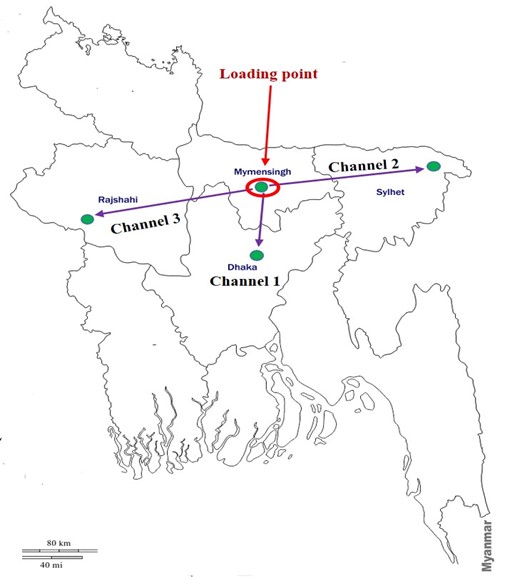
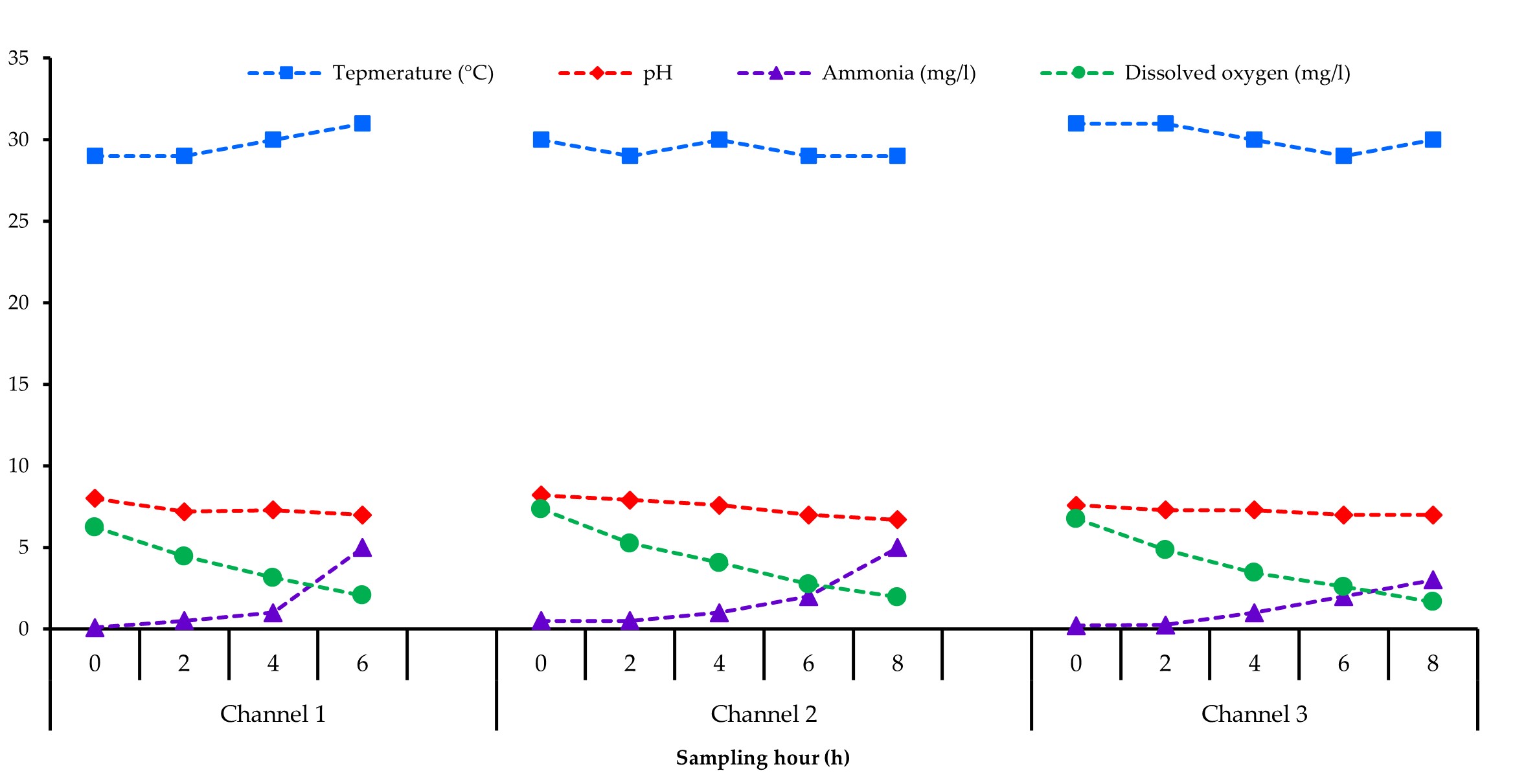
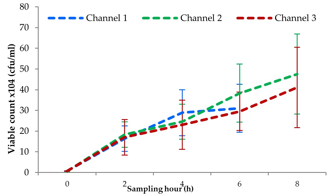
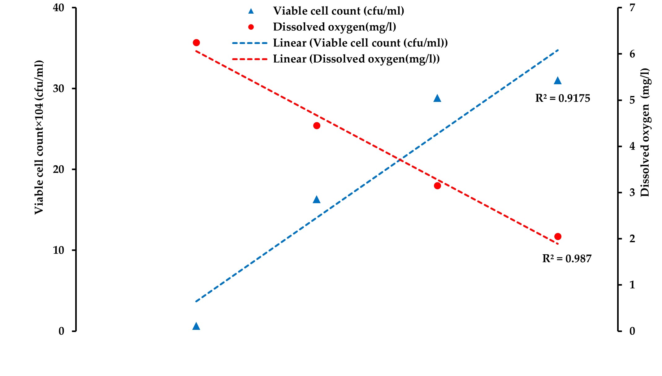
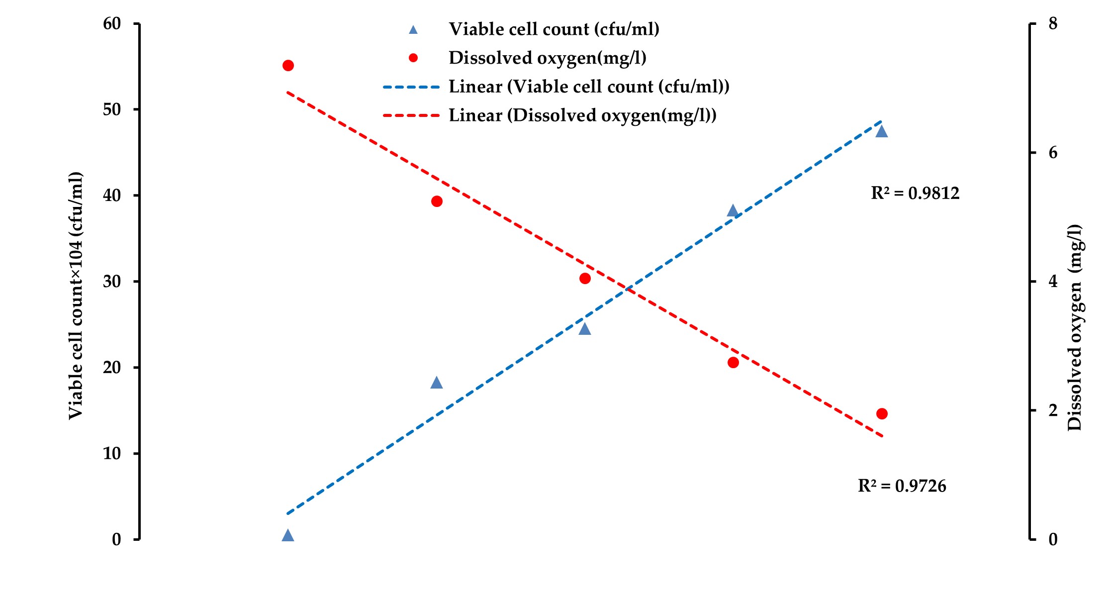
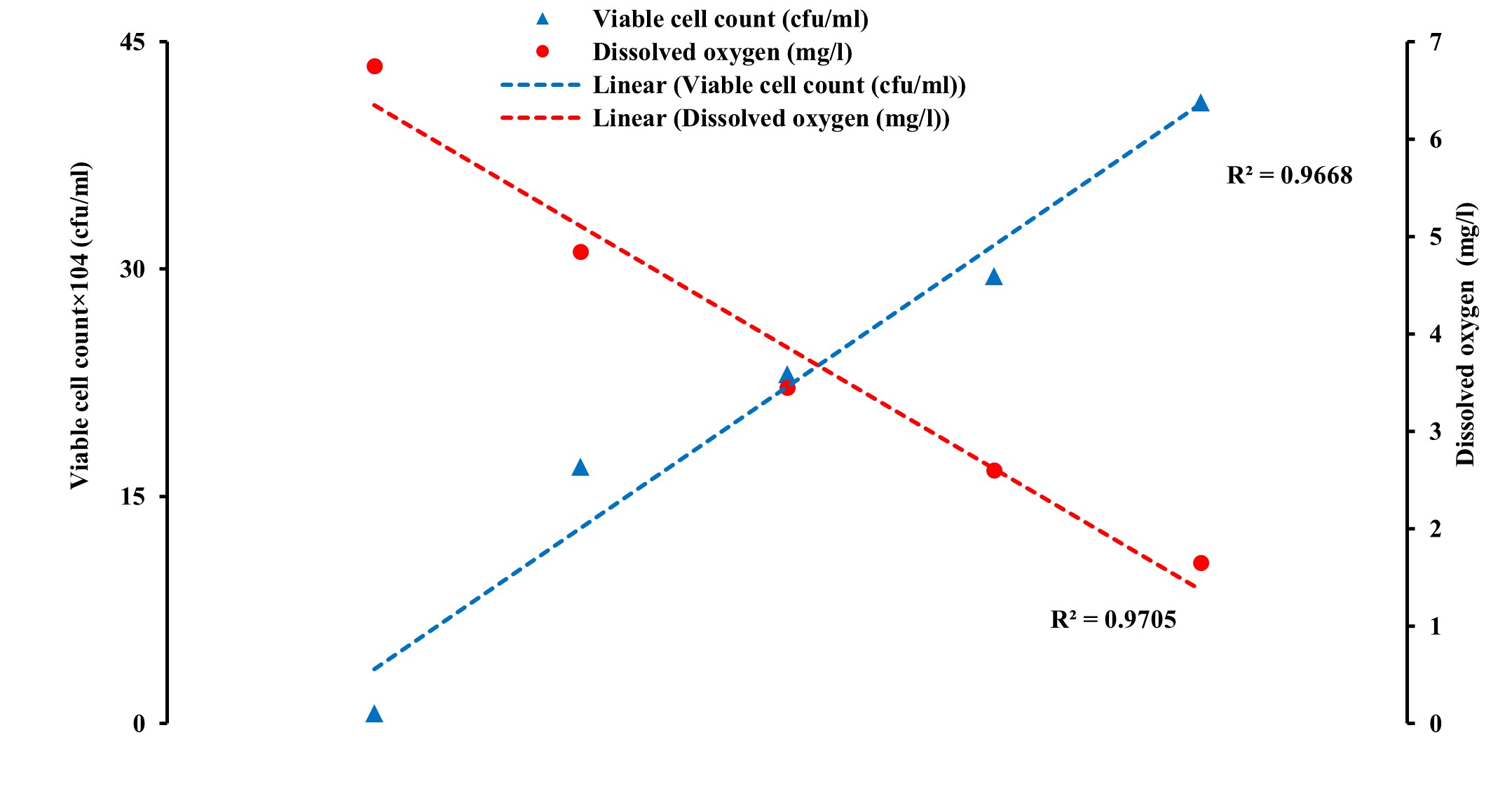
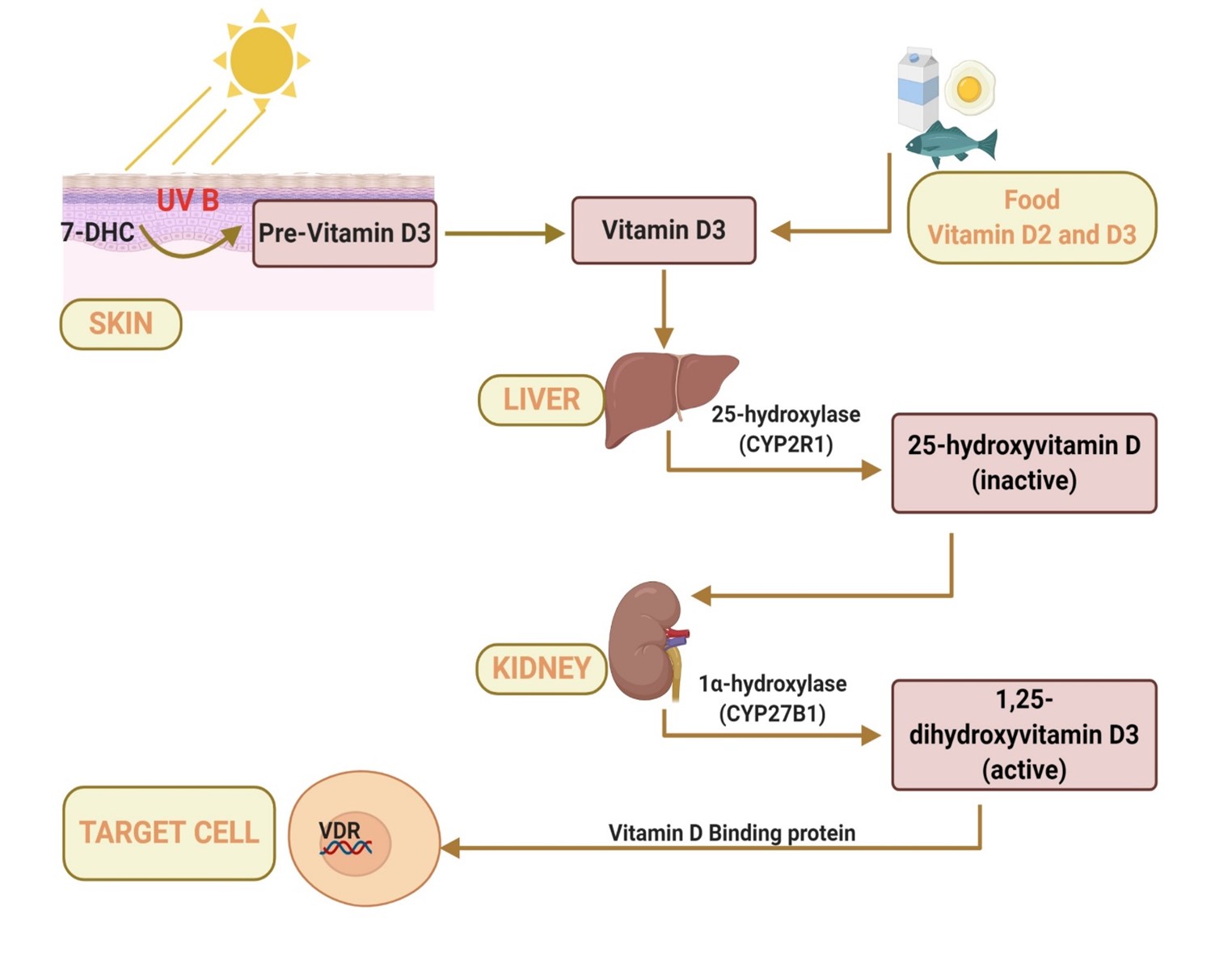
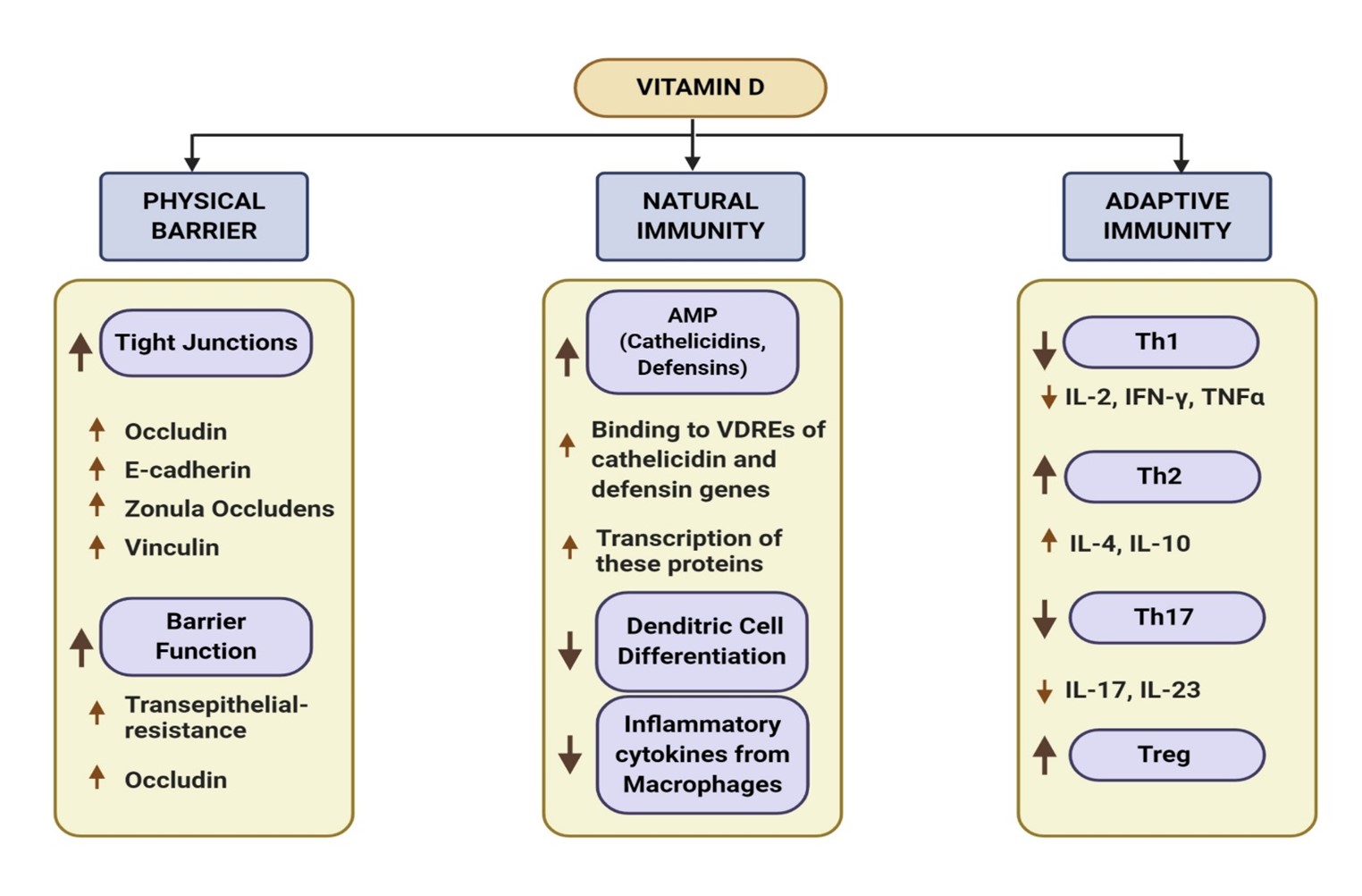
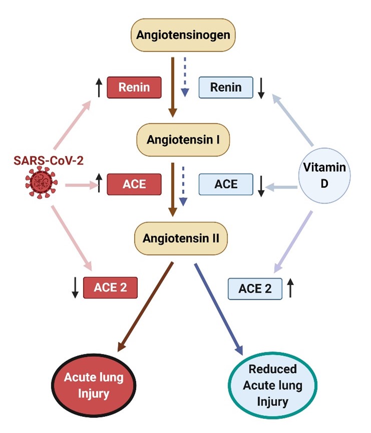
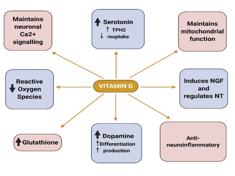
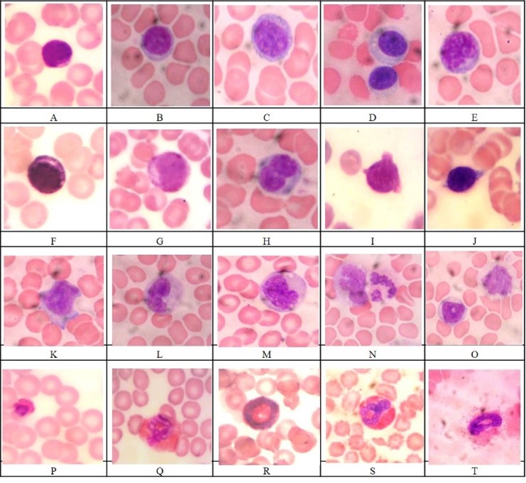
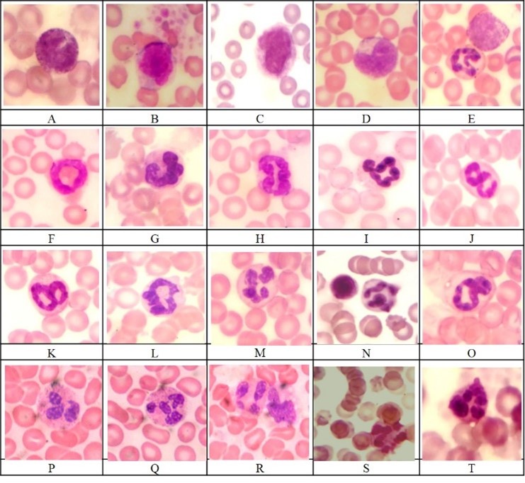
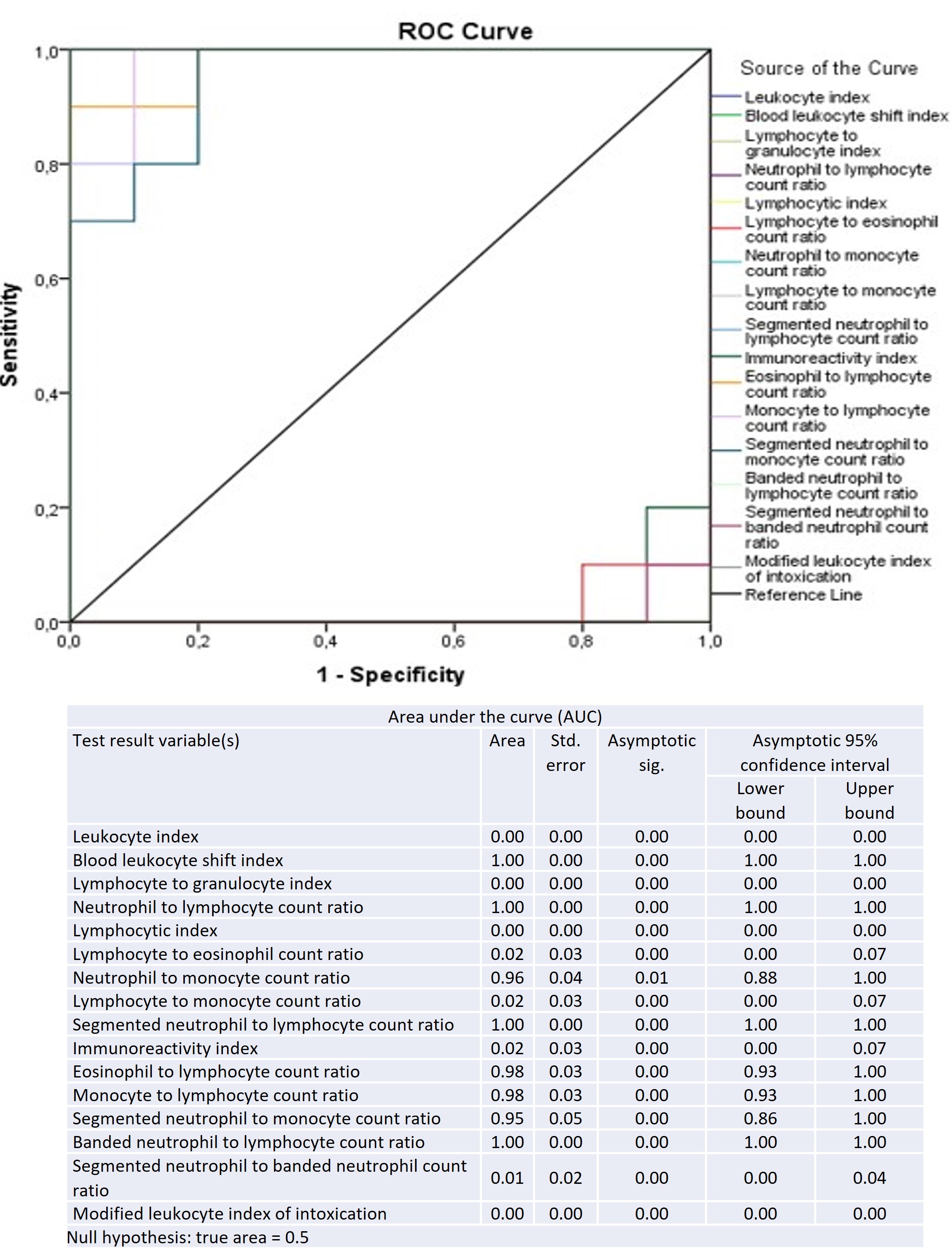
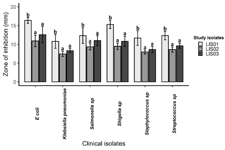
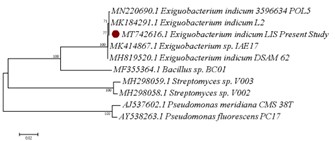
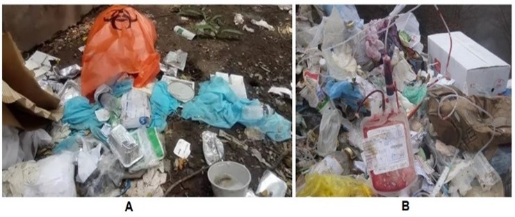
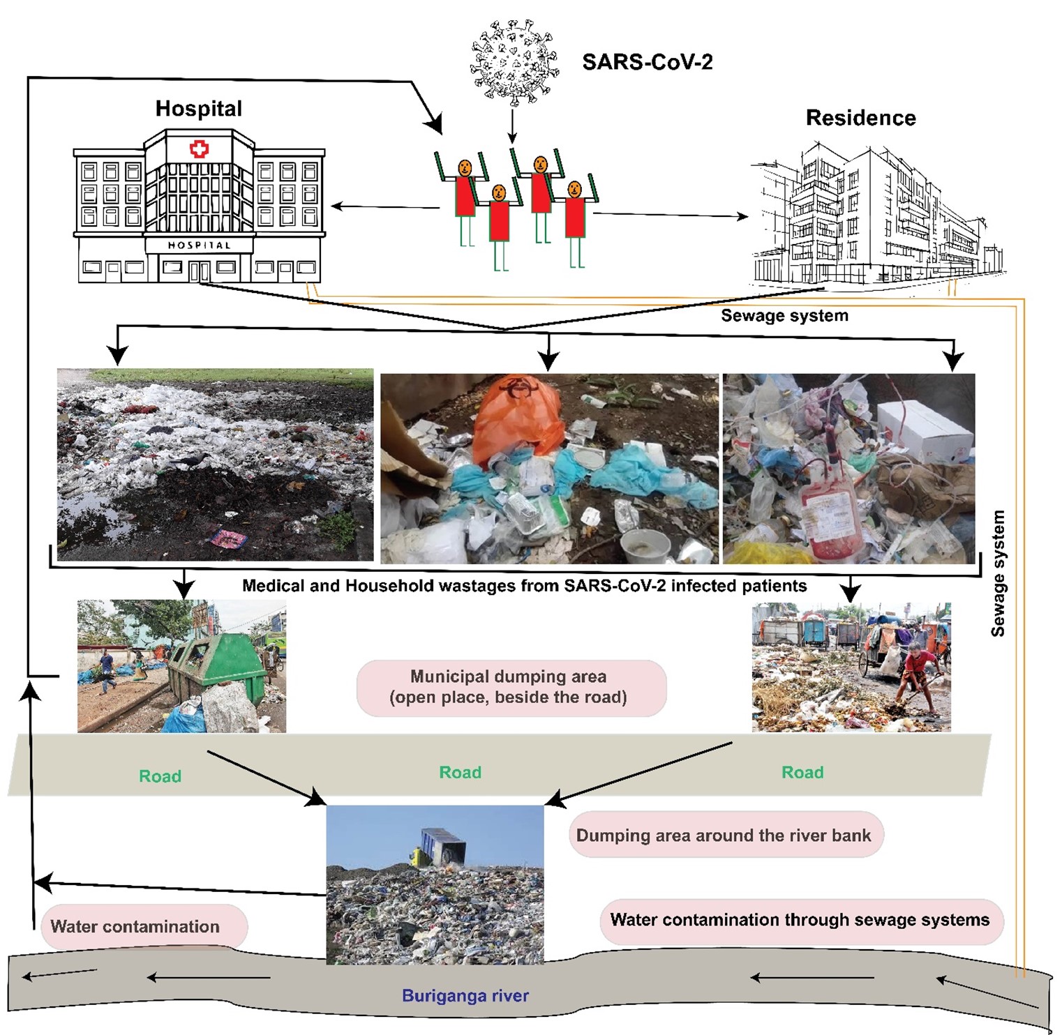
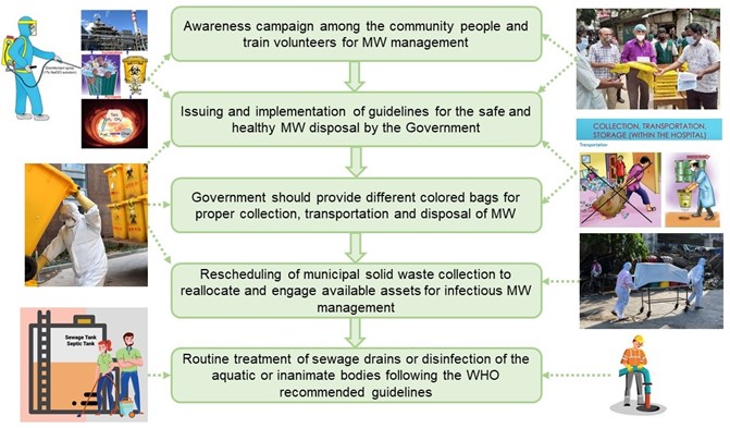
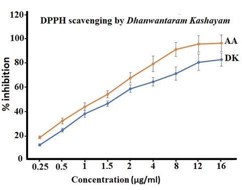
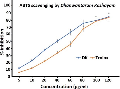
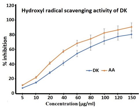
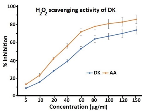
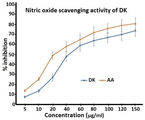
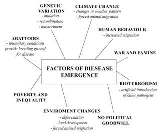
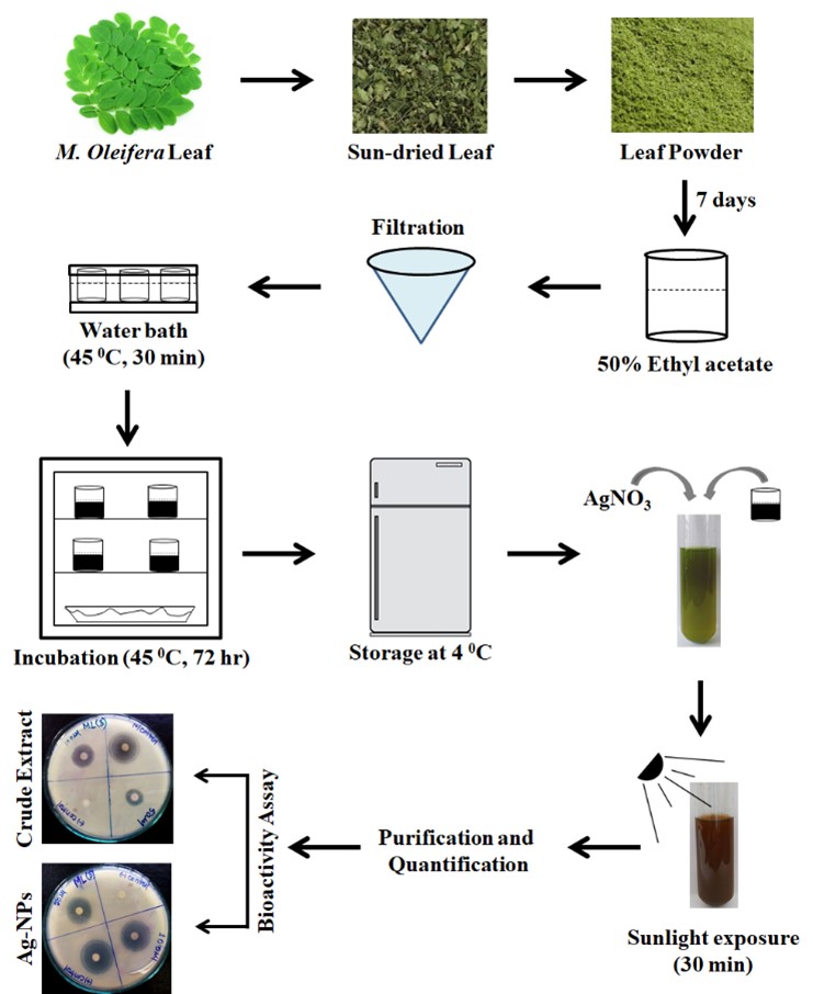
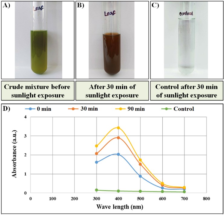
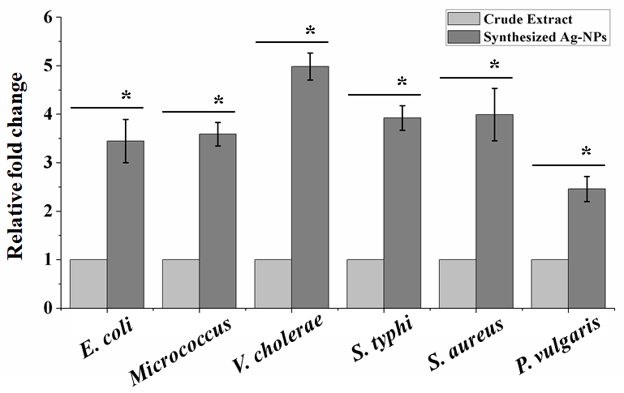
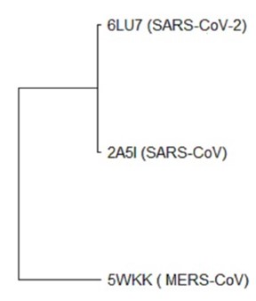
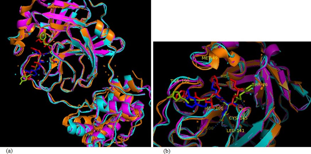
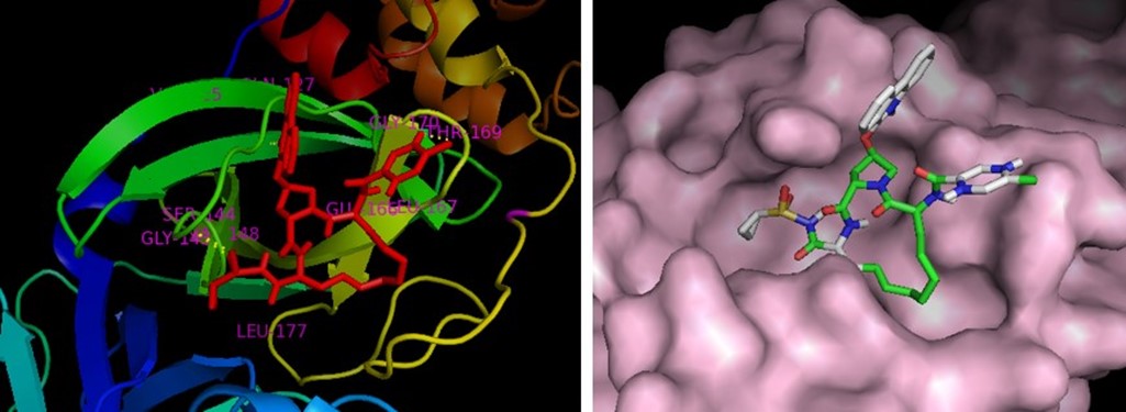
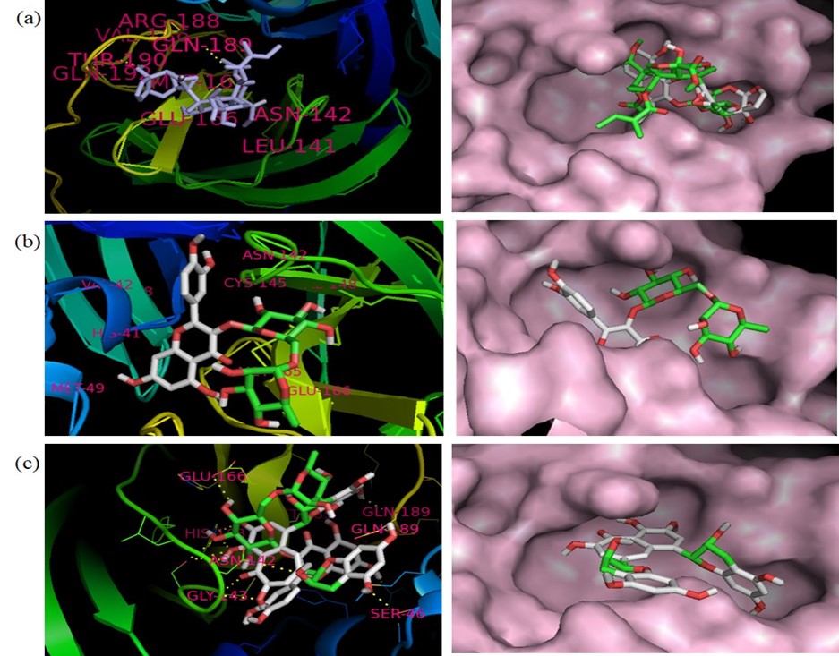
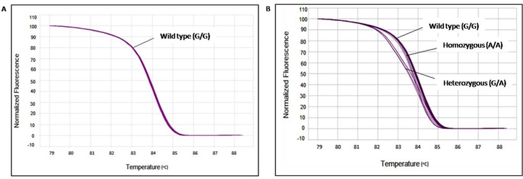