HTTP 200 OK
Allow: GET, HEAD, OPTIONS
Content-Type: application/json
Vary: Accept
{
"count": 319,
"next": "https://jabet.bsmiab.org/articles/?format=api&page=23",
"previous": "https://jabet.bsmiab.org/articles/?format=api&page=21",
"results": [
{
"id": 185,
"slug": "178-1617798303-detection-of-multidrug-resistant-salmonella-spp-from-healthy-and-diseased-broilers-having-potential-public-health-significance",
"featured": false,
"slider": false,
"issue": "Vol4 Issue2",
"type": "original_article",
"manuscript_id": "178-1617798303",
"recieved": "2021-04-10",
"revised": null,
"accepted": "2021-05-22",
"published": "2021-05-24",
"pdf_file": "https://jabet.bsmiab.org/media/pdf_file/2023/48/178-1617798303.pdf",
"title": "Detection of multidrug resistant Salmonella spp. from healthy and diseased broilers having potential public health significance",
"abstract": "<p>Multidrug resistant (MDR) <em>Salmonella</em> spp. poses significant global public health concern by causing food-borne infections. This study aimed to detect MDR <em>Salmonella </em>spp<em>.</em> from healthy and diseased broiler chickens in the Mymensingh and Jamalpur districts of Bangladesh. Total 70 samples comprising feces (n=20), chicken meat (n=30), and visceral organs i.e. liver, lung, and kidney (n=20) were collected. <em>Salmonella</em> were isolated and identified by culture, biochemical tests and PCR. The antibiogram study was performed by the disk diffusion method. By PCR, 30% (21/70; 95% CI: 19.32-40.05%) samples were positive for <em>Salmonella </em>spp., of which significantly (p=0.005) higher occurrence were detected in feces (50%; 95% CI: 29.93-70.07%) compared to chicken meat (10%; 95% CI: 3.46-25.62%) and visceral organs (40%; 95% CI: 21.88-61.34%). By antibiogram, all the <em>Salmonella</em> isolates were resistant to amoxicillin, and frequently (90.48-19.05%) resistant to tetracycline, ceftazidime, chloramphenicol, colistin, and ciprofloxacin. The significantly higher resistance of chloramphenicol, tetracycline, and ceftazidime were observed in the internal organs of broilers. Interestingly, 80.95% (17/21; 95% CI: 59.99-92.33%) <em>Salmonella</em> isolates were MDR in nature. The range of multiple antibiotic resistance (MAR) index of <em>Salmonella</em> isolates varied from 0.29 to 0.86. The high occurrence of MDR and MAR <em>Salmonella</em> in broilers detected in our present study could reveal a high risk to public health and these organisms could be transmitted to humans through the food supply. We suggest that effective prevention and control measures should be implemented to reduce their potential contamination and to minimize the emergence of antibiotic resistance.</p>",
"journal_reference": "J Adv Biotechnol Exp Ther. 2021; 4(2): 248-255.",
"academic_editor": "Md. Masudur Rahman, PhD; Sylhet Agricultural University, Bangladesh",
"cite_info": "",
"keywords": [
"Salmonellosis",
"Public health",
"Foodborne pathogen",
"MDR",
"invA",
"MAR"
],
"DOI": "10.5455/jabet.2021.d125",
"sections": [
{
"section_number": 1,
"section_title": "INTRODUCTION",
"body": "<p>Poultry farming has become a profitable and dependable agricultural business in Bangladesh. In addition, it plays a momentous role in the employment generation and economic growth of Bangladesh [<a href=\"#r-1\">1</a>]. Poultry provides additional income to rural people [<a href=\"#r-2\">2</a>]. Furthermore, poultry delivers about 37% of the total meat supply to the people of Bangladesh and covers more than 12.5% of total daily proteins per capita [<a href=\"#r-3\">3</a>]. But the entry of different infectious diseases e.g. salmonellosis, avian colibacillosis, mycoplasmosis, fowl cholera, avian influenza, Newcastle disease, infectious bronchitis, aspergillosis, and others hinder the further advancement of poultry production [<a href=\"#r-4\">4</a>]. Among them, multidrug resistant (MDR) <em>Salmonella</em> spp. are deemed as major botherations in the uplifting of Bangladesh’s poultry sector by causing drastic poultry illness and deaths annually [<a href=\"#r-5\">5</a>].<br />\r\n<em>Salmonella</em> spp. is one of the most frequently isolated foodborne pathogens that develops approximately 153 million enteric diseases and 155,000 deaths per year globally [<a href=\"#r-6\">6-8]</a>. In poultry, <em>Salmonella</em> spp. is devastating for developing avian salmonellosis, increasing mortality rates, and reducing hatchability and fertility rates [<a href=\"#r-3\">3</a>]. Poultry products especially meat and eggs play a pivotal role in <em>Salmonella</em> contamination. Incidences of food-poisoning diseases triggered by these pathogens have been increasing remarkably in the last several years. In human, <em>Salmonella</em> spp. cause human salmonellosis. Poultry-originated foods are thought to be the main reasons for human salmonellosis, as poultry especially broilers are important reservoirs of <em>Salmonella</em> spp. [<a href=\"#r-9\">9</a>]. In addition, poultry and poultry-originated foods generally act as crucial sources for the sporadic outbreaks of human salmonellosis globally. As <em>Salmonella</em> spp. are naturally gut-originated pathogens in poultry, the food supply chain makes an important scope for the transmission of <em>Salmonella</em> infections to humans [<a href=\"#r-10\">10</a>].<br />\r\nAntimicrobial resistance (AMR) is considered a worldwide health problem jeopardizing all one-health components [<a href=\"#r-11\">11</a>]. The indiscriminate use of antibiotics triggers selection pressure and develops antibiotic resistance in bacteria [<a href=\"#r-12\">12</a>]. In addition, antibiotics are being used as growth promoters in modern poultry, especially broiler production that also triggers the development of AMR in poultry. Globally, the speculation deaths due to AMR consequences will be more than 300 million per year, if significant steps won’t be taken by 2050 [<a href=\"#r-13\">13</a>]. The world critics have warned that the low- and middle-income countries will face the worst impacts of AMR. According to the world health organization, Bangladesh is at high risk of AMR consequences [<a href=\"#r-14\">14</a>].<br />\r\nThe detection of MDR <em>Salmonella</em> spp. from broilers was previously recorded in Bangladesh [<a href=\"#r-5\">5</a>, <a href=\"#r-15\">15</a>, <a href=\"#r-16\">16</a>]. However, it needs regular surveillance to determine the actual prevalence of <em>Salmonella</em> in broilers in Bangladesh. Therefore, the present study was carried out to detect MDR <em>Salmonella</em> in both healthy (feces, and meat) and diseased (visceral organs) broiler samples.</p>"
},
{
"section_number": 2,
"section_title": "MATERIALS AND METHODS",
"body": "<p><strong>Sample size calculation</strong><br />\r\nThe sample size of our present study was calculated following by the prevalence of <em>Salmonella</em> spp. (23.53%) isolated from broilers in Bangladesh [<a href=\"#r-16\">16</a>]. The formula we followed for the sample size calculation was described previously [<a href=\"#r-17\">17</a>]: n = Z<sup>2</sup>pq/d<sup>2</sup>, where, n = desired sample size, Z = the standard normal deviation (1.96 at 95% confidence level), p = prevalence (23.53% or 0.2353), q = 1-p = 1-0.2353 = 0.7647, d = precision at 10% (d = 0.1). So, n= (1.96)<sup>2</sup>×0.2353×0.7647/ (0.1)<sup>2 </sup>= 69.123. Therefore, we collected 70 samples from broiler chickens.</p>\r\n\r\n<p> </p>\r\n\r\n<p><strong>Sampling site and sampling</strong><br />\r\nThis study was performed from June 2018 to November 2019 in Mymensingh (24.7539° N, 90.4073° E) and Jamalpur (24.9250° N, 89.9463° E) districts of Bangladesh. The study areas are showed in <a href=\"#figure1\">Figure 1</a>.<br />\r\nA total of 70 broiler samples comprising feces (n=20), chicken meat i.e. thigh, breast, and wings (n=30) from healthy birds, and visceral organs i.e. liver, lungs, and kidneys (n=20) from diseased birds were collected aseptically. Sterile cotton buds were used to collect freshly dropped fecal samples. Meat samples were collected by processing broilers from different markets. By post-mortem examination, visceral organs were collected from each bird that had lesions of avian salmonellosis. 5 gm of each samples was collected aseptically. Immediately after collection, samples were taken into sterile zip-lock bags with particular tag numbers and transferred to the laboratory maintaining a cool chain. After bringing to the laboratory, samples were seeded to sterile test tubes containing 5 ml sterile nutrient broth and incubated overnight at 37°C. All the experimental procedures and protocols used in this study were approved by the animal welfare and experimentation ethics committee of Bangladesh agricultural university (No. AWEEC/BAU/2019(28)).</p>\r\n\r\n<div id=\"figure1\">\r\n<figure class=\"image\"><img alt=\"\" height=\"393\" src=\"/media/article_images/2024/15/04/178-1617798303-Figure1.jpg\" width=\"500\" />\r\n<figcaption><strong>Figure 1. </strong>Study area map produced by ArcMap (version 10.7) software (ESRI, Redlands, CA, USA).</figcaption>\r\n</figure>\r\n\r\n<p> </p>\r\n</div>\r\n\r\n<p><strong>Isolation of <em>Salmonella</em> spp.</strong><br />\r\nIsolation of <em>Salmonella</em> spp. was performed by culture on Xylose Lysine Deoxycholate (XLD) agar (HiMedia, India) plates. Overnight enriched samples were streaked on XLD agar plates and incubated aerobically for 18-24 hours at 37°C to get pure colonies. Black-centered colonies on XLD agar plates were suspected as the growth of <em>Salmonella</em> spp. Gram’s staining and biochemical tests (urease test, sugar fermentation test, methyl red test, Voges-Proskauer test) were performed for further confirmation [<a href=\"#r-18\">18</a>].</p>\r\n\r\n<p> </p>\r\n\r\n<p><strong>DNA extraction and PCR confirmation of <em>Salmonella</em> spp.</strong><br />\r\nIsolated <em>Salmonella</em> spp. were finally confirmed by polymerase chain reaction (PCR) targeting the <em>invA</em> gene (F: 5′-ATCAGTACCAGTCGTCTTATCTTGAT-3′ and R: 5′-TCTGTTTACCGGGCATACCAT-3′) with 211 amplicon size [<a href=\"#r-19\">19</a>]. For PCR, bacterial DNA was extracted by boiling and freeze-thawing method as previously described [<a href=\"#r-20\">20</a>]. Briefly, initially 1 ml of overnight enriched culture was centrifuged at 5,000 rotation per minute (rpm) for 5 minutes and the supernatant was discarded. Subsequently, a similar process was performed after mixing 1 ml of phosphate buffer solution (PBS). After discarding supernatant, the pellet was suspended to 200 µL PBS; followed by boiling and cooling of the suspension for 10 minutes in each step. Finally, the suspension was again centrifuged for 10 minutes at 10,000 rpm and the supernatant was collected as genomic DNA. The collected genomic DNA was then stored at -20°C for further use.<br />\r\nA final volume of 20 µL consisting of 10 µL of the master mix (2X) (Promega, Madison, WI, USA), 4 µL of nuclease-free water, 1 µL of each primer, and 4 µL of genomic DNA (50 ng/ µL) was used to carry out the PCR amplification. The thermo-cycle conditions were as follows: initial denaturation at 95°C for 5 min, followed by 30 cycles of denaturation at 94°C for 30 s, annealing at 52°C for 2 min, extension at 72°C for 45 s, and final extension was conducted at 72°C for 45 s.<br />\r\nAfter amplification, PCR products were analyzed by 1.5% agarose (Invitrogen, USA) gel electrophoresis, stained with ethidium bromide (0.5 μg/ml) for 10 min in a dark place, and finally, the expected amplicon sizes were audited and captured under ultra-violet trans-illuminator (Biometra, Germany). A 100 bp DNA ladder (Promega, Madison, WI, USA) was used to check the targeted amplicon size.</p>\r\n\r\n<p> </p>\r\n\r\n<p><strong>Antibiotic susceptibility test</strong><br />\r\nThe antibiotic susceptibility test (AST) was done by the disk diffusion method [<a href=\"#r-21\">21</a>]. Seven commonly used antibiotics under seven classes were employed: penicillins (amoxicillin- 30 μg), fluoroquinolones (ciprofloxacin- 5 μg), amphenicols (chloramphenicol- 30 μg), polypeptides (colistin- 10 μg), aminoglycosides (gentamicin- 10 μg), tetracyclines (tetracycline- 30 μg), and cephalosporins (ceftazidime- 30 μg). The AST was done by spreading freshly <em>Salmonella</em> growth culture having an equal concentration of 0.5 McFarland solution on Mueller-Hinton agar (HiMedia, India) plates. The guidelines of the clinical and laboratory standard institute [<a href=\"#r-22\">22</a>] were followed to interpret the results. Any isolates showing resistance against three or more classes of antibiotics were deemed as MDR [<a href=\"#r-23\">23</a>]. Furthermore, the multiple antibiotic resistance (MAR) index was evaluated by the following formula: MAR= a/b, where ‘‘a” denotes the number of antibiotics which were resistant to a particular isolate, and ‘‘b” denotes the total number of antibiotics tested [<a href=\"#r-24\">24</a>].</p>\r\n\r\n<p> </p>\r\n\r\n<p><strong>Statistical analysis</strong><br />\r\nData obtained from this study were incorporated in Microsoft Excel-2010 (Los Angeles, CA, USA), and exported to the GraphPad Prism 8.4.2 (GraphPad Software, Inc.) and the Statistical Package for the Social Sciences (SPSS) software (IBM SPSS- version 25.0, USA) for statistical analysis. By SPSS, a Pearson chi-square test for goodness-of-fit was performed to observe the possible variations in the occurrence of <em>Salmonella</em> spp. and the resistance profiles of different antibiotics among different collected samples. Statistically significant <em>p</em>-value was less than 0.05. Furthermore, GraphPad Prism following the Wilson/Brown Hybrid method as previously described [<a href=\"#r-25\">25</a>] was used to calculate the binomial 95% confidence intervals.</p>"
},
{
"section_number": 3,
"section_title": "RESULTS",
"body": "<p><strong>Occurrence of <em>Salmonella</em> isolates</strong><br />\r\nOut of 70 samples, 46 (65.71%, 95% confidence interval: 54.04-75.75%) samples were positive for <em>Salmonella</em> spp. based on their colony characteristics and biochemical tests. Of these 46 isolates, 30% (21/70) samples were PCR positive for <em>Salmonella</em> spp. targeting <em>invA</em> gene; among which healthy broiler sample- feces (50%, 10/20) exhibited significantly higher occurrence of <em>Salmonella</em> spp., compared to internal organs (40; 7/20) from diseased broilers, and meat (10%, 3/30) samples from healthy broilers (<a href=\"#Table-1\">Table 1</a>).</p>\r\n\r\n<div id=\"Table-1\">\r\n<p><a href=\"https://jabet.bsmiab.org/table/178-1617798303-table1/\">Table-1</a><strong>Table 1. </strong>Occurrence of <em>Salmonella</em> spp. from different broiler samples.</p>\r\n\r\n<p> </p>\r\n</div>\r\n\r\n<p><strong>Antibiogram profiles of isolates <em>Salmonella</em> spp.</strong><br />\r\nFrom the antibiotic susceptibility test, all the <em>Salmonella</em> isolates were resistant to amoxicillin; frequently resistant to tetracycline (90.48%), ceftazidime (61.90%), chloramphenicol (38.10%), and colistin (33.33%). On contrary, gentamicin showed higher sensitivity to <em>Salmonella</em> isolates (<a href=\"#figure2\">Figure 2</a>). <em>Salmonella</em> from visceral organs (diseased broiler samples) revealed peak resistance against most of the used antibiotics, where a statistically significant correlation was found for chloramphenicol, tetracycline, and ceftazidime (<a href=\"#Table-2\">Table 2</a>).</p>\r\n\r\n<div id=\"figure2\">\r\n<figure class=\"image\"><img alt=\"\" height=\"337\" src=\"/media/article_images/2024/15/04/178-1617798303-Figure2.jpg\" width=\"500\" />\r\n<figcaption><strong>Figure 2.</strong> Antibiogram profiles of <em>Salmonella</em> isolated from broiler samples. Here, AMX= Amoxicillin, CIP= Ciprofloxacin, C= Chloramphenicol, CL= Colistin, GEN= Gentamicin, TE= Tetracycline, CAZ= Ceftazidime.</figcaption>\r\n</figure>\r\n\r\n<p> </p>\r\n</div>\r\n\r\n<div id=\"Table-2\">\r\n<p><a href=\"https://jabet.bsmiab.org/table/178-1617798303-table2/\">Table-2</a><strong>Table 2.</strong> Resistance profiles of <em>Salmonella</em> isolated from broiler samples. </p>\r\n\r\n<p> </p>\r\n</div>\r\n\r\n<p><strong>Occurrence of MDR patterns and MAR index of <em>Salmonella</em> isolates</strong><br />\r\nOut of 21 <em>Salmonella</em> isolates, 17 (80.95%; 95% CI: 59.99-92.33%) were MDR in nature. Overall, nine resistance patterns were audited, among them, the highest 23.53% (4/17; 95% CI: 9.56-47.26%) <em>Salmonella</em> isolates showed the resistance pattern no. 9 (AMX-TE-CAZ). One isolate showed resistance against six classes of antibiotics (six antibiotics) (pattern no. 1). The antibiotic resistance profile of each <em>Salmonella</em> isolate was found to vary with MAR indices ranging from 0.29 to 0.86. All the <em>Salmonella</em> isolates were resistant against at least two antibiotics representing two classes (<a href=\"#Table-3\">Table 3</a>).</p>\r\n\r\n<div id=\"Table-3\">\r\n<p><a href=\"https://jabet.bsmiab.org/table/178-1617798303-table3/\">Table-3</a><strong>Table 3.</strong> Occurrence of multidrug resistance and multiple antibiotic resistance index of <em>Salmonella</em> isolated from broiler samples. </p>\r\n\r\n<p> </p>\r\n</div>"
},
{
"section_number": 4,
"section_title": "DISCUSSION",
"body": "<p>Avian Salmonellosis is a major threat to both the poultry industry (causing serious economic losses) and human health (showing zoonotic significance). In addition, infections developed by MDR <em>Salmonella</em> spp. are difficult to control. Broiler meat, eggs, fecal materials, and visceral organs have been recorded as cardinal sources of <em>Salmonella</em> contamination [<a href=\"#r-26\">26</a>]. Here, we reported the detection of MDR <em>Salmonella</em> from broiler chickens which show serious public health significance.<br />\r\nThe <em>invA</em> gene of <em>Salmonella</em> usually comprises specific DNA sequences which proves the <em>invA</em> as a compatible gene to detect <em>Salmonella</em> genotypically [<a href=\"#r-27\">27</a>]. In addition, the <em>invA</em> gene is available in almost all <em>Salmonella</em> serovars. This gene encodes a protein (inner membrane) that assists <em>Salmonella</em> to invade their epithelial cells [<a href=\"#r-3\">3</a>]. In this study, the overall occurrence of <em>Salmonella</em> spp. targeting <em>invA</em> gene in broiler samples was 30% (21/70) which is lined with the previous study conducted in Bangladesh [<a href=\"#r-15\">15</a>]. Conversely, both higher [<a href=\"#r-5\">5</a>] and lower [<a href=\"#r-16\">16</a>] prevalence rate of <em>Salmonella</em> spp. from broilers than our study were also recorded previously in Bangladesh. Globally, variable findings as 7.9% [<a href=\"#r-26\">26</a>] and 0.75% [<a href=\"#r-28\">28</a>] were recorded previously. This observed variations in the occurrence of <em>Salmonella</em> spp. might have linkage with the variations of the management systems of farms (biosecurity, hygiene, sanitary, etc.), sample size, types of samples, geographical and seasonal distributions, and method related factors. The occurrence of <em>Salmonella</em> in broilers suggests that the farms’ and poultry processing environments might contain poor-hygienic protocols. Furthermore, the presence of virulence gene <em>invA</em> in <em>Salmonella</em> isolates denotes their pathogenicity which can develop foodborne pathogens after introducing into food.<br />\r\nIn the current study, a significantly higher occurrence of <em>Salmonella</em> spp. was observed in fecal samples (50%) of healthy broilers in relation to visceral organs (40%) of diseased broilers, and meat samples (10%) of healthy broilers. Previously several studies reported the presence of <em>Salmonella</em> spp. in broiler meat [<a href=\"#r-29\">29</a>], fecal materials [<a href=\"#r-5\">5</a>], and visceral organs [<a href=\"#r-15\">15</a>]. The significantly higher occurrence of <em>Salmonella</em> in fecal materials is not unusual, as <em>Salmonella</em> are naturally found in the gastrointestinal tract of avian species [<a href=\"#r-30\">30</a>]. These <em>Salmonella</em> contaminations can be introduced into the production system from broilers via feces, contaminated water or feed, and others. In addition, the presence of <em>Salmonella</em> in feces samples indicates that broiler droppings can shed <em>Salmonella</em> to other birds of the flocks. The presence of <em>Salmonella</em> spp. in meat samples denotes that <em>Salmonella</em> spp. have the potential to be transmitted to humans via the food supply chain. Furthermore, consumption of undercooked poultry and poultry products contaminated by <em>Salmonella</em> has also the potential in the transmission of <em>Salmonella</em> to humans [<a href=\"#r-28\">28</a>].<br />\r\nAntimicrobial resistance is an emerging problem in the world and has the most significant public health challenge of this century globally [<a href=\"#r-31\">31</a>]. Poultry and poultry products are huge sources of antibiotic reservoirs [<a href=\"#r-32\">32</a>]. In our present study, all the <em>Salmonella</em> isolates were resistant to amoxicillin, and frequently resistant to tetracycline, ceftazidime, chloramphenicol, and colistin. Visceral organs exhibited a higher occurrence of antibiotic resistance compared to other selected samples (in the most antibiotics used). In addition, <em>Salmonella</em> resistance to tetracycline, ceftazidime, and chloramphenicol was significantly higher in visceral organs of diseased broilers. Interestingly, <em>Salmonella</em> isolates showed resistance to ceftazidime (61.90%) and colistin (33.33%) which is alarming for both human and animal health-care facilities. Ceftazidime is a 3<sup>rd</sup> generation cephalosporin antibiotic which usually used to treat severe bacterial infections in humans [<a href=\"#r-33\">33</a>]. In addition, colistin is a reserved group of antibiotics which generally used only in severe infections developed by MDR Gram-negative bacteria [<a href=\"#r-34\">34</a>]. However, MIC and molecular assays should be employed before drawing any conclusions.<br />\r\nInfections caused by MDR and MAR bacteria are serious global health concern as it is expensive for treatment and it may cause fatal consequences. MDR <em>Salmonella</em> has emerged as a cardinal human health issue throughout the world. The alarming situation was that 80.95% of <em>Salmonella</em> isolates were MDR in nature. Previously, Alam et al. [<a href=\"#r-5\">5</a>] detected 100% MDR <em>Salmonella</em> spp. from broilers in Bangladesh. In addition, MAR indices of isolated <em>Salmonella</em> from our study were ranged from 0.29 to 0.86. More than 0.29 of MAR index denotes that antibiotics were frequently used in the sources from where <em>Salmonella</em> were isolated showing high-risk sources for MDR and MAR bacteria. The development of MDR and MAR in <em>Salmonella</em> may be the results of selective pressure triggered by the misuse and overuse of antibiotics in broilers [<a href=\"#r-5\">5</a>]. These MDR and MAR <em>Salmonella</em> show severe public health significance by transmitting to humans through the food supply chain. In addition, these MDR and MAR bacteria can also spread in the environments and transfer their resistance genes to other bacteria horizontally.</p>"
},
{
"section_number": 5,
"section_title": "CONCLUSION",
"body": "<p>High occurrence of MDR <em>Salmonella</em> spp. detected in our present study reveals a potential human and animal health risk. There is potential in the transmission of <em>Salmonella</em> spp. from broilers to one-health components through the food chain, and ultimately to contaminate them. Future studies including the detection of virulence and antibiotic resistance genes of <em>Salmonella</em> spp. from healthy and diseased broilers may clarify the actual dynamics of their transmission and dissemination to one-health components. Effective control strategies and sustained implementation of comprehensive risk reduction practices including strict biosecurity throughout the production continuum are required to minimize the emergence of MDR and MAR zoonotic <em>Salmonella</em> pathogens.</p>"
},
{
"section_number": 6,
"section_title": "ACKNOWLEDGMENT",
"body": "<p>The authors are so grateful to farm owners for giving us access to samples during the whole study. The authors are also very much grateful to Dr. Khalada Zesmin, Upazila Livestock Officer, Kishoreganj, Bangladesh, for her valuable comments and suggestions during the whole study and the preparation of the manuscript. The authors are very much grateful to the Ministry of Education, Government of Bangladesh for providing funds through a research project (project number: LS2018686) to facilitate the present study.</p>"
},
{
"section_number": 7,
"section_title": "AUTHORS CONTRIBUTIONS",
"body": "<p>Conceptualization, MFRK and MTR; Sample collection, MT and MSI; Methodology, MT and MSI; Software, MSI; Validation, MFRK and MTR; Formal analysis, MSI, MAS and MTR; Investigation, MT, MSI, SI and MN; Data curation, MSI and MT; Writing-original draft preparation, MSI and MT; Writing- review and editing, MSI, MAS, MFRK, FMB and MTR; Visualization, MSI, and MTR; Supervision, MFRK and MTR; Fund acquisition, MFRK and MTR; Critical revisions and writing, MFRK and MTR. All authors have read and agreed to the published version of the manuscript.</p>"
},
{
"section_number": 8,
"section_title": "CONFLICTS OF INTEREST",
"body": "<p>The authors declare no conflict of interest.</p>"
}
],
"figures": [
{
"figure": "https://jabet.bsmiab.org/media/article_images/2024/15/04/178-1617798303-Figure1.jpg",
"caption": "Figure 1. Study area map produced by ArcMap (version 10.7) software (ESRI, Redlands, CA, USA).",
"featured": false
},
{
"figure": "https://jabet.bsmiab.org/media/article_images/2024/15/04/178-1617798303-Figure2.jpg",
"caption": "Figure 2. Antibiogram profiles of Salmonella isolated from broiler samples. Here, AMX= Amoxicillin, CIP= Ciprofloxacin, C= Chloramphenicol, CL= Colistin, GEN= Gentamicin, TE= Tetracycline, CAZ= Ceftazidime.",
"featured": false
}
],
"authors": [
{
"id": 788,
"affiliation": [
{
"affiliation": "Department of Microbiology and Hygiene, Faculty of Veterinary Science, Bangladesh Agricultural University, Mymensingh-2202, Bangladesh"
}
],
"first_name": "Mithun",
"family_name": "Talukder",
"email": null,
"author_order": 1,
"ORCID": null,
"corresponding": false,
"co_first_author": false,
"co_author": false,
"corresponding_author_info": "",
"article": 185
},
{
"id": 789,
"affiliation": [
{
"affiliation": "Department of Microbiology and Hygiene, Faculty of Veterinary Science, Bangladesh Agricultural University, Mymensingh-2202, Bangladesh"
}
],
"first_name": "Md. Saiful",
"family_name": "Islam",
"email": null,
"author_order": 2,
"ORCID": null,
"corresponding": false,
"co_first_author": false,
"co_author": false,
"corresponding_author_info": "",
"article": 185
},
{
"id": 790,
"affiliation": [
{
"affiliation": "Department of Microbiology and Hygiene, Faculty of Veterinary Science, Bangladesh Agricultural University, Mymensingh-2202, Bangladesh"
}
],
"first_name": "Samina",
"family_name": "Ievy",
"email": null,
"author_order": 3,
"ORCID": null,
"corresponding": false,
"co_first_author": false,
"co_author": false,
"corresponding_author_info": "",
"article": 185
},
{
"id": 791,
"affiliation": [
{
"affiliation": "Department of Microbiology and Hygiene, Faculty of Veterinary Science, Bangladesh Agricultural University, Mymensingh-2202, Bangladesh"
}
],
"first_name": "Md. Abdus",
"family_name": "Sobur",
"email": null,
"author_order": 4,
"ORCID": null,
"corresponding": false,
"co_first_author": false,
"co_author": false,
"corresponding_author_info": "",
"article": 185
},
{
"id": 792,
"affiliation": [
{
"affiliation": "Department of Microbiology and Hygiene, Faculty of Veterinary Science, Bangladesh Agricultural University, Mymensingh-2202, Bangladesh"
}
],
"first_name": "Fatimah Mohammed",
"family_name": "Ballah",
"email": null,
"author_order": 5,
"ORCID": null,
"corresponding": false,
"co_first_author": false,
"co_author": false,
"corresponding_author_info": "",
"article": 185
},
{
"id": 793,
"affiliation": [
{
"affiliation": "Department of Microbiology and Hygiene, Faculty of Veterinary Science, Bangladesh Agricultural University, Mymensingh-2202, Bangladesh"
}
],
"first_name": "Md.",
"family_name": "Najibullah",
"email": null,
"author_order": 6,
"ORCID": null,
"corresponding": false,
"co_first_author": false,
"co_author": false,
"corresponding_author_info": "",
"article": 185
},
{
"id": 794,
"affiliation": [
{
"affiliation": "Department of Microbiology and Hygiene, Faculty of Veterinary Science, Bangladesh Agricultural University, Mymensingh-2202, Bangladesh"
}
],
"first_name": "Md. Bahanur",
"family_name": "Rahman",
"email": null,
"author_order": 7,
"ORCID": null,
"corresponding": false,
"co_first_author": false,
"co_author": false,
"corresponding_author_info": "",
"article": 185
},
{
"id": 795,
"affiliation": [
{
"affiliation": "Department of Microbiology and Hygiene, Faculty of Veterinary Science, Bangladesh Agricultural University, Mymensingh-2202, Bangladesh"
}
],
"first_name": "Md. Tanvir",
"family_name": "Rahman",
"email": "tanvirahman@bau.edu.bd",
"author_order": 8,
"ORCID": "https://orcid.org/0000-0001-5432-480X",
"corresponding": true,
"co_first_author": false,
"co_author": false,
"corresponding_author_info": "Md. Tanvir Rahman, PhD, Department of Microbiology and Hygiene, Faculty of Veterinary Science, Bangladesh Agricultural University, Mymensingh-2202, Bangladesh. Email: tanvirahman@bau.edu.bd",
"article": 185
},
{
"id": 796,
"affiliation": [
{
"affiliation": "Department of Microbiology and Hygiene, Faculty of Veterinary Science, Bangladesh Agricultural University, Mymensingh-2202, Bangladesh"
}
],
"first_name": "Mohammad Ferdousur Rahman",
"family_name": "Khan",
"email": "frkhanbau80@yahoo.com",
"author_order": 9,
"ORCID": null,
"corresponding": true,
"co_first_author": false,
"co_author": false,
"corresponding_author_info": "Mohammad Ferdousur Rahman\r\nKhan, PhD; Department of Microbiology and Hygiene, Faculty of Veterinary Science, Bangladesh Agricultural University, Mymensingh-2202, Bangladesh. e-mail: frkhanbau80@yahoo.com",
"article": 185
}
],
"views": 1476,
"downloads": 105,
"references": [
{
"id": 6168,
"serial_number": 1,
"pmc": null,
"reference": "Sabuj AA, Mahmud T, Barua N, Rahman MA, Islam MS, Bary MA. Passive surveillance of clinical poultry diseases in an Upazila Government Veterinary Hospital of Bangladesh. African Journal of Microbiology Research. 2019;13(29):632-639.",
"DOI": null,
"article": 185
},
{
"id": 6169,
"serial_number": 2,
"pmc": null,
"reference": "Islam MS, Sabuj AA, Haque ZF, Pondit A, Hossain MG, Saha S. Seroprevalence and risk factors of avian reovirus in backyard chickens in different areas of Mymensingh district in Bangladesh. Journal of Advanced Veterinary and Animal Research. 2020;7(3):546-553.",
"DOI": null,
"article": 185
},
{
"id": 6170,
"serial_number": 3,
"pmc": null,
"reference": "Tawyabur M, Islam M, Sobur M, Hossain M, Mahmud M, Paul S, Hossain MT, Ashour HM, Rahman M. Isolation and Characterization of Multidrug-Resistant Escherichia coli and Salmonella spp. from Healthy and Diseased Turkeys. Antibiotics. 2020;9(11):770.",
"DOI": null,
"article": 185
},
{
"id": 6171,
"serial_number": 4,
"pmc": null,
"reference": "Ievy S, Islam M, Sobur M, Talukder M, Rahman M, Khan MF, Rahman M. Molecular detection of avian pathogenic Escherichia coli (APEC) for the first time in layer farms in Bangladesh and their antibiotic resistance patterns. Microorganisms. 2020;8(7):1021.",
"DOI": null,
"article": 185
},
{
"id": 6172,
"serial_number": 5,
"pmc": null,
"reference": "Alam SB, Mahmud M, Akter R, Hasan M, Sobur A, Nazir KH, Noreddin A, Rahman T, El Zowalaty ME, Rahman M. Molecular detection of multidrug resistant Salmonella species isolated from broiler farm in Bangladesh. Pathogens. 2020;9(3):201.",
"DOI": null,
"article": 185
},
{
"id": 6173,
"serial_number": 6,
"pmc": null,
"reference": "Kirk MD, Pires SM, Black RE, Caipo M, Crump JA, Devleesschauwer B, Döpfer D, Fazil A, Fischer-Walker CL, Hald T, Hall AJ. World Health Organization estimates of the global and regional disease burden of 22 foodborne bacterial, protozoal, and viral diseases, 2010: a data synthesis. PLoS Medicine. 2015;12(12):e1001921.",
"DOI": null,
"article": 185
},
{
"id": 6174,
"serial_number": 7,
"pmc": null,
"reference": "Vindigni SM, Srijan A, Wongstitwilairoong B, Marcus R, Meek J, Riley PL, Mason C. Prevalence of foodborne microorganisms in retail foods in Thailand. Foodborne Pathogens and Disease. 2007;4(2):208-215.",
"DOI": null,
"article": 185
},
{
"id": 6175,
"serial_number": 8,
"pmc": null,
"reference": "Rahman M, Sobur M, Islam M, Ievy S, Hossain M, El Zowalaty ME, Rahman AM, Ashour HM. Zoonotic Diseases: Etiology, Impact, and Control. Microorganisms. 2020;8(9):1405.",
"DOI": null,
"article": 185
},
{
"id": 6176,
"serial_number": 9,
"pmc": null,
"reference": "Antunes P, Mourão J, Campos J, Peixe L. Salmonellosis: the role of poultry meat. Clinical Microbiology and Infection. 2016;22(2):110-121.",
"DOI": null,
"article": 185
},
{
"id": 6177,
"serial_number": 10,
"pmc": null,
"reference": "Aditya A. Drug resistant Salmonella in broiler chicken sold at local market in Bangladesh and its public health significance. African Journal of Biotechnology. 2015;14(43):2995-3000.",
"DOI": null,
"article": 185
},
{
"id": 6178,
"serial_number": 11,
"pmc": null,
"reference": "Islam M, Nayeem M, Hasan M, Sobur M, Ievy S, Rahman S, Kafi M, Ashour HM, Rahman M. Virulence Determinants and Multidrug Resistance of Escherichia coli Isolated from Migratory Birds. Antibiotics. 2021;10(2):190.",
"DOI": null,
"article": 185
},
{
"id": 6179,
"serial_number": 12,
"pmc": null,
"reference": "Davies J, Davies D. Origins and evolution of antibiotic resistance. Microbiology and molecular biology reviews. 2010;74(3):417-433.",
"DOI": null,
"article": 185
},
{
"id": 6180,
"serial_number": 13,
"pmc": null,
"reference": "Orubu ES, Zaman MH, Rahman MT, Wirtz VJ. Veterinary antimicrobial resistance containment in Bangladesh: Evaluating the national action plan and scoping the evidence on implementation. Journal of Global Antimicrobial Resistance. 2020;21:105-115.",
"DOI": null,
"article": 185
},
{
"id": 6181,
"serial_number": 14,
"pmc": null,
"reference": "Chereau F, Opatowski L, Tourdjman M, Vong S. Risk assessment for antibiotic resistance in South East Asia. Bmj. 2017;358:j3393.",
"DOI": null,
"article": 185
},
{
"id": 6182,
"serial_number": 15,
"pmc": null,
"reference": "Mridha D, Uddin MN, Alam B, Akhter AT, Islam SS, Islam MS, Khan MS, Kabir SL. Identification and characterization of Salmonella spp. from samples of broiler farms in selected districts of Bangladesh. Veterinary World. 2020;13(2):275.",
"DOI": null,
"article": 185
},
{
"id": 6183,
"serial_number": 16,
"pmc": null,
"reference": "Al Mamun MA, Kabir SL, Islam MM, Lubna M, Islam SS, Akhter AT, Hossain MM. Molecular identification and characterization of Salmonella species isolated from poultry value chains of Gazipur and Tangail districts of Bangladesh. African Journal of Microbiology Research. 2017;11(11):474-481.",
"DOI": null,
"article": 185
},
{
"id": 6184,
"serial_number": 17,
"pmc": null,
"reference": "Thrusfield, M. Veterinary Epidemiology, 2nd ed.; Blackwell Science: London, UK, 1995.",
"DOI": null,
"article": 185
},
{
"id": 6185,
"serial_number": 18,
"pmc": null,
"reference": "Sobur A, Hasan M, Haque E, Mridul AI, Noreddin A, El Zowalaty ME, Rahman T. Molecular detection and antibiotyping of multidrug-resistant Salmonella isolated from houseflies in a fish market. Pathogens. 2019;8(4):191.",
"DOI": null,
"article": 185
},
{
"id": 6186,
"serial_number": 19,
"pmc": null,
"reference": "Shanmugasundaram M, Radhika M, Murali HS, Batra HV. Detection of Salmonella enterica serovar Typhimurium by selective amplification of fli C, flj B, iro B, inv A, rfb J, STM2755, STM4497 genes by polymerase chain reaction in a monoplex and multiplex format. World Journal of Microbiology and Biotechnology. 2009;25(8):1385-1394.",
"DOI": null,
"article": 185
},
{
"id": 6187,
"serial_number": 20,
"pmc": null,
"reference": "Queipo-Ortuño MI, Colmenero JD, Macias M, Bravo MJ, Morata P. Preparation of bacterial DNA template by boiling and effect of immunoglobulin G as an inhibitor in real-time PCR for serum samples from patients with brucellosis. Clinical and Vaccine Immunology. 2008;15(2):293-296.",
"DOI": null,
"article": 185
},
{
"id": 6188,
"serial_number": 21,
"pmc": null,
"reference": "Bauer AT. Antibiotic susceptibility testing by a standardized single disc method. American Journal of Clinical Pathology. 1966;45:149-158.",
"DOI": null,
"article": 185
},
{
"id": 6189,
"serial_number": 22,
"pmc": null,
"reference": "Clinical and Laboratory Standards Institute (CLSI). Performance Standards for Antimicrobial Susceptibility Testing, M100-S28. Clinical and Laboratory Standards Institute, Wayne, PA. 2018.",
"DOI": null,
"article": 185
},
{
"id": 6190,
"serial_number": 23,
"pmc": null,
"reference": "Sweeney MT, Lubbers BV, Schwarz S, Watts JL. Applying definitions for multidrug resistance, extensive drug resistance and pandrug resistance to clinically significant livestock and companion animal bacterial pathogens. Journal of Antimicrobial Chemotherapy. 2018;73(6):1460-1463.",
"DOI": null,
"article": 185
},
{
"id": 6191,
"serial_number": 24,
"pmc": null,
"reference": "Krumperman PH. Multiple antibiotic resistance indexing of Escherichia coli to identify high-risk sources of fecal contamination of foods. Applied and environmental microbiology. 1983;46(1):165-170.",
"DOI": null,
"article": 185
},
{
"id": 6192,
"serial_number": 25,
"pmc": null,
"reference": "Brown LD, Cai TT, DasGupta A. Interval estimation for a binomial proportion. Statistical science. 2001;15(2):101-117.",
"DOI": null,
"article": 185
},
{
"id": 6193,
"serial_number": 26,
"pmc": null,
"reference": "Duc VM, Nakamoto Y, Fujiwara A, Toyofuku H, Obi T, Chuma T. Prevalence of Salmonella in broiler chickens in Kagoshima, Japan in 2009 to 2012 and the relationship between serovars changing and antimicrobial resistance. BMC Veterinary Research. 2019;15(1):1-8.",
"DOI": null,
"article": 185
},
{
"id": 6194,
"serial_number": 27,
"pmc": null,
"reference": "Yanestria SM, Rahmaniar RP, Wibisono FJ, Effendi MH. Detection of invA gene of Salmonella from milkfish (Chanos chanos) at Sidoarjo wet fish market, Indonesia, using polymerase chain reaction technique. Veterinary World. 2019;12(1):170-175.",
"DOI": null,
"article": 185
},
{
"id": 6195,
"serial_number": 28,
"pmc": null,
"reference": "Sedeik ME, Nahed A, Awad AM, Elfeky SM, Abd El-Hack ME, Hussein EO, Alowaimer AN, Swelum AA. Isolation, conventional and molecular characterization of Salmonella spp. from newly hatched broiler chicks. AMB Express. 2019;9(1):1-6.",
"DOI": null,
"article": 185
},
{
"id": 6196,
"serial_number": 29,
"pmc": null,
"reference": "Rizwan M, Yar SA, Achakzai SK, Shakoor M. PCR based detection of Salmonella from fresh and processed chicken meat from Quetta, Pakistan. International Journal of Biosciences. 2017;10(4):363-371.",
"DOI": null,
"article": 185
},
{
"id": 6197,
"serial_number": 30,
"pmc": null,
"reference": "Machado Junior PC, Chung C, Hagerman A. Modeling Salmonella Spread in Broiler Production: Identifying Determinants and Control Strategies. Frontiers in Veterinary Science. 2020;7:564.",
"DOI": null,
"article": 185
},
{
"id": 6198,
"serial_number": 31,
"pmc": null,
"reference": "Lambertini E, Ruzante JM, Chew R, Apodaca VL, Kowalcyk BB. The public health impact of different microbiological criteria approaches for Salmonella in chicken parts. Microbial Risk Analysis. 2019;12:44-59.",
"DOI": null,
"article": 185
},
{
"id": 6199,
"serial_number": 32,
"pmc": null,
"reference": "Hedman HD, Vasco KA, Zhang L. A Review of Antimicrobial Resistance in Poultry Farming within Low-Resource Settings. Animals. 2020;10(8):1264.",
"DOI": null,
"article": 185
},
{
"id": 6200,
"serial_number": 33,
"pmc": null,
"reference": "Gashe F, Mulisa E, Mekonnen M, Zeleke G. Antimicrobial Resistance Profile of Different Clinical Isolates against Third-Generation Cephalosporins. Journal of pharmaceutics. 2018;2018:5070742- 5070742.",
"DOI": null,
"article": 185
},
{
"id": 6201,
"serial_number": 34,
"pmc": null,
"reference": "Pogue JM, Ortwine JK, Kaye KS. Clinical considerations for optimal use of the polymyxins: a focus on agent selection and dosing. Clinical Microbiology and Infection. 2017;23(4):229-233.",
"DOI": null,
"article": 185
}
]
},
{
"id": 184,
"slug": "178-1619144787-exercise-and-oral-melatonin-attenuate-anxiety-and-depression-like-behavior-in-type-2-diabetic-rats",
"featured": false,
"slider": false,
"issue": "Vol4 Issue2",
"type": "original_article",
"manuscript_id": "178-1619144787",
"recieved": "2021-04-16",
"revised": null,
"accepted": "2021-05-18",
"published": "2021-05-22",
"pdf_file": "https://jabet.bsmiab.org/media/pdf_file/2023/15/178-1619144787.pdf",
"title": "Exercise and oral melatonin attenuate anxiety and depression like behavior in type 2 diabetic rats",
"abstract": "<p>This study was performed to evaluate the therapeutic efficacy of exercise and oral melatonin on metabolic syndrome (MS), anxiety and depression-like behavior (ADB) in type 2 diabetes mellitus (T2DM) model in rat. Rats were allocated into five groups: non diabetes group, diabetes group, three treated group; diabetes rats disciplined with swimming exercise (40 min, 5 days per week) or oral melatonin (10 mg/kg bwt per day at 19.00 PM) alone or with combination. Exercise and oral melatonin significantly attenuated MS evidenced by improvement of hyperglycemia, insulin resistance, dyslipidemia, hyperleptinemia, and hypoadiponectinemia level in comparison with diabetes group. The ADB also markedly improved in exercise and oral melatonin treated rats as represented by decreased anxiety index, increased open arm entries and time spent in elevated plus maze, and reduced freezing behavior, increased entries and time spent in center in open field test. To know underlying molecular mechanisms, hippocampus tissue was analyzed. Interestingly, exercise combined with oral melatonin synergistically reduced serum corticosterone and hippocampus tissue level inflammatory cytokines and improved ATP level. Furthermore, this combination up-regulated the expression of brain-derived neurotrophic factor (BDNF), peroxisome proliferator-activated receptor gamma coactivator-1α (PGC-1α) and mitochondrial biogenesis related proteins, glucose transporter type 4 (GLUT4) in hippocampus tissue. Exercise and oral melatonin synergistically attenuated ADB in T2DM rats by attenuation of MS, neuroinflammation and normalizing corticosterone level via up-regulation of PGC-1α, mitochondrial biogenesis, BDNF, GLUT4, expression and ATP level. Thus, this treatment combination can be a promising tool in the management of MS, anxiety and depression in T2DM patients.</p>",
"journal_reference": "J Adv Biotechnol Exp Ther. 2021; 4(2): 238-247.",
"academic_editor": "Md Jamal Uddin, PhD; Ewha Womans University, Seoul, South Korea.",
"cite_info": "Rahman MM, Park SJ, et al. Exercise and oral melatonin attenuate anxiety and depression like behavior in type 2 diabetic rats. J Adv Biotechnol Exp Ther. 2021; 4(2): 238-247.",
"keywords": [
"Melatonin",
"Depression",
"Metabolic syndrome",
"Exercise",
"Anxiety",
"Type 2 diabetes."
],
"DOI": "10.5455/jabet.2021.d124",
"sections": [
{
"section_number": 1,
"section_title": "INTRODUCTION",
"body": "<p>Type 2 diabetes mellitus (T2DM) is known as complex metabolic disease comorbidities demonstrated mainly by high blood glucose and insulin resistance (IR) caused by abnormal secretion of insulin or alteration of cellular up regulation [<a href=\"#r-1\">1</a>]. It induces for the imbalance between energy consumption and expenditure. The incidence of T2DM is rapidly increasing worldwide due to habits in modern socio-economic and technology based lifestyle which has reduced the laborious activities or increased physical inactivity [<a href=\"#r-1\">1, 2</a>]. Currently, 425 million adults have diabetes and 1 in 2 remains undiagnosed and by 2030 it is anticipated that about 552 million adults worldwide will be affected by this disease [<a href=\"#r-3\">3</a>]. Diabetes and its hyperglycemia exert a remarkable threat to health across the globe due to its severe impacts on the microvascular systems which lead to dysfunction of multiple organs especially the kidneys, heart, eyes, brain, nerves, blood vessels [<a href=\"#r-4\">4</a>]. The occurrence of neurohormonal disorders and neuropsychiatric symptoms including depression and cognitive dysfunction are higher in T2DM [<a href=\"#r-1\">1</a>, <a href=\"#r-5\">5, 6</a>]. Altogether, these symptoms and complications in diabetic patient significantly deteriorate the quality of life. Moreover, T2DM and depression are two major issues of morbidity and mortality, currently more than 9% and 5% respectively, of the global population [<a href=\"#r-6\">6</a>]. One fourth patients with T2DM suffer form of depression, five-times severe than recorded in the general public [<a href=\"#r-7\">7</a>]. Depression disease comorbid with symptoms of anxiety between 50 and 75% with many common factors, including hypothalamic-pituitary-adrenal axis dysregulation, activation of inflammatory sequence, and impact on functional imbalance [<a href=\"#r-6\">6</a>]. Tiller JW, [<a href=\"#r-8\">8</a>] reported that 90% of patients with anxiety disorder have depression and about 85% of patients with depression have significant anxiety. Unsurprisingly, this comorbid have a poorer prognosis, with greater severity, and prolong treatment period [<a href=\"#r-9\">9</a>], it might be severe when combined with diabetes.<br />\r\nPhysical exercise is believed to have many beneficial effects on the incidence of cardiovascular diseases, obesity, and diabetes; conversely, a sedentary lifestyle has been recognized as a risk factor for many diseases [<a href=\"#r-10\">10, 11</a>]. Acute exercise induces transient oxidative stress but regular controlled exercise improves metabolic diseases and antioxidant activities [<a href=\"#r-1\">1</a>, <a href=\"#r-12\">12-14</a>]. Since it is frequently recommended as an important tool to promote optimal health and life expectancy. Melatonin is primly produced by the pineal gland regulates many biological functions including cardiovascular and immune functions, neuroendocrine, circadian rhythms, direct or indirect powerful anti-oxidative activities, anti-inflammatory effects and enhancing the capacity of mitochondrial activity [<a href=\"#r-1\">1</a>, <a href=\"#r-15\">15, 16</a>]. Many studies reported therapeutic efficacy of melatonin against diabetes [<a href=\"#r-15\">15</a>, <a href=\"#r-17\">17, 18</a>]. However, a few studies [<a href=\"#r-1\">1</a>, <a href=\"#r-19\">19</a>] have found that deals with the joint actions of exercise and oral melatonin, but still many mechanisms remain to be elucidated.<br />\r\nTherefore, the purpose of this study was to evaluate the combined effect of oral melatonin and exercise on metabolic syndrome, depression in T2DM rat model and to gain insight into the underlying molecular mechanisms.</p>"
},
{
"section_number": 2,
"section_title": "MATERIALS AND METHODS",
"body": "<p><strong>Study design</strong><br />\r\nMale white Sprague-Dawley rats (Orient Bio, Gyeonggi-do, Korea) average body weight was 219±1 g. The 75 rats were equally allocated into five groups: non-diabetes normal control group (NC), T2DM control group (DM), diabetes rats were disciplined with exercise (DME), diabetes rats treated with oral melatonin (DMM) and diabetes rats treated with oral melatonin and exercise (DMME) groups. Melatonin was supplied orally 10 mg/kg body weight/day at 19.00 PM and 40 min swimming per day, 5 days per week were disciplined after diabetes confirmation. Fresh melatonin (Sigma Chemical Co, St Louis, MO, USA) administered at the dose of 10 mg/kg body weight [<a href=\"#r-1\">1</a>, <a href=\"#r-20\">20, 21</a>] by oral gavage at 19.00 PM. Swimming exercise was disciplined 40 min/day and 5 days/week. Swimming exercise regimentation was done in a specially designed temperature controlled swimming pool as described previously [<a href=\"#r-1\">1</a>]. The three animals together at a time were enforced for swimming 40 minutes at a time. T2DM was induced by feeding 60% high fat diet (45 days, then streptozotocin (40 mg/kg body) and nicotinamide (200 mg/kg body) IP injection followed by method as described previously [<a href=\"#r-1\">1</a>, <a href=\"#r-22\">22</a>]. The fasting blood glucose (FBG) levels were measured and rats with 12 mmol/l were considered T2DM [<a href=\"#r-1\">1</a>, <a href=\"#r-22\">22</a>]. The study protocol was approved by laboratory animal ethics committee of KNOTUS Co., Ltd, Korea (Registration number: 16-KE-100-2).</p>\r\n\r\n<p> </p>\r\n\r\n<p><strong>Elevated plus-maze</strong><br />\r\nTypical anxiety-like behaviors of rodents considered in elevated plus-maze by reducing number of entries and less time spent in the open arms as well as increased amount of time in the closed arms. This test was performed as described elsewhere [<a href=\"#r-23\">23</a>]. Briefly, the cross shaped elevated plus maze consisted of total four arms and one center square, two open arms (10 x 50 cm) and two closed arms (10 x 50 x 40 cm) interconnected by a central square (10 x 10). In addition, 0.5 x 0.5 cm wooden ledges were attached to the edges of the open arms to prevent falling. It was made of black Plexiglas and set 50 cm above the floor. The 5.1 lux illumination at the central square was used of the maze during all testing. The rats were gently placed on the central square facing to the closed arm individually which allowed freely for 5 minutes for exploration on the maze. The behaviors were recorded by a video tracking system. The numbers of entries, the total time spent in each arm and center square were recorded. Total exploration was calculated by the summation of open and closed arms entries. An entry was considered only when all four paws of the rat were entered in an individual arm. Anxiety index were measured by following equation-<br />\r\nAnxiety index= 1- [(Time spent in open arms/Total time on the maze) + (Number of entries to the open arms/ total exploration on the maze)]/2 [<a href=\"#r-24\">24</a>].</p>\r\n\r\n<p> </p>\r\n\r\n<p><strong>Open-field test (OFT)</strong><br />\r\nDepression-like behaviors in rodents also measured by OFT by evaluating time spending in center zone, number of central zone crossing, locomotor activity and freezing behavior [<a href=\"#r-23\">23</a>, <a href=\"#r-25\">25</a>, <a href=\"#r-26\">26</a>]. The open field instruments made of a rectangular area of 76 × 76 cm fenced by a wall (46 cm high) and lux intensity was kept at 130-140 lux. It contained of two zones, peripheral zone and central (34 × 34 cm) which were marked by a white line on the maze. All rats were placed on one specific selected corner of the OFT for 5 min exploration in and its activity was assessed by using SMART program (PanLab, Barcelona, Spain).</p>\r\n\r\n<p> </p>\r\n\r\n<p><strong>Measurement of biochemical profile</strong><br />\r\nSerum levels of free fatty acid (FFA), low density lipoprotein (LDL), high-density lipoprotein (HDL), very low density lipoprotein cholesterol (VLDL), low density lipoprotein (LDL), total cholesterol (TC), triglyceride (TG), leptin and adiponectin and insulin resistance were measured as described [<a href=\"#r-1\">1</a>]. The concentration of IL-6 and TNF-α in brain tissue extracts were quantified with ELISA kits from R&D Systems (Biocalvin Company, Suzhou, China). The serum corticosterone was measured by commercial corticosterone ELISA Kit (Enzo Life Sciences, NY, USA).</p>\r\n\r\n<p> </p>\r\n\r\n<p><strong>Determination of ATP concentration and cellular gene expression</strong><br />\r\nATP concentration was determined as described previously [<a href=\"#r-27\">27</a>]. Briefly, fresh hippocampal tissue was homogenized in a tissue protein extraction reagents contained solution (Pierce, IL, USA), and ATP was determined in duplicate 50-µl aliquots of the supernatant by using a luciferase bioluminescence assay following the manufacturer’s protocol (ATP Bioluminescence Assay kit CLS II, Roche Applied Science). Light emitted from a luciferase-mediated reaction and determined by a tube luminometer (Berthold Detection Systems GmbH, Pforzheim, Germany) was used to calculate the measured values. Light emission at 10-s interval from a luciferase-mediated reaction was determined in a Lmax microplate luminometer (Molecular Devices, Sunnyvale, CA) to calculate the measured values. The gene expression of NRF1, mtTFA, PGC-1 α, brain derived neurotrophic factor (BDNF) were investigated in hippocampal cell lysates by q RT-PCR as described previously [<a href=\"#r-1\">1</a>]. Primer sequences are shown in <a href=\"#Table-1\">Table 1</a>.</p>\r\n\r\n<div id=\"Table-1\">\r\n<p><a href=\"https://jabet.bsmiab.org/table/178-1619144787-table1/\">Table-1</a><strong>Table 1.</strong> Primer sequences.</p>\r\n\r\n<p> </p>\r\n</div>\r\n\r\n<p><strong>Statistical analysis</strong><br />\r\nStatistical analysis was performed by using Prism 5.03 software (GraphPad Software Inc., San Diego, CA, USA). Data were displayed as mean ± standard error of the mean. The paired Student’s t-test or Bonferroni post hoc test following one-way ANOVA were used. Statistical significance was considered at p < 0.05.</p>"
},
{
"section_number": 3,
"section_title": "RESULTS",
"body": "<p><strong>Effect of exercise and oral melatonin on anxiety or depression like behavior</strong><br />\r\nThe rats in DM group displayed a significant trend for decrease time spent in the open arms entries (4±1), time spent in open arms (96±9 s) and total exploration (15±1), the NC group (<a href=\"#figure1\">Figure 1</a>), which was reflected to the anxiety index. The anxiety index was NC, DM, DME, DMM and DMME were 0.52±0.02, 0.71±0.02, 0.44±0.02, 0.47±0.03 and 0.40±0.02, respectively.<br />\r\nAs shown in Figure 2, the reduced total distance travelled (NC, DM, DME, DMM and DMME were, 1505±162, 1030±126, 1858±125, 1569±83 and 2292±185, respectively) number of center crossing (7±2, 3±1, 7±1, 5±1 and 10±2, respectively), and time spent in the central squares of the open field (23, 15, 25, 20 and 32%, respectively) in the DM group were improved significantly by exercise and oral melatonin in DMME group. Moreover, time spent in periphery (77, 86, 75, 80 and 68 % respectively) and freezing behavior was also markedly reduced in DMME group (28, 43, 15, 29 and 14% respectively).</p>\r\n\r\n<div id=\"figure1\">\r\n<figure class=\"image\"><img alt=\"\" height=\"272\" src=\"/media/article_images/2024/33/04/178-1619144787-Figure1.jpg\" width=\"500\" />\r\n<figcaption><strong>Figure 1. </strong>Anxiety-like effect of exercise and oral melatonin in the elevated plus-maze test (EPM) in type-2 diabetes mellitus rat. NC, normal control; DM, diabetes mellitus control group; DME. diabetic rat with exercise group; DMM. diabetic rat with melatonin supplemented group, DMME, diabetic rat with exercise and melatonin group. Data are presented as mean ± SE, *p<0.05 vs. control, and #p < 0.05 vs. DM.</figcaption>\r\n</figure>\r\n</div>\r\n\r\n<div id=\"figure2\">\r\n<figure class=\"image\"><img alt=\"\" height=\"271\" src=\"/media/article_images/2024/33/04/178-1619144787-Figure2.jpg\" width=\"500\" />\r\n<figcaption><strong>Fgure 2.</strong> Antidepressant-like effect of exercise and oral melatonin in the open field test (OFT) in type-2 diabetes mellitus rat. NC, normal control; DM, diabetes mellitus control group; DME. diabetic rat with exercise group; DMM. diabetic rat with melatonin supplemented group, DMME, diabetic rat with exercise and melatonin group. Data are presented as mean ± SE, *p<0.05 vs. control, and #p < 0.05 vs. DM.</figcaption>\r\n</figure>\r\n\r\n<p> </p>\r\n</div>\r\n\r\n<p><strong>Effects of exercise and oral melatonin on blood and serum biochemistry</strong><br />\r\nThe rats of DM group displayed hyperglycemia, hyperinsulinemia, dyslipidemia, hyperleptinemia and hypoadiponectenemia which were gradually corrected by the exercise and oral melatonin. At the end of the experiment, the blood glucose levels of NC, DM, DME, DMM and DMME groups were 6.5±0.2, 17.5±0.9, 13.0±0.4, 11.2±0.8 and 8.8±0.6 mmol/L, respectively (<a href=\"#Table-2\">Table 2</a>).<br />\r\nAt the end of the day the blood FBG concentration in DM group was significantly elevated compared (p<0.001) to NC group. However, in DME, DMM and DMME group the FBG were reduced significantly by 21.61%, 33.18%, and 39.12%, respectively than DM group (<a href=\"#Table-2\">Table 2</a>). The concentration of insulin (0.69 fold), IR (3.37 fold) and leptin (0.88 fold) were significantly elevated and adiponectin (-0.40 fold) were significantly lowered in DM group compared to NC group but were significantly attenuated synergistically by exercise and oral melatonin in DMME group (<a href=\"#Table-2\">Table 2</a>). The serum concentration of TG, TC and LDL were increased but HDL decreased in the DM group than NC group. However, these alterations were synergistically attenuated significantly in DMME group. Similarly, serum corticosterone level was also synergistically reduced in DMME group than DM group (<a href=\"#Table-2\">Table 2</a>).</p>\r\n\r\n<div id=\"Table-2\">\r\n<p><a href=\"https://jabet.bsmiab.org/table/178-1619144787-table2/\">Table-2</a><strong>Table 2.</strong> Effect of exercise and oral melatonin on blood glucose, insulin, insulin resistance, lipid profiles, leptin and adiponectin levels in rats. </p>\r\n\r\n<p> </p>\r\n</div>\r\n\r\n<p><strong>Effects of exercise and oral melatonin on hippocampal biochemistry</strong><br />\r\nThe hippocampal inflammatory cytokines TNF-α and IL-6 were markedly increased in DM group and were reduced by the exercise and oral melatonin in DMME group. The level of ATP was significantly decreased in DM group than NC group which was improved in DMME group. However, these effects were elevated in the treatment groups especially by exercise and oral melatonin (<a href=\"#figure3\">Figure 3</a>). The expression of PGC1α, GLUT4, NRF1, NRF2, mtTFA and BDNF significantly lowered in hippocampus in DM group as compared with NC group. However, the changes of these proteins were effectively up-regulated by exercise and oral melatonin showed in DMME group (<a href=\"#figure4\">Figure 4</a>).</p>\r\n\r\n<div id=\"figure3\">\r\n<figure class=\"image\"><img alt=\"\" height=\"132\" src=\"/media/article_images/2024/33/04/178-1619144787-Figure3.jpg\" width=\"500\" />\r\n<figcaption><strong>Figure 3. </strong>Effect of exercise and oral melatonin on inflammatory cytokines and ATP in hippocampal tissue in type-2 diabetes mellitus rat. NC, normal control; DM, diabetes mellitus control group; DME. diabetic rat with exercise group; DMM. diabetic rat with melatonin supplemented group, DMME, diabetic rat with exercise and melatonin group. Data are presented as mean ± SE, *p<0.05 vs. control, and #p < 0.05 vs. DM.</figcaption>\r\n</figure>\r\n</div>\r\n\r\n<div id=\"figure4\">\r\n<figure class=\"image\"><img alt=\"\" height=\"276\" src=\"/media/article_images/2024/33/04/178-1619144787-Figure4.jpg\" width=\"500\" />\r\n<figcaption><strong>Figure 4. </strong>Effect of exercise and oral melatonin on the expression of proteins hippocampal tissue in type-2 diabetes mellitus rat. PGC-1 a, Peroxisome proliferator activated receptor gamma coactivator 1 a; NRF-1, nuclear respiratory factor 1; NRF-2, nuclear respiratory factor 2, mtTFA, mitochondrial transcription factor A, brain-derived neurotrophic factor (BDNF), glucose transporter type 4 (GLUT4). NC, normal control; DM, diabetes mellitus control group; DME. diabetic rat with exercise group; DMM. diabetic rat with melatonin supplemented group, DMME, diabetic rat with exercise and melatonin group. Data are presented as mean ± SE, *p<0.05 vs. control, and #p < 0.05 vs. DM.</figcaption>\r\n</figure>\r\n\r\n<p> </p>\r\n</div>"
},
{
"section_number": 4,
"section_title": "DISCUSSION",
"body": "<p>The findings indicate that melatonin and exercise combination synergistically ameliorated the anxiety-like behavior in DME rats as represented by increased entries and time spent in center in open field test, reduced open arm entries and time spent in elevated plus maze. Furthermore, combined exercise and oral melatonin synergistically reduced serum corticosterone level, suppressed hippocampus tissue level of inflammation, and improved ATP level and up-regulated the expression of BDNF, GLUT4, PGC-1 α, NRF1, NRF2 and mTFA in hippocampus tissue.<br />\r\nMetabolic syndrome is characterized by a cluster of signs which is strongly associated with T2DM. The predominant indications of this disorder are obesity, hyperinsulinemia, hyperlipidemia, IR and abnormal fasting glucose concentration. When three or more of these signs are present then it is clinically considered as T2DM. These conditions are directly or indirectly associated with depression or neurologic disorders [<a href=\"#r-5\">5-7</a>, <a href=\"#r-23\">23</a>, <a href=\"#r-25\">25</a>, <a href=\"#r-28\">28</a>]. Low level of HDL cholesterol [<a href=\"#r-28\">28, 29</a>] and hypertriglyceridemia [<a href=\"#r-30\">30</a>] independent associations with depression. Although both high and low concentration of serum LDL are correlated with depression, the former one is related to metabolic syndrome [<a href=\"#r-31\">31</a>]. In this study the metabolic syndrome significantly corrected by exercise and oral melatonin evidenced by synergistic reduction of hyperglycemia, IR, hypertriglyceridemia, high level of LDL as well as elevation of lowered HDL level thereby improving anxiety-like behavior in rats. Furthermore, adiponectin and leptin play an important role in the pathophysiology of depressive disorder [<a href=\"#r-32\">32</a>]. Hyperleptinemia is frequently present in obesity and T2DM, is associated to insulin resistance, metabolic syndrome, inflammatory responses and oxidative stress [<a href=\"#r-1\">1</a>, <a href=\"#r-33\">33</a>, <a href=\"#r-34\">34</a>] which may lead to depression [<a href=\"#r-35\">35</a>]. Furthermore, hypoadiponectinemia has been also reported to aggravate obesity-related diseases such as T2DM as it decreased fatty acid oxidation, decreased glucose uptake, and increased gluconeogenesis consequently, cause insulin resistance, hyperglycemia, inflammatory response and oxidative stress [<a href=\"#r-36\">36, 37</a>] finally contribute to induce depression [<a href=\"#r-38\">38</a>]. So, controlling hyperleptinemia and hypoadiponectimia is important for ameliorating T2DM and depression. In this study we found that leptin also elevated and adiponectin lowered in the DM group and these were corrected in the DMME group.<br />\r\nSystemic and neuroinflammation plays a critical role to induce depression in diabetes [<a href=\"#r-5\">5</a>, <a href=\"#r-25\">25</a>, <a href=\"#r-39\">39</a>]. Inflammatory response suppresses hippocampal neurogenesis and grounds hypothalamic–pituitary–adrenal (HPA) axis hyperactivity increased corticosterone concentration, which is primarily considered to be a result of cytokine induced disturbance of negative feedback via glucocorticoid receptors in the anterior pituitary and hypothalamus [<a href=\"#r-40\">40</a>]. In this experiment, elevated level of IL-6, TNF-α and corticosterone in DM group designates the association of proinflammatory mediators and corticosterone in depression in diabetes. Exercise and oral melatonin combination synergistically reduced the concentration of these pro-inflammatory mediators and corticosterone exhibiting another anti-depressant mechanism. Elevated corticosterone decreases BDNF in hippocampus, consequently increasing depression [<a href=\"#r-41\">41</a>]. Furthermore, BDNF is one of the major neurotrophic factors in the central nervous system which suppress depression [<a href=\"#r-41\">41</a>]. It is regulates neurogenesis, the manipulation of synaptic plasticity, and the release of neurotransmitters [<a href=\"#r-42\">42</a>]. Antidepressants increase hippocampal neurogenesis through mediation of BDNF and its receptor [<a href=\"#r-43\">43</a>]. To explore the neurotrophic mechanism in the antianxiety-like effects of melatonin and exercise combination, BDNF expression in the hippocampus was measured. The results herein indicated that T2DM reduced BDNF expression in the hippocampus which correlates with anxiety-like behavior [<a href=\"#r-43\">43, 44</a>]. Interestingly, melatonin and exercise combination synergistically up-regulated BDNF expression in DMME group, suggesting the potential role of BDNF involvement in the antidepressant-like effects.<br />\r\nThe primary fuel of brain for energy metabolism, neural activation and normal function is glucose. Glucose is either oxidized to produce ATP or used to synthesize glycogen [<a href=\"#r-45\">45</a>]. GLUT4 plays a pivotal role in glucose uptake, utilization and the generation of energy in brain tissue [<a href=\"#r-46\">46, 47</a>]. Therefore, lack expression or impairment of GLUT4, deficits in glucose utilization and energy metabolism which is a common feature of T2DM [<a href=\"#r-1\">1</a>, <a href=\"#r-48\">48, 49</a>]. In this study we found that GLUT4 expression as well as tissue ATP reduced in the hippocampal tissues in diabetes rat which were needed for brain function might be responsible for showing anxiety-like behavior in the diabetes rats in this study. Interestingly, the GLUT4 expression were upregulated, ATP level elevated along with ameliorated anxiety-like behavior by the synergistic action of exercise and oral melatonin in DMME group. Dysfunction of mitochondrial is an important influencing factor in a many disorder such as diabetes, cardiovascular and neurodegenerative (Parkinson’s, Alzheimer’s, and Huntington’s) diseases [<a href=\"#r-1\">1</a>, <a href=\"#\">50, 51</a>]. Melatonin [<a href=\"#r-15\">15, 16</a>, <a href=\"#r-52\">52</a>] and exercise [<a href=\"#r-11\">11, 12</a>] are effective tools for up-regulation of PGC1α and to increase mitochondrial biogenesis. Wrann CD et al [<a href=\"#r-53\">53</a>] reported that exercise increased PGC-1a and BDNF expression in the brain. In our previous study we found that exercise and oral melatonin synergistically upregulated PGC1α along with mtFA, NRFs, and GLUT4 expression in muscle and cardiac tissue thereby ameliorating glucose metabolism and diabetes induced cardiac dysfunction. Here, we investigated effects on hippocampal tissue and found that melatonin and exercise synergistically upregulated PGC1α, improved mitochondrial biogenesis in hippocampal tissue manifested by upregulation of NRFs, mtTFA and GLUT4 expression. Importantly, these protein up-regulation and mitochondrial biogenesis play a vital role in the metabolism of glucose [<a href=\"#r-1\">1</a>] and may contribute to ameliorate brain function [<a href=\"#r-45\">45</a>, <a href=\"#r-48\">48</a>, <a href=\"#r-54\">54</a>] and thereby attenuating anxiety-like behavior. Moreover, PGC1α primarily regulates mitochondrial biogenesis which regulates oxidative metabolism, systemic inflammation, increases energy expenditure and manipulates glucose homeostasis [<a href=\"#r-55\">55</a>]. Thus, improvement of metabolic syndrome and neuro-behavioral function in DMME could be associated with the upregulation of GLUT4, PGC1α and boosting up mitochondrial biogenesis [<a href=\"#r-1\">1</a>, <a href=\"#r-49\">49</a>, <a href=\"#r-56\">56</a>].</p>"
},
{
"section_number": 5,
"section_title": "CONCLUSIONS",
"body": "<p>A combinatorial therapy of melatonin and exercise attenuated metabolic syndrome and normalized anxiety and depressive mood in type-2 diabetic rats by regulating IR, hyperlipidemia, leptin, adiponectin, inflammatory cytokines and corticosterone level and up-regulation of GLUT4, PGC-1α, mitochondrial biogenesis and ATP level in hippocampus. Thus, exercise and oral melatonin may rebound in popularity as a treatment tool in T2DM patient with metabolic, anxiety and depression syndrome.</p>"
},
{
"section_number": 6,
"section_title": "ACKNOWLEDGEMENT",
"body": "<p>This research was supported by a research fund of the R&D project of KNOTUS Co., Ltd.</p>"
},
{
"section_number": 7,
"section_title": "AUTHOR CONTRIBUTIONS",
"body": "<p>MMR and SK designed the experiment and draft the manuscript Conceptualization, MMR, and SK. Methodology and data collection: MMR, HYJ, and SJP; Data curation and analysis: MMR, and SK. Writing—original draft preparation: MMR. Funding acquisition: SK. All authors have revised and agreed to the final version of the manuscript.</p>"
},
{
"section_number": 8,
"section_title": "CONFLICTS OF INTEREST",
"body": "<p>There is no conflict of interest among the authors.</p>"
}
],
"figures": [
{
"figure": "https://jabet.bsmiab.org/media/article_images/2024/33/04/178-1619144787-Figure1.jpg",
"caption": "Figure 1. Anxiety-like effect of exercise and oral melatonin in the elevated plus-maze test (EPM) in type-2 diabetes mellitus rat. NC, normal control; DM, diabetes mellitus control group; DME. diabetic rat with exercise group; DMM. diabetic rat with melatonin supplemented group, DMME, diabetic rat with exercise and melatonin group. Data are presented as mean ± SE, *p<0.05 vs. control, and #p < 0.05 vs. DM.",
"featured": false
},
{
"figure": "https://jabet.bsmiab.org/media/article_images/2024/33/04/178-1619144787-Figure2.jpg",
"caption": "Figure 2. Antidepressant-like effect of exercise and oral melatonin in the open field test (OFT) in type-2 diabetes mellitus rat. NC, normal control; DM, diabetes mellitus control group; DME. diabetic rat with exercise group; DMM. diabetic rat with melatonin supplemented group, DMME, diabetic rat with exercise and melatonin group. Data are presented as mean ± SE, *p<0.05 vs. control, and #p < 0.05 vs. DM.",
"featured": false
},
{
"figure": "https://jabet.bsmiab.org/media/article_images/2024/33/04/178-1619144787-Figure3.jpg",
"caption": "Figure 3. Effect of exercise and oral melatonin on inflammatory cytokines and ATP in hippocampal tissue in type-2 diabetes mellitus rat. NC, normal control; DM, diabetes mellitus control group; DME. diabetic rat with exercise group; DMM. diabetic rat with melatonin supplemented group, DMME, diabetic rat with exercise and melatonin group. Data are presented as mean ± SE, *p<0.05 vs. control, and #p < 0.05 vs. DM.",
"featured": false
},
{
"figure": "https://jabet.bsmiab.org/media/article_images/2024/33/04/178-1619144787-Figure4.jpg",
"caption": "Figure 4. Effect of exercise and oral melatonin on the expression of proteins hippocampal tissue in type-2 diabetes mellitus rat. PGC-1 a, Peroxisome proliferator activated receptor gamma coactivator 1 a; NRF-1, nuclear respiratory factor 1; NRF-2, nuclear respiratory factor 2, mtTFA, mitochondrial transcription factor A, brain-derived neurotrophic factor (BDNF), glucose transporter type 4 (GLUT4). NC, normal control; DM, diabetes mellitus control group; DME. diabetic rat with exercise group; DMM. diabetic rat with melatonin supplemented group, DMME, diabetic rat with exercise and melatonin group. Data are presented as mean ± SE, *p<0.05 vs. control, and #p < 0.05 vs. DM.",
"featured": false
}
],
"authors": [
{
"id": 784,
"affiliation": [
{
"affiliation": "KNOTUS Co., Ltd., Research Center, Incheon, Republic of Korea"
}
],
"first_name": "Md. Mahbubur",
"family_name": "Rahman",
"email": "mahbub@knotus.co.kr",
"author_order": 1,
"ORCID": null,
"corresponding": true,
"co_first_author": false,
"co_author": false,
"corresponding_author_info": "Md. Mahbubur Rahman, PhD; KNOTUS Co., Ltd., Research Center, Incheon, Republic of Korea. Email: mahbub@knotus.co.kr",
"article": 184
},
{
"id": 785,
"affiliation": [
{
"affiliation": "KNOTUS Co., Ltd., Research Center, Incheon, Republic of Korea"
},
{
"affiliation": "Lab of Hygienic Pharmacy, College of Pharmacy, Chungbuk National University, Cheongju, Republic of Korea"
}
],
"first_name": "Sung-Jin",
"family_name": "Park",
"email": null,
"author_order": 2,
"ORCID": null,
"corresponding": false,
"co_first_author": false,
"co_author": false,
"corresponding_author_info": "",
"article": 184
},
{
"id": 786,
"affiliation": [
{
"affiliation": "KNOTUS Co., Ltd., Research Center, Incheon, Republic of Korea"
},
{
"affiliation": "Lab of Hygienic Pharmacy, College of Pharmacy, Chungbuk National University, Cheongju, Republic of Korea"
}
],
"first_name": "Ha-Young",
"family_name": "Jeon",
"email": null,
"author_order": 3,
"ORCID": null,
"corresponding": false,
"co_first_author": false,
"co_author": false,
"corresponding_author_info": "",
"article": 184
},
{
"id": 787,
"affiliation": [
{
"affiliation": "KNOTUS Co., Ltd., Research Center, Incheon, Republic of Korea"
}
],
"first_name": "Sokho",
"family_name": "Kim",
"email": "skim@knotus.co.kr",
"author_order": 5,
"ORCID": null,
"corresponding": true,
"co_first_author": false,
"co_author": false,
"corresponding_author_info": "Sokho Kim, KNOTUS Co., Ltd., Research Center, Incheon, Republic of Korea. Email: skim@knotus.co.kr",
"article": 184
}
],
"views": 1225,
"downloads": 129,
"references": [
{
"id": 6112,
"serial_number": 1,
"pmc": null,
"reference": "Rahman MM, Kwon HS, Kim MJ, Go HK, Oak MH, Kim DH. Melatonin supplementation plus exercise behavior ameliorate insulin resistance, hypertension and fatigue in a rat model of type 2 diabetes mellitus. Biomedicine & pharmacotherapy 2017;92:606-14.",
"DOI": null,
"article": 184
},
{
"id": 6113,
"serial_number": 2,
"pmc": null,
"reference": "Rahman MM, Kim MJ, Kim JH, Kim SH, Go HK, Kweon MH, et al. Desalted Salicornia europaea powder and its active constituent, trans-ferulic acid, exert anti-obesity effects by suppressing adipogenic-related factors. Pharmaceutical biology 2018;56:183-91.",
"DOI": null,
"article": 184
},
{
"id": 6114,
"serial_number": 3,
"pmc": null,
"reference": "El-Marasy SA, Abdallah HM, El-Shenawy SM, El-Khatib AS, El-Shabrawy OA, Kenawy SA. Anti-depressant effect of hesperidin in diabetic rats. Canadian journal of physiology and pharmacology 2014;92:945-52.",
"DOI": null,
"article": 184
},
{
"id": 6115,
"serial_number": 4,
"pmc": null,
"reference": "Go HK, Rahman MM, Kim GB, Na CS, Song CH, Kim JS, et al. Antidiabetic Effects of Yam (Dioscorea batatas) and Its Active Constituent, Allantoin, in a Rat Model of Streptozotocin-Induced Diabetes. Nutrients 2015;7:8532-44.",
"DOI": null,
"article": 184
},
{
"id": 6116,
"serial_number": 5,
"pmc": null,
"reference": "Stuart MJ, Baune BT. Depression and type 2 diabetes: inflammatory mechanisms of a psychoneuroendocrine co-morbidity. Neuroscience and biobehavioral reviews 2012;36:658-76.",
"DOI": null,
"article": 184
},
{
"id": 6117,
"serial_number": 6,
"pmc": null,
"reference": "Naicker K, Johnson JA, Skogen JC, Manuel D, Overland S, Sivertsen B, et al. Type 2 Diabetes and Comorbid Symptoms of Depression and Anxiety: Longitudinal Associations With Mortality Risk. Diabetes care 2017;40:352-8.",
"DOI": null,
"article": 184
},
{
"id": 6118,
"serial_number": 7,
"pmc": null,
"reference": "Semenkovich K, Brown ME, Svrakic DM, Lustman PJ. Depression in type 2 diabetes mellitus: prevalence, impact, and treatment. Drugs 2015;75:577-87.",
"DOI": null,
"article": 184
},
{
"id": 6119,
"serial_number": 8,
"pmc": null,
"reference": "Tiller JW. Depression and anxiety. The Medical journal of Australia 2013;199:S28-31.",
"DOI": null,
"article": 184
},
{
"id": 6120,
"serial_number": 9,
"pmc": null,
"reference": "Hofmeijer-Sevink MK, Batelaan NM, van Megen HJ, Penninx BW, Cath DC, van den Hout MA, et al. Clinical relevance of comorbidity in anxiety disorders: a report from the Netherlands Study of Depression and Anxiety (NESDA). Journal of affective disorders 2012;137:106-12.",
"DOI": null,
"article": 184
},
{
"id": 6121,
"serial_number": 10,
"pmc": null,
"reference": "Knight JA. Physical inactivity: associated diseases and disorders. Annals of clinical and laboratory science 2012;42:320-37.",
"DOI": null,
"article": 184
},
{
"id": 6122,
"serial_number": 11,
"pmc": null,
"reference": "Safdar A, Little JP, Stokl AJ, Hettinga BP, Akhtar M, Tarnopolsky MA. Exercise increases mitochondrial PGC-1alpha content and promotes nuclear-mitochondrial cross-talk to coordinate mitochondrial biogenesis. The Journal of biological chemistry 2011;286:10605-17.",
"DOI": null,
"article": 184
},
{
"id": 6123,
"serial_number": 12,
"pmc": null,
"reference": "Miotto PM, Holloway GP. Exercise-induced reductions in mitochondrial ADP sensitivity contribute to the induction of gene expression and mitochondrial biogenesis through enhanced mitochondrial H2O2 emission. Mitochondrion 2018.",
"DOI": null,
"article": 184
},
{
"id": 6124,
"serial_number": 13,
"pmc": null,
"reference": "Fisher-Wellman K, Bloomer RJ. Acute exercise and oxidative stress: a 30 year history. Dynamic medicine : DM 2009;8:1.",
"DOI": null,
"article": 184
},
{
"id": 6125,
"serial_number": 14,
"pmc": null,
"reference": "Rahman MM, Lee SJ, Mun AR, Adam GO, Park RM, Kim GB, et al. Relationships between blood Mg2+ and energy metabolites/enzymes after acute exhaustive swimming exercise in rats. Biological trace element research 2014;161:85-90.",
"DOI": null,
"article": 184
},
{
"id": 6126,
"serial_number": 15,
"pmc": null,
"reference": "Yu L, Gong B, Duan W, Fan C, Zhang J, Li Z, et al. Melatonin ameliorates myocardial ischemia/reperfusion injury in type 1 diabetic rats by preserving mitochondrial function: role of AMPK-PGC-1alpha-SIRT3 signaling. Scientific reports 2017;7:41337.",
"DOI": null,
"article": 184
},
{
"id": 6127,
"serial_number": 16,
"pmc": null,
"reference": "Cardinali DP, Pagano ES, Scacchi Bernasconi PA, Reynoso R, Scacchi P. Melatonin and mitochondrial dysfunction in the central nervous system. Hormones and behavior 2013;63:322-30.",
"DOI": null,
"article": 184
},
{
"id": 6128,
"serial_number": 17,
"pmc": null,
"reference": "Zephy D, Ahmad J. Type 2 diabetes mellitus: Role of melatonin and oxidative stress. Diabetes & metabolic syndrome 2015;9:127-31.",
"DOI": null,
"article": 184
},
{
"id": 6129,
"serial_number": 18,
"pmc": null,
"reference": "Peschke E. Melatonin, endocrine pancreas and diabetes. Journal of pineal research 2008;44:26-40.",
"DOI": null,
"article": 184
},
{
"id": 6130,
"serial_number": 19,
"pmc": null,
"reference": "Colberg SR, Albright AL, Blissmer BJ, Braun B, Chasan-Taber L, Fernhall B, et al. Exercise and type 2 diabetes: American College of Sports Medicine and the American Diabetes Association: joint position statement. Exercise and type 2 diabetes. Medicine and science in sports and exercise 2010;42:2282-303.",
"DOI": null,
"article": 184
},
{
"id": 6131,
"serial_number": 20,
"pmc": null,
"reference": "Mei Q, Diao L, Xu JM, Liu XC, Jin J. A protective effect of melatonin on intestinal permeability is induced by diclofenac via regulation of mitochondrial function in mice. Acta pharmacologica Sinica 2011;32:495-502.",
"DOI": null,
"article": 184
},
{
"id": 6132,
"serial_number": 21,
"pmc": null,
"reference": "Kantar S, Turkozkan N, Bircan FS, Pasaoglu OT. Beneficial effects of melatonin on serum nitric oxide, homocysteine, and ADMA levels in fructose-fed rats. Pharmaceutical biology 2015;53:1035-41.",
"DOI": null,
"article": 184
},
{
"id": 6133,
"serial_number": 22,
"pmc": null,
"reference": "Chen CW, Cheng HH. A rice bran oil diet increases LDL-receptor and HMG-CoA reductase mRNA expressions and insulin sensitivity in rats with streptozotocin/nicotinamide-induced type 2 diabetes. The Journal of nutrition 2006;136:1472-6.",
"DOI": null,
"article": 184
},
{
"id": 6134,
"serial_number": 23,
"pmc": null,
"reference": "Rebolledo-Solleiro D, Roldan-Roldan G, Diaz D, Velasco M, Larque C, Rico-Rosillo G, et al. Increased anxiety-like behavior is associated with the metabolic syndrome in non-stressed rats. PloS one 2017;12:e0176554.",
"DOI": null,
"article": 184
},
{
"id": 6135,
"serial_number": 24,
"pmc": null,
"reference": "Cohen H, Liu T, Kozlovsky N, Kaplan Z, Zohar J, Mathe AA. The neuropeptide Y (NPY)-ergic system is associated with behavioral resilience to stress exposure in an animal model of post-traumatic stress disorder. Neuropsychopharmacology : official publication of the American College of Neuropsychopharmacology 2012;37:350-63.",
"DOI": null,
"article": 184
},
{
"id": 6136,
"serial_number": 25,
"pmc": null,
"reference": "Aswar U, Chepurwar S, Shintre S, Aswar M. Telmisartan attenuates diabetes induced depression in rats. Pharmacological reports : PR 2017;69:358-64.",
"DOI": null,
"article": 184
},
{
"id": 6137,
"serial_number": 26,
"pmc": null,
"reference": "Qiu ZK, He JL, Liu X, Zhang GH, Zeng J, Nie H, et al. The antidepressant-like activity of AC-5216, a ligand for 18KDa translocator protein (TSPO), in an animal model of diabetes mellitus. Scientific reports 2016;6:37345.",
"DOI": null,
"article": 184
},
{
"id": 6138,
"serial_number": 27,
"pmc": null,
"reference": "Chuang YC, Lin JW, Chen SD, Lin TK, Liou CW, Lu CH, et al. Preservation of mitochondrial integrity and energy metabolism during experimental status epilepticus leads to neuronal apoptotic cell death in the hippocampus of the rat. Seizure 2009;18:420-8.",
"DOI": null,
"article": 184
},
{
"id": 6139,
"serial_number": 28,
"pmc": null,
"reference": "Dunbar JA, Reddy P, Davis-Lameloise N, Philpot B, Laatikainen T, Kilkkinen A, et al. Depression: an important comorbidity with metabolic syndrome in a general population. Diabetes care 2008;31:2368-73.",
"DOI": null,
"article": 184
},
{
"id": 6140,
"serial_number": 29,
"pmc": null,
"reference": "Maes M, Smith R, Christophe A, Vandoolaeghe E, Van Gastel A, Neels H, et al. Lower serum high-density lipoprotein cholesterol (HDL-C) in major depression and in depressed men with serious suicidal attempts: relationship with immune-inflammatory markers. Acta psychiatrica Scandinavica 1997;95:212-21.",
"DOI": null,
"article": 184
},
{
"id": 6141,
"serial_number": 30,
"pmc": null,
"reference": "Kamezaki F, Sonoda S, Nakata S, Okazaki M, Tamura M, Abe H, et al. Elevated depressive symptoms are associated with hypertriglyceridemia in Japanese male workers. Internal medicine 2011;50:2485-90.",
"DOI": null,
"article": 184
},
{
"id": 6142,
"serial_number": 31,
"pmc": null,
"reference": "Persons JE, Fiedorowicz JG. Depression and serum low-density lipoprotein: A systematic review and meta-analysis. Journal of affective disorders 2016;206:55-67.",
"DOI": null,
"article": 184
},
{
"id": 6143,
"serial_number": 32,
"pmc": null,
"reference": "Carvalho AF, Rocha DQ, McIntyre RS, Mesquita LM, Kohler CA, Hyphantis TN, et al. Adipokines as emerging depression biomarkers: a systematic review and meta-analysis. Journal of psychiatric research 2014;59:28-37.",
"DOI": null,
"article": 184
},
{
"id": 6144,
"serial_number": 33,
"pmc": null,
"reference": "de Carvalho-Ferreira JP, Masquio DC, da Silveira Campos RM, Dal Molin Netto B, Corgosinho FC, Sanches PL, et al. Is there a role for leptin in the reduction of depression symptoms during weight loss therapy in obese adolescent girls and boys? Peptides 2015;65:20-8.",
"DOI": null,
"article": 184
},
{
"id": 6145,
"serial_number": 34,
"pmc": null,
"reference": "Pandey G, Shihabudeen MS, David HP, Thirumurugan E, Thirumurugan K. Association between hyperleptinemia and oxidative stress in obese diabetic subjects. Journal of diabetes and metabolic disorders 2015;14:24.",
"DOI": null,
"article": 184
},
{
"id": 6146,
"serial_number": 35,
"pmc": null,
"reference": "Milaneschi Y, Simonsick EM, Vogelzangs N, Strotmeyer ES, Yaffe K, Harris TB, et al. Leptin, abdominal obesity, and onset of depression in older men and women. The Journal of clinical psychiatry 2012;73:1205-11.",
"DOI": null,
"article": 184
},
{
"id": 6147,
"serial_number": 36,
"pmc": null,
"reference": "Sheng T, Yang K. Adiponectin and its association with insulin resistance and type 2 diabetes. Journal of genetics and genomics = Yi chuan xue bao 2008;35:321-6.",
"DOI": null,
"article": 184
},
{
"id": 6148,
"serial_number": 37,
"pmc": null,
"reference": "Caselli C. Role of adiponectin system in insulin resistance. Molecular genetics and metabolism 2014;113:155-60.",
"DOI": null,
"article": 184
},
{
"id": 6149,
"serial_number": 38,
"pmc": null,
"reference": "Chan JS, Li A, Ng SM, Ho RT, Xu A, Yao TJ, et al. Adiponectin Potentially Contributes to the Antidepressive Effects of Baduanjin Qigong Exercise in Women With Chronic Fatigue Syndrome-Like Illness. Cell transplantation 2017;26:493-501.",
"DOI": null,
"article": 184
},
{
"id": 6150,
"serial_number": 39,
"pmc": null,
"reference": "Benatti C, Blom JM, Rigillo G, Alboni S, Zizzi F, Torta R, et al. Disease-Induced Neuroinflammation and Depression. CNS & neurological disorders drug targets 2016;15:414-33.",
"DOI": null,
"article": 184
},
{
"id": 6151,
"serial_number": 40,
"pmc": null,
"reference": "Stranahan AM, Arumugam TV, Cutler RG, Lee K, Egan JM, Mattson MP. Diabetes impairs hippocampal function through glucocorticoid-mediated effects on new and mature neurons. Nature neuroscience 2008;11:309-17.",
"DOI": null,
"article": 184
},
{
"id": 6152,
"serial_number": 41,
"pmc": null,
"reference": "Huang Z, Zhong XM, Li ZY, Feng CR, Pan AJ, Mao QQ. Curcumin reverses corticosterone-induced depressive-like behavior and decrease in brain BDNF levels in rats. Neuroscience letters 2011;493:145-8.",
"DOI": null,
"article": 184
},
{
"id": 6153,
"serial_number": 42,
"pmc": null,
"reference": "Bae JS, Han M, Shin HS, Shon DH, Lee ST, Shin CY, et al. Lycopersicon esculentum Extract Enhances Cognitive Function and Hippocampal Neurogenesis in Aged Mice. Nutrients 2016;8.",
"DOI": null,
"article": 184
},
{
"id": 6154,
"serial_number": 43,
"pmc": null,
"reference": "Ho N, Sommers MS, Lucki I. Effects of diabetes on hippocampal neurogenesis: links to cognition and depression. Neuroscience and biobehavioral reviews 2013;37:1346-62.",
"DOI": null,
"article": 184
},
{
"id": 6155,
"serial_number": 44,
"pmc": null,
"reference": "Li FH, Yu HT, Xiao L, Liu YY. Response of BAX, Bcl-2 Proteins, and SIRT1/PGC-1alpha mRNA Expression to 8-Week Treadmill Running in the Aging Rat Skeletal Muscle. Advances in experimental medicine and biology 2016;923:283-9.",
"DOI": null,
"article": 184
},
{
"id": 6156,
"serial_number": 45,
"pmc": null,
"reference": "Rao J, Oz G, Seaquist ER. Regulation of cerebral glucose metabolism. Minerva endocrinologica 2006;31:149-58.",
"DOI": null,
"article": 184
},
{
"id": 6157,
"serial_number": 46,
"pmc": null,
"reference": "Alam F, Islam MA, Khalil MI, Gan SH. Metabolic Control of Type 2 Diabetes by Targeting the GLUT4 Glucose Transporter: Intervention Approaches. Current pharmaceutical design 2016;22:3034-49.",
"DOI": null,
"article": 184
},
{
"id": 6158,
"serial_number": 47,
"pmc": null,
"reference": "de la Monte SM. Brain insulin resistance and deficiency as therapeutic targets in Alzheimer’s disease. Current Alzheimer research 2012;9:35-66.",
"DOI": null,
"article": 184
},
{
"id": 6159,
"serial_number": 48,
"pmc": null,
"reference": "Matioli M, Nitrini R. Mechanisms linking brain insulin resistance to Alzheimer’s disease. Dementia & neuropsychologia 2015;9:96-102.",
"DOI": null,
"article": 184
},
{
"id": 6160,
"serial_number": 49,
"pmc": null,
"reference": "de la Monte SM, Longato L, Tong M, Wands JR. Insulin resistance and neurodegeneration: roles of obesity, type 2 diabetes mellitus and non-alcoholic steatohepatitis. Current opinion in investigational drugs 2009;10:1049-60.",
"DOI": null,
"article": 184
},
{
"id": 6161,
"serial_number": 50,
"pmc": null,
"reference": "Jesse S, Bayer H, Alupei MC, Zugel M, Mulaw M, Tuorto F, et al. Ribosomal transcription is regulated by PGC-1alpha and disturbed in Huntington’s disease. Scientific reports 2017;7:8513.",
"DOI": null,
"article": 184
},
{
"id": 6162,
"serial_number": 51,
"pmc": null,
"reference": "Austin S, St-Pierre J. PGC1alpha and mitochondrial metabolism–emerging concepts and relevance in ageing and neurodegenerative disorders. Journal of cell science 2012;125:4963-71.",
"DOI": null,
"article": 184
},
{
"id": 6163,
"serial_number": 52,
"pmc": null,
"reference": "Tan DX, Manchester LC, Qin L, Reiter RJ. Melatonin: A Mitochondrial Targeting Molecule Involving Mitochondrial Protection and Dynamics. International journal of molecular sciences 2016;17.",
"DOI": null,
"article": 184
},
{
"id": 6164,
"serial_number": 53,
"pmc": null,
"reference": "Wrann CD, White JP, Salogiannnis J, Laznik-Bogoslavski D, Wu J, Ma D, et al. Exercise induces hippocampal BDNF through a PGC-1alpha/FNDC5 pathway. Cell metabolism 2013;18:649-59.",
"DOI": null,
"article": 184
},
{
"id": 6165,
"serial_number": 54,
"pmc": null,
"reference": "Chen Z, Zhong C. Decoding Alzheimer’s disease from perturbed cerebral glucose metabolism: implications for diagnostic and therapeutic strategies. Progress in neurobiology 2013;108:21-43.",
"DOI": null,
"article": 184
},
{
"id": 6166,
"serial_number": 55,
"pmc": null,
"reference": "Correia JC, Ferreira DM, Ruas JL. Intercellular: local and systemic actions of skeletal muscle PGC-1s. Trends in endocrinology and metabolism: TEM 2015;26:305-14.",
"DOI": null,
"article": 184
},
{
"id": 6167,
"serial_number": 56,
"pmc": null,
"reference": "Neth BJ, Craft S. Insulin Resistance and Alzheimer’s Disease: Bioenergetic Linkages. Frontiers in aging neuroscience 2017;9:345.",
"DOI": null,
"article": 184
}
]
},
{
"id": 182,
"slug": "178-1619276772-lactoferrin-potential-functions-pharmacological-insights-and-therapeutic-promises",
"featured": false,
"slider": false,
"issue": "Vol4 Issue2",
"type": "review_article",
"manuscript_id": "178-1619276772",
"recieved": "2021-04-11",
"revised": null,
"accepted": "2021-05-20",
"published": "2021-05-22",
"pdf_file": "https://jabet.bsmiab.org/media/pdf_file/2023/55/178-1619276772.pdf",
"title": "Lactoferrin: potential functions, pharmacological insights, and therapeutic promises",
"abstract": "<p>Lactoferrin (LF) is an iron-binding multifunctional glycoprotein, act as a natural protective agent. In general, LF is involved in various physiological activities, including antibacterial, antifungal, antiviral, antiparasitic, anticarcinogenic and iron metabolism. The LF is most frequently found in milk as well as many other exocrine secretions such as saliva, bronchial mucus, seminal fluids, and gastro-intestinal fluids, respectively. Increased expression and secretion of LF may play a significant role in the first line of host defense. One of the primary functions of LF is to scavenge free iron in fluids and inflamed areas to avoid free radical-facilitated damage. LF influences the proliferation, maturation, and activity of immune cells at the cellular level. LF plays a significant protective role in inflammation, oxidative stress, fibrosis, endoplasmic reticulum (ER) stress, autophagy dysfunction, and mitochondrial dysfunction. Also, LF was found protective against various pathologies including anemia, sepsis, and diarrhoea in clinical settings. This article reviews the protective role of LF against different pathophysiological conditions and its therapeutic advances as well as further research prospects.</p>",
"journal_reference": "J Adv Biotechnol Exp Ther. 2021; 4(2): 223-237.",
"academic_editor": "Md Nabiul Islam, PhD; Yamaguchi University, Japan",
"cite_info": "Ahmed KA, Saikat ASM, et al. Lactoferrin: potential functions, pharmacological insights, and therapeutic promises. J Adv Biotechnol Exp Ther. 2021; 4(2): 223-237.",
"keywords": [
"Pathophysiology",
"Protective effects",
"Lactoferrin",
"Therapeutic promises",
"Functions"
],
"DOI": "10.5455/jabet.2021.d123",
"sections": [
{
"section_number": 1,
"section_title": "INTRODUCTION",
"body": "<p>Lactoferrin (LF) is a glycoprotein and a part of the transferrin family, formerly known as lactotransferrin. These proteins conjugate and transfer Fe<sup>3+</sup> ions [<a href=\"#r-1\">1</a>]. LF is one of the essential components of the body’s immune system, primarily a member of mucosal innate defenses [<a href=\"#r-2\">2</a>]. LF is a component of the iron-resistant immune system incorporated in vertebrate species to detoxify and sequester toxic metals [<a href=\"#r-3\">3</a>]. LF was first isolated from bovine milk by Sorensen and Sorensen in 1939. In 1960, it was simultaneously determined by three independent laboratories as the main iron-binding protein in human milk with a higher concentration in colostrum (range 6–7 g/L) than in mature milk (range 2 g/L) [<a href=\"#r-1\">1</a>][<a href=\"#r-4\">4</a>]. LF existing in mammalian milk, tears, saliva, cerebrospinal fluid, and other excretory fluids [<a href=\"#r-4\">4</a>] is a 78-kDa glycoprotein, folded into two globular lobes with a single amino acid chain. Each lobe is connected to a 3-kDa glycan chain via an N-glycosidic linkage. These lobes bind to each of the iron-binding sites firmly with the active residue being two tyrosines, a histidine, and an aspartate [<a href=\"#r-3\">3</a>]. LF is excreted from the exocrine glands and identified in particular granules of neutrophils. At the end of degranulation, neutrophils are the primary source of LF in blood plasma.<br />\r\nLF provides non-specific protection against pathogens and other pathologies due to its antimicrobial, anti-inflammatory, and anticancer properties, and it is an important component of mammalian innate immunity [<a href=\"#r-5\">5</a>]. It has broad spectrum of biological functions, including cell proliferation and differentiation, iron metabolism, antiparasitic, antifungal, antiviral, and antibacterial activities [<a href=\"#r-1\">1</a>]. LF played an important role in defense mechanisms by the chelating of iron. Since bacteria require iron to grow, LF can inhibit them by chelating it [<a href=\"#r-6\">6</a>]. Inflammation is a vital part of the host’s protection against bacterial infection. Seeking new therapeutics is important in the fight against infectious diseases. A milk derivative known as bovine LF (bLF) has recently been discovered to be an effective regulator of iron and inflammatory homeostasis, with a strong effect on reducing inflammatory host responses [<a href=\"#r-7\">7</a>]. Furthermore, several studies have reported the antioxidant effect of LF, and binding affinity of LF to the cells, which limit the membrane lipid peroxidation process because LF is not fully saturated and can scavenge free iron radicals that are cytotoxic activators of the lipid peroxidation and oxidative stress and suppresses free radical-mediated damage [<a href=\"#r-8\">8</a>]. LF, a nutraceutical protein, is implicated in certain immunogenic processes and has been suggested to play a part in neurodevelopment and neuropathy [<a href=\"#r-9\">9</a>]. Recently, LF has been suggested as potential preventative and adjunct treatment for COVID-19 [<a href=\"#r-10\">10</a>]. These observations of LF indicate that it may have significant therapeutic potential, and this review was intended to deliver an insight of scientific information on this biomolecule and its activities.</p>"
},
{
"section_number": 2,
"section_title": "METHODS",
"body": "<p>The literature was collected by searching the published research articles from PubMed, Google Scholar, and Scopus. To conduct the searching, we used several keywords such as LF vs “structure, sources, functions, inflammation, oxidative stress, fibrosis, endoplasmic reticulum stress, autophagy dysfunction, and mitochondrial dysfunction”. Also, we used ClinicalTrials.gov to find out recent clinical trials on LF. Figures were generated using BioRender.com.</p>"
},
{
"section_number": 3,
"section_title": "POTENTIAL FUNCTIONS OF LACTOFERRIN",
"body": "<p>LF exists in biological liquids such as milk, seminal fluid, and saliva [<a href=\"#r-6\">6</a>]. It also exists on mucosal surfaces as well as in some granules found in polymorphonuclear leukocytes. Human milk, as well as bovine milk, are the most plentiful source of LF. LF concentrations in milk vary significantly from one lactation period to another. LF concentrations in colostrum varies in different species with humans having 5.80 ± 4.30 mg/mL [<a href=\"#r-11\">11</a>], bovine0.82 ± 0.54 mg/mL [<a href=\"#r-12\">12</a>], goat 0.39 ± 0.07 mg/mL [<a href=\"#r-13\">13</a>], camel0.81 ± 0.31 mg/mL [<a href=\"#r-14\">14</a>] of LF, respectively. Additionally LF can also be found in different concentrations in human milk 2.00 – 3.30 mg/mL [<a href=\"#r-11\">11</a>], bovine milk 0.03 – 0.49 mg/mL [6], goat milk0.17 – 0.59 mg/mL [<a href=\"#r-15\">15</a>], camel milk 0.06 – 0.89 mg/ml [<a href=\"#r-14\">14</a>], and human tears1.13 ± 0.29 mg/mL [<a href=\"#r-16\">16</a>], respectively.</p>\r\n\r\n<p> </p>\r\n\r\n<p><strong>Structure</strong><br />\r\nLF is comprised of a glycoprotein-rich polypeptide chain with a weight of approximately 78 kDa. Detailed structural studies have shown that in the human and bovine LF, there are respectively 691 and 696 amino acids [<a href=\"#r-17\">17</a>][<a href=\"#r-18\">18</a>]. LF has an analogous sequence of amino acids from mammalian species. Bovine and human LF have a similarity in structure of about 70 percent, and chimpanzee and human LF share nearly 97 percent [<a href=\"#r-19\">19</a>]. Human and bovine LFs amino acids (AA) composition and secondary structural elements are represented in <a href=\"#Table-1\">Table 1</a>.<br />\r\nThe protein sequence of LF present in human and bovine was collected from the UniProtKB server (https://www.uniprot.org/) with the accession IDs of P02788, and B9VPZ5, respectively. The ProtParam server (https://web.expasy.org/protparam/) and the NPS-SOPMA server (<a href=\"https://npsa-prabi.ibcp.fr/cgi-bin/npsa_automat.pl?page=/NPSA/npsa_sopma.html\">https://npsa-prabi.ibcp.fr/cgi-bin/npsa_automat.pl?page=/NPSA/npsa_sopma.html</a>) were executed to calculate the amino acid composition and the secondary structural elements, accordingly.</p>\r\n\r\n<div id=\"Table-1\">\r\n<p><a href=\"https://jabet.bsmiab.org/table/178-1619276772-table1/\">Table-1</a><strong>Table 1.</strong> The composition of amino acids and the secondary structural elements of Human (<em>Homo sapiens</em>) and Bovine (<em>Bos taurus</em>) LF.</p>\r\n\r\n<p> </p>\r\n</div>\r\n\r\n<p><strong>Functions</strong><br />\r\nIn this section, we have summarized various function of LF as below and in the <a href=\"#figure1\">Figure 1</a>.</p>\r\n\r\n<p><em>Iron Metabolism</em><br />\r\nIron is a cofactor for vital enzymes involved in many fundamental cell functions and metabolic pathways. Iron insufficiency can lead to reduced immunity and fatigue [<a href=\"#r-20\">20</a>][<a href=\"#r-21\">21</a>]. LF has an excellent iron affinity with a constant balance dissociation (KD) of approximately 10<sup>-20</sup> and is required to maintain a regular iron harmony and eliminate iron deficiency and iron overload. In the presence of two molecules of carbonate ions (CO<sub>3</sub><sup>2-</sup>), two ferric ions (Fe<sup>3+</sup>) can bind with one LF molecule [<a href=\"#r-22\">22</a>], thus acting as an iron transporter in the iron recycling process, which is why numerous studies demonstrated therapeutic benefits and the nutritional advantages of LF [<a href=\"#r-23\">23</a>][<a href=\"#r-20\">20</a>][<a href=\"#r-24\">24</a>]. Oral delivery of bovine LF is effective in preventing anemia in pregnant women [<a href=\"#r-25\">25</a>]. Therefore, LF can be a significant iron source for people with iron deficiency [<a href=\"#r-26\">26</a>].</p>\r\n\r\n<p> </p>\r\n\r\n<p><em>Antibacterial properties</em><br />\r\nNumerous widely diversified LF-sensitive pathogens have proved to have bactericidal and/or bacteriostatic impact [<a href=\"#r-27\">27</a>]. One of the factors that underlie LF’s bacteriostatic behavior is its ability to bind vast quantities of iron and inhibit its use for development by microorganisms [<a href=\"#r-28\">28</a>].The bactericidal function of LF occurs predominantly by direct contact with bacterial surfaces by interfering with the lipopolysaccharide (LPS) lipid A and its eventual neutralization by which the Gram-negative bacterial membrane’s permeability may be impaired [<a href=\"#r-28\">28</a>]. LF also has bactericidal and bacteriostatic properties and has a wide range of inhibitions, especially in <em>Bacillus stearothermophilus </em>[<a href=\"#r-29\">29</a>], <em>Staphylococcus aureus </em>[<a href=\"#r-30\">30</a>], <em>Bacillus subtilis </em>[<a href=\"#r-31\">31</a>], <em>Streptococcus parasanguinis </em>[<a href=\"#r-32\">32</a>] of gram-positive bacteria; and <em>Chlamydophila psittaci </em>[<a href=\"#r-33\">33</a>], <em>Haemophillus influenzae </em>[<a href=\"#r-34\">34</a>], <em>Vibrio cholerae </em>[<a href=\"#r-35\">35</a>], <em>Mycobacterium tuberculosis </em>[<a href=\"#r-36\">36</a>][<a href=\"#r-37\">37</a>], <em>Samonella enteritidis </em>[38] of gram-negative bacteria. By interacting with LPS, LF can conflict with bacterial attachment and thus prevent one of the vital virulence effects of these microorganisms [<a href=\"#r-39\">39</a>][<a href=\"#r-40\">40</a>][<a href=\"#r-27\">27</a>].</p>\r\n\r\n<p> </p>\r\n\r\n<p><em>Antifungal properties</em><br />\r\nThe broad array of LF antifungal activity against mold and yeast is now well documented [<a href=\"#r-41\">41</a>][<a href=\"#r-42\">42</a>].Owing to their powerful iron (Fe<sub>3</sub><sup>+</sup>) scavenging effects, LF and its related peptides can effectively function on a diverse array of fungal organisms. LF has been reported to have antifungal potentials against <em>Candida krusei</em>, <em>Candida albicans</em>, and <em>Aspergillus fumigatus </em>[<a href=\"#r-43\">43</a>][<a href=\"#r-44\">44</a>]. In addition to its iron-depriving activity, LF attaches to the cell’s fungal surface followed by destroying the layer, and enhances the cell membrane’s permeability, resulting in death of the organism. Because of these attributes, LF is included in a variety of antifungal therapies, such as those required to treat oral candidiasis [<a href=\"#r-45\">45</a>].</p>\r\n\r\n<p> </p>\r\n\r\n<p><em>Antiviral properties</em><br />\r\nLF is a broad spectrum antiviral agent that effectively prevents naked and coated DNA and/or RNA viruses from infecting animals and humans [<a href=\"#r-46\">46</a>][<a href=\"#r-47\">47</a>]. The LF domain that functions against viral infections is distinct from that of bacteria. LF bacteriostatic peptide was not observed as being selective for viruses [<a href=\"#r-46\">46</a>], therefore LF antiviral activity is well demonstrated against a broad range of viruses, including herpesvirus, human immunodeficiency virus (HIV), friend virus complex (FVC), human hepatitis B virus (HBV), human hepatitis C virus, parainfluenza virus (PIV), respiratory syncytial virus (RSV), alphavirus, hantavirus, human papillomavirus (HPV), rotavirus, adenovirus, and picornavirus [<a href=\"#r-48\">48</a>].</p>\r\n\r\n<div id=\"figure1\">\r\n<figure class=\"image\"><img alt=\"\" height=\"434\" src=\"/media/article_images/2024/28/04/178-1619276772-Figure1.jpg\" width=\"500\" />\r\n<figcaption><strong>Figure 1. </strong>Functions of lactoferrin. This diagram depicts lactoferrin’s diverse biological roles, which include iron metabolism, antiparasitic, anticarcinogenic, antiviral, antifungal, and antibacterial properties. Its ability to metabolize iron aids in maintaining homeostasis and preventing anemia, as well as providing other therapeutic benefits. Lactoferrin’s antiparasitic properties have been linked to immune cell proliferation, which aids in parasitism prevention. Its anticarcinogenic effects inhibit carcinogenesis via several cancer pathway alterations. Lactoferrin has antiviral characteristics that resist such naked and coated DNA and/or RNA viruses of both animals and humans effectively. It has antifungal potential against a variety of fungi and is used as an antifungal therapy. Lactoferrin’s bactericidal activity has demonstrated promising results against gram-positive and gram-negative bacteria, as well as inhibiting bacterial attachment.</figcaption>\r\n</figure>\r\n\r\n<p> </p>\r\n</div>\r\n\r\n<p><em>Antiparasitic properties</em><br />\r\nLF’s antiparasitic commitment is different and varies between species. The noteworthy underlying mechanism in many of these parasites is the sequestration of iron, which plays a prominent part in hosting parasites. Numerous investigations indicated that the T-cell reaction is enhanced as an intervention mechanism [<a href=\"#r-49\">49</a>]. The improvement in T CD4 + lymphocytes after LF is administered to both immunocompetent and immunosuppressed mice before <em>Toxoplasma Gondii</em> infections have been reported. However, on the contrary, many parasites, including <em>Trichomonas vaginalis</em> and <em>Trichomonas fetus </em>use LF for development [<a href=\"#r-28\">28</a>].</p>\r\n\r\n<p> </p>\r\n\r\n<p><em>Anticarcinogenic properties</em><br />\r\nLF enforces antineoplastic involvement through various cancer-type response pathways, such as altering the cell membrane, cell immunomodulation, metastasis inhibition, cell cycle arrest, antiangiogenic action, apoptosis induction, and cell necrosis. LF’s chelating involvement is by far the most extensively reported biological mechanism towards tumor development. Oxidative stress (OS) on genetic material is found to increase iron in the tissue, which induces carcinogenesis, and thus, LF prevents this by trapping this ion on the surface of the tissue [<a href=\"#r-50\">50</a>].</p>"
},
{
"section_number": 4,
"section_title": "PHARMACOLOGICAL POTENTIALS OF LACTOFERRIN AGAINST VARIOUS PATHOLOGICAL CONDITIONS",
"body": "<p>LF is a cell-secreted mediator that links innate and adaptive immunity in mammals [<a href=\"#r-51\">51</a>]. LF binding affects the target cells in a cellular signaling pathway and activates target genes [<a href=\"#r-52\">52</a>]. The pharmacological potential of LF against various pathological conditions such as oxidative stress, inflammation, fibrosis, endoplasmic reticulum (ER) stress, autophagy dysfunction, and mitochondrial dysfunction (<a href=\"#figure2\">Figure 2</a>).</p>\r\n\r\n<p> </p>\r\n\r\n<p><strong>Inflammation</strong><br />\r\nInflammation is an adaptive response that involves a wide range of physiological and pathological processes stimulated by noxious stimuli and conditions [<a href=\"#r-53\">53</a>][<a href=\"#r-54\">54</a>]. It is a complex interaction between soluble components and cells in any tissue in reply to traumatic, infectious, post-ischemic, toxic, acute kidney injury or autoimmune injury [<a href=\"#r-55\">55</a>][<a href=\"#r-56\">56</a>][<a href=\"#r-57\">57</a>]. LF is known to be up regulated in inflammatory diseases, including inflammatory bowel disease, allergic skin, lung disorders, neurodegenerative disease, and arthritis [<a href=\"#r-52\">52</a>].<br />\r\nLF acts as a natural regulator of host defense [<a href=\"#r-51\">51</a>] with significant anti-inflammatory activities related to the scavenging of free iron that accumulates in inflamed tissue and catalyzes the production of tissue-toxic hydroxyl radicals, making LF a potent therapeutic candidate in the treatment of common inflammatory diseases [<a href=\"#r-58\">58</a>]. LF is one of the first factors expressed by neutrophils after exposure to pathogens and contributes to innate activation of adaptive responses [<a href=\"#r-51\">51</a>] by releasing active neutrophils into the site of inflammation. The concentration of LF in the blood is usually low(0.2–0.6 g/ml), and may increase significantly due to exposure to neutrophils at the inflammation site and exceed 200 g/ml [<a href=\"#r-59\">59</a>]. It has been demonstrated that LF may inhibit inflammation related to microbial challenge [<a href=\"#r-52\">52</a>] and also been reported to bind to bacterial endotoxin lipopolysaccharides (LPS), a key mediator of the inflammatory response to bacterial infections.<br />\r\nConsequently, the interaction of LPS with receptors is disrupted, and events such as the upregulation of inflammatory cytokines are reduced [<a href=\"#r-60\">60</a>]. <em>In vitro</em> studies have shown LF administration to protect the gut mucosal integrity induced by LPS challenge, gastritis from Helicobacter pylori, and endotoxemia and lethality in response to systemic challenge with E. coli or LPS. Studies on monocytes suggest that LF’s anti-inflammatory activity in response to the challenge of LPS may inhibit pro-inflammatory cytokine synthesis after LF translocation into the nucleus by inhibiting NF-kB activation [<a href=\"#r-52\">52</a>].<br />\r\nAnother possible role of LF is removing free iron in inflammatory centers e.g. rheumatoid joints, by preventing the catalysis of harmful free radicals [60]. Rheumatoid arthritis patients often have iron deposits in their synovia which when bound with LF, cannot react with superoxide to produce injurious hydroxyl radicals that cause vital tissue damage in rheumatoid arthritis. LF has been injected intra-articularly into animal models of inflammation and has been reported to inhibit inflammation. Therefore, LF’s local administration and consequence of increased LF in the synovium is initially thought to be anti-inflammatory and may be effective in the treatment of rheumatoid arthritis [<a href=\"#r-59\">59</a>].<br />\r\nFascinatingly, in neurodegenerative diseases where iron accumulation contributes to oxidative stress and neuronal death, excessive expression of LF in some regions of the brain has been reported [<a href=\"#r-61\">61</a>]. This phenomenon may play a role in limiting oxidative stress in the brain with transcytosis of plasma LF by the blood-brain barrier during inflammation [<a href=\"#r-58\">58</a>][<a href=\"#r-62\">62</a>].<br />\r\nIn vivo studies have shown that LF can protect against skin and lung allergies [<a href=\"#r-63\">63</a>][<a href=\"#r-64\">64</a>]. LF is highly expressed in allergic patients [<a href=\"#r-65\">65</a>], a mechanism that contains the activation of mast cells and basophils and IL-1β and TNF-α-triggered translocation of antigen-presenting cells. In skin allergies, a mechanism has been proposed that LF binds to keratinocytes and prevents the release of TNF-α from those cells [<a href=\"#r-66\">66</a>]. LF can reduce cutaneous inflammation by inhibiting the migration of Langerhans cells [<a href=\"#r-60\">60</a>]. In humans and mice, it is shown that LF protects from IL-1β induced cutaneous inflammation and inflammatory bowel disease that chemically influenced. In many cases, this protection was significantly associated with an increase in anti-inflammatory cytokines, including IL-10, and a decrease in pro-inflammatory cytokines, including TNF-α and IL-1β. The capability of LF to interact with specific receptors in several immune cells, including monocytes, macrophages, neutrophils, and in addition to epithelial cells, indicates that the anti-inflammatory activity of LF may be an outcome of a direct effect on modulating cytokine production by these cells via receptor-facilitated signaling pathways [<a href=\"#r-52\">52</a>].<br />\r\nLF is called first-line defense glycoprotein due to its protection system against the progression of systemic inflammatory response syndrome (SIRS) and sepsis. The clinical significance of LF in controlling these processes has been demonstrated through studies based on neonates, where dietary supplementation with LF has reduced the incidence of late-onset sepsis. LF has proven to be a primary innate immune modulator that plays a crucial role in controlling acute septic inflammation [<a href=\"#r-60\">60</a>]. In the case of infection, the systemic monocyte or macrophage reacts with the generation of inflammatory mediators, which then persuades bone marrow to form new immune cells and trigger the degranulation of mature neutrophils. Consequently, a vast amount of LF is released from neutrophil’s secondary granules to fight infection [60]. Thus, the anti-inflammatory effects of LF signify a systematic sequence of events during the development of acute inflammation and could be seen as a further manifestation of the role of neutrophils in inflammation [<a href=\"#r-60\">60</a>][<a href=\"#r-67\">67</a>].</p>\r\n\r\n<div id=\"figure2\">\r\n<figure class=\"image\"><img alt=\"\" height=\"362\" src=\"/media/article_images/2024/28/04/178-1619276772-Figure2.jpg\" width=\"500\" />\r\n<figcaption><strong>Figure 2. </strong>Pharmacological potentials of lactoferrin against pathological conditions. The diagram illustrates the complex pharmacological potentials of LF in relation to pathological disorders, including fibrosis, oxidative stress, endoplasmic reticulum (ER) stress, autophagy dysfunction, mitochondrial dysfunction, and inflammation. In renal fibrosis and liver fibrosis, LF has been shown to play an antifibrotic role. It maintains the physiological balance of ROS activity and protects cells from oxidative damage in a variety of pathological conditions. Lactoferrin reduces ER stress in ob/ob mice liver tissues. It has also been documented that it stimulates autophagy by activating several pathways in order to avoid autophagy dysfunction and apoptosis. Lactoferrin regulates the mitochondrial synthesis of H<sub>2</sub>O<sub>2</sub> to protect the liver and other organs from mitochondrial dysfunctions. Its anti-inflammatory effects assist in the prevention of inflamed tissue inflammation as well as mediating host immunity.</figcaption>\r\n</figure>\r\n\r\n<p> </p>\r\n</div>\r\n\r\n<p><strong>Oxidative stress</strong><br />\r\nAn imbalance of pro-oxidants and antioxidants is called oxidative stress, which is portrayed as a disturbance of redox signaling and control mechanisms [<a href=\"#r-68\\\">68</a>]. Oxidative stress arises when free radical levels exceed the cell’s ability to fight them causing an imbalance between free radical production and the antioxidant defense system [<a href=\"#r-69\">69</a>]. It can occur due to exposure to reactive oxygen mediators, which can damage proteins, nucleic acids, and cell membranes [<a href=\"#r-70\">70</a>]. Free iron radicals are toxic, damaging cellular components, or producing reactive oxygen species (ROS) that are cytotoxic [<a href=\"#r-71\">71</a>]. Antioxidants can reduce oxidative damage and improve life quality by preventing or delaying the onset of degenerative diseases.<br />\r\nbLF, with its iron-binding capability, appeared as antioxidant activity [<a href=\"#r-72\">72</a>] and was considered an antioxidant [<a href=\"#r-73\">73</a>][<a href=\"#r-74\">74</a>][<a href=\"#r-75\">75</a>], and LFs from other animal sources, such as humans [<a href=\"#r-76\">76</a>] and camels [<a href=\"#r-77\">77</a>], showed antioxidant activity too [<a href=\"#r-78\">78</a>]. According to iron sequestration, LF regulates the physiological balance of ROS production and their exclusion rate that naturally protects from oxidative cell injury.<br />\r\nOn the other hand, the superoxide radical can undergo a two-step, non-enzymatic degradation process in the presence of free ferric ions (Fe<sup>3+</sup>) [<a href=\"#r-67\">67</a>]. In the first step, a superoxide molecule responds with ferric ion (Fe<sup>3+</sup>), generates ferrous salt (Fe<sup>2+</sup>) and the ground state oxygen. In the second step, ferrous (Fe<sup>2+</sup>) ions react with hydrogen peroxide generates ferric salt (Fe<sup>3+</sup>), a hydroxyl radical, and alcohol, which is known as the Fenton reaction [<a href=\"#r-67\">67</a>]. Microbicidal activity is strongly involved in phagocytic and lipid peroxidation events, especially in polyunsaturated fatty acids, to form hydroxyl radicals through an iron-dependent reaction. The reaction of the hydroxyl radical with polyunsaturated fatty acids results in the notion of a hydrogen atom. It begins the lipid peroxidation and the formation of intermediates like hydroxyalkenals. These new radicals can induce functional alterations in many biologically essential macromolecules, including DNA, proteins, and lipids. LF aids in the sequestering of ferric (Fe<sup>3+</sup>) ions to protect from oxidative stress’s harmful effects [<a href=\"#r-67\">67</a>].<br />\r\nThe tissues of the anterior part of the eye are mainly susceptible to oxidative stress, and they have developed numerous antioxidant defense mechanisms to avoid injury. Deficiencies in the antioxidative apparatus and immune systems are involved in various ocular pathologies, for example, keratoconus (KC), dry eye, and Sjögren syndrome [<a href=\"#r-79\">79</a>]. Tear fluid contains antioxidative compounds such as LF, vitamin C, glutathione, and superoxide dismutase (SOD), which protect the corneal epithelium against the effects of ultraviolet irradiation, chemical agents, and direct airflow [<a href=\"#r-79\">79</a>]. The main antioxidant molecules in the cornea are enzymes superoxide dismutase catalase, and glutathione peroxidase. Besides, the synthesis of reactive oxygen and nitrogen species can be noticed in KC tissues. Significantly, the decline of antioxidant molecules was observed in the KC cornea compared to healthy corneas. Besides, LF is a protein with vital antioxidant activity, is present in the tears of high concentration (1.1 ± 0.3 mg/mL vs. 7.0 ± 1.6 mg/mL, total tear protein content). This natural defense against oxidative stress can play a crucial role in preventing such eye disorders progression [<a href=\"#r-79\">79</a>].<br />\r\nMainly in human medicine, bLF is used as a food additive due to its availability that designated by the U.S. Food and Drug Administration, which is generally recognized as safe [<a href=\"#r-71\">71</a>]. The bLF can regulate the physiological balance of ROS generation and their elimination rate by iron sequestration. Many researchers have proven that bLF is capable of modulating the adaptive immune response and that it has crucial regulatory activity in cellular redox through the uptake of vital antioxidant enzymes [<a href=\"#r-80\">80</a>][<a href=\"#r-81\">81</a>][<a href=\"#r-82\">82</a>][<a href=\"#r-83\">83</a>]. It is shown that oral supplementation of bLF enhances total, helper, and cytotoxic T-cell activation and hydrophilic antioxidant status. The bLF may otherwise be a useful nutritional supplement to support immunity and antioxidant status in healthy individuals [<a href=\"#r-73\">73</a>].<br />\r\nLF can protect human mesenchymal stem cells against oxidative stress-induced senescence and apoptosis [<a href=\"#r-84\">84</a>]. Mesenchymal stem cells (MSCs) have been proposed as a crucial candidate for cell therapy due to their ability to self-regulate and differentiate. MSCs poorly live after exposure to environmental factors such as oxidative stress and endure senescence or apoptosis when transplanted. The clinical application of these cells is still limited due to its survival issue. Therefore, the effectiveness of MSC therapy can be improved by reducing oxidative stress through LF administration. The study revealed that pretreatment of LF is effective against oxidative stress-induced senescence and apoptosis of human MSCs (hMSCs). ROS measurement discovered that LF inhibited hydrogen peroxide-induced senescence of hMSCs by inhibiting caspase-3 and Akt activation [<a href=\"#r-84\">84</a>].<br />\r\nOxidative stress has also been associated with pregnancy complications. Administration of LF to the vagina reduced the oxidative stress of amniotic fluid (AF) in pregnant women with mid-trimester genetic amniocentesis. So, the stable generation of ROS is crucial for various reproductive processes, from oocyte maturation to fetal development to delivery stimulation [<a href=\"#r-85\">85</a>][<a href=\"#r-86\">86</a>]. Oxidative stress may lead to a variety of pregnancy-related complications directly or indirectly, for example, spontaneous abortion, recurrent miscarriage, preterm labor, and preterm pre-labor rupture of membranes (PPROM) [<a href=\"#r-87\">87</a>]. Vaginal LF administration can control the expression of inflammatory markers and matrix metalloproteinases (MMPs), intensely associated with preterm labor, pre-labor rupture of membranes, and PPROM [<a href=\"#r-88\">88</a>][<a href=\"#r-89\">89</a>][<a href=\"#r-90\">90</a>]. It reduces oxidative stress assessed in pregnant women’s amniotic fluid enduring mid-trimester genetic amniocentesis [<a href=\"#r-91\">91</a>]. As the LF enters into the amniotic sac, it can reduce lipoperoxides production by directly mediating intermediate ROS scavenging or eliminating Fe3+ from the Fenton reaction [<a href=\"#r-92\">92</a>][<a href=\"#r-84\">84</a>]. Transvaginal LF can directly exert an immunomodulatory effect through modulating the <em>in vivo</em> and <em>in vitro</em> ROS generation. LF can limit oxidative stress by interfering with lipid peroxidation products formation. LF can be used as a therapeutic to treat pregnancy-related complications [<a href=\"#r-91\">91</a>].</p>\r\n\r\n<p> </p>\r\n\r\n<p><strong>Fibrosis</strong><br />\r\nFibrosis is defined as excessive growth, stiffness, or scarring of several tissues and is identified as causing excessive deposition of extracellular matrix components, including collagen. Fibrosis is the final result of chronic inflammatory reactions stimulated by various stimuli, including persistent infections, autoimmune reactions, allergic reactions, chemical insults, radiation, and tissue injuries [<a href=\"#r-93\">93</a>]. LF plays an antifibrotic role in human kidney proximal tubular cells. Excess matrix proteins, fibroblasts accumulation, and nephrons functional damage are the leading pathological features of progressive CKD and the cause of renal fibrosis. Transforming growth factor-β1 (TGF-β1) is an essential mediator in renal fibrosis [<a href=\"#r-94\">94</a>]. Connective tissue growth factor (CTGF) and plasminogen activator inhibitor-1 (PAI-1) are known to be effective inducers of tissue fibrosis. It is documented that TGF-β1 amplifies CTGF, PAI-1, and collagen 1 in a concentration-dependent manner.<br />\r\nTo scrutinize the capabilities of LF against fibrosis, stimulated cultured renal epithelial cells (HK-2) with TGF-β1 were observed in the presence or absence of LF to examine whether LF prevents the TGF-β1-induced fibrosis signaling pathway. The investigation exhibited that LF decreased the profibrogenic TGF-β1 target genes PAI-1, CTGF, and collagen I thus indicating that LF inhibits TGF-β1-induced renal fibrosis [<a href=\"#r-94\">94</a>].<br />\r\nLiver fibrosis is the most crucial pathological concern of all chronic liver diseases. Certain drugs, autoimmune disorders, and genetic diseases are the significant causes of liver fibrosis. Antifibrotic therapy can restore the normal functioning condition of the liver. LF has an antiviral effect against a wide variety of viruses, including hepatitis C. LF prohibited hepatocellular necrosis and showed a direct cytoprotective function in the liver. In a recent study on rats using thioacetamide (TAA), a model that revealed LF antifibrotic potentiality against liver fibrosis resembles human liver fibrosis [<a href=\"#r-95\">95</a>].<br />\r\nAerosolized bovine LF diminishes infection, inflammation, and iron imbalance in a cystic fibrosis mouse model of <em>Pseudomonas aeruginosa </em>mediated chronic lung infection [96]. Chronic airway infections are often sustained by <em>Pseudomonas aeruginosa </em>[<a href=\"#r-97\">97</a>][<a href=\"#r-98\">98</a>]. <em>P. aeruginosa </em>mediated chronic infection associated with decreased lung function as well as increased morbidity and mortality [<a href=\"#r-99\">99</a>]. Along with infection, inflammation, and instability of iron homeostasis in CF airways and high levels of iron (up to >100 µM) in airway secretions has been observed [<a href=\"#r-100\">100</a>][<a href=\"#r-101\">101</a>][<a href=\"#r-102\">102</a>]. Above all, increased expression of ferroportin (Fpn), ferritin (Ftn), and transferrin (Tf) were observed in the lung tissue of CF patients, which together with accumulated high levels of iron in the lower respiratory tract of CF subjects [<a href=\"#r-102\">102</a>][<a href=\"#r-103\">103</a>][<a href=\"#r-104\">104</a>]. Excessive iron in CF airways increases growth and the biofilm lifestyle of <em>P. aeruginosa</em>, thus exacerbates inflammatory conditions and host injury [<a href=\"#r-105\">105</a>][<a href=\"#r-101\">101</a>][<a href=\"#r-106\">106</a>]. LF can chelate two Fe3+ ions per molecule with high affinity, which is synthesized by exocrine glands and neutrophils at infection and inflammation sites. bLF is a milk derivative, with which it shares a high sequence homology of human proteins that looks identical with characteristics [<a href=\"#r-107\">107</a>]. The bLF prevents host cell invasion by specific intracellular attachments or obligate bacterial<br />\r\npathogens [<a href=\"#r-108\">108</a>][<a href=\"#r-109\">109</a>][<a href=\"#r-110\">110</a>]. It employs a strong anti-inflammatory activity that contributes to protecting mucus from inflammatory damage [<a href=\"#r-108\">108</a>][<a href=\"#r-109\">109</a>][<a href=\"#r-111\">111</a>][<a href=\"#r-112\">112</a>]. Treatment with aerosolized bLF or saline after infection proved that aerosolized bLF effectively reduced bacterial lung load and infiltrated leukocytes in infected CF mice. By reducing pulmonary iron overload in both wild-type (WT) and CF mice, bLF acts as a powerful multi-targeting agent capable of breaking down the cycle induced by<em> P. aeruginosa</em>, inflammation, and iron imbalance, thereby reducing the severity of CF-related pathology [<a href=\"#r-96\">96</a>].</p>\r\n\r\n<p> </p>\r\n\r\n<p><strong>ER stress</strong><br />\r\nThe ER is a protein-folding apparatus which is made up of protein chaperones. ER catalyzes protein folding and senses the existence of misfolded or unfolded proteins [<a href=\"#r-113\">113</a>]. Pathological evidences exhibited that ER stress is a common cause of many diseases where the stress is so severe or chronic that cells may die or damage [<a href=\"#r-114\">114</a>]. Cellular dysfunction and cell death often occur when ER stress is prolonged, and a load of proteins in the ER exceeds its folding capacity [<a href=\"#r-115\">115</a>]. Disruption of the ER’s normal functions lead to an evolutionary preserved cell stress response, an unfolded protein response, which is primarily intended to compensate for damage but can ultimately lead to cell death due to severe or chronic ER dysfunction [<a href=\"http://6\">116</a>].<br />\r\nLF prevents oxidative stress and ER stress in the liver tissues of ob/ob mice. In that experiment, leptin-deficient (ob/ob) mice were used as rodent models of NAFLD (non-alcoholic fatty liver disease). Remarkably, ER stress has been currently recognized as a reason for controlled iron homeostasis via the contribution of hepcidin induced obesity and hepatic lipid accumulation. To evaluate LF’s hepatoprotective effects, recombinant human LF is administered by intraperitoneal injection to alleviate or delay the pathological process of NAFLD. It is shown that the activation of ERK1/2 and eIF2α, like NF-κB activation and oxidative stress, was seemingly blocked in the liver tissues of LF-treated ob/ob mice compared with vehicle-treated ob/ob mice. As a result, it recommends that LF’s cytoprotective role may be related to ER stress prevention. It is indicated that hepatosteatosis-induced ER stress can be prevented by LF administration because the expression levels of hepatic p-eIF2α and p-NF-κB were considerably higher in ob/ob mice than in wild-type (WT) or LF-treated ob/ob mice. It has been shown that LF may be responsible for inhibition of ER stress and progression of autophagy of damaged hepatocytes and induction of up-regulation of hypoxia-inducible factor-1α/vascular endothelial growth factor (HIF-lα/VEGF) to assist liver function recovery due to its cytoprotective role [<a href=\"#r-117\">117</a>].</p>\r\n\r\n<p> </p>\r\n\r\n<p><strong>Autophagy dysfunction</strong><br />\r\nAutophagy is an intracellular degradation and energy recycling system, plays crucial roles in the immune responses and abnormal pathways that have been related to numerous disease conditions [<a href=\"#r-118\">118</a>]. Typically, it is activated in nutritional deprivation conditions through the mTOR and AMPK signaling pathways to improve cell survival [<a href=\"#r-119\">119</a>][<a href=\"#r-120\">120</a>][<a href=\"#r-121\">121</a>]. Autophagy dysfunction is defined as extreme autophagy initiation or block of autophagy flux, which cause possible cell death process, resulting in apoptosis or autophagic cell death [<a href=\"#r-122\">122</a>].<br />\r\nLF induces autophagy by activating AMPK and inhibiting the Akt/mTOR Pathway. Cells were treated with different concentrations of LF and determined that LF augmenting autophagy in HK-2 cells (a human kidney proximal tubular epithelial cell line). It was observed that LF did not cause apparent cell function changes, but high concentrations of LF (200 µg/mL and 400 µg/mL) slightly improved HK-2 cells’ viability. Besides, western blot analysis of autophagy-related proteins predicts that LF induces autophagy [<a href=\"#r-94\">94</a>]. It is reported that levels of beclin-1 and LC3-II were significantly raised in cells treated with LF. Furthermore, determining the percentage of punctate LC3-stained cells by fluorescence microscopy showed that LF treatment caused a concentration-dependent increase in LC3 dots in HK-2 cells. Another previous study also reported that autophagy induction could be regulated by activating AMPK and blocking the Akt/mTOR pathway. LF enhances AMPK phosphorylation and inhibits Akt and mTOR phosphorylation. Moreover, increased beclin-1 and LC3-II expression are caused by forced expression of the exogenous LF gene. These investigations specify that LF induces autophagy by activating AMPK and inhibiting the Akt /mTOR pathway. Augmenting autophagy in HK-2 cells, LF inhibits oxidative stress-induced cell death and apoptosis [<a href=\"#r-94\">94</a>].</p>\r\n\r\n<p> </p>\r\n\r\n<p><strong>Mitochondrial dysfunction</strong><br />\r\nMitochondria are vital organelles of different cell types that play a significant role in cell survival and apoptotic cell death [<a href=\"#r-123\">123</a>]. The definition of mitochondrial dysfunction is defined as the inability of mitochondria to produce and maintain adequate ATP levels through oxidative phosphorylation in response to energy needs [<a href=\"#r-124\">124</a>][<a href=\"#r-125\">125</a>]. Mitochondrial dysfunction is related to ROS-mediated damage [<a href=\"#r-126\">126</a>][<a href=\"#r-127\">127</a>]. A variety of degenerative diseases occurred due to mitochondrial oxidative injury. In neurodegenerative disorders, oxidative stress-induced neurodegeneration is occurred by ROS generation [<a href=\"#r-128\">128</a>]. Oxidative stress is the leading cause of mitochondrial-mediated apoptotic cell death. Abnormal regulation of mitochondrial dynamic proteins can lead to neuropathological changes in prion disorders due to mitochondrial dysfunction. Neurodegenerative diseases like Prion disorder are caused by the accumulation of prion protein (PrP) scrapie isoform (PrPsc) in the central nervous system. PrPsc mediates neuronal cell death by raising the intracellular production of ROS [<a href=\"#r-126\">126</a>].<br />\r\nLF has antioxidant capability due to the scavenging of ROS. LF treatment prevents against PrP -induced neuronal cell death and reduces ROS production. Decreased ROS production stopped PrP -induced mitochondrial dysfunction. Furthermore, PrP -induced protein activation and c-Jun N-terminal kinase and caspase-3 were inhibited by LF treatment [<a href=\"#r-123\">123</a>]. Significantly, LF protects against mitochondrial dysfunction in the liver and other organs by decreased release of H<sub>2</sub>O<sub>2</sub> from mitochondria. Also, LF protects against the development of insult-induced SIRS and its progression into septic conditions <em>in vivo </em>[<a href=\"#r-129\">129</a>]. LF can diminish oxidative insult at the cellular and tissue levels after lipopolysaccharide exposure through the possible mechanism. Acute inflammatory responses mediated by cell injury and cell death are responsible for SIRS progression into sepsis. Both apoptotic and necrotic cell death is firmly related to mitochondrial dysfunction. It frequently happened due to increased ROS generation, increased membrane permeability, loss of mitochondrion integrity, and cellular ATP levels [<a href=\"#r-129\">129</a>]. LF pretreatment in cultured cells and animal (mice) model of endotoxemia showed decline in LPS-induced elevation of ROS levels, decreased damage to nuclear and mtDNA. Early LF administration may provide comprehensive protection from mitochondrial dysfunction.<br />\r\nIt was currently revealed that LF could stop the progression of SIRS into sepsis in endotoxemic mice. LF pretreatment of cells in a dose-dependent manner reduces LPS-mediated oxidative insults. Pretreatment of LF in experimental animals considerably (P<0.05) depressed LPS-induced mitochondrial dysfunction <em>in vivo</em>. Notably, LF can prevent mitochondrial ROS generation and the accumulation of oxidative damage in the DNA. It has been shown that LF pretreatment reduced the release of H<sub>2</sub>O<sub>2</sub> and DNA damage in the mitochondria. Thus, LF can be a potential option for the prevention and treatment of SIRS [<a href=\"#r-129\">129</a>].</p>"
},
{
"section_number": 5,
"section_title": "CURRENT UPDATE ON CLINICAL TRIALS",
"body": "<p>Beside preclinical studies, clinical trial with LF is also undergoing (<a href=\"#Table-2\">Table 2</a>). A preliminary research indicates that LF may be a potential new oral iron replacement approach [<a href=\"#r-130\">130</a>]. Some trials were performed based on the principal theory that oral bLF may decrease death risks or significant morbidities of preterm infants through its antioxidant, antimicrobial, and anti-inflammatory effects [<a href=\"#r-131\">131</a>][<a href=\"#r-132\">132</a>].</p>\r\n\r\n<div id=\"Table-2\">\r\n<p><a href=\"https://jabet.bsmiab.org/table/178-1619276772-table2/\">Table-2</a><strong>Table 2. </strong>Current update on clinical trials of LF.</p>\r\n</div>"
},
{
"section_number": 6,
"section_title": "FUTURE PERSPECTIVES AND CONCLUSIONS",
"body": "<p>LF is a multifunctional natural iron-binding protein. LF is widely active in different physiological processes, include metabolism of iron, antibacterial, antifungal, antiviral, antiparasitic, and anticarcinogenic functions. One of the primary features of LF is to scavenge free iron in biological fluids and ingrown regions in order to avoid free, easy-to-use, damage and reduce the involvement of metals to assault microbial and neoplastic cells. In other defensive mechanisms, LF plays a vital function, such as antioxidant prevention and protection against different kidney injuries. Also, LF is a cell-secreted mediator that links innate and adaptive immunity in mammals. LF binding affects the target cells in a cellular signaling pathway and activates the target genes. The pharmacological potential of LF against various pathological conditions, including oxidative stress, inflammation, fibrosis, ER stress, autophagy dysfunction, and mitochondrial dysfunction. Also, LF was found potential against various pathologies in clinical settings.<br />\r\nMore research is eventually needed to better understand the role of Lf in metabolism, especially in adipose tissues, lipolysis, and hepatic and intestinal lipid metabolism [<a href=\"#r-133\">133</a>]. Study is also required to better understand the molecular features of Lf in cancer localization [<a href=\"#r-134\">134</a>]. This study provides insights into LF’s protective role against various pathological mechanisms or conditions and outlines the updates on its clinical uses.</p>"
},
{
"section_number": 7,
"section_title": "ACKNOWLEDGEMENT",
"body": "<p>This work acknowledges National Research Foundation (No. 2020R1I1A1A01072879), and Brain Pool program funded by the Ministry of Science and ICT through the National Research Foundation (No. 2020H1D3A2A02110924), Republic of Korea.</p>"
},
{
"section_number": 8,
"section_title": "AUTHOR CONTRIBUTIONS",
"body": "<p>This work is a collaboration among all the authors. MJU and AM designed outlines. KAA and ASMS wrote the initial draft of the manuscript. ASMS prepared the tables and illustrated the figures. MJU, SAMK, AM, KAA, and MRI edited and reviewed the scientific contents described in the manuscript. All authors read and approved the final submitted version of the manuscript.</p>"
},
{
"section_number": 9,
"section_title": "CONFLICTS OF INTEREST",
"body": "<p>There is no conflict of interest among the authors.</p>"
}
],
"figures": [
{
"figure": "https://jabet.bsmiab.org/media/article_images/2024/28/04/178-1619276772-Figure1.jpg",
"caption": "Figure 1. Functions of lactoferrin. This diagram depicts lactoferrin's diverse biological roles, which include iron metabolism, antiparasitic, anticarcinogenic, antiviral, antifungal, and antibacterial properties. Its ability to metabolize iron aids in maintaining homeostasis and preventing anemia, as well as providing other therapeutic benefits. Lactoferrin's antiparasitic properties have been linked to immune cell proliferation, which aids in parasitism prevention. Its anticarcinogenic effects inhibit carcinogenesis via several cancer pathway alterations. Lactoferrin has antiviral characteristics that resist such naked and coated DNA and/or RNA viruses of both animals and humans effectively. It has antifungal potential against a variety of fungi and is used as an antifungal therapy. Lactoferrin's bactericidal activity has demonstrated promising results against gram-positive and gram-negative bacteria, as well as inhibiting bacterial attachment.",
"featured": false
},
{
"figure": "https://jabet.bsmiab.org/media/article_images/2024/28/04/178-1619276772-Figure2.jpg",
"caption": "Figure 2. Pharmacological potentials of lactoferrin against pathological conditions. The diagram illustrates the complex pharmacological potentials of LF in relation to pathological disorders, including fibrosis, oxidative stress, endoplasmic reticulum (ER) stress, autophagy dysfunction, mitochondrial dysfunction, and inflammation. In renal fibrosis and liver fibrosis, LF has been shown to play an antifibrotic role. It maintains the physiological balance of ROS activity and protects cells from oxidative damage in a variety of pathological conditions. Lactoferrin reduces ER stress in ob/ob mice liver tissues. It has also been documented that it stimulates autophagy by activating several pathways in order to avoid autophagy dysfunction and apoptosis. Lactoferrin regulates the mitochondrial synthesis of H2O2 to protect the liver and other organs from mitochondrial dysfunctions. Its anti-inflammatory effects assist in the prevention of inflamed tissue inflammation as well as mediating host immunity.",
"featured": false
}
],
"authors": [
{
"id": 773,
"affiliation": [
{
"affiliation": "ABEx Bio-Research Center, Azampur, Dakkhinkhan, Dhaka-1230, Bangladesh"
},
{
"affiliation": "Department of Biochemistry and Molecular Biology, Bangabandhu Sheikh Mujibur Rahman Science and Technology University, Gopalganj, Bangladesh"
}
],
"first_name": "Kazi Ahsan",
"family_name": "Ahmed",
"email": null,
"author_order": 1,
"ORCID": "https://orcid.org/0000-0002-4831-7758",
"corresponding": false,
"co_first_author": false,
"co_author": false,
"corresponding_author_info": "",
"article": 182
},
{
"id": 774,
"affiliation": [
{
"affiliation": "ABEx Bio-Research Center, Azampur, Dakkhinkhan, Dhaka-1230, Bangladesh"
},
{
"affiliation": "Department of Biochemistry and Molecular Biology, Bangabandhu Sheikh Mujibur Rahman Science and Technology University, Gopalganj, Bangladesh"
}
],
"first_name": "Abu Saim Mohammad",
"family_name": "Saikat",
"email": null,
"author_order": 2,
"ORCID": null,
"corresponding": false,
"co_first_author": false,
"co_author": false,
"corresponding_author_info": "",
"article": 182
},
{
"id": 775,
"affiliation": [
{
"affiliation": "ABEx Bio-Research Center, Azampur, Dakkhinkhan, Dhaka-1230, Bangladesh"
}
],
"first_name": "Akhi",
"family_name": "Moni",
"email": null,
"author_order": 3,
"ORCID": "https://orcid.org/0000-0002-5812-7755",
"corresponding": false,
"co_first_author": false,
"co_author": false,
"corresponding_author_info": "",
"article": 182
},
{
"id": 776,
"affiliation": [
{
"affiliation": "ABEx Bio-Research Center, Azampur, Dakkhinkhan, Dhaka-1230, Bangladesh"
}
],
"first_name": "Sadia Akhter Mallik",
"family_name": "Kakon",
"email": null,
"author_order": 4,
"ORCID": null,
"corresponding": false,
"co_first_author": false,
"co_author": false,
"corresponding_author_info": "",
"article": 182
},
{
"id": 777,
"affiliation": [
{
"affiliation": "Department of Genetics and Animal Breeding, Faculty of Veterinary and Animal Science, Hajee Mohammad Danesh Science and Technology University, Dinajpur-5200, Bangladesh"
}
],
"first_name": "Md. Rashedul",
"family_name": "Islam",
"email": null,
"author_order": 5,
"ORCID": null,
"corresponding": false,
"co_first_author": false,
"co_author": false,
"corresponding_author_info": "",
"article": 182
},
{
"id": 778,
"affiliation": [
{
"affiliation": "ABEx Bio-Research Center, Azampur, Dakkhinkhan, Dhaka-1230, Bangladesh"
},
{
"affiliation": "Graduate School of Pharmaceutical Sciences, College of Pharmacy, Ewha Womans University, Seoul, Korea"
}
],
"first_name": "Md Jamal",
"family_name": "Uddin",
"email": "hasan800920@gmail.com",
"author_order": 6,
"ORCID": "https://orcid.org/0000-0003-2911-3255",
"corresponding": true,
"co_first_author": false,
"co_author": false,
"corresponding_author_info": "Md Jamal Uddin, PhD; Graduate School of Pharmaceutical Sciences, College of Pharmacy, Ewha Womans University, Seoul 03760, Republic of Korea. Email: hasan800920@gmail.com",
"article": 182
}
],
"views": 2364,
"downloads": 195,
"references": [
{
"id": 6078,
"serial_number": 1,
"pmc": null,
"reference": "Adlerova L, Bartoskova A, Faldyna M. Lactoferrin: A review. Vet Med (Praha) 2008;53:457–68.",
"DOI": null,
"article": 182
},
{
"id": 8753,
"serial_number": 2,
"pmc": null,
"reference": "Német K, Simonovits I. The biological role of lactoferrin. Haematologia (Budap) 1985;18:3–12.",
"DOI": null,
"article": 182
},
{
"id": 8754,
"serial_number": 3,
"pmc": null,
"reference": "Weinberg ED. Human lactoferrin: a novel therapeutic with broad spectrum potential. J Pharm Pharmacol 2010;53:1303–10.",
"DOI": null,
"article": 182
},
{
"id": 8755,
"serial_number": 4,
"pmc": null,
"reference": "Liu J, Li B, Lee C, Zhu H, Zheng S, Pierro A. Protective effects of lactoferrin on injured intestinal epithelial cells. J Pediatr Surg 2019;54:2509–13.",
"DOI": null,
"article": 182
},
{
"id": 8756,
"serial_number": 5,
"pmc": null,
"reference": "Giansanti F, Panella G, Arienzo A, Leboffe L, Antonini G. Nutraceutical Peptides from Lactoferrin. Adv Dairy Res 2018;6:1–4.",
"DOI": null,
"article": 182
},
{
"id": 8757,
"serial_number": 6,
"pmc": null,
"reference": "Cheng JB, Wang JQ, Bu DP, Liu GL, Zhang CG, Wei HY, et al. Factors affecting the lactoferrin concentration in bovine milk. J Dairy Sci 2008;91:970–6.",
"DOI": null,
"article": 182
},
{
"id": 8758,
"serial_number": 7,
"pmc": null,
"reference": "Berlutti F, Pilloni A, Pietropaoli M, Polimeni A, Valenti P. Lactoferrin and oral diseases: current status and perspective in periodontitis. Ann Stomatol (Roma) 2011;2:10–8.",
"DOI": null,
"article": 182
},
{
"id": 8759,
"serial_number": 8,
"pmc": null,
"reference": "Hegazy R, Salama A, Mansour D, Hassan A. Renoprotective effect of lactoferrin against chromium-induced acute kidney injury in rats: Involvement of IL-18 and IGF-1 inhibition. PLoS One 2016;11:e0151486.",
"DOI": null,
"article": 182
},
{
"id": 8760,
"serial_number": 9,
"pmc": null,
"reference": "Iglesias-Figueroa BF, Espinoza-Sánchez EA, Siqueiros-Cendón TS, Rascón-Cruz Q. Lactoferrin as a nutraceutical protein from milk, an overview. Int Dairy J 2019;89:37–41.",
"DOI": null,
"article": 182
},
{
"id": 8761,
"serial_number": 10,
"pmc": null,
"reference": "Chang R, Ng TB, Sun WZ. Lactoferrin as potential preventative and adjunct treatment for COVID-19. Int J Antimicrob Agents 2020;56:106118.",
"DOI": null,
"article": 182
},
{
"id": 8762,
"serial_number": 11,
"pmc": null,
"reference": "Montagne P, Cuillière ML, Molé C, Béné MC, Faure G. Changes in lactoferrin and lysozyme levels in human milk during the first twelve weeks of lactation. Adv. Exp. Med. Biol., vol. 501, Kluwer Academic/Plenum Publishers; 2001, p. 241–7.",
"DOI": null,
"article": 182
},
{
"id": 8763,
"serial_number": 12,
"pmc": null,
"reference": "Kehoe SI, Jayarao BM, Heinrichs AJ. A survey of bovine colostrum composition and colostrum management practices on Pennsylvania dairy farms. J Dairy Sci 2007;90:4108–16.",
"DOI": null,
"article": 182
},
{
"id": 8764,
"serial_number": 13,
"pmc": null,
"reference": "Hiss S, Meyer T, Sauerwein H. Lactoferrin concentrations in goat milk throughout lactation. Small Rumin Res 2008;80:87–90.",
"DOI": null,
"article": 182
},
{
"id": 8765,
"serial_number": 14,
"pmc": null,
"reference": "Konuspayeva G, Faye B, Loiseau G, Levieux D. Lactoferrin and immunoglobulin contents in camel’s milk (Camelus bactrianus, Campus dromedarius, and Hybrids) from Kazakhstan. J Dairy Sci 2007;90:38–46.",
"DOI": null,
"article": 182
},
{
"id": 8766,
"serial_number": 15,
"pmc": null,
"reference": "Chen PW, Chen WC, Mao FC. Increase of lactoferrin concentration in mastitic goat milk. J Vet Med Sci 2004;66:345–50.",
"DOI": null,
"article": 182
},
{
"id": 8767,
"serial_number": 16,
"pmc": null,
"reference": "Balasubramanian SA, Pye DC, Willcox MDP. Levels of lactoferrin, secretory IgA and serum albumin in the tear film of people with keratoconus. Exp Eye Res 2012;96:132–7.",
"DOI": null,
"article": 182
},
{
"id": 8768,
"serial_number": 17,
"pmc": null,
"reference": "Baker HM, Baker CJ, Smith CA, Baker EN. Metal substitution in transferrins: Specific binding of cerium(IV) revealed by the crystal structure of cerium-substituted human lactoferrin. J Biol Inorg Chem 2000;5:692–8.",
"DOI": null,
"article": 182
},
{
"id": 8769,
"serial_number": 18,
"pmc": null,
"reference": "SA M, BF A, CR G, M H, EN B. Three-dimensional structure of diferric bovine lactoferrin at 2.8 Å resolution. J Mol Biol 1997;274:222–36.",
"DOI": null,
"article": 182
},
{
"id": 8770,
"serial_number": 19,
"pmc": null,
"reference": "Yount NY, Andrés MT, Fierro JF, Yeaman MR. The γ-core motif correlates with antimicrobial activity in cysteine-containing kaliocin-1 originating from transferrins. Biochim Biophys Acta – Biomembr 2007;1768:2862–72.",
"DOI": null,
"article": 182
},
{
"id": 8771,
"serial_number": 20,
"pmc": null,
"reference": "Phaniendra A, Jestadi DB, Periyasamy L. Free Radicals: Properties, Sources, Targets, and Their Implication in Various Diseases. Indian J Clin Biochem 2015;30:11–26.",
"DOI": null,
"article": 182
},
{
"id": 8772,
"serial_number": 21,
"pmc": null,
"reference": "Bokare AD, Choi W. Review of iron-free Fenton-like systems for activating H2O2 in advanced oxidation processes. J Hazard Mater 2014;275:121–35.",
"DOI": null,
"article": 182
},
{
"id": 8773,
"serial_number": 22,
"pmc": null,
"reference": "Baker HM, Baker EN. A structural perspective on lactoferrin function. Biochem Cell Biol 2012;90:320–8.",
"DOI": null,
"article": 182
},
{
"id": 8774,
"serial_number": 23,
"pmc": null,
"reference": "Lopez A, Cacoub P, Macdougall IC, Peyrin-Biroulet L. Iron deficiency anaemia. Lancet 2016;387:907–16.",
"DOI": null,
"article": 182
},
{
"id": 8775,
"serial_number": 24,
"pmc": null,
"reference": "Gupta PM, Perrine CG, Mei Z, Scanlon KS. Iron, anemia, and Iron deficiency anemia among young children in the United States. Nutrients 2016;8:10–3.",
"DOI": null,
"article": 182
},
{
"id": 8776,
"serial_number": 25,
"pmc": null,
"reference": "Rezk M, Kandil M, Dawood R, Shaheen A, Allam A. Oral lactoferrin versus ferrous sulphate and ferrous fumerate for the treatment of iron deficiency anemia during pregnancy. J Adv Nutr Hum Metab 2015:1–5.",
"DOI": null,
"article": 182
},
{
"id": 8777,
"serial_number": 26,
"pmc": null,
"reference": "Paesano R, Berlutti F, Pietropaoli M, Pantanella F, Pacifici E, Goolsbee W, et al. Lactoferrin efficacy versus ferrous sulfate in curing iron deficiency and iron deficiency anemia in pregnant women. BioMetals 2010;23:411–7.",
"DOI": null,
"article": 182
},
{
"id": 8778,
"serial_number": 27,
"pmc": null,
"reference": "García-Montoya IA, Cendón TS, Arévalo-Gallegos S, Rascón-Cruz Q. Lactoferrin a multiple bioactive protein: An overview. Biochim Biophys Acta – Gen Subj 2012;1820:226–36.",
"DOI": null,
"article": 182
},
{
"id": 8779,
"serial_number": 28,
"pmc": null,
"reference": "Jenssen H, Hancock REW. Antimicrobial properties of lactoferrin. Biochimie 2009;91:19–29.",
"DOI": null,
"article": 182
},
{
"id": 8780,
"serial_number": 29,
"pmc": null,
"reference": "J O, FF B. Effect of food components on growth of Bacillus stearothermophilus. J Food Sci n.d.;39:1157–60.",
"DOI": null,
"article": 182
},
{
"id": 8781,
"serial_number": 30,
"pmc": null,
"reference": "Oliver SP, Duby RT, Prange RW, Tritschler JP. Residues in Colostrum Following Antibiotic Dry Cow Therapy. J Dairy Sci 1984;67:3081–4.",
"DOI": null,
"article": 182
},
{
"id": 8782,
"serial_number": 31,
"pmc": null,
"reference": "Oram JD, Reiter B. Inhibition of bacteria by lactoferrin and other iron-chelating agents. BBA – Gen Subj 1968;170:351–65.",
"DOI": null,
"article": 182
},
{
"id": 8783,
"serial_number": 32,
"pmc": null,
"reference": "Flores-Villaseñor H, Canizalez-Román A, Reyes-Lopez M, Nazmi K, De La Garza M, Zazueta-Beltrán J, et al. Bactericidal effect of bovine lactoferrin, LFcin, LFampin and LFchimera on antibiotic-resistant Staphylococcus aureus and Escherichia coli. BioMetals 2010;23:569–78.",
"DOI": null,
"article": 182
},
{
"id": 8784,
"serial_number": 33,
"pmc": null,
"reference": "Beeckman DSA, Van Droogenbroeck CMAD, De Cock BJA, Van Oostveldt P, Vanrompay DCG. Effect of ovotransferrin and lactoferrins on Chlamydophila psittaci adhesion and invasion in HD11 chicken macrophages. Vet Res 2007;38:729–39.",
"DOI": null,
"article": 182
},
{
"id": 8785,
"serial_number": 34,
"pmc": null,
"reference": "Qiu J, Hendrixson DR, Baker EN, Murphy TF, St. Geme JW, Plaut AG. Human milk lactoferrin inactivates two putative colonization factors expressed by Haemophilus influenzae. Proc Natl Acad Sci U S A 1998;95:12641–6.",
"DOI": null,
"article": 182
},
{
"id": 8786,
"serial_number": 35,
"pmc": null,
"reference": "Hendrixson DR, Qiu J, Shewry SC, Fink DL, Petty S, Baker EN, et al. Human milk lactoferrin is a serine protease that cleaves Haemophilus surface proteins at arginine-rich sites. Mol Microbiol 2003;47:607–17.",
"DOI": null,
"article": 182
},
{
"id": 8787,
"serial_number": 36,
"pmc": null,
"reference": "Schaible UE, Collins HL, Priem F, Kaufmann SHE. Correction of the iron overload defect in β-2-microglobulin knockout mice by lactoferrin abolishes their increased susceptibility to tuberculosis. J Exp Med 2002;196:1507–13.",
"DOI": null,
"article": 182
},
{
"id": 8788,
"serial_number": 37,
"pmc": null,
"reference": "Hwang SA, Kruzel ML, Actor JK. Lactoferrin augments BCG vaccine efficacy to generate T helper response and subsequent protection against challenge with virulent Mycobacterium tuberculosis. Int Immunopharmacol 2005;5:591–9.",
"DOI": null,
"article": 182
},
{
"id": 8789,
"serial_number": 38,
"pmc": null,
"reference": "Wu J, Hu Y, Du C, Piao J, Yang L, Yang X. The effect of recombinant human lactoferrin from the milk of transgenic cows on Salmonella enterica serovar typhimurium infection in mice. Food Funct 2016;7:308–14.",
"DOI": null,
"article": 182
},
{
"id": 8790,
"serial_number": 39,
"pmc": null,
"reference": "Legrand D, Pierce A, Elass E, Carpentier M, Mariller C, Mazurier J. Lactoferrin structure and functions. Adv Exp Med Biol 2008;606:163–94.",
"DOI": null,
"article": 182
},
{
"id": 8791,
"serial_number": 40,
"pmc": null,
"reference": "Reyes-cortes R, Acosta-smith E, Mondragón-flores R. Antibacterial and cell penetrating-effects of LFcin17-30 , LFampin265-284 and LFchimera on enteroaggregative Escherichia coli. Cell Biol 2017;95:76–81.",
"DOI": null,
"article": 182
},
{
"id": 8792,
"serial_number": 41,
"pmc": null,
"reference": "Van Der Does AM, Joosten SA, Vroomans E, Bogaards SJP, Van Meijgaarden KE, Ottenhoff THM, et al. The antimicrobial peptide hLF1-11 drives monocyte-dendritic cell differentiation toward dendritic cells that promote antifungal responses and enhance Th17 polarization. J Innate Immun 2012;4:284–92.",
"DOI": null,
"article": 182
},
{
"id": 8793,
"serial_number": 42,
"pmc": null,
"reference": "Silva T, Adão R, Nazmi K, Bolscher JGM, Funari SS, Uhríková D, et al. Structural diversity and mode of action on lipid membranes of three lactoferrin candidacidal peptides. Biochim Biophys Acta – Biomembr 2013;1828:1329–39.",
"DOI": null,
"article": 182
},
{
"id": 8794,
"serial_number": 43,
"pmc": null,
"reference": "Al-Sheikh H. Effect of lactoferrin and iron on the growth of human pathogenic Candida species. Pakistan J Biol Sci 2009;12:91–4.",
"DOI": null,
"article": 182
},
{
"id": 8795,
"serial_number": 44,
"pmc": null,
"reference": "Zarember KA, Sugui JA, Chang YC, Kwon-Chung KJ, Gallin JI. Human Polymorphonuclear Leukocytes Inhibit Aspergillus fumigatus Conidial Growth by Lactoferrin-Mediated Iron Depletion . J Immunol 2007;178:6367–73.",
"DOI": null,
"article": 182
},
{
"id": 8796,
"serial_number": 45,
"pmc": null,
"reference": "Takakura N, Wakabayashi H, Ishibashi H, Teraguchi S, Tamura Y, Yamaguchi H, et al. Oral lactoferrin treatment of experimental oral candidiasis in mice. Antimicrob Agents Chemother 2003;47:2619–23.",
"DOI": null,
"article": 182
},
{
"id": 8797,
"serial_number": 46,
"pmc": null,
"reference": "Van der Strate BWA, Beljaars L, Molema G, Harmsen MC, Meijer DKF. Antiviral activities of lactoferrin. Antiviral Res 2001;52:225–39.",
"DOI": null,
"article": 182
},
{
"id": 8798,
"serial_number": 47,
"pmc": null,
"reference": "Redwan EM, Uversky VN, El-Fakharany EM, Al-Mehdar H. Potential lactoferrin activity against pathogenic viruses. Comptes Rendus – Biol 2014;337:581–95.",
"DOI": null,
"article": 182
},
{
"id": 8799,
"serial_number": 48,
"pmc": null,
"reference": "Berlutti F, Pantanella F, Natalizi T, Frioni A, Paesano R, Polimeni A, et al. Antiviral properties of lactoferrin\\-a natural immunity molecule. Molecules 2011;16:6992–7012.",
"DOI": null,
"article": 182
},
{
"id": 8800,
"serial_number": 49,
"pmc": null,
"reference": "SF M. Prophylactic effect of bovine lactoferrin against acute toxoplasmosis in immunocompetent and immunosuppressed mice. J Egypt Soc Parasitol 2009;39:1033–47.",
"DOI": null,
"article": 182
},
{
"id": 8801,
"serial_number": 50,
"pmc": null,
"reference": "Rodrigues L, Teixeira J, Schmitt F, Paulsson M, Månsson HL. Lactoferrin and cancer disease prevention. Crit Rev Food Sci Nutr 2009;49:203–17.",
"DOI": null,
"article": 182
},
{
"id": 8802,
"serial_number": 51,
"pmc": null,
"reference": "JK A, SA H, M K. Lactoferrin as a natural immune modulator. Curr Pharm Des 2009;15:1956–73.",
"DOI": null,
"article": 182
},
{
"id": 8803,
"serial_number": 52,
"pmc": null,
"reference": "Ward PP, Paz E, Conneely OM. Multifunctional roles of lactoferrin: A critical overview. Cell Mol Life Sci 2005;62:2540–8.",
"DOI": null,
"article": 182
},
{
"id": 8804,
"serial_number": 53,
"pmc": null,
"reference": "Medzhitov R. Origin and physiological roles of inflammation. Nature 2008;454:428–35.",
"DOI": null,
"article": 182
},
{
"id": 8805,
"serial_number": 54,
"pmc": null,
"reference": "Majno G, Joris I. Cells, tissues, and disease: principles of general pathology. Oxford University Press; 2004.",
"DOI": null,
"article": 182
},
{
"id": 8806,
"serial_number": 55,
"pmc": null,
"reference": "Nathan C. Points of control in inflammation. Nature 2002;420:846–52.",
"DOI": null,
"article": 182
},
{
"id": 8807,
"serial_number": 56,
"pmc": null,
"reference": "Uddin MJ, Dorotea D, Pak ES, Ha H. Fyn kinase: A potential therapeutic target in acute kidney injury. Biomol Ther 2020;28:213–21.",
"DOI": null,
"article": 182
},
{
"id": 8808,
"serial_number": 57,
"pmc": null,
"reference": "Moni A, Iqbal A, Uddin MJ. Resveratrol attenuates inflammation through tristetraprolin expression in human hepatocytes. J Adv Biotechnol Exp Ther 2018;1:78–82.",
"DOI": null,
"article": 182
},
{
"id": 8809,
"serial_number": 58,
"pmc": null,
"reference": "Legrand D, Elass E, Pierce A, Mazurier J. Lactoferrin and host defence: An overview of its immuno-modulating and anti-inflammatory properties. BioMetals 2004;17:225–9.",
"DOI": null,
"article": 182
},
{
"id": 8810,
"serial_number": 59,
"pmc": null,
"reference": "Wong SH, Francis N, Chahal H, Raza K, Salmon M, Scheel-toellner D, et al. Lactoferrin is a survival factor for neutrophils in rheumatoid synovial fluid. Rheumatology 2009;48:39–44.",
"DOI": null,
"article": 182
},
{
"id": 8811,
"serial_number": 60,
"pmc": null,
"reference": "Brock JH. The physiology of lactoferrin. Biochem. Cell Biol., vol. 80, Biochem Cell Biol; 2002, p. 1–6.",
"DOI": null,
"article": 182
},
{
"id": 8812,
"serial_number": 61,
"pmc": null,
"reference": "Fillebeen C, Ruchoux MM, Mitchell V, Vincent S, Benassa M, Pierce A. Lactoferrin is synthesized by activated microglia in the human substantia nigra and its synthesis by the human microglial CHME cell line is upregulated by tumor necrosis factor α or 1-methyl-4-phenylpyridinium treatment. Mol Brain Res 2001;96:103–13.",
"DOI": null,
"article": 182
},
{
"id": 8813,
"serial_number": 62,
"pmc": null,
"reference": "Fillebeen C, Dehouck B, Benaïssa M, Dhennin-Duthille I, Cecchelli R, Pierce A. Tumor necrosis factor-α increases lactoferrin transcytosis through the blood-brain barrier. J Neurochem 1999;73:2491–500.",
"DOI": null,
"article": 182
},
{
"id": 8814,
"serial_number": 63,
"pmc": null,
"reference": "Elrod KC, Moore WR, Abraham WM, Tanaka RD. Lactoferrin, a potent tryptase inhibitor, abolishes late-phase airway responses in allergic sheep. Am J Respir Crit Care Med 1997;156:375–81.",
"DOI": null,
"article": 182
},
{
"id": 8815,
"serial_number": 64,
"pmc": null,
"reference": "Griffiths CEM, Cumberbatch M, Tucker SC, Dearman RJ, Andrew S, Headon DR, et al. Exogenous topical lactoferrin inhibits allergen-induced Langerhans cell migration and cutaneous inflammation in humans. Br J Dermatol 2001;144:715–25.",
"DOI": null,
"article": 182
},
{
"id": 8816,
"serial_number": 65,
"pmc": null,
"reference": "Zweiman B, Kucich U, Shalit M, Von Allmen C, Moskovitz A, Weinbaum G, et al. Release of lactoferrin and elastase in human allergic skin reactions. J Immunol 1990;144:3953–60.",
"DOI": null,
"article": 182
},
{
"id": 8817,
"serial_number": 66,
"pmc": null,
"reference": "Kimber I, Cumberbatch M, Dearman RJ, Headon DR, Bhushan M, Griffiths CEM. Lactoferrin: Influences on Langerhans cells, epidermal cytokines, and cutaneous inflammation. Biochem. Cell Biol., vol. 80, Biochem Cell Biol; 2002, p. 103–7.",
"DOI": null,
"article": 182
},
{
"id": 8818,
"serial_number": 67,
"pmc": null,
"reference": "Kruzel ML, Zimecki M, Actor JK. Lactoferrin in a context of inflammation-induced pathology. Front Immunol 2017;8.",
"DOI": null,
"article": 182
},
{
"id": 8819,
"serial_number": 68,
"pmc": null,
"reference": "Jones DP. Redefining oxidative stress. Antioxidants Redox Signal 2006;8:1865–79.",
"DOI": null,
"article": 182
},
{
"id": 8820,
"serial_number": 69,
"pmc": null,
"reference": "Finaud J, Lac G, Filaire E. Oxidative stress: Relationship with exercise and training. Sport Med 2006;36:327–58.",
"DOI": null,
"article": 182
},
{
"id": 8821,
"serial_number": 70,
"pmc": null,
"reference": "Storz G, Imlay JA. Oxidative stress. Curr Opin Microbiol 1999;2:188–94.",
"DOI": null,
"article": 182
},
{
"id": 8822,
"serial_number": 71,
"pmc": null,
"reference": "Superti F. Lactoferrin from bovine milk: A protective companion for life. Nutrients 2020;12:1–26.",
"DOI": null,
"article": 182
},
{
"id": 8823,
"serial_number": 72,
"pmc": null,
"reference": "Shinmoto H, Dosako SI, Nakajima I. Anti-oxidant Activity of Bovine Lactoferrin on Iron/Ascorbate Induced Lipid Peroxidation. Biosci Biotechnol Biochem 1992;56:2079–80.",
"DOI": null,
"article": 182
},
{
"id": 8824,
"serial_number": 73,
"pmc": null,
"reference": "Mulder AM, Connellan PA, Oliver CJ, Morris CA, Stevenson LM. Bovine lactoferrin supplementation supports immune and antioxidant status in healthy human males. Nutr Res 2008;28:583–9.",
"DOI": null,
"article": 182
},
{
"id": 8825,
"serial_number": 74,
"pmc": null,
"reference": "Okazaki Y, Kono I, Kuriki T, Funahashi S, Fushimi S, Iqbal M, et al. Bovine lactoferrin ameliorates ferric nitrilotriacetate-induced renal oxidative damage in rats. J Clin Biochem Nutr 2012;51:84–90.",
"DOI": null,
"article": 182
},
{
"id": 8826,
"serial_number": 75,
"pmc": null,
"reference": "Wang YZ, Xu CL, An ZH, Liu JX, Feng J. Effect of dietary bovine lactoferrin on performance and antioxidant status of piglets. Anim Feed Sci Technol 2008;140:326–36.",
"DOI": null,
"article": 182
},
{
"id": 8827,
"serial_number": 76,
"pmc": null,
"reference": "Shoji H, Oguchi S, Shinohara K, Shimizu T, Yamashiro Y. Effects of iron-unsaturated human lactoferrin on hydrogen peroxide-induced oxidative damage in intestinal epithelial cells. Pediatr Res 2007;61:89–92.",
"DOI": null,
"article": 182
},
{
"id": 8828,
"serial_number": 77,
"pmc": null,
"reference": "Habib HM, Ibrahim WH, Schneider-Stock R, Hassan HM. Camel milk lactoferrin reduces the proliferation of colorectal cancer cells and exerts antioxidant and DNA damage inhibitory activities. Food Chem 2013;141:148–52.",
"DOI": null,
"article": 182
},
{
"id": 8829,
"serial_number": 78,
"pmc": null,
"reference": "Lee K-C, Hsieh K-T, Chen R-B, Lin W-C, Wang C-S, Lee T-T, et al. Expression and Characterization of Rice-produced Recombinant Porcine Lactoferrin and its Antioxidant Activities. Open Biotechnol J 2020;14:94–106.",
"DOI": null,
"article": 182
},
{
"id": 8830,
"serial_number": 79,
"pmc": null,
"reference": "Pastori V, Tavazzi S, Lecchi M. Lactoferrin-loaded contact lenses: Eye protection against oxidative stress. Cornea 2015;34:693–7.",
"DOI": null,
"article": 182
},
{
"id": 8831,
"serial_number": 80,
"pmc": null,
"reference": "Zimecki M, Mazurier J, Spik G, Kapp JA. Human lactoferrin induces phenotypic and functional changes in murine splenic B cells. Immunology 1995;86:122–7.",
"DOI": null,
"article": 182
},
{
"id": 8832,
"serial_number": 81,
"pmc": null,
"reference": "Zimecki M, Kociȩba M, Kruzel M. Immunoregulatory activities of lactoferrin in the delayed type hypersensitivity in mice are mediated by a receptor with affinity to mannose. Immunobiology 2002;205:120–31.",
"DOI": null,
"article": 182
},
{
"id": 8833,
"serial_number": 82,
"pmc": null,
"reference": "Legrand D. Overview of Lactoferrin as a Natural Immune Modulator. J Pediatr 2016;173:S10–5.",
"DOI": null,
"article": 182
},
{
"id": 8834,
"serial_number": 83,
"pmc": null,
"reference": "Kruzel ML, Actor JK, Zimecki M, Wise J, Płoszaj P, Mirza S, et al. Novel recombinant human lactoferrin: Differential activation of oxidative stress related gene expression. J Biotechnol 2013;168:666–75.",
"DOI": null,
"article": 182
},
{
"id": 8835,
"serial_number": 84,
"pmc": null,
"reference": "Park SY, Jeong A-J, Kim G-Y, Jo A, Lee JE, Leem S-H, et al. Lactoferrin Protects Human Mesenchymal Stem Cells from Oxidative Stress-Induced Senescence and Apoptosis. J Microbiol Biotechnol 2017;27:1877–84.",
"DOI": null,
"article": 182
},
{
"id": 8836,
"serial_number": 85,
"pmc": null,
"reference": "Agarwal A, Aponte-Mellado A, Premkumar BJ, Shaman A, Gupta S. The effects of oxidative stress on female reproduction: A review. Reprod Biol Endocrinol 2012;10:1–31.",
"DOI": null,
"article": 182
},
{
"id": 8837,
"serial_number": 86,
"pmc": null,
"reference": "Agarwal A, Gupta S, Sharma RK. Role of oxidative stress in female reproduction. Reprod Biol Endocrinol 2005;3:1–21.",
"DOI": null,
"article": 182
},
{
"id": 8838,
"serial_number": 87,
"pmc": null,
"reference": "Duhig K, Chappell LC, Shennan AH. Oxidative stress in pregnancy and reproduction. Obstet Med 2016;9:113–6.",
"DOI": null,
"article": 182
},
{
"id": 8839,
"serial_number": 88,
"pmc": null,
"reference": "Vesce F, Giugliano E, Bignardi S, Cagnazzo E, Colamussi C, Marci R, et al. Vaginal lactoferrin administration before genetic amniocentesis decreases amniotic interleukin-6 levels. Gynecol Obstet Invest 2014;77:245–9.",
"DOI": null,
"article": 182
},
{
"id": 8840,
"serial_number": 89,
"pmc": null,
"reference": "Trentini A, Maritati M, Cervellati C, Manfrinato MC, Gonelli A, Volta CA, et al. Vaginal lactoferrin modulates PGE2, MMP-9, MMP-2, and TIMP-1 amniotic fluid concentrations. Mediators Inflamm 2016.",
"DOI": null,
"article": 182
},
{
"id": 8841,
"serial_number": 90,
"pmc": null,
"reference": "Maritati M, Comar M, Zanotta N, Seraceni S, Trentini A, Corazza F, et al. Influence of vaginal lactoferrin administration on amniotic fluid cytokines and its role against inflammatory complications of pregnancy. J Inflamm (United Kingdom) 2017;14:1–8.",
"DOI": null,
"article": 182
},
{
"id": 8842,
"serial_number": 91,
"pmc": null,
"reference": "Trentini A, Maritati M, Rosta V, Cervellati C, Manfrinato MC, Hanau S, et al. Vaginal Lactoferrin Administration Decreases Oxidative Stress in the Amniotic Fluid of Pregnant Women: An Open-Label Randomized Pilot Study. Front Med 2020;7:555.",
"DOI": null,
"article": 182
},
{
"id": 8843,
"serial_number": 92,
"pmc": null,
"reference": "Safaeian L, Javanmard SH, Mollanoori Y, Dana N. Cytoprotective and antioxidant effects of human lactoferrin against H2O2-induced oxidative stress in human umbilical vein endothelial cells. Adv Biomed Res 2015;4:188–188.",
"DOI": null,
"article": 182
},
{
"id": 8844,
"serial_number": 93,
"pmc": null,
"reference": "Wynn TA. Cellular and molecular mechanisms of fibrosis. J Pathol 2008;214:199–210.",
"DOI": null,
"article": 182
},
{
"id": 8845,
"serial_number": 94,
"pmc": null,
"reference": "Hsu YH, Chiu IJ, Lin YF, Chen YJ, Lee YH, Chiu HW. Lactoferrin contributes a renoprotective effect in acute kidney injury and early renal fibrosis. Pharmaceutics 2020;12.",
"DOI": null,
"article": 182
},
{
"id": 8846,
"serial_number": 95,
"pmc": null,
"reference": "Hessin A, Hegazy R, Hassan A, Yassin N, Kenawy S. Lactoferrin enhanced apoptosis and protected against thioacetamide-induced liver fibrosis in rats. Maced J Med Sci 2015;3:195–201.",
"DOI": null,
"article": 182
},
{
"id": 8847,
"serial_number": 96,
"pmc": null,
"reference": "Cutone A, Lepanto MS, Rosa L, Scotti MJ, Rossi A, Ranucci S, et al. Aerosolized bovine lactoferrin counteracts infection, inflammation and iron dysbalance in a cystic fibrosis mouse model of pseudomonas aeruginosa chronic lung infection. Int J Mol Sci 2019;20:2128.",
"DOI": null,
"article": 182
},
{
"id": 8848,
"serial_number": 97,
"pmc": null,
"reference": "Salsgiver EL, Fink AK, Knapp EA, LiPuma JJ, Olivier KN, Marshall BC, et al. Changing epidemiology of the respiratory bacteriology of patients with cystic fibrosis. Chest 2016;149:390–400.",
"DOI": null,
"article": 182
},
{
"id": 8849,
"serial_number": 98,
"pmc": null,
"reference": "LiPuma JJ. The changing microbial epidemiology in cystic fibrosis. Clin Microbiol Rev 2010;23:299–323.",
"DOI": null,
"article": 182
},
{
"id": 8850,
"serial_number": 99,
"pmc": null,
"reference": "Emerson J, Rosenfeld M, McNamara S, Ramsey B, Gibson RL. Pseudomonas aeruginosa and other predictors of mortality and morbidity in young children with cystic fibrosis. Pediatr Pulmonol 2002;34:91–100.",
"DOI": null,
"article": 182
},
{
"id": 8851,
"serial_number": 100,
"pmc": null,
"reference": "Reid DW, Withers NJ, Francis L, Wilson JW, Kotsimbos TC. Iron deficiency in cystic fibrosis: Relationship to lung disease severity and chronic Pseudomonas aeruginosa infection. Chest 2002;121:48–54.",
"DOI": null,
"article": 182
},
{
"id": 8852,
"serial_number": 101,
"pmc": null,
"reference": "Reid DW, Lam QT, Schneider H, Walters EH. Airway iron and iron-regulatory cytokines in cystic fibrosis. Eur Respir J 2004;24:286–91.",
"DOI": null,
"article": 182
},
{
"id": 8853,
"serial_number": 102,
"pmc": null,
"reference": "Reid DEW, Carroll V, O’May C, Champion A, Kirov SM. Increased airway iron as a potential factor in the persistence of Pseudomonas aeruginosa infection in cystic fibrosis. Eur Respir J 2007;30:286–92.",
"DOI": null,
"article": 182
},
{
"id": 8854,
"serial_number": 103,
"pmc": null,
"reference": "Ghio AJ, Roggli VL, Soukup JM, Richards JH, Randell SH, Muhlebach MS. Iron accumulates in the lavage and explanted lungs of cystic fibrosis patients. J Cyst Fibros 2013;12:390–8.",
"DOI": null,
"article": 182
},
{
"id": 8855,
"serial_number": 104,
"pmc": null,
"reference": "Ward DM, Kaplan J. Ferroportin-mediated iron transport: Expression and regulation. Biochim Biophys Acta – Mol Cell Res 2012;1823:1426–33.",
"DOI": null,
"article": 182
},
{
"id": 8856,
"serial_number": 105,
"pmc": null,
"reference": "Hogardt M, Heesemann J. Microevolution of pseudomonas aeruginosa to a chronic pathogen of the cystic fibrosis lung. Curr Top Microbiol Immunol 2013;358:91–118.",
"DOI": null,
"article": 182
},
{
"id": 8857,
"serial_number": 106,
"pmc": null,
"reference": "Singh PK, Parsek MR, Greenberg EP, Welsh MJ. A component of innate immunity prevents bacterial biofilm development. Nature 2002;417:552–5.",
"DOI": null,
"article": 182
},
{
"id": 8858,
"serial_number": 107,
"pmc": null,
"reference": "Valenti P, Antonini G. Lactoferrin: An important host defence against microbial and viral attack. Cell Mol Life Sci 2005;62:2576–87.",
"DOI": null,
"article": 182
},
{
"id": 8859,
"serial_number": 108,
"pmc": null,
"reference": "Frioni A, Conte MP, Cutone A, Longhi C, Musci G, Di Patti MCB, et al. Lactoferrin differently modulates the inflammatory response in epithelial models mimicking human inflammatory and infectious diseases. BioMetals 2014;27:843–56.",
"DOI": null,
"article": 182
},
{
"id": 8860,
"serial_number": 109,
"pmc": null,
"reference": "Berlutti F, Schippa S, Morea C, Sarli S, Perfetto B, Donnarumma G, et al. Lactoferrin downregulates pro-inflammatory cytokines upexpressed in intestinal epithelial cells infected with invasive or noninvasive Escherichia coli strains. Biochem. Cell Biol., vol. 84, Biochem Cell Biol; 2006, p. 351–7.",
"DOI": null,
"article": 182
},
{
"id": 8861,
"serial_number": 110,
"pmc": null,
"reference": "Sessa R, Di Pietro M, Filardo S, Bressan A, Rosa L, Cutone A, et al. Effect of bovine lactoferrin on Chlamydia trachomatis infection and inflammation. Biochem Cell Biol 2017;95:34–40.",
"DOI": null,
"article": 182
},
{
"id": 8862,
"serial_number": 111,
"pmc": null,
"reference": "Legrand D. Lactoferrin, a key molecule in immune and inflammatory processes. Biochem Cell Biol 2012;90:252–68.",
"DOI": null,
"article": 182
},
{
"id": 8863,
"serial_number": 112,
"pmc": null,
"reference": "Valenti P, Rosa L, Capobianco D, Lepanto MS, Schiavi E, Cutone A, et al. Role of lactobacilli and lactoferrin in the mucosal cervicovaginal defense. Front Immunol 2018;9:1.",
"DOI": null,
"article": 182
},
{
"id": 8864,
"serial_number": 113,
"pmc": null,
"reference": "Malhotra JD, Kaufman RJ. Endoplasmic reticulum stress and oxidative stress: A vicious cycle or a double-edged sword? Antioxidants Redox Signal 2007;9:2277–93.",
"DOI": null,
"article": 182
},
{
"id": 8865,
"serial_number": 114,
"pmc": null,
"reference": "Uddin MJ, Pak ES, Ha H. Carbon monoxide releasing molecule-2 protects mice against acute kidney injury through inhibition of ER stress. Korean J Physiol Pharmacol 2018;22:567–75.",
"DOI": null,
"article": 182
},
{
"id": 8866,
"serial_number": 115,
"pmc": null,
"reference": "Sano R, Reed JC. ER stress-induced cell death mechanisms. Biochim Biophys Acta – Mol Cell Res 2013;1833:3460–70.",
"DOI": null,
"article": 182
},
{
"id": 8867,
"serial_number": 116,
"pmc": null,
"reference": "Xu C, Bailly-Maitre B, Reed JC. Endoplasmic reticulum stress: Cell life and death decisions. J Clin Invest 2005;115:2656–64.",
"DOI": null,
"article": 182
},
{
"id": 8868,
"serial_number": 117,
"pmc": null,
"reference": "Guo C, Xue H, Guo T, Zhang W, Xuan WQ, Ren YT, et al. Recombinant human lactoferrin attenuates the progression of hepatosteatosis and hepatocellular death by regulating iron and lipid homeostasis in: Ob / ob mice. Food Funct 2020;11:7183–96.",
"DOI": null,
"article": 182
},
{
"id": 8869,
"serial_number": 118,
"pmc": null,
"reference": "Bhattacharya A, Wei Q, Shin JN, Abdel Fattah E, Bonilla DL, Xiang Q, et al. Autophagy Is Required for Neutrophil-Mediated Inflammation. Cell Rep 2015;12:1731–9.",
"DOI": null,
"article": 182
},
{
"id": 8870,
"serial_number": 119,
"pmc": null,
"reference": "Li L, Zhang X, Le W. Autophagy Dysfunction in Alzheimer’s Disease. Neurodegener Dis 2010;7:265–71.",
"DOI": null,
"article": 182
},
{
"id": 8871,
"serial_number": 120,
"pmc": null,
"reference": "He L, Zhang J, Zhao J, Ma N, Kim SW, Qiao S, et al. Autophagy: The last defense against cellular nutritional stress. Adv Nutr 2018;9:493–504.",
"DOI": null,
"article": 182
},
{
"id": 8872,
"serial_number": 121,
"pmc": null,
"reference": "Sohn M, Kim K, Uddin MJ, Lee G, Hwang I, Kang H, et al. Delayed treatment with fenofibrate protects against high-fat diet-induced kidney injury in mice: the possible role of ampk autophagy. Am J Physiol – Ren Physiol 2017;312:F323–34.",
"DOI": null,
"article": 182
},
{
"id": 8873,
"serial_number": 122,
"pmc": null,
"reference": "Stern ST, Adiseshaiah PP, Crist RM. Autophagy and lysosomal dysfunction as emerging mechanisms of nanomaterial toxicity. Part Fibre Toxicol 2012;9:1–17.",
"DOI": null,
"article": 182
},
{
"id": 8874,
"serial_number": 123,
"pmc": null,
"reference": "Park YG, Jeong JK, Lee JH, Lee YJ, Seol JW, Kim SJ, et al. Lactoferrin protects against prion protein-induced cell death in neuronal cells by preventing mitochondrial dysfunction. Int J Mol Med 2013;31:325–30.",
"DOI": null,
"article": 182
},
{
"id": 8875,
"serial_number": 124,
"pmc": null,
"reference": "Kusminski CM, Scherer PE. Mitochondrial dysfunction in white adipose tissue. Trends Endocrinol Metab 2012;23:435–43.",
"DOI": null,
"article": 182
},
{
"id": 8876,
"serial_number": 125,
"pmc": null,
"reference": "Brand MD, Nicholls DG. Assessing mitochondrial dysfunction in cells. Biochem J 2011;435:297–312.",
"DOI": null,
"article": 182
},
{
"id": 8877,
"serial_number": 126,
"pmc": null,
"reference": "Wang J, Yu Y, Hashimoto F, Sakata Y, Fujii M, Hou DX. Baicalein induces apoptosis through ROS-mediated mitochondrial dysfunction pathway in HL-60 cells. Int J Mol Med 2004;14:627–32.",
"DOI": null,
"article": 182
},
{
"id": 8878,
"serial_number": 127,
"pmc": null,
"reference": "Pak ES, Uddin MJ, Ha H. Inhibition of src family kinases ameliorates lps-induced acute kidney injury and mitochondrial dysfunction in mice. Int J Mol Sci 2020;21:1–17.",
"DOI": null,
"article": 182
},
{
"id": 8879,
"serial_number": 128,
"pmc": null,
"reference": "Park KW, Jin BK. Thrombin-induced oxidative stress contributes to the death of hippocampal neurons: Role of neuronal NADPH oxidase. J Neurosci Res 2008.",
"DOI": null,
"article": 182
},
{
"id": 8880,
"serial_number": 129,
"pmc": null,
"reference": "Kruzel ML, Actor JK, Radak Z, Bacsi A, Saavedra-Molina A, Boldogh I. Lactoferrin decreases LPS-induced mitochondrial dysfunction in cultured cells and in animal endotoxemia model. Innate Immun 2010;16:67–79.",
"DOI": null,
"article": 182
},
{
"id": 8881,
"serial_number": 130,
"pmc": null,
"reference": "ClinicalTrials.gov Identifier: NCT03481790. Lactoferrin Versus Ferrous Sulphate for Treatment of Iron Deficiency Anaemia During Pregnancy 2018.",
"DOI": null,
"article": 182
},
{
"id": 8882,
"serial_number": 131,
"pmc": null,
"reference": "ClinicalTrials.gov Identifier: NCT03367013. Lactoferrin Infant Feeding Trial – LIFT_Canada 2020.",
"DOI": null,
"article": 182
},
{
"id": 8883,
"serial_number": 132,
"pmc": null,
"reference": "ClinicalTrials.gov Identifier: NCT03163212. Safety and Tolerability of Lactoferrin/FOS in Very Low Birth Weight Infants 2017.",
"DOI": null,
"article": 182
},
{
"id": 8884,
"serial_number": 133,
"pmc": null,
"reference": "Mayeur S, Spahis S, Pouliot Y, Levy E. Lactoferrin, a Pleiotropic Protein in Health and Disease. Antioxidants Redox Signal 2016;24:813–36.",
"DOI": null,
"article": 182
},
{
"id": 8885,
"serial_number": 134,
"pmc": null,
"reference": "Kondapi AK. Targeting cancer with lactoferrin nanoparticles: recent advances. Nanomedicine 2020;15:2071–83.",
"DOI": null,
"article": 182
}
]
},
{
"id": 180,
"slug": "178-1615324379-hemostatic-activities-of-methanol-leaf-extracts-of-croton-megalocarpus-hutch-and-lantana-camara-linn",
"featured": false,
"slider": false,
"issue": "Vol4 Issue2",
"type": "original_article",
"manuscript_id": "178-1615324379",
"recieved": "2021-03-05",
"revised": null,
"accepted": "2021-04-27",
"published": "2021-05-08",
"pdf_file": "https://jabet.bsmiab.org/media/pdf_file/2023/28/178-1615324379.pdf",
"title": "Hemostatic activities of methanol leaf extracts of Croton megalocarpus Hutch and Lantana camara Linn",
"abstract": "<p>The effects of <em>Croton megalocarpus</em> Hutch and <em>Lantana camara</em> Linn plants leave on blood coagulation has not been scientifically tested despite their common use in bleeding control. The current study investigates the effects of the plants’ leaves extracts on mice plasma coagulation time. Leaves of <em>C. megalocarpus</em> (H.) and <em>L. camara</em> (L.) were harvested, washed, air dried, crushed, extracted in absolute methanol and concentrated to dry powders. Preliminary qualitative phytochemical screen of both extracts was done. A total of 55 mice plasma samples were mixed with solutions of leaves extracts of both plants, before prothrombin time (PT) and activated partial thromboplastin time (aPTT) were determined. Data was analyzed using one – way ANOVA. Phytochemical screen on separate extracts of <em>C. megalocarpus</em> (H.) and <em>L. camara</em> (L.) revealed the presence of tannins, phenols, cardiac glycosides, and terpenoids. Moreover, <em>C. megalocarpus</em> (H.) extract contained steroids and flavonoids while that of <em>L. camara</em> (L.) had saponins. Solutions of the various concentrations of methanol leave extracts of <em>C. megalocarpus </em>(H.).<em> L. camara </em>(L.) and leaves blend extracts from both plants reduced aPTT and PT significantly (p<0.05) when mixed with normal mice plasma. Significant reduction of aPTT and PT of normal treated plasma was associated with the activity of tannins, phenols, flavonoids and saponins detected in the plant extracts.</p>",
"journal_reference": "J Adv Biotechnol Exp Ther. 2021; 4(2): 215-222.",
"academic_editor": "Talha Bin Emran, PhD; BGC Trust University, Bangladesh",
"cite_info": "Muindi HM, Kibiti CM, et al. Hemostatic activities of methanol leaf extracts of Croton megalocarpus Hutch and Lantana camara Linn. J Adv Biotechnol Exp Ther. 2021; 4(2): 215-222.",
"keywords": [
"Hemostasis",
"Thromboplastin time",
"Prothrombin time",
"Hemostatic",
"Plant extracts"
],
"DOI": "10.5455/jabet.2021.d122",
"sections": [
{
"section_number": 1,
"section_title": "INTRODUCTION",
"body": "<p>Vessels in the vascular network are lined with endothelium whose smooth surface resists the adhesion of blood cells on its surface. There exists an innate emergency system called hemostasis, which utilizes proteins and mucopolysaccharides to minimize blood loss when the integrity of blood vessels is breached due to injury or surgery [<a href=\"#r-1\">1</a>]. Hemostasis involves an elaborate interaction between platelets and proteins leading to the formation of a fibrin clot, sealing the injured site [<a href=\"#r-2\">2</a>]. The mechanism involves two distinct phases: the primary and secondary hemostasis which take place simultaneously [<a href=\"#r-3\">3, 4</a>]. Primary hemostasis results in vasoconstriction at the injury site, arresting blood loss following vascular spasm [<a href=\"#r-3\">3</a>]. Following the activity of Von Willebrand factor, platelets are activated and attracted to the site of injury during primary hemostasis [<a href=\"#r-5\">5</a>]. The activated platelets also bind to each other to form a temporary platelet plug over the bleeding site, occluding the injured surface [<a href=\"#r-3\">3</a>]. Secondary hemostasis results in the production of fibrin fibers over the temporary platelet plug formed during primary hemostasis [<a href=\"#r-4\">4</a>]. The secondary phase of hemostasis involves the coagulation cascade, which is made up of three distinct but interrelated branches; the intrinsic, extrinsic and the common pathways [<a href=\"#r-1\">1</a>, <a href=\"#r-5\">5</a>].<br />\r\nEach pathway is made up of a group of inactive coagulation enzymes called zymogens [<a href=\"#r-6\">6</a>]. Incase of injury, the exposure of the endothelium leads to sequential activation of initial zymogens in the cascade into serine proteins [<a href=\"#r-6\">6</a>]. The serine proteases formed act as catalysts for the activation of the next zymogen chain, converting it into a serine protein, ultimately resulting in the conversion of fibrinogen into fibrin fibers [<a href=\"#r-2\">2</a>]. Major injuries may overwhelm hemostasis leading to massive blood loss and death [<a href=\"#r-3\">3</a>].<br />\r\nBlood loss control is an important factor in the survival following vascular injury [<a href=\"#r-4\">4</a>]. Between 30 and 40% of trauma related deaths occurs within the first few hours after injury with about 10% of them being preventable [<a href=\"#r-7\">7</a>]. In Kenya, road traffic accidents form 61.7% of the total disease burden [<a href=\"#r-8\">8</a>]. This makes bleeding associated with traumatic injury one of the top ten leading causes of mortality in Kenya [<a href=\"#r-9\">9</a>]. Hemostatic agents control bleeding by enhancing coagulation through the stimulation of fibrin formation or inhibition of fibrinolysis [<a href=\"#r-10\">10</a>]. The ideal hemostatic agent needs to be inexpensive, easy to use, and capable of extended storage in a wide range of temperatures, ability to stop bleeding in 2 minutes and void of mixing or pre-application preparation [<a href=\"#r-5\">5</a>, <a href=\"#r-11\">11,13</a>, <a href=\"#r-14\">14</a>]. Currently, no conventional hemostat is perfect [<a href=\"#r-10\">10</a>]. Conventional hemostatic agents range from coagulation proteins to complex carbohydrates [<a href=\"#r-11\">11</a>]. These include platelet sealants, oxidized cellulose, thrombin, fibrin sealants, microfibrillar collagen, and gelatin [<a href=\"#r-11\">11</a>].<br />\r\nConventional agents are associated with various adverse effects. Hemostats prepared from human sources which include thrombin and fibrin sealants carry risks of transmission of blood-borne viruses. [<a href=\"#r-12\">12</a>, <a href=\"#r-15\">15</a>]. In addition, products derived from bovine sources are known to elicit immunologic reactions to users [<a href=\"#r-11\">11</a>, <a href=\"#r-15\">15</a>]. Gelatin swells when applied to bleeding sites, making its use in body cavities to be undesirable [<a href=\"#r-11\">11</a>]. Furthermore, oxidized cellulose was found to form fibrous scars after wound healing [<a href=\"#r-11\">11</a>]. Besides unwanted effects, thrombin-based hemostats like fibrin sealants and microfibrillar collages are expensive to manufacture [<a href=\"#r-11\">11,12</a>, <a href=\"#r-16\">16</a>]. Currently, the world is experiencing a surge in interest, acceptance and use of herbal remedies, in both developed and developing countries [<a href=\"#r-17\">17</a>]. However, herbal remedies need to be evaluated for their effects to avoid relying on traditional beliefs and assumptions [<a href=\"#r-18\">18</a>].<br />\r\nTraditionally, sap from fresh leaves of <em>Croton megalocarpus </em>(Hutch) is used topically to arrest bleeding and wound healing [<a href=\"#r-19\">19</a>]. Paste from fresh leaves of <em>Lantana camara</em> (Linn) is applied topically to stop bleeding from cuts [<a href=\"#r-20\">20, 21, 22</a>]. Therefore, this study was designed to evaluate the effects of methanol extracts of <em>C. megalocarpus </em>(MECM.) and<em> L. camara </em>(MELC) on plasma coagulation. MECM and MELC extracts were both found to reduce coagulation time of mice plasma involving both intrinsic and extrinsic pathways <em>ex vivo</em>.</p>"
},
{
"section_number": 2,
"section_title": "MATERIALS AND METHODS",
"body": "<p><strong>Extract preparation</strong><br />\r\nGuided by folk literature, fresh leaves of <em>C. megalocarpus </em>(H.) and<em> L. camara </em>(L.) were collected with help of local herbalists at Mbooni, Makueni County. The leaves were transported to the study site at Biochemistry laboratories, situated at Kenya Methodist University School of Medicine and Health Sciences, Meru campus. The samples were verified and authenticated by a paratoxonomist. Voucher specimens were deposited at the United States International University – Africa’s School of Pharmacy and Health Sciences herbarium (<em>C. megalocarpus </em>(H.) MUINDI.H/014; <em>L. camara</em> (L.) MUINDI.H/013).<br />\r\nLeaves were washed, air dried under shade for 14 days and milled to fine powders using an electric mill (Christy and Norris Model 8). Separate powders of <em>C. megalocarpus </em>(H.) and <em>L. camara</em> (L.) leaves were weighed using an analytical balance (Citizen Scales, Edison, USA) and placed in separate beakers then mixed with absolute methanol (ratio of 150 grams of powder to 1.5 liters of methanol) and allowed to extract for 48 hours with agitation. Leaves from both plants were blended (75g of <em>C. megalocarpus </em>(H.) powder and an equal weight of <em>L. camara</em> (L.) powder) before extraction in 1.5 liters of absolute methanol. Following filtration with Whatman filter paper number 1, the extracts were then concentrated by use of a rotary evaporator (Stuart RE400, Stuart, Germany) at 45<sup>o</sup>c under pressure to dry powders.</p>\r\n\r\n<p> </p>\r\n\r\n<p><strong>Extracts and control preparation</strong><br />\r\nPowders of MECM and MELC extracts were weighed using an analytical balance (Citizen Scales, Edison, USA) and then dissolved in distilled water to make concentrations of 0.25, 0.5 and 1 mg/ml. A volume of 0.1 ml of dimethyl sulfoxide (DMSO) was added to each extract solution to enhance solubility. An aqueous solution of the reference drug (<em>StopsBleeding</em>™, CoAg Medical, Minnesota, USA) was prepared to a concentration of 0.5 mg/ml. Normal control was prepared by adding 0.1ml of DMSO to 4.90 ml of distilled water.</p>\r\n\r\n<p> </p>\r\n\r\n<p><strong>Plasma sample collection and preparation</strong><br />\r\nThe approval for use of laboratory animals for the research was obtained from the National Commission for Science, Technology, and Innovation (Reference number NACOSTI/P/20/4729). Fifty-five adult (4-month-old), laboratory-bred Swiss white albino mice, weighing between 200 and 300 grams were obtained. They were allowed to acclimatize in the laboratory for 1 week with unlimited access to water and rodent pellets. Blood was collected from all mice by cardiac puncture into labelled plastic tubes containing sodium citrate anticoagulant (Vacutainer, BD), making a ratio of 9:1. Samples were immediately centrifuged for 5 minutes at 7000rpm using Eppendorf 5427R refrigerated centrifuge (Eppendorf AG, Germany). Plasma was separated into cryogenic plastic tubes and stored at -25<sup>o</sup>C awaiting evaluation.</p>\r\n\r\n<p> </p>\r\n\r\n<p><strong>Experimental design</strong><br />\r\nThe study used a randomized controlled <em>ex vivo</em> experimental design where plasma of 55 mice were randomly placed into 11 treatment groups with 5 samples each. Prothrombin Time (PT) and activated Partial Thromboplastin Time (aPTT) tests were used to evaluate hemostatic effects of MECM, MELC and MECM – MELC blend leaves extracts and controls on treated mice plasma following standard method [<a href=\"#r-24\">24</a>].<br />\r\nGroup I (normal control) plasma samples were mixed with distilled water (vehicle) and dimethyl sulfoxide. Groups II (positive control) plasma samples were mixed with the reference hemostatic drug (<em>StopsBleeding</em>™), at a concentration of 0.50 mg/ml. Groups III, IV and V samples were mixed with MECM extract solutions at concentrations of 0.25, 0.50 and 1 mg/ml respectively. Groups VI, VII and VIII samples were mixed with MELC extract solutions at concentrations of 0.25, 0.50 and 1 mg/ml respectively. Groups IX, X and XI samples were mixed with blend methanol leaves extracts of MECM-MELC extract at concentrations of 0.25, 0.50 and 1 mg/ml respectively as shown in <a href=\"#Table-1\">table 1</a>.</p>\r\n\r\n<div id=\"Table-1\">\r\n<p><a href=\"https://jabet.bsmiab.org/table/178-1615324379-table1/\">Table-1</a><strong>Table 1.</strong> Experimental design for the determination of hemostatic effects. </p>\r\n\r\n<p> </p>\r\n</div>\r\n\r\n<p><strong>Effects of extracts on prothrombin time</strong><br />\r\nOn the Start4 coagulometer user menu, PT test mode was selected, and the sample identification number entered. A set of three test cuvettes were obtained and analysis metal balls added. A volume of 50 µL of the respective extract/ control solution was added into three cuvettes containing analysis metal balls [<a href=\"#r-24\">24</a>]. A similar volume of platelet poor plasma was then added and mixed with the extract/ control. The mixture was incubated for 5 minutes at 37<sup>o</sup>C in Start 4 (Diagnostica Stage, France) coagulometer incubation channels [<a href=\"#r-24\">24</a>]. Pre-incubated Neoplastine Cl Plus (100 µL) was added using the equipment timer-linked pipette then coagulation detected automatically, and results printed. The average from the three test runs for each sample was calculated and recorded [<a href=\"#r-24\">24</a>].</p>\r\n\r\n<p> </p>\r\n\r\n<p><strong>Effects on activated partial thromboplastin time</strong><br />\r\naPTT test mode was selected by navigating Start4 coagulometer user menu and sample identification number entered. Analysis metal balls dispensed into three test cuvettes. A solution of respective sample/control solution (50 µL) was added into the test cuvettes [<a href=\"#r-24\">24</a>]. A volume of 50 µL of respective platelet-poor plasma was then added to the extract solution and mixed well and incubated for 5 minutes at 37<sup>o</sup>C in the incubation channels [<a href=\"#r-24\">24</a>]. Pre-incubated PTT reagent (PTT Automate) was added and incubated for 180 seconds. The equipment’s timer-linked pipette was used to deliver 50 µL of 0.025M calcium chloride into the reaction to the reaction cuvettes [<a href=\"#r-24\">24</a>]. Clot formation was detected automatically by the equipment and results printed. The average for the three test runs for each sample was calculated.</p>\r\n\r\n<p> </p>\r\n\r\n<p><strong>Statistical analysis</strong><br />\r\nData was transferred to Microsoft Excel then exported to Minitab version 21 (MinitabLLC, Pennsylvania, USA). The data was subjected to descriptive statistics and expressed as Mean ± SEM, analyzed using One-way ANOVA followed by Tukey’s post-hoc test for pairwise comparison of means. The values of p≤ 0.05 were considered significant. Results were presented using bar charts.</p>"
},
{
"section_number": 3,
"section_title": "RESULTS",
"body": "<p><strong>Methanol extraction yield</strong><br />\r\n<em>C. megalocarpus </em>(H.) leaves yielded 17.3% of methanol extract. Leaves of <em>L. camara</em> (L.) produced 12% yield after extraction.</p>\r\n\r\n<p> </p>\r\n\r\n<p><strong><em>Ex vivo</em></strong><strong> effects of leaves extract on prothrombin time in normal mice plasma</strong><br />\r\nMECM and MELC extracts at 0.25, 0.5 and 1 mg/ml concentration reduced PT significantly (p<0.05 compared to the normal controls as shown in <a href=\"#figure1\">Figure 1</a>. At all the levels evaluated, extracts showed a dose dependent activity where higher concentration was found to show a lower PT (p<0.05).<br />\r\nThe effects of MECM, MELC extracts and MECM-MELC blend were not significantly different (p=0.112) at 0.25 mg/ml concentration (<a href=\"#figure2\">Figure 2</a>). The same trend was noted in the effects of MECM at the extract concentration of 0.5 mg/ml, compared to the blended leaves extract at a similar concentration, where the activity was not statistically different (p=0.083) a similar concentration. At 0.5 and 1 mg/ml concentration, the activity of MECM and MELC extracts was not significantly different (p=0.104) compared to each other as illustrated in <a href=\"#figure2\">Figure 2</a>.</p>\r\n\r\n<div id=\"figure1\">\r\n<figure class=\"image\"><img alt=\"\" height=\"298\" src=\"/media/article_images/2024/49/04/178-1615324379-Figure1.jpg\" width=\"500\" />\r\n<figcaption><strong>Figure 1. </strong>Effects of leaves extracts on prothrombin time (PT). MECM (methanol leaves extract of Croton megalocarpus), MELC (methanol leaves extract of Lantana camara), MECM-MELC (methanol Croton megalocarpus and Lantana camara blended leaves extract). Bars with a different letter within individual clusters are significantly different (one-way ANOVA followed by Tukey post hock test p<0.05). Letters in order of increasing hemostatic activity: a, b, c, d.</figcaption>\r\n</figure>\r\n</div>\r\n\r\n<div id=\"figure2\">\r\n<figure class=\"image\"><img alt=\"\" height=\"388\" src=\"/media/article_images/2024/49/04/178-1615324379-Figure2.jpg\" width=\"500\" />\r\n<figcaption><strong>Figure 2. </strong>Comparison of effects leaves extracts on PT. MECM (methanol leaves extract of Croton megalocarpus), MELC (methanol leaves extract of Lantana camara), MECM-MELC (methanol Croton megalocarpus and Lantana camara blended leaves extract). Bars with a different letter within individual clusters are significantly different (one-way ANOVA followed by Tukey post hock test; p<0.05). Letters in order of increasing hemostatic activity: a, b.</figcaption>\r\n</figure>\r\n</div>\r\n\r\n<p> </p>\r\n\r\n<p><strong><em>Ex vivo</em></strong><strong> effects of leaves extract on activated partial thromboplastin time in normal mice plasma</strong><br />\r\nMECM, MELC extracts and the MECM – MELC blend caused a significant shortening of activated Partial Thromboplastin Time <em>ex vivo</em> compared to the negative control, as illustrated in <a href=\"#figure3\">Figure 3</a>. The effects of the extract on aPTT were dose dependent, where the highest concentration had a higher reduction in coagulation time (p<0.05; <a href=\"#figure3\">Figure 3</a>).<br />\r\nThe effect of the blend extract on aPTT was significantly higher (p<0.05) compared to separate extracts of the same plants across all concentrations studied as illustrated in Figure 4. At lower extract concentrations (0.25 and 0.5 mg/ml) the effects of MECM and MELC extracts on aPTT were, however, not significantly different (p>0.092). In contrast, at 1 mg/ml concentrations, the MELC extract had more activity than that of MECM (p<0.05; <a href=\"#figure4\">Figure 4</a>).</p>\r\n\r\n<div id=\"figure3\">\r\n<figure class=\"image\"><img alt=\"\" height=\"304\" src=\"/media/article_images/2024/49/04/178-1615324379-Figure3.jpg\" width=\"500\" />\r\n<figcaption><strong>Figure 3. </strong>Comparison of effects of leaves extracts on aPTT. MECM (methanol leaves extract of Croton megalocarpus), MELC (methanol leaves extract of Lantana camara), MECM-MELC (methanol Croton megalocarpus and Lantana camara blended leaves extract). Bars with a different letter within individual clusters are significantly different (one-way ANOVA followed by Tukey post hock test p<0.05). Letters in order of increasing hemostatic activity: a, b, c, d.</figcaption>\r\n</figure>\r\n</div>\r\n\r\n<div id=\"figure4\">\r\n<figure class=\"image\"><img alt=\"\" height=\"304\" src=\"/media/article_images/2024/49/04/178-1615324379-Figure4.jpg\" width=\"500\" />\r\n<figcaption><strong>Figure 4. </strong>Comparison of effects of leaves extracts on aPTT. MECM (methanol leaves extract of Croton megalocarpus), MELC (methanol leaves extract of Lantana camara), MECM-MELC (methanol Croton megalocarpus and Lantana camara blended leaves extract). Bars with different letter within individual clusters are significantly different (one-way ANOVA followed by Tukey post hock test p<0.05). Letters in order of increasing hemostatic activity: a, b, c.</figcaption>\r\n</figure>\r\n</div>\r\n\r\n<p> </p>\r\n\r\n<p><strong>Phytochemical analysis</strong><strong> of leaves extract</strong><br />\r\nQualitative phytochemical analysis on MECM extract revealed the presence of tannins, phenols, steroids, flavonoids, cardiac glycosides, terpenoids, reducing sugars and carbohydrates. MELC extract was found to have saponins, tannins, phenols cardiac glycosides, terpenoids, reducing sugars and carbohydrates as listed in <a href=\"#Table-2\">table 2</a>.</p>\r\n\r\n<div id=\"Table-2\">\r\n<p><a href=\"https://jabet.bsmiab.org/table/178-1615324379-table2/\">Table-2</a><strong>Table 2.</strong> Phytochemical Screen of methanol extracts of <em>C. megalocarpus</em> (H.) and <em>L. camara </em>(L.).</p>\r\n</div>"
},
{
"section_number": 4,
"section_title": "DISCUSSION",
"body": "<p>Overall, leaves extract of MECM and MELC and those of MECM – MELC blend demonstrated a significant reduction of the PT and aPTT of treated plasma. This is an indication of hemostatic activity involving extrinsic, intrinsic and common pathways of blood coagulation. The activity of the individual extracts of both plants on PT was noted to be the same at similar concentrations. However, MELC extract demonstrated higher activity on aPTT compared to MECM at 1 mg/ml concentration.<br />\r\nThe effects of the plant extracts evaluated in this study could be attributed to phytochemicals detected in the qualitative screen. Literature identifies tannins, phenols, flavonoids, and serine proteases as some of the phytochemicals linked to a reduction of PT and aPTT in treated plasma [<a href=\"#r-25\">25</a>]. Additionally, saponins are associated with the shortening of plasma coagulation time involving aPTT [<a href=\"#r-25\">25</a>]. Saponins isolated from <em>Paris forgesii</em> plant were associated with a reduction of prothrombin time in normal plasma [<a href=\"#r-26\">26</a>]. The presence of saponins and phenols in plant extracts is associated with their reduction of PT in normal plasma [<a href=\"#r-27\">27, 28</a>]. In study involving <em>Chromolaena ordata</em>, it was noted that flavonoids and tannins had blood clotting properties, leading to acceleration of plasma coagulation [<a href=\"#r-29\">29</a>].<br />\r\nAstragalin, a flavonoid isolated from an extract of <em>Rosa chinesis</em> (J.) flowers, caused significant shortening of PT compared to the model group [<a href=\"#r-30\">30</a>]. A similar finding was made in evaluation of kaempferol and phloridzin, (both flavonoids) isolated from <em>Malus pumila</em> Mill flower extract, where the compounds shortened normal plasma PT [<a href=\"#r-31\">31</a>]. Gallic acid and Vanillin acid, (both phenols) isolated from <em>Sedum aizoon</em> (L.) leaves extracts, were associated with a reduction in plasma Prothrombin Time [<a href=\"#r-27\">27</a>]. Phloretin (a phenol) isolated from <em>Malus pumila</em> Mill flowers extract reduced PT of treated plasma [<a href=\"#r-31\">31</a>]. Phenols and flavonoids are also associated with the reduced coagulation time affecting the extrinsic pathway [<a href=\"#r-32\">32, 33</a>].<br />\r\nGallic acid and Vanillin acid (both phenols), isolated <em>Sedum aizoon</em> (L.) leaves extract were linked with reduction of plasma aPTT and Thrombin time [<a href=\"#r-27\">27</a>]. On the other hand, phloretin, (a phenol) isolated, from <em>Malus pumila </em>M. flower extract was found to reduce aPTT of treated plasma significantly [<a href=\"#r-31\">31</a>]. Serine glycoprotease isolated from <em>Cucumis savitus</em> L. fruit extract reduced aPTT of treated plasma significantly [<a href=\"#r-36\">36</a>]. Furthermore, saponins are generally associated with reduced blood clotting time when applied topically [<a href=\"#r-32\">32</a>]. In a study involving <em>Paris polyphylla</em> V., the reduction in aPTT in normal plasma was attributed to saponins present in the extract [<a href=\"#r-37\">37</a>].<br />\r\nLikewise, tannins have been found to reduce aPTT as demonstrated by activities of <em>Mirabilis jalapa </em>and <em>Paris polyphylla</em> V. extracts, in normal rabbit plasma [<a href=\"#r-37\">37, 38</a>]. Strong complexes formed when tannins are mixed with proteins are associated with the phytochemical’s ability to shorten coagulation time [<a href=\"#r-39\">39</a>]. In a study conducted by Yin <em>et al</em>. (2018), kaempferol and phloridzin, (both flavonoids) isolated from <em>Malus pumila</em> M. flower extract decreased aPTT of treated plasma significantly [<a href=\"#r-31\">31</a>]. In addition, Chalcone (X), a flavonoid, separated from <em>Oxytropis falcate</em> extract reduced plasma re-calcification time significantly [<a href=\"#r-40\">40</a>].<br />\r\nSaponins, detected in MELC extract are postulated to mediate the aggregation of thrombin and fibrinogen during hemostasis, a mechanism that leads to shorter PT and aPTT coagulation time [<a href=\"#r-25\">25</a>]. Flavonoids (found in MECM extract) shorten PT and aPTT coagulation time by participating in the direct conversion of factor X to Xa, bypassing the time-consuming cascade within the intrinsic and extrinsic pathways [<a href=\"#r-43\">43</a>]. Tannins (revealed in both extracts) are postulated to reduce PT and aPTT of treated plasma by enhancing the function of fibrinogen in hemostasis by increasing its potency [<a href=\"#r-42\">42</a>]. Phenols (found in both extracts) believed to shorten plasma coagulation time by facilitating the conversion of factor X to Xa in the common pathway, thereby reducing PT and aPTT [<a href=\"#r-43\">43</a>].<br />\r\nThe MECM-MELC blend extract had an enhanced activity lowering the PT and aPTT of mice plasma significantly at the highest evaluated concentration compared to individual extracts at similar concentrations. This is an indication of an additive synergistic effect, which could be linked to an increasing pool of active compounds and a possible interaction among hemostatic phytochemicals. In a study involving ethanolic flower extracts of <em>Bauhinia acuminate</em>, the presence of flavonoids, saponins, and phenols created synergistic effects on hemostasis [<a href=\"#r-41\">41</a>]. In a similar study, blend leaves extract of <em>Vernonia amygdalina</em> and <em>Chromolea ordata</em> caused a significant reduction of PT in Wistar rat plasma [<a href=\"#r-34\">34</a>]. In a study on synergistic effects of honey and <em>Nigella sativa</em> extract, there was enhanced activity when blended compared to the control. This is attributed to the combined pool of flavonoids, phenolic acids, enzymes and vitamins which are known to improve wound healing [<a href=\"#r-35\">35</a>].</p>"
},
{
"section_number": 5,
"section_title": "CONCLUSION",
"body": "<p>The findings of this study indicate that leaf extracts of <em>C. megalocarpus </em>(H.) and <em>L. camara </em>(L) can be used to control peripheral bleeding. This study recommends that further study be conducted on the effects of fractions of methanol extracts of both plants on plasma coagulation, bleeding time and wound healing properties. <em>L. camara</em> (L.) and <em>C. megalocarpus</em> (H.) plants have also been identified as candidates for future drug development studies.</p>"
},
{
"section_number": 6,
"section_title": "ACKNOWLEDGEMENT",
"body": "<p>This research received no external funding. We acknowledge Kenya Methodist University, School of Medicine and Health Sciences for allowing us to use their laboratories and equipment for this study. We also acknowledge United States International University – Africa, School of Pharmacy and Health Sciences herbarium for assistance with plants samples indexing and archiving.</p>"
},
{
"section_number": 7,
"section_title": "AUTHOR CONTRIBUTIONS",
"body": "<p>HM and MN were involved in the conception and design of the experiments. HM, CK, and MN contributed to performing the experiments. CK and MN analyzed data. HM contributed to drafting the article. CK and MN contributed to revising it critically for important intellectual content. HM made the final approval of the version to be published.</p>"
},
{
"section_number": 8,
"section_title": "CONFLICTS OF INTEREST",
"body": "<p>There is no conflict of interest among the authors.</p>"
}
],
"figures": [
{
"figure": "https://jabet.bsmiab.org/media/article_images/2024/49/04/178-1615324379-Figure1.jpg",
"caption": "Figure 1. Effects of leaves extracts on prothrombin time (PT). MECM (methanol leaves extract of Croton megalocarpus), MELC (methanol leaves extract of Lantana camara), MECM-MELC (methanol Croton megalocarpus and Lantana camara blended leaves extract). Bars with a different letter within individual clusters are significantly different (one-way ANOVA followed by Tukey post hock test p<0.05). Letters in order of increasing hemostatic activity: a, b, c, d.",
"featured": false
},
{
"figure": "https://jabet.bsmiab.org/media/article_images/2024/49/04/178-1615324379-Figure2.jpg",
"caption": "Figure 2. Comparison of effects leaves extracts on PT. MECM (methanol leaves extract of Croton megalocarpus), MELC (methanol leaves extract of Lantana camara), MECM-MELC (methanol Croton megalocarpus and Lantana camara blended leaves extract). Bars with a different letter within individual clusters are significantly different (one-way ANOVA followed by Tukey post hock test; p<0.05). Letters in order of increasing hemostatic activity: a, b.",
"featured": false
},
{
"figure": "https://jabet.bsmiab.org/media/article_images/2024/49/04/178-1615324379-Figure3.jpg",
"caption": "Figure 3. Comparison of effects of leaves extracts on aPTT. MECM (methanol leaves extract of Croton megalocarpus), MELC (methanol leaves extract of Lantana camara), MECM-MELC (methanol Croton megalocarpus and Lantana camara blended leaves extract). Bars with a different letter within individual clusters are significantly different (one-way ANOVA followed by Tukey post hock test p<0.05). Letters in order of increasing hemostatic activity: a, b, c, d.",
"featured": false
},
{
"figure": "https://jabet.bsmiab.org/media/article_images/2024/49/04/178-1615324379-Figure4.jpg",
"caption": "Figure 4. Comparison of effects of leaves extracts on aPTT. MECM (methanol leaves extract of Croton megalocarpus), MELC (methanol leaves extract of Lantana camara), MECM-MELC (methanol Croton megalocarpus and Lantana camara blended leaves extract). Bars with different letter within individual clusters are significantly different (one-way ANOVA followed by Tukey post hock test p<0.05). Letters in order of increasing hemostatic activity: a, b, c.",
"featured": false
}
],
"authors": [
{
"id": 764,
"affiliation": [
{
"affiliation": "Department of Biochemistry, Microbiology & Biotechnology, School of Pure and Applied Sciences, Kenyatta University, Nairobi-43844-00100, Kenya"
}
],
"first_name": "Hezron",
"family_name": "Muindi",
"email": "hezronmuindi@gmail.com",
"author_order": 1,
"ORCID": null,
"corresponding": true,
"co_first_author": false,
"co_author": false,
"corresponding_author_info": "Hezron Muindi, Department of Biochemistry, Microbiology & Biotechnology, School of Pure and Applied Sciences, Kenyatta University, Nairobi-43844-00100, Kenya. Email: hezronmuindi@gmail.com",
"article": 180
},
{
"id": 765,
"affiliation": [
{
"affiliation": "Department of Pure and Applied Sciences, Technical University of Mombasa, Mombasa-90420-80100, Kenya"
}
],
"first_name": "Cromwell",
"family_name": "Kibiti",
"email": null,
"author_order": 2,
"ORCID": null,
"corresponding": false,
"co_first_author": false,
"co_author": false,
"corresponding_author_info": "",
"article": 180
},
{
"id": 766,
"affiliation": [
{
"affiliation": "Department of Biochemistry, Microbiology & Biotechnology, School of Pure and Applied Sciences, Kenyatta University, Nairobi-43844-00100, Kenya"
}
],
"first_name": "Mathew",
"family_name": "Ngugi",
"email": null,
"author_order": 3,
"ORCID": null,
"corresponding": false,
"co_first_author": false,
"co_author": false,
"corresponding_author_info": "",
"article": 180
}
],
"views": 1039,
"downloads": 141,
"references": [
{
"id": 6000,
"serial_number": 1,
"pmc": null,
"reference": "Austin, (2017). Hemostasis. Medicine, 45 (4), 204 -208.",
"DOI": null,
"article": 180
},
{
"id": 6001,
"serial_number": 2,
"pmc": null,
"reference": "Conway, E. (2018). Compliment – coagulation connections. International Journal of Hemostasis and Thrombosis, 29(3), 243 – 251.",
"DOI": null,
"article": 180
},
{
"id": 6002,
"serial_number": 3,
"pmc": null,
"reference": "Bain, J., Bates, I., Laffan, M. and Lewis, M. (2011). Practical haematology (11th ed.). London: Elsevier Churchill Livingstone.",
"DOI": null,
"article": 180
},
{
"id": 6003,
"serial_number": 4,
"pmc": null,
"reference": "Versteeg, H., Heemskerk, M., Levi, M. and Reitsma, P. (2013). New fundamentals in hemostasis. Physiological Reviews, 93(1), 327–358.",
"DOI": null,
"article": 180
},
{
"id": 6004,
"serial_number": 5,
"pmc": null,
"reference": "Camp, M., (2014). Hemostatic agents: A guide to safe practice for perioperative nurses. Association of Perioperative Registered Nurses Journal, 100(2), 131-144.",
"DOI": null,
"article": 180
},
{
"id": 6005,
"serial_number": 6,
"pmc": null,
"reference": "Chaudhry, R., Usama, S. and Babiker, H. (2020). Physiology, coagulation pathways. Treasure Island, Florida: StatPearls Publishing.",
"DOI": null,
"article": 180
},
{
"id": 6006,
"serial_number": 7,
"pmc": null,
"reference": "Kushimoto, S., Kudo,D. and Kawazae, Y. (2017). Acute traumatic coagulopathy and trauma induced coagulopathy: an overview. Journal of Intensive Care, 5 (1), 1-7.",
"DOI": null,
"article": 180
},
{
"id": 6007,
"serial_number": 8,
"pmc": null,
"reference": "Saidi, H., Mutiso, B. K., and Ogengo, J. (2014). Mortality after road traffic crashes in a system with limited trauma data capability. Journal of Trauma Management and Outcomes, 8(4), 4–9.",
"DOI": null,
"article": 180
},
{
"id": 6008,
"serial_number": 9,
"pmc": null,
"reference": "Janeway, H., O’reilly. G. Schmachtenberg, F., Khaeva, N. and Wachira, B. (2019). Characterstics of tertiary referral hospital in Kenya. PLoSONE, 14 (7), e0220 – 179.",
"DOI": null,
"article": 180
},
{
"id": 6009,
"serial_number": 10,
"pmc": null,
"reference": "Ebrahim, F. Torbati, M., Mohmoudi, J, and Valizadeh, H. (2020). Medicinal plants as potential hemostatic agents. Journal of Pharmacy and Pharmaceutical Sciences, 23(1), 10 – 23.",
"DOI": null,
"article": 180
},
{
"id": 6010,
"serial_number": 11,
"pmc": null,
"reference": "Achneck, H., Sileshi, B., Jamiolkowski, R., Shapiro, M., and Lawson, H. (2010). A comprehensive review of topical hemostatic agents efficacy and recommendations for use. Annals of Surgery, 251(2), 217–228.",
"DOI": null,
"article": 180
},
{
"id": 6011,
"serial_number": 12,
"pmc": null,
"reference": "Schreiber, M. and Neveleff, D. (2011). Achieving hemostasis with topical hemostats: making clinically and economically appropriate decisions in the surgical and trauma settings. Association of PeriOperative Registered Nurses, 94(5), s1 – s20.",
"DOI": null,
"article": 180
},
{
"id": 6012,
"serial_number": 13,
"pmc": null,
"reference": "Wang, J., Lian, P., Yu, O., Wei, J. and Kang, W.(2017). Purification, characterization and procoagulant activity of polysaccharides from Angelica dahuricae roots. Chemistry Central Journal, 11(1), 17.",
"DOI": null,
"article": 180
},
{
"id": 6013,
"serial_number": 14,
"pmc": null,
"reference": "Mani, M., Ebenezer, V., and Balakrishnan, R. (2014). Impact of hemostatic agents in oral surgery. Biomedical & Pharmacology Journal, 7(1), 215–219.",
"DOI": null,
"article": 180
},
{
"id": 6014,
"serial_number": 15,
"pmc": null,
"reference": "Tompek, A., Gajdhar, A., Dowling, M., Johnson, S., Barie, P., Winchell,R., King, D., Scala,T., Britt,L. and Nelyaran, M.(2020). A comprehensive review of topical hemostatic agents: the good, the bad, and the novel. Journal of Trauma and Acute Care Surgery, 88 (1), pe2 – pe20.",
"DOI": null,
"article": 180
},
{
"id": 6015,
"serial_number": 16,
"pmc": null,
"reference": "Behrens, A., Sikorshi, M. and Kofinas (2014). Hemostatic strategies for traumatic and surgical bleeding, Journal of Biomedical Materials Research, 102A, 4182 – 4194.",
"DOI": null,
"article": 180
},
{
"id": 6016,
"serial_number": 17,
"pmc": null,
"reference": "Ekor, M. (2014). The growing use of herbal medicines: issues relating to adverse reactions and challenges in monitoring safety. Front Pharmacol, 4, 177.",
"DOI": null,
"article": 180
},
{
"id": 6017,
"serial_number": 18,
"pmc": null,
"reference": "Firenzuoli, F., and Gori, L. (2007). Herbal medicine today : clinical and research issues. Evidence-Based Complementary and Alternative Medicine, 4, 37–40.",
"DOI": null,
"article": 180
},
{
"id": 6018,
"serial_number": 19,
"pmc": null,
"reference": "Maroyi, A. (2017).Croton megalocarpus Hutch in tropical Africa: phytochemistry, Pharmacology and Medicinal Potential, 11(4), 124 – 133.",
"DOI": null,
"article": 180
},
{
"id": 6019,
"serial_number": 20,
"pmc": null,
"reference": "Badola, H., and Pradhan, K. (2013). Plants used in healthcare practices by Limboo tribe in South -West of Khangchendzonga Biosphere Reserve, Sikkim, India. Indian Journal of Traditional Knowledge, 12(3), 355–369.",
"DOI": null,
"article": 180
},
{
"id": 6020,
"serial_number": 21,
"pmc": null,
"reference": "Lozano, A., Araújo, E., Medeiros, M. and Albuquerque, U. (2014). The apparency hypothesis applied to a local pharmacopoeia in the Brazilian northeast. Journal Ethnobiology and Ethnomedicine, 10(1), 2 – 18.",
"DOI": null,
"article": 180
},
{
"id": 6021,
"serial_number": 22,
"pmc": null,
"reference": "Uprety, Y., Poudel, R., Gurung,J., Chettri, N. and Chaudhary, R. (2016). Traditional use and management of NTFPs in Kangchenjunga Landscape: implications for conservation and livelihoods. Journal of Ethnobiology and Ethnomedicine, 12(1),19.",
"DOI": null,
"article": 180
},
{
"id": 6022,
"serial_number": 23,
"pmc": null,
"reference": "Algelaagbe and Osamudiamen (2009). Phytochemical screening for active compounds in Mangifera indica leaves from Ibadan, Oyo State. Plant Sciences Research, 2(1), 11-13.",
"DOI": null,
"article": 180
},
{
"id": 6023,
"serial_number": 24,
"pmc": null,
"reference": "Cordier, W., Cromarty, A., Botha, E. and Steenkamp, V. (2012). Effects of selected South African plant extracts on haemolysis and coagulation. Human & Experimental Toxicology, 31 (3), 250–257.",
"DOI": null,
"article": 180
},
{
"id": 6024,
"serial_number": 25,
"pmc": null,
"reference": "Wen, F., Chen, T., Yin, H., Lin, J. and Zhang, H. (2019]. In vitro effects on Thrombin of paris saponins and in vitro hemostatic activity evaluation of paris fargesii Van brevipetala. Molecules 24(7), 1420",
"DOI": null,
"article": 180
},
{
"id": 6025,
"serial_number": 26,
"pmc": null,
"reference": "Liu, Z., Li, N.,Gao, W., Man, S., Yin, S., and Liu, C. (2012). Comparative study of hemostatic, cytotoxic and hemolytic activities of different speccies of Paris L.Journal of ethnopharmacology 142(1) 789-794.",
"DOI": null,
"article": 180
},
{
"id": 6026,
"serial_number": 27,
"pmc": null,
"reference": "Lin, Z., Fang, V., Huang, A., Chen, L., Guo, S. and Cheng, L. (2014). Chemical constrituents from Sedum aizoon and their hemostatic activity. Pharmaceutical Biology, 52(11), 1429 – 1434.",
"DOI": null,
"article": 180
},
{
"id": 6027,
"serial_number": 28,
"pmc": null,
"reference": "Kedari, A. and Gupta, R (2017) Evaluation of hemostatic and antimicrobial ability of different parts of plant Tridax procumbens with phytochemical screening. International Journal of Plant, Animal and Environmental Sciences, 7(1), 128- 136.",
"DOI": null,
"article": 180
},
{
"id": 6028,
"serial_number": 29,
"pmc": null,
"reference": "Mathew, J. Vazha, P., Sajeshkumar, N. and Jaim, J. (2016). Phytochemical analysis and in vitro hemostatic activity of Mimosa pudica, Hemigraphs colorata and Chromolaena odorata leaf extracts. CIB Tech Journal of Pharmaceutical Sciences, 5 (3) 16 – 34.",
"DOI": null,
"article": 180
},
{
"id": 6029,
"serial_number": 30,
"pmc": null,
"reference": "Li, C., Hu, M., Jiang, S., Liang, Z., Wang, J., Liu, Z., Wang, H. and Kang, W. (2020). Evaluation of procoagulant activity and mechanism of astragalin. Molecules, 25(1), 177.",
"DOI": null,
"article": 180
},
{
"id": 6030,
"serial_number": 31,
"pmc": null,
"reference": "Yin, Z., Zhang, Y., Zhang, J., Wang, J. and Kang, W.(2018). Coagulatory active constituents of Malus pumila Mill. Flowers. Chemistry Central Journal, 12(1), 1- 7.",
"DOI": null,
"article": 180
},
{
"id": 6031,
"serial_number": 32,
"pmc": null,
"reference": "Sinulingga, S., Subandrate, Kesumaputri, B. and Anggraini, G. (2017). Hemostatic effect of ethanol extract of Piper betle, Linn leaves to male mice. Molekul, 12(1), 23 – 29.",
"DOI": null,
"article": 180
},
{
"id": 6032,
"serial_number": 33,
"pmc": null,
"reference": "Hasan, H., Raharja, W. (2016). Differences in effectiveness of water steeping from green tea leaf and black tea leaf as hemostasis effect on wound cut-tail mice (musmusculus). Journal of Dentomaxillofacial Science, 1(3), 163-165.",
"DOI": null,
"article": 180
},
{
"id": 6033,
"serial_number": 34,
"pmc": null,
"reference": "Matthew, L. and Osime, E. (2019). Haemostatic properties of Vernonia amygdalina and Chromolea ordata leaf exracts using wister rat model. Universa medica, 38(2), 132-138.",
"DOI": null,
"article": 180
},
{
"id": 6034,
"serial_number": 35,
"pmc": null,
"reference": "Javadi, S., Hashemi, M., Mohammadi, Y., Mammohammadi, A., Sharifi,A., Makarchian,H. (2018). Synergistic effect of honey and Nigella Sativa on wound healing in rats. Acta Cirurgica Brasileira, 33(6), 518 -523.",
"DOI": null,
"article": 180
},
{
"id": 6035,
"serial_number": 36,
"pmc": null,
"reference": "Nafeesa, Z., Shivalingu, B., Neema, K., Achar, R., Venkatesh, B., Hanchinal, V., Priya, B. and Swamy, S. (2017). Procoagulant serine glycoprotease from Cucumis sativas L. action on human fibrinogen and fibrin clot. 3Biotech, 7(2), 96.",
"DOI": null,
"article": 180
},
{
"id": 6036,
"serial_number": 37,
"pmc": null,
"reference": "Liu, Z., Li, N., Gao, W., Man, S., Yin, S. and Liu, C. (2012). Comparative study of hemostatic, cytotoxic activities of different species of Paris, L. Journal of Ethnopharmacology, 142(1), 789 – 794.",
"DOI": null,
"article": 180
},
{
"id": 6037,
"serial_number": 38,
"pmc": null,
"reference": "Alam, M., Shamim, S., Ali, S., and Masood, S. (2012). Studies to evaluate the effect of seeds of Mirabilis Jalapa on blood clotting and bleeding time in rabbit. Pakistan Journal of Medicl and Health Sciences, 6(3), 637- 639.",
"DOI": null,
"article": 180
},
{
"id": 6038,
"serial_number": 39,
"pmc": null,
"reference": "Chamara, A. and Thiripuranathar, G. (2020). Assesment of hemostatic activity of medicinal plants using in vitro methods: a concise review. Journal of Pharmacy and Biological Sciences, 15(1), 26-34.",
"DOI": null,
"article": 180
},
{
"id": 6039,
"serial_number": 40,
"pmc": null,
"reference": "Zhang, M., Jiang, K., Yang, J and Shi, Y. (2019). Flavonoids as the key bioactive components of Oxytropsis falcata Bubge, a Traditional anti-inflammatory and analgesic tibetan medicine. Natural Product Research, 34(23), 3335-3352.",
"DOI": null,
"article": 180
},
{
"id": 6040,
"serial_number": 41,
"pmc": null,
"reference": "Biwas, P. Panda, S. and Pani, S. (2019). Phytochemical Screening and evaluation of wound healing potential of ethanolic leaf extracts of Bauhinia acuminata L. in wistar rats. International Journal of Pharmacognosy. 6(6), 224- 227.",
"DOI": null,
"article": 180
},
{
"id": 6041,
"serial_number": 42,
"pmc": null,
"reference": "Deng, L., Qi, Y., Liu, Z., Xi, Y.and Xue, W. (2019).Effect of tannic acid on blood components and function, Colloides and surfaces B. biointerfaces 184 110505",
"DOI": null,
"article": 180
},
{
"id": 6042,
"serial_number": 43,
"pmc": null,
"reference": "Omar G., Abdallah, L., Barakat, A., Othman, R., and Bourinee, H. (2020). In vitro hemostatic efficiency of aqueous, methanol and ethanol plant extracts of three medicinal plant species in Palestine. Brazillian Journal of Biology 80(4) 763 – 768.",
"DOI": null,
"article": 180
}
]
},
{
"id": 179,
"slug": "178-1610465541-analysis-of-the-risk-of-cardiovascular-diseases-among-people-with-diabetes-according-to-triglyceride-level",
"featured": false,
"slider": false,
"issue": "Vol4 Issue2",
"type": "original_article",
"manuscript_id": "178-1610465541",
"recieved": "2021-01-12",
"revised": null,
"accepted": "2021-03-25",
"published": "2021-04-03",
"pdf_file": "https://jabet.bsmiab.org/media/pdf_file/2023/02/178-1610465541.pdf",
"title": "Analysis of the risk of cardiovascular diseases among people with diabetes according to triglyceride level",
"abstract": "<p>The low percentage of high-density lipoprotein-cholesterol (HDL-C) is strongly accompanied with cardiovascular diseases (CVD) where female are at higher risk than male and are the most dangerous reason of a disease called type 2 diabetes mellitus (T2DM). Although the relationship between gender-specific HDL-C / LDL-C level and T2DM is difficult to understand, however, it is suspected to be linked. To determine the HDL-C and LDL-C ratios among males and females with T2DM subjects, a study was conducted in the Department of Biochemistry Laboratory of BUHS general hospital, Mirpur, Dhaka, Bangladesh from July 2018 to November 2018 among 60 T2DM patients consisting of 30 males and 30 females. Study subjects with ages ranging from 35-65 were included in this study. Serum glucose and lipid profile was analyzed by enzymatic colorimetric method. Among the glycemic profile, the fasting serum glucose levels and 2 h after breakfast (ABF) glucose levels in male and female subjects were measured. The total serum cholesterol was significantly higher in the subject of females T2DM patient compared to male T2DM subjects (p=0.043). However, HDL-C and LDL-C did not show any significant difference between male and female T2DM subjects. In conclusion, our results show hypercholesterolemia among female T2DM subjects compared to male counterparts who might have higher risk of cardiovascular diseases.</p>",
"journal_reference": "J Adv Biotechnol Exp Ther. 2021; 4(2): 210-214.",
"academic_editor": "Md Jamal Uddin, PhD; Ewha Womans University, Seoul, South Korea.",
"cite_info": "Obaidullah MM, Chowdhury MR, et al. Analysis of the risk of cardiovascular diseases among people with diabetes according to triglyceride level. J Adv Biotechnol Exp Ther. 2021; 4(2): 210-214.",
"keywords": [
"Type 2 diabetes mellitus",
"HDL-C",
"LDL-C",
"Triglyceride",
"Cardiovascular diseases"
],
"DOI": "10.5455/jabet.2021.d121",
"sections": [
{
"section_number": 1,
"section_title": "INTRODUCTION",
"body": "<p>The prevalence of diabetes mellitus is overgrowing; around 34 million people in US are living with diabetes [<a href=\"#r-1\">1</a>]. The most prevalent form of heart diseases in individuals with diabetes is coronary heart disease [<a href=\"#r-2\">2</a>]. The type 2 diabetes mellitus (T2DM) is a known cause in the progression of cardiovascular diseases (CVDs). It is naturally associated with some atopic disorders, including low triglyceride levels, high LDL-C, hypertriglyceridemia, and low HDL-C [<a href=\"#r-3\">3-5</a>]. Although the risk of CVDs are relatively higher in T2DM male, it is more common in T2DM female when compared with non-diabetes women [<a href=\"#r-6\">6-9</a>]. Several new studies have shown that female with diabetes are more likely to have CVDs than male who might have higher risk of death [<a href=\"#r-10\">10</a>, <a href=\"#r-8\">8</a>]. However, women with type-2 diabetes have estrogen scarcity compared with non-diabetic women and decline in estrogen level is one of the major causes of CVDs in the female with diabetes. Moreover, other additional factors including higher level of triglyceride cholesterol, obesity, dyslipidemia, decreased level of HDL are also associated with CVDs [<a href=\"#r-11\">11-13</a>]. Several intensive studies have shown the beneficial impact of lowering LDL-C in the prevention of both primary and secondary CVDs with T2DM treatment [<a href=\"#r-14\">14</a>, <a href=\"#r-9\">9</a>]. Triglycerides levels may undoubtedly play a role, however, it is not only responsible for CVDs in subjects with T2DM [<a href=\"#r-15\">15</a>]. HDL helps prevent cardiovascular disorders, including atherosclerosis and low levels of HDL-C is the most prominent risk factor of CVDs [<a href=\"#r-16\">16</a>]. Besides, low concentrations of HDL may increase the risk of evolving T2DM through insulin resistance and lowering the function of the β cell of the pancreas [<a href=\"#r-17\">17</a>]. Thus, increasing plasma HDL cholesterol to reduce the risk of type 2 diabetes has been suggested as a unique therapeutic option although the levels of HDL cholesterol associated with type 2 diabetes are virtually unknown [<a href=\"#r-18\">18, 19</a>]. Recently, some works are denoting the relationship among the higher level of triglyceride levels, decreased HDL cholesterol levels, a higher proportion of smaller and denser LDL particles, with diabetics [<a href=\"#r-20\">20</a>]. Therefore, it is becoming more important for male and female to explain the risk factors and clinical manifestations individually to develop separate drugs to get better treatment outcome [<a href=\"#r-21\">21</a>]. So, several works are considering in this regards. The purpose of this study is to determine the level of lipidemic status among T2DM subjects and HDL-C and LDL-C levels between male T2DM and female T2DM.<strong> </strong></p>"
},
{
"section_number": 2,
"section_title": "MATERIALS AND METHODS",
"body": "<p><strong>T2DM subjects</strong><br />\r\nIt was an observational study where a total of 60 T2DM subjects comprising 30 males and 30 females with ages ranging from 35-65 were recruited from outpatient department (OPD) of Bangladesh University of Health Sciences (BUHS) general hospital. The study was conducted in the Department of Biochemistry Laboratory of BUHS general hospital, Mirpur, Dhaka, Bangladesh from July 2018 to November 2018. Our study was approved by the ethical review board of the Institute of Biological Sciences of the University of Rajshahi, Bangladesh under the certificate number of 255(14)/320IAMEBBC/IBSc. Informed written consents were obtained from all patients enrolled in this study.</p>\r\n\r\n<p> </p>\r\n\r\n<p><strong>Laboratory technique for blood examination</strong><br />\r\nGlucose Oxidase (GOD-PAP) method was used to calculate serum glucose level, serum total cholesterol, and serum high density lipoprotein cholesterol (HDL-C) was measured by enzymatic colourimetric (CHOD-PAP) method using commercially available reagents (T-CHO KL for total cholesterol, Sysmex Co., Hyogo, Japan; Pureauto S TG-N and Cholestest N HDL, respectively, for Serum triglyceride and HDL-C, Sekisui Medical Co., Tokyo, Japan). Serum low density lipoprotein cholesterol (LDL-C) was calculated by the Friedwald formula [<a href=\"#r-22\">22</a>]. Briefly, the original plasma samples had been obtained 12 to 14 h after the last meal, mixed with EDTA (1mg/ml), and immediately stored at 4°C until analyzed. Total plasma cholesterol and tri-glycerides were measured, and data on HDL-C, and LDL-C obtained by a combination of ultracentrifugation and precipitation procedures. All tests analysis were done by using a clinical chemistry analyzer (Dimension RxLsiemens, USA).</p>\r\n\r\n<p> </p>\r\n\r\n<p><strong>Statistical analysis</strong><br />\r\nStatistical analysis was done using MS Excel for graph representation and SPSS 17.0 for the value of mean ± SD. A <em>P</em> value<0.05 was considered as statistically significant.</p>"
},
{
"section_number": 3,
"section_title": "RESULTS",
"body": "<p><strong>Glycemic status of the total study subjects</strong><br />\r\n<a href=\"#Table-1\">Table 1 </a>showed the glycemic status of the total study subjects of T2DM. Among glycemic profile, mean (±SD) fasting and postprandial serum glucose of the total study subjects were 9.55±3.88 and 13.21±4.91, respectively, indicating in diabetic patients’ postprandial serum glucose level remain higher.</p>\r\n\r\n<div id=\"Table-1\">\r\n<p><a href=\"https://jabet.bsmiab.org/table/178-1610465541-table1/\">Table-1</a><strong>Table 1.</strong> Glycemic status of the total study subjects.</p>\r\n\r\n<p> </p>\r\n</div>\r\n\r\n<p><strong>Lipidemic profile of the total study subjects</strong><br />\r\n<a href=\"#Table-2\">Table 2</a> showed the lipidemic status of the total study subjects of T2DM. Among the lipidemic status, mean (±SD) of HDL-C, LDL-C, total cholesterol, triglyceride, and the ratio of HDL- C and LDL-C of the total study subjects were 41.08±11.53, 126.45±38.82, 203.92±45.34, 221.15±203.06, and 0.35±0.14, respectively. Closer scrutiny of these results revealed that most such patients had very high plasma triglyceride concentrations.</p>\r\n\r\n<div id=\"Table-2\">\r\n<p><a href=\"https://jabet.bsmiab.org/table/178-1610465541-table2/\">Table-2</a><strong>Table 2.</strong> Lipidemic profile of the total study subjects. </p>\r\n\r\n<p> </p>\r\n</div>\r\n\r\n<p><strong>Glycemic and lipidemic status among male and female T2DM subjects</strong><br />\r\n<a href=\"#Table-3\">Table 3</a> showed the comparison of glycemic and lipidemic status among male and female T2DM subjects. Among the glycemic profile, the fasting serum glucose levels in male and female subjects were 10.44±3.94 and 6.25±0.53, respectively. Whereas, the 2 h ABF serum glucose levels in male and female subjects were 13.61±5.43 and 11.87±2.20, respectively. The results indicating that the fasting blood glucose in males were substantially higher than in females (p<0.001).</p>\r\n\r\n<div id=\"Table-3\">\r\n<p><a href=\"https://jabet.bsmiab.org/table/178-1610465541-table3/\">Table-3</a><strong>Table 3.</strong> Glycemic and lipidemic status among male and female T2DM subjects. </p>\r\n\r\n<p> </p>\r\n</div>\r\n\r\n<p><strong>Lipidemic status among male and female T2DM subjects</strong><br />\r\n<a href=\"#Table-4\">Table 4</a> showed the lipidemic status among female and male T2DM subjects. In female T2DM subjects, the high-density lipoprotein cholesterol, low-density lipoprotein cholesterol, total cholesterol, triglyceride, and HDL- C/LDL-C were 40.3±11.70, 128.11±38.03, 209.93±46.13, 238.20±224.34, and 0.33±0.13, respectively. Accordingly, in male counterparts, these values were 43.50±11, 121±42.29, 184.14±37.61, 165.14±92.29, and 0.39±0.14, respectively. Among lipidemic status, serum cholesterol was significantly higher in the subject of T2DM male compared to the T2DM female (p = 0.043). However, the HDL-C and LDL-C does not show any significant difference between male and female T2DM subjects. Therefore, HDL-c and LDL-c level according to different age group would be effective for identifying risk status of CVDs.</p>\r\n\r\n<div id=\"Table-4\">\r\n<p><a href=\"https://jabet.bsmiab.org/table/178-1610465541-table4/\">Table-4</a><strong>Table 4.</strong> Lipidemic status among male and female T2DM subjects. </p>\r\n</div>"
},
{
"section_number": 4,
"section_title": "DISCUSSION",
"body": "<p>People with diabetes have lately been considered as one of the critical triggers of heart failure, ischemic heart disease and others commonly called cardio vascular diseases. The cardiovascular diseases with higher triglycerides, low HDL cholesterol, and high-density LDL particles have recently been considered as a marker to characterize type 2 diabetes [<a href=\"#r-23\">23-25</a>]. Although type 2 diabetes and CVDs are more common in male than in female, the female often have a higher risk of severe complications and death [<a href=\"#r-26\">26, 27</a>]. The risk of cardiovascular diseases are slightly greater in diabetes patients than in non-diabetic one. Nevertheless, growing evidence suggests that the risk of developing coronary artery diseases (CADs) varies according to gender where female faces worse situation than male [<a href=\"#r-28\">28, 29</a>]. For comparing cardiovascular diseases among people with diabetes in males and females, our recent work can provide valuable clues to researchers and policy makers for its effective management. The outcomes of the work indicated that fasting blood glucose was significantly higher in male compared to the female T2DM subjects, (p<0.001). The HDL-C, LDL-C, total cholesterol, triglyceride, and HDL- C/LDL-C were 40.30±11.70, 128.11±38.03, 209.93±46.13, 238.20±224.34 and 0.33±0.13 in female and 43.50±11, 121±42.29, 184.14±37.61, 165.14±92.29 and 0.39±0.14 in male, respectively. The average HDL- C/LDL-C was non-significantly higher in male T2DM relative to female T2DM.The other researchers are supporting our findings. A narrative study, which involved a mixture of 37 possible trials, found that the relative risk of severe coronary heart diseases were more significant in female than in male with diabetes [<a href=\"#r-8\">8</a>]. LDL management in diabetic patients considering age and gender evaluated that as a high level of LDL is related to CVDs [<a href=\"#r-9\">9</a>]. Although the symptoms of diabetes is more or less similar in males and females, females become more susceptible to CVDs than males. Further findings showed that in the absence of diabetes, young female and middle-aged female appear to develop less CADs than men, however, in the presence of diabetes, there was not significant differences for the incidence of CADs between men and women [<a href=\"#r-30\">30</a>]. Upon consideration of hypertension, BMI, smoking, HDL, and non-HDL cholesterol, and medicine use, the CADs hazard ratio differed significantly in non-diabetic men than that of women, however, differences was less in case of diabetic patients [<a href=\"#r-30\">30</a>]. Therefore, management of triglyceride level is essential in patients with diabetics, especially in female due to having a higher level of LDL, triglycerides, and lower level of HDL. Modern drugs should launch considering risk factors associated with people with diabetes related CVD [<a href=\"#r-31\">31</a>].</p>"
},
{
"section_number": 5,
"section_title": "CONCLUSION",
"body": "<p>Increased levels of triglycerides and LDL in the female with diabetes and a decrease in HDL levels indicate that females are in more vulnerable conditions due to diabetes. So, the physicians should consider the goal of personalizing their therapeutic activities to be able to influence their gender-specific conditions, especially CVDs risk in T2DM individuals.</p>"
},
{
"section_number": 6,
"section_title": "ACKNOWLEDGMENTS",
"body": "<p>The authors wish to thank Department of Biochemistry and Cell Biology, Bangladesh University of Health Sciences (BUHS), Mirpur, Dhaka-1216 and Department of Genetic Engineering and Biotechnology, University of Rajshahi, Rajshahi-6205, Bangladesh, for providing financial support and other facilities during the whole research work. The authors received not specific funding for this research.<strong> </strong></p>"
},
{
"section_number": 7,
"section_title": "AUTHOR CONTRIBUTIONS",
"body": "<p>MMO, I.A.H., M.N.M.; Conceptualization. MMO, I.A.H., M.N.M.; Methodology. I.A.H., M.N.M.; Investigation. MMO, SI, M.R.C.; Writing-original draft preparation. MMO, M.R.C., SI, M.N.M.; Writing-review and editing. MMO, M.R.C., SI; Manuscript revision. I.A.H., M.N.M.; Supervision. All authors read and approve the final version of the article.</p>"
},
{
"section_number": 8,
"section_title": "CONFLICTS OF INTEREST",
"body": "<p>There is no conflict of interest among the authors.</p>"
}
],
"figures": [],
"authors": [
{
"id": 758,
"affiliation": [
{
"affiliation": "Department of Biochemistry and Cell Biology, Bangladesh University of Health Sciences (BUHS), Mirpur, Dhaka 1216, Bangladesh"
}
],
"first_name": "Md. Mim",
"family_name": "Obaidullah",
"email": null,
"author_order": 1,
"ORCID": null,
"corresponding": false,
"co_first_author": true,
"co_author": false,
"corresponding_author_info": "",
"article": 179
},
{
"id": 759,
"affiliation": [
{
"affiliation": "Department of Genetic Engineering and Biotechnology, University of Rajshahi, Rajshahi-6205, Bangladesh"
}
],
"first_name": "Md. Rayhan",
"family_name": "Chowdhury",
"email": null,
"author_order": 2,
"ORCID": null,
"corresponding": false,
"co_first_author": true,
"co_author": false,
"corresponding_author_info": "",
"article": 179
},
{
"id": 760,
"affiliation": [
{
"affiliation": "Department of Genetic Engineering and Biotechnology, University of Rajshahi, Rajshahi-6205, Bangladesh"
}
],
"first_name": "Shirmin",
"family_name": "Islam",
"email": null,
"author_order": 3,
"ORCID": null,
"corresponding": false,
"co_first_author": false,
"co_author": false,
"corresponding_author_info": "",
"article": 179
},
{
"id": 761,
"affiliation": [
{
"affiliation": "Department of Biochemistry and Cell Biology, Bangladesh University of Health Sciences (BUHS), Mirpur, Dhaka 1216, Bangladesh"
}
],
"first_name": "Ashok Kumar",
"family_name": "Barmon",
"email": null,
"author_order": 4,
"ORCID": null,
"corresponding": false,
"co_first_author": false,
"co_author": false,
"corresponding_author_info": "",
"article": 179
},
{
"id": 762,
"affiliation": [
{
"affiliation": "Department of Biochemistry and Cell Biology, Bangladesh University of Health Sciences (BUHS), Mirpur, Dhaka 1216, Bangladesh"
}
],
"first_name": "Israt Ara",
"family_name": "Hossain",
"email": null,
"author_order": 5,
"ORCID": null,
"corresponding": false,
"co_first_author": false,
"co_author": false,
"corresponding_author_info": "",
"article": 179
},
{
"id": 763,
"affiliation": [
{
"affiliation": "Department of Genetic Engineering and Biotechnology, University of Rajshahi, Rajshahi-6205, Bangladesh"
}
],
"first_name": "Mohammad Nurul",
"family_name": "Matin",
"email": "nmatin@ru.ac.bd",
"author_order": 6,
"ORCID": null,
"corresponding": true,
"co_first_author": false,
"co_author": false,
"corresponding_author_info": "Mohammad Nurul Matin, PhD; Department of Genetic Engineering and Biotechnology, University of Rajshahi, Rajshahi6205, Bangladesh. Email: nmatin@ru.ac.bd",
"article": 179
}
],
"views": 871,
"downloads": 127,
"references": [
{
"id": 5968,
"serial_number": 1,
"pmc": null,
"reference": "Centers for Disease Control and Prevention. National diabetes statistics report, 2020. Atlanta, GA: Centers for Disease Control and Prevention, US Department of Health and Human Services. 2020. https://www.cdc.gov/diabetes/pdfs/data/statistics/national-diabetes-statistics-report.pdf",
"DOI": null,
"article": 179
},
{
"id": 5969,
"serial_number": 2,
"pmc": null,
"reference": "Ahmed HH, Shousha WG, El-mezayen HA, Emara IA, Hassan ME. New Biomarkers as Prognostic Factors for Cardiovascular Complications in Type 2 Diabetic Patients. Indian J Clin Biochem. 2020;35:54–62.",
"DOI": null,
"article": 179
},
{
"id": 5970,
"serial_number": 3,
"pmc": null,
"reference": "Dhas Y, Banerjee J, Mishra N. Blood Viscosity, Glycemic Markers and Blood Pressure: A Study in Middle-Aged Normotensive and Hypertensive Type 2 Diabetics. Indian J Clin Biochem. 2020;35:102–8.",
"DOI": null,
"article": 179
},
{
"id": 5971,
"serial_number": 4,
"pmc": null,
"reference": "Quispe R, Martin SS, Jones SR. Triglycerides to high-density lipoprotein-cholesterol ratio, glycemic control and cardiovascular risk in obese patients with type 2 diabetes. Curr Opin Endocrinol Diabetes Obes. 2016;23:150–6.",
"DOI": null,
"article": 179
},
{
"id": 5972,
"serial_number": 5,
"pmc": null,
"reference": "Surekha Rani H, Madhavi G, Ramachandra Rao V, Sahay BK, Jyothy A. Risk factors for coronary heart disease in type II diabetes mellitus. Indian J Clin Biochem. 2005;20:75–80.",
"DOI": null,
"article": 179
},
{
"id": 5973,
"serial_number": 6,
"pmc": null,
"reference": "Akhileshwar V, Patel SP, Katyare SS. Diabetic cardiomyopathy and reactive oxygen species (R.O.S.) related parameters in male and female rats: A comparative study. Indian J Clin Biochem. 2007;22:84–90.",
"DOI": null,
"article": 179
},
{
"id": 5974,
"serial_number": 7,
"pmc": null,
"reference": "Kannel WB, McGee DL. Diabetes and glucose tolerance as risk factors for cardiovascular disease: The Framingham study. Diabetes Care. 1979;2:120–6.",
"DOI": null,
"article": 179
},
{
"id": 5975,
"serial_number": 8,
"pmc": null,
"reference": "Huxley R, Barzi F, Woodward M. Excess risk of fatal coronary heart disease associated with diabetes in men and women: Meta-analysis of 37 prospective cohort studies. Br Med J. 2006;332:73–6.",
"DOI": null,
"article": 179
},
{
"id": 5976,
"serial_number": 9,
"pmc": null,
"reference": "Russo G, Pintaudi B, Giorda C, Lucisano G, Nicolucci A, Cristofaro MR, et al. Age- and gender-related differences in LDL-cholesterol management in outpatients with type 2 diabetes mellitus. Int J Endocrinol. 2015;2015.",
"DOI": null,
"article": 179
},
{
"id": 5977,
"serial_number": 10,
"pmc": null,
"reference": "Rivellese AA, Riccardi G, Vaccaro O. Cardiovascular risk in women with diabetes. Nutr Metab Cardiovasc Dis. 2010;20:474–80.",
"DOI": null,
"article": 179
},
{
"id": 5978,
"serial_number": 11,
"pmc": null,
"reference": "Adinortey MB, Gyan BE, Adjimani J, Nyarko P, Sarpong C, Tsikata FY, et al. Dyslipidaemia associated with type 2 diabetics with micro and macrovascular complications among Ghanaians. Indian J Clin Biochem. 2011;26:261–8.",
"DOI": null,
"article": 179
},
{
"id": 5979,
"serial_number": 12,
"pmc": null,
"reference": "Almourani R, Chinnakotla B, Patel R, Kurukulasuriya LR, Sowers J. Diabetes and Cardiovascular Disease: an Update. Curr Diab Rep. 2019;19:161",
"DOI": null,
"article": 179
},
{
"id": 5980,
"serial_number": 13,
"pmc": null,
"reference": "Dantas APV, Fortes ZB, de Carvalho MHC. Vascular disease in diabetic women: Why do they miss the female protection? Exp Diabetes Res. 2012; doi:10.1155/2012/570598.",
"DOI": null,
"article": 179
},
{
"id": 5981,
"serial_number": 14,
"pmc": null,
"reference": "Colhoun HM, Betteridge DJ, Durrington PN. Primary prevention of cardiovascular disease with atorvastatin in type 2 diabetes in the Collaborative Atorvastatin Diabetes Study (CARDS): Multicentre randomized placebo-controlled trial. A.C.C. Curr J Rev. 2004;13:34.",
"DOI": null,
"article": 179
},
{
"id": 5982,
"serial_number": 15,
"pmc": null,
"reference": "Ye X, Kong W, Zafar MI, Chen LL. Serum triglycerides as a risk factor for cardiovascular diseases in type 2 diabetes mellitus: A systematic review and meta-analysis of prospective studies. Cardiovasc Diabetol. 2019;18.",
"DOI": null,
"article": 179
},
{
"id": 5983,
"serial_number": 16,
"pmc": null,
"reference": "Vergeer M, Holleboom AG, Kastelein J.J.P., Kuivenhoven JA. The HDL hypothesis: Does high-density lipoprotein protect from atherosclerosis? J Lipid Res. 2010;51:2058–73.",
"DOI": null,
"article": 179
},
{
"id": 5984,
"serial_number": 17,
"pmc": null,
"reference": "Kostapanos MS. High density lipoproteins and type 2 diabetes: Emerging concepts in their relationship. World J Exp Med. 2014;4:1.",
"DOI": null,
"article": 179
},
{
"id": 5985,
"serial_number": 18,
"pmc": null,
"reference": "Giri R, Kesavulu MM, Kameswara Rao B, Ramana V, Appa Rao C. Hyperlipidemia, increased lipid peroxidation and changes in antioxidant enzymes, Na+-K+-atpase in erythrocytes of type 2 diabetic patients in Andhra Pradesh. Indian J Clin Biochem. 1999;14:168–75.",
"DOI": null,
"article": 179
},
{
"id": 5986,
"serial_number": 19,
"pmc": null,
"reference": "Haase CL, Tybjerg-Hansen A, Nordestgaard BG, Frikke-Schmidt R. HDL cholesterol and risk of type 2 diabetes: A mendelian randomization study. Diabetes. 2015;64:3328-33.",
"DOI": null,
"article": 179
},
{
"id": 5987,
"serial_number": 20,
"pmc": null,
"reference": "Lazarte J, Hegele RA. Dyslipidemia Management in Adults With Diabetes. Can J Diabetes. 2020;44:53-60.",
"DOI": null,
"article": 179
},
{
"id": 5988,
"serial_number": 21,
"pmc": null,
"reference": "Mosca L, Benjamin EJ, Berra K, Bezanson JL, Dolor RJ, Lloyd-Jones DM, et al. Effectiveness-based guidelines for the prevention of cardiovascular disease in women-2011 update: a guideline from the American Heart Association. J Am Coll Cardiol. 2011;57:1404-23.",
"DOI": null,
"article": 179
},
{
"id": 5989,
"serial_number": 22,
"pmc": null,
"reference": "riedewald WT, Levy RI, Fredrickson DS. Estimation of the concentration of low-density lipoprotein cholesterol in plasma, without use of the preparative ultracentrifuge. Clin Chem. 1972; 18(6):499-502.",
"DOI": null,
"article": 179
},
{
"id": 5990,
"serial_number": 23,
"pmc": null,
"reference": "Howard BV, Robbins DC, Sievers ML, Lee ET, Rhoades D, Devereux RB, et al. LDL cholesterol as a strong predictor of coronary heart disease in diabetic individuals with insulin resistance and low LDL: The Strong Heart Study. Arterioscler Thromb Vasc Biol. 2000;20:830–5.",
"DOI": null,
"article": 179
},
{
"id": 5991,
"serial_number": 24,
"pmc": null,
"reference": "Garg A, Grundy SM. Treatment of dyslipidemia in patients with NIDDM. Diabetes Rev. 1995;3:433-45.",
"DOI": null,
"article": 179
},
{
"id": 5992,
"serial_number": 25,
"pmc": null,
"reference": "Weerarathna TP, Herath HMM, Liyanage G. Prevalence of low HDL cholesterol and its associations among Sri Lankan patients with diabetes mellitus on statin therapy. Diabetes Metab Syndr Clin Res Rev. 2017;11:S253–6.",
"DOI": null,
"article": 179
},
{
"id": 5993,
"serial_number": 26,
"pmc": null,
"reference": "Siddiqui M, Khan M, Carline T. Gender Differences in Living with Diabetes Mellitus. Mater Socio Medica. 2013;25:140.",
"DOI": null,
"article": 179
},
{
"id": 5994,
"serial_number": 27,
"pmc": null,
"reference": "Gao Z, Chen Z, Sun A, Deng X. Gender differences in cardiovascular disease. Med Nov Technol Devices. 2019;4:100025.",
"DOI": null,
"article": 179
},
{
"id": 5995,
"serial_number": 28,
"pmc": null,
"reference": "Maas AHEM, Appelman YEA. Gender differences in coronary heart disease. Neth Heart J.2010;18:598–603.",
"DOI": null,
"article": 179
},
{
"id": 5996,
"serial_number": 29,
"pmc": null,
"reference": "Madonna R, Balistreri C, De Rosa S, Muscoli S, Selvaggio S, Selvaggio G, et al. Impact of Sex Differences and Diabetes on Coronary Atherosclerosis and Ischemic Heart Disease. J Clin Med. 2019;8:98.",
"DOI": null,
"article": 179
},
{
"id": 5997,
"serial_number": 30,
"pmc": null,
"reference": "Kalyani R.R., Kalyani RR, Lazo M, Ouyang P, Turkbey E, Chevalier K, et al. Sex differences in diabetes and risk of incident coronary artery disease in healthy young and middle-aged adults. Diabetes Care. 2014;37:830–8.",
"DOI": null,
"article": 179
},
{
"id": 5998,
"serial_number": 31,
"pmc": null,
"reference": "Avogaro A, Giorda C, Maggini M, Mannucci E, Raschetti R, Lombardo F, Spila-Alegiani S, Turco S, Velussi M, Ferrannini E. Incidence of coronary heart disease in type 2 diabetic men and women: impact of microvascular complications, treatment, and geographic location. Diabetes care. 2007;30: 1241-7.",
"DOI": null,
"article": 179
},
{
"id": 5999,
"serial_number": 32,
"pmc": null,
"reference": "WHO; World Health Organization. https://www.who.int/data/gho/indicator-metadata-registry/imr-details/2380.",
"DOI": null,
"article": 179
}
]
},
{
"id": 174,
"slug": "178-1613537836-characterization-of-human-articular-cartilage-derived-mesenchymal-progenitor-cells-from-osteoarthritis-patients",
"featured": false,
"slider": false,
"issue": "Vol4 Issue2",
"type": "original_article",
"manuscript_id": "178-1613537836",
"recieved": "2021-01-29",
"revised": null,
"accepted": "2021-03-20",
"published": "2021-03-28",
"pdf_file": "https://jabet.bsmiab.org/media/pdf_file/2023/55/178-1613537836.pdf",
"title": "Characterization of human articular cartilage derived mesenchymal progenitor cells from osteoarthritis patients",
"abstract": "<p>Osteoarthritis (OA) is a leading pathological condition resulting in the degeneration and destruction of articular cartilage. The presence of inherent mesenchymal progenitor cells (MPCs) within the articular cartilage has led to explore the possible reparative mechanisms to regenerate and restore the functional and mechanical properties of hyaline cartilage. The present <em>in vitro</em> study was aimed to identify and characterize the MPCs derived from OA cartilage. MPCs derived from the explant culture of OA cartilage were analyzed in terms of cellular and biological properties, and multilineage differentiation abilities. Upon cell surface marker analysis, MPCs were CD73+, CD90+, CD166+, CD146-, CD34-, CD45-, and HLA-DR-, whose expression defines stemness and chondroprogenitor status. MPCs exhibited a higher proliferative index and limited or no senescence activity till later passages. Trilineage differentiation towards osteogenesis, adipogenesis, and chondrogenesis was observed with cytochemical staining and also by mRNA expression of lineage-specific markers by RT-qPCR. The results showed that OA cartilage harbors a viable pool of MPCs with greater chondrogenic potential. These cell niches could serve as a superior cell source for cartilage regeneration due to their committed progeny and hence could prevent heterotypic cartilage formation.</p>",
"journal_reference": "J Adv Biotechnol Exp Ther. 2021; 4(2): 200-209.",
"academic_editor": "Md Jamal Uddin, PhD; Ewha Womans University, Seoul, South Korea.",
"cite_info": "Manjappa AB, Rao S, et al. Characterization of human articular cartilage derived mesenchymal progenitor cells from osteoarthritis patients. J Adv Biotechnol Exp Ther. 2021; 4(2): 200-209.",
"keywords": [
"Osteoarthritis",
"Chondrogenesis",
"Articular cartilage",
"In vitro",
"Mesenchymal progenitor cells"
],
"DOI": "10.5455/jabet.2021.d120",
"sections": [
{
"section_number": 1,
"section_title": "INTRODUCTION",
"body": "<p>Articular cartilage is a highly specialized connective tissue encircling the epiphysis of diarthrodial joints. This subtype of hyaline cartilage is greatly resilient and evenly distributes mechanical shear forces to assist the locomotion [<a href=\"#r-1\">1</a>]. Articular cartilage harbors a divergent subgroup of mesenchymal progenitor cells (MPCs) among the other sparsely distributed terminally differentiated chondrocytes in a dense extracellular matrix [<a href=\"#r-2\">2</a>]. The MPCs are multipotent cells residing in the articular cartilage, owing to the aging-induced MPCs scarcity, avascularity, and disease-induced malformations, the indigenous repair mechanisms are barely functional. To address these, therapies based on exogenous MPCs were evolved [<a href=\"#r-3\">3</a>]. Also, over the years, a large body of literature has confirmed the phenotypic destabilization of highly differentiated chondrocytes during various pathological conditions [<a href=\"#r-1\">1,4</a>].<br />\r\nOsteoarthritis (OA) is a chronic degenerative disease of joints, previously believed to be associated with aging or trauma but recent studies depict the involvement of lifestyle, genetics, and other idiopathic factors [<a href=\"#r-5\">5–8</a>]. OA is one of the leading causes of disability and discomfort around the world affecting different age groups [<a href=\"#r-8\">8</a>]. Primarily, OA is observed as an increase in anabolic and catabolic activity leading towards the destruction of articular cartilage, but the eventual progression of the disease is associated with hypertrophy of subchondral bone, osteophyte formation, synovial inflammation, and changes in the joint capsule, ligaments, and periarticular muscles [<a href=\"#r-5\">5,6</a>]. Present treatment options for OA are mainly non-conservative and rather focus on pain management or lifestyle modifications to slow down the progression of the disease and also the prosthetic joint replacement in severe conditions. Although recent techniques in cell-based therapies, such as autologous articular chondrocyte implantation (ACI) and more advanced matrix-induced autologous chondrocyte implantation (MACI) were slightly successful, the quality of neo-cartilage formed is questionable. The relatable explanation for the compromised results of ACI or MACI could be the dedifferentiation of chondrocytes during <em>ex vivo</em> expansion leading to the formation of fibrocartilage [<a href=\"#r-3\">3</a>,<a href=\"#r-9\">9</a>].<br />\r\nFor successful regeneration of diseased articular cartilage either by exogenous strategies, such as ACI or stimulating the intrinsic repair, it is crucial to understand the characteristics of MPCs. Despite the lack of key markers to identify MPCs, the present <em>in vitro</em> study aimed at investigating the cellular and biological properties, and plasticity complying with the International Society for Cellular Therapy criteria [<a href=\"#r-10\">10</a>].</p>"
},
{
"section_number": 2,
"section_title": "MATERIALS AND METHODS",
"body": "<p><strong>Chemicals and media</strong><br />\r\nAll chemicals were purchased from Sigma-Aldrich (St. Louis, MO, USA) and media from Gibco (Invitrogen, Thermofisher Scientific, Grand Island, NY, USA), unless otherwise specified.</p>\r\n\r\n<p> </p>\r\n\r\n<p><strong>Ethics statement</strong><br />\r\nThis study followed the Declaration of Helsinki on medical protocol and ethics. This study design, including the experimental protocols, was reviewed and approved by the Central Ethics Committee (Ref: NU/CEC/2018/0177/A) and Institutional Committee for Stem Cell Research (Ref: NU/ICSCR/2017-2018/014/P2A). All the subjects involved in the study accorded with prior written informed consent.</p>\r\n\r\n<p> </p>\r\n\r\n<p><strong>Sample collection and isolation of OA cartilage-derived MPCs</strong><br />\r\nOA articular cartilage (n=3, mean age: 63 years) from distal femoral condyles were collected from the patients presented with stiffness or tenderness in the knee joint due to grade 3 or grade 4 osteoarthritis (OA) (Kellgren and Lawrence system for classification of OA knee) and also were voluntarily undergoing total knee replacement surgery (TKR). The articular cartilage tissue fragments were stochastically chosen from the areas adjacent to osteoarthritic lesions. To isolate MPCs, cartilage tissue pieces were washed twice with Dulbecco’s phosphate-buffered saline (DPBS) supplemented with 100 U/ml penicillin, and 100 μg/ml streptomycin (1X Pen-Strep) and were minced into small pieces using a sterile scalpel and enzymatically digested with 0.1% collagenase type I for 2-3 hours at 37° C in a humidified 5% CO<sub>2</sub> incubator. Following this, the semi-digested tissue pieces were seeded for explant culture onto 60 mm or 100 mm culture dish pre-coated with 0.1X attachment factor overnight at 37° C in a humidified 5% CO<sub>2</sub> incubator with DMEM/F12 (1: 1) supplemented with 20% fetal bovine serum (FBS), 1X glutamax, 0.1 mM mercaptoethanol, 1X non-essential amino acids (MEM-NEA) and 1X Pen-Strep. The medium was replenished on every third day and followed by achieving confluency MPCs were subpassaged and further cultured with DMEM/F12 consisting of 10% FBS, 1% glutamax, 0.1 mM mercaptoethanol, 1X MEM-NEA, and 1X Pen-Strep.</p>\r\n\r\n<p> </p>\r\n\r\n<p><strong>Morphology, viability, and growth kinetics</strong><br />\r\nMPCs were assessed for morphology at different time points of culture using a phase-contrast microscope (Olympus, Tokyo, Japan) and images were acquired. To assess viability and growth kinetics, cells at 1×10<sup>5</sup> at passage 2 were seeded per well of a 12-well culture dish in triplicates specifically for different time intervals such as 24 h, 72 h, 144 h, 216 h, and 288 h. Cells were harvested at the mentioned time intervals and counted using a hemocytometer under phase-contrast microscope. The initial seeding density and the recorded cell counts on different time points were used to draw a growth curve. Population doubling time (PDT) was calculated as, PDT = t (log2) / (log Nt-log No), where “t” was culture time, and “No” and “Nt” were the cell numbers before and after seeding, respectively. The Viability of harvested cells at various time points of a particular passage and between different passages was recorded using a 0.4% trypan blue exclusion test, according to which the blue-stained dead cells were excluded from the total cell count and unstained viable cells were expressed as a percentage viability.</p>\r\n\r\n<p> </p>\r\n\r\n<p><strong>Senescence activity</strong><br />\r\nReplicative senescence of MPCs was measured qualitatively by using Senescence associated β-Galactosidase Staining Kit (SA-β-GAL) (Cell signaling technology, USA). For the assay, MPCs at passage 5 were seeded at a density of 1×10<sup>4</sup> cells per 35 mm dish and cultured until they reached 70-80% confluence. Then, MPCs were stained according to the manufacturer’s instructions. Briefly, cells were washed with DPBS and incubated for 15 min with 3.7% formaldehyde, and again washed twice with DPBS. Colour development was observed by incubation overnight at 37°C with the staining solution provided. Plates were then observed for the development of blue colour under phase-contrast microscope.</p>\r\n\r\n<p> </p>\r\n\r\n<p><strong>Colony-forming ability</strong><br />\r\nTo assess the colony-forming ability, MPCs in the passage 3 were seeded at a density of 5×10<sup>2</sup> cells in a 100 mm culture dish and cultured for 14 days followed by staining with crystal violet solution. The colonies with >50 cells were considered positive and were scored both microscopically as well as macroscopically.</p>\r\n\r\n<p> </p>\r\n\r\n<p><strong>Alkaline phosphatase activity</strong><br />\r\nCulture expanded MPCs at passage 3 were plated at a density of 1×10<sup>5</sup> cells onto 35 mm cell culture dishes in duplicates and one of the replicate dishes was fed with basal media, the other with osteogenic induction media (described under phenotypic plasticity) for 11 days. Then cultures were stained with BCIP/NBT color development substrate kit (Promega, Madison, WI, USA) according to the manufacturer protocol. After the incubation period, the images were acquired using a phase-contrast microscope. Another set of 35 mm cell culture dishes with the same seeding density were maintained under similar culture conditions. After 11 days of incubation instead of staining with BCIP/NBT, they were subjected to the quantitative analysis of alkaline phosphatase (ALP) activity through mRNA expression by RT-qPCR.</p>\r\n\r\n<p> </p>\r\n\r\n<p><strong>Immunophenotyping analysis</strong><br />\r\nMPCs at passage 3 were harvested and post washing with DPBS fixed in 3.7% paraformaldehyde for 15-20 min at room temperature. Followed by washing with cell staining buffer, cells were incubated with monoclonal mouse anti-human CD73 (Biolegend, USA), anti-human CD90 (e-Bioscience, Thermo Fisher Scientific, USA), anti-human CD105 (Biolegend), anti-human CD146 (e-Bioscience), anti-human CD166 (Biolegend), anti-human CD34 (Biolegend), anti-human CD45 (e-Bioscience), anti-HLA-DR (Biolegend), for 2 hrs in a water bath maintained at 37°C. After which the primary antibody labeled cells were incubated in dark with FITC-conjugated secondary antibody (e-Bioscience) for 2 hrs at room temperature. The standard was established by isotope-matched control (e-Bioscience). A total of 10,000 FITC-labeled cells were acquired and analyzed by flow cytometry (BD FACSCalibur, Becton Dickinson, NJ, USA).</p>\r\n\r\n<p> </p>\r\n\r\n<p><strong>Phenotypic plasticity</strong><br />\r\nMPCs were induced to differentiate into osteocytes, adipocytes, and chondrocytes to determine phenotypic plasticity. Osteogenic, adipogenic, and chondrogenic differentiation was performed in monolayer cultures. Briefly, 2×10<sup>5</sup> cells were seeded per well of a 12-well culture dish and cultured in DMEMF12 medium supplemented with 10% FBS until they reached 70-80% confluency. Thereby media was regularly changed every 3 days with appropriate differentiation media for 28 days. Osteogenic differentiation media consisted of DMEM/F12 media supplemented with 10% FBS, 1% glutamax, 0.1 mM mercaptoethanol, 1X MEM NEA, 1X Pen-Strep 10 nM dexamethasone, 10mM β-glycerophosphate disodium and 0.1mM L-ascorbic-acid-2-phosphate. Adipogenic differentiation media contained DMEM/F12, 10% FBS, 1% glutamax, 0.1 mM mercaptoethanol, 1X MEM-NEA, 1X Pen-Strep, 1 µM dexamethasone, 100 µM indomethacin, 500 µM 3-isobutyl-1-methylxanthine, and 10 µg/ml human recombinant insulin. Chondrogenic media comprised of DMEM/F12, 10% FBS, 1% glutamax, 0.1 mM mercaptoethanol, 1X MEM-NEA, 1X Pen-Strep, 0.1 µM dexamethasone, 1% insulin-transferring-selenium (ITS), 0.1 mM L-ascorbic-acid-2-phosphate and 10 ng/ml transforming growth factor –β3 (MP Biomedicals, NJ, USA).</p>\r\n\r\n<p> </p>\r\n\r\n<p><strong>Gene expression by real-time PCR (RT-qPCR)</strong><br />\r\nThe mRNA expression level of osteogenic markers: Runt-related transcription factor 2 (RUNX2), osteopontin (OPN), and osteonectin (ON), adipogenic markers: lipoprotein lipase (LPL), adipocyte protein 2 (AP2), and peroxisome proliferator activated receptor-gamma (PPAR-γ) and chondrogenic markers: aggrecan (ACAN), collagen 2 alpha 1 (COL2-α1), SOX9, transforming growth factor-beta 3 (TGF-β3), and vimentin (VIM) were assessed in MPCs cultured for 28 days in specific induction media. MPCs cultured in basal media for 28 days were considered as control. Total RNA was extracted using RNAiso Plus kit (Takara, Japan) and was quantified using a nanodrop 1000 spectrophotometer (Thermo Fisher Scientific). Then, cDNA was synthesized using Prime script TM (Perfect real-time) RT Reagent kit (Takara, Japan) according to manufacturers’ protocol. The mRNA expression level of target genes was quantified by real-time PCR (Applied Biosystems, StepOnePlusTM) using SYBR® Premix Ex Taq TM II (Takara, Japan). ΔΔCT method was used to evaluate the relative fold expression of target genes, which was normalized against the expression level of β-actin for every sample. The primer sequences of the studied genes are presented in <a href=\"#Table-1\">Table 1</a>.</p>\r\n\r\n<div id=\"Table-1\">\r\n<p><a href=\"https://jabet.bsmiab.org/table/178-1613537836-table1/\">Table-1</a><strong>Table 1. </strong>Primer sequences used for RT-qPCR.<strong> </strong></p>\r\n\r\n<p> </p>\r\n</div>\r\n\r\n<p><strong>Statistical analysis</strong><br />\r\nThe data are expressed in terms of the mean ± standard deviation (SD) of each experiment performed in triplicates. The data were analyzed by GraphPad Prism 8 software. One-way or two-way ANOVA followed by Tukey’s multiple comparison tests were used. P < 0.05 was considered statistically significant.</p>"
},
{
"section_number": 3,
"section_title": "RESULTS",
"body": "<p><strong>Morphology and viability</strong><br />\r\nA total of three cell lines, designated as MPCs-1, MPCs-2 and MPCs-3 were successfully established in the present study. MPCs were plastic adherent cells with small spindle-shaped fibroblast-like morphology since their release in the primary culture (P0) (<a href=\"#figure1\">Figure 1A</a>) up to late passages (P5) (<a href=\"#figure1\">Figure 1B</a>). When the viability was assessed at different passages, the mean percentage viability varied between 98% and 83% from early to later passages. However, a statistically significant difference was observed at passage 3 between MPCs-1 Vs MPCs-2 (P=0.0182) and passage 5, MPCs-2 Vs MPCs-3 (P=0.0275) (<a href=\"#figure2\">Figure 2A</a>). Viability was also assessed at different time points of passage 2, but there was no statistically significant (P>0.05) difference in the mean percentage viability (>95%) (<a href=\"#figure2\">Figure 2B</a>).</p>\r\n\r\n<div id=\"figure1\">\r\n<figure class=\"image\"><img alt=\"\" height=\"243\" src=\"/media/article_images/2024/13/04/178-1613537836-Figure1.jpg\" width=\"500\" />\r\n<figcaption><strong>Figure 1. </strong>Morphology of osteoarthritis (OA) cartilage-derived mesenchymal progenitor cells (MPCs). (A) MPCS exhibited their early release from tissue explants and firm attachment onto the cell culture dish at passage 0 (P0). (B) The adherent cells formed a relatively homogeneous population and showed a similar fibroblast-like (short to elongated spindle) morphology. Images (4x).</figcaption>\r\n</figure>\r\n</div>\r\n\r\n<p> </p>\r\n\r\n<p><strong>Proliferative index</strong><br />\r\nGrowth kinetics analysis depicted a linear proliferation rate of MPCs with an ideal growth phase consisted of a transient lag phase followed by a logarithmic phase and lastly, the stationary phase began at the onset of day 9. Despite the increase in the proportion of live cells over time upon statistical testing, there was a significant difference in the proliferation rate of MPCs-1 Vs MPCs-2 (P=0.0171) and MPCs-2 Vs MPCs-3 (P=0.0439) on day 6 (<a href=\"#figure2\">Figure 2C</a>). Also, the mean PDT of MPCs was 49.72±7.94 hrs without any significant (P>0.05) difference between the cell lines (<a href=\"#figure2\">Figure 2D</a>).</p>\r\n\r\n<div id=\"figure2\">\r\n<figure class=\"image\"><img alt=\"\" height=\"378\" src=\"/media/article_images/2024/13/04/178-1613537836-Figure2.jpg\" width=\"500\" />\r\n<figcaption><strong>Figure 2. </strong>Viability and growth kinetics of MPCs. (A & B) The viability of MPCs at different passages and various time points of passage 2. The mean percentage values are plotted. (C) The proliferation rate of MPCs was determined by counting the cells at different time intervals, such as day 0, 3, 6, 9, and 12. (D) The growth potential of MPCs is presented as PDT in hours. Superscripts a, b and c indicate statistically significant differences at P<0.05.</figcaption>\r\n</figure>\r\n\r\n<p> </p>\r\n</div>\r\n\r\n<p><strong>SA-β-GAL assay</strong><br />\r\nAt the end of the incubation period of SA-β-GAL staining, the characteristic blue-stained cells generally observed due to the catalytic action of SA β-Galactosidase in the hydrolysis of X-gal were not apparent under standard culture conditions. The results indicated the absence of viable quiescent enlarged cells undergoing replicative senescence (<a href=\"#figure3\">Figure 3A</a>).</p>\r\n\r\n<div id=\"figure3\">\r\n<figure class=\"image\"><img alt=\"\" height=\"415\" src=\"/media/article_images/2024/13/04/178-1613537836-Figure3.jpg\" width=\"500\" />\r\n<figcaption><strong>Figure 3. </strong>Senescence associated β-Galactosidase Staining Kit (SA-β-GAL) and colony-forming unit (CFU) assay of MPCs. (A) SA-β-Gal activity of MPCs. Images (10x). (B) MPCs displayed crystal violet stained colonies on the 14<sup>th</sup> day. Both macro and microphotographs indicate the clonogenic potential. Image: 4x (C) The mean percentage of colonies formed by MPCs. Superscripts a, b and c indicate statistically significant differences at P<0.05.</figcaption>\r\n</figure>\r\n\r\n<p> </p>\r\n</div>\r\n\r\n<p><strong>Colony-forming ability</strong><br />\r\nThe OA-derived MPCs were capable of forming colonies, each consisted of >50 cells and were stained positive for crystal violet solution (<a href=\"#figure3\">Figure 3B</a>). When the colonies were quantified from two replicates of an individual experiment, the mean percentage colony-forming unit ability was 28.35%. Statistical analysis implied a remarkable difference in colony-formation ability amongst cell lines MPCs-1 Vs MPCs-2 (P=0.0015), MPCs-1 Vs MPCs-3 (P=0.0336) and MPCs-2 Vs MPCs-3 (P=0.0053) (<a href=\"#figure3\">Figure 3C</a>).</p>\r\n\r\n<p> </p>\r\n\r\n<p><strong>Alkaline phosphatase activity</strong><br />\r\nMPCS showed clear positive staining for ALP activity on the 11<sup>th</sup> day further manifesting inherent stemness and differentiation potential (<a href=\"#figure4\">Figure 4A</a> and <a href=\"#figure4\">4B</a>). Further, the ALP staining of MPCs was also evident in the osteogenesis-induced cells than the non-induced with an altered morphology. These observations were validated by the mRNA expression of ALP using RT-qPCR (<a href=\"#figure4\">Figure 4C</a>).</p>\r\n\r\n<div id=\"figure4\">\r\n<figure class=\"image\"><img alt=\"\" height=\"791\" src=\"/media/article_images/2024/13/04/178-1613537836-Figure4.jpg\" width=\"492\" />\r\n<figcaption><strong>Figure 4. </strong> Alkaline phosphatase activity (ALP) of MPCs. (A&B) MPCs exhibited ALP activity in the 11<sup>th</sup>-day cultures. Purple staining was observed in both non-induced and osteogenesis-induced MPCs. Both macro and microphotographs indicated evident BCIP/NBT staining in Induced MPCs for obvious reasons. Images (4x). (C) ALP activity of MPCs was quantified by RT-qPCR. A significantly higher mRNA expression was observed in the induced MPCs (OI-MPCs) than the non-induced MPCs (Control-MPCs). Superscripts a and b indicate statistically significant differences at P<0.05.</figcaption>\r\n</figure>\r\n\r\n<p> </p>\r\n</div>\r\n\r\n<p><strong>Phenotypic characterization</strong><br />\r\nFlow cytometry results revealed a clear positive expression of cluster of differentiation markers, such as CD73, CD90, and CD166. Negative expression of hematopoietic cell markers, such as CD34, CD45, and HLA-DR affirmed MPCs existence (<a href=\"#figure5\">Figure 5</a>). CD146, a stem/progenitor cell marker, was found negative in MPCs.</p>\r\n\r\n<div id=\"figure5\">\r\n<figure class=\"image\"><img alt=\"\" height=\"273\" src=\"/media/article_images/2024/13/04/178-1613537836-Figure5.jpg\" width=\"500\" />\r\n<figcaption><strong>Figure 5.</strong> Representative images of immuno-phenotyping analysis of MPCs by flow cytometry. MPCs displayed positive staining for markers, such as CD73, CD90, CD166 and negative staining for CD146, CD34, CD45 and HLA-DR. Red and green filled histograms show isotype control and expression of indicated markers, respectively.</figcaption>\r\n</figure>\r\n\r\n<p> </p>\r\n</div>\r\n\r\n<p><strong>Phenotypic plasticity</strong><br />\r\nAt the end of the differentiation, the osteogenesis induced cell cultures displayed positivity to Alizarin red staining of mineral nodules (<a href=\"#figure6\">Figure 6A</a>) and adipogenesis induced MPCs were able to form lipid-filled cytoplasmic vacuoles as stained with Oil red O (<a href=\"#figure7\">Figure 7A</a>). Chondrogenesis-induced MPCs were also able to form Alcian blue-stained spheroids which were distinguishable both microscopically and macroscopically (<a href=\"#figure8\">Figure 8A</a>). All the specific staining images were assessed against non-induced MPCs.</p>\r\n\r\n<div id=\"figure6\">\r\n<figure class=\"image\"><img alt=\"\" height=\"334\" src=\"/media/article_images/2024/13/04/178-1613537836-Figure6.jpg\" width=\"500\" />\r\n<figcaption><strong>Figure 6. </strong>Osteogenesis of MPCs. (A) MPCs were capable of differentiating into osteocytes upon induction for 28 days as confirmed by Alizarin red staining, which stained calcium deposits. Inset: Macroscopic image. No staining was observed in control. Images: 4x. (B) Relative mRNA expression levels of genes RUNX2, OPN, and ON analyzed by RT-qPCR. Superscripts a, b, c indicate statistically significant difference amongst the MPCs at P<0.05.</figcaption>\r\n</figure>\r\n</div>\r\n\r\n<div id=\"figure7\">\r\n<figure class=\"image\"><img alt=\"\" height=\"337\" src=\"/media/article_images/2024/13/04/178-1613537836-Figure7.jpg\" width=\"500\" />\r\n<figcaption><strong>Figure 7.</strong> Adipogenesis of MPCs. (A) Adipogenesis-induced MPCs were capable of forming lipid vacuoles as indicated by oil red O staining. No staining was observed in control. Images: 4x. (B) Relative mRNA expression levels of genes LPL, AP2, and PPAR-γ analyzed by RT-qPCR. Superscripts a and b indicate statistically significant difference amongst the MPCs at P<0.05.</figcaption>\r\n</figure>\r\n</div>\r\n\r\n<div id=\"figure8\">\r\n<figure class=\"image\"><img alt=\"\" height=\"529\" src=\"/media/article_images/2024/13/04/178-1613537836-Figure8.jpg\" width=\"500\" />\r\n<figcaption><strong>Figure 8.</strong> Chondrogenesis of MPCs. (A) Chondrogenic induction of MPCs for 28 days resulted in the formation of characteristic spheroids, demonstrated by Alcian blue staining. Inset: Macroscopic image. No staining was observed in control. Images: 4x. (B) Relative mRNA expression levels of genes ACAN, COL2-α1, SOX9, TGF-β3 and VIM analyzed by RT-qPCR. Superscripts a and b indicate statistically significant difference amongst the MPCs at P<0.05.</figcaption>\r\n</figure>\r\n\r\n<p> </p>\r\n</div>\r\n\r\n<p><strong>Gene expression analysis</strong><br />\r\nOsteogenesis, adipogenesis, and chondrogenesis induced MPCs showed positive mRNA expression for the selected panel of genes by RT-qPCR. Also, there was a statistically significant (P<0.05) difference in the osteogenic and adipogenic specific relative mRNA expression among the cell lines. RUNX2: MPCS-1 Vs MPCs-2 (P=0.0034) and MPCS-1 Vs MPCs-3 (P=0.0053). OPN: MPCs-1 Vs MPCs-2 (P=0.0021), MPCs-1 Vs MPCs-3 (P=0.0004) and MPCs-2 Vs MPCs-3 (P<0.0001). ON: MPCs-1 VS MPCs-2 (P=0.0089) and MPCs-1 Vs MPCs-3 (P=0.0178). LPL: MPCs-1 Vs MPCs-3 (P=0.0439) and MPCs-2 Vs MPCs-3 (P=0.0303). AP2: MPCs-1 Vs MPCs-3 (P=0.0114) and MPCs-2 Vs MPCs-3 (P=0.0110). PPAR-γ: MPCs-1 Vs MPCs-2 (P=0.0414) (<a href=\"#figure6\">Figure 6B</a> and <a href=\"#figure7\">7B</a>). Chondrogenesis specific mRNA expression revealed no statistically notable difference in the relative expression of a panel of genes between the cell lines except for SOX9: MPCs-1 Vs MPCs-2 (P=0.0399) (<a href=\"#figure8\">Figure 8B</a>).</p>"
},
{
"section_number": 4,
"section_title": "DISCUSSION",
"body": "<p>The MPCs are a readily available source of cells with self-renewal and multilineage differentiation ability. Although the physiology of OA disease progression and repair mechanisms involving MPCs remain unknown, the discovery of OA articular cartilage-derived MPCs with higher chondrogenic potential has led the researchers to attempt MPCs mediated cartilage repair in humans [<a href=\"#r-11\">11</a>]. MPCs could be a heterogeneous population of cells with the varied phenotype and differentiation abilities attributed to different stages of OA, hence it is crucial to identify and enrich the cell source with appropriate chondrogenic lineage-committed cells to prevent irrelevant heterotopic cartilage formation [<a href=\"#r-7\\\">7</a>,<a href=\"#r-12\">12</a>]. This study was successful in isolating and characterizing the MPCs derived from OA cartilage and it further substantially strengthened the notion of the existence of MPCs in the later stages of OA cartilage.<br />\r\nThe <em>in vitro</em> cultured cells were plastic adherent since their release in the primary culture, and exhibited a well-defined short spindle-shaped morphology resembling any other sources of MSCs derived from bone marrow, umbilical cord, placenta, and adipose tissue [<a href=\"#r-13\">13</a>]. The viability and proliferation rate of MPCs play a key role in tissue engineering. It has been reported that, the chondrocytes are terminally differentiated cells and cannot thus proliferate or differentiate [<a href=\"#r-14\">14</a>]. The current study observed a linear proliferation rate and higher viability which was maintained until later passages.<br />\r\nColony-forming ability is an exceptional trait of stem/progenitor cells and can be very useful in the identification of MPCs. In the deficit of MPCs specific biomarkers, the colony-forming assay can be used extensively to distinguish clonal populations of progenitor cells [<a href=\"#r-5\">5</a>]. Senescence is an irreversible cell cycle arrest instigated either by the shortening of telomeres or by stress-induced epigenetic alterations. Given that stem cells are known for their ability of self-renewal, plasticity, and also lack of replicative senescence, there is increasing evidence of cellular senescence impeding stem cell properties during advanced passages by barely understood mechanisms. Contrarily, extensive <em>in vitro</em> propagation of MPCs is crucial to obtain adequate cells for therapeutic applications [<a href=\"#r-15\">15,16</a>]. Senescence-associated beta-galactosidase, along with p16<sup>Ink4A</sup>, is regarded to be a biomarker of cellular senescence and the most commonly used for the detection of cellular senescence [<a href=\"#r-17\">17,18</a>]. In the current study, owing to the later stages and age of OA cartilage, the derived MPCs at passage 5 were qualitatively subjected to SA-β-Gal activity, which inferred least or no SA-β-Gal activity. Although the presence of divergent populations of senescent progenitor cells have been identified in many tissues, such as the epidermis of the skin, where the clonogenic cells decrease in aging. This does not seem to be observed in normal cartilage or OA cartilage-derived MPCs, probably due to the more quiescent state, unlike the epidermal cells [<a href=\"#r-5\">5</a>].<br />\r\nAlkaline phosphatase (ALP) is a ubiquitous membrane-bound glycoprotein expressed in a wide variety of tissues and cells. High ALP activity, besides being considered as a traditional marker of pluripotent stem cells, is also attributed to the presence of undifferentiated cell phenotypes. Contrary to the prior notion in the current study high ALP activity was observed in osteogenesis induced MPCs than the non-induced MPCs, which suggests it can also be correlated with the status of differentiation rather than stemness. Unfortunately, the significance of ALP activity in pluripotent cells and MPCs is vaguely understood [19]. We hypothesize that the ALP expression in MPCs could be an indicator of progenitor cells rather than stemness, which can be supported by similar studies that documented high ALP activity in differentiated cells [<a href=\"#r-19\">19,20</a>].<br />\r\nAs specific surface markers representing the MPCs population are not known to date, the present study examined the phenotypic characteristics of MPCs using a set of surface antigen markers that represented both MSCs and MPCs. In addition to plastic adherence and plasticity, the cultured MPCs demonstrated positive expression for CD73, CD90, and CD166 and negative expression for CD146, CD34, CD45, and HLA-DR. These results established the presence of MSCs by confirming the ISCT position statement [<a href=\"#r-10\">10</a>]. However, interestingly the MPCs were CD166+ and CD146-, both of which are amongst the others scarcely known markers that might represent progenitor cell niches. Only a few studies have reported the presence of MPCs in both normal and osteoarthritic cartilage and also have demonstrated significantly higher prevalence in OA cartilage [<a href=\"#r-2\">2</a>,<a href=\"#r-5\">5</a>,<a href=\"#r-21\">21,22</a>]. Two studies have proven the superior chondrogenic ability of MPCs by using fluorescence-activated cell sorting for CD105 and CD166 [<a href=\"#r-2\">2</a>,<a href=\"#r-23\">23</a>]. CD166 has also been reported to be expressed in the progenitor cells of the perichondrium [<a href=\"#r-24\">24</a>]. Few studies have considered CD146 as a marker of chondroprogenitor cell subpopulations with higher chondrogenesis and clonogenic potential [<a href=\"#r-25\">25,26</a>]. Contrary to the previous observations, in the present study, MPCs were CD146-, but still were capable of undergoing chondrogenesis along with enhanced colony-forming ability.<br />\r\nPlasticity is an exceptional trait of MPCs to undergo trilineage differentiation into osteocytes, adipocytes, and chondrogenesis. In this study, MPCs in monolayer cultures were capable of undergoing osteogenesis, adipogenesis, and chondrogenesis when cultured in the appropriate medium, as reported previously [<a href=\"#r-2\">2</a>,<a href=\"#r-6\">6</a>,<a href=\"#r-9\">9</a>,<a href=\"#r-12\">12</a>]. CD105+/CD166+ cells do not express markers of differentiated chondrocytes, such as ACAN, COL2-α1 [<a href=\"#r-2\">2</a>]. Vimentin, although a marker of the cytoskeleton is a regulator of chondrogenesis, which has been confirmed by few studies where siRNA-mediated knockdown of vimentin inhibited cartilage-specific ECM production [<a href=\"#r-27\">27</a>]. However, MPCs upon chondrogenic induction in monolayer cultures for 28 days expressed chondrocyte markers (ACAN, COL2-α1, SOX9, TGF-β3, and VIM) and also formed typical Alcian blue stained spheroids. The higher relative expression of these markers further confirmed the chondrogenic lineage differentiation of MPCs in a monolayer culture [<a href=\"#r-9\">9</a>,<a href=\"#r-12\">12</a>].</p>"
},
{
"section_number": 5,
"section_title": "CONCLUSIONS",
"body": "<p>The results of the present <em>in vitro</em> study indicated that MPCs exist in the articular cartilage of OA patients possessing stem cell-like characteristics with enhanced ability towards chondrogenesis. Stem cell-based therapy might be a promising approach for treating the articular cartilage defects caused due to noncurable diseases, such as OA. However, studies are warranted to further establish the potency and plasticity of MPCs under <em>in vivo</em> conditions using suitable animal models prior to their applications in cartilage regeneration therapy.</p>"
},
{
"section_number": 6,
"section_title": "ACKNOWLEDGEMENT",
"body": "<p>This research was funded by Nitte (Deemed to be University) Mangaluru, India.</p>"
},
{
"section_number": 7,
"section_title": "AUTHOR CONTRIBUTIONS",
"body": "<p>ABM and SS were involved in conception and design of the experiments. ABM and SR contributed to perform the experiments. ABM and VS analyzed data. ABM and MKB contributed to drafting the article. SSM and ASA contributed to revising it critically for important intellectual content. SSM and MKB made the final approval of the version to be published.</p>"
},
{
"section_number": 8,
"section_title": "CONFLICTS OF INTEREST",
"body": "<p>There is no conflict of interest among the authors.</p>"
}
],
"figures": [
{
"figure": "https://jabet.bsmiab.org/media/article_images/2024/13/04/178-1613537836-Figure1.jpg",
"caption": "Figure 1. Morphology of osteoarthritis (OA) cartilage-derived mesenchymal progenitor cells (MPCs). (A) MPCS exhibited their early release from tissue explants and firm attachment onto the cell culture dish at passage 0 (P0). (B) The adherent cells formed a relatively homogeneous population and showed a similar fibroblast-like (short to elongated spindle) morphology. Images (4x).",
"featured": false
},
{
"figure": "https://jabet.bsmiab.org/media/article_images/2024/13/04/178-1613537836-Figure2.jpg",
"caption": "Figure 2. Viability and growth kinetics of MPCs. (A & B) The viability of MPCs at different passages and various time points of passage 2. The mean percentage values are plotted. (C) The proliferation rate of MPCs was determined by counting the cells at different time intervals, such as day 0, 3, 6, 9, and 12. (D) The growth potential of MPCs is presented as PDT in hours. Superscripts a, b and c indicate statistically significant differences at P<0.05.",
"featured": false
},
{
"figure": "https://jabet.bsmiab.org/media/article_images/2024/13/04/178-1613537836-Figure3.jpg",
"caption": "Figure 3. Senescence associated β-Galactosidase Staining Kit (SA-β-GAL) and colony-forming unit (CFU) assay of MPCs. (A) SA-β-Gal activity of MPCs. Images (10x). (B) MPCs displayed crystal violet stained colonies on the 14th day. Both macro and microphotographs indicate the clonogenic potential. Image: 4x (C) The mean percentage of colonies formed by MPCs. Superscripts a, b and c indicate statistically significant differences at P<0.05.",
"featured": false
},
{
"figure": "https://jabet.bsmiab.org/media/article_images/2024/13/04/178-1613537836-Figure4.jpg",
"caption": "Figure 4. Alkaline phosphatase activity (ALP) of MPCs. (A&B) MPCs exhibited ALP activity in the 11th-day cultures. Purple staining was observed in both non-induced and osteogenesis-induced MPCs. Both macro and microphotographs indicated evident BCIP/NBT staining in Induced MPCs for obvious reasons. Images (4x). (C) ALP activity of MPCs was quantified by RT-qPCR. A significantly higher mRNA expression was observed in the induced MPCs (OI-MPCs) than the non-induced MPCs (Control-MPCs). Superscripts a and b indicate statistically significant differences at P<0.05.",
"featured": false
},
{
"figure": "https://jabet.bsmiab.org/media/article_images/2024/13/04/178-1613537836-Figure5.jpg",
"caption": "Figure 5. Representative images of immuno-phenotyping analysis of MPCs by flow cytometry. MPCs displayed positive staining for markers, such as CD73, CD90, CD166 and negative staining for CD146, CD34, CD45 and HLA-DR. Red and green filled histograms show isotype control and expression of indicated markers, respectively.",
"featured": false
},
{
"figure": "https://jabet.bsmiab.org/media/article_images/2024/13/04/178-1613537836-Figure6.jpg",
"caption": "Figure 6. Osteogenesis of MPCs. (A) MPCs were capable of differentiating into osteocytes upon induction for 28 days as confirmed by Alizarin red staining, which stained calcium deposits. Inset: Macroscopic image. No staining was observed in control. Images: 4x. (B) Relative mRNA expression levels of genes RUNX2, OPN, and ON analyzed by RT-qPCR. Superscripts a, b, c indicate statistically significant difference amongst the MPCs at P<0.05.",
"featured": false
},
{
"figure": "https://jabet.bsmiab.org/media/article_images/2024/13/04/178-1613537836-Figure7.jpg",
"caption": "Figure 7. Adipogenesis of MPCs. (A) Adipogenesis-induced MPCs were capable of forming lipid vacuoles as indicated by oil red O staining. No staining was observed in control. Images: 4x. (B) Relative mRNA expression levels of genes LPL, AP2, and PPAR-γ analyzed by RT-qPCR. Superscripts a and b indicate statistically significant difference amongst the MPCs at P<0.05.",
"featured": false
},
{
"figure": "https://jabet.bsmiab.org/media/article_images/2024/13/04/178-1613537836-Figure8.jpg",
"caption": "Figure 8. Chondrogenesis of MPCs. (A) Chondrogenic induction of MPCs for 28 days resulted in the formation of characteristic spheroids, demonstrated by Alcian blue staining. Inset: Macroscopic image. No staining was observed in control. Images: 4x. (B) Relative mRNA expression levels of genes ACAN, COL2-α1, SOX9, TGF-β3 and VIM analyzed by RT-qPCR. Superscripts a and b indicate statistically significant difference amongst the MPCs at P<0.05.",
"featured": false
}
],
"authors": [
{
"id": 740,
"affiliation": [
{
"affiliation": "Nitte University Centre for Stem Cell Research and Regenerative Medicine, KS Hegde Medical Academy, Nitte Deemed to be University, Deralakatte-575018, Mangaluru, India"
}
],
"first_name": "Akshay Bairapura",
"family_name": "Manjappa",
"email": null,
"author_order": 1,
"ORCID": null,
"corresponding": false,
"co_first_author": false,
"co_author": false,
"corresponding_author_info": "",
"article": 174
},
{
"id": 741,
"affiliation": [
{
"affiliation": "Nitte University Centre for Stem Cell Research and Regenerative Medicine, KS Hegde Medical Academy, Nitte Deemed to be University, Deralakatte-575018, Mangaluru, India"
}
],
"first_name": "Shama",
"family_name": "Rao",
"email": null,
"author_order": 2,
"ORCID": null,
"corresponding": false,
"co_first_author": false,
"co_author": false,
"corresponding_author_info": "",
"article": 174
},
{
"id": 742,
"affiliation": [
{
"affiliation": "Nitte University Centre for Stem Cell Research and Regenerative Medicine, KS Hegde Medical Academy, Nitte Deemed to be University, Deralakatte-575018, Mangaluru, India"
},
{
"affiliation": "Department of Orthopaedics, KS Hegde Medical Academy, Nitte Deemed to be University, Deralakatte-575018, Mangaluru, India"
}
],
"first_name": "Siddharth",
"family_name": "Shetty",
"email": null,
"author_order": 3,
"ORCID": null,
"corresponding": false,
"co_first_author": false,
"co_author": false,
"corresponding_author_info": "",
"article": 174
},
{
"id": 743,
"affiliation": [
{
"affiliation": "Nitte University Centre for Stem Cell Research and Regenerative Medicine, KS Hegde Medical Academy, Nitte Deemed to be University, Deralakatte-575018, Mangaluru, India"
}
],
"first_name": "Veena",
"family_name": "Shetty",
"email": null,
"author_order": 4,
"ORCID": null,
"corresponding": false,
"co_first_author": false,
"co_author": false,
"corresponding_author_info": "",
"article": 174
},
{
"id": 744,
"affiliation": [
{
"affiliation": "Nitte University Centre for Stem Cell Research and Regenerative Medicine, KS Hegde Medical Academy, Nitte Deemed to be University, Deralakatte-575018, Mangaluru, India"
},
{
"affiliation": "Canterbury Christ Church University, Faculty of Health and Wellbeing, Kent, United Kingdom"
}
],
"first_name": "Ananthram Shetty",
"family_name": "Asode",
"email": null,
"author_order": 5,
"ORCID": null,
"corresponding": false,
"co_first_author": false,
"co_author": false,
"corresponding_author_info": "",
"article": 174
},
{
"id": 745,
"affiliation": [
{
"affiliation": "Nitte University Centre for Stem Cell Research and Regenerative Medicine, KS Hegde Medical Academy, Nitte Deemed to be University, Deralakatte-575018, Mangaluru, India"
},
{
"affiliation": "Department of Orthopaedics, KS Hegde Medical Academy, Nitte Deemed to be University, Deralakatte-575018, Mangaluru, India"
}
],
"first_name": "Shantharam Shetty",
"family_name": "Molahalli",
"email": null,
"author_order": 6,
"ORCID": null,
"corresponding": false,
"co_first_author": false,
"co_author": false,
"corresponding_author_info": "",
"article": 174
},
{
"id": 749,
"affiliation": [
{
"affiliation": "Nitte University Centre for Stem Cell Research and Regenerative Medicine, KS Hegde Medical Academy, Nitte Deemed to be University, Deralakatte-575018, Mangaluru, India"
}
],
"first_name": "Mohana Kumar",
"family_name": "Basavarajappa",
"email": "mohanakumar@nitte.edu.in",
"author_order": 7,
"ORCID": null,
"corresponding": true,
"co_first_author": false,
"co_author": false,
"corresponding_author_info": "Mohana Kumar Basavarajappa, PhD; Nitte University Centre for Stem Cell Research and Regenerative Medicine, KS Hegde Medical Academy, Nitte Deemed to be University, Deralakatte-575018, Mangaluru, India. Email: mohanakumar@nitte.edu.in",
"article": 174
}
],
"views": 615,
"downloads": 81,
"references": [
{
"id": 5820,
"serial_number": 1,
"pmc": null,
"reference": "Sophia Fox AJ, Bedi A, Rodeo SA. The basic science of articular cartilage: structure, composition, and function. Sports Health. 2009;1(6):461-468.",
"DOI": null,
"article": 174
},
{
"id": 5821,
"serial_number": 2,
"pmc": null,
"reference": "Alsalameh S, Amin R, Gemba T, Lotz M. Identification of mesenchymal progenitor cells in normal and osteoarthritic human articular cartilage. Arthritis Rheum. 2004;50(5):1522-1532.",
"DOI": null,
"article": 174
},
{
"id": 5822,
"serial_number": 3,
"pmc": null,
"reference": "Zhang S, Hu B, Liu W, et al. Articular cartilage regeneration: The role of endogenous mesenchymal stem/progenitor cell recruitment and migration. Semin Arthritis Rheum. 2020;50(2):198-208.",
"DOI": null,
"article": 174
},
{
"id": 5823,
"serial_number": 4,
"pmc": null,
"reference": "Chagin AS, Medvedeva EV. Regenerative medicine: Cartilage stem cells identified, but can they heal?. Nat Rev Rheumatol. 2017;13(9):522-524.",
"DOI": null,
"article": 174
},
{
"id": 5824,
"serial_number": 5,
"pmc": null,
"reference": "Fellows CR, Williams R, Davies IR, et al. Characterisation of a divergent progenitor cell sub-populations in human osteoarthritic cartilage: the role of telomere erosion and replicative senescence. Sci Rep. 2017;7:41421.",
"DOI": null,
"article": 174
},
{
"id": 5825,
"serial_number": 6,
"pmc": null,
"reference": "Man GS, Mologhianu G. Osteoarthritis pathogenesis – a complex process that involves the entire joint. J Med Life. 2014;7(1):37-41.",
"DOI": null,
"article": 174
},
{
"id": 5826,
"serial_number": 7,
"pmc": null,
"reference": "Baghaban Eslaminejad M, Malakooty Poor E. Mesenchymal stem cells as a potent cell source for articular cartilage regeneration. World J Stem Cells. 2014;6(3):344-354.",
"DOI": null,
"article": 174
},
{
"id": 5827,
"serial_number": 8,
"pmc": null,
"reference": "Neogi T. The epidemiology and impact of pain in osteoarthritis. Osteoarthritis Cartilage. 2013;21(9):1145-1153.",
"DOI": null,
"article": 174
},
{
"id": 5828,
"serial_number": 9,
"pmc": null,
"reference": "Lee WY, Wang B. Cartilage repair by mesenchymal stem cells: Clinical trial update and perspectives. J Orthop Translat. 2017;9:76-88.",
"DOI": null,
"article": 174
},
{
"id": 5829,
"serial_number": 10,
"pmc": null,
"reference": "Dominici M, Le Blanc K, Mueller I, et al. Minimal criteria for defining multipotent mesenchymal stromal cells. The International Society for Cellular Therapy position statement. Cytotherapy. 2006;8(4):315-317.",
"DOI": null,
"article": 174
},
{
"id": 5830,
"serial_number": 11,
"pmc": null,
"reference": "Koelling S, Kruegel J, Irmer M, et al. Migratory chondrogenic progenitor cells from repair tissue during the later stages of human osteoarthritis. Cell Stem Cell. 2009;4(4):324-335.",
"DOI": null,
"article": 174
},
{
"id": 5831,
"serial_number": 12,
"pmc": null,
"reference": "Jiang Y, Tuan RS. Origin and function of cartilage stem/progenitor cells in osteoarthritis. Nat Rev Rheumatol. 2015;11(4):206-212.",
"DOI": null,
"article": 174
},
{
"id": 5832,
"serial_number": 13,
"pmc": null,
"reference": "Sandrasaigaran P, Algraittee SJR, Ahmad AR, Vidyadaran S, Ramasamy R. Characterisation and immunosuppressive activity of human cartilage-derived mesenchymal stem cells. Cytotechnology. 2018;70(3):1037-1050.",
"DOI": null,
"article": 174
},
{
"id": 5833,
"serial_number": 14,
"pmc": null,
"reference": "Xue K, Zhang X, Gao Z, Xia W, Qi L, Liu K. Cartilage progenitor cells combined with PHBV in cartilage tissue engineering. J Transl Med. 2019;17(1):104. Published 2019 Mar 29.",
"DOI": null,
"article": 174
},
{
"id": 5834,
"serial_number": 15,
"pmc": null,
"reference": "Schultz MB, Sinclair DA. When stem cells grow old: phenotypes and mechanisms of stem cell aging. Development. 2016;143(1):3-14.",
"DOI": null,
"article": 174
},
{
"id": 5835,
"serial_number": 16,
"pmc": null,
"reference": "Liu J, Ding Y, Liu Z, Liang X. Senescence in Mesenchymal Stem Cells: Functional Alterations, Molecular Mechanisms, and Rejuvenation Strategies. Front Cell Dev Biol. 2020;8:258.",
"DOI": null,
"article": 174
},
{
"id": 5836,
"serial_number": 17,
"pmc": null,
"reference": "Bertolo A, Baur M, Guerrero J, Pötzel T, Stoyanov J. Autofluorescence is a Reliable in vitro Marker of Cellular Senescence in Human Mesenchymal Stromal Cells. Sci Rep. 2019;9(1):2074.",
"DOI": null,
"article": 174
},
{
"id": 5837,
"serial_number": 18,
"pmc": null,
"reference": "Bellotti C, Capanni C, Lattanzi G, Donati D, Lucarelli E, Duchi S. Detection of mesenchymal stem cells senescence by prelamin A accumulation at the nuclear level. Springerplus. 2016;5(1):1427.",
"DOI": null,
"article": 174
},
{
"id": 5838,
"serial_number": 19,
"pmc": null,
"reference": "Štefková K, Procházková J, Pacherník J. Alkaline phosphatase in stem cells. Stem Cells Int. 2015;2015:628368.",
"DOI": null,
"article": 174
},
{
"id": 5839,
"serial_number": 20,
"pmc": null,
"reference": "Kim YH, Yoon DS, Kim HO, Lee JW. Characterization of different subpopulations from bone marrow-derived mesenchymal stromal cells by alkaline phosphatase expression. Stem Cells Dev. 2012;21(16):2958-2968.",
"DOI": null,
"article": 174
},
{
"id": 5840,
"serial_number": 21,
"pmc": null,
"reference": "Jayasuriya CT, Hu N, Li J, et al. Molecular characterization of mesenchymal stem cells in human osteoarthritis cartilage reveals contribution to the OA phenotype. Sci Rep. 2018;8(1):7044.",
"DOI": null,
"article": 174
},
{
"id": 5841,
"serial_number": 22,
"pmc": null,
"reference": "Mazor M, Cesaro A, Ali M, Best TM, Lespessaille E, Toumi H. Progenitor Cells from Cartilage: Grade Specific Differences in Stem Cell Marker Expression. Int J Mol Sci. 2017;18(8):1759.",
"DOI": null,
"article": 174
},
{
"id": 5842,
"serial_number": 23,
"pmc": null,
"reference": "Pretzel D, Linss S, Rochler S, et al. Relative percentage and zonal distribution of mesenchymal progenitor cells in human osteoarthritic and normal cartilage. Arthritis Res Ther. 2011;13(2):R64.",
"DOI": null,
"article": 174
},
{
"id": 5843,
"serial_number": 24,
"pmc": null,
"reference": "Arai F, Ohneda O, Miyamoto T, Zhang XQ, Suda T. Mesenchymal stem cells in perichondrium express activated leukocyte cell adhesion molecule and participate in bone marrow formation. J Exp Med. 2002;195(12):1549-1563.",
"DOI": null,
"article": 174
},
{
"id": 5844,
"serial_number": 25,
"pmc": null,
"reference": "Su X, Zuo W, Wu Z, et al. CD146 as a new marker for an increased chondroprogenitor cell sub-population in the later stages of osteoarthritis. J Orthop Res. 2015;33(1):84-91.",
"DOI": null,
"article": 174
},
{
"id": 5845,
"serial_number": 26,
"pmc": null,
"reference": "Jiang Y, Cai Y, Zhang W, et al. Human Cartilage-Derived Progenitor Cells From Committed Chondrocytes for Efficient Cartilage Repair and Regeneration. Stem Cells Transl Med. 2016;5(6):733-744.",
"DOI": null,
"article": 174
},
{
"id": 5846,
"serial_number": 27,
"pmc": null,
"reference": "Steward AJ, Wagner DR, Kelly DJ. The pericellular environment regulates cytoskeletal development and the differentiation of mesenchymal stem cells and determines their response to hydrostatic pressure. Eur Cell Mater. 2013;25:167-178.",
"DOI": null,
"article": 174
}
]
},
{
"id": 172,
"slug": "178-1611907708-present-scenario-of-covid-19-in-bangladesh-and-government-preparedness-for-facing-challenges",
"featured": false,
"slider": false,
"issue": "Vol4 Issue2",
"type": "review_article",
"manuscript_id": "178-1611907708",
"recieved": "2021-01-29",
"revised": null,
"accepted": "2021-03-13",
"published": "2021-03-27",
"pdf_file": "https://jabet.bsmiab.org/media/pdf_file/2023/36/178-1611907708.pdf",
"title": "Present scenario of COVID-19 in Bangladesh and government preparedness for facing challenges",
"abstract": "<p>The COVID-19 pandemic caused by SARS-CoV-2 has been showing a speedy growth in the number of infected patients with a remarkable mortality rate, thus it has become a worldwide public health concern. From March 8, 2020, the virus started spreading in Bangladesh. Since then, people got infected so exponentially that the country positions at the list of top infected countries in the world. Therefore, the objective of this comprehensive review was representing the overall scenario of COVID-19 in different sectors of Bangladesh. Because of insufficient scientific publications, we chose materials published by several agencies, media outlets, newspapers, and policy experts to retrieve details information. Up to 4 March 2021, 547,930 confirmed cases and 8,428 deaths were reported in Bangladesh. An alarming fact is that while the global mutation rate of coronavirus is 7.23 % in average, the rate is 12.6 % in Bangladesh. Although the government ruled preventive strategies such as nationwide lockdown, social distancing, contact monitoring, quarantine, and isolation, it was difficult to implement those due to lack of public awareness, inappropriate attitudes and so on. Moreover, the overburdened healthcare system had a weak response at initial stage because of insufficient healthcare facilities. Consequently, this pandemic affected severely almost all the important sectors of the country, specifically the economy, agriculture, education, and health sector. Hence, focusing on healthcare system, maintaining social distance, and other essential precautions can limit the spread of infection and help to alleviate the severity of this pandemic.</p>",
"journal_reference": "J Adv Biotechnol Exp Ther. 2021; 4(2): 187-199.",
"academic_editor": "Md. Masudur Rahman, PhD; Sylhet Agricultural University, Bangladesh",
"cite_info": "Rahman MR, Sajib EH, et al. Present scenario of COVID-19 in Bangladesh and government preparedness for facing challenges. J Adv Biotechnol Exp Ther. 2021; 4(2): 187-199.",
"keywords": [
"SARS-CoV-2",
"COVID-19",
"Bangladesh",
"Health sector",
"Biotechnology",
"Pandemic situation"
],
"DOI": "10.5455/jabet.2021.d119",
"sections": [
{
"section_number": 1,
"section_title": "INTRODUCTION",
"body": "<p>The pandemic of coronavirus disease of 2019 (known as COVID-19) is considered as a potential threat to public health that gained global attention after the 2003 Severe Acute Respiratory Syndrome (SARS) [<a href=\"#r-1\">1</a>], followed by 2012 Middle East Respiratory Syndrome (MERS) [<a href=\"#r-2\">2</a>]. The chance of COVID-19 mortality is around 1% and can kill infants, adults, and elderly people with previous medical issues [<a href=\"#r-3\">3</a>]. The clinical presentation varies from asymptomatic illness to severe respiratory failure and shock [<a href=\"#r-4\">4, 5, 6</a>]. Symptoms include cough, fever, and shortness of breath, and associated with headache, dyspnea, sore throat, myalgia, rhinorrhea, vomiting and nausea to different degrees [<a href=\"#r-4\">4</a>, <a href=\"#r-5\">5</a>, <a href=\"#r-7\">7</a>]. Besides, patients also show neurological symptoms such as ataxia, convulsion [<a href=\"#r-8\">8</a>] and some neurological signs such as hemorrhagic stroke, dizziness, psychiatric disturbances, acute necrotizing encephalopathy [<a href=\"#r-9\">9, 10</a>].<br />\r\nOn December 29, 2019, the virus emerged in the town of Wuhan, the capital of China’s Hubei province [<a href=\"#r-6\">6</a>, <a href=\"#r-7\">7</a>]. Since then, the infection spread to other regions of Asia, gradually reaching Europe, North America, South America, Africa and Oceania, rendering it a pandemic by March 2020 [<a href=\"#r-4\">4</a>, <a href=\"#r-11\">11</a>, <a href=\"#r-12\">12</a>]. The International Committee of Taxonomy of Viruses (ICTV) classified the novel virus as severe acute respiratory syndrome coronavirus 2 (SARS-CoV-2) [<a href=\"#r-13\">13, 14</a>]. On January 30, the World Health Organization (WHO) categorized the 2019-nCoV virus as the sixth Public Health Emergency of International Concern [<a href=\"#r-15\">15</a>] and announced COVID-19 as a global epidemic on March 11, 2020.<br />\r\nThe Institute of Epidemiology, Disease Control and Research (IEDCR), a research institute for monitoring COVID-19 in Bangladesh, first confirmed three reported cases of COVID-19 on March 8, 2020 [<a href=\"#r-16\">16</a>]. Immediately, the government declared a nationwide lockdown on March 22 for 10 days (March 26 to April 04) [<a href=\"#r-17\">17</a>]. Government of Bangladesh (GoB) has agreed to deploy armed forces since 24 March to promote social distance and disease prevention [<a href=\"#r-18\">18</a>]. Until the end of March, infection was low but experienced a steep increase in April [<a href=\"#r-19\">19</a>]. The disease growth percentage in Bangladesh became the highest in Asia on April 11 [<a href=\"#r-20\">20</a>]. On June 13, the number of cases in Bangladesh outpaced the number of cases in China [<a href=\"#r-21\">21</a>], whereas on September 11, the total death number became 4,668 surpassing the total death number of China [<a href=\"#r-22\">22</a>]. Moreover, different districts of the country experienced variable scenario in terms of COVID-19 cases (Figure 1 and Table 1).<br />\r\nIn 2020, the economy of Bangladesh has witnessed a drop of 6 percentage points from 2019 due to the lockdown [<a href=\"#r-23\">23</a>]. Resource constraints and inadequate medical facilities, insufficient testing facilities, personal protective equipment (PPE) and other protection measures also created barriers to combat Covid-19. Besides, public awareness and attitude, social distancing challenges, price hike and natural disasters made the crisis more difficult. A study by the Bangladesh Council of Scientific and Industrial Research (BCSIR) revealed that the mutation rate of Coronavirus in Bangladesh was 12.6 %, while the present global average is 7.23 % [<a href=\"#r-24\">24</a>].</p>"
},
{
"section_number": 2,
"section_title": "CIRCUMSTANCES DURING COVID-19 IN BANGLADESH",
"body": "<p>In Bangladesh, COVID-19 is a humanitarian concern for public health. The crisis has triggered panic-buying, racism, and mistrust even in places where cases were not officially confirmed, or few cases were reported [<a href=\"#r-25\">25</a>]. However, inconveniences and sufferings that appeared in Bangladesh during this pandemic situation are represented below:</p>\r\n\r\n<div id=\"figure1\">\r\n<figure class=\"image\"><img alt=\"\" height=\"624\" src=\"/media/article_images/2024/34/04/178-1611907708-Figure1.jpg\" width=\"500\" />\r\n<figcaption><strong>Figure 1. </strong>District-wise scenario of COVID-19 confirmed cases in Bangladesh up to 15 December 2020. The gradient of yellow to orange is representing the number of confirmed cases per 100,000 people in each district classified into five classes and the circle is representing the total confirmed cases per district. The data of confirmed cases was adopted from IEDCR (https://iedcr.gov.bd) and the data of population for each district was adopted from the Bangladesh Bureau of Statistics (BBS).</figcaption>\r\n</figure>\r\n\r\n<p> </p>\r\n</div>\r\n\r\n<p><strong>Mass panic among people</strong><br />\r\nThough media portals transmitted substantial updates to mass individuals, it provoked negative consequences. For example, mass panic was highly visible even before announcing the first case from Bangladesh: breaking down of family relationships; patients and staff fleeing from a hospital after an immigrant with flu-like symptoms was admitted [<a href=\"#r-26\">26</a>]. Bangladesh also faced panic buying, terror, social stigma and hate during the lockdown. Mass panic is usually a consequence of misinformation, rumors, exaggeration, and fake news circulated in different social media platforms. Owing to the fear of infection, family doctors, clinics and hospitals in residential and remote areas were also shut down.</p>\r\n\r\n<div id=\"Table-1\">\r\n<p><a href=\"https://jabet.bsmiab.org/table/178-1611907708-table1/\">Table-1</a><strong>Table 1. </strong>District wise COVID-19 confirmed cases in Bangladesh up to 15 December 2020 (Institute of Epidemiology, Disease Control and Research. Available from: https://iedcr.gov.bd).<strong> </strong></p>\r\n\r\n<p> </p>\r\n</div>\r\n\r\n<p><strong>Social distancing challenges</strong><br />\r\nImplementation of the WHO recommended social distancing is difficult in congested regions, especially in marginal communities [<a href=\"#r-27\">27</a>]. In a heavily populated country like Bangladesh, strict social isolation is very difficult to enforce. There are many factors, including close proximity of residents, lack of healthcare concern, inadequate preparation, etc. Maintaining adequate distance is a major challenge for the residents sharing common kitchen, toilets, and corridors [<a href=\"#r-28\">28</a>]. Among the 49.5 million economically active workers, there are a large percentage of daily wage earners for whom the lockout is a curse. Besides, the garments industry, the second largest clothing supplier in 2017 with an earning of USD 29 billion, has been a significant obstacle against social distancing [<a href=\"#r-29\">29</a>].</p>\r\n\r\n<p> </p>\r\n\r\n<p><strong>Public awareness and attitude towards pandemic</strong><br />\r\nBuilding Resources Across Communities (BRAC) survey (March 31 – April 05, 2020) revealed that around 40% of the respondents are unaware of avoiding infection [<a href=\"#r-30\">30</a>]. Despite the lockdown, many of the people were disobeying the rules. Garment employees walked the streets for getting their unpaid wages [<a href=\"#r-31\">31</a>]. Surprisingly, about 100,000 people attended a funeral of a famous Muslim cleric, rejecting calls from multiple places to retain physical distance and hygiene [<a href=\"#r-32\">32</a>]. In print and online media, many other violations inside and outside the capital city were also reported, including excessive public meeting and roaming without excuse [<a href=\"#r-33\">33</a>]. The risk is exacerbated by thousands of Bangladeshi returning from countries affected by COVID-19 [<a href=\"#r-34\">34</a>].</p>\r\n\r\n<p> </p>\r\n\r\n<p><strong>Rohingya refugees</strong><br />\r\nBangladesh hosts about 1 million Myanmar Rohingya refugees, with approximately 626,500 residing inthe Kutupalong-Balukhali Expansion Site [<a href=\"#r-35\">35</a>]. With more than 46,000 inhabitants per square kilometer, this location may be considered one of the densest settlements on earth [<a href=\"#r-36\">36</a>]. The 84% refugees reported not having concern about water, sanitation, and hygiene [<a href=\"#r-37\">37</a>]. Every four out of ten children in the camps are afraid to die or lose a friend at COVID-19 [<a href=\"#r-38\">38</a>]. On 9 April, GoB announced a lockdown in Cox’s Bazar and directed prohibitions, but refugees are still arriving in Bangladesh [<a href=\"#r-39\">39</a>]. Despite the preventive measures, first cases were detected in Rohingya camps on 14 May [<a href=\"#r-40\">40</a>].</p>\r\n\r\n<p> </p>\r\n\r\n<p><strong>Effects on economic sector</strong><br />\r\nDue to closing international borders, the scarcity of raw material sources and orders cancellation created a burden on Bangladesh. Readymade Garments Industry (RMG) got the attention because of the cancellation of nearly $3 billion worth of work-orders that may affect approximately 4 million people directly involved in this industry [<a href=\"#r-41\">41</a>]. Banks in Bangladesh were suffering before the pandemic; the situation is becoming vulnerable day by day. Most pharmaceuticals companies also experienced a slump in sales, except for a few top ones. Drugs were not sold much, with the exception of those for hyperacidity, fever and cold [<a href=\"#r-42\">42</a>]. Besides, the prices of basic commodities increased at a noticeable rate because of some unscrupulous traders who raised prices. The airline industry also experienced considerable negative effects due to the cancellation of every international and domestic flight. According to the International Air Transport Association (IATA), revenue loss of this sector may become USD 252,000,000 due to the outbreak [<a href=\"#r-43\">43</a>]. Besides, according to the SANEM forecast, the poverty rate in Bangladesh will increase from 20.5% to 40.9% if Covid-19 leads to a 25% decrease in family income. Consequently, the successes over the past two decades in alleviating poverty can fizzle out [<a href=\"#r-44\">44</a>].</p>\r\n\r\n<p> </p>\r\n\r\n<p><strong>Effects on agricultural sector</strong><br />\r\nAccess to agricultural products, materials, markets and advisory services was restricted during the lockdown, which greatly impacted farming. Agricultural processing and trade faced problems with impaired production activities by quarantine measures and low consumer demand, particularly due to limited hotel, restaurant and coffee shop operations [<a href=\"#r-45\">45</a>]. Owing to logistical bottlenecks, farmers are still facing difficulties with having mineral fertilizers, veterinary supplies, and machinery spare parts. The country’s fish and dairy farmers are now bearing their brunt. Crab, shrimp, and fish farmers faced export restrictions, resulting in major economic losses. Exports from Bangladesh constitute more than 70% of crabs in the Chinese market. The export ban in China is a big setback for the crab industry [<a href=\"#r-46\">46</a>]<em>.</em> Additionally, the prices of fruits, cattle and other agricultural products are also dropping. According to the Food and Agriculture Organization (FAO), the pandemic is causing major economic downturns linked to rising of hunger. It is necessary to avoid any distortions in the supply chain during harvest and to ensure food security during the second half of the year while planting spring crops in May and June.</p>\r\n\r\n<p> </p>\r\n\r\n<p><strong>Effects on education sector</strong><br />\r\nLike countries around the world, Bangladesh also closed educational institutions since March to inhibit the spread of COVID-19. However, after extending it for several times, on 22 February 2021, it was announced that public and private universities will start in-person education from 24 May 2021, while the decision regarding reopening of schools and colleges is still to take [<a href=\"#r-47\">47</a>]. After the closing of institutions, extreme uncertainty flourished among students [<a href=\"#r-48\">48</a>], and above 3.15 million tertiary students faced this doubtfulness [<a href=\"#r-49\">49</a>]. In this adverse situation, students started to feel anxiety, depression, and self-harm. A recent online based study on undergraduate level students revealed that 57.5% were mentally stressed, 30.2% had anxiety, and 58.8% were suffering from depression [<a href=\"#r-50\">50</a>]. To tackle this problem, the government declared online education to keep the students in touch with education throughout the pandemic. But, many students could not attend online classes due to lack of resources, internet with low speed, financial inconveniences, and mental instability [<a href=\"#r-51\">51</a>]. Therefore, educationists claimed an irreparable disaster for the education sector because of long-term closure of educational institutions.</p>\r\n\r\n<p> </p>\r\n\r\n<p><strong>Effects on </strong><strong>health sector</strong><br />\r\nThe public healthcare system in Bangladesh is overburdened. The country spends less than 5 % of it’s Gross domestic product (GDP) on health [<a href=\"#r-52\">52</a>]. China, for example, has a per capita health spending 10 times that of Bangladesh. Health professionals complained of not having sufficient PPE, proper training and other healthcare facilities that created difficulties in coping with the outbreak. Different aspects of healthcare sector of Bangladesh during COVID-19 are briefly represented below:</p>\r\n\r\n<p> </p>\r\n\r\n<p><em>Quantity and quality of COVID-19 test</em><br />\r\nAccording to IEDCR, on April 18, 2020, the rate of tests per million in Bangladesh was 124, holding the country among the worst performing nations, only above North Korea, Nigeria and Myanmar [<a href=\"#r-53\"><em>5</em><em>3</em></a>]<em>.</em> Then, with the increase of the number of tests, the number of positive cases increased quickly indicating that many of the patients were not tested and thus caused transmission excessively (Figure 2). As number of positive cases raised, the death rate as well as the recovery rate showed upward trends (<a href=\"#figure3\">Figure 3</a> & <a href=\"#figure4\">4</a>). However, all of them, number of tests, positive cases, death rate, and recovery rate, started to fall after December 2020 (<a href=\"#figure2\">Figure 2</a>, <a href=\"#figure3\">3</a> & <a href=\"#figure4\">4</a>). As of 4 March 2021, a total of 4,089,336 tests were performed, reporting 547,930 confirmed cases, of which 499,327 completely recovered and 8,428 died. A total of 94 laboratories throughout the country are performing tests. RT-PCR laboratory test is basically available in these labs. Bangladesh faced a new challenge because, after retesting, false results have been revealed [<a href=\"#r-54\">54</a>]. According to WHO, a retest is required for an accurate result. Dr. Meerjady, director of IEDCR, said, “We do a retest of full samples once a lab begins research. When the laboratory is stable, retesting is steadily decreased” [<a href=\"#r-54\">54</a>]. Meanwhile, a group of Bangladeshi scientists, Dr. Bijon Kumar Sil and his fellow researchers developed a quick low cost ($3.25) test kit that uses the dot blot testing technique to scan for antibodies in the blood [<a href=\"#r-55\">55</a>]. But some scholars including Dr. Kamrul Hasan Khan (a leading Bangladeshi physician) and Dr. Zahidur Rahman (a virologist) doubted the quality, since it had not been certified by international health organizations [<a href=\"#r-55\">55</a>]. However, there are other tests available worldwide that may be employed in Bangladesh to mitigate these drawbacks. For example, genetic sequencing of the blood or respiratory tract swab samples is highly homologous with the known 2019-nCoV, which can be used as diagnosis purpose. Besides, as a key diagnostic instrument, Computed Chest Tomography, [<a href=\"#r-56\">56</a>], as well as serology-based research can be used as parameters of screening [<a href=\"#r-57\">57</a>].</p>\r\n\r\n<div id=\"figure2\">\r\n<figure class=\"image\"><img alt=\"\" height=\"264\" src=\"/media/article_images/2024/34/04/178-1611907708-Figure2.jpg\" width=\"500\" />\r\n<figcaption><strong>Figure 2. </strong>Monthly COVID-19 tests and positive cases in Bangladesh up to 28 February 2021 (Available from <a href=\"https://www.iedcr.gov.bd/\">https://iedcr.gov.bd</a>).</figcaption>\r\n</figure>\r\n</div>\r\n\r\n<div id=\"figure3\">\r\n<figure class=\"image\"><img alt=\"\" height=\"287\" src=\"/media/article_images/2024/34/04/178-1611907708-Figure3.jpg\" width=\"500\" />\r\n<figcaption><strong>Figure 3. </strong>Distribution of number of deaths per month (from 08 March to 28 February) caused by COVID-19 (Available from <a href=\"https://iedcr.gov.bd/\">https://iedcr.gov.bd</a>).</figcaption>\r\n</figure>\r\n</div>\r\n\r\n<div id=\"figure4\">\r\n<figure class=\"image\"><img alt=\"\" height=\"323\" src=\"/media/article_images/2024/34/04/178-1611907708-Figure4.jpg\" width=\"500\" />\r\n<figcaption><strong>Figure 4. </strong>Distribution of number of recoveries per month (from 08 March to 28 February) caused by COVID-19 (Available from <a href=\"https://iedcr.gov.bd/\">https://iedcr.gov.bd</a>).</figcaption>\r\n</figure>\r\n\r\n<p> </p>\r\n</div>\r\n\r\n<p><em>Safety equipment’s and treatment facilities</em><br />\r\nUnavailability of PPE, masks and hand gloves was the major barrier to effective treatment faced by health service providers. About 4,000 health workers – doctors, nurses, pathologists and technicians – have been infected until June 23 [<a href=\"#r-58\">58</a>]. The mortality rate for physicians was about 4% nationally; the highest in the world, where the overall mortality rate for physicians was 2.5% and experts said low-quality PPE as the primary factor [<a href=\"#r-58\">58</a>]. In Bangladesh, there are a total of 1,169 intensive care unit (ICU) beds, 432 of which are in government hospitals and 737 in private hospitals, compared to 170 million inhabitants [<a href=\"#r-59\">59</a>]. There have 141,903 general hospital beds, means only 0.84 beds for every 1,000 individuals [<a href=\"#r-59\">59</a>]. But the number of ICU beds in the hospitals should be in between 5% and 12% [<a href=\"#r-60\">60, 61</a>]. According to international norms, there should be at least five ICUs in a 100-bed facility [<a href=\"#r-62\">62</a>]. Besides, most of the ICU beds and ventilators are concentrated in the major urban centers, like the Dhaka city making it hard for rural communities to access.</p>\r\n\r\n<p> </p>\r\n\r\n<p><em>Health service providers</em><br />\r\nShortage of Doctors and Nurses is another trouble for Bangladesh. According to the Directorate General of Health Services, there are 93,358 MBBS and 9,569 BDS doctors in the region [<a href=\"#r-63\">63</a>]. According to the new Health Bulletin issued by the Ministry of Health, the country has only six physicians, nurses and midwives for every 10,000 people [<a href=\"#r-64\">64</a>]. Initially, private hospitals were reluctant to treat Covid-19 patients for the safety of other patients and a lack of training of medical personnel [<a href=\"#r-65\">65</a>] though later they started treating. Thus, the patients suffered due to a lack of treatment and the disease spread rapidly. However, the government agreed on April 27 to appoint 2,000 physicians and 6,000 nurses within a week and also 2,654 service providers, including lab attendants, helps, ward boys and conservancy workers for the next six months [<a href=\"#r-66\">66</a>].</p>\r\n\r\n<p> </p>\r\n\r\n<p><em>Drugs</em><br />\r\nSeveral research studies have been running worldwide to develop vaccine or drugs against COVID-19 since the first days. Initially, WHO concentrated on four potential therapies: an experimental compound named remdesivir; the chloroquine and hydroxychloroquine malaria drugs; a combination of lopinavir and ritonavir; and that same combination plus interferon-beta [<a href=\"#r-67\">67</a>]. Next, Russia approved the anti-influenza vaccine, aviifavir [<a href=\"#r-68\">68</a>], the European Medicines Agency (EMA) proposed remdesivir for being approved [<a href=\"#r-69\">69</a>], the UK experts claimed dexamethasone as a major breakthrough [<a href=\"#r-70\">70</a>]. However, a major study including thousands of patients led by the University of Oxford showed hydroxychloroquine is ineffective [<a href=\"#r-71\">71</a>] and studies involving animal models found that favipiravir had a very small effect [<a href=\"#r-72\">72</a>].<br />\r\nWhile in Bangladesh, physicians have been prescribing multiple medications, including remdesivir and ivermectin [<a href=\"#r-73\">73</a>]. After the UK news of dexamethasone, people in Bangladesh were in a mad rush to purchase the drug, which usually benefited the most severely ill patients requiring respiratory or ventilator support [<a href=\"#r-74\">74</a>]. A Bangladeshi team headed by medical specialist Professor Tarek Alam reported pioneering findings of 100% recovery (60 of 60 patients) within 4 days using ivermectin, antiprotozoal medication and doxycycline as a drug mixture [<a href=\"#r-75\">75</a>]. On 17 June, Bangladesh released these two medications for a clinical trial of 72 infected patients aged between 40 and 65 years with moderate illness for less than 7 days. The results have been good so far, but it still requires detailed research [<a href=\"#r-76\">76</a>].</p>\r\n\r\n<p> </p>\r\n\r\n<p><em>Convalescent plasma therapy (CPT)</em><br />\r\nConvalescent Plasma Therapy (CPT) refers to transferring plasma from recovered patients to the infected patients with the same microbes. In this way, antibodies developed in the body of survivors are inserted into the newly diseased bodies, and thus resulting in the passive immunization in the recipients [<a href=\"#r-77\">77</a>]. This technique has been applied in earlier pandemics including Spanish flu, West Nile virus, SARS-CoV, and the Ebola virus7–11. CPT plays a crucial function by lowering the need of ventilator, particularly in low- and middle-class country. Therefore, on 24 March 2020, the FDA approved physicians to use plasma obtained from recovered patients to treat seriously affected COVID- 19 patients [<a href=\"#r-78\">78</a>]. On May 6, at Evercare Hospital Dhaka in Bangladesh, CPT was used to treat a COVID-19 patient for the first time [<a href=\"#r-79\">79</a>]. Then, plasma therapy started to be used at an increased rate in the country. Dhaka Medical College Hospital (DMCH) of Bangladesh led the collection of plasma from survivors and treating of patients using CPT. Dr. MA Khan, head of the Hematology Department, Dhaka Medical College and Hospital (DMCH) encouraged the use of CPT mentioning the therapy as harmless [<a href=\"#r-80\">80</a>]. An online network launched by the GoB and a number of plasma banks also emerged across the country.</p>\r\n\r\n<p> </p>\r\n\r\n<p><em>Vaccine</em><br />\r\nTo launch the vaccine in Bangladesh, the ambitious efforts of Globe Biotech Ltd. attracted all because the company had successfully conducted a preliminary animal trial [<a href=\"#r-81\">81</a>]. After successful two-phase animal trials in the pre-clinical stage, they were ready to initiate clinical trials of the homegrown vaccine [<a href=\"#r-82\">82</a>]. On the other hand, on November 5, 2020, GoB, Serum Institute of India (SII), and Beximco signed an agreement to import the Oxford-AstraZeneca vaccine. As a result, Beximco Pharma received the first 5 million (another 2 million as a gift from Indian government) AstraZeneca vaccines from the SII on 25 January 2021 [<a href=\"#r-83\">83</a>] and the first vaccination was started on 27 January [<a href=\"#r-84\">84</a>]. After that, the nationwide vaccine inoculation was initiated from 7 February targeting 80% of its population to be inoculated within 6 months [<a href=\"#r-85\">85</a>]. In the first month, the government wanted 6 million people to be vaccinated, but it reduced to 3.5 million doses due to the mistrust, confusion and lack of concern among people [<a href=\"#r-86\">86</a>]. For this reason, spot registration was launched along with online registration and within few days’ registration increased notably. Very few examples of post vaccination side effects were reported. For instance, until 15 February, only 426 people had side-effects among 9,06,033 vaccinated people across the country [<a href=\"#r-87\">87</a>]. However, symptoms such as arm pain and swelling, feverish feeling, tiredness, etc. for few days are not accidental in vaccination because these indicate the response of immunity in building protection.</p>\r\n\r\n<p> </p>\r\n\r\n<p><em>Alternative medications</em><br />\r\nCOVID-19 has caused a devastating impact worldwide, and there are limited drugs and vaccines available to combat this pandemic. In this adverse situation, several natural therapies can be used as alternatives having no or minimum side effects unlike drugs and vaccines. For example, extract of <em>Nigella Sativa</em> contains different active compounds, including thymoquinone, t‐anetol, ρ‐simen, longifoline, and 4‐terpineol, which showed antiviral, antimicrobial and antitumor activities [<a href=\"#r-88\">88</a>]. A report suggested that <em>N. sativa</em> extract induce Interleukin 8 (IL‐8), and thus it may inhibit SARS‐CoV‐2 virus [<a href=\"#r-89\">89</a>]. Among several ingredients of <em>N. sativa</em> seed, thymiquinone, nigellidine, and α‐hederin might be the efficient herbal drugs against COVID‐19 [<a href=\"#r-90\">90</a>]. Honey can be considered as another alternative because it has protective function to combat various enveloped viruses, and it is also an antagonist for platelet-activating factor (PAF) involving in COVID-19 [<a href=\"#r-91\">91, 92</a>]. By boosting immune response, honey may play a beneficial role in COVID-19 patients [<a href=\"#r-93\">93</a>]. Moreover, there are evidences that vitamin C plays a protective role to fight lung infection [94, 95]. Some recent studies revealed the potential function of vitamin C in COVID-19 patients, suggesting the significance of this type of vitamin [<a href=\"#r-96\">96, 97</a>]. In addition, self- confidence is important for COVID-19 patients which mollify mental stresses caused by the disease, and therefore can strengthen the immune system [<a href=\"#r-98\">98</a>].</p>\r\n\r\n<p> </p>\r\n\r\n<p><strong>Strategy of GoB for COVID-19 confirmed cases</strong><br />\r\nCOVID-19 cases have been divided into 4 classes, including mild (Influenza like illness), moderate (pneumonia), severe (severe pneumonia and sepsis), and critical (acute respiratory distress syndrome and septic shock) [<a href=\"#r-99\">99</a>]. Among them, mild and moderate cases are managed at home to undermine the overload of hospitals and evade transmission. These cases can consult doctors by utilizing the telephone or telemedicine services ensured by different government and non-government organizations. On the other hand, severe and critical cases are treated at hospital. Patients with severe symptoms are often provided oxygenation support. Moreover, high dependency unit (HDU) or intensive care unit (ICU) may be required for patients with sepsis. In terms of acute respiratory distress syndrome (ARDS), mechanical ventilation is recommended. Besides, refractory hypoxia patients require ECMO (extracorporeal membrane oxygenation) in ICU setting. During treatment, different therapies are also recommended, such as systemic corticosteroids are prescribed in adults under mechanical ventilation with COVID-19 and ARDS [<a href=\"#r-99\">99</a>]. Several Pharmacological drugs, such as chloroquine, hydroxychloroquine, lopinavir, ritonavir, remdesivir, favipiravir, ribavain and tocilizumab, were indicated for the treatment of patients with pulmonary syndrome without hypoxia [<a href=\"#r-99\">99</a>]. After death of a patient, adequate preventive measures are maintained to prepare and pack the body in a patient room for transferring to an autopsy unit, mortuary, burial site or crematorium. The body is disinfected with 0.05% hypochlorite solution and kept in body bags with resistant of fluid extravasation. People attending to a dead body are provided with proper hand hygiene and personal protective equipment (PPE). The used materials and places are recommended to be cleaned properly to avoid further spreading of COVID-19 [<a href=\"#r-100\">100</a>].</p>\r\n\r\n<p> </p>\r\n\r\n<p><strong>Impact of COVID-19 on children</strong><br />\r\nThis pandemic has a significant adverse effect on mental health and development of children, who is going to lead Bangladesh in future. To maintain social distancing, academic institutions have been closed and depended on online based classes. In that lockdown situation, children could interact rarely with their peers and had reduced opportunities of physical activities [<a href=\"#r-101\">101</a>]. As a consequence, they experienced several health problems, including anxiety, sleeping disorder, stress and depression [<a href=\"#r-102\">102, 103, 104</a>]. Moreover, it was predicted that pandemic factors such as increasing infection rate, news of death, lack of environment of gaining knowledge, abnormal behavior of stressed and anxious parents, boredom, and economical damage of family may cause devastating psychological impacts on children [<a href=\"#r-105\">105</a>]. A recent study on children of Bangladesh found that 30.5%, 19.3%, and 7.2% of children had mild, moderate, and severe level of mental disturbances, respectively, during COVID-19 [<a href=\"#r-106\">106</a>]. Therefore, it’s crucial to ensure adequate mental treatment, proper behavior with children, proper nourishment of children, suitable opportunities of physical activities, and financial improvement in order to ameliorate the psychological condition of children of Bangladesh.</p>"
},
{
"section_number": 3,
"section_title": "LIMITATIONS AND FUTURE DIRECTIONS",
"body": "<p>Considering the devastating and irreparable damages, Biotechnology may become the blessing for upcoming days to counteract COVID-19 in Bangladesh. Biotechnology has been changing the world through advances in agriculture, medicine, pharmaceuticals, environment and others. While biotechnology is showing it’s blessings in this crisis in medical and pharmaceutical advances, the sector in Bangladesh needs more importance. Every year students of 25 universities in Bangladesh are being involved in several research activities worldwide after doing graduation in Biotechnology. For the first time in Bangladesh, Professor Dr. Samir Kumar Saha, Senjuti Saha and their team from the Child Health Research Foundation (CHRF) sequenced the genome of SARS CoV-2 [<a href=\"#r-107\">107</a>]. Later, many scientists unveiled the genome sequence of the virus, which showed similarity with the viruses of Saudi Arabia, Singapore, Russia, and Australia [<a href=\"#r-107\">107</a>], that opened the milestone to study the virus to produce an effective vaccine against it. More than fifty graduates from the Biotechnology background who hold a graduate or post-graduate degree are serving as frontline fighters on a fully voluntary basis in the country by directly helping to set up laboratories and perform tests [<a href=\"#r-108\">108</a>]. Though they are most eligible ones because of theoretical background and practical experience, they are not considered equally for Covid-19 testing, laboratory setup and any associated technical posts during recruitment. A few days ago, bioengineers from Stanford University developed a technique that suppressed 90% of coronaviruses, called prophylactic antiviral CRISPR in human cells, or PAC-MAN. It consists of an enzyme that destroys the Cas13 virus and a guide RNA strand that tells Cas13 to destroy unique nucleotide sequences in the Coronavirus genome [<a href=\"#r-109\">109</a>]. Genetic Engineers from Bangladesh can come forward with this technique if adequate provisions are provided by the GoB. Besides, computed tomography (CT) of the chest can play a significant role in diagnosis by testing COVID-19 suspects who are negative on qRT-PCR with chest X-ray (CXR) but show medium to high respiratory symptoms [<a href=\"#r-110\">110</a>]. Serology based test, which is detection of antibody specific for SARS CoV2, should also be focused increasingly by following the guidance of CDC as recently antibody test for Covid patients has been approved [<a href=\"#r-111\">111</a>]. Pharmaceutical companies should be supported in the research by GoB for designing drugs or developing vaccine specifically for Bangladeshi variants.<br />\r\nAs the deadly COVID-19 is a recent outbreak, some drawbacks have been experienced throughout this study. The lack of availability of adequate data such as scientific publications was one of the noticeable limitations. Therefore, to collect information reflecting the ultimate impacts, numerous materials published by different research agencies, media outlets, policy experts, and newspapers were selected. This review was organized based on themes such as effects of COVID-19 on common people, economic sector, agricultural sector, education sector and healthcare system. However, plenty of more data could be added to each section to represent the COVID-19 situation. Besides, other crucial aspects including transport and communication, politics, tourism, banking, sports, private services, unemployment, poverty, entertainment industry, etc. have not been included in this study.</p>"
},
{
"section_number": 4,
"section_title": "CONCLUSION",
"body": "<p>The spread of the SARS CoV-2 has a significant impact on almost all crucial sectors of Bangladesh including economy, agriculture, education and especially the health sector. Inadequate public awareness and attitudes and disobeying social distancing rules have added exceeding obstacles to combat the pandemic. Though the GoB has taken several initiatives such as the lockdown strategy, different stimulus packages, Tk 50 billion granted for the agriculture sector or the noticeable focus on health sector, it has been greatly difficult to fight COVID-19 by this developing country. The government, different organizations, experts like doctors, researchers, biotechnologists as well as the general people must function actively and simultaneously to overcome the devastating effects, while international support is remarkably important (<a href=\"#figure5\">Figure 5</a>). Particularly, emphasizing on the health sector is highly recommended to reduce the demolishing impacts of the pandemic as well as to mitigate the after-effects in Bangladesh.</p>\r\n\r\n<div id=\"figure5\">\r\n<figure class=\"image\"><img alt=\"\" height=\"135\" src=\"/media/article_images/2024/34/04/178-1611907708-Figure5.jpg\" width=\"325\" />\r\n<figcaption><strong>Figure 5.</strong> Graphical representation on COVID-19 situation in Bangladesh, public response to this pandemic, challenges faced by the people, and government in this pandemic and the initiatives taken by the government to mitigate the situation.</figcaption>\r\n</figure>\r\n</div>"
},
{
"section_number": 5,
"section_title": "ACKNOWLEDGEMENT",
"body": "<p>None.</p>"
},
{
"section_number": 6,
"section_title": "AUTHOR CONTRIBUTIONS",
"body": "<p>Conceptualization- MRR, EHS, and AB. Data handling- EHS, IMC, and AB. Original manuscript writing- MRR, EHS, IMC, and AB. Manuscript review and editing- MRR, and RB. GIS mapping- HA. Supervision- MRR, and RB.</p>"
},
{
"section_number": 7,
"section_title": "CONFLICTS OF INTEREST",
"body": "<p>There is no conflict of interest among the authors.</p>"
}
],
"figures": [
{
"figure": "https://jabet.bsmiab.org/media/article_images/2024/34/04/178-1611907708-Figure1.jpg",
"caption": "Figure 1. District-wise scenario of COVID-19 confirmed cases in Bangladesh up to 15 December 2020. The gradient of yellow to orange is representing the number of confirmed cases per 100,000 people in each district classified into five classes and the circle is representing the total confirmed cases per district. The data of confirmed cases was adopted from IEDCR (https://iedcr.gov.bd) and the data of population for each district was adopted from the Bangladesh Bureau of Statistics (BBS).",
"featured": false
},
{
"figure": "https://jabet.bsmiab.org/media/article_images/2024/34/04/178-1611907708-Figure2.jpg",
"caption": "Figure 2. Monthly COVID-19 tests and positive cases in Bangladesh up to 28 February 2021 (Available from https://iedcr.gov.bd).",
"featured": false
},
{
"figure": "https://jabet.bsmiab.org/media/article_images/2024/34/04/178-1611907708-Figure3.jpg",
"caption": "Figure 3. Distribution of number of deaths per month (from 08 March to 28 February) caused by COVID-19 (Available from https://iedcr.gov.bd).",
"featured": false
},
{
"figure": "https://jabet.bsmiab.org/media/article_images/2024/34/04/178-1611907708-Figure4.jpg",
"caption": "Figure 4. Distribution of number of recoveries per month (from 08 March to 28 February) caused by COVID-19 (Available from https://iedcr.gov.bd).",
"featured": false
},
{
"figure": "https://jabet.bsmiab.org/media/article_images/2024/34/04/178-1611907708-Figure5.jpg",
"caption": "Figure 5. Graphical representation on COVID-19 situation in Bangladesh, public response to this pandemic, challenges faced by the people, and government in this pandemic and the initiatives taken by the government to mitigate the situation.",
"featured": false
}
],
"authors": [
{
"id": 734,
"affiliation": [
{
"affiliation": "Department of Molecular Biology and Genetic Engineering, Sylhet Agricultural University, Sylhet-3100, Bangladesh"
}
],
"first_name": "Mohammad Rejaur",
"family_name": "Rahman",
"email": "rejaur.mge@sau.ac.bd",
"author_order": 1,
"ORCID": null,
"corresponding": true,
"co_first_author": false,
"co_author": false,
"corresponding_author_info": "Mohammad Rejaur Rahman, Department of Molecular Biology and Genetic Engineering, Sylhet Agricultural University, Sylhet3100, Bangladesh. Email: rejaur.mge@sau.ac.bd",
"article": 172
},
{
"id": 735,
"affiliation": [
{
"affiliation": "Faculty of Biotechnology & Genetic Engineering, Sylhet Agricultural University, Sylhet-3100, Bangladesh"
}
],
"first_name": "Emran Hossain",
"family_name": "Sajib",
"email": null,
"author_order": 2,
"ORCID": null,
"corresponding": false,
"co_first_author": false,
"co_author": false,
"corresponding_author_info": "",
"article": 172
},
{
"id": 736,
"affiliation": [
{
"affiliation": "Department of Molecular Biology and Genetic Engineering, Sylhet Agricultural University, Sylhet-3100, Bangladesh"
}
],
"first_name": "Ishtiak Malique",
"family_name": "Chowdhury",
"email": null,
"author_order": 3,
"ORCID": null,
"corresponding": false,
"co_first_author": false,
"co_author": false,
"corresponding_author_info": "",
"article": 172
},
{
"id": 737,
"affiliation": [
{
"affiliation": "Faculty of Biotechnology & Genetic Engineering, Sylhet Agricultural University, Sylhet-3100, Bangladesh"
}
],
"first_name": "Anik",
"family_name": "Banik",
"email": null,
"author_order": 4,
"ORCID": null,
"corresponding": false,
"co_first_author": false,
"co_author": false,
"corresponding_author_info": "",
"article": 172
},
{
"id": 738,
"affiliation": [
{
"affiliation": "Department of Basic Science and Language, Sylhet Agricultural University, Sylhet-3100, Bangladesh"
}
],
"first_name": "Rahul",
"family_name": "Bhattacharya",
"email": null,
"author_order": 5,
"ORCID": null,
"corresponding": false,
"co_first_author": false,
"co_author": false,
"corresponding_author_info": "",
"article": 172
},
{
"id": 739,
"affiliation": [
{
"affiliation": "Department of Forestry and Environmental Science, Shahjalal University of Science and Technology, Sylhet-3114, Bangladesh"
}
],
"first_name": "Hasan",
"family_name": "Ahmed",
"email": null,
"author_order": 6,
"ORCID": null,
"corresponding": false,
"co_first_author": false,
"co_author": false,
"corresponding_author_info": "",
"article": 172
}
],
"views": 2128,
"downloads": 146,
"references": [
{
"id": 5757,
"serial_number": 1,
"pmc": null,
"reference": "World Health Organization (WHO). Summary of Probable SARS Cases with Onset of Illness from 1 November 2002 to 31 July 2003, https://www.who.int/csr/sars/country/table2004_04_21/en/; 2004. [Accessed]",
"DOI": null,
"article": 172
},
{
"id": 8643,
"serial_number": 2,
"pmc": null,
"reference": "World Health Organization (WHO). Middle East Respiratory Syndrome Coronavirus (MERSCoV), https://www.who.int/csr/don/2013_12_02/en/; 2013. [Accessed]",
"DOI": null,
"article": 172
},
{
"id": 8644,
"serial_number": 3,
"pmc": null,
"reference": "Gates B. Responding to Covid-19—a once-in-a-century pandemic?. N. Engl. J. Med. 2020;382(18):1677-9.",
"DOI": null,
"article": 172
},
{
"id": 8645,
"serial_number": 4,
"pmc": null,
"reference": "Chan JF, Yuan S, Kok KH, To KK, Chu H, Yang J, et al. A familial cluster of pneumonia associated with the 2019 novel coronavirus indicating person-to-person transmission: a study of a family cluster. The Lancet. 2020;395(10223):514-23.",
"DOI": null,
"article": 172
},
{
"id": 8646,
"serial_number": 5,
"pmc": null,
"reference": "Chen N, Zhou M, Dong X, Qu J, Gong F, Han Y, et al. Epidemiological and clinical characteristics of 99 cases of 2019 novel coronavirus pneumonia in Wuhan , China : a descriptive study. Lancet. 2020;395(10223):507-13.",
"DOI": null,
"article": 172
},
{
"id": 8647,
"serial_number": 6,
"pmc": null,
"reference": "Yang X, Yu Y, Xu J, Shu H, Liu H, Wu Y, et al. Clinical course and outcomes of critically ill patients with SARS-CoV-2 pneumonia in Wuhan, China: a single-centered, retrospective, observational study. The Lancet Respiratory Medicine. 2020; 395: 497–506.",
"DOI": null,
"article": 172
},
{
"id": 8648,
"serial_number": 7,
"pmc": null,
"reference": "hu N, Zhang D, Wang W, Li X, Yang B, Song J, et al. A novel coronavirus from patients with pneumonia in China, 2019. N. Engl. J. Med. 2020;382: 727–733.",
"DOI": null,
"article": 172
},
{
"id": 8649,
"serial_number": 8,
"pmc": null,
"reference": "Baig AM. Neurological manifestations in COVID‐19 caused by SARS‐CoV‐2. CNS NeurosciTher. 2020;26(5):499. doi: 10.1111/cns.13372.",
"DOI": null,
"article": 172
},
{
"id": 8650,
"serial_number": 9,
"pmc": null,
"reference": "Li Y, Wang M, Zhou Y, Chang J, Xian Y, Mao L, et al. Acute cerebrovascular disease following COVID-19: a single center, retrospective, observational study.SSRN (Preprint). 2020. doi:10.2139/ssrn.3550025.",
"DOI": null,
"article": 172
},
{
"id": 8651,
"serial_number": 10,
"pmc": null,
"reference": "Mao L, Jin H, Wang M, Hu Y, Chen S, He Q, et al. Neurological manifestations of hospitalized patients with covid-19 in Wuhan, China: a retrospective case series study. SSRN 3544840 [Preprint]. 2020.",
"DOI": null,
"article": 172
},
{
"id": 8652,
"serial_number": 11,
"pmc": null,
"reference": "CSSE J. Coronavirus COVID-19 Global Cases by the Center for Systems Science and Engineering (CSSE) at Johns Hopkins University (JHU). 2020-03-15]. https://gisanddata. maps. arcgis. com/apps/opsdashboard/index. html#/bda7594740fd40 299423467b48e9ecf6. 2020.",
"DOI": null,
"article": 172
},
{
"id": 8653,
"serial_number": 12,
"pmc": null,
"reference": "Rothe C, Schunk M, Sothmann P, Bretzel G, Froeschl G, Wallrauch C, et al. Transmission of 2019-nCoV infection from an asymptomatic contact in Germany. N. Engl. J. Med. 2020 Mar 5;382(10):970-1. 382, 970–971.",
"DOI": null,
"article": 172
},
{
"id": 8654,
"serial_number": 13,
"pmc": null,
"reference": "Gorbalenya AE, Baker SC, Baric R, Groot RJ, Drosten C, Gulyaeva AA, et al. Severe acute respiratory syndrome-related coronavirus: The species and its viruses–a statement of the Coronavirus Study Group. bioRxiv. 2020.",
"DOI": null,
"article": 172
},
{
"id": 8655,
"serial_number": 14,
"pmc": null,
"reference": "World Health Organization (WHO). Coronavirus Disease (COVID-2019) Situation Reports, https://www.who.int/emergencies/diseases/novel-coronavirus-2019/situation-reports; 2020. [Accessed]",
"DOI": null,
"article": 172
},
{
"id": 8656,
"serial_number": 15,
"pmc": null,
"reference": "World Health Organization (WHO). Statement on the Second Meeting of the International Health Regulations (2005) Emergency Committee Regarding the Outbreak of Novel Coronavirus (2019-nCoV), https://www.who.int/news/item/30-01-2020-statement-on-the-second-meeting-of-the-international-health-regulations-(2005)-emergency-committee-regarding-the-outbreak-of-novel-coronavirus-(2019-ncov);2020. [Accessed]",
"DOI": null,
"article": 172
},
{
"id": 8657,
"serial_number": 16,
"pmc": null,
"reference": "TBS Report. First coronavirus cases detected in Bangladesh. The Business Standard. 2020 March 08.",
"DOI": null,
"article": 172
},
{
"id": 8658,
"serial_number": 17,
"pmc": null,
"reference": "Mamun S. “Coronavirus: Bangladesh declares public holiday from March 26 to April 4”. Dhaka Tribune. 2020 March 23.",
"DOI": null,
"article": 172
},
{
"id": 8659,
"serial_number": 18,
"pmc": null,
"reference": "Kamruzzaman M. Bangladesh to deploy troops to ensure social distancing. Anadolu Agency. 2020 March 23.",
"DOI": null,
"article": 172
},
{
"id": 8660,
"serial_number": 19,
"pmc": null,
"reference": "Nabi MS and Shovon FR. 20-fold rise in Covid-19 cases in Bangladesh since April 1. Dhaka Tribune. 2020 April 14.",
"DOI": null,
"article": 172
},
{
"id": 8661,
"serial_number": 20,
"pmc": null,
"reference": "Devulapalli S &Dantewadia P. Mint Covid Tracker: India’s corona trajectory has tapered this week but still remains steeper than Asian peers. Mint. 2020 April 11.",
"DOI": null,
"article": 172
},
{
"id": 8662,
"serial_number": 21,
"pmc": null,
"reference": "Tribune Desk. Covid-19: Bangladesh surpasses China with 84,379 confirmed cases, deaths top 1,100. The Dhaka Tribune. 2020 June 13.",
"DOI": null,
"article": 172
},
{
"id": 8663,
"serial_number": 22,
"pmc": null,
"reference": "UNB NEWS. Bangladesh’s Coronavirus cases surge to 334,762. UNB. 2020 September 11.",
"DOI": null,
"article": 172
},
{
"id": 8664,
"serial_number": 23,
"pmc": null,
"reference": "IMF Country Focus. Helping Bangladesh Recover from COVID-19. IMF News.2020 June 12.",
"DOI": null,
"article": 172
},
{
"id": 8665,
"serial_number": 24,
"pmc": null,
"reference": "nline Report. Mutation of coronavirus in Bangladesh faster than global rate: BCSIR study. The Daily Star. 2020 September 06.",
"DOI": null,
"article": 172
},
{
"id": 8666,
"serial_number": 25,
"pmc": null,
"reference": "Shimizu K. 2019-nCoV, fake news, and racism. The lancet. 2020 Feb 29;395(10225):685-6. doi: https://doi.org/10.1016/S0140-6736(20)30357-3.",
"DOI": null,
"article": 172
},
{
"id": 8667,
"serial_number": 26,
"pmc": null,
"reference": "Kamal RS. “Fear, hatred and stigmatization grip Bangladesh amid Covid- 19 outbreak.” The Business Standard. 2020 March 26.",
"DOI": null,
"article": 172
},
{
"id": 8668,
"serial_number": 27,
"pmc": null,
"reference": "Topader RA. COVID-19 threatens to cause a humanitarian crisis. Banglamirrornews. 2020 April 10.",
"DOI": null,
"article": 172
},
{
"id": 8669,
"serial_number": 28,
"pmc": null,
"reference": "Ahmed N, Rony RJ, Zaman KT. Social Distancing Challenges for Marginal Communities during COVID-19 Pandemic in Bangladesh. J of Biomed Analytics. 2020 May 21;3(2):5-14.",
"DOI": null,
"article": 172
},
{
"id": 8670,
"serial_number": 29,
"pmc": null,
"reference": "BGMEA. “Bangladesh Garments Manufacturers and Exporters Association,”URL http://www.bgmea.com.bd/home/pages/TradeInformation; 2020. [Accessed]",
"DOI": null,
"article": 172
},
{
"id": 8671,
"serial_number": 30,
"pmc": null,
"reference": "BRAC. Bangladesh. Rapid Perception Survey On COVID19 Awareness and EconomicImpact, https://reliefweb.int/report/bangladesh/rapid-perception-survey-covid-19-awareness-and-economic-impact-final-draft-may; 2020. [Accessed]",
"DOI": null,
"article": 172
},
{
"id": 8672,
"serial_number": 31,
"pmc": null,
"reference": "Reuters. Bangladesh Textile Workers Demand Wages in Protest. The Telegraph. 2020 April 27.",
"DOI": null,
"article": 172
},
{
"id": 8673,
"serial_number": 32,
"pmc": null,
"reference": "Mahmud A & Garcia J. 100,000 people gather for funeral in Bangladesh, defying coronavirus lockdown. CNN. 2020 April 19.",
"DOI": null,
"article": 172
},
{
"id": 8674,
"serial_number": 33,
"pmc": null,
"reference": "Editorial Desk. Lockdown is a must to contain coronavirus. E-Prothom Alo. 2020 April 08.",
"DOI": null,
"article": 172
},
{
"id": 8675,
"serial_number": 34,
"pmc": null,
"reference": "Monjur MR, Hassan MZ. Early phases of COVID-19 management in a low-income country: Bangladesh. Infect Control HospEpidemiol. 2020; 41(9):1116-1117.",
"DOI": null,
"article": 172
},
{
"id": 8676,
"serial_number": 35,
"pmc": null,
"reference": "Cousins S. Rohingya threatened by infectious diseases. The Lancet Infect dis. 2018;18(6):609-10.https://doi.org/10.1016/s1473- 3099(18)30304-9.",
"DOI": null,
"article": 172
},
{
"id": 8677,
"serial_number": 36,
"pmc": null,
"reference": "UNHCR Mapping Unit. Rohingya Refugee Emergency at a Glance,https://www.arcgis.com/apps/Cascade/index.html?appid=5fdca0f47f1a46498002f39894fcd26f; 2018. [Accessed March 9, 2020].",
"DOI": null,
"article": 172
},
{
"id": 8678,
"serial_number": 37,
"pmc": null,
"reference": "Rahman MA, Shamol HH, Griffiths MD, Naher S, Hsan K. Factors associated with the practice of water, sanitation, and hygiene (WASH) among the Rohingya refugees in Bangladesh. J Water SanitHyg Dev. 2019;9(4):794–800.",
"DOI": null,
"article": 172
},
{
"id": 8679,
"serial_number": 38,
"pmc": null,
"reference": "Taylor E. COVID-19: forty percent of rohingya refugee children in bangladesh are scared of dying or losing a loved one to the virus. Save the Children. 2020 June 25.",
"DOI": null,
"article": 172
},
{
"id": 8680,
"serial_number": 39,
"pmc": null,
"reference": "Kuddus A and Uddin G. Rohingyas at sea for 2months, 32 die. Prothom Alo. 2020 April 17.",
"DOI": null,
"article": 172
},
{
"id": 8681,
"serial_number": 40,
"pmc": null,
"reference": "TBS Report. Coronavirus cases detected in Cox’s Bazar Rohingya camps. The Business Standard. 2020 May 14.",
"DOI": null,
"article": 172
},
{
"id": 8682,
"serial_number": 41,
"pmc": null,
"reference": "Paul TC. COVID-19 and its impact on Bangladesh economy. The Financial Express. 2020 June 19.",
"DOI": null,
"article": 172
},
{
"id": 8683,
"serial_number": 42,
"pmc": null,
"reference": "Noyon AU. A virus that even eats into pharma sector. TBS News. 2020 May 13.",
"DOI": null,
"article": 172
},
{
"id": 8684,
"serial_number": 43,
"pmc": null,
"reference": "Deb SK, Nafi SM. Impact of COVID-19 Pandemic on Tourism: Perceptions from Bangladesh. Available at SSRN 3632798. 2020 Jun 22. https://ssrn.com/abstract=3632798 or http://dx.doi.org/10.2139/ssrn.3632798.",
"DOI": null,
"article": 172
},
{
"id": 8685,
"serial_number": 44,
"pmc": null,
"reference": "Ahmed HU. Economic ramifications of Covid-19 in Bangladesh. The Financial Express, 2020 May 07.",
"DOI": null,
"article": 172
},
{
"id": 8686,
"serial_number": 45,
"pmc": null,
"reference": "Assaubayeva D and Bi Yi PW. Response to Covid-19 for sustainable agriculture transformation. The Financial Express. 2020 May 30.",
"DOI": null,
"article": 172
},
{
"id": 8687,
"serial_number": 46,
"pmc": null,
"reference": "Roy R. Covid-19: Increasing economic resilience of the agriculture sector. The Business Standard.2020 May 05.",
"DOI": null,
"article": 172
},
{
"id": 8688,
"serial_number": 47,
"pmc": null,
"reference": "Kamruzzaman. Universities in Bangladesh to reopen on May 24. Anadolu Agency. 2021 February 22.",
"DOI": null,
"article": 172
},
{
"id": 8689,
"serial_number": 48,
"pmc": null,
"reference": "Zaman NU. Prospects and perils of online education in Bangladesh. New Age. 2020 May 31.",
"DOI": null,
"article": 172
},
{
"id": 8690,
"serial_number": 49,
"pmc": null,
"reference": "Ramij M, Sultana A. Preparedness of online classes in developing countries amid COVID-19 Outbreak: A Perspective from Bangladesh. Available at SSRN. https://ssrn.com/abstract=3638718 or http://dx.doi.org/10.2139/ssrn.3638718.",
"DOI": null,
"article": 172
},
{
"id": 8691,
"serial_number": 50,
"pmc": null,
"reference": "Banna MH, Sayeed A, Kundu S, Christopher E, Hasan MT, Begum MR, Kormoker T, Dola ST, Hassan MM, Chowdhury S, Khan MS. The impact of the COVID-19 pandemic on the mental health of the adult population in Bangladesh: a nationwide cross-sectional study. International Journal of Environmental Health Research. 2020 Aug 3:1-2. https://doi.org/10.1080/096031 23.2020.1802409.",
"DOI": null,
"article": 172
},
{
"id": 8692,
"serial_number": 51,
"pmc": null,
"reference": "Islam MA, Barna SD, Raihan H, Khan MN, Hossain MT. Depression and anxiety among university students during the COVID-19 pandemic in Bangladesh: A web-based cross-sectional survey. PloS one. 2020 Aug 26;15(8):e0238162.",
"DOI": null,
"article": 172
},
{
"id": 8693,
"serial_number": 52,
"pmc": null,
"reference": "Khatun F. Health sector cannot be improved only by resources. The Daily Star. 2020 May 11.",
"DOI": null,
"article": 172
},
{
"id": 8694,
"serial_number": 53,
"pmc": null,
"reference": "Maswood MH. Bangladesh among lowest testing countries. New Age.2020 Apr 19.",
"DOI": null,
"article": 172
},
{
"id": 8695,
"serial_number": 54,
"pmc": null,
"reference": "Mamun S. Covid-19: False positive tests raise concerns. Dhaka Tribune. 2020 April 26.",
"DOI": null,
"article": 172
},
{
"id": 8696,
"serial_number": 55,
"pmc": null,
"reference": "Islam A. Are Bangladesh’s low-cost coronavirus test kits reliable? DW News. 2020 April 17.",
"DOI": null,
"article": 172
},
{
"id": 8697,
"serial_number": 56,
"pmc": null,
"reference": "Ai T, Yang Z, Hou H, Zhan C, Chen C, Lv W, et al. Correlation of chest CT and RT-PCR testing in coronavirus disease 2019 (COVID-19) in China: a report of 1014 cases. Radiol. 2020;296:32-34.",
"DOI": null,
"article": 172
},
{
"id": 8698,
"serial_number": 57,
"pmc": null,
"reference": "ang YW, Schmitz JE, Persing DH, Stratton CW. Laboratory diagnosis of COVID-19: current issues and challenges. J of clinical microbiol. 2020;58(6): e00512-20.",
"DOI": null,
"article": 172
},
{
"id": 8699,
"serial_number": 58,
"pmc": null,
"reference": "Corraya S. Doctors and health workers die from lack of quality protective equipment. Asia News.2020 June 23.",
"DOI": null,
"article": 172
},
{
"id": 8700,
"serial_number": 59,
"pmc": null,
"reference": "Mamun A. Number of ICU beds insufficient to combat Covid-19 pandemic. The Daily DhakaTribune. 2020 March 21.",
"DOI": null,
"article": 172
},
{
"id": 8701,
"serial_number": 60,
"pmc": null,
"reference": "Kennedy P, Pronovost P. Shepherding change: how the market, healthcare providers, and public policy can deliver quality care for the 21st century. Crit Care Med. 2006;34(3):1-6.",
"DOI": null,
"article": 172
},
{
"id": 8702,
"serial_number": 61,
"pmc": null,
"reference": "Mostafa N. Critical Care Medicine: Bangladesh Perspective. Adv J Emerg Med. 2018;2(3):e27. doi: 10.22114/AJEM.v0i0.79.",
"DOI": null,
"article": 172
},
{
"id": 8703,
"serial_number": 62,
"pmc": null,
"reference": "Rungta N, Zirpe KG, Dixit SB, Mehta Y, Chaudhry D, Govil D, Mishra RC, Sharma J, Amin P, Rao BK, Khilnani GC. Indian society of critical care medicine experts committee consensus statement on ICU planning and designing, 2020. Indian J Crit Care Med. 2020;24(Suppl 1):43–60.",
"DOI": null,
"article": 172
},
{
"id": 8704,
"serial_number": 63,
"pmc": null,
"reference": "Debnath B &Tajmim T. Doctors, nurses getting sick: Protective gear, curbs on non-essential services can save lives. The Business Standard/ Covid-19 in Bangladesh. 2020 April 16.",
"DOI": null,
"article": 172
},
{
"id": 8705,
"serial_number": 64,
"pmc": null,
"reference": "Jahangir AR. Coronavirus: Bangladesh cannot afford losing doctors. UNB News. 2020 April 26.",
"DOI": null,
"article": 172
},
{
"id": 8706,
"serial_number": 65,
"pmc": null,
"reference": "Tajmim T. Private hospitals unable, unwilling to treat coronavirus patients. The Business Standard. 2020 March 10.",
"DOI": null,
"article": 172
},
{
"id": 8707,
"serial_number": 66,
"pmc": null,
"reference": "Opu MH. Govt to recruit 2,654 health service providers for 6 months. The Dhaka Tribune. 2020May 5.",
"DOI": null,
"article": 172
},
{
"id": 8708,
"serial_number": 67,
"pmc": null,
"reference": "Kupferschmidt K& Jon Cohen. WHO launches global megatrial of the four most promising coronavirus treatments. Science. 2020; https://www.utfa.org/content/who-launches-global-megatrial-four-most-promising-coronavirus-treatments. [Accessed]",
"DOI": null,
"article": 172
},
{
"id": 8709,
"serial_number": 68,
"pmc": null,
"reference": "Sandberg J. Russians claim to have an effective treatment for the coronavirus, which hospitals will start using this month. Orthospinenews.2020 June 3.",
"DOI": null,
"article": 172
},
{
"id": 8710,
"serial_number": 69,
"pmc": null,
"reference": "Euronews. Remdesivir becomes first COVID-19 drug put forward for use in the EU. Euronews.2020 June 25.",
"DOI": null,
"article": 172
},
{
"id": 8711,
"serial_number": 70,
"pmc": null,
"reference": "Roberts M. Coronavirus: Dexamethasone proves first life-saving drug. BBC News. 2020 June 16.",
"DOI": null,
"article": 172
},
{
"id": 8712,
"serial_number": 71,
"pmc": null,
"reference": "Boseley S. Hydroxychloroquine does not cure Covid-19, say drug trial chiefs.The Guardian. 2020 June 5.",
"DOI": null,
"article": 172
},
{
"id": 8713,
"serial_number": 72,
"pmc": null,
"reference": "FavipiravirAnd COVID-19. Favipiravir: Studies Involving Animal Models Shows Favipiravir Has Very Weak Effect On SARS-CoV-2 And Not Viable As An Effective Therapeutic. Thailand Medical News. 2020 Jun 23.",
"DOI": null,
"article": 172
},
{
"id": 8714,
"serial_number": 73,
"pmc": null,
"reference": "Masum O. Bangladesh experts advise against using unproven drugs to treat COVID-19. bdnews24.com. 2020 June 01.",
"DOI": null,
"article": 172
},
{
"id": 8715,
"serial_number": 74,
"pmc": null,
"reference": "TBS Report. Mad rush for coronavirus drug in Dhaka. The Business Standard. 2020 June 17.",
"DOI": null,
"article": 172
},
{
"id": 8716,
"serial_number": 75,
"pmc": null,
"reference": "Singh N. Found effective drug combination to cure Covid-19 patients: Bangladeshi doctors. Hindustan Times. 2020 May 19.",
"DOI": null,
"article": 172
},
{
"id": 8717,
"serial_number": 76,
"pmc": null,
"reference": "Press Trust of India. Bangladesh launches clinical trial of two-drug combination to treat Covid-19. Hindustan Times, 2020 June 17.",
"DOI": null,
"article": 172
},
{
"id": 8718,
"serial_number": 77,
"pmc": null,
"reference": "Marano G, Vaglio S, Pupella S, Facco G, Catalano L, Liumbruno GM, Grazzini G. Convalescent plasma: new evidence for an old therapeutic tool?. Blood Transfusion. 2016 Mar;14(2):152.",
"DOI": null,
"article": 172
},
{
"id": 8719,
"serial_number": 78,
"pmc": null,
"reference": "NPR. FDA Expedites Treatment of Seriously Ill COVID-19 Patients with Experimental Plasma, https://www.npr.org/sections/coronavirus-live updates/2020/03/24/820939536/fda-expedites-treatment-of-seriously-ill-covid-19 patients-with-experimental-pla; 2020. [Accessed]",
"DOI": null,
"article": 172
},
{
"id": 8720,
"serial_number": 79,
"pmc": null,
"reference": "Hossain MI. COVID-19: Bangladesh also exploring plasma therapy as a ray of hope. UNB NEWS. 2020 May 23.",
"DOI": null,
"article": 172
},
{
"id": 8721,
"serial_number": 80,
"pmc": null,
"reference": "Masum O. Bangladesh experts advise against using unproven drugs to treat COVID-19. bdnews24.com. 2020 June 01.",
"DOI": null,
"article": 172
},
{
"id": 8722,
"serial_number": 81,
"pmc": null,
"reference": "FE REPORT. Bangladeshi firm Globe Biotech in race to develop Covid-19 vaccine. The Financial Express. 2020 July 03.",
"DOI": null,
"article": 172
},
{
"id": 8723,
"serial_number": 82,
"pmc": null,
"reference": "Hasan J. COVID vaccine: Globe Biotech ready to start clinical trial. The Financial Express.2020 September 12.",
"DOI": null,
"article": 172
},
{
"id": 8724,
"serial_number": 83,
"pmc": null,
"reference": "Tribune Report. 5 million Covid-19 vaccine doses arrive in Dhaka. Dhaka Tribune. 2021 January 25.",
"DOI": null,
"article": 172
},
{
"id": 8725,
"serial_number": 84,
"pmc": null,
"reference": "News agencies. Bangladesh starts COVID vaccination drive. Al Jazeera. 2021 January 28.",
"DOI": null,
"article": 172
},
{
"id": 8726,
"serial_number": 85,
"pmc": null,
"reference": "Reuters.‘The wait is over’: Bangladesh begins COVID-19 vaccinations. Arab News. 2021 February 7.",
"DOI": null,
"article": 172
},
{
"id": 8727,
"serial_number": 86,
"pmc": null,
"reference": "Fahmida Khatun. An impressive start to Covid vaccination in Bangladesh. The Daily Star. 2021 February 15.",
"DOI": null,
"article": 172
},
{
"id": 8728,
"serial_number": 87,
"pmc": null,
"reference": "TBS Report. 426 cases of Covid vaccine side-effects reported so far. The Business Standard. 2021 February 15.",
"DOI": null,
"article": 172
},
{
"id": 8729,
"serial_number": 88,
"pmc": null,
"reference": "Salem ML, Hossain MS. In vivo acute depletion of CD8+ T cells before murine cytomegalovirus infection upregulated innate antiviral activity of natural killer cells. International journal of immunopharmacology. 2000 Sep 1;22(9):707-18.",
"DOI": null,
"article": 172
},
{
"id": 8730,
"serial_number": 89,
"pmc": null,
"reference": "Ulasli M, Gurses SA, Bayraktar R, Yumrutas O, Oztuzcu S, Igci M, Igci YZ, Cakmak EA, Arslan A. The effects of Nigella sativa (Ns), Anthemis hyalina (Ah) and Citrus sinensis (Cs) extracts on the replication of coronavirus and the expression of TRP genes family. Molecular biology reports. 2014 Mar;41(3):1703-11.",
"DOI": null,
"article": 172
},
{
"id": 8731,
"serial_number": 90,
"pmc": null,
"reference": "Islam MN, Hossain KS, Sarker PP, Ferdous J, Hannan MA, Rahman MM, Chu DT, Uddin MJ. Revisiting pharmacological potentials of Nigella sativa seed: A promising option for COVID‐19 prevention and cure. Phytotherapy Research. 2020 May.",
"DOI": null,
"article": 172
},
{
"id": 8732,
"serial_number": 91,
"pmc": null,
"reference": "Semidalas CE, Demopoulos C, Antonopoulou S, Koussissis S. PAF antagonists in food Isolation and identification of PAF antagonists in honey and wax. Etude Rech N4. 1994; 127–32.",
"DOI": null,
"article": 172
},
{
"id": 8733,
"serial_number": 92,
"pmc": null,
"reference": "Demopoulos CA. Is Platelet-Activating Factor (PAF) a missing link for elucidating the mechanism of action of the coronavirus SARS-CoV-2 and explaining the side effects-complications of Covid-19 disease?. Preprints. 2020: 2020060253. doi: 10.20944/preprints202006.0253.v1.",
"DOI": null,
"article": 172
},
{
"id": 8734,
"serial_number": 93,
"pmc": null,
"reference": "Hossain KS, Hossain MG, Moni A, Rahman MM, Rahman UH, Alam M, Kundu S, Rahman MM, Hannan MA, Uddin MJ. Prospects of honey in fighting against COVID-19: pharmacological insights and therapeutic promises. Heliyon. 2020 Dec 1;6(12):e05798.",
"DOI": null,
"article": 172
},
{
"id": 8735,
"serial_number": 94,
"pmc": null,
"reference": "Fowler III AA, Kim C, Lepler L, Malhotra R, Debesa O, Natarajan R, Fisher BJ, Syed A, DeWilde C, Priday A, Kasirajan V. Intravenous vitamin C as adjunctive therapy for enterovirus/rhinovirus induced acute respiratory distress syndrome. World journal of critical care medicine. 2017 Feb 4;6(1):85. doi: 10.5492/wjccm.v6.i1.85.",
"DOI": null,
"article": 172
},
{
"id": 8736,
"serial_number": 95,
"pmc": null,
"reference": "Salvayre R, Negre-Salvayre A, Camaré C. Oxidative theory of atherosclerosis and antioxidants. Biochimie. 2016 Jun 1;125:281-296.",
"DOI": null,
"article": 172
},
{
"id": 8737,
"serial_number": 96,
"pmc": null,
"reference": "Carr AC. A new clinical trial to test high-dose vitamin C in patients with COVID-19. Critical Care. 2020 Dec;24(1):1-2.",
"DOI": null,
"article": 172
},
{
"id": 8738,
"serial_number": 97,
"pmc": null,
"reference": "Farjana M, Moni A, Sohag AAM, Hasan A, Hannan MA, Hossain MG, Uddin MJ. Repositioning Vitamin C as a Promising Option to Alleviate Complications associated with COVID-19. Infect Chemother. 2020 Dec;52(4):461-477.",
"DOI": null,
"article": 172
},
{
"id": 8739,
"serial_number": 98,
"pmc": null,
"reference": "Hannan MA, Islam MN, Uddin MJ. Self-confidence as an immune-modifying psychotherapeutic intervention for COVID-19 patients and understanding of its connection to CNS-endocrine-immune axis. J Adv Biotechnol Exp Ther. 2020;3:14-17.",
"DOI": null,
"article": 172
},
{
"id": 8740,
"serial_number": 99,
"pmc": null,
"reference": "National Guidelines on Clinical Management of Coronavirus Disease 2019 (Covid-19), Version 4.0. Disease Control Division. Directorate General of Health Services. Ministry of Health & Family Welfare. Government of the People’s Republic of Bangladesh. 2020 March 30.",
"DOI": null,
"article": 172
},
{
"id": 8741,
"serial_number": 100,
"pmc": null,
"reference": "Bangladesh Preparedness and Response Plan for COVID-19. Health Services Division. Ministry of Health & Family Welfare. Government of the People’s Republic of Bangladesh. 2020 July.",
"DOI": null,
"article": 172
},
{
"id": 8742,
"serial_number": 101,
"pmc": null,
"reference": "Jiao WY, Wang LN, Liu J, Fang SF, Jiao FY, Pettoello-Mantovani M, Somekh E. Behavioral and emotional disorders in children during the COVID-19 epidemic. The journal of Pediatrics. 2020 Jun; 221:264.",
"DOI": null,
"article": 172
},
{
"id": 8743,
"serial_number": 102,
"pmc": null,
"reference": "Dunleavy BP. 20% of children on lockdown in China suffer depression, anxiety, study finds. United Press International. 2020 April 24.",
"DOI": null,
"article": 172
},
{
"id": 8744,
"serial_number": 103,
"pmc": null,
"reference": "Ramchandani P. Covid-19: We can ward off some of the negative impacts on children. News Scientist. 2020 April 8.",
"DOI": null,
"article": 172
},
{
"id": 8745,
"serial_number": 104,
"pmc": null,
"reference": "Rawstrone A. Survey reveals impact of lockdown on children. Nursery World. 2020 May 1.",
"DOI": null,
"article": 172
},
{
"id": 8746,
"serial_number": 105,
"pmc": null,
"reference": "Brooks SK, Webster RK, Smith LE, Woodland L, Wessely S, Greenberg N, Rubin GJ. The psychological impact of quarantine and how to reduce it: rapid review of the evidence. The lancet. 2020 Mar 14;395(10227):912-20.",
"DOI": null,
"article": 172
},
{
"id": 8747,
"serial_number": 106,
"pmc": null,
"reference": "Yeasmin S, Banik R, Hossain S, Hossain MN, Mahumud R, Salma N, Hossain MM. Impact of COVID-19 pandemic on the mental health of children in Bangladesh: A cross-sectional study. Children and youth services review. 2020 Oct 1;117: 105277.",
"DOI": null,
"article": 172
},
{
"id": 8748,
"serial_number": 107,
"pmc": null,
"reference": "Tribune Desk. Bangladeshi father-daughter duo sequence coronavirus genome. Dhaka Tribune. 2020 May 20.",
"DOI": null,
"article": 172
},
{
"id": 8749,
"serial_number": 108,
"pmc": null,
"reference": "Staff Correspondent. Consider us equally for Covid-19 management jobs: biotech graduates. The Daily Star. 2020 June 15.",
"DOI": null,
"article": 172
},
{
"id": 8750,
"serial_number": 109,
"pmc": null,
"reference": "ISAAA. CRISPR Technique 90% Effective in Reducing SARS-CoV-2 Coronavirus, http://www.isaaa.org/kc/cropbiotechupdate/article/default.asp?ID=18157; 2020. [Accessed]",
"DOI": null,
"article": 172
},
{
"id": 8751,
"serial_number": 110,
"pmc": null,
"reference": "Garg M, Prabhakar N, Bhalla AS, Irodi A, Sehgal I, Debi U, Suri V, Agarwal R, Yaddanapudi LN, Puri GD, Sandhu MS. Computed tomography chest in COVID-19: When & why? Indian J Med Res. 2021 Jan 6. doi: 10.4103/ijmr.IJMR_3669_20.",
"DOI": null,
"article": 172
},
{
"id": 8752,
"serial_number": 111,
"pmc": null,
"reference": "Tribune Report. Covid-19: Bangladesh finally approves antibody test. Dhaka Tribune. 2021 January 24.",
"DOI": null,
"article": 172
}
]
},
{
"id": 168,
"slug": "178-1610280511-antibacterial-activity-of-silver-nanoparticles-synthesized-from-leaf-and-flower-extracts-of-galinsoga-formosa",
"featured": false,
"slider": false,
"issue": "Vol4 Issue2",
"type": "original_article",
"manuscript_id": "178-1610280511",
"recieved": "2021-01-10",
"revised": null,
"accepted": "2021-03-11",
"published": "2021-03-20",
"pdf_file": "https://jabet.bsmiab.org/media/pdf_file/2023/53/178-1610280511.pdf",
"title": "Antibacterial activity of silver nanoparticles synthesized from leaf and flower extracts of Galinsoga formosa",
"abstract": "<p>Silver Nanoparticles (Ag-NPs) are progressively exercised as an antimicrobial agent among myriad applications. The rapid emergence of microbial resistance to conventional antibiotics by multidrug-resistant pathogens has become a threat to the global health community. Traditionally used herbal plants are good sources of bioactive phytochemicals and <em>Galinsoga formosa</em> is one of them. Thus, Ag-NPs are biologically synthesized, intending to evaluate the antibacterial activity of <em>G. formosa. </em>Disc diffusion assay was used to assess the antibacterial activity of leaf and flower crude extracts distinctly as well as of green synthesized Ag-NPs <em>in vitro</em>. The biosynthesis of Ag-NPs was primarily confirmed by brownish color solution and later by UV-visible spectrophotometer. Ag-NPs synthesized from <em>G. formosa </em>leaf, and flower extract showed antibacterial activity against gram-positive (<em>S. aureus, S. mutans,</em> and <em>S. epidermidis</em>) and gram-negative (<em>K. pneumoniae </em>and <em>P. aeruginosa</em>) bacteria, where gram-negative bacteria were more sensitive than gram-positive bacteria. The highest zone of inhibition was observed against <em>P. aeruginosa</em> (13.33±0.58 mm) by applying Ag-NPs synthesized from <em>G. formosa </em>flower extract. In contrast, the lowest zone of inhibition was observed against <em>S. epidermidis</em> (6.33±0.58 mm). Antibacterial activity of Ag-NPs from flower and leaf extracts was considerably higher as compared to their respective crude extract. Further, Ag-NPs from the flower extract was exhibited more growth inhibitory response than the leaf extract. Hence, the findings of this research suggested that the synthesized Ag-NPs from <em>G. formosa </em>leaf and flower extract were exhibited antibacterial activity. Such synthesized Ag-NPs might help to develop new drug for combating against various diseases.</p>",
"journal_reference": "J Adv Biotechnol Exp Ther. 2021; 4(2): 178-186.",
"academic_editor": "Hasan-Al-Faruque, PhD; DGIST, South Korea",
"cite_info": "Mahmod MR, Junayed A, et al. Antibacterial activity of silver nanoparticles synthesized from leaf and flower extracts of Galinsoga formosa. J Adv Biotechnol Exp Ther. 2021; 4(2): 178-186.",
"keywords": [
"Antibacterial activity",
"Leaf and flower extracts",
"Silver nanoparticle",
"Galinsoga formosa"
],
"DOI": "10.5455/jabet.2021.d118",
"sections": [
{
"section_number": 1,
"section_title": "INTRODUCTION",
"body": "<p>The gradual resistance of antimicrobial drugs, especially antibacterial agents, has become a serious issue and a global threat to the public health community[<a href=\"#r-1\">1,2</a>]. In the US, over 2.8 million antibiotic-resistant infections occur annually, and quite 35,000 people are died according to the antibiotic resistance threats reports 2019 [<a href=\"#r-3\">3</a>]. Antimicrobial resistance is one of the highest ten global public health challenges declared by the World Health Organization (WHO), and in Bangladesh, up to 80% of deaths occur in intensive care units (ICUs) [<a href=\"#r-4\">4</a>]. However, medicinal plants are commonly used in rural areas as a remedy for various diseases and such plants can find a solution for these challenging bacterial infections because they are rich source of biologically active compounds like antimicrobials [<a href=\"#r-5\">5</a>]. Plants have been using as prophylaxis for a long time since they possess secondary metabolites with several biological activities, such as antimicrobial, antioxidant, antifungal, and anti-inflammatory [<a href=\"#r-6\">6,7</a>]. The disposition to use plants has augmented due to having trepidation from side effects of synthetic chemical drugs as well as the resistance of drugs to a specific treatment [<a href=\"#r-8\">8,9</a>]. <em>Galinsoga</em>, a genus of flowering herbs in the family of Asteraceae, has been being used especially flower as folk medicine owing to different medicinal activities [<a href=\"#r-10\">10</a>]. In specific, <em>G. formosa</em> is commonly used to alleviate gum pain, mouth infections, and serves as an anti-inflammatory, antimicrobial, or wound healing agent [<a href=\"#r-11\">11</a>].<br />\r\nNanotechnology to medicine (nanomedicine) has profound application in the pharmaceutical and biotechnology industries [<a href=\"#r-12\">12–15</a>]. In the advent of bacterial multidrug resistance, silver nanoparticles provided a radically new solution to the problem as well as in other fields [<a href=\"#r-16\">16</a>]. The biological synthesis of silver nanoparticles can enhance the antimicrobial activity of plant bioactive compounds in the crude extract as well as the activity of conventional inactive antibiotics [<a href=\"#r-17\">17–19</a>]. In modern nanoscience and nanotechnology, silver nanoparticles play a key role in medicine, and because of their distinctive features like the crossing of brain-blood barrier, it has concentrated on their potential uses in the detection and treatment of cancer [<a href=\"#r-20\">20,21</a>]. Ag-NPs can be synthesized in several ways, where conventional physical and chemical techniques appear to be very costly and challenging. Still, biological approaches are likely to be simple, fast, non-toxic, effective, and environmentally sustainable [<a href=\"#r-22\">22–25</a>]. Because of its broad-spectrum efficacy and low cytotoxicity, it has been shown to be effective as an antiseptic and antimicrobial against both gram-positive and gram-negative bacteria. [<a href=\"#r-26\">26,27</a>]. Therefore, this research aimed to investigate the medicinal value of <em>G. formosa </em>leaf and flower extracts in terms of antibacterial activity by the synthesis of Ag-NPs against five pathogenic bacteria.</p>"
},
{
"section_number": 2,
"section_title": "MATERIALS AND METHODS",
"body": "<p><strong>Sample collection</strong><br />\r\nFresh <em>G. formosa </em>leaves and flowers were collected during the late summer (June 2019 ) from the Mawlana Bhashani Science and Technology University campus at Tangail, Bangladesh (24°14’09.3″N 89°53’26.2″E).</p>\r\n\r\n<p> </p>\r\n\r\n<p><strong>Extract preparation of <em>G. formosa </em>leaf and flower</strong><br />\r\nFreshly collected <em>G. formosa </em>leaf and flower were washed with tap water and then rinsed with distilled and deionized water (Total water purification, Dhaka, Bangladesh.) correspondingly to remove dust and other unwanted particles from the surface area. The leaf and flower of the <em>G. formosa </em>were ground separately with mortar and pestle. The pulverized leaf and flower were mixed with deionized water and filtered with a cotton cloth and Whatman No.1 filter paper respectively. Then it was centrifuged at 6000 rpm for 10 minutes. Finally, the extracts were stored at 4 °C for further use.</p>\r\n\r\n<p> </p>\r\n\r\n<p><strong>Synthesis of Ag-NPs from <em>G. formosa</em></strong><br />\r\nDifferent concentrations (10 mM and 5 mM) of AgNO<sub>3 </sub>(Merck KGaA, Germany) solutions were mixed with <em>G. formosa </em>leaf and flower extract at a ratio of 2:3 to a final volume of 10 mL. The extracts were mixed properly and kept at room temperature for 24h in a dark place to avoid photoactivation of AgNO<sub>3</sub> until the brownish color was formed [<a href=\"#r-28\">28,29</a>]. For the separation of silver nanoparticles, the mixtures were subjected to centrifugation at 9000 rpm for 15 minutes. Removal of excessive silver ions was completed through the rinsing of precipitates of Ag-NPs. Finally, Ag-NPs solutions were obtained by re-dispersed in deionized water, and the sample was then stored at 4<sup>°</sup>C for further analysis.</p>\r\n\r\n<p> </p>\r\n\r\n<p><strong>UV- visible spectroscopy analysis</strong><br />\r\nThe reduction of silver ions in silver nanoparticles was monitored by a UV-visible spectrophotometer (T60 UV-visible Spectrophotometer, PG Instruments, UK) at the wavelength range of 280 – 600 nm. The absorbance of silver nanoparticle aliquots was measured after they were diluted with deionized water.</p>\r\n\r\n<p> </p>\r\n\r\n<p><strong>Disc diffusion method for antibacterial activity</strong><br />\r\nThe antimicrobial activities of synthesized Ag-NPs were tested using disc diffusion bioassay described by Bakht <em>et al</em>. (2011) with slight modification [<a href=\"#r-30\">30</a>]. Six hours old bacterial cultures on MHA (Mueller Hinton Agar; Becton, Dickinson, and Company) plates were uniformly spread with the glass spreader. Three discs of 5 mm diameter were placed on each MHA plate which contained 15 µl synthesized Ag-NPs from plant extract as sample, deionized water without sample as negative control, and ampicillin and azithromycin (Himedia, India) as positive control. The test samples were incubated at 37°C for 18 hours, and then the zone of inhibition was measured. Diameters of the inhibition zone formed were measured in millimeters using a measuring scale. To better understand the entire study, a pictorial representation of the procedure is outlined in <a href=\"#figure1\">Figure 1.</a></p>\r\n\r\n<div id=\"figure1\">\r\n<figure class=\"image\"><img alt=\"\" height=\"372\" src=\"/media/article_images/2024/56/04/178-1610280511-Figure1.jpg\" width=\"500\" />\r\n<figcaption><strong>Figure 1.</strong> Overview of the antibacterial activity of synthesized Ag-NPs from <em>G. Formosa.</em></figcaption>\r\n</figure>\r\n\r\n<p> </p>\r\n</div>\r\n\r\n<p><strong>Statistical analysis</strong><br />\r\nOne way analysis of variance (ANOVA) and independent-sample t-test were performed by Statistical Packages for Social Sciences (SPSS Statistics for Windows, Version 20. Armonk, NY: IBM Corp) software, where <em>p</em><0.05 was considered as significant and Microsoft Excel 2013 were used for graphical evaluations. All experiments were repeated three times and results were displayed as Mean ± SD.</p>"
},
{
"section_number": 3,
"section_title": "RESULTS",
"body": "<p><strong>Confirmation of the synthesized Ag-NPs</strong><strong> by observing visible color change and UV-visible spectral analysis</strong><br />\r\nThe <em>G. formosa </em>leaf and flower water extracts were used in the synthesis of Ag-NPs. The bio-reductive formation of Ag-NPs was primarily confirmed by visual observation. The addition of aqueous AgNO<sub>3</sub> solution with <em>G. formosa </em>leaf and flower extract resulted in the formation of a brownish color solution after overnight incubation at room temperature in the dark, which indicated the biosynthesis of Ag-NPs. The control AgNO<sub>3</sub> solution without plant extract was not shown any color change under similar conditions. Besides, the formation of Ag-NPs was confirmed by a UV-visible spectrophotometer (<a href=\"#figure2\">Figure 2</a>). The Ag-NPs formed by leaf extract showed a peak near 350 nm to 400 nm wavelength, and a sharp peak was observed around 300 nm for the <em>G, formosa</em> flower extract.</p>\r\n\r\n<div id=\"figure2\">\r\n<figure class=\"image\"><img alt=\"\" height=\"619\" src=\"/media/article_images/2024/56/04/178-1610280511-Figure2.jpg\" width=\"500\" />\r\n<figcaption><strong>Figure 2. </strong>Confirmation of synthesized Ag-NPs of the leaf (A) and flower (B) from <em>G. formosa</em> by UV-visible Spectrophotometer.</figcaption>\r\n</figure>\r\n\r\n<p> </p>\r\n</div>\r\n\r\n<p><strong>Antibacterial activity of </strong><strong>crude extract of </strong><strong><em>G. formosa</em></strong><br />\r\nThe crude extract of <em>G. formosa </em>leaves and flowers was applied on 5 distinct bacterial strains to detect their antibacterial activity. Before using, the plant crude extracts were filtered through a millipore filter to remove existing bacteria. Among the five pathogenic strains, three were gram-positive (<em>S. aureus</em>; GCA_000013425.1,<em> S. epidermidis</em>; GCA_006094375.1, and <em>S. mutans</em>; GCA_001558215.1) and two were gram-negative (<em>K. pneumoniae</em>; GCA_000240185.2 and<em> P. aeruginosa</em>; GCA_000006765.1). However, both of these crude extracts hardly showed considerable antibacterial activity against the bacterial strains.</p>\r\n\r\n<p> </p>\r\n\r\n<p><strong>Higher antibacterial activity of </strong><strong>synthesized Ag-NPs</strong><strong> than crude extracts</strong><br />\r\nThe antibacterial activity of the synthesized Ag-NPs from <em>G. formosa</em> was increased despite the limited activity of crude extract. In this experiment, it was observed that silver nanoparticles showed better performance than crude extracts.</p>\r\n\r\n<p> </p>\r\n\r\n<p><strong>Flower extract showed higher antibacterial activity than leaf extract</strong><br />\r\nThe Ag-NPs synthesized from <em>G. formosa </em>flower extract showed more growth inhibitory response against the tested bacteria. The maximum 13.33±0.58 mm zone of inhibition was recorded for flower extract against <em>P. aeruginosa, </em>whereas Ag-NPs from <em>G. formosa </em>leaf extract showed an 11.33±0.58 mm zone of inhibition. The synthesized Ag-NPs were more sensitive against the aforementioned two bacterial strains, where it is clear that Ag-NPs from flower extract were more potent than leaf extract. The detailed result is depicted in <a href=\"#figure3\">Figure 3</a>.</p>\r\n\r\n<p> </p>\r\n\r\n<p><strong>Antimicrobial activity of Ag-NPs was not affected by silver nitrate solutions</strong><br />\r\nTwo different concentrations of silver nitrate solutions (10 mM (A) and 5 mM (B)) were used to synthesize Ag-NPs from <em>G. formosa </em>plant extract. No remarkable differences were observed in the two different concentrations in terms of their activity (<a href=\"#figure3\">Figure 3</a>). In most of the cases, they pointed very similar results. So, the concentration of silver nitrate solution made no major changes in antibacterial activity by synthesizing particular Ag-NPs.</p>\r\n\r\n<div id=\"figure3\">\r\n<figure class=\"image\"><img alt=\"\" height=\"318\" src=\"/media/article_images/2024/56/04/178-1610280511-Figure3.jpg\" width=\"500\" />\r\n<figcaption><strong>Figure 3. </strong>Comparative illustration of the activity of leaf and flower extract at 10 mM AgNO<sub>3­ </sub>(<strong>A</strong>) vs 5mM AgNO<sub>3 </sub>(<strong>B</strong>), and gram-negative vs gram-positive bacteria.</figcaption>\r\n</figure>\r\n\r\n<p> </p>\r\n</div>\r\n\r\n<p><strong>Gram-negative bacteria are more sensitive than gram-positive bacteria to synthesized nanoparticles</strong><br />\r\nThe synthesized Ag-NPs were investigated for their antibacterial activity assay against both gram-positive (<em>S. aureus,</em> <em>S. epidermidis, </em>and <em>S. mutans</em>) and gram-negative (<em>K. pneumoniae </em>and<em> P. aeruginosa</em>) bacteria by disc diffusion method. In this study, a maximal 13.33±0.58 mm zone of inhibition was recorded for <em>G. formosa </em>flower extract and 11.33±0.58 mm for <em>G. formosa </em>leaf extract against<em> P. aeruginosa </em>(<a href=\"#figure4a\">Figure 4a</a>; P-5 and <a href=\"#figure4b\">Figure 4b</a>: P-6; <a href=\"#Table-1\">Table 1</a>). On the contrary, the lowest zone of inhibition was observed against <em>S. epidermidis</em> both for <em>G. formosa </em>leaf and flower extract (<a href=\"#figure4a\">Figure 4a</a>; P-4 and <a href=\"#figure4b\">Figure 4b</a>: P-9; <a href=\"#Table-1\">Table 1</a>). Among the five tested pathogenic bacteria, the synthesized Ag-NPs from <em>G. formosa </em>was more effective towards gram-negative bacteria as compared to gram-positive bacteria (<a href=\"#figure4a\">Figure 4a</a> and <a href=\"#figure4b\">4b</a>; <a href=\"#Table-1\">Table 1</a>). The comparison of such differences between gram-negative and gram-positive bacteria was disclosed in <a href=\"#figure3\">Figure 3</a>.<br />\r\nThe synthesized nanoparticles from <em>G. formosa </em>leaf and flower extract exhibited minimal growth inhibitory effects against <em>S. epidermidis </em>(<a href=\"#figure4a\">Figure 4a</a>: P-4 and <a href=\"#figure4b\">Figure 4b</a>: P-9) and <em>S. aureus </em>(<a href=\"#figure4a\">Figure 4a</a>: P-1 and <a href=\"#figure4b\">Figure 4b</a>: P-7) as well as <em>S. mutans </em>(<a href=\"#figure4a\">Figure 4a</a>: P-3 and <a href=\"#figure4b\">Figure 4b</a>: P-8) but showed maximal growth inhibitory effects against <em>K. pneumonia </em>(<a href=\"#figure4a\">Figure 4a</a>; P-2 and <a href=\"#figure4b\">Figure 4b</a>: P-10) and<em> P. aeruginosa </em>(<a href=\"#figure4a\">Figure 4a</a>: P-5 and <a href=\"#figure4b\">Figure 4b</a>: P-6). Surprisingly, no zone of inhibition was found for ampicillin (25 µg/mL) against those strains, but azithromycin (30 µg/mL) showed a broad-spectrum antibacterial activity (<a href=\"#figure4a\">Figure 4a</a> and <a href=\"#figure4b\">4b)</a>.</p>\r\n\r\n<div id=\"figure4a\">\r\n<figure class=\"image\"><img alt=\"\" height=\"314\" src=\"/media/article_images/2024/56/04/178-1610280511-Figure4.jpg\" width=\"500\" />\r\n<figcaption><strong>Figure 4a.</strong> Antibacterial activity of Ag-NPs synthesized from <em>G. formosa </em>leaf extract against <em>S. aureus </em>(P-1),<em> K. pneumoniae </em>(P-2),<em> S. mutans </em>(P-3),<em> S. epidermidis </em>(P-4), and<em> P. aeruginosa </em>(P-5). Ag-NPs using 10 mM (A) and 5 mM (B) AgNO<sub>3 </sub>respectively, a disc with deionized water as a negative control (C), and ampicillin disc (D).</figcaption>\r\n</figure>\r\n</div>\r\n\r\n<div id=\"figure4b\">\r\n<figure class=\"image\"><img alt=\"\" height=\"320\" src=\"/media/article_images/2024/56/04/178-1610280511-Figure5.jpg\" width=\"500\" />\r\n<figcaption><strong>Figure 4b. </strong>Antibacterial activity of Ag-NPs synthesized from <em>G. formosa </em>flower extract against <em>P. aeruginosa </em>(P-6<em>). S. aureus </em>(P-7<em>), S. mutans (</em>P-8),<em> S. epidermidis </em>(P-9), and<em> K. pneumoniae </em>(P-10). Ag-NPs using 10 mM (A) and 5 mM (B) AgNO<sub>3 </sub>respectively, a disc with deionized water as a negative control (C), and ampicillin disc (D).</figcaption>\r\n</figure>\r\n\r\n<p> </p>\r\n</div>\r\n\r\n<div id=\"Table-1\">\r\n<p><a href=\"https://jabet.bsmiab.org/table/178-1610280511-table1/\">Table-1</a><strong>Table 1.</strong> Antibacterial activity of synthesized Ag-NPs.</p>\r\n</div>"
},
{
"section_number": 4,
"section_title": "DISCUSSION",
"body": "<p>Bacterial resistance is a burning issue in the medical sector now-a-days, where Ag-NPs might be a solution due to their bactericidal properties. The scientists focus on a variation in the synthesis of nanoparticles for the development of antibiotics against microorganisms as Ag-NPs can improve the bioactivity of natural compounds, but the activity depends on the size of the nanoparticles [<a href=\"#r-31\">31,32</a>]. The biologically synthesized Ag-NPs are eco-friendly, safe, and easy to use [<a href=\"#r-33\">33</a>]. To synthesize Ag-NPs, <em>G. formosa</em> leaf and flower crude extract were treating with AgNO<sub>3</sub> solution and initially confirmed by a visual color change to brown [<a href=\"#r-34\">34,35</a>]. This is the primary indication for the formation of Ag-NPs, which is further confirmed by a UV-visible spectrophotometer. After confirmation, the synthesized nanoparticles from <em>G. formosa </em>leaf and flower extract were applied on five bacterial strains and exhibited growth-inhibitory response against all the strains<em>, </em>but showed prominent growth inhibitory effects against two strains namely, <em>K. pneumonia </em>and<em> P. aeruginosa</em>. According to the results, it was observed that the gram-negative bacteria showed more sensitivity to synthesized Ag-NPs than gram-positive bacteria, where flower extract was more potent than leaf extract. Alfuraydi <em>et al</em>. (2019) reported that synthesized Ag-NPs using sesame oil cake inhibited the growth of gram-negative bacteria such as <em>P. aeruginosa</em>, <em>K. pneumonia, </em>and <em>E. coli</em>, while <em>Bacillus subtilis</em> and <em>S. aureus</em> were not sensitive [<a href=\"#r-36\">36</a>]. The outcomes of this study are also in agreement with the previous findings reported on the <em>Datura stramunium</em> leaf extract-assisted Ag-NPs and assessment of their antibacterial activity [<a href=\"#r-37\">37</a>]. Senthil <em>et al.</em> (2017<em>)</em> observed that Ag-NPs enhanced protein leakage from bacterial cells, where they were more susceptible to gram-negative bacteria than gram-positive bacteria [<a href=\"#r-38\">38</a>]. Such findings indicated that the nanoparticles might inhibit the growth of gram-negative bacteria more specifically than gram-positive bacteria.<br />\r\nHowever, the exact mechanism remains unclear; the adherence of Ag-NPs to bacterial cells, cell disruption, generation of reactive oxygen species, and free radical production, as well as the regulation of bacterial signal transduction pathways, have been recognized as the most important modes of antimicrobial action [<a href=\"#r-39\">39,40</a>]. The discriminate bactericidal performance of Ag-NPs might depend on distinct interaction patterns between the bacterial cell wall and Ag-NPs. On the contrary, the charge of Ag-NPs is correlated with bacterial cell damage as gram-negative bacteria are more susceptible to Ag­­<sup>+ </sup>ions invasion than gram-positive bacteria [<a href=\"#r-41\">41,42</a>]. Due to the difference in peptidoglycan structure, and denser outer membrane, some researchers described that Ag-NPs are more effective in gram-negative bacteria [<a href=\"#r-34\">34</a>,<a href=\"#r-42\">42,43</a>]. The gram-positive bacteria are resistant to the action of the nanoparticles because of their variations in the cell wall structures, and the activity of the nanoparticles towards the pathogens might be interrelated with the diverse alterations in the morphological features between gram-negative and gram-positive bacteria [<a href=\"#r-36\">36</a>]. But several studies showed that Ag-NPs are effective in both gram-positive and gram-negative bacteria [<a href=\"#r-44\">44–47</a>]. Although the antibacterial activity of Ag-NPs depends on particle charge, size, shape, and interaction of phytochemicals, the bacterial cell wall structures might be another important factor. Limitations of this study were smaller sample size, and unavailability of advanced characterization of the synthesized Ag-NPs. While the fresh<em> G. formosa</em> is commonly used as an antimicrobial as well as an anti-inflammatory agent, this experiment could not find such strong behavior of fresh aqueous crude extract. It is assumed that there are active ingredients in the plant that need to be identified in further study.</p>"
},
{
"section_number": 5,
"section_title": "CONCLUSION",
"body": "<p>The antibacterial activity of bioderived silver nanoparticles from <em>G. formosa</em> flower and leaf extract was assessed against five pathogenic bacteria. The findings of this research summarized that the synthesized Ag-NPs inhibited the growth of gram-negative bacteria more specifically as compared to gram-positive bacteria. The synthesized Ag-NPs from flower extract has more antibacterial activity than leaf extract. This might be due to the existence of unique natural compounds or different concentrations of the same compounds in the different parts of the plants. The plant extract might contain novel ingredients that should be identified in further research. Such bioactive natural compounds could be served as supporting materials for the formulation of new medication against various bacterial infections.</p>"
},
{
"section_number": 6,
"section_title": "ACKNOWLEDGMENT",
"body": "<p>The authors would like to thank the Department of Biochemistry and Molecular Biology, Mawlana Bhashani Science and Technology University, Tangail-1902, Bangladesh for financial and laboratory support to carry out this research work.</p>"
},
{
"section_number": 7,
"section_title": "AUTHORS CONTRIBUTION",
"body": "<p>MRM, AJ, and MAS planned and designed the research. MAS supervised the whole research. MRM, AJ, CB, SAS, TS, and TA conducted the entire laboratory works. MAS, MAZ, and MK arranged the entire facilities for the research. MRM and RM interpreted the results and performed the statistical analysis. MRM drafted the manuscript. MAS, MZA, and MAI have thoroughly edited and revised the manuscript. All authors read and approved the manuscript.</p>"
},
{
"section_number": 8,
"section_title": "CONFLICTS OF INTEREST",
"body": "<p>There is no conflict of interest among the authors.</p>"
}
],
"figures": [
{
"figure": "https://jabet.bsmiab.org/media/article_images/2024/56/04/178-1610280511-Figure1.jpg",
"caption": "Figure 1. Overview of the antibacterial activity of synthesized Ag-NPs from G. Formosa.",
"featured": false
},
{
"figure": "https://jabet.bsmiab.org/media/article_images/2024/56/04/178-1610280511-Figure2.jpg",
"caption": "Figure 2. Confirmation of synthesized Ag-NPs of the leaf (A) and flower (B) from G. formosa by UV-visible Spectrophotometer.",
"featured": false
},
{
"figure": "https://jabet.bsmiab.org/media/article_images/2024/56/04/178-1610280511-Figure3.jpg",
"caption": "Figure 3. Comparative illustration of the activity of leaf and flower extract at 10 mM AgNO3¬ (A) vs 5mM AgNO3 (B), and gram-negative vs gram-positive bacteria.",
"featured": false
},
{
"figure": "https://jabet.bsmiab.org/media/article_images/2024/56/04/178-1610280511-Figure4.jpg",
"caption": "Figure 4a. Antibacterial activity of Ag-NPs synthesized from G. formosa leaf extract against S. aureus (P-1), K. pneumoniae (P-2), S. mutans (P-3), S. epidermidis (P-4), and P. aeruginosa (P-5). Ag-NPs using 10 mM (A) and 5 mM (B) AgNO3 respectively, a disc with deionized water as a negative control (C), and ampicillin disc (D).",
"featured": false
},
{
"figure": "https://jabet.bsmiab.org/media/article_images/2024/56/04/178-1610280511-Figure5.jpg",
"caption": "Figure 4b. Antibacterial activity of Ag-NPs synthesized from G. formosa flower extract against P. aeruginosa (P-6). S. aureus (P-7), S. mutans (P-8), S. epidermidis (P-9), and K. pneumoniae (P-10). Ag-NPs using 10 mM (A) and 5 mM (B) AgNO3 respectively, a disc with deionized water as a negative control (C), and ampicillin disc (D).",
"featured": false
}
],
"authors": [
{
"id": 708,
"affiliation": [
{
"affiliation": "Department of Biochemistry and Molecular Biology, Mawlana Bhashani Science and Technology University, Tangail-1902, Bangladesh"
}
],
"first_name": "Md. Rashel",
"family_name": "Mahmod",
"email": null,
"author_order": 1,
"ORCID": null,
"corresponding": false,
"co_first_author": false,
"co_author": false,
"corresponding_author_info": "",
"article": 168
},
{
"id": 709,
"affiliation": [
{
"affiliation": "Department of Biochemistry and Molecular Biology, Mawlana Bhashani Science and Technology University, Tangail-1902, Bangladesh"
}
],
"first_name": "Asadullah",
"family_name": "Junayed",
"email": null,
"author_order": 2,
"ORCID": null,
"corresponding": false,
"co_first_author": false,
"co_author": false,
"corresponding_author_info": "",
"article": 168
},
{
"id": 710,
"affiliation": [
{
"affiliation": "Department of Biochemistry and Molecular Biology, Mawlana Bhashani Science and Technology University, Tangail-1902, Bangladesh"
}
],
"first_name": "Chaity",
"family_name": "Bhowmick",
"email": null,
"author_order": 3,
"ORCID": null,
"corresponding": false,
"co_first_author": false,
"co_author": false,
"corresponding_author_info": "",
"article": 168
},
{
"id": 711,
"affiliation": [
{
"affiliation": "Department of Biochemistry and Molecular Biology, Mawlana Bhashani Science and Technology University, Tangail-1902, Bangladesh"
}
],
"first_name": "Sagarika Adhikary",
"family_name": "Sompa",
"email": null,
"author_order": 4,
"ORCID": null,
"corresponding": false,
"co_first_author": false,
"co_author": false,
"corresponding_author_info": "",
"article": 168
},
{
"id": 712,
"affiliation": [
{
"affiliation": "Department of Biochemistry and Molecular Biology, Mawlana Bhashani Science and Technology University, Tangail-1902, Bangladesh"
}
],
"first_name": "Tania",
"family_name": "Sultana",
"email": null,
"author_order": 5,
"ORCID": null,
"corresponding": false,
"co_first_author": false,
"co_author": false,
"corresponding_author_info": "",
"article": 168
},
{
"id": 713,
"affiliation": [
{
"affiliation": "Department of Biochemistry and Molecular Biology, Mawlana Bhashani Science and Technology University, Tangail-1902, Bangladesh"
}
],
"first_name": "Tanjina",
"family_name": "Akter",
"email": null,
"author_order": 6,
"ORCID": null,
"corresponding": false,
"co_first_author": false,
"co_author": false,
"corresponding_author_info": "",
"article": 168
},
{
"id": 714,
"affiliation": [
{
"affiliation": "Department of Food Technology and Nutritional Science, Mawlana Bhashani Science and Technology University, Tangail -1902, Bangladesh"
}
],
"first_name": "Md. Zainul",
"family_name": "Abedin",
"email": null,
"author_order": 7,
"ORCID": null,
"corresponding": false,
"co_first_author": false,
"co_author": false,
"corresponding_author_info": "",
"article": 168
},
{
"id": 715,
"affiliation": [
{
"affiliation": "Department of Food Technology and Nutritional Science, Mawlana Bhashani Science and Technology University, Tangail -1902, Bangladesh"
}
],
"first_name": "Md. Abu",
"family_name": "Zubair",
"email": null,
"author_order": 8,
"ORCID": null,
"corresponding": false,
"co_first_author": false,
"co_author": false,
"corresponding_author_info": "",
"article": 168
},
{
"id": 716,
"affiliation": [
{
"affiliation": "Department of Microbiology, Jagannath University, Dhaka-1100, Bangladesh"
}
],
"first_name": "Mohammad Ariful",
"family_name": "Islam",
"email": null,
"author_order": 9,
"ORCID": null,
"corresponding": false,
"co_first_author": false,
"co_author": false,
"corresponding_author_info": "",
"article": 168
},
{
"id": 717,
"affiliation": [
{
"affiliation": "Department of Biochemistry and Molecular Biology, Mawlana Bhashani Science and Technology University, Tangail-1902, Bangladesh"
}
],
"first_name": "Md. Roman",
"family_name": "Mogal",
"email": null,
"author_order": 10,
"ORCID": null,
"corresponding": false,
"co_first_author": false,
"co_author": false,
"corresponding_author_info": "",
"article": 168
},
{
"id": 718,
"affiliation": [
{
"affiliation": "Department of Biochemistry and Molecular Biology, Mawlana Bhashani Science and Technology University, Tangail-1902, Bangladesh"
}
],
"first_name": "Marina",
"family_name": "Khatun",
"email": null,
"author_order": 11,
"ORCID": null,
"corresponding": false,
"co_first_author": false,
"co_author": false,
"corresponding_author_info": "",
"article": 168
},
{
"id": 719,
"affiliation": [
{
"affiliation": "Department of Biochemistry and Molecular Biology, Mawlana Bhashani Science and Technology University, Tangail-1902, Bangladesh"
}
],
"first_name": "Md. Asaduzzaman",
"family_name": "Sikder",
"email": "sikderbmb@gmail.com",
"author_order": 12,
"ORCID": null,
"corresponding": true,
"co_first_author": false,
"co_author": false,
"corresponding_author_info": "Md. Asaduzzaman Sikder, Department of Biochemistry and Molecular Biology, Mawlana Bhashani Science and Technology University, Tangail -1902, Bangladesh. Email: sikderbmb@gmail.com",
"article": 168
}
],
"views": 1625,
"downloads": 197,
"references": [
{
"id": 5615,
"serial_number": 1,
"pmc": null,
"reference": "Manandhar S, Luitel S, Dahal RK. In Vitro Antimicrobial Activity of Some Medicinal Plants against Human Pathogenic Bacteria. J Trop Med 2019;2019.",
"DOI": null,
"article": 168
},
{
"id": 5616,
"serial_number": 2,
"pmc": null,
"reference": "Giamarellou H. Multidrug-resistant Gram-negative bacteria: How to treat and for how long. Int J Antimicrob Agents 2010;36.",
"DOI": null,
"article": 168
},
{
"id": 5617,
"serial_number": 3,
"pmc": null,
"reference": "Center for Disease Control. Biggest Threats and Data | Antibiotic/Antimicrobial Resistance | CDC. 2019 2019:1.",
"DOI": null,
"article": 168
},
{
"id": 5618,
"serial_number": 4,
"pmc": null,
"reference": "Superbugs linked to 80% of deaths in Bangladeshi ICUs | Dhaka Tribune n.d. https://www.dhakatribune.com/health/2019/04/24/superbugs-linked-to-80-of-deaths-in-bangladeshi-icus (accessed February 28, 2021).",
"DOI": null,
"article": 168
},
{
"id": 5619,
"serial_number": 5,
"pmc": null,
"reference": "Cushnie TPT, Cushnie B, Lamb AJ. Alkaloids: An overview of their antibacterial, antibiotic-enhancing and antivirulence activities. Int J Antimicrob Agents 2014;44:377–386.",
"DOI": null,
"article": 168
},
{
"id": 5620,
"serial_number": 6,
"pmc": null,
"reference": "Sakkas H, Papadopoulou C. Antimicrobial activity of basil, oregano, and thyme essential oils. J Microbiol Biotechnol 2017;27:429–438.",
"DOI": null,
"article": 168
},
{
"id": 5621,
"serial_number": 7,
"pmc": null,
"reference": "Koyuncu I, Gönel A, Akdağ A, Yilmaz MA. Identification of phenolic compounds, antioxidant activity and anti-cancer effects of the extract obtained from the shoots of Ornithogalum narbonense L. Cell Mol Biol 2018;64:75–83.",
"DOI": null,
"article": 168
},
{
"id": 5622,
"serial_number": 8,
"pmc": null,
"reference": "Mohan L. Plant-Based Drugs as an Adjuvant to Cancer Chemotherapy. Altern. Med. 2020.",
"DOI": null,
"article": 168
},
{
"id": 5623,
"serial_number": 9,
"pmc": null,
"reference": "Gorlenko CL, Kiselev HY, Budanova E V., Zamyatnin AA, Ikryannikova LN. Plant secondary metabolites in the battle of drugs and drug-resistant bacteria: New heroes or worse clones of antibiotics? Antibiotics 2020;9.",
"DOI": null,
"article": 168
},
{
"id": 5624,
"serial_number": 10,
"pmc": null,
"reference": "Bazylko A, Boruc K, Borzym J, Kiss AK. Aqueous and ethanolic extracts of Galinsoga parviflora and Galinsoga ciliata. Investigations of caffeic acid derivatives and flavonoids by HPTLC and HPLC-DAD-MS methods. Phytochem Lett 2015;11:394–398.",
"DOI": null,
"article": 168
},
{
"id": 5625,
"serial_number": 11,
"pmc": null,
"reference": "Visht S, Rathi V, Saini A, Anjum N. Antibacterial activity of volatile oil from areal plant Galinsoga formosa. Int J Curr Adv Res 2018;7:11446–11448.",
"DOI": null,
"article": 168
},
{
"id": 5626,
"serial_number": 12,
"pmc": null,
"reference": "Farokhzad OC, Langer R. Impact of nanotechnology on drug delivery. ACS Nano 2009;3:16–20.",
"DOI": null,
"article": 168
},
{
"id": 5627,
"serial_number": 13,
"pmc": null,
"reference": "Shi J, Xiao Z, Kamaly N, Farokhzad OC. Self-assembled targeted nanoparticles: Evolution of technologies and bench to bedside translation. Acc Chem Res 2011;44:1123–1134.",
"DOI": null,
"article": 168
},
{
"id": 5628,
"serial_number": 14,
"pmc": null,
"reference": "Shi J, Votruba AR, Farokhzad OC, Langer R. Nanotechnology in drug delivery and tissue engineering: From discovery to applications. Nano Lett 2010;10:3223–3230.",
"DOI": null,
"article": 168
},
{
"id": 5629,
"serial_number": 15,
"pmc": null,
"reference": "Wagner V, Dullaart A, Bock AK, Zweck A. The emerging nanomedicine landscape. Nat Biotechnol 2006;24:1211–7.",
"DOI": null,
"article": 168
},
{
"id": 5630,
"serial_number": 16,
"pmc": null,
"reference": "Rai MK, Deshmukh SD, Ingle AP, Gade AK. Silver nanoparticles: The powerful nanoweapon against multidrug-resistant bacteria. J Appl Microbiol 2012;112:841–852.",
"DOI": null,
"article": 168
},
{
"id": 5631,
"serial_number": 17,
"pmc": null,
"reference": "Panáček A, Smékalová M, Večeřová R, Bogdanová K, Röderová M, Kolář M, et al. Silver nanoparticles strongly enhance and restore bactericidal activity of inactive antibiotics against multiresistant Enterobacteriaceae. Colloids Surfaces B Biointerfaces 2016;142:392–399.",
"DOI": null,
"article": 168
},
{
"id": 5632,
"serial_number": 18,
"pmc": null,
"reference": "Garibo D, Borbón-Nuñez HA, de León JND, García Mendoza E, Estrada I, Toledano-Magaña Y, et al. Green synthesis of silver nanoparticles using Lysiloma acapulcensis exhibit high-antimicrobial activity. Sci Rep 2020;10:12805.",
"DOI": null,
"article": 168
},
{
"id": 5633,
"serial_number": 19,
"pmc": null,
"reference": "Islam A, Mandal C, Habib A. Antibacterial potential of synthesized silver nanoparticles from leaf extract of Moringa oleifera. J Adv Biotechnol Exp Ther 2021;4:67.",
"DOI": null,
"article": 168
},
{
"id": 5634,
"serial_number": 20,
"pmc": null,
"reference": "Zhang XF, Liu ZG, Shen W, Gurunathan S. Silver nanoparticles: Synthesis, characterization, properties, applications, and therapeutic approaches. Int J Mol Sci 2016;17.",
"DOI": null,
"article": 168
},
{
"id": 5635,
"serial_number": 21,
"pmc": null,
"reference": "Liao C, Li Y, Tjong SC. Bactericidal and cytotoxic properties of silver nanoparticles. Int J Mol Sci 2019;20.",
"DOI": null,
"article": 168
},
{
"id": 5636,
"serial_number": 22,
"pmc": null,
"reference": "Gurunathan S, Park JH, Han JW, Kim JH. Comparative assessment of the apoptotic potential of silver nanoparticles synthesized by Bacillus tequilensis and Calocybe indica in MDA-MB-231 human breast cancer cells: Targeting p53 for anticancer therapy. Int J Nanomedicine 2015;10:4203–23.",
"DOI": null,
"article": 168
},
{
"id": 5637,
"serial_number": 23,
"pmc": null,
"reference": "Gurunathan S, Kalishwaralal K, Vaidyanathan R, Venkataraman D, Pandian SRK, Muniyandi J, et al. Biosynthesis, purification and characterization of silver nanoparticles using Escherichia coli. Colloids Surfaces B Biointerfaces 2009;74:328–335.",
"DOI": null,
"article": 168
},
{
"id": 5638,
"serial_number": 24,
"pmc": null,
"reference": "Hamelian M, Zangeneh MM, Amisama A, Varmira K, Veisi H. Green synthesis of silver nanoparticles using Thymus kotschyanus extract and evaluation of their antioxidant, antibacterial and cytotoxic effects. Appl Organomet Chem 2018;32.",
"DOI": null,
"article": 168
},
{
"id": 5639,
"serial_number": 25,
"pmc": null,
"reference": "Khan M, Khan M, Adil SF, Tahir MN, Tremel W, Alkhathlan HZ, et al. Green synthesis of silver nanoparticles mediated by Pulicaria glutinosa extract. Int J Nanomedicine 2013;8:1507–1516.",
"DOI": null,
"article": 168
},
{
"id": 5640,
"serial_number": 26,
"pmc": null,
"reference": "Franci G, Falanga A, Galdiero S, Palomba L, Rai M, Morelli G, et al. Silver nanoparticles as potential antibacterial agents. Molecules 2015;20:8856–8874.",
"DOI": null,
"article": 168
},
{
"id": 5641,
"serial_number": 27,
"pmc": null,
"reference": "Bindhu MR, Umadevi M. Antibacterial and catalytic activities of green synthesized silver nanoparticles. Spectrochim Acta – Part A Mol Biomol Spectrosc 2015;135:373–378.",
"DOI": null,
"article": 168
},
{
"id": 5642,
"serial_number": 28,
"pmc": null,
"reference": "Abdel-Aziz MS, Shaheen MS, El-Nekeety AA, Abdel-Wahhab MA. Antioxidant and antibacterial activity of silver nanoparticles biosynthesized using Chenopodium murale leaf extract. J Saudi Chem Soc 2014;18:356–363.",
"DOI": null,
"article": 168
},
{
"id": 5643,
"serial_number": 29,
"pmc": null,
"reference": "Sarkar S, Kotteeswaran V. Green synthesis of silver nanoparticles from aqueous leaf extract of Pomegranate (Punica granatum) and their anticancer activity on human cervical cancer cells. Adv Nat Sci Nanosci Nanotechnol 2018;9.",
"DOI": null,
"article": 168
},
{
"id": 5644,
"serial_number": 30,
"pmc": null,
"reference": "Bakht J, Islam A, Ali H, Tayyab M , Shafi M. Antimicrobial potentials of Eclipta alba by disc diffusion method. African J Biotechnol 2011;10:7658–7667.",
"DOI": null,
"article": 168
},
{
"id": 5645,
"serial_number": 31,
"pmc": null,
"reference": "Morones JR, Elechiguerra JL, Camacho A, Holt K, Kouri JB, Ramírez JT, et al. The bactericidal effect of silver nanoparticles. Nanotechnology 2005;16:2346–2353.",
"DOI": null,
"article": 168
},
{
"id": 5646,
"serial_number": 32,
"pmc": null,
"reference": "Espinosa-Cristóbal LF, Martínez-Castañón GA, Martínez-Martínez RE, Loyola-Rodríguez JP, N. Patiño-Marín, Reyes-Macías JF, et al. Antibacterial effect of silver nanoparticles against Streptococcus mutans. Mater Lett 2009;63:2603–2606.",
"DOI": null,
"article": 168
},
{
"id": 5647,
"serial_number": 33,
"pmc": null,
"reference": "Patil MP, Singh RD, Koli PB, Patil KT, Jagdale BS, Tipare AR, et al. Antibacterial potential of silver nanoparticles synthesized using Madhuca longifolia flower extract as a green resource. Microb Pathog 2018;121:184–189.",
"DOI": null,
"article": 168
},
{
"id": 5648,
"serial_number": 34,
"pmc": null,
"reference": "Asaduzzaman AKM, Chun B-S, Kabir SR. Vitis vinifera Assisted Silver Nanoparticles with Antibacterial and Antiproliferative Activity against Ehrlich Ascites Carcinoma Cells . J Nanoparticles 2016;2016:1–9.",
"DOI": null,
"article": 168
},
{
"id": 5649,
"serial_number": 35,
"pmc": null,
"reference": "Singh H, Du J, Yi TH. Green and rapid synthesis of silver nanoparticles using Borago officinalis leaf extract: anticancer and antibacterial activities. Artif Cells, Nanomedicine Biotechnol 2017;45:1310–1316.",
"DOI": null,
"article": 168
},
{
"id": 5650,
"serial_number": 36,
"pmc": null,
"reference": "Alfuraydi AA, Devanesan S, Al-Ansari M, AlSalhi MS, Ranjitsingh AJ. Eco-friendly green synthesis of silver nanoparticles from the sesame oil cake and its potential anticancer and antimicrobial activities. J Photochem Photobiol B Biol 2019;192:83–89.",
"DOI": null,
"article": 168
},
{
"id": 5651,
"serial_number": 37,
"pmc": null,
"reference": "Gomathi M, Rajkumar PV, Prakasam A, Ravichandran K. Green synthesis of silver nanoparticles using Datura stramonium leaf extract and assessment of their antibacterial activity. Resour Technol 2017;3:280–284.",
"DOI": null,
"article": 168
},
{
"id": 5652,
"serial_number": 38,
"pmc": null,
"reference": "Senthil B, Devasena T, Prakash B, Rajasekar A. Non-cytotoxic effect of green synthesized silver nanoparticles and its antibacterial activity. J Photochem Photobiol B Biol 2017;177:1–7.",
"DOI": null,
"article": 168
},
{
"id": 5653,
"serial_number": 39,
"pmc": null,
"reference": "Dakal TC, Kumar A, Majumdar RS, Yadav V. Mechanistic basis of antimicrobial actions of silver nanoparticles. Front Microbiol 2016;7.",
"DOI": null,
"article": 168
},
{
"id": 5654,
"serial_number": 40,
"pmc": null,
"reference": "de Lacerda Coriolano D, de Souza JB, Bueno EV, Medeiros SM de FR dos S, Cavalcanti IDL, Cavalcanti IMF. Antibacterial and antibiofilm potential of silver nanoparticles against antibiotic-sensitive and multidrug-resistant Pseudomonas aeruginosa strains. Brazilian J Microbiol 2020, 52(1):267-278.",
"DOI": null,
"article": 168
},
{
"id": 5655,
"serial_number": 41,
"pmc": null,
"reference": "Malapermal V, Botha I, Krishna SBN, Mbatha JN. Enhancing antidiabetic and antimicrobial performance of Ocimum basilicum, and Ocimum sanctum (L.) using silver nanoparticles. Saudi J Biol Sci 2017, 24(6):1294-1305.",
"DOI": null,
"article": 168
},
{
"id": 5656,
"serial_number": 42,
"pmc": null,
"reference": "Liao C, Li Y, Tjong SC. Bactericidal and cytotoxic properties of silver nanoparticles. Int J Mol Sci 2019;20.",
"DOI": null,
"article": 168
},
{
"id": 5657,
"serial_number": 43,
"pmc": null,
"reference": "Singh G, Babele PK, Shahi SK, Sinha RP, Tyagi MB, Kumar A. Green synthesis of silver nanoparticles using cell extracts of Anabaena doliolum and screening of its antibacterial and antitumor activity. J Microbiol Biotechnol 2014;24:1354–1367.",
"DOI": null,
"article": 168
},
{
"id": 5658,
"serial_number": 44,
"pmc": null,
"reference": "Ameen F, Srinivasan P, Selvankumar T, Kamala-Kannan S, Al Nadhari S, Almansob A, et al. Phytosynthesis of silver nanoparticles using Mangifera indica flower extract as bioreductant and their broad-spectrum antibacterial activity. Bioorg Chem 2019;88.",
"DOI": null,
"article": 168
},
{
"id": 5659,
"serial_number": 45,
"pmc": null,
"reference": "Tang S, Zheng J. Antibacterial Activity of Silver Nanoparticles: Structural Effects. Adv Healthc Mater 2018;7.",
"DOI": null,
"article": 168
},
{
"id": 5660,
"serial_number": 46,
"pmc": null,
"reference": "Shehzad A, Qureshi M, Jabeen S, Ahmad R, Alabdalall AH, Aljafary MA, et al. Synthesis, characterization and antibacterial activity of silver nanoparticles using Rhazya stricta. PeerJ 2018;2018.",
"DOI": null,
"article": 168
},
{
"id": 5661,
"serial_number": 47,
"pmc": null,
"reference": "Brahmachari G, Sarkar S, Ghosh R, Barman S, Mandal NC, Jash SK, et al. Sunlight-induced rapid and efficient biogenic synthesis of silver nanoparticles using aqueous leaf extract of Ocimum sanctum Linn. with enhanced antibacterial activity. Org Med Chem Lett 2014, 4(1):18.",
"DOI": null,
"article": 168
}
]
},
{
"id": 165,
"slug": "178-1610833489-a-molecular-phylogeny-of-taeniophyllum-thrj-inferred-from-dna-barcode-regions",
"featured": false,
"slider": false,
"issue": "Vol4 Issue2",
"type": "original_article",
"manuscript_id": "178-1610833489",
"recieved": "2021-01-16",
"revised": null,
"accepted": "2021-03-08",
"published": "2021-03-19",
"pdf_file": "https://jabet.bsmiab.org/media/pdf_file/2023/06/178-1610833489.pdf",
"title": "A molecular phylogeny of Taeniophyllum THRJ inferred from DNA barcode regions",
"abstract": "<p><em>Taeniophyllum</em> is one of the smallest orchids in the world. This genus has around 210 species listed in the plant list. One type of this orchid is found in the Juanda Forest Park (THRJ) Bandung. This orchid is difficult to identify at the species level due to its limited morphological character and very small size. The taxonomy and conservation status of this plant are unknown; therefore, it can be used as a model for developing DNA barcodes for this genus. In this study, two DNA barcode markers (<em>mat</em>K and ITS) were used to reconstruct the phylogenetic relationship of <em>Taeniophyllum</em> THRJ. By using <em>mat</em>K sequences, <em>Taeniophyllum</em> THRJ was grouped with <em>Taeniophyllum glandulosum</em> and <em>T. aphyllum</em>. However, using the ITS sequence, <em>Taeniophyllum</em> THRJ was positioned together with <em>Microtatorchis</em> sp., <em>T. smithii</em>, and <em>T. complanatum</em>. The results showed that the <em>mat</em>K gene can be used for DNA barcoding of <em>Taeniophyllum </em>orchids. The use of the ITS sequence for the <em>Taeniophyllum </em>group still cannot be confirmed yet. Based on <em>mat</em>K sequences and possibly ITS sequences, it can be concluded that <em>Taeniophyllum </em>THRJ is not <em>T. glandulosum</em> but is another species of <em>Taeniophyllum</em>.</p>",
"journal_reference": "J Adv Biotechnol Exp Ther. 2021; 4(2): 171-177.",
"academic_editor": "Shahed Uddin Ahmed Shazib, PhD; Smith College, USA",
"cite_info": "Tallei TE, Pelealu JJ, et al. A molecular phylogeny of Taeniophyllum THRJ inferred from DNA barcode regions. J Adv Biotechnol Exp Ther. 2021; 4(2): 171-177.",
"keywords": [
"Taeniophyllum",
"Phylogenetic",
"Orchid",
"Molecular conservation",
"DNA barcode"
],
"DOI": "10.5455/jabet.2021.d117",
"sections": [
{
"section_number": 1,
"section_title": "INTRODUCTION",
"body": "<p>As a high source of germplasm, Indonesia has a variety of orchids, starting from the largest orchid (<em>Grammatophyllum speciosum</em>) to the smallest orchid (<em>Taeniophyllum</em> sp.) in the world. Orchid conservation needs to be done considering its active compounds that are potential to be developed as medicines from natural ingredients. The active compounds found in orchids include alkaloids, terpenoids [<a href=\"#r-1\">1-3</a>], and phenols [<a href=\"#r-4\">4</a>]. Approximately 494 soil and epiphytic orchid species, 49 of them were used as traditional medicines in Africa to treat cough, symptoms of diarrhea, treat pain, and worms [<a href=\"#r-5\">5</a>].<br />\r\n<em>Taeniophyllum </em>is one of the genera of the Orchidaceae family (orchids) and consists of about 221 species. This orchid can be found in Indonesia, Malaysia, the Philippines, and several places in China, Japan, Africa, and Australia. This smallest orchid population decreases due to the limited number of host plants. <em>Taeniophyllum</em> can only grow on meranti (<em>Shorea</em> sp.) trees. Meranti trees are woody plants that are often used as materials for building houses and furniture. Also, the host plant is increasingly diminished due to habitat loss caused by several factors including the conversion of land into residential and agricultural areas. This in turn will have an impact on the survival of this orchid.<br />\r\nTo overcome the constraints of lack of taxonomists and plant characters, DNA-based identification has been developed. One such method is DNA barcoding. This method has been used for species delineation and analyze species’ genetic diversity [<a href=\"#r-6\">6</a>]. This DNA-based method is increasingly used because it is easier to do, faster, and provides more accurate results than morphological identification [<a href=\"#r-7\">7</a>]. Therefore, this method has been used in the fields of taxonomy, phylogenetics, and biodiversity analysis.<br />\r\nThe taxonomists in the world have begun to turn to DNA barcoding technology for the identification of orchids. Several studies of orchid barcoding DNA have been carried out in previous studies [<a href=\"#r-8\">8 – 11</a>]. This technology, besides being used to accurately identify species, can also be used for the reconstruction of phylogenetic trees and to know the kinship between species. The genes used as DNA barcodes for plants are <em>ribulose bisphosphate carboxylase large chain</em> (<em>rbc</em>L), <em>maturase</em> K (<em>mat</em>K), <em>intergenic spacers</em> (IGS) such as <em>nuclear ribosomal internal transcribed spacer</em> 1 (nrITS 1) and 2 (nrITS 2), as well as <em>chloroplast</em> IGS which lies between the transfer of RNA for leucine and phenylalanine (cp <em>trn</em>L-<em>trn</em>F IGS) [<a href=\"#r-12\">12</a>]. In DNA barcoding methodology, two standard genes have been approved and recommended by the Barcode of Life (CBoL) Consortium. Both of these genes are <em>rbc</em>L and <em>mat</em>K [<a href=\"#r-13\">13</a>]. In various studies, the <em>mat</em>K gene was used more frequently than the <em>rbc</em>L gene because the <em>mat</em>K gene was even more difficult to apply but provided a higher resolution compared to the <em>rbc</em>L gene [<a href=\"#r-14\">14</a>]. The <em>m</em><em>at</em>K gene is considered to be more accurate in identifying because it can differentiate up to the species level while the <em>rbc</em>L gene can only distinguish up to the genus level [<a href=\"#r-15\">15</a>]. However, the best DNA barcodes for <em>Dendrobium </em>orchids are loci 18S-ITS1-5.8S-ITS2-28S [<a href=\"#r-12\">12</a>]. Given the importance of accuracy in the identification process, then the purpose of this study is to evaluate several DNA barcodes to be used in identifying the smallest orchid in the world, <em>Taeniophyllum</em> THRJ, and its taxonomic implications.</p>"
},
{
"section_number": 2,
"section_title": "MATERIALS AND METHODS",
"body": "<p><strong>Sample preparation</strong><br />\r\nThe plant was obtained from Juanda Forest Park Bandung, West Java, Indonesia (coordinate 6°51′24″S 107°37′57″E). A sample of 50 mg of orchid roots was crushed in an Eppendorf tube using a mini pestle. DNA extraction was performed using the Plant Genomic DNA Mini Kit (Geneaid Biotech, New Taipei, Taiwan) according to the manual procedure provided. Lysis buffer was added to the tube then incubated for 10 minutes at 65<sup>o</sup>C to optimize the cell wall lysis process. The lysed cells were then separated by centrifugation for 1 minute at a speed of 5000 rpm, then added with a buffer, and followed by centrifugation for 2 minutes at 10000 rpm. The supernatant was then filtered through the filter column. The total DNA obtained from the centrifugation process was washed from the remains of protein and salt. Furthermore, DNA was eluted for 2-5 minutes and centrifuged for 30 seconds at 10000 rpm.</p>\r\n\r\n<p> </p>\r\n\r\n<p><strong>Amplification of DNA barcode regions</strong><br />\r\nThe amplification process was done using MyTaq HS Red Mix (Bioline, Meridian Bioscience, London, UK), which consists of 20 µl, 1.5 µl (10uM) of each primer, 2 µl DNA template, 15 µl MiliQ water. The primer pairs used in this research for amplification and sequencing for each DNA barcode can be seen in <a href=\"#Table-1\">Table 1</a>. Amplification condition was as follow: Denaturation at 95°C for 2 minutes, followed by 35 cycles of denaturation at 95°C for 30 seconds, annealing at 50°C for 30 seconds, and polymerization at 72°C for 50 seconds. Final polymerization was done at 72°C for 1 minute [<a href=\"#r-15\">15</a>].</p>\r\n\r\n<div id=\"Table-1\">\r\n<p><a href=\"https://jabet.bsmiab.org/table/178-1610833489-table1/\">Table-1</a><strong>Table 1. </strong>Primer pairs used for DNA amplification and sequencing.</p>\r\n\r\n<p> </p>\r\n</div>\r\n\r\n<p><strong>Phylogenetic analysis</strong><br />\r\nThe phylogenetic analysis was performed using the stepwise procedure developed previously [<a href=\"#r-22\">22</a>]. The chromatograms were processed using Geneious 10.1.3 [<a href=\"#r-23\">23</a>]. The primer sequences were removed by trimming approximately 50 nucleotides at the beginning of DNA sequences. Errors of the reading of the nucleotides were corrected accordingly. Consensus sequences were generated using a pairwise alignment of forward and reverse sequences. All similar sequences obtained from Genebank were aligned using multiple sequence alignment with hierarchical clustering [<a href=\"#r-24\">24</a>], which is available online at http://multalin.toulouse.inra.fr/multalin/, and trimmed accordingly to obtain the core sequence. The history of evolution is concluded using the Maximum Likelihood method based on the Tamura-Nei model [<a href=\"#r-25\">25</a>]. Tree construction was carried out using 1000 x bootstrap by applying Neighbor-Joining and BioNJ algorithms on a pairwise distance matrix estimated using the Maximum Composite Likelihood (MCL) approach. Evolutionary analyses were conducted in MEGA7 [<a href=\"#r-26\">26</a>].</p>"
},
{
"section_number": 3,
"section_title": "RESULTS",
"body": "<p><strong>Morphology of <em>Taeniophyllum </em>THRJ</strong><br />\r\nThe orchid (<a href=\"#figure1\">Figure 1</a>) has green terete roots, approximately 1 mm in diameters. The flowers were yellowish green with a 2-3 mm long tube of sepals.</p>\r\n\r\n<div id=\"figure1\">\r\n<figure class=\"image\"><img alt=\"\" height=\"179\" src=\"/media/article_images/2024/13/04/178-1610833489-Figure1.jpg\" width=\"500\" />\r\n<figcaption><strong>Figure 1. </strong>Root and flowers of<em> Taeniophyllum </em>THRJ.</figcaption>\r\n</figure>\r\n\r\n<p> </p>\r\n</div>\r\n\r\n<p><strong>Amplification of DNA barcode regions</strong><br />\r\nThe regions of DNA barcode which were successfully amplified were only rbcL, matK, and ITS (<a href=\"#figure2\">Figure 2</a>), while the region trnH–psbA failed to be amplified using primer pairs stated in <a href=\"#Table-1\">Table 1</a>.<br />\r\nKimura’s 2-parameter model was used to estimate the genetic distance (<a href=\"#Table-2\">Table 2</a>) because it can show base substitution per site between sequences of <em>mat</em>K genes. The genetic distance of <em>Taeniophyllum</em> THRJ was 0.039 (3.9%) with <em>T. glandulosum</em> and 0.044 (4.4%) with <em>T. aphyllum</em>. This implies this plant is more closely related to <em>T. glandulosum</em> than <em>T. aphyllum</em>. However, with a genetic distance of 0.033 (3.3%), <em>T. aphyllum</em> is more related to <em>T. glandulosum</em> than <em>Taeniophyllum</em> THRJ. This can assume that <em>Taeniophyllum</em> THRJ is neither <em>T. glandulosum</em> nor<em> T. aphyllum</em>.</p>\r\n\r\n<div id=\"figure2\">\r\n<figure class=\"image\"><img alt=\"\" height=\"246\" src=\"/media/article_images/2024/13/04/178-1610833489-Figure2.jpg\" width=\"310\" />\r\n<figcaption><strong>Figure 2.</strong> The DNA barcode regions of <em>rbc</em>L and <em>mat</em>K genes, and ITS which were succe</figcaption>\r\n</figure>\r\n</div>\r\n\r\n<div id=\"Table-2\">\r\n<p><a href=\"https://jabet.bsmiab.org/table/178-1610833489-table2/\">Table-2</a><strong>Table 2. </strong>The genetic distance of <em>mat</em>K gene of <em>Taeniophyllum</em> THRJ with its allied taxa using Kimura’s 2-parameter method.</p>\r\n</div>\r\n\r\n<p>Sequencing results showed that using the <em>mat</em>K gene, the <em>Taeniophyllum</em> THRJ has an identity of 96.1% with <em>Taeniophyllum glandulosum</em> voucher Z.J.Liu 5458 (KJ733612.1) [<a href=\"#r-27\">27</a>], and 95.6% with <em>Taeniophyllum aphyllum</em> 5458 (AB217766.1) [<a href=\"#r-28\">28</a>]. The results of phylogenetic tree reconstruction (<a href=\"#figure3\">Figure 3</a>). the <em>Taeniophyllum</em> THRJ is closely related to <em>T. glandulosum</em> and <em>T. aphyllum</em> (clade 2). Both clusters 1 and 2 are from the Aeridinae subtribe.<br />\r\nssfully amplified. Using <em>rbc</em>L gene for identification, <em>Taeniophyllum</em> THRJ has 98% identity with <em>Drymoanthus adversus</em> voucher CHR:596828 (KT007199.1), <em>Gastrochilus japonicus</em> voucher PDBK2015-1266 (KX871236.1), <em>Gastrochilus calceolaris</em> voucher CPG25086 (KX527436.1), <em>Gastrochilus obliquus</em> voucher CPG25085 (KX527435.1), and <em>Plectorrhiza tridentata</em> FN870901.1.</p>\r\n\r\n<div id=\"figure3\">\r\n<figure class=\"image\"><img alt=\"\" height=\"337\" src=\"/media/article_images/2024/13/04/178-1610833489-Figure3.jpg\" width=\"500\" />\r\n<figcaption><strong>Figure 3. </strong>Phylogenetic tree reconstruction of <em>Taeniophyllum</em> THRJ of <em>mat</em>K gene (assigned as <em>mat</em>K <em>Taeniphyllum</em>) using maximum likelihood method. Grouping by similarity to sequences is indicated. Proportions of bootstrap are indicated near the nodes.</figcaption>\r\n</figure>\r\n</div>\r\n\r\n<p>Identification of the specimen using ITS sequence showed that <em>Taeniophyllum</em> THRJ has an identity of 90% with <em>Microtatorchis</em> sp. (DQ091723.1), 89% with <em>Taeniophyllum smithii</em> (DQ091726.1), <em>Taeniophyllum aphyllum</em> (AB217590.1), <em>Pteroceras pallidum</em> (AB217578.1), and <em>Taeniophyllum complanatum</em> (DQ091724.1). Genetic distance calculation using p-distance for ITS can be seen in <a href=\"#Table-3\">Table 3</a>. <em>Taeniophyllum</em> THRJ has a genetic distance of 0.745 with <em>P. pallidum</em> AB217578, 0.738 with <em>P. kunstleri</em> AY912232, 0.731 with <em>Microtatorchis</em> sp. DQ091723, 0.729 with <em>Taeniophyllum smithii</em>, 0.724 with <em>T. aphyllum</em>, 0.715 with <em>T. complanatum</em>, and 0.694 with <em>T. glandulosum</em>. <a href=\"#figure4\">Figure 4</a> shows phylogenetic tree reconstruction of <em>Taeniophyllum</em> THRJ using ITS sequences. In this tree, <em>Taeniophyllum</em> THRJ is clustered together in clade 2 with <em>Microtatorchis, P. gigantea, T. smithii, </em>and <em>T. complanatum</em>. On the other hand, <em>T. glandulosum </em>and <em>T. aphyllum</em> are clustered in clade 1.<br />\r\nThe summary of genetic distance using <em>mat</em>K gene (Kimura’s 2-parameter) and ITS (p-distance) is shown in <a href=\"#Table-4\">Table 4</a>. Both results agree that <em>Taeniophyllum</em> THRJ is closer to T. <em>glandulosum</em> than <em>T. aphyllum</em>, while <em>T. glandulosum</em> is closer to <em>T. aphyllum</em> than <em>Thaeniophyllum</em> THRJ.</p>\r\n\r\n<div id=\"figure4\">\r\n<figure class=\"image\"><img alt=\"\" height=\"343\" src=\"/media/article_images/2024/13/04/178-1610833489-Figure4.jpg\" width=\"500\" />\r\n<figcaption><strong>Figure 4. </strong>Phylogenetic tree reconstruction of <em>Taeniophyllum</em> THRJ ITS sequence using maximum likelihood. Grouping by similarity to sequences is indicated. Proportions of bootstrap are indicated near the nodes.</figcaption>\r\n</figure>\r\n</div>\r\n\r\n<div id=\"Table-3\">\r\n<p><a href=\"https://jabet.bsmiab.org/table/178-1610833489-table3/\">Table-3</a><strong>Table 3. </strong>The genetic distance of ITS of Taeniophyllum THRJ with its allied taxa using p-distance method.</p>\r\n</div>\r\n\r\n<div id=\"Table-4\">\r\n<p><a href=\"https://jabet.bsmiab.org/table/178-1610833489-table4/\">Table-4</a><strong>Table 4. </strong>The summary of genetic distance using <em>mat</em>K gene and ITS sequences.</p>\r\n</div>"
},
{
"section_number": 4,
"section_title": "DISCUSSION",
"body": "<p>The two genes, <em>mat</em>K and <em>rbc</em>L, are coding genes recommended as DNA barcodes for terrestrial plants [<a href=\"#r-29\">29</a>]. Of them, the <em>mat</em>K gene has the most varied regions of the angiosperms plant group. By using <em>mat</em>K, it was found that <em>Taeniophyllum</em> THRJ is closely related to <em>T. glandulosum</em> and <em>T. aphyllum</em>. However, estimation of genetic distance using Kimura’s 2-parameter indicates that <em>Taeniophyllum</em> THRJ is not <em>T. glandulosum</em> nor <em>T. aphyllum</em>. This fact is supported by the previous statement that a minimum intraspecific distance of orchid <em>Dendrobium</em> is 0 and maximum is 1.14%, and a minimum interspecific distance is 0 and maximum is 10.1% [<a href=\"#r-9\">9</a>]. For orchid <em>Paphiopedilum</em>, the maximum intraspecific and intraspecific distances were 1.5% and 4.5%, respectively [<a href=\"#r-11\">11</a>]. Because of the genetic distance between <em>Taeniophyllum</em> THRJ with <em>T. glandulosum</em> (3.9%) or with <em>T. aphyllum</em> (4.4%) greater than 1.5%, it is certain that <em>Taeniophyllum </em>THRJ obtained from Tahura Juanda Bandung is not <em>T. glandulosum</em> nor <em>T. aphyllum. </em>An interesting fact is, however, that <em>Taeniophyllum aphyllum</em> Makino is a synonym of <em>Taeniophyllum glandulosum</em> Blume [<a href=\"#r-30\">30</a>]. Using a molecular approach, the genus <em>Microtatorchis</em> (Schltr.) was combined with <em>Taeniophyllum</em>, therefor currently genus <em>Microtatorchis</em> has been renamed as genus <em>Taeniophyllum</em> [<a href=\"#r-31\">31</a>].<br />\r\nTaxa kinship will get closer if it has a small genetic distance value [<a href=\"#r-15\">15</a>]. For example, based on the <em>mat</em>K gene, the intraspecific distance is 0.14% for <em>Astragalus </em>(Fabaceae) [<a href=\"#r-32\">32</a>], 0.26% for <em>Acacia</em> (Fabaceae) [<a href=\"#r-33\">33</a>], 0.5% for the family Myristicaceae [<a href=\"#r-34\">34</a>], and 0.16% for Rosaceae [<a href=\"#r-35\">35</a>].<br />\r\nThe use of the rbcL sequence did not get the proper identification results. We assume that there no information on the <em>rbc</em>L sequence of <em>Taeniophyllum</em> in GenBank. We also assume that the <em>rbc</em>L gene cannot be used for <em>Taeniophyllum</em> THRJ species identification. A previous study also provided the view that the <em>rbc</em>L sequence cannot be considered as an identification tool for closely related species of <em>Dendrobium</em> [<a href=\"#r-36\">36</a>].<br />\r\nThe maximum intraspecific distance of ITS for <em>Dendrobium</em> using p-distance was 0.82 [<a href=\"#r-9\">9</a>], while the genetic distance of <em>Taeniophyllum</em> THRJ and <em>Taeniophyllum glandulosum</em> is 0.694, and <em>T. aphyllum</em> is 0.724. This also reinforces the notion that <em>Taeniophyllum</em> THRJ is not <em>T. glandulosum </em>nor <em>T. aphyllum.</em><br />\r\nHowever, due to the large genetic distance using <em>mat</em>K and ITS sequences, it can be summarized that <em>Taeniophyllum</em> THRJ is not <em>T. glandulosum</em> nor <em>T. aphyllum</em>. To be assigned a new name, the results must be accompanied by the scrutinization of morphological character. Nevertheless, this finding implies that revisitation of <em>Taeniophyllum</em> taxonomy and reclassification has to be done.</p>"
},
{
"section_number": 5,
"section_title": "CONCLUSIONS",
"body": "<p>It can be concluded that the <em>mat</em>K gene can be used as a core DNA barcode in the <em>Taeniophyllum </em>group, possibly in combination with the ITS sequence. This finding concludes also that <em>Taeniophyllum</em> THRJ found at Juanda forest park in Bandung is not <em>T. glandulosum</em> nor <em>T. aphyllum</em>.</p>"
},
{
"section_number": 6,
"section_title": "ACKNOWLEDGEMENT",
"body": "<p>The authors wish to thank the University of Sam Ratulangi for providing financial support for this research through Riset Dasar Unggulan Universitas Scheme, Fiscal Year 2018.</p>"
},
{
"section_number": 7,
"section_title": "AUTHOR CONTRIBUTIONS",
"body": "<p>TET and TBE conceived and design the analysis; LL collected the samples and verified the analytical method; JJP and BJK contributed data analysis; and TET, SM, and TBE wrote the manuscript. All authors discussed the results and contributed to the final manuscript.</p>"
},
{
"section_number": 8,
"section_title": "CONFLICTS OF INTEREST",
"body": "<p>There is no conflict of interest among the authors.</p>"
}
],
"figures": [
{
"figure": "https://jabet.bsmiab.org/media/article_images/2024/13/04/178-1610833489-Figure1.jpg",
"caption": "Figure 1. Root and flowers of Taeniophyllum THRJ.",
"featured": false
},
{
"figure": "https://jabet.bsmiab.org/media/article_images/2024/13/04/178-1610833489-Figure2.jpg",
"caption": "Figure 2. The DNA barcode regions of rbcL and matK genes, and ITS which were successfully amplified.",
"featured": false
},
{
"figure": "https://jabet.bsmiab.org/media/article_images/2024/13/04/178-1610833489-Figure3.jpg",
"caption": "Figure 3. Phylogenetic tree reconstruction of Taeniophyllum THRJ of matK gene (assigned as matK Taeniphyllum) using maximum likelihood method. Grouping by similarity to sequences is indicated. Proportions of bootstrap are indicated near the nodes.",
"featured": false
},
{
"figure": "https://jabet.bsmiab.org/media/article_images/2024/13/04/178-1610833489-Figure4.jpg",
"caption": "Figure 4. Phylogenetic tree reconstruction of Taeniophyllum THRJ ITS sequence using maximum likelihood. Grouping by similarity to sequences is indicated. Proportions of bootstrap are indicated near the nodes.",
"featured": false
}
],
"authors": [
{
"id": 692,
"affiliation": [
{
"affiliation": "Department of Biology, Faculty of Mathematics and Natural Sciences, Sam Ratulangi University, Manado 95115, North Sulawesi, Indonesia"
}
],
"first_name": "Trina Ekawati",
"family_name": "Tallei",
"email": "trina_tallei@unsrat.ac.id",
"author_order": 1,
"ORCID": null,
"corresponding": true,
"co_first_author": false,
"co_author": false,
"corresponding_author_info": "Prof. Dr. Trina E. Tallei, Department of Biology, Faculty of Mathematics and Natural Sciences, Universitas Sam Ratulangi, Manado, North Sulawesi-95115, Indonesia, Email: trina_tallei@unsrat.ac.id",
"article": 165
},
{
"id": 695,
"affiliation": [
{
"affiliation": "Department of Biology, Faculty of Mathematics and Natural Sciences, Sam Ratulangi University, Manado 95115, North Sulawesi, Indonesia"
}
],
"first_name": "Johanis Jullian",
"family_name": "Pelealu",
"email": null,
"author_order": 2,
"ORCID": null,
"corresponding": false,
"co_first_author": false,
"co_author": false,
"corresponding_author_info": "",
"article": 165
},
{
"id": 696,
"affiliation": [
{
"affiliation": "Department of Biology, Faculty of Mathematics and Natural Sciences, Sam Ratulangi University, Manado 95115, North Sulawesi, Indonesia"
}
],
"first_name": "Beivy Jonathan",
"family_name": "Kolonam",
"email": null,
"author_order": 3,
"ORCID": null,
"corresponding": false,
"co_first_author": false,
"co_author": false,
"corresponding_author_info": "",
"article": 165
},
{
"id": 697,
"affiliation": [
{
"affiliation": "Balai Taman Hutan Raya Ir. H. Juanda Bandung. Kompleks Tahura, Jl. Ir. H. Djuanda No.99, Ciburial, Cimenyan, Bandung 40198, West Java, Indonesia"
}
],
"first_name": "Lianda",
"family_name": "Lubis",
"email": null,
"author_order": 4,
"ORCID": null,
"corresponding": false,
"co_first_author": false,
"co_author": false,
"corresponding_author_info": "",
"article": 165
},
{
"id": 698,
"affiliation": [
{
"affiliation": "Microbiology Laboratory, Department of Genetic Engineering and Biotechnology, University of Rajshahi, Rajshahi-6205, Bangladesh"
}
],
"first_name": "Shafi",
"family_name": "Mahmud",
"email": null,
"author_order": 5,
"ORCID": null,
"corresponding": false,
"co_first_author": false,
"co_author": false,
"corresponding_author_info": "",
"article": 165
},
{
"id": 699,
"affiliation": [
{
"affiliation": "Department of Pharmacy, BGC Trust University Bangladesh, Chittagong-4381, Bangladesh"
}
],
"first_name": "Talha Bin Emran",
"family_name": "Emran",
"email": null,
"author_order": 6,
"ORCID": null,
"corresponding": true,
"co_first_author": false,
"co_author": false,
"corresponding_author_info": "Talha Bin Emran, PhD; Department of Pharmacy, BGC Trust University Bangladesh, Chittagong-4381, Bangladesh. Email: talhabmb@bgctub.ac.bd",
"article": 165
}
],
"views": 1222,
"downloads": 132,
"references": [
{
"id": 5511,
"serial_number": 1,
"pmc": null,
"reference": "Bulpitt CJ, Li Y, Bulpitt PF, Wang J. 2007. The use of orchids in Chinese medicine. J. Royal Soc. Med. 100(12): 558-563.",
"DOI": null,
"article": 165
},
{
"id": 5512,
"serial_number": 2,
"pmc": null,
"reference": "Pant B. 2013. Medicinal orchids and their uses: Tissue culture a potential alternative for conservation. African J. Plant Sci.7(10): 448-467.",
"DOI": null,
"article": 165
},
{
"id": 5513,
"serial_number": 3,
"pmc": null,
"reference": "Sut S, Maggi F, Dall’Acqua S. 2017. Bioactive secondary metabolites from Orchids (Orchidaceae). Chemistry & Biodiversity 14(11): e1700172.",
"DOI": null,
"article": 165
},
{
"id": 5514,
"serial_number": 4,
"pmc": null,
"reference": "Minh TN, Khang DT, Tuyen PT, Minh LT, Anh LH, Quan NV, Ha PTT. Quan NT, Toan NP, Elzaawely AA, Xuan TD. 2016. Phenolic compounds and antioxidant activity of Phalaenopsis Orchid hybrids. Antioxidants 5(31):1-12.",
"DOI": null,
"article": 165
},
{
"id": 5515,
"serial_number": 5,
"pmc": null,
"reference": "Chinsamy M, Finnie JF, Van Staden J. 2011. The ethnobotany of South African medicinal orchids. South African J. Botany 77(1): 2-9",
"DOI": null,
"article": 165
},
{
"id": 5516,
"serial_number": 6,
"pmc": null,
"reference": "Hubert N, Hanner R. 2015. DNA Barcoding, species delineation and taxonomy: a historical perspective. DNA Barcodes 3: 44-58.",
"DOI": null,
"article": 165
},
{
"id": 5517,
"serial_number": 7,
"pmc": null,
"reference": "Kress WJ, Erickson DL. 2008. DNA barcodes: genes, genomics, and bioinformatics. PNAS 105(8):2761-2762.",
"DOI": null,
"article": 165
},
{
"id": 5518,
"serial_number": 8,
"pmc": null,
"reference": "Subedi A, Kunwar B, Choi Y. 2013. Collection and trade of wild-harvested orchids in Nepal. J. Ethnobiology and Ethnomedicine 9:64.",
"DOI": null,
"article": 165
},
{
"id": 5519,
"serial_number": 9,
"pmc": null,
"reference": "Xu S, Li D, Li J, Xiang X, Huang W. 2015. Evaluation of the DNA Barcodes in Dendrobium (Orchidaceae) from Mainland Asia. PLoS ONE. 10(1): e0115168.",
"DOI": null,
"article": 165
},
{
"id": 5520,
"serial_number": 10,
"pmc": null,
"reference": "Kumar R, Mahadani P, Kishore R, Meitei AL, Singh DR. 2016. DNA Barcoding of Indian Orchids. Technical Bulletin 48, National Research Center for Orchids. Astral International, New Delhi.",
"DOI": null,
"article": 165
},
{
"id": 5521,
"serial_number": 11,
"pmc": null,
"reference": "Guo Y-Y, Huang L-Q, Liu Z-J, Wang X-Q. 2016. Promise and challenge of DNA barcoding in Venus Slipper (Paphiopedilum). PLoS ONE 11(1): e0146880.",
"DOI": null,
"article": 165
},
{
"id": 5522,
"serial_number": 12,
"pmc": null,
"reference": "Pritam C, Goutam B, Nirmalya B. 2017. Distinguishing orchid species by DNA barcoding: Increasing the resolution of population studies in plant biology. OMICS: J Int. Biol. 21(12): 711-720.",
"DOI": null,
"article": 165
},
{
"id": 5523,
"serial_number": 13,
"pmc": null,
"reference": "Kress WJ, Erickson DL, Swenson NG, Thompson J, Uriarte M, Zimmerman JK. 2010. Advances in the use of DNA barcodes to build a community phylogeny for tropical trees in a Puerto Rican forest dynamics plot. PLoS ONE 5(11): e15409.",
"DOI": null,
"article": 165
},
{
"id": 5524,
"serial_number": 14,
"pmc": null,
"reference": "Hollingsworth PM, Graham SW, Little DP. 2011. Choosing and using a plant DNA barcode. PLoS ONE 6 (5): e 19254.",
"DOI": null,
"article": 165
},
{
"id": 5525,
"serial_number": 15,
"pmc": null,
"reference": "Tallei TE, Rembet RE, Pelealu JJ, Kolondam BJ. 2016. Sequence variation and phylogenetic analysis of Sansevieria trifasciata (Asparagaceae). Bioscience Res.13(1): 01-07",
"DOI": null,
"article": 165
},
{
"id": 5526,
"serial_number": 16,
"pmc": null,
"reference": "Costion C, Ford A, Cross H, Crayn D, Harrington M, Lowe A. 2011. Plant DNA barcodes can accurately estimate species richness in poorly known floras. PLoS ONE 6(11): e26841.",
"DOI": null,
"article": 165
},
{
"id": 5527,
"serial_number": 17,
"pmc": null,
"reference": "Levin RA, Wagner WL, Hoch PC, Nepokroeff M, Pires JC, Zimmer EA, Sytsma KJ. 2003. Family-level relationships of Onagraceae based on chloroplast rbcL and ndhF data. American J Bot. 90: 107–115.",
"DOI": null,
"article": 165
},
{
"id": 5528,
"serial_number": 18,
"pmc": null,
"reference": "Kress WJ, Erickson DL, Jones FA, Swenson NG, Perez R, Sanjur O, Bermingham E. 2009. Plant DNA barcodes and a community phylogeny of a tropical forest dynamic plot in Panama. PNAS 106: 18621-18626.",
"DOI": null,
"article": 165
},
{
"id": 5529,
"serial_number": 19,
"pmc": null,
"reference": "aldwin BG, Sanderson MJ, Porter JM, Wojcicchowski MF, Campbell CS, Donoghue MJ. 1995. The ITS region of Nuclear Ribosomal DNA: A Valuable Source of Evidence on Angiosperm Phylogeny. Ann. Missouri Bot. Gard. 82: 247-277.",
"DOI": null,
"article": 165
},
{
"id": 5530,
"serial_number": 20,
"pmc": null,
"reference": "Tate JA, Simpson BB. 2003. Paraphyly of Tarasa (Malvaceae) and diverse origins of the polyploid species. Systematic Botany 28: 723–737.",
"DOI": null,
"article": 165
},
{
"id": 5550,
"serial_number": 21,
"pmc": null,
"reference": "Sang T, Crawford DJ, Stuessy TF. 1997. Chloroplast DNA phylogeny, reticulate evolution, and biogeography of Paeonia (Paeoniaceae). American J. Bot. 84: 1120–1136.",
"DOI": null,
"article": 165
},
{
"id": 5551,
"serial_number": 22,
"pmc": null,
"reference": "Tallei TE, Kolondam BJ. 2015. DNA Barcoding of Sangihe Nutmeg (Myristica fragrans) using matK Gene. HAYATI J. Biosciences 22(1): 41-47",
"DOI": null,
"article": 165
},
{
"id": 5552,
"serial_number": 23,
"pmc": null,
"reference": "Kearse M, Moir R, Wilson A, Stones-Havas S, Cheung M, Sturrock S, Thierer T, et al. 2012. Geneious basic: An integrated and extendable desktop software platform for the organization and analysis of sequence data. Bioinformatics 28: 1647-1649.",
"DOI": null,
"article": 165
},
{
"id": 5553,
"serial_number": 24,
"pmc": null,
"reference": "Corpet F. 1988. Multiple sequence alignment with hierarchical clustering. Nucl. Acids Res. 16(22): 10881-10890.",
"DOI": null,
"article": 165
},
{
"id": 5554,
"serial_number": 25,
"pmc": null,
"reference": "amura K, Nei M. 1993. Estimation of the number of nucleotide substitutions in the control region of mitochondrial DNA in humans and chimpanzees. Mol. Biol. Evol. 10(3): 512-26.",
"DOI": null,
"article": 165
},
{
"id": 5555,
"serial_number": 26,
"pmc": null,
"reference": "Kumar S, Stecher G, Tamura K. 2016. MEGA7: Molecular evolutionary genetics analysis version 7.0 for bigger datasets. Mol. Biol. Evol. 33: 1870-1874.",
"DOI": null,
"article": 165
},
{
"id": 5556,
"serial_number": 27,
"pmc": null,
"reference": "Zou LH, Huang JX, Zhang GQ, Liu ZJ, Zhuang XY. 2015. A molecular phylogeny of Aeridinae (Orchidaceae: Epidendroideae) inferred from multiple nuclear and chloroplast regions. Mol. Phylogen. Evol. 85: 247-254.",
"DOI": null,
"article": 165
},
{
"id": 5557,
"serial_number": 28,
"pmc": null,
"reference": "Topik H, Yukawa T, Ito M. 2005. Molecular phylogenetics of subtribe Aeridinae (Orchidaceae): insights from plastid matK and nuclear ribosomal ITS sequences. J. Plant. Res.,118: 271-284",
"DOI": null,
"article": 165
},
{
"id": 5558,
"serial_number": 29,
"pmc": null,
"reference": "de Vere N, Rich TC, Trinder SA, Long C. 2015. DNA barcoding for plants. Methods Mol Biol. 1245: 101-118.",
"DOI": null,
"article": 165
},
{
"id": 5559,
"serial_number": 30,
"pmc": null,
"reference": "Xinqi C, Wood JJ. 2009. Taeniophyllum Blume, Bijdr. 355. 1825. Flora of China: 25: 444.",
"DOI": null,
"article": 165
},
{
"id": 5560,
"serial_number": 31,
"pmc": null,
"reference": "Kocyan A, Schuiteman A. 2014. New combinations in Aeridinae (Orchidaceae). Phytotaxa 161: 61-85.",
"DOI": null,
"article": 165
},
{
"id": 5561,
"serial_number": 32,
"pmc": null,
"reference": "Zheng SH, Ren WG, Wan ZH, Huang LF. 2014. Use of chloroplast DNA barcodes to identify Osmunda japonica and its adulterants. Plant Syst. Evol. 301(7): 1843 – 1850.",
"DOI": null,
"article": 165
},
{
"id": 5562,
"serial_number": 33,
"pmc": null,
"reference": "Newmaster SG, Ragupathy S. 2009. Testing plant barcoding in a sister species complex of pantropical Acacia (Mimosoideae, Fabaceae). Mol. Ecol. Res. 9 Suppl s1: 172-180",
"DOI": null,
"article": 165
},
{
"id": 5563,
"serial_number": 34,
"pmc": null,
"reference": "Newmaster SG, Fazekas AJ, Steeves AD, Janovec J. 2008. Testing candidate plant barcodes regions in the Myristicaceae. Mol. Ecol. Res. 8(3): 480-490.",
"DOI": null,
"article": 165
},
{
"id": 5564,
"serial_number": 35,
"pmc": null,
"reference": "Pang X, Song J, Zhu Y, Xu H, Huang L, Chen S. 2010. Applying plant DNA barcodes for Rosaceae species identification. Cladistics 27(2): 165-170.",
"DOI": null,
"article": 165
},
{
"id": 5565,
"serial_number": 36,
"pmc": null,
"reference": "Chattopadhyay P, Banerjee G, Banerjee N. 2017. Distinguishing orchid species by DNA barcoding: increasing the resolution of population studies in plant biology. OMICS 21(12):711-720.",
"DOI": null,
"article": 165
}
]
},
{
"id": 163,
"slug": "178-1611767486-methanolic-extract-of-moringa-oleifera-leaves-mediates-anticancer-activities-through-inhibiting-nf-b-and-enhancing-ros-in-ehrlich-ascites-carcinoma-cells-in-mice",
"featured": false,
"slider": false,
"issue": "Vol4 Issue2",
"type": "original_article",
"manuscript_id": "178-1611767486",
"recieved": "2021-01-29",
"revised": null,
"accepted": "2021-03-05",
"published": "2021-03-15",
"pdf_file": "https://jabet.bsmiab.org/media/pdf_file/2023/17/178-1611767486.pdf",
"title": "Methanolic extract of Moringa oleifera leaves mediates anticancer activities through inhibiting NF-𝜅B and enhancing ROS in Ehrlich ascites carcinoma cells in mice",
"abstract": "<p>Cancer is one of the vital causes of morbidity and mortality worldwide and recognized as the second most leading cause of death. Considering the side effects and high cost of synthetic anticancer drugs, plant might act as a source of novel anticancer agents with better host safety. In this investigation, Moringa oleifera leaves methanolic extract (MOLME) was subjected to evaluate its anti-proliferative effect with molecular mechanism in Ehrlich ascites carcinoma (EAC) cells in Swiss albino mice. MOLME inhibited EAC cell growth approximately 64% at the dose of 200mg/kg body weight. DAPI stained cells had shown nuclear condensation and fragmentation in treated cells. Up-regulation of pro-apoptotic gene, <em>Bax</em> and tumor suppressor gene, <em>p53</em>; and down-regulation of anti-apoptotic gene <em>Bcl-2</em> confirm the induction of EAC cells followed by MOLME treatment. Moreover, MOLME inhibited NF-𝜅B activity and enhanced ROS generation. Results suggest that the anticancer effect of MOLME is associated with the induction of apoptosis through activation of p53 and reciprocal expression of Bax and Bcl-2. The apoptosis might be triggered by the inactivation of NF-𝜅B and generation of ROS. GC-MS analysis revealed that MOLME contains several bioactive compounds, thus it is assumed that the anticancer properties could be attributed by the active leads and could be potential resource of chemotherapeutics.</p>",
"journal_reference": "J Adv Biotechnol Exp Ther. 2021; 4(2): 161-170.",
"academic_editor": "Md Jamal Uddin, PhD; Ewha Womans University, Seoul, South Korea.",
"cite_info": "Das PK, Asha YS, et al. Methanolic extract of Moringa oleifera leaves mediates anticancer activities through inhibiting NF-𝜅B and enhancing ROS in Ehrlich ascites carcinoma cells in mice. J Adv Biotechnol Exp Ther. 2021; 4(2): 161-170.",
"keywords": [
"ROS",
"NF-𝜅B",
"Moringa oleifera",
"Apoptosis",
"Ehrlich ascites carcinoma cells"
],
"DOI": "10.5455/jabet.2021.d116",
"sections": [
{
"section_number": 1,
"section_title": "INTRODUCTION",
"body": "<p>Despite the advancement in understanding the molecular basis, detection and treatment of cancer, mortality is still high and there is still not a proper treatment to eradicate the growth of tumor [<a href=\"#r-1\">1</a>] Cancer claims over six million lives every year and remains one of the deadliest threats of human survival [<a href=\"#r-2\">2</a>]. To block the development of cancer, a better therapeutic option with less adverse effects is of great importance. Now-a-days, using synthetic or natural agents (alone or synergistically) is an extremely promising way to countervail the growth of tumor [<a href=\"#r-3\">3,4</a>]. However, in the developing countries like Bangladesh, it is difficult to get the expensive synthetic drugs adequately and always has some serious side effects [<a href=\"#r-5\">5</a>].<br />\r\nTherefore, because of having less or no side effects and universal availability, naturally occurring or plant derived anti-cancer agents are getting more and more attention in the pharmacological evaluation for cancer therapy [<a href=\"#r-6\">6</a>]. The plant used in this study is <em>Moringa oleifera</em> (Moringaceae), very common all over the Indian subcontinent including Bangladesh. It is known as miracle tree and an excellent source of vitamins, minerals and proteins [<a href=\"#r-7\">7</a>]. Every part of the plant is toothsome and considered as ‘natural nutrition’ of the tropical area [<a href=\"#r-8\">8</a>]. Moreover, <em>Moringa oleifera</em> has been reported to being the panacea of several maladies including hypertension, liver disease, inflammation and hypercholesterolemia [<a href=\"#r-9\">9, 10, 11</a>]. Some important metabolites like quercetin, zeatin, campesterol, sitosterol, kaempferol etc., which are attributed to various medicinal uses of <em>M. oleifera</em> [<a href=\"#r-10\">10</a>]. A recent study has shown significant antibacterial activity of silver nanoparticles from leaf extract of <em>Moringa oleifera</em> [<a href=\"#r-12\">12</a>]. Several studies have also shown that aqueous extract of <em>M. oleifera</em> possesses anti-proliferative activity against human alveolar and cholangiocarcinoma cell lines [<a href=\"#r-13\">13, 14</a>]. However, in vivo growth inhibitory effect of MOLME against Ehrlich ascites carcinoma (EAC) cells has not been reported elsewhere. We hypothesized that MOLME might inhibits EAC cell proliferation by inducing apoptosis.<br />\r\nTherefore, the purpose of the present study was to evaluate the in vivo anti-proliferative activity of MOLME against EAC cells in mice model. Here, we report that MOLME inhibited EAC cell growth by inducing apoptosis through inactivation of NF-𝜅B and generation of intracellular ROS.</p>"
},
{
"section_number": 2,
"section_title": "MATERIALS AND METHODS",
"body": "<p><strong>Chemicals</strong><br />\r\nMethanol was purchased from Duksan, South korea; 2´,7´-dichlorodihydrofluoresce in diacetate (DCFH-DA) were purchased from Sigma Aldrich, MO, USA; Agarose and Tris were purchased from MP Biomedicals, LLC, USA; RNA extraction kit was purchased from Favorgen, Taiwan; RT PCR kit was purchased from Promega, Madison-Wiscosin, USA. All other chemicals and reagents were of analytical grade.</p>\r\n\r\n<p> </p>\r\n\r\n<p><strong>Preparation of <em>Moringa oleifera</em> leaves methanolic extract (MOLME)</strong><br />\r\nPreparation of extract was carried out using the method as described previously [<a href=\"#r-11\">11</a>]. Matured leaves of <em>Moringa oleifera </em>were collected from the local area and authenticated by Department of Botany, University of Rajshahi, Bangladesh. The fresh leaves were washed in tap water and dried in shed. After complete drying, leaves were grinded into coarse powder. About 150 gm of the powder was soaked in 500 ml of methanol. After 24 h of stirring, dissolved portion of the plant sample was separated. About 100 ml methanol was further added and previous step was repeated again. The resulting extract was filtered through Whitman No.1 filter paper and then was centrifuged with a speed of 10000 rpm for 10 minutes. Afterwards, the solvent was evaporated through a rotary evaporator (HAHNSHIN S&T Co., Ltd, South Korea; Model no: HS-2005S-N) and the residue was kept in refrigerator until use.</p>\r\n\r\n<p> </p>\r\n\r\n<p><strong>Test animals</strong><br />\r\nAdult Swiss albino mice aged 6 to 8 weeks (25±4g body weight) were collected from the animal house of Department of Pharmacy, Jahangirnagar University, Savar, Bangladesh. Mice were housed in polypropylene cages in well-ventilated rooms, with maximum of six animals in a cage under hygienic conditions. Standard food and drinking water were given <em>ad libitum </em>at natural day night cycle.</p>\r\n\r\n<p> </p>\r\n\r\n<p><strong>Ethical clearance</strong><br />\r\nPermission to use mice model in this study was approved by the Institutional Animal, Medical Ethics, Biosafety and Biosecurity Committee (IAMEBBC) for experimentation on Animal, Human, Microbes and Living Natural Sources (128/320-IAMEBBC/IBSc), Institute of Biological Sciences, University of Rajshahi, Bangladesh.</p>\r\n\r\n<p> </p>\r\n\r\n<p><strong>Cell line</strong><br />\r\nEAC cells were collected with the courtesy of Department of Pharmacy, Jahangirnagar University, Savar, Bangladesh. The cells were maintained as ascites tumor in Swiss albino mice by weekly intraperitoneal (i.p.) inoculation of 1×10<sup>6</sup> cells per mouse.</p>\r\n\r\n<p> </p>\r\n\r\n<p><strong>Determination of average tumor weight</strong><br />\r\nSurvival time and tumor weight was determined as described by our previous study [<a href=\"#r-15\">15</a>]. Animals were divided into 5 groups, consisting of 4 mice in each and inoculated with 1.0×10<sup>6</sup> cells/mice on the day ‘0’. After 24 h of inoculation, treatment (i.p.) with MOLME was started on group 1, 2 and 3 at doses of 50,100 and 200 mg/kg/mice/day respectively and continued for 10 days. The tumor weight of each mouse was measured after every 48 h. This process was continued for 20 days after EAC cell inoculation.</p>\r\n\r\n<p> </p>\r\n\r\n<p><strong>Determination of cell growth inhibition</strong><br />\r\nDetermination of in vivo tumor growth inhibition was carried out by the method as described in the previous study [<a href=\"#r-16\">16</a>]. At first, 1×10<sup>6</sup> EAC cells were inoculated in each mouse of every group on day ‘0’. Treatment was started after 24 h of EAC cells inoculation and continued for 5 days. Groups 1 to 3 were applied with MOLME at doses 50, 100 and 200mg/kg body weight per day per mouse respectively. In every case, the volume of the test solutions injected (i.p.) was 0.1ml/day per mouse. Group 4 received standard bleomycin (0.3 mg/kg, i.p) and were considered as positive control. Whereas Group 5 were used as control receiving solvent only (0.98% NaCl). Mice of each group were sacrificed on day 6 and the total intraperitoneal tumor cells were harvested by normal saline (0.98% NaCl). Viable cells were first identified with Trypan blue (0.4%) and then counted with a haemocytometer under an inverted microscope (XDS-1R, Optika, Italy). Total numbers of viable cells in every animal of the treated groups were compared with those of the control group.<br />\r\nThe cell growth inhibition was calculated using the following formula: %Cell growth inhibition = (1- Tw / Cw) x 100<br />\r\nWhere, Tw = Mean of number of tumor cells of the treated group of mice and Cw = Mean of number of tumor cells of the control group.</p>\r\n\r\n<p> </p>\r\n\r\n<p><strong>Observation of morphological appearance and nuclear damage of EAC cells by DAPI staining</strong><br />\r\nMOLME induced cellular apoptosis was investigated by the method as described previously with slight modification [<a href=\"#r-17\">17</a>]. Morphological observation of both treated and untreated EAC cells were studied using a fluorescence microscope (Olympus IX71, Japan). At first, EAC cells were collected from the mice receiving MOLME at the dose of 200mg/kg body weight and saline (non-treated control) and stained with 25 µl (1mg/ml) of DAPI (4′,6-diamidino-2-phenylindole) at 37<sup>°</sup>C for 20 minutes. Then the cells were washed with phosphate buffer saline (PBS) with low light intensity and re-suspended in PBS for observation of morphological changes under the fluorescence microscope.</p>\r\n\r\n<p> </p>\r\n\r\n<p><strong>Reverse transcriptase polymerase chain reaction (RT-PCR)</strong><br />\r\nExpression of proliferation related gene such as p53 and Bcl-2, Bax and NF-𝜅B was studied by PCR, where GAPDH, a housekeeping gene, was used as control. Total RNA from both treated and non-treated EAC cells was isolated using RNA extraction kit following manufacturer’s guidelines. The cDNA was prepared through reverse transcription PCR (polymerase chain reaction) using 2 µl total RNA, 1µl of each forward and reverse primer, 1µl dNTPs, 2µl MgCl<sub>2</sub>, inhibitor 0.50µl and 1µl of reverse transcriptase and 4 µl reaction buffer. The sequences of primer used in the experiment are shown in <a href=\"#Table-1\">Table 1</a>. Each 10 µl of PCR reaction mixture contained 5µl of master mix, 1µl each of forward and reverse primer, template 1 µl and 2µl of nuclease free water. The PCR products of these genes and GAPDH were electrophoresed in 1.0% agarose gel. The gels were stained with EtBr (ethidium bromide) and visualized in UV-trans illuminator.</p>\r\n\r\n<div id=\"Table-1\">\r\n<p><a href=\"https://jabet.bsmiab.org/table/178-1611767486-table1/\">Table-1</a><strong>Table 1. </strong>The sequence of primers used for PCR amplification.<strong> </strong></p>\r\n\r\n<p> </p>\r\n</div>\r\n\r\n<p><strong>Measurement of reactive oxygen species (ROS) generation</strong><br />\r\nThe generation of ROS positive EAC cells were examined using 2´,7´-dichlorodihydrofluoresce in diacetate (DCFH-DA). The cells were first collected from control mice and treated mice on day six of tumor inoculation and then washed with PBS at 1200 rpm for 2 minutes. Then the cells were incubated with 10µl of 50µM DCFH-DA at 37°C for 30 minutes in the dark. The cells were washed with PBS again and maintained in 1ml culture medium and finally ROS generation was assessed using a fluorescence microscope at excitation and emission wavelengths of 485 nm and 530 nm, respectively [<a href=\"#r-18\">18</a>].</p>\r\n\r\n<p> </p>\r\n\r\n<p><strong>Gas chromatography–mass spectrometry (GC-MS) analysis</strong><br />\r\nGC-MS analysis of active compounds from methanolic extract of <em>M. oleifera</em> leaves was carried out by Varian GC (Model Varian CP-3800, USA) with MS (Model: Varian saturn-2200) spectrometer equipped with a flame ionization detector and capillary column with VF-5 ms (30m×0.25mm, 0.25µm). The instrument was operated in electron impact mode at ionization voltage (70 eV), injector temperature (250°C), and detector temperature (280°C). The carrier gas used was helium at a flow rate of 1 ml/minute and about 1 µl of the sample was injected. The temperature program for the column was from 40°C (1 minute) to 310 °C at a rate of 10°C/minute and then held at 10 minutes. The chemical compounds and measurements of peak areas were identified by GC/MS NIST LIBRARY (NIST 05).</p>\r\n\r\n<p> </p>\r\n\r\n<p><strong>Statistical Analysis</strong><br />\r\nData are expressed as mean ± SD (Standard Deviation). Data have been analyzed with one-way analysis of variance (ANOVA) followed by Duncan’s multiple range test using SPSS software of 16 version. P<0.05 was considered to be statistically significant.</p>"
},
{
"section_number": 3,
"section_title": "RESULTS",
"body": "<p><strong>MOLME reduced tumor weight in EAC-bearing mice</strong><br />\r\nTreatment of MOLME on mice previously inoculated with EAC cells, resulted in the inhibition of tumor weight. In case of control group, the tumor weight was increased by 16.11 gm in 20 days when compared to the normal mice. On the contrary, treatment with MOLME caused significant inhibition of tumor weight. For the doses 50 mg/kg, 100 mg/kg and 200 mg/kg the tumor weight was increased by 10.55 gm, 7.33 gm and 5.77gm, respectively, in 20 days which were significantly lower from untreated control mice. Tumor weight of treated and untreated mice is given in <a href=\"#figure1\">Figure 1</a>.</p>\r\n\r\n<div id=\"figure1\">\r\n<figure class=\"image\"><img alt=\"\" height=\"357\" src=\"/media/article_images/2024/16/05/178-1611767486-Figure1.jpg\" width=\"500\" />\r\n<figcaption><strong>Figure 1. </strong>Tumor weight of EAC bearing mice treated with MOLME and bleomycin. Results are shown as mean ± SD (standard deviation) (n=4). Mean with different lowercase letters are significantly different at P < 0.05 by Duncan’s multiple-range test.</figcaption>\r\n</figure>\r\n\r\n<p> </p>\r\n</div>\r\n\r\n<p><strong>MOLME significantly decreased EAC cells proliferation</strong><br />\r\nEffects of MOLME at the doses of 50, 100 and 200 mg/kg/day and bleomycin (0.3 mg/kg/day) on EAC cell growth on day 6 are shown in <a href=\"#figure2\">Figure 2</a>. Treatment with MOLME resulted in maximum cell growth inhibition at the doses of 100 and 200 mg/kg (i.p.), as they showed 41.15% and 64.62% inhibition, respectively.</p>\r\n\r\n<div id=\"figure2\">\r\n<figure class=\"image\"><img alt=\"\" height=\"303\" src=\"/media/article_images/2024/16/05/178-1611767486-Figure2.jpg\" width=\"500\" />\r\n<figcaption><strong>Figure 2. </strong>Effects of MOLME on EAC cell growth inhibition. Data were expressed as mean ± SD (n=4). Mean with different lowercase letters are significantly different at P < 0.05 by Duncan’s multiple-range test.</figcaption>\r\n</figure>\r\n\r\n<p> </p>\r\n</div>\r\n\r\n<p><strong>MOLME induced morphological changes of EAC cells</strong><br />\r\nMorphological changes of EAC cells collected from both control and treated mice were examined by DAPI staining. The morphological alteration from both control and treated cells under fluorescence and optical microscope were marked by arrow which is shown in <a href=\"#figure3\">Figure 3</a>. Under microscope, round, regular, and normal shaped nucleus were observed in cells from control mice. On the contrary, irregular, fragmented, and condensed nucleuses were found in treated cells.</p>\r\n\r\n<div id=\"figure3\">\r\n<figure class=\"image\"><img alt=\"\" height=\"236\" src=\"/media/article_images/2024/16/05/178-1611767486-Figure3.jpg\" width=\"500\" />\r\n<figcaption><strong>Figure 3. </strong> DAPI staining of EAC cell. (A) represents fluorescence and optical microscopic of untreated EAC cells, whereas (B) represents MOLME treated cells. Under microscope, normal and round shape nucleuses as well as cells appeared where cells from MOLME treated mice showed the nucleuses and cells having apoptotic properties which are indicated by arrows.</figcaption>\r\n</figure>\r\n\r\n<p> </p>\r\n</div>\r\n\r\n<p><strong>Induction of apoptosis in EAC cells by </strong><strong>MOLME</strong><br />\r\nIt was found that among the three doses 200mg/kg was more effective against EAC cells. Therefore, it was chosen to evaluate the gene expression levels. As it is well established that various genes play crucial roles in apoptosis, we examined whether or not MOLME affected the expression of pro-apoptotic genes like <em>p53</em>, and <em>Bax</em> and the expression of anti-apoptotic gene <em>Bcl-2</em>. Both the control and treated cells showed the identical bands of similar expression level at 475 bp position in the agarose gel that is shown in Figure 4. Another gene namely p53, a tumor suppressor gene, and it’s up regulation was found in MOLME treated EAC cells. In <a href=\"#figure4\">Figure 4</a>, the expression of p53 gene was higher compared to control at the 458 bp position on the gel. Consistently, the expression of pro-apoptotic gene Bax was increased at the 479 bp level in MOLME treated EAC cells compared to control EAC cells. On the other hand, the Bcl-2 is an anti-apoptotic gene, and its altered expression plays a critical role in regulating the cell’s apoptosis. Interestingly, the expression of Bcl-2 gene was lower compared to control at the 304bp position on the gel which is shown in Figure 4. The results of gene amplification study reported that the treated mice showed up regulation of p53 and pro-apoptotic gene Bax mRNA level and down regulation of Bcl-2 mRNA level (when compared with their respective control) which indicated that the experimental extract work to cause mitochondrial mediated apoptosis of EAC cells.</p>\r\n\r\n<div id=\"figure4\">\r\n<figure class=\"image\"><img alt=\"\" height=\"426\" src=\"/media/article_images/2024/16/05/178-1611767486-Figure4.jpg\" width=\"500\" />\r\n<figcaption><strong>Figure 4. </strong>Induction of apoptosis and Inactivation of NF-𝜅B in EAC cells. Amplification of cell proliferation related genes <em>p53, Bax, Bcl-2</em>, and <em>NF-</em><em>𝜅</em><em>B</em> and control gene <em>GAPDH</em>. PCR reaction products separated on agarose gel stained with ethidium bromide. T RNA from EAC cells of MOLME treated mice and C RNA from EAC cells of MOLME untreated mice. (a) Expression of <em>GAPDH </em>in case of both control and treated EAC cells, (b) Upregulation of <em>p53</em> gene in treated cells in compare to control, (c) Upregulation of <em>Bax </em>gene in treated cells in compare to control, (d) Downregulation of <em>Bcl-2</em> gene in case of treatment compared to control, and (e) Down-regulation of <em>NF-</em><em>𝜅</em><em>B</em> gene in treated cells compared to untreated control.</figcaption>\r\n</figure>\r\n\r\n<p> </p>\r\n</div>\r\n\r\n<p><strong>In</strong><strong>activation </strong><strong>of NF-</strong><strong>𝜅</strong><strong>B in MOLME treated EAC cells</strong><br />\r\nNF-𝜅B is involved in cell proliferation and survival and in the induction of apoptosis [19, 20]. To analyze if the effects of MOLME on inhibited cell growth are related to NF-𝜅B activity, the expression of NF-𝜅B in EAC cells treated with MOLME was determined. Interestingly, it was found that MOLME significantly decreased the level of the NF-𝜅B expression relative to the control (<a href=\"#figure4\">Figure 4</a>). This result indicates that inhibiting NF-𝜅B activity might be involved in the inhibition of EAC cell growth by MOLME.</p>\r\n\r\n<p> </p>\r\n\r\n<p><strong>Generation of intracellular ROS in EAC Cells</strong><br />\r\nDCFH-DA is a common fluorescent probe that was used for detecting ROS in MOLME treated EAC cells. The DCFH-DA first hydrolyzed to DCFH by intracellular esterase, which is then oxidized by reactive species and originates a fluorescent compound 2, 7- dichlorofluorescein (DCF), whose fluorescence intensity is proportional to the levels of ROS. As shown in <a href=\"#figure5\">Figure 5</a>, the untreated control cells displayed little green fluorescence, while treatment with MOLME resulted in much stronger signals by producing the stronger intensity.</p>\r\n\r\n<div id=\"figure5\">\r\n<figure class=\"image\"><img alt=\"\" height=\"272\" src=\"/media/article_images/2024/16/05/178-1611767486-Figure5.jpg\" width=\"500\" />\r\n<figcaption><strong>Figure 5.</strong> Effects of MOLME in intracellular ROS generation in EAC cells. Observation of cells by fluorescence microscope after incubation with DCFH-DA. (A) Control EAC cells, (B) MOLME treated cells.</figcaption>\r\n</figure>\r\n\r\n<p> </p>\r\n</div>\r\n\r\n<p><strong>Chemical composition of MOLME</strong><br />\r\nGC/MS analysis of MOLME enabled the identification of 42 compounds (<a href=\"#Table-2\">Table 2</a>) (Figure 6) of different chemical families. The compounds were account for 96.74% of total plant extract. The major identified compounds are 2,4-Imidazolidinedione, 5-[3,4-bis[(trimethylsilyl)oxy] phenyl]-3-methyl-5-phenyl-1-(trimethylsilyl)- (13.73%), psi.,.psi.-Carotene, 3,3′,4,4′-tetradehydro-1,1′,2,2′-tetrahydro-1-hydroxy-1′-methoxy- (9.2%), Canthaxanthin (5.51%), Cephalotaxine, 11-(acetyloxy)-, acetate (ester), (11.alpha.)- (5.86%), Methyl (Z)-5,11,14,17-eicosatetraenoate (7.23%), Hexadecanoic acid, methyl ester (9.9%), 3,7,11,15-Tetramethyl-2-hexadecen-1-ol (4.46%), 3,8,12-Tri-O-acetoxy-7-desoxyingol-7-one (3.85%), and prominent compounds were Spiro[9,9′]difluorene, 2,2′-(2,5,8,11-tetraoxadodecane-1,12-diyl)-, 3,9.beta.;14,15-Diepoxypregn-16-en-20-one, 3,11.beta.,18-triacetoxy-, 3,8,12-Tri-O-acetylingol 7-phenylacetate, Pregn-4-ene-3,11-dione, 17,20,21-tris [(trimethylsilyl)oxy]-, 3-(O-methyloxime), (20S)- etc.</p>\r\n\r\n<div id=\"figure6\">\r\n<figure class=\"image\"><img alt=\"\" height=\"224\" src=\"/media/article_images/2024/16/05/178-1611767486-Figure6.jpg\" width=\"454\" />\r\n<figcaption><strong>Figure 6. </strong>GC/MS chromatogram of MOLME.</figcaption>\r\n</figure>\r\n\r\n<p> </p>\r\n</div>\r\n\r\n<p><a href=\"https://jabet.bsmiab.org/table/178-1611767486-table2/\">Table-2</a><strong>Table 2. </strong>Chemical composition of MOLME analyzed by GC.</p>"
},
{
"section_number": 4,
"section_title": "DISCUSSION",
"body": "<p>Cancer is one of the deadliest diseases all over the world with towering rate of mortality, even if the success of its therapeutic option is not satisfactory [<a href=\"#r-21\">21</a>]. Thus, it is a major concern for the scientists to discover potential safer treatment options in more effective manner. Interestingly, cell growth inhibition, inhibition in tumor weight and increase in survival time suggest MOLME as a potential source of anticancer agents.<br />\r\nGenerally, prolongation of lifespan of the treated animal at the same time decreasing WBC count of blood is considered as the reliable criteria for evaluating an anticancer drug [<a href=\"#r-22\">22</a>]. Our results showed an increased lifespan and reduction of WBC count in MOLME treated mice. Studies on induction of apoptosis also suggest that MOLME can inhibit EAC cell growth. Lacking apoptosis, results in abnormal accumulation of cells which eventually causes cancer [<a href=\"#r-23\">23</a>]. In <a href=\"#figure3\">Figure 3</a>, EAC cells from the MOLME treated mice showed cell growth inhibition and some morphological changes like membrane blebbing, cell shrinkage, chromosomal condensation and nuclear fragmentation under fluorescence and optical microscope which are the features of apoptosis. Molecular studies demonstrated that apoptosis is controlled by some regulatory proteins such as Bcl-2, Bcl-X, Bax, Bak, p53, Caspase-3,8,9, etc. [<a href=\"#r-17\">17</a>,<a href=\"#r-24\">24</a>]. It has been reported that, downregulation of Bcl-2 and upregulation of Bax and p53 mRNA illustrate mitochondria damage mediated apoptosis [<a href=\"#r-17\">17</a>]. Therefore, in this study we investigated the expressions of p53, Bcl-2 and Bax mRNA of MOLME treated mice along with control. In support of morphological appearance of MOLME treated EAC cells, upregulation of p53, Bax and downregulation of Bcl-2 were found, indicating the mitochondria mediated apoptosis. The results of cell growth inhibition, morphological appearance along with apoptotic gene expression analysis of MOLME treated mice suggest MOLME induced anti-proliferation of EAC cells.<br />\r\nIt is believed that ROS acts as a double-edged sword in cancer progression and prevention. In one hand, moderate levels of ROS maintain cancer cell survival, such as proliferation and angiogenesis, on the other hand high levels of ROS lead to destructive effects on cancer cells through pathways such as apoptosis and autophagy [<a href=\"#r-25\">25-27</a>]. In this study, we revealed that treatment with MOLME increased ROS production in EAC cells significantly. Moreover, ROS are elevated in apoptotic cancer cells whereas NF-𝜅B activation is inhibited [<a href=\"#r-28\">28,29</a>]. Therefore, we intended to evaluate whether elevated level of ROS production is associated with NF-𝜅B activity in MOLME treated EAC cells (<a href=\"#figure4\">Figure 4</a>). Not surprisingly, reduced expression of NF-𝜅B was found in MOLME treated EAC cells than untreated control. It was suggested that many chemical compounds exhibiting anti-carcinogenic activity can reduce NF-𝜅B activity and elevate ROS level in cancer cells. A report showed that, in acute myelogenous leukemia stem cells niclosamide (an anti-carcinogenic compound) reduces NF-𝜅B activity and at the same time it increases ROS concentration [<a href=\"#r-30\">30</a>]. Evidently, the loss of NF-𝜅B causes the accumulation of ROS because NF-𝜅B activity might suppress the expression of some antioxidant genes [<a href=\"#r-31\">31-33</a>]. The elevated ROS decreases the fluidity of the biological membranes and increases membrane permeability, as a result depolarizes the cell membrane, subsequently increasing cellular death.<br />\r\nGC/MS analysis suggests significant bioactive compounds in MOLME. Many of the identified compounds are known to possess several pharmacological activities [<a href=\"#r-34\">34, 35</a>]. Hexadecanoic acid, methyl ester is one of the bioactive compounds found in <em>Pleurotus ferulae</em> ethanol extract. <em>Pleurotus ferulae</em> extract reported to inhibit the proliferation of murine melanoma B16 cells, human esophageal cancer Eca-109 cells, and human gastric cancer BGC823 cells through induction of apoptosis [<a href=\"#r-34\">34</a>]. Canthaxanthin, another prominent bioactive compound of MOLME, appeared to prevent cancer initiation. It was suggested that dietary supplementation of Canthaxanthin resulted in 65 % reduction of mammary cancer in rats induced by dimethylbenzanthracene [<a href=\"#r-35\">35</a>]. Carotene, another compound found in MOLME, has shown growth inhibitory activity against breast cancer cell line (MCF-7) [<a href=\"#r-36\">36</a>]. It was also suggested that carotenes stimulate the immune system and regulate cell cycle and apoptosis [<a href=\"#r-37\">37</a>]. Lycoxanthin, a carotenoid present in MOLME, has been attributed with various beneficial biological activities such as antioxidant, anticancer, anti-inflammatory, anti-obesity, and neuroprotective activities [<a href=\"#r-38\">38</a>]. Therefore, observed activities of MOLME might be due to synergistic activity of these compounds.<br />\r\nIn conclusion, it can be concluded that MOLME shows significant anti-proliferative effect through ROS induced apoptosis by inactivation of NF-𝜅B activity. Therefore, it could be considered as a promising resource of cancer chemotherapy with better host safety. However, before assuming that, it is important to carry out more research on this plant at advanced level, using other cell lines and higher animal models.</p>"
},
{
"section_number": 5,
"section_title": "ACKNOWLEDGEMENT",
"body": "<p>This study was carried out in the Department of Biochemistry and Molecular Biology, University of Rajshahi, Bangladesh. Professor Jahan Ara Khanam provided all about support to execute the study. We acknowledge Mr. ARMT, Environmental and Organic Laboratory, Atomic Energy Centre, Dhaka, Bangladesh for his support for the GC-MS experiment of the sample. This research did not receive any specific grant from funding agencies in the public, commercial, or not-for-profit sectors.</p>"
},
{
"section_number": 6,
"section_title": "AUTHOR CONTRIBUTIONS",
"body": "<p>PKD was involved in conception and design and perform the experiments. SYS and MAS analyzed data. MAR contributed to drafting the article. ARMT and AS experimented GC-Mass and analyzed data. FI and JAK contributed to revising it critically for important intellectual content. MAR made the final approval of the version to be published.</p>"
},
{
"section_number": 7,
"section_title": "CONFLICTS OF INTEREST",
"body": "<p>There is no conflict of interest among the authors.</p>"
}
],
"figures": [
{
"figure": "https://jabet.bsmiab.org/media/article_images/2024/16/05/178-1611767486-Figure1.jpg",
"caption": "Figure 1. Tumor weight of EAC bearing mice treated with MOLME and bleomycin. Results are shown as mean ± SD (standard deviation) (n=4). Mean with different lowercase letters are significantly different at P < 0.05 by Duncan’s multiple-range test.",
"featured": false
},
{
"figure": "https://jabet.bsmiab.org/media/article_images/2024/16/05/178-1611767486-Figure2.jpg",
"caption": "Figure 2. Effects of MOLME on EAC cell growth inhibition. Data were expressed as mean ± SD (n=4). Mean with different lowercase letters are significantly different at P < 0.05 by Duncan’s multiple-range test.",
"featured": false
},
{
"figure": "https://jabet.bsmiab.org/media/article_images/2024/16/05/178-1611767486-Figure3.jpg",
"caption": "Figure 3. DAPI staining of EAC cell. (A) represents fluorescence and optical microscopic of untreated EAC cells, whereas (B) represents MOLME treated cells. Under microscope, normal and round shape nucleuses as well as cells appeared where cells from MOLME treated mice showed the nucleuses and cells having apoptotic properties which are indicated by arrows.",
"featured": false
},
{
"figure": "https://jabet.bsmiab.org/media/article_images/2024/16/05/178-1611767486-Figure4.jpg",
"caption": "Figure 4. Induction of apoptosis and Inactivation of NF-𝜅B in EAC cells. Amplification of cell proliferation related genes p53, Bax, Bcl-2, and NF-𝜅B and control gene GAPDH. PCR reaction products separated on agarose gel stained with ethidium bromide. T RNA from EAC cells of MOLME treated mice and C RNA from EAC cells of MOLME untreated mice. (a) Expression of GAPDH in case of both control and treated EAC cells, (b) Upregulation of p53 gene in treated cells in compare to control, (c) Upregulation of Bax gene in treated cells in compare to control, (d) Downregulation of Bcl-2 gene in case of treatment compared to control, and (e) Down-regulation of NF-𝜅B gene in treated cells compared to untreated control.",
"featured": false
},
{
"figure": "https://jabet.bsmiab.org/media/article_images/2024/16/05/178-1611767486-Figure5.jpg",
"caption": "Figure 5. Effects of MOLME in intracellular ROS generation in EAC cells. Observation of cells by fluorescence microscope after incubation with DCFH-DA. (A) Control EAC cells, (B) MOLME treated cells.",
"featured": false
},
{
"figure": "https://jabet.bsmiab.org/media/article_images/2024/16/05/178-1611767486-Figure6.jpg",
"caption": "Figure 6. GC/MS chromatogram of MOLME.",
"featured": false
}
],
"authors": [
{
"id": 681,
"affiliation": [
{
"affiliation": "Department of Biochemistry and Molecular Biology, University of Rajshahi, Rajshahi-6205, Bangladesh."
}
],
"first_name": "Plabon Kumar",
"family_name": "Das",
"email": null,
"author_order": 1,
"ORCID": null,
"corresponding": false,
"co_first_author": false,
"co_author": false,
"corresponding_author_info": "",
"article": 163
},
{
"id": 682,
"affiliation": [
{
"affiliation": "Department of Biochemistry and Molecular Biology, University of Rajshahi, Rajshahi-6205, Bangladesh."
}
],
"first_name": "Saharia Yeasmin",
"family_name": "Asha",
"email": null,
"author_order": 2,
"ORCID": null,
"corresponding": false,
"co_first_author": false,
"co_author": false,
"corresponding_author_info": "",
"article": 163
},
{
"id": 683,
"affiliation": [
{
"affiliation": "Department of Biochemistry and Molecular Biology, University of Rajshahi, Rajshahi-6205, Bangladesh."
}
],
"first_name": "Mst. Ayesha",
"family_name": "Siddika",
"email": null,
"author_order": 3,
"ORCID": null,
"corresponding": false,
"co_first_author": false,
"co_author": false,
"corresponding_author_info": "",
"article": 163
},
{
"id": 684,
"affiliation": [
{
"affiliation": "Institute of Tissue Banking and Biomaterial Research, Atomic Energy Research Establishment, Gonakbari, Savar, Dhaka-1349, Bangladesh"
}
],
"first_name": "Ayesha",
"family_name": "Siddika",
"email": null,
"author_order": 4,
"ORCID": null,
"corresponding": false,
"co_first_author": false,
"co_author": false,
"corresponding_author_info": "",
"article": 163
},
{
"id": 685,
"affiliation": [
{
"affiliation": "Environmental and Organic Laboratory, Atomic Energy Centre, Dhaka, Bangladesh"
}
],
"first_name": "A. R. M",
"family_name": "Tareq",
"email": null,
"author_order": 5,
"ORCID": null,
"corresponding": false,
"co_first_author": false,
"co_author": false,
"corresponding_author_info": "",
"article": 163
},
{
"id": 686,
"affiliation": [
{
"affiliation": "Department of Biochemistry and Molecular Biology, University of Rajshahi, Rajshahi-6205, Bangladesh."
}
],
"first_name": "Farhadul",
"family_name": "Islam",
"email": null,
"author_order": 6,
"ORCID": null,
"corresponding": false,
"co_first_author": false,
"co_author": false,
"corresponding_author_info": "",
"article": 163
},
{
"id": 687,
"affiliation": [
{
"affiliation": "Department of Biochemistry and Molecular Biology, University of Rajshahi, Rajshahi-6205, Bangladesh."
}
],
"first_name": "Jahan Ara",
"family_name": "Khanam",
"email": null,
"author_order": 7,
"ORCID": null,
"corresponding": false,
"co_first_author": false,
"co_author": false,
"corresponding_author_info": "",
"article": 163
},
{
"id": 688,
"affiliation": [
{
"affiliation": "Department of Biochemistry and Molecular Biology, University of Rajshahi, Rajshahi-6205, Bangladesh."
}
],
"first_name": "Md. Abdur",
"family_name": "Rakib",
"email": "mar@ru.ac.bd",
"author_order": 8,
"ORCID": null,
"corresponding": true,
"co_first_author": false,
"co_author": false,
"corresponding_author_info": "Md. Abdur Rakib, PhD; Associate Professor, Department of\r\nBiochemistry and Molecular Biology, University of Rajshahi, Rajshahi-6205, Bangladesh. Email: mar@ru.ac.bd",
"article": 163
}
],
"views": 811,
"downloads": 126,
"references": [
{
"id": 5392,
"serial_number": 1,
"pmc": null,
"reference": "Das PK, Zahan T, Abdur Rakib M, Khanam JA, Pillai S, Islam F. Natural Compounds Targeting Cancer Stem Cells: A Promising Resource for Chemotherapy. Anticancer Agents Med Chem 2019; 19:1796-1808.",
"DOI": null,
"article": 163
},
{
"id": 5393,
"serial_number": 2,
"pmc": null,
"reference": "Abdullaev FI, Luna RR, Roitenburd BV, Espinosa AJ. Pattern of childhood cancer mortility in Mexico. Arch Med Res 2000; 31:526–31.",
"DOI": null,
"article": 163
},
{
"id": 5394,
"serial_number": 3,
"pmc": null,
"reference": "Islam F, Gopalan V, Lam AKY, Kabir SR. Kaempferia rotunda tuberous rhizomelectin induces apoptosis and growth inhibition of colon cancer cells in vitro. Int J Biol Macromol 2019; 141:775-82.",
"DOI": null,
"article": 163
},
{
"id": 5395,
"serial_number": 4,
"pmc": null,
"reference": "Das PK, Rakib MA, Khanam JA, Pillai S, Islam F. Novel Therapeutics Against Breast Cancer Stem Cells by Targeting Surface Markers and Signaling Pathways. Curr Stem Cell Res Ther 2019; 14:669-82",
"DOI": null,
"article": 163
},
{
"id": 5396,
"serial_number": 5,
"pmc": null,
"reference": "Khatun M, Habib MR, Rabbi MA, Amin R, Islam MF, Nurujjaman M, et al. Antioxidant, cytotoxic and antineoplastic effects of Carissa carandas Linn. leaves. Exp Toxicol Pathol 2017; 69:469-476.",
"DOI": null,
"article": 163
},
{
"id": 5397,
"serial_number": 6,
"pmc": null,
"reference": "Islam F, Khatun H, Khatun M, Ali SM, Khanam JA. Growth inhibition and apoptosis of Ehrlich ascites carcinoma cells by the methanol extract of Eucalyptus camaldulensis. Pharm Biol 2014; 52:281-90.",
"DOI": null,
"article": 163
},
{
"id": 5398,
"serial_number": 7,
"pmc": null,
"reference": "Saini RK, Sivanesan I, Keum YS. Phytochemicals of Moringa oleifera: a review of their nutritional, therapeutic and industrial significance. 3 Biotech 2016; 6:203.",
"DOI": null,
"article": 163
},
{
"id": 5399,
"serial_number": 8,
"pmc": null,
"reference": "Vats S, Gupta T. Evaluation of bioactive compounds and antioxidant potential of hydroethanolic extract of Moringa oleifera Lam. from Rajasthan, India. Physiol Mol Biol Plants 2017; 23:239-248.",
"DOI": null,
"article": 163
},
{
"id": 5400,
"serial_number": 9,
"pmc": null,
"reference": "Anwar F, Latif S, Ashraf M, Gilani AH. Moringa oleifera: a food plant with multiple medicinal uses. Phytother Res 2007; 21:17-25.",
"DOI": null,
"article": 163
},
{
"id": 5401,
"serial_number": 10,
"pmc": null,
"reference": "Leone A, Spada A, Battezzati A, Schiraldi A, Aristil J, Bertoli S. Cultivation, Genetic, Ethnopharmacology, Phytochemistry and Pharmacology of Moringa oleifera Leaves: An Overview. Int J Mol Sci 2015; 16:12791-835.",
"DOI": null,
"article": 163
},
{
"id": 5402,
"serial_number": 11,
"pmc": null,
"reference": "Aktar S, Das PK, Asha SY, Siddika MA, Islam F, Khanam JA, Rakib MA. Moringa oleifera leaves methanolic extract inhibits angiotensin converting enzyme activity in vitro which ameliorates hypertension. J Adv Biotechnol Exp Ther. 2019; 2(2): 73-77.",
"DOI": null,
"article": 163
},
{
"id": 5403,
"serial_number": 12,
"pmc": null,
"reference": "Anti S, Chanchal M, Ahsan H. Antibacterial potential of synthesized silver nanoparticles from leaf extract of Moringa oleifera. J Adv Biotechnol Exp Ther. 2021; 4(1): 67-73.",
"DOI": null,
"article": 163
},
{
"id": 5404,
"serial_number": 13,
"pmc": null,
"reference": "Tiloke C, Phulukdaree A, Chuturgoon AA. The antiproliferative effect of Moringa oleifera crude aqueous leaf extract on cancerous human alveolar epithelial cells. BMC complementary and alternative medicine 2013; 13:226.",
"DOI": null,
"article": 163
},
{
"id": 5405,
"serial_number": 14,
"pmc": null,
"reference": "Leelawat S, Leelawat K. Moringa oleifera extracts induce cholangiocarcinoma cell apoptosis by induction of reactive oxygen species production. Int J Pharmacog Phytochem Res 2014; 6:183-189.",
"DOI": null,
"article": 163
},
{
"id": 5406,
"serial_number": 15,
"pmc": null,
"reference": "Yesmin R, Das PK, Belal H, Aktar S, Siddika A, Asha SY, et al. Anticancer Potential of Michelia champaca Linn. Bark Against Ehrlich Ascites Carcinoma (EAC) Cells in Swiss Albino Mice. Nat Prod J 2019; 9.",
"DOI": null,
"article": 163
},
{
"id": 5407,
"serial_number": 16,
"pmc": null,
"reference": "Perveen R, Islam F, Khanum J, Yeasmin T. Preventive effect of ethanol extract of Alpinia calcarata Rosc on Ehrlich’s ascitic carcinoma cell induced malignant ascites in mice. Asian Pac J Trop Med 2012; 5:121-5.",
"DOI": null,
"article": 163
},
{
"id": 5408,
"serial_number": 17,
"pmc": null,
"reference": "Syed Abdul Rahman SN, Abdul Wahab N, Abd Malek SN. In Vitro Morphological Assessment of Apoptosis Induced by Antiproliferative Constituents from the Rhizomes of Curcuma zedoaria. Evid Based Complement Alternat Med 2013; 2013:257108.",
"DOI": null,
"article": 163
},
{
"id": 5409,
"serial_number": 18,
"pmc": null,
"reference": "Rakib MA, Kim YS, Jang WJ, Jang JS, Kang SJ, Ha YL. Preventive effect of t,t-conjugated linoleic acid on 12-O-tetradecanoylphorbol-13-acetate-induced inhibition of gap junctional intercellular communication in human mammary epithelial MCF-10A cells. J Agric Food Chem, 59:4164-70.",
"DOI": null,
"article": 163
},
{
"id": 5410,
"serial_number": 19,
"pmc": null,
"reference": "Meyer C, Sims AH, Morgan K, Harrison B, Muir M, Bai J. Transcript and protein profiling identifies signaling, growth arrest, apoptosis, and NF-κB survival signatures following GNRH receptor activation. Endocr Relat Cancer 2013; 20:123-36.",
"DOI": null,
"article": 163
},
{
"id": 5411,
"serial_number": 20,
"pmc": null,
"reference": "Alonso F, Krattinger N, Mazzolai L, Simon A, Waeber G, Meda P, et al. An angiotensin IIand NF-𝜅B-dependent mechanism increases connexin 43 in murine arteries targeted by renin-dependent hypertension. Cardiovasc Res 2010; 87:166–176.",
"DOI": null,
"article": 163
},
{
"id": 5412,
"serial_number": 21,
"pmc": null,
"reference": "Al-Mamun MA, Husna J, Khatun M, Hasan R, Kamruzzaman M, Hoque KM, et al. Assessment of antioxidant, anticancer and antimicrobial activity of two vegetable species of Amaranthus in Bangladesh. BMC Complement Altern Med, 2016; 16:157.",
"DOI": null,
"article": 163
},
{
"id": 5413,
"serial_number": 22,
"pmc": null,
"reference": "Hirsch J. An anniversary for cancer chemotherapy. JAMA 2006; 296:1518-20.",
"DOI": null,
"article": 163
},
{
"id": 5414,
"serial_number": 23,
"pmc": null,
"reference": "Brown JM, Attardi LD. Te role of apoptosis in cancer development and treatment response. Nat Rev Cancer, 2005; 5:231–237.",
"DOI": null,
"article": 163
},
{
"id": 5415,
"serial_number": 24,
"pmc": null,
"reference": "Islam MA, Kim YS, Jang WJ, Lee SM, Kim HG, Kim SY, Kim JO, Ha YL. A Mixture of trans, trans Conjugated Linoleic Acid Induces Apoptosis in MCF-7 Human Breast Cancer Cells with Reciprocal Expression of Bax and Bcl-2. J. Agric. Food Chem. 2008; 56,5970–5976.",
"DOI": null,
"article": 163
},
{
"id": 5416,
"serial_number": 25,
"pmc": null,
"reference": "Stone JR, Collins T. The role of hydrogen peroxide in endothelial proliferative responses, Endothelium. J. Endothe cell Research, 2002; 9:231-238.",
"DOI": null,
"article": 163
},
{
"id": 5417,
"serial_number": 26,
"pmc": null,
"reference": "Chen Y, Azad MB, Gibson SB. Superoxide is the major reactive oxygen species regulating autophagy. Cell death differ, 2009; 16:1040-1052.",
"DOI": null,
"article": 163
},
{
"id": 5418,
"serial_number": 27,
"pmc": null,
"reference": "Holbrook NJ, Ikeyama S. Age-related decline in cellular response to oxidative stress; links to growth factor signaling pathways with common defects. Biochem Pharmacology 2002; 64:999-1005.",
"DOI": null,
"article": 163
},
{
"id": 5419,
"serial_number": 28,
"pmc": null,
"reference": "Hsu YC, Ip MM. Conjugated linoleic acidinduced apoptosis in mouse mammary tumor cells is mediated by both G protein coupled receptor-dependent activation of the AMP-activated protein kinase pathway and by oxidative stress. Cell Signal 2011; 23:2013-20.",
"DOI": null,
"article": 163
},
{
"id": 5420,
"serial_number": 29,
"pmc": null,
"reference": "Song HJ, Sneddon AA, Heys SD, Wahle KW. Induction of apoptosis and inhibition of NF-kappaB activation in human prostate cancer cells by the cis-9, trans-11 but not the trans-10, cis-12 isomer of conjugated linoleic acid. Prostate 2006; 66:839-46.",
"DOI": null,
"article": 163
},
{
"id": 5421,
"serial_number": 30,
"pmc": null,
"reference": "Jin Y, Lu Z, Ding K, Li J, Du X, Chen C, et al. Antineoplastic mechanisms of niclosamide in acute myelogenous leukemia stem cells: inactivation of the NF-kappaB pathway and generation of reactive oxygen species. Cancer Res 2010; 70:2516-27.",
"DOI": null,
"article": 163
},
{
"id": 5422,
"serial_number": 31,
"pmc": null,
"reference": "Lampiasi N, Azzolina A, D’Alessandro N, Umezawa K, McCubrey JA, Montalto G, et al. Antitumor effects of dehydroxymethylepoxyquinomicin, a novel nuclear factor-kappaB inhibitor, in human liver cancer cells are mediated through a reactive oxygen species-dependent mechanism. Mol Pharmacol 2009; 76:290-300.",
"DOI": null,
"article": 163
},
{
"id": 5423,
"serial_number": 32,
"pmc": null,
"reference": "Stein SJ, Baldwin AS. NF-κB suppresses ROS levels in BCR-ABL(+) cells to prevent activation of JNK and cell death. Oncogene 2011; 30:4557-66.",
"DOI": null,
"article": 163
},
{
"id": 5424,
"serial_number": 33,
"pmc": null,
"reference": "Pozo-Guisado E, Merino JM, Mulero-Navarro S, Lorenzo-Benayas MJ, Centeno F, Alvarez-Barrientos A, et al. Resveratrol-induced apoptosis in MCF-7 human breast cancer cells involves a caspase-independent mechanism with downregulation of Bcl-2 and NF-kappaB. Int J Cancer 2005; 115:74-84.",
"DOI": null,
"article": 163
},
{
"id": 5425,
"serial_number": 34,
"pmc": null,
"reference": "Yang Y, Fu C, Zhou F, Luo X, Li J, Zhao J, et al. Chemical composition, antioxidant and antitumor activities of sub-fractions of wild and cultivated Pleurotus ferulae ethanol extracts. PeerJ 2018; 6:e6097.",
"DOI": null,
"article": 163
},
{
"id": 5426,
"serial_number": 35,
"pmc": null,
"reference": "Grubbs CJ, Eto I, Juliana MM, Whitaker LM. Effect of canthaxanthin on chemically induced mammary carcinogenesis. Oncology 1991; 48:239-45.",
"DOI": null,
"article": 163
},
{
"id": 5427,
"serial_number": 36,
"pmc": null,
"reference": "Gloria NF, Soares N, Brand C, Oliveira FL, Borojevic R, Teodoro AJ. Lycopene and beta-carotene induce cell-cycle arrest and apoptosis in human breast cancer cell lines. Anticancer Res 2014; 34:1377-86.",
"DOI": null,
"article": 163
},
{
"id": 5428,
"serial_number": 37,
"pmc": null,
"reference": "Gong X, Smith JR, Swanson HM, Rubin LP. Carotenoid Lutein Selectively Inhibits Breast Cancer Cell Growth and Potentiates the Effect of Chemotherapeutic Agents through ROS-Mediated Mechanisms. Molecules 2018; 23:905.",
"DOI": null,
"article": 163
},
{
"id": 5429,
"serial_number": 38,
"pmc": null,
"reference": "Kim SK, Pangestuti R. Biological activities and potential health benefits of fucoxanthin derived from marine brown algae. Adv Food Nutr Res 2011; 64:111-28.",
"DOI": null,
"article": 163
}
]
}
]
}
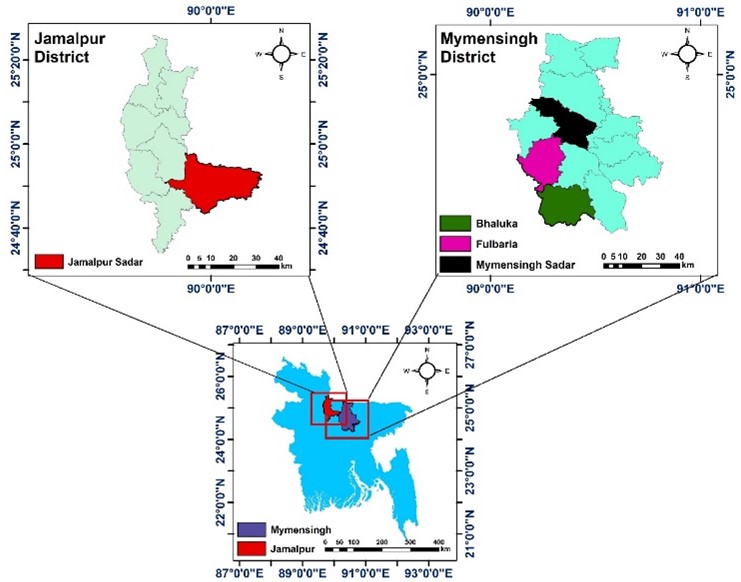
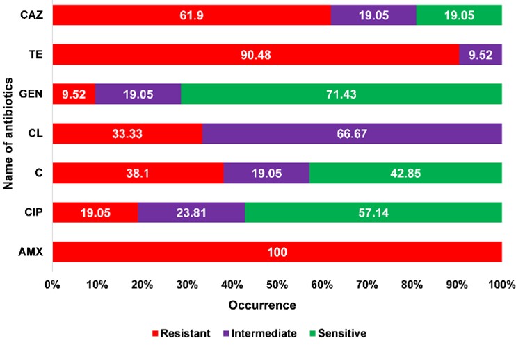
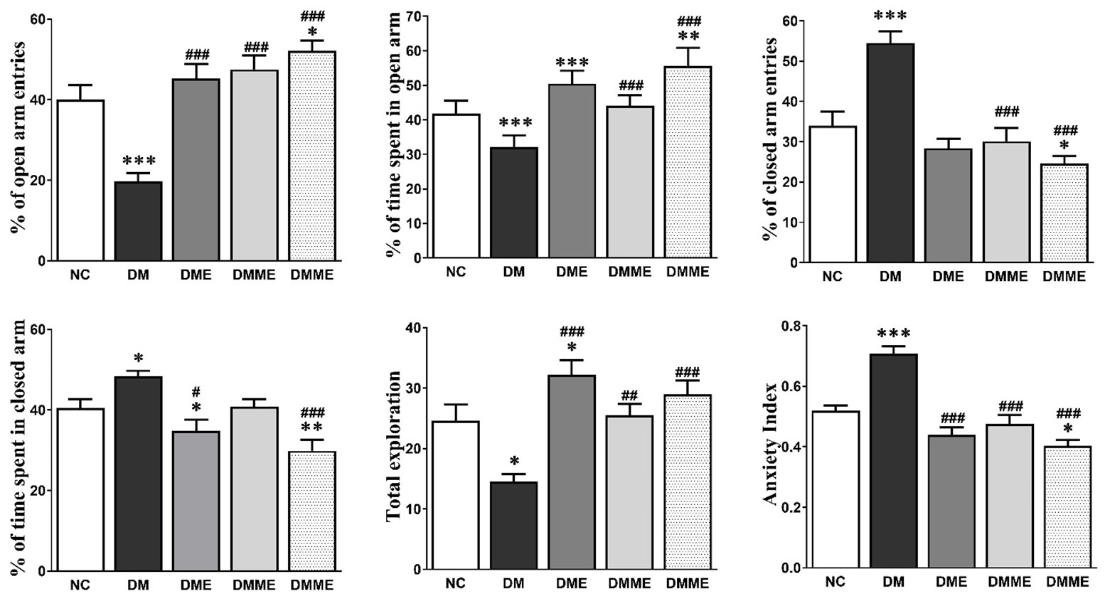
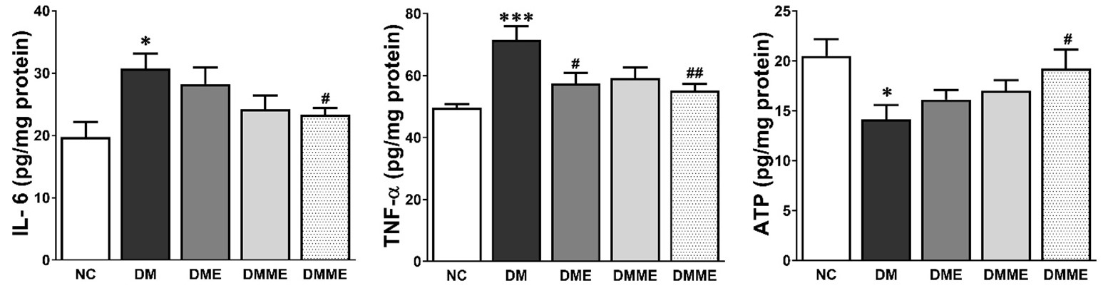
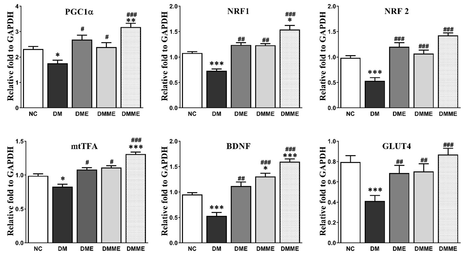
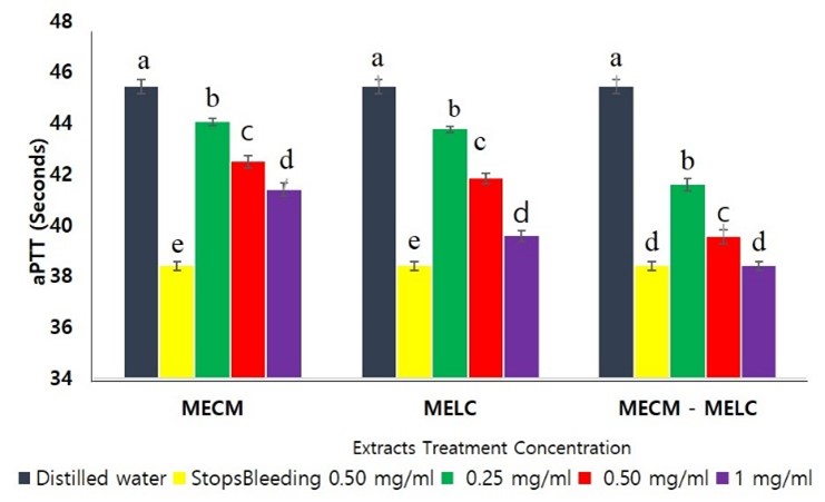
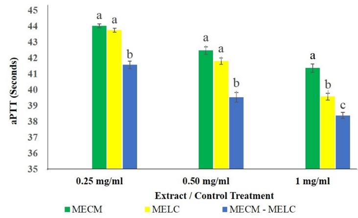
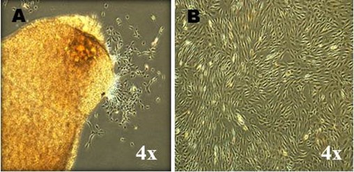
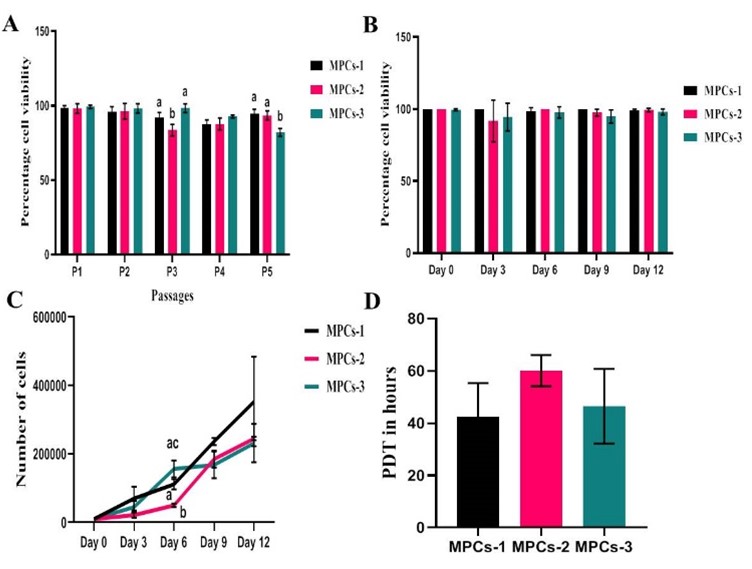
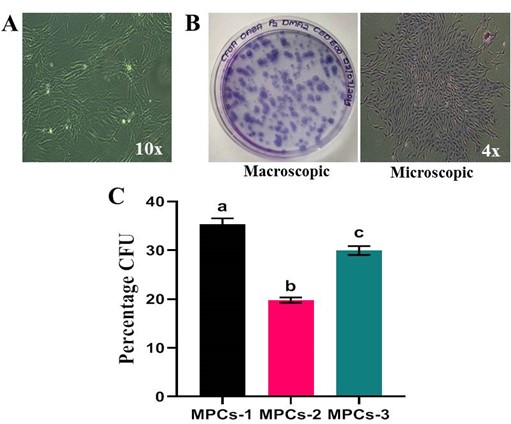
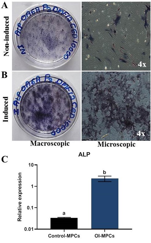
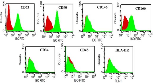
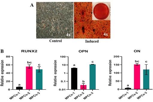
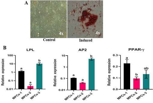
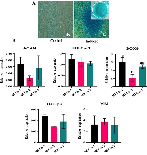
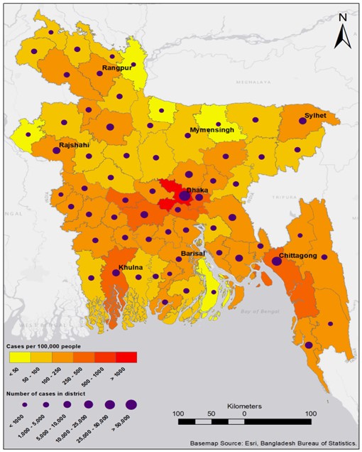
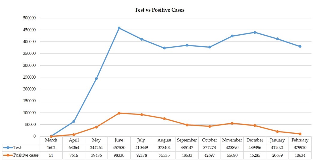
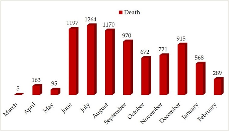
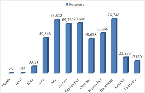
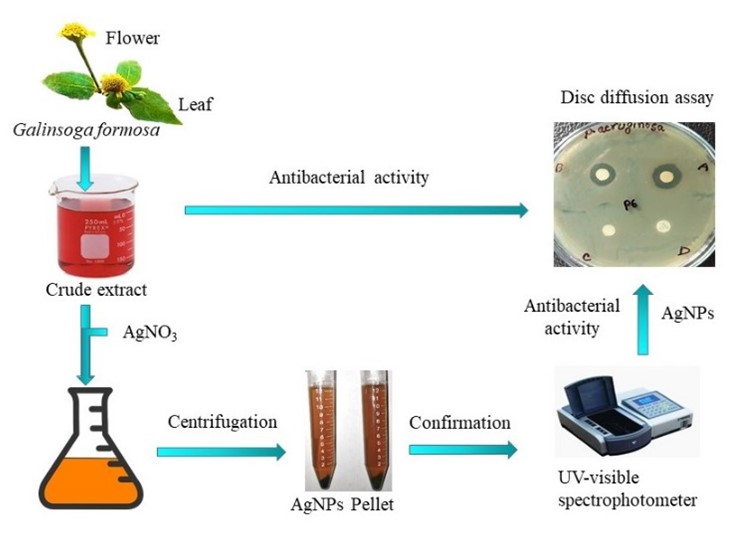
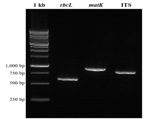
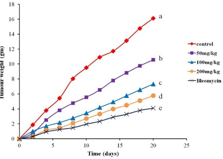
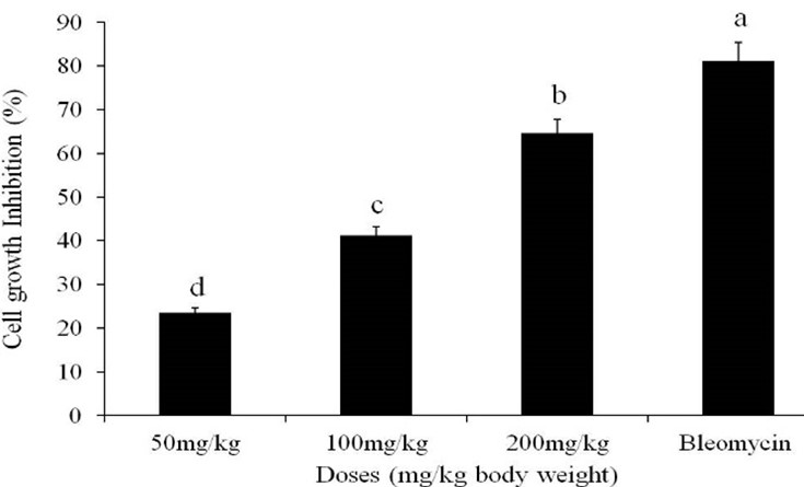
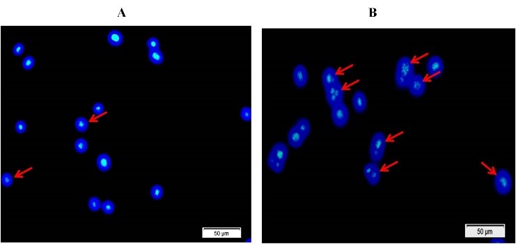
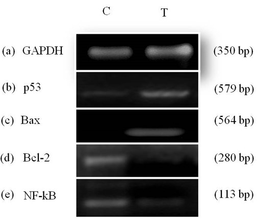
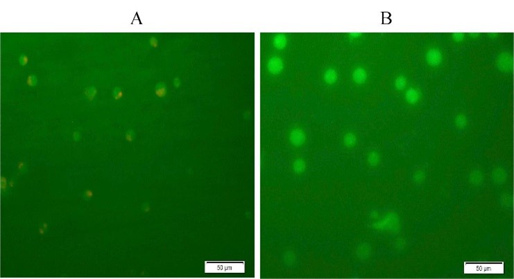
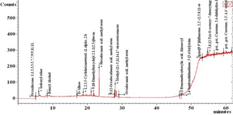