HTTP 200 OK
Allow: GET, HEAD, OPTIONS
Content-Type: application/json
Vary: Accept
{
"count": 321,
"next": "https://jabet.bsmiab.org/articles/?format=api&page=21",
"previous": "https://jabet.bsmiab.org/articles/?format=api&page=19",
"results": [
{
"id": 39,
"slug": "178-1628892758-molecular-and-immunological-activity-of-terminalia-chebula-extracts",
"featured": false,
"slider": false,
"issue": "Vol5 Issue1",
"type": "original_article",
"manuscript_id": "178-1628892758",
"recieved": "2021-08-13",
"revised": null,
"accepted": "2021-10-29",
"published": "2021-11-09",
"pdf_file": "https://jabet.bsmiab.org/media/pdf_file/2023/36/178-1628892758.pdf",
"title": "Molecular and immunological activity of Terminalia chebula extracts",
"abstract": "<p>Several plant extracts and Ayurvedic formulations were used to treat ailments and such studies were well documented within the recent decades. Even a number of the plants were screened for immunomodulators to revive and rejuvenate the system. The present study is a screening trail for any possible healing activity of <em>Terminalia chebula</em> on IL-2 and IFN-γ levels. The raw and dried fruits of the sample were pulverized finely and extracted with methanol. Following which their aqueous solutions are re extracted with hexane, ester and chloroform to review the possible cytotoxic effects. Lipopolysaccharide (LPS)-stimulated macrophage cells were used throughout the study to assess the effect of extracts on nitric oxide (NO) production using Griess method. Expression of Cyclooxygenase-2 (COX2) and tumor necrosis factor (TNF)-α were studied by real time PCR quantification alongside estimation of IL-1β and IL-6 cytokine levels using the enzyme-linked immunosorbent assay (ELISA). The present study confirmed the positive effect of the chloroform extract in reducing the NO secretion and also by showing an inhibition within the expression of COX2, IL-1β, IL-6, and TNF-α. Thus, to conclude <em>T. chebula</em> might be used as anti-inflammatory candidate drug and also be used additionally to the various chemical compounds available within the medical markets.</p>",
"journal_reference": "J Adv Biotechnol Exp Ther. 2022; 5(1): 176-188.",
"academic_editor": "",
"cite_info": "Al-Mousawi HTM, Mushtaq ABN, Bohan AH. Molecular and immunological activity of Terminalia chebula extracts. J Adv Biotechnol Exp Ther. 2022; 5(1): 176-188.",
"keywords": [
"Real time PCR",
"COX2",
"Terminalia",
"IL-6",
"Antiinflammation"
],
"DOI": "10.5455/jabet.2022.d106",
"sections": [
{
"section_number": 1,
"section_title": "INTRODUCTION",
"body": "<p>Immunomodulation is an adaptive terminology of the body to suppress its extra aggressive immune responses. During the immune reaction, several immune molecules get activated and this processes many free radicals like reactive oxygen and reactive nitrogen species are generated [<a href=\"#r-1\">1</a>]. Gas nitric oxide (NO) which may be by product of reactive nitrogen species produced inducible nitric oxide synthase (iNOS) and superoxide (O2−) are released by pyrogen-activated macrophages and activated immune cells respectively increasing the cellular toxicity and oxidative stress within the body [<a href=\"#r-2\">2</a>]. Such an imbalance between the radical molecules and therefore the anti-oxidative system results in accumulation of free radicals which eventually results in inflammation. Such an outsized number of free radicals inducing oxidative stress results in structural changes within the biomolecules causing mutagenesis [<a href=\"#r-3\">3</a>]. This stress was also found to assist in developing chronic diseases like pulmonary and cardiovascular diseases including cancer [<a href=\"#r-4\">4</a>]. Thus, studies are entirely focussed of the way to immunosuppress the system and to extend the activity of the anti-oxidative systems to scale back the inflammatory activities. Many natural compounds which are derived from traditional botanical plants and herbs are found to be rich in anti-oxidative substances which are studied and being studied to scavenge the free radicals.<br />\r\n<em>Tithonia diversifolia</em>, member of Asteraceae that able to grow commonly in several parts of Central and South America and Asian countries [<a href=\"#r-5\">5</a>] was utilized in healing dermatitis, fever, arthritis and lots of pathogen infections [<a href=\"#r-6\">6</a>]. Chronic inflammation is taken into account a serious threat which progresses into cancer because the inflammatory molecules generated during this process creates a hostile environment for the tumor and aids in metastasis. In many instances, such an environment is claimed to be the main predisposing factor for viral mediated tumorigenesis. On entry of the pathogen into the body, the innate system gets activated recruiting granulocytes to the injured site, producing inflammatory mediators like TNF-α, IL-1β and IL-6 and lots of lipid mediators like Prostaglandins (PGs) and leukotrienes (LTs) [<a href=\"#r-7\">7</a>].<br />\r\nMediators invoke an acute inflammatory process within hours to clear away the infection from the damaged tissues [<a href=\"#r-8\">8</a>]. In due course, such an acute inflammation gets resolved when all of the pathogens and damaged tissue debris are cleared far away from the damaged site. But sometimes, these acute cases become chronic within weeks to months or sometimes years, resulting in autoimmune, neurodegenerative and vascular diseases [8]. Interleukin-2 (IL-2) was considered as a cytokine liable for the proliferation and differentiation of effector T cells and aids in cancer and a number of other infectious diseases. IL-2 is employed in passive therapy on the patients with melanoma or renal cell carcinoma which has been approved by the US Food and Drug Administration [<a href=\"#r-9\">9</a>]. IL-2 is that the first line of defensive molecule which aids in treating autoimmunity and intrinsically considered as potent therapeutic agent [<a href=\"#r-10\">10</a>]. The cytokines primarily produced by the induced T cells and dendritic cells (DCs) [<a href=\"#r-11\">11</a>] but sometimes differential expression of IL-2 receptors also results in selective IL-2-driven expansion of Tregs which increases the affinity for IL-2 receptor alpha which pairs with the IL-2 receptor beta [<a href=\"#r-12\">12</a>]. Current studies confirmed that under expression of IL-2 will resolve the inflammation by attracting and expanding the immune regulatory cells [<a href=\"#r-13\">13</a>]. Cyclooxygenase 2 (COX-2) is one among such inflammatory mediators which is seen to be over expressed during an immune reaction against the pathogens. COX, also referred to as prostaglandin (PG) H synthase, catalyses the primary committed step within the synthesis of prostanoids, an outsized family of arachidonic acid metabolites comprising PGs, prostacyclin, and thromboxanes [<a href=\"#r-14\">14</a>]. COX-2 is rapidly expressed in several cell types in response to growth factors, cytokines, and pro-inflammatory molecules and has emerged because the isoform primarily liable for prostanoid production in acute and chronic inflammatory conditions [<a href=\"#r-15\">15</a>]. COX-2 is additionally a molecule which metabolizes the accumulated PGE2.<br />\r\nThese results in up-regulation of several signalling pathways and down-regulation of apoptotic proteins which helps in physiological processes like proliferation, angiogenesis and metastasis [<a href=\"#r-16\">16</a>]. When COX-2 is seen to urge over expressed, inflammation increases within the body resulting in reduction in apoptosis and metastasis which finally develops into cancer. COX-2 was said to make an immunosuppressive tumor environment where the pro-inflammatory eicosanoids, cytokines, chemokines and carcinoma cells all aids in forming an immunosuppressive environment. It also results in down-regulation of TH1 cytokines like TNF-α, IFNγ, IL-2, IL-12, and also said to upregulate the TH2 cytokines like IL-4, IL-10 which are proven immunosuppressive agents [<a href=\"#r-17\">17</a>]. Many traditional herbs were screened for their anti-inflammatory nature. <em>Terminalia chebula</em> was found to exhibit a huge number of medicinal properties due to their phyto-constituents. Its fruit is claimed to possess vast number of health benefits and is employed since ages for several and human ailments. <em>T. chebula</em> was also quoted extensively in Ayurveda, Unani and Homoeopathic literatures [<a href=\"#r-18\">18</a>]. Its aqueous extract was found to contain anti-inflammatory properties inhibiting the inducible gas synthesis [<a href=\"#r-19\">19</a>]. Even studies done thus far, proved that the Chebulagic acid present within these fruits could suppress the onset and progression of arthritis within mice. <em>T. chebula</em> was also found to be a potent anti-inflammatory agent and is employed in Freund’s adjuvant [<a href=\"#r-20\">20</a>].<br />\r\nThus, this study was designed to review the effect of the fruit extracts as anti-inflammatory agents. <em>T. chebula</em> was studied for its anti-oxidative stress-related immunomodulation properties. But none of the studies were focused on the inflammatory mediators like TNF-α and IL-6. The study was planned to screen the expression of the inflammatory mediators like TNF-α, IL-6, COX2 by real time amplification and ELISA methods. LPS induced (J774.1A) macrophage cell lines were utilized in the study.</p>"
},
{
"section_number": 2,
"section_title": "MATERIALS AND METHODS",
"body": "<p><strong>Materials</strong><br />\r\nELISA kits for IL1β and IL6 were purchased from Everone Biosciences. All the reagents utilized in the study were of molecular grade and were purchased from HiMedia Deionized water was produced in-house employing a Milli-Q System from Millipore. All the primers were ordered from Sigma Aldrich.</p>\r\n\r\n<p> </p>\r\n\r\n<p><strong>Cell culture</strong><br />\r\nMurine macrophage cell line (J774.1A) was donated from Stellixir Ltd, Bangalore, and was cultured in Dulbecco’s modified eagle medium (DMEM) containing 2mM L-glutamine, 100U penicillin/mL, 0.1mg streptomycin/mL, and 10% heat-inactivated FBS (HiMedia) and incubated during a humidified CO2 incubator. The cells were maintained until confluence and were passaged until further use.</p>\r\n\r\n<p> </p>\r\n\r\n<p><strong>Extract preparation</strong><br />\r\nRaw fruits and dried ripened fruits of <em>T. chebula</em> were collected from the nearby farms, Bangalore. The samples each of 500gm were extracted with 1000mL methanol for 3 days using soxhlet extraction (45°C). The extract obtained was then concentrated during a rotary evaporator and lyophilized. About 5gm of this this extract was suspended in water then partitioned with n-hexane, ester and chloroform extracts. The extracts obtained were then concentrated during a rotary evaporator and therefore the residues obtained were stored at -20 °C until further use. The yields obtained were 16.89, 18.21 and 19.26% (w/w) for n-hexane, ester and chloroform extracts respectively. Each extract was then weighed and suspended in 5% sterile dimethyl sulfoxide (DMSO) to offer a 20mg/mL solution [<a href=\"#r-21\">21</a>].</p>\r\n\r\n<p> </p>\r\n\r\n<p><strong>Cell viability assay</strong><br />\r\nThe effect of the obtained extracts on the viability of the cells were determined by 3-(4,5-dimethylthiazol-2-yl)-2,5-diphenyl-tetrazolium bromide (MTT) assay. About 7500-9000 cells per well were incubated with 1-200μg/mL of the extracts for 24hr at 37°C during a humidified CO2 incubator. All the tests were wiped out triplicates. Wells containing DMSO because the solvent was considered as negative control. 10μL of MTT (5mg/mL) was added to all or any the wells and therefore the plate was incubated for 4hr at 37°C. Following incubation, the plates were removed and added with lysis buffer (0.1N HCl in isopropanol) to dissolve the formazan produced from the treated cells. Absorbance was measured with a microplate reader (Genetix, Germany) at 570nm. Absorbance of the negative control wells was considered as 100% viability [<a href=\"#r-22\">22</a>]. The Results were expressed as percentages and was calculated using the subsequent equation: [%viability =(ODsample-ODBlank)/ (ODcontrol-ODBlank) *100].</p>\r\n\r\n<p> </p>\r\n\r\n<p><strong>Quantification of nitric oxide production</strong><br />\r\nNitric oxide production was assayed according to the protocol prescribed by [<a href=\"#r-23\">23</a>]. In brief, the cell lines at a concentration of 1×10<sup>6</sup>/100μl were seeded into 96-well plates. The seeded cells were then treated with 1μL lipopolysaccharide (LPS, HiMedia; 1μg/ml) and extracts at varying concentrations of 6.25-200μg/ml for 24hr. All the tests were done in triplicates and the cells treated with LPS and 0.1% DMSO were considered as positive control and negative control, respectively. NG-nitro-L-arginine-methyl ester (L-NAME, SIGMA; 1mM/mL) was used as an iNOS inhibitor in the study.<br />\r\nFollowing 24hr incubation, the supernatants were collected from the cultures and assayed for the production of NO with Griess reagent. The supernatant was incubated with the reagent for 15min at room temperature and the absorbance was read at 550nm in a plate reader (Genetix). The concentration of nitrate and the % of NO inhibition along with IC<sub>50</sub> was calculated by sodium nitrite standard curve. Positive control was considered 100%.</p>\r\n\r\n<p> </p>\r\n\r\n<p><strong>Cytokine assay</strong><br />\r\nThe cytokines like IL1β and TNFα which would be released by the cell lines were measured using the ELISA kit (Everone Biosciences). The protocol was followed according to the manufacturer’s instructions. The supernatant of cells cultured (control and treatment) was spine down at 8000rpm, 10min and stored at −20°C until further use. The cells without stimulation with LPS was used as negative control. The day before the assay, the 96well plate was impregnated with the capture antibody against the specific molecules. The samples were added in their respective wells and done in triplicates.<br />\r\nThe plate was incubated for about 2hours and following washing with secondary antibody conjugated with HRP. H<sub>2</sub>O<sub>2</sub>-tetramethylbenzidine was sued as the substrate and after the incubation, the reaction was stopped using 2N H<sub>2</sub>SO<sub>4</sub> and the absorbance was read at 405nm in a plate reader (Genetix). The concentration of the study proteins released into the media was estimated using the standard graph.</p>\r\n\r\n<p> </p>\r\n\r\n<p><strong>RNA extraction</strong><br />\r\nSince the chloroform extract showed maximum rate of reduction, it was considered for the expression studies. The cells (treated and control) with confluency were used for the RNA extraction [<a href=\"#r-24\">24</a>] using RNeasy Mini Kit (Qiagen74104). The test was done according to the manual instructions. Following trypsinization, the cells were spin down at 6000rpm for 5min and resuspended in about 560μl of AVL buffer and 560μl chilled ethanol. After thorough mixing, the contents were centrifuged at 8000rpm for about 1min and the column was then washed with 700μl of wash buffer and then incubated with 60μl of AVE buffer. The RNA was then eluted and stored at −20°C. The RNA was analyzed qualitatively using UV spectrophotometer (260/280) and used in the cDNA synthesis.</p>\r\n\r\n<p> </p>\r\n\r\n<p><strong>cDNA synthesis</strong><br />\r\ncDNA synthesis was carried according to the manual instructions from RT PCR kit using SuperScript TMII Reverse Transcriptase, 200U/μl (HiMedia). About 1.22μl (concentration obtained was 1.97μg/μl) of the RNA was used in the reaction. Random primers and 1μl of RT enzyme were added and incubated at 25°C for 10min. Following incubation at 70°C for 45 min, the cDNA was then stored and used in the real-time PCR assay.</p>\r\n\r\n<p> </p>\r\n\r\n<p><strong>Real-time PCR</strong><br />\r\nPrimers used in the real time assay (<a href=\"#Table-1\">Table 1</a>) were designed using primer3 software and were purchased from Sigma-Aldrich. The real-time amplification was carried according to [<a href=\"#r-25\">25</a>] using the iQTM SYBR Green Supermix (HiMedia). The primers (600nM) and 1μl of the RT products were used and the reaction was carried in a total volume of 12.5μl. All the reactions were done in duplicates along with their respective negative controls.</p>\r\n\r\n<div id=\"Table-1\">\r\n<p><a href=\"https://jabet.bsmiab.org/table/178-1628892758-table1/\">Table-1</a><strong>Table 1.</strong> List of the primers used in the real time PCR.</p>\r\n\r\n<p> </p>\r\n</div>\r\n\r\n<p><strong>Expression of COX-2 </strong>and<strong> TNF-</strong><strong>α</strong><strong> members</strong><br />\r\nQuantitative PCR was done on the samples (Control and treatment) in the Corbett Research cycler (Bio-Rad). The COX-2 (forward: 5’ GTACTCCCGATTGAAGCCCC 3’ and reverse: 5’ TCGTGTAGCGGTGAAAGTGG 3’; 138bp) and TNF- α (forward: 5’ GGGTGTGAGAAGAGAGATGGG 3’ and reverse: 5’ GGCCAGAGGGCTGATTAGAG 3’; 342bp) of about 600nM was used in the amplification study. 1.22μl of the RNA products were used, and the program was run for about 40 cycles at 93°C for 60s, 62°C for 40s, and with an elongation at 72°C for 60s. The housekeeping gene glyceraldehyde-3-phosphate dehydrogenase (GAPDH) (forward: 5’ GCTGAGTACGTCGTGGAGTC 3’ and reverse: 5’ CCCATTCCCCAGCTCTCATA 3’; 455 bp) was also amplified along with the gene members for a comparative analysis of the mRNA expression. The comparative analysis was done using Δ<sup>Ct</sup> method and the Ct values obtained for each sample were normalized to the housekeeping gene (GAPDH).</p>\r\n\r\n<p> </p>\r\n\r\n<p><strong>Statistical analysis</strong><br />\r\nThroughout the study the data was analysed using a one-way ANOVA and using SPSS software (USA). All the values were expressed as means ± SD of at least three independent experiments and the significance level was maintained at <em>P </em>< 0.05.</p>"
},
{
"section_number": 3,
"section_title": "RESULTS",
"body": "<p><strong>Effect of extract on cell viability assay</strong><br />\r\nMTT colorimetric assay was done to assay the possible cytotoxic effect of varying concentrations (6.25to 100.00μg/ml) of the extracts on the growth of J774.1A cells. Extracts of n-hexane, ethyl acetate and chloroform showed no significant cytotoxicity on the cell lines even at a concentration of 100μg/ml. None of the extracts showed any reducing effect on the cell lines.</p>\r\n\r\n<div id=\"figure1\">\r\n<figure class=\"image\"><img alt=\"\" height=\"255\" src=\"/media/article_images/2023/19/02/178-1628892758-Figure1.jpg\" width=\"500\" />\r\n<figcaption><strong>Figure 1. </strong>Cytotoxicity assay of <em>T. chebula</em> extracts on J774.1A cells. Cell viability was determined by MTT method. Negative control was considered 100% viable. The values were expressed as mean ±S.D and were performed in triplicates (p<0.05).</figcaption>\r\n</figure>\r\n\r\n<p> </p>\r\n</div>\r\n\r\n<p><strong>Reduction in NO production</strong><br />\r\nThe cell lines were treated with LPS, extracts and inhibitors to study the effect on NO production. Cells added with LPS stimulant showed a significant increase in the NO production to 87.23 ± 2.1μmol/L (p<0.005). Negative control without LPS treatment showed 15.67± 1.1μmol/L. The cells treated with inhibitor obviously showed a reduction in the NO production to 16.94± 2.9μmol/L (<em>P </em>< 0.005).<br />\r\nChloroform extract showed significant reduction when compared to ethyl acetate and n-hexane. Chloroform extract showed significant reduction from 89.83± 1.6 μmol/L to 18.31± 1.6μmol/L. Hexane extract showed a reduction from 92.16 ± 3.5% at 6.25μg/ml to 22.3 ± 10.1% at 100μg/ml (<a href=\"#figure2\">Figure 2B)</a>. Ethyl acetate on the other hand also showed reduction but was significantly lower when compared to the other two extracts (32.4 ± 3.9% from 96.11± 2.3% at 100μg/ml, p<0.005).</p>\r\n\r\n<div id=\"figure2\">\r\n<figure class=\"image\"><img alt=\"\" height=\"323\" src=\"/media/article_images/2023/19/02/178-1628892758-Figure2.jpg\" width=\"500\" />\r\n<figcaption><strong>Figure 2. </strong>Effect of <em>T. chebula</em> extracts on LPS stimulated nitric oxide production from J774.1A cells. C-: Negative control (absence of LPS stimulus); C+: positive control (LPS treatment); inhibitor: L-NAME, NG-nitro-L-arginine-methyl ester; NO, nitric oxide. All the tests were done in triplicates and expressed as percent ± SD. (*<em>P</em> < 0.05). A: Hexane treatment; B: Ethylacetate; C: Chloroform of <em>T. chebula</em> extracts.</figcaption>\r\n</figure>\r\n\r\n<p> </p>\r\n</div>\r\n\r\n<p><strong>Effect of extract on </strong><strong>cytokine assay</strong><br />\r\nThis ELISA method was used in estimating the expression of the IL-1β and IL-6 cytokines on treatment with the hexane and chloroform extracts only (ethyl acetate was excluded from the study). From the figure it was found that LPS stimulation has significantly increased the levels of IL-1β levels (47.84 ± 1.53pg/ml) when compared to the negative control (21.54 ± 2.11pg/ml), and even IL-6 levels were also increased to 894±17.01pg/ml from 11.3 ± 2.54pg/ml (negative control). Chloroform extract showed significantly higher reduction of the IL-1β and IL-6 levels when compared to the hexane extract.<br />\r\nChloroform extract with varying concentrations (6.25-100μg/ml) significantly reduced IL-1β levels to 44.22 ± 0.31pg/ml (6.25μg/ml), 40.31 ± 1.48pg/ml (12.5μg/ml), 32.9 ± 0.21pg/m (25μg/mL), 28.75 ± 1.48pg/ml (50μg/mL), and 21.23 ± 0.21pg/ml (100μg/mL); <em>P </em>< 0.005. Hexane extract also showed reduction in the levels with varying concentrations (6.25-100μg/ml) to 46.12 ± 0.31pg/ml (6.25μg/ml), 42.82 ± 2.48pg/ml (12.5μg/ml), 39.45 ± 1.21pg/ml (25μg/mL), 32.45 ± 1.48pg/ml (50μg/mL), and 30.83 ± 2.21pg/ml (100μg/mL); <em>P </em>< 0.005. Similar results were seen in IL-6 secretion also and chloroform extract showed significantly lower levels of 45.67± 3.31pg/ml (100μg/ml) and 789.67± 1.39pg/ml (6.25μg/ml). Hexane showed reduction in IL-6 levels to 234.54± 1.77pg/ml (100μg/ml) and 821.91± 3.12pg/ml (6.25μg/ml).</p>\r\n\r\n<div id=\"figure3\">\r\n<figure class=\"image\"><img alt=\"\" height=\"563\" src=\"/media/article_images/2023/19/02/178-1628892758-Figure3.jpg\" width=\"500\" />\r\n<figcaption><strong>Figure 3. </strong>Graph showing the effects of the extracts on LPS stimulated cytokine levels. Cells are treated with 6.25-100μg/mL of the chloroform and hexane extracts on stimulus with LPS for 24hr. A: IL-1β; B: IL-6 cytokine levels. Values are expressed as pg/ml ± SD. All the values were average of triplicates (<em>P</em> < 0.05).</figcaption>\r\n</figure>\r\n\r\n<p> </p>\r\n</div>\r\n\r\n<p><strong>Expression of genes</strong><br />\r\nThe results obtained were normalized to GAPDH for comparative expression study. The mRNA expression was studied separately for each gene member (GAPDH, TNF- α and COX-2) was studied separately. The sample with the lowest ΔΔ<sup>Ct</sup> value was used as calibrator, and the remaining members were compared in relation to calibrator.<br />\r\nThe Ct values of the GAPDH, TNF- α and COX-2 were found to be 11, 21, and 22 respectively for the control. And the Ct values for the GAPDH, TNF- α and COX-2 after treatment was found to be 11, 30 and 33, respectively.<br />\r\nFrom the Ct values, (<a href=\"#figure4\">Figure 4</a>) after normalizing with the GAPDH, it was observed that the relative expression of both the gene members were downregulated in treatment samples when compared to the control. The GAPDH gene expression was considered as 100%.<br />\r\nFrom the graph, (<a href=\"#figure5\">Figure 5</a>) and the calculated 2<sup>−ΔΔCt </sup>values, it was found that the relative expression of the COX-2 and TNF-<strong> α</strong> was downregulated by 12 and 7 times, respectively when compared to their respective controls.</p>\r\n\r\n<div id=\"figure4\">\r\n<figure class=\"image\"><img alt=\"\" height=\"534\" src=\"/media/article_images/2023/19/02/178-1628892758-Figure4.jpg\" width=\"500\" />\r\n<figcaption><strong>Figure 4. </strong>Real time curves for the GAPDH, TNF- α and COX-2. A: GAPDH; B: TNF- α and COX-2. Ct values are average of duplicates.</figcaption>\r\n</figure>\r\n</div>\r\n\r\n<div id=\"figure5\">\r\n<figure class=\"image\"><img alt=\"\" height=\"196\" src=\"/media/article_images/2023/19/02/178-1628892758-Figure5.jpg\" width=\"393\" />\r\n<figcaption><strong>Figure 5. </strong>Graph showing the expression levels of GAPDH, TNF- α and COX-2 on treatment with the chloroform extracts. Negative control was without stimulus and not shown in the graph. GAPDH was used as calibrator. Its expression was considered as 100%. (<em>P</em><0.005).</figcaption>\r\n</figure>\r\n\r\n<p> </p>\r\n</div>"
},
{
"section_number": 4,
"section_title": "DISCUSSION",
"body": "<p>Macrophages are considered very critical in case of inflammation due to their characteristic antigen presenting nature. Moreover, they are good at phagocytosis and other immunomodulation properties. The main role of these macrophages is to enhance the production of inflammatory molecules and mediators like cytokines [<a href=\"#r-26\">26</a>]. During this crucial time, host tries to suppress the activity of these cytokines to minimise the damage caused to the tissues. Many previous studies reported that several medicinal plants are employed to heal the inflammatory process [<a href=\"#r-27\">27</a>]. <em>E. amoenum</em>is studied for its anti-inflammatory and analgesic properties and as such commonly used as folk medicine in Iran to cure the common cold [<a href=\"#r-28\">28</a>]. In the present study we studied the effects of <em>T. chebula</em> as anti-inflammatory on stimulation with LPS.<br />\r\nWe first studied the extracts for their healing effects on J774.1A macrophage cell lines viability to nullify the cytotoxic effects, followed by estimating the inhibition of NO production. NO is said to be crucial mediator in regulating the inflammation process [<a href=\"#r-29\">29</a>] as such its effects were studied. In the current study, the percent of cytotoxicity as shown by MTT assay determines the safety of using the sample material. None of the extracts (hexane, ethyl acetate and chloroform) showed any cytotoxicity on the cell lines. The cytotoxicity was significantly less even at the highest concentration of 100μg/ml.<br />\r\nIn this study, <em>T. chebula</em> extract showed significant inhibition on the NO production in a dose dependent manner. Most of the studies reported so far suggest that the extracts which are significantly good at lowering the No production are good anti-inflammatory agents [<a href=\"#r-30\">30</a>]. From the results, it was proved that chloroform extract (18.31± 1.6μmol/L) was significantly good at reducing maximum NO produced followed by hexane (22.3 ± 10.1 μmol/L) at highest concentration of 100μg/ml. The inhibition activity was quite promising when compared to the inhibitor reduction activity (16.94± 2.9μmol/L). This reduction might be either due to the inhibition of iNOS activity or at the level of locking the transcription signalling methods. As such the study was planned in the method specified by [<a href=\"#r-31\">31</a>].<br />\r\nThe present study so far concluded the possible evaluation on the inhibition of NO by the extracts. Additionally, there are several studies reporting the same NO reducing activities. <em>Eucalyptus globules </em>and <em>Thymus vulgaris </em>[<a href=\"#r-26\">26</a>] were studied for their capability in reducing the NO production among the LPS induced J774.1A cells. Even studies done on <em>Andrograp hispaniculata </em>[<a href=\"#r-32\">32</a>] and <em>Echinacea </em>[<a href=\"#r-29\">29</a>] also confirmed the possible inhibitory role of the extracts in NO production and iNOS activity among the LPS-stimulated macrophages. <em>Mentha longifolia </em>also was concluded that it is a good anti-inflammatory agent on regulating the production of NO among the LPS-stimulated J774.1A cells [<a href=\"#r-27\">27</a>].<br />\r\nCOX2 is also said to play a crucial role in inflammation [<a href=\"#r-33\">33</a>] which converts arachidonic acid to prostaglandins. Moreover, NSAIDs are used to suppress the inflammatory process by inhibiting the COX enzyme [<a href=\"#r-34\">34</a>]. Studies done on the hexane extract of <em>E. amoenum </em>proved that they are potent anti-inflammatory by inhibiting the COX2 gene expression among the J774.1A macrophage cell lines [<a href=\"#r-3\">3</a>].<br />\r\nOur study confirmed that the extracts strongly showed a reduction in the COX2 and TNF-α gene expressions. TNF-α and IL-1β were said to cytokines mostly involved in inflammation, where TNF-α produced by the macrophages takes part in initiating the acute phase of inflammation. This mode of action is done by attracting the neutrophils to the target site.<br />\r\nIL-1β also aids in bringing an inflammatory response on infection with a pathogen. Many chemical moieties were used as inhibitors to reduce the effect of IL-1β so as to relieve the inflammatory arthritis among the experimental organisms [<a href=\"#r-11\">11</a>]. IL-6 is also said to be proinflammatory in nature and takes part in the generation of inflammation [<a href=\"#r-35\">35</a>]. Our study confirmed that, chloroform extract showed a significantly downregulation of these two gene members (COX2 and TNF-α).<br />\r\nThe real time expression studies done on the cell lines on induction with LPS confirmed the possible role of the extracts (chloroform) in downregulating the expression of inflammatory chemokines. COX2 and TNF-α were downregulated by 12 and 7 times when compared to the control. Even studies done on several medicinal plants <em>Mentha longifolia </em>[<a href=\"#r-27\">27</a>] and <em>Echinacea </em>[<a href=\"#r-29\">29</a>] showed that they could reduce the expression of TNF-α, IL-6, and IL-1β proteins at the mRNA level among the LPS induced macrophages. As major reduction effects were observed with chloroform extracts on the cell lines, further we need to plan to isolate and purify the phytochemicals from the extract and study the same at pinpoint level.</p>"
},
{
"section_number": 5,
"section_title": "CONCLUSION",
"body": "<p>The plant material <em>T. chebula</em> and its extracts were studied for the possible effects on the macrophages as anti-inflammatory agents. Among them chloroform extract showed significant reduction of NO production followed by the hexane extract. Chloroform extract was found to be more effective in reducing the levels of IL-1B and Il-6 gene members. Real time PCR analysis also confirmed that the chloroform extract was significantly down regulating the expression of COX2 and TNF-α levels when compared to control. The findings in the current study reveal that the plant material can act as strong anti-inflammatory agent as confirmed on the LPS induced macrophages. Further if the study could isolate and purify the extract into its constituent photochemical, then the mode of action could be traced to the pinpoint level. Our study confirms the possible potent role of <em>T. chebula</em> as strong anti-inflammatory agent.</p>"
},
{
"section_number": 6,
"section_title": "ACKNOWLEDGEMENT",
"body": "<p>None.</p>"
},
{
"section_number": 7,
"section_title": "AUTHOR CONTRIBUTIONS",
"body": "<p>HA, select the scope of the study, and AB and AM, supervised the lab work. All authors wrote and read the manuscript and approved it.</p>"
},
{
"section_number": 8,
"section_title": "CONFLICTS OF INTEREST",
"body": "<p>There is no conflict of interest among the authors.</p>"
}
],
"figures": [
{
"figure": "https://jabet.bsmiab.org/media/article_images/2023/19/02/178-1628892758-Figure1.jpg",
"caption": "Figure 1. Cytotoxicity assay of T. chebula extracts on J774.1A cells. Cell viability was determined by MTT method. Negative control was considered 100% viable. The values were expressed as mean ±S.D and were performed in triplicates (p<0.05).",
"featured": false
},
{
"figure": "https://jabet.bsmiab.org/media/article_images/2023/19/02/178-1628892758-Figure2.jpg",
"caption": "Figure 2. Effect of T. chebula extracts on LPS stimulated nitric oxide production from J774.1A cells. C-: Negative control (absence of LPS stimulus); C+: positive control (LPS treatment); inhibitor: L-NAME, NG-nitro-L-arginine-methyl ester; NO, nitric oxide. All the tests were done in triplicates and expressed as percent ± SD. (*P < 0.05). A: Hexane treatment; B: Ethylacetate; C: Chloroform of T. chebula extracts.",
"featured": false
},
{
"figure": "https://jabet.bsmiab.org/media/article_images/2023/19/02/178-1628892758-Figure3.jpg",
"caption": "Figure 3. Graph showing the effects of the extracts on LPS stimulated cytokine levels. Cells are treated with 6.25-100μg/mL of the chloroform and hexane extracts on stimulus with LPS for 24hr. A: IL-1β; B: IL-6 cytokine levels. Values are expressed as pg/ml ± SD. All the values were average of triplicates (P < 0.05).",
"featured": false
},
{
"figure": "https://jabet.bsmiab.org/media/article_images/2023/19/02/178-1628892758-Figure4.jpg",
"caption": "Figure 4. Real time curves for the GAPDH, TNF- α and COX-2. A: GAPDH; B: TNF- α and COX-2. Ct values are average of duplicates.",
"featured": false
},
{
"figure": "https://jabet.bsmiab.org/media/article_images/2023/19/02/178-1628892758-Figure5.jpg",
"caption": "Figure 5. Graph showing the expression levels of GAPDH, TNF-α and COX-2 on treatment with the chloroform extracts. Negative control was without stimulus and not shown in the graph. GAPDH was used as calibrator. Its expression was considered as 100%. (P<0.005).",
"featured": false
}
],
"authors": [
{
"id": 98,
"affiliation": [
{
"affiliation": "Al-Qasim Green University, College of Biotechnology, Babylon Province, Iraq"
}
],
"first_name": "Haider Turky Mousa",
"family_name": "Al-Mousawi",
"email": "almusawi@biotech.uoqasim.edu.iq",
"author_order": 1,
"ORCID": null,
"corresponding": true,
"co_first_author": false,
"co_author": false,
"corresponding_author_info": "Haider Turky Mousa Al-Mousawi, Al-Qasim Green University, College of\r\nBiotechnology, Babylon Province, Iraq, e-mail: \r\nalmusawi@biotech.uoqasim.edu.iq",
"article": 39
},
{
"id": 99,
"affiliation": [
{
"affiliation": "Al-Qasim Green University, College of Biotechnology, Babylon Province, Iraq"
}
],
"first_name": "Al-Bderee Nadhim",
"family_name": "Mushtaq",
"email": "naz1988@biotech.uoqasim.edu.iq",
"author_order": 2,
"ORCID": null,
"corresponding": true,
"co_first_author": false,
"co_author": false,
"corresponding_author_info": "Al-Bderee Nadhim Mushtaq, Al-Qasim Green University, College of\r\nBiotechnology, Babylon Province, Iraq, e-mail: naz1988@biotech.uoqasim.edu.iq",
"article": 39
},
{
"id": 100,
"affiliation": [
{
"affiliation": "Diabetic national centre, Mustansiriyah University, Iraq"
}
],
"first_name": "Alyaa Hasan",
"family_name": "Bohan",
"email": null,
"author_order": 3,
"ORCID": null,
"corresponding": false,
"co_first_author": false,
"co_author": false,
"corresponding_author_info": "",
"article": 39
}
],
"views": 843,
"downloads": 68,
"references": [
{
"id": 807,
"serial_number": 1,
"pmc": null,
"reference": "Schaue D, Kachikwu EL, McBride WH. Cytokines in radiobiological responses: a review. Radiation research, 2012; 178(6), 505-523.",
"DOI": null,
"article": 39
},
{
"id": 808,
"serial_number": 2,
"pmc": null,
"reference": "Lundberg JO, Carlström M, Larsen F J, Weitzberg E. Roles of dietary inorganic nitrate in cardiovascular health and disease. Cardiovascular research, 2011; 89(3), 525-532. 187.",
"DOI": null,
"article": 39
},
{
"id": 809,
"serial_number": 3,
"pmc": null,
"reference": "Jones D P, Sies H. The redox code. Antioxidants & redox signaling, 2015; 23(9), 734-746.",
"DOI": null,
"article": 39
},
{
"id": 810,
"serial_number": 4,
"pmc": null,
"reference": "Penticuff JC, Kyprianou N. Therapeutic challenges in renal cell carcinoma. American journal of clinical and experimental urology, 2015; 3(2), 77.",
"DOI": null,
"article": 39
},
{
"id": 811,
"serial_number": 5,
"pmc": null,
"reference": "Sharrock, R. A., Sinclair, F. L., Gliddon, C., Rao, I. M., Barrios, E., Mustonen, P. J., Godbold, D. L. A global assessment using PCR techniques of mycorrhizal fungal populations colonizing Tithoniadiversifolia. Mycorrhiza, 2004; 14(2), 103-109.",
"DOI": null,
"article": 39
},
{
"id": 812,
"serial_number": 6,
"pmc": null,
"reference": "Maregesi, S. M., Ngassapa, O. D., Pieters, L., &Vlietinck, A. J. Ethnopharmacological survey of the Bunda district, Tanzania: Plants used to treat infectious diseases. Journal of ethnopharmacology, 2007; 113(3), 457-470.",
"DOI": null,
"article": 39
},
{
"id": 813,
"serial_number": 7,
"pmc": null,
"reference": "AL-Saad, N.F., Nadhim, M.H. and Nawar, HY. Serological, Culture and Urea Breath Test for Detection of H. Pylori in Gastric Ulcers Patients. Indian Journal of Forensic Medicine & Toxicology, 2020; 14(4), p.1317.",
"DOI": null,
"article": 39
},
{
"id": 814,
"serial_number": 8,
"pmc": null,
"reference": "Yao, C., &Narumiya, S. Prostaglandin‐cytokine crosstalk in chronic inflammation. British journal of pharmacology, 2019; 176(3), 337-354.",
"DOI": null,
"article": 39
},
{
"id": 815,
"serial_number": 9,
"pmc": null,
"reference": "Zhu, J., Paul, W. E. CD4 T cells: fates, functions, and faults. Blood, The Journal of the American Society of Hematology, 2008; 112(5), 1557-1569.",
"DOI": null,
"article": 39
},
{
"id": 816,
"serial_number": 10,
"pmc": null,
"reference": "Lan, R. Y., Selmi, C., & Gershwin, M. E. The regulatory, inflammatory, and T cell programming roles of interleukin-2 (IL-2). Journal of autoimmunity, 2008; 31(1), 7-12.",
"DOI": null,
"article": 39
},
{
"id": 817,
"serial_number": 11,
"pmc": null,
"reference": "Dayer, J. M. Interleukin 1 or tumor necrosis factor-alpha: which is the real target in rheumatoid arthritis. The Journal of rheumatology Supplement, 2002; 65, 10-15.",
"DOI": null,
"article": 39
},
{
"id": 818,
"serial_number": 12,
"pmc": null,
"reference": "Khan, S. H., Martin, M. D., Starbeck-Miller, G. R., Xue, H. H., Harty, J. T., &Badovinac, V. P. The timing of stimulation and IL-2 signaling regulate secondary CD8 T cell responses. PLoS pathogens, 2015; 11(10), e1005199.",
"DOI": null,
"article": 39
},
{
"id": 819,
"serial_number": 13,
"pmc": null,
"reference": "Granucci, F., Feau, S., Angeli, V., Trottein, F., &Ricciardi-Castagnoli, P. Early IL-2 production by mouse dendritic cells is the result of microbial-induced priming. The Journal of Immunology, 2003; 170(10), 5075-5081.",
"DOI": null,
"article": 39
},
{
"id": 820,
"serial_number": 14,
"pmc": null,
"reference": "Hinz, B., & Brune, K. Cyclooxygenase-2—10 years later. Journal of pharmacology and experimental therapeutics, 2002; 300(2), 367-375.",
"DOI": null,
"article": 39
},
{
"id": 821,
"serial_number": 15,
"pmc": null,
"reference": "Vane, J. R. Inhibition of prostaglandin synthesis as a mechanism of action for aspirin-like drugs. Nature new biology, 1971; 231(25), 232-235.",
"DOI": null,
"article": 39
},
{
"id": 822,
"serial_number": 16,
"pmc": null,
"reference": "Satoh, H., Amagase, K., Ebara, S., Akiba, Y., & Takeuchi, K. Cyclooxygenase (COX)-1 and COX-2 both play an important role in the protection of the duodenal mucosa in cats. Journal of Pharmacology and Experimental Therapeutics, 2013; 344(1), 189-195.",
"DOI": null,
"article": 39
},
{
"id": 823,
"serial_number": 17,
"pmc": null,
"reference": "Stolina, M., Sharma, S., Lin, Y., Dohadwala, M., Gardner, B., Luo, J., .Dubinett, S. M. Specific inhibition of cyclooxygenase 2 restores antitumor reactivity by altering the balance of IL-10 and IL-12 synthesis. The Journal of Immunology, 2000; 164(1), 361-370.",
"DOI": null,
"article": 39
},
{
"id": 824,
"serial_number": 18,
"pmc": null,
"reference": "Bag, A., Bhattacharyya, S. K., & Chattopadhyay, R. R. The development of Terminalia chebula Retz. (Combretaceae) in clinical research. Asian Pacific journal of tropical biomedicine, 2013; 3(3), 244-252.",
"DOI": null,
"article": 39
},
{
"id": 825,
"serial_number": 19,
"pmc": null,
"reference": "Moeslinger, T., Friedl, R., Volf, I., Brunner, M., Koller, E., Spieckermann, P. G. Inhibition of inducible nitric oxide synthesis by the herbal preparation Padma 28 in macrophage cell line. Canadian journal of physiology and pharmacology, 2000; 78(11), 861-866.",
"DOI": null,
"article": 39
},
{
"id": 826,
"serial_number": 20,
"pmc": null,
"reference": "Pratibha, N., Saxena, V. S., Amit, A., D’souza, P., Bagchi, M., &Bagchi, D. Anti-inflammatory activities of Aller-7, a novel polyherbal formulation for allergic rhinitis. International journal of tissue reactions, 2004; 26(1-2), 43-51.",
"DOI": null,
"article": 39
},
{
"id": 827,
"serial_number": 21,
"pmc": null,
"reference": "Saha, S., & Verma, R. J. Antioxidant activity of polyphenolic extract of Terminalia chebulaRetzius fruits. Journal of taibah university for science, 2016; 10(6), 805-812.",
"DOI": null,
"article": 39
},
{
"id": 828,
"serial_number": 22,
"pmc": null,
"reference": "Wang, M., Yang, L., Ji, M., Zhao, P., Sun, P., Bai, R. & Li, C. Aqueous extract of Terminalia chebula induces apoptosis in lung cancer cells via a mechanism involving mitochondria-mediated pathways. Brazilian Archives of Biology and Technology, 2015; 58, 208-215.",
"DOI": null,
"article": 39
},
{
"id": 829,
"serial_number": 23,
"pmc": null,
"reference": "Naseri, N., Kalantar, K., & Amirghofran, Z. Anti-inflammatory activity of Echiumamoenum extract on macrophages mediated by inhibition of inflammatory mediators and cytokines expression. Research in pharmaceutical sciences, 2018; 13(1), 73.",
"DOI": null,
"article": 39
},
{
"id": 830,
"serial_number": 24,
"pmc": null,
"reference": "Abbas, S., & Malla, S. Cytotoxicity and expression studies of angiogenesis-promoting genes in cancer cell lines under the treatment of cancer candidate drugs. Asian J Pharm Clin Res, 2019; 12(5), 130-134.",
"DOI": null,
"article": 39
},
{
"id": 831,
"serial_number": 25,
"pmc": null,
"reference": "Doersch, K. M., DelloStritto, D. J., & Newell-Rogers, M. K. The contribution of interleukin-2 to effective wound healing. Experimental Biology and Medicine, 2017; 242(4), 384-396.",
"DOI": null,
"article": 39
},
{
"id": 832,
"serial_number": 26,
"pmc": null,
"reference": "Vigo, E., Cepeda, A., Perez‐Fernandez, R., & Gualillo, O. In‐vitro anti‐inflammatory effect of Eucalyptus globulus and Thymus vulgaris: nitric oxide inhibition in J774A. 1 murine macrophages. Journal of Pharmacy and Pharmacology, 2004; 56(2), 257-263.",
"DOI": null,
"article": 39
},
{
"id": 833,
"serial_number": 27,
"pmc": null,
"reference": "Karimian, P., Kavoosi, G., &Amirghofran, Z. Anti-inflammatory effect of Menthalongifolia in lipopolysaccharide-stimulated macrophages: reduction of nitric oxide production through inhibition of inducible nitric oxide synthase. Journal of immunotoxicology, 2013; 10(4), 393-400.",
"DOI": null,
"article": 39
},
{
"id": 834,
"serial_number": 28,
"pmc": null,
"reference": "Heidari, M. R., Azad, E. M., & Mehrabani, M. Evaluation of the analgesic effect of EchiumamoenumFisch& CA Mey. extract in mice: possible mechanism involved. Journal of ethnopharmacology, 2006; 103(3), 345-349.",
"DOI": null,
"article": 39
},
{
"id": 835,
"serial_number": 29,
"pmc": null,
"reference": "Zhai, Z., Solco, A., Wu, L., Wurtele, E. S., Kohut, M. L., Murphy, P. A., & Cunnick, J. E. Echinacea increases arginase activity and has anti-inflammatory properties in RAW 264.7 macrophage cells, indicative of alternative macrophage activation. Journal of ethnopharmacology, 2009; 122(1), 76-85.",
"DOI": null,
"article": 39
},
{
"id": 836,
"serial_number": 30,
"pmc": null,
"reference": "Stangeland, T., Alele, P. E., Katuura, E., & Lye, K. A. Plants used to treat malaria in Nyakayojo sub-county, western Uganda. Journal of ethnopharmacology, 2011; 137(1), 154-166.",
"DOI": null,
"article": 39
},
{
"id": 837,
"serial_number": 31,
"pmc": null,
"reference": "Booke, M., Hinder, F., McGuire, R., Traber, L. D., &Traber, D. L. Selective inhibition of inducible nitric oxide synthase: effects on hemodynamics and regional blood flow in healthy and septic sheep. Critical care medicine, 1999; 27(1), 162-167.",
"DOI": null,
"article": 39
},
{
"id": 838,
"serial_number": 32,
"pmc": null,
"reference": "Minaiyan, M., Asghari, G., Sadraei, H., & Feili, E. Anti-inflammatory effect of Pycnocyclaspinosa extract and its component isoacetovanillone on acetic acid induced colitis in rats. Research in pharmaceutical sciences, 2015; 10(4), 345.",
"DOI": null,
"article": 39
},
{
"id": 839,
"serial_number": 33,
"pmc": null,
"reference": "Hugo, H. J., Saunders, C., Ramsay, R. G., & Thompson, E. W. New insights on COX-2 in chronic inflammation driving breast cancer growth and metastasis. Journal of mammary gland biology and neoplasia, 2015; 20(3), 109-119.",
"DOI": null,
"article": 39
},
{
"id": 840,
"serial_number": 34,
"pmc": null,
"reference": "Buffum, M., Buffum, J. C. Nonsteroidal anti-inflammatory drugs in the elderly. Pain Management Nursing, 2000; 1(2), 40-50.",
"DOI": null,
"article": 39
},
{
"id": 841,
"serial_number": 35,
"pmc": null,
"reference": "Tanaka T, Narazaki M, Kishimoto T. IL-6 in inflammation, immunity, and disease. Cold Spring Harbor perspectives in biology, 2014; 6(10), a016295.",
"DOI": null,
"article": 39
}
]
},
{
"id": 37,
"slug": "178-1628917330-impact-of-process-validation-and-equipment-qualification-in-production-of-bio-therapeutics",
"featured": false,
"slider": false,
"issue": "Vol5 Issue1",
"type": "review_article",
"manuscript_id": "178-1628917330",
"recieved": "2021-09-12",
"revised": null,
"accepted": "2021-10-29",
"published": "2021-11-06",
"pdf_file": "https://jabet.bsmiab.org/media/pdf_file/2023/34/178-1628917330.pdf",
"title": "Impact of process validation and equipment qualification in production of bio-therapeutics",
"abstract": "<p>Production of therapeutic proteins like Biosimilars uses a complex cell based fermentation and purification process. There are chances of errors in these complex production processes due to complexities in the molecular pathway that every protein follows. Therefore to produce a desired therapeutic protein that elicit the correct immunological response in the patient, one need to ensure that it must have correct sequence, size, structure so that they are recognized by the specific receptors. The quality of these therapeutic proteins is characterized through standard assays and methods such as SDS PAGE (sodium dodecyl sulphate–polyacrylamide gel electrophoresis), IEF (Isoelectric focusing), Western Blot and other methods those are based on HPLC, Mass spectrometry and capillary electrophoresis to conform the desired molecular size, purity and identity of proteins. Thus in the process of validation of a method, equipment plays a critical role in ensuring the quality and safety of a biopharmaceutical product. It is always necessary to generate scientific data and information such as IQ (Installation qualification), OQ (Operational qualification) and PQ (Performance qualification) protocol and reports for each instrument employed in the process at the beginning and before the actual testing of the desired product. It is only after the completion of these activities, the product under different manufacturing steps can be characterized. Involvement of a software in conjunction to an equipment is also common and it is important that the software is in use with the equipment also meets the requirements of the CFR 21 FDA (Code of Federal Regulations, 21 Part 11 of US Food and Drug Administration), which is a legal requirement in a modern manufacturing process and hence it is important to comply with these rules as set of guidelines that will help to ensure the integrity and safety of data.</p>",
"journal_reference": "J Adv Biotechnol Exp Ther. 2022; 5(1): 163-175.",
"academic_editor": "Md Jamal Uddin, PhD; Ewha Womans University, Seoul, South Korea",
"cite_info": "Ghosh G, Bodroth RP, Dutta B. Impact of process validation and equipment qualification in production of bio-therapeutics.J Adv Biotechnol Exp Ther. 2022; 5(1): 163-175.",
"keywords": [
"Therapeutic Protein",
"Installation Qualification",
"Operational Qualification",
"Design Qualification",
"Equipment Validation",
"Performance Qualification"
],
"DOI": "10.5455/jabet.2022.d105",
"sections": [
{
"section_number": 1,
"section_title": "INTRODUCTION",
"body": "<p>The fundamental goal of any pharmaceutical industry is to reliably deliver the product of the necessary quality and at the most minimal conceivable expense. Validation has for quite some time been a significant process in the pharmaceutical industry, yet it has gotten more consideration lately as the industry turns out to be more worried about quality confirmation and improving efficiency [<a href=\"#r-1\">1</a>].<br />\r\nThe words ‘validation’ and ‘qualification’ are often mixed up and used interchangeably leading to misleading their actual sense of scope. The word “validation’ originates from the Latin word “validus” somewhere in the mid-17<sup>th</sup> century and is best described as the process of establishing material evidence by virtue of documentation. Validation is an archived demonstration of showing that a process, procedure, device, material, action or framework can really lead to an expected results [<a href=\"#r-2\">2, 3</a>]. Validation gives adaptability in controlling the manufacturing process to accomplish the desired properties in a therapeutic protein / drugs while keeping away from undesirable one [<a href=\"#r-4\">4</a>]. This essentially assures that a specific process will show consistency to produce a product which will meet its predetermined specifications and quality [<a href=\"#r-5\">5</a>]. The word qualification also originates from Medieval Latin of “qualificare” and is described as the act or process to assure that something complies with an expected outcome, standard or a set of specific requirements. There are three distinct phases in a validation process [<a href=\"#r-6\">6, 7</a>].<br />\r\nThe purpose of this paper is to succinctly review the recent progresses in validation process and the understanding on how to minimize the risk of failure of a product in manufacturing in biopharmaceutical industry. Thus the current article addresses the steps involves in this validation process of a technology and the role of equipment qualification in ensuring that it will deliver the desired quality of the product.</p>"
},
{
"section_number": 2,
"section_title": "PRE-VALIDATION PHASE OR QUALIFICATION PHASE",
"body": "<p>It covers all activities relating to product in research and development, formulation of pilot batch studies, scale-up studies, transfer of technology to commercial scale batches, establishing stability conditions, storage and handling of in-process and finished dosage forms, equipment qualification, installation qualification, master production document, operational qualification and process capacity [<a href=\"#r-8\">8</a>].</p>"
},
{
"section_number": 3,
"section_title": "PROCESS VALIDATION PHASE OR PROCESS QUALIFICATION PHASE",
"body": "<p>It is designed to verify that all established limits of the critical process parameters are valid and that satisfactory products can be produced even under the “worst case” conditions [<a href=\"#r-9\">9, 10</a>].</p>"
},
{
"section_number": 4,
"section_title": "VALIDATION MAINTENANCE PHASE",
"body": "<p>This phase requires frequent review of all process related documents, including validation of audit report to assure that there has been no changes, deviations, failures, modifications to the production process and that all SOPs have been followed, including change control procedure. At this stage the validation team also ensures that there is no change or deviations that should have resulted in requalification and revalidation [<a href=\"#r-11\">11</a>].<br />\r\nTherapeutic proteins or as commonly referred as “biosimilar” are such biological products which are most routinely produced through cell culture process involving bioreactors. The amplified proteins after harvesting goes through a complex downstream process to get the desired purified product [<a href=\"#r-12\">12</a>]. The mechanism through which these therapeutic proteins are obtained by harnessing the machinery of the cells is a very complex molecular pathways such as transcription and translation. The complexity of these processes are often prone to errors which can takes place at different levels of their production process. To deliver their desired functionality, it is mandatory after post translation modification for these therapeutic proteins to have a correct structure, size and sequence [<a href=\"#r-13\">13</a>]. This is needed for their recognition by specific receptor(s) and should be able to elicit the correct immunological response in the patient. Thus, it is very important to monitor and ensure a correct and consistent structure, sequence, purity and stability during the production of these proteins. All aspects of the product thus obtained should undergo analysis at different stages during the processing steps right from the stages of discovery to the upstream and downstream process development leading to final therapeutic product [<a href=\"#r-14\">14</a>].<br />\r\nIndustries employ different techniques with varying complexities as characterization tools which involve simple methods like protein estimation assays to quantify the yield, Isoelectric Focusing (IEF) to determine the isoforms, Sodium Dodecyl Sulfate–Polyacrylamide Gel Electrophoresis (SDS–PAGE) to analyse the size of the protein and Western Blot to check their identity and specificity. The other commonly used method are HPLC, Mass Spectrometry and Capillary Electrophoresis based [<a href=\"#r-15\">15, 16</a>]. All these analysis provide vital data with respect to the identity, charge, molecular size and purity of these therapeutic proteins [<a href=\"#r-17\">17, 18</a>]. Thus the process of validation of method and the instruments play a critical role in the quality and safety of the product [<a href=\"#r-50\">50</a>]. It is necessary and important to have the qualification and validation of the instruments in order to obtain an authentic, reliable and repeatable analysis. Accordingly, before the analysis of these products by various methodologies and its validation, all laboratory instruments involved in the process must have performed four major qualification elements [<a href=\"#r-19\">19, 20</a>], such as design qualification (DQ), installation qualification (IQ), operational qualification (OQ), and performance qualification (PQ) [<a href=\"#r-21\">21,22</a>]. Once the instruments are qualified, the technique or analytical methodologies must be validated according to ICH Q2 guidelines [<a href=\"#r-23\">23</a>].<br />\r\nThe concept of qualification is incorporated in validation (<a href=\"#figure1\">Figure 1</a>) wherein the scope of validation study is larger and involves various qualifications to attend the end result. Thus, in validation or qualification, the requirement for establishing documented evidence in terms of the nature of activities supersedes its importance than the mere name of these activities [<a href=\"#r-24\">24, 25</a>]. It is always necessary that at the beginning and prior to the actual testing of the desired product, the generation of scientific data and information, the IQ, OQ and PQ protocol and reports for the respective instruments are completed. A method validation protocol and report should also be generated for individual test methods prior to analysis of the test product. It is after completion of these activities, the product under different manufacturing steps can be characterized. Tests scan then be initiated to characterize the product through the manufacturing process. Involvement of a software in conjunction to an equipment is also common and it is important that this software also fulfil the provisions of CFR 21 FDA (Code of Federal Regulations, 21Part 11), which is a legal requirement and help ensure the reliability and protection of the data [<a href=\"#r-25\">25</a>].</p>\r\n\r\n<div id=\"figure1\">\r\n<figure class=\"image\"><img alt=\"\" height=\"246\" src=\"/media/article_images/2023/43/02/178-1628917330-Figure1.jpg\" width=\"383\" />\r\n<figcaption><strong>Figure 1. </strong>Process validation flowchart.</figcaption>\r\n</figure>\r\n\r\n<p> </p>\r\n</div>"
},
{
"section_number": 5,
"section_title": "SCOPE AND IMPORTANCE OF VALIDATION",
"body": "<p>Modern day biotechnology provides discoveries of recombinant DNA technology to discover and develop therapeutic biologics such as monoclonal antibiotics, therapeutic vaccines, growth hormones, blood production-stimulating proteins, therapeutic enzymes, cytokines, recombinant proteins, Insulin and its analogs, gene therapy, fusion proteins, biosimilars to name a few [<a href=\"#r-26\">26, 27</a>].<br />\r\nManufacturing scale bioreactors are used to produce intended therapeutic proteins from specific genetically engineered cells. This is then harvested and further purified for final product. The culture cycle and protein production in the cell occurs through a complex mechanism involving DNA transcription in the nucleus and the protein translation in the cytoplasm. The final biologically active protein is obtained through folding and post-translational modifications of the initial polypeptide chains. During these complex cellular processes, a variety of errors are encountered at several stages during the cellular process. A correct size, a correct structure, and a correct sequence followed by an accurate post-translational modifications is mandatory for a desired effect to be recognized by their intended binding partner. A biologically active therapeutic protein with proper physiochemical and immunochemical properties can only yield the appropriate beneficial response for the ailing person’s intended therapy and to achieve accuracy in such critical product development, the process has to run through a robust validation process involving following steps [<a href=\"#r-28\">28, 29, 30</a>].</p>\r\n\r\n<ul>\r\n\t<li>Quality assurance.</li>\r\n\t<li>Process optimization.</li>\r\n\t<li>Validate commercially reproducible process designs and acquire knowledge into the process.</li>\r\n\t<li>To spot the worst cases and risks that may arise during the production of quality products.</li>\r\n\t<li>Validation to examine the deviations caused during the process.</li>\r\n\t<li>Variability within and between batches can be evaluated.</li>\r\n\t<li>Reduce the production cost of the products.</li>\r\n\t<li>Avoidance of capital expenditures.</li>\r\n</ul>\r\n\r\n<p>All the above attributes are commonly complied within the various regulatory requirement such as GMP (good manufacturing practices), FDA (Food and Drug Administration, USA), MHRA (medicines and healthcare products regulatory agency, UK), TGA (therapeutic goods administration, Australia) and other regulatory agencies) [<a href=\"#r-31\">31, 32</a>]. Therefore, monitoring, controlling and ensuring the purity of a therapeutic protein, its structural sequence, size and stability play a vital role during production of such bio-molecules [<a href=\"#r-51\">51</a>]. Any errors with the above mentioned parameters with respect to the therapeutic protein could fail in eliciting beneficial effects to the patient, or in worse case, these inaccuracies could affect the safety of the drug and have detrimental affect resulting in an undesired immunogenic response which may lead to very dangerous anaphylaxis shock and even death [<a href=\"#r-24\">24</a>, <a href=\"#r-33\">33, 34</a>].</p>"
},
{
"section_number": 6,
"section_title": "STEPS INVOLVED IN BIOLOGICAL PRODUCT DEVELOPMENT",
"body": "<p>A typical biologic product development milestone involves proof of concept followed by process development which is further subdivided into upstream, downstream and analytical product characterization followed by engineering run which includes material generation for toxicology, animal studies (safety), reference standard preparations (efficacy) and stability studies (process validation) which are clubbed under pre-clinical stages[<a href=\"#r-28\">28, 29</a>]. The pre-clinical stage is followed by the clinical studies which include production of clinical grade material under GMP conditions to support phase 1, phase 2 and phase 3 trials [<a href=\"#r-25\">25</a>,<a href=\"#r-35\"> 35, 36</a>]. The data arising from pre-clinical and clinical studies are submitted separately to the regulatory bodies for review and further approvals for manufacturing at a commercial scale [<a href=\"#r-30\">30</a>].</p>"
},
{
"section_number": 7,
"section_title": "THE REGULATORY AUTHORITIES",
"body": "<p>There are several guidance documents provided by regulatory bodies such as FDA, MHRA, TGA and others help industries to analyze the product during intermediate stages of the development which include the discovery stage, the upstream processing stages, downstream or purification stages till the final polished product is obtained [<a href=\"#r-31\">31</a>, <a href=\"#r-37\">37, 38</a>]. These analyses the product at various stages of its manufacture, once incorporated into the production system will ensures that the product is reliable, stable, safe and pure from start to finish [39, 40].<br />\r\nRegulatory agencies in leading countries are named differently and are also vary in their continuously evolving stringency level. In United States it is United States food and drug administration -USFDA, in Europe it is European medical agency –EMA [<a href=\"#r-41\">41, 42, 43</a>], in Canada they have – Health Canada, in Australia it is the therapeutic goods administration – TGA, in China, it is China’s state food and drug administration. In India the primary regulatory agency for drugs and pharmaceutical is known as central drugs standard control organization (CDSCO) under the Indian ministry of health and family welfare [<a href=\"#r-44\">44, 45</a>]. Various nations that control biologics has their own regulations. However often there are opinions those are overlapping which may potentially create confusions. The international conference on harmonizing (ICH) Technical Requirements for the registration of pharmaceuticals for human use was formed in 1990 to unite these various regulatory bodies. The role of this organization is to bring together these regulatory authorities to discuss scientific and technical aspects of drug registration and harmonized regulatory guidance documents to help biopharmaceutical companies to register and develop safe, quality and effective drugs. These documents involve guidelines encompassing quality, safety and efficacy [<a href=\"#r-33\">33</a>, <a href=\"#r-53\">53</a>].</p>"
},
{
"section_number": 8,
"section_title": "ASPECTS",
"body": "<p>To ensure production meets quality standards, it is mandatory to have calibrated equipment, monitoring and testing of in-process functionalities, personnel training, development of SOPs, and maintenance of log book, batch production and control record. These are documented as quality standards that enhance the potential for monitoring a validated process. Equipment used for any process or analysis of the product should be installed according to its requirements. The calibration, maintenance and cleaning protocol developed for respective instrument used in the process as well as testing has to be documented as SOPs followed by test conducted to ensure the instrument is operating correctly. This also includes an operator training manual.</p>"
},
{
"section_number": 9,
"section_title": "INSTRUMENT QUALIFICATION",
"body": "<p>Four different phases are involved in qualification of an instrument (<a href=\"#figure2\">figure 2</a>). Instead of a single and continuous process, the results are obtained from many discrete activities and these four activities including DQ – design qualification, IQ – installation qualification, OQ – operational qualification, and PQ – performance qualification.<br />\r\nAnalytical Instrument Qualification (AIQ) is accepted widely within the community of users, manufacturers and quality assurance across industries and manufacturers. Manufacturing process validation also has these qualification phases originating at different stages of its orientations [<a href=\"#r-46\">46,</a> <a href=\"#r-36\">36</a>, <a href=\"#r-47\">47</a>]. Thus, it is very crucial that the required AIQ activities are in place. Some AIQ activities may be carried out in one or the other qualification process and it is not mandatory for these individual activities to be captured under single qualification head, can be performed and reported under different subgroups under which the individual activity is performed or reported [<a href=\"#r-48\">48, 49</a>, <a href=\"#r-52\">52, 54</a>].</p>\r\n\r\n<div id=\"figure2\">\r\n<figure class=\"image\"><img alt=\"\" height=\"518\" src=\"/media/article_images/2023/43/02/178-1628917330-Figure2.jpg\" width=\"500\" />\r\n<figcaption><strong>Figure 2. </strong>Equipment qualification flowchart.</figcaption>\r\n</figure>\r\n\r\n<p> </p>\r\n</div>\r\n\r\n<p><strong>DQ- design qualification</strong><br />\r\nThe activity of design qualification is usually done by the developer or the manufacturer. Since the instrument design is already fixed for the commercial purchase, the user does not need to repeat all aspects of DQ. However, the general practice is that the users ensure that the manufacturer has adopted a system of quality for the development, production and testing of the instrument. It is to be checked that the manufacturers and vendors should adequately support installation, training, and services. The suitability of an instrument and its intended use depends on the functionality and design of the instrument and the complexity of the intended application for the instrument to perform. This is also extended to some extent to the user’s earlier experience with the manufacturer. The documents provided by the vendor also play a critical role for DQ purpose. The support quality and services given by vendors and the information of various instruments with respect to the suitability and its design attributes are often gathered through peer level technical interactions. A common practice also includes an informal visit to other users and/or vendor’s premises to analyze representative samples with specific instrument to determine the suitability of the instrument with respect to the intended use. The selection of an instrument becomes much easier a task if a due diligence of vendor support can be gathered through discussions with peer users.</p>\r\n\r\n<p> </p>\r\n\r\n<p><strong>IQ-installation qualification</strong><br />\r\nA collection of documented activities performed during installation of an instrument at the user’s premises in termed as Installation Qualification. This is performed on a new or pre-owned instrument. It can also be performed on an existing instrument present onsite but had no previous records of IQ. IQ involves the following associated activates and its documentations:</p>\r\n\r\n<ul>\r\n\t<li>Description of the instrument: It is a set of documents which describes the instrument in terms of its design, the model number, serial number and the software details with the current version it runs on etc. and in some cases drawings or flowcharts are used wherever necessary.</li>\r\n\t<li>Delivery of the instrument: This is to ensure that the purchase order components like the instrument, its user manual, software and other accessories as specified in the PO should reach at the user’s end undamaged. Documents such as user manuals should be obtained for pre-owned instruments.</li>\r\n\t<li>Installation location: A place identified and selected for the instrument to be installed should satisfactorily meet the vendor requirement as per the working environment of the instrument. This requires a common judgment and understanding of the standard voltage that runs through the power ports in the installation area. Temperature and humidity condition of the area is also critical and should be in accordance with the working condition of the instrument to be installed.</li>\r\n\t<li>Network connection and storage of data: There are instruments which require network connection for data storage and further accessibility of these data to the user. If the instrument requires a network connection, then it is a must feature required at the installation site. The instrument should always be connected to the network and checked for its functionality.</li>\r\n\t<li>Assembly and installation: An initial diagnostics and further testing can be performed by the user after its assembly if a less complex instrument is involved. Whereas, it is best to leave the assembling and installation of complex instruments to the qualified engineers specialized for this purpose as provided by the vendor. The acceptance of a complex instrument is guided by various installation tests performed by the vendor which in turn forms a standard reference and a documentation is necessary if any abnormality is observed during the process of assembly and installation. This process is also followed for instruments which are pre-owned or have not yet gone through this qualification.</li>\r\n\t<li>Installation verification: After installation of an instrument, preliminary diagnostics check and testing is performed. The installation is only considered successful when the test results are found to be within the acceptable specifications by the user or the installation engineer. It is after which the next phase of qualification are to be taken up.</li>\r\n</ul>\r\n\r\n<p> </p>\r\n\r\n<p><strong>OQ-operational qualification</strong><br />\r\nIf the instrument meets the criteria of a successful installation qualification, next it is checked for its OQ which consists of the following tests:</p>\r\n\r\n<ul>\r\n\t<li>Fixed parameters: This parameter involves the non-variable aspects of the instrument like its dimensions (height, length, width) and weight etc, which does not change through the life of the instrument. Once the values of these parameters are obtained, they are never repeated. If the user is satisfied that the vendor has supplied the instrument according to its specification, then he or she may waive this determination. However, the parameters can be checked at the user’s site if they insist on a confirmation of these parameters.</li>\r\n\t<li>Backup and archive: A written procedure should be in place for the test of storage, backup, and archiving with a primary aim of handling data security.</li>\r\n\t<li>Instrument functionality: User of the instrument should verify that it is operating at par with his requirement and as claimed by the manufacturer by selecting essential instrument parameters for testing at the beginning and periodically according to the instrument’s projected use. A trained individual should identify these specifications and perform suitability tests to ascertain if the instrument performs as per user requirements. There can be two modes for OQ tests, like complete and modular. In a modular setup, the testing for individual critical component of the instrument is tested. This type of testing helps in interchangeability of components waving off the requalification steps. However, complete tests, wherein the total system is accounted for, are acceptable as against modular testing. The instrument is now qualified for use in regulated samples testing post the successful operational qualification tests yields satisfactory results.</li>\r\n</ul>\r\n\r\n<p> </p>\r\n\r\n<p><strong>PQ-performance qualification</strong><br />\r\nA performance qualification is proved by the instrument’s continued suitability for its desired use after the IQ and OQ has been completed. The following parameters are included in PQ:</p>\r\n\r\n<p>-Performance checks: A single or a series of tests can be performed to confirm the satisfactory working of the instrument for its desired use. PQ are usually checked based on the user’s set tests for which the instrument was purchased for in the first place. Some of these tests may be same as accomplished during OQ, but the specifications for their outcome can be set in a different way.<br />\r\nIt is not just that performance qualification is required for newly installed instruments but also is routinely performed on a working instrument. Hence, PQ specifications can be slightly flexible than OQ specifications. Performance qualification should exhibit the instrument’s operation for its intended applications and a trouble free experience for its user. Separate PQ tests should be performed on the instrument segregating them from the routine analytical testing. The tests performed for this can also be in two different setups of either modular or complete like the one done for OQ. However, an effective evaluation of the entire or complete instrument is preferred over the separate testing of individual modules. The ruggedness and criticality of the test to be performed define the testing frequency of this parameter. It can either be unscheduled, i.e, before each time the instrument is used or it may be scheduled, where the testing frequency is defined at intervals of weeks, months or year. The testing frequency also depends on the experience of the user with the instrument. Generally, an identified critical selected set of PQ tests are conducted each time so that a history of the instrument’s performance can be monitored, captured and documented. The performance suitability of the instrument is also integrated with routine testing of samples by setting up quality control checks and system suitability tests.</p>\r\n\r\n<p>-Preventive maintenance: If the results of a PQ test do not meet the set specifications, then the instrument may require maintenance or repair. Thus, it is always recommended that a periodical preventive maintenance is in place [<a href=\"#r-49\">49</a>].</p>\r\n\r\n<p>-Standard operating procedure for operation, calibration, and maintenance: To monitor and maintain the calibration details of an instrument, a system of standard operating procedures should be in place which may include the use of a logbook, binder or electronic record as documental proof which is necessarily a good practice.<br />\r\nTherefore the IQ, OQ, and PQ protocols and their reports need to be prepared and accepted for each piece of equipment at the very beginning of the test methodologies to evaluate the product. After these activities are completed, the characterization of product can be initiated through different testing techniques for the samples from different stages of its manufacturing. The role of a software associated with an instrument is also significant and during the validation process it should be mandatory that it meets the requirements of the CFR 21 FDA (Code of federal regulations, 21 Part 11), which is a legal requirement and is also important to look at the rule as set of guidelines that will help ensure the integrity and safety of data [<a href=\"#r-24\">24</a>].<br />\r\nThe FDA (Food and drug administration) in 1997, released the 21 CFR Part 11 final rule whereby it was recommended that electronic technology should be utilized at every possible opportunity. There are two sections in this parameter such as Electronic Records and Electronic Signatures.<br />\r\nThe traditional use of paper records are further extended to these digitized forms of documentation. The security of data along with hand written signatures are provided in paper records which in turn indicates that certain data is correct and verified and any deliberate illegal changes in the data or signatures are easily noticeable. A high level of confidence is reflected in a securely generated electronic record which is equivalent to that of paper records. An equivalent alternative is an electronic signature through which both the operator and the supervisor can authenticate a document like they would have done in the case for their paper counterparts. The uses of biometric authentication such as fingerprint identification, face recognition, iris scan, voice analysis or retinal scan devices are also permitted in this rule. A significant amount of advantage for data management and data recovery is obtained by means of electronic documentation forms. In order to have confidence in an electronic format of data, the FDA established the 21 CFR Part 11 rules that describe the necessary steps for their security [<a href=\"#r-24\">24</a>, <a href=\"#r-43\">43, 50</a>].</p>\r\n\r\n<p> </p>\r\n\r\n<p><em>Easiness of validation</em><br />\r\nElectronically integrated data significantly reduces the validation period and analysis by using controlled, integrated features to comply with the FDA’s 21 CFR Part 11. It provides data collection at all stages, locally and across the plant. The data on no occasion is lost as it can have multiple recorded copies and secure backup without increasing any cost as in case of paper documents which requires a steady supply of stationaries and space. A centralized stable system ensures that the client accounts and passwords are overseen from multiple sites.</p>\r\n\r\n<p> </p>\r\n\r\n<p><em>Electronic Records</em><br />\r\nElectronic records are a safe mechanism that involves audit trails and values (incidents, operator behavior, login / log-out, operator notes, alarms and electronic signatures). This too offers information security by twofold, compacted, check-summed records and ensures intervals are scaled to a defined clock source through automatic time synchronization. Precise time stamps are the provision for the electronic copying of data for the archive while export facility providing human readable viewing of protected documents.</p>\r\n\r\n<p> </p>\r\n\r\n<p><em>Electronic signatures</em><br />\r\nIt offers user-specific authority-level access and all user activities can be configured to allow signing or allow both signing and authorization. The signature aspect controls and maintains unique user signature, minimum password length, automatic log-out, password expiry, automatic disabling and notification of failed login attempts, and ensures users does not remove their accounts and do not delete any previously uploaded documents.</p>\r\n\r\n<p> </p>\r\n\r\n<p><em>Audit trail</em><br />\r\nSecurity managers can achieve a significant amount of cost savings and convenience of use which enables user accounts and passwords to be managed from different sites. An updated password change by the user on a local PC/ system can take effect automatically through all the systems that they have access at different locations.<br />\r\nThe advantages include a common safety method across several ranges of items. Constant help with an in-built audit trail for 21 CFR Part 11 validations for different security zones provides automated version control and support for electronic signatures which can be modified by the user and delivered to a multiple systems in one and more locations [<a href=\"#r-44\">44</a>].</p>"
},
{
"section_number": 10,
"section_title": "CONCLUSION",
"body": "<p>In addition to the regulatory requirement, the validation of analytical procedures is a critical aspect for their efficient and reliable long-term application. For executing the performance of an analytical procedure correctly it is important that the analyst is able to identify the relevant parameters adequately so as to design the experimental validation studies appropriately and to define acceptance criteria.<br />\r\nThus the process of validation of method and the instruments play a critical role in the quality and safety of the product. The certification and validation process is indeed important and demanding. Therefore, before validating the specific test methodologies, all laboratory equipment must be installed and certified (IQd), operationally certified (OQd), and certified based upon performance (PQd). The validation of analytical procedures is another integral part for an industry with respect to their product efficacy and safety and is covered in ICH Q2 guidelines.<br />\r\nEstablishing an acceptable analytical method for the given product is of central importance as the acceptance criteria can be dependent on the test results. The reliability of the analytical test is thus most sought after. The consistency and reliability of a validated process is therefore to produce a quality product is very important for industries.</p>"
},
{
"section_number": 11,
"section_title": "ACKNOWLEDGEMENTS",
"body": "<p>Authors would like to acknowledge the support provided by Panacea Biotec Ltd. a leading Biopharmaceutical company in India and GIET University for facilitating in the development of this review article.</p>"
},
{
"section_number": 12,
"section_title": "AUTHOR CONTRIBUTIONS",
"body": "<p>GG for Conceptualization this review report based on his long & real-time experience in biopharmaceutical product development, GG and BD has shared their experience in process development and analytics. GG, BD and RPD has contributed in writing and original draft preparation, GG, BD and RPB has contributed in further review and editing. All authors have read and agreed to the published version of the manuscript.</p>"
},
{
"section_number": 13,
"section_title": "CONFLICTS OF INTEREST",
"body": "<p>There is no conflict of interest among the authors.</p>"
}
],
"figures": [
{
"figure": "https://jabet.bsmiab.org/media/article_images/2023/43/02/178-1628917330-Figure1.jpg",
"caption": "Figure 1. Process validation flowchart.",
"featured": false
},
{
"figure": "https://jabet.bsmiab.org/media/article_images/2023/43/02/178-1628917330-Figure2.jpg",
"caption": "Figure 2. Equipment qualification flowchart.",
"featured": false
}
],
"authors": [
{
"id": 93,
"affiliation": [
{
"affiliation": "Department of Biotechnology, Gandhi Institute of Engineering and Technology University, Gunupur - 765002, Odisha, India"
}
],
"first_name": "Goutam",
"family_name": "Ghosh",
"email": "goutamghosh1@rediffmail.com",
"author_order": 1,
"ORCID": "https://orcid.org/0000-0002-8887-3310",
"corresponding": true,
"co_first_author": false,
"co_author": false,
"corresponding_author_info": "Goutam Ghosh, Professor; Department of Biotechnology, Gandhi Institute of Engineering and Technology University, Gunupur -765002, Odisha, India\r\nPhone: 9971019164, email: goutamghosh1@rediffmail.com",
"article": 37
},
{
"id": 94,
"affiliation": [
{
"affiliation": "Department of Biotechnology, Gandhi Institute of Engineering and Technology University, Gunupur- 765002, Odisha, India"
}
],
"first_name": "Rabi Prasad",
"family_name": "Bodroth",
"email": null,
"author_order": 2,
"ORCID": "https://orcid.org/0000-0003-0556-2494",
"corresponding": false,
"co_first_author": false,
"co_author": false,
"corresponding_author_info": "",
"article": 37
},
{
"id": 95,
"affiliation": [
{
"affiliation": "Drug Regulatory Affairs Div, Panacea Biotec Limited, New Delhi, India"
}
],
"first_name": "Basabjit",
"family_name": "Dutta",
"email": null,
"author_order": 3,
"ORCID": "https://orcid.org/0000-0002-4240-2732",
"corresponding": false,
"co_first_author": false,
"co_author": false,
"corresponding_author_info": "",
"article": 37
}
],
"views": 971,
"downloads": 546,
"references": [
{
"id": 727,
"serial_number": 1,
"pmc": null,
"reference": "Nash R A, Wachter AH. Pharmaceutical Process Validation, (3rd edn). Marcel Dekker, Inc, New York, USA. 2003.",
"DOI": null,
"article": 37
},
{
"id": 728,
"serial_number": 2,
"pmc": null,
"reference": "Abdrhman M G. Validation as applied for Pharmaceutical Processes: Adv. Pharm. Edu. and Res.2015; 5(2): 77-86.",
"DOI": null,
"article": 37
},
{
"id": 729,
"serial_number": 3,
"pmc": null,
"reference": "Sharma V, Rana AC, Seth N. Industrial process validation of solid dosages form-A review. Int Res J Pharm. 2013; 4(5): 67-70.",
"DOI": null,
"article": 37
},
{
"id": 730,
"serial_number": 4,
"pmc": null,
"reference": "Varshney P, Shah M, Patel P, Rohit M. Different Aspects Involved In Process Validation. Innovare Journal of Science. 2013; 1(2): 16-19.",
"DOI": null,
"article": 37
},
{
"id": 731,
"serial_number": 5,
"pmc": null,
"reference": "Wazade MB, Walde S R, Ittadwar AM. Process validation: an essential process in pharmaceutical industry. International Journal of Pharmaceutical Sciences and Research.2012; 3(9): 3007-3022.",
"DOI": null,
"article": 37
},
{
"id": 732,
"serial_number": 6,
"pmc": null,
"reference": "Aswal N, Joshi P, Choudhary A, Kothiyal K. Prospective Validation of Paracetamol Tablet Dosage Form. International journal of pharmaceutical and chemical sciences. 2013; 2(3): 1416-1425.",
"DOI": null,
"article": 37
},
{
"id": 733,
"serial_number": 7,
"pmc": null,
"reference": "A, Saini S. Process validation of solid dosage form: a review. International Journal of Research in Pharmacy and Science.2013; 3(2): 12-30.",
"DOI": null,
"article": 37
},
{
"id": 734,
"serial_number": 8,
"pmc": null,
"reference": "Sharma V, Rana AC, Seth N. Industrial process validation of solid dosages form- A review. International Res J Pharm.2013; 4(5): 67-70.",
"DOI": null,
"article": 37
},
{
"id": 735,
"serial_number": 9,
"pmc": null,
"reference": "Prasad K. Science and Risk Based Approach to the process Validation – link from quality by design to process validation. International Journal of Pharmaceutical Science and Research. 2016; 7(3): 914-929.",
"DOI": null,
"article": 37
},
{
"id": 736,
"serial_number": 10,
"pmc": null,
"reference": "Chapman KG. Proposed Validation Standard VS-1. Journal of Validation Technology.2000; 6 (2):502–521.",
"DOI": null,
"article": 37
},
{
"id": 737,
"serial_number": 11,
"pmc": null,
"reference": "Jain NK. A text book of Pharmaceutical Product development, CBS publisher.2005; 525-533.",
"DOI": null,
"article": 37
},
{
"id": 738,
"serial_number": 12,
"pmc": null,
"reference": "Richard K, Gabriela K, Krista HC. Key Regulatory Guidelines for the Development of Biologics in the United States and Europe. Biological Drug Products: Development and Strategies. Wiley, First Edition.2014; 4:75-109.",
"DOI": null,
"article": 37
},
{
"id": 739,
"serial_number": 13,
"pmc": null,
"reference": "Jenkins N. Modifications of therapeutic proteins: challenges and prospects. Cytotechnology.2007; 53 (1-3): 121-5.",
"DOI": null,
"article": 37
},
{
"id": 740,
"serial_number": 14,
"pmc": null,
"reference": "Gronemeyer P, Ditz R, Strube J. Trends in Upstream and Downstream Process Development for Antibody Manufacturing. Bioengineering. 2014; 1 (4): 188-212.",
"DOI": null,
"article": 37
},
{
"id": 741,
"serial_number": 15,
"pmc": null,
"reference": "Umut B, Sümeyra T, Yildirim E, Algin Y. An overview of analytical method validation. Universal Journal of Pharmaceutical Research.2020; 5 (1): 47-52.",
"DOI": null,
"article": 37
},
{
"id": 742,
"serial_number": 16,
"pmc": null,
"reference": "Mahar P V. A pharmaceutical process validation: an Overview. International Journal of Pharmaceutical Research and Bio-Science. 2014; 3(4): 243-262.",
"DOI": null,
"article": 37
},
{
"id": 743,
"serial_number": 17,
"pmc": null,
"reference": "Marceline M F, Steffen P,Hartmut S. Tools for the analysis and characterization of therapeutic protein species. Biosimilars. 2016; (6): 17-24.",
"DOI": null,
"article": 37
},
{
"id": 744,
"serial_number": 18,
"pmc": null,
"reference": "Struble E B, Kirschbaum N, Liu J, Marszal E, Shapiro M. Characterization of Therapeutic Proteins. Protein Therapeutics.2016; 69-121.",
"DOI": null,
"article": 37
},
{
"id": 745,
"serial_number": 19,
"pmc": null,
"reference": "Zameeruddin M, Kale S S, Jadhav S B, Kadam, V S, Chaware S S. Process validation of oral solid dosage form: tablet – an overview. World Journal of Pharmacy and Pharmaceutical Sciences.2015; 4(12):358- 373.",
"DOI": null,
"article": 37
},
{
"id": 746,
"serial_number": 20,
"pmc": null,
"reference": "Kashid VA, Nikam VK, Somwanshi S B, Dolas R T, Dhamak K B, Gaware V M, Kotade K B. Brief overview of pharmaceutical facility validation. Journal of Current Pharma Research. 2014; 4 (2):1134-1137.",
"DOI": null,
"article": 37
},
{
"id": 747,
"serial_number": 21,
"pmc": null,
"reference": "Gowrisankar D, Abbulu K, Bala SO, Sujana K. Validation and Calibration of Analytical Instruments. J Biomed Sci. and Res. 2010; 2(2): 89-99.",
"DOI": null,
"article": 37
},
{
"id": 748,
"serial_number": 22,
"pmc": null,
"reference": "USP<1058> AIQ Risk-Based Instrument Qualification Guidelines. Lab compliance solutions. 2011.",
"DOI": null,
"article": 37
},
{
"id": 749,
"serial_number": 23,
"pmc": null,
"reference": "Validation of Analytical Procedures: Text and Methodology; Q2 (R1); ICH Harmonised Tripartite Guideline.2005; 4: 1-13.",
"DOI": null,
"article": 37
},
{
"id": 750,
"serial_number": 24,
"pmc": null,
"reference": "Cho KH, Kim JS, Jeon MS, Lee K, Chung MK, Chang WS. Basic Principles of the Validation for Good Laboratory Practice Institutes. Toxicol, Res.2009; 25 (1):1-8.",
"DOI": null,
"article": 37
},
{
"id": 751,
"serial_number": 25,
"pmc": null,
"reference": "Michael H. Director of Product Management at Complion Inc, Presentation on Facilitating Compliances with 21 CFR Part 11. 2018; 1-13.",
"DOI": null,
"article": 37
},
{
"id": 752,
"serial_number": 26,
"pmc": null,
"reference": "Shrivastava A, Tripathi N K. Recent Developments in Bioprocessing of Recombinant Proteins: Expression Hosts and Process Development. Frontiers in bioengineering and biotechnology.2019; 7: 420.",
"DOI": null,
"article": 37
},
{
"id": 753,
"serial_number": 27,
"pmc": null,
"reference": "Patil R, Walther J. Continuous Manufacturing of Recombinant Therapeutic Proteins: Upstream and Downstream Technologies. Advances in biochemical engineering/biotechnology. 2018; 165: 277-322.",
"DOI": null,
"article": 37
},
{
"id": 754,
"serial_number": 28,
"pmc": null,
"reference": "Validation of Solid Dosage Forms the FDA view.1989; 15(6-7): 1119-1133.",
"DOI": null,
"article": 37
},
{
"id": 755,
"serial_number": 29,
"pmc": null,
"reference": "Guidelines for Process Validation of Pharmaceutical Dosage Forms. Saudi Food and Drug Authority, Kingdom of Saudi Arabia.2010.",
"DOI": null,
"article": 37
},
{
"id": 756,
"serial_number": 30,
"pmc": null,
"reference": "Nandhakumar L, Dharmamoorthy G, Rameshkumar S, Chandrasekaran S. An overview of pharmaceutical validation: quality Assurance view point. International journal of research in pharmacy and chemistry. 2011; 1 (4): 1003-1014.",
"DOI": null,
"article": 37
},
{
"id": 757,
"serial_number": 31,
"pmc": null,
"reference": "US Department of human and health services, Food and Drug Administration, Center for drug evaluation and research (CDER), Center for biologics evaluation and research (CBER), Center for veterinary medicine (CVM), Guidance for industry, Process Validation: General principles and practices.2008.",
"DOI": null,
"article": 37
},
{
"id": 758,
"serial_number": 32,
"pmc": null,
"reference": "Gupta GD, Garg R, and Aggarwal S. Guidelines on general principles of Validation: solid, liquid and sterile dosage forms. 2008; 6(1): 28-33.",
"DOI": null,
"article": 37
},
{
"id": 759,
"serial_number": 33,
"pmc": null,
"reference": "Varaprasad R K. Biotech regulation in India: Problems and promises. Biotechnol. J. 2009; 4: 306–309.",
"DOI": null,
"article": 37
},
{
"id": 760,
"serial_number": 34,
"pmc": null,
"reference": "Kiranbala J, Princy A, Meenakshi B. A Review on Pharmaceutical Validation and Its Implications. International Journal of Pharmacy and Biological Sciences. 2018; 8 (2): 117-126.",
"DOI": null,
"article": 37
},
{
"id": 761,
"serial_number": 35,
"pmc": null,
"reference": "FDA’s guidance for industry, Quality Systems Approach to Pharmaceutical Current Good Manufacturing Practice Regulations Food and Drug Administration.2006; 1061, 20852.",
"DOI": null,
"article": 37
},
{
"id": 762,
"serial_number": 36,
"pmc": null,
"reference": "Basic Principles of GMP; Qualification and Validation; WHO. 2006; 4: 2-28.",
"DOI": null,
"article": 37
},
{
"id": 763,
"serial_number": 37,
"pmc": null,
"reference": "Sruthi K, Raviteja MN, Vishal N G. Comparison of Global Regulatory Approvals for Biosimilar Products. Intl. Journal of PharmTech Research. 2013; 5 (3): 924-935.",
"DOI": null,
"article": 37
},
{
"id": 764,
"serial_number": 38,
"pmc": null,
"reference": "FDA Guidance for industry, “Process validation: general principles and practice”. http://www.ema.europa.eu/ema/pages/includes/document/open_document.jsp,web Content Id=WC500002916.2017.",
"DOI": null,
"article": 37
},
{
"id": 765,
"serial_number": 39,
"pmc": null,
"reference": "Guidance for Industry Process Validation: General Principles and Practices. Current Good Manufacturing Practices (CGMP), U.S. FDA.2011; 1: 1-22.",
"DOI": null,
"article": 37
},
{
"id": 766,
"serial_number": 40,
"pmc": null,
"reference": "Guidance for Industry: Process Validation: General Principles and Practices. U.S. Department of Health and Human Services, Food and Drug Administration, Centre for Drug Evaluation and Research (CDER), Centre for Biologics Evaluation and Research (CBER), Centre for Veterinary Medicine (CVM). 2011.",
"DOI": null,
"article": 37
},
{
"id": 767,
"serial_number": 41,
"pmc": null,
"reference": "John et al. The Biomanufacturing of Biotechnology Products; Biotechnology Product Development.2014; 26:351-385.",
"DOI": null,
"article": 37
},
{
"id": 768,
"serial_number": 42,
"pmc": null,
"reference": "Devesh K, Ruchi B V, Diwaker D. An Overview of Analytical Instrument Qualification with Reference to Pharmaceutical Industry. Journal of Drug Delivery and Therapeutics. 2018; 8(5): 99-103.",
"DOI": null,
"article": 37
},
{
"id": 769,
"serial_number": 43,
"pmc": null,
"reference": "Maher P, Verma A. Pharmaceutical Process Validation An overview. International Journal of Pharmaceutical research and Bio-Science.2014; 3(4): 243-262.",
"DOI": null,
"article": 37
},
{
"id": 770,
"serial_number": 44,
"pmc": null,
"reference": "Guidance for Industry; Part 11, Electronic Records; Electronic Signatures. Scope and Application; U.S. Department of Health and Human Services, Food and Drug Administration. 2003.",
"DOI": null,
"article": 37
},
{
"id": 771,
"serial_number": 45,
"pmc": null,
"reference": "u, C. F. Regulations on Biotechnological Research and Biologically Modified Products. Harvard Library, Office for Scholarly Communication. 2002; 1-36.",
"DOI": null,
"article": 37
},
{
"id": 772,
"serial_number": 46,
"pmc": null,
"reference": "Ludwig H. Analytical instrument qualification and system validation. http://www.chem. Agilent.com/Library/primers/Public/5990-3288EN.pdf.2017.",
"DOI": null,
"article": 37
},
{
"id": 773,
"serial_number": 47,
"pmc": null,
"reference": "Varshney P, Shah M, Patel P, Rohit M. Different Aspects Involved In Process Validation. Innovare Journal of Science.2013; 1(2): 16-19.",
"DOI": null,
"article": 37
},
{
"id": 774,
"serial_number": 48,
"pmc": null,
"reference": "Surendra K B et al. Qualification of Analytical Instruments for Use in the Pharmaceutical Industry. A Scientific Approach, AAPS Pharm Sci Tech. 2004; 5 (1):1-8.",
"DOI": null,
"article": 37
},
{
"id": 775,
"serial_number": 49,
"pmc": null,
"reference": "McElroy J. An Introduction to Analytical Instrument Qualification and Validation – Meeting FDA Expectations. Pharmaceutical Online. 2018.",
"DOI": null,
"article": 37
},
{
"id": 776,
"serial_number": 50,
"pmc": null,
"reference": "Note for Guidance on Process Validation – The Europe Agency for Evaluation of Medicinal Products; CPMP/QWP/848/96; EMEA/CVMP/598/1999.",
"DOI": null,
"article": 37
},
{
"id": 777,
"serial_number": 51,
"pmc": null,
"reference": "Yajun J W, Brian H. Analytical Characterization of Proteins and Peptides in Biological Drug Products: Development and Strategies.2013; 2.10: 285-323.",
"DOI": null,
"article": 37
},
{
"id": 778,
"serial_number": 52,
"pmc": null,
"reference": "Satinder K. Pharmaceutical Process Validation: A CGMP Concept. (2012); 1269.",
"DOI": null,
"article": 37
},
{
"id": 779,
"serial_number": 53,
"pmc": null,
"reference": "K Ho. Manufacturing Process of Biologics. Int. Conf. on Harmonisation of Technical Requirements for Registration of Pharmaceuticals for Human Use. Afssaps, France. 2011.",
"DOI": null,
"article": 37
},
{
"id": 780,
"serial_number": 54,
"pmc": null,
"reference": "Nash R A, Wachter A H. Pharmaceutical Process Validation. An International Third Edition, Revised and Expended, Marcel Dekkar, Inc., New York.2003; 17-40.",
"DOI": null,
"article": 37
}
]
},
{
"id": 36,
"slug": "178-1628422274-characterization-of-virulence-gene-distribution-and-antibiotic-susceptibility-profiles-of-diarrheagenic-escherichia-coli-from-chicken-faeces",
"featured": false,
"slider": false,
"issue": "Vol5 Issue1",
"type": "original_article",
"manuscript_id": "178-1628422274",
"recieved": "2021-08-17",
"revised": null,
"accepted": "2021-10-16",
"published": "2021-10-20",
"pdf_file": "https://jabet.bsmiab.org/media/pdf_file/2023/57/178-1628422274.pdf",
"title": "Characterization of virulence gene distribution and antibiotic susceptibility profiles of diarrheagenic Escherichia coli from chicken faeces",
"abstract": "<p>The inappropriate use of antibiotics in the poultry industry for prophylaxis and growth promotion has contributed to the development and spread of antibiotic resistance in zoonotic pathogens. This study aimed to understand the prevalence of pathogenic <em>Escherichia coli </em>(<em>E. coli</em>) in chicken faeces and their antibiotic susceptibility profiles. Forty-five <em>E. coli</em> isolated from the chicken faeces were further tested for virulence genes associated with diarrheagenic <em>E. coli </em>and extended-spectrum β-lactamase (ESBL) genes <em>bla</em><sub>CTX</sub>, <em>bla</em><sub>TEM</sub>, <em>bla</em><sub>SHV</sub> and Metallo-beta-lactamase gene <em>bla</em><sub>NDM</sub>. Results showed that <em>eaeA</em> and <em>astA</em> genes were detected in 60% of the isolates, followed by <em>bfpA </em>in 57.8%, <em>lt</em> in 26.7% and <em>st </em>gene in 2.2% of the isolates. Further, 15.5% of the isolates produced ESBL phenotypically whereas genotypically, one isolate was positive for <em>bla</em><sub>CTX</sub>, one for <em>bla</em><sub>TEM</sub>, and one for the <em>bla</em><sub>SHV </sub>gene. Twelve isolates (26.7%) harbored gene <em>bla</em><sub>NDM</sub>. High levels of resistance were observed against cefepime (93.3%), ertapenem (78.9%), meropenem (73.6%), tetracycline (88.9%), and cotrimoxazole (75.6%). This study highlights the importance of poultry as a source of human pathogenic multidrug-resistant (MDR) <em>E. coli </em>and their environmental dissemination.</p>",
"journal_reference": "J Adv Biotechnol Exp Ther. 2022; 5(1): 148-162.",
"academic_editor": "Md. Masudur Rahman, PhD; Sylhet Agricultural University, Bangladesh",
"cite_info": "Giri S, Kumar SH, K GS, et al. Characterization of virulence gene distribution and antibiotic susceptibility profiles of diarrheagenic Escherichia coli from chicken faeces.J Adv Biotechnol Exp Ther. 2022; 5(1): 148-162.'",
"keywords": [
"Food safety",
"Chicken faeces",
"Diarrhoea",
"Escherichia coli",
"One-health.",
"Multidrugresistance"
],
"DOI": "10.5455/jabet.2022.d104",
"sections": [
{
"section_number": 1,
"section_title": "INTRODUCTION",
"body": "<p><em>Escherichia coli</em> is a common inhabitant of the intestinal tract of humans and animals and can be easily disseminated into different ecosystems through the food chain and water supplies [<a href=\"#r-1\">1, 2</a>]. <em>E. coli,</em> being a part of the endogenous microbiota of animals and birds, can acquire resistance against antibiotics used in livestock [<a href=\"#r-3\">3</a>]. The poultry industry is one of the fast-growing food production businesses worldwide. It is estimated that the global poultry population will reach 8.5 billion by 2030 [<a href=\"#r-4\">4</a>]. Poultry meat is the low-cost common source of animal protein among the meat-consuming population. Hence, to fulfil their increasing demand, animal husbandry has met with extensive use of antibiotics [<a href=\"#r-5\">5</a>]. The global consumption of antibiotics used for poultry will increase by 67%, from 63,151 tons in 2010 to 105,596 tons in 2030, primarily by BRICS (Brazil, Russia, India, China, and South Africa) countries [<a href=\"#r-6\">6</a>]. The major reason behind this increase is the unregulated use of antibiotics employed to promote poultry growth and to control various infectious diseases in the poultry industry [<a href=\"#r-7\">7</a>]. As a result, there is a high antibiotic selection pressure leading to bacterial resistance in poultry and consequently, their faecal flora contains a relatively high proportion of resistant bacteria [<a href=\"#r-8\">8-10</a>].<br />\r\nThe drug-resistant and animal intestinal bacteria such as <em>E. coli</em> can be transmitted to humans through direct contact with infected birds at various stages like slaughtering, handling, processing, packaging, storing, and decomposing the carcasses, as well as the consumption of contaminated poultry meat [<a href=\"#r-11\">11</a>]. In addition, <em>E. coli </em>often carries multiple drug-resistant plasmids and under stress, they readily transfer those plasmids to other bacteria present in the intestine by horizontal gene transfer mechanisms [<a href=\"#r-12\">12</a>].<br />\r\n<em>E. coli</em> causing diarrhoea in humans carry distinct virulence genes that enable their detection by polymerase chain reaction (PCR), and these genes are used as targets to identify different pathogroups of <em>E. coli</em> in food [<a href=\"#r-13\">13</a>]. These include the <em>astA encoding enterotoxin EAST1 </em><em>of enteroaggregative</em><em> E. coli (EAEC) </em><em>heat-stable enterotoxin</em><em> (EAST1)</em>, <em>bfpA </em>encoding bundle forming pilus of enteropathogenic <em>E. coli </em>(EPEC), <em>ipaH</em>encoding invasion plasmid antigen gene of enteroinvasive <em>E. coli </em>(EIEC), <em>lt</em> and <em>st</em> genes encoding a heat-labile toxin and a heat-stable toxin, respectively of enterotoxigenic <em>E. coli </em>(ETEC), and <em>stx1</em>and <em>stx2</em> encoding Shiga-toxin1 and 2 of enterohaemorrhagic <em>E. coli </em>(EHEC) [<a href=\"#r-13\">13, 14</a>]. There is a lack of evidence supporting the relationship between <em>E. coli </em>causing gastroenteritis in humans and the multidrug-resistant virulent genes of <em>E. coli </em>isolated from poultry [<a href=\"#r-15\">15</a>]. Hence, identification of the virulence genes by screening diarrheagenic <em>E. coli </em>(DEC) pathotypes is crucial to understand the risk of human infections from these food sources. We hypothesize that since there are very few studies from India about screening the prevalence of diarrheagenic <em>E. coli</em> from chicken faeces [<a href=\"#r-16\">16,17</a>] and without reliable evidence to estimate the antibiotic consumption in livestock, the links between antibiotic consumption and resistance patterns are poorly investigated. To test this hypothesis, the present study is carried out to understand the use of antibiotics in the poultry industry in and around Deralakatte, Mangalore, determine the occurrence of DEC from chicken faces, and study the antibiotic resistance pattern of the obtained isolates.</p>"
},
{
"section_number": 2,
"section_title": "MATERIALS AND METHODS",
"body": "<p><strong>Study setting </strong><br />\r\nA prospective observational study was conducted on chicken faeces collected from 30 different chicken farms (broilers, egg-layers and mixed) situated in the Deralakatte suburb of Mangalore, India. A total of 50 chicken faeces samples were collected between April and September 2019.</p>\r\n\r\n<p> </p>\r\n\r\n<p><strong>Data collection from the survey of poultry farms </strong><br />\r\nThe knowledge, attitude, and perspectives of farmworkers and managers were assessed towards certain aspects such as management of poultry farms in terms of cleanliness and sanitation, awareness about zoonotic infections, use of antibiotics in poultry and drug resistance. The assessment was done through a structured questionnaire-based survey, randomly collected every week from different farms. The farmworkers or poultry handlers were requested to take up the survey and answer the questions as per their knowledge. Participation was entirely voluntary, and participants were informed about the confidentiality of the information collected. Written informed consent was obtained from each participant. The questionnaire was designed in the English language. It was translated to the farmworkers in the preferred local language (Hindi or Kannada) if they were found having difficulty in understanding or writing in English. The study design was approved by the central ethics committee NITTE (Deemed to be University), Ref: NU/CEC/2018/0175, dated 19/01/2018.</p>\r\n\r\n<p> </p>\r\n\r\n<p><strong>Sample collection, processing and identification of </strong><em><strong>Escherichia coli</strong><strong> </strong></em><br />\r\nApproximately 15 g of fresh faecal droppings were collected each week randomly in a sterile plastic container. Ten grams of the sample was mixed with 90 ml of sterile normal saline (0.85% NaCl) and homogenized. Another 5 g sample was inoculated into selective pre-enrichment trypticase soy broth (TSB) (HiMedia, Mumbai, India). Ten microliters of the homogenized specimen and the pre-enriched sample were cultured onto Eosin methylene blue agar (EMB) and MacConkeyagar (MAC) plates (HiMedia, Mumbai, India). All the plates were incubated at 37ºC for 24 h.<br />\r\nThe isolates were identified by standard microbiological techniques such as colony morphology, Gram staining, oxidase reaction from the colony, and standard biochemical reactions (Indole, methyl red, Voges-Proskauer, citrate utilization, urease, triple sugar iron agar test and mannitol motility test) for <em>E. coli</em> as described in the Bergey’s manual [<a href=\"#r-18\">18</a>]. All the confirmed isolates were subcultured onto trypticasesoy agar (TSA) (HiMedia, Mumbai, India) and incubated at 37<sup>0</sup>C for 24 h to yield pure growth. The pure isolated colonies were preserved in Luria Bertani (LB) broth (HiMedia, Mumbai, India) with 30 % glycerol and stored at -20°C for further molecular work.</p>\r\n\r\n<p> </p>\r\n\r\n<p><strong>Antibiotic susceptibility testing</strong><br />\r\nEach confirmed isolate was tested using the standard Kirby-Bauer disk diffusion technique [<a href=\"#r-19\">19</a>] on Muller-Hinton agar (MHA) (HiMedia, Mumbai, India) as recommended by the Clinical and Laboratory Standards Institute (CLSI) 2017 guidelines [<a href=\"#r-20\">20</a>]<em>.</em> A panel of antibiotics (HiMedia, Mumbai, India) used as per CLSI guidelines for <em>E. coli</em> susceptibility testing which included ampicillin (30 ), amikacin (30 µg), amoxyclav (20/10 µg), ceftazidime (30 µg), ceftazidime clavulanic acid (30/10 µg), ceftriaxone (30 µg), cefotaxime (30 µg), cefuroxime (30 µg), cefepime (30 µg), cefoperazone sulbactam (75/15 µg), cotrimoxazole (25 µg), chloramphenicol (30 µg), ciprofloxacin (5 µg), imipenem (10 µg), meropenem (10 µg), ertapenem (10 µg), erythromycin (15 µg), gentamicin (10 µg), tigecycline (15 µg), tetracycline (30 µg) and piperacillin-tazobactam (100/10 µg). The zones of inhibition were measured and interpreted as sensitive or resistant. <em>E. coli</em> ATCC 25922 (USA) was used as a positive control. The multiple antibiotic resistance (MAR) index was determined as the ratio of the total antibiotics used to the number of antibiotics to which the bacterium was resistant [<a href=\"#r-21\">21</a>].</p>\r\n\r\n<p> </p>\r\n\r\n<p><strong>Detection of extended-spectrum beta-lactamase (ESBL) and metallo-</strong><strong>b</strong><strong>– lactamase (MBL)</strong><br />\r\nScreening of ESBL isolates was done by phenotypic confirmatory disk diffusion test (PCDDT) as described by the CLSI guidelines and Indernath S et al. 2018 [<a href=\"#r-22\">22</a>]. Initially, based on antibiogram analysis, the isolates exhibiting resistance to at least one of the cephalosporin antibiotics such as ceftazidime (30 µg), cefotaxime (30 µg) or ceftriaxone (30 µg), were presumed to be positive for ESBL production. Later to confirm, strains were tested using ceftazidime (30 µg) alone and in combination with inhibitor clavulanic acid (30/10 µg). An isolate was phenotypically confirmed as an ESBL producer when the difference in the zone of inhibition of the drug and the inhibitor was ³ 5 mm compared to the cephalosporin alone [CLSI 2017].<br />\r\nThe strains were also confirmed for ESBL by Triple ESBL detection strip Ezy MIC<sup>TM</sup>(MIX<sup>+</sup>/MIX) (HiMedia, Mumbai, India) as per the manufacturer’s instructions. The upper half of the ESBL detection strip designated as ‘MIX+’ is coated with ceftazidime, cefotaxime and cefepime plus the inhibitor clavulanic acid, while the lower half of the strip designated as ‘MIX’ is coated with the same antibiotics in a reverse concentration gradient, but without an inhibitor. The strip was placed on the MHA plate inoculated by the test bacterium and minimum inhibitory concentrations (MICs) were read from the MIX and MIX+ ends of the strip. The isolate was considered ESBL-positive if the MIX/MIX+ value was ≥ 8 µg/ml.<br />\r\nDetection of metallo-β-lactamase (MBL) production was done using the E-strips (HiMedia, Mumbai, India) containing only meropenem (4-256 µg/ml) at one side and a combination of meropenem with EDTA (1-64 µg/ml) on the other side. MHA plate was inoculated with the test bacterium by lawn and the E-strip was placed in the centre of the inoculated plate. After overnight incubation at 37°C, MBL production was defined when the zone of inhibition ratio of meropenem alone and meropenem + EDTA was ≥ 8 mm. A representative image of ESBL and MBL detection is shown in <a href=\"#figure1\">Figure 1</a>.</p>\r\n\r\n<div id=\"figure1\">\r\n<figure class=\"image\"><img alt=\"\" height=\"353\" src=\"/media/article_images/2023/55/02/178-1628422274-Figure1.jpg\" width=\"500\" />\r\n<figcaption><strong>Figure 1. </strong>A) ESBL detection by phenotypic confirmatory disk diffusion test. CAZ: ceftazidime; CAC: ceftazidime + clavulanic acid. B) Triple ESBL detection by E-strip method. C) MBL detection by E-strip method.</figcaption>\r\n</figure>\r\n\r\n<p> </p>\r\n</div>\r\n\r\n<div id=\"Table-1\">\r\n<p><a href=\"https://jabet.bsmiab.org/table/178-1628422274-table1/\">Table-1</a><strong>Table1.</strong> List of primers used in the study.</p>\r\n\r\n<p> </p>\r\n</div>\r\n\r\n<p><strong>DNA extraction and PCR</strong><br />\r\nDNA was extracted using the crude method. <em>E. coli is</em>olates preserved in LB broth with glycerol were subcultured onto fresh MAC. After overnight incubation at 37°C, pure colonies from each plate were inoculated into 5 ml of LB broth and incubated at 37°C for 24 h. The overnight broth was centrifuged at 10,000 rpm for 10 min. The supernatant was discarded, and 50 μl pellet of this culture was added to 450 μl of 1X TE (Tris-HCl and EDTA) buffer. After vortexing, it was incubated at 95°C – 97°C in a dry bath for 15 min and kept on ice for 10 min. The DNA thus obtained was stored at -20°C [<a href=\"#r-23\">23</a>]. Purity and the DNA concentration measured using Nano Drop® spectrophotometer (ND-1000, Thermo Fisher Scientific). DNA concentration between 150-200 ng was used for PCR.<br />\r\nThe isolates were tested for the virulence genes <em>astA </em>for the presence of enteroaggregative <em>E. coli </em>(EAEC), <em>bfpA</em> and <em>eaeA</em> gene for enteropathogenic <em>E. coli </em>(EPEC), <em>ipaH</em> gene for enteroinvasive <em>E. coli </em>(EIEC), <em>lt</em> and <em>st</em> gene for enterotoxigenic <em>E. coli </em>(ETEC) and <em>stx 1</em>&<em>stx 2</em> for enterohaemorrhagic <em>E. coli </em>(EHEC). Previously confirmed isolates with gene sequencing, were used as a positive control for DEC. Distilled water was used everywhere as the negative control. The isolates were also screened for antibiotic resistance genes <em>bla</em><sub>CTX</sub>, <em>bla</em><sub>SHV</sub>, <em>bla</em><sub>TEM </sub>and <em>bla</em><sub>NDM </sub>by PCR. For ESBL genes, <em>Klebsiella pneumoniae </em>ATCC 76003 was used as a positive control and for <em>bla</em><sub>NDM</sub>, <em>Klebsiella pneumoniae </em>ATCC 2146 was used as the positive control. The primer sequences used in this study and their references are shown in <a href=\"#Table-1\">Table-1</a> [<a href=\"#r-24\">24- 29</a>].<br />\r\nPCR was carried out in a 30 μl reaction mixture containing 10X buffer, 200 mM of dNTPs, 10 pM each of forward and reverse primers, and 1.0 unit of Taq DNA polymerase enzyme. Taq DNA polymerase buffer, dNTPs, and Taq Polymerase and primers were obtained from GeNei, Bangalore, India through Juniper life sciences Pvt. Ltd. Amplification was carried out in an Eppendorf Master cycler nexus GX2 (Thermo Fisher Scientific, Waltham, MA, USA) with an optimized PCR program [27]. Resulting PCR products (10 μl) were mixed with 5 μl of loading dye and analyzed by electrophoresis in 1.5% agarose gel (HiMedia, Mumbai, India) containing 1x Tris-acetate EDTA (1X TAE buffer) and 5 μl of ethidium bromide and visualized in a gel documentation system (Bio-Rad, USA).</p>\r\n\r\n<p> </p>\r\n\r\n<p><strong>Statistical analysis</strong><br />\r\nAntibiotic resistance data were treated as a binary variable (1= susceptible; 2= resistant). Descriptive statistics were performed. The data were analyzed using SPSS (Version 20.0, IBM Corp., Armonk, NY, USA) software and the collected information was summarized using frequency and percentage for qualitative data.</p>"
},
{
"section_number": 3,
"section_title": "RESULTS",
"body": "<p><strong>Analysis of data collected from poultry farms</strong><br />\r\nTable 2 highlights the information obtained through the questionnaire-based survey undertaken from 30 different poultry farms. These were broiler chicken farms mainly for meat or mixed layer farms that kept other livestock, including ducks, birds, sheep, pigs, etc., but two were sole egg layer farms. The results highlighted that 18 (60%) farm workers belonged to the age group of 20-40 years and majority of them had education until primary and secondary school. When asked, the workers agreed that they wash their hands frequently and maintain a hygienic environment by cleaning the farms daily. Many participants had adequate knowledge about use of antibiotics in poultry industry and agreed that antibiotics are widely used for prophylaxis and as growth promoters and could also notify the most commonly used antibiotics in their farm were tetracyclines, fluoroquinolones, and colistin but others were unaware of the generic antibiotic names. Complete detail about the survey has been provided in <a href=\"#Table-2\">Table 2</a>. Information about antibiotics used has been given in <a href=\"#Table-3\">Table 3</a>.</p>\r\n\r\n<div id=\"Table-2\">\r\n<p><a href=\"https://jabet.bsmiab.org/table/178-1628422274-table2/\">Table-2</a><strong>Table 2.</strong> Characteristics of 30 chicken farmworkers based on structured questionnaire survey on antibiotic usage in the poultry industry.</p>\r\n</div>\r\n\r\n<div id=\"Table-3\">\r\n<p><a href=\"https://jabet.bsmiab.org/table/178-1628422274-table3/\">Table-3</a><strong>Table 3. </strong>Details of antibiotics used in the chicken farms obtained through structured questionnaire Survey.</p>\r\n</div>\r\n\r\n<p> </p>\r\n\r\n<p><strong>Prevalence of Escherichia coli and antibiotic-resistant pattern</strong><br />\r\nOf chicken faeces samples, 45 (90%) confirmed <em>E. coli</em> strains were isolated. Only one <em>E. coli </em>isolate was selected from one sample for further investigations to prevent duplication of results. There were 7 (15.21%) <em>E. coli</em> isolates, which were positive for phenotypic detection of ESBL. Isolates were highly resistant to cefepime (93.3%), tetracycline (88.9%), ertapenem (78.9%), cotrimoxazole (75.6%), meropenem (73.6%), piperacillin-tazobactam (65.7%), and ciprofloxacin (57.8%) (<a href=\"#figure2\">Figure 2</a>). The resistance pattern proves that the isolates were resistant to fourth generation cephalosporins, carbapenems, and beta-lactam drugs. Resistance to fluoroquinolones was observed in 57.8% of isolates and, 65% of the isolates were also resistant to piperacillin-tazobactam, a beta-lactam inhibitor. There were no isolates that were susceptible to one drug from any three classes of antibiotics. There was no correlation observed between the antibiotic resistance pattern and the type of farm from where the samples were collected.</p>\r\n\r\n<div id=\"figure2\">\r\n<figure class=\"image\"><img alt=\"\" height=\"201\" src=\"/media/article_images/2023/55/02/178-1628422274-Figure2.jpg\" width=\"500\" />\r\n<figcaption><strong>Figure 2. </strong>Graph showing the antibiotic resistance pattern towards a set of antibiotics used for Escherichia coli isolates from chicken faces. Resistance percentage is mentioned above each antibiotic.</figcaption>\r\n</figure>\r\n\r\n<p> </p>\r\n</div>\r\n\r\n<p><strong>Prevalence of ESBL and MBL genes</strong><br />\r\nAll the isolates were tested for <em>bla</em><sub>CTX</sub>, <em>bla</em><sub>SHV</sub>, and <em>bla</em><sub>TEM </sub>(<a href=\"#figure3\">Figure 3</a>). Although seven isolates were positive for phenotypic detection of ESBL, only a few showed genotypic confirmations by PCR. One isolate was positive for <em>bla</em><sub>CTX</sub>, one for <em>bla</em><sub>TEM</sub>, and one for the <em>bla</em><sub>SHV</sub> gene. The isolate, which was phenotypically detected negative, was confirmed to be positive when tested by PCR. There were no isolates that were positive for all three ESBL genes tested. There was no correlation observed between ESBL detection and types of farms.<br />\r\nMetallo-beta lactamase-encoding gene <em>bla</em><sub>NDM </sub>was detected in 12 (26.67%) <em>E. coli </em>isolates. Out of which, eight isolates were susceptible to imipenem, and the rest were resistant. All the 12 isolates exhibited resistance to meropenem and ertapenem.</p>\r\n\r\n<div id=\"figure3\">\r\n<figure class=\"image\"><img alt=\"\" height=\"452\" src=\"/media/article_images/2023/55/02/178-1628422274-Figure3.jpg\" width=\"500\" />\r\n<figcaption><strong>Figure 3. </strong>PCR images for ESBL genes. (a) <em>blaCTX</em> gene. Lane M: 100 bp DNA ladder; lane 1: positive control; lane 2: negative control; lane 3 and 4: <em>Escherichia coli</em> isolates. (b) <em>blaSHV</em> gene. Lane M: 100 bp DNA ladder; lane 1: positive control; lane 2: negative control; lane 3 and 4: <em>Escherichia coli</em> isolates. (c) <em>blaTEM</em> gene. Lane M: 100 bp DNA ladder; lane 1: negative control; lane 2: positive control; lane 3,4 and 5: <em>Escherichia coli</em> isolates.</figcaption>\r\n</figure>\r\n\r\n<p> </p>\r\n</div>\r\n\r\n<p><strong>Prevalence of diarrheagenic <em>Escherichia coli</em></strong><br />\r\n<a href=\"#figure4\">Figure 4</a> shows the representative images of DEC pathotypes. Analysis of virulence gene composition revealed presence of <em>astA</em> gene of EAEC in 27 (60%) <em>E. coli </em>isolates out of 45. <em>bfpA</em> and <em>eaeA</em> gene of EPEC were positive for 26 (57.8 %) and 27 (60%) of <em>E. coli</em> isolates respectively. <em>lt </em>gene of ETEC was positive for 12 (12.67%) of the isolates, and the<em> st</em> gene of ETEC was detected in only one isolate (2.22%) of <em>E. coli</em>. <em>ipaH</em> gene of EIEC, <em>stx-1</em> & <em>stx-2 </em>genes<em> </em>of EHEC was not found in any of the isolates tested. There were 16 (35.6%) isolates, which were positive for both <em>astA </em>and <em>bfpA</em> genes. Hence, the most prevalent type of DEC from this study were the EAEC and EPEC pathotypes. Among EPEC, 17 (37.78%) isolates were positive for both <em>eaeA</em> and <em>bfpA</em> genes together. At the same time, 10(22.22%) isolates were positive only for <em>eaeA</em> and not for the<em> bfpA </em>gene, which means our study reports a high prevalence of typical EPEC over atypical EPEC.</p>\r\n\r\n<div id=\"figure4\">\r\n<figure class=\"image\"><img alt=\"\" height=\"366\" src=\"/media/article_images/2023/55/02/178-1628422274-Figure4.jpg\" width=\"500\" />\r\n<figcaption><strong>Figure 4. </strong>PCR assay for DEC pathotypes. (a) <em>astA</em> gene of EAEC. Lane M: 100 bp DNA ladder; lane PC: positive control; lane NC: negative control; lane 1, 2 and 3: positive isolates; lane 4: negative isolate. (b) <em>bfpA</em> gene of EPEC. Lane M: 100 bp DNA ladder; lane PC: positive control; lane 1, 3, 4 and 5: positive isolates; lane 2 and 6: negative isolates. (c) <em>lt</em> gene of ETEC. Lane M: 100 bp DNA ladder; lane NC: negative control; lane 2, 3 and 4: positive isolates; lane 1: negative isolate. (d) <em>eaeA</em> gene of EPEC. Lane M: 100 bp DNA ladder; lane PC: positive control; lane NC: negative control; lanes 1, 2, 3 and 4: positive isolates; lane 5: negative isolate. (e) <em>st</em> gene of ETEC. Lane M: 100 bp DNA ladder; lane NC: negative control; lane 1: positive isolate; lane 2: negative isolate. (f) <em>blaNDM</em> gene for metallo-beta-lactamase. Lane M: 100 bp DNA ladder; lane PC: positive control; lane NC: negative control; lane 1 and 2: positive isolates; lane 3: negative isolate.</figcaption>\r\n</figure>\r\n\r\n<p> </p>\r\n</div>\r\n\r\n<div id=\"Table-4\">\r\n<p><a href=\"https://jabet.bsmiab.org/table/178-1628422274-table4/\">Table-4</a><strong>Table 4. </strong>Multiple antibiotic resistance (MAR) indices of <em>Escherichia coli</em> isolates from chicken faeces.</p>\r\n\r\n<p> </p>\r\n</div>\r\n\r\n<p><strong>Multidrug resistance in <em>Escherichia coli</em> isolates</strong><br />\r\nOf the 45 isolates, 38 (84.44%) were resistant to more than four antibiotics tested with a MAR index of > 0.24, and there were no isolates that were resistant to a minimum of four antibiotics tested (<a href=\"#Table-4\">Table 4</a>). Out of the 21 antibiotics tested, two isolates were resistant to 17 (77.27%) antibiotics, and two isolates were resistant to 15 (68.18%) antibiotics. Eight isolates were resistant to 10 (45.45%) antibiotics tested. No isolate was resistant to all the antibiotics, and none of the isolates were susceptible to all the antibiotics tested.</p>"
},
{
"section_number": 4,
"section_title": "DISCUSSION",
"body": "<p>This study is the first of its kind from this part of south India, which was undertaken to check the prevalence of multidrug-resistant DEC from chicken faeces using PCR. There are few studies from north India, but their main focus was on Shiga- toxigenic <em>E. coli </em>(STEC) [<a href=\"#r-16\">16, 17</a>, <a href=\"#r-30\">30</a>]. A study from South Africa shows that DEC’s pathotypes can cause diarrhoea in domestic farm animals, such as chickens depicting its potential for wide circulation and distribution amongst animals and humans [<a href=\"#r-31\">31</a>]. Our study shows a high prevalence (90%) of <em>E. coli</em> isolated from chicken faeces samples. A similar study from Bangladesh has reported a 100% prevalence rate from poultry faeces [<a href=\"#r-32\">32</a>]. However, more studies from India have reported a high prevalence of multidrug-resistant <em>E. coli</em> from poultry, being 51% [<a href=\"#r-33\">33</a>] and 97.9% [<a href=\"#r-34\">34</a>].<br />\r\nThere was a low prevalence of ESBL producers from our study, accounting for 15.21%. Contrary to a previous study from north India, which has reported a high prevalence of ESBL-producing <em>E. coli</em> from broiler chickens [<a href=\"#r-35\">35</a>]. Favoring our results, another study from Odisha, India, reported a low prevalence (5.06%) of ESBL-producing <em>E. coli</em> from poultry [<a href=\"#r-36\">36</a>]. It is quite surprising that few isolates that were negative for phenotypic detection showed the presence of ESBL genes when analyzed by molecular technique. This finding shows the need for routine genotypic confirmations for <em>beta-lactamase</em> genes harboring isolates that can easily circulate in the environment and result in multidrug-resistance.<br />\r\nThe study isolates were tested for 21 antibiotics and the results showed a high prevalence i.e. 84.44% of multi-drug resistance (MDR) <em>E. coli </em>isolated from chicken faeces. There were 31 (68.8 %) isolates, which were resistant to 10 to 15 antibiotics, 9 (20 %) isolates resistant to 5 to 10 antibiotics, and 5 (11.11 %) isolates that were resistant to 15 to 20 antibiotics. The least resistance was seen in only five isolates. According to the CDC definition, an MDR strain was resistant to at least one drug from three different classes of antibiotics and extreme drug-resistance (XDR) as the one that was resistant to one or more antibiotics from all major classes of antibiotics [<a href=\"#r-37\">37</a>]. This study’s resulting drug resistance pattern alarms the presence of highly drug-resistant enteric pathogens prevalent in farm chickens. When they enter the food chain, humans will become passive carriers of these pathotypes.<br />\r\nIn the present study, resistance to carbapenems, the drug of choice for treating multi-drug-resistant beta-lactam positive strains, leaves the health authorities with no alternating treatment option. The high resistance towards carbapenems (ertapenem and meropenem) shows possibility of cross contamination by humans and vice-versa. A study by Kock et al. proves the possible contamination of carbapenem-resistant-Enterobacteriaceae (CRE) between livestock, its environment and humans where, <em>bla</em><sub>NDM</sub> gene was located on different plasmids analyzed by multi-locus sequencing technology (MLST) types ST10 and ST156 [<a href=\"#r-38\">38</a>].<br />\r\nIn 2006, the European Union restricted antibiotics use in poultry due to evidence of emerging multi-drug resistance, ultimately disseminating in humans [<a href=\"#r-39\">39</a>]. In 2005, fluoroquinolones were prohibited in the USA as resistance was seen in humans and animals, and fluoroquinolones were used as an effective drug for respiratory diseases [<a href=\"#r-40\">40</a>]. The present study also reveals high resistance to fluoroquinolones, which is a concerning issue. According to a study from the University of Minnesota, the emergence of resistance to fluoroquinolones was observed, a drug of choice for treating <em>E. coli</em> infections [<a href=\"#r-41\">41</a>]. Also, it is essential in conditions where resistance to trimethoprim-sulfamethoxazole and extended-spectrum beta-lactams was observed.<br />\r\nThe prevalence of DEC varies globally among different locations, regions, and countries. EPEC is the most commonly isolated pathotype of diarrheagenic <em>E. coli. </em><em>The</em><em> </em>virulent gene is involved in the expression of bundle forming pilus (<em>bfpA</em>) toxin, which causes acute and chronic watery diarrhoea, especially the atypical EPEC, which causes diarrhoea in domestic animals. EAEC is also associated with acute and persistent diarrhoea. <em>The</em><em> ipaH</em> gene of EIEC is similar to <em>Shigella spp</em>., responsible for inducing dysentery in humans. STEC infections are rare in India, but few studies proved high chances of their emergence in the future [<a href=\"#r-28\">28</a>]. <em>stx1</em> and <em>stx2</em> genes of EHEC or STEC mainly cause hemolytic uremic syndrome. Our study shows that poultry is the leading carrier of <em>the</em><em> astA</em> gene of EAEC (60%), followed by <em>the</em><em> bfpA </em>(57.8%) and <em>eaeA </em>(60%) genes of EPEC. A similar result for EPEC was noted in Burkina Faso’s study, accounting for a 37 % prevalence of EPEC and only a 6% prevalence rate for EAEC from chicken faeces samples [<a href=\"#r-14\">14</a>].<br />\r\nThe present study mainly highlights the occurrence of EPEC and EAEC pathotypes of DEC from the Deralakatte suburb of Mangalore city, which demands a big-scale investigation on pathotypes of <em>E. coli </em>to be detected in other areas. The outcome of the questionnaire-based survey highlights the regular use of antibiotics in poultry for therapeutic and prophylactic purposes. Though the workers were not aware of the antibiotic’s generic or common names when asked but produced the vials of antibiotics used for poultry. Among frequently used antibiotics, quinolones, penicillin, and tetracyclines were the most commonly used as per the survey (Table 3). A study from Sudan shows results similar to the present study, where the most frequently used antibiotics for animal farming were quinolones and tetracyclines [<a href=\"#r-42\">42</a>]. In the present study, in addition to quinolones and tetracyclines, high resistance was observed for carbapenems and fourth-generation cephalosporin, i.e., cefepime. However, there is no evidence if these antibiotics were used for disease prevention or as a growth promoter.<br />\r\nLimitations of the present study include a brief study period with a smaller number of samples. The survey was conducted only on 30 chicken farms that demand future studies to explore more farms. Despite these limitations, this study delivers adequate knowledge about the high prevalence of EAEC and EPEC from chicken faeces in this rural part of Mangalore.</p>"
},
{
"section_number": 5,
"section_title": "CONCLUSION",
"body": "<p>Our data suggested that diarrheagenic <em>E. coli</em>, mainly EAEC and EPEC from the study farms may act as reservoirs of antibiotic drug resistance which may be mobilized into human populations. Survey data suggested that maintaining personal and farm hygiene, cleaning cages/ egg-laying trays/ slaughter areas and providing clean feed will reduce infections among poultry and farmworkers. Finally, there should be public awareness towards restricting the excessive and non-therapeutic use of medically important antibiotics in the poultry industry. There should be programs and policies created by different organizations to work towards achieving better health outcomes which will help achieve a one health approach.</p>"
},
{
"section_number": 6,
"section_title": "ACKNOWLEDGEMENTS",
"body": "<p>This research received no external funding. The authors acknowledge gratitude to KS Hegde Medical Academy, NITTE (Deemed to be University), Mangalore for supporting the successful conduct of the research and providing funding support (grant no. NUFR3/2017/06/03) in the form of a Ph.D. Scholarship. The authors thank Mr. Nandith P.B for helping during the survey and communicating with the farmworkers in their understandable local language.</p>"
},
{
"section_number": 7,
"section_title": "AUTHOR CONTRIBUTIONS",
"body": "<p>Conceptualization, A.V.S. and S.G.; data curation, S.G., A.V.S., S.H.K. and G.S.K.; formal analysis, S.H.K., A.V.S. and S.G.; investigation, S.G.; methodology, S.G.; project administration, S.G. and A.V.S.; resources, S.G., A.V.S. and G.S.K.; software, S.H.K., A.V.S., S.G. and G.S.K.; supervision, S.H.K. and A.V.S.; writing—original draft, S.G.; writing—review & editing, S.G., S.H.K. and A.V.S. All authors have read and agreed to the published version of the manuscript.</p>"
},
{
"section_number": 8,
"section_title": "CONFLICTS OF INTEREST",
"body": "<p>There is no conflict of interest among the authors.</p>"
}
],
"figures": [
{
"figure": "https://jabet.bsmiab.org/media/article_images/2023/55/02/178-1628422274-Figure1.jpg",
"caption": "Figure 1. A) ESBL detection by phenotypic confirmatory disk diffusion test. CAZ: ceftazidime; CAC: ceftazidime + clavulanic acid. B) Triple ESBL detection by E-strip method. C) MBL detection by E-strip method.",
"featured": false
},
{
"figure": "https://jabet.bsmiab.org/media/article_images/2023/55/02/178-1628422274-Figure2.jpg",
"caption": "Figure 2. Graph showing the antibiotic resistance pattern towards a set of antibiotics used for Escherichia coli isolates from chicken faces. Resistance percentage is mentioned above each antibiotic.",
"featured": false
},
{
"figure": "https://jabet.bsmiab.org/media/article_images/2023/55/02/178-1628422274-Figure3.jpg",
"caption": "Figure 3. PCR images for ESBL genes. (a) blaCTX gene. Lane M: 100 bp DNA ladder; lane 1: positive control; lane 2: negative control; lane 3 and 4: Escherichia coli isolates. (b) blaSHV gene. Lane M: 100 bp DNA ladder; lane 1: positive control; lane 2: negative control; lane 3 and 4: Escherichia coli isolates. (c) blaTEM gene. Lane M: 100 bp DNA ladder; lane 1: negative control; lane 2: positive control; lane 3,4 and 5: Escherichia coli isolates.",
"featured": false
},
{
"figure": "https://jabet.bsmiab.org/media/article_images/2023/55/02/178-1628422274-Figure4.jpg",
"caption": "Figure 4. PCR assay for DEC pathotypes. (a) astA gene of EAEC. Lane M: 100 bp DNA ladder; lane PC: positive control; lane NC: negative control; lane 1, 2 and 3: positive isolates; lane 4: negative isolate. (b) bfpA gene of EPEC. Lane M: 100 bp DNA ladder; lane PC: positive control; lane 1, 3, 4 and 5: positive isolates; lane 2 and 6: negative isolates. (c) lt gene of ETEC. Lane M: 100 bp DNA ladder; lane NC: negative control; lane 2, 3 and 4: positive isolates; lane 1: negative isolate. (d) eaeA gene of EPEC. Lane M: 100 bp DNA ladder; lane PC: positive control; lane NC: negative control; lanes 1, 2, 3 and 4: positive isolates; lane 5: negative isolate. (e) st gene of ETEC. Lane M: 100 bp DNA ladder; lane NC: negative control; lane 1: positive isolate; lane 2: negative isolate. (f) blaNDM gene for metallo-beta-lactamase. Lane M: 100 bp DNA ladder; lane PC: positive control; lane NC: negative control; lane 1 and 2: positive isolates; lane 3: negative isolate.",
"featured": false
}
],
"authors": [
{
"id": 87,
"affiliation": [
{
"affiliation": "KS Hegde Medical Academy, Nitte (Deemed to be University), Mangalore- 575018, India"
}
],
"first_name": "Shobha",
"family_name": "Giri",
"email": null,
"author_order": 1,
"ORCID": "https://orcid.org/0000-0002-8098-4868",
"corresponding": false,
"co_first_author": false,
"co_author": false,
"corresponding_author_info": "",
"article": 36
},
{
"id": 89,
"affiliation": [
{
"affiliation": "Post-Harvest Technology, ICAR-Central Institute of Fisheries Education (CIFE), Versova, Andheri (W), Mumbai- 400061, India"
}
],
"first_name": "Sanath H.",
"family_name": "Kumar",
"email": null,
"author_order": 2,
"ORCID": "https://orcid.org/0000-0003-0223-9069",
"corresponding": false,
"co_first_author": false,
"co_author": false,
"corresponding_author_info": "",
"article": 36
},
{
"id": 90,
"affiliation": [
{
"affiliation": "College of Fisheries, Karnataka Veterinary, Animal and Fisheries Sciences University, Matsyanagar, Mangalore- 575002, India"
}
],
"first_name": "Girisha S.",
"family_name": "K",
"email": null,
"author_order": 3,
"ORCID": null,
"corresponding": false,
"co_first_author": false,
"co_author": false,
"corresponding_author_info": "",
"article": 36
},
{
"id": 91,
"affiliation": [
{
"affiliation": "KS Hegde Medical Academy, Nitte (Deemed to be University), Mangalore- 575018, India"
}
],
"first_name": "A. Veena",
"family_name": "Shetty",
"email": "veenashetty@nitte.edu.in",
"author_order": 4,
"ORCID": "https://orcid.org/0000-0002-9522-1185",
"corresponding": true,
"co_first_author": false,
"co_author": false,
"corresponding_author_info": "A. Veena Shetty, PhD; Department of Microbiology, KS Hegde Medical Academy, Nitte (Deemed to be University), Mangalore- 575018, India.\r\ne-mail: veenashetty@nitte.edu.in",
"article": 36
}
],
"views": 1080,
"downloads": 149,
"references": [
{
"id": 685,
"serial_number": 1,
"pmc": null,
"reference": "[1] Perreten V. Resistance in the food chain and in bacteria from animals: relevance to human infections. Frontiers in antimicrobial resistance: a tribute to Stuart B. Levy. 2005:446-464.",
"DOI": null,
"article": 36
},
{
"id": 686,
"serial_number": 2,
"pmc": null,
"reference": "Jadhav S, Hussain A, Devi S, Kumar A, Parveen S, Gandham N, et al. Virulence characteristics and genetic affinities of multiple drug resistant uropathogenic Escherichia coli from a semi urban locality in India. PloS one. 2011;6(3): e18063.",
"DOI": null,
"article": 36
},
{
"id": 687,
"serial_number": 3,
"pmc": null,
"reference": "Van den Bogaard AE, London N, Driessen CA, Stobberingh EE. Antibiotic resistance of faecal Escherichia coli in poultry, poultry farmers and poultry slaughterers. Journal of Antimicrobial chemotherapy. 2001;47(6):763-771.",
"DOI": null,
"article": 36
},
{
"id": 688,
"serial_number": 4,
"pmc": null,
"reference": "Xiong W, Wang Y, Sun Y, Ma L, Zeng Q, Jiang X, et al. Antibiotic-mediated changes in the fecal microbiome of broiler chickens define the incidence of antibiotic resistance genes. Microbiome. 2018;6(1):1-1.",
"DOI": null,
"article": 36
},
{
"id": 689,
"serial_number": 5,
"pmc": null,
"reference": "Landers TF, Cohen B, Wittum TE, Larson EL. A review of antibiotic use in food animals: perspective, policy, and potential. Public health reports. 2012;127(1):4-22.",
"DOI": null,
"article": 36
},
{
"id": 690,
"serial_number": 6,
"pmc": null,
"reference": "Van Boeckel TP, Brower C, Gilbert M, Grenfell BT, Levin SA, Robinson TP, et al. Global trends in antimicrobial use in food animals. Proceedings of the National Academy of Sciences. 2015;112(18):5649-5654.",
"DOI": null,
"article": 36
},
{
"id": 691,
"serial_number": 7,
"pmc": null,
"reference": "Mubito E, Shahada F, Kimanya M, Buza J. Antimicrobial use in the poultry industry in Dar-es-Salaam, Tanzania and public health implications. 2014.",
"DOI": null,
"article": 36
},
{
"id": 692,
"serial_number": 8,
"pmc": null,
"reference": "van den Bogaard AE, Stobberingh EE. Antibiotic usage in animals. Drugs. 1999;58(4):589-607.",
"DOI": null,
"article": 36
},
{
"id": 693,
"serial_number": 9,
"pmc": null,
"reference": "Rugumisa BT, Call DR, Mwanyika GO, Mrutu RI, Luanda CM, Lyimo BM, et al. Prevalence of antibiotic-resistant fecal Escherichia coli isolates from penned broiler and scavenging local chickens in arusha, Tanzania. Journal of food protection. 2016;79(8):1424-1429.",
"DOI": null,
"article": 36
},
{
"id": 694,
"serial_number": 10,
"pmc": null,
"reference": "Ibrahim RA, Cryer TL, Lafi SQ, Basha EA, Good L, Tarazi YH. Identification of Escherichia coli from broiler chickens in Jordan, their antimicrobial resistance, gene characterization and the associated risk factors. BMC veterinary research. 2019;15(1):1-6.",
"DOI": null,
"article": 36
},
{
"id": 695,
"serial_number": 11,
"pmc": null,
"reference": "Kuldeep D, Sandip C, Barathidasan R, Ruchi T, Rajagunalan S, Singh SD. Escherichia coli, an economically important avian pathogen, its disease manifestations, diagnosis and control, and public health significance: a review. Research Opinions in Animal and Veterinary Sciences. 2013;3(6):179-194.",
"DOI": null,
"article": 36
},
{
"id": 696,
"serial_number": 12,
"pmc": null,
"reference": "Amer MM, Mekky HM, Amer AM, Fedawy HS. Antimicrobial resistance genes in pathogenic Escherichia coli isolated from diseased broiler chickens in Egypt and their relationship with the phenotypic resistance characteristics. Veterinary world. 2018;11(8):1082.",
"DOI": null,
"article": 36
},
{
"id": 697,
"serial_number": 13,
"pmc": null,
"reference": "aton AW, Paton JC. Detection and characterization of Shiga toxigenic Escherichia coli by using multiplex PCR assays for stx 1, stx 2, eaeA, enterohemorrhagic E. coli hlyA, rfb O111, and rfb O157. Journal of clinical microbiology. 1998;36(2):598-602.",
"DOI": null,
"article": 36
},
{
"id": 698,
"serial_number": 14,
"pmc": null,
"reference": "Kagambega A, Martikainen O, Siitonen A, Traore AS, Barro N, Haukka K. Prevalence of diarrheagenic Escherichia coli virulence genes in the feces of slaughtered cattle, chickens, and pigs in Burkina Faso. MicrobiologyOpen. 2012;1(3):276-284.",
"DOI": null,
"article": 36
},
{
"id": 699,
"serial_number": 15,
"pmc": null,
"reference": "Zhang J, Massow A, Stanley M, Papariella M, Chen X, Kraft B, et al. Contamination rates and antimicrobial resistance in Enterococcus spp., Escherichia coli, and Salmonella isolated from “No antibiotics added”–labeled chicken products. Foodborne pathogens and disease. 2011;8(11):1147-1152.",
"DOI": null,
"article": 36
},
{
"id": 700,
"serial_number": 16,
"pmc": null,
"reference": "Wani SA, Samanta I, Bhat MA, Nishikawa Y. Investigation of shiga toxin‐producing Escherichia coli in avian species in India. Letters in applied microbiology. 2004;39(5):389-394.",
"DOI": null,
"article": 36
},
{
"id": 701,
"serial_number": 17,
"pmc": null,
"reference": "Dutta TK, Roychoudhury P, Bandyopadhyay S, Wani SA, Hussain I. Detection & characterization of Shiga toxin producing Escherichia coli (STEC) & enteropathogenic Escherichia coli (EPEC) in poultry birds with diarrhoea. The Indian journal of medical research. 2011;133(5):541.",
"DOI": null,
"article": 36
},
{
"id": 702,
"serial_number": 18,
"pmc": null,
"reference": "Forbes, B.A., Sahm, D.F., Weissfeld, A.S., Bailey, W.R.Bailey & Scott’s Diagnostic Microbiology. 12th Edition, Elsevier Mosby, St. Louis. 2007.",
"DOI": null,
"article": 36
},
{
"id": 703,
"serial_number": 19,
"pmc": null,
"reference": "Bauer AW. Antibiotic susceptibility testing by a standardized single disc method. Am J clinpathol.1966;45:149-158.",
"DOI": null,
"article": 36
},
{
"id": 704,
"serial_number": 20,
"pmc": null,
"reference": "CLSI.Performance standards for Antimicrobial Susceptibility Testing; 27th ed. CLSI Supplement M100.Wayne, PA: Clinical and Laboratory Standards Institute; 2017.",
"DOI": null,
"article": 36
},
{
"id": 705,
"serial_number": 21,
"pmc": null,
"reference": "Krumperman PH. Multiple antibiotic resistance indexing of Escherichia coli to identify high-risk sources of fecal contamination of foods. Applied and environmental microbiology. 1983;46(1):165-170.",
"DOI": null,
"article": 36
},
{
"id": 706,
"serial_number": 22,
"pmc": null,
"reference": "Indernath S, Priyadharsini RI, Manikkanan M, Babu SV. Prevalence and phenotypic characterization of ESBL producing E. coli and Klebsiella among the fecal isolates of normal population. Indian J Microbiol Res. 2018;5(1):52-6.",
"DOI": null,
"article": 36
},
{
"id": 707,
"serial_number": 23,
"pmc": null,
"reference": "Nisha KV, Veena SA, Rathika SD, Vijaya SM, Avinash SK. Antimicrobial susceptibility, risk factors and prevalence of bla cefotaximase, temoneira, and sulfhydryl variable genes among Escherichia coli in community-acquired pediatric urinary tract infection. Journal of laboratory physicians. 2017;9(03):156-162.",
"DOI": null,
"article": 36
},
{
"id": 708,
"serial_number": 24,
"pmc": null,
"reference": "Colomer-Lluch M, Jofre J, Muniesa M. Antibiotic resistance genes in the bacteriophage DNA fraction of environmental samples. PloS one. 2011;6(3):e17549.",
"DOI": null,
"article": 36
},
{
"id": 709,
"serial_number": 25,
"pmc": null,
"reference": "Jemima SA, Verghese S. Multiplex PCR for blaCTX-M & blaSHV in the extended spectrum beta lactamase (ESBL) producing gram-negative isolates. Indian Journal of Medical Research. 2008;128(3):313.",
"DOI": null,
"article": 36
},
{
"id": 710,
"serial_number": 26,
"pmc": null,
"reference": "Teo J, Ngan G, Balm M, Jureen R, Krishnan P, Lin R. Molecular characterization of NDM-1 producing Enterobacteriaceae isolates in Singapore hospitals. Western Pacific surveillance and response journal: WPSAR. 2012;3(1):19.",
"DOI": null,
"article": 36
},
{
"id": 711,
"serial_number": 27,
"pmc": null,
"reference": "Bisi-Johnson MA, Obi CL, Vasaikar SD, Baba KA, Hattori T. Molecular basis of virulence in clinical isolates of Escherichia coli and Salmonella species from a tertiary hospital in the Eastern Cape, South Africa. Gut pathogens. 2011;3(1):1-8.",
"DOI": null,
"article": 36
},
{
"id": 712,
"serial_number": 28,
"pmc": null,
"reference": "Shetty VA, Kumar SH, Shetty AK, Karunasagar I, Karunasagar I. Prevalence and characterization of diarrheagenic Escherichia coli isolated from adults and children in Mangalore, India. Journal of laboratory physicians. 2012;4(01):024-029.",
"DOI": null,
"article": 36
},
{
"id": 713,
"serial_number": 29,
"pmc": null,
"reference": "Binet R, Deer DM, Uhlfelder SJ. Rapid detection of Shigella and enteroinvasive Escherichia coli in produce enrichments by a conventional multiplex PCR assay. Food microbiology. 2014; 40:48-54.",
"DOI": null,
"article": 36
},
{
"id": 714,
"serial_number": 30,
"pmc": null,
"reference": "Debbarma S, Bora DP, Hazarika RA, Tamuly S, Barua AG, Das P, et al. Molecular characterization of Escherichia Coli isolates from food animals. Indian Journal of Animal Research. 2020;54(8):985-993.",
"DOI": null,
"article": 36
},
{
"id": 715,
"serial_number": 31,
"pmc": null,
"reference": "Aijuka M, Buys EM. Persistence of foodborne diarrheagenic Escherichia coli in the agricultural and food production environment: Implications for food safety and public health. Food microbiology. 2019; 82:363-370.",
"DOI": null,
"article": 36
},
{
"id": 716,
"serial_number": 32,
"pmc": null,
"reference": "Bashar T, Rahman M, Rabbi FA, Noor R, Rahman MM. Enterotoxin profiling and antibiogram of Escherichia coli isolated from poultry feces in Dhaka district of Bangladesh. Stamford journal of microbiology. 2011;1(1):51-57.",
"DOI": null,
"article": 36
},
{
"id": 717,
"serial_number": 33,
"pmc": null,
"reference": "Hussain A, Shaik S, Ranjan A, Nandanwar N, Tiwari SK, Majid M, et al. Risk of transmission of antimicrobial resistant Escherichia coli from commercial broiler and free-range retail chicken in India. Frontiers in microbiology. 2017; 8:2120.",
"DOI": null,
"article": 36
},
{
"id": 718,
"serial_number": 34,
"pmc": null,
"reference": "Saharan VV, Verma P, Singh AP. Escherichia coli, Salmonella spp., and Staphylococcus aureus susceptibility to antimicrobials of human and veterinary importance in poultry sector of India. Journal of Food Safety. 2020;40(1): e12742.",
"DOI": null,
"article": 36
},
{
"id": 719,
"serial_number": 35,
"pmc": null,
"reference": "Brower CH, Mandal S, Hayer S, Sran M, Zehra A, Patel SJ, et al. The prevalence of extended-spectrum beta-lactamase-producing multidrug-resistant Escherichia coli in poultry chickens and variation according to farming practices in Punjab, India. Environmental health perspectives. 2017;125(7):077015.",
"DOI": null,
"article": 36
},
{
"id": 720,
"serial_number": 36,
"pmc": null,
"reference": "Kar D, Bandyopadhyay S, Bhattacharyya D, Samanta I, Mahanti A, Nanda PK, et al. Molecular and phylogenetic characterization of multidrug resistant extended spectrum beta-lactamase producing Escherichia coli isolated from poultry and cattle in Odisha, India. Infection, Genetics and Evolution. 2015; 29:82-90.",
"DOI": null,
"article": 36
},
{
"id": 721,
"serial_number": 37,
"pmc": null,
"reference": "Magiorakos AP, Srinivasan A, Carey RB, Carmeli Y, Falagas ME, Giske CG, et al. Multidrug-resistant, extensively drug-resistant and pandrug-resistant bacteria: an international expert proposal for interim standard definitions for acquired resistance. Clinical microbiology and infection. 2012;18(3):268-281.",
"DOI": null,
"article": 36
},
{
"id": 722,
"serial_number": 38,
"pmc": null,
"reference": "Köck R, Daniels-Haardt I, Becker K, Mellmann A, Friedrich AW, Mevius D, Schwarz S, Jurke A. Carbapenem-resistant Enterobacteriaceae in wildlife, food-producing, and companion animals: a systematic review. Clinical Microbiology and Infection. 2018;24(12):1241-50.",
"DOI": null,
"article": 36
},
{
"id": 723,
"serial_number": 39,
"pmc": null,
"reference": "EU R. Ban on antibiotics as growth promoters in animal feed enters into effect. Brussels: Europa. 2005.",
"DOI": null,
"article": 36
},
{
"id": 724,
"serial_number": 40,
"pmc": null,
"reference": "Nelson JM, Chiller TM, Powers JH, Angulo FJ. Fluoroquinolone-resistant Campylobacter species and the withdrawal of fluoroquinolones from use in poultry: a public health success story. Clinical Infectious Diseases. 2007;44(7):977-980.",
"DOI": null,
"article": 36
},
{
"id": 725,
"serial_number": 41,
"pmc": null,
"reference": "ohnson JR, Kuskowski MA, Menard M, Gajewski A, Xercavins M, Garau J. Similarity between human and chicken Escherichia coli isolates in relation to ciprofloxacin resistance status. The Journal of infectious diseases. 2006;194(1):71-78.",
"DOI": null,
"article": 36
},
{
"id": 726,
"serial_number": 42,
"pmc": null,
"reference": "Eltayb A, Barakat S, Marrone G, Shaddad S, StålsbyLundborg C. Antibiotic use and resistance in animal farming: a quantitative and qualitative study on knowledge and practices among farmers in Khartoum, Sudan. Zoonoses and Public Health. 2012;59(5):330-338.",
"DOI": null,
"article": 36
}
]
},
{
"id": 33,
"slug": "178-1627910384-analysis-of-floristic-diversity-of-the-forest-ecosystems-of-the-zat-valley-high-atlas-of-morocco-valorization-and-conservation-perspectives",
"featured": false,
"slider": false,
"issue": "Vol5 Issue1",
"type": "original_article",
"manuscript_id": "178-1627910384",
"recieved": "2021-08-21",
"revised": null,
"accepted": "2021-09-30",
"published": "2021-10-03",
"pdf_file": "https://jabet.bsmiab.org/media/pdf_file/2023/13/178-1627910384.pdf",
"title": "Analysis of floristic diversity of the forest ecosystems of the Zat valley- High Atlas of Morocco: Valorization and Conservation perspectives",
"abstract": "<p>From a management and conservation perspective, measuring the biodiversity of an ecosystem is an indispensable tool for assessing its health. Currently, scientists and authorities are giving great importance to the Zat valley because of its ecological, socio-economic and cultural importance. This study was carried out to assess the floristic biodiversity and to determine the different dominant plant groups in the Zat Valley. Based on 33 phyto-ecological surveys, the floristic diversity of the Zat valley revealed the existence of 114 species and subspecies distributed among 44 families and 96 genera, belonging to various biological types. As a first result, 9 taxa were reported as rare taxa according to the flora of Morocco, whereas 7 species were reported as rare and threatened species. A Correspondence Factor Analysis was performed in this study and allowed the individualization of five groups of plants: Pine formation, Thuja formation, Tamarix formation, Holm Oaks formation and Thuriferous formation, each of these formations was described by its geographical distribution, the biodiversity of its flora and its ecological conditions. This study provides an essential tool for the sustainable management of this valley through the implementation of a management and conservation plan.</p>",
"journal_reference": "J Adv Biotechnol Exp Ther. 2022; 5(1): 126-135.",
"academic_editor": "Md. Niamul Haque, PhD; Incheon National University, South Korea",
"cite_info": "Mostakim L, Guennoun FZ, Fetnassi N , et al. Analysis of floristic diversity of the forest ecosystems of the Zat valley- High Atlas of Morocco: Valorization and Conservation perspectives. J Adv Biotechnol Exp Ther. 2022; 5(1): 126-135.",
"keywords": [
"Floristic diversity",
"Phyto-ecological surveys",
"Morocco",
"CFA",
"Zat valley.",
"Endemism"
],
"DOI": "10.5455/jabet.2022.d102",
"sections": [
{
"section_number": 1,
"section_title": "INTRODUCTION",
"body": "<p>Morocco is one of the rarest countries in North Africa to have a set of endemic ecosystems with remarkable biodiversity particularly in terms of forest areas [<a href=\"#r-1\">1</a>-<a href=\"#r-2\">2</a>-<a href=\"#r-3\">3</a>]. It is composed of four major high mountains: Rif, Middle Atlas, High Atlas and Anti Atlas, which are important sectors regarding endemism of vascular plants [<a href=\"#r-3\">3</a>]. The Zat valley is located within the High Atlas Mountains, it is mainly composed of wet grasslands and pozzines, which constitute a highly coveted pastoral resource due to the geomorphology and evolution of surrounding slopes. Its flora is characterized by a great specific richness [<a href=\"#r-4\">4</a>], for instance, in the case of high Atlas meadows, 4200 species were recorded distributed in 940 genus and 135 families where 550 species are endemic to Morocco [<a href=\"#r-5\">5</a>].<br />\r\nThe Zat Valley is considered one of the most watered valleys in the High Atlas due to the contribution of solid precipitation allowing a rich vegetation cover. There are few studies about vegetation in Zat Valley where tree main forests have been quoted: holm oak, Juniper and Thuja which are highly exploited by local population due to their socio-economical assets [<a href=\"#r-7\">7</a>]. Moreover, only a few scientific works have been carried out to describe the main floristic diversity of this ecosystem [<a href=\"#r-8\">8</a>].<br />\r\nSince it provides several ecosystem services, the vegetation of Zat Valley is threatened by the expansion and development of anthropogenic factors such as agriculture, industry and urbanization, which climate change is expected to further exacerbate, thus jeopardizing their integrity and sustainability.<br />\r\nThe aim of the present work is to construct a floristic checklist and to develop a typology of plant communities and an interpretation of the relationships existing between these later and environmental factors using floristic and ecological descriptors.</p>"
},
{
"section_number": 2,
"section_title": "MATERIALS AND METHODS",
"body": "<p><strong>Study area</strong><br />\r\nThe Zat valley is located between 31° 17′ and 31° 32′ North and 07° 29′ and 07° 34′ West, it is integrated into the Tensift watershed and overlaps with two rural communes, Ait Ourir and Arbia Tighdouine (<a href=\"#figure1\">Figure 1</a>). It is bounded to the south by the axial zone of the High Atlas, to the north by the Haouz plain, to the east by R’Dat basin and to the west by Ourika basin.<br />\r\nFrom a pedological point of view, the Zat valley is composed of 46 % impermeable land, 40 % semi-permeable land and 14 % permeable land [<a href=\"http://9\">9</a>]. Consequently, these geological formations favor a sustained surface flow and possibly the development of important floods in case of heavy rains [<a href=\"#r-9\">9</a>].<br />\r\nThe hydrographic network of the studied area is characterized by the Zat River which has its source at the foot of Taska n’Zat (3905 m) on the right bank and Tougroudaden (3736 m) on the left bank. It drains a catchment area of about 525 Km2 and flows from south to north to Aït Ourir where it branches into two parts, one joins the Oued Ourika, and the other the Oued Tensift.</p>\r\n\r\n<div id=\"figure1\">\r\n<figure class=\"image\"><img alt=\"\" height=\"357\" src=\"/media/article_images/2023/23/02/178-1627910384-Figure1.jpg\" width=\"500\" />\r\n<figcaption><strong>Figure 1</strong>. Geographical location of prospected stations in the Zat Valley.</figcaption>\r\n</figure>\r\n\r\n<p> </p>\r\n</div>\r\n\r\n<p><strong>Sampling</strong><br />\r\n33 localities were surveyed in order to cover the entire study area. Floristic sampling was carried out according to the phytoecological approach described by Mueller-Dumbois and Ellenberg (1974) [<a href=\"#r-10\">10</a>]. The principle of its establishment consisted in collecting data on physical environment and vegetation in the smallest area that contains almost all of a floristic homogenous surface (relevé) in order to have representative results of the entire study area. This method made it possible to define, analyze the different floristic groups and to highlight their relationships with the physical environment using fixed ecological and structural parameters. The main measured environmental parameters were altitude, slope and orientation, substrate and soil type, anthropogenic activities such as grazing intensity, fire and wood cutting.<br />\r\nAs for the biological types, we adopted the life-forms of encountered species, according to the Raunkiær classification system [<a href=\"#r-11\">11</a>], as follows: phanerophytes, chamaephytes, hemicryptophytes, therophytes and geophytes.<br />\r\nThe collected specimens were dried and pressed using the herbarium press of Museum of Natural History in Marrakesh. They were then identified using the Practical flora of Morocco: manual of the determination of vascular plants [<a href=\"#r-12\">12-13</a>] and by consulting old collection of herbarium of Museum of Natural History of Marrakesh. The status of the studied taxa was based on the catalogue of rare, threatened or endemic vascular plants in Morocco [<a href=\"#r-14\">14</a>].<br />\r\nThe Correspondence Factor Analysis (CFA) was performed in this study using Past software (version 3.1) [<a href=\"#r-15\">15</a>] based on a table of 33 surveys (R.1 to 33) and described by 26 dominant species. Indeed, for this analysis, we selected only those species with an abundance-dominance index greater than or equal to 2.</p>"
},
{
"section_number": 3,
"section_title": "RESULTS",
"body": "<p><strong>Floristic composition</strong><br />\r\nAs a result of the examination of 33 phyto-ecological surveys, the Zat valley is characterized by a great floristic richness estimated to 114 vascular species (<a href=\"#Table-1\">Table 1</a>). These species are distributed among 96 genera and 44 botanical families. The most dominant family was <em>Asteraceae</em> with 11 species (9.73%), followed by <em>Poaceae</em> with 10 species (8.75%), <em>Leguminoseae </em>and <em>Labietae</em> with 7 species (6.19%) each, and <em>Umbellifereae</em> with 5 species (4.42%). The following 17 families are represented by a single species: <em>Anacardiaceae, Apocynaceae, Convolvulaceae, Ericaceae, Fagaceae, Globulariaceae, Juglandaceae, Malvaceae, Moraceae, Myrtaceae, Papaveraceae, Punicaceae, Rosaceae, Rubiaceae, Tamaricaceae, Urticaceae, Zygophyllaceae</em> (<a href=\"#Table-1\">Table 1</a>).<br />\r\nAccording to the catalogue of rare, threatened or endemic vascular plants of Morocco [<a href=\"http://#r-18\">18</a>], the endemic flora in Zat valley is represented by nine species; seven of them are endemic to Morocco while two species are reported as sub-endemic (<a href=\"#Table-2\">Table 2</a>) whereas 7 taxa are reported as rare and threatened species, namely: <em>Juniperus communis</em> L; <em>Juniperus thurifera subsp. africana</em>. Maire;<em> Matricaria aurea </em>(Loefl.) Sch. Bip; <em>Trifolium cernuum</em> Brot; <em>Diplotaxis ollivieri</em> Maire; <em>Poa pratensis</em> L; and <em>Rumex ginii</em> Jahandiez and Maire.<br />\r\nAccording to Raunkiaer’s classification [<a href=\"#r-11\">11</a>], this floristic list is dominated by Phanerophytes with 36 species (31.86%), followed by Therophytes with 29 species (25.66%), then Hemicryptophytes with 24 species (21.24%), Chamaephytes with 20 species (16.81%) and Geophytes with five species (4.42%) (<a href=\"#figure2\">Figure 2</a>).<br />\r\nThe sampled area has 6 halophilic species belonging to 4 different families according to the halophile flora [<a href=\"#r-21\">21</a>]. We quote: <em>Juncus acutus </em>L.<em>, Salsola vermiculata </em>L.<em>, Atreplix halimus </em>L.<em>, Frankenia sp, Plantago coronopus </em>L<em> and Phragmite australis </em>L. These species are located mostly in the Amassine River in the northwestern part of the study area.</p>\r\n\r\n<div id=\"Table-1\">\r\n<p><a href=\"https://jabet.bsmiab.org/table/178-1627910384-table1/\">Table-1</a><strong>Table 1.</strong> Specific richness and proportion relative of 37 families of Zat valley.</p>\r\n</div>\r\n\r\n<div id=\"Table-2\">\r\n<p><a href=\"https://jabet.bsmiab.org/table/178-1627910384-table2/\">Table-1</a><strong>Table 2.</strong> List of endemic species of the Zat Valley “E: Moroccan endemic, A: endemic to Morocco and Algeria, I: Moroccan-Iberian.</p>\r\n</div>\r\n\r\n<div id=\"figure2\">\r\n<figure class=\"image\"><img alt=\"\" height=\"281\" src=\"/media/article_images/2023/23/02/178-1627910384-Figure2.jpg\" width=\"500\" />\r\n<figcaption><strong>Figure 2. </strong>Biological types and relative proportion of vascular plants recorded in the Zat Valley.</figcaption>\r\n</figure>\r\n\r\n<p> </p>\r\n</div>\r\n\r\n<p><strong>Typology of plant groups</strong><br />\r\nThe processing of the CFA results showed the differentiation of five groups of plants (<a href=\"#figure3\">Figure 3</a>), Pine formation, Tamarix formation, Thuja formation, Holm oaks formation and Thuriferous formation. Each type of these formations is defined by its geographical distribution, floristic biodiversity and ecological conditions.<br />\r\nAleppo pine formation is located in the low altitudes between 600 and 800 m, often developed on a sandy clay soil poor in organic matter, with a low specific richness estimated by 9 taxa. This formation occupies a privileged space of the downstream part of the Zat valley (<a href=\"#figure4\">Figure 4</a>), it shows the abundance of the following characteristic species: <em>Pinus halepensis L, Juniperus phoenicea L, Chamaerops humilis L., Drimia maritima L., Asparagus albus L., Lygeum spartum L., Stipa capensis Thumb.</em><br />\r\nTamarix formation is located in the alluvial zones of Zat River and its tributaries; it is along the river in Zat valley (<a href=\"#figure4\">Figure 4</a>). It is generally narrow and forms a fringe about 100 m wide in which epiphytes, creepers and riparian plants are found. Tamarix formation includes: <em>Tamarix africana L, Populus alba L., Salix pedicellata Desf., Nerium oleander L., Dittrichia viscosa L., Phragmite australis L., Arundo donax L., Juncus acutus L., </em>we also noticed the presence of four rare and threatened species: <em>Matricaria aurea (Loefl.) Sch. Bip. Trifolium cernuum Brot; Diplotaxis ollivieri Maire</em> and <em>Poa pratensis L.</em><br />\r\nThuja formation is characterized by an estimated high species richness of 21 species and a general cover of more than 60%. Two types of facies can be distinguished in the Zat valley, which differ according to the exposure of the slopes, the most frequent species in this formation are: <em>Tetraclinais articulata </em>(Vahl) <em>Mast., Juniperus phoanicea L., Pestacia lentiscus L., Olea europea L., Ceratonia siliqua (L.) Willk, Macrochloa antiatlantica </em>(Barrena et al) H. Scholz and Valdes., Thymus satureioides Cosson., and Polygala balansa Cosson. These last three species are endemic according to the catalogue of endemic vascular plants of Morocco [<a href=\"#r-21\">21</a>].<br />\r\nHolm oaks formation holds a special place in the central part of the Zat valley (<a href=\"#figure4\">Figure 4</a>), it is located in high altitudes ranging from 1800 to 2550 m, especially in the upstream of Tighadouine village. This formation is home to a rich and diverse flora, it is composed of 23 species and subspecies, of which 7 are endemic. We witnessed the abundance of Que<em>rcus rotundifolia L., Juniperus oxycedrus L.., Pestacia lenstiscus L., Thymus satureioides, Cistus</em> <em>monspeliensis L., Lavendula dentata L., Cistus salvifolius L.</em><br />\r\nThuriferous formation is little represented in the study area, it represents 7% of the Zat valley and is located in the high-altitude zones which can reach up to 3500 m of altitude (Yagour plateau) (<a href=\"#figure4\">Figure 4</a>), it grows on a schistous soil and is characterized by the presence of the following characteristic species: <em>Juniperus thurifera subsp. africana. Maire, Alyssum spinosum L., Bupleurum spinosum (Gouan) O. Bolòs and Vigo., Festuca maroccana L., Genista florida L. ssp. maroccana Ball, Retama dasycarpa L., Stipa nitens Ball.</em></p>\r\n\r\n<div id=\"figure3\">\r\n<figure class=\"image\"><img alt=\"\" height=\"314\" src=\"/media/article_images/2023/23/02/178-1627910384-Figure3.jpg\" width=\"500\" />\r\n<figcaption><strong>Figure 3. </strong>Ordination plots of CFA illustrating the relationship between environmental factors and surveys prospected. Abbreviations (Alt: Altitude; GR%: General recovering%; RS: Richness species).</figcaption>\r\n</figure>\r\n</div>\r\n\r\n<div id=\"figure4\">\r\n<figure class=\"image\"><img alt=\"\" height=\"334\" src=\"/media/article_images/2023/23/02/178-1627910384-Figure4.jpg\" width=\"500\" />\r\n<figcaption><strong>Figure 4. </strong>Different plant formations recorded in the Zat valley (a. Aleppo pine formation; b. Riparian vegetation; c. Holm oaks formation; d. Thuriferous formation).</figcaption>\r\n</figure>\r\n</div>"
},
{
"section_number": 4,
"section_title": "CONCLUSIONS",
"body": "<p>Rarity, Endemism and number of species of plants are of great importance in the Zat Valley. The list of the flora of the Zat Valley includes 114 species of vascular plants. The study of the vegetation was made by a preliminary global analysis of 33 phytoecological surveys carried out during numerous missions. This analysis based on the floristic composition allowed the identification of five plants formations: Pine formation. Tamarix formation, Thuja formation, Holm oaks formation and Thuriferous formation.<br />\r\nFor a better conservation and efficient management of this valley, it is recommended to deepen the knowledge of this environment and its natural resources, as well as to identify the different existing and potential threats, mainly due to human activities.</p>"
},
{
"section_number": 5,
"section_title": "ACKNOWLEDGEMENT",
"body": "<p>This work was carried out by Geopark Project H2020 Rise and financed by the Museum of Natural History of Marrakesh</p>"
},
{
"section_number": 6,
"section_title": "AUTHOR CONTRIBUTIONS",
"body": "<p>Mostakim L. and Guennoun F.Z. were involved in the conception and design of the experiments. Mostakim L. and Fetnassi N. collected the samples, analyzed data and prepared a draft of manuscript. Mostakim L., Guennoun F.Z. and Fetnassi N. contributed to revising it critically for important intellectual content. Ghamizi M. supervised the research work. The final manuscript was approved by all authors.</p>"
},
{
"section_number": 7,
"section_title": "CONFLICTS OF INTEREST",
"body": "<p>There is no conflict of interest among the authors.</p>"
}
],
"figures": [
{
"figure": "https://jabet.bsmiab.org/media/article_images/2023/23/02/178-1627910384-Figure1.jpg",
"caption": "Figure 1. Geographical location of prospected stations in the Zat Valley.",
"featured": false
},
{
"figure": "https://jabet.bsmiab.org/media/article_images/2023/23/02/178-1627910384-Figure2.jpg",
"caption": "Figure 2. Biological types and relative proportion of vascular plants recorded in the Zat Valley.",
"featured": false
},
{
"figure": "https://jabet.bsmiab.org/media/article_images/2023/23/02/178-1627910384-Figure3.jpg",
"caption": "Figure 3. Ordination plots of CFA illustrating the relationship between environmental factors and surveys prospected. Abbreviations (Alt: Altitude; GR%: General recovering%; RS: Richness species).",
"featured": false
},
{
"figure": "https://jabet.bsmiab.org/media/article_images/2023/23/02/178-1627910384-Figure4.jpg",
"caption": "Figure 4. Different plant formations recorded in the Zat valley (a. Aleppo pine formation; b. Riparian vegetation; c. Holm oaks formation; d. Thuriferous formation).",
"featured": false
}
],
"authors": [
{
"id": 74,
"affiliation": [
{
"affiliation": "Research Center of the Museum of Natural History, Cadi Ayyad University, Marrakesh, Morocco"
},
{
"affiliation": "Laboratory of water, biodiversity and climate change, Faculty of Sciences Semlalia, Cadi Ayyad University, Marrakesh, Morocco."
}
],
"first_name": "Lahcen",
"family_name": "Mostakim",
"email": "mostakim.lahcen@gmail.com",
"author_order": 1,
"ORCID": null,
"corresponding": true,
"co_first_author": false,
"co_author": false,
"corresponding_author_info": "Lahcen Mostakim, \r\nResearch Center of the Museum of Natural History, Cadi Ayyad\r\nUniversity, Marrakesh, Morocco\r\nPhone: +212-698-94-7320",
"article": 33
},
{
"id": 75,
"affiliation": [
{
"affiliation": "Research Center of the Museum of Natural History, Cadi Ayyad University, Marrakesh, Morocco"
},
{
"affiliation": "Laboratory of water, biodiversity and climate change, Faculty of Sciences Semlalia, Cadi Ayyad University, Marrakesh, Morocco."
}
],
"first_name": "Fatima Zahra",
"family_name": "Guennoun",
"email": null,
"author_order": 2,
"ORCID": null,
"corresponding": false,
"co_first_author": false,
"co_author": false,
"corresponding_author_info": "",
"article": 33
},
{
"id": 76,
"affiliation": [
{
"affiliation": "Research Center of the Museum of Natural History, Cadi Ayyad University, Marrakesh, Morocco"
},
{
"affiliation": "Laboratory of water, biodiversity and climate change, Faculty of Sciences Semlalia, Cadi Ayyad University, Marrakesh, Morocco."
}
],
"first_name": "Nidal",
"family_name": "Fetnassi",
"email": null,
"author_order": 3,
"ORCID": null,
"corresponding": false,
"co_first_author": false,
"co_author": false,
"corresponding_author_info": "",
"article": 33
},
{
"id": 77,
"affiliation": [
{
"affiliation": "Research Center of the Museum of Natural History, Cadi Ayyad University, Marrakesh, Morocco"
},
{
"affiliation": "Laboratory of water, biodiversity and climate change, Faculty of Sciences Semlalia, Cadi Ayyad University, Marrakesh, Morocco."
}
],
"first_name": "Mohamed",
"family_name": "Ghamizi",
"email": null,
"author_order": 4,
"ORCID": null,
"corresponding": false,
"co_first_author": false,
"co_author": false,
"corresponding_author_info": "",
"article": 33
}
],
"views": 1143,
"downloads": 266,
"references": [
{
"id": 574,
"serial_number": 1,
"pmc": null,
"reference": "Taleb MS, Fennane M. Etude des groupements steppiques du Parc National du Haut Atlas Oriental et ses bordures. Bull. Inst. Sci, Rabat, Section Sciences de la Vie. 2003; 25, 25-41.",
"DOI": null,
"article": 33
},
{
"id": 575,
"serial_number": 2,
"pmc": null,
"reference": "Ajbilou R, Marañón T, Arroyo J. Ecological and biogeographical analyses of Mediterranean forest of northern Morocco. Acta Oecol. 2006; 29, 104-113.",
"DOI": null,
"article": 33
},
{
"id": 576,
"serial_number": 3,
"pmc": null,
"reference": "Taleb MS. Moroccan mountains: Forest ecosystems and Biodiversity conservation strategies. World Academy of Science, Int. J. Environ. Eco. Eng. 2016; 10(1): 100-103.",
"DOI": null,
"article": 33
},
{
"id": 577,
"serial_number": 4,
"pmc": null,
"reference": "El Alaoui Harouni S, Alifriqui M, Ouhammou A. La diversité floristique des pelouses humides d’altitude: cas de quelques sites du haut atlas marocain. Acta. Bot. Mal. 2009; (34): 91-106.",
"DOI": null,
"article": 33
},
{
"id": 578,
"serial_number": 5,
"pmc": null,
"reference": "Ouhammou A. Flore et végétation du Parc National Toubkal (Haut-Atlas de Marrakesh, Maroc) Typologie, écologie et conservation. Thèse Docteur d’Etat Es. Sciences, Faculté des sciences Marrakesh,. Plan de développement de la commune. 2005; 244.",
"DOI": null,
"article": 33
},
{
"id": 579,
"serial_number": 6,
"pmc": null,
"reference": "Hanich L, Simoneaux G, Boulet V, Chehbouni AG. Hydrologie des bassins versants du Haut Atlas marocain, Programme d’Action Intégrée Volubilis, Gestion durable des ressources en eau dans le bassin versant de Tensift (région de Marrakesh, n°MA/148/06)). 2008; 24.",
"DOI": null,
"article": 33
},
{
"id": 580,
"serial_number": 7,
"pmc": null,
"reference": "AHT Groupe Ag-Resing. Diagnostic du Sous Bassin de Zat. Association Agir. Programme d’appui à la gestion des ressources en eau. 2016 ; 104.",
"DOI": null,
"article": 33
},
{
"id": 581,
"serial_number": 8,
"pmc": null,
"reference": "Hanich L, Simoneaux-G.Boulet V, Chehbouni AG. Hydrologie des bassins versants du Haut Atlas marocain, Programme d’Action IntégréeVolubilis, Gestion durable des ressources en eau dans le bassin versant de Tensift (région de Marrakech, n°MA/148/06)). 2008 ; 24.",
"DOI": null,
"article": 33
},
{
"id": 582,
"serial_number": 9,
"pmc": null,
"reference": "Haddani. Etude du fonctionnement hydrogéologique des bassins versants de Rhéraya, Ourika et Zat (Tensift Marrakesh, Maroc) : Hydrochimie isotopique. Mémoire Master II, Université Cadi Ayyad Faculté des Sciences et Techniques Marrakesh. 2012 ; 111.",
"DOI": null,
"article": 33
},
{
"id": 583,
"serial_number": 10,
"pmc": null,
"reference": "Mueller-Dombois D, Ellenberg H. Aims and methods of vegetation ecology. (John Wiley and Sons: New York).1974.",
"DOI": null,
"article": 33
},
{
"id": 584,
"serial_number": 11,
"pmc": null,
"reference": "Raunkiaer C. The Life Forms of Plants and Statistical Plants Geography. – Clarendon Press Oxford. 1934 ; 623p.",
"DOI": null,
"article": 33
},
{
"id": 585,
"serial_number": 12,
"pmc": null,
"reference": "Fennane M, Ibn Tattou M, Mathez J, Ouyahya A, Oualidi J. Flore pratique de Maroc, Manuel de détermination des plantes vasculaires, volume 1. Travaux de l’institut Scientifique. Série Botanique, n° 36, Rabat, Maroc. 1999 ; 558.",
"DOI": null,
"article": 33
},
{
"id": 586,
"serial_number": 13,
"pmc": null,
"reference": "Fennane M, Ibn Tattou M, Ouyahya A, EL Oualidi J. Flore pratique du Maroc, manuel de détermination des plantes vasculaires, volume (2). Travaux de l’institut scientifique. Série Botanique. N°38. Rabat. 2014 ; 636.",
"DOI": null,
"article": 33
},
{
"id": 587,
"serial_number": 14,
"pmc": null,
"reference": "Fennane M, Ibn-Tattou M. Catalogue des plantes vasculaires rares, menacées ou endémiques du Maroc. -Bocconea. 1998 ; 8 : 243.",
"DOI": null,
"article": 33
},
{
"id": 588,
"serial_number": 15,
"pmc": null,
"reference": "Hammer Ø, Harper DAT, Ryan PD. PAST: Paleontological Statistics Software Package for Education and Data Analysis. Palaeontol. Electron. 2001 ; 4(1): 9.",
"DOI": null,
"article": 33
},
{
"id": 589,
"serial_number": 16,
"pmc": null,
"reference": "Ramsar. Maroc. Haut Oued Lakhdar. Fiche descriptive Ramsar. Formulaire FDR créé par le SISR V.1.6 – 20 mai 2020. https://rsis.ramsar.org/ris/2372",
"DOI": null,
"article": 33
},
{
"id": 590,
"serial_number": 17,
"pmc": null,
"reference": "Garcin A. The lower valley of wadi Drâa: phytogeographical and floristic gem of South West Morocco. Bull. Inst. Sci, Rabat, Section Sciences de la Vie. 2016 ; n° 38.",
"DOI": null,
"article": 33
},
{
"id": 591,
"serial_number": 18,
"pmc": null,
"reference": "Fennane M, Ibn-Tattou M. Catalogue des plantes vasculaires rares, menacées ou endémiques du Maroc. Bocconea.1998 ; 8 : 243.",
"DOI": null,
"article": 33
},
{
"id": 592,
"serial_number": 19,
"pmc": null,
"reference": "Ouhammou A, Aubin P. II – Aperçu sur l’étagement de la végétation du bassin de l’Ourika (Haut-Atlas de Marrakech,Maroc). Bulletin mensuel de la Société linnéenne de Lyon, 60ᵉ année, décembre 1991 ; pp. 401-405. http://dx.doi.org/2F10.3406%2Flinly.1991.14131",
"DOI": null,
"article": 33
},
{
"id": 593,
"serial_number": 20,
"pmc": null,
"reference": "El Ghalabzouri A, Ajbilou R, Mariotti MG, Targuisti K, Ater M. Vegetation of Beni Bousera (northern Morocco) ultramafic soils and adjacent non-ultramafic soils in relation to edaphic factors. Aust. J. Bot. 2015. http://dx.doi.org/10.1071/BT14320",
"DOI": null,
"article": 33
},
{
"id": 594,
"serial_number": 21,
"pmc": null,
"reference": "Boulli A, Baaziz M, M’Hirit O. Polymorphism of natural populations of Pinus halepensis Mill. in Morocco as revealed by morphological characters. Euphytica. 2001 ; 119 : 309–316p.",
"DOI": null,
"article": 33
},
{
"id": 595,
"serial_number": 22,
"pmc": null,
"reference": "Alaoui A, Laaribya S, Gmira N. Production, croissance et modèles de conduite sylvicoles des principales essences (le pin maritime et le pin d’Alep) de reboisement au Maroc. J. For.Fac. 2011 ; 11(1): 68 – 84.",
"DOI": null,
"article": 33
},
{
"id": 596,
"serial_number": 23,
"pmc": null,
"reference": "Hammada S, Dakki M, Ibn Tattou M, Ouyahya A, Fennane M. Analyse de la biodiversité floristique des zones humides du Maroc. Flore rare, menacée et halophile. – Acta. Bot. Mal. 2004 ; 29. 43-66.",
"DOI": null,
"article": 33
},
{
"id": 597,
"serial_number": 24,
"pmc": null,
"reference": "Hammada S. Etudes sur la végétation des zones humides du Maroc : Catalogue et analyse de la biodiversité floristique et identification des principaux groupement végétaux. Thèse de Doctorat d’Etat en Sciences. – Université Mohammed V-Agdal. Faculté des Sciences. Rabat. 2007; 199.",
"DOI": null,
"article": 33
},
{
"id": 598,
"serial_number": 25,
"pmc": null,
"reference": "Mostakim L, Fetnassi N, Ghamizi M. Floristic study and assessment of the environmental factors governing the distribution of riparian plants in the Zat sub-Basin: Tensift Watershed, Morocco. J. Anim. Plant Sci. 2020; Vol. 45 (2): 7900-7915p. https://doi.org/10.35759/JAnmPlSci.v45-2.3",
"DOI": null,
"article": 33
},
{
"id": 599,
"serial_number": 26,
"pmc": null,
"reference": "Taleb MS, Fennane M. Zones importantes pour les plantes au Maroc dans zones importantes pour les plantes en méditerranée méridionale et orientale : sites prioritaires pour la conservation. IUCN, plantlife et WWF. 2011 ; 29-32.",
"DOI": null,
"article": 33
},
{
"id": 600,
"serial_number": 27,
"pmc": null,
"reference": "Ouhammou A. Recherche sur l’étagement de la végétation dans le bassin versant de l’oued Ourika. (Haut Atlas central, Maroc). Université Cadi Ayyad. Marrakech. 1986.",
"DOI": null,
"article": 33
},
{
"id": 601,
"serial_number": 28,
"pmc": null,
"reference": "Gauquelin T. Le genévrier thurifère (Juniperus thurifera L.), un arbre emblématique des Monegros. In: A. Melic and J. Blasco-Zumeta (eds.), Manifesto cientifico por los Monegros. Volumen monográfico del Boletin de la Sociedad Entomológica aragonesa. 1999.",
"DOI": null,
"article": 33
}
]
},
{
"id": 32,
"slug": "178-1628834342-epidemiological-comparison-of-the-first-and-second-wave-of-covid-19-pandemic-in-dhaka-bangladesh-a-cross-sectional-study-among-suspected-cases",
"featured": false,
"slider": false,
"issue": "Vol5 Issue1",
"type": "original_article",
"manuscript_id": "178-1628834342",
"recieved": "2021-08-10",
"revised": null,
"accepted": "2021-09-24",
"published": "2021-10-01",
"pdf_file": "https://jabet.bsmiab.org/media/pdf_file/2023/07/178-1628834342.pdf",
"title": "Epidemiological comparison of the first and second wave of COVID-19 pandemic in Dhaka, Bangladesh: A cross-sectional study among suspected cases",
"abstract": "<p>Many countries have been seen two wave patterns of reported cases of coronavirus diseases. Still, a limited number of studies compared the epidemiological attributes of the first wave and second wave of coronavirus diseases 2019 (COVID-19) outbreak. This study was carried out to compare the epidemics of two waves in Bangladesh. This observational study data of suspected cases was collected from June 2020 to April 2021 from a local authorized diagnostic center. From June 27, 2020, to July 27, 2020, was considered as the first wave and from March 20, 2021, to April 20, 2021, was considered a second wave. This period was determined because of observing the highest number of positive cases during that period. Out of 21,359 cases, including 2429 (11.37%) individuals in the first wave and 4989 (23.36%) individuals in the second wave had RT-PCR test for disease confirmation. Among them, 315 people in the first wave and 630 in the second wave were positive for SARS-CoV-2 RNA. Male were more vernalized to the positive case, including 204 (64.8%) and 421 (66.8%) during the first and second waves, respectively. People under 41 found 56.2% positive cases during the first wave while it was 65.6% in the second wave. We also found 128 positive cases among international travelers. In conclusion, a large extent of different epidemiological attributes was found in the second wave compared to the first wave of the outbreaks.</p>",
"journal_reference": "J Adv Biotechnol Exp Ther. 2022; 5(1): 115-125.",
"academic_editor": "Md Jamal Uddin, PhD; Ewha Womans University, Seoul, South Korea",
"cite_info": "Islam MM, Israk MF, Jahan MS. Epidemiological comparison of the first and second wave of COVID-19 pandemic in Dhaka, Bangladesh: A cross-sectional study among suspected cases. J Adv Biotechnol Exp Ther. 2022; 5(1): 115-125.",
"keywords": [
"COVID-19",
"First wave",
"Bangladesh",
"Epidemiology",
"Second wave."
],
"DOI": "10.5455/jabet.2022.d101",
"sections": [
{
"section_number": 1,
"section_title": "INTRODUCTION",
"body": "<p>COVID-19 is the most devastating epidemic caused by severe acute respiratory syndrome Corona virus-2 (SARS-CoV-2) was first identified in Wuhan, China, in late December [<a href=\"#r-1\">1</a>]. Coronavirus has spread across the world through international travelers [<a href=\"#r-1\">1</a>] Even experts do not still know how the extent and how long it will continue even what will be the future of this diseases [<a href=\"#r-2\">2</a>]<sup>. </sup>World Health Organization (WHO) considered the 2019-novel coronavirus the sixth public health emergency regarding international concern on January 30. In Bangladesh the first confirmed case was identified on March 08, 2020 [<a href=\"http://#r-3\">3</a>] currently, it ranks 26 among the affected countries. Bangladesh experienced a surge in COVID-19 infections between mid-June and mid-July last year with 3000-4000 patients per day. Experts termed this period as the first wave.[<a href=\"#r-4\">4</a>] Although Bangladesh is experiencing the worst situation of the pandemic in April 2021 and the health experts said that is the ongoing period of the second wave of COVID-19 that reached its peak in the first week. The infection percentage growth in Bangladesh became the highest in Asia on April 11, 2020. [<a href=\"#r-4\">4</a>], and on June 13 and the total number of identified cases exceed the number of cases in China [<a href=\"#r-5\">5</a>]. WHO defined a pandemic, consider better control if the infection below 5% and thankfully Bangladesh observe this situation from late august after finish first wave to march 2021 [<a href=\"#r-6\">6</a>].<br />\r\nAlthough, as an early response to the COVID-19 pandemic, the government of Bangladesh announced the prohibition on normal movement to maintain social distancing from March 26 and extended it up to May 30, 2020, in seven different slots [<a href=\"#r-7\">7</a>]. But because of the long-term lockdown, Bangladesh’s economic growth has experienced a drop of 6% from 2019 [<a href=\"#r-8\">8</a>]. Similarly, as a heavily populated country like Bangladesh strict maintenance of lockdown and enforcement social isolation is very difficult where there are about 49.5 million are economically active daily workers [9]and having our garments industry, the second-largest clothing supplier [<a href=\"#r-10\">10</a>].To tackle the COVID-19 situation Bangladesh government also closed the educational institute from May 24, 2021, and after extending several times, the same decision is reached to May 29 2021 [<a href=\"#r-11\">11</a>].But different studies find that this situation adversely affected the student’s mentality they started anxiety, depression, and self-harm [<a href=\"#r-12\">12</a>].Similar containment strategies such as stick lockdown, restricted movement have been taken during the second wave pattern of the COVID-19 pandemic in March – April 2021.<br />\r\nThe COVID-19 may vary depending on the underlying comorbidities (diabetes, heart diseases, hypertension, COPD), age, sex, and geographic locations in both waves [<a href=\"#r-13\">13</a>]. Even though Coronavirus can affect individuals, the risk is severe for people aged 60 years or more with comorbidities like cardiovascular sicknesses, constant respiratory infections, diabetes, and malignancy. In Bangladesh, there are approximately 10,742,500 people aged 60 years or above. This huge number of older people indicates a very much vulnerability to COVID -19 as the virus is spreading across the country [<a href=\"#r-3\">3</a>].<br />\r\nThe proportion of men contracting the infection was 2.5 which are more than women; men rule the outdoor activities and are less cautious towards staying aware of individual cleanliness. [<a href=\"#r-13\">13</a>] The death rate among males was found higher in various studies around the world, men’s infections rates (71%) and deaths (77%) than females [<a href=\"#r-13\">13</a>]. The infection rate and the death rate also showed a significant difference during the two waves in different territories around the world.<br />\r\nDifferent countries have been experienced two wave patterns of the COVID-19 reported cases with the first wave in spring and the second wave in late summer and autumn [<a href=\"#r-14\">14</a>]. In Spain, they also experienced two waves pattern of COVID-19 pandemic in which the first wave began from early March 2020 and the second wave was from late August [<a href=\"#r-15\">15</a>]. While South Korea has been depicted the second wave of COVID-19 from August to September 2020 and third wave from November 2020 to January 2021, the new cases were increased. In Iran, during the first wave, they required more respiratory support [<a href=\"#r-16\">16</a>] Compared to their second wave. Although Bangladesh has been experiencing two wave patterns of the COVID-19 pandemic, the epidemiological attributes of the two waves are hardly found. However, the understanding of the characteristics of two waves is necessary not only for this country, but also for the world to get a concept about the COVID-19 wave scenario and taking the early accurate containment strategy to mitigate the virus transmission.<br />\r\nWe aim to investigate the epidemiological characteristics of the two waves of the COVID-19 situation in Bangladesh. Here we evaluated the age, gender, rate of COVID-19 positivity, confirmed COVID-19 case in different seasons among the test sample. This study is carried out to give an insight into the epidemiological sorts of the pandemic in Bangladesh. The more important strong point of this study is its uniqueness in exploring the differences of two waves of the COVID-19 outbreak regarding Bangladesh’s perspective.</p>"
},
{
"section_number": 2,
"section_title": "MATERIALS AND METHODS",
"body": "<p><strong>Data collection</strong><br />\r\nWe conducted a prospective study on suspects who tested RT-PCR for SARS-CoV-2 of a government-recognized local diagnosis center from June 27, 2020, to April 20, 2021. A total of 21358 samples were tested during this period. We collected the data included age, gender, nationality, and their living division in Bangladesh. The suspected whose nasopharyngeal specimens were positive for SARS-CoV-2 RNA using real-time polymerase chain reaction (RT-PCR) has been considered as the positive case. Tests were carried out by isolating mRNA by Auto extraction machine with genolutuon followed by the sentinel Real-Time PCR detection kit (STAT-NAT<sup>R</sup>COVID-19 B, Italy).</p>\r\n\r\n<p> </p>\r\n\r\n<p><strong>Study design</strong><br />\r\nThe start points for both the first and second wave were defined during the time of highest infection and fatality was observed. We took a total of both 30 days during the peak time of infection in Bangladesh for analysis of two waves. We considered the first wave from June 15, 2020, to July 15, 2020, and the second wave from March 15, 2021, to April 15, 2021. Although the second wave was predicted to occur from late March because of taking similar time duration, we calculated the period from March 15, 2021. And after April 15, 2021, the second wave also carried out a couple of days. To analyze the prevalence of COVID-19 among age groups, we categorized the suspect age with 20 years interval. We also determined the possible age groups with their gender preference of infectivity through cross-tabulation analysis.<br />\r\nWe also analyzed the test result of those who tested themselves before going abroad or after arrival in Bangladesh to observe the prevalence of infection rates among the international travelers. Here we analyzed a total of about 3263 suspect cases who were international travelers.<br />\r\nAnd finally, we categorized our suspect’s data according to our season. Bangladesh has a temperate climate condition because of its physical location. It is called the land of six seasons consist of summer (mid- April to mid-June), the rainy season (mid-June to mid-August), autumn (mid-August to mid-October), late autumn (mid-October to mid-December), winter (mid-December to mid-February), and the spring (mid-February to mid-April). We analyzed the data through categorizing with seasonal level to predict the possible infection rate of coronavirus associated with the season.</p>\r\n\r\n<p> </p>\r\n\r\n<p><strong>Statistical analysis</strong><br />\r\nData were given as an actual number and text format. Statistical data analysis was performed by using PASW statistic software version 18. Some of the analysis was also performed in Microsoft Excel software.</p>"
},
{
"section_number": 3,
"section_title": "RESULTS",
"body": "<p><strong>Total population</strong><br />\r\nDuring our data collection period a total of 21,359 suspected cases were tested for COVID-19 from our data collection center. Among them, 17046 (79.8%) were male, and 4313 (20.2%) were female. (<a href=\"#Table-1\">Table 1</a>) A total number of 1637 (7.7%) cases were positive for COVID-19 by conforming through the RT-PCR. (<a href=\"#Table-2\">Table 2</a>) Day’s wise distribution of positive cases with COVID-19 has been depicted in <a href=\"#figure1\">Figure 1</a> for both waves.</p>\r\n\r\n<div id=\"Table-1\">\r\n<p><a href=\"https://jabet.bsmiab.org/table/178-1628834342-table1/\">Table-1</a><strong>Table 1. </strong>Gender frequency and percentage of total suspects.</p>\r\n</div>\r\n\r\n<div id=\"Table-2\">\r\n<p><a href=\"https://jabet.bsmiab.org/table/178-1628834342-table2/\">Table-2</a><strong>Table 2. </strong>Test result frequency and percentage of all suspects.</p>\r\n</div>\r\n\r\n<div id=\"figure1\">\r\n<figure class=\"image\"><img alt=\"\" height=\"517\" src=\"/media/article_images/2023/44/02/178-1628834342-Figure1.jpg\" width=\"500\" />\r\n<figcaption><strong>Figure 1. </strong>Severity of the infection rate during second wave compared to the first wave among our collected data.</figcaption>\r\n</figure>\r\n\r\n<p> </p>\r\n</div>\r\n\r\n<p><strong>Infected cases during two different waves</strong><br />\r\n2429 (11.37%) cases were tested during the first wave of the COVID-19 epidemic, and 4989 (23.36%) were tested during the second wave. It shows that the rough number of suspects tested during the second wave of the diseases was 2.05% more than the suspect people during the first time. Out of 2429 and 4989 cases, 315 people in the first wave and 630 people in the second wave were examined positive for SARS-CoV-2 RNA, respectively (<a href=\"#Table-3\">Table 3</a>).</p>\r\n\r\n<div id=\"Table-3\">\r\n<p><a href=\"https://jabet.bsmiab.org/table/178-1628834342-table3/\">Table-3</a><strong>Table 3.</strong> Comparison of epidemiological view between the first wave and second wave of the coronavirus diseases (Covid-19) pandemic in Bangladesh. (Analysis of collected data).</p>\r\n\r\n<p> </p>\r\n</div>\r\n\r\n<p><strong>Gender distribution among positive cases</strong><br />\r\nAmong 315 confirmed cases with COVID-19 in the first wave, 204(64.8%) individuals were males and 111(35.2%) were female; however, this proportion of for male gender was 421(66.8%) and for female gender was 209(33.2%) in second the wave. (<a href=\"#Table-3\">Table 3</a>)</p>\r\n\r\n<p> </p>\r\n\r\n<p><strong>Age distribution among positive cases</strong><br />\r\nAge distribution of confirmed cases has been presented in Figure 2. This shows that the highest prevalence of infected cases was highest between the 21- and 40-years age group during both wave periods. The number of positive cases between 1 and 20 years, 21 and 40 years, 41 and 60 years, and 81 and 100 years were 5.1%, 51.1%, 31.4%, 12.4%, and 0%, respectively in the first wave. In the second wave the positive cases between the age 1 and 20 years, 21 and 40 years, 41 and 60 years, 61 and 80 years, and 81 and 100 years were 6.7%, 58.9%, 26.8%, 7%, and 0.6% respectively.<br />\r\nSurprisingly, about 56.2% of individuals were positive below the age of 40 in the first wave, while during the second wave, this number was increased and reached 65.6%. We also find out the age distribution with gender through cross tab analysis which reveals that almost similar number of positive cases was found in both genders for the individuals below the age of 20 (<a href=\"#Table-4\">Table 4</a>).</p>\r\n\r\n<div id=\"figure2\">\r\n<figure class=\"image\"><img alt=\"\" height=\"201\" src=\"/media/article_images/2023/44/02/178-1628834342-Figure2.jpg\" width=\"500\" />\r\n<figcaption><strong>Figure 2. </strong>Distribution by age intervals of the positive cases of Covid-19 during the first wave and second wave.</figcaption>\r\n</figure>\r\n\r\n<p> </p>\r\n</div>\r\n\r\n<div id=\"Table-4\">\r\n<p><a href=\"https://jabet.bsmiab.org/table/178-1628834342-table4/\">Table-4</a><strong>Table 4.</strong> Crosstab analysis between age distribution and gender for first wave and second wave.</p>\r\n\r\n<p> </p>\r\n</div>\r\n\r\n<p><strong>Positives cases among international travelers</strong><br />\r\nWe evaluated the prevalence of positive cases among the international travelers to find out the scenario to spread the coronavirus through them. Here we find a total of 128 positive cases among international travelers. While in the first wave this number was 24(7.6%) which is around 5 times slower than the second wave. Our study also reveals that Indian ranked first with 37 total positive cases among individuals from 80 countries from other national holders. Total 7 positive cases were found in both American and German citizens, 3 positive cases for Chinese and French, 2 for British, Jordanian, Sri Lankan, and a single positive case for Australian, Belgian, Netherlands, Somali, South Korean, Swedish, Turkish and Vietnams (<a href=\"#Table-5\">Table 5</a>).</p>\r\n\r\n<div id=\"Table-5\">\r\n<p><a href=\"https://jabet.bsmiab.org/table/178-1628834342-table5/\">Table-5</a><strong>Table 5.</strong> Suspect case and the positive case count among international travelers who had test for diseases confirmation.</p>\r\n\r\n<p> </p>\r\n</div>\r\n\r\n<p><strong>Seasonal variation among positive cases</strong><br />\r\nFinally, we also find out the season wise variation among the positive cases during our data collection period. The highest positive case was identified in the spring season about 629 cases from 7339 suspects. The percentage of COVID-19 positive was highest in summer season about 12.99% infection case was identified in this season followed by 12.34% in the rainy season, 9.79% in the late-autumn season, 9.37% in spring, 8.23% in autumn and 1.19% in the winter season (<a href=\"#Table-6\">Table 6</a>).</p>\r\n\r\n<div id=\"Table-6\">\r\n<p><a href=\"https://jabet.bsmiab.org/table/178-1628834342-table6/\">Table-6</a><strong>Table 6.</strong> Seasonal distribution of confirmed cases and the percentage of infection rate.</p>\r\n\r\n<p> </p>\r\n</div>"
},
{
"section_number": 4,
"section_title": "DISCISSION",
"body": "<p>A few studies have been performed to compare the epidemiological features of the first and second waves of the COVID-19 pandemic. This study describes the comparative partial scenario of two waves COVID-19 epidemic in Bangladesh. Our result showed that the first wave occurred in June and July, which is relatively moderate warmer months of the year. However, the second wave was beginning in late and was considered a warmer month of the year. Seasonality preference of SARS-CoV-2 is insufficient, but the human coronavirus showed a clear preference for the winter season [<a href=\"#r-17\">17</a>]. COVID-19 data showed temperature region as the prevalence warmer zone [<a href=\"#r-17\">17</a>] and a similar observation is also found in this study so far. Although the wave of optimism by the scientist is that warmer weather might improve COVID-19 situation [<a href=\"#r-18\">18</a>] Respiratory viruses would like to follow seasonal pattern either preferring summer or winter, some of them are year-round virus [<a href=\"#r-17\">17</a>]. In South Korea, they showed that when social activity was increased in the summer, the number of cases grew from middle August. But even maintenance of strengthening of social distancing policies they were experienced with the continued increases case number in November, but this period is the winter season of this country [<a href=\"#r-19\">19</a>].<br />\r\nDifferent studies reveal that the COVID-19 positive case can be found in any age range even its mortality is about 1% among the infected person and can kill adult, infant and older people with serious medical issues [<a href=\"#r-20\">20</a>]. Whereas in this study, the first wave was venerable for the aged people, but the highest infection cases were observed among young people during the second wave people. Similarly, studies on 100 confirmed cases of COVID-19 patients during the first wave in Iran showed that the average age of the patients was 60.12, where half of them were female. They also found the most common clinical manifestation of the patients were dry cough, fever, dyspnea, anorexia, and fatigue [<a href=\"#r-21\">21</a>]. Although the tendency of infection among males was observed always dominant number during both wave periods. But alternatively, Northern Iran found that women were more vulnerable to this disease during the second wave compared to the first wave to COVID-19 [<a href=\"#r-22\">22</a>].<br />\r\nWe also observed the highest numbers of positive cases were identified among Indian travelers who came to Bangladesh during the pandemic period (Table 5). This may occur because a large number of borders have been shared between Bangladesh and India. The risk of infection is also exacerbated by thousands of Bangladeshis returning from other countries affected by COVID-19 [<a href=\"#r-23\">23</a>], especially from India, where a new Variant of coronavirus worsens the situation and WHO listed this variant as a “variant of interest”. Bangladesh has already exposed the infection with Indian variant on April 29 from two men aged 41 and 23 who recently visiting India [<a href=\"#r-24\">24</a>].<br />\r\nAlthough the characteristics of similarity and difference between the first and second wave of COVID-19 remain unknown in many countries. It is presumed that the incidence of infection of COVID-19 in the early months of the pandemic was much higher than had been reported [<a href=\"#r-25\">25</a>].<br />\r\nBangladesh has already faced a significant impact on almost all the crucial sectors such as agriculture, education, economy, and particularly in the health sector because of coronavirus spreading with its two waves flow. Although the Bangladesh government has been taking the necessary steps after experiencing the first wave of COVID-19 infection including movement restriction, area wise shut down and so on but the situation was more devastating in the second slot. The possible cause may be the variant of the virus was more infectious compared to the first wave period. A study from the Bangladesh Council of Scientific and Industrial Research (BCSIR) showed that the mutation rate of coronavirus in Bangladesh was 12.6% whereas the current global average is 7.23% [<a href=\"#r-26\">26</a>]. It has been challenging to fight against COVID-19 by this developing country. Apart from this, international support is remarkably important to mitigate this problem, particularly in a developing country like Bangladesh so that the humanitarian crisis does not face during the lockdown period.<br />\r\nThe government, different organizations, experts, researchers as well as general public must work actively so that the possible upcoming third wave cannot flow devastatingly in this country. Otherwise, the possible third slot will be more worsen compared to previous times. South Korea is one of the countries which already experienced a long period (more than 56 days) with the third wave compared to 36 days of the second wave. A high level of fatality rate was also exhibited during the third wave compared to the second wave [<a href=\"#r-19\">19</a>]. As the short serial interval (3.9 days) nature and rapid transmissible capability of Coronavirus [<a href=\"#r-27\">27</a>]. Late intervention may have rapid spread in the large community in Bangladesh has to underlie in high risk of the third wave. And studies suggest that late pandemic waves in different periods may be provoked by failure to enforce intervention because of public lassitude [<a href=\"#r-28\">28</a>]. On the other hand, when Bangladesh faced the vaccine crisis and delayed supplying the second dose of the vaccine because of vaccine supplying restriction, the situation would not take a long time to get the worst condition of the pandemic. Although the Bangladesh government is trying their best to mitigate the crisis and importing the vaccine from other countries [<a href=\"#r-26\">26</a>]. As a developing country and having a vast population, Bangladesh needs a long-term strategic plan to tackle the pandemic.</p>"
},
{
"section_number": 5,
"section_title": "CONCLUSION",
"body": "<p>In conclusion, the two wave patterns showed quite a different impression regarding infection in Bangladesh, where the second one was more devastating than the first wave. However, prospects are difficult to forecast. But as many countries already experienced with third-wave more badly. At present, the whole world is in the last stage of second wave or perhaps the third wave, as our study indicate the epidemiological features may vary over time it is necessary to know the details information regarding the clinical feature of this disease, mortality rate and other attributes of epidemiological features to co-ordinate the global action to mitigate the crisis. Therefore, if the initial response is taking to delay regarding the third wave of the COVID-19 pandemic, undoubtedly it will be difficult to control the broadcast explosively in local communities.<br />\r\nThere have several limitations to this study. Firstly, the full period of the peak time of the second wave does not cover this study while expertise believed that the second wave is still going on. Secondly, other seasonal characteristics like temperature, humidity, and wind were not considered in this study. Thirdly, the genomic variants are one of the main reasons to measure the possible transmission and infection rate that is not included in this study.</p>"
},
{
"section_number": 6,
"section_title": "ACKNOWLEDGEMENT",
"body": "<p>The authors are thankful to Dr. Iftekhar Alam for giving some valuable comments during this study.</p>"
},
{
"section_number": 7,
"section_title": "AUTHOR CONTRIBUTIONS",
"body": "<p>MMI was involved to design the project. MMI and MFI helped to analyze the data. MSJ helped to collect the data and other information. MFI and MMI both were involved to collect the information. All three authors were involved to write up and final version of the manuscript.</p>"
},
{
"section_number": 8,
"section_title": "CONFLICTS OF INTEREST",
"body": "<p>There is no conflict of interest among the authors.</p>"
}
],
"figures": [
{
"figure": "https://jabet.bsmiab.org/media/article_images/2023/44/02/178-1628834342-Figure1.jpg",
"caption": "Figure 1. Severity of the infection rate during second wave compared to the first wave among our collected data.",
"featured": false
},
{
"figure": "https://jabet.bsmiab.org/media/article_images/2023/44/02/178-1628834342-Figure2.jpg",
"caption": "Figure 2. Distribution by age intervals of the positive cases of Covid-19 during the first wave and second wave.",
"featured": false
}
],
"authors": [
{
"id": 71,
"affiliation": [
{
"affiliation": "Department of Biotechnology and Genetic Engineering, Bangabandhu Sheikh Mujibur Rahman Science and Technology University, Gopalgonj8100, Bangladesh"
}
],
"first_name": "Md. Mirajul",
"family_name": "Islam",
"email": "mirajulbge033@gmail.com",
"author_order": 1,
"ORCID": "https://orcid.org/0000-0003-1475-8034",
"corresponding": true,
"co_first_author": false,
"co_author": false,
"corresponding_author_info": "Md. Mirajul Islam; Department of Biotechnology and Genetic Engineering\r\nBangabandhu Sheikh Mujibur Rahman Science and Technology\r\nUniversity, Gopalgonj-8100, Bangladesh. e-mail: mirajulbge033@gmail.com",
"article": 32
},
{
"id": 72,
"affiliation": [
{
"affiliation": "Department of Biotechnology and Genetic Engineering, Bangabandhu Sheikh Mujibur Rahman Science and Technology University, Gopalgonj8100, Bangladesh."
}
],
"first_name": "Md. Fahim",
"family_name": "Ishrak",
"email": null,
"author_order": 2,
"ORCID": null,
"corresponding": false,
"co_first_author": false,
"co_author": false,
"corresponding_author_info": "",
"article": 32
},
{
"id": 73,
"affiliation": [
{
"affiliation": "Department of Biochemistry and Molecular Biology, Gono Bishwabidyalay, Dhaka-1344, Bangladesh."
}
],
"first_name": "Md. Shoykot",
"family_name": "Jahan",
"email": null,
"author_order": 3,
"ORCID": null,
"corresponding": false,
"co_first_author": false,
"co_author": false,
"corresponding_author_info": "",
"article": 32
}
],
"views": 1480,
"downloads": 154,
"references": [
{
"id": 540,
"serial_number": 1,
"pmc": null,
"reference": "Ali I. COVID-19: Are We Ready for the Second Wave?. Disaster medicine and public health preparedness. 2020, 14(5), e16–e18. doi.org/10.1017/dmp.2020.149",
"DOI": null,
"article": 32
},
{
"id": 541,
"serial_number": 2,
"pmc": null,
"reference": "WHO. Statement on the Second Meeting of the International Health Regulations Emergency Committee Regarding the Outbreak of Novel Coronavirus (2019-nCoV). January 30 2020.",
"DOI": null,
"article": 32
},
{
"id": 542,
"serial_number": 3,
"pmc": null,
"reference": "https://www.who.int/news/item/30-01-2020-statement-on-the-second-meeting-of-the-international-health-regulations-(2005)-emergency-committee-regarding-the-outbreak-of-novel-coronavirus-(2019-ncov)",
"DOI": null,
"article": 32
},
{
"id": 543,
"serial_number": 4,
"pmc": null,
"reference": "Islam MM, Rakhi NN, Islam OK, Saha O, Rahaman MM. Challenges to be considered to evaluate the COVID-19 preparedness and outcome in Bangladesh. International Journal of Healthcare Management. 20200(0), 1–2. doi.org/10.1080/20479700.2020.1765120",
"DOI": null,
"article": 32
},
{
"id": 544,
"serial_number": 5,
"pmc": null,
"reference": "Devulapalli S, Dantewadia P. Mint Covid Tracker: India’s corona trajectory has tapered this week but still remains steeper than Asian peers. Mint. 2020 April 11. https://www.livemint.com/news/india/india-s-corona-trajectory-tapered-but-still-remains-steeper-than-asian-peers-11586576536468.html",
"DOI": null,
"article": 32
},
{
"id": 545,
"serial_number": 6,
"pmc": null,
"reference": "Tribune Desk. Covid-19: Bangladesh surpasses China with 84,379 confirmed cases, deaths top 1,100. The Dhaka Tribune. 2020 June 13.",
"DOI": null,
"article": 32
},
{
"id": 546,
"serial_number": 7,
"pmc": null,
"reference": "https://www.dhakatribune.com/bangladesh/2020/06/13/covid-19-deaths-cross-1-100-cases-top-84-000",
"DOI": null,
"article": 32
},
{
"id": 547,
"serial_number": 8,
"pmc": null,
"reference": "Mamun S, Experts: Bangladesh’s second Covid wave has peaked, situation likely to improve in May,DhakaTribune,Monday, May 31, 2021.",
"DOI": null,
"article": 32
},
{
"id": 548,
"serial_number": 9,
"pmc": null,
"reference": "https://www.dhakatribune.com/bangladesh/2021/04/17/experts-bangladesh-s-second-covid-wave-has-peaked-situation-likely-to-improve-in-may",
"DOI": null,
"article": 32
},
{
"id": 549,
"serial_number": 10,
"pmc": null,
"reference": "Shammi M, Bodrud-Doza M, Islam A, Rahman MM. Strategic assessment of COVID-19 pandemic in Bangladesh: comparative lockdown scenario analysis, public perception, and management for sustainability. Environment, development and sustainability. 2020,1–44. Advance online publication.doi.org/10.1007/s10668-020-00867-y",
"DOI": null,
"article": 32
},
{
"id": 550,
"serial_number": 11,
"pmc": null,
"reference": "IMF Country Focus. Helping Bangladesh Recover from COVID-19. IMF News.2020 June 12.",
"DOI": null,
"article": 32
},
{
"id": 551,
"serial_number": 12,
"pmc": null,
"reference": "https://www.imf.org/en/News/Articles/2020/06/11/na-06122020-helping-bangladesh-recover-from-covid-19",
"DOI": null,
"article": 32
},
{
"id": 552,
"serial_number": 13,
"pmc": null,
"reference": "Ahmed N, Rony RJ, Zaman KT. Social Distancing Challenges for Marginal Communities during COVID-19 Pandemic in Bangladesh. J of Biomed Analytics. 2020 May 21;3(2):5-14. doi.org/10.30577/jba.v3i2.45",
"DOI": null,
"article": 32
},
{
"id": 553,
"serial_number": 14,
"pmc": null,
"reference": "BGMEA. “Bangladesh Garments Manufacturers and Exporters Association,” Available at: http://www.bgmea.com.bd/home/pages/TradeInformation; 2020",
"DOI": null,
"article": 32
},
{
"id": 554,
"serial_number": 15,
"pmc": null,
"reference": "ovid: Bangladesh not reopening schools, colleges till May 29,DhakaTribune, May 15, 2021, https://www.dhakatribune.com/bangladesh/education/2021/05/15/bangladesh-not-reopening-educational-institutions-on-may-23-24",
"DOI": null,
"article": 32
},
{
"id": 555,
"serial_number": 16,
"pmc": null,
"reference": "Islam MA, Barna SD, Raihan H, Khan MN, Hossain MT. Depression and anxiety among university students during the COVID-19 pandemic in Bangladesh: A web-based cross-sectional survey. PloS one. 2020 Aug 26; 15(8):e0238162. doi.org/10.1371/journal.pone.0238162",
"DOI": null,
"article": 32
},
{
"id": 556,
"serial_number": 17,
"pmc": null,
"reference": "Siam MHB, Hasan MM, Raheem E, Khan MHR, Siddiqee MH, Hossain MS. Insights into the first wave of the COVID-19 pandemic in Bangladesh: Lessons learned from a high-risk country. MedRxiv. 2020.doi.org/10.1101/2020.08.05.20168674",
"DOI": null,
"article": 32
},
{
"id": 557,
"serial_number": 18,
"pmc": null,
"reference": "Vahidy FS, Drews AL, Masud FN, Schwartz RL, Boom ML, Phillips RA, et al. Characteristics and outcomes of COVID-19 patients during initial peak and resurgence in the Houston metropolitan area. JAMA. 2020; 324: 998–1000. doi: 10.1001/jama.2020.15301.",
"DOI": null,
"article": 32
},
{
"id": 558,
"serial_number": 19,
"pmc": null,
"reference": "Iftimie S, Lo´pez-Azcona AF, Vallverdu´ I, Herna´ndez-Flix S, de Febrer G, Parra S, et al. First and second waves of coronavirus disease-19: A comparative study in hospitalized patients in Reus, Spain. PLoS ONE 2021, 16(3): e0248029. doi.org/10.1371/journal. pone.0248029",
"DOI": null,
"article": 32
},
{
"id": 559,
"serial_number": 20,
"pmc": null,
"reference": "Seyfi S, Amri P, Mouodi S. New modalities for non-invasive positive pressure ventilation: A review article. Caspian J Intern Med. 2019;10(1):1-6. doi:10.22088/cjim.10.1.1",
"DOI": null,
"article": 32
},
{
"id": 560,
"serial_number": 21,
"pmc": null,
"reference": "Moriyama, M., Hugentobler, W.J., Iwasaki, A.: Seasonality of Respiratory Viral Infections. Annual review of virology. 2020. doi:10.1146/annurev-virology- 012420-022445.",
"DOI": null,
"article": 32
},
{
"id": 561,
"serial_number": 22,
"pmc": null,
"reference": "Wang, J., Tang, K., Feng, K., Lin, X., Lv, W., Chen, K.X.et al. : High Temperature and High Humidity Reduce the Transmission of COVID-19. SSRN 2020. doi:10.2139/ssrn.3551767",
"DOI": null,
"article": 32
},
{
"id": 562,
"serial_number": 23,
"pmc": null,
"reference": "Seong H, Hyun Hj, Yun JG, Noh JY, Cheong HJ, Kim WJ and Song JY. Comparison of the second and third waves of the COVID-19 pandemic in South Korea: Importance of early public health intervention. International Journal of Infectious Diseases 2021, 104: 742-745. doi.org/10.1016/j.ijid.2021.02.004",
"DOI": null,
"article": 32
},
{
"id": 563,
"serial_number": 24,
"pmc": null,
"reference": "Gates B. Responding to Covid-19—a once-in-a-century pandemic?. N. Engl. J. Med. 2020;382(18):1677-9. doi: 10.1056/NEJMp2003762",
"DOI": null,
"article": 32
},
{
"id": 564,
"serial_number": 25,
"pmc": null,
"reference": "Javanian M, Bayani M, Shokri M, et al. Clinical and laboratory findings from patients with COVID-19 pneumonia in Babol North of Iran: a retrospective cohort study. Rom J Intern Med. 2020 September 01;58(3):161-167. doi: 10.2478/rjim-2020-0013.",
"DOI": null,
"article": 32
},
{
"id": 565,
"serial_number": 26,
"pmc": null,
"reference": "Jalali SF, Ghassemzadeh M, Mouodi S, Javanian M, Akbari Kani M, Ghadimi R, Bijani A. Epidemiologic comparison of the first and second waves of coronavirus disease in Babol, North of Iran. Caspian J Intern Med. 2020 Fall;11(Suppl 1):544-550. doi: 10.22088/cjim.11.0.544.",
"DOI": null,
"article": 32
},
{
"id": 566,
"serial_number": 27,
"pmc": null,
"reference": "Monjur MR, Hassan MZ. Early phases of COVID-19 management in a low-income country: Bangladesh. Infect Control HospEpidemiol. 2020, 41(9):1116-1117. doi.org/10.1017/ice.2020.147.",
"DOI": null,
"article": 32
},
{
"id": 567,
"serial_number": 28,
"pmc": null,
"reference": "Star Digital Report,Indian variant of Covid-19 found in Bangladesh,The daily star,May 08, 2021,",
"DOI": null,
"article": 32
},
{
"id": 568,
"serial_number": 29,
"pmc": null,
"reference": "https://www.thedailystar.net/coronavirus-deadly-new-threat/news/indian-variant-covid-19-found-bangladesh-2090309",
"DOI": null,
"article": 32
},
{
"id": 569,
"serial_number": 30,
"pmc": null,
"reference": "Yang J, Chen X, Deng X, Chen Z, Gong H, Yan H et al. Disease burden and clinical severity of the first pandemic wave of COVID-19 in Wuhan, China. Nat Commun. 2020, 11: 5411. doi: 10.1038/s41467-020-19238-2.",
"DOI": null,
"article": 32
},
{
"id": 570,
"serial_number": 31,
"pmc": null,
"reference": "Mutation of coronavirus in Bangladesh faster than global rate: BCSIR study. The Daily Star. 2020 September 06.",
"DOI": null,
"article": 32
},
{
"id": 571,
"serial_number": 32,
"pmc": null,
"reference": "https://www.thedailystar.net/coronavirus-deadly-new-threat/news/mutation-coronavirus-bangladesh-faster-global-rate-bcsir-study-1956841.",
"DOI": null,
"article": 32
},
{
"id": 572,
"serial_number": 33,
"pmc": null,
"reference": "Petersen E, Koopmans M, Go U, Hamer DH, Petrosillo N, Castelli F, et al. Comparing SARS-CoV-2 with SARS-CoV and influenza pandemics. Lancet Infect Dis 2020; 20:e238–44. doi.org/10.1016/ S1473-3099(20)30484-9",
"DOI": null,
"article": 32
},
{
"id": 573,
"serial_number": 33,
"pmc": null,
"reference": "Kamruzzaman M,Bangladesh: Uncertainty looms around COVID-19 vaccination,ANADOLU AGENCY,25.04.2021, https://www.aa.com.tr/en/asia-pacific/bangladesh-uncertainty-looms-around-covid-19-vaccination/2219859.",
"DOI": null,
"article": 32
}
]
},
{
"id": 31,
"slug": "178-1631157254-multidisciplinary-approaches-to-coping-with-neurodegenerative-disorders-amid-covid-19-pandemic",
"featured": false,
"slider": false,
"issue": "Vol5 Issue1",
"type": "review_article",
"manuscript_id": "178-1631157254",
"recieved": "2021-08-17",
"revised": null,
"accepted": "2021-09-26",
"published": "2021-10-01",
"pdf_file": "https://jabet.bsmiab.org/media/pdf_file/2023/43/178-1631157254.pdf",
"title": "Multidisciplinary approaches to coping with neurodegenerative disorders amid COVID-19 pandemic",
"abstract": "<p>Neurodegenerative disorders, including Alzheimer’s and Parkinson’s, are the leading causes of dementia in the elderly. In the coming days, an alarming upsurge of dementia patients is expected with increasing life expectancy. This is the scenario not only in the developed world but also in the developing world, where older people live in vulnerable situations. Even in the COVID-19 (coronavirus disease-19) pandemic, the situation has worsened. Due to the limitations of conventional therapeutic strategies, it is necessary to explore integrated approaches consisting of both pharmacological and non-pharmaceutical interventions. As existing anti-dementia drugs pose many adverse effects on patients, pharmacological intervention through naturally occurring agents should be employed to explore targeted therapy. Alongside, non-pharmacological interventions such as cognitive and motor rehabilitation, occupational therapy, and psychological therapy need to be explored. From this perspective, multidisciplinary approaches need to be employed in order to develop a sustainable patient-friendly treatment strategy for the management of these emerging health issues with tremendous social burdens.</p>",
"journal_reference": "J Adv Biotechnol Exp Ther. 2022; 5(1): 100-114.",
"academic_editor": "Md Nabiul Islam, PhD\r\nYamaguchi University, Japan",
"cite_info": "Hannan MA, Dash R, Timalsina B, et al. Multidisciplinary approaches to coping with neurodegenerative disorders amid COVID-19 pandemic. J Adv Biotechnol Exp Ther. 2022; 5(1): 100-114.",
"keywords": [
"COVID-19",
"neurological disorders",
"non-pharmaceutical interventions",
"pharmacological interventions",
"Integrated approach"
],
"DOI": "10.5455/jabet.2022.d100",
"sections": [
{
"section_number": 1,
"section_title": "INTRODUCTION",
"body": "<p>Neurological disorders, particularly those associated with dementia such as Alzheimer’s disease and Parkinson’s disease, constitute a major public health issue in elderly people. Globally, there are approximately 50 million people over the age of 65 with dementia, and 70% of them have Alzheimer’s disease (AD), and Parkinson’s disease stands next [<a href=\"#r-1\">1</a>]. Aging is one of the most prominent risk factors for dementia that increases the incidence of neurological disorders [<a href=\"#r-2\">2</a>, <a href=\"#r-3\">3</a>]. As life expectancy increases with the improved lifestyle, the incidence of dementia is expected to be increased in the future. More than any other natural calamities, the COVID-19 pandemic, in particular, puts elderly people with dementia at greater risk. Because of their multifactorial nature and the underlying complications such as COVID-19, the management of neurodegenerative disorders calls for an integrated approach.<br />\r\nDespite tremendous efforts from the scientific community, no successful therapeutic agent is available that can interfere or reverse disease pathology [<a href=\"#r-4\">4</a>]. The only therapy that is currently in use is symptomatic and does not play any interfering role in disease progression, rather poses several adverse effects on patients. In order to accelerate the development of clinical agents, there is an urgent need to pay for collaborative efforts from multidisciplinary fields. Along with the pharmacological approach, strategies that include non-pharmacological approaches need to be incorporated to better cope with the growing incidence of these diseases.<br />\r\nThis review sheds light on the pathobiology, and the prevalence and current treatment strategy of neurodegenerative disorders. Advances in pharmacological intervention, particularly those of natural origin, with a special focus on phytochemicals are outlined. An integrated approach combining pharmacological and nonpharmacological interventions to curbing the incidence of neurodegenerative disorders has been discussed.</p>"
},
{
"section_number": 2,
"section_title": "METHODOLOGY",
"body": "<p>Online databases, namely PubMed, Web of Science, Scopus, and Google Scholar were accessed to retrieve the information using a pair combination of keywords such as neurological disorders, dementia, Alzheimer’s and Parkinson’s, pathobiology, epidemiology, multidisciplinary approach, pharmacological interventions, non-pharmaceutical interventions, and COVID-19.</p>"
},
{
"section_number": 3,
"section_title": "HALLMARKS OF NEURODEGENERATIVE DISORDERS",
"body": "<p>Protein homeostasis (proteostasis) is crucial for maintaining the physiological function of the brain [5]. Impaired protein homeostasis and the resulting protein misfolding constitute major factors contributing to the pathobiology of neurological disorders [<a href=\"#r-2\">2</a>]. It has been ascertained that the protein homeostasis system usually reduces protein aggregation by either correcting or degrading misfolded protein after the translation through chaperone function or autophagy mechanisms [<a href=\"http://#r-6\">6</a>]. However, under proteotoxic stress, these homeostasis systems are swamped by the overproduced misfolded proteins; thus, misfolded proteins, which are escaped from these systems, are aggregated into high oligomers and amorphous assemblies, leading to high ordered amyloid fibrils and plaques. This scenario is also supported by many other signaling cascades, including environmental changes, post‐translational modifications, mitochondrial dysfunction, calcium-induced protein misfolding, and post‐translational modifications, affecting protein homeostasis systems and promotes protein misfolding bidirectionally [<a href=\"http://#r-7\">7</a>]. Furthermore, excessive intracellular calcium accumulation caused NMDA receptor overactivation, leading to ROS/RNS production and endoplasmic reticulum (ER) stress. Oxidative stress, together with misfolded protein, promotes the activation of microglia and astrocytes which release inflammatory mediators and reactive free radicals that take part in neuroinflammation and thereby damage the blood-brain barrier [8]. Neuroinflammation has a dual role in the brain, where mild inflammation provides immunity, while chronic inflammation causes tissue damage. Neuroinflammation generates the “vicious circle” phenomena, triggering an increased level of ROS/RNS, which reduces protein homeostasis capacity, which produces additional misfolded proteins and protein aggregates, directing to mitochondrial dysfunction, and neuronal injury [<a href=\"http://#r-9\">9</a>, <a href=\"http://#r-10\">10</a>].</p>"
},
{
"section_number": 4,
"section_title": "EPIDEMIOLOGY AND CURRENT MANAGEMENT STRATEGY OF NEURODEGENERATIVE DISORDERS",
"body": "<p>The two most prevalent neurodegenerative disorders include Alzheimer’s and Parkinson’s diseases that represent primary causes of dementia.</p>\r\n\r\n<p> </p>\r\n\r\n<p><strong>Alzheimer’s disease</strong><br />\r\nAlzheimer’s disease is the most common neurodegenerative disorder worldwide and is responsible for the impairment of numerous key features in cognitive domains. For example, Alzheimer’s disease (AD) among the elderly affects not only mood, spatial abilities, functional execution, but also language and memory processes [<a href=\"#r-11\">11</a>]. Currently, AD prevails in approximately six million Americans aged 65 and over in the United States and is estimated to be 13.8 million by 2050 and is the sixth leading cause of death [<a href=\"#r-1\">1</a>]. Pathological confirmation of AD is primarily characterized by the extracellular formation of amyloid plaques due to the aggregation of amyloid-beta (Aβ) and the intraneuronal deposition of neurofibrillary tangles (NFT) by hyperphosphorylated tau protein in the brain. With the multifunctional etiology and complexity of the disease, nearly all the drug treatments tested for AD have failed so far to show any efficacy or most of them are symptomatic. However, clearance of Aβ and phosphorylated tau depositions could be a curative for AD treatment. Indeed, several potential pathways have been shown to be involved in Aβ clearance and could be a curative for improving brain functions [<a href=\"#r-12\">12</a>]. In addition, tau-targeting therapies have shown promise in numerous preclinical studies to improve cognitive impairments in AD [<a href=\"#r-13\">13</a>]. It is also known that cholinergic synaptic transmission is known to be involved in memory processing in the main brain region, the hippocampus, and alterations with this neurotransmitter result in the degeneration of cholinergic neurons. Similarly, evidence from the post-mortem AD brain also shows the declination of choline acetyltransferase (ChAT) enzymes, neurotransmitter choline acetyltransferase (Ach) and its nicotinic and muscarinic receptors in the cerebral cortex and hippocampus [<a href=\"#r-14\">14</a>]. Regarding the treatment of AD, most of the drugs currently available on the market are therefore predominantly based on the inhibition of cholinesterase, although their efficacy remains questionable as many of them may possess adverse side effects and are unable to fully comply to halt the progression of the disease [<a href=\"#r-15\">15</a>].</p>\r\n\r\n<p> </p>\r\n\r\n<p><strong>Parkinson’s disease</strong><br />\r\nParkinson’s disease, also known as Lewy Body disease, is a chronic, progressive neurodegenerative condition of aging and is associated with motor dysfunction. Abnormality in motor function such as postural reflex, resting tremor, bradykinesia, and rigidity is a common feature in Parkinson’s disease (PD), however, variable non-motor constellation symptoms that are autonomic, sensory, cognitive, and psychiatric changes have also been well documented. PD is the second most common neurodegenerative disorder after AD, affecting more than 6 million people worldwide and is expected to double by 2040 [<a href=\"#r-16\">16]</a>. Due to a lack of knowledge regarding the underlying pathobiology of PD, there are currently no complete curative therapies. Although some of the symptomatic treatments are available, it has very few or no capabilities to halt the disease progression. So even though U.S. FDA-approved levodopa is an effective treatment option especially for early-stage PD that improves the motor features dramatically, however, prolonged use of this drug results in significant adverse side effects [<a href=\"#r-17\">17</a>]. There is therefore novel method such as restoration of striatal dopamine by restoring dopamine-producing cells using stem cell-derived neurons [<a href=\"#r-18\">18</a>] or reduction in α-Synuclein production either halting the translation of the α-synuclein gene [<a href=\"#r-19\">19</a>] or by enhancing its clearance [<a href=\"http://20\">20</a>] or restoration of the nigrostriatal pathway [<a href=\"#r-21\">21</a>] could be a useful therapy for prevention of ongoing neurodegeneration and progression of the disease.</p>"
},
{
"section_number": 5,
"section_title": "MANAGEMENT OF NEURODEGENERATIVE DISORDERS THROUGH MULTIDISCIPLINARY APPROACHES",
"body": "<p>Being multifactorial diseases, the proper management of neurodegenerative disorders requires a multidisciplinary approach that represents pharmacological and non-pharmacological interventions (<a href=\"#figure1\">Figure 1</a>). While these approaches are individually inadequate, their combination may hold substantial clinical prospects against neurodegenerative disorders.</p>\r\n\r\n<div id=\"figure1\">\r\n<figure class=\"image\"><img alt=\"\" height=\"216\" src=\"/media/article_images/2023/17/02/178-1631157254-Figure1.jpg\" width=\"500\" />\r\n<figcaption><strong>Figure 1. </strong>An outline on the multidisciplinary approaches in the management of neurodegenerative disorders.</figcaption>\r\n</figure>\r\n\r\n<p> </p>\r\n</div>\r\n\r\n<p><strong>Pharmacological interventions</strong><br />\r\nSeveral compounds have shown pharmacological potential against pathological outcomes of neurodegenerative disorders. Compounds that confer neuroprotection through activating antioxidant and pro-survival systems, and protein clearance systems are of particular importance as degenerating neurons suffer from the exhausted antioxidant mechanism and compromised cell survival system [<a href=\"#r-22\">22</a>-<a href=\"#r-25\">25</a>] (<a href=\"#figure2\">Figure 2</a>). Of neuroprotective agents, natural products, especially phytochemicals, offer a promising alternative to synthetic chemicals [<a href=\"#r-26-42\">26-42</a>]. In addition to neuroprotective agents, natural substances that are prospective against COVID-19 may also help patients with COVID-19 and associated complications [<a href=\"#r-43-47\">43-47</a>].</p>\r\n\r\n<div id=\"figure2\">\r\n<figure class=\"image\"><img alt=\"\" height=\"274\" src=\"/media/article_images/2023/17/02/178-1631157254-Figure2.jpg\" width=\"500\" />\r\n<figcaption><strong>Figure 2. </strong>Pharmacological interventions through phytochemicals as a potential approach against neurodegenerative disorders. Phytochemicals can activate Nrf2 pathway and thereby induce antioxidant defense system that protects against oxidative stress-induced cellular damage. Phytochemicals can also activate protein clearance system and thereby protect neurons from degeneration. KEAP1, Kelch-like ECH-associated protein 1; Nrf2, Nuclear factor erythroid 2-related factor 2; GPx, Glutathione peroxidase; SOD, Superoxide dismutase; HO-1, Heme oxygenase-1; GSH, Glutathione; ROS, Reactive oxygen species.</figcaption>\r\n</figure>\r\n\r\n<p> </p>\r\n</div>\r\n\r\n<p><strong><em>Activating neuronal a</em></strong><strong><em>ntioxidant defense and survival by natural compounds</em></strong><br />\r\n<em>Phenolic compounds</em><br />\r\nNeuroprotective potentials of numerous non-phenolics natural products are frequently studied against OS in AD and other NDDs models (<a href=\"#Table-1\">Table 1</a>). Brassicaphenanthrene A, a non-phenolic compound from <em>Brassica rapa</em> showed HT-22 neuronal cell protection against excitotoxicity evidenced by increased HO-1, Nrf2 (its content and translocation) and, GSH, glutamine cysteine ligase, ARE promoter activity and phosphorylation Akt via regulating JNK and PI3K/Akt regulatory pathways [<a href=\"#r-55\">55</a>]. Similarly, Acerogenin A, extracted from <em>Acer nikoense,</em> mediated activation of the PI3K/Akt/Nrf2/HO-1 pathway in HT22 cells displayed protection against glutamate-mediated oxidative injury [56]. It decreased ROS production and enhanced Nrf2 translocation, HO-1 expression and phosphorylation consequently protecting cell death [<a href=\"#r-56\">56</a>]. Besides, several bio-active non-phenolic compounds have efficient neuroprotective effects and thus, enhance their implications in alleviating neurodegenerative diseases.</p>\r\n\r\n<p> </p>\r\n\r\n<p><em>Non-phenolic compounds</em><br />\r\nNeuroprotective potentials of numerous non-phenolics natural products are frequently studied against OS in AD and other NDDs models (<a href=\"#Table-1\">Table 1</a>). Brassicaphenanthrene A, a non-phenolic compound from <em>Brassica rapa</em> showed HT-22 neuronal cell protection against excitotoxicity evidenced by increased HO-1, Nrf2 (its content and translocation) and, GSH, glutamine cysteine ligase, ARE promoter activity and phosphorylation Akt via regulating JNK and PI3K/Akt regulatory pathways [<a href=\"#r-55\">55</a>]. Similarly, Acerogenin A, extracted from <em>Acer nikoense,</em> mediated activation of the PI3K/Akt/Nrf2/HO-1 pathway in HT22 cells displayed protection against glutamate-mediated oxidative injury [<a href=\"#r-56\">56</a>]. It decreased ROS production and enhanced Nrf2 translocation, HO-1 expression and phosphorylation consequently protecting cell death [<a href=\"#r-56\">56</a>]. Besides, several bio-active non-phenolic compounds have efficient neuroprotective effects and thus, enhance their implications in alleviating neurodegenerative diseases.</p>\r\n\r\n<div id=\"Table-1\">\r\n<p><a href=\"https://jabet.bsmiab.org/table/178-1631157254-table1/\">Table-1</a><strong>Table 1.</strong> Natural compounds activating cell survival system.</p>\r\n\r\n<p> </p>\r\n</div>\r\n\r\n<p><strong><em>Activating protein clearance system by natural compounds</em></strong><br />\r\n<em>Phenolic compounds</em><br />\r\nSeveral phenolic compounds are found to be effective in clearing protein aggregates (<a href=\"#Table-2\">Table 2</a>). For instance, a natural phenolic component of extra-virgin olive oil, oleocanthal, can ameliorate Alzheimer’s disease by enhancing Aβ clearance from the brain through up-regulation of P-glycoprotein (P-gp) and LDL lipoprotein receptor-related protein-1 (LRP1), major Aβ transport proteins, at the blood-brain barrier (BBB) [<a href=\"#r-57\">57</a>]. Oleuropein aglycone activated neuronal autophagy in Huntington disease mice [<a href=\"#r-58\">58</a>]. Another phenolic compound kaempferol, derived from kale, beans, tea, spinach and broccoli also acts as an autophagic enhancer by increasing the microtubule-associated protein light chain-3 (LC3-II) suggesting more general protection in Parkinson’s disease [<a href=\"#r-59\">59</a>].</p>\r\n\r\n<div id=\"Table-2\">\r\n<p><a href=\"https://jabet.bsmiab.org/table/178-1631157254-table2/\">Table-2</a><strong>Table 2.</strong> Natural compounds enhancing the clearance of protein aggregates.</p>\r\n\r\n<p> </p>\r\n</div>\r\n\r\n<p><em>Non-phenolic compounds</em><br />\r\nResearchers are also interested in the non-phenolics natural products to observe their effect on protein aggregates clearance in different neurodegenerative diseases as depicted in Table 2. In a study, transgenic N171-82Q mice model of HD as well as Htt transfected non-neuronal cell model (HEK 293 cells) were monitored for the alleviation of motor dysfunction and cell survival. Diseased mice were treated with 40 mg/ kg BW berberine whereas the HEK 293 cells were treated with 50uM berberine. The compound induced autophagy promoting the degradation of mutant huntingtin reducing the expression of P62 meanwhile elevating the LC3B-II protein expression [<a href=\"#r-60\">60</a>]. The UPS-mediated proteolysis activity of sulforaphane (0.5 mg/kg and 10uM) in GFPu transgenic UPS function model mouse as well as in HeLa and HEK293 cells overexpressed with GFP-tagged Htt-exon1 containing 74Q [<a href=\"#r-61\">61</a>]. Sulforaphane induced both autophagic and proteasomal activities in an <em>in vitro </em>and a transgenic mouse model by reducing mHtt mediated neurotoxicity and accumulation in HD cell models. A significant increment in the caspase-like, trypsin-like and chymotrypsin-like activities including the expression of LC3-I and LC3-II protein levels were reported [<a href=\"#r-61\">61</a>]. Fujikake et al. observed the effect of geldanamycin (500 nM) in Drosophila with Q128 R6/2 HTT N-terminal fragment and mouse model of HTT exon 1 Q150 mutations including the cell lines like COS1 cells expressing N-terminal HTT Q51. Geldanamycin induced multiple molecular chaperones on polyQ-induced neurodegeneration with the increment in Hsp70, Hsp40, and Hsp90 expressions promoting aggregates clearance [<a href=\"#r-62\">62</a>]. Celastrol was treated (1.6 μM) in various cell lines transfected with Q57-YFP to make the HD model in HeLa, PC12 cells, HSF1+/+ as well as HSF1-/- mouse embryo fibroblast (MEF) cells. It significantly up-regulated HSP gene expression (HSF1) promoting the solubility of polyglutamine aggregates in sodium dodecyl sulfate (SDS) with the increased expression of autophagy markers Hsp70 and accelerated the clearance of Q57-YFP aggregates [<a href=\"#r-63\">63</a>]. All these studies provide the significant contribution of non-phenolic natural products in protein aggregates clearance in various neurodegenerative diseases by promoting autophagy or chaperone-mediated pathways.</p>\r\n\r\n<p> </p>\r\n\r\n<p><strong>Nonpharmacological interventions</strong><br />\r\nNon-pharmacological interventions (NPI) represent an important complement to standard pharmacological treatment in various neurodegenerative disorders. Especially in the time of pandemic situation, non-pharmacological strategies instead of therapeutic regimens can be easier to follow by the patients. Accumulating evidence suggests that NPI is not only effective in reducing cognitive decline but also in improving psychosocial problems in those with mild cognitive impairment. Since there is no effective treatment for dementia, what is available is only to relieve symptoms [<a href=\"#r-64\">64</a>], NPI is considered a favorable alternative to preventive strategies, because of no or leas side effects. Furthermore, the efficacy of the currently available drug is very limited and because of brain plasticity, the interest in NPI for managing patients with dementia is expanding day by day.<br />\r\nPatients with dementia have been treated with many non-pharmacological treatments, targeting functional, cognitive, and neuropsychiatric aspects have been proposed [<a href=\"#r-65\">65</a>]. In the cognitive approach, which is also emotion-oriented, patients with dementia can improve cognitive, emotional, and social functioning by some common treatments including, reality orientation therapy, reminiscence therapy, and validation therapy [<a href=\"#r-66\">66</a>]. In addition, self-confidence may also potentially contribute to the management of dementia complicated with pandemic such as COVID-19 [<a href=\"#r-67\">67</a>].<br />\r\nReality orientation (RO) is a process of cognitive stimulation [<a href=\"#r-68\">68</a>], that helps the patients talk about various arguments related to their daily activities and recent events. Encouraging the patient to be socially connected is an important part of the therapy [<a href=\"#r-69,70\">69, 70</a>]. Reports suggest that RO therapy presents the patient with continuous memory and orientation information about the personal environment and problems [<a href=\"#r-69,70\">69, 70</a>].<br />\r\nAnother interesting non-pharmacological intervention is “Reminiscence therapy”. This therapy can give a feeling of satisfaction, fulfillment, and comfort, which helps patients with memory and other neurodegenerative disorder. The therapy involves recalling the events from memories. it encourages older patients to communicate and interact with a listener in the present. The therapy settings can be either in a group session or in a one-on-one setting [<a href=\"#r-71\">71</a>]. Digital therapy is also an option, allowing multiple users to participate in therapy at the same time. In addition, Digital RT provides the facility, for example, to upload personal content and to present individual triggers for personal memory [<a href=\"#r-72\">72</a>]. In a recent study, digital therapy has been introduced which can be a solution in the pandemic time for patients.<br />\r\nPhysical activity has been suggested as one of the basic methods of improving cognitive function, and it can undoubtedly improve the disease situation of neurodegeneration disorder patients [<a href=\"#r-73\">73</a>]. Regular physical activity helps improve cognition and reduce the risk of AD, dementia, or other NDDs and delay their progression. Several studies reported some promising outcomes with exercise in mild to moderate cognitive impairment in depressed older adults [<a href=\"#r-73\">73</a>]. However, more research is essential in this area to delineate the mechanism of physical activity in NDD as a non-pharmacological intervention.<br />\r\nPsychological therapy is one of the most common and widely known therapy for memory-related diseases. Psychological interventions have been studied very widely in improving the general psychological condition and disease condition of patients [<a href=\"#r-74, 75\">74, 75</a>]. Both one-on-one and group session methods are found effective in dementia and depression-related problems of patients. The overall success of psychiatric interventions in a patient with dementia or mild cognitive impairment depends on the outcomes of depression, anxiety, psychological distress, or mental health-related quality of life. Cognitive-behavioral therapy, psychodynamic therapy, interpersonal therapy, and supportive counseling are the main psychotherapeutic approaches, including evidence of efficacy in treating related disorders in elderly patients.<br />\r\nApart from these, there are some other therapies or methods that can improve the condition of patients and delay the progression of the disease. Although there is no clear and specific mechanism, the treatment methods with some different kinds of therapy can play a great role in improving a patient’s condition and mental state. Lately, different methods such as art, music, aromatherapy, and meditations have gained attention in the field of non-pharmacological intervention of NDD management. Clinical studies have proved the efficiency of these therapies. Singing and music-with movement have been shown to be effective in people with dementia. In music therapy, singing can create a sense of wellbeing and strengthen the patient’s positive self-esteem, sense of achievement, and sense of kinship [<a href=\"#r-76\">76</a>]. Also in clinical studies, the promising effects of musical interventions such as group music therapy (GMT) and recreational choir singing (RCS) have been shown in elderly dementia patients [<a href=\"#r-77\">77</a>]. Music therapy is generally held by expert personnel, still, it can play a great role in reducing the caregiver burden in a situation when social distancing is mandatory.<br />\r\nClinical research has also been conducted for proving the efficiency of art-based therapy for patients with memory and neuronal disorders [<a href=\"#r-78 #r-79\">78, 79</a>]. Art therapy is a treatment for problems of the mind and behavior. Art can be used as a way to express and communicate thoughts and feelings. The goal of art therapy is to help patients in ways that help them change and ‘grow’ on a personal level. Generally, traditional art therapies include mainly simple art activities such as painting with colors, sketching drawing graffiti, making collages and coloring pictures, which guide the patients to express feelings and share stories through the artwork which assist them to vent out emotions, improve attention, release stress and improve mental condition and mood during the treatment process [<a href=\"#r-80 #r-81\">80, 81</a>]. Creative arts (CA) modalities, including dance movement, drama, visual arts, are also used internationally for the treatment of depression and related symptoms [<a href=\"#r-78\">78</a>]. Meditation is a term that encompasses a wide range of techniques and is an integral part of mindfulness-oriented intervention [<a href=\"#r-82\">82</a>]. In its various forms, meditation has been shown to be associated with a reduction in symptoms in medical and psychiatric conditions, and beneficial brain changes in neuroimaging with long-term practice [<a href=\"#r-83 #r-84 #r-85\">83-85</a>]. Practicing mindfulness meditation ultimately develops psychological well-being by increasing mindfulness and weakening responsiveness to mental stimuli by helping to divert attention from stimuli. It has also been found that the practice of meditation regularly is significantly responsible for enhancing cognitive flexibility and attentive effectiveness [<a href=\"#r-86 #r-87\">86, 87</a>].<br />\r\nIn the time of global pandemic like COVID-19, maintaining social distancing and taking safety measurements is a non-negotiable matter for public health. In this situation, managing the health and mental state of patients with a neurodegenerative disorder, especially related to memory impairment is a very critical question. That’s why following some of the non-pharmacological methods for managing the condition can provide lots of benefits. The digital therapies, meetings, and classes will reduce the burden of caregiving to some extent and play important role in patient management.</p>\r\n\r\n<p> </p>\r\n\r\n<p><strong><em>Care farm</em></strong><br />\r\nHealing agriculture or care farm is the concept of mentally healing the participants through agricultural activities related to horticulture, animals, and insects, resources, and environment related to gardening, animals, and insects, and ultimately connecting agriculture and people [<a href=\"#r-88 #r-89\">88, 89</a>]. A large-scale national policy of the dementia national responsibility system and the “Healing Agriculture R&D and Promotion Act”, which were promulgated in March 2020, laid the foundation for the development of healing agriculture in South Korea. The expansion of the domestic healing farm (care farm) industry is expected as the Ministry of Health and Welfare (MOHW) and Rural Development Administration (RDA) systematize cooperation to cure dementia and vitalizing rural areas. Recently, the Ministry of Health and Welfare (MOHW) announced an expansion of the use of healing programs for strengthening the cognitive function of the Dementia Relief Center (DRC) and healing programs of care farm through a cooperative MOU with the Rural Development Administration.<br />\r\nCurrently, the domestic care farm industry is in its infancy, but overseas is receiving systematic support from the state by recognizing the effects of improving healing, education, and quality of life based on the pluralistic functions of agriculture [<a href=\"#r-90\">90</a>]. In South Korea, the combination of a relief center and a healing farm will provide priority support to dementia patients. Accordingly, additional expenses such as fees for use of agricultural healing facilities were stipulated to be available in the budget for the relief center. It is said that there is a high possibility that the form of a healing farm will develop into a form to support dementia patients. The key to strengthening dementia treatment and revitalizing agriculture is to establish a smooth collaboration system.<br />\r\nThe Promotion Agency decided that the purpose of this collaboration was to develop a source of income for farmers and to promote the emotional stability of the people through the commercialization of agricultural healing functions through plants, animals, and insects. The initial model conceived for this is the field application of a healing tour program. It is divided into exchange healing type (increased life satisfaction), relaxation healing type (improving subjective happiness), and exercise healing type (increasing recovery elasticity and subjective vitality). In addition, it is planning to expand the operation of a dementia program to the village and a healing farming program linked to the safety center and farm.<br />\r\nAs the development of healing farms in the area of dementia is predicted, it is expected that a smooth connection with the development of agriculture should be achieved through the development of various programs and verification of the effectiveness. Recently, healing farming has been in the spotlight as one of the most effective programs for dementia prevention and cognitive support. In particular, it is attracting attention because it is an outdoor activity that has a low risk of infection and a sufficient distance when indoor activities are difficult due to COVID19. Therefore, care farm is expected to promote both rural economy and mental health.</p>"
},
{
"section_number": 6,
"section_title": "CONCLUSIONS AND FUTURE DIRECTIONS",
"body": "<p>The current review highlights the prevalence of major neurological disorders that underlie dementia in the elderly and discussed multidisciplinary approaches, including pharmacological and nonpharmacological interventions to address these emerging public health issues. Pharmacological intervention through natural agents, particularly phytochemicals, hold significant promise in the development of therapeutics for neurological disorders, while existing drugs pose many side effects. However, phytochemical-based drug development has several limitations, including aqueous instability and poor bioavailability, and thus the need for advanced drug delivery systems such as nanoparticle-mediated drug delivery. On the other hand, nonpharmacological interventions represent a promising tool that can be combined with a pharmacological strategy to better cope with these multifactorial diseases. An integrated approach is, therefore, crucial for developing a sustainable strategy for the management of patients with neurodegenerative disorders that may be complicated by the current COVID-19 pandemic.</p>"
},
{
"section_number": 7,
"section_title": "AUTHOR CONTRIBUTIONS",
"body": "<p>MAH planned and drafted the manuscript. RD contributed to manuscript preparation. BT contributed to manuscript and table preparation. YAM contributed to manuscript and table preparation. DFO contributed to manuscript preparation. MNH contributed to manuscript preparation. AAMS contributed to manuscript and table preparation. MDNM planned and revised the manuscript. ISM planned, supervised, and revised the manuscript. All authors read and approved the manuscript.</p>"
},
{
"section_number": 8,
"section_title": "ACKNOWLEDGEMENT",
"body": "<p>Our research was supported by the Basic Science Research Program (grant number 2021R1A2C1008564 to ISM) through the National Research Foundation of Korea (NRF) funded by the Korean government Ministry of Science and ICT.</p>"
},
{
"section_number": 9,
"section_title": "CONFLICTS OF INTEREST",
"body": "<p>There is no conflict of interest among the authors.</p>"
}
],
"figures": [
{
"figure": "https://jabet.bsmiab.org/media/article_images/2023/17/02/178-1631157254-Figure1.jpg",
"caption": "Figure 1. An outline on the multidisciplinary approaches in the management of neurodegenerative disorders.",
"featured": false
},
{
"figure": "https://jabet.bsmiab.org/media/article_images/2023/17/02/178-1631157254-Figure2.jpg",
"caption": "Figure 2. Pharmacological interventions through phytochemicals as a potential approach against neurodegenerative disorders. Phytochemicals can activate Nrf2 pathway and thereby induce antioxidant defense system that protects against oxidative stress-induced cellular damage. Phytochemicals can also activate protein clearance system and thereby protect neurons from degeneration. KEAP1, Kelch-like ECH-associated protein 1; Nrf2, Nuclear factor erythroid 2-related factor 2; GPx, Glutathione peroxidase; SOD, Superoxide dismutase; HO-1, Heme oxygenase-1; GSH, Glutathione; ROS, Reactive oxygen species.",
"featured": false
}
],
"authors": [
{
"id": 61,
"affiliation": [
{
"affiliation": "Department of Anatomy, Dongguk University College of Medicine, Gyeongju 38066, Republic of Korea"
},
{
"affiliation": "Department of Biochemistry and Molecular Biology, Bangladesh Agricultural University, Mymensingh-2202, Bangladesh"
}
],
"first_name": "Md. Abdul",
"family_name": "Hannan",
"email": null,
"author_order": 1,
"ORCID": "https://orcid.org/0000-0001-6640-4495",
"corresponding": false,
"co_first_author": false,
"co_author": false,
"corresponding_author_info": "",
"article": 31
},
{
"id": 63,
"affiliation": [
{
"affiliation": "Department of Anatomy, Dongguk University College of Medicine, Gyeongju 38066, Republic of Korea"
}
],
"first_name": "Raju",
"family_name": "Dash",
"email": null,
"author_order": 2,
"ORCID": "https://orcid.org/0000-0003-4128-5308",
"corresponding": false,
"co_first_author": false,
"co_author": false,
"corresponding_author_info": "",
"article": 31
},
{
"id": 64,
"affiliation": [
{
"affiliation": "Department of Anatomy, Dongguk University College of Medicine, Gyeongju 38066, Republic of Korea"
}
],
"first_name": "Binod",
"family_name": "Timalsina",
"email": null,
"author_order": 3,
"ORCID": null,
"corresponding": false,
"co_first_author": false,
"co_author": false,
"corresponding_author_info": "",
"article": 31
},
{
"id": 65,
"affiliation": [
{
"affiliation": "Department of Anatomy, Dongguk University College of Medicine, Gyeongju 38066, Republic of Korea"
}
],
"first_name": "Yeasmin Akter",
"family_name": "Munni",
"email": null,
"author_order": 4,
"ORCID": null,
"corresponding": false,
"co_first_author": false,
"co_author": false,
"corresponding_author_info": "",
"article": 31
},
{
"id": 66,
"affiliation": [
{
"affiliation": "Department of Anatomy, Dongguk University College of Medicine, Gyeongju 38066, Republic of Korea"
}
],
"first_name": "Mitra",
"family_name": "Sarmistha",
"email": null,
"author_order": 5,
"ORCID": null,
"corresponding": false,
"co_first_author": false,
"co_author": false,
"corresponding_author_info": "",
"article": 31
},
{
"id": 67,
"affiliation": [
{
"affiliation": "Department of Anatomy, Dongguk University College of Medicine, Gyeongju 38066, Republic of Korea"
}
],
"first_name": "Diyah Fatimah",
"family_name": "Oktaviani",
"email": null,
"author_order": 6,
"ORCID": null,
"corresponding": false,
"co_first_author": false,
"co_author": false,
"corresponding_author_info": "",
"article": 31
},
{
"id": 68,
"affiliation": [
{
"affiliation": "Department of Anatomy, Dongguk University College of Medicine, Gyeongju 38066, Republic of Korea"
},
{
"affiliation": "Department of Fisheries Biology and Genetics, Patuakhali Science and Technology University, Patuakhali-8602, Bangladesh"
}
],
"first_name": "Md. Nazmul",
"family_name": "Haque",
"email": null,
"author_order": 7,
"ORCID": null,
"corresponding": false,
"co_first_author": false,
"co_author": false,
"corresponding_author_info": "",
"article": 31
},
{
"id": 69,
"affiliation": [
{
"affiliation": "Department of Biochemistry and Molecular Biology, Bangladesh Agricultural University, Mymensingh-2202, Bangladesh"
}
],
"first_name": "Abdullah Al Mamun",
"family_name": "Sohag",
"email": null,
"author_order": 8,
"ORCID": null,
"corresponding": false,
"co_first_author": false,
"co_author": false,
"corresponding_author_info": "",
"article": 31
},
{
"id": 70,
"affiliation": [
{
"affiliation": "Faculty of Fisheries and Marine Science, Center for Maritime Biosciences Studies, Jenderal Soedirman University, Purwokerto 53123, Indonesia"
}
],
"first_name": "Maria Dyah Nur",
"family_name": "Meinita",
"email": null,
"author_order": 9,
"ORCID": null,
"corresponding": false,
"co_first_author": false,
"co_author": false,
"corresponding_author_info": "",
"article": 31
},
{
"id": 62,
"affiliation": [
{
"affiliation": "Department of Anatomy, Dongguk University College of Medicine, Gyeongju 38066, Republic of Korea"
}
],
"first_name": "Il Soo",
"family_name": "Moon",
"email": "moonis@dongguk.ac.kr",
"author_order": 10,
"ORCID": "https://orcid.org/0000-0002-1608-016X",
"corresponding": true,
"co_first_author": false,
"co_author": false,
"corresponding_author_info": "Il Soo Moon, PhD; Department of Anatomy, Dongguk University, College of\r\nMedicine, Gyeongju 38066, Republic of Korea, e-mail: moonis@dongguk.ac.kr",
"article": 31
}
],
"views": 1032,
"downloads": 133,
"references": [
{
"id": 448,
"serial_number": 1,
"pmc": null,
"reference": "2020 Alzheimer’s disease facts and figures. Alzheimers Dement 2020;16:391-460. https://doi.org/10.1002/alz.12068",
"DOI": null,
"article": 31
},
{
"id": 449,
"serial_number": 2,
"pmc": null,
"reference": "Dash R, Ali MC, Jahan I, Munni YA, Mitra S, Hannan MA, et al. Emerging potential of cannabidiol in reversing proteinopathies. Ageing Res Rev 2021;65. https://doi.org/10.1016/j.arr.2020.101209",
"DOI": null,
"article": 31
},
{
"id": 450,
"serial_number": 3,
"pmc": null,
"reference": "Dash R, Jahan I, Ali MC, Mitra S, Munni YA, Timalsina B, et al. Potential roles of natural products in the targeting of proteinopathic neurodegenerative diseases. Neurochem Int 2021;145.",
"DOI": null,
"article": 31
},
{
"id": 451,
"serial_number": 4,
"pmc": null,
"reference": "Cummings J, Lee G, Zhong K, Fonseca J, Taghva K. Alzheimer’s disease drug development pipeline: 2021. TRCI 2021;7:e12179. https://doi.org/10.1002/trc2.12179",
"DOI": null,
"article": 31
},
{
"id": 452,
"serial_number": 5,
"pmc": null,
"reference": "Hetz C. Adapting the proteostasis capacity to sustain brain healthspan. Cell 2021;184:1545-60. https://doi.org/10.1016/j.cell.2021.02.007",
"DOI": null,
"article": 31
},
{
"id": 453,
"serial_number": 6,
"pmc": null,
"reference": "Gandhi J, Antonelli AC, Afridi A, Vatsia S, Joshi G, Romanov V, et al. Protein misfolding and aggregation in neurodegenerative diseases: a review of pathogeneses, novel detection strategies, and potential therapeutics. Rev Neurosci 2019;30:339-58. https://doi.org/10.1515/revneuro-2016-0035",
"DOI": null,
"article": 31
},
{
"id": 454,
"serial_number": 7,
"pmc": null,
"reference": "Vanni S, Colini Baldeschi A, Zattoni M, Legname G. Brain aging: A Ianus-faced player between health and neurodegeneration. J Neurosci Res 2020;98:299-311. https://doi.org/10.1002/jnr.24379",
"DOI": null,
"article": 31
},
{
"id": 455,
"serial_number": 8,
"pmc": null,
"reference": "Ransohoff RM, Perry VH. Microglial physiology: unique stimuli, specialized responses. Annu Rev Immunol 2009;27:119-45. https://doi.org/10.1146/annurev.immunol.021908.132528",
"DOI": null,
"article": 31
},
{
"id": 456,
"serial_number": 9,
"pmc": null,
"reference": "Seneci P. Chapter 6 – Targeting Assembly and Disassembly of Protein Aggregates: A Raggle-taggle Bunch with High Hopes. In: Seneci P, editor. Chemical Modulators of Protein Misfolding and Neurodegenerative Disease. San Diego: Academic Press; 2015. p. 173-228.",
"DOI": null,
"article": 31
},
{
"id": 457,
"serial_number": 10,
"pmc": null,
"reference": "Hipp MS, Park SH, Hartl FU. Proteostasis impairment in protein-misfolding and -aggregation diseases. Trends Cell Biol 2014;24:506-14. https://doi.org/10.1016/j.tcb.2014.05.003",
"DOI": null,
"article": 31
},
{
"id": 458,
"serial_number": 11,
"pmc": null,
"reference": "Chen X-Q, Mobley WC. Alzheimer Disease Pathogenesis: Insights From Molecular and Cellular Biology Studies of Oligomeric Aβ and Tau Species. Front Neurosci 2019;13:659. https://doi.org/10.3389/fnins.2019.00659",
"DOI": null,
"article": 31
},
{
"id": 459,
"serial_number": 12,
"pmc": null,
"reference": "Sun B-L, Li W-W, Zhu C, Jin W-S, Zeng F, Liu Y-H, et al. Clinical Research on Alzheimer’s Disease: Progress and Perspectives. Neurosci Bull 2018;34:1111-8. https://dx.doi.org/10.1007%2Fs12264-018-0249-z",
"DOI": null,
"article": 31
},
{
"id": 460,
"serial_number": 13,
"pmc": null,
"reference": "Congdon EE, Sigurdsson EM. Tau-targeting therapies for Alzheimer disease. Nat Rev Neurol 2018;14:399-415. https://doi.org/10.1038/s41582-018-0013-z",
"DOI": null,
"article": 31
},
{
"id": 461,
"serial_number": 14,
"pmc": null,
"reference": "Tata AM, Velluto L, D’Angelo C, Reale M. Cholinergic system dysfunction and neurodegenerative diseases: cause or effect? CNS Neurol Disord Drug Targets 2014;13:1294-303.",
"DOI": null,
"article": 31
},
{
"id": 462,
"serial_number": 15,
"pmc": null,
"reference": "Sharma K. Cholinesterase inhibitors as Alzheimer’s therapeutics (Review). Mol Med Rep 2019;20:1479-87. https://doi.org/10.3892/mmr.2019.10374",
"DOI": null,
"article": 31
},
{
"id": 463,
"serial_number": 16,
"pmc": null,
"reference": "Global, regional, and national burden of neurological disorders, 1990-2016: a systematic analysis for the Global Burden of Disease Study 2016. Lancet Neurol 2019;18:459-80. https://doi.org/10.1016/s1474-4422(18)30499-x",
"DOI": null,
"article": 31
},
{
"id": 464,
"serial_number": 17,
"pmc": null,
"reference": "Huot P, Johnston TH, Koprich JB, Fox SH, Brotchie JM. The pharmacology of L-DOPA-induced dyskinesia in Parkinson’s disease. Pharmacol Rev 2013;65:171-222. https://doi.org/10.1124/pr.111.005678",
"DOI": null,
"article": 31
},
{
"id": 465,
"serial_number": 18,
"pmc": null,
"reference": "Barker RA, Parmar M, Studer L, Takahashi J. Human Trials of Stem Cell-Derived Dopamine Neurons for Parkinson’s Disease: Dawn of a New Era. Cell stem cell 2017;21:569-73. https://doi.org/10.1016/j.stem.2017.09.014",
"DOI": null,
"article": 31
},
{
"id": 466,
"serial_number": 19,
"pmc": null,
"reference": "Mittal S, Bjørnevik K, Im DS, Flierl A, Dong X, Locascio JJ, et al. β2-Adrenoreceptor is a regulator of the α-synuclein gene driving risk of Parkinson’s disease. Sci (New York, NY). 2017;357:891-8. https://doi.org/10.1126/science.aaf3934",
"DOI": null,
"article": 31
},
{
"id": 467,
"serial_number": 20,
"pmc": null,
"reference": "Moors TE, Hoozemans JJ, Ingrassia A, Beccari T, Parnetti L, Chartier-Harlin MC, et al. Therapeutic potential of autophagy-enhancing agents in Parkinson’s disease. Mol Neurodegener 2017;12:11. https://doi.org/10.1186/s13024-017-0154-3",
"DOI": null,
"article": 31
},
{
"id": 468,
"serial_number": 21,
"pmc": null,
"reference": "Kirkeby A, Barker RA. Parkinson disease and growth factors – is GDNF good enough? Nat Rev Neurol 2019;15:312-4. https://doi.org/10.1038/s41582-019-0180-6",
"DOI": null,
"article": 31
},
{
"id": 469,
"serial_number": 22,
"pmc": null,
"reference": "Hannan MA, Dash R, Sohag AAM, Haque MN, Moon IS. Neuroprotection Against Oxidative Stress: Phytochemicals Targeting TrkB Signaling and the Nrf2-ARE Antioxidant System. Front Mol Neurosci 2020;13. https://doi.org/10.3389/fnmol.2020.00116",
"DOI": null,
"article": 31
},
{
"id": 470,
"serial_number": 23,
"pmc": null,
"reference": "Rahman MA, Rahman MH, Biswas P, Hossain MS, Islam R, Hannan MA, et al. Potential therapeutic role of phytochemicals to mitigate mitochondrial dysfunctions in Alzheimer’s disease. Antioxidants 2021;10:1-18. https://doi.org/10.3390/antiox10010023",
"DOI": null,
"article": 31
},
{
"id": 471,
"serial_number": 24,
"pmc": null,
"reference": "Rahman MA, Dash R, Sohag AAM, Alam M, Rhim H, Ha H, et al. Prospects of Marine Sterols against Pathobiology of Alzheimer’s Disease: Pharmacological Insights and Technological Advances. Mar Drugs 2021;19. https://doi.org/10.3390/md19030167",
"DOI": null,
"article": 31
},
{
"id": 472,
"serial_number": 25,
"pmc": null,
"reference": "Dash R, Mitra S, Ali MC, Oktaviani DF, Hannan MA, Choi SM, et al. Phytosterols: Targeting neuroinflammation in neurodegeneration. Curr Pharm Des 2021;27:383-401. https://doi.org/10.2174/1381612826666200628022812",
"DOI": null,
"article": 31
},
{
"id": 473,
"serial_number": 26,
"pmc": null,
"reference": "Haque MN, Hannan MA, Dash R, Choi SM, Moon IS. The potential LXRβ agonist stigmasterol protects against hypoxia/reoxygenation injury by modulating mitophagy in primary hippocampal neurons. Phytomedicine 2021;81. https://doi.org/10.1016/j.phymed.2020.153415",
"DOI": null,
"article": 31
},
{
"id": 474,
"serial_number": 27,
"pmc": null,
"reference": "Hannan MA, Haque MN, Munni YA, Oktaviani DF, Timalsina B, Dash R, et al. Centella asiatica promotes early differentiation, axodendritic maturation and synaptic formation in primary hippocampal neurons. Neurochem Int 2021;144. https://doi.org/10.1016/j.neuint.2021.104957",
"DOI": null,
"article": 31
},
{
"id": 475,
"serial_number": 28,
"pmc": null,
"reference": "Hannan MA, Sohag AAM, Dash R, Haque MN, Mohibbullah M, Oktaviani DF, et al. Phytosterols of marine algae: Insights into the potential health benefits and molecular pharmacology. Phytomedicine 2020;69:153201. https://doi.org/10.1016/j.phymed.2020.153201",
"DOI": null,
"article": 31
},
{
"id": 476,
"serial_number": 29,
"pmc": null,
"reference": "Hannan MA, Haque MN, Mohibbullah M, Dash R, Hong YK, Moon IS. Gelidium amansii attenuates hypoxia/reoxygenation-induced oxidative injury in primary hippocampal neurons through suppressing GluN2B expression. Antioxidants 2020;9. https://dx.doi.org/10.3390%2Fantiox9030223",
"DOI": null,
"article": 31
},
{
"id": 477,
"serial_number": 30,
"pmc": null,
"reference": "Hannan MA, Dash R, Haque MN, Mohibbullah M, Sohag AAM, Rahman MA, et al. Neuroprotective Potentials of Marine Algae and Their Bioactive Metabolites: Pharmacological Insights and Therapeutic Advances. Mar Drugs 2020;18. https://doi.org/10.3390/md18070347",
"DOI": null,
"article": 31
},
{
"id": 478,
"serial_number": 31,
"pmc": null,
"reference": "Hannan MA, Dash R, Haque MN, Choi SM, Moon IS. Integrated system pharmacology and in silico analysis elucidating neuropharmacological actions of withania somnifera in the treatment of alzheimer’s disease. CNS Neurol Disord Drug Targets 2020;19:541-56. https://doi.org/10.2174/1871527319999200730214807",
"DOI": null,
"article": 31
},
{
"id": 479,
"serial_number": 32,
"pmc": null,
"reference": "Hannan MA, Dash R, Mamun Sohag AA, Moon IS. Deciphering molecular mechanism of the neuropharmacological action of fucosterol through integrated system pharmacology and in silico analysis. Mar Drugs 2019;17. https://doi.org/10.3390/md17110639",
"DOI": null,
"article": 31
},
{
"id": 480,
"serial_number": 33,
"pmc": null,
"reference": "Hannan MA, Mohibbullah M, Hong YK, Moon IS. Proteomic Analysis of the Neurotrophic Effect of Gelidium amansii in Primary Cultured Neurons. J Med Food 2017;20:279-87. https://doi.org/10.1089/jmf.2016.3848",
"DOI": null,
"article": 31
},
{
"id": 481,
"serial_number": 34,
"pmc": null,
"reference": "Mohibbullah M, Bhuiyan MMH, Hannan MA, Getachew P, Hong YK, Choi JS, et al. The Edible Red Alga Porphyra yezoensis Promotes Neuronal Survival and Cytoarchitecture in Primary Hippocampal Neurons. Cell Mol Neurobiol 2016;36:669-82. https://doi.org/10.1007/s10571-015-0247-x",
"DOI": null,
"article": 31
},
{
"id": 482,
"serial_number": 35,
"pmc": null,
"reference": "Mohibbullah M, Hannan MA, Choi JY, Bhuiyan MMH, Hong YK, Choi JS, et al. The Edible Marine Alga Gracilariopsis chorda Alleviates Hypoxia/Reoxygenation-Induced Oxidative Stress in Cultured Hippocampal Neurons. J Med Food 2015;18:960-71. https://doi.org/10.1089/jmf.2014.3369",
"DOI": null,
"article": 31
},
{
"id": 483,
"serial_number": 36,
"pmc": null,
"reference": "Bhuiyan MMH, Mohibbullah M, Hannan MA, Hong YK, Choi JS, Choi IS, et al. Undaria pinnatifida promotes spinogenesis and synaptogenesis and potentiates functional presynaptic plasticity in hippocampal neurons. Am J Chin Med 2015;43:529-42. https://doi.org/10.1142/s0192415x15500330",
"DOI": null,
"article": 31
},
{
"id": 484,
"serial_number": 37,
"pmc": null,
"reference": "Bhuiyan MMH, Mohibbullah M, Hannan MA, Hong YK, Han CH, Kim YK, et al. The neuritogenic and synaptogenic effects of the ethanolic extract of radix Puerariae in cultured rat hippocampal neurons. J Ethnopharmacol 2015;173:172-82. https://doi.org/10.1016/j.jep.2015.07.013",
"DOI": null,
"article": 31
},
{
"id": 485,
"serial_number": 38,
"pmc": null,
"reference": "Hannan MA, Mohibbullah M, Hwang SY, Lee K, Kim YC, Hong YK, et al. Differential neuritogenic activities of two edible brown macroalgae, undaria pinnatifida and saccharina japonica. Am J Chin Med 2014;42:1371-84. https://doi.org/10.1142/s0192415x14500864",
"DOI": null,
"article": 31
},
{
"id": 486,
"serial_number": 39,
"pmc": null,
"reference": "Hannan MA, Mohibbullah M, Hong YK, Nam JH, Moon IS. Gelidium amansii promotes dendritic spine morphology and synaptogenesis, and modulates NMDA receptor-mediated postsynaptic current. In Vitro Cell Dev Biol Anim 2014;50:445-52. https://doi.org/10.1007/s11626-013-9721-2",
"DOI": null,
"article": 31
},
{
"id": 487,
"serial_number": 40,
"pmc": null,
"reference": "Hannan MA, Kang JY, Mohibbullah M, Hong YK, Lee H, Choi JS, et al. Moringa oleifera with promising neuronal survival and neurite outgrowth promoting potentials. J Ethnopharmacol 2014;152:142-50. https://doi.org/10.1016/j.jep.2013.12.036",
"DOI": null,
"article": 31
},
{
"id": 488,
"serial_number": 41,
"pmc": null,
"reference": "Hannan MA, Kang JY, Hong YK, Lee H, Choi JS, Choi IS, et al. The marine alga Gelidium amansii promotes the development and complexity of neuronal cytoarchitecture. Phytother Res 2013;27:21-9. https://doi.org/10.1002/ptr.4684",
"DOI": null,
"article": 31
},
{
"id": 489,
"serial_number": 42,
"pmc": null,
"reference": "Hannan MA, Kang JY, Hong YK, Lee H, Chowdhury MTH, Choi JS, et al. A brown alga Sargassum fulvellum facilitates neuronal maturation and synaptogenesis. In Vitro Cell Dev Biol Anim 2012;48:535-44. https://doi.org/10.1007/s11626-012-9537-5",
"DOI": null,
"article": 31
},
{
"id": 490,
"serial_number": 43,
"pmc": null,
"reference": "Mahamud ASU, Kabir ME, Sohag AAM, Chen C, Hannan MA, Sikder MH, et al. Food-Derived Bioactive Peptides: A Promising Substitute to Chemosynthetic Drugs Against the Dysregulated Renin-Angiotensin System in COVID-19 Patients. J Biol Act Prod Nat 2021:1-31. https://doi.org/10.1080/22311866.2021.1945494",
"DOI": null,
"article": 31
},
{
"id": 491,
"serial_number": 44,
"pmc": null,
"reference": "Islam MN, Hossain KS, Sarker PP, Ferdous J, Hannan MA, Rahman MM, et al. Revisiting pharmacological potentials of Nigella sativa seed: A promising option for COVID-19 prevention and cure. Phytother Res 2021;35:1329-44. https://doi.org/10.1002/ptr.6895",
"DOI": null,
"article": 31
},
{
"id": 492,
"serial_number": 45,
"pmc": null,
"reference": "Hannan MA, Rahman MA, Sohag AAM, Uddin MJ, Dash R, Sikder MH, et al. Black Cumin (Nigella sativa L.): A Comprehensive Review on Phytochemistry, Health Benefits, Molecular Pharmacology, and Safety. Nutrients 2021;13:1784. https://doi.org/10.3390/nu13061784",
"DOI": null,
"article": 31
},
{
"id": 493,
"serial_number": 46,
"pmc": null,
"reference": "Hossain KS, Hossain MG, Moni A, Rahman MM, Rahman UH, Alam M, et al. Prospects of honey in fighting against COVID-19: pharmacological insights and therapeutic promises. Heliyon 2020;6. https://doi.org/10.1016/j.heliyon.2020.e05798",
"DOI": null,
"article": 31
},
{
"id": 494,
"serial_number": 47,
"pmc": null,
"reference": "Farjana M, Moni A, Sohag AAM, Hasan A, Hannan MA, Hossain MG, et al. Repositioning vitamin c as a promising option to alleviate complications associated with COVID-19. Infect Chemother 2020;52:461-77. https://doi.org/10.3947/ic.2020.52.4.461",
"DOI": null,
"article": 31
},
{
"id": 495,
"serial_number": 48,
"pmc": null,
"reference": "Kwon SH, Ma SX, Hwang JY, Lee SY, Jang CG. Involvement of the Nrf2/HO-1 signaling pathway in sulfuretin-induced protection against amyloid beta25-35 neurotoxicity. Neurosci 2015;304:14-28. https://doi.org/10.1016/j.neuroscience.2015.07.030",
"DOI": null,
"article": 31
},
{
"id": 496,
"serial_number": 49,
"pmc": null,
"reference": "Hui Y, Chengyong T, Cheng L, Haixia H, Yuanda Z, Weihua Y. Resveratrol Attenuates the Cytotoxicity Induced by Amyloid-β(1-42) in PC12 Cells by Upregulating Heme Oxygenase-1 via the PI3K/Akt/Nrf2 Pathway. Neurochem Res 2018;43:297-305. https://doi.org/10.1007/s11064-017-2421-7",
"DOI": null,
"article": 31
},
{
"id": 497,
"serial_number": 50,
"pmc": null,
"reference": "Kong D, Yan Y, He XY, Yang H, Liang B, Wang J, et al. Effects of Resveratrol on the Mechanisms of Antioxidants and Estrogen in Alzheimer’s Disease. BioMed Res Int 2019;2019:8983752. https://doi.org/10.1155/2019/8983752",
"DOI": null,
"article": 31
},
{
"id": 498,
"serial_number": 51,
"pmc": null,
"reference": "Thabet NM, Moustafa EM. Protective effect of rutin against brain injury induced by acrylamide or gamma radiation: role of PI3K/AKT/GSK-3β/NRF-2 signalling pathway. Arch Physiol Biochem 2018;124:185-93. https://doi.org/10.1080/13813455.2017.1374978",
"DOI": null,
"article": 31
},
{
"id": 499,
"serial_number": 52,
"pmc": null,
"reference": "Wang D, Liu L, Zhu X, Wu W, Wang Y. Hesperidin alleviates cognitive impairment, mitochondrial dysfunction and oxidative stress in a mouse model of Alzheimer’s disease. Cell Mol Neurobio 2014;34:1209-21. https://doi.org/10.1007/s10571-014-0098-x",
"DOI": null,
"article": 31
},
{
"id": 500,
"serial_number": 53,
"pmc": null,
"reference": "Yu H, Liu P, Tang H, Jing J, Lv X, Chen L, et al. Oleuropein, a natural extract from plants, offers neuroprotection in focal cerebral ischemia/reperfusion injury in mice. Eur J Pharmacol 2016;775:113-9. https://doi.org/10.1016/j.ejphar.2016.02.027",
"DOI": null,
"article": 31
},
{
"id": 501,
"serial_number": 54,
"pmc": null,
"reference": "Li Y, Tian Q, Li Z, Dang M, Lin Y, Hou X. Activation of Nrf2 signaling by sitagliptin and quercetin combination against β-amyloid induced Alzheimer’s disease in rats. Drug Dev Res 2019;80:837-45. https://doi.org/10.1002/ddr.21567",
"DOI": null,
"article": 31
},
{
"id": 502,
"serial_number": 55,
"pmc": null,
"reference": "Lee H, Ko W, Chowdhury A, Li B, Kim SC, Oh H, et al. Brassicaphenanthrene A from Brassica rapa protects HT22 neuronal cells through the regulation of Nrf2‑mediated heme oxygenase‑1 expression. Mol Med Rep 2020;21:493-500. https://doi.org/10.3892/mmr.2019.10824",
"DOI": null,
"article": 31
},
{
"id": 503,
"serial_number": 56,
"pmc": null,
"reference": "Lee DS, Cha BY, Woo JT, Kim YC, Jang JH. Acerogenin A from Acer nikoense Maxim Prevents Oxidative Stress-Induced Neuronal Cell Death through Nrf2-Mediated Heme Oxygenase-1 Expression in Mouse Hippocampal HT22 Cell Line. Molecules 2015;20:12545-57. https://doi.org/10.3390/molecules200712545",
"DOI": null,
"article": 31
},
{
"id": 504,
"serial_number": 57,
"pmc": null,
"reference": "Abuznait AH, Qosa H, Busnena BA, El Sayed KA, Kaddoumi A. Olive-oil-derived oleocanthal enhances β-amyloid clearance as a potential neuroprotective mechanism against Alzheimer’s disease: in vitro and in vivo studies. ACS Chem Neurosci 2013;4:973-82. https://doi.org/10.1021/cn400024q",
"DOI": null,
"article": 31
},
{
"id": 505,
"serial_number": 58,
"pmc": null,
"reference": "Luccarini I, Grossi C, Rigacci S, Coppi E, Pugliese AM, Pantano D, et al. Oleuropein aglycone protects against pyroglutamylated-3 amyloid-ß toxicity: biochemical, epigenetic and functional correlates. Neurobiol Aging 2015;36:648-63. https://doi.org/10.1016/j.neurobiolaging.2014.08.029",
"DOI": null,
"article": 31
},
{
"id": 506,
"serial_number": 59,
"pmc": null,
"reference": "Filomeni G, Graziani I, De Zio D, Dini L, Centonze D, Rotilio G, et al. Neuroprotection of kaempferol by autophagy in models of rotenone-mediated acute toxicity: possible implications for Parkinson’s disease. Neurobiol Aging 2012;33:767-85. https://doi.org/10.1016/j.neurobiolaging.2010.05.021",
"DOI": null,
"article": 31
},
{
"id": 507,
"serial_number": 60,
"pmc": null,
"reference": "Jiang W, Wei W, Gaertig MA, Li S, Li X-J. Therapeutic effect of berberine on Huntington’s disease transgenic mouse model. PLoS One 2015;10:e0134142. https://doi.org/10.1371/journal.pone.0134142",
"DOI": null,
"article": 31
},
{
"id": 508,
"serial_number": 61,
"pmc": null,
"reference": "Liu Y, Hettinger CL, Zhang D, Rezvani K, Wang X, Wang H. Sulforaphane enhances proteasomal and autophagic activities in mice and is a potential therapeutic reagent for Huntington’s disease. J Neurochem 2014;129:539-47. https://doi.org/10.1111/jnc.12647",
"DOI": null,
"article": 31
},
{
"id": 509,
"serial_number": 62,
"pmc": null,
"reference": "Fujikake N, Nagai Y, Popiel HA, Okamoto Y, Yamaguchi M, Toda T. Heat shock transcription factor 1-activating compounds suppress polyglutamine-induced neurodegeneration through induction of multiple molecular chaperones. JBC 2008;283:26188-97. https://doi.org/10.1074/jbc.M710521200",
"DOI": null,
"article": 31
},
{
"id": 510,
"serial_number": 63,
"pmc": null,
"reference": "Zhang YQ, Sarge KD. Celastrol inhibits polyglutamine aggregation and toxicity though induction of the heat shock response. J Mol Med 2007;85:1421-8. https://doi.org/10.1007/s00109-007-0251-9",
"DOI": null,
"article": 31
},
{
"id": 511,
"serial_number": 64,
"pmc": null,
"reference": "Dash R, Choi HJ, Moon IS. Mechanistic insights into the deleterious roles of Nasu-Hakola disease associated TREM2 variants. Sci Rep 2020;10:1-14. https://doi.org/10.1038/s41598-020-60561-x",
"DOI": null,
"article": 31
},
{
"id": 512,
"serial_number": 65,
"pmc": null,
"reference": "Acevedo A, Loewenstein DA. Nonpharmacological cognitive interventions in aging and dementia. J Geriatr Psychiatry Neurol 2007;20:239-49. https://doi.org/10.1177/0891988707308808",
"DOI": null,
"article": 31
},
{
"id": 513,
"serial_number": 66,
"pmc": null,
"reference": "Finnema E, Dröes RM, Ribbe M, Van Tilburg W. The effects of emotion-oriented approaches in the care for persons suffering from dementia: a review of the literature. Int J Geriatr 2000;15:141-61. https://doi.org/10.1002/(sici)1099-1166(200002)15:2%3C141::aid-gps92%3E3.0.co;2-5",
"DOI": null,
"article": 31
},
{
"id": 514,
"serial_number": 67,
"pmc": null,
"reference": "Hannan MA, Islam MN, Uddin MJ. Self-confidence as an immune-modifying psychotherapeutic intervention for covid-19 patients and understanding of its connection to cns-endocrine-immune axis. JABET 2020;3:14-7. https://doi.org/10.5455/jabet.2020.d151",
"DOI": null,
"article": 31
},
{
"id": 515,
"serial_number": 68,
"pmc": null,
"reference": "Choi J, Twamley EW. Cognitive rehabilitation therapies for Alzheimer’s disease: a review of methods to improve treatment engagement and self-efficacy. Neuropsychol Rev 2013;23:48-62. https://doi.org/10.1007/s11065-013-9227-4",
"DOI": null,
"article": 31
},
{
"id": 516,
"serial_number": 69,
"pmc": null,
"reference": "Camargo CH, Justus FF, Retzlaff G. The effectiveness of reality orientation in the treatment of Alzheimer’s disease. AJADD 2015;30:527-32. https://doi.org/10.1177/1533317514568004",
"DOI": null,
"article": 31
},
{
"id": 517,
"serial_number": 70,
"pmc": null,
"reference": "Spector A, Orrell M, Woods B. Cognitive Stimulation Therapy (CST): effects on different areas of cognitive function for people with dementia. Int J Geriatr 2010;25:1253-8. https://doi.org/10.1002/gps.2464",
"DOI": null,
"article": 31
},
{
"id": 518,
"serial_number": 71,
"pmc": null,
"reference": "Klever S. Reminiscence therapy: Finding meaning in memories. Nursing 2020. 2013;43. https://doi.org/10.1097/01.nurse.0000427988.23941.51",
"DOI": null,
"article": 31
},
{
"id": 519,
"serial_number": 72,
"pmc": null,
"reference": "Moon S, Park K. The effect of digital reminiscence therapy on people with dementia: a pilot randomized controlled trial. BMC Geriatr 2020;20:166. https://doi.org/10.1186/s12877-020-01563-2",
"DOI": null,
"article": 31
},
{
"id": 520,
"serial_number": 73,
"pmc": null,
"reference": "Williams CL, Tappen RM. Exercise training for depressed older adults with Alzheimer’s disease. Aging Ment Health 2008;12:72-80. https://dx.doi.org/10.1080%2F13607860701529932",
"DOI": null,
"article": 31
},
{
"id": 521,
"serial_number": 74,
"pmc": null,
"reference": "Farrand P, Matthews J, Dickens C, Anderson M, Woodford J. Psychological interventions to improve psychological well-being in people with dementia or mild cognitive impairment: systematic review and meta-analysis protocol. BMJ Open 2016;6:e009713. http://dx.doi.org/10.1136/bmjopen-2015-009713",
"DOI": null,
"article": 31
},
{
"id": 522,
"serial_number": 75,
"pmc": null,
"reference": "Selwood A, Johnston K, Katona C, Lyketsos C, Livingston G. Systematic review of the effect of psychological interventions on family caregivers of people with dementia. J Affect Disord 2007;101:75-89. https://doi.org/10.1016/j.jad.2006.10.025",
"DOI": null,
"article": 31
},
{
"id": 523,
"serial_number": 76,
"pmc": null,
"reference": "Ray KD, Götell E. The Use of Music and Music Therapy in Ameliorating Depression Symptoms and Improving Well-Being in Nursing Home Residents With Dementia. Front Med (Lausanne) 2018;5:287. https://dx.doi.org/10.3389%2Ffmed.2018.00287",
"DOI": null,
"article": 31
},
{
"id": 524,
"serial_number": 77,
"pmc": null,
"reference": "Gold C, Eickholt J, Assmus J, Stige B, Wake JD, Baker FA, et al. Music Interventions for Dementia and Depression in ELderly care (MIDDEL): protocol and statistical analysis plan for a multinational cluster-randomised trial. BMJ open 2019;9:e023436-e. https://doi.org/10.1136/bmjopen-2018-023436",
"DOI": null,
"article": 31
},
{
"id": 525,
"serial_number": 78,
"pmc": null,
"reference": "Deshmukh SR, Holmes J, Cardno A. Art therapy for people with dementia. CDSR 2018;9:CD011073-CD. https://doi.org/10.1002/14651858.cd011073.pub2",
"DOI": null,
"article": 31
},
{
"id": 526,
"serial_number": 79,
"pmc": null,
"reference": "Uttley L, Stevenson M, Scope A, Rawdin A, Sutton A. The clinical and cost effectiveness of group art therapy for people with non-psychotic mental health disorders: a systematic review and cost-effectiveness analysis. BMC Psychiatry 2015;15:151. https://doi.org/10.1186/s12888-015-0528-4",
"DOI": null,
"article": 31
},
{
"id": 527,
"serial_number": 80,
"pmc": null,
"reference": "Chancellor B, Duncan A, Chatterjee A. Art Therapy for Alzheimer’s Disease and Other Dementias. J Alzheimer’s Dis 2014;39:1-11. https://doi.org/10.3233/jad-131295",
"DOI": null,
"article": 31
},
{
"id": 528,
"serial_number": 81,
"pmc": null,
"reference": "Wang Q-Y, Li D-M. Advances in art therapy for patients with dementia. Chin Nurs Res 2016;3:105-8. https://doi.org/10.1016/j.cnre.2016.06.011",
"DOI": null,
"article": 31
},
{
"id": 529,
"serial_number": 82,
"pmc": null,
"reference": "Keng S-L, Smoski MJ, Robins CJ. Effects of mindfulness on psychological health: a review of empirical studies. Clin Psychol Rev 2011;31:1041-56. https://dx.doi.org/10.1016%2Fj.cpr.2011.04.006",
"DOI": null,
"article": 31
},
{
"id": 530,
"serial_number": 83,
"pmc": null,
"reference": "Wahbeh H, Elsas S-M, Oken BS. Mind–body interventions: Applications in neurology. Neurology 2008;70:2321-8. https://doi.org/10.1212/01.wnl.0000314667.16386.5e",
"DOI": null,
"article": 31
},
{
"id": 531,
"serial_number": 84,
"pmc": null,
"reference": "Chiesa A, Serretti A. A systematic review of neurobiological and clinical features of mindfulness meditations. Psychol Med 2010;40:1239-52. https://doi.org/10.1017/s0033291709991747",
"DOI": null,
"article": 31
},
{
"id": 532,
"serial_number": 85,
"pmc": null,
"reference": "Vestergaard-Poulsen P, van Beek M, Skewes J, Bjarkam CR, Stubberup M, Bertelsen J, et al. Long-term meditation is associated with increased gray matter density in the brain stem. Neuroreport. 2009;20:170-4. https://doi.org/10.1097/wnr.0b013e328320012a",
"DOI": null,
"article": 31
},
{
"id": 533,
"serial_number": 86,
"pmc": null,
"reference": "Moore A, Malinowski P. Meditation, mindfulness and cognitive flexibility. Conscious Cogn 2009;18:176-86. https://doi.org/10.1016/j.concog.2008.12.008",
"DOI": null,
"article": 31
},
{
"id": 534,
"serial_number": 87,
"pmc": null,
"reference": "Hodgins HS, Adair KC. Attentional processes and meditation. Conscious Cogn 2010;19:872-8. https://doi.org/10.1016/j.concog.2010.04.002",
"DOI": null,
"article": 31
},
{
"id": 535,
"serial_number": 88,
"pmc": null,
"reference": "Detweiler MB, Sharma T, Detweiler JG, Murphy PF, Lane S, Carman J, et al. What is the evidence to support the use of therapeutic gardens for the elderly? Psychiatry Investig 2012;9:100-10. https://dx.doi.org/10.4306%2Fpi.2012.9.2.100",
"DOI": null,
"article": 31
},
{
"id": 536,
"serial_number": 89,
"pmc": null,
"reference": "Yewon Cho, Jan Hassink, Vaandrager L. Exploring the Development of Care Farming in South Korea. Korean Society for Agricultural and Food Policy 2019;46:420-43. http://doi.org/10.30805/KJAMP.2019.46.3.420",
"DOI": null,
"article": 31
},
{
"id": 537,
"serial_number": 90,
"pmc": null,
"reference": "de Bruin SR, Pedersen I, Eriksen S, Hassink J, Vaandrager L, Patil GG. Care Farming for People with Dementia; What Can Healthcare Leaders Learn from This Innovative Care Concept? J Healthc Leadersh 2020;12:11-8. https://doi.org/10.2147/jhl.s202988",
"DOI": null,
"article": 31
},
{
"id": 538,
"serial_number": 91,
"pmc": null,
"reference": "Tamilselvam K, Nataraj J, Janakiraman U, Manivasagam T, Essa MM. Antioxidant and anti-inflammatory potential of hesperidin against 1-methyl-4-phenyl-1, 2, 3, 6-tetrahydropyridine-induced experimental Parkinson’s disease in mice. Int J Nutr Pharmacol Neurol Dis 2013;3:294. https://www.ijnpnd.com/text.asp?2013/3/3/294/114875",
"DOI": null,
"article": 31
},
{
"id": 539,
"serial_number": 92,
"pmc": null,
"reference": "Tamilselvam K, Nataraj J, Janakiraman U, Manivasagam T, Essa MM. Antioxidant and anti-inflammatory potential of hesperidin against 1-methyl-4-phenyl-1, 2, 3, 6-tetrahydropyridine-induced experimental Parkinson’s disease in mice. Int J Nutr Pharmacol Neurol Dis 2013;3:294. https://www.ijnpnd.com/text.asp?2013/3/3/294/114875",
"DOI": null,
"article": 31
}
]
},
{
"id": 237,
"slug": "178-1629358490-investigation-of-growth-performance-lipid-profile-and-liver-histotexure-of-mice-treated-with-butyric-acid",
"featured": false,
"slider": false,
"issue": "Vol4 Issue3",
"type": "original_article",
"manuscript_id": "178-1629358490",
"recieved": "2021-07-12",
"revised": null,
"accepted": "2021-09-15",
"published": "2021-09-20",
"pdf_file": "https://jabet.bsmiab.org/media/pdf_file/2023/46/178-1629358490.pdf",
"title": "Investigation of growth performance, lipid profile, and liver histotexure of mice treated with butyric acid",
"abstract": "<p>Obesity is a pandemic which adds greatly to health care costs through its association with type 2 diabetes, metabolic syndrome, cardiovascular disease, and cancers. The aim of this research is to investigate the effects of different concentrations of butyric acid on changes of body weight, blood biochemical parameters, and liver histotexture in mice. Total twenty Swiss albino mice were divided into 4 groups viz Group A, B, C and D. Group A act as control while Group B, Group C and Group D were received 3, 5, and 7 mM/kg body weight butyric acid respectively, throughout the experimental period. On the 60th day, blood and liver samples were collected for biochemical analysis and histopathology respectively. Results showed that body weight increased significantly (p<0.05) in Group C and D compared to control. The total cholesterol (TC) and triglyceride (TG) were significantly higher in Group B (p<0.05) and D (p<0.01) compared to the control group. The high-density lipoprotein (HDL) was increased significantly (p<0.05) in Group D whereas no significant changes were found in low density lipoprotein (LDL) level compared to control. The serum glucose was significantly higher in Group B, C (p<0.05) and D(p<0.01) than control group. In histopathological studies, liver of group B showed slight hepatocellular degeneration whereas group C and D showed vacuolation of hepatocytes. From the above findings it can be concluded that higher concentration of butyric acid may cause rise in body weight, the lipid profile and degenerative hepatocellular changes in mice, whereas lower concentration did not make significant changes.</p>",
"journal_reference": "J Adv Biotechnol Exp Ther. 2021; 4(3): 415-423.",
"academic_editor": "Md Jamal Uddin, PhD Ewha Womans University,Seoul, South Korea.",
"cite_info": "Akter MT, Bhuiyan MEJ, et al. Investigation of growth performance, lipid profile, and liver histotexure of mice treated with butyric acid. J Adv Biotechnol Exp Ther. 2021; 4(3): 415-423.",
"keywords": [
"Liver",
"Lipid profile",
"Growth performance",
"Butyric acid",
"Glucose"
],
"DOI": "10.5455/jabet.2021.d140",
"sections": [
{
"section_number": 1,
"section_title": "INTRODUCTION",
"body": "<p>The short chain fatty acids (SCFAs) such as acetate, propionate, and butyrate are the main metabolites of dietary fibers those are produced in the colon of digestive tract by bacterial fermentation. Highly-fermentable fiber residues from resistant starch, oat bran, pectin and are converted into SCFAs by <a href=\"https://en.wikipedia.org/wiki/Gut_flora\">colonic bacteria</a> [<a href=\"#r-1\">1</a>]. The SCFAs such as; acetate, propionate, and butyrate are a product of the metabolism of polysaccharides that are not digested by the digestive system enzymes [<a href=\"#r-2\">2</a>]. Butyrate is commonly found in animal fats and plant oils in a esterified form [<a href=\"#r-3\">3</a>]. One of the best sources of dietary butyric acid is <a href=\"https://www.healthline.com/nutrition/foods/butter\">butter</a> and butter like dietary products. Among the fat constituents of Swedish milk, almost 70% is saturated fat of which around 11% comprises SCFAs, and nearly half of which is butyric acid [<a href=\"#r-4\">4</a>]. Bovine milk, breast-milk, butter, parmesan cheese, body odor, vomit, produced by anaerobic fermentation can be noted as some other sources of butyric acid [<a href=\"#r-5\">5</a>]. Consumption of butyrate rich dietary sources is linked to so many beneficial effects for health like energy homeostasis, immune system regulation, brain function, amelioration of cancer etc. [<a href=\"#r-6\">6</a>][<a href=\"#r-7\">7</a>]. Sodium butyrate is one of the most common forms of the supplement and as a supplemental therapy microencapsulated sodium butyrate is capable to reduce the frequency of selected clinical symptoms in patients with IBS (Irritable Bowel Disease), without being significantly involved on reducing symptom severity [<a href=\"#r-8\">8</a>]. Through the production of anti-inflammatory cytokines and induction of tolerogenic dendritic cells, butyrate regulates epithelial inflammation and tolerance to antigens. It plays a significant role in the pathogenesis and treatment of malabsorptive states and intestinal neoplasia, and provides scope for the potential translation of butyrate’s cellular function into clinical therapies [<a href=\"#r-9\">9</a>]. Butyrate is very effective in inducing clinical improvement/remission of various diseases such as Crohn’s disease where oral butyrate is recommended as safe and well tolerated [<a href=\"#r-10\">10</a>].<br />\r\nAlthough there are lots of health benefits of butyric acid, high concentration of saturated fat consumption is detrimental to our health. Generally, diets containing high amount of saturated fats and restricted in certain essential nutrients, like choline and methionine, have been shown to cause obesogenic changes, and alterations in serum biochemical parameters which can lead to hypercholesterolemia, hypertriglyceridemia and fatty livers in different strains and species of rodents [<a href=\"#r-11\">11</a>]. Obesity as well as fatty liver disease is a common incidence in high fat diet enriched in saturated fatty acids such as butyric acid. When rats are fed with high fat diets, some develop obesity while others display an obesity-resistant phenotype [<a href=\"#r-12\">12</a>]. High fat diets aggravates atherosclerosis, hyperglycemia and renal failure in mice [<a href=\"#r-13\">13</a>], induce rapid increase in plasma total cholesterol level [<a href=\"#r-14\">14</a>] and also lead to concomitant features of the metabolic syndromes like weight gain, decreased HDL levels, hyperinsulinemia, and insulin resistance [<a href=\"#r-15\">15</a>]. Adiposity can be caused by consumption of high fat diets enriched in saturated fat induces, can lead to metabolic complications, as well as increased risk of fatty liver disease [<a href=\"#r-16\">16</a>]. Increased adiposity and insulin resistance contribute to the progression of non-alcoholic steatohepatitis to fibrosis through the development of profibrotic milieu in the liver including increased hepatocellular death [<a href=\"#r-17\">17</a>]. In addition, loss of fur and skin integrity, changes in plasma lipids, and hepatic steatosis can evolve by these diets, independent of atherosclerosis, these suggest that over nutrition with saturated fat like butyrate might play a role in the genesis of obesity related liver problems, fatty change and other risk factors associated with metabolic complications [<a href=\"#r-18\">18</a>] which has been identified as a risk factor for depressive mood, anxiety, and apathy [<a href=\"#r-19\">19</a>] and increased dementia incidence [<a href=\"#r-20\">20</a>]. Mice have been considered as a useful model for the study of human pathophysiology. So far, limited information is available regarding the obesogenic effect of butyric acid considering the blood profile level and liver histology. Therefore, the present research work was designed to determine the effect of butyric acid on body weight gain, the lipid profile and serum glucose status of mice due to butyric acid intake, to investigate the obesogenic effects of butyric acid on liver histoarchitecture.</p>"
},
{
"section_number": 2,
"section_title": "MATERIALS AND METHODS",
"body": "<p><strong>Experimental animals</strong><br />\r\nThe mice were purchased from ICDDR’B, Dhaka, Bangladesh and housed in a compartmentalized square wooden cage wrapped with wire mesh under controlled conditions of temperature (26-30) °C and relative humidity of 70-80% with natural day light. All the experimental procedures and protocols used in this study were approved by the animal welfare and experimentation ethics committee of Bangladesh agricultural university (No. AWEEC/BAU/2019(28).</p>\r\n\r\n<p> </p>\r\n\r\n<p><strong>Experimental design</strong><br />\r\nThe experiment was conducted in the Department of Physiology, Bangladesh Agricultural University (BAU), Mymensingh. A total 20 male Swiss albino mice (<em>Mus musculus</em>), 6-8 weeks old with an average body weight of 25-27 gm were used. n-Butyric Acid was purchased from Research-Lab Fine Chem Industries, Mumbai 400 002 (India). The mice were randomly divided into 4 groups, each consisted of 5 mice. Control group (A) received only basal mouse pellet feed with fresh drinking water. Whereas Group B, Group C and Group D were administered with 3 mM, 5 mM, and 7 mM/kg body weight (bw) butyric acid respectively, orally mixing with drinking water throughout the experimental period of 60 days. In reference to previous works with butyric acid and related topic, the doses of butyric acid were fixed.</p>\r\n\r\n<p> </p>\r\n\r\n<p><strong>Body weight</strong><br />\r\nInitial body weight of each mouse was measured by using a digital weight balance. Body weight was taken at day 0 of experiment and then 15 days’ intervals until end of experiments. Body weight gain and growth performance was determined by the following formula: weight (g)= mean final weight (g)-mean initial weight (g)</p>\r\n\r\n<p> </p>\r\n\r\n<p><strong>Collection of blood and serum</strong><br />\r\nAt the end of the experiment (on 60<sup>th</sup> day), blood samples were collected by cardiac puncture after overnight fasting. At first, the mice were placed one by one in an airtight container containing diethyl ether pre-soaked cotton. Diethyl ether was purchased from Labscan Asia Co., Ltd., Bangkok10330, Thailand. Then they were being looked over for insensibility and taken out from the airtight vessel and blood was collected directly from heart by a sterile syringe. Firstly, about 1 to 1.5 ml of blood was collected directly from the heart, then 0.5-0.75 ml blood was transferred to an anticoagulant tri-sodium citrate containing tube and the remaining half of blood was transferred to another tube without anticoagulant for serum preparation. Tri-sodium citrate was purchased from Research-Lab Fine Chem Industries, Mumbai 400 002 (India). As per conventional method serum was collected by centrifuging the blood sample at 1500 rpm for 30 min and stored at – 20 °C temperature until tested.</p>\r\n\r\n<p> </p>\r\n\r\n<p><strong>Biochemical studies</strong><br />\r\nEstimation of lipid profile like serum total cholesterol (TC), triglycerides (TG), high density lipoprotein (HDL) and low density lipoprotein (LDL) were performed colorimetrically using Humalyzer 2000 (Human Diagnostic, 65205 Wiesbaden, Germany) following instructions provided by Trinder [<a href=\"#r-21\">21</a>]. The blood glucose level was measured by using Glucose (GOD-PAP) reagent kit (Human Diagnostic, 65205 Wiesbaden, Germany).</p>\r\n\r\n<p> </p>\r\n\r\n<p><strong>Histology</strong><br />\r\nThe liver from each group of mice were collected after complete removal of blood by perfusion with phosphate buffered saline and kept in 10% neutral buffered formalin for 24 h. The well-fixed tissues were processed, sectioned and stained with hematoxylin and eosin (H & E) stain as per standard procedure [<a href=\"#r-22\">22</a>] in the Department of Anatomy and Histology, Bangladesh Agricultural University, Mymensingh-2202. The stained slides were observed under microscope (Labomed, Inc., Los Angeles, USA) and photographs of the characteristic findings were taken.</p>\r\n\r\n<p> </p>\r\n\r\n<p><strong>Statistical analysis</strong><br />\r\nData were continuous and normally distributed. All data were expressed as mean ± SD, and differences among the groups of animals were compared using one-way ANOVA with post-hoc Duncans test [<a href=\"#r-23\">23</a>]. Paired t-tests were used to compare treatment group with control group. Statistical significance was set at <em>P</em>< 0.05. Statistical analysis was performed using SPSS software version 17 (SPSS Inc., Chicago, IL, USA).</p>"
},
{
"section_number": 3,
"section_title": "RESULTS",
"body": "<p><strong>Effects of butyric acid on body weight in mice</strong><br />\r\nThe impact of dietary supplementation of butyric acid on body weight of different groups of mice were presented in <a href=\"#figure1\">Figure 1</a>. The initial body weight of all groups of mice at 8 weeks of age was almost similar (25-27g). At the end of experiment, there was significant (<em>P</em><0.05) difference in the body weight of group C and D compared to that of control mice. The maximum body weight was recorded in group D at 60th day of experiment.</p>\r\n\r\n<div id=\"figure1\">\r\n<figure class=\"image\"><img alt=\"\" height=\"428\" src=\"/media/article_images/2024/00/06/178-1629358490-Figure1.jpg\" width=\"500\" />\r\n<figcaption><strong>Figure 1.</strong> Effects of butyric acid on body weight gain in mice (mean ± SD). Group A is control group; Group B was treated with 3 mM/kg bw butyric acid; Group C was treated with 5 mM/kg bw butyric acid; group D was treated with 7 mM/kg bw butyric acid. *P < 0.05 (group A vs. group C and group A vs. group D).</figcaption>\r\n</figure>\r\n\r\n<p> </p>\r\n</div>\r\n\r\n<p><strong>Effects of butyric acid on serum total cholesterol (TC) in mice</strong><br />\r\nThe effect of butyric acid on serum TC level of different groups of mice were presented in the <a href=\"#figure2\">Figure 2</a>. The TC level of group B and group D were significantly increased than control group. On the other hand, the group C (5 mM/kg bw butyric acid) was statistically insignificant.</p>\r\n\r\n<div id=\"figure2\">\r\n<figure class=\"image\"><img alt=\"\" height=\"403\" src=\"/media/article_images/2024/00/06/178-1629358490-Figure2.jpg\" width=\"500\" />\r\n<figcaption><strong>Figure 2. </strong>Effects of butyric acid on lipid profile (TC, TG, HDL and LDL) in mice (mean ± SD). Group A is control group; Group B was treated with 3 mM/kg bw butyric acid; Group C was treated with 5 mM/kg bw butyric acid; group D was treated with 7 mM/kg bw butyric acid. *P < 0.05 (group A vs. group C and group A vs. group D). In case of effects of butyric acid on serum TC in mice, *P < 0.05 (group A vs. group B) and **P < 0.01 (group A vs. group D), in case of effects of butyric acid on TG in mice, *P < 0.05 (group A vs. group C and group A vs. group D), in case of effects of butyric acid on HDL in mice, *P < 0.05 (group A vs. group D).</figcaption>\r\n</figure>\r\n\r\n<p> </p>\r\n</div>\r\n\r\n<p><strong>Effects of butyric acid on triglyceride (TG) in mice</strong><br />\r\nThe effect of supplementation of butyric acid on triglyceride (TG) level of different groups of mice were presented in the <a href=\"#figure2\">Figure 2</a>. The TG level of group B was statistically insignificant compared to control group, while TG level of group C and group D were significantly (<em>P</em><0.05) increased than control group.</p>\r\n\r\n<p> </p>\r\n\r\n<p><strong>Effects of butyric acid on high density lipoprotein (HDL) in mice</strong><br />\r\nThe serum HDL level in blood after supplementation of butyric acid in different groups of mice were presented in the <a href=\"#figure2\">Figure 2</a>. The HDL level was recorded in group B and group C were statistically insignificant than control. Whereas group D differed statistically significant (<em>P</em><0.05) than control group.</p>\r\n\r\n<p> </p>\r\n\r\n<p><strong>Effects of butyric acid on low density lipoprotein (LDL) in mice</strong><br />\r\nThe serum LDL level in different groups of mice were presented in the <a href=\"#figure2\">Figure 2</a>. The LDL level of group B, group C, group D increased compared to the control group but not statistically significant.</p>\r\n\r\n<p> </p>\r\n\r\n<p><strong>Effects of butyric acid on blood glucose level in mice</strong><br />\r\nThe effects of butyric acid on blood glucose level of different groups of mice were presented in the <a href=\"#figure3\">Figure 3</a>. After the treatment, the serum glucose level of group B, C, and D were significantly higher compared to control group.</p>\r\n\r\n<div id=\"figure3\">\r\n<figure class=\"image\"><img alt=\"\" height=\"409\" src=\"/media/article_images/2024/00/06/178-1629358490-Figure3.jpg\" width=\"500\" />\r\n<figcaption><strong>Figure 3. </strong>Effects of butyric acid on serum glucose level in mice (mean ± SD). Group A is control group; Group B was treated with 3 mM/kg bw butyric acid; Group C was treated with 5 mM/kg bw butyric acid; group D was treated with 7 mM/kg bw butyric acid. *P < 0.05 (group A vs. group C and group A vs. group D). where *P< 0.05 than control (group A vs. group B and group A vs. group C) and **P < 0.01 (group A vs. group D).</figcaption>\r\n</figure>\r\n\r\n<p> </p>\r\n</div>\r\n\r\n<p><strong>Relationship between TC and body weight after the treatment of butyric acid in mice</strong><br />\r\nThe Relationship between cholesterol and body weight is presented in <a href=\"#Table-1\">Table 1</a>. The result shows that there is a strong negative (correlation coefficient, r = -0.8045) relationship between TC and body weight of control group, that means increase of body weight is not dependent on TC level. The relationship between TC and body weight of Group B is weakly positive, Group C showed weakly negative relationship between cholesterol and body weight. Group D showed strong positive (r= 0.8136) between cholesterol and body weight.</p>\r\n\r\n<div id=\"Table-1\">\r\n<p><a href=\"https://jabet.bsmiab.org/table/178-1629358490-table1/\">Table-1</a><strong>Table 1. </strong>Relationship between TC and body weight, glucose and body weight, glucose and TC, TC and TG, glucose and TG after treatment with butyric acid in mice.</p>\r\n\r\n<p> </p>\r\n</div>\r\n\r\n<p><strong>Relationship between glucose and body weight after treatment with butyric acid in mice</strong><br />\r\nThe relationship between glucose and body weight is presented in the <a href=\"#Table-1\">Table 1</a>. The following result shows that in control group there is a weak positive (r = 0.3959) relationship between blood glucose and body weight. The relationship between the blood glucose and the body weight of Group B and C is negative. In group D, the glucose level and body weight are positively correlated, (r = 0.4069).</p>\r\n\r\n<p> </p>\r\n\r\n<p><strong>Relationship between glucose and TC after treatment with butyric acid in mice</strong><br />\r\nThe relationship between glucose and TC is presented in the <a href=\"#Table-1\">Table 1</a>. The following result shows that in control group, there is a weak positive (r = 0.1913) relationship between the glucose and TC. The relationship between glucose and TC level of Group B is weakly positive (r = 0.0130). There is a positive relationship between glucose and cholesterol levels in group C and group D.</p>\r\n\r\n<p> </p>\r\n\r\n<p><strong>Relationship between TC and TG after treatment with butyric acid in mice</strong><br />\r\nThe relationship between cholesterol and triglyceride (TG) is presented in the <a href=\"#Table-1\">Table 1</a>. The result shows that in control group there is a weak positive (r = 0.4212) relationship between TC and TG level. In both group B and group D, the relationship between the TG and TC level is weakly negative, but in group C they are strongly positive (r = 0.8960).</p>\r\n\r\n<p> </p>\r\n\r\n<p><strong>Relationship between glucose and TG after treatment with butyric acid in mice</strong><br />\r\nThe relationship between glucose and TG is presented in the <a href=\"#Table-1\">Table 1</a>. The following result shows that in control group there is a weak positive (r = 0.1214) relationship between glucose and TG. The relationship between the glucose level and TG level in Group B is strongly positive (r = 0.5798) and in group C and Group D it is negatively and positively related, respectively.</p>\r\n\r\n<p> </p>\r\n\r\n<p><strong>Effects of butyric acid on the histopathological alterations of liver in mice</strong><br />\r\nIn the present study, histopathology of the liver sample was performed to identify the pathological changes due to exposure of butyric acid. In histology, specific lesions were found in the liver of the most treated groups as compared to the control (<a href=\"#figure4\">Figure 4</a>). Slight hepatocellular degeneration was found in group B (<a href=\"#figure4\">Figure 4B</a>) in comparison to the control group. However, group C (<a href=\"#figure4\">Figure 4C</a>) showed vacuolar degeneration in the cytoplasm of hepatocyte and degeneration of nuclei of hepatocytes. Group D (<a href=\"#figure4\">Figure 4D</a>) showed enlargement of central vein, degeneration of hepatocytes around the central vein and hepatocellular death.</p>\r\n\r\n<div id=\"figure4\">\r\n<figure class=\"image\"><img alt=\"\" height=\"440\" src=\"/media/article_images/2024/00/06/178-1629358490-Figure4.jpg\" width=\"485\" />\r\n<figcaption><strong>Figure 4. </strong>Histopathological changes in liver of mice. Liver of each experimental group was collected and was processed for histopathological study. Photographs were taken under microscope (Labomed, Inc., Los Angeles, USA) at 400 X magnification. Group A is control group; Group B was treated with 3 mM/kg bw butyric acid; Group C was treated with 5 mM/kg bw butyric acid; group D was treated with 7 mM/kg bw butyric acid. Marked hepatocellular changes were observed in group D such as enlargement of central vein, degeneration of hepatocytes around the central vein and hepatocellular death.</figcaption>\r\n</figure>\r\n</div>"
},
{
"section_number": 4,
"section_title": "DISCUSSION",
"body": "<p>The experiment was designed and conducted to determine the effect of butyric acid on body weight gain of mice, to investigate and study its effect on the lipid profile and blood glucose status and investigate its effects on liver histoarchitecture. The initial body weight of all groups of mice at 6-8 weeks of age was almost similar (25-27g). After the treatment of butyric acid body weight differed significantly (p<0.05) from their respective pretreatment values. The maximum body weight was recorded in 7 mM/kg bw butyric acid supplemented mice group at 60th day of experiment. This finding is similar to that of Cho et al. [16] and SarnaLindsei et al. [<a href=\"#r-24\">24</a>] who found that diets of SCFAs facilitate weight gain, a predisposing factor of obesity in various animal models. This result revealed that, the application of butyric acid has significant effect on bw in mice.<br />\r\nIn this study, butyric acid caused significant changes in the lipid profile in mice. The TC level of serum was increased in the butyric acid treatment groups than control group. The result can be verified with the study of Ohlsson [<a href=\"#r-25\">25</a>] who reported that the saturated SCFAs increases total plasma cholesterol specially LDL-C. However, group C was statistically insignificant compared to control. This finding is nearly similar to that of Zhao et al. [<a href=\"#r-26\">26</a>], who found that diets rich in SCFAs reduces the serum TC level in male Syrian hamsters [<a href=\"#r-27\">27</a>]. They also stated that oral administration of SCFAs could attenuate fat deposition in weaned pigs via reducing lipogenesis and enhancing lipolysis of different tissues thus decreased the concentrations of TG, TC, HDL-cholesterol. This result revealed that treatment with butyric acid decreases the total serum cholesterol level, but high doses of butyric acid increase the serum cholesterol level in mice. The HDL level in blood after supplementation of butyric acid were statistically insignificant in group B and C than control, whereas group D were statistically significant (<em>P</em><0.05) than control group. It can be related with some similar findings from Peng et al. [<a href=\"#r-28\">28</a>] who reported that hypertriglyceridemia is associated with incremental atherosclerotic cardiovascular disease. Irani et al. [<a href=\"#r-29\">29</a>] also reported that the use of <em>butyric acid</em><em> </em>glycerides in diet causes a significant increase in <em>blood triglycerides</em> characterized by elevated TC level in blood plasma. The HDL level in blood after supplementation of butyric acid were statistically insignificant in group B and C, but group D differed statistically significant (<em>P</em><0.05) than control group. We can relate our study with the finding of Sharma et al.[<a href=\"#r-30\">30</a>], who reported that butyric acid containing ghee produces significant increase in HDL- C levels in North Indian adult population. This result revealed that treatment with butyric acid in diet increased the HDL level in mice. However, Jiao et al.[<a href=\"#r-27\">27</a>] showed that oral administration of SCFAs decreases the concentrations of HDL-cholesterol in pigs. The LDL level of group B, C, and D increased compared to the control group but not statistically significant. These data can be compared with the findings of Nguyen et al. [<a href=\"#r-31\">31</a>] who found that saturated fatty acids reduced LDL-receptors in the liver in rats, resulting in a subsequent elevation of LDL in blood. On the other hand, Mansoub [<a href=\"#r-32\">32</a>] stated that saturated fatty acids decreased LDL level in blood. Comparing our results with the findings of them, it can be stated that we couldn’t’ get expected effect of butyric acid on LDL level in mice. These results revealed that, the application of butyric acid causes rise in the lipid profile in mice.<br />\r\nAfter treatment, the serum glucose level of all the treated groups were significantly higher compared to control. This results are in agreement with the study of Mozaffarian [<a href=\"#r-33\">33</a>] and Hoefel et al. [<a href=\"#r-34\">34</a>] who reported that in liver, glycogen synthesis and concentration were higher in rats receiving short chain of fatty acid such as butyric acid and normocaloric saturated fat. This result revealed that, the application of butyric acid increases the blood glucose level significantly.<br />\r\nThe study expressed some correlations between different parameters. Our result showed that there is a positive relationship (strong positive relationship in high dose group D), between the cholesterol and body weight, when the body weight increase cholesterol level is also increased. which also indicate that when TC level increase the body weight also strongly increased. This result is similar with the result of Hannah et al.[<a href=\"#r-35\">35</a>] who describe that the women with overweight has the elevated TC. The study revealed that there is a positive relationship between the body weight and blood glucose level which may indicate that when glucose level increases the body weight will also be increased. This result is similar with the result of Agrawal et al.[<a href=\"#r-36\">36</a>] who found a positive correlation between fasting blood glucose level and body mass index (BMI). BMI and blood glucose level are positively correlated, and subjects are therefore at risk of obesity and its related conditions. Our study stated that, if the glucose level in blood increase the TC level will be increased dramatically. This result is similar with the result of Asaduzzaman et al. [<a href=\"#r-37\">37</a>] where they stated that increased diastolic blood pressure, serum triglyceride, and cholesterol level were observed to be significantly (p < 0.05) associated with increased blood glucose level, the prevalence of diabetes. The study also suggested that if the triglyceride (TG) level increase the cholesterol (TC) level will also be increased. Miyake and Iizuka [<a href=\"#r-38\">38</a>] also found a strongly positive relationship between the TC and TG level by agarose gel electrophoresis in VLDL, LDL, and VLDL subfractions. According to our study<strong>, </strong>there is a significant correlation between the blood sugar and triglyceride level. So, the present study indicates that if the glucose level increase the TG level will also be increased. Observation of Zheng et al.[<a href=\"#r-39\">39</a>], Beshara et al. [<a href=\"#r-40\">40</a>], and Lin et al. [<a href=\"#r-41\">41</a>] were familiar with that elevated TG level increased the risk of diabetes, impaired fasting glucose level, and impaired glucose tolerance. The analysis of the correlations revealed that the parameters like TC and body weight, glucose and body weight, glucose and TC, TC and TG, glucose and TG after treatment with butyric acid in mice are positively correlated. Increase in one parameter can causes increase of another.<br />\r\nThe histopathological findings of our study indicate that increased concentration of butyric acid causes hepatocellular changes. The study could be compared with some other studies of Cho et al. [<a href=\"#r-16\">16</a>] who stated consumption of high fat diets enriched in saturated fat induces excessive weight gain due to adiposity, which can lead to metabolic complications, as well as increased risk of fatty liver diseases and of who stated that increased adiposity and insulin resistance contribute to the progression from nonalcoholic steatohepatitis to fibrosis through the development of profibrotic milieu in the liver including increased hepatocellular death. NAFLD has been recognized as one of the most prevalent etiologies of chronic liver disease worldwide [<a href=\"#r-42\">42</a>] which is mainly characterized by fat accumulation in the hepatocytes. Such kind of lipid accumulation in excessive amount can develop into non-alcoholic steatohepatitis, cirrhosis, and, finally, hepatocellular carcinoma [<a href=\"#r-43\">43</a>]. Thus, patients with NAFLD have a high risk of progressing to cirrhosis and hepatocellular carcinoma and are at increased risk for liver diseases [<a href=\"#r-44\">44</a>]. The results of histology revealed that butyric acid causes hepatocellular degenerations and increased concentration causes degenerative hepatocellular changes.</p>\r\n\r\n<div id=\"figure5\">\r\n<figure class=\"image\"><img alt=\"\" height=\"176\" src=\"/media/article_images/2024/00/06/178-1629358490-Figure5.jpg\" width=\"248\" />\r\n<figcaption><strong>Figure 5. </strong>Graphical representation of abstract. Briefly, at first the adaptation period with normal diet, then treatment with different doses of butyric acid such as 3, 5 and 7 mM/kg bw whereas other than them one group is remained control. Secondly, sacrificed at day 70 and samples were collected for histopathology and biochemical analysis.Caption</figcaption>\r\n</figure>\r\n</div>\r\n\r\n<p> </p>"
},
{
"section_number": 5,
"section_title": "CONCLUSION",
"body": "<p>Butyric acid is an obesogenic agent which causes rise in lipid profile and blood sugar. When the concentration of butyric acid increase, the body weight and the lipid profile also increase. Liver of mice can fight against lower concentration of butyric acid but increase in concentration cause degenerative hepatocellular changes. From the above findings, it can be concluded that although mice can tolerate lower concentrations of butyric acid, higher concentrations of butyric acid may cause hepatocellular changes and rise in the lipid profile and the blood glucose level in mice (Figure 5). These data can be used as a reference for consumption of safe dose of butyric acid in other animals and human being as well.</p>"
},
{
"section_number": 6,
"section_title": "ACKNOWLEDGEMENTS",
"body": "<p>This work is funded by Ministry of Science and Technology (MoST), Government of the People’s Republic of Bangladesh.</p>"
},
{
"section_number": 7,
"section_title": "AUTHOR CONTRIBUTIONS",
"body": "<p>MK Islam designed and supervised the overall research work; MT Akter performed the research work; MEJ Bhuiyan, KM Sujan, S Akter, and MG Hossain wrote the manuscript and analyzed the data; Z Haque critically overviewed the manuscript. All authors revised and approved the final version of the manuscript.</p>"
},
{
"section_number": 8,
"section_title": "CONFLICTS OF INTEREST",
"body": "<p>There is no conflict of interest among the authors.</p>"
}
],
"figures": [
{
"figure": "https://jabet.bsmiab.org/media/article_images/2024/00/06/178-1629358490-Figure1.jpg",
"caption": "Figure 1. Effects of butyric acid on body weight gain in mice (mean ± SD). Group A is control group; Group B was treated with 3 mM/kg bw butyric acid; Group C was treated with 5 mM/kg bw butyric acid; group D was treated with 7 mM/kg bw butyric acid. *P < 0.05 (group A vs. group C and group A vs. group D).",
"featured": false
},
{
"figure": "https://jabet.bsmiab.org/media/article_images/2024/00/06/178-1629358490-Figure2.jpg",
"caption": "Figure 2. Effects of butyric acid on lipid profile (TC, TG, HDL and LDL) in mice (mean ± SD). Group A is control group; Group B was treated with 3 mM/kg bw butyric acid; Group C was treated with 5 mM/kg bw butyric acid; group D was treated with 7 mM/kg bw butyric acid. *P < 0.05 (group A vs. group C and group A vs. group D). In case of effects of butyric acid on serum TC in mice, *P < 0.05 (group A vs. group B) and **P < 0.01 (group A vs. group D), in case of effects of butyric acid on TG in mice, *P < 0.05 (group A vs. group C and group A vs. group D), in case of effects of butyric acid on HDL in mice, *P < 0.05 (group A vs. group D).",
"featured": false
},
{
"figure": "https://jabet.bsmiab.org/media/article_images/2024/00/06/178-1629358490-Figure3.jpg",
"caption": "Figure 3. Effects of butyric acid on serum glucose level in mice (mean ± SD). Group A is control group; Group B was treated with 3 mM/kg bw butyric acid; Group C was treated with 5 mM/kg bw butyric acid; group D was treated with 7 mM/kg bw butyric acid. *P < 0.05 (group A vs. group C and group A vs. group D). where *P< 0.05 than control (group A vs. group B and group A vs. group C) and **P < 0.01 (group A vs. group D).",
"featured": false
},
{
"figure": "https://jabet.bsmiab.org/media/article_images/2024/00/06/178-1629358490-Figure4.jpg",
"caption": "Figure 4. Histopathological changes in liver of mice. Liver of each experimental group was collected and was processed for histopathological study. Photographs were taken under microscope (Labomed, Inc., Los Angeles, USA) at 400 X magnification. Group A is control group; Group B was treated with 3 mM/kg bw butyric acid; Group C was treated with 5 mM/kg bw butyric acid; group D was treated with 7 mM/kg bw butyric acid. Marked hepatocellular changes were observed in group D such as enlargement of central vein, degeneration of hepatocytes around the central vein and hepatocellular death.",
"featured": false
},
{
"figure": "https://jabet.bsmiab.org/media/article_images/2024/00/06/178-1629358490-Figure5.jpg",
"caption": "Figure 5. Graphical representation of abstract. Briefly, at first the adaptation period with normal diet, then treatment with different doses of butyric acid such as 3, 5 and 7 mM/kg bw whereas other than them one group is remained control. Secondly, sacrificed at day 70 and samples were collected for histopathology and biochemical analysis.",
"featured": false
}
],
"authors": [
{
"id": 1094,
"affiliation": [
{
"affiliation": "Department of Physiology, Bangladesh Agricultural University, Mymensingh-2202, Bangladesh"
}
],
"first_name": "Most. Tania",
"family_name": "Akter",
"email": null,
"author_order": 1,
"ORCID": null,
"corresponding": false,
"co_first_author": false,
"co_author": false,
"corresponding_author_info": "",
"article": 237
},
{
"id": 1095,
"affiliation": [
{
"affiliation": "Department of Physiology, Bangladesh Agricultural University, Mymensingh-2202, Bangladesh"
}
],
"first_name": "Md. Eftakhar Jahan",
"family_name": "Bhuiyan",
"email": null,
"author_order": 2,
"ORCID": null,
"corresponding": false,
"co_first_author": false,
"co_author": false,
"corresponding_author_info": "",
"article": 237
},
{
"id": 1096,
"affiliation": [
{
"affiliation": "Department of Physiology, Bangladesh Agricultural University, Mymensingh-2202, Bangladesh"
}
],
"first_name": "Khaled Mahmud",
"family_name": "Sujan",
"email": null,
"author_order": 3,
"ORCID": null,
"corresponding": false,
"co_first_author": false,
"co_author": false,
"corresponding_author_info": "",
"article": 237
},
{
"id": 1097,
"affiliation": [
{
"affiliation": "Department of Anatomy and Histology, Bangladesh Agricultural University, Mymensingh-2202, Bangladesh"
}
],
"first_name": "Ziaul",
"family_name": "Haque",
"email": null,
"author_order": 4,
"ORCID": null,
"corresponding": false,
"co_first_author": false,
"co_author": false,
"corresponding_author_info": "",
"article": 237
},
{
"id": 1098,
"affiliation": [
{
"affiliation": "Department of Physiology, Bangladesh Agricultural University, Mymensingh-2202, Bangladesh"
}
],
"first_name": "Sharmin",
"family_name": "Akter",
"email": null,
"author_order": 5,
"ORCID": null,
"corresponding": false,
"co_first_author": false,
"co_author": false,
"corresponding_author_info": "",
"article": 237
},
{
"id": 1099,
"affiliation": [
{
"affiliation": "Department of Microbiology and Hygiene, Bangladesh Agricultural University, Mymensingh-2202, Bangladesh"
}
],
"first_name": "Md. Golzar",
"family_name": "Hossain",
"email": null,
"author_order": 6,
"ORCID": null,
"corresponding": false,
"co_first_author": false,
"co_author": false,
"corresponding_author_info": "",
"article": 237
},
{
"id": 1100,
"affiliation": [
{
"affiliation": "Department of Physiology, Bangladesh Agricultural University, Mymensingh-2202, Bangladesh"
}
],
"first_name": "Md. Kamrul",
"family_name": "Islam",
"email": "k.physiol@bau.edu.bd",
"author_order": 7,
"ORCID": null,
"corresponding": true,
"co_first_author": false,
"co_author": false,
"corresponding_author_info": "Md. Kamrul Islam, PhD Department of Physiology, Bangladesh Agricultural \r\nUniversity, Mymensingh-2202, Bangladesh. E-mail: k.physiol@bau.edu.bd",
"article": 237
}
],
"views": 1361,
"downloads": 174,
"references": [
{
"id": 7793,
"serial_number": 1,
"pmc": null,
"reference": "Morrison DJ, Preston T. Formation of short chain fatty acids by the gut microbiota and their impact on human metabolism. Gut Microbes 2016;7:189–200.",
"DOI": null,
"article": 237
},
{
"id": 7794,
"serial_number": 2,
"pmc": null,
"reference": "Pascale A, Marchesi N, Marelli C, Coppola A, Luzi L, Govoni S, et al. Microbiota and metabolic diseases. Endocrine 2018;61:357–71.",
"DOI": null,
"article": 237
},
{
"id": 7795,
"serial_number": 3,
"pmc": null,
"reference": "National Center for Biotechnology Information. PubChem Compound Summary for CID 58150421. PubChem 2021.",
"DOI": null,
"article": 237
},
{
"id": 7796,
"serial_number": 4,
"pmc": null,
"reference": "Lindmark Månsson H. Fatty acids in bovine milk fat. Food Nutr Res 2008;52:1–3.",
"DOI": null,
"article": 237
},
{
"id": 7797,
"serial_number": 5,
"pmc": null,
"reference": "McNabney SM, Henagan TM. Short chain fatty acids in the colon and peripheral tissues: A focus on butyrate, colon cancer, obesity and insulin resistance. Nutrients 2017;9:1–28.",
"DOI": null,
"article": 237
},
{
"id": 7798,
"serial_number": 6,
"pmc": null,
"reference": "Joon Yau Leong Ranjith Ramasamy ASP. 乳鼠心肌提取 HHS Public Access. Physiol Behav 2017;176:139–48.",
"DOI": null,
"article": 237
},
{
"id": 7799,
"serial_number": 7,
"pmc": null,
"reference": "[7] Stilling RM, van de Wouw M, Clarke G, Stanton C, Dinan TG, Cryan JF. The neuropharmacology of butyrate: The bread and butter of the microbiota-gut-brain axis? Neurochem Int 2016;99:110–32.",
"DOI": null,
"article": 237
},
{
"id": 7800,
"serial_number": 8,
"pmc": null,
"reference": "Banasiewicz T, Krokowicz, Stojcev Z, Kaczmarek BF, Kaczmarek E, Maik J, et al. Microencapsulated sodium butyrate reduces the frequency of abdominal pain in patients with irritable bowel syndrome. Color Dis 2013;15:204–9.",
"DOI": null,
"article": 237
},
{
"id": 7801,
"serial_number": 9,
"pmc": null,
"reference": "Salvi PS, Cowles RA. Butyrate and the Intestinal Epithelium: Modulation of Proliferation and Inflammation in Homeostasis and Disease. Cells 2021;10:1775. https://doi.org/10.3390/cells10071775.",
"DOI": null,
"article": 237
},
{
"id": 7802,
"serial_number": 10,
"pmc": null,
"reference": "Di Sabatino A, Morera R, Ciccocioppo R, Cazzola P, Gotti S, Tinozzi FP, et al. Oral butyrate for mildly to moderately active Crohn’s disease. Aliment Pharmacol Ther 2005;22:789–94.",
"DOI": null,
"article": 237
},
{
"id": 7803,
"serial_number": 11,
"pmc": null,
"reference": "Koteish A, Diehl A. Animal models of steatosis. Thieme Med Publ Inc 2001;21:89–104. https://doi.org/10.1055/s-2001-12932.",
"DOI": null,
"article": 237
},
{
"id": 7804,
"serial_number": 12,
"pmc": null,
"reference": "Zhang X, Zhou D, Strakovsky R, Zhang Y, Pan YX. Hepatic cellular senescence pathway genes are induced through histone modifications in a diet-induced obese rat model. Am J Physiol – Gastrointest Liver Physiol 2012;302:558–64. https://doi.org/10.1152/ajpgi.00032.2011.",
"DOI": null,
"article": 237
},
{
"id": 7805,
"serial_number": 13,
"pmc": null,
"reference": "Ghosh SS, Righi S, Krieg R, Kang L, Carl D, Wang J, et al. High fat high cholesterol diet (Western Diet) aggravates atherosclerosis, hyperglycemia and renal failure in nephrectomized LDL receptor knockout mice: Role of intestine derived lipopolysaccharide. PLoS One 2015;10. https://doi.org/10.1371/journal.pone.0141109.",
"DOI": null,
"article": 237
},
{
"id": 7806,
"serial_number": 14,
"pmc": null,
"reference": "St-Amand R, Ngo Sock ÉT, Quinn S, Lavoie JM, St-Pierre DH. Two weeks of western diet disrupts liver molecular markers of cholesterol metabolism in rats. Lipids Health Dis 2020;19:1–11. https://doi.org/10.1186/s12944-020-01351-2.",
"DOI": null,
"article": 237
},
{
"id": 7807,
"serial_number": 15,
"pmc": null,
"reference": "Hartvigsen K, Binder CJ, Hansen LF, Rafia A, Juliano J, Hörkkö S, et al. A diet-induced hypercholesterolemic murine model to study atherogenesis without obesity and metabolic syndrome. Arterioscler Thromb Vasc Biol 2007;27:878–85. https://doi.org/10.1161/01.ATV.0000258790.35810.02.",
"DOI": null,
"article": 237
},
{
"id": 7808,
"serial_number": 16,
"pmc": null,
"reference": "Cho SJ, Jung UJ, Choi MS. Differential effects of low-dose resveratrol on adiposity and hepatic steatosis in diet-induced obese mice. Br J Nutr 2012;108:2166–75. https://doi.org/10.1017/S0007114512000347.",
"DOI": null,
"article": 237
},
{
"id": 7809,
"serial_number": 17,
"pmc": null,
"reference": "Chiang DJ, Pritchard MT, Nagy LE. Obesity, diabetes mellitus, and liver fibrosis. Am J Physiol – Gastrointest Liver Physiol 2011;300:697–702.",
"DOI": null,
"article": 237
},
{
"id": 7810,
"serial_number": 18,
"pmc": null,
"reference": "Gajda AM, Pellizzon MA, Ricci MR, Ulman EA. Diet-Induced Metabolic Syndrome. Anim Lab News 2007.",
"DOI": null,
"article": 237
},
{
"id": 7811,
"serial_number": 19,
"pmc": null,
"reference": "Moretti R, Caruso P, Gazzin S. Non-alcoholic fatty liver disease and neurological defects. Ann Hepatol 2019;18:563–70. https://doi.org/10.1016/j.aohep.2019.04.007.",
"DOI": null,
"article": 237
},
{
"id": 7812,
"serial_number": 20,
"pmc": null,
"reference": "Ma Y, Ajnakina O, Steptoe A, Cadar D. Higher risk of dementia in English older individuals who are overweight or obese. Int J Epidemiol 2020;49:1353–65.",
"DOI": null,
"article": 237
},
{
"id": 7813,
"serial_number": 21,
"pmc": null,
"reference": "Trinder P. A Simple Turbidimetric Method for the Determination of Serum Cholesterol. Ann Clin Biochem 1969;6:165–6. https://doi.org/10.1177/000456326900600505.",
"DOI": null,
"article": 237
},
{
"id": 7814,
"serial_number": 22,
"pmc": null,
"reference": "Cardiff RD, Miller CH, Munn RJ. Manual hematoxylin and eosin staining of mouse tissue sections. Cold Spring Harb Protoc 2014;2014:655–8.",
"DOI": null,
"article": 237
},
{
"id": 7815,
"serial_number": 23,
"pmc": null,
"reference": "Steel RGDH, Torrie J. Principles and procedures of statistics a biometrical approach. Auckland: McGraw-Hill International Book Company; 1980.",
"DOI": null,
"article": 237
},
{
"id": 7816,
"serial_number": 24,
"pmc": null,
"reference": "SarnaLindsei K, WuNan, WangPengqi, HwangSun-Young, SiowYaw, L. A, Karmin O. Folic acid supplementation attenuates high fat diet induced hepatic oxidative stress via regulation of NADPH oxidase. Can J Physiol Pharmacol 2012;90:155–65.",
"DOI": null,
"article": 237
},
{
"id": 7817,
"serial_number": 25,
"pmc": null,
"reference": "Ohlsson L. Dairy products and plasma cholesterol levels. Food Nutr Res 2010;54:0–9.",
"DOI": null,
"article": 237
},
{
"id": 7818,
"serial_number": 26,
"pmc": null,
"reference": "Zhao Y, Liu J, Hao W, Zhu H, Liang N, He Z, et al. Structure-Specific Effects of Short-Chain Fatty Acids on Plasma Cholesterol Concentration in Male Syrian Hamsters. J Agric Food Chem 2017;65:10984–92. https://doi.org/10.1021/acs.jafc.7b04666.",
"DOI": null,
"article": 237
},
{
"id": 7819,
"serial_number": 27,
"pmc": null,
"reference": "Jiao AR, Diao H, Yu B, He J, Yu J, Zheng P, et al. Oral administration of short chain fatty acids could attenuate fat deposition of pigs. PLoS One 2018;13:1–12. https://doi.org/10.1371/journal.pone.0196867.",
"DOI": null,
"article": 237
},
{
"id": 7820,
"serial_number": 28,
"pmc": null,
"reference": "Peng J, Luo F, Ruan G, Peng R, Li X. Hypertriglyceridemia and atherosclerosis. Lipids Health Dis 2017;16. https://doi.org/10.1186/s12944-017-0625-0.",
"DOI": null,
"article": 237
},
{
"id": 7821,
"serial_number": 29,
"pmc": null,
"reference": "Irani M, Gharahveysi S, Rahmatian R, Zamani M, Pour VR. The effect of butyric acid glycerides on serum lipids and carcass analysis of broiler chickens. African J Biotechnol 2011;10:11087–92. https://doi.org/10.5897/ajb11.1181.",
"DOI": null,
"article": 237
},
{
"id": 7822,
"serial_number": 30,
"pmc": null,
"reference": "Sharma HB, Vyas S, Kumar J, Manna S. Beneficial effect of ghee consumption over mustard oil on lipid profile: A study in North Indian adult population. J Complement Integr Med 2018;15. https://doi.org/10.1515/jcim-2017-0101.",
"DOI": null,
"article": 237
},
{
"id": 7823,
"serial_number": 31,
"pmc": null,
"reference": "Nguyen TD, Prykhodko O, Hållenius FF, Nyman M. Monobutyrin reduces liver cholesterol and improves intestinal barrier function in rats fed high-fat diets. Nutrients 2019;11. https://doi.org/10.3390/nu11020308.",
"DOI": null,
"article": 237
},
{
"id": 7824,
"serial_number": 32,
"pmc": null,
"reference": "Mansoub NH. Comparitive effect of butyric acid and probiotic on performance and serum composition of broiler chickens. Adv Environ Biol 2011;5:1188–91.",
"DOI": null,
"article": 237
},
{
"id": 7825,
"serial_number": 33,
"pmc": null,
"reference": "Mozaffarian D. Saturated fatty acids and type 2 diabetes: More evidence to re-invent dietary guidelines. Lancet Diabetes Endocrinol 2014;2:770–2.",
"DOI": null,
"article": 237
},
{
"id": 7826,
"serial_number": 34,
"pmc": null,
"reference": "Hoefel A, Hansen F, Rosa, PD, Assis A, Silveira S, Denardin C, Pettenuzzo L, et al. The effects of hypercaloric diets on glucose homeostasis in the rat: influence of saturated and monounsaturated dietary lipids. Cell Biochem Funct 2011;29:569–76.",
"DOI": null,
"article": 237
},
{
"id": 7827,
"serial_number": 35,
"pmc": null,
"reference": "Hannah JS, Jablonski KA, Howard B V. The relationship between weight and response to cholesterol-lowering diets in women. Int J Obes 1997;21:445–50.",
"DOI": null,
"article": 237
},
{
"id": 7828,
"serial_number": 36,
"pmc": null,
"reference": "Agrawal N, Kumar Agrawal M, Kumari T, Kumar S. Correlation between Body Mass Index and Blood Glucose Levels in Jharkhand Population. Int J Contemp Med Res 2017;4:1633–6.",
"DOI": null,
"article": 237
},
{
"id": 7829,
"serial_number": 37,
"pmc": null,
"reference": "Asaduzzaman M, Chowdhury S, Shahed JH, Kafi MAH, Uzzaman MN, Flowra MT, et al. Prevalence of Type 2 Diabetes Mellitus Among Urban Bihari Communities in Dhaka, Bangladesh: A Cross-sectional Study in a Minor Ethnic Group. Cureus 2018.",
"DOI": null,
"article": 237
},
{
"id": 7830,
"serial_number": 38,
"pmc": null,
"reference": "Miyake N, Iizuka K. The relationship of cholesterol (CHO) and triglycerides (TG) in VLDL, LDL, and VLDL subfractions by agarose gel electrophoresis, using CHO and TG dual staining (AG-CHO, TG staining). Rinsho Byori 2002;50:605–12.",
"DOI": null,
"article": 237
},
{
"id": 7831,
"serial_number": 39,
"pmc": null,
"reference": "Zheng D, Dou J, Liu G, Pan Y, Yan Y, Liu F, et al. Association between triglyceride level and glycemic control among insulin-treated patients with type 2 diabetes. J Clin Endocrinol Metab 2019;104:1211–20.",
"DOI": null,
"article": 237
},
{
"id": 7832,
"serial_number": 40,
"pmc": null,
"reference": "Beshara A, Cohen E, Goldberg E, Lilos P, Garty M, Krause I. Triglyceride levels and risk of type 2 diabetes mellitus: A longitudinal large study. J Investig Med 2016;64:383–7. https://doi.org/10.1136/jim-2015-000025.",
"DOI": null,
"article": 237
},
{
"id": 7833,
"serial_number": 41,
"pmc": null,
"reference": "Lin SX, Berlin I, Younge R, Jin Z, Sibley CT, Schreiner P, et al. Does elevated plasma triglyceride level independently predict impaired fasting glucose? the Multi-Ethnic Study of Atherosclerosis (MESA). Diabetes Care 2013;36:342–7. https://doi.org/10.2337/dc12-0355.",
"DOI": null,
"article": 237
},
{
"id": 7834,
"serial_number": 42,
"pmc": null,
"reference": "Younossi ZM, Koenig AB, Abdelatif D, Fazel Y, Henry L, Wymer M. Global epidemiology of nonalcoholic fatty liver disease—Meta-analytic assessment of prevalence, incidence, and outcomes. Hepatology 2016;64:73–84. https://doi.org/10.1002/hep.28431.",
"DOI": null,
"article": 237
},
{
"id": 7835,
"serial_number": 43,
"pmc": null,
"reference": "Karim, MF, Al-Mahtab, M, Rahman, S, Debnath C. Non-alcoholic Fatty Liver Disease (NAFLD)–A Review. Mymensingh Med J 2015;24:873–80.",
"DOI": null,
"article": 237
},
{
"id": 7836,
"serial_number": 44,
"pmc": null,
"reference": "Dulai PS, Singh S, Patel J, Soni M, Prokop LJ, Younossi Z, et al. Increased risk of mortality by fibrosis stage in nonalcoholic fatty liver disease: Systematic review and meta-analysis. Hepatology 2017;65:1557–65.",
"DOI": null,
"article": 237
}
]
},
{
"id": 234,
"slug": "178-1597824526-comparison-of-major-nutritional-constituents-and-genetic-diversity-analysis-of-five-strains-of-oyster-mushrooms",
"featured": false,
"slider": false,
"issue": "Vol4 Issue3",
"type": "original_article",
"manuscript_id": "178-1597824526",
"recieved": "2021-07-17",
"revised": null,
"accepted": "2021-09-07",
"published": "2021-09-11",
"pdf_file": "https://jabet.bsmiab.org/media/pdf_file/2023/00/178-1597824526.pdf",
"title": "Comparison of major nutritional constituents and genetic diversity analysis of five strains of oyster mushrooms",
"abstract": "<p>Oyster mushroom is the second runner-up among commercially produced mushrooms due to its delicious taste, higher nutritional and medicinal properties. The objectives of this study were to determine the nutritional variation through the analysis of obtained nutritional values and to assess the genetic diversity through RAPD marker of five different strains of oyster mushrooms namely <em>Pleurotus cystidiosus </em>(strain: pcys2)<em>; Pleurotus djamor </em>(pop1)<em>; Pleurotus ostreatus </em>(ws); <em>Pleurotus ostreatus </em>(po3) <em>and Pleurotus geesteranus </em>(pg4). The strains showed variations in moisture, protein, fiber, lipid, ash and carbohydrate content ranged from 86.10-87.33%; 17.8-24.13 gm/100gm; 18.16-25.46 gm/100gm; 3.16-5.16 gm/100gm; 9.16-11.46 gm/100gm; and 35.4-45.33 gm/100gm respectively. In case of genetic diversity, the segregation of five strains of oyster mushrooms were grouped through un-weighted pair group method of arithmetic means average (UPGMA), where strains were grouped into two main clusters and the generated linkage distance was 48. The strains pop1 and po3 were aligned in cluster two (C2) due to their genetic similarity but showed dissimilarities with other strains. Though the strains pcys2<em>, </em>ws, and pg4 were aligned in the same cluster (C1), the strain ws was aligned in a different sub-cluster due to its few dissimilarities with the other two strains. The variation of nutritional values and genetic diversities among the mushroom strains indicates nutritional and genetic variabilities. The findings of current study indicate that, though these mushrooms were genetically dissimilar, all strains were nutritious with high protein and fiber contents with low fat. However, mushroom breeder can consider strain po3 for high protein content, strain ws for high fiber content and strain pop1 for low fat content.</p>",
"journal_reference": "J Adv Biotechnol Exp Ther. 2021; 4(3): 405-414.",
"academic_editor": "Akhi Moni, PhD ABEx Bio-Research Center Bangladesh",
"cite_info": "Sultana A, Sarker NC, et al. Comparison of major nutritional constituents and genetic diversity analysis of five strains of oyster mushrooms. J Adv Biotechnol Exp Ther. 2021; 4(3): 405-414.",
"keywords": [
"RAPD analysis",
"Oyster mushroom",
"Genetic variation",
"Mycology",
"Nutritional value"
],
"DOI": "10.5455/jabet.2021.d139",
"sections": [
{
"section_number": 1,
"section_title": "INTRODUCTION",
"body": "<p>Mushroom is regarded as an ingredient of gourmet cuisine all over the world, particularly for its unique flavor and culinary wonder appreciated by humankind. Over 10,000 mushroom species exist in nature and around 200 mushroom species have served worldwide as functional foods for many years [<a href=\"#r-1\">1</a>] while only 35 species have been cultivated commercially [<a href=\"#r-2\">2</a>]. In recent decades, mushroom consumption has increased considerably due to scientific evidence that they help to fight and prevent a variety of diseases and helps the body to meet the nutritional requirements [<a href=\"#r-3\">3</a>]. They are good source of non-starchy carbohydrates, high content of dietary fiber, moderate quantities of proteins and vitamins like ascorbic acid, niacin, riboflavin, and thiamine [<a href=\"#r-4\">4</a>]; [<a href=\"#r-5\">5</a>]. Mushrooms generate 20-35% of the protein dry weight, which is low in fats and contains all nine essential amino acids [<a href=\"#r-6\">6</a>]. A number of secondary metabolites are accumulated by mushrooms including phenolic substance, polyketides, terpene, diboviquinone and steroids [<a href=\"#r-7\">7</a>]. Additionally, mushrooms contain a large variety of bioactive compounds and have been shown to be especially efficacious as an antioxidant, antifungal, anti-cancer, anti-tumor, immuno-stimulant, and antimicrobial agents, which is seeking great attention from food and pharmaceutical sectors [<a href=\"#r-8\">8</a>], [<a href=\"#r-3\">3</a>], [<a href=\"#r-9\">9</a>].<br />\r\n<em>Pleurotus</em> species require a short growth time compared to other mushrooms and are one of the highest-produced mushrooms in the world [<a href=\"#r-10\">10</a>]. The genus <em>Pleurotus</em> is distributed throughout the world with 200 saprophytic species based on their temperature and tropical climate [<a href=\"#r-11\">11</a>]. It is an edible mushroom that contains essential bio-active molecules, nutritional ingredients like essential proteins, carbohydrates, vitamins, calcium, and iron and also has numerous biological effects [<a href=\"#r-12\">12</a>], [<a href=\"#r-13\">13</a>]. Its extract can reduce cholesterol in the same way as dietary supplements [<a href=\"#r-14\">14</a>]. Due to their extraordinary ligninolytic properties, <em>Pleurotus</em> (also known as oyster mushroom) is one of the most widely studied White-Rot fungi [<a href=\"#r-8\">8</a>]. <em>Pleurotus</em> cultivation has the potency to be a highly profitable agribusiness because of its significant nutritional and therapeutic value [<a href=\"#r-15\">15</a>].<br />\r\nDNA fingerprinting has emerged as a critical resource in the characterization of mushrooms [<a href=\"#r-16\">16</a>]. The rapid generation of reliably reproducible DNA fragments is possible by using RAPD markers to a wide variety of mushroom species. Ravash <em>et al.</em> (2009) stated that the genomic wide and random nature of the RAPD technique is best to specify overall genetic affinity than the morphological study [<a href=\"#r-17\">17</a>]. Alam <em>et al.,</em> (2010) reported that RAPD able to generate polymorphism successfully that helps to discriminate of closely connected genotypes [<a href=\"#r-18\">18</a>]. The proper identification of the <em>Pleurotus</em> sp. is important to leverage their full potential in the food industry [<a href=\"#r-19\">19</a>]. Selecting high yield strains is vital for efficient cultivation. But, due to consecutive sub-culturing and production, the efficiency of commercial mushroom strains continue to decrease [<a href=\"#r-20\">20</a>]. As toxic elements may present, a few more information on oyster mushrooms is required to detect proper strains for commercial production. Hence, the objective of this study was to analyze the nutritional values and to determine the genetic diversity of five strains of oyster mushrooms. The findings would help mushroom breeders in designing and developing good quality strains.</p>"
},
{
"section_number": 2,
"section_title": "MATERIALS AND METHODS",
"body": "<p><strong>Collection of strains</strong><br />\r\nFruiting bodies of five different species of oyster mushrooms namely <em>P. cystidiosus </em>(pcys2), <em>P. </em><em>djamor </em>(pop1)<em>, </em><em>P. </em><em>ostreatus </em>(ws), <em>P. </em><em>ostreatus </em>(po3), <em>and </em><em>P. </em><em>geesteranus </em>(pg4) were obtained from culture house of the National Mushroom Development and Extension Centre, Savar, Dhaka, Bangladesh where the standard growing condition was ensured properly. The five species of fresh mushrooms were collected in clean polyethylene and were sun-dried. Then the dried samples were ground to fine powder by using a blender and finally with mortar and pestle, preserved in a desiccator until analysis. These dried samples were further dried in an oven at 60°C until a constant weight was obtained for elemental analysis. Samples were triplicated for each experiment and the experiment was repeated three times to reduce experimental errors.</p>\r\n\r\n<p> </p>\r\n\r\n<p><strong>Nutritional quality analysis of oyster mushrooms</strong><br />\r\n<em>Determination of moisture content</em><br />\r\nFor the determination of moisture content, 5g of fresh mushroom was taken into an automatic moisture analyzer (Weighed moisture box, A & D company ltd. N 92; P1011656; Japan). The moisture content of five mushroom strains was determined according to Raghuramulu <em>et al.</em> (2003) [<a href=\"#r-21\">21</a>]. In short, fresh mushrooms were dried at 100-105°C in an oven and were cooled in a desiccator. The heating and cooling were performed repeatedly until getting moisture-free mushroom. The following formula was used to calculate the moisture content percentages:<br />\r\nMoisture content (%) = (Initial weight­-final weight) × 100/weight of sample</p>\r\n\r\n<p> </p>\r\n\r\n<p><em>Determination of protein content</em><br />\r\nIn order to determine protein content, five grams of grinded mushroom were mixed with 50 ml (1.25N) NaOH and allowed to boil for 30 minutes. The solution was left for cooling until the temperature reached 25 <sup>0</sup>C and it was then centrifuged in a tabletop centrifuge machine at 1000 x <em>g</em>. The total protein content of the collected supernatant was measured according to the Biuret method [<a href=\"#r-22\">22</a>]. Briefly, the peptide bonds inside the proteins react with Cu<sup>2+</sup> ions in alkaline solutions. Here the absorbance was measured by spectrophotometer (at 540 nm) and it was directly proportional to protein content.</p>\r\n\r\n<p> </p>\r\n\r\n<p><em>Determination of </em><em>lipid content</em><br />\r\nLipid content was determined according to the method described by Folch <em>et al.</em> (1957) with a few modifications [<a href=\"#r-23\">23</a>]. Briefly, five grams of grinded mushroom was suspended and mixed thoroughly in 50 ml of chloroform-methanol mixture (2:1 v/v) and the solution was kept for 3 days. The solution was then filtrated and centrifuged at 1000 x <em>g</em>. The methanol was removed from the upper layer by Pasteur pipette while chloroform was evaporated by heating. The rest substance was crude lipid, which was determined by the following formula:<br />\r\nLipid content (g/100g sample) = Weight of lipid × 100/Weight of sample taken</p>\r\n\r\n<p> </p>\r\n\r\n<p><em>Determination of </em><em>fiber content</em><br />\r\nIn the case of fiber content determination, five grams of moisture and the fat-free sample was taken into 200 ml of boiling H<sub>2</sub>S0<sub>4</sub> (0.255N) in a beaker and was mixed properly. The mixture was then boiled at a constant volume for 30 minutes by adding water continually. Therefore, the mixture was filtered through a muslin cloth, and the residue was washed with hot water until it was free from acid. The filtered material was transferred into the same beaker, adding 200 ml of boiling NaOH (0.313N). This boiling and washing step was repeated once again. The material was washed again with some alcohol and ether until it was free from alkali. It was then moved to a crucible and let dry overnight at 80-100° C and weighed (W1) in an electrical balance. The crucible was heated for 8 hours in a muffle furnace at 600° C, cooled, and weighed again (W2). The difference between initial and final weight (W1-W2) represents the weight of crude fiber.<br />\r\nCrude Fiber Content (g/100g sample) = [100 − (moisture + fat)]×(W1-W2)/Weight of sample [<a href=\"#r-19\">19</a>].</p>\r\n\r\n<p> </p>\r\n\r\n<p><em>Determination of </em><em>ash</em><em> content</em><br />\r\nAsh content was determined through the traditionally used method. Briefly, one gram of the sample was taken in a crucible and was placed on a triangle of clay pipes. It was then heated over a low flame until all the material had been fully charred, followed by heating around 8 hours in a muffle furnace at 600° C temperature. It was then kept in a desiccator for cooling and measuring. The crucible was heated again in the muffle furnace for 1 hour, cooled and measured, to ensure the completion of ash. The procedure was repeated until two consecutive weights were the same. Total ash content was calculated by the following formula:<br />\r\nAsh content (g/100g sample) = Weight of the ash × 100/ Weight of sample taken [<a href=\"#r-19\">19</a>]</p>\r\n\r\n<p> </p>\r\n\r\n<p><em>D</em><em>etermination of carbohydrate content</em><br />\r\nThe content of the available carbohydrate was determined by the following formula:<br />\r\nCarbohydrate content (g/100g sample) = 100-[(Moisture + protein + Lipid + Fiber + Ash) g/100gm] [<a href=\"#r-19\">19</a>]</p>\r\n\r\n<p> </p>\r\n\r\n<p><strong>Molecular characterization of mushroom</strong><br />\r\n<em>DNA extraction</em><br />\r\nFilamentous fungi have solid walls of the cells, which are tempting to rupture. The modified method of Aljanabi <em>et al.</em> (1999) was used to extract the genomic DNA from the mushroom [<a href=\"#r-24\">24</a>]. DNA of five strains of oyster mushrooms was extracted from the 0.2-0.3 g fruiting body of each species. It was grinded with a mortar pestle in extraction buffer (200 mM Tris-HCl, pH-8.5, 250 mM NaCl, 0.5% SDS, 25 mM EDTA). The lysates were incubated for 40 minutes at 65°C in a water bath then centrifuged for 30 minutes at 10,000 x g. After adding an equal amount of isopropanol, DNA was precipitated and the resulting pellet was washed with 70% ethanol. The DNA pellet was air-dried and dissolved in 50μl TE buffer for long time storage at -20°C.</p>\r\n\r\n<p> </p>\r\n\r\n<p><em>RAPD analysis</em><br />\r\nThe Random Amplified Polymorphic DNA (RAPD) technique was carried out to determine the genetic diversity of mushroom samples. Genomic DNA was amplified by the RAPD method described by Williams <em>et al.</em> (1990) [<a href=\"#r-25\">25</a>], where six different types of unspecified oligonucleotide primers (Operon Technologies Inc.) were used to generate amplified fragments <strong>(</strong><a href=\"#Table-1\">Table 1</a><strong>)</strong>. RAPD method was used in the study because of available resources and also in the light of existent pieces of literature [<a href=\"#r-26\">26,27</a>]. Besides, a recent study conducted by Familoni and colleagues used RAPD to determine the genetic diversity of wild type <em>Pleurotus</em> species in Nigeria [<a href=\"#r-28\">28</a>]. The reaction mixture for each PCR reaction has consisted of 7.5 µl PCR master mix (containing Taq DNA polymerase, dNTPs, MgCl<sub>2,</sub> and reaction buffers), 1.5 µl primer, 2 µl DNA template, 4 µl nuclease-free water in a final reaction volume of 15 µl. PCR amplification was done in an oil-free thermal cycler (Piko Tm PCR machine), following the thermal cycle of 95°C for 2 minutes (initial denaturation) followed by 40 cycles of 30 sec denaturation at 95°C, 30 sec annealing at 37°C, and elongation or extension at 72°C for 1 minute where the final extension was 5 minutes at 72 °C. Finally, the PCR product was held at 4°C. At gel electrophoresis, 100bp and 1 kb DNA ladder were used to measure the DNA bands.</p>\r\n\r\n<div id=\"Table-1\">\r\n<p><a href=\"https://jabet.bsmiab.org/table/178-1597824526-table1/\">Table-1</a><strong>Table 1. </strong>The name of the primers and their sequences are listed below.</p>\r\n\r\n<p> </p>\r\n</div>\r\n\r\n<p><em>RAPD data scoring</em><br />\r\nFollowing electrophoresis, the size of amplification products was estimated by comparing the migration of each amplified fragment with that of a known size fragment of molecular marker l00bp to 1kb DNA ladder. To analyze the presence or absence of particular alleles, each amplification product produced by each RAPD primer was scored as ‘1’ or ‘0’, respectively. A cluster analysis UPGMA based on Nei’s and Li’s [<a href=\"#r-29\">29</a>] was carried out using the ‘STATISTICA’ software [<a href=\"#r-30\">30</a>]. The similarity and genetic distance between different species were estimated while a dendrogram was generated also.</p>\r\n\r\n<p> </p>\r\n\r\n<p><strong>Statistical analysis</strong><br />\r\nMean, standard deviation and ‘p’ value were calculated using standard statistical methods [<a href=\"#r-31\">31, 32</a>].</p>"
},
{
"section_number": 3,
"section_title": "RESULTS",
"body": "<p><strong>Nutritional quality analysis in different oyster mushrooms</strong><br />\r\nThe present study was done to determine and compare nutrient contents of five strains of Oyster mushrooms such as<em> P. cystidiosus </em>(pcys2)<em>; P. djamor </em>(pop1)<em>; P. ostreatus </em>(ws)<em>; P. ostreatus </em>(po3)<em>; P. geesteranus </em>(pg4). The nutritional composition of different oyster mushrooms, including moisture, protein, lipid, fiber, ash, and carbohydrate was determined, shown in <a href=\"#Table-2\">Table 2</a>.</p>\r\n\r\n<div id=\"Table-2\">\r\n<p><a href=\"https://jabet.bsmiab.org/table/178-1597824526-table2/\">Table-2</a><strong>Table 2. </strong>Comparison of nutrient content among five strains of Oyster mushrooms. </p>\r\n\r\n<p> </p>\r\n</div>\r\n\r\n<p><em>Moisture content </em><br />\r\nThe moisture content of the five strains namely<em> P. cystidiosus </em>(pcys2)<em>; P. djamor </em>(pop1)<em>; P. ostreatus </em>(ws)<em>; P. ostreatus </em>(po3)<em>; P. geesteranus </em>(pg4) were 86.53%, 87.33%, 87.06%, 87.23%, 86.10% respectively. There was no significant difference (P ˃ 0.05) among the values of moisture content of five strains of mushrooms.</p>\r\n\r\n<p> </p>\r\n\r\n<p><em>Protein content</em><br />\r\nAs mentioned in the <a href=\"#Table-2\">Table 2</a>, the protein contents (g/100g) determined in this study ranged from 17.76g to 24.13g. There was a significant difference (P ˂ 0.05) among the protein content of the strains.</p>\r\n\r\n<p> </p>\r\n\r\n<p><em>Lipid content</em><br />\r\nIn this study, the lipid content (g/100g) of <em>P. cystidiosus </em>(pcys2)<em>; P. djamor </em>(pop1)<em>; P. ostreatus </em>(ws)<em>; P. ostreatus </em>(po3)<em>; P. geesteranus </em>(pg4) mushrooms were found 5.16g, 3.16g, 3.66g, 4.10g, and 4.10g, respectively. Among all strains, <em>P. cystidiosus </em>(pcys2) contains the highest lipid mass (5.16g). Overall, Lipid content was found statistically significant (P ˂ 0.05).</p>\r\n\r\n<p> </p>\r\n\r\n<p><em>Fiber</em><em> content</em><br />\r\nIn this research, an appreciable amount of fiber was present in the assessed mushrooms. The quantity of five different strains of oyster mushrooms (g/100g) fell within the range of 18.16g to 25.46g, which were also statistically significant (P ˂ 0.05).</p>\r\n\r\n<p> </p>\r\n\r\n<p><em>Ash content</em><br />\r\nThe Ash content of foods represents their mineral element composition. Mushrooms are strong bio accumulators, which is revealed by their medicinal attributes. The ash content of five different mushrooms was found 11.33g, 10.66g, 11.46g, 9.66g, 9.16g, respectively and statistically proven as significant (P ˂ 0.05).</p>\r\n\r\n<p> </p>\r\n\r\n<p><em>Carbohydrate content</em><br />\r\nIn this study, the carbohydrate content of five different mushrooms was found 42.16g, 45.33g, 35.36g, 39.33g, and 39.56g, for <em>P. cystidiosus </em>(pcys2)<em>; P. djamor </em>(pop1)<em>; P. ostreatus </em>(ws)<em>; P. ostreatus </em>(po3) and<em> P. geesteranus </em>(pg4) respectively.</p>\r\n\r\n<p> </p>\r\n\r\n<p><strong>DNA fingerprinting using RAPD markers</strong><br />\r\nDNA fingerprinting of five different mushroom strains was performed using six RAPD markers (OPA-03, OPF-01, OPA-09, OPAK-04, OPG-05, OPG-04) developed by Operon Tech., Inc., Alameda, California, USA. Among the six RAPD primers tested, OPA-03, OPAK-04, and OPG- 05 primers provided a relatively maximum amount of low smearing and high-intensity bands.<br />\r\nAll the six RAPD primers used a total of 173 amplified bands from the five different oyster mushrooms using the Thermal Cycler (Piko Tm PCR machine). Images of electrophoresis using the primers OPA-03, OPF-01, OPA-09, OPAK-04, OPG-05, and OPG-04 were shown in <a href=\"#figure1\">Figure 1A, 1B</a>, and <a href=\"#figure1\">1C</a> respectively. The primer, OPAK-04 amplified the highest number of 33 bands; primer, OPA-03 amplified a total of 32 bands; primer, OPF-01 amplified a total of 29 bands; primer, OPA-09 amplified a total of 28 bands; primer, OPG-05 amplified a total of 32 bands and primer, OPG-04 amplified the lowest number of 19 bands (Table 3). Out of the 173 bands, 151 bands were polymorphic, 3 bands were monomorphic bands and 19 bands were unique bands. The primers OPA-03, OPF-04, OPA-09, OPAK-04, OPG-05 and OPG-04 produced 30 (93.75%), 26(89.65%), 23(82.14%), 29(87.87%), 28(87.50%) and 15 (78.28%) polymorphic bands respectively (<a href=\"#Table-3\">Table 3</a>).</p>\r\n\r\n<div id=\"figure1\">\r\n<figure class=\"image\"><img alt=\"\" height=\"392\" src=\"/media/article_images/2024/16/06/178-1597824526-Figure1.jpg\" width=\"500\" />\r\n<figcaption><strong>Figure 1. </strong>RAPD profiles of primers generated from 5 different Oyster mushrooms through 1.4% agarose gel electrophoresis. Herein, at figure (A), (B) and (C): M1 and M2 denotes DNA ladder (Marker); Lane 1(S1) = <em>P. cystidiosus </em>(pcys2), Lane 2(S2) = <em>P. djamor </em>(pop1), Lane 3(S3) = <em>P. ostreatus </em>(ws), Lane 4(S4) = <em>P. ostreatus </em>(po3), Lane 5(S5) = <em>P. geesteranus </em>(pg4).</figcaption>\r\n</figure>\r\n\r\n<p> </p>\r\n</div>\r\n\r\n<div id=\"Table-3\">\r\n<p><a href=\"https://jabet.bsmiab.org/table/178-1597824526-table3/\">Table-3</a><strong>Table 3. </strong>RAPD primers with their corresponding DNA bands score in five different strains of oyster mushrooms.</p>\r\n\r\n<p> </p>\r\n</div>\r\n\r\n<p><strong>Linkage distances</strong><br />\r\nThe values of pair-wise comparisons of the genetic distances scrutinized using the software program ‘STATISTICA’. The linkage distance among the strains was calculated from the cumulative data for the six primers ranging from 9 to 53. The highest linkage distance 53 was recorded in two cases between two strain pairs <em>P</em><em>. djamor </em>(pop1) and <em>P. ostreatus </em>(ws). The lowest linkage distance 9 was recorded between the strains pairs <em>P. djamor</em> (pop1) and <em>P. ostreatus</em> (po3). Details of linkage distance were shown in <a href=\"#Table-4\">Table 4</a>.</p>\r\n\r\n<div id=\"Table-4\">\r\n<p><a href=\"https://jabet.bsmiab.org/table/178-1597824526-table4/\">Table-4</a><strong>Table 4. </strong>Summary of linkage distances for different pairs of five different oyster mushrooms using RAPD markers.</p>\r\n\r\n<p> </p>\r\n</div>\r\n\r\n<p><strong>Cluster analysis (Tree diagram)</strong><br />\r\nThe RAPD based dendrogram revealed two major clusters by using the UPGMA. UPGMA indicated the segregation of five strains of oyster mushrooms into two main clusters (C1 and C2) presented in <a href=\"#figure2\">Figure 2</a> were produced at the linkage distance 48.<em> P. djamor</em> (pop1) and <em>P. ostreatus</em> (po3) in cluster two (C2) showed genetic similarity but were quite different from other strains. Further, the Dendrogram demonstrated that cluster one (C1) divided into two sub-cluster, where sub-cluster (SC2) <em>P. ostreatus </em>(ws) was different from sub-cluster (SC1) containing the strains <em>P. cystidiosus</em> (pcys2) and <em>P. geesteranus </em>(pg4)<em>.</em></p>\r\n\r\n<div id=\"figure2\">\r\n<figure class=\"image\"><img alt=\"\" height=\"292\" src=\"/media/article_images/2024/16/06/178-1597824526-Figure2.jpg\" width=\"500\" />\r\n<figcaption><strong>Figure 2. </strong>Cluster analysis of five oyster mushrooms based on six RAPD markers by UPGMA.</figcaption>\r\n</figure>\r\n</div>"
},
{
"section_number": 4,
"section_title": "DISCUSSION",
"body": "<p>Presence of different nutrient components in a standard level is a prerequisite of a healthy food. Hence, the nutrient content of five strains of oyster mushroom was observed and compared. The strains showed variations in moisture, protein, fiber, lipid, ash and carbohydrate content. The moisture content was ranged from 86.10-87.33% where the protein content ranged from 17.8-24.13 gm/100gm, fiber content ranged from 18.16-25.46 gm/100gm, lipid content ranged from 3.16-5.16 gm/100gm, ash content ranged from 9.16-11.46 gm/100gm and carbohydrate content ranged from 35.4-45.33 gm/100gm. The highest moisture, protein, lipid, fiber, ash and carbohydrate content were observed in the strains <em>P. djamor</em> (pop1), <em>P. ostreatus </em>(po3), <em>P. cystidiosus </em>(pcys2), <em>P. ostreatus </em>(ws), <em>P. ostreatus</em> (ws), and <em>P. djamor </em>(pop1) respectively while the lowest content were observed in the strains <em>P. geesteranus</em> (pg4),<em> P. cystidiosus </em>(pcys2), <em>P. djamor</em>(pop1),<em> P. djamor </em>(pop1), <em> </em><em>P. geesteranus </em>(pg4),<em> </em>and <em>P. ostreatus </em>(ws) respectively<em>.</em><br />\r\nHigh moisture contents promote susceptibility to microbial growth and enzyme activity [<a href=\"#r-33\">33</a>].The moisture content percentage of oyster mushrooms was determined by Moni <em>et al.</em> (2004) [<a href=\"#r-34\">34</a>] and Alam <em>et al.</em> (2008) [<a href=\"#r-35\">35</a>] where the percentage were 88.15-91.64, and 87-87.5 respectively. Kumela and Solomon (2017) also found the moisture content 88.75% in 100g fresh mushrooms. These findings were quite similar to present study [<a href=\"#r-36\">36</a>]. In case of protein content, in two different studies, Li et al. (2017) and Tolera and Abera (2017) reported the ranges of the protein of <em>P. ostreatus</em> mushroom was from 20.4g to 28.8g per 100g dried fruit bodies [<a href=\"#r-12\">12</a>], [<a href=\"#r-36\">36</a>], whereas Kortei <em>et al.</em> (2014) reported protein content ranging from 10.48% to 10.78% [<a href=\"#r-33\">33</a>]. In another study, Ahmed et al. (2013) found lipid content ranged 3.5-4.7% in different oyster mushrooms [<a href=\"#r-37\">37</a>]. The findings of other researches were also relevant to our findings [<a href=\"#r-35\">35</a>,<a href=\"#r-38\">38</a>]. Nevertheless, lower values were reported by some researchers as well [<a href=\"#r-33\">33</a>,<a href=\"#r-39\">39,40</a>], [<a href=\"#r-41\">41,42</a>]. In case of fiber content , Ahmed <em>et al.</em> (2013) and Hoa <em>et al.</em> (2014) found (20.05-29.75)% dietary fiber in different oyster mushrooms, which were quite similar to our present study [<a href=\"#r-37\">37</a>]; [<a href=\"#r-41\">41</a>]. However, lower values are obtained by Patil <em>et al.</em> (2010) [<a href=\"#r-39\">39</a>]. In case of ash content, Li <em>et al.</em> (2017), Patil <em>et al.</em> (2010), and Kortei <em>et al.</em> (2014) obtained the range of ash content from 5.90% to 9.9%, which is less than the range of our findings [<a href=\"#r-12\">12</a>] [<a href=\"#r-33\">33</a>,<a href=\"#r-39\">39</a>]. In case of carbohydrate content, Dundar <em>et al.</em> (2008) and Hoa <em>et al.</em> (2015) evaluated the carbohydrate quantity of varied <em>Pleurotus</em> species ranged from 30.78 to 51.93 (g/100g), which were relevant to our findings [<a href=\"#r-43\">43</a>], [<a href=\"#r-41\">41</a>]. However, higher values were stated by several researchers as well [<a href=\"#r-33\">33</a>,<a href=\"#r-38\">38</a>,<a href=\"#r-40\">40</a>].<br />\r\nMolecular markers, such as RFLP, RAPD, and genotyping have been used to discriminate mushroom species or strains of <em>Agaricus, Auricularia, Ganoderma, Lentinula, Stropharia, Rugoso-annulata,</em> and <em>Volvariella</em>. All of these technologies provided data for mushroom strain identification and protection [<a href=\"#r-44\">44</a>]. Genetic diversity of mushrooms has been determined previously using molecular markers especially RAPD [<a href=\"#r-28\">28</a>], [<a href=\"#r-27\">27</a>], [<a href=\"#r-45\">45</a>] and this technique is used to assess the genetic diversity among 37 <em>Pleurotus</em> species of mushrooms and found that it provides better discrimination than morphological analysis [<a href=\"#r-46\">46</a>]. Besides, several published research used primers ranging from 3 to 8 to conclude their study [<a href=\"#r-26\">26</a>,<a href=\"#r-47\">47</a>], [<a href=\"#r-48\">48</a>]. In this study, six RAPD primers were used and the highest linkage distance 53 was observed twice between the strain-pair <em>P</em><em>. djamor </em>(pop1) and <em>P. ostreatus </em>(ws) while the lowest linkage distance 9 was found between the strain-pair <em>P. djamor</em> (pop1) and <em>P. ostreatus</em> (po3). The dissimilarity of the number of bands may be due to the primer’s sequence and availability of annealing sites in the genome [<a href=\"#r-49\">49</a>] (Kernodle et al., 1993). Further, the Dendrogram demonstrated that cluster one (C1) divided into two sub-cluster, where sub-cluster (SC2) <em>P. ostreatus </em>(ws) was different from sub-cluster (SC1) containing the strains <em>P. cystidiosus</em> (pcys2) and <em>P. geesteranus </em>(pg4)<em>.</em> Khan et al. (2011) reported that the formation of sub-groups in cluster A, by P-56 (<em>Pleurotus sajor-caju</em>) and P – 17 (<em>Pleurotus florida</em>), in cluster B, P-19 (<em>Pleurotus ostreatus</em>) and P-7 (<em>Pleurotus flabelatus</em>) and in cluster C, P-9 (<em>Pleurotus warm-starm</em>) and P-16 (<em>Pleurotus .eryngii</em>) are because of genetic distance 0.86, 0.80 and 0.81 respectively. They concluded that, the species within a distinct subgroup might be due to their same genus <em>Pleurotus</em> and same ancestry [<a href=\"#r-50\">50</a>].</p>"
},
{
"section_number": 5,
"section_title": "CONCLUSION",
"body": "<p>The current study suggests that the strains of oyster mushrooms differ from each other considering their nutritional composition although they are of the same genus, each species are nutritious with high protein and fiber content with low fat. The statistical analysis revealed that the protein, lipid, fiber, ash, and carbohydrate content of five strains of oyster mushroom were highly significant except for moisture content. In particular, the low lipid and high fiber contents of the oyster mushrooms make them health beneficial food items, especially against heart diseases and diabetes. The use of oyster mushrooms as food or ingredient for processed food products are promising because of their nutritional attributes and potential benefits for health. The six RAPD primers used in this study were able to identify the genetic diversity and to determine the phylogenetic relationship among the strains of <em>Pleurotus</em> species of oyster mushroom. This study further confirms that RAPD-PCR is a suitable tool for phylogenetic relationship determination and maintenance of good quality spawn of oyster mushrooms. All the strains used in this study were nutritious with high protein and fiber contents with low fat even though they were genetically dissimilar which were indicated through RAPD analysis. Hence, mushroom breeder can consider strain <em>P. ostreatus</em> (po3) for high protein content, strain <em>P. ostreatus </em>(ws) for high fibre content and strain <em>P. djamor</em> (pop1) for low fat content. In the future, this research work may help to determine the genetic relationship among other useful edible mushrooms as well.</p>"
},
{
"section_number": 6,
"section_title": "ACKNOWLEDGEMENT",
"body": "<p>This work acknowledges the Research Laboratory, National Mushroom Development and Extension Centre, Savar, Dhaka, Bangladesh.</p>"
},
{
"section_number": 7,
"section_title": "AUTHOR CONTRIBUTION",
"body": "<p>AS, DKP, NCS, and AJK contributed to study the design and did the study. AS, AR, and TE, collected the data. AS, SKB, SBS, and DKP contributed to data analysis and interpretation. AS, SKB, SBS, and AR drafted the article with the help of all authors. DKP, SST, SKB, TE, and NCS review the manuscript critically. DKP and NCS supervised the project. All authors read and approved the final manuscript.</p>"
},
{
"section_number": 8,
"section_title": "CONFLICTS OF INTEREST",
"body": "<p>There is no conflict of interest among the authors.</p>"
}
],
"figures": [
{
"figure": "https://jabet.bsmiab.org/media/article_images/2024/16/06/178-1597824526-Figure1.jpg",
"caption": "Figure 1. RAPD profiles of primers generated from 5 different Oyster mushrooms through 1.4% agarose gel electrophoresis. Herein, at figure (A), (B) and (C): M1 and M2 denotes DNA ladder (Marker); Lane 1(S1) = P. cystidiosus (pcys2), Lane 2(S2) = P. djamor (pop1), Lane 3(S3) = P. ostreatus (ws), Lane 4(S4) = P. ostreatus (po3), Lane 5(S5) = P. geesteranus (pg4).",
"featured": false
},
{
"figure": "https://jabet.bsmiab.org/media/article_images/2024/16/06/178-1597824526-Figure2.jpg",
"caption": "Figure 2. Cluster analysis of five oyster mushrooms based on six RAPD markers by UPGMA.",
"featured": false
}
],
"authors": [
{
"id": 1078,
"affiliation": [
{
"affiliation": "Department of Applied Nutrition and Food Technology, Faculty of Biological Sciences, Islamic University, Kushtia - 7003, Bangladesh"
}
],
"first_name": "Aklima",
"family_name": "Sultana",
"email": null,
"author_order": 1,
"ORCID": null,
"corresponding": false,
"co_first_author": false,
"co_author": false,
"corresponding_author_info": "",
"article": 234
},
{
"id": 1079,
"affiliation": [
{
"affiliation": "National Mushroom Development and Extension Centre, Savar, Dhaka, Bangladesh."
}
],
"first_name": "Nirod Chandra",
"family_name": "Sarker",
"email": null,
"author_order": 2,
"ORCID": null,
"corresponding": false,
"co_first_author": false,
"co_author": false,
"corresponding_author_info": "",
"article": 234
},
{
"id": 1080,
"affiliation": [
{
"affiliation": "National Mushroom Development and Extension Centre, Savar, Dhaka, Bangladesh."
}
],
"first_name": "Akhter Jahan",
"family_name": "Kakon",
"email": null,
"author_order": 3,
"ORCID": null,
"corresponding": false,
"co_first_author": false,
"co_author": false,
"corresponding_author_info": "",
"article": 234
},
{
"id": 1081,
"affiliation": [
{
"affiliation": "Department of Biotechnology and Genetic Engineering, Faculty of Biological Sciences, Islamic University, Kushtia - 7003, Bangladesh"
}
],
"first_name": "Shifath Bin",
"family_name": "Syed",
"email": null,
"author_order": 4,
"ORCID": null,
"corresponding": false,
"co_first_author": false,
"co_author": false,
"corresponding_author_info": "",
"article": 234
},
{
"id": 1082,
"affiliation": [
{
"affiliation": "Department of Applied Nutrition and Food Technology, Faculty of Biological Sciences, Islamic University, Kushtia - 7003, Bangladesh"
}
],
"first_name": "Md. Toufiq",
"family_name": "Elahi",
"email": null,
"author_order": 5,
"ORCID": null,
"corresponding": false,
"co_first_author": false,
"co_author": false,
"corresponding_author_info": "",
"article": 234
},
{
"id": 1083,
"affiliation": [
{
"affiliation": "Department of Biotechnology and Genetic Engineering, Faculty of Biological Sciences, Islamic University, Kushtia - 7003, Bangladesh"
}
],
"first_name": "Sudhangshu Kumar",
"family_name": "Biswas",
"email": null,
"author_order": 6,
"ORCID": null,
"corresponding": false,
"co_first_author": false,
"co_author": false,
"corresponding_author_info": "",
"article": 234
},
{
"id": 1084,
"affiliation": [
{
"affiliation": "Division of Microbiology, Institute of Biological Sciences, Faculty of Science, University of Malaya, 50603 Kuala Lumpur, Malaysia"
}
],
"first_name": "Swee-Seong",
"family_name": "Tang",
"email": null,
"author_order": 7,
"ORCID": null,
"corresponding": false,
"co_first_author": false,
"co_author": false,
"corresponding_author_info": "",
"article": 234
},
{
"id": 1085,
"affiliation": [
{
"affiliation": "Department of Applied Nutrition and Food Technology, Faculty of Biological Sciences, Islamic University, Kushtia - 7003, Bangladesh"
}
],
"first_name": "Anisur",
"family_name": "Rahman",
"email": null,
"author_order": 8,
"ORCID": null,
"corresponding": false,
"co_first_author": false,
"co_author": false,
"corresponding_author_info": "",
"article": 234
},
{
"id": 1086,
"affiliation": [
{
"affiliation": "Department of Applied Nutrition and Food Technology, Faculty of Biological Sciences, Islamic University, Kushtia - 7003, Bangladesh"
}
],
"first_name": "Dipak Kumar",
"family_name": "Paul",
"email": "dpaul@anft.iu.ac.bd",
"author_order": 9,
"ORCID": null,
"corresponding": true,
"co_first_author": false,
"co_author": false,
"corresponding_author_info": "Dipak Kumar Paul, PhD Department of Applied Nutrition and Food Technology, Faculty of Biological Sciences, Islamic University, Kushtia - 7003, Bangladesh e-mail: dpaul@anft.iu.ac.bd",
"article": 234
}
],
"views": 680,
"downloads": 121,
"references": [
{
"id": 7668,
"serial_number": 1,
"pmc": null,
"reference": "Kalač P. A review of chemical composition and nutritional value of wild-growing and cultivated mushrooms. J Sci Food Agric 2013;93:209–18. https://doi.org/10.1002/jsfa.5960.",
"DOI": null,
"article": 234
},
{
"id": 7669,
"serial_number": 2,
"pmc": null,
"reference": "Aida FMNA, Shuhaimi M, Yazid M, Maaruf AG. Mushroom as a potential source of prebiotics: a review. Trends Food Sci Technol 2009;20:567–75. https://doi.org/10.1016/j.tifs.2009.07.007.",
"DOI": null,
"article": 234
},
{
"id": 7670,
"serial_number": 3,
"pmc": null,
"reference": "Heleno SA, Ferreira RC, Antonio AL, Queiroz MJRP, Barros L, Ferreira ICFR. Nutritional value, bioactive compounds and antioxidant properties of three edible mushrooms from Poland. Food Biosci 2015;11:48–55. https://doi.org/10.1016/j.fbio.2015.04.006.",
"DOI": null,
"article": 234
},
{
"id": 7671,
"serial_number": 4,
"pmc": null,
"reference": "Khatun S, Islam A, Guler P, Cakilcioglu U, Chatterjee NC. Hypoglycemic activity of a dietary mushroom Pleurotus florida (Mont.) Singer on alloxan induced diabetic rats. Biol Divers Conserv 2013.",
"DOI": null,
"article": 234
},
{
"id": 7672,
"serial_number": 5,
"pmc": null,
"reference": "Adebayo EA, Oloke JK, Azeez MA, Omomowo IO, Bora TC. Assessment of the genetic diversity among ten genotypes of Pleurotus (oyster mushroom) using nutrient and mineral compositions. Sci Hortic (Amsterdam) 2014;166:59–64. https://doi.org/10.1016/j.scienta.2013.12.010.",
"DOI": null,
"article": 234
},
{
"id": 7673,
"serial_number": 6,
"pmc": null,
"reference": "Kalač P. Chemical composition and nutritional value of European species of wild growing mushrooms: A review. Food Chem 2009;113:9–16. https://doi.org/10.1016/j.foodchem.2008.07.077.",
"DOI": null,
"article": 234
},
{
"id": 7674,
"serial_number": 7,
"pmc": null,
"reference": "Khatun S, Islam A, Cakilcioglu U, Guler P, Chandra N. NJAS – Wageningen Journal of Life Sciences Nutritional qualities and antioxidant activity of three edible oyster mushrooms ( Pleurotus spp .). NJAS – Wageningen J Life Sci 2015;72–73:1–5. https://doi.org/10.1016/j.njas.2012.03.003.",
"DOI": null,
"article": 234
},
{
"id": 7675,
"serial_number": 8,
"pmc": null,
"reference": "Bellettini MB, Fiorda FA, Maieves HA, Teixeira GL, Ávila S, Hornung PS, et al. Factors affecting mushroom Pleurotus spp. Saudi J Biol Sci 2019;26:633–46. https://doi.org/10.1016/j.sjbs.2016.12.005.",
"DOI": null,
"article": 234
},
{
"id": 7676,
"serial_number": 9,
"pmc": null,
"reference": "Khatun S, Islam A, Cakilcioglu U. Research on Mushroom as a Potential Source of Nutraceuticals : A Review on Indian Perspective 2012;2:47–73.",
"DOI": null,
"article": 234
},
{
"id": 7677,
"serial_number": 10,
"pmc": null,
"reference": "Park B, Ha BS, Kim MK, Lee B, Choi JI, Ryu J. Characterization of simple sequence repeats in the Pleurotus ostreatus cultivars , ‘ Heuktari ’ and ‘ Miso ’ 2016:174–8.",
"DOI": null,
"article": 234
},
{
"id": 7678,
"serial_number": 11,
"pmc": null,
"reference": "Carrasco-González JA, Serna-Saldívar SO, Gutiérrez-Uribe JA. Nutritional composition and nutraceutical properties of the Pleurotus fruiting bodies: Potencial use as food ingredient. J Food Compos Anal 2017;58:69–81. https://doi.org/10.1016/j.jfca.2017.01.016.",
"DOI": null,
"article": 234
},
{
"id": 7679,
"serial_number": 12,
"pmc": null,
"reference": "Li H, Zhang Z, Li M, Li X, Sun Z. Yield, size, nutritional value, and antioxidant activity of oyster mushrooms grown on perilla stalks. Saudi J Biol Sci 2017;24:347–54. https://doi.org/10.1016/j.sjbs.2015.10.001.",
"DOI": null,
"article": 234
},
{
"id": 7680,
"serial_number": 13,
"pmc": null,
"reference": "Yang Z, Xu J, Fu Q, Fu X, Shu T, Bi Y, et al. Antitumor activity of a polysaccharide from Pleurotus eryngii on mice bearing renal cancer. Carbohydr Polym 2013;95:615–20. https://doi.org/10.1016/j.carbpol.2013.03.024.",
"DOI": null,
"article": 234
},
{
"id": 7681,
"serial_number": 14,
"pmc": null,
"reference": "Khatun K, Mahtab H, Khanam PA, Sayeed MA, Khan AKA. Oyster mushroom reduced blood glucose and cholesterol in diabetic subjects: a short term intervention trial. Mymensingh Med J 2007;16.",
"DOI": null,
"article": 234
},
{
"id": 7682,
"serial_number": 15,
"pmc": null,
"reference": "Khatun K, Mahtab H, Khanam PA, Sayeed MA, Khan AKA. Oyster mushroom reduced blood glucose and cholesterol in diabetic subjects: a short term intervention trial. Mymensingh Med J 2007;16.",
"DOI": null,
"article": 234
},
{
"id": 7683,
"serial_number": 16,
"pmc": null,
"reference": "Shinwari ZK, Terauchi R, Kawano S. Molecular Systematics of Liliaceae — Asparagoideae — Polygonatae. I. RFLP Analysis of cpDNA in Several Asiatic Disporum Species. Plant Species Biol 1994;9:11–8. https://doi.org/10.1111/j.1442-1984.1994.tb00076.x.",
"DOI": null,
"article": 234
},
{
"id": 7684,
"serial_number": 17,
"pmc": null,
"reference": "Ravash R, Shiran B, Alavi A, Zarvagis J. Evaluation of genetic diversity in oyster mushroom (Pleurotus eryngii) isolates using RAPD marker. Journal of Science and Technology of Agriculture and Natural Resources. 2009;13(47 (B)):729-39.",
"DOI": null,
"article": 234
},
{
"id": 7685,
"serial_number": 18,
"pmc": null,
"reference": "Alam N, Lee J, Lee T. Mycelial growth conditions and molecular phylogenetic relationships of Pleurotus ostreatus. World Applied Sciences Journal. 2010;9(8):928-37.",
"DOI": null,
"article": 234
},
{
"id": 7686,
"serial_number": 19,
"pmc": null,
"reference": "Pawlik A, Janusz G, Koszerny J, Małek W, Rogalski J. Genetic diversity of the edible mushroom Pleurotus sp. by amplified fragment length polymorphism. Curr Microbiol 2012;65:438–45. https://doi.org/10.1007/s00284-012-0175-7.",
"DOI": null,
"article": 234
},
{
"id": 7687,
"serial_number": 20,
"pmc": null,
"reference": "Naraian R, Srivastava J, Garg SK. Influence of dairy spent wash (DSW) on different cultivation phases and yield response of two Pleurotus mushrooms. Ann Microbiol 2011;61:853–62. https://doi.org/10.1007/s13213-011-0206-9.",
"DOI": null,
"article": 234
},
{
"id": 7688,
"serial_number": 21,
"pmc": null,
"reference": "Raghuramulu N, Madhavan NK, Kalyanasundaram S. A Manual of Laboratory Techniques. Natl Inst Nutr Indian Counc Med Res Hyderabad-500 007, India 2003:56–8.",
"DOI": null,
"article": 234
},
{
"id": 7689,
"serial_number": 22,
"pmc": null,
"reference": "Burtis CA, Ashwood ER. Teitz Fundamentals of Clinical Chemistry (5th ed). Reed Elsevier India Priv Limited, New Delhi, India 2006:348–9.",
"DOI": null,
"article": 234
},
{
"id": 7690,
"serial_number": 23,
"pmc": null,
"reference": "Folch J, Lees M, Sloane Stanley G. A simple method for the isolation and purification of total lipids from animal tissues. J Biol Chem 1957;55:999–1033.",
"DOI": null,
"article": 234
},
{
"id": 7691,
"serial_number": 24,
"pmc": null,
"reference": "Aljanabi SM, Forget L, Dookun A. An Improved and Rapid Protocol for the Isolation of Polysaccharide- and Polyphenol-Free Sugarcane DNA. Plant Mol Biol Report 1999;17:1–8. https://doi.org/10.1023/A:1007692929505.",
"DOI": null,
"article": 234
},
{
"id": 7692,
"serial_number": 25,
"pmc": null,
"reference": "Williams JGK, Kubelik AR, Livak KJ, Rafalski JA, Tingey S V. DNA polymorphisms amplified by arbitrary primers are useful as genetic markers. Nucleic Acids Res 1990;18:6531–5. https://doi.org/10.1093/nar/18.22.6531.",
"DOI": null,
"article": 234
},
{
"id": 7693,
"serial_number": 26,
"pmc": null,
"reference": "Dwivedi S, Singh S, Chauhan UK, Tiwari MK. Inter and intraspecific genetic diversity (RAPD) among three most frequent species of macrofungi (Ganoderma lucidum, Leucoagricus sp. and Lentinus sp.) of Tropical forest of Central India. J Genet Eng Biotechnol 2018. https://doi.org/10.1016/j.jgeb.2017.11.008.",
"DOI": null,
"article": 234
},
{
"id": 7694,
"serial_number": 27,
"pmc": null,
"reference": "Alam N, Shim MJ, Lee MW, Shin PG, Yoo YB, Lee TS. Vegetative Growth and Phylogenetic Relationship of Commercially Cultivated Strains of Pleurotus eryngii based on ITS sequence and RAPD . Mycobiology 2009;37:258. https://doi.org/10.4489/myco.2009.37.4.258.",
"DOI": null,
"article": 234
},
{
"id": 7695,
"serial_number": 28,
"pmc": null,
"reference": "Familoni TV, Ogidi CO, Akinyele BJ, Onifade AK. Genetic diversity, microbiological study and composition of soil associated with wild Pleurotus ostreatus from different locations in Ondo and Ekiti States, Nigeria. Chem Biol Technol Agric 2018. https://doi.org/10.1186/s40538-018-0119-y.",
"DOI": null,
"article": 234
},
{
"id": 7696,
"serial_number": 29,
"pmc": null,
"reference": "Nei M, Li WH. Mathematical model for studying genetic variation in terms of restriction endonucleases. Proc Natl Acad Sci U S A 1979;76:5269–73. https://doi.org/10.1073/pnas.76.10.5269.",
"DOI": null,
"article": 234
},
{
"id": 7697,
"serial_number": 30,
"pmc": null,
"reference": "Demidova L, Ivkina M, Zhdankina E, Krylova O, Sofyin E, Reshetova V, et al. Software Package STATISTICA and Educational Process. SHS Web Conf 2016;29:02011. https://doi.org/10.1051/shsconf/20162902011.",
"DOI": null,
"article": 234
},
{
"id": 7698,
"serial_number": 31,
"pmc": null,
"reference": "Lee DK, In J, Lee S. Standard deviation and standard error of the mean. Korean journal of anesthesiology. 2015 Jun;68(3):220.",
"DOI": null,
"article": 234
},
{
"id": 7699,
"serial_number": 32,
"pmc": null,
"reference": "McHugh ML. The chi-square test of independence. Biochemia medica. 2013 Jun 15;23(2):143-9.",
"DOI": null,
"article": 234
},
{
"id": 7700,
"serial_number": 33,
"pmc": null,
"reference": "Kortei NK, Dzogbefia VP, Obodai M. Assessing the Effect of Composting Cassava Peel Based Substrates on the Yield, Nutritional Quality, and Physical Characteristics of Pleurotus ostreatus (Jacq. ex Fr.) Kummer . Biotechnol Res Int 2014;2014:1–9. https://doi.org/10.1155/2014/571520.",
"DOI": null,
"article": 234
},
{
"id": 7701,
"serial_number": 34,
"pmc": null,
"reference": "Moni KH, Ramabardan R, Eswaran A. Studies on some physiological, cultural and post harvest aspects of Oyster mushroom Pleurotus ostreatus (Berk). Trop Agric Res 2004.",
"DOI": null,
"article": 234
},
{
"id": 7702,
"serial_number": 35,
"pmc": null,
"reference": "Alam N, Amin R, Khan A, Ara I, Shim MJ, Lee MW, et al. Nutritional Analysis of Cultivated Mushrooms in Bangladesh – Pleurotus ostreatus , Pleurotus sajor-caju , Pleurotus florida and Calocybe indica . Mycobiology 2008;36:228. https://doi.org/10.4489/myco.2008.36.4.228.",
"DOI": null,
"article": 234
},
{
"id": 7703,
"serial_number": 36,
"pmc": null,
"reference": "Tolera KD, Abera S. Nutritional quality of Oyster Mushroom (Pleurotus Ostreatus) as affected by osmotic pretreatments and drying methods. Food Sci Nutr 2017;5:989–96. https://doi.org/10.1002/fsn3.484.",
"DOI": null,
"article": 234
},
{
"id": 7704,
"serial_number": 37,
"pmc": null,
"reference": "Ahmed M, Abdullah N, Ahmed KU, Borhannuddin Bhuyan MHM. Yield and nutritional composition of oyster mushroom strains newly introduced in Bangladesh. Pesqui Agropecu Bras 2013;48:197–202. https://doi.org/10.1590/S0100-204X2013000200010.",
"DOI": null,
"article": 234
},
{
"id": 7705,
"serial_number": 38,
"pmc": null,
"reference": "Phan CW, Wong WL, David P, Naidu M, Sabaratnam V. Pleurotus giganteus (Berk.) Karunarathna & K.D. Hyde: Nutritional value and in vitro neurite outgrowth activity in rat pheochromocytoma cells. BMC Complement Altern Med 2012;12:1. https://doi.org/10.1186/1472-6882-12-102.",
"DOI": null,
"article": 234
},
{
"id": 7706,
"serial_number": 39,
"pmc": null,
"reference": "Sopanrao PATIL S, Abrar AHMED S, Manoharrao TELANG S, Mushtaq Vaseem BAIG M. the Nutritional Value of Pleurotus Ostreatus (Jacq.:Fr.) Kumm Cultivated on Different Lignocellulosic Agro-Wastes. Innov Rom Food Biotechnol 2010;7:66–76.",
"DOI": null,
"article": 234
},
{
"id": 7707,
"serial_number": 40,
"pmc": null,
"reference": "Akyüz M, Kirbaǧ S. Yenen yabani ve kültür mantarlarin besin deǧerleri. Turkish J Biol 2010;34:97–102. https://doi.org/10.3906/biy-0805-17.",
"DOI": null,
"article": 234
},
{
"id": 7708,
"serial_number": 41,
"pmc": null,
"reference": "Hoa HT, Wang CL, Wang CH. The effects of different substrates on the growth, yield, and nutritional composition of two oyster mushrooms (Pleurotus ostreatus and Pleurotus cystidiosus). Mycobiology 2015;43:423–34. https://doi.org/10.5941/MYCO.2015.43.4.423.",
"DOI": null,
"article": 234
},
{
"id": 7709,
"serial_number": 42,
"pmc": null,
"reference": "Abou Fayssal S, Alsanad MA, El Sebaaly Z, Ismail AIH, Sassine YN. Valorization of Olive Pruning Residues through Bioconversion into Edible Mushroom Pleurotus ostreatus (Jacq. Ex Fr.) P. Kumm. (1871) of Improved Nutritional Value. Scientifica (Cairo) 2020;2020. https://doi.org/10.1155/2020/3950357.",
"DOI": null,
"article": 234
},
{
"id": 7710,
"serial_number": 43,
"pmc": null,
"reference": "Dundar A, Acay H, Yildiz A. Yield performances and nutritional contents of three oyster mushroom species cultivated on wheat stalk. African J Biotechnol 2008;7:3497–501. https://doi.org/10.5897/AJB08.594.",
"DOI": null,
"article": 234
},
{
"id": 7711,
"serial_number": 44,
"pmc": null,
"reference": "Chandra S, Ghosh K, Acharya K. Comparative studies on the Indian cultivated Pleurotus species by RAPD fingerprinting. Nat Sci 2010;8.",
"DOI": null,
"article": 234
},
{
"id": 7712,
"serial_number": 45,
"pmc": null,
"reference": "Staniaszek M, Marczewski W, Szudyga K, Maszkiewicz J, Czaplicki A, Qian G. Genetic relationship between Polish and Chinese strains of the mushroom Agaricus bisporus (Lange) Sing., determined by the RAPD method. J Appl Genet 2002.",
"DOI": null,
"article": 234
},
{
"id": 7713,
"serial_number": 46,
"pmc": null,
"reference": "Stajić M, Sikorski J, Wasser SP, Nevo E. Genetic similarity and taxonomic relationships within the genus Pleurotus (higher Basidiomycetes) determined by RAPD analysis. Mycotaxon 2005.",
"DOI": null,
"article": 234
},
{
"id": 7714,
"serial_number": 47,
"pmc": null,
"reference": "Irfan M, Yang S, Yuxin L, Sun JX. Genetic diversity analysis of Morchella sp.by RAPD. Mol Biol Res Commun 2017;6:27–31. https://doi.org/10.22099/mbrc.2017.3955.",
"DOI": null,
"article": 234
},
{
"id": 7715,
"serial_number": 48,
"pmc": null,
"reference": "Ahlawat OP, Gupta P, Kamal S, Dhar BL. Variability in intra-specific and monosporous isolates of Volvariella volvacea based on enzyme activity, ITS and RAPD. Indian J Microbiol 2010. https://doi.org/10.1007/s12088-010-0031-z.",
"DOI": null,
"article": 234
},
{
"id": 7716,
"serial_number": 49,
"pmc": null,
"reference": "Kernodle SP, CANNON E, Scandalios JG. Rapid and simple phage DNA isolation. Bio techniques. 1993;14(3):360-2.",
"DOI": null,
"article": 234
},
{
"id": 7717,
"serial_number": 50,
"pmc": null,
"reference": "Khan SM, Nawaz A, Malik W, Javed N, Yasmin T, ur Rehman M, Qayyum A, Iqbal Q, Ahmad T, Khan AA. Morphological and molecular characterization of Oyster mushroom (Pleurotus spp.). African Journal of Biotechnology. 2011;10(14):2638-43.",
"DOI": null,
"article": 234
}
]
},
{
"id": 34,
"slug": "178-1631542818-morphology-diversity-and-phylogenetic-analysis-of-spodoptera-exigua-lepidoptera-noctuidae-in-north-sulawesi-by-employing-partial-mitochondrial-cytochrome-oxidase-1-gene-sequences",
"featured": false,
"slider": false,
"issue": "Vol5 Issue1",
"type": "original_article",
"manuscript_id": "178-1631542818",
"recieved": "2021-08-27",
"revised": null,
"accepted": "2021-09-05",
"published": "2021-09-09",
"pdf_file": "https://jabet.bsmiab.org/media/pdf_file/2023/12/178-1631542818.pdf",
"title": "Morphology, diversity and phylogenetic analysis of Spodoptera exigua (Lepidoptera: Noctuidae) in North Sulawesi by employing partial mitochondrial cytochrome oxidase 1 gene sequences",
"abstract": "<p><em>Spodoptera exigua</em> (Hübner, 1808) (Lepidoptera: Noctuidae) is a significant agricultural crop pest in Indonesia, causing significant economic losses in recent years. This species’ ability to survive on a wide variety of host plants provides an adaptive advantage for survival in the environment, which is facilitated by its high mobility, fecundity, and capability to acquire resistance to a broad spectrum of chemical pesticides. It is well-established that knowledge of diversity and evolutionary origins facilitate the development of pest management strategies. In the present study, we report the morphology, diversity and phylogeny analysis of <em>S. exigua</em> from North Sulawesi, Indonesia. The specimen from Rurukan have a body size and other segments that are longer than in Langowan and Modoinding. Dendrogram analysis shows that the similarity distance based on morphology ranges from 1-25%, which forms four clusters, where the specimen from Rurukan is separated from the rest of the specimens. The phylogeny of <em>S. exigua</em> from North Sulawesi, Indonesia, based on CO1 (Cytochrome c oxidase subunit 1) gene fragment, which is juxtaposed with CO1 data of the allied species from many geographical locations. A total of twenty-five isolates representing Indonesia, Japan, Germany, Thailand, India, UK, USA, Spain and Australia were compared. Nineteen sequences of <em>S. exigua</em> retrieved from GenBank were selected as references based on previous published phylogenic trees. The twenty-four isolates were scattered in two distinct clades indicating <em>S. exigua</em> is polyphyletic, but <em>S. exigua</em> from North Sulawesi, Indonesia is monophyletic.</p>",
"journal_reference": "J Adv Biotechnol Exp Ther. 2022; 5(1): 136-147.",
"academic_editor": "Shahed Uddin Ahmed Shazib, PhD; Smith College, USA.",
"cite_info": "Satiman U, Tulung M, Pelealu J, et al. Morphology, diversity and phylogenetic analysis of Spodoptera exigua (Lepidoptera: Noctuidae) in North Sulawesi by employing partial mitochondrial cytochrome oxidase 1 gene sequences. J Adv Biotechnol Exp Ther. 2022; 5(1): 136-147.",
"keywords": [
"Phylogeny",
"CO1 gene",
"North Sulawesi.",
"Diversity",
"Spodoptera exigua"
],
"DOI": "10.5455/jabet.2022.d103",
"sections": [
{
"section_number": 1,
"section_title": "INTRODUCTION",
"body": "<p>The beet armyworm, <em>Spodoptera exigua</em> Hübner (Lepidoptera: Noctuidae), has a wide range of distribution throughout the world, including Indonesia. It is considered a worldwide distribution pest due to its migratory capacity, which significantly contributes to the population outbreak and facilitates the geographic expansion of the population [<a href=\"#r-1\">1,2</a>]. This species is native to Southeast Asia, has established itself as a significant insect pest of edible vegetables, and It is also resistant to a variety of insecticides [<a href=\"#r-3\">3</a>]. The species is a polyphagous that may feed on more than 50 plant species belonging to more than ten plant families worldwide, including soybean, sugar beet, cabbage, cauliflower, brussels sprout, tomato, maize, cotton, lettuce, peanut, alfalfa, shallot, pastures crops, and various wild hosts [<a href=\"#r-4\">4</a>].<br />\r\nIn Indonesia, the pest was first reported attacking shallot plants in Java in December 1930 in large numbers. Currently, the pest has spread to virtually all parts of Indonesia, including Java, Sumatra, Sulawesi, Kalimantan, Bali, and East Nusa Tenggara. This insect has the capacity to spread rapidly on shallot plants in both the highlands and lowlands and is unaffected by the seasons throughout the year [<a href=\"#r-5\">5</a>]. Based on the level of damage caused by this pest, it generally occurs in onion plants. Hence, it is called an important pest on onion plant [<a href=\"#r-6\">6,7</a>]. Also, the insect is an obstacle to increasing crop yields of scallion due to a decrease in yield quantity and quality of 57-100% [8].<br />\r\nStudy of the <em>S. exigua</em> in Indonesia has been conducted on population [<a href=\"#r-6\">6</a>], pest management [<a href=\"#r-9\">9</a>], life cycle [<a href=\"#r-10\">10</a>], invasion and attack level [<a href=\"#r-11\">11</a>], control using insecticides [<a href=\"#r-9\">9</a>,<a href=\"#r-12\">12</a>], and natural enemies [<a href=\"#r-13\">13</a>]. The phylogeny of the Indonesian <em>S. exigua</em>, on the other hand, has not yet been reported in any publications. The knowledge of whether this insect was introduced into Indonesia and/or evolved locally implicates to practical pest management. Therefore, in the present study, we investigated the phylogeny of Indonesian <em>S. exigua </em>from North Sulawesi, Indonesia, and compared it with data on the same species in several geographical locations in the world. Evolutionary inferences were made by constructing gene genealogies from partial DNA sequences of the mitochondrial gene cytochrome c oxidase subunit 1 (COI).</p>"
},
{
"section_number": 2,
"section_title": "MATERIALS AND METHODS",
"body": "<p><strong>Sample collection</strong><br />\r\nThe specimen of <em>S. exigua</em> were collected during night time (07.00 – 08.00 p.m.) from different hosts, namely shallots, corn and peanuts in three different regions in North Sulawesi, Indonesia, including Tomohon City, Minahasa Regency and South Minahasa Regency (<a href=\"#figure1\">Figure 1</a>). Adult insects (imago) were collected and stored in glass bottles, covered in gauze, and transported to the laboratory for morphological observation and molecular identification of the specimens.</p>\r\n\r\n<div id=\"figure1\">\r\n<figure class=\"image\"><img alt=\"\" height=\"327\" src=\"/media/article_images/2023/59/03/178-1631542818-Figure1.jpg\" width=\"500\" />\r\n<figcaption><strong>Figure 1. </strong>Sampling location.</figcaption>\r\n</figure>\r\n\r\n<p> </p>\r\n</div>\r\n\r\n<p><strong>Multivariate morphometric analysis</strong><br />\r\nObservations of morphological characteristics were conducted using the approach described by Balvin et al. [<a href=\"#r-14\">14</a>]. The qualitative parameters observed included eye color, wing color, wing pattern, body color, and body hair intensity. Quantitative parameters which are morphometric characters included head length (HL), thorax length (THL), abdomen length (ADL), antenna length (ANL), eye diameter (ED), wing length (WL), wingspan (WS), femur length (FL) and tibia length (TL). Observations of these morphological characteristics were carried out with a HIROX KH-8700 Digital Microscope (Hirox, Europe). Multivariate clustering analysis for morphometric characters was performed using SPSS IBM 20.</p>\r\n\r\n<p> </p>\r\n\r\n<p><strong>DNA extraction</strong><br />\r\nTotal DNA was extracted from fresh specimens using the innuPrep DNA Micro Kit (Analytik Jena) according to the manufacturer’s instructions. To achieve higher concentration of DNA yield, a slight modification was done according to Kolondam [<a href=\"#r-15\">15</a>] by increasing the time for incubation in lysis solution (and proteinase K) to one hour. The purified DNA samples were preserved in the freezer (-20°C).</p>\r\n\r\n<p> </p>\r\n\r\n<p><strong>Polymerase chain reaction (PCR)</strong><br />\r\nThe PCR of samples were carried out using MyTaq HS Red Mix (Bioline) PCR kit. Every 40 µl reaction contained 20 µl of PCR premix, 15 pmol of each primer used, and 1 µl of DNA sample. Autoclaved MilliQ water was used to complete the volume to 40 µl. The primers employed in this research were based on Folmer et al. [<a href=\"#r-16\">16</a>] as follows: LCO1490 (5′-GGT CAA CAA ATC ATA AAG ATA TTG G-3′) as forward primer and HC02198 (5′-TAA ACT TCA GGG TGA CCA AAA AAT CA-3′) as reverse primer. The Thermocycler (TPersonal, Biometra) setting was 95°C (3 minutes) for initial denaturation and continued with 35 cycles of 95°C (20 seconds) of denaturation, 50°C (30 seconds) of primer annealing, and 72°C (20 seconds) of DNA elongation. DNA separation of PCR products were done in agarose gel electrophoresis (0.8%) contained ethidium bromide. The PCR products were visualized using UV light to detect the 710 bp amplified band in PCR reaction.</p>\r\n\r\n<p> </p>\r\n\r\n<p><strong>DNA sequencing</strong><br />\r\nThe Sequencing were conducted by BigDye<sup>TM</sup> Terminator v3.1 cycle sequencing kit chemistry in ABI PRISM 3730xl Genetic Analyzer (Applied Biosystems) by First Base C.O. (Malaysia). All of the samples were sequenced bi-directionally with both primers used in the PCR reaction. Chromatograms were assembled using MUSCLE algorithm [<a href=\"#r-17\">17</a>], and edited under Geneious v5.6 [<a href=\"#r-18\">18</a>] platform. The 658 bp long fragment generated were used for data analysis.</p>\r\n\r\n<p> </p>\r\n\r\n<p><strong>Molecular identification of specimens</strong><br />\r\nSequences of local <em>S. exigua</em> were deposited in GenBank. The GenBank accession numbers of Indonesian <em>S. exigua</em> as well accompanied data are shown in <a href=\"#Table-1\">Table 1</a>. Identification was accomplished through the use of the BLAST identity search feature supplied by the same platform (https://blast.ncbi.nlm.nih.gov/Blast.cgi).</p>\r\n\r\n<div id=\"Table-1\">\r\n<p><a href=\"https://jabet.bsmiab.org/table/178-1631542818-table1/\">Table-1</a><strong>Table 1.</strong> Sources and accession of <em>S. exigua</em> (No. 1-24;), and <em>S. litura</em> (No. 25) used in this study.</p>\r\n\r\n<p> </p>\r\n</div>\r\n\r\n<p><strong>Phylogenetic analysis</strong><br />\r\nThe sequences of Indonesian <em>S. exigua</em> were aligned with reference sequences obtained from GenBank using the multiple alignment program CLUSTALW (v.1.83) plug-in integrated in Geneious v.5.3.6. The alignment was edited manually using Geneious v.5.3.6 and all polymorphisms were confirmed by re-examining the electropherograms. The evolutionary history was inferred by the Maximum Likelihood (ML) method based on the Kimura 2-parameter (K2P) model [<a href=\"#r-19\">19</a>]. Evolutionary analyses were conducted in MEGA v10.0.4 [<a href=\"#lr-20\">20</a>].</p>"
},
{
"section_number": 3,
"section_title": "RESULTS",
"body": "<p><strong>Morphometric analysis of <em>S. exigua</em> from North Sulawesi</strong><br />\r\nThe morphometric study employed specimens from three localities in North Sulawesi, namely Tomohon, Minahasa, and South Minahasa. Visual examinations using a magnifying equipment revealed similarities in the morphology of moths from three districts in Tomohon City (Rurukan), Minahasa Regency (Langowan), and South Minahasa Regency (Modoinding). The color of the forewings was grayish brown, while the hind wings were white and slightly brownish, black eyes, antennae like threads and at rest the position of the wings like a precarious arrangement on the abdomen. However, as seen in <a href=\"#Table-2\">Table 2</a>, the average morphometry of all specimens varied.<br />\r\nMorphological characteristics that can be used in general as a marker or identification of this moth species are the wings. The forewings are rather narrow, while the hind wings are broad, and are mostly covered by feathers or scales, have irregular stripes and black spots on the sides. The outer edge of the wing is dark, with dark irregular stripes with yellow-orange spots. Some have silver spots on the forewings, quadruped cubital hind wings, and long and slender filamentous antennae in males and females, which varies in the color of the thorax and back. The color of the abdomen and the intensity of the hair on the organs, especially the legs, the appendages on the genitalia in the form of a collection of feathers lighter in color than the abdomen.</p>\r\n\r\n<div id=\"Table-2\">\r\n<p><a href=\"https://jabet.bsmiab.org/table/178-1631542818-table2/\">Table-2</a><strong>Table 2.</strong> Average morphometry (mm) of specimens from Rurukan, Langowan, and Modoinding.</p>\r\n\r\n<p> </p>\r\n</div>\r\n\r\n<p><strong>Cluster analysis of <em>S. exigua</em> from North Sulawesi</strong><br />\r\nCluster analysis is a statistical clustering method used to analyze large amounts of data divided into clustered segments [<a href=\"#r-21\">21</a>]. Clustering has been widely used since the 1990s aimed at grouping objects based on the similarity of characteristics between these objects. In this study, cluster analysis was carried out to determine the similarity of the morphological characteristics of <em>S. exigua</em> in different habitats, namely Rurukan, Langowan, and Modoinding. Each of these areas was represented by three samples with nine main morphological characters used, namely head length (HL), thorax length (THL), abdomen length (ADL), antenna length (ANL), eye diameter (ED), wing length (WL), wingspan (WS), femur length (FL) and tibia length (TL). The morphological character variables have distances which are arranged in <a href=\"#Table-3\">Table 3</a>.<br />\r\nThe distance matrix table shows the distances between variables in nine morphological characters of <em>S. exigua</em> in North Sulawesi represented by three onion-producing centers, namely Tomohon (Rurukan), Minahasa (Langowan) and South Minahasa (Modoinding). The smaller the Euclidean distance, the more similar the two morphometric character variables were analyzed together from the three habitats of the <em>S. exigua</em> moth. The more similar characters will form a cluster (<a href=\"#Table-4\">Table 4</a>).<br />\r\nFollowing cluster analysis, groups are determined based on the degree of similarity. The dendrogram analysis of <em>S. exigua</em> insect samples from Rurukan, Langowan, and Modoinding using SPSS IBM ver. 21.0 resulted in a similarity index as shown in <a href=\"#figure2\">Figure 2</a>.<br />\r\nThe dendrogram study of insects from Rurukan, Langowan, and Modoinding revealed that the distance of similarity based on morphology was between 1 and 25%, forming four groups. The first cluster, consisting of insects from Langowan (L1, L2, L3) and Modoinding (M1, M2, M3), demonstrated morphological resemblance by creating a single group with a similarity level of 1%. The second cluster, insects from Rurukan (T1 and T2), also demonstrated morphological similarity at a 1% similarity index, resulting in the formation of a new group distinct from the first cluster. At a 2% similarity index, the third grouping, insects from Rurukan (T1 and T3), demonstrated morphological resemblance. Meanwhile, the fourth cluster demonstrates that the third cluster, <em>S. exigua</em> from Rurukan (T1 and T3), forms a single group with the first cluster, at a 25 percent similarity index.</p>\r\n\r\n<div id=\"figure2\">\r\n<figure class=\"image\"><img alt=\"\" height=\"423\" src=\"/media/article_images/2023/59/03/178-1631542818-Figure2.jpg\" width=\"500\" />\r\n<figcaption><strong>Figure 2. </strong>Dendrogram of morphological character grouping of <em>S. exigua</em> sampled from Rurukan, Langowan, and Modoinding.</figcaption>\r\n</figure>\r\n\r\n<p> </p>\r\n</div>\r\n\r\n<div id=\"Table-3\">\r\n<p><a href=\"https://jabet.bsmiab.org/table/178-1631542818-table3/\">Table-3</a><strong>Table 3.</strong> Distance between variables of morphological character of <em>S. exigua</em> from Rurukan, Langowan and Modoinding.</p>\r\n</div>\r\n\r\n<div id=\"Table-4\">\r\n<p><a href=\"https://jabet.bsmiab.org/table/178-1631542818-table4/\">Table-4</a><strong>Table 4.</strong> Formation of the morphological cluster of S. exigua from North Sulawesi.</p>\r\n\r\n<p> </p>\r\n</div>\r\n\r\n<p><strong>Phylogeny analysis of<em> S. exigua</em> using the CO1 gene</strong><br />\r\nThis CO1 gene has been frequently utilized in insect research to assess inter- and intraspecific genetic variation [<a href=\"#r-22\">22</a>]. It is also used to supplement standard morphological-based species identification for more precise findings [<a href=\"#r-23\">23,24</a>]. <a href=\"#figure3\">Figure 3 </a>depicts a molecular phylogenetic study using the neighbor joining method and the K2P model. The tree reveals that specimens form two clades, while specimens from North Sulawesi are clustered in one clade. Clade I include <em>S. exigua</em> from Japan (3 specimens), Germany (3 specimens), Thailand (1 specimen), Indonesia (6 specimens), India (1 specimen), UK (3 specimens), USA (3 specimens), and one specimen from Spain, with strong bootstrap support (100 %). Clade II is made up of only three specimens of the Australian <em>S. exigua</em> with bootstrap support of 100 %.<br />\r\n<a href=\"#Table-5\">Table 5 </a>summarizes the genetic distances between the specimens examined. <em>S. exigua</em> (HQ950504) from Australia and <em>S. litura</em> (HQ950413) from the Northern Territory of Australia have a genetic distance of 0.075. The farthest genetic distance between <em>S. exigua</em> was between specimens HQ950504 (Australia) and JF415658 (Germany), which was 0.052. Meanwhile, the genetic distance between <em>S. litura</em> (HQ950413) and <em>S. exigua</em> varies between 0.071 and 0.073.</p>\r\n\r\n<div id=\"figure3\">\r\n<figure class=\"image\"><img alt=\"\" height=\"528\" src=\"/media/article_images/2023/44/04/178-1631542818-Figure3.jpg\" width=\"500\" />\r\n<figcaption><strong>Figure 3. </strong>Molecular phylogenetic analysis inferred by using Maximum Likelihood method based on Kimura 2-parameter (19). The percentage of trees with the relevant taxa clustered together is presented alongside the branches.</figcaption>\r\n</figure>\r\n\r\n<p> </p>\r\n</div>\r\n\r\n<div id=\"Table-5\">\r\n<p><a href=\"https://jabet.bsmiab.org/table/178-1631542818-table5/\">Table-5</a><strong>Table 5.</strong> Estimation of evolutionary divergence amongst <em>S. exigua</em> and <em>S. litura</em> specimens based on K2P method. The number of base substitutions per site from between sequences are shown in the lower diagonal; the number of base differences per site from between sequences are shown on the upper diagonal.</p>\r\n</div>"
},
{
"section_number": 4,
"section_title": "DISCUSSION",
"body": "<p>Despite extensive use of agrochemicals, insect pests continue to pose a significant threat to agricultural output and yields. For this reason, correct identification of the species is critical in the establishment of integrated pest control programs (IPM). Kinship relationship may be used to quantify the degree of similarity between species or populations [<a href=\"#r-25\">25</a>]. This is in contrast to the diversity coefficient, which is used to quantify the degree of variation across species or populations on a set of characteristics [<a href=\"#r-26\">26</a>]. It may be deduced from this connection that the further the kinship relationship, the greater the amount of variety and the lesser the level of uniformity, and vice versa.<br />\r\nIn comparison to other animal species, insects have the greatest phenotypic plasticity within the animal group [<a href=\"#r-27\">27</a>]. As a result, even within a single species, numerous morphological differences were discovered in insects. Environmental factors such as habitat and ecosystem conditions at the sampling location greatly impact the factors that determine the size difference across several populations [<a href=\"#r-28\">28</a>]. Morphological variety in insects occurs as a result of environmental variables such as food and climatic availability, geographic position, and the existence of natural enemies, as well as the process of natural selection [<a href=\"#r-29\">29</a>]. Morphological diversity occurs mostly in the shape and size of bodily organs in insects [<a href=\"#r-30\">30</a>].<br />\r\nAnother aspect that contributed to the Rurukan specimens having the lowest degree of kinship with the Langowan and Modoinding specimens was the considerably lower intensity of pesticide usage to suppress <em>S. exigua</em> on leek plants in the Rurukan region. This was feasible due to the low prevalence of <em>S. exigua</em> infestation on leek plants in the Langowan and Modoinding regions. According to farmers in Langowan and Modoinding, leeks were frequently treated with pesticides four to five times during the growing season, but farmers in Rurukan sprayed insecticides just two to three times during the growing season. According to Benítez et al. [<a href=\"#r-29\">29</a>], morphological variation is widespread among insects because it is associated with adaptation to the environment in which they exist. <em>S. exigua</em> that resides in the Rurukan was assumed to undergo character changes, both in terms of nature and appearance or phenotype. These are referred to as environmental variants since they are generated only by changes in the environment while the genetic information stays unchanged. Environmental effects might manifest themselves in the form of climate variables and established agricultural practices [<a href=\"#r-31\">31</a>].<br />\r\nWhile morphometric study revealed morphometry variation across <em>S. exigua</em> specimens from North Sulawesi, genetic distance analyses using the K2P method revealed no genetic variation among all of these specimens. This indicates that the environment has an effect on the morphological distinctions between the specimens without creating any intra-specific variation. Thus, <em>S. exigua</em> from North Sulawesi exhibited solely morphological plasticity.<br />\r\nIntra-specific genetic variation among specimens in Clade I ranges from 0.000-0.008, whereas among specimens from Australia (Clade II), it ranges from 0.000-0.002. According to Ashfaq et al. [<a href=\"#r-32\">32</a>], intraspecific divergences in 81 butterfly species in North-central Pakistan ranged from 0.0 to 1.6% with a mean of 0.2%. Based on these findings, it may be concluded that the specimens of <em>S. exigua</em> examined in this study are most likely no longer <em>S. exigua</em>, since intraspecific variation has reached 0.052 (5.2%). As a result, a revisiting of the species <em>S. exigua</em> based on a more comprehensive morphometric investigation and multi-barcode regions is highly suggested.<br />\r\nResult of our analyses confirms that <em>S. exigua</em> seems well differentiated into two clades and that may correspond to two distinct species. The clade one constitutes a first putative species cluster whereas the clade two from Australia constitute another one. It is supported by Barcoding of life database (BOLD) that <em>S. exigua</em> specimens from Australia are grouped into a distinct barcode cluster (BOLD:AAA6645) that differ from another barcode cluster (BOLD:AAA6644) grouping all remaining <em>S. exigua</em> individuals. The result of this study is consistent with Shashank et al. [<a href=\"#r-33\">33</a>] and Dumas et al. [<a href=\"#r-34\">34</a>]. They reported that <em>S. exigua</em> is divided into two cluster groups and the Australian population of <em>S. exigua</em> is entirely different from other populations. This result requires more study using specific gene in order to determine whether these specimens correspond to a new species or to a case of cryptic species complex. The present study of phylogeny has confirmed that <em>S. exigua</em> is polyphyletic, whereas <em>S. exigua</em> from North Sulawesi, Indonesia is monophyletic.</p>"
},
{
"section_number": 5,
"section_title": "CONCLUSIONS",
"body": "<p>The specimens from Rurukan have a body size and other segments that are longer than in Langowan and Modoinding. The similarity distance based on morphology ranges from 1-25%, which forms four clusters, where the specimen from Rurukan is separated from the rest of the specimens. Morphometric study revealed morphological variation across <em>S. exigua</em> specimens from North Sulawesi. However genetic distance analyses using the K2P method revealed no genetic variation among all of these specimens. The phylogeny of <em>S. exigua</em> from North Sulawesi, Indonesia, based on the CO1 (Cytochrome c oxidase subunit 1) gene fragment, which is juxtaposed with the CO1 data of the allied species from many geographical locations, is polyphyletic. However, <em>S. exigua</em> from North Sulawesi, Indonesia is monophyletic.</p>"
},
{
"section_number": 6,
"section_title": "ACKNOWLEDGEMENT",
"body": "<p>The authors wish to thank the Ministry of Research, Technology and Higher Education for providing financial support for this research through the Doctoral Dissertation Research Scheme, Fiscal Year 2018.</p>"
},
{
"section_number": 7,
"section_title": "AUTHORS CONTRIBUTION",
"body": "<p>Conceptualization, U.S., M.T. and A.P.; Methodology, U.S., J.P. and C.L.S.; Formal analysis, T.E.T. and A.P.; Data curation, U.S. and B.J.K.; Writing-original draft preparation, U.S., A.P., B.J.K., T.E.T. and T.B.E.; Writing- review and editing, A.P, T.E.T. and T.B.E.; Visualization, U.S. and T.E.T.; Supervision, M.T., J.P. and T.E.T.; Critical revisions and writing, T.E.T., A.P. and T.B.E. All authors have read and agreed to the published version of the manuscript.</p>"
},
{
"section_number": 8,
"section_title": "CONFLICTS OF INTEREST",
"body": "<p>There is no conflict of interest among the authors.</p>"
}
],
"figures": [
{
"figure": "https://jabet.bsmiab.org/media/article_images/2023/59/03/178-1631542818-Figure1.jpg",
"caption": "Figure 1. Sampling location.",
"featured": false
},
{
"figure": "https://jabet.bsmiab.org/media/article_images/2023/59/03/178-1631542818-Figure2.jpg",
"caption": "Figure 2. Dendrogram of morphological character grouping of S. exigua sampled from Rurukan, Langowan, and Modoinding.",
"featured": false
},
{
"figure": "https://jabet.bsmiab.org/media/article_images/2023/44/04/178-1631542818-Figure3.jpg",
"caption": "Figure 3. Molecular phylogenetic analysis inferred by using Maximum Likelihood method based on Kimura 2-parameter (19). The percentage of trees with the relevant taxa clustered together is presented alongside the branches.",
"featured": false
}
],
"authors": [
{
"id": 78,
"affiliation": [
{
"affiliation": "Entomology Study Program, Postgraduate Program, Sam Ratulangi University, Manado, 95115, North Sulawesi, Indonesia."
}
],
"first_name": "Utari",
"family_name": "Satiman",
"email": null,
"author_order": 1,
"ORCID": null,
"corresponding": false,
"co_first_author": false,
"co_author": false,
"corresponding_author_info": "",
"article": 34
},
{
"id": 79,
"affiliation": [
{
"affiliation": "Entomology Study Program, Postgraduate Program, Sam Ratulangi University, Manado, 95115, North Sulawesi, Indonesia."
}
],
"first_name": "Max",
"family_name": "Tulung",
"email": null,
"author_order": 2,
"ORCID": null,
"corresponding": false,
"co_first_author": false,
"co_author": false,
"corresponding_author_info": "",
"article": 34
},
{
"id": 80,
"affiliation": [
{
"affiliation": "Entomology Study Program, Postgraduate Program, Sam Ratulangi University, Manado, 95115, North Sulawesi, Indonesia."
}
],
"first_name": "Jantje",
"family_name": "Pelealu",
"email": null,
"author_order": 3,
"ORCID": null,
"corresponding": false,
"co_first_author": false,
"co_author": false,
"corresponding_author_info": "",
"article": 34
},
{
"id": 81,
"affiliation": [
{
"affiliation": "Entomology Study Program, Postgraduate Program, Sam Ratulangi University, Manado, 95115, North Sulawesi, Indonesia."
}
],
"first_name": "Christina Leta",
"family_name": "Salaki",
"email": null,
"author_order": 4,
"ORCID": null,
"corresponding": false,
"co_first_author": false,
"co_author": false,
"corresponding_author_info": "",
"article": 34
},
{
"id": 82,
"affiliation": [
{
"affiliation": "Department of Biology, Faculty of Mathematics and Natural Sciences, Sam Ratulangi University, Manado-95115, North Sulawesi, Indonesia"
}
],
"first_name": "Beivy Jonathan",
"family_name": "Kolondam",
"email": null,
"author_order": 5,
"ORCID": null,
"corresponding": false,
"co_first_author": false,
"co_author": false,
"corresponding_author_info": "",
"article": 34
},
{
"id": 83,
"affiliation": [
{
"affiliation": "Department of Biology, Faculty of Mathematics and Natural Sciences, Sam Ratulangi University, Manado-95115, North Sulawesi, Indonesia"
}
],
"first_name": "Trina Ekawati",
"family_name": "Tallei",
"email": null,
"author_order": 6,
"ORCID": null,
"corresponding": false,
"co_first_author": false,
"co_author": false,
"corresponding_author_info": "",
"article": 34
},
{
"id": 84,
"affiliation": [
{
"affiliation": "Department of Pharmacy, BGC Trust University Bangladesh, Chittagong-4381, Bangladesh"
}
],
"first_name": "Talha Bin",
"family_name": "Emran",
"email": "talhabmb@bgctub.ac.bd",
"author_order": 7,
"ORCID": "https://orcid.org/0000-0003-3188-2272",
"corresponding": true,
"co_first_author": false,
"co_author": false,
"corresponding_author_info": "Talha Bin Emran, PhD; Department of Pharmacy, BGC Trust University Bangladesh, Chittagong-4381, Bangladesh\r\ne-mail: talhabmb@bgctub.ac.bd",
"article": 34
},
{
"id": 85,
"affiliation": [
{
"affiliation": "Department of Agrotechnology, Faculty of Agriculture Sam Ratulangi University, Manado 95115, North Sulawesi, Indonesia"
}
],
"first_name": "Arthur",
"family_name": "Pinaria",
"email": "arthur.pinaria@unsrat.ac.id",
"author_order": 8,
"ORCID": null,
"corresponding": true,
"co_first_author": false,
"co_author": false,
"corresponding_author_info": "Arthur Pinaria, PhD; Department of Agrotechnology, Faculty of Agriculture Sam Ratulangi University, Manado 95115, North Sulawesi, Indonesia. e-mail: arthur.pinaria@unsrat.ac.id",
"article": 34
}
],
"views": 1199,
"downloads": 162,
"references": [
{
"id": 602,
"serial_number": 1,
"pmc": null,
"reference": "Feng HQ, Wu KM, Cheng DF, Guo YY. Radar observations of the autumn migration of the beet armyworm Spodoptera exigua (Lepidoptera: Noctuidae) and other moths in northern China. Bull Entomol Res. 2003;93(2):115–24.",
"DOI": null,
"article": 34
},
{
"id": 603,
"serial_number": 2,
"pmc": null,
"reference": "Fu X, Feng H, Liu Z, Wu K. Trans-regional migration of the beet armyworm, Spodoptera exigua (Lepidoptera: Noctuidae), in North-East Asia. PLoS One. 2017;12(8):e0183582-e0183582.",
"DOI": null,
"article": 34
},
{
"id": 604,
"serial_number": 3,
"pmc": null,
"reference": "de Castro AA, Legaspi JC, Tavares W de S, Meagher RLJ, Miller N, Kanga L, et al. Lethal and behavioral effects of synthetic and organic insecticides on Spodoptera exigua and its predator Podisus maculiventris. PLoS One. 2018;13(11):e0206789.",
"DOI": null,
"article": 34
},
{
"id": 605,
"serial_number": 4,
"pmc": null,
"reference": "Sarnthoy O. Comparative study of artificial diet and soybean leaves on growth, development and fecundity of beet armyworm, Spodoptera exigua (Hubner) (Lepidoptera : Noctuidae). Kasetsart Journal Nat Sci. 2000;34:339–44.",
"DOI": null,
"article": 34
},
{
"id": 606,
"serial_number": 5,
"pmc": null,
"reference": "Moekasan T. Penerapan ambang pengendalian organisme pengganggu tumbuhan pada budidaya bawang merah dalam upaya mengurangi penggunaan pestisida. J Hortik. 2012;22(1):47.",
"DOI": null,
"article": 34
},
{
"id": 607,
"serial_number": 6,
"pmc": null,
"reference": "Rauf A. Dinamika populasi Spodoptera exigua (Hubner) (Lepidoptera: Noctuidae) pada pertanaman bawang merah di dataran rendah. Bul Hama dan Penyakit Tumbuh. 1999;11(2):39–47.",
"DOI": null,
"article": 34
},
{
"id": 608,
"serial_number": 7,
"pmc": null,
"reference": "Azidah AA. Population study of Spodoptera exigua (Lepidoptera: Noctuidae) larva and its affecting factors in Sekinchan, Selangor. Pakistan J Biol Sci PJBS. 2007;10(13):2152–8.",
"DOI": null,
"article": 34
},
{
"id": 609,
"serial_number": 8,
"pmc": null,
"reference": "Putrasamedja S, Setiawati W, Lukman L, Hasyim A. Penampilan beberapa klon bawang merah dan hubungannya dengan intensitas serangan organisme pengganggu tumbuhan. J Hortik. 2016;22(4):349.",
"DOI": null,
"article": 34
},
{
"id": 610,
"serial_number": 9,
"pmc": null,
"reference": "Aldini GM, Trisyono YA, Wijonarko A, Witjaksono W, De Putter H. Farmers’ practices in using insecticides to control Spodoptera exigua infesting shallot Allium cepa var. aggregatum in the shallot production centers of Java. J Perlindungan Tanam Indones. 2020;24(1):75.",
"DOI": null,
"article": 34
},
{
"id": 611,
"serial_number": 10,
"pmc": null,
"reference": "Farahani S, Talebi AA, Fathipour Y. Life table of Spodoptera exigua (Lepidoptera: Noctuidae) on five soybean cultivars. Psyche (Stuttg). 2012;2012:513824.",
"DOI": null,
"article": 34
},
{
"id": 612,
"serial_number": 11,
"pmc": null,
"reference": "Lestari AD, Suryanto A, Trisilowati. Dynamics of caterpillar pests (Spodoptera exigua) and purple blotch transmission in Lembah Palu red onion plants. AIP Conf Proc 2020;2264(1):50002.",
"DOI": null,
"article": 34
},
{
"id": 613,
"serial_number": 12,
"pmc": null,
"reference": "Moekasan TK, Murtiningsih R. Pengaruh campuran insektisida terhadap ulat bawang Spodoptera exigua Hubn. J Hortik 2010;20(1):67–79.",
"DOI": null,
"article": 34
},
{
"id": 614,
"serial_number": 13,
"pmc": null,
"reference": "Nusyirwan. Study of natural enemy Spodoptera exigua on onion agroecosystem. J Penelit Pertan Terap. 2013;13(1):33-37.",
"DOI": null,
"article": 34
},
{
"id": 615,
"serial_number": 14,
"pmc": null,
"reference": "Balvín O, Munclinger P, Kratochvíl L, Vilímová J. Mitochondrial DNA and morphology show independent evolutionary histories of bedbug Cimex lectularius (Heteroptera: Cimicidae) on bats and humans. Parasitol Res. 2012 Jul;111(1):457–69.",
"DOI": null,
"article": 34
},
{
"id": 616,
"serial_number": 15,
"pmc": null,
"reference": "Kolondam B. Applying matk gene for identification of liliopsida plant species fromnorth sulawesi through Bold systems. Int J Appl Biol Pharm. 2015;6(2):242-245",
"DOI": null,
"article": 34
},
{
"id": 636,
"serial_number": 15,
"pmc": null,
"reference": "Kolondam B. Applying matk gene for identification of liliopsida plant species fromnorth sulawesi through Bold systems. Int J Appl Biol Pharm. 2015;6(2):242-245",
"DOI": null,
"article": 34
},
{
"id": 637,
"serial_number": 16,
"pmc": null,
"reference": "Folmer O, Black M, Hoeh W, Lutz R, Vrijenhoek R. DNA primers for amplification of mitochondrial cytochrome c oxidase subunit I from diverse metazoan invertebrates. Mol Mar Biol Biotechnol. 1994;3(5):294–9.",
"DOI": null,
"article": 34
},
{
"id": 617,
"serial_number": 16,
"pmc": null,
"reference": "Folmer O, Black M, Hoeh W, Lutz R, Vrijenhoek R. DNA primers for amplification of mitochondrial cytochrome c oxidase subunit I from diverse metazoan invertebrates. Mol Mar Biol Biotechnol. 1994;3(5):294–9.",
"DOI": null,
"article": 34
},
{
"id": 618,
"serial_number": 17,
"pmc": null,
"reference": "Edgar RC. MUSCLE: multiple sequence alignment with high accuracy and high throughput. Nucleic Acids Res. 2004;32(5):1792–7.",
"DOI": null,
"article": 34
},
{
"id": 638,
"serial_number": 17,
"pmc": null,
"reference": "Edgar RC. MUSCLE: multiple sequence alignment with high accuracy and high throughput. Nucleic Acids Res. 2004;32(5):1792–7.",
"DOI": null,
"article": 34
},
{
"id": 639,
"serial_number": 18,
"pmc": null,
"reference": "Kearse M, Moir R, Wilson A, Stones-Havas S, Cheung M, Sturrock S, et al. Geneious Basic: an integrated and extendable desktop software platform for the organization and analysis of sequence data. Bioinformatics. 2012;28(12):1647-1649.",
"DOI": null,
"article": 34
},
{
"id": 619,
"serial_number": 18,
"pmc": null,
"reference": "Kearse M, Moir R, Wilson A, Stones-Havas S, Cheung M, Sturrock S, et al. Geneious Basic: an integrated and extendable desktop software platform for the organization and analysis of sequence data. Bioinformatics. 2012;28(12):1647-1649.",
"DOI": null,
"article": 34
},
{
"id": 640,
"serial_number": 19,
"pmc": null,
"reference": "Kimura M. A simple method for estimating evolutionary rates of base substitutions through comparative studies of nucleotide sequences. J Mol Evol. 1980;16(2):111-120.",
"DOI": null,
"article": 34
},
{
"id": 620,
"serial_number": 19,
"pmc": null,
"reference": "Kimura M. A simple method for estimating evolutionary rates of base substitutions through comparative studies of nucleotide sequences. J Mol Evol. 1980;16(2):111-120.",
"DOI": null,
"article": 34
},
{
"id": 641,
"serial_number": 20,
"pmc": null,
"reference": "Kumar S, Stecher G, Li M, Knyaz C, Tamura K. MEGA X: Molecular Evolutionary Genetics Analysis across Computing Platforms. Mol Biol Evol. 2018;35(6):1547–9.",
"DOI": null,
"article": 34
},
{
"id": 621,
"serial_number": 20,
"pmc": null,
"reference": "Kumar S, Stecher G, Li M, Knyaz C, Tamura K. MEGA X: Molecular Evolutionary Genetics Analysis across Computing Platforms. Mol Biol Evol. 2018;35(6):1547–9.",
"DOI": null,
"article": 34
},
{
"id": 622,
"serial_number": 21,
"pmc": null,
"reference": "Omran MGH, Engelbrecht AP, Salman A. An overview of clustering methods. Intell Data Anal. 2007;11(6):583–605.",
"DOI": null,
"article": 34
},
{
"id": 642,
"serial_number": 21,
"pmc": null,
"reference": "Omran MGH, Engelbrecht AP, Salman A. An overview of clustering methods. Intell Data Anal. 2007;11(6):583–605.",
"DOI": null,
"article": 34
},
{
"id": 643,
"serial_number": 22,
"pmc": null,
"reference": "Caterino MS, Cho S, Sperling FA. The current state of insect molecular systematics: a thriving Tower of Babel. Annu Rev Entomol. 2000;45:1–54.",
"DOI": null,
"article": 34
},
{
"id": 623,
"serial_number": 22,
"pmc": null,
"reference": "Caterino MS, Cho S, Sperling FA. The current state of insect molecular systematics: a thriving Tower of Babel. Annu Rev Entomol. 2000;45:1–54.",
"DOI": null,
"article": 34
},
{
"id": 624,
"serial_number": 23,
"pmc": null,
"reference": "Tallei T, Koneri R, Kolondam B. Sequence analysis of the cytochrome c oxidase subunit 1 gene of Pseudagrion pilidorsum (Odonata: Coenagrionidae). Makara J Sci. 2017;21:43–52.",
"DOI": null,
"article": 34
},
{
"id": 644,
"serial_number": 23,
"pmc": null,
"reference": "Tallei T, Koneri R, Kolondam B. Sequence analysis of the cytochrome c oxidase subunit 1 gene of Pseudagrion pilidorsum (Odonata: Coenagrionidae). Makara J Sci. 2017;21:43–52.",
"DOI": null,
"article": 34
},
{
"id": 625,
"serial_number": 24,
"pmc": null,
"reference": "Lombogia CA, Posangi J, Pollo HN, Tulung M, Tallei TE. Assessment of genetic variation in Apis nigrocincta (Hymenoptera: Apidae) in Sulawesi revealed by partial mitochondrial cytochrome oxidase 1 gene sequences. Scientifica. 2020;2020:1609473.",
"DOI": null,
"article": 34
},
{
"id": 645,
"serial_number": 24,
"pmc": null,
"reference": "Lombogia CA, Posangi J, Pollo HN, Tulung M, Tallei TE. Assessment of genetic variation in Apis nigrocincta (Hymenoptera: Apidae) in Sulawesi revealed by partial mitochondrial cytochrome oxidase 1 gene sequences. Scientifica. 2020;2020:1609473.",
"DOI": null,
"article": 34
},
{
"id": 646,
"serial_number": 25,
"pmc": null,
"reference": "Ochoa A, Storey JD. Estimating FST and kinship for arbitrary population structures. PLoS Genet. 2021;17(1):e1009241.",
"DOI": null,
"article": 34
},
{
"id": 626,
"serial_number": 25,
"pmc": null,
"reference": "Ochoa A, Storey JD. Estimating FST and kinship for arbitrary population structures. PLoS Genet. 2021;17(1):e1009241.",
"DOI": null,
"article": 34
},
{
"id": 627,
"serial_number": 26,
"pmc": null,
"reference": "Tamaki I, Setsuko S, Tomaru N. Genetic variation and differentiation in populations of a threatened tree, Magnolia stellata: factors influencing the level of within-population genetic variation. Heredity. 2008;100(4):415-423.",
"DOI": null,
"article": 34
},
{
"id": 647,
"serial_number": 26,
"pmc": null,
"reference": "Tamaki I, Setsuko S, Tomaru N. Genetic variation and differentiation in populations of a threatened tree, Magnolia stellata: factors influencing the level of within-population genetic variation. Heredity. 2008;100(4):415-423.",
"DOI": null,
"article": 34
},
{
"id": 628,
"serial_number": 27,
"pmc": null,
"reference": "Moczek AP. Phenotypic plasticity and diversity in insects. Philos Trans R Soc Lond B Biol Sci. 2010;365(1540):593–603.",
"DOI": null,
"article": 34
},
{
"id": 648,
"serial_number": 27,
"pmc": null,
"reference": "Moczek AP. Phenotypic plasticity and diversity in insects. Philos Trans R Soc Lond B Biol Sci. 2010;365(1540):593–603.",
"DOI": null,
"article": 34
},
{
"id": 649,
"serial_number": 28,
"pmc": null,
"reference": "Khaliq A, Javed M, Sohail M, Sagheer M. Environmental effects on insects and their population dynamics. J Entomol Zool Stud. 2014;2:1-7.",
"DOI": null,
"article": 34
},
{
"id": 629,
"serial_number": 28,
"pmc": null,
"reference": "Khaliq A, Javed M, Sohail M, Sagheer M. Environmental effects on insects and their population dynamics. J Entomol Zool Stud. 2014;2:1-7.",
"DOI": null,
"article": 34
},
{
"id": 650,
"serial_number": 29,
"pmc": null,
"reference": "Benítez HA, Püschel T, Lemic D, Čačija M, Kozina A, Bažok R. Ecomorphological variation of the wireworm cephalic capsule: studying the interaction of environment and geometric shape. PLoS One. 2014;9(7):e102059.",
"DOI": null,
"article": 34
},
{
"id": 630,
"serial_number": 29,
"pmc": null,
"reference": "Benítez HA, Püschel T, Lemic D, Čačija M, Kozina A, Bažok R. Ecomorphological variation of the wireworm cephalic capsule: studying the interaction of environment and geometric shape. PLoS One. 2014;9(7):e102059.",
"DOI": null,
"article": 34
},
{
"id": 651,
"serial_number": 30,
"pmc": null,
"reference": "Jiang L, Hua Y, Hu G-L, Hua B-Z. Habitat divergence shapes the morphological diversity of larval insects: insights from scorpionflies. Sci Rep. 2019;9(1):12708.",
"DOI": null,
"article": 34
},
{
"id": 631,
"serial_number": 30,
"pmc": null,
"reference": "Jiang L, Hua Y, Hu G-L, Hua B-Z. Habitat divergence shapes the morphological diversity of larval insects: insights from scorpionflies. Sci Rep. 2019;9(1):12708.",
"DOI": null,
"article": 34
},
{
"id": 652,
"serial_number": 31,
"pmc": null,
"reference": "Skendžić S, Zovko M, Živković IP, Lešić V, Lemić D. The impact of climate change on agricultural Insect pests. Insects. 2021;12(5).",
"DOI": null,
"article": 34
},
{
"id": 632,
"serial_number": 31,
"pmc": null,
"reference": "Skendžić S, Zovko M, Živković IP, Lešić V, Lemić D. The impact of climate change on agricultural Insect pests. Insects. 2021;12(5).",
"DOI": null,
"article": 34
},
{
"id": 653,
"serial_number": 32,
"pmc": null,
"reference": "Ashfaq M, Akhtar S, Khan AM, Adamowicz SJ, Hebert PDN. DNA barcode analysis of butterfly species from Pakistan points towards regional endemism. Mol Ecol Resour. 2013;13(5):832–43.",
"DOI": null,
"article": 34
},
{
"id": 633,
"serial_number": 32,
"pmc": null,
"reference": "Ashfaq M, Akhtar S, Khan AM, Adamowicz SJ, Hebert PDN. DNA barcode analysis of butterfly species from Pakistan points towards regional endemism. Mol Ecol Resour. 2013;13(5):832–43.",
"DOI": null,
"article": 34
},
{
"id": 634,
"serial_number": 33,
"pmc": null,
"reference": "Shashank PR, Thomas A, Ramamurthy V V. DNA barcoding and phylogenetic relationships of Spodoptera litura and S. exigua (Lepidoptera: Noctuidae). Florida Entomol. 2015;98(1):223–8.",
"DOI": null,
"article": 34
},
{
"id": 654,
"serial_number": 33,
"pmc": null,
"reference": "Shashank PR, Thomas A, Ramamurthy V V. DNA barcoding and phylogenetic relationships of Spodoptera litura and S. exigua (Lepidoptera: Noctuidae). Florida Entomol. 2015;98(1):223–8.",
"DOI": null,
"article": 34
},
{
"id": 655,
"serial_number": 34,
"pmc": null,
"reference": "Dumas P, Barbut J, Le Ru B, Silvain J-F, Clamens A-L, d’Alençon E, et al. Phylogenetic molecular species delimitations unravel potential new species in the pest genus Spodoptera Guenée, 1852 (Lepidoptera, Noctuidae). PLoS One. 2015;10(4):e0122407–e0122407.",
"DOI": null,
"article": 34
},
{
"id": 635,
"serial_number": 34,
"pmc": null,
"reference": "Dumas P, Barbut J, Le Ru B, Silvain J-F, Clamens A-L, d’Alençon E, et al. Phylogenetic molecular species delimitations unravel potential new species in the pest genus Spodoptera Guenée, 1852 (Lepidoptera, Noctuidae). PLoS One. 2015;10(4):e0122407–e0122407.",
"DOI": null,
"article": 34
}
]
},
{
"id": 233,
"slug": "178-1624968570-assessment-of-the-anti-leukemic-and-antioxidant-potential-of-the-methanol-extract-of-a-wild-edible-and-novel-mushroom-astraeus-hygrometricus-and-unraveling-its-metabolomic-profile",
"featured": false,
"slider": false,
"issue": "Vol4 Issue3",
"type": "original_article",
"manuscript_id": "178-1624968570",
"recieved": "2021-06-17",
"revised": null,
"accepted": "2021-08-23",
"published": "2021-09-01",
"pdf_file": "https://jabet.bsmiab.org/media/pdf_file/2024/22/178-1624968570.pdf",
"title": "Assessment of the anti-leukemic and antioxidant potential of the methanol extract of a wild, edible, and novel mushroom, Astraeus hygrometricus, and unraveling its metabolomic profile",
"abstract": "<p>Mushrooms are enriched with a plethora of bioactive molecules that play a vital role in the prevention of human diseases. The balance between ROS generation and cancer growth is one of the main prerequisites for efficient cancer treatment. In this study, to testify the aforesaid theory, five wild edible mushrooms were initially screened based on their anti-proliferative efficiency, and the best mushroom extract was selected for the assessment of their antioxidant potentialities <em>in vitro </em>in various artificially generated free radicals such as DPPH, ABTS+, and by FRAP experiment. The reason behind the antiproliferative potentiality and antioxidant capability of the most potent extract was also correlated by profiling its metabolites through GC-MS analysis. The study reveals, that the methanolic extract of <em>Astraeus hygrometricus </em>is the most potent anti-leukemic extract (IC<sub>50 </sub>22.7 ±0.23 µg/mL) followed by <em>Serpula </em>sp. (75.7 ±0.44 µg/mL), <em>Phallus </em>sp. (60.53±0.36 µg/mL), <em>Tricholoma </em>sp<em>.</em> (53.76±0.46 µg/mL), <em>Lentinus </em>sp. (58 ±0.13 µg/mL) against the Jurkat cell line. The assessment of the antioxidant profile <em>Astraeus hygrometricus </em>reveals its moderate antioxidant efficacy against several artificially generated free radicals, such as DPPH (76.9±0.16 µg/mL), ABTS+ (142±0.66 µg/mL) and moderate iron chelating efficacy (32.37±2.31µM). The GC-MS analysis of both methanol and ethyl acetate extracts of <em>Astraeus hygrometricus </em>have found 53 and 52 compounds respectively, with wide diverse ranges of chemically classified biomolecules such as alkane, alcohol, fatty acid, organic acid, polycyclic and heterocyclic compounds, amino acid, vitamin, and hormone, etc. with a wide array of biological activity such as anticancer and antioxidant potentiality. In conclusion, it can be said that these wild edible mushroom <em>Astraeus hygrometricus </em>are a repository of novel biomolecules that can be used in the treatment of Leukemia in the future.</p>",
"journal_reference": "J Adv Biotechnol Exp Ther. 2021; 4(3): 388-404.",
"academic_editor": "Md Jamal Uddin, PhD Ewha Womans University, Seoul, South Korea",
"cite_info": "Pal A, Ray R, et al. Assessment of the anti-leukemic and antioxidant potential of the methanol extract of a wild, edible, and novel mushroom, Astraeus hygrometricus, and unraveling its metabolomic profile. J Adv Biotechnol Exp Ther. 2021; 4(3): 388-404.",
"keywords": [
"Antioxidant",
"Mushroom",
"Anti-leukemic",
"Astraeus hygrometricus",
"And GC-MS"
],
"DOI": "10.5455/jabet.2021.d138",
"sections": [
{
"section_number": 1,
"section_title": "INTRODUCTION",
"body": "<p>The basic prerequisite for all living organisms is energy which is produced by the oxidation process, and the generation of reactive oxygen species (ROS) as a byproduct is an inevitable outcome of it. However, the indigenous antioxidant mechanism to counter oxidative stress is present. Still, the continuous production of these uncontrolled free radicals <em>in vivo</em> may lead to various health disorders and even lead to the development of various non-communicable diseases like cancer [<a href=\"#r-1\">1</a>–<a href=\"#r-3\">3</a>]. Today's cancer treatment is mostly confined to the use of different combinations of chemotherapeutic drugs, but most of the commercially available chemotherapeutic drugs show various degrees of side effects, most commonly the overproduction of ROS, and thus leads to oxidative stress. To counteract the negative consequences of the phenomenon, various synthetic antioxidant molecules such as butylated hydroxyanisole (BHA), butylated hydroxytoluene (BHT), <em>tert-</em>butylated hydroxyquinine (TBHQ), etc. have been used as an adjuvant in chemotherapy, but these synthetic antioxidant drugs in most the cases show various side effects [<a href=\"#r-4\">4</a>,<a href=\"#r-5\">5</a>]. So, to allay the negative consequences of the existing drugs, there is an urgent need for the development of safe anticancer and antioxidant exogenous drugs preferably from natural sources.</p>\r\n\r\n<p>In recent years, mushroom-derived foods, as it contains a wide range of secondary metabolites have attracted attention as a natural reservoir of effective commercial antioxidant and anticancer molecules [<a href=\"#r-6\">6</a>-<a href=\"#r-8\">8</a>]. Mushroom contains a wide variety of bioactive compounds such as diverse phenol and flavonoid molecules, polysaccharides, glycosides, tocopherols, carotenoids, alkaloids, volatile oils, organic acids, and has the potential to serve as an alternative reservoir for naturally occurring antioxidants, anti-inflammatory, cardiovascular, anti-microbial, immunomodulation, anticancer, and anti-diabetic drug [<a href=\"#r-3\">3</a>,<a href=\"#r-9\">9</a>–<a href=\"#r-11\">11</a>]. Low molecular weight substances isolated from wood degrading fungus <em>Cerrena unicolor </em>have shown dual efficiency, as an anticancer molecule towards two breast cancer cell lines MDA-MB-231, MCF7 and one prostatic carcinoma cells PC3 with IC<sub>50</sub> value less than 7 mg/mL and also as antioxidant molecule against artificially generated free radicle 1,1-diphenyl-2-picryl-hydrazyl, with promising half-maximal inhibitory concentration ranging in between 20.39 μg/mL to 64.14 μg/mL [<a href=\"#r-12\">12</a>]. Another impotent β-Glucan, lentinan isolated from edible mushroom <em>Lentinus edodes,</em> is also used as both an anticancer and antioxidant agent [<a href=\"#r-13\">13</a>]. </p>\r\n\r\n<p>Profiling of total primary and secondary metabolome has been considered as one of the reliable methods to investigate primarily the possible group of molecules responsible for the desired bioactivity. In order to look at the metabolomic profile, GC-MS method has been widely used to detect several abundant molecules such as steroids, essential oil, triterpenoid, diterpenoid, phytosterol group of compounds etc. that are responsible for bioactivity of any extract are individually detected reliably, even they are present in heterogenous extract [<a href=\"#r-14\">14</a>]. The metabolomic profiling, through the GC-MS analysis of methanolic extract of a wild mushroom <em>Phallus </em>sp. has revealed presence of different derivatives of anticancer sterol ergo sterol thus directly pointing out its anti-cancer efficacy of <em>Phallus</em> sp. [<a href=\"#r-4\">4</a>].</p>\r\n\r\n<p>Therefore, the present study was carried out to compare the anticancer potentiality between methanol and ethyl acetate extracts of five wild edible mushrooms <em>Astraeus hygrometricus</em>, <em>Phallus </em>sp<em>., Lentinus </em>sp., <em>Tricholoma </em>sp.<em>, </em>and <em>Serpula </em>sp. and the most effective extract of best mushroom was selected for antioxidant assessment to investigate the relationship between anti-cancer activity and ROS management <em>in vitro</em>. Consequently, the metabolomics profile of potent extracts was also conducted to point out the probable bioactive components responsible for this anticancer and antioxidant efficacies.</p>"
},
{
"section_number": 2,
"section_title": "MATERIALS AND METHODS",
"body": "<p><strong>Sample collection and identification</strong><br />\r\nMushroom fruit bodies were collected from various locations of West Bengal, the eastern state of India in between the months of May to August between 2015 –2019. The mature fruit body of <em>Phallus </em>sp. was collected from Lolegaon (27.0206°N, 88.5650°E) Kalimpong District positioned under Himalayan foothills, <em>Tricholoma </em>sp. and <em>Serpula </em>sp. from Mathurapur (22.1203° N, 88.3943° E) hinterland of Bay of Bengal South 24-Parganas District, <em>Astraeus hygrometricus </em>from Jamboni (22.4502° N, 86.8998° E), <em>Lentinus </em>sp<em>. </em>From Sigram (22.4479° N, 86.8967° E) belonged to the laterite region of Jhargram District (<a href=\"#figure1\">Figure 1a-e</a>). All possible morpho-organoleptic characteristics such as color, size, and the shape were tested immediately after collection.</p>\r\n\r\n<div id=\"figure1\">\r\n<figure class=\"image\"><img alt=\"\" height=\"375\" src=\"/media/article_images/2024/04/06/178-1624968570-Figure1.jpg\" width=\"500\" />\r\n<figcaption><strong>Figure 1. </strong>Habitat of the collected mushrooms morphology in its habitat: a. Astraeus hygrometricus (AH), b. Tricholoma sp(Tricho), c. Phallus sp(Pha), d. Serpula sp. (Ser), e .Lentinus sp. ‎ (Len). Selective anti-proliferative activity of ME (1f) and EA (1g) of five different mushrooms on Jurkat cell line (f,g).</figcaption>\r\n</figure>\r\n\r\n<p> </p>\r\n</div>\r\n\r\n<p><strong>Identification of the mushroom </strong></p>\r\n\r\n<p>Regarding the identification of mushrooms, both classical methods (morphological and anatomical methods) and molecular identification methods were applied. <em>Astraeus hygrometricus, Serpula </em>sp.<em>, </em>was identified by Prof. Krishnendu Acharya, and <em>Lentinus </em>sp<em>. </em>was identified by Prof. Santanu Paul with the consultation of various standard published keys, and the specimen sample was preserved in Calcutta University Herbarium with voucher number CUH/AM/681 (<em>Astraeus hygrometricus)</em> and CUH/FN/JH/SP/04 (<em>Lentinus </em>sp.). Identification of the other two samples was validated by molecular identification, by amplifying its conserved internal transcribed spacer regions (ITS1, 2, 5.8s rDNA) along with conventional identification methods. </p>\r\n\r\n<p><strong> </strong></p>\r\n\r\n<p><strong>Molecular identification of the mushroom-</strong></p>\r\n\r\n<p>DNA extraction: The genomic DNA of the mushroom was extracted from air-dried samples in accordance with the manufacturer protocol of fungal gDNA Mini Kit (Xcelris Genomics, Ahmedabad, India).</p>\r\n\r\n<p>Polymerase Chain Reaction (PCR) -Amplification of its specific Internal transcribed spacer regions (ITS1, 2, 5.8s rDNA), was done by selecting a pair of primer sequence ITS1 (forward primer 5’ TCC GTA GGT GAA CCT GCGG 3’) and ITS4 (reverse primer 5’ TCC TCC GCT TAT TGA TAT GC3’) [<a href=\"#r-15\">15</a>]. The PCR reaction mixture was prepared as per the method described by Ray<em> et.al. </em>2020[4]<strong>. </strong>The amplification reaction was designed as initial denaturation for 4 min at 94°C followed by 35 cycles consisting of 1 minute at 94°C (denaturation) then 1 minute in 56°C for annealing, 1 min at 72°C (extension), and finally one cycle for 10 min at 72°C for final elongation [<a href=\"#r-4\">4</a>].</p>\r\n\r\n<p>Sequencing- Automated DNA sequencing method, performed on an ABI3730XL-15104-028DNA Analyzer (Applied Bio systems, USA) was used for the sequencing of the ITS regions. The same sets of primers identical with amplicons for the ITS rDNA region were used for the sequencing reaction. The newly generated sequences were deposited in GenBank (<a href=\"about:blank\">www.ncbi.nlm.nih.gov</a>).</p>\r\n\r\n<p><strong> </strong></p>\r\n\r\n<p><strong>Preparation of mushroom extracts </strong></p>\r\n\r\n<p>The collected mushroom samples were dried and ground to powder. Three types of solvents hexane, ethyl acetate, and methanol were selected based on the polarity for the preparation of mushroom extracts [<a href=\"#r-8\">8</a>,<a href=\"#r-16\">16</a>]. In brief, initially, 50 g of mushroom powder was taken and then percolated in hexane for three days to remove the fatty substances and the hexane extract was prepared through filtration followed by evaporation in a rotary evaporator. Repetition of the same procedure was performed to prepare the Ethyl Acetate (EAE) and Methanol Extract (ME) respectively. All the extracts were stored at 4°C for further experiments. The methanol extract was selected for further evaluation of the antioxidant and antiproliferative properties of selected mushrooms.</p>\r\n\r\n<p><strong> </strong></p>\r\n\r\n<p><strong>Chemicals</strong></p>\r\n\r\n<p>All chemicals that were used for the experiments were analytical grade and freshly prepared. DMSO, Gallic acid (Merck), Sodium Carbonate (Merck), FolinCiocalteau (Merck), Quercetin (SRL), Sodium nitrite (Himedia), Aluminium chloride (Merck), Sodium hydroxide (SRL)<em>, </em>2, 2-diphenyl-1-picrylhydrazyl (DPPH) (SRL), Methanol (Merck), Acetic acid (Merck), Thiobarbituric acid, n-Butanol (Himedia), ABTS, FeSO<sub>4</sub>,7H<sub>2</sub>O (MERK), acetate buffer, Sodium phosphate buffer, Phosphate buffer.</p>\r\n\r\n<p> </p>\r\n\r\n<p><strong>Cell line and cell culture</strong></p>\r\n\r\n<p>In order to assay the efficacy of the anti-proliferative potentiality of the crude extracts, one leukemic cell line, Jurkat (T Acute Lymphoblastic Leukemia) was selected, and the cell line was maintained in the RPMI medium supplemented with 10% FBS, 100U/mL penicillin, and 100 U/mL streptomycin incubated at 37<sup>o</sup>C in a humidified atmosphere containing 5% CO<sub>2.</sub></p>\r\n\r\n<p><strong> </strong></p>\r\n\r\n<p><strong>MTT assay</strong></p>\r\n\r\n<p>The antiproliferative effect of both the crude extracts ME and EAE against Jurkat cells was determined by the MTT dye uptake method in accordance with Gopal <em>et.al</em>. 2014 [<a href=\"#r-17\">17</a>]. Briefly, 1X10<sup>4</sup> cells were seeded in a 96-well plate with various concentrations of mushroom extracts 10, 25, 50 100 µg/mL for 24 hours at 37<sup>o</sup>C. Thereafter, 25μL MTT solution (5 mg/mL in PBS) was added to each well and the optical density (OD) was optically measured at 590. To measure 100% lysis of the cell, after the incubation of 2.5 hours at 37<sup>o</sup>C, 0.1 mL extraction buffer (20% SDS) was added and incubated overnight at 37<sup>o</sup>C, and finally, the OD was recorded at 590 mn. The percentage of cell viability was calculated according to the following equation: % cell viability = (O.D. sample – O.D. 100%lysis) / (O.D. 0%lysis – O.D.100%lysis) × 100 [<a href=\"#r-17\">17</a>].</p>\r\n\r\n<p><strong> </strong></p>\r\n\r\n<p><strong>Measurement of the antioxidant potentiality of the ME </strong></p>\r\n\r\n<p><em>Determination of DPPH radical scavenging activity </em></p>\r\n\r\n<p>DPPH scavenging activity of methanolic extract of mushrooms was evaluated according to the method of Hajra <em>et al.</em> 2018 and Pal <em>et al.</em> 2019 [<a href=\"#r-18\">18</a>,<a href=\"#r-8\">8</a>] with slight modification. Briefly, 800μL of DPPH solution (0.1mM/mL methanol) was added to 200 μL of ME extract ranging from 100 μg/mL-1000 μg/mL and incubated for one hour in the dark, the absorption was finally spectrophotometrically recorded at 517 nm. The experiment was performed in duplicates for each concentration. The percent of scavenging of DPPH radical was calculated according to the following equation: % DPPH reduction = [{(Ac–As)/Ac} ×100] (Where; As is the absorbance of sample, Ac is the absorbance of control) [<a href=\"#r-8\">8</a>].</p>\r\n\r\n<p> </p>\r\n\r\n<p><em>Determination of ABTS•+radical scavenging activity </em></p>\r\n\r\n<p>The ABTS•+assay was performed in accordance of the method of Ray <em>et al. </em>2020 [<a href=\"#r-4\">4</a>] with minute modifications. Briefly, the working solution of ABTS•+ was prepared by allowing the reaction of the two stock solutions (7.4 mM ABTS and 2.6 mM potassium persulfate solution) in equal quantities for 12 hours at room temperature in the dark. After that, the final reaction solution of the ABTS radical was prepared by diluting the aforesaid solution in such a way, that obtain a spectrometric measurement of the absorbance of 0.7 (± 0.2) at 734 nm. ME of each mushroom with the concentration range 100μg/mL to 1000μg/mL was allowed to react with 2.85 mL of the ABTS solution for 2 hours in a dark condition, finally the absorbance was taken at 734 nm using the spectrophotometer. The percent of scavenging of the ABTS•+radical was calculated according to the following equation: % ABTS•+radical reduction = [{(Ac–As)/Ac} ×100] (Where; As is the absorbance of sample, Ac is the absorbance of control) [<a href=\"#r-4\">4</a>].</p>\r\n\r\n<p><strong> </strong></p>\r\n\r\n<p><strong>Determination of percentage inhibition of lipid peroxidation </strong></p>\r\n\r\n<p>Thiobarbituric acid-reactive species (TBARS) assay was considered to measure the percentage inhibition of lipid peroxidation (LPO) by ME in accordance with the protocol of Tokuret<em> al. </em>2006 [<a href=\"#r-19\">19</a>] with some modifications. Briefly, 20% v/v egg homogenate and 0.1 mL ME (concentration range 100μg/mL-1000μg/mL) along with 0.07 M FeSO<sub>4 </sub>was incubated for 30 minutes. After that, acetic acid (pH 3.5), TBA in 1% SDS, was added to the reaction mixture and was vortexed and heated at 95°C for an hour. Finally, Butanol was added to each tube after the cooling down of the reaction mixture, and the tubes were centrifuged at 3000 rpm for 10 min. The upper organic layer was taken for spectroscopic measurement at 532 nm. Percentage inhibition of LPO by the crude methanolic extract was calculated according to the following equation: [(Absorbance Control-Absorbance Sample)/ Absorbance Control] ×100.</p>\r\n\r\n<p><strong> </strong></p>\r\n\r\n<p><strong>Determination of FRAP activity </strong></p>\r\n\r\n<p>FRAP activity of the ME was evaluated by following the protocol of Benzie <em>et.al.</em>1996 [<a href=\"#r-20\">20</a>] with minor modification. In brief, the working solution of FRAP was prepared by mixing 300 mM acetate buffer, TPTZ, and FeCl<sub>3</sub>, 6H<sub>2</sub>O solution (10:1:1), and then warmed at 37<sup>0</sup>C before use. Mushroom extracts (1.5 mL) were allowed to react with 2.85 ml of the FRAP solution for 30 minutes in dark conditions and finally, the resulted colored product [ferrous tripyridyltriazine complex] was measured at 593 nm by spectrophotometer. The biological property of any mushroom extract depends on the phytochemical compounds that present in the extracts. The phenol and flavonoid are considered the major phytochemical components present in biological mixtures. So, the total phenol and flavonoid content were considered and also quantitatively evaluated<strong>.</strong></p>\r\n\r\n<p><strong> </strong></p>\r\n\r\n<p><strong>Determination of total phenol content </strong></p>\r\n\r\n<p>The total phenol content of the methanol extract of the five mushrooms was evaluated according to the method of Mridha <em>et al.</em> 2017 and Ray <em>et al. </em>2020 by using the Folin-Ciocalteu reagent [<a href=\"#r-4\">4</a>,<a href=\"#r-21\">21</a>]. Briefly, an equal volume of the ME and 0.2 N Folin-Ciocalteu reagent was incubated for 3 minutes, followed by the addition of 10% Sodium carbonate and the mixture was vortexed and incubated in dark at room temperature for 40 minutes and finally, it was spectrophotometrically measured at 760 nm. Gallic acid was considered as standard phenol and a concentration range of Gallic acid (100µg/mL-1000 µg/mL) was prepared as a standard curve. The experimental replica was prepared in a duplicate manner. The result was expressed as mg of gallic acid equivalents (GAE) per gram of methanolic extract.</p>\r\n\r\n<p><strong> </strong></p>\r\n\r\n<p><strong>Determination of total flavonoid content </strong></p>\r\n\r\n<p>The total flavonoid content of the methanolic extracts of mushrooms was evaluated according to the method of Kamtekar <em>et al., 2014</em> [<a href=\"#r-22\">22</a>]. Briefly, 1 mL of the crude extract, 4 mL of distilled water, and 5% sodium nitrite solution were incubated for 5 minutes, followed by the addition of 10% aluminum chloride and 1 M NaOH, and the whole mixture was vortexed and finally spectrophotometrically measured at 510 nm. Different concentrations of Quercetin (10µg/mL-100 µg/mL) were considered to prepare a standard curve of standard flavonoid. The experimental replica was prepared in a duplicate manner. The result was expressed as mg of Quercetin equivalents (QE) per gram of methanolic extract.</p>\r\n\r\n<p><strong> </strong></p>\r\n\r\n<p><strong>Identification by Gas chromatography-mass spectrometry (GC-MS) analysis</strong></p>\r\n\r\n<p>The GC-MS profile of both EAE and ME of <em>Astraeus hygrometricus </em>was performed by gas chromatographic detectors 7890A (Agilent-Technologies) coupled with a mass spectrometer (MSD 7000). A capillary non-polar column HP-5MS (5% Phenyl Methyl Silox: Agilent 19091S-433) with the dimension of 30 m X 250 μm, along with film thickness: 0.25μm was considered to perform gas chromatographic analysis and gas chromatographic analysis and helium mobile phase with a constant flow rate of 2.25 mL min<sup>-1</sup>. 1 μL of the sample with a split ratio of 2:1 at 320 °C was considered for the injection and the oven was programmed as initially at 60 °C, then raised to 320°C for 12 min at 6 °C min<sup>-1</sup> rates. The mass spectrometric analysis was programmed in a full scan module, with an electron impact of 70 eV in a range of 50-550 (m/z). The phytochemicals were identified by analyzing and comparing their related fragmentation profile with spectra present in the library NIST version 2.2 [<a href=\"#r-4\">4</a>,<a href=\"#r-14\">14</a>].</p>\r\n\r\n<p><strong> </strong></p>\r\n\r\n<p><strong>Statistical analysis</strong></p>\r\n\r\n<p>All the experimental data, that was obtained from three independent experiments represented as mean ± standard deviation and created by GraphPad Prism, version 7.0 (GraphPad Software Inc, San Diego, CA, USA).</p>"
},
{
"section_number": 3,
"section_title": "RESULTS",
"body": "<p><strong>Identification of the mushroom</strong><br />\r\nDNA barcoding is a new emerging and reproducible molecular tool frequently used for the identification and taxonomic revision of biological specimens. <em>Phallus</em> sp. was confirmed by our previous publication and reference to the Genbank accession number was MT00752 [<a href=\"#r-4\">4</a>]. <em>Tricholoma </em>sp. was verified through molecular identification method through DNA barcoding, the newly generated sequence of 661 bp long, was submitted to Genbank with accession number MT103100. The closest hit of MT103100 is <em>Tricholoma</em> sp. BAB-4947 (GeneBank accession number KR155037) with sequence identity 661/662 (99%); gaps 1/662 (0%).</p>\r\n\r\n<p> </p>\r\n\r\n<p><strong>Extraction yield</strong><br />\r\nThe extraction yield from both ME and EAE of five mushrooms (<em>Lentinus </em>sp<strong><em>.,</em></strong><em> Astraeus hygrometricus, Serpula </em>sp<em>., Tricholoma </em>sp.,<em> Phallus </em>sp.<em>) </em>was depicted in <a href=\"#Table-1\">Table 1</a>. As the percentage of the yield was much higher in ME than EAE, it can be interpreted that there was more polar compound in mushrooms than non-polar compounds. There was no significant difference (P<0.05) in the extraction yield between the extracts of the two mushrooms</p>\r\n\r\n<div id=\"Table-1\">\r\n<p><a href=\"https://jabet.bsmiab.org/table/178-1624968570-table1/\">Table-1</a><strong>Table 1. </strong>Percentage yield of the methanolic and ethyl acetate extract of the collected mushrooms.</p>\r\n\r\n<p> </p>\r\n</div>\r\n\r\n<p><strong>Antiproliferative assay</strong><br />\r\nMTT result indicated that the two different extracts of the five different genera of mushrooms showed dose-dependent differential anti-proliferative efficacy against Jurkat cells (<a href=\"#figure1\">Figure 1 f, g</a>) and the result was also found to be statistically significant (p<0.05). The efficacy of the methanolic extract (ME) of the five different mushrooms were evaluated and it has been found that <em>Astraeus </em><em>hygrometricus </em>showed the highest anti-proliferative activity with IC50 value of 22.7 ±0.23 µg/mL followed by <em>Serpula </em>sp. 75.7 ±0.44 µg/mL, <em>Phallus </em>sp (Pha) 60.53±0.36 µg/mL, <em>Tricholoma </em>sp<em>. </em>(Tricho) 53.76±0.46 µg/mL, <em>Lentinus </em>sp. 58 ±0.13 µg/mL against the Jurkat cell line. On the other hand, we also checked the anti-proliferative activity of the ethyl acetate (EAE) of the same mushrooms (<a href=\"#figure1\">Figure 1g</a>), in that case, all ethyl acetate extracts showed much less efficacy than ME extracts. So, the result indicated that among five different mushrooms, in all cases the ME extracts performed well than EAE extracts, in particular, the ME extract of <em>Astraeus hygrometricus </em>showed robust anti-proliferative activity on leukemic cells (Jurkat) in both cases with promising IC<sub>50</sub> values than its counterpart EAE (IC<sub>50 </sub>68.9 ±0.33 µg/mL). <br />\r\nThis result led us to carry the metabolic profiling of both methanolic and ethylacetate extracts, which was performed through GC-MS. In order to decipher the link between ROS generation and Cancer we checked the antioxidant potentiality of the methanolic extracts of all the mushrooms.<br />\r\nline (f,g).</p>\r\n\r\n<p> </p>\r\n\r\n<p><strong>DPPH radical scavenging assay</strong><br />\r\nThe result of the DPPH radical scavenging assay was represented (<a href=\"#figure2\">Figure 2a</a>) in respect of its free radical scavenging potentiality which was carried out with five different concentrations (100, 200, 500, 800, and 1000 μg/mL) of methanolic extract (ME) of five mushrooms <em>Lentinus </em>sp<em>. </em>(Len)<strong><em>,</em></strong><em> Astraeus hygrometricus </em>(AH)<em>, Serpula </em>sp. (Ser)<em>, Tricholoma</em>sp<em>. </em>(Tricho)<em>, Phallus </em>sp. (Pha). Results indicated that the free radical scavenging potentiality of ME of five mushrooms occurred in a concentration-dependent manner. In respect of the IC<sub>50 </sub>value of the standard antioxidant molecule Ascorbic acid 9.9 ± 0.62 µg/mL, <em>Astraeus hygrometricus </em>(AH) and <em>Phallus </em>sp. (Pha) have shown promising results with IC<sub>50</sub> value 76.9±0.16 µg/mL and 632.91±0.32 µg/mL respectively. The IC<sub>50</sub> value of <em>Lentinus </em>sp. (Len) was less than 100 µg/mL and <em>Tricholoma </em>sp<em>. </em>(Trico) and <em>Serpula </em>sp. (Ser) was more than 1000 µg/mL.</p>\r\n\r\n<div id=\"figure2\">\r\n<figure class=\"image\"><img alt=\"\" height=\"375\" src=\"/media/article_images/2024/04/06/178-1624968570-Figure2.jpg\" width=\"500\" />\r\n<figcaption><strong>Figure 2.</strong> (a-d) Comparative study of antioxidant properties of the methanolic extract of five mushrooms <em>Astraeus hygrometricus </em>(AH), b. <em>Tricholoma </em>sp (Tricho), c. <em>Phallus</em> sp (Pha), and d. <em>Serpula </em>sp. (Ser) against DPPH free radical (2 a), ABTS+ free radical (2 b), Lipid peroxidation assay (2 d), and FRAP assay (2 c).</figcaption>\r\n</figure>\r\n</div>\r\n\r\n<p> </p>\r\n\r\n<p><strong>ABTS•+ free radical scavenging assay</strong><br />\r\nABTS•+ free radical scavenging assay was performed with a concentration range (100-500 µg/mL) of the ME of five mushrooms (<a href=\"#figure2\">Figure 2b</a>). The percentage of scavenging of ABTS•+ free radical proportionally increased with the increase of the concentration of mushroom extract in a linear manner. The maximum scavenging percentage of ME at the concentration of 500 µg/mL was ranging between 80% to 92%. The IC<sub>50</sub> value of the five mushrooms <em>Astraeus hygrometricus </em>(AH), <em>Phallus </em>sp<em>. </em>(Pha)<em>, Lentinus </em>sp. (Len), <em>Tricholoma </em>sp. (Tricho)<em>, </em>and <em>Serpula </em>sp (Ser). AH, Tricho, Pha, Ser, Len against the ABTS•+ free radical was 142±0.66 µg/mL, 227.27 ±0.72 µg/mL, 178.5 ± 0.32 µg/mL, 153.8 ± 0.48 µg/mL and 178.5 ± 0.36 µg/mL respectively. This result indicated that antioxidant molecules present in ME extract of mushrooms were more effective on DPPH free radical than ABTS•+ free radical.</p>\r\n\r\n<p> </p>\r\n\r\n<p><strong>Lipid peroxidation assay</strong><br />\r\nThe Lipid Peroxidation inhibition (LPO) potentiality of any biological extract is a depiction of its antioxidant property. The LPO of ME of five mushrooms with a concentration range of (100-1000 μg/mL) is represented in (<a href=\"#figure2\">Figure 2d</a>). The IC<sub>50 </sub>value of five mushrooms as follows <em>Astraeus hygrometricus</em> (AH) 192.2±0.25 µg/mL, <em>Tricholoma </em>sp. (Tricho) 1440±0.32 µg/mL, <em>Phallus </em>sp<em>.</em> (Pha) 1219 ± 0.65 µg/mL, <em>Serpula </em>sp. (Ser) 925 ±0 .65 µg/mL and <em>Lentinus </em>sp. (Len) 561.79 ± 0.48µg/mL with respect to the IC<sub>50</sub> value of the standard antioxidant Ascorbic acid 29.63 ±0.22 µg/mL.</p>\r\n\r\n<p> </p>\r\n\r\n<p><strong>FRAP </strong><br />\r\nThe result of the FRAP assay the antioxidant activities of the mushrooms is represented in (Figure 2c) and expressed as the concentrations of antioxidants having a ferric reducing ability equivalent to that of 1 mM of FeSO4. The extracts showed a considerable antioxidant effect, <em>Astraeus hygrometricus </em>(AH) 32.37±2.31µM, <em>Tricholoma </em>sp. (Tricho) 1.15±2.50 µM, <em>Phallus </em>sp. (Pha) 1.27±1.9 µM, <em>Serpula</em> sp. (Ser) 13.23±1.23µM, <em>Lentinus </em>sp. (Len) 1.22 µM ±2.3 and equivalent of FeSO4/100 g dry mushroom. All the mushroom samples possessed antioxidant activity, while<em> Astraeus hygrometricus</em> (AH) and <em>Serpula</em> sp (Ser), showed significantly the highest results better than standard ascorbic acid, 2.017±2.9 µM of FeSO4/100 g equivalent.<strong><span style=\"font-size:10.0pt\"><span style=\"font-family:"Palatino Linotype",serif\"> </span></span></strong></p>\r\n\r\n<p><strong>Total phenol content</strong><br />\r\nThe total phenolic content of ME of five mushrooms was determined by the Folin-Ciocalteu method and is expressed as mg of gallic acid equivalents per gram of extract represented in (<a href=\"#figure3\">Figure 3e</a>). The total phenol content of the mushroom extracts was calculated from the equation y=0.0002x-0.0125 (x = gallic acid concentration, y =absorbance of ME, obtained from the standard curve of different concentration of gallic acid, R<sup>2</sup>=0.9974) and the result indicated that the <em>Astraeus hygrometricus</em>, <em>Tricholoma </em>sp.<em>, Phallus </em>sp<em>., Serpula </em>sp. and <em>Lentinus </em>sp., (AH, Tricho, Pha, Ser, and Len) contained 3.427 ± 0.33 mg, 1.237 ± 0.65 mg,1.367±0.9 mg,1.512 ±04 mg and 2.4±0.62 mg of GAE per g of mushroom dry weight.</p>\r\n\r\n<p> </p>\r\n\r\n<p><strong>Total flavonoid content</strong><br />\r\nThe total flavonoid content of ME of five mushrooms was determined by Aluminium chloride assay and the result is expressed as milligram of quercetin equivalents flavonoid present per gram of mushroom extract. Total flavonoid content of the mushroom extracts (Figure 3f) was calculated from the equation y=0.001x+0.002, R<sup>2</sup>=0.953 (x = quercetin concentration, y =absorbance of each <em>mushroom </em>methanolic extract)) and the result indicated that the A<em>straeus hygrometricus</em>, <em>Tricholoma </em>sp.<em>, Phallus </em>sp<em>., Serpula </em>sp., and <em>Lentinus </em>sp. (AH, Tricho, Pha, Ser, and Len) contained 74±0.4 mg, 49 ±0.33 mg, 31±0.90 mg, 14±0.65mg, and 53 ±0.25 mg of GAE per g of mushroom dry weight.<br />\r\nFlavonoid and phenolic compounds are considered as most impactful naturally occurring bioactive compounds which depict the antioxidant profile of any biological extracts [<a href=\"#r-4\">4</a>]. The ME has shown higher flavonoid content than phenol content, so it may be predicted that the flavonoids are a major factor behind the significant antioxidant activity of the ME extracts.</p>\r\n\r\n<p> </p>\r\n\r\n<p><strong>Metabolomic profiling of methanolic extract of <em>Astraeus hygrometricus</em></strong><br />\r\nAmong the five mushrooms, the <em>Astraeus hygrometricus</em> (AH) had shown antiproliferative activity against T cell leukemia cell line, Jurkat, and also exhibited moderate antioxidant activity, that provoked us to execute the GC-MS analysis for a comprehensive assessment of the metabolomic profile of both the ME and EAE extracts. On the other hand, in order to address the less anti-proliferative potentiality of ethyl acetate extract of AH we have compared the metabolomic profiling of both extracts. In this study, compounds obtained from the EAE and ME of AH were identified by comparing the mass spectra with pre-existing molecular library NIST version 2.2. The number of compounds present in each sample at different retention times and percentages of the area is depicted in (<a href=\"#Table-2\">Tables 2</a> and <a href=\"#Table-3\">3</a>). GC-MS analysis of the ethyl acetate (EAE) extract exhibited a total of 52 compounds (<a href=\"#Table-2\">Table 2</a> and <a href=\"#figure3\">Figure 3 a,c</a>) at different retention times. The total compounds in each extract are divided into major and minor compounds. Major compounds are those having a peak area percentage of more than two whereas minor compounds are less than two. These major and minor compounds of the two extracts are compared on the basis of peak area percentage (<a href=\"#figure4\">Figure 4</a>). The major components of both ethyl acetate and methanolic extract are enlisted on the basis of the presence or absence of bioactivity of the compound in (<a href=\"#Table-4\">Table 4</a>). Total compounds were categorized into broad chemical groups, in the case of EAE, the compounds were categorized into twelve broad groups viz. alkane (7), alcohol(4), fatty acid(10), aromatic group of compounds (5), secondary metabolites (4), organic acid (11), polycyclic compound (4), amino acid (1), a heterocyclic group-containing compound (1), vitamin (2), polymer (1) and hormone (2). The percentage of the area of respective compounds in accordance with resulted GC peak is represented as a pie chart in (Figure 3b). On other hand, the methanolic extracts (ME) of AH showed a total number of 53 compounds (<a href=\"#figure3\">Figure 3d</a>) along with retention time and area percentage are presented in (<a href=\"#Table-3\">Table 3</a>). The compounds are categorized in the same manner of EAE into eleven categories of broad chemical groups viz. monocyclic compound (4), alkane (8), alcohol (4), fatty acid (7), aromatic (6), secondary metabolites (4), organic acid (8), polycyclic (7), heterocyclic (2), vitamin (1) and sugar (2). The percentage of the area of respective compounds in accordance with resulted GC peak is represented as a pie chart in (<a href=\"#figure3\">Figure 3d</a>).</p>\r\n\r\n<div id=\"figure3\">\r\n<figure class=\"image\"><img alt=\"\" height=\"375\" src=\"/media/article_images/2024/04/06/178-1624968570-Figure3.jpg\" width=\"500\" />\r\n<figcaption><strong>Figure 3. </strong>(a-d) Metabolomic profiling of ethyl acetate and methanolic extract of <em>Astraeus hygrometricus</em>(AH). GC-MS chromatogram Ethyl acetate (EA) fraction of <em>Astraeus hygrometricus </em>(3a). GC-MS chromatogram Methanolic fraction of <em>Astraeus hygrometricus</em>(3b). Pie chart that has different sectors. Each sector represents a large group. Each extract of <em>Astraeus hygrometricus</em> has many compounds. Now compounds with similar chemical functional or structural structures are categorized under large groups. Each compound has its individual area percentage at a particular retention time. The area percentage of similar compounds in the particular large group represents the area percentage of a large group, this cumulative area percentage of each large group presents an angular sector in the pie chart. (3c) represents different large groups of compounds of ethyl acetate extract of <em>Astraeus hygrometricus</em>. There are 12 large groups present. (3d) represents different large groups of compounds of methanolic extract of <em>Astraeus hygrometricus</em>. There are 11 large groups present. (3e) Total Phenol content in respect to GAE/g of the methanolic extract of the collected mushrooms AH, Tricho, Pha, Ser, Len. (3f) Total Flavonoid content in respect to quercetin of the methanolic extract of AH, Tricho, Pha, Ser, Len.</figcaption>\r\n</figure>\r\n</div>\r\n\r\n<div id=\"figure4\">\r\n<figure class=\"image\"><img alt=\"\" height=\"375\" src=\"/media/article_images/2024/04/06/178-1624968570-Figure4.jpg\" width=\"500\" />\r\n<figcaption><strong>Figure 4. </strong>The two columns represent the compounds of ethyl acetate and methanolic extracts of <em>Astraeus hygrometricus.</em> The narrow column represents the gradual continuation shades of color black to white. It corresponds to a scale of 0-2 in Fig a, and 0-20 in Fig b. Here the scale denotes area percentage. Total compounds present in both the extract are compared on the basis of the area percentage of each compound. As maximum compounds are restricted to each extract but some are common to both extracts. Though they present in both extracts the common compounds differ in their area percentage. (a) Compounds of <em>Astraeus hygrometricus</em> both the extract having area percentage <2. (b) Compounds of <em>Astraeus hygrometricus</em> both the extract having area percentage > 2.</figcaption>\r\n</figure>\r\n</div>\r\n\r\n<div id=\"Table-2\">\r\n<p><a href=\"https://jabet.bsmiab.org/table/178-1624968570-table2/\">Table-2</a><strong>Table 2. </strong> List of compounds identified in ethyl acetate extract (EAE) of <em>Astraeus hygrometricus</em> in GCMS analysis.</p>\r\n\r\n<p> </p>\r\n</div>\r\n\r\n<div id=\"Table-3\">\r\n<p><a href=\"https://jabet.bsmiab.org/table/178-1624968570-table3/\">Table-3</a><strong>Table 3.</strong> List of compounds identified in methanolic (ME) of <em>Astraeus hygrometricus</em> in GCMS analysis.</p>\r\n\r\n<p> </p>\r\n</div>\r\n\r\n<div id=\"Table-4\">\r\n<p><a href=\"https://jabet.bsmiab.org/table/178-1624968570-table4/\">Table-4</a><strong>Table 4.</strong> Bioactivity of major compounds (area >2%) of Ethyl acetate and Methanolic extract of <em>Astraeus hygrometricus.</em></p>\r\n</div>"
},
{
"section_number": 4,
"section_title": "DISCUSSION",
"body": "<p>Mushrooms are rich in secondary metabolites [<a href=\"#r-23\">23</a>] and proved to be a source of high-potential anti-oxidant properties and antiproliferative activity [<a href=\"#r-24\">24</a>]. In this study, the five mushrooms are collected, dried, and extracted by solvent percolation method. We have observed that the yielding percentage of methanolic extract of every five mushrooms are appreciable over their respective ethyl acetate extracts. The methanolic extract of <em>Astraeus hygrometricus </em>was selected on the basis of the screening studies between methanolic and ethyl acetate extracts, of five different wild and edible mushrooms viz. <em>Lentinus </em>sp. (Len)<strong><em>,</em></strong><em> Astraeus hygrometricus </em>(AH)<em>, Serpula </em>sp. (Ser)<em>, Tricholoma </em>sp. (Tricho)<em>, Phallus </em>sp. (Pha) collected from different geographical regions of West Bengal. In all cases of initial screening, we have found that ME of <em>Astraeus hygrometricus</em> showed very promising antiproliferative activity by MTT assay that shows indirect cell-mediated cytotoxicity based on the hydrolysis of MTT by mitochondrial dehydrogenases of living cells with IC<sub>50</sub> value22.7 ±0.23 µg/mL [<a href=\"#r-25\">25</a>]. According to many reports Cancer and ROS management are directly related phenomena [<a href=\"#r-26\">26</a>], so we have also correlated the antioxidant capacity of all the extracts with their antiproliferative efficacy, in this study, in all cases the methanolic extracts of the <em>Astraeus hygrometricus </em>have expressed much better antioxidant potentiality that fascinated us to array its metabolic profile. This initial data upholds the possibility of the upregulation of intermediate cancer signaling molecules such as PI3K/Akt, Wnt, Notch, mTOR, and JaK/Stat pathways, which can be an interesting experimental sphere in the future [<a href=\"#r-27\">27</a>].</p>\r\n\r\n<p>The volatile compounds in <em>Astraeus </em>sp. previously reported indicated that the volatiles found in this Gasteromycetidae were almost exclusively formed from fatty acid [<a href=\"#r-28\">28</a>]. <em>Astraeus hygrometricus</em> from Southwest India reported to content tannin, phenols and flavonoids [<a href=\"#r-29\">29</a>]. In the present study, the investigation of ethyl acetate and methanolic extracts from fruit body of <em>Astraeus hygrometricus</em> revealed the presence of various potent compounds, including Alkane, Alcohol, Fatty acid, aromatic, Secondary metabolites, Acid, Amino acid, Polycyclic, Hormone, vitamin, Polymer, heterocyclic. These bioactive phytoconstituents apart from fatty acid and secondary metabolites are reported as pioneers and could be responsible for the therapeutic ability of methanolic extracts of <em>Astraeus hygrometricus.</em> The analysis was carried out by gas chromatography-mass spectrometry (GC–MS), one of the most widely used techniques for separation of phytoconstituents. The GC–MS investigation of methanolic <em>Astraeus hygrometricus</em> extracts revealed the presence of 53 compounds, which could contribute to the medicinal properties of this mushroom species. As the methanolic extract contains many polar compounds, it can be interpreted that most of the bioactive molecules of the mushroom are polar. By comparing the profile of compounds found in both the methanolic and ethyl acetate extract of <em>Astraeus hygrometricus</em>, it can be inferred that some compounds such as limonene, tetradecane, and hexadecane are common in both extracts but present in different percentages. As it was previously reported C8 compounds, including 1-octen-3-ol, (E)-2-octen-1-ol, 1-octen-3-one, 3-octanone, and 1-octanol, were the main volatile compounds in fresh <em>Astraeus hygrometricus</em>. These C8 compounds are characteristic aroma-active compounds in mushrooms, and they contribute greatly to the flavor properties of <em>Astraeus</em> sp. whereas it was reported previously that Sulfur-containing compounds (dimethyl sulfone) in the pine mushroom [28] here in this study we had also obtained 2 Acetamide -2-deoxy-d-manno lactone, Hexanoic acid 2-ethyl, Acetophenone, Benzeneaceto nitrile 4 Hydroxy, 4-Hydroxy phenylacetamide as C8 compounds 3[2-Methyl propenyl], Octane-5-ethyl-2methyl, 2 Acetamide -2-deoxy-d-manno lactone, 1-Dodecanethiol, 1,8,15,22-Tricosatetrayne ,these are alkanes found only in methanolic extract. Eicosane, 7-hexyl-,1,6- Octadeine 2,7 dimethyl (terpenoid), 2(1H)Naphthalenone octahydro 4a Phenyl-trans, 1- Benzoyl-2-(pyrrolidinomethyl) piperine are secondary metabolites. Benzene-1 methyl-4-(1-methylethenyl), 4-Butoxyphenylacetonitrile, Morpholine 4[2-(4-tert-butyl benzene sulfoxyl cyclo hexyl], Benzene-n –butyl are monocyclic compounds. Above these compounds are found only in the methanolic extract of <em>Astraeus hygrometricus. </em>Literature survey reveals that most of the listed compounds have cytotoxic, anticancer, anti-inflammatory, anti-diabetic and anti-microbial activity except Dinoergosta-5-33-den-3ol, 1H-Indene, 1-ethylidene-, 4a,8a-Methaniminomethano naphthalene 9,11dione,10-phenyl, and Benzene 1methyl 3(1-methyl ethyl) [<a href=\"#r-30\">30</a>]. Among the compounds of methanolic extract Benzene-1 methyl-4-(1-methyl phenyl), (1S,3S,4S,5R)-1-Isopropyl-4-methylbicyclo [3.1.0] hexan-3-ol, 14-Butyl Phenyl Acetonitrile H-Indene, 1-ethylidene, Hexadecanoic acid, 2-hydroxy-1-(hydroxymethyl) ethyl ester, Octadecanoic acid, 2,3-dihydroxy propyl ester, and Eicosane, 7-hexyl are not reported as a bioactive compound. Rest compounds are mainly anti-microbial inactivity. The predominant molecules that exclusively present on methanolic extracts are Cymene, 2 Pyrrolidinone 1- methyl or 1, 2-Benzenedicarboxylic acid, bis (2-methyl propyl) ester may have a role in inducing robust anti-proliferative activity against Jurkat cell line. Though there are two reports of anti-cancer activity by <em>Astraeus hygrometricus </em>on hepatocellular cell lines [<a href=\"#r-31\">31</a>,<a href=\"#r-32\">32</a>] and one tumor regression [<a href=\"#r-33\">33</a>], we are the first to report its anti-leukemic potentiality. Thus, the methanolic extract of <em>Astraeus hygrometricus </em>can be selected as an alternative anti-leukemic drug and also as an antioxidant adjuvant.</p>"
},
{
"section_number": 5,
"section_title": "CONCLUSION",
"body": "<p>Screening studies between methanolic and ethyl acetate extracts, of five different wild and edible mushrooms viz. <em>Lentinus </em>sp. (Len)<strong><em>,</em></strong><em> Astraeus hygrometricus</em>(AH)<em>, Serpula </em>sp. (Ser)<em>, Tricholoma </em>sp. (Tricho)<em>, Phallus </em>sp. (Pha) collected from different geographical regions of West Bengal, showed the most robust anti-proliferative activity on Jurkat cell line by the methanolic extract of the <em>Astraeus hygrometricus</em>. As the methanolic extract showed much better anti-proliferative efficacy than its counterpart, the ethyl acetate extract, the antioxidant potentiality of the methanolic extracts of all five mushrooms was only evaluated. Among the five different mushrooms, the methanol extract of <em>Astraeus hygrometricus </em>exhibited appreciable anti-antioxidant efficacy. The anti-proliferative assessment study indicated that the IC<sub>50</sub> value of the methanolic extract of <em>Astraeus hygrometricus </em>was 22.7 ± 0.23 µg/mL more efficient than ethyl acetate extract which is 68.9 ± 0.33 µg/mL.</p>\r\n\r\n<p>The comprehensive comparison of the metabolomic profile of both extracts of <em>Astraeus hygrometricus </em>was executed through the GC-MS method. GC-MS chromatogram inferred that there were 52 and 53 compounds from ethyl acetate and methanolic extract respectively. These compounds were categorized according to the chemical family, the compounds detected from the ethyl acetate extract were divided into twelve groups whereas in methanolic extract the compounds were categorized into eleven chemical groups. The compounds of each extract were compared and inferred that the Naphthalene, 2-ethyl-, Naphthalene, 1,7-dimethyl-,Naphthalene, 1,4-dimethyl-,Biphenyl, Hexanoic acid 2-ethyl, Octadecanoic acid,2,3-dihydroxy propyl ester, Limonene, Tetradecane, and Hexadecane were common in both the extract but had different percentage area and retention time. On analyzing the bioactivity of the major compounds of each extract it was found that mainly the compounds were reported as antimicrobial, anti-cancer even anti-inflammatory.</p>\r\n\r\n<p>In a nutshell, it can be concluded that the methanolic extract of <em>Astraeus hygrometricus</em>is composed of novel biomolecules that have the efficacy of inducing robust anti-leukemic activity. Further, the extract exhibits appreciable antioxidant activity, which may be a promising potent anti-cancer agent in the future.</p>"
},
{
"section_number": 6,
"section_title": "ACKNOWLEDGMENT",
"body": "<p>The authors are indebted to the UGC-UPE and UGC-CAS program at the Department of Botany, the University of Calcutta for financial support. The project was funded by Department of Biotechnology, Government of West Bengal (2014-2017).</p>"
},
{
"section_number": 7,
"section_title": "AUTHOR CONTRIBUTIONS",
"body": "<p>Prof. Santanu Paul designed the entire experiment. Dr. Krishnendu Acharya identified mushrooms. Ribhu Ray collected mushrooms and performed molecular identification. Amrita Pal collected and dried the mushrooms, prepared extracts, and performed all the laboratory tests. Prof. Santanu Paul and Amrita Pal analyzed data and wrote the manuscript. All authors approved the final manuscript.</p>"
},
{
"section_number": 8,
"section_title": "CONFLICTS OF INTEREST",
"body": "<p>There is no conflict of interest among the authors.</p>"
}
],
"figures": [
{
"figure": "https://jabet.bsmiab.org/media/article_images/2024/04/06/178-1624968570-Figure1.jpg",
"caption": "Figure 1. Habitat of the collected mushrooms morphology in its habitat: a. Astraeus hygrometricus (AH), b. Tricholoma sp(Tricho), c. Phallus sp(Pha), d. Serpula sp. (Ser), e .Lentinus sp. (Len). Selective anti-proliferative activity of ME (1f) and EA (1g) of five different mushrooms on Jurkat cell line (f,g).",
"featured": false
},
{
"figure": "https://jabet.bsmiab.org/media/article_images/2024/04/06/178-1624968570-Figure2.jpg",
"caption": "Figure 2. (a-d) Comparative study of antioxidant properties of the methanolic extract of five mushrooms Astraeus hygrometricus (AH), b. Tricholoma sp (Tricho), c. Phallus sp (Pha), and d. Serpula sp. (Ser) against DPPH free radical (2 a), ABTS+ free radical (2 b), Lipid peroxidation assay (2 d), and FRAP assay (2 c).",
"featured": false
},
{
"figure": "https://jabet.bsmiab.org/media/article_images/2024/04/06/178-1624968570-Figure3.jpg",
"caption": "Figure 3. (a-d) Metabolomic profiling of ethyl acetate and methanolic extract of Astraeus hygrometricus(AH). GC-MS chromatogram Ethyl acetate (EA) fraction of Astraeus hygrometricus (3a). GC-MS chromatogram Methanolic fraction of Astraeus hygrometricus(3b). Pie chart that has different sectors. Each sector represents a large group. Each extract of Astraeus hygrometricus has many compounds. Now compounds with similar chemical functional or structural structures are categorized under large groups. Each compound has its individual area percentage at a particular retention time. The area percentage of similar compounds in the particular large group represents the area percentage of a large group, this cumulative area percentage of each large group presents an angular sector in the pie chart. (3c) represents different large groups of compounds of ethyl acetate extract of Astraeus hygrometricus. There are 12 large groups present. (3d) represents different large groups of compounds of methanolic extract of Astraeus hygrometricus. There are 11 large groups present. (3e) Total Phenol content in respect to GAE/g of the methanolic extract of the collected mushrooms AH, Tricho, Pha, Ser, Len. (3f) Total Flavonoid content in respect to quercetin of the methanolic extract of AH, Tricho, Pha, Ser, Len.",
"featured": false
},
{
"figure": "https://jabet.bsmiab.org/media/article_images/2024/04/06/178-1624968570-Figure4.jpg",
"caption": "Figure 4. The two columns represent the compounds of ethyl acetate and methanolic extracts of Astraeus hygrometricus. The narrow column represents the gradual continuation shades of color black to white. It corresponds to a scale of 0-2 in Fig a, and 0-20 in Fig b. Here the scale denotes area percentage. Total compounds present in both the extract are compared on the basis of the area percentage of each compound. As maximum compounds are restricted to each extract but some are common to both extracts. Though they present in both extracts the common compounds differ in their area percentage. (a) Compounds of Astraeus hygrometricus both the extract having area percentage <2. (b) Compounds of Astraeus hygrometricus both the extract having area percentage > 2.",
"featured": false
}
],
"authors": [
{
"id": 1074,
"affiliation": [
{
"affiliation": "Laboratory of Cell and Molecular Biology, Department of Botany, University of Calcutta, Kolkata 700019, India"
}
],
"first_name": "Amrita",
"family_name": "Pal",
"email": null,
"author_order": 1,
"ORCID": null,
"corresponding": false,
"co_first_author": false,
"co_author": false,
"corresponding_author_info": "",
"article": 233
},
{
"id": 1075,
"affiliation": [
{
"affiliation": "Laboratory of Cell and Molecular Biology, Department of Botany, University of Calcutta, Kolkata 700019, India"
}
],
"first_name": "Ribhu",
"family_name": "Ray",
"email": null,
"author_order": 2,
"ORCID": null,
"corresponding": false,
"co_first_author": false,
"co_author": false,
"corresponding_author_info": "",
"article": 233
},
{
"id": 1076,
"affiliation": [
{
"affiliation": "Molecular and Applied Mycology and Plant Pathology Laboratory, Department of Botany, University of Calcutta, Kolkata- 700019, India"
}
],
"first_name": "Krishnendu",
"family_name": "Acharya",
"email": null,
"author_order": 3,
"ORCID": null,
"corresponding": false,
"co_first_author": false,
"co_author": false,
"corresponding_author_info": "",
"article": 233
},
{
"id": 1077,
"affiliation": [
{
"affiliation": "Laboratory of Cell and Molecular Biology, Department of Botany, University of Calcutta, Kolkata 700019, India"
}
],
"first_name": "Santanu",
"family_name": "Paul",
"email": "spaul_1971@yahoo.com",
"author_order": 4,
"ORCID": null,
"corresponding": true,
"co_first_author": false,
"co_author": false,
"corresponding_author_info": "Professor Santanu PaulLaboratory of Cell and Molecular Biology, Department of Botany, University of Calcutta, 35 Ballygunge Circular Road, \r\nKolkata 700019, West Bengal, India.e-mail: spaul_1971@yahoo.com",
"article": 233
}
],
"views": 1553,
"downloads": 137,
"references": [
{
"id": 7616,
"serial_number": 1,
"pmc": null,
"reference": "Ames BN, Shigenaga MK, Hagen TM. Oxidants, antioxidants, and the degenerative diseases of aging. Proc Natl Acad Sci USA 1993; 90:7915–22.",
"DOI": null,
"article": 233
},
{
"id": 7617,
"serial_number": 2,
"pmc": null,
"reference": "Tripoli E, Giammanco M, Tabacchi G, Di Majo D, Giammanco S, La Guardia M. The phenolic compounds of olive oil: structure, biological activity and beneficial effects on human health. Nutr Res Rev 2005; 18:98–112.",
"DOI": null,
"article": 233
},
{
"id": 7618,
"serial_number": 3,
"pmc": null,
"reference": "Barros L, Ferreira MJ, Queiros B, Ferreira I, Baptista P. Total phenols, ascorbic acid, β-carotene and lycopene in Portuguese wild edible mushrooms and their antioxidant activities. Food Chem 2007; 103:413–9.",
"DOI": null,
"article": 233
},
{
"id": 7619,
"serial_number": 4,
"pmc": null,
"reference": "Ray R, Pal A, Paul S. Assessment of the Impact of Wild Stinkhorn Mushroom Extracts on Different Cancer Cell Proliferation and Study of Primary Metabolites. Pharmacogn J 2020; 12.",
"DOI": null,
"article": 233
},
{
"id": 7620,
"serial_number": 5,
"pmc": null,
"reference": "Jayalakshmi CP, Sharma JD. Effect of butylated hydroxyanisole (BHA) and butylated hydroxytoluene (BHT) on rat erythrocytes. Environ Res 1986; 41:235–8.",
"DOI": null,
"article": 233
},
{
"id": 7621,
"serial_number": 6,
"pmc": null,
"reference": "Decker E. Phenolics: Prooxidants or Antioxidants? Nutr Rev 1997; 55:396–8.",
"DOI": null,
"article": 233
},
{
"id": 7622,
"serial_number": 7,
"pmc": null,
"reference": "Ferreira ICFR, Barros L, Abreu RM V. Antioxidants in wild mushrooms. Curr Med Chem 2009; 16:1543–60..",
"DOI": null,
"article": 233
},
{
"id": 7623,
"serial_number": 8,
"pmc": null,
"reference": "Pal A, Chouni A, Das A, Ray R, Paul S. Evaluation of Anti-proliferative Potential and Antioxidant Activity of a Wild Edible Mushroom Macrocybe crassa (Sacc.) Pegler and Lodge. Pharmacogn J 2019; 11:1504–10.",
"DOI": null,
"article": 233
},
{
"id": 7624,
"serial_number": 9,
"pmc": null,
"reference": "Sarikurkcu C, Tepe B, Yamac M. Evaluation of the antioxidant activity of four edible mushrooms from the Central Anatolia, Eskisehir – Turkey: Lactarius deterrimus, Suillus collitinus, Boletus edulis, Xerocomus chrysenteron. Bioresour Technol 2008; 99:6651–5.",
"DOI": null,
"article": 233
},
{
"id": 7625,
"serial_number": 10,
"pmc": null,
"reference": "Synytsya A, Míčková K, Synytsya A, Jablonský I, Spěváček J, Erban V, et al. Glucans from fruit bodies of cultivated mushrooms Pleurotus ostreatus and Pleurotus eryngii: Structure and potential prebiotic activity. Carbohydr Polym 2009; 76:548–56.",
"DOI": null,
"article": 233
},
{
"id": 7626,
"serial_number": 11,
"pmc": null,
"reference": "Patel S, Goyal A. Recent developments in mushrooms as anti-cancer therapeutics: a review. 3 Biotech 2012; 2:1–15.",
"DOI": null,
"article": 233
},
{
"id": 7627,
"serial_number": 12,
"pmc": null,
"reference": "Matuszewska A, Jaszek M, Stefaniuk D, Ciszewski T, Matuszewski Ł. Anticancer, antioxidant, and antibacterial activities of low molecular weight bioactive subfractions isolated from cultures of wood degrading fungus Cerrena unicolor. PLoS One 2018; 13:e0197044.",
"DOI": null,
"article": 233
},
{
"id": 7628,
"serial_number": 13,
"pmc": null,
"reference": "Zhang M, Zhang Y, Zhang L, Tian Q. Mushroom polysaccharide lentinan for treating different types of cancers: A review of 12 years clinical studies in China. Prog Mol Biol Transl Sci 2019; 163:297–328.",
"DOI": null,
"article": 233
},
{
"id": 7629,
"serial_number": 14,
"pmc": null,
"reference": "Chouni A, Pal A, Gopal PK, Paul S. GC-MS Analysis and Screening of Anti-Proliferative Potential of Methanolic Extract of Garcinia cowa on Different Cancer Cell Lines. Pharmacogn J 2020; 13:347–61.",
"DOI": null,
"article": 233
},
{
"id": 7630,
"serial_number": 15,
"pmc": null,
"reference": "White TJ, Bruns T, Lee S, Taylor J. Amplification and direct sequencing of fungal ribosomal rna genes for phylogenetics. Pcr Protoc 1990:315–22.",
"DOI": null,
"article": 233
},
{
"id": 7631,
"serial_number": 16,
"pmc": null,
"reference": "Chakraborty T, Basak AP, Mridha A, Gopal PK, Paul S. Anti-cancer and anti-oxidant potential of Indian carpet weed Glinus oppositifolius (L.) Aug. DC J Pharmacogn Phytochem 2017; 6:464–8.",
"DOI": null,
"article": 233
},
{
"id": 7632,
"serial_number": 17,
"pmc": null,
"reference": "Gopal P, Paul M, Paul S. Curcumin Induces Caspase Mediated Apoptosis in JURKAT Cells by Disrupting the Redox Balance. Asian Pac J Cancer Prev 2014; 15:93–100.",
"DOI": null,
"article": 233
},
{
"id": 7633,
"serial_number": 18,
"pmc": null,
"reference": "Hajra D, Paul S. Study of glucose uptake enhancing potential of fenugreek (Trigonella foenum graecum) leaves extract on 3T3 L1 cells line and evaluation of its antioxidant potential. Pharmacognosy Res 2018; 10:347–53.",
"DOI": null,
"article": 233
},
{
"id": 7634,
"serial_number": 19,
"pmc": null,
"reference": "Tokur B, Korkmaz K, Ayas D. Comparison of Two Thiobarbituric Acid (TBA) Method for Monitoring Lipid Oxidation in Fish. U J Fish Aquat Sci Cilt Sayı/Issue 2006; 23:3–4.",
"DOI": null,
"article": 233
},
{
"id": 7635,
"serial_number": 20,
"pmc": null,
"reference": "Benzie, F IF, Strain JJ. The Ferric Reducing Ability of Plasma (FRAP) as a Measure of ‘. Ferric Reducing Abil Plasma (FRAP) as a Meas ‘“Antioxid Power”’ FRAP Assay 1996; 76:70–6.",
"DOI": null,
"article": 233
},
{
"id": 7636,
"serial_number": 21,
"pmc": null,
"reference": "Mridha A, Nandi C, Pal R, Paul S. Studies on few fresh water green algal species reveals Spirogyra triplicata as the repository of high phenolic and flavonoid content exhibiting enhanced anti-oxidant property. J Pharmacogn Phytochem 2017; 6:1291–7.",
"DOI": null,
"article": 233
},
{
"id": 7637,
"serial_number": 22,
"pmc": null,
"reference": "Kamtekar S, Keer V, Patil V. Estimation of phenolic content, flavonoid content, antioxidant and alpha amylase inhibitory activity of marketed polyherbal formulation. J Appl Pharm Sci 2014; 4:61–5.",
"DOI": null,
"article": 233
},
{
"id": 7638,
"serial_number": 23,
"pmc": null,
"reference": "Wandati T, Kenji G, Onguso J. Phytochemicals in Edible Wild Mushrooms From Selected Areas in Kenya. J Food Res 2013;2.",
"DOI": null,
"article": 233
},
{
"id": 7639,
"serial_number": 24,
"pmc": null,
"reference": "Sohretoglu D, Huang S. Ganoderma lucidum Polysaccharides as An Anti-cancer Agent. Anticancer Agents Med Chem 2018; 18:667–74.",
"DOI": null,
"article": 233
},
{
"id": 7640,
"serial_number": 25,
"pmc": null,
"reference": "Sun Y, Yin T, Chen X-H, Zhang G, Curtis RB, Lu Z-H, et al. In vitro antitumor activity and structure characterization of ethanol extracts from wild and cultivated Chaga medicinal mushroom, Inonotus obliquus (Pers.:Fr.) Pilát (Aphyllophoromycetideae). Int J Med Mushrooms 2011; 13:121–30.",
"DOI": null,
"article": 233
},
{
"id": 7641,
"serial_number": 26,
"pmc": null,
"reference": "Srinivas US, Tan BWQ, Vellayappan BA, Jeyasekharan AD. ROS and the DNA damage response in cancer. Redox Biol 2019; 25:101084.",
"DOI": null,
"article": 233
},
{
"id": 7642,
"serial_number": 27,
"pmc": null,
"reference": "Gopal P, Paul M, Paul S. Role of different aberrant cell signalling pathways prevalent in acute lymphoblastic leukemia. Biologia (Bratisl) 2014; 69:1097–107.",
"DOI": null,
"article": 233
},
{
"id": 7643,
"serial_number": 28,
"pmc": null,
"reference": "Kakumyan P, Matsui K. Characterization of volatile compounds in Astraeus spp. Biosci Biotechnol Biochem 2009;73:2742–5. https://doi.org/10.1271/bbb.90282.",
"DOI": null,
"article": 233
},
{
"id": 7644,
"serial_number": 29,
"pmc": null,
"reference": "Pavithra M, Sridhar KR, Greeshma AA, Tomita-Yokotani K. Bioactive potential of the wild mushroom Astraeus hygrometricus in South-west India. Mycology 2016; 7:191–202.",
"DOI": null,
"article": 233
},
{
"id": 7645,
"serial_number": 30,
"pmc": null,
"reference": "Chowdhury P, Paul S. The Potential Role of Mushrooms in The Prevention and Treatment of Diabetes: A Review. J Biol Act Prod from Nat 2020; 10:429–54.",
"DOI": null,
"article": 233
},
{
"id": 7646,
"serial_number": 31,
"pmc": null,
"reference": "Dasgupta A, Dey D, Ghosh D, Lai TK, Bhuvanesh N, Dolui S, et al. Astrakurkurone, a sesquiterpenoid from wild edible mushroom, targets liver cancer cells by modulating Bcl-2 family proteins. IUBMB Life 2019; 71:992–1002.",
"DOI": null,
"article": 233
},
{
"id": 7647,
"serial_number": 32,
"pmc": null,
"reference": "Nandi S, Chandra S, Sikder R, Bhattacharya S, Ahir M, Biswal D, et al. Characterization and Inception of a Triterpenoid Astrakurkurol, as a Cytotoxic Molecule on Human Hepatocellular Carcinoma Cells, Hep3B. J Agric Food Chem 2019; 67:7660–73.",
"DOI": null,
"article": 233
},
{
"id": 7648,
"serial_number": 33,
"pmc": null,
"reference": "Mallick SK, Maiti S, Bhutia SK, Maiti TK. Antitumor properties of a heteroglucan isolated from Astraeus hygrometricus on Dalton’s lymphoma bearing mouse. Food Chem Toxicol 2010; 48:2115–21.",
"DOI": null,
"article": 233
},
{
"id": 7649,
"serial_number": 34,
"pmc": null,
"reference": "Al-Marzoqi AH, Hadi MY, Hameed IH. Determination of metabolites products by Cassia angustifolia and evaluate antimicobial activity. J Pharmacogn Phyther 2016; 8:25–48.",
"DOI": null,
"article": 233
},
{
"id": 7650,
"serial_number": 35,
"pmc": null,
"reference": "Custódio DL, Burgo RP, Moriel B, Barbosa A de M, Rezende MI, Daniel JF de S, et al. Antimicrobial activity of essential oils from Pimenta pseudocaryophyllus and Tynanthus micranthus. Brazilian Arch Biol Technol 2010; 53:1363–9.",
"DOI": null,
"article": 233
},
{
"id": 7651,
"serial_number": 36,
"pmc": null,
"reference": "Talita P de SF, Gil R dos S, Ilsamar MS, Sergio DA, Tarso da CA, Chrystian de AS, et al. Secondary metabolites from endophytic fungus from Lippia sidoides Cham. J Med Plants Res 2017; 11:296–306.",
"DOI": null,
"article": 233
},
{
"id": 7652,
"serial_number": 37,
"pmc": null,
"reference": "Marchese A, Arciola CR, Barbieri R, Silva AS, Nabavi SF, Tsetegho Sokeng AJ, et al. Update on Monoterpenes as Antimicrobial Agents: A Particular Focus on p-Cymene. Mater (Basel, Switzerland) 2017; 10.",
"DOI": null,
"article": 233
},
{
"id": 7653,
"serial_number": 38,
"pmc": null,
"reference": "Lis-Balchin M, Hart S. A preliminary study of the effect of essential oils on skeletal and smooth muscle in vitro. J Ethnopharmacol 1997; 58:183–7.",
"DOI": null,
"article": 233
},
{
"id": 7654,
"serial_number": 39,
"pmc": null,
"reference": "Miller JA, Lang JE, Ley M, Nagle R, Hsu C-H, Thompson PA, et al. Human breast tissue disposition and bioactivity of limonene in women with early-stage breast cancer. Cancer Prev Res (Phila) 2013; 6:577–84.",
"DOI": null,
"article": 233
},
{
"id": 7655,
"serial_number": 40,
"pmc": null,
"reference": "Phaechamud T, Mahadlek J, Charoenteeraboon J, Choopun S. Characterization and Antimicrobial Activity of N-Methyl-2-pyrrolidone-loaded Ethylene Oxide-Propylene Oxide Block Copolymer Thermosensitive Gel. Indian J Pharm Sci 2012; 74:498–504.",
"DOI": null,
"article": 233
},
{
"id": 7656,
"serial_number": 41,
"pmc": null,
"reference": "Hosseinzadeh Z, Ramazani A, Hosseinzadeh K, Razzaghi-Asl N. An Overview on Chemistry and Biological Importance of Pyrrolidinone. Curr Org Synth 2017; 14.",
"DOI": null,
"article": 233
},
{
"id": 7657,
"serial_number": 42,
"pmc": null,
"reference": "Paritala V, Chiruvella KK, Thammineni C, Ghanta RG, Mohammed A. Phytochemicals and antimicrobial potentials of mahogany family. Rev Bras Farmacogn 2015; 25:61–83.",
"DOI": null,
"article": 233
},
{
"id": 7658,
"serial_number": 43,
"pmc": null,
"reference": "Banakar P, Jayaraj M. Pharmacognosy, Phytochemistry and GC-MS Analysis of Ethanolic Stem Extract of Waltheria indica L. – A Potent Medicinal Plant. J Biol Act Prod from Nat 2017; 7:369–78.",
"DOI": null,
"article": 233
},
{
"id": 7659,
"serial_number": 44,
"pmc": null,
"reference": "Krishnan K, Mani A, Jasmine S. Cytotoxic Activity of Bioactive Compound 1, 2- Benzene Dicarboxylic Acid, Mono 2- Ethylhexyl Ester Extracted from a Marine Derived Streptomyces sp. VITSJK8. Int J Mol Cell Med 2014; 3:246–54.",
"DOI": null,
"article": 233
},
{
"id": 7660,
"serial_number": 45,
"pmc": null,
"reference": "Forte G, Fortuna CG, Salerno L, Modica MN, Siracusa MA, Cardile V, et al. Antitumor properties of substituted (αE)-α-(1H-indol-3-ylmethylene)benzeneacetic acids or amides. Bioorg Med Chem 2013; 21:5233–45.",
"DOI": null,
"article": 233
},
{
"id": 7661,
"serial_number": 46,
"pmc": null,
"reference": "Astudillo AM, Meana C, Guijas C, Pereira L, Lebrero P, Balboa MA, et al. Occurrence and biological activity of palmitoleic acid isomers in phagocytic cells. J Lipid Res 2018; 59:237–49.",
"DOI": null,
"article": 233
},
{
"id": 7662,
"serial_number": 47,
"pmc": null,
"reference": "Elagbar ZA, Naik RR, Shakya AK, Bardaweel SK. Fatty Acids Analysis, Antioxidant and Biological Activity of Fixed Oil of Annona muricata L. Seeds. J Chem 2016; 2016:6948098.",
"DOI": null,
"article": 233
},
{
"id": 7663,
"serial_number": 48,
"pmc": null,
"reference": "Johannes E, Litaay M. The Bioactivity of Hexadecanoic Acid Compound Isolated from Hydroid Aglaophenia Cupressin A Lamoureoux As Antibacterial Agent Against Salmonella typhi. Int J Biol Med Res 2016; 6:5469–72.",
"DOI": null,
"article": 233
},
{
"id": 7664,
"serial_number": 49,
"pmc": null,
"reference": "Anagement M, Capparaceae CE, Ojinnaka CM, Nwachukwu K The Chemical Constituents and Bioactivity of the seed (Fruit) extracts of Buchholzia JASEM 2015.",
"DOI": null,
"article": 233
},
{
"id": 7665,
"serial_number": 50,
"pmc": null,
"reference": "Guerrero RV, Vargas RA, Petricevich VL. Chemical Compounds and Biological Activity of an Extract From Bougainvillea X Buttiana (Var. Rose) Holttum and Standl. Int J Pharm Pharm Sci 2017; 9:42.",
"DOI": null,
"article": 233
},
{
"id": 7666,
"serial_number": 51,
"pmc": null,
"reference": "Kalaras MD, Beelman RB, Elias RJ. Effects of postharvest pulsed UV light treatment of white button mushrooms (Agaricus bisporus) on vitamin D2 content and quality attributes. J Agric Food Chem 2012; 60:220–5.",
"DOI": null,
"article": 233
},
{
"id": 7667,
"serial_number": 52,
"pmc": null,
"reference": "Oliveira L de M, Teixeira FME, Sato MN. Impact of Retinoic Acid on Immune Cells and Inflammatory Diseases. Mediators Inflamm 2018; 2018:3067126.",
"DOI": null,
"article": 233
}
]
}
]
}
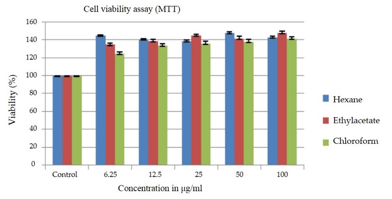
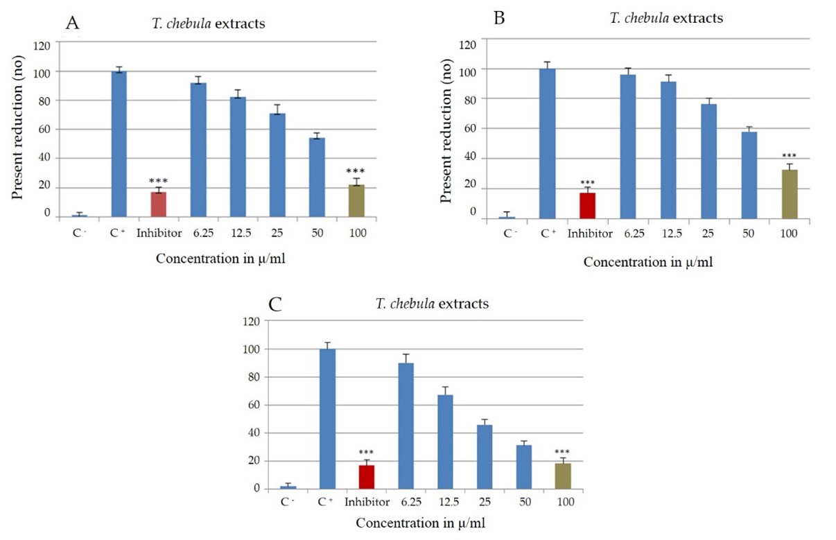
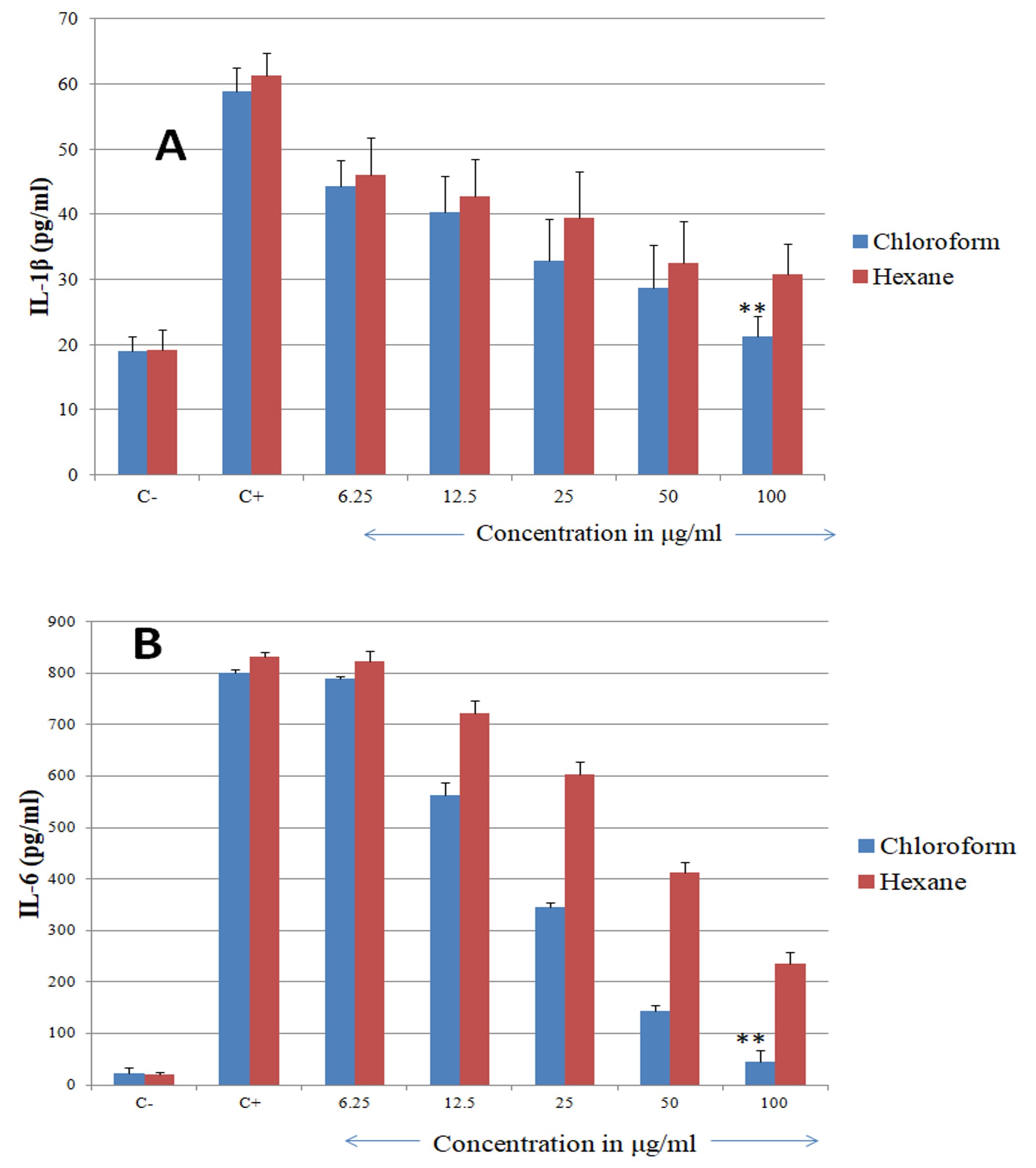
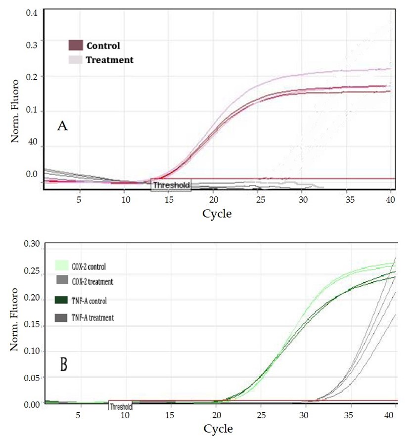
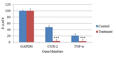
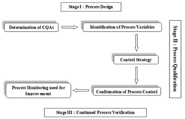
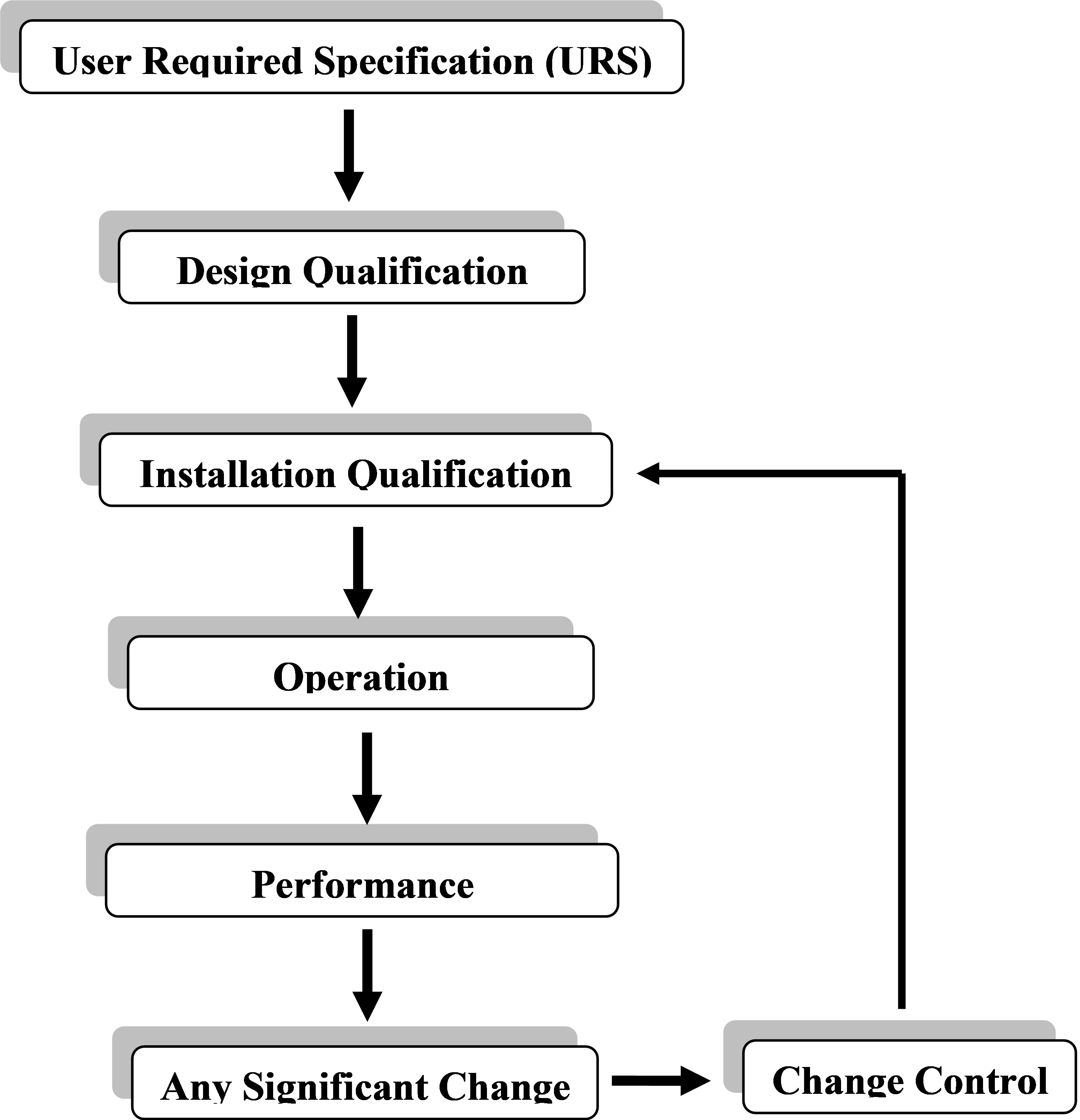
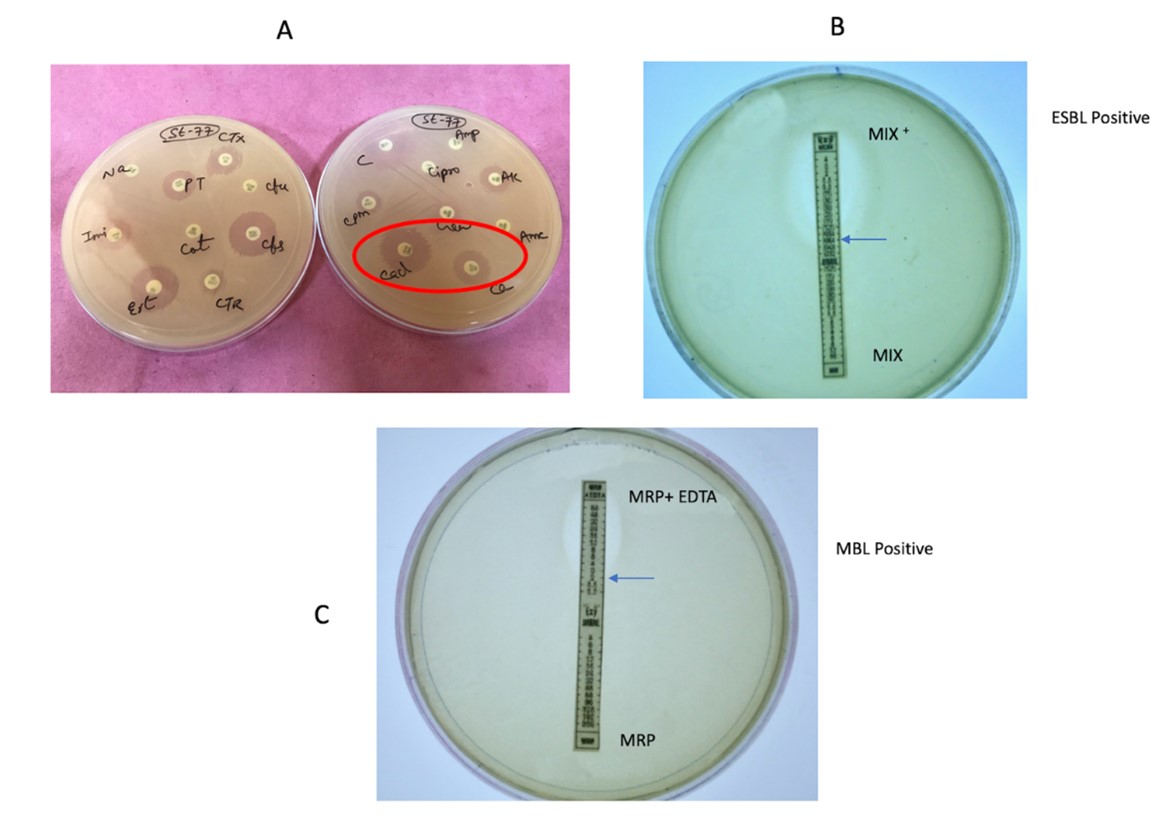
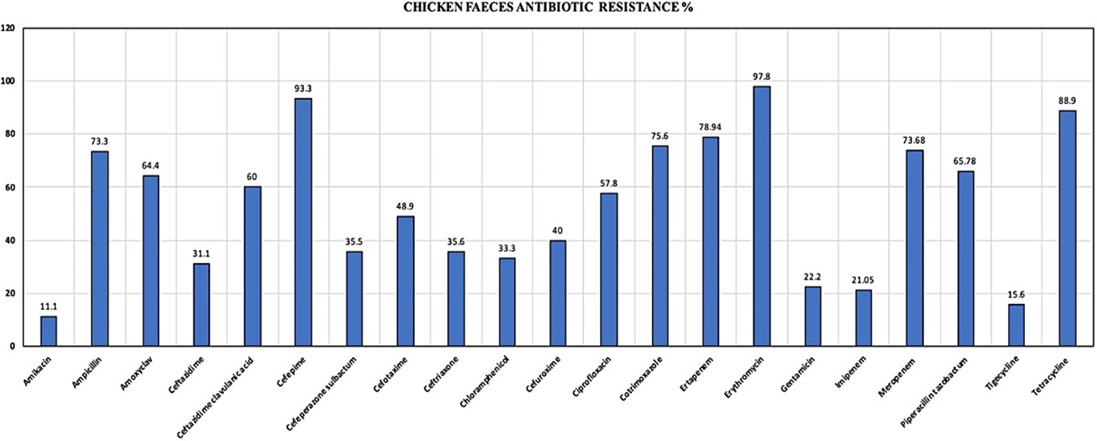
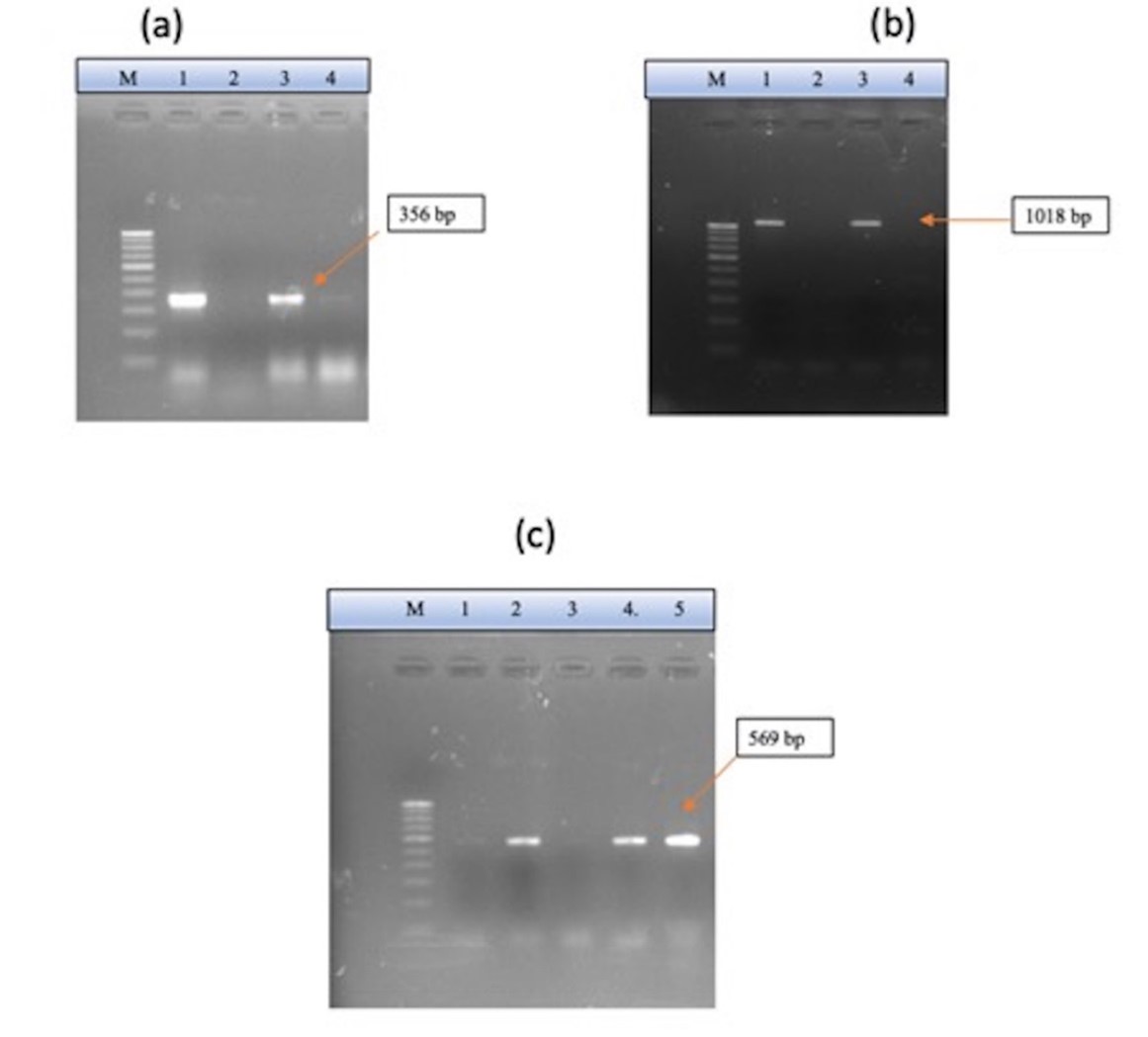
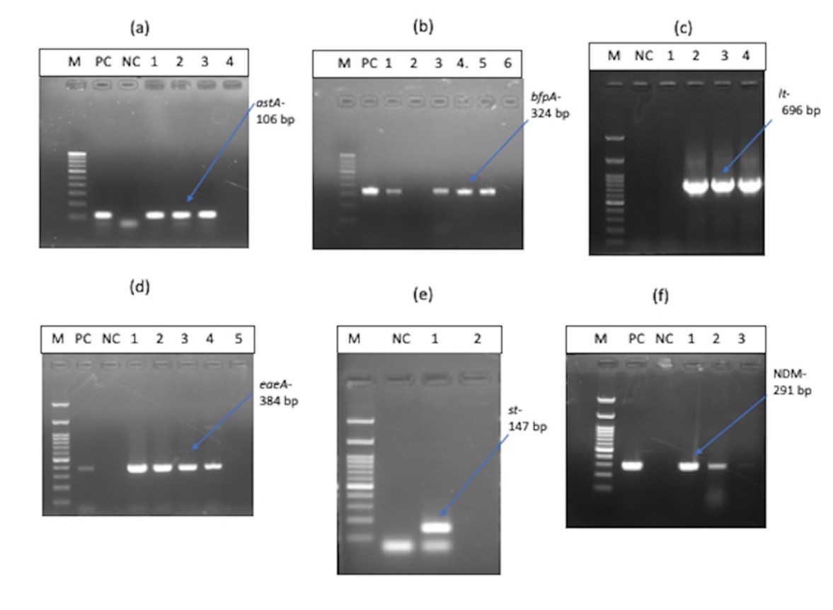
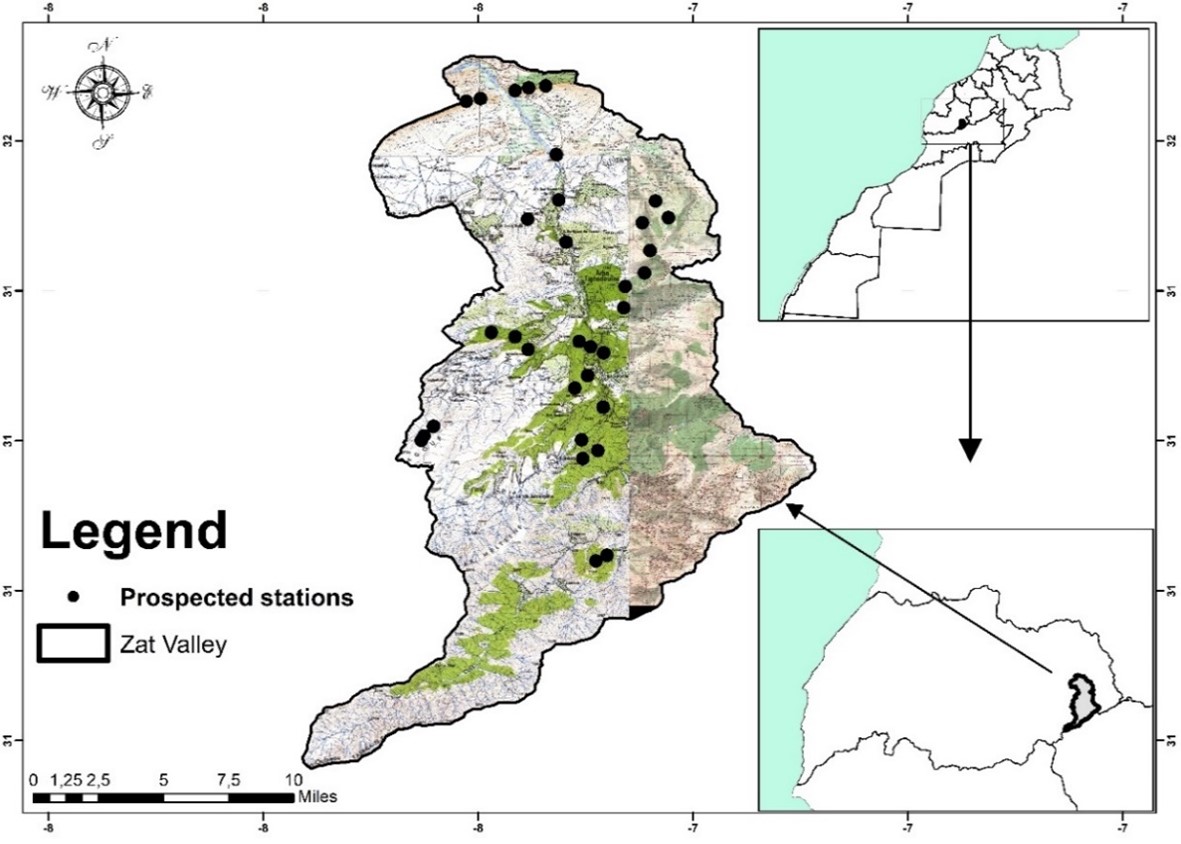
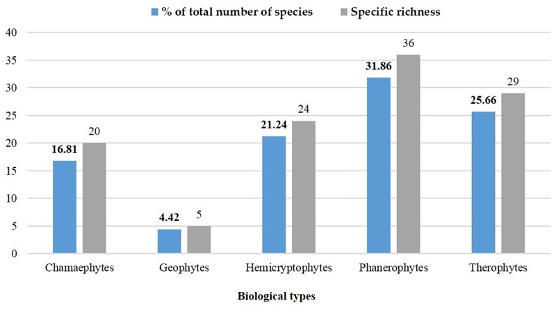
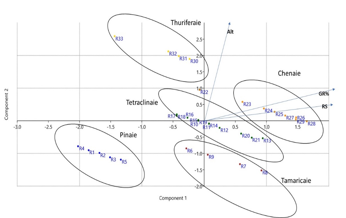
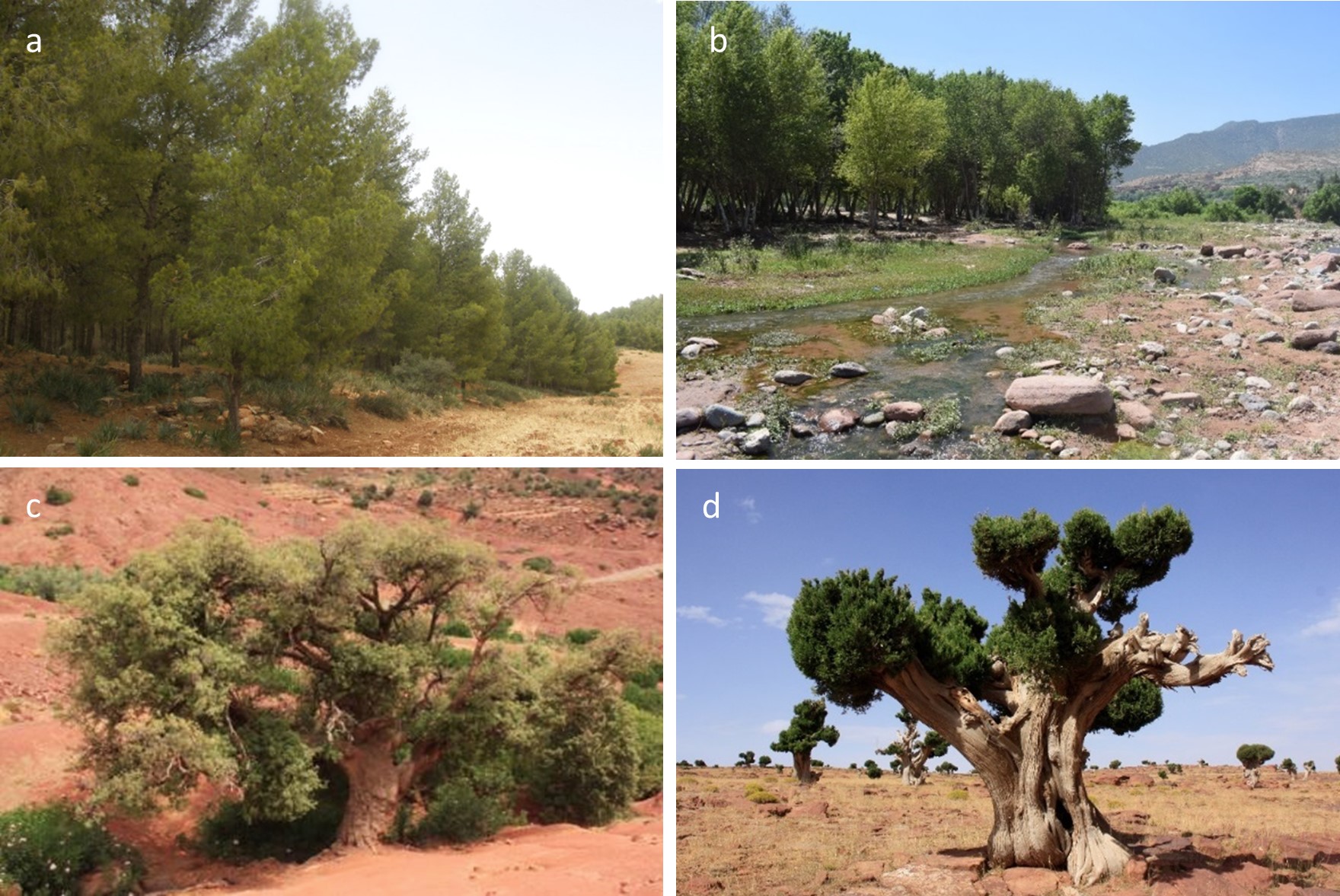
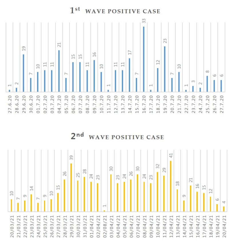
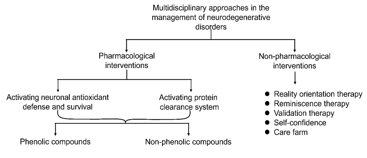
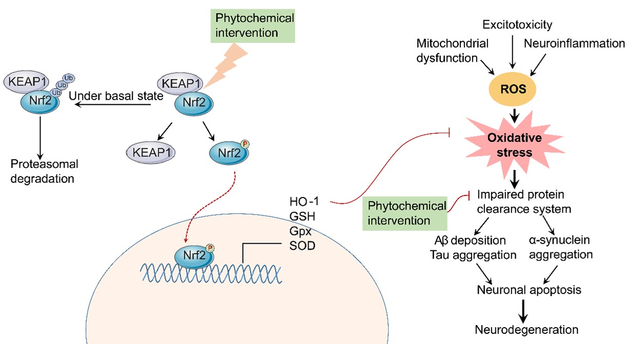
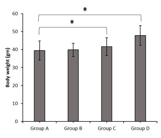
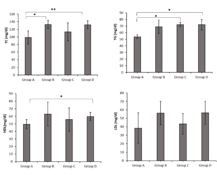
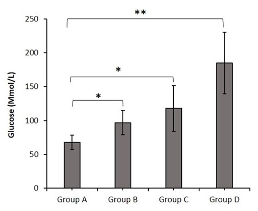
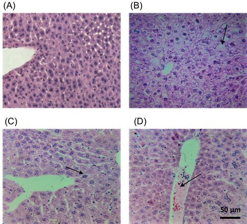
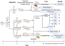
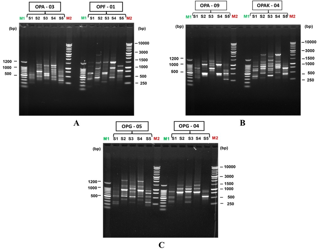