HTTP 200 OK
Allow: GET, HEAD, OPTIONS
Content-Type: application/json
Vary: Accept
{
"count": 321,
"next": "https://jabet.bsmiab.org/articles/?format=api&page=18",
"previous": "https://jabet.bsmiab.org/articles/?format=api&page=16",
"results": [
{
"id": 115,
"slug": "178-1653047882-effect-of-kiss1-gene-variants-rs372790354-ga-and-rs4889-ga-on-kisspeptin-in-patients-with-polycystic-ovary-syndrome-in-iraq",
"featured": false,
"slider": false,
"issue": "Vol5 Issue3",
"type": "original_article",
"manuscript_id": "178-1653047882-",
"recieved": "2022-05-20",
"revised": null,
"accepted": "2022-06-15",
"published": "2022-06-30",
"pdf_file": "https://jabet.bsmiab.org/media/pdf_file/2023/34/178-1653047882.pdf",
"title": "Effect of KISS1 gene variants (rs372790354 G>A and rs4889 G>A) on kisspeptin in patients with polycystic ovary syndrome in Iraq",
"abstract": "<p>Polycystic ovary syndrome (PCOS) is a heterogeneous genetic disorder categorized by hyperandrogenism that affects early reproductive age in females. KISS1 has played role in regulating the hypothalamic-pituitary-gonad axis. It also plays a key role in human reproductive function. Imbalance-of-function mutations is often found KISS1 gene of patients with polycystic ovary syndrome. Blood samples were collected from 120 patients (60 control are divided into 30 normal weight and 30 obese) and (60 PCOS) females are divided into 30 normal weight and 30 obese. DNA was extracted and genotyped for KISS1 variants by HRM-PCR and measured the level of kisspeptin by ELIAS, while LH, FSH, DHEA and free testosterone by CLIA. The value of LH, Testosterone, DHEA-S and kisspeptin is elevated in the patient group, while the decline of FSH in serum level patients value, rs372790354 G > A and rs4889 G>A was associated with PCOS in dominant, recessive, co-dominant (P-value< 0.05), rs37279054 AA was not found the effect of obese group and linked with normal weight PCOS put present study no effect on the parameters, rs4889 GG/GA was the effect on all subgroups except the genotype GA not effected on obese female, the highly significant ( P-value<0.05) of rs4889 GA influenced on measured of WHR, LH/FSH ratio and DHEA-S in the patient compared to control, rs4889 GG/AA was influenced on normal-weight patient compare to an obese patient, the WHR was higher in an obese patient in both genotype. While the level of kisspeptin in normal weight with genotype AA was higher level compared to obese and (P-value<0.05). We concluded that the KISS1 levels were higher in PCOS females compared to controls and decreased with increasing BMI, KISS1 polymorphism rs372790354 G>A and rs4889 G>A may be associated with the pathophysiology of PCOS and lead to increased serum level of LH that due to hyperandrogenism.</p>",
"journal_reference": "J Adv Biotechnol Exp Ther. 2022; 5(3): 562-576.",
"academic_editor": "Md Jamal Uddin, PhD; ABEx Bio-Research Center, Dhaka-1230, Bangladesh",
"cite_info": "Musawi NJA, Qaysi SAA , et al. Effect of KISS1 gene variants (rs372790354 G>A and rs4889 G>A) on kisspeptin in patients with polycystic ovary syndrome in Iraq. J Adv Biotechnol Exp Ther. 2022; 5(3): 562-576.",
"keywords": [
"PCOS",
"Gene polymorphism",
"HRM-PCR",
"KISS1 gene"
],
"DOI": "doi.org/10.5455/jabet.2022.d136",
"sections": [
{
"section_number": 1,
"section_title": "INTRODUCTION",
"body": "<p>Polycystic ovary syndrome (PCOS) is one of the most common endocrine disorders characterized by multiple hormonal imbalances. Increased gonadotrophin-releasing hormone (GnRH) pulsatility in the hypothalamus increases luteinizing hormone (LH) secretion from the pituitary gland, leading to ovarian hyperandrogenism, ovulatory dysfunction(irregular menstrual cycle), and polycystic ovarian (PCO) morphology that affects at early reproductive age [<a href=\"#r-1\">1</a>]. The National Institutes of Health (NIH) in 1990, the Rotterdam criteria (ROT) in 2003, and the Androgen Excess and PCOS Association (AE-PCOS) in 2006 used and developed three distinct ways to diagnose the condition [<a href=\"#r-2\">2</a>]. The prevalence of PCOS is estimated at 4% to 8% [<a href=\"#r-3\">3</a>] [<a href=\"#r-4\">4</a>]. The diagnostic criteria for PCOS, according to the AE-PCOS Society, included polycystic ovaries (≥12 small follicles in the ovary) and/or ovarian dysfunction: ovulatory dysfunction (oligo or amenorrhea, infertility), less than 6-9 menstruation per year [<a href=\"#r-3\">3,5</a>], and clinical and/or biochemical hyperandrogenism such as hirsutism, acne and androgenic alopecia (modified Ferriman-Gallwey score >8) [<a href=\"#r-6\">6</a>].<br />\r\nThe KISS1 gene is one of the candidate genes contributing to a regulatory role in the female reproductive system. KISS1 plays a vital role in gonadotropin secretion of the HPG axis [<a href=\"#r-5\">5</a>]. It is located on the long arm of chromosome1 (1q32.1), length: 6,151 nucleotides, gene ID: 3814. The human KISS1 mRNA is transcribed from the KISS1 gene. The transcription involves four exons, of which only the third and fourth exons are to end translated into the sequencing of 145 amino acids as a peptide called kisspeptin-145. Subsequently, it is cleaved into four formulas of active kisspeptin consisting of 13, 14, 54, and 10 amino acids [<a href=\"#r-5\">5,7</a>]. Some single nucleotide polymorphisms (SNPs) found in the KISS1 gene affect healthy female reproductive system function by interfering with the HPG axis, which plays an essential key in PCOS etiopathogenesis such as the missense effect [<a href=\"#r-6\">6</a>].<br />\r\nBecause disorders that simulate PCOS are relatively easy to rule out, all females have their TSH and prolactin levels checked. Amenorrhea or oligomenorrhea are two symptoms of hyperprolactinemia [<a href=\"#r-7\">7</a>]. Menstrual irregularities are a symptom of thyroid illness. Non-classical congenital adrenal hyperplasia should be checked out in females with hyperandrogenism, as it occurs in 1.5% to 6.8% of these individuals. Other disorders, such as Cushing’s syndrome, should be investigated, and the 17-OHP level should be evaluated in the selected female with amenorrhea [<a href=\"#r-8\">8,9</a>]. PCOS is caused by a dysfunctional interaction of behavior, environmental, and hereditary variables. The most typical clinical presentation of PCOS includes enlargement of both ovaries and secretion of androgens levels higher than normal theca cells. Enzymatic hyperactivity involved in steroid synthesis causes increased androgenic secretion. Defects of the hypothalamic-pituitary-ovarian axis, including intra-ovarian, autocrine/paracrine managers [<a href=\"#r-10\">10</a>]. Instead, regulators outside the reproductive axis may be involved in the genesis and conservation of hypersecretion of LH, the thecal-stromal cell hyperactivity, and hypofunction of the FSH-granulosa cell axis. Resulting in hyperandrogenism and ovarian dysfunction [<a href=\"#r-11\">11</a>]. Hypothalamic kisspeptin neurons (products of the KISS1 gene) acting via G protein-coupled receptor 54 (GPR54) are localized in two regions: the anterior and the posterior region of the hypothalamus [<a href=\"#r-12\">12</a>]. Kisspeptin secretion further regulates the pulsatile release of GnRH and LH [<a href=\"#r-13\">13</a>]. In the pathogenesis of the polycystic ovarian disease, abnormality in the hypothalamic-pituitary-ovarian or adrenal axis has been imposed. The relative increase in LH to FSH release is caused by a disruption in the secretion pattern of the gonadotropin-releasing hormone (GnRH) [<a href=\"#r-14\">14-18</a>].</p>"
},
{
"section_number": 2,
"section_title": "MATERIALS AND METHODS",
"body": "<p><strong>Sample collection</strong><br />\r\nblood samples were collected from 120 patients (60 control are divided into 30 normal weight and 30 obese) and (60 PCOS females are divided into 30 normal weight and 30 obese). The blood was collected from the females with PCOS between the second and fifth day of the cycle, and the patients were selected according to AE PCOS criteria. Using the Ferriman–Gallwey scale, a physical examination and hirsutism assessment were performed on all patients after obtaining their medical histories. Ferriman–Gallwey scale is a method of evaluating and quantifying hirsutism in women. The method was originally published in 1961 by D. Ferriman and J.D. Gallwey in the Journal of Clinical Endocrinology. The original method used 11 body areas to assess hair growth but was decreased to 9 body areas [<a href=\"#r-19\">19</a>] .female having other causes of hyperandrogenism or menstrual irregular were excluded as hyper prolactinoma, Cushing disease, congenital adrenal hyperplasia, and female was pregnancy.</p>\r\n\r\n<p> </p>\r\n\r\n<p><strong>Ethical statement</strong><br />\r\nEvery volunteer has given written informed permission. This research received ethical approval (DSM/HO-65031) for scientific research from the Ministry of Health MOH and Ministry of Higher Education and Scientific Research MOHESR ethics committees in Iraq.</p>\r\n\r\n<p> </p>\r\n\r\n<p><strong>Biochemical analysis</strong><br />\r\nFollicle Stimulating Hormone (FSH) and Luteinizing Hormone (LH) were measured by Chemiluminescence (CLIA), kisspeptin was determined by enzyme-linked immunosorbent assay (ELISA) kit, were performed in the University of Babylon medicine collage. In the extraction of DNA from fresh whole blood, the concentration and purity of the DNA were determined by using a nanodrop spectrophotometer by G-spin kit, SNPs of KISS1 gene determination and using real-time PCR to amplify the KISS1 gene. Then by HRM technique to a genotyping analysis using the following amplification primer and positive and negative control.<br />\r\n<a href=\"#Table-1\">Table 1</a> showed the PCR primer for genotyping of KISS1 gene rs372790354 alleles: G>A and rs4889 alleles G>A, and <a href=\"#Table-2\">Table 2</a> showed the PCR Condition for genotyping of KISS1 gene rs372790354 alleles: G>A and rs4889 alleles G>A.</p>\r\n\r\n<div id=\"Table-1\">\r\n<p><a href=\"https://jabet.bsmiab.org/table/178-1653047882-table1/\">Table-1</a><strong>Table 1. </strong>PCR primer for genotyping of KISS1 gene rs372790354 alleles: G>A and rs4889 alleles G>A.</p>\r\n\r\n<p> </p>\r\n</div>\r\n\r\n<div id=\"Table-2\">\r\n<p><a href=\"https://jabet.bsmiab.org/table/178-1653047882-table2/\">Table-2</a><strong>Table 2. </strong>PCR Condition for genotyping of KISS1 gene rs372790354 alleles: G>A and rs4889 alleles G>A.</p>\r\n\r\n<p> </p>\r\n</div>\r\n\r\n<p><strong>Statistical analysis</strong><br />\r\nStatistical calculations were performed using the statistical software SPSS version 23. Data descriptive are communicated as the mean ± standard error (Mean± SE); the ANOVA test was used to compare the mean value between subgroups to investigate the correlation between the continuous variables using Pearson’s correlation statistics. The hormonal level was compared between control and patient with PCOS (normal and obese). considered P-value of < 0.05 statistically significant.<br />\r\nGenotype and allele frequency of KISS1 gene polymorphism were calculated, and consequently, the Hardy-Weinberg equilibrium, Chi square (x2) test was used for categorical variables.</p>"
},
{
"section_number": 3,
"section_title": "RESULTS",
"body": "<p><strong>Biochemical assay</strong><br />\r\n<a href=\"#Table-3\">Table 3</a> showed the demographic distribution of the study groups. No significant changes in age and BMI were seen between PCOS patients and the control groups. In this investigation, WHR was significantly different across the studied groups.<br />\r\n<a href=\"#Table-4\">Table 4</a> showed the mean difference and comparison of hormonal parameters between the subgroups, where the biochemical markers are also different between the patients and the control group. In PCOS, LH and DHEAS are high, whereas FSH is decreased compared to control groups (normal weight and obese), and kisspeptin was significantly higher in normal-weight PCOS compared to other subgroups.<br />\r\n<a href=\"#Table-5\">Table 5</a> showed the correlation coefficients between the level of parameters in normal weight and obese patients. There are significant correlations between hormonal parameters in normal-weight PCOS, which showed positive correlations between LH with LH/FSH Ratio, LH with kisspeptin, and LH/FSH Ratio with kisspeptin, while found a negative correlation between FSH with LH/FSH ratio.</p>\r\n\r\n<div id=\"Table-3\">\r\n<p><a href=\"https://jabet.bsmiab.org/table/178-1653047882-table3/\">Table-3</a><strong>Table 3. </strong>Demographic data of studied groups. </p>\r\n\r\n<p> </p>\r\n</div>\r\n\r\n<div id=\"Table-4\">\r\n<p><a href=\"https://jabet.bsmiab.org/table/178-1653047882-table4/\">Table-4</a><strong>Table 4. </strong>Mean difference and comparison of hormonal parameters between the subgroups. </p>\r\n\r\n<p> </p>\r\n</div>\r\n\r\n<div id=\"Table-5\">\r\n<p><a href=\"https://jabet.bsmiab.org/table/178-1653047882-table5/\">Table-5</a><strong>Table 5.</strong> Correlation coefficients between level of parameters in normal and obese patients. </p>\r\n\r\n<p> </p>\r\n</div>\r\n\r\n<p><strong>Genotyping assay</strong><br />\r\n<a href=\"#Table-6\">Table 6</a> showed the gene polymorphism of KISS1 in studied groups for rs372790354 G>A. When comparing the (genotype and allele frequency) for each SNP between PCOS females and controls, Hardy-Weinberg equilibrium was applied to the control groups (P>0.05). A novel SNPs homozygous (GG), heterozygosis (GA), and mutant (AA) of both SNPs information. <a href=\"#figure1\">Figure 1 </a>showed the amplification and genotype of rs372790354 G>A for Control and Samples. <a href=\"#figure2\">Figure 2</a> showed the amplification and genotype of rs4889 G>A for Control and Samples. <a href=\"#Table-7\">Table 7 </a>showed the association of rs372790354 genotypes with PCOS under different inheritance models. Significant differences were exhibited in the co-dominant model for GG, GA, and AA groups at (0.026 and 0.0036), respectively. In the dominant model, significant differences showed for GG and GA-AA groups at (0.0021), respectively. In the recessive model, significant differences showed for GG- GA and AA groups at (0.011), respectively. In contrast, there are no significant differences in over dominant model for AA-GG and GA groups.<br />\r\n<a href=\"#Table-8\">Table 8</a> showed the distribution of genotype frequency of rs372790354 G>A of KISS1 gene polymorphism between patient and control group within subgroup normal weight and obese where there are significant differences for AA group at 0.059.<br />\r\n<a href=\"#Table-9\">Table 9</a> showed the association of rs4889 genotypes with PCOS under different models of inheritance where there are significant differences in the co-dominant model for GG, GA, and AA groups at (0.0003 and 0.0004), respectively. In the dominant model, significant differences showed for GG and GA-AA groups at (0.0001), respectively. In the recessive model, significant differences showed for GG- GA and AA groups at (0.0035), respectively. Finally, the Over dominant model for AA-GG and GA groups showed significant differences at (0.04).<br />\r\n<a href=\"#Table-10\">Table 10</a> showed the distribution of genotype frequency of rs4889 G>A of KISS1 gene polymorphism between patient and control group within normal subgroup weight and obese. Significant differences in GA type for normal weight at (0.0002 and 0.0042), respectively, while there are no significant differences in the obese group. In AA type, there are significant differences between the normal-weight group at (0.0007 and 0.07) and the obese group at (0.03), respectively.<br />\r\n<a href=\"#Table-11\">Table 11</a> showed the alleles frequency and allelic association of rs372790354 G>A and rs4889G>A by Hardy-Weinberg equilibrium law of KISS1 gene polymorphism between the patient group and control group. Where there are significant differences in rs372790354 SNP for G allele at (0.0002) and odd ratio value (0.34) between 0.2 – 0.5, While for A allele at (0.0002) and odd ratio value (2.96) between 1.68 – 5.19. In <em>rs4889 SNP, </em>there are significant differences for the G allele at (< 0.0001) and odd ratio value (0.2) between 0.1 – 0.3. While for the A allele at (< 0.0001) and odd ratio value (4.9) between 2.7 – 8.8.<br />\r\n<a href=\"#Table-12\">Table 12</a> showed the influence of rs4889 G>A polymorphism of KISS1 gene on the Mean differences of characteristics and parameters between genotypes normal weight (patient and control) groups. Where there are significant differences for WHR, LH/FSH Ratio, F-Testosterone, and DHEA-S markers at (<0.01) for all of them, while there are no significant differences for LH, FSH, and kisspeptin markers<br />\r\n<a href=\"#Table-13\">Table 13</a> showed the influence of rs4889 G>A polymorphism of KISS1 gene on the Mean differences of characteristics and parameters between genotypes patient (normal weight and obese) groups. There are significant differences for WHR, and kisspeptin markers at (<0.01 and 0.007), respectively, while there are no significant differences for LH, FSH, Ratio FSH/LH F-Testosterone, and DHEA-S markers.<br />\r\n<a href=\"#Table-14\">Table 14</a> showed the Genotype combination of SNPs rs4889 and rs372790354 in the KISS1 gene between the patient group and control group (Allele Frequencies detected by Hardy-Weinberg equilibrium law). There are no significant differences for rs4889 + rs372790354 SNPs for all groups under study depending on P-value and odd ratio Hardy-Weinberg equilibrium.</p>\r\n\r\n<div id=\"figure1\">\r\n<figure class=\"image\"><img alt=\"\" height=\"676\" src=\"/media/article_images/2023/57/26/178-1653047882-Figure1.jpg\" width=\"500\" />\r\n<figcaption><strong>Figure 1.</strong> Amplification and genotype of rs372790354 G>A for control and samples. A. Amplification Step, B. Genotype Step, C. Control Genotype. Yellow, GG; blue, GA; red, AA.</figcaption>\r\n</figure>\r\n</div>\r\n\r\n<div id=\"figure2\">\r\n<figure class=\"image\"><img alt=\"\" height=\"741\" src=\"/media/article_images/2023/57/26/178-1653047882-Figure2.jpg\" width=\"500\" />\r\n<figcaption><strong>Figure 2.</strong> Amplification and genotype of rs4889 G>A for control and samples. A. Amplification Step, B. Genotype Step, C. Control Genotype. Green, GG; blue, GA; purple, AA.</figcaption>\r\n</figure>\r\n</div>\r\n\r\n<p> </p>\r\n\r\n<div id=\"Table-6\">\r\n<p><a href=\"https://jabet.bsmiab.org/table/178-1653047882-table6/\">Table-6</a><strong>Table 6.</strong> Gene polymorphism of KISS1 in studied groups. </p>\r\n\r\n<p> </p>\r\n</div>\r\n\r\n<div id=\"Table-7\">\r\n<p><a href=\"https://jabet.bsmiab.org/table/178-1653047882-table7/\">Table-7</a><strong>Table 7.</strong> Association of rs372790354 genotypes with PCOS under different models of inheritance.</p>\r\n\r\n<p> </p>\r\n</div>\r\n\r\n<div id=\"Table-8\">\r\n<p><a href=\"https://jabet.bsmiab.org/table/178-1653047882-table8/\">Table-8</a><strong>Table 8. </strong>Distribution to genotype frequency of rs372790354 G>A of KISS1 gene polymorphism between patient and control group within normal subgroup weight and obese. </p>\r\n\r\n<p> </p>\r\n</div>\r\n\r\n<div id=\"Table-9\">\r\n<p><a href=\"https://jabet.bsmiab.org/table/178-1653047882-table9/\">Table-9</a><strong>Table 9.</strong> Association of rs4889 genotypes with PCOS under different models of inheritance.</p>\r\n\r\n<p> </p>\r\n</div>\r\n\r\n<div id=\"Table-10\">\r\n<p><a href=\"https://jabet.bsmiab.org/table/178-1653047882-table10/\">Table-10</a><strong>Table 10. </strong>Distribution to genotype frequency of rs4889 G>A of KISS1 gene polymorphism between patient and control group within normal subgroup weight and obese. </p>\r\n\r\n<p> </p>\r\n</div>\r\n\r\n<div id=\"Table-11\">\r\n<p><a href=\"https://jabet.bsmiab.org/table/178-1653047882-table11/\">Table-11</a><strong>Table 11. </strong>Allele’s frequency and allelic association of rs372790354 G>A and rs4889G>A by Hardy-Weinberg equilibrium law of KISS1 gene polymorphism between patient group and control group. </p>\r\n\r\n<p> </p>\r\n</div>\r\n\r\n<div id=\"Table-12\">\r\n<p><a href=\"https://jabet.bsmiab.org/table/178-1653047882-table12/\">Table-12</a><strong>Table 12. </strong>The influence of rs4889 G>A polymorphism of KISS1 gene on the mean difference of characteristics and parameters between genotypes normal weight (patient and control) group. </p>\r\n\r\n<p> </p>\r\n</div>\r\n\r\n<div id=\"Table-13\">\r\n<p><a href=\"https://jabet.bsmiab.org/table/178-1653047882-table13/\">Table-13</a><strong>Table 13. </strong>The influence of rs4889 G>A polymorphism of KISS1 gene on the mean differences of characteristics and parameters between genotypes patient (normal weight and obese) group. </p>\r\n\r\n<p> </p>\r\n</div>\r\n\r\n<div id=\"Table-14\">\r\n<p><a href=\"https://jabet.bsmiab.org/table/178-1653047882-table14/\">Table-14</a><strong>Table14.</strong> Genotype combination of SNPs rs4889 and rs372790354 in KISS1 gene between patient group and control group (Allele Frequencies detected by Hardy-Weinberg equilibrium law). </p>\r\n</div>"
},
{
"section_number": 4,
"section_title": "DISCUSSION",
"body": "<p>The disturbance in Hypothalamus-Pituitary-Gonadal (HPG) axis is related to PCOS. Kisspeptin is a neuropeptide expressed by the KISS1 gene that has a highly active action on the HPG axis and plays a crucial role in human reproduction [<a href=\"#r-20\">20</a>]. KISS1 gene is mainly expressed in the hypothalamus to the secretion of GnRH that is regulatory of LH and FSH, the genetic factor responsible for the etiopathogenesis of PCOS [<a href=\"#r-21\">21</a>]. The present study investigated KISS1 gene polymorphism which could be used as a marker of PCOS and its risk factor in Iraqi females. Two novel SNPs (rs372790354G>A and rs4889 G>A) in the KISS1 gene were identified and investigated in this study. The effect of the two SNPs was studied by analyzing and comparing genotype groups, which leads to endocrine disturbances (kisspeptin, LH, FSH, LH/FSH ratio, and DHEA-S) in the female with PCOS.<br />\r\nThe result of demographic data was shown in <a href=\"#Table-1\">Table 1</a> that there was no significant difference in mean age and BMI between the studied group. At the same time, there was a highly significant difference in mean WHR. The present study showed in <a href=\"#Table-2\">Table 2</a> that the level of kisspeptin was slightly higher in females obese without PCOS compared to the obese female with PCOS group were not significantly different, supporting these findings by Yerlikaya et al. [<a href=\"#r-22\">22</a>]. The mean of kisspeptin in obese females with and without PCOS was approximately the same. It may be due to the stimulatory action of obesity on kisspeptin neurons, which are also regulated by leptin, insulin resistance, ghrelin, and adiponectin (excitatory) (inhibitory) [<a href=\"#r-20\">20</a>]. The current study showed a higher significance in the mean of normal-weight patients compared to control and obese patient (P-value <0.01). Three previous studies[<a href=\"#r-23\">23–25</a>] reported a higher kisspeptin level in females with PCOS. Another study [<a href=\"#r-26\">26</a>] reported lower levels in females with PCOS compared to controls. Nearmeen and her colleagues found that kisspeptin levels among the PCOS group there was a significantly lower level in the underweight, overweight, and obese compared to the normal weight group [<a href=\"#r-27\">27</a>].<br />\r\nIn the present study, table 2 showed that LH values were higher in PCOS females than in controls. High levels of LH contribute to increasing levels of androgens along with low levels of FSH secretion compared to the control group [<a href=\"#r-28\">28</a>]. These data suggest that endocrine hormone concentrations were significantly higher in PCOS [<a href=\"#r-29\">29</a>]. The present study shows PCOS females with high BMI have elevated androgen levels.<br />\r\nThe correlation between biochemical parameters of PCOS patients, as shown in<a href=\"#Table-3\"> Table 3</a>, serum kisspeptin was positively correlated with the LH and LH/FSH ratio in normal-weight patients and with LH/FSH ratio only in the obese patients. Also, DHEA positively correlated with LH and FSH levels in obese patients. Obesity, insulin resistance, and dyslipidemia are PCOS-related morbidities that correlate with the LH/FSH ratio. Kisspeptin may stimulate LH secretion. In the normal patients, kisspeptin is higher than in the obese group, leading to higher LH levels in normal-weight than in obese. However, the directed pituitary effects of kisspeptin in regulating gonadotropin secretion remain controversial [<a href=\"#r-30\">30</a>]. In all models, the genotype (homozygous AA and heterozygous GA) of rs372790354 and rs4889 (homozygous AA and heterozygous GA) was significantly (P 0.05) more frequent in PCOS than in controls (<a href=\"#Table-5\">Table 5</a> and <a href=\"#Table-7\">7</a>). While within subgroups, rs372790354 (AA) is highly significant (P-value<0.001) in normal-weight patients as a compared to normal-weight control. That means the SNP link to PCOS as shown in <a href=\"#Table-6\">Table 6</a>, rs372790354 genotype AA normal weight patients showed no significant influence on the value of demographic characteristics and endocrine (the result are not shown). The SNP rs372791354 does not influence the level of kisspeptin and other hormones. There was no difference in its level in the two different genotypes of rs372790354.<br />\r\nThese findings disagree with the results of Maha and his colleagues [<a href=\"#r-31\">31</a>], which showed a significant influence of rs372790354 G/A on the risk of PCOS and the increase of LH levels, kisspeptin, and WHR in PCOS females. Maybe this SNP is located in 5 prime untranslated regions of the mRNA KISS1 gene that have both stimulatory and inhibitory mechanisms, including regulation of mRNA transcription and post-transcriptional modification (secondary structure and mRNA stability), localization, and mRNA translation. It also regulates protein features such as protein complex formation and post-translational modifications and may alter protein conformation [<a href=\"#r-23\">23, 24</a>]. That means affecting kisspeptin conformation or its binding to the receptor. Therefore, this SNP with two genotypes has higher significant frequencies and risk factors associated with PCOS.<br />\r\nAllele frequency of rs372790354 G>A and rs4889G>A were also significant in both SNPs. The mutant had a higher predisposition (P<0.05) in patient groups compared with control, and the allele G was protective, as shown in <a href=\"#Table-9\">Table 9.</a> The rs4889 GA and AA have a higher frequency in normal-weight patients compared to the obese patients, and all subgroups of control as shown in <a href=\"#Table-8\">Table 8</a>, except the genotype GA was not significant in obese with PCOS compared to obese without PCOS. Polymorphism of rs4889 introduced a substitution of proline at the 81 positions. This substitution was observed in kisspeptin-54 but not in the other three forms of kisspeptin (kisspeptin-14, -13,-10) [<a href=\"#r-34\">34</a>].<br />\r\nThe result of rs4889 genotype AA and GA between patient and control, normal weight, and the obese patient is shown in <a href=\"#Table-10\">Tables 10</a> and<a href=\"#Table-11\"> 11</a>. The obesity-linked parameters that have influenced the pathology of PCOS, including WHR, were significantly in PCOS groups with genotype GA compared to control and significantly higher in ( obese compared to normal weight ) patients with genotype GA, AA. In other studies, the allele C instead of allele A for this SNP. These findings confirm the results of Mazin and his colleagues’ study [<a href=\"#r-35\">35</a>]. The importance of WHR as a prognostic marker, higher WHR in PCOS compared to obese control, confirms the contribution of abdominal fat as an etiological mechanism in PCOS. The PCOS genotype GA group showed the feature of endocrine disturbances that increase the value of LH/FSH ratio and DHEA-S (P-value =0.01).<br />\r\nIn contrast, kisspeptin, FSH, and LH showed no significant difference compared to GA genotype control. In addition, higher levels of kisspeptin (normal weight compared to obese P-value= 0.007) were found in patients with genotype AA. In a previous study, Albalawi and his colleagues reported no significant difference in the kisspeptin level between PCOS females and controls [<a href=\"#r-34\">34</a>].<br />\r\nNo significant elevation of the kisspeptin level in PCOS patients may be due to the low sample size. The effect of rs4889 GA and AA polymorphism of the KISS1 gene may directly impact the functional activity of kisspeptin in terms of its altered behavior and binding capacity of kisspeptin to its receptor GPR54 [<a href=\"#r-35\">35</a>]. Consequently, the disturbed kisspeptin-GPR54 pathway and dysregulation in GnRH secretion lead to LH hypersecretion, leading to hyperandrogenism. Increased testosterone level is induced by the direct action of high stimulation of LH on gonads; it appears that rs4889 influences the mechanism by which kisspeptin activates the secretion of LH but not FSH [<a href=\"#r-36\">36</a>]. Finally, the females with both AA and GA genotypes from the two SNPs in this study have a higher risk of developing PCOS.</p>"
},
{
"section_number": 5,
"section_title": "CONCLUSION",
"body": "<p>We concluded that the KISS1 levels were higher in PCOS females compared to controls and lowered with increasing BMI. Consequently, the value of kisspeptin is higher in normal-weight patients compared to obese females. KISS1 polymorphism rs372790354 G>A and rs4889 G>A may be associated with the pathophysiology of PCOS and lead to the increased serum level of LH due to hyperandrogenism; rs4889 AA may cause hyperactivity of the KISS1 gene.</p>"
},
{
"section_number": 6,
"section_title": "ACKNOWLEDGEMENT",
"body": "<p>The authors would like to thank Dr. Yasir Haider Al-Mawlah and Dr. Ameer Mezher Hadi (DNA Research Center, University of Babylon. Pune for their kind support with all laboratory equipment and provide the suitable facilities, also for drafting the manuscript to make this work done.</p>"
},
{
"section_number": 7,
"section_title": "AUTHOR CONTRIBUTIONS",
"body": "<p>Conception and design of the study: Noor J. T. Al-Musawi, Suhayr Aesa Al- Qaysi, and Suha J. Witwit; Drafting the manuscript: Suhayr Aesa Al- Qaysi; Analysis and/or interpretation of data: Suha J. Witwit.</p>"
},
{
"section_number": 8,
"section_title": "CONFLICTS OF INTEREST",
"body": "<p>There is no conflict of interest among the authors.</p>"
}
],
"figures": [
{
"figure": "https://jabet.bsmiab.org/media/article_images/2023/57/26/178-1653047882-Figure1.jpg",
"caption": "Figure 1. Amplification and genotype of rs372790354 G>A for control and samples. A. Amplification Step, B. Genotype Step, C. Control Genotype. Yellow, GG; blue, GA; red, AA.",
"featured": false
},
{
"figure": "https://jabet.bsmiab.org/media/article_images/2023/57/26/178-1653047882-Figure2.jpg",
"caption": "Figure 2. Amplification and genotype of rs4889 G>A for control and samples. A. Amplification Step, B. Genotype Step, C. Control Genotype. Green, GG; blue, GA; purple, AA.",
"featured": false
}
],
"authors": [
{
"id": 459,
"affiliation": [
{
"affiliation": "DNA research center / University of Babylon, Iraq, Hillah, Babylon state, 51001, Iraq"
}
],
"first_name": "Noor J. T. Al",
"family_name": "Musawi",
"email": "noorchemist30@gmail.com",
"author_order": 1,
"ORCID": null,
"corresponding": true,
"co_first_author": false,
"co_author": false,
"corresponding_author_info": "Noor J. T. Al-Musawi, PhD; DNA research center / University of Babylon, Iraq, Hillah, Babylon state, 51001, Iraq, e-mail: noorchemist30@gmail.com",
"article": 115
},
{
"id": 460,
"affiliation": [
{
"affiliation": "Departments of Medical Sciences and Biochemistry, College of Medicine, University of Babylon, Babylon, Iraq"
}
],
"first_name": "Suhayr Aesa Al",
"family_name": "Qaysi",
"email": null,
"author_order": 2,
"ORCID": null,
"corresponding": false,
"co_first_author": false,
"co_author": false,
"corresponding_author_info": "",
"article": 115
},
{
"id": 461,
"affiliation": [
{
"affiliation": "Departments of obstetrics and gynecology, College of Medicine, University of Babylon, Babylon, Iraq"
}
],
"first_name": "Suha J",
"family_name": "Witwit",
"email": null,
"author_order": 3,
"ORCID": null,
"corresponding": false,
"co_first_author": false,
"co_author": false,
"corresponding_author_info": "",
"article": 115
}
],
"views": 1071,
"downloads": 138,
"references": [
{
"id": 3585,
"serial_number": 1,
"pmc": null,
"reference": "Branavan U, Muneeswaran K, Wijesundera S. Identification of selected genetic polymorphisms in polycystic ovary syndrome in Sri Lankan women using low-cost genotyping techniques. PLoS One 2018; 13(12): e0209830.",
"DOI": null,
"article": 115
},
{
"id": 3586,
"serial_number": 2,
"pmc": null,
"reference": "H. Chaudhary, J. Patel, N. K. Jain, et al. The role of polymorphism in various potential genes on polycystic ovary syndrome susceptibility and pathogenesis. Journal of Ovarian Research 2021; vol. 14, no. 1.",
"DOI": null,
"article": 115
},
{
"id": 3587,
"serial_number": 3,
"pmc": null,
"reference": "F. A. K. Khazaal, A. H. Liebi, and I. J. Mahmoud. Prevalence and presenting features of polycystic ovarian syndrome in Iraqi obese females. Iraqi journal of infertility research, baghdad 2014; pp. 14–18.",
"DOI": null,
"article": 115
},
{
"id": 3588,
"serial_number": 4,
"pmc": null,
"reference": "W. A. March, V. M. Moore, K. J. Willson, et al. The prevalence of polycystic ovary syndrome in a community sample assessed under contrasting diagnostic criteria Wendy. original article reproductive epidemiology 2011; pp. 544–551.",
"DOI": null,
"article": 115
},
{
"id": 3589,
"serial_number": 5,
"pmc": null,
"reference": "Kiconco, Sylvia, Mousa et al. Pcos phenotype in unselected populations study (P-pup): Protocol for a systematic review and defining pcos diagnostic features with pooled individual participant data. Diagnostics 2021; vol. 11, no. 11.",
"DOI": null,
"article": 115
},
{
"id": 3590,
"serial_number": 6,
"pmc": null,
"reference": "M. K. C. C. Ilagan, E. Paz-Pacheco, D. Z. Totesora, et al. The modified Ferriman-Gallwey score and hirsutism among Filipino women. Endocrinology and Metabolism 2019; vol. 34, no. 4. pp. 374–381, 2019",
"DOI": null,
"article": 115
},
{
"id": 3591,
"serial_number": 7,
"pmc": null,
"reference": "Vermeulen And S. Ando. Prolactin and adrenal androgen secretion. Clinical Endocrinology 1978; vol. 8, no. 4. pp. 295–303.",
"DOI": null,
"article": 115
},
{
"id": 3592,
"serial_number": 8,
"pmc": null,
"reference": "E. Jones. Diagnosis and treatment of polycystic ovarian syndrome. Nursing times 2005; vol. 101, no. 3. pp. 40–43.",
"DOI": null,
"article": 115
},
{
"id": 3593,
"serial_number": 9,
"pmc": null,
"reference": "Paul W. Ladenson, MD, Peter A. Singer, MD, Kenneth B. Ain, MD, et al. American thyroid association guidelines for detection of thyroid dysfunction. Archives of Internal Medicine 2000; vol. 160, no. 11. pp. 1573–1575. 10.1001/archinte.160.11.1573.DOI: 10.1001/archinte.160.11.1573.",
"DOI": null,
"article": 115
},
{
"id": 3594,
"serial_number": 10,
"pmc": null,
"reference": "Gerard Conway, Didier Dewailly, Evanthia Diamanti-Kandarakis, et al. The polycystic ovary syndrome: a position statement from the European Society of Endocrinology Gerard. 2015; no. 8 (72).",
"DOI": null,
"article": 115
},
{
"id": 3595,
"serial_number": 11,
"pmc": null,
"reference": "J. Morales, G. A. Laughlin, T. Bijtzow, H. Maheshwari, et al. Insulin, somatotropic, and luteinizing hormone in lean and obese women with polycystic ovary syndrome: Common and distinct features Axes A. 1996;",
"DOI": null,
"article": 115
},
{
"id": 3596,
"serial_number": 12,
"pmc": null,
"reference": "J. Roa, J. M. Castellano, V. M. Navarro, et al. Kisspeptins and the control of gonadotropin secretion in male and female rodents. Peptides, 2009; vol. 30, no. 1. pp. 57–66. 10.1016/j.peptides.2008.08.009.",
"DOI": null,
"article": 115
},
{
"id": 3597,
"serial_number": 13,
"pmc": null,
"reference": "N. Bhalakiya, N. Haque, and P. Patel. Kisspeptin: A novel regulator in reproductive physiology. Int. J. Livest. Res.2019; p. 1.",
"DOI": null,
"article": 115
},
{
"id": 3598,
"serial_number": 14,
"pmc": null,
"reference": "Z. Saadia. Follicle stimulating hormone (LH: FSH) ratio in polycystic ovary syndrome (PCOS) – obese vs. non- obese women. Medical archives (Sarajevo, Bosnia and Herzegovina)2020; vol. 74, no. 4. pp. 289–293.",
"DOI": null,
"article": 115
},
{
"id": 3634,
"serial_number": 15,
"pmc": null,
"reference": "S. Z. Nejad, F. R. Tehrani, and A. Zadeh-Vakili. The role of kisspeptin in female reproduction. International Journal of Endocrinology and Metabolism 2017; vol. 15, no. 3.",
"DOI": null,
"article": 115
},
{
"id": 3635,
"serial_number": 16,
"pmc": null,
"reference": "E. Oakley, D. K. Clifton, and R. A. Steiner. Kisspeptin signaling in the brain. Endocrine Reviews2009; vol. 30, no. 6. pp. 713–743.",
"DOI": null,
"article": 115
},
{
"id": 3636,
"serial_number": 17,
"pmc": null,
"reference": "NCBI, “KISS1 gene location.” [Online]. Available: https://www.ncbi.nlm.nih.gov/gene/3814. [Accessed: 29-Mar-2022].",
"DOI": null,
"article": 115
},
{
"id": 3637,
"serial_number": 18,
"pmc": null,
"reference": "“Kisspeptin structure.” [Online]. Available: https://www.uniprot.org/uniprot/Q15726#showFeaturesTable. [Accessed: 29-Mar-2022].",
"DOI": null,
"article": 115
},
{
"id": 3638,
"serial_number": 19,
"pmc": null,
"reference": "G. J. Ferriman David. Clinical assessment of body hair in women. J Clin Endocrinol Meta., 1961; vol. 21, no. January. pp. 1440–7.",
"DOI": null,
"article": 115
},
{
"id": 3639,
"serial_number": 20,
"pmc": null,
"reference": "J. Padda et al. Role of kisspeptin on hypothalamic-pituitary-gonadal pathology and its effect on reproduction. Cureus. 2021.",
"DOI": null,
"article": 115
},
{
"id": 3640,
"serial_number": 21,
"pmc": null,
"reference": "O. Valkenburg et al. Genetic polymorphisms of GnRH and gonadotrophic hormone receptors affect the phenotype of polycystic ovary syndrome. Hum. Reprod.2009; vol. 24, no. 8, pp. 2014–2022.",
"DOI": null,
"article": 115
},
{
"id": 3641,
"serial_number": 22,
"pmc": null,
"reference": "E. Yerlikaya et al. Plasma kisspeptin levels in polycystic ovary syndrome. in Endocrine Abstracts 2013; vol. 32.",
"DOI": null,
"article": 115
},
{
"id": 3642,
"serial_number": 23,
"pmc": null,
"reference": "S. A. Yilmaz. Metastin levels in relation with hormonal and metabolic profile in patients with polycystic ovary syndrom. Eur. J. Obstet. Gynecol. Reprod. Biol.2014; vol. 180, no. 1, pp. 56–60.",
"DOI": null,
"article": 115
},
{
"id": 3643,
"serial_number": 24,
"pmc": null,
"reference": "X. Chen, Y. Mo, L. Li, et al. Increased plasma metastin levels in adolescent women with polycystic ovary syndrome. Eur. J. Obstet. Gynecol. Reprod. Biol.2010; vol. 149, no. 1, pp. 72–76.",
"DOI": null,
"article": 115
},
{
"id": 3644,
"serial_number": 25,
"pmc": null,
"reference": "Y. E. Jeon et al. Kisspeptin, leptin, and retinol-binding protein 4 in women with polycystic ovary syndrome. Gynecol. Obstet. Invest.,2013; vol. 75, no. 4, pp. 268–274.",
"DOI": null,
"article": 115
},
{
"id": 3645,
"serial_number": 26,
"pmc": null,
"reference": "D. Panidis et al. Plasma metastin levels are negatively correlated with insulin resistance and free androgens in women with polycystic ovary syndrome. Fertil. Steril.2006; vol. 85, no. 6, pp. 1778–1783.",
"DOI": null,
"article": 115
},
{
"id": 3646,
"serial_number": 27,
"pmc": null,
"reference": "N. M. Rashad, R. M. Al-sayed, M. S. Yousef, et al. Kisspeptin and body weight homeostasis in relation to phenotypic features of polycystic ovary syndrome; metabolic regulation of reproduction. Diabetes Metab. Syndr. Clin. Res. Rev.2019; vol. 13, no. 3, pp. 2086–2092.",
"DOI": null,
"article": 115
},
{
"id": 3647,
"serial_number": 28,
"pmc": null,
"reference": "Z. Guleken, H. Bulut, P. Y. Bahat, et al. Elevated serum level of DHEAS as a hormone and IL-6 as a proinflammatory cytokine may better indicate metabolic syndrome in PCOS women. J. Med. Physiol. Biophys. 2021.",
"DOI": null,
"article": 115
},
{
"id": 3648,
"serial_number": 29,
"pmc": null,
"reference": "Yuan, X. Liu, Y. Mao, F. et al. Polycystic ovary syndrome patients with high BMI tend to have functional disorders of androgen excess: A prospective study. Journal of Biomedical Research 2016; vol. 30, no. 3. pp. 197–202.",
"DOI": null,
"article": 115
},
{
"id": 3649,
"serial_number": 30,
"pmc": null,
"reference": "J. M. Castellano. Changes in hypothalamic KiSS-1 system and restoration of pubertal activation of the reproductive axis by kisspeptin in undernutrition. Endocrinology 2005; vol. 146, no. 9, pp. 3917–3925.",
"DOI": null,
"article": 115
},
{
"id": 3650,
"serial_number": 31,
"pmc": null,
"reference": "M. H. Daghestani. Influence of KISS1 gene polymorphisms on the risk of polycystic ovary syndrome and its associated variables, in Saudi women. British Journal of Biomedical Science2020; vol. 77, no. 4. pp. 185–190.",
"DOI": null,
"article": 115
},
{
"id": 3651,
"serial_number": 32,
"pmc": null,
"reference": "Z. Lin and W. H. Li. Evolution of 5′ untranslated region length and gene expression reprogramming in yeasts. Mol. Biol. Evol.2012; vol. 29, no. 1, pp. 81–89.",
"DOI": null,
"article": 115
},
{
"id": 3652,
"serial_number": 33,
"pmc": null,
"reference": "Q. Fan et al. Functional polymorphism in the 5′-UTR of CR2 is associated with susceptibility to nasopharyngeal carcinoma. Oncology Reports 2013; vol. 30, no. 1. pp. 11–16.",
"DOI": null,
"article": 115
},
{
"id": 3653,
"serial_number": 34,
"pmc": null,
"reference": "F. S. Albalawi, M. H. Daghestani, M. H. Daghestani, et al. rs4889 polymorphism in KISS1 gene, its effect on polycystic ovary syndrome development and anthropometric and hormonal parameters in Saudi women. 2018.",
"DOI": null,
"article": 115
},
{
"id": 3654,
"serial_number": 35,
"pmc": null,
"reference": "M. H. Daghestani. Adverse effects of selected markers on the metabolic and endocrine profiles of obese women with and without PCOS. Front. Endocrinol. (Lausanne).2021; vol. 12.",
"DOI": null,
"article": 115
},
{
"id": 3655,
"serial_number": 36,
"pmc": null,
"reference": "K. Daghestani, M. H. Daghestani Mazin H. Daghistani, Mamoon. Ambreen. Relevance of KISS1 gene polymorphisms in susceptibility to polycystic ovary syndrome and its associated endocrine and metabolic disturbances. British Journal of Biomedical Science Journal 2020, 77(4):185-190",
"DOI": null,
"article": 115
}
]
},
{
"id": 112,
"slug": "178-1653687235-a-study-of-arginase-1-activity-and-lipid-profile-in-patients-with-myocardial-infarction",
"featured": false,
"slider": false,
"issue": "Vol5 Issue3",
"type": "original_article",
"manuscript_id": "178-1653687235",
"recieved": "2022-04-29",
"revised": null,
"accepted": "2022-06-15",
"published": "2022-06-30",
"pdf_file": "https://jabet.bsmiab.org/media/pdf_file/2023/38/178-1653687235.pdf",
"title": "A study of arginase-1 activity and lipid profile in patients with myocardial infarction",
"abstract": "<p>Myocardial necrosis caused by ischemia is called a myocardial infarction (MI). which interrupts coronary blood supply. When the oxygen supply to the heart is insufficient to meet metabolic demands, myocardial ischemia occurs. Atherosclerosis, which obstructs the coronary arteries, is the most common underlying cause of myocardial ischemia. The role of arginase-1 (ARG-1) and serum lipids in the pathogenesis of myocardial infarction is becoming clearer. This study aims to see if there is a link between ARG-1 activity and MI in the Iraqi population. Between the first of November 2021 and the first of February 2022, 90 people were separated into two groups: 45 patients with MI and 45 healthy controls. Human ARG-1 was measured in serum blood using the ELISA method. The serum lipid was measured using the spectrophotometry technique. The current investigation discovered a substantial (p=0.01) rise in ARG-1 concentration compared to control groups, as well as a significant difference in blood lipid content between patients and control groups (p<0.05). Finally, ARG-1 may have a role to play role in the pathogenesis of MI.</p>",
"journal_reference": "J Adv Biotechnol Exp Ther. 2022; 5(3): 553-561.",
"academic_editor": "Md Jamal Uddin, PhD; ABEx Bio-Research Center, Dhaka-1230, Bangladesh",
"cite_info": "Anbari AAA, Alta’ee AH, et al. A study of arginase-1 activity and lipid profile in patients with myocardial infarction. J Adv Biotechnol Exp Ther. 2022; 5(3): 553-561.",
"keywords": [
"Myocardial infarction",
"BMI",
"Serum lipids",
"Arginase 1"
],
"DOI": "10.5455/jabet.2022.d135",
"sections": [
{
"section_number": 1,
"section_title": "INTRODUCTION",
"body": "<p>Myocardial Infarction (MI) is the most common kind of vascular illness and the leading cause of death worldwide among all cardiovascular diseases (CVD) [<a href=\"#r-1\">1</a>].The rupture, erosion, blockage, or blood clot formation in the coronary artery leads to stopped blood that reaches the heart causing a myocardial infarction disease. In addition, the heart cells may die as myocardial infarction causes permanent coronary artery blockage in about 30% of patients [<a href=\"#r-2\">2</a>]. MI also refers to myocardial necrosis that occurs as a result of ischemia, which interrupts coronary blood flow. Ischemia causes necrosis in the sub-endocardial myocardium, which starts 15 to 20 minutes after the coronary artery is blocked [<a href=\"#r-3\">3</a>]. A recent study suggests that arginase-1 (ARG1) has a role in the onset, progression, and consequences of MI [<a href=\"#r-4\">4</a>].<br />\r\nArginase- 1 (ARG-1) is one of the important enzymes in the urea cycle, which is universally called (EC: 3.5.3.1), and has a role in protein catabolism and ammonia breakdown [<a href=\"#r-5\">5, 6</a>]. In addition, this enzyme was found in many cells and tissues, including phagocytic cells, endothelial cells, and smooth muscle, and it had a role in nitric oxidation and arginine metabolism in liver tissue [<a href=\"#r-7\">7</a>]. Several studies reported that patients with myocardial infarction showed an increase in the concentration of arginase enzyme in the blood [<a href=\"#r-8\">8</a>]. Where elevated ARG1 levels are inversely correlated with the left ventricular ejection fraction in patients, this enzyme could serve as a functional marker by which to detect an individual’s susceptibility to heart defects [<a href=\"#r-9\">9</a>]. According to a 2007 study, blood total cholesterol is linked to cardiovascular disease in a favorable and substantial way (CVD). Cholesterol plays a critical role in the health of the human heart. High serum cholesterol levels are a major risk factor for human cardiovascular diseases like coronary artery disease and stroke. Plaque (a thick, hard deposit) can form in artery walls when too much cholesterol is in the blood [<a href=\"#r-10\">10</a>].<br />\r\nHeart diseases cause many disorders in the body, including high levels of lipids in the body, especially triglycerides, so it can be considered a sign of dysfunction in the performance of the heart, in addition to the increase in the level of triglycerides and decrease in the levels of low-density lipoprotein (LDL) or vice versa causes atherosclerosis. It can increase the risk of stroke and heart attack as a result of the accumulation of fat in the walls of the arteries [<a href=\"#r-11\">11</a>]. Another type of lipid called “good cholesterol” or high-density lipoprotein (HDL) is usually removed from body tissues and blood vessel walls by the liver. The amount of these lipids is inversely proportional to atherosclerosis, as the higher its concentration, the lower the incidence of disease. HDL transports cholesterol from other regions of the body to the liver, where it is excreted. As a result, HDL helps to prevent cholesterol from forming in the arteries’ walls [<a href=\"#r-12\">12</a>].<br />\r\nLDL carries the majority of cholesterol in the blood, and LDL cholesterol is the primary cause of artery damage and blockage. As a result, the higher the level of LDL in human blood, the greater the risk of heart disease [<a href=\"#r-13\">13</a>]. The liver is where very-low-density lipoprotein is made. Their diameter varies from 40 to 200 nm, depending on the quantity of their core lipid, particularly TG. TG and sterols are found in the liver (mostly CE). It also has other functions, including transporting triglycerides and fatty acids from the liver to the peripheral tissues [<a href=\"#r-13\">13</a>]. VLDL remnants are a kind of VLDL that, like LDL, promotes atherosclerosis. VLDL remnants are made up of partially degraded VLDL and are high in cholesterol ester [<a href=\"#r-14\">14</a>].</p>"
},
{
"section_number": 2,
"section_title": "MATERIALS AND METHODS",
"body": "<p><strong>Sample collection</strong><br />\r\nThis study design was a case-control study and was made in the clinical biochemistry laboratories of the College of the Medicine / University of Babylon. A total of 90 people took part in this prospective case-control study, 45 of whom had a myocardial infarction (45 patients) and 45 of whom appeared to be in good health. All the samples were obtained between November 1, 2021, and February 25, 2022. Marjan Teaching Hospital/ Shaheed Al-Mehrab Center in Hilla, Babylon Province, Iraq was used to collect samples.</p>\r\n\r\n<p> </p>\r\n\r\n<p><strong>Inclusion and exclusion criteria</strong><br />\r\nInclusion Criteria were the patients with Myocardial infarction. We have excluded the patients with renal disease, diabetic ketoacidosis (DKA), cardiogenic shock liver disease</p>\r\n\r\n<p> </p>\r\n\r\n<p><strong>Ethical statement</strong><br />\r\nWritten permissions were taken by volunteers before taking samples for research and the procedures for this research were carried out under the ethical approval numbered (DSM/HO-15314) for scientific research from the ethics committees of the Ministry of Higher Education and Scientific Research and the Iraqi Ministry of Health.</p>\r\n\r\n<p> </p>\r\n\r\n<p><strong>Biochemical analysis</strong><br />\r\nThe biochemical tests was making on the Myocardial infarction patients ranged in age from 42 to 73 years old, including the body mass index (BMI) equation test was used to show the ratio of weight to height in the body, which is often used by nutritionists to determine the weight as healthy or unhealthy [<a href=\"#r-15\">15</a>], so the BMI (kg/m2 ) = weight (kg) / height (m2 ), and determination of Serum Arginase-1 in the patient and control groups by using ELISA assay from Bioassay (China) according to the manufacturer’s instructions, The plate has been pre-coated with Human ARG1 antibody. ARG1 present in the sample is added and binds to antibodies coated on the wells. Then biotinylated Human ARG1 antibody is added and binds to ARG1 in the sample. Then Streptavidin HRP is added and binds to the Biotinylated ARG1 antibody. After incubation unbound Streptavidin-HRP is washed away during a washing step. The substrate solution is then added, and color develops in proportion to the amount of Human ARG1. The reaction is terminated by the addition of acidic stop solution and absorbance is measured at 450 nm [<a href=\"#r-16\">16</a>].</p>\r\n\r\n<p> </p>\r\n\r\n<p><strong>Determination of serum lipids</strong><br />\r\nThe concentrations of total cholesterol (TC), TG, HDL-C, LDL-C, and VLDL-C were determined using a spectrophotometric technique. Total cholesterol, Triglyceride, HDL-C kits Biolabo SA (France).</p>\r\n\r\n<p> </p>\r\n\r\n<p><strong>Determination of serum total cholesterol</strong><br />\r\nCholesterol concentration was determined enzymatically according to the method described by Allain C. et al. [<a href=\"#r-17\">17</a>]. as shown in the following reactions</p>\r\n\r\n<p> </p>\r\n\r\n<p><strong>Determination of serum HDL-C</strong><br />\r\nChylomicron, LDL, and VLDL were precipitated by phosphotungstic acid and magnesium chloride. HDL-cholesterol obtained in the supernatant after centrifugation is then measured with TC reagent [<a href=\"#r-18\">18</a>]</p>\r\n\r\n<p> </p>\r\n\r\n<p><strong>Determination of serum triglyceride (TG)</strong><br />\r\nTriglyceride concentration was determined by an enzymatic procedure corresponding to the method expressed by Fossati P. and the principal method associated with the Trinder reaction, as shown in the following reactions [<a href=\"#r-19\">19</a>]<br />\r\nThe absorbance of the colored complex (quinonimine), at 500nm is proportional to the amount of triglycerides in the specimen.</p>\r\n\r\n<p> </p>\r\n\r\n<p><strong>Determination of serum VLDL-C</strong><br />\r\nThe concentration of VLDL-C was determined by dividing the triglyceride value, by 5 VLDL-cholesterol ((mg)⁄(dl)) = TG/5.</p>\r\n\r\n<p> </p>\r\n\r\n<p><strong>Determination of serum LDL-C</strong><strong><em> </em></strong><br />\r\nConcentration of LDL was calculated by using Fried Ewald equation. LDL-cholesterol (mg/dL) = Total-cholesterol − HDL-cholesterol – TG/ 5 [<a href=\"#r-20\">20</a>].</p>\r\n\r\n<p> </p>\r\n\r\n<p><strong>Statistical analysis</strong><br />\r\nThe data were statistically analyzed using SPSS version 26, where the variables, percentages, variances, and mean of differences were found depending on the probability at p > 0.05 [<a href=\"#r-16\">16</a>].</p>"
},
{
"section_number": 3,
"section_title": "RESULTS",
"body": "<p><strong>Demographic characteristics of the subject of study</strong><br />\r\n<a href=\"#Table-1\">Table 1</a> showed the mean age between myocardial infarction patients and the control group, where the percentage of infection of age ≥ 55 years shows at (71.2 %), while the patients with age ≤ 55 years show (28.8 %).</p>\r\n\r\n<div id=\"Table-1\">\r\n<p><a href=\"https://jabet.bsmiab.org/table/178-1653687235-table1/\">Table-1</a><strong>Table 1. </strong>The association between myocardial infarction patients and the control group according to age.</p>\r\n\r\n<p> </p>\r\n</div>\r\n\r\n<p><strong>Determination of the body mass index in serum of MI patients</strong><br />\r\n<a href=\"#Table-2\">Table 2</a> showed the association between MI patients and the control group according to body mass index, where the mean of patients at 30.1 ± 3.74. the percentage of normal people group shows at (13.3 %), while in overweight group shows at (49%), in addition, the obese group shows at (37.7%).</p>\r\n\r\n<div id=\"Table-2\">\r\n<p><a href=\"https://jabet.bsmiab.org/table/178-1653687235-table2/\">Table-2</a><strong>Table 2.</strong> The association between patients and the control group according to body mass index.</p>\r\n\r\n<p> </p>\r\n</div>\r\n\r\n<p><strong>Determination of the arginase enzyme 1 in serum of MI patients</strong><br />\r\n<a href=\"#figure1\">Figure 1</a> showed the mean correlation between the MI patients and arginase 1 activity, where the mean of patients shows at (33.5 ± 11.08), while the mean in the control group was (24.9 ± 6.56).</p>\r\n\r\n<div id=\"figure1\">\r\n<figure class=\"image\"><img alt=\"\" height=\"335\" src=\"/media/article_images/2023/19/27/178-1653687235-Figure1.jpg\" width=\"500\" />\r\n<figcaption><strong>Figure 1.</strong> Correlation between MI patients and ARG1 activity. * Refer to groups that show significant differences when compared to control groups.</figcaption>\r\n</figure>\r\n\r\n<p> </p>\r\n</div>\r\n\r\n<p><strong>Determination of the lipid profile in serum of MI patients</strong><br />\r\n<a href=\"#figure2\">Figure 2</a> showed the mean correlation between MI patients and lipid profile groups, where the mean of TC shows significant differences at (205.1 ± 53.64), TG show at (226.1 ± 68.03), HDL-C shows at (42.4 ± 9.28), LDL-C shows at (114.9 ± 39.5), and VLDL-C shows at (46.5± 13.6).<br />\r\n<a href=\"#figure3\">Figure 3</a> showed the mean correlation between arginase 1 activity and lipid profile groups, where the mean of TC shows significant differences at (205.16 ± 53.64), TG show at (226.16 ± 74.12), HDL-C shows at (42.18 ± 9.48), LDL-C shows at (114.91 ± 53.66), and VLDL-C shows at (46.32 ± 16.34).</p>\r\n\r\n<div id=\"figure2\">\r\n<figure class=\"image\"><img alt=\"\" height=\"369\" src=\"/media/article_images/2023/19/27/178-1653687235-Figure2.jpg\" width=\"500\" />\r\n<figcaption><strong>Figure 2. </strong>Correlation between MI patients and lipid profile. * Refer to groups which show significant differences when compared to control groups.</figcaption>\r\n</figure>\r\n</div>\r\n\r\n<div id=\"figure3\">\r\n<figure class=\"image\"><img alt=\"\" height=\"373\" src=\"/media/article_images/2023/19/27/178-1653687235-Figure3.jpg\" width=\"500\" />\r\n<figcaption><strong>Figure 3.</strong> Correlation between arginase1 activity patients and lipid profile. * Refer to groups that show significant differences when compared to control groups.</figcaption>\r\n</figure>\r\n\r\n<p> </p>\r\n</div>"
},
{
"section_number": 4,
"section_title": "DISCUSSION",
"body": "<p>In this study, it was found that most patients with MI fall within the age group of more than 55 years. These results are supported by research from around the world. Also, this study agrees with another which states that MI may occur at any age, but it occurs mainly in the age between 55-85 years old [<a href=\"#r-17\">17</a>]. Many diseases are associated with age, including coronary atherosclerosis (CAD) and cardiovascular disease (CVD), where these diseases increase by 50% in people over the age of 60 years, so the highest incidence of coronary artery disease is in older patients age, which is often accompanied by an increase in morbidity and mortality rates [<a href=\"#r-18\">18</a>].<br />\r\nThe results that we obtained in <a href=\"#Table-2\">Table 2</a> indicated that myocardial infarction disease was more incidence in obese /overweight people in contrast to the control group, this was consistent with the Yusuf et al [<a href=\"#r-19\">19</a>] who found that the percentage of myocardial infraction was more in obese or overweight people compared to others. In addition, Sandfort et al [<a href=\"#r-20\">20</a>] mentioned that there are many diseases whose morbidity increases with the increase in obesity, including diabetes mellitus, high blood pressure, and cardiovascular diseases, in addition to myocardial infarction, which may increase the death rate in these people.<br />\r\nIn the current study, the percentage of arginase 1 enzyme was high in patients with myocardial infarction. These results were consistent with the study made by Shah et al [<a href=\"#r-21\">21</a>] which indicated that there is a strong correlation between myocardial infarction and arginase 1, and the percentage of this enzyme increases with increased disease severity, which reached high rates compared to healthy controls. In addition, there is a statistically significant correlation between the ratio of arginase-1 enzyme and myocardial infarction patients, and it may be related to the development of symptoms of the disease and an increase in its severity [<a href=\"#r-22\">22–24</a>].<br />\r\nPatients with atherosclerosis and during myocardial ischemia-reperfusion suffer from an increase in the activity and percentage of the enzyme arginase in the blood [<a href=\"#r-25\">25</a>]. In addition, the study conducted by Molek et al. on patients with myocardial infarction indicated an increase in arginase that was due to the production of metabolism of eNOS to arginase 1, as well as an increase in the proportion of amino acids in plasma [<a href=\"#r-26\">26</a>]. The experiments also showed the upregulation of the enzyme arginase 1 and its reperfusion after ischemia located at endothelial cells, smooth muscle cells, and cardiomyocytes [<a href=\"#r-27\">27</a>]. Nitric Oxide (NO) has an important role in regulating cardiovascular homeostasis through its vasodilating, anti-inflammatory, and anti-thrombotic effects, and is an important protective factor against myocardial infarction and atherosclerosis [<a href=\"#r-28\">28, 29</a>]. Atherosclerotic diseases and myocardial infarction lead to a dysfunction in the lining of blood vessels which causes an increase in competition of arginase 1 with nitric oxide synthase (NOS) for the common substrate – L-arginine due to an increase in its concentration and inhibits the biosynthesis of nitric oxide (NO) [<a href=\"#r-30\">30</a>]. Therefore, an increase in arginase-1 activity leads to a decrease in the bioavailability of nitrogen oxide and an increase in susceptibility to ischemia and reperfusion infection, and these events lead to dysfunction and plaque formation in the vascular endothelium [<a href=\"#r-31\">31</a>].<br />\r\nLipid profile plays a pivotal role in the development of CVD [<a href=\"#r-32\">32</a>]. The current study results were in agreement with a previous study done in Turkey, by Dun et al.,2019 that showed a statistically significant relationship between high TC levels and myocardial infarction incidence with a p-value (<0.05). They reported that increased TC in patients with MI than in the control group [<a href=\"#r-32\">32</a>].<br />\r\nThe increase in the concentration of cholesterol leads to the formation of some blood clots within the arteries, which can be carried by the blood to different parts and organs of the body. In addition, atherosclerosis leads to the narrowing of the walls of the blood vessels, which slows the movement of blood, and with the presence of blood clots, a myocardial infarction occurs [<a href=\"#r-33\">33</a>], this was consistent with our study that shows increasing of cholesterol in the myocardial infarction patients. The researcher Folsom et al [<a href=\"#r-34\">34</a>] mention that the incidence of myocardial infarction increases with increasing levels of both TG and LDL-C, in addition, triglycerides are considered one of the most prevalent types of lipids in the body, which is the result of increased body lipids and an increase in metabolic disturbances related to the abnormal concentration of TGs in the blood [<a href=\"#r-35\">35</a>], which may increase the risk of developing myocardial infarction, as shown in <a href=\"#Table-4\">Table 4</a> .<br />\r\nResults of the present study also agreed with a study by Park et al., [<a href=\"#r-36\">36</a>] which showed a statistically significant relation between HDL-C levels and myocardial infraction with a p-value (<0.05) and mentioned that an increase in the ratio of HDL-C decreases the risk of cardiovascular disease, while a decrease in its ratio increases the risk of recurrent myocardial infarction and cardiovascular death [<a href=\"#r-35\">35, 37</a>]. Another study by Michael V. Holmes, Iona Y. Millwood et al.,2018 in China showed a statistically significant relationship between high VLDL-C levels and myocardial infarction incidence with a p-value (<0.05) [<a href=\"#r-6\">6</a>].</p>"
},
{
"section_number": 5,
"section_title": "CONCLUSIONS",
"body": "<p>Myocardial Infarction seems to link with age and males are more susceptible than females. The high levels of ARGI in the patients compared to control may give an impression it may play a role in the pathogenesis of MI. High total cholesterol, triglycerides, VLDL-cholesterol, LDL-cholesterol, and low HDL-cholesterol concentrations are important risk factors in the development of coronary artery disease, so a complete lipid profile is always advisable.</p>"
},
{
"section_number": 6,
"section_title": "ACKNOWLEDGEMENT",
"body": "<p>The authors would like to thank Dr. Yasir Haider Al-Mawlah and Dr. Ameer Mezher Hadi (DNA Research Center, University of Babylon. Pune for their kind support with all laboratory equipment and provide the suitable facilities, also for drafting the manuscript to make this work done.</p>"
},
{
"section_number": 7,
"section_title": "AUTHOR CONTRIBUTIONS",
"body": "<p>Conception and design of the study: Ali A. Al-Anbari. Drafting the manuscript: Abdulsamie H. Alta’ee. Analysis and/or interpretation of data: Shokry F. Al-Saad.</p>"
},
{
"section_number": 8,
"section_title": "CONFLICTS OF INTEREST",
"body": "<p>There is no conflict of interest among the authors.</p>"
}
],
"figures": [
{
"figure": "https://jabet.bsmiab.org/media/article_images/2023/19/27/178-1653687235-Figure1.jpg",
"caption": "Figure 1. Correlation between MI patients and ARG1 activity. * Refer to groups that show significant differences when compared to control groups.",
"featured": false
},
{
"figure": "https://jabet.bsmiab.org/media/article_images/2023/19/27/178-1653687235-Figure2.jpg",
"caption": "Figure 2. Correlation between MI patients and lipid profile. * Refer to groups which show significant differences when compared to control groups.",
"featured": false
},
{
"figure": "https://jabet.bsmiab.org/media/article_images/2023/19/27/178-1653687235-Figure3.jpg",
"caption": "Figure 3. Correlation between arginase1 activity patients and lipid profile. * Refer to groups that show significant differences when compared to control groups.",
"featured": false
}
],
"authors": [
{
"id": 447,
"affiliation": [
{
"affiliation": "College of Medicine, University of Babylon, Hilla, Babylon state, 51001, Iraq"
}
],
"first_name": "Ali A. Al",
"family_name": "Anbari",
"email": "alichemicalmgcl@gmail.com",
"author_order": 1,
"ORCID": null,
"corresponding": true,
"co_first_author": false,
"co_author": false,
"corresponding_author_info": "Ali A. Al-Anbari, PhD; College of Medicine, University of Babylon, Hilla, Babylon state, Iraq, e-mail: alichemicalmgcl@gmail.com",
"article": 112
},
{
"id": 448,
"affiliation": [
{
"affiliation": "College of Medicine, University of Babylon, Hilla, Babylon state, 51001, Iraq"
}
],
"first_name": "Abdulsamie H.",
"family_name": "Alta'ee",
"email": null,
"author_order": 2,
"ORCID": null,
"corresponding": false,
"co_first_author": false,
"co_author": false,
"corresponding_author_info": "",
"article": 112
},
{
"id": 449,
"affiliation": [
{
"affiliation": "College of Medicine, University of Babylon, Hilla, Babylon state, 51001, Iraq"
}
],
"first_name": "Shokry F. Al",
"family_name": "Saad",
"email": null,
"author_order": 3,
"ORCID": null,
"corresponding": false,
"co_first_author": false,
"co_author": false,
"corresponding_author_info": "",
"article": 112
}
],
"views": 797,
"downloads": 138,
"references": [
{
"id": 3548,
"serial_number": 1,
"pmc": null,
"reference": "M Sameen A, Qais Al-Ani M. Study of some physiological and biochemical aspects in the serum of myocardial infarction patients. Kirkuk University Journal-Scientific Studies 2011; 6: 63–72. DOI: 10.13140/RG.2.2.15106.84164",
"DOI": null,
"article": 112
},
{
"id": 3549,
"serial_number": 2,
"pmc": null,
"reference": "Walker BR, Colledge NR. Davidson’s principles and practice of medicine e-book. Elsevier Health Sciences, 2013.",
"DOI": null,
"article": 112
},
{
"id": 3550,
"serial_number": 3,
"pmc": null,
"reference": "Isaksson R-M. Symptoms, prehospital delay and long-term survival in men vs. women with myocardial infarction: a combined register and qualitative study. 2011; (Doctoral dissertation, Umeå universitet).",
"DOI": null,
"article": 112
},
{
"id": 3551,
"serial_number": 4,
"pmc": null,
"reference": "Tengbom J, Cederstrom S, Verouhis D, et al. P6599Upregulation of protein and gene expression of arginase-1 in patients with ST elevation myocardial infarction. European Heart Journal. 2019; 40: ehz746-1187.",
"DOI": null,
"article": 112
},
{
"id": 3552,
"serial_number": 5,
"pmc": null,
"reference": "Lee WY, Suh JY, Rhee EJ, et al. Plasma CRP, apolipoprotein A-1, apolipoprotein B and Lp(a) levels according to thyroid function status. Archives of Medical Research. 2004; 35(6): 540-545. DOI: 10.1016/j.arcmed.2004.08.003",
"DOI": null,
"article": 112
},
{
"id": 3553,
"serial_number": 6,
"pmc": null,
"reference": "Holmes M v., Millwood IY, Kartsonaki C, et al. Lipids, lipoproteins, and metabolites and risk of myocardial infarction and stroke. J Am Coll Cardiol. 2017; 71(6): 620-632. DOI: 10.1016/j.jacc.2017.12.006.",
"DOI": null,
"article": 112
},
{
"id": 3554,
"serial_number": 7,
"pmc": null,
"reference": "Kim PS, Iyer RK, Lu K v., et al. Expression of the liver form of arginase in erythrocytes. Molecular Genetics and Metabolism. 2002; 76(2): 100-110. DOI: 10.1016/S1096-7192(02)00034-3.",
"DOI": null,
"article": 112
},
{
"id": 3555,
"serial_number": 8,
"pmc": null,
"reference": "Teupser D, Burkhardt R, Wilfert W, et al. Identification of macrophage arginase I as a new candidate gene of atherosclerosis resistance. Arteriosclerosis, Thrombosis, and Vascular Biology. 2006; 26(2): 365-371. DOI: 10.1161/01.ATV.0000195791.83380.4c.",
"DOI": null,
"article": 112
},
{
"id": 3556,
"serial_number": 9,
"pmc": null,
"reference": "Gonon AT, Jung C, Katz A, et al. Local arginase inhibition during early reperfusion mediates cardioprotection via increased nitric oxide production. PLoS ONE. 2012; 7. DOI: 10.1371/journal.pone.0042038.",
"DOI": null,
"article": 112
},
{
"id": 3557,
"serial_number": 10,
"pmc": null,
"reference": "Bekpinar S, Gurdol F, Unlucerci Y, et al. Serum levels of arginase I are associated with left ventricular function after myocardial infarction. Clinical Biochemistry. 2011; 44(13): 1090-1093. DOI: 10.1016/j.clinbiochem.2011.06.003.",
"DOI": null,
"article": 112
},
{
"id": 3558,
"serial_number": 11,
"pmc": null,
"reference": "Patil V, Källqvist T, Olsen E, et al. Fatty acid composition of 12 microalgae for possible use in aquaculture feed. Aquaculture International. 2007; 15(1): 1-9. DOI: 10.1007/s10499-006-9060-3.",
"DOI": null,
"article": 112
},
{
"id": 3559,
"serial_number": 12,
"pmc": null,
"reference": "Bandeali S, Farmer J. High-density lipoprotein and atherosclerosis: The role of antioxidant activity. Current Atherosclerosis Reports. 2012; 14(2): 101-107. DOI: 10.1007/s11883-012-0235-2.",
"DOI": null,
"article": 112
},
{
"id": 3560,
"serial_number": 13,
"pmc": null,
"reference": "Segrest JP, Jones MK, de Loof H, et al. Structure of apolipoprotein B-100 in low density lipoproteins. Journal of Lipid Research. 2001; 42(9): 1346-1367. DOI: 10.1016/s0022-2275(20)30267-4.",
"DOI": null,
"article": 112
},
{
"id": 3561,
"serial_number": 14,
"pmc": null,
"reference": "Linton MF, Yancey PG, Davies SS, et al. The role of lipids and lipoproteins in atherosclerosis. Science (1979). 1950; 111(2877): 166-186.",
"DOI": null,
"article": 112
},
{
"id": 3562,
"serial_number": 15,
"pmc": null,
"reference": "Wahhab DN, Alta’ee AH, Hindawi AHAL. Assessment of circulating estrogen and osteoprotegerin levels in osteoporosis post-menopausal women in Babylon province. Biochem Cell Arch. 2020; 20(2): 4793-4796. DOI: 10.1210/jc.2002-020396",
"DOI": null,
"article": 112
},
{
"id": 3563,
"serial_number": 16,
"pmc": null,
"reference": "Green, S. B., Salkind, N. J., & Green, S. B. Using SPSS for windows and macintosh. Analyzing and understanding data. 2005.",
"DOI": null,
"article": 112
},
{
"id": 3564,
"serial_number": 17,
"pmc": null,
"reference": "Kim DH, Nah HW, Park HS, et al. Impact of prehospital intervention on delay time to thrombolytic therapy in a stroke center with a systemized stroke code program. Journal of Stroke and Cerebrovascular Diseases. 2016; 25(7): 1665-1670. DOI: 10.1016/j.jstrokecerebrovasdis.2016.02.011.",
"DOI": null,
"article": 112
},
{
"id": 3565,
"serial_number": 18,
"pmc": null,
"reference": "Wahhab DN, Alta’ee AH, al Hindawi AH. Assessment of circulating estrogen and osteoprotegerin levels in osteoporosis post-menopausal women in Babylon province. Biochem. Cell. Arch. 2020; 20(2): 4793-4796.",
"DOI": null,
"article": 112
},
{
"id": 3566,
"serial_number": 19,
"pmc": null,
"reference": "Yusuf S, Hawken S, Ôunpuu S, et al. Obesity and the risk of myocardial infarction in 27 000 participants from 52 countries: A case-control study. The Lancet. 2005; 366(9497): 1640-1649. DOI: 10.1016/S0140-6736(05)67663-5.",
"DOI": null,
"article": 112
},
{
"id": 3567,
"serial_number": 20,
"pmc": null,
"reference": "Sandfort V, Lai S, Ahlman MA, et al. Obesity is associated with progression of atherosclerosis during statin treatment. J Am Heart Assoc. 2016; 5(7): e003621. DOI: 10.1161/JAHA.116.003621.",
"DOI": null,
"article": 112
},
{
"id": 3568,
"serial_number": 21,
"pmc": null,
"reference": "Hussain Shah SR, Almugadam BS, Hussain A, et al. Epidemiology and risk factors of Helicobacter pylori infection in Timergara city of Pakistan: A cross-sectional study. Clinical Epidemiology and Global Health. 2021; 12: 100909. DOI: 10.1016/j.cegh.2021.100909.",
"DOI": null,
"article": 112
},
{
"id": 3569,
"serial_number": 22,
"pmc": null,
"reference": "North BJ, Sinclair DA. The intersection between aging and cardiovascular disease. Circulation Research. 2012; 110(8): 1097-1108. DOI: 10.1161/CIRCRESAHA.111.246876.",
"DOI": null,
"article": 112
},
{
"id": 3570,
"serial_number": 23,
"pmc": null,
"reference": "Tsai YY, Rainey WE, Johnson MH, et al. VLDL-activated cell signaling pathways that stimulate adrenal cell aldosterone production. Molecular and Cellular Endocrinology. 2016; 433: 138-146. DOI: 10.1016/j.mce.2016.05.018.",
"DOI": null,
"article": 112
},
{
"id": 3571,
"serial_number": 24,
"pmc": null,
"reference": "Menotti A, Scanga M, Morisi G. Serum triglycerides in the prediction of coronary artery disease (an Italian experience). The American Journal of Cardiology. 1994; 73(1): 29-32. DOI: 10.1016/0002-9149(94)90722-6.",
"DOI": null,
"article": 112
},
{
"id": 3572,
"serial_number": 25,
"pmc": null,
"reference": "Pernow J, Jung C. Arginase as a potential target in the treatment of cardiovascular disease: Reversal of arginine steal? Cardiovascular Research. 2013; 98(3): 334-343. DOI: 10.1093/cvr/cvt036.",
"DOI": null,
"article": 112
},
{
"id": 3573,
"serial_number": 26,
"pmc": null,
"reference": "Molek P, Zmudzki P, Wlodarczyk A, et al. The shifted balance of arginine metabolites in acute myocardial infarction patients and its clinical relevance. Scientific Reports. 2021; 11(1): 1-13. DOI: 10.1038/s41598-020-80230-3.",
"DOI": null,
"article": 112
},
{
"id": 3574,
"serial_number": 27,
"pmc": null,
"reference": "Hein TW, Zhang C, Wang W, et al. Ischemia‐reperfusion selectively impairs nitric oxide‐ mediated dilation in coronary arterioles: counteracting role of arginase. The FASEB Journal. 2003; 17(1)5: 2328-2330. DOI: 10.1096/fj.03-0115fje.",
"DOI": null,
"article": 112
},
{
"id": 3575,
"serial_number": 28,
"pmc": null,
"reference": "Durante W, Johnson FK, Johnson RA. Arginase: A critical regulator of nitric oxide synthesis and vascular function. Clinical and Experimental Pharmacology and Physiology. 2007; 34(9): 906. DOI: 10.1111/j.1440-1681.2007.04638.x.",
"DOI": null,
"article": 112
},
{
"id": 3576,
"serial_number": 29,
"pmc": null,
"reference": "Yasir Al-Mawlah, Alaa H, Aljelawi SO. Antibacterial activity of three plant extracts against multidrug resistance Pseudomonas Aeruginosa. Asian Journal of Pharmaceutical and Clinical Research. 2017; 10(12): 193-197. DOI: https://doi.org/10.22159/ajpcr.2017.v10i12.21071.",
"DOI": null,
"article": 112
},
{
"id": 3577,
"serial_number": 30,
"pmc": null,
"reference": "Santhanam L, Lim HK, Lim HK, et al. Inducible NO synthase-dependent S-nitrosylation and activation of arginase1 contribute to age-related endothelial dysfunction. Circulation Research. 2007; 101(7): 692-702. DOI: 10.1161/CIRCRESAHA.107.157727.",
"DOI": null,
"article": 112
},
{
"id": 3578,
"serial_number": 31,
"pmc": null,
"reference": "Romero MJ, Platt DH, Tawfik HE, et al. Diabetes-induced coronary vascular dysfunction involves increased arginase activity. Circulation Research. 2008; 102(1): 95-102. DOI: 10.1161/CIRCRESAHA.107.155028.",
"DOI": null,
"article": 112
},
{
"id": 3579,
"serial_number": 32,
"pmc": null,
"reference": "Dun Y, Thomas RJ, Medina-Inojosa JR, et al. High-intensity interval training in cardiac rehabilitation: impact on fat mass in patients with myocardial infarction. Mayo Clinic Proceedings. 2019; p: 1718-1730. DOI: 10.1016/j.mayocp.2019.04.033.",
"DOI": null,
"article": 112
},
{
"id": 3580,
"serial_number": 33,
"pmc": null,
"reference": "Lin HJ, Loh CH, Liao MT, et al. Attenuation of hypertriglyceridemia-induced pressor effect in rats with fructose-induced insulin resistance. Chinese Journal of Physiology. 2007; 50: 16-21. DOI: 10.1016/j.amjhyper.2004.04.006.",
"DOI": null,
"article": 112
},
{
"id": 3581,
"serial_number": 34,
"pmc": null,
"reference": "Folsom AR, Gottesman RF, Appiah D, et al. Plasma d-dimer and incident ischemic stroke and coronary heart disease: The atherosclerosis risk in communities study. Stroke. 2016; 47(1): 18-23. DOI: 10.1161/STROKEAHA.115.011035.",
"DOI": null,
"article": 112
},
{
"id": 3582,
"serial_number": 35,
"pmc": null,
"reference": "Duran EK, Aday AW, Cook NR, et al. Triglyceride-rich lipoprotein cholesterol, small dense LDL cholesterol, and incident cardiovascular disease. J Am Coll Cardiol. 2020; 75(17): 2122-2135. DOI: 10.1016/j.jacc.2020.02.059.",
"DOI": null,
"article": 112
},
{
"id": 3583,
"serial_number": 36,
"pmc": null,
"reference": "Park JS, Cha KS, Lee HW, et al. Predictive and protective role of high-density lipoprotein cholesterol in acute myocardial infarction. Cardiology Journal. 2019; 26(2): 176-185. DOI: 10.5603/CJ.a2018.0020.",
"DOI": null,
"article": 112
},
{
"id": 3584,
"serial_number": 37,
"pmc": null,
"reference": "Pellegrino D. Antioxidants and cardiovascular risk factors. Diseases. 2016; 4(1): 11. DOI: 10.3390/diseases4010011.",
"DOI": null,
"article": 112
}
]
},
{
"id": 108,
"slug": "178-1649745934-a-hybrid-pretreatment-strategy-for-delignification-of-aloe-vera-processing-waste-and-its-effectiveness-towards-saccharification",
"featured": false,
"slider": false,
"issue": "Vol5 Issue3",
"type": "original_article",
"manuscript_id": "178-1649745934",
"recieved": "2022-05-02",
"revised": null,
"accepted": "2022-06-12",
"published": "2022-06-30",
"pdf_file": "https://jabet.bsmiab.org/media/pdf_file/2023/05/178-1649745934.pdf",
"title": "A hybrid pretreatment strategy for delignification of Aloe vera processing waste and its effectiveness towards saccharification",
"abstract": "<p>A copious amount of rigid <em>Aloe vera</em> leaf rind (AVLR) has been produced from the aloe gel processing industries are majorly disposed as wastes since it has no commercial value. The cell wall compositional analysis revealed that significant quantity of cellulose (46% ± 0.76, w/w) and hemicellulose (18.5% ± 0.24, w/w) which justifies as potent source for bioethanol production. However, high lignin content (13.95% ± 0.45, w/w) hinders depolymerization of polysaccharides into fermentable sugars and subsequent fermentation for ethanol production. In the present study, microwave-assisted alkali (MAA) pretreatment of AVLR was performed by varying the power level (160 W, 320 W and 480 W) which showed a maximum delignification (66.38%) at 320 W. Scanning Electron Microscope (SEM), Fourier-transform infrared spectroscopy (FTIR) and X-ray powder diffraction (XRD) based characterization were performed to study the extent of delignification in AVLR biomass. The Gas Chromatography-Mass Spectrometry (GC-MS) analysis was performed for the liquid hydrolysate obtained after MAA pretreatment at 320 W indicated that the hydrolysate contained more of oxidized phenolic hydrocarbons that can be potentially utilized for other value-added product synthesis. A comparison of saccharification efficiency was performed using two different cellulase producers namely <em>Aspergillus niger</em> and <em>Aspergillus</em> sp. A 2.8-fold increase in sugar yield was achieved by <em>Aspergillus</em> sp., with a maximum saccharification of 68.5% ± 0.34 on comparing with untreated AVLR biomass. This indicates the feasibility of MAA pretreatment for AVLR biomass in order to improve the accessibility of fermentable sugars available for ethanol production.</p>",
"journal_reference": "J Adv Biotechnol Exp Ther. 2022; 5(3): 537-552.",
"academic_editor": "Akhi Moni, PhD; ABEx Bio-Research Center, Dhaka-1230, Bangladesh",
"cite_info": "Gunasekaran R, Jacob S. A hybrid pretreatment strategy for delignification of Aloe vera processing waste and its effectiveness towards saccharification. J Adv Biotechnol Exp Ther. 2022; 5(3): 537-552.",
"keywords": [
"Cellulase",
"Aloe vera leaf rind",
"Delignification",
"Microwave-assisted alkali pretreatment",
"Saccharification"
],
"DOI": "10.5455/jabet.2022.d134",
"sections": [
{
"section_number": 1,
"section_title": "INTRODUCTION",
"body": "<p>In recent years due to change in lifestyle patterns and increased awareness among consumers regarding their health lead to a paradigm shift in consumer preference towards natural alternatives and herbal based nutraceuticals. Aloe vera finds a notable position among the natural beverages, personal and healthcare medication due to its extensive superior pharmacological and nutritional properties [<a href=\"#r-1\">1</a>]. For the bygone year, the market value for Aloe vera based drinks is found to be around $77.8 million and forecast to grow with 11.3% of compound annual growth rate by 2027 [<a href=\"#r-2\">2</a>]. Many industrial units processing Aloe vera for gel/sap extraction has been already established on the basis of market demand, and it has been estimated that there are more than 300 Aloe vera processing industries in the country [<a href=\"#r-3\">3</a>]. The peripheral rind of the Aloe vera leaf has been stripped off and the inner leaf material (i.e., aloe gel) were processed for Aloe vera juice preceded by washing/ rinsing away the latex. Aloe vera leaf rind (AVLR) which is lignocellulosic biomass impedes its usage as a cattle feed due to the bitterness (aloin – quinone derivative) and thorns on the lateral surfaces. Currently, it is applied either as a biomanure or disposed as waste. However, analysis on AVLR indicated that it constitutes α-cellulose of about 57.72% ± 2.18 (w/w) that was used for nanofibre synthesis [<a href=\"#r-4\">4</a>]. Hellen Sathya et al. has reported that AVLR can be utilized as raw material for bioethanol by performing acid-based pretreatment and hydrolysis of cellulose to obtain reducing sugars for fermentation [<a href=\"#r-5\">5</a>]. However, acid-based pretreatment of lignocellulosic biomass has certain operational and furfural problems [<a href=\"#r-6\">6, 7</a>]. Therefore, recently to have a cost-effective and efficient pretreatment process, certain hybrid strategies such as thermo-chemical processes are being employed. Microwave pretreatment is one such process where several advantages that include high heating efficiency, ease of operation with instantaneous start and stop facility and uniform selective processing of biomass [<a href=\"#r-7\">7, 8</a>]. There were successful demonstrations of employing microwave treatment to degrade lignin, modify cellulose for its increased accessibility for enzymatic treatment for hydrolysis [<a href=\"#r-9\">9</a>]. Hence, microwave treatment could be assisted with mild chemical agents (acid/alkali) to increase the rate of pretreatment at the same time reducing the impact of inhibitors. In this present study, an attempt has been made to investigate the efficiency of the process by employing Microwave-Assisted Alkali (MAA) pretreatment of AVLR followed by enzymatic hydrolysis of cellulose using crude cellulase produced by two different strains viz., Aspergillus niger and Aspergillus sp. To the best of authors’ knowledge, this is the first study to inculcate such process strategy (MAA + cellulase hydrolysis) for AVLR to be used as raw material for second-generation bioethanol production. Through this study, the effect of different microwave radiation power and its consequence on the efficiency of cellulase-mediated hydrolysis was reported. Morphological (Scanning Electron Microscope (SEM)) and structural studies (Fourier-transform infrared spectroscopy (FTIR) and X-ray diffraction (XRD)) were done to analyze the delignification, and the intermediary degradation product released during MAA pretreatment was analysis by Gas Chromatography Mass Spectrometry (GC-MS).</p>"
},
{
"section_number": 2,
"section_title": "MATERIALS AND METHODS",
"body": "<p><strong>Substrate</strong><br />\r\nAVLR was procrued from the aloe gel processing unit located in Tiruchirappalli, Tamil Nadu, India (10.7905° N, 78.7047° E). The feedstock was sun-dried and pulverized to 0.18 mm particle size, stored in an air-tight container under the moisture-free condition at room temperature for further use.</p>\r\n\r\n<p> </p>\r\n\r\n<p><strong>Microorganism and inoculum preparation</strong><br />\r\nCellulolytic fungus, <em>Aspergillus niger</em> was procured from culture collection center, National Chemical Laboratory (NCL), Pune. Another fungus that has been isolated from old decaying wood in a nearby locality is used for cellulase production and was identified through preliminary screening in the laboratory as <em>Aspergillus </em>sp. Further, the characterization of the organism to identify the species is in progress. These cultures were grown on Potato dextrose agar (PDA) and sub-cultured regularly. The purpose of using the standard cellulase producer is to compare the hydrolytic efficiency of the newly isolated strain.</p>\r\n\r\n<p> </p>\r\n\r\n<p><strong>Structural composition</strong><strong> of AVLR feedstock</strong><br />\r\n<em>Proximate and biochemical analysis</em><br />\r\nAnalysis of moisture content, volatile solids, and ash content for AVLR were carried out using standard protocol [<a href=\"#r-10\">10</a>].</p>\r\n\r\n<p><em>Estimation of lignin</em><br />\r\nEstimation of AVLR lignin content by iodometric method was done as reported by Hussain et al. where equal volume (7.5 mL) of 4 N sulphuric acid and standard 0.1 N potassium permanganate were mixed and further added to 0.05 g of AVLR biomass for oxidizing the AVLR lignin [<a href=\"#r-11\">11</a>]. After incubation at 25 °C for about 10 minutes the reaction was subsequently stopped by the addition of 1.5 mL potassium iodide (1N), followed by titrating sodium thiosulphate (0.1 N) with the free iodine using a starch indicator. AVLR lignin content was calculated using Eq. 1.</p>\r\n\r\n<p><em>Estimation of hemicellulose and cellulose</em><br />\r\nAs reported by Marlett and Lee, the hemicellulose content in the AVLR biomass was estimated by treating the dried AVLR biomass with 1%, v/v of 1 N sulphuric acid and incubated at 100 °C. After 4 hours of incubation, the AVLR biomass was dried overnight and total soluble sugars difference between treated and untreated (control) AVLR biomass was used for determining the hemicellulose composition [<a href=\"#r-12\">12</a>]. The cellulose content of AVLR biomass was determined as reported by Sun et al. where 5 g of AVLR biomass was treated with an acid mixture of 100 mL of acetic acid (80%, (v/v)) and 10 mL of nitric acid (70%, (v/v)) incubated at 100 °C for about 20 minutes. Until pH remains neutral, repeated washing was done using distilled water and ethanol (95%, (v/v)) in order to remove acid and the reaction breakdown products. Then, the residual biomass was subjected to overnight drying at 60 °C in a hot air oven [<a href=\"#r-13\">13</a>]. The dry weight fraction of final treated and initial sample gives the cellulose percentage in AVLR biomass as provided in Eq. 3. The Reducing sugar were determined by Dinitrosalicylic acid method [<a href=\"#r-14\">14</a>].</p>\r\n\r\n<p> </p>\r\n\r\n<p><strong>Production of cellulase by solid state fermentation</strong><br />\r\nSugarcane bagasse has been used as a substrate for solid state fermentation (SSF) was collected from the local markets of Chennai, Tamil Nadu, India (13.0827° N, 80.2707° E). Before use in SSF, the substrate was dried and chopped into different size. The biomass was sieved to determine the different particle size of the substrate. For placing the SSF set 5 g of sugarcane bagasse having particle size of ~0.5 mm was taken in a polybag and mixed with the mineral media at a ratio of 1:3 (solid to liquid). The composition of mineral media is given as follows (g/L): Potassium dihydrogen phosphate (KH<sub>2</sub>PO<sub>4</sub>) – 2; Ferrous sulphate (FeSO<sub>4</sub>)- 0.005; Tryptone – 0.75; Calcium chloride (CaCl<sub>2</sub>) – 0.3; Manganese sulphate (MnSO<sub>4</sub>·H<sub>2</sub>O) – 0.0016; Ammonium sulphate ((NH<sub>4</sub>)<sub>2</sub>SO<sub>4</sub>) – 1.4; Magnesium sulphate (MgSO<sub>4</sub>)- 0.3; Zinc sulphate (ZnSO<sub>4</sub>)- 0.0014; Tween 80 – 2mL. The SSF sets were sterilized and inoculated with 5 mL (~2.4 x 10<sup>5</sup> cells/ mL) spore suspension of <em>Aspergillus niger </em>and <em>Aspergillus</em> sp., in respective bags for cellulase production. The SSF sets were incubated for 5 days at 32° C with relative humidity of 80%.</p>\r\n\r\n<p> </p>\r\n\r\n<p><strong>Cellulase extraction and assay</strong><br />\r\nThe fermented biomass was soaked in 15 mL of glycerol solution (5% (v/v)) followed by incubation for 2 h at room temperature. The extracellular enzyme was extracted by squeezing, filtering through cheesecloth and subjected to centrifugation (5000 rpm for about 10 minutes at 4 °C) to remove the suspending particles. The supernatant was analyzed for the total cellulase (endoglucanase and exoglucanase) and endoglucanase activities using FPA (filter paper assay) and CMC (Carboxy Methyl Cellulose) assay respectively [<a href=\"#r-15\">15</a>]. An enzyme activity (IU/mL) was calculated as the one micromole of glucose released from the substrate during the hydrolysis reaction per milliliter of enzyme per min by using the formula as given in Eq. 4.</p>\r\n\r\n<p> </p>\r\n\r\n<p><strong>Microwave-assisted alkali delignification of AVLR</strong><br />\r\nLaboratory microwave oven (LG, model MC2149BPB, India) was used to carry out pretreatment which provided the microwave radiation at a different power level. Sodium hydroxide solution of about 10 mL ((NaOH- 2%, w/v) was added to 1 g of dried AVLR biomass and exposed to different power levels of microwave radiation (160W, 320W and 480W) for 10 minutes. The reaction time and the power were set based on the previous studies as reported by author itself [<a href=\"#r-16\">16</a>]. The residual AVLR biomass obtained after MAA pretreatment was neutralized by washing with 200 mL of distilled water followed by centrifugation at 5000 rpm for about 10-15 minutes to separate the solid biomass and liquid hydrolysate. Then, the solid biomass was sun dried until it attains the constant biomass weight and further storing it at room temperature for later studies. Further, the liquid hydrolysate obtained after MAA pretreatment was subjected to reducing sugar estimation and the volatile fraction of depolymerized lignin was analyzed by GC-MS [<a href=\"#r-17\">17, 18</a>].</p>\r\n\r\n<p> </p>\r\n\r\n<p><strong>Cellulase hydrolysis of delignified AVLR</strong><br />\r\nEnzymatic hydrolysis by crude cellulase was carried out as reported by Mukhopadhyay et al. with the reaction parameters like solid to liquid ratio 1:18 (w/v) at 50 °C for 6 h [<a href=\"#r-19\">19</a>]. In our study, MAA pretreated AVLR biomass was mixed with crude enzyme at the ratio of 1:18 (w/v) with the obtained optimal maximum activity ((6.57 IU/mL CMC-ase and (0.89- Filter paper units/mL for –<em> Aspergillus </em>sp<em>.</em>), (3.19 IU/mL CMC-ase and 0.42 Filter paper units/mL for <em>Aspergillus niger</em>)) at the end of SSF. After incubation, the supernatant was analyzed for reducing sugar and Eq. 5 is employed for calculating the percentage of saccharification [<a href=\"#r-20\">20</a>].</p>\r\n\r\n<p> </p>\r\n\r\n<p><strong>Structural characterization of treated and untreated AVLR</strong><br />\r\n<em>SEM characterization</em><br />\r\nSEM images were used to analyze the morphology of AVLR before and after MAA pretreatment. Dried samples were spray coated with gold particle and observed under SEM (FEI QUANTA 200, USA) [<a href=\"#r-21\">21</a>].</p>\r\n\r\n<p> </p>\r\n\r\n<p><em>FTIR characterization</em><br />\r\nTo analyze the various functional groups of AVLR structural components, FTIR (Agilent Technologies, Cary 600 Series, USA) was employed where KBr pellet technique was performed using FTIR spectrometer. A spectrum range of 400 cm<sup>−1 </sup>to 4000 cm<sup>−1</sup> and 0.5 cm<sup>−1 </sup>of spectral resolution was analyzed [<a href=\"#r-22\">22</a>].</p>\r\n\r\n<p> </p>\r\n\r\n<p><em>XRD characterization</em><br />\r\nXRD diffraction pattern for untreated and MAA pretreated AVLR was obtained using X’pert XRD system (Malvern Panalytical, AERIS- High resolution bench-top XRD, UK). The changes in crystallinity of cellulose were determined using the CuKα radiation at a wavelength of 1.54 Å and scanned with a speed of 3°min<sup>-1</sup> from 10 to 75° (2θ) at 40 kV. Crystallinity index of the cellulose before and after pretreatment was calculated by using the following formula (Eq. 6) provided by the Segal method for native cellulose [<a href=\"#r-23\">23</a>].</p>\r\n\r\n<p> </p>\r\n\r\n<p><strong>GC-MS analysis of AVLR lignin degradatory compounds</strong><br />\r\nGC-MS analysis of volatile fractions of lignin degraded by-products after different polar and non-polar solvents recovery was performed (Agilent Technologies, 7890B GC system combined with 5977A MSD system, USA). A column (30 m×250 μm×0.25 μm) made of HP 5MS 5% phenyl-methyl-siloxane was used for separating the AVLR lignin degradatory compounds of the liquid hydrolysate obtained after MAA pretreatment. The carrier gas (helium) in a split ratio of 1:10 with 1.2mL/min as flow rate. The analysis was done at 45 °C for 1 min, followed by ramp (45 to 300 °C) at an interval of 10 °C/min withhold time of 1 min at 300 °C. The resulting spectra from the GC-MS analysis were compared with the reference spectra from the NIST library [<a href=\"#r-18\">18</a>].</p>\r\n\r\n<p> </p>\r\n\r\n<p><strong>Statistical analysis</strong><br />\r\nThe values obtained in this study are average of triplicate experiments and the standard deviation is represented as (±) calculated from mean and three independent trials. The significance between the treated and untreated AVLR group has been analysed by student t-test using GraphPad prism v.8.0. p<0.05 is considered statistically significant.</p>"
},
{
"section_number": 3,
"section_title": "RESULTS",
"body": "<p><strong>Analysis of AVLR structural composition</strong><br />\r\nThe biochemical constituents such as lignin, hemicellulose and cellulose of AVLR biomass was found to be 13.95% ± 0.450, w/w, 18.5% ± 0.24, w/w and 46% ± 0.76, w/w respectively. Table 1 represents the proximate parameters of AVLR where the total volatile solid constituted about 84.31% (w/w) that provide high scope for AVLR to be subjected for biological processing.<br />\r\nThe values are average of triplicate experiments and the standard deviation is represented as (±) calculated from mean and three independent trials; a – wet weight basis; b – dry weight basis</p>\r\n\r\n<div id=\"Table-1\">\r\n<p><a href=\"https://jabet.bsmiab.org/table/178-1649745934-table1/\">Table-1</a><strong>Table 1. </strong>Proximate and structural component analysis of AVLR biomass.</p>\r\n\r\n<p> </p>\r\n</div>\r\n\r\n<p><strong>Delignification efficiency of MAA pretreatment for AVLR biomass</strong><br />\r\nIn the present study, a microwave power of 160, 320 and 480 W were employed for the delignification process. After MAA pretreatment, the solid AVLR biomass and liquid hydrolysate were subjected to estimation of lignin and reducing sugar yield respectively to determine the non-targeted hydrolysis during pretreatment as given in Table 2. The MAA pretreatment showed a maximum delignification of about 66.38% (w/w) when 1 g of AVLR feedstock was mixed with 10 mL of 2%, (w/v) NaOH and exposed to the microwave irradiation at 320 W for 10 minutes. As the power is increased to 480 W, a pronounced effect in the degradation of cellulose was observed that is reflected from increased reducing sugar (0.98 mg/mL) in the liquid hydrolysate obtained from the MAA pretreatment.</p>\r\n\r\n<div id=\"Table-2\">\r\n<p><a href=\"https://jabet.bsmiab.org/table/178-1649745934-table2/\">Table-2</a><strong>Table 2. </strong>Percentage of delignification in MAA pre-treated biomass and reducing sugar yield from the liquid wash obtained after pre-treatment. </p>\r\n\r\n<p> </p>\r\n</div>\r\n\r\n<p><strong>Surface morphology analysis of AVLR biomass</strong><br />\r\nThe surface characteristic of the untreated AVLR and MAA pretreatment AVLR biomass was analyzed by SEM. From <a href=\"#figure1\">Figure 1 (a-d)</a>, it is evident that an increase in the roughness and formation of cracks in the treated sample compared with untreated biomass, which might be due to the distortion of a highly organized cell wall surface.</p>\r\n\r\n<figure class=\"image\"><img alt=\"\" height=\"383\" src=\"/media/article_images/2023/44/27/178-1649745934-Figure1.jpg\" width=\"500\" />\r\n<figcaption><strong>Figure 1. </strong>SEM image representing the surface morphology of (a) Untreated AVLR, and MAA pretreatment AVLR (b) 160 W (c) 320 W (d) 480 W. (Magnification- 2,500 X under high vacuum and 20 µm).</figcaption>\r\n</figure>\r\n\r\n<p> </p>\r\n\r\n<p><strong>Functional group characterization of AVLR biomass</strong><br />\r\nFrom the FTIR spectra of raw and delignified AVLR biomass Figure 2 (a-d), the core structure of lignin was inferred as syringyl group (S type) by the presence of its typical IR absorption spectra around the region of 1375 cm<sup>-1 </sup>[<a href=\"#r-24\">24</a>]. The appearance of the peak between 1610 – 1635 cm<sup>-1 </sup>in AVLR biomass is due to the structural vibration of aromatic phenyl propane groups and the decrease in the intensity in MAA-treated sample as given in Figure 2 (b-d) suggest that the doublet phenolic lignin is reduced after pretreatment [<a href=\"#r-25\">25</a>]. Further, the C-O and C-H stretching vibration of methyl, methylene of syringyl ring of lignin were attributed by the peak that appeared around 1316 cm<sup>-1</sup> and 2938-2921 cm<sup>-1</sup>. One of the major important considerations for lignin removal is the syringyl (S)/ guaiacyl (G) ratio, higher the S/G ratio lower the affinity of enzyme to bind over the lignin layer [<a href=\"#r-26\">26</a>]. The characteristic peak of guaiacyl ring is obtained around the region of 1270-1260 cm<sup>-1</sup>, 1516 cm<sup>-1</sup> and 1161cm<sup>-1</sup>. Nearly absence of these G type lignin peaks is observed in the AVLR sample.<br />\r\nFrom the<a href=\"#figure2\"> Figure 2 (b and d)</a>, the appearance of peak around 874 cm<sup>-1</sup> region was attributed to the C-O-C stretching of monomeric units of cellulose at the β-(1, 4) glycosidic linkage [<a href=\"#r-27\">27</a>] and the increase in peak intensity at 1103 cm<sup>-1</sup> which could be observed only after pretreatment suggest that the MAA pretreatment of AVLR resulted in delignification of biomass and exposed the cellulose layer. One of the significant functional groups of polymeric lignin i.e. methyl- CH<sub>3</sub> that links the poly aromatic structure whose transmittance at 1427 cm<sup>-1 </sup>is relatively reduced after the pretreatment suggest that the deformation of lignin to some extent and distortion of the crystalline structure of cellulose at 320 W which could add justification to the above discussion as the peak also contributes to the symmetric bending of CH<sub>2 </sub>in cellulose crystallinity [<a href=\"#r-28\">28</a>]. The transmittance at 2930–2910 cm<sup>-1 </sup>were assigned to the stretching vibration of –CH, as well as 3450–3300 cm<sup>-1</sup> for –OH groups present in lignin and the broadening of this band indicates the loosening of cellulose after the pretreatment [<a href=\"#r29\">29</a>]. Peaks between 1600 and 1635 cm<sup>-1</sup> (aromatic skeletal vibrations) imply the splitting of aliphatic side chains of lignin during microwave irradiation.</p>\r\n\r\n<div id=\"figure2\">\r\n<figure class=\"image\"><img alt=\"\" height=\"526\" src=\"/media/article_images/2023/44/27/178-1649745934-Figure2.jpg\" width=\"500\" />\r\n<figcaption><strong>Figure 2. </strong>FTIR spectra of functional groups in the dried AVLR biomass obtained by KBr pelleting technique with a spectrum range of 400 cm−1 to 4000 cm−1 (a) Untreated and MAA pretreated (b) 160 W (c) 320 W (d) 480 W and X-ray diffraction patterns of (e) Untreated AVLR and MAA pretreatment AVLR (f) 160W (g) 320 W (h) 480 W.</figcaption>\r\n</figure>\r\n\r\n<p> </p>\r\n</div>\r\n\r\n<p><strong>Crystallinity analysis of raw and MAA pretreated AVLR</strong><br />\r\nThe XRD spectra of untreated and treated biomass at different power levels have been illustrated in <a href=\"#figure2\">Figure 2 (e-h)</a>. As shown in <a href=\"#Table-3\">Table 3</a>, the crystallinity index (CrI) of the AVLR feedstock before and after MAA treatment was calculated from XRD spectra. The CrI value (21.57%) at 320 W was slightly less compared to 160 W (28.53%) which might be due to the disruption of some of the crystalline structure of cellulose which can be further justified by a peak disappearance (around 1427cm<sup>-1</sup>) which corresponds to the symmetric bending of CH<sub>2 </sub>in cellulose crystallinity. Therefore, it has been observed that pretreated biomass is highly amorphous which enables the enhanced cellulase accessibility for saccharification. Further, an increase in the microwave power (480 W) did not lead to a significant increase in CrI value (16.74) after MAA pretreatment when compared to other microwave power as shown in <a href=\"#Table-3\">Table 3</a>. As expected, the CrI value of the biomass was increased after the pretreatment than the untreated sample that supports the slight reduction in the transmittance of the peak in the vicinity of 1320 cm<sup>-1 </sup>corresponding to the distortion of lignin in the pretreated biomass as discussed earlier in the FTIR analysis <a href=\"#figure2\">Figure 2 (b-d)</a>.</p>\r\n\r\n<div id=\"Table-3\">\r\n<p><a href=\"https://jabet.bsmiab.org/table/178-1649745934-table3/\">Table-3</a><strong>Table 3. </strong>Crystallinity index percentage of AVLR biomass after MAA pretreatment.</p>\r\n</div>\r\n\r\n<p> </p>\r\n\r\n<p><strong>Intermediary degradation products of MAA pre-treated AVLR</strong><br />\r\nThe GC-MS spectra that reflect the intermediary degradation product obtain after MAA treatment and solvent extraction have been shown in <a href=\"#figure3\">Figure 3 (a-d)</a>. The purpose of using solvent extraction with different polar solvents (acetonitrile, methanol) and non-polar solvents (hexane, chloroform) is to fractionate the lignin-depolymerized by-products/intermediates based on the chemical nature. From the chromatograph, it was clear that depolymerized lignin products that range from aromatic and aliphatic hydrocarbons along with some low molecular weight compounds such as alcohol, acid, ester, etc. The relative proportion of these components obtained from different solvents has been represented in a heatmap <a href=\"#figure3\">Figure 3 (e)</a>. The major compounds obtained using polar solvents were represented in <a href=\"#Table-4\">Table 4 </a>that includes aromatic hydrocarbons (pyridine, Benzene, 1,3-bis(1,1-dimethylethyl)-, Benzaldehyde, 4-propyl) and comparatively less phenolic compounds (phenol, 2,4-bis(1,1-dimethylethyl)-, phenol, 2,6-bis(1,1-dimethylethyl)-4-ethyl-) and small number of esters, acids and compounds with methoxy group were detected. It is notable that the relative area% of the aromatic hydrocarbon from liquid hydrolysate was relatively high in non-polar solvents (20.51%) than polar solvents (14.21%).</p>\r\n\r\n<div id=\"figure3\">\r\n<figure class=\"image\"><img alt=\"\" height=\"368\" src=\"/media/article_images/2023/44/27/178-1649745934-Figure3.jpg\" width=\"500\" />\r\n<figcaption><strong>Figure 3.</strong> Chromatogram profile of aromatic and aliphatic hydrocarbons obtained in liquid hydrolysate after MAA pretreatment of AVLR using different polar and non-polar solvents (a) acetonitrile (b) methanol (c) hexane (d) chloroform and (e) heatmap representation of relative area proportion of intermediary degradation products.</figcaption>\r\n</figure>\r\n\r\n<p> </p>\r\n</div>\r\n\r\n<div id=\"Table-4\">\r\n<p><a href=\"https://jabet.bsmiab.org/table/178-1649745934-table4/\">Table-4</a><strong>Table 4.</strong> Profile of intermediary degradatory products obtained through solvent extraction after MAA pretreatment.</p>\r\n\r\n<p> </p>\r\n</div>\r\n\r\n<p><strong>Enzymatic hydrolysis of cellulose in AVLR biomass</strong><br />\r\nIn the current study, AVLR biomass that resulted in maximum delignification through MAA pretreatment (320 W) was subjected to the enzymatic hydrolysis by adding cellulase produced from the <em>Aspergillus niger </em>and <em>Aspergillus sp</em>., under SSF. The crude enzymes obtained with maximum cellulase activity were directly subjected to the saccharification process of MAA delignified AVLR biomass. After subjecting hydrolysis for 6 h with the pre-fixed conditions 1:18 (w/v) solid to liquid ratio, 50 °C, <em>Aspergillus</em> <em>niger</em> with 3.19 IU/mL (CMC-ase) and 0.42 (FPU/mL) activity has resulted in the reducing sugar yield of about 281.11 ± 1.43 mg/g, whereas, <em>Aspergillus </em>sp., resulted in 350.00 ± 2.32 mg/g of reducing sugar with 6.57 IU/mL CMC-ase and 0.89 FPU/mL activity as shown in <a href=\"#figure4\">Figure 4</a>. Among the strains used, <em>Aspergillus </em>sp. resulted in a maximum saccharification percentage of about 68.5% ± 0.34 for treated biomass when compared to untreated biomass (24.26% ± 0.51) with a 2.8% of fold increase. Similarly, for <em>Aspergillus niger </em>it was observed that the saccharification percentage for treated AVLR sample was found to be 55% ± 2.50 than untreated sample (7.56% ± 0.02).</p>\r\n\r\n<div id=\"figure4\">\r\n<figure class=\"image\"><img alt=\"\" height=\"353\" src=\"/media/article_images/2023/44/27/178-1649745934-Figure4.jpg\" width=\"500\" />\r\n<figcaption><strong>Figure 4.</strong> Comparison of reducing sugar yield and saccharification percentage between MAA treated and untreated AVLR after 6 h of enzymatic hydrolysis. ****p < 0.0001. The values are average of triplicate experiments and the standard deviation is represented as (±) calculated from mean and three independent trials.</figcaption>\r\n</figure>\r\n</div>"
},
{
"section_number": 4,
"section_title": "DISCUSSION",
"body": "<p>The main purpose of the study is to establish an efficient hybrid pretreatment strategy where MAA pretreatment for AVLR was performed to enhance the accessibility of cellulose for subsequent fermentation perhaps serve as a promising alternative strategy to the conventional acid pretreatment. AVLR, an industrial processing waste generated in copious amount from <em>Aloe vera</em> during gel extraction has no commercial application. The presence of a high fraction of holocellulosic components (cellulose and hemicellulose- 64.5%, w/w) affirmed that the AVLR biomass as a potent lignocellulose candidate for bioethanol production. However, the presence of significant quantity of lignin (13.95%, w/w) is a prime bottleneck in accessing of holocellulosic component for bioethanol production. Therefore, there is a requirement for an efficient pretreatment step to carry over saccharification and fermentation. The cell wall composition of AVLR utilized in this study was nearly equal to the previous studies [<a href=\"#r-4\">4</a>] with slight variations. Seasonal, geographical differences and methods used for the structural component analysis might be the reason for the slight difference.<br />\r\nDuring MAA pretreatment, optimum microwave power (up to 480 W) has been utilized to avoid the wastage of energy [<a href=\"#r-30\">30</a>] and achieve effective removal of AVLR lignin (66.38%, w/w) with a residual lignin of 4.68%, w/w by rapid oscillation of polar substance due to microwave irradiation. Likewise, Keshwani and Cheng found the lignin reduction of about 70% in switch grass and coastal Bermuda grass using MAA pretreatment [<a href=\"#r-31\">31</a>]. This study showed the feasibility of utilizing NaOH (2%, w/v) for microwave pretreatment of AVLR biomass that could be capable of achieving almost similar depolymerization percentage (66%, (w/w)) while comparing with the acid microwave pretreatment of AVLR biomass [<a href=\"#r-16\">16</a>]. A study by Nomanbhay et al. resulted in 74% of lignin removal when oil palm empty fruit bunch fiber was pretreated with 3% NaOH solution at 180 W for about 12 minutes [<a href=\"#r-32\">32</a>]. Similarly, various studies on NaOH pretreatment assisted with microwave irradiation for rice straw has been carried out in order to increase the enzymatic hydrolysis and subsequent fermentation [<a href=\"#r-21\">21</a>, <a href=\"#r-33\">33</a>]. Thus, the alkali (NaOH) assisted microwave pretreatment rather than using acid could circumvent the pre-requisite for the acid corrosion resistant reactor. The possible reason behind the solubilization of lignin in aqueous NaOH solution is that it could act as a microwave absorber and transfers the energy to the organic molecules thereby enhancing the efficiency of the delignification process.<br />\r\nFurther, to elucidate the efficiency of MAA pretreatment of AVLR biomass, the structural and functional characterization of untreated and pretreated AVLR has been analyzed by SEM, FTIR and XRD analysis. In SEM analysis, the disintegration of cell wall matrix of AVLR biomass observed after MAA pretreatment might be due to the lignin solubilization [<a href=\"#r-33\">33</a>]. Thus, it could enhance the surface area of the AVLR biomass, which ultimately increases the cellulase accessibility for the biomass degradation. According to Singh et al. the similar kind of morphological changes where distortion of cell wall was observed in the microwave- NaOH treated rice straw [<a href=\"#r-21\">21</a>]. Based on the FTIR analysis, AVLR biomass with higher S type lignin, rich S/G lignin ratio and low guaiacyl units might be unsuitable for enzymatic hydrolysis due to its less affinity towards laccase (ligninolytic enzyme) [<a href=\"#r-26\">26</a>]. Therefore, a hybrid pretreatment strategy like microwave assisted alkali delignification was adapted in this study and the result indicates that the MAA pretreatment was effective for the delignification of lignocellulosic biomass that can be used for subsequent saccharification.<br />\r\nWhereas the effectiveness of MAA pretreatment on inducing the changes in the cellulose crystallinity of AVLR biomass for enhanced enzymatic saccharification was studied using XRD diffraction patterns. The crI value of the MAA treated AVLR was observed to be increased with a microwave irradiation of 160 W and 320 W, further increase in irradiation power decreased the crI value. This might be due to the unique feature of the microwave heating which creates hotspots in heterogeneous complex of AVLR biomass that perhaps resulted in the disintegration of some useful components caused by explosion effect between particles at higher microwave power [<a href=\"#r-34\">34</a>]. As observed from the GC-MS chromatogram, the alkyl substituted derivative compounds of monomeric phenols are present in the liquid hydrolysate of pretreated AVLR biomass which is mainly because of the aromatic nature of lignin that corroborated with the results obtained using ethanol reported by Cheng et al. during the study with alkali lignin [<a href=\"#r-35\">35</a>]. In case of non-polar solvents, the liquid hydrolysate is rich in light organic fractions of aliphatic hydrocarbons (n-alkanes) and significantly fewer arenes [<a href=\"#r-36\">36, 37</a>].<br />\r\nThese degradatory compounds tapped through distillation and fractionation that could be further utilized for value-added products which provide an economic edge over the process. For example, benzaldehyde is used as a denaturant in cosmetics; pyridine and benzene were applied in the manufacture of dyes and rubber [<a href=\"#r-38\">38-40</a>]. Additionally, MAA pretreatment of AVLR biomass resulted in no traces of toxic compounds such as furfural, 5-hydroxymethylfurfural and furan derivatives that were produced during acid microwave pretreatment [<a href=\"#r-16\">16</a>] that could inhibit the subsequent saccharification and fermentation.<br />\r\nIn order to compare the efficiency of cellulase producers, a standard cellulase producer i.e. <em>Aspergillus niger</em> was utilized in this study and the newly isolated species <em>Aspergillus </em>sp., is found to be potential candidate for saccharification. An enhanced saccharification (68.5%) observed in the MAA treated AVLR sample under un-optimized condition justifies the efficient lignin removal and providing a suitable exposed cellulose layer for saccharification without the formation of any toxic inhibitory compounds unlike in acid assisted microwave pretreatment resulted in 63% of saccharification [<a href=\"#r-16\">16</a>]. Nomanbhay et al. reported that the reducing sugar yield of 411 mg/g when oil palm empty fruit bunch was subjected to one stage microwave pretreatment with 3% NaOH at 180 W for about 12 minutes [<a href=\"#r-32\">32</a>]. Further optimization experiments are to be executed for saccharification to increase the fermentable sugar yield available for bioethanol production. Nevertheless, reducing sugar yield (350.00 ± 2.32 mg/g) obtained after MAA pretreatment is comparable to the reported studies on different lignocelluloses [<a href=\"#r-41\">41-43</a>].<br />\r\nThis study postulates that, MAA pretreatment of AVLR proceeded by the subsequent saccharification (MAA+ cellulase hydrolysis) was carried out for the first time. The presence of oxidized phenolic hydrocarbons in liquid hydrolysate after MAA pretreatment seem to be utilized in other high value-added product synthesis. With the global efforts of maintaining sustainable development, the utilization of natural products as well as industrial processing waste could be a good alternative and thereby AVLR finds a notable position as it could ensure a zero-waste policy. Thus, the adaptation of sustainable biorefinery based strategy for AVLR could balance both bioeconomy and ecology at the same time.</p>"
},
{
"section_number": 5,
"section_title": "CONCLUSIONS",
"body": "<p>In this study, MAA pretreatment of AVLR biomass at 320 W has showed a significant removal of AVLR lignin by 66.38% and the subsequent cellulose hydrolysis by Aspergillus sp., increased the fermentable sugar release by 2.8-fold as compared to the untreated AVLR biomass. In addition, the intermediary degradation products obtained after MAA pretreatment could be used as value-added chemicals through its commercial utilization. Therefore, AVLR could serve as a suitable substrate for biofuel and value-added chemicals production thereby paving ways for sustainable zero waste utilization strategy.</p>"
},
{
"section_number": 6,
"section_title": "ACKNOWLEDGEMENT",
"body": "<p>The authors would like to acknowledge the Department of Biotechnology, School of Bioengineering, SRM Institute of Science and Technology for the financial support provided for the research as well as infrastructure to carry out the research work.</p>"
},
{
"section_number": 7,
"section_title": "AUTHORS CONTRIBUTION",
"body": "<p>GR has contributed to execution of the experiment, data generation, data analysis, and drafting the manuscript. SJ has provided the inputs towards the conceptualization, methodology, critical suggestion, and technical evaluation of the manuscript.</p>"
},
{
"section_number": 8,
"section_title": "CONFLICTS OF INTEREST",
"body": "<p>There is no conflict of interest among the authors.</p>"
}
],
"figures": [
{
"figure": "https://jabet.bsmiab.org/media/article_images/2023/44/27/178-1649745934-Figure1.jpg",
"caption": "Figure 1. SEM image representing the surface morphology of (a) Untreated AVLR, and MAA pretreatment AVLR (b) 160 W (c) 320 W (d) 480 W. (Magnification- 2,500 X under high vacuum and 20 µm).",
"featured": false
},
{
"figure": "https://jabet.bsmiab.org/media/article_images/2023/44/27/178-1649745934-Figure2.jpg",
"caption": "Figure 2. FTIR spectra of functional groups in the dried AVLR biomass obtained by KBr pelleting technique with a spectrum range of 400 cm−1 to 4000 cm−1 (a) Untreated and MAA pretreated (b) 160 W (c) 320 W (d) 480 W and X-ray diffraction patterns of (e) Untreated AVLR and MAA pretreatment AVLR (f) 160W (g) 320 W (h) 480 W.",
"featured": false
},
{
"figure": "https://jabet.bsmiab.org/media/article_images/2023/44/27/178-1649745934-Figure3.jpg",
"caption": "Figure 3. Chromatogram profile of aromatic and aliphatic hydrocarbons obtained in liquid hydrolysate after MAA pretreatment of AVLR using different polar and non-polar solvents (a) acetonitrile (b) methanol (c) hexane (d) chloroform and (e) heatmap representation of relative area proportion of intermediary degradation products.",
"featured": false
},
{
"figure": "https://jabet.bsmiab.org/media/article_images/2023/44/27/178-1649745934-Figure4.jpg",
"caption": "Figure 4. Comparison of reducing sugar yield and saccharification percentage between MAA treated and untreated AVLR after 6 h of enzymatic hydrolysis. ****p < 0.0001. The values are average of triplicate experiments and the standard deviation is represented as (±) calculated from mean and three independent trials.",
"featured": false
}
],
"authors": [
{
"id": 424,
"affiliation": [
{
"affiliation": "Department of Biotechnology, School of Bioengineering, College of Engineering and Technology, Faculty of Engineering and Technology, SRM\r\nInstitute of Science and Technology, Kattankulathur - 603203, Chengalpattu District, Tamil Nadu, India"
}
],
"first_name": "Rajeswari",
"family_name": "Gunasekaran",
"email": null,
"author_order": 1,
"ORCID": "http://orcid.org/0000-0003-1224-9762",
"corresponding": false,
"co_first_author": false,
"co_author": false,
"corresponding_author_info": "",
"article": 108
},
{
"id": 425,
"affiliation": [
{
"affiliation": "Department of Biotechnology, School of Bioengineering, College of Engineering and Technology, Faculty of Engineering and Technology, SRM\r\nInstitute of Science and Technology, Kattankulathur - 603203, Chengalpattu District, Tamil Nadu, India"
}
],
"first_name": "Samuel",
"family_name": "Jacob",
"email": "samueljb@srmist.edu.in",
"author_order": 2,
"ORCID": "http://orcid.org/0000-0001-9615-153X",
"corresponding": true,
"co_first_author": false,
"co_author": false,
"corresponding_author_info": "Samuel Jacob, PhD; Department of Biotechnology, School of Bioengineering, College of Engineering and Technology, Faculty of Engineering and Technology, SRM\r\nInstitute of Science and Technology, Kattankulathur - 603203, Chengalpattu Dist, Tamil Nadu, India, e-mail: samueljb@srmist.edu.in",
"article": 108
}
],
"views": 1145,
"downloads": 211,
"references": [
{
"id": 3388,
"serial_number": 1,
"pmc": null,
"reference": "Upadhyay RK. Nutraceutical, therapeutic, and pharmaceutical potential of Aloe vera: A review. Int J Green Pharm. 2018. http://dx.doi.org/10.22377/ijgp.v12i01.160.1.",
"DOI": null,
"article": 108
},
{
"id": 3389,
"serial_number": 2,
"pmc": null,
"reference": "Grand view research report (2022). https://www.grandviewresearch.com/industry-analysis/aloe-vera-drinks-market (Accessed on June 04, 2022).",
"DOI": null,
"article": 108
},
{
"id": 3390,
"serial_number": 3,
"pmc": null,
"reference": "Farming India report (2019). https://www.farmingindia.in/aloe-vera-farming/ (Accessed on June 08, 2022).",
"DOI": null,
"article": 108
},
{
"id": 3391,
"serial_number": 4,
"pmc": null,
"reference": "Nizam NHM, Rawi NFM, Ramle SFM, Aziz AA, Abdullah CK, Rashedi A, Kassim MHM. Physical, thermal, mechanical, antimicrobial and physicochemical properties of starch-based film containing aloe vera: A review. J Mater Res Technol. 2021; 15: 1572–1589.",
"DOI": null,
"article": 108
},
{
"id": 3392,
"serial_number": 5,
"pmc": null,
"reference": "Hellen Sathya DJ, Turakhia AM, Kumar MA, Balaji N, Selvanaveen S, Vinodhini G et al. Bioethanol from saccharified lignocellulosic rich Aloe vera rinds using Saccharomyces cerevisiae MTCC 4779. Energy source part A, Recovery utilization and environmental effects. 2017; 39(13): 1347–1352.",
"DOI": null,
"article": 108
},
{
"id": 3393,
"serial_number": 6,
"pmc": null,
"reference": "Kim J-H, Cho S-M, Choi J-H, Jeong H, Lee SM, Koo B, Choi I-G. A simultaneous conversion and extraction of furfural from pentose in dilute acid hydrolysate of quercus mongolica using an aqueous biphasic system. Appl Sci. 2020; 11: 163.",
"DOI": null,
"article": 108
},
{
"id": 3394,
"serial_number": 7,
"pmc": null,
"reference": "Kumar AK, Sharma S. Recent updates on different methods of pretreatment of lignocellulosic feedstocks: A review. Bioresour Bioprocess. 2017.",
"DOI": null,
"article": 108
},
{
"id": 3395,
"serial_number": 8,
"pmc": null,
"reference": "Ethaib S, Omar R, Kamal SMM, Biak DRA, Zubaidi SL. Microwave-Assisted Pyrolysis of Biomass Waste: A Mini Review. Processes 2020; 8(9): 1190.",
"DOI": null,
"article": 108
},
{
"id": 3396,
"serial_number": 9,
"pmc": null,
"reference": "Li X, Shi Y, Kong W, Wei J, Song W, Wang S. Improving enzymatic hydrolysis of lignocellulosic biomass by bio-coordinated physicochemical pretreatment—A review. Energy Rep. 2022; 8: 696–709.",
"DOI": null,
"article": 108
},
{
"id": 3397,
"serial_number": 10,
"pmc": null,
"reference": "AOAC. Official methods of analysis. Association of Official Analytical Chemists, Inc. Maryland, USA.",
"DOI": null,
"article": 108
},
{
"id": 3398,
"serial_number": 11,
"pmc": null,
"reference": "Hussain MA, Huq ME, Rahman SM, Ahmed S. Estimation of lignin in jute by titration method. Pak J Biol Sci. 2002; 5(5): 521-522.",
"DOI": null,
"article": 108
},
{
"id": 3399,
"serial_number": 12,
"pmc": null,
"reference": "Marlett JA, Lee SC. Dietry fiber, lignocellulose and hemicellulose contents of selected foods determined by modified and unmodified van soest procedures. J Food Sci. 1980; 45(6): 1688–1893.",
"DOI": null,
"article": 108
},
{
"id": 3400,
"serial_number": 13,
"pmc": null,
"reference": "Sun JX, Sun XF, Zhao H, Sun RC. Isolation and characterization of cellulose from sugarcane bagasse. Polym Degrad Stabil. 2004; 84(2): 331-339..",
"DOI": null,
"article": 108
},
{
"id": 3401,
"serial_number": 14,
"pmc": null,
"reference": "Miller GL. Use of dinitrosalicylic acid reagent for determination of reducing sugar. Anal Chem. 1959; 31(3): 426-428.",
"DOI": null,
"article": 108
},
{
"id": 3402,
"serial_number": 15,
"pmc": null,
"reference": "Eveleigh DE, Mandels M, Andreotti R, Roche C. Measurement of saccharifying cellulase. Biotechnol Biofuels. 2009; 2: 21.",
"DOI": null,
"article": 108
},
{
"id": 3403,
"serial_number": 16,
"pmc": null,
"reference": "Rajeswari G, Arutselvy B, Jacob S. Delignification of Aloe vera Rind by Mild Acid Associated Microwave Pretreatment to Persuade Enhanced Enzymatic Saccharification. Waste Biomass Valor. 2019; 11: 5965–5975.",
"DOI": null,
"article": 108
},
{
"id": 3404,
"serial_number": 17,
"pmc": null,
"reference": "Li C, Knierim B, Manisseri C, Arora R, Scheller HV, Auer M et al. Comparison of dilute acid and ionic liquid pretreatment of switchgrass: Biomass recalcitrance, delignification and enzymatic saccharification. Bioresour Technol. 2010; 101(13): 4900–4906.",
"DOI": null,
"article": 108
},
{
"id": 3405,
"serial_number": 18,
"pmc": null,
"reference": "Varanasi P, Singh P, Auer M, Adams PD, Simmons BA, Singh S. Survey of renewable chemicals produced from lignocellulosic biomass during ionic liquid pretreatment. Biotechnol Biofuels. 2013; 28: 6(1)-14.",
"DOI": null,
"article": 108
},
{
"id": 3406,
"serial_number": 19,
"pmc": null,
"reference": "Mukhopadhyay M, Kuila A, Tuli DK, Banerjee R. Enzymatic depolymerization of Ricinus communis, a potential lignocellulosic for improved saccharification. Biomass Bioener. 2011; 35: 3584-3591.",
"DOI": null,
"article": 108
},
{
"id": 3407,
"serial_number": 20,
"pmc": null,
"reference": "Aggarwal N, Goyal V, Saini A, Yadav A, Gupta R. Enzymatic saccharification of pretreated rice straw by cellulases from Aspergillus niger BK01. 3 Biotech. 2017; 7: 158.",
"DOI": null,
"article": 108
},
{
"id": 3408,
"serial_number": 21,
"pmc": null,
"reference": "Singh R, Tiwari S, Srivastava M, Shukla A. Microwave Assisted Alkali Pretreatment of Rice Straw for Enhancing Enzymatic Digestibility. J Energy. 2014; 3: 1-7.",
"DOI": null,
"article": 108
},
{
"id": 3490,
"serial_number": 22,
"pmc": null,
"reference": "Trinsoutrot I, Monrozier LJ, Cellier J, Waton H, Alamercery S, Nicolardot B. Assessment of the biochemical composition of oilseed rape (Brassica napus L.) 13C-labelled residues by global methods, FTIR and 13C NMR CP/MAS. Plant and Soil. 2001; 234: 61–72.",
"DOI": null,
"article": 108
},
{
"id": 3491,
"serial_number": 23,
"pmc": null,
"reference": "Segal L, Creely JJ, Martin AE, Conrad CM. An Empirical Method for Estimating the Degree of Crystallinity of Native Cellulose Using the X-Ray Diffractometer. Text Res J 1969; 29: 786–794.",
"DOI": null,
"article": 108
},
{
"id": 3492,
"serial_number": 24,
"pmc": null,
"reference": "Manara P, Zabaniotou A, Vanderghem C, Richel A. Lignin extraction from Mediterranean agro-wastes: Impact of pretreatment conditions on lignin chemical structure and thermal degradation behavior. Catal Today. 2014; 223: 25–34.",
"DOI": null,
"article": 108
},
{
"id": 3493,
"serial_number": 25,
"pmc": null,
"reference": "Corredor DY, Salazar JM, Hohn KL, Bean S, Bean B, Wang D. Evaluation and Characterization of Forage Sorghum as Feedstock for Fermentable Sugar Production. Appl Biochem Biotech. 2009; 158(1): 164–179.",
"DOI": null,
"article": 108
},
{
"id": 3494,
"serial_number": 26,
"pmc": null,
"reference": "Avanthi A, Banerjee R. A strategic laccase mediated lignin degradation of lignocellulosic feedstocks for ethanol production. Ind Crops Prod. 2016; 92: 174–185.",
"DOI": null,
"article": 108
},
{
"id": 3495,
"serial_number": 27,
"pmc": null,
"reference": "Liu CF, Xu F, Sun JX, Ren JL, Curling S, Sun RC et al. Physicochemical characterization of cellulose from perennial ryegrass leaves (Lolium perenne). Carbohydr Res. 2006; 341(16): 2677–2687.",
"DOI": null,
"article": 108
},
{
"id": 3496,
"serial_number": 28,
"pmc": null,
"reference": "Fan M, Dai D, Huang B. Fourier Transform Infrared Spectroscopy for Natural Fibres. Fourier Transform – Materials Analysis. In Salih S (ed). IntechOpen. 2012.",
"DOI": null,
"article": 108
},
{
"id": 3497,
"serial_number": 29,
"pmc": null,
"reference": "Xu F, Yu J, Tesso T, Dowell F, Wang D. Qualitative and quantitative analysis of lignocellulosic biomass using infrared techniques: A mini-review. Appl Energ. 2013; 104: 801–809.",
"DOI": null,
"article": 108
},
{
"id": 3498,
"serial_number": 30,
"pmc": null,
"reference": "Ethaib S, Omar R, Kamal SMM, Biak DRA. Microwave-assisted pretreatment of lignocellulosic biomass: A review. J Eng Sci Technol. 2015; 10: 97-109.",
"DOI": null,
"article": 108
},
{
"id": 3499,
"serial_number": 31,
"pmc": null,
"reference": "Keshwani DR, Cheng JJ. Microwave-based alkali pretreatment of switchgrass and coastal bermudagrass for bioethanol production. Biotechnol Progr. 2009; 26(3): 644–652.",
"DOI": null,
"article": 108
},
{
"id": 3500,
"serial_number": 32,
"pmc": null,
"reference": "Nomanbhay SM, Hussain R, Palanisamy K. Microwave-Assisted Alkaline Pretreatment and Microwave Assisted Enzymatic Saccharification of Oil Palm Empty Fruit Bunch Fiber for Enhanced Fermentable Sugar Yield. J Sustain Bioenergy Syst. 2013; 3(1): 7-17.",
"DOI": null,
"article": 108
},
{
"id": 3501,
"serial_number": 33,
"pmc": null,
"reference": "Phitsuwan P, Permsriburasuk C, Baramee S, Teeravivattanakit T, Ratanakhanokchai K. Structural Analysis of Alkaline Pretreated Rice Straw for Ethanol Production. Int J Polym Sci. 2017.",
"DOI": null,
"article": 108
},
{
"id": 3502,
"serial_number": 34,
"pmc": null,
"reference": "Pang F, Xue S, Yu S, Zhang C, Li B, Kang Y. Effects of microwave power and microwave irradiation time on pretreatment efficiency and characteristics of corn stover using combination of steam explosion and microwave irradiation (SE–MI) pretreatment. Bioresour Technol. 2012; 118: 111–119.",
"DOI": null,
"article": 108
},
{
"id": 3503,
"serial_number": 35,
"pmc": null,
"reference": "Cheng S, Wilks C, Yuan Z, Leitch M, Xu C. (Charles). Hydrothermal degradation of alkali lignin to bio-phenolic compounds in sub/supercritical ethanol and water–ethanol co-solvent. Polym Degrad Stab. 2012; 97(6): 839–848.",
"DOI": null,
"article": 108
},
{
"id": 3504,
"serial_number": 36,
"pmc": null,
"reference": "Movil-Cabrera O, Rodriguez-Silva A, Arroyo-Torres C, Staser JA. Electrochemical conversion of lignin to useful chemicals. Biomass Bioener. 2016; 88: 89–96.",
"DOI": null,
"article": 108
},
{
"id": 3505,
"serial_number": 37,
"pmc": null,
"reference": "Piskorz J, Majerski P, Radlein D, Scott DS. Conversion of lignins to hydrocarbon fuels. Energ Fuel. 1989; 3: 723–726.",
"DOI": null,
"article": 108
},
{
"id": 3506,
"serial_number": 38,
"pmc": null,
"reference": "Cosmetic info report (2016). https://www.cosmeticsinfo.org/ingredients/benzaldehyde/ (Accessed on April 04, 2022).",
"DOI": null,
"article": 108
},
{
"id": 3507,
"serial_number": 39,
"pmc": null,
"reference": "New World Encyclopedia report (2015). https://www.newworldencyclopedia.org/entry/Pyridine (Accessed on April 04, 2022).",
"DOI": null,
"article": 108
},
{
"id": 3508,
"serial_number": 40,
"pmc": null,
"reference": "New World Encyclopedia report (2016). https://www.newworldencyclopedia.org/entry/Benzene (Accessed on April 04, 2022).",
"DOI": null,
"article": 108
},
{
"id": 3509,
"serial_number": 41,
"pmc": null,
"reference": "Gupta R, Mehta G, Khasa YP, Kuha RC. Fungal delignification of lignocellulosic biomass improves the saccharification of cellulosics. Biodegradation. 2011; 22(4): 797–804.",
"DOI": null,
"article": 108
},
{
"id": 3510,
"serial_number": 42,
"pmc": null,
"reference": "Han L, Feng J, Zhang S, Ma Z, Wang Y, Zhang X. Alkali pretreated of wheat straw and its enzymatic hydrolysis. Braz J Microbiol. 2012; 43(1): 1517-8382.",
"DOI": null,
"article": 108
},
{
"id": 3511,
"serial_number": 43,
"pmc": null,
"reference": "Kataria R, Ruhal R, Babu R, Ghosh S. Saccharification of alkali treated biomass of Kans grass contributes higher sugar in contrast to acid treated biomass. Chem Eng J. 2013; 230: 36–47.",
"DOI": null,
"article": 108
}
]
},
{
"id": 106,
"slug": "178-1653153639-determining-the-disease-outcome-by-cytokine-storm-during-infectious-diseases-and-targeting-cytokines-during-sepsis-possible-therapeutic-options",
"featured": false,
"slider": false,
"issue": "Vol5 Issue3",
"type": "review_article",
"manuscript_id": "178-1653153639",
"recieved": "2022-05-21",
"revised": null,
"accepted": "2022-06-05",
"published": "2022-06-09",
"pdf_file": "https://jabet.bsmiab.org/media/pdf_file/2023/12/178-1653153639.pdf",
"title": "Determining the disease outcome by cytokine storm during infectious diseases and targeting cytokines during sepsis: Possible therapeutic options",
"abstract": "<p>The term ‘sepsis’ can be referred to as the dysfunction of organ(s) because of dysregulated and uncontrolled response of the host to that particular infection. As per statistics, more than 15 million sepsis cases have been recorded every year with around a 20% mortality rate. So, it is needless to mention the major threat this cytokine storm-induced syndrome is posing to public health throughout the world as both infectious and noninfectious diseases are involved with cytokine storm. A lot of evidence can be found on the major pathophysiological impact of cytokines during an infection, but no specific or effective treatment is available to target any inflammation effectively in sepsis. Numerous research has pointed out that it is possible to reduce the rate of mortality in severe sepsis by administering a low dosage of corticosteroids but unfortunately, no clinical benefits have been recorded during a large-scale clinical trial. But it is proven in a meta-analysis that anti-TNF treatment had been able to demonstrate a major reduction in the mortality rate of sepsis. The review would highlight some of the therapeutic interventions currently available to treat sepsis to get an overview. We would also focus on the association of cytokine storm in inducing sepsis.</p>",
"journal_reference": "J Adv Biotechnol Exp Ther. 2022; 5(3): 523-536.",
"academic_editor": "Md Jamal Uddin, PhD; ABEx Bio-Research Center, Dhaka-1230, Bangladesh",
"cite_info": "Amin T, Hossain A, et al. Determining the disease outcome by cytokine storm during infectious diseases and targeting cytokines during sepsis: Possible therapeutic options. J Adv Biotechnol Exp Ther. 2022; 5(3): 523-536.",
"keywords": [
"Sepsis",
"Cytokine",
"Immunotherapy",
"Cytokine storm",
"Immunology"
],
"DOI": "10.5455/jabet.2022.d133",
"sections": [
{
"section_number": 1,
"section_title": "INTRODUCTION",
"body": "<p>A wide range of pathogens originating from different sources might be the reason for sepsis causing the diagnosis and prognosis very complicated because of several symptoms [<a href=\"#r-1\">1</a>]. Sepsis usually results from a bloodstream infection caused by bacteria and is considered to be a major clinical concern as it might cause a life-threatening situation or even organ dysfunction due to dysregulated immune response [<a href=\"#r-2\">2</a>]. Such abnormally regulated immune response is apparent by the primary hyper-inflammatory response mainly driven by chemokines and cytokines [<a href=\"#r-3\">3</a>]. Sepsis has been identified as one of the most serious clinical burdens with the estimated cost of hospitalizations at $38 billion as of 2017 which had been around $20 billion as of 2011 [<a href=\"#r-4\">4</a>]. Among the pathogens that cause sepsis, the most prevalent ones include <em>Klebsiella pneumoniae</em>, <em>Escherichia coli</em>, <em>Pseudomonas aeruginosa</em>, <em>Staphylococcus epidermidis</em>, and <em>Staphylococcus aureus</em> [<a href=\"#r-5\">5</a>]. Lower respiratory tract infections could lead to sepsis, and they are one of the most frequent nosocomial infections in ICU [<a href=\"#r-1\">1</a>]. In terms of the usage of mechanical ventilation, the risk factors of developing lower respiratory tract infection increases which could also lead to sepsis, and based on statistics, ventilator-associated pneumonia can vary up to 66% of the patients requiring ventilation [<a href=\"#r-6\">6</a>]. From the beginning of 1970, the pathogenesis of sepsis has been considered as the involvement of imprudent immune inflammation thus suggesting the importance of the downregulation of immunity [<a href=\"#r-7\">7</a>]. Cytokines are one of the most dominant pleiotropic regulators of immunity and they play a major role in the complicated pathophysiology of sepsis having both the pro-inflammatory and anti-inflammatory properties that are proficient to coordinate defense mechanisms for invading pathogens [<a href=\"#r-8\">8</a>]. At the same time, they might dysregulate the immune response while promoting inflammation resulting in tissue damage [<a href=\"#r-8\">8</a>]. A wide range of inflammatory responses is regulated by cytokines that include the migration of certain immune cells to the location of the infection which is highly required for the prevention of systemic infection. But if the release of cytokines is poorly regulated, endothelial dysfunction could occur which would be characterized by increased capillary permeability as well as vasodilation; eventually causing hemoconcentration, hypertension, edema, and macromolecular extravasation [<a href=\"#r-9\">9</a>].<br />\r\nThe relation between sepsis and cytokine storm has been considered to be essential in the discovery of effective therapeutic interventions. Focusing on this aspect would allow researchers to unveil a different outlook on cytokines and their elevated levels in sepsis patients [<a href=\"#r-12\">12</a>]. Since growth factors play a major role in the sepsis pathogenesis and the colony stimulating factors induce cell proliferation, the escalating cells are responsible for the increased levels of cytokines [<a href=\"#r-31\">31</a>]. The involvement of other inflammatory cytokines such as IL-6, IL-7, IL-12 and IL-1β is also evident in sepsis [<a href=\"#r-12\">12</a>]. With rigorous research on the interconnectedness between cytokine storm and sepsis, it could be possible to pave the path toward the implementation of novel potential treatments in reducing the mortality rate of sepsis.<br />\r\nThe lack of clinically predictive and relevant animal models happens to be one of the major obstacles to the development of effective therapeutics in treating sepsis; nevertheless, more than hundreds of animal models have been used by researchers over the past few years [80]. But some therapeutics agents have shown positive results in sepsis treatment whereas the traditional interventions revealed some crucial adverse impacts; for instance, high dose corticosteroids worsen the disease outcome in sepsis patients by making them vulnerable to several secondary infections [<a href=\"#r-60\">60</a>]. That is why it is very important to assess the therapeutic interventions and weigh their negative impacts against the positive ones before their initial implementation in sepsis.<br />\r\nThe initial treatment for sepsis is administering a wide range of antibiotics though a very specific therapeutic intervention would be required especially in terms of negative blood culture where the causative agent of the infection remains unknown while avoiding increased microbial resistance to antibiotics available now [<a href=\"#r-10\">10</a>]. IG therapy, anti-TNF mAb, and the usage of CSFs could be effective though a wide range of controversies remains regarding their effectiveness in reducing sepsis mortality [<a href=\"#r-1\">1</a>]. Apart from that, the usage of moderate-dose steroids, limiting the overall tidal volume in lung complications, early goal-directed therapy, and intensive insulin therapy could have some positive impact in reducing sepsis-related death rates as well [<a href=\"#r-11\">11</a>].</p>"
},
{
"section_number": 2,
"section_title": "RELATION BETWEEN CYTOKINE STORM AND SEPSIS",
"body": "<p>The term ‘cytokine storm’ sometimes can also be denoted as ‘cytokine cascade’ which happens to be one of the main reasons for the diverse, remote, and local signs that are associated with an infection. At long last, if the threshold is crossed somehow, sepsis could be observed which has a major impact on both mortality and morbidity [<a href=\"#r-12\">12</a>]. The very first association of cytokine storm had been observed in Graft Versus Host Disease (GVHD) back in 1993 [<a href=\"#r-13\">13</a>]. After that, a major interconnectedness has been observed between cytokine storm and influenza [<a href=\"#r-14\">14</a>]. Later studies have revealed its association with a wide range of fungal, bacterial as well as viral infections [<a href=\"#r-15\">15</a>]. Cytokines can be termed as small proteins weighing less than 80 kDa [<a href=\"#r-16\">16</a>] and they can be categorized into various groups including interleukins, interferons, chemokines, growth factors, and tumor necrosis factors [<a href=\"#r-12\">12</a>]. One of the main actions of interleukins is in the growth and differentiation of leukocytes, interferons regulate the innate immunity while activating different antiviral properties, chemokines control the whole process of chemotaxis and the recruitment of leukocytes, and tumor necrosis factors activate the cytotoxic T lymphocytes [<a href=\"#r-15\">15</a>].<br />\r\nThe term ‘interleukin’, sometimes denoted as IL had been selected in describing a particular group of proteins that are produced by either the tissue macrophages or monocytes for mediating communication between associated leukocytes [<a href=\"#r-17\">17</a>]. IL-1 was the very first interleukin to be brought to light, which had been identified as one of the most essential factors enhancing the T-cell responses to antigens or mitogens [<a href=\"#r-18\">18</a>]. Interleukins are one of the most essential ones released in the infectious processes. The artificial categorization of interleukins can be divided into pro-inflammatory interleukins and anti-inflammatory interleukins. During sepsis, the involvement of several inflammatory cytokines including IL-1β, IL-12, IL-6, and IL-7 can be seen [<a href=\"#r-12\">12</a>]. Apart from that, the levels of IL-1β had been noticed to be higher in the patients who have died in comparison with the survivors [<a href=\"#r-19\">19</a>].<br />\r\nIL-6 is considered pleiotropic and their role in cytokine storm has always been very complex. Several inflammatory diseases including autoimmune diseases, cardiovascular diseases, and cancer were observed to have high IL-6 levels. Moreover, IL-6 had also been shown high association with the severity of sepsis, and with a higher number of IL-6 demonstrated much worse outcomes among the sepsis patients [<a href=\"#r-20\">20</a>]. The interconnectedness between sepsis and IL-6 could be associated with the activation of complement pathways [<a href=\"#r-21\">21</a>].<br />\r\nDuring sepsis, IL-12 is also increased [<a href=\"#r-12\">12</a>]. They are produced by macrophages and dendritic cells. And because of this reason, they induce huge loads of INFγ [<a href=\"#r-22\">22</a>]. IFN-γ is an effector cytokine that contributes substantially to immunity [<a href=\"#r-23\">23</a>]. They are secreted by activated lymphocytes such as CD8 cytotoxic T cells and CD4 Th1 [<a href=\"#r-24\">24</a>]. It is worth mentioning that IFN-γ associated signaling could have suppressive immune regulatory impacts on antitumor [<a href=\"#r-25\">25</a>], autoimmune [<a href=\"#r-26\">26</a>], and antiviral [<a href=\"#r-27\">27</a>] responses. INFγ has demonstrated its ability to promote inflammatory responses but its production is reduced in sepsis [<a href=\"#r-28\">28</a>]. It could happen because of the hyporesponsiveness of lymphocytes mostly in an immunosuppressed state [<a href=\"#r-29\">29</a>]. IL-3 is identified as one of the key regulators of cytokine storm during sepsis [<a href=\"#r-30\">30</a>]. IRA B cells produce the IL-3, and they fuel the inflammatory cascade with the promotion of emergency myelopoiesis [<a href=\"#r-12\">12</a>].<br />\r\nGrowth factors are also highly associated with the pathogenesis of sepsis. Among a wide range of growth factors secreted in sepsis, the most associated ones in inducing cytokine storm include hematopoietic targeted granulocyte-macrophage colony-stimulating factors, granulocyte colony-stimulating factors, as well as macrophage colony-stimulating factors. However, in addition to IL-3, colony-stimulating factors would induce myeloid cell proliferation and differentiation [<a href=\"#r-12\">12</a>]. Because of this reason, a huge load of activated cells would contribute to the synthesis of elevated cytokines; thus, promoting cytokine storm [<a href=\"#r-31\">31</a>]. <a href=\"#figure1\">Figure 1</a> would depict the cytokine cascade in sepsis.</p>\r\n\r\n<div id=\"figure1\">\r\n<figure class=\"image\"><img alt=\"\" height=\"331\" src=\"/media/article_images/2023/28/27/178-1653153639-Figure1.jpg\" width=\"500\" />\r\n<figcaption><strong>Figure 1.</strong> Cytokine cascade in sepsis.</figcaption>\r\n</figure>\r\n\r\n<p> </p>\r\n</div>"
},
{
"section_number": 3,
"section_title": "INFECTIONS ASSOCIATED WITH CYTOKINE STORM",
"body": "<p>Cytokine storms are involved with a wide range of infectious and noninfectious diseases, and it does not bring fortunate consequences in therapeutic interventions [<a href=\"#r-32\">32</a>]. Cytokine storm was previously known as an influenza type syndrome occurring after the systemic infection including sepsis and after immunotherapy including Coley’s toxins [<a href=\"#r-33\">33</a>]. Infections associated with <em>Yersinia pestis</em> had been one of the reasons for major pandemics such as the Black Death while triggering the alveolar macrophage in producing huge loads of cytokines leading to cytokine storm [<a href=\"#r-34\">34</a>]. A wide range of disorders is resulting in from cytokine storm like primary and secondary hemophagocytic lymphohistiocytosis, sepsis, COVID-19, and associated autoinflammatory disorders [<a href=\"#r-35\">35</a>].<br />\r\nMost of the patients with cytokine storm seem to be feverish and might also have a fever in terms of severe cases [<a href=\"#r-36\">36</a>]. Moreover, patients could have fatigue, headache, anorexia, rash, myalgia, diarrhea as well as arthralgia. All these symptoms could be present because of the tissue damage by the cytokine storm. However, the acute phase physiological alternations and immune cell-mediated responses could also be responsible for this aspect. Some of the patients could also have respiratory disorders such as tachypnea and cough that could lead to acute respiratory distress syndrome (ARDS) requiring mechanical ventilation [<a href=\"#r-35\">35</a>]. Moreover, the combination of coagulopathy, hyper-inflammation, and low counts of platelet the patients with cytokine storms could increase the risk of spontaneous hemorrhage. In severe cases, acute liver injury, renal failure, and stress-related cardiomyopathy could develop as well [<a href=\"#r-37\">37</a>]. The combination of endothelial cell death, renal dysfunction, and acute phage hypoalbuminemia could lead to anasarca and capillary leak syndrome [<a href=\"#r-38\">38</a>].<br />\r\nThe term cytokine storm was first used in infectious diseases back in 2000 when research began on cytomegalovirus [<a href=\"#r-39\">39</a>], severe acute respiratory syndrome (SARS-CoV) [<a href=\"#r-40\">40</a>], a group of <em>Streptococcus</em> [<a href=\"#r-41\">41</a>] as well as variola virus [<a href=\"#r-42\">42</a>]. Moreover, rigorous studies had been carried out on cytokine storm because of the public interest in bird flu [<a href=\"#r-15\">15</a>]. Even though the term had not been stated explicitly, studies have addressed potential molecular and cellular mechanisms that are contributing to cytokine storm in several viral diseases [<a href=\"#r-43\">43</a>]; and the focus was given to influenza specifically [<a href=\"#r-44\">44</a>].</p>\r\n\r\n<p> </p>\r\n\r\n<p><strong>Cytokine storm and COVID-19</strong><br />\r\nThe macrophages [<a href=\"#r-81\">81</a>] and dendritic cells are primarily responsible for triggering the initial response in SARS-CoV-2, which also include the release of cytokine [<a href=\"#r-82\">82</a>]. Additionally, the inflammatory response contributes to the destruction of lymphocytes in order to stop the infection of SARS-CoV-2. The activation of NOD, LRR-, and pyrin domain-containing protein 3; also abbreviated as NLRP-3 inflammasome alongside the dull response of histone deacetylase 2 on the Nuclear Factor Kappa Betta (NF-κB) complex is pointing out the association with cytokine storm [<a href=\"#r-82\">82</a>]. The pathophysiological mechanisms concerning SARS-CoV-2 induced cytokine storm are illustrated in <a href=\"#figure2\">Figure 2</a>.</p>\r\n\r\n<div id=\"figure2\">\r\n<figure class=\"image\"><img alt=\"\" height=\"336\" src=\"/media/article_images/2023/28/27/178-1653153639-Figure2.jpg\" width=\"500\" />\r\n<figcaption><strong>Figure 2.</strong> Mechanisms concerning SARS-CoV-2 induced cytokine storm.</figcaption>\r\n</figure>\r\n\r\n<p> </p>\r\n</div>"
},
{
"section_number": 4,
"section_title": "SEPSIS: THERAPEUTIC INTERVENTIONS",
"body": "<p>The most effective strategy for addressing cytokine storm is associated with the supportive care in maintaining the functions of critical organs, controlling the underlying health complications while eliminating the possible triggers that could activate an abnormal immune system all for limiting the collateral damages caused by the activated immune system [<a href=\"#r-35\">35</a>]. Substantial progress has been identified in the progress of therapeutic interventions in reducing the mortality rate in sepsis. Some of the most effective ones include limiting the tidal volume in Acute Respiratory Distress Syndrome (ARDS) or Acute Lung Injury (ALI), early goal-directed therapy, stem cell therapy, Ig therapy, and usage of moderate dosage of steroids [<a href=\"#r-11\">11</a>].<br />\r\nALI can be seen in more than 25% of patients with sepsis [<a href=\"#r-45\">45</a>]. A tidal volume of 6 ml/kg of the body weight (ideal) happens to be at the lower end range of physiologic ventilation; even though it has been tested on ALI and ARDS patients only [<a href=\"#r-11\">11</a>]. Since patients with severe sepsis develop ALI and ARDS, low tidal volume therapy could prevent the further progression of these conditions. Early Goal-Directed Therapy (EGDT) is also effective against sepsis. The main purpose of this therapy is to adjust the cardiac preload, afterload as well as contractility in balancing the systemic oxygen delivery. Patients with severe sepsis have shown a positive response in maintaining proper cellular perfusion while preventing organ dysfunction [<a href=\"#r-11\">11</a>].<br />\r\nMesenchymal Stem Cells (MSCs) have several anti-immune regulatory and anti-inflammatory roles as it has the potential for the inhibition of the secretion of cytokines (pro-inflammatory) including IL-1, IL-12, IL-6, IFN-γ, and TNF-α; thus, suppressing the cytokine storm activation [<a href=\"#r-46\">46</a>]. Moreover, mesenchymal stem cells can secrete IL-10, hepatocyte, and keratinocyte which would assist collectively to inhibit the formation of fibrosis while repairing the damaged tissues of the lung [<a href=\"#r-47\">47</a>]. Ig therapy is proven to facilitate the systemic neutralization and opsonization of bacteria, nonetheless, the success rate in clinical trials is only modest [<a href=\"#r-1\">1</a>]. But because of very minimal certainty in reducing sepsis mortality [<a href=\"#r-48\">48</a>], the Surviving Sepsis Guideline panel stopped recommending in 2016 [<a href=\"#r-49\">49</a>].<br />\r\nThe impact of steroids in treating patients with sepsis has been highly debated for a long time. Several randomized, well-designed and controlled trials have failed to show positive impacts of steroids in improving the survival of the patients with sepsis [<a href=\"#r-50\">50</a>]. But some strong evidence highlights the usage of steroids in patients diagnosed with refractory septic shock. Moderate dosages of steroids might restore the sensitivity of cells to vasopressors [<a href=\"#r-51\">51</a>]. It could potentially reduce the intensity of inflammatory responses while decreasing the possibility of organ dysfunction. That is why low-dose steroid therapy could be well tolerated [<a href=\"#r-52\">52</a>]. A lower dose has the potential to induce a greater response as per a meta-analysis [<a href=\"#r-53\">53</a>].<br />\r\nIn patients with ARDS, the exogenous corticosteroid administration entirely blocked the NFκb in the lungs [<a href=\"#r-54\">54</a>]. Corticosteroids have also been proven to be effective in suppressing the renal iNOS activities right after endotoxemia for preventing hypoxic injury; thus, improving the renal oxygen delivery and restoring oxygen consumption [<a href=\"#r-55\">55</a>]. Moreover, corticosteroids also improved the glomerular endothelium permeability among patients with septic shock [<a href=\"#r-56\">56</a>]. The positive effects of corticosteroids on organ perfusions have also been proven for the brain [<a href=\"#r-57\">57</a>] and heart [<a href=\"#r-58\">58</a>]. Even though several positive aspects can be observed in the administration of corticosteroids in sepsis patients, the rationale for its usage is dependent on the concepts of critical health deterioration concerning the insufficiency of corticosteroids [<a href=\"#r-59\">59</a>]. It is worth mentioning that high dose corticosteroids (30 mg of methylprednisolone/kg of body weight) do not have any positive impact on sepsis patients, and it does not improve the survival rate either; instead, worsen the situation by making the patient more susceptible to secondary infections [<a href=\"#r-60\">60</a>].<br />\r\nApart from that, different colony-stimulating factors like Granulocyte-Macrophage Colony Stimulating Factor (GM-CSF) and Granulocyte-Colony Stimulating Factor (G-CSF) have been examined as therapeutics in increasing the macrophage and neutrophil number for improving the bacterial clearance process in sepsis [<a href=\"#r-1\">1</a>]. But again, only modest improvement had been seen in the patient recovery [<a href=\"#r-61\">61</a>], mostly among the patients who had sepsis-related immunosuppression [<a href=\"#r-62\">62</a>].<br />\r\nResearchers have identified the positive impact of intensive insulin therapy for hyperglycemia in sepsis patients. It is demonstrated that if the glucose level of blood is maintained at 4.4 to 6.1 mmol/liter (80-110 mg/deciliter), it is possible to reduce mortality and morbidity among the patients who have been critically ill and received conventional therapy [<a href=\"#r-63\">63</a>]. The frequency of episodes of sepsis was reduced to 46 percent by intensive insulin therapy and the patients with bacteremia treated with this intensive insulin therapy had low mortality compared to the patients who received conventional therapy [<a href=\"#r-64\">64</a>]. Moreover, the death rate caused by a multiple-organ failure in sepsis was also reduced in insulin therapy regardless of the diabetes history of the patients. The mechanism associated with the antiapoptotic impact of insulin also plays a major role [<a href=\"#r-65\">65</a>]. Insulin is effective in preventing apoptotic cell death from several stimuli as it activates the phosphatidylinositol 3-kinase-Akt pathway [<a href=\"#r-66\">66</a>].</p>\r\n\r\n<p> </p>\r\n\r\n<p><strong>Anti-TNF treatment</strong><br />\r\nTumor necrosis factor (TNF) is a protein weighing 17 kDa and made up of more than 150 amino acids; being produced by activated T lymphocytes, natural killer (NK) cells, and macrophages [<a href=\"#r-67\">67</a>]. Research has shown that TNF might be the reason for the symptoms associated with both endotoxemia and malaria [<a href=\"#r-68\">68</a>]. Cytotoxic factors are released during infection, and it is seen that TNF had been capable of inducing tissue injury and shock [<a href=\"#r-69\">69</a>]; while the anti-TNF antibodies could prevent the lethality and complexities caused by bacteremia [<a href=\"#r-70\">70</a>]. Because of this reason, anti-TNF treatment could be one of the most effective therapeutic interventions in reducing the sepsis mortality rate [<a href=\"#r-71\">71</a>]. Some of the chosen inhibitors of tumor necrosis factors have been proven effective in limiting inflammation and joint injury to manage rheumatoid arthritis [<a href=\"#r-72\">72</a>]. During their therapeutic development, similar anti-TNF agents had been tested in sepsis [<a href=\"#r-73\">73</a>]. The preclinical studies on sepsis had shown that the level of TNF was elevated while some of the inhibition of TNF prevented deaths [<a href=\"#r-74\">74</a>].<br />\r\nBut ongoing investigations have pointed out that, selective anti-TNF agents produced remarkable survival benefits among sepsis patients. These agents are still undergoing vast investigation. Apart from that, results have shown that if patients with severe sepsis are given immunotherapy with associated anti-TNF agents, it is possible to reduce the overall patient mortality. In patients with shock or high IL-6, anti-TNF therapy has been proven to reduce the death rate though it was not statistically significant. [<a href=\"#r-75\">75</a>]. However, one of the major setbacks of the assessment of anti-TNF factor as an effective therapeutic intervention is associated with large scale trials which could be logistically complicated [<a href=\"#r-71\">71</a>].<br />\r\nOver the past decades, hundreds of diverse animal models have been used by the researchers for sepsis and the studies have been used as a major developmental pathway for new sepsis therapeutic agents [<a href=\"#r-80\">80</a>]. Mice can be seen as one of the most common animals used in sepsis models as the inbred strains are available and they pose little to no threat to anyone working in the laboratory. However, the usage of genetically modified strains creates a great opportunity in exploring the significance of specific gene products in sepsis pathogenesis [<a href=\"#r-80\">80</a>]. The below table would highlight some of the pharmacological agents that have been evaluated in the sepsis animal model with the potential results.</p>\r\n\r\n<div id=\"Table-1\">\r\n<p><a href=\"https://jabet.bsmiab.org/table/178-1653153639-table1/\">Table-1</a><strong>Table 1. </strong>Sepsis animal model.</p>\r\n\r\n<p> </p>\r\n</div>\r\n\r\n<p><strong>POTENTIAL MANAGEMENT STRATEGY AGAINST CYTOKINE STORM</strong><br />\r\nSepsis patients are sometimes treated in intensive care facilities since they need intense therapeutic support and close monitoring [<a href=\"#r-93\">93</a>]. Among some of the early treatments in addressing the issue, blood purification techniques have been implemented [<a href=\"#r-94\">94</a>]. Some of the most common ones include haemoperfusion, haemofiltration, continuous or intermittent high-volume haemofiltration as well as plasmapheresis. The main purpose of such approaches is to ensure ‘immune homeostasis’ that reduces the damage caused by the host’s dysregulated response to a particular infection [<a href=\"#r-95\">95</a>]. It could be heralded by a significant rise of cytokines, contributing to the severe systemic impact of sepsis mostly in septic shock [<a href=\"#r-96\">96</a>].<br />\r\nThe usage of CytoSorb<sup> ® </sup>in cytokine haemoadsorption in sepsis and septic shock had been discussed by a panel of clinicians. It is a sorbent technology for reducing cytokine levels. The CytoSorb<sup> ® </sup>device is made up of a single use haemoadsorption cartridge that is possible to use with standard blood pumps like a haemoperfusion device [<a href=\"#r-97\">97</a>]. The CytoSorb cartridge is a CE-marked device that is capable of lowering elevated cytokines in severe sepsis [<a href=\"#r-95\">95</a>]. The cartridge is usually filled with sorbent beads which are made from porous polymers that ensure capturing and adsorbing cytokines when the blood is passed through the device. The whole process is dependent on concentration and because of this reason, if the blood contains higher levels of cytokines, the level would be reduced faster [<a href=\"#r-95\">95</a>]. The removal of cytokines had been confirmed in a feasibility study and the rate of cytokine extraction had been 30% [<a href=\"#r-98\">98</a>]. One of the major findings of the study was that the plasma concentration of both TNF-α and IL-6 had been reduced after the first hour of the therapy, but the concentration of IL-10 was not reduced. Moreover, the most significant removal was noticed in IL-6; whereas the least had been the tumor necrosis factors [<a href=\"#r-98\">98</a>].<br />\r\nIn septic shock, the usage of haemoadsorption with CytoSorb has been supported by several experimental evidence; nevertheless, the evidence is not significant from human studies. More than 100 cases have been identified where the CytoSorb was used in clinical scenarios and the treatment had been well-tolerated though the clinical studies have not been conducted on large scale [<a href=\"#r-99\">99</a>]. Patients with high severity of illness would be more likely to benefit from the intervention [<a href=\"#r-100\">100</a>]. A refractory shock that has been indicated by high doses of vasopressor support alongside early organ failure with cytokine storm must be preferred as well [<a href=\"#r-101\">101</a>].</p>"
},
{
"section_number": 5,
"section_title": "FUTURE PROSPECTS",
"body": "<p>The current therapeutics and clinical trials in reducing cytokine storm have not been effective and failed to decrease the inflammatory responses even with the advancement in transcriptomic, genomics, metabolomics, and proteomic progress of sepsis [<a href=\"#r-12\">12</a>]. The proposed study would be helpful for researchers to get further insight into the association of sepsis and cytokine storm while proposing effective therapeutics. Since a complete overview of the orchestration of cytokine storm is required for highly effective therapeutics, the study could be of help by taking into consideration this aspect.<br />\r\nIt is very important to understand the cytokine profile in sepsis patients as it could be effective in assessing the severity of the disease and predicting mortality, resulting in much better patient management. Targeting cytokines during sepsis is still considered one of the major therapeutic challenges despite the huge amount of explanation on the pathophysiology of cytokines in an infection. This study would focus more on the unexplored aspects of cytokine targeting in sepsis. Corticosteroids have been used as a therapeutic intervention that decreases inflammation while inhibiting the activation of both the AP-1 and NF-κB<strong> </strong>pathways [<a href=\"#r-76\">76</a>]. Several studies have identified that a low dosage of corticosteroids has the potential to reduce mortality in both severe septic shock and sepsis [<a href=\"#r-77\">77</a>]. But unfortunately, large-scale clinical trials did not show any clinical benefits in the systematic usage of corticosteroids in sepsis [<a href=\"#r-78\">78</a>]. Some targeted therapies have also been considered and tested. Even though the capture of compounds including IL-1, LS, and TNF-α has shown positive preclinical results in mice models but failed to reduce the sepsis mortality [<a href=\"#r-79\">79</a>]. The study would also focus on assessing different therapeutic interventions that have already been tested and compare their positive and negative impact on reducing sepsis mortality in cytokine storm.<br />\r\nA wide range of therapeutic interventions are clinically available for treating patients with sepsis, but it is important to assess the adverse impact they could deteriorate the patient’s health conditions. The review accumulates some prosperous and promising interventions in reducing the mortality of sepsis and some of them are focused on sorbent technology that helps lower the cytokine levels [<a href=\"#r-97\">97</a>]. Moreover, the study is also highlighting the association between cytokine storm and sepsis while depicting some of the mechanisms of infections associated with cytokine storm. Future researchers interested who are interested in discovering effective therapeutic interventions could be guided by this research since an overview of sepsis pathogenesis in association with cytokine storm would be provided to them alongside the existing therapeutic interventions and cytokine storm management strategy. The progress of this review can be evaluated by the organization of information on sepsis pathogenesis, the involvement of elevated cytokines and the management strategies in reducing the death rate. With more research on sepsis, it would be possible to develop promising immunomodulating treatment strategies while unveiling unexplored and undiscovered pathophysiological features of sepsis [<a href=\"#r-8\">8</a>].</p>"
},
{
"section_number": 6,
"section_title": "CONCLUSIONS",
"body": "<p>Sepsis is still one of the major concerns for both researchers and clinicians. Even though rigorous studies have carried on throughout the years, but no substantial therapeutic development has been observed in large clinical trials. One of the main reasons is associated with the lack of adequate understanding of the pathophysiology and interconnectedness with cytokine storm since it is characterized as a dynamic and complex disease process. Moreover, heterogeneous patients are affected by sepsis with diverse comorbidities and etiologies; making the effective discovery of potential therapeutics more challenging and aggravating. Researchers have been unveiling all associated pathophysiologic processes in sepsis and the essential regulatory roles played by cytokines in the progression of the disease. As of now, intensive insulin therapy, anti-TNF treatment and low dosage of steroid administration are giving a ray of hope, nonetheless having setbacks in large-scale clinical trials. We hope that the ongoing research in this aspect would help expand our knowledge of the mechanism of the disease while developing novel strategies to fight sepsis which would bring only positive results in the clinical trials.</p>"
},
{
"section_number": 7,
"section_title": "ACKNOWLEDGMENT",
"body": "<p>We would like to show our gratitude to the corresponding author Mr. Fahd Bin Zahed, core faculty member of the Department of Biochemistry and Microbiology, North South University, Dhaka, Bangladesh for his remarkable assistance in nurturing the idea that substantially enhanced the quality of the manuscript.</p>"
},
{
"section_number": 8,
"section_title": "AUTHOR CONTRIBUTIONS",
"body": "<p>FBZ conceived the idea and prepared the outline of the review. TA and AH performed the literature search and data extraction, analysis of extracted data and manuscript preparation. FBZ supervised the manuscript preparation and prepared the final draft. All authors read and accepted the final version of the manuscript.</p>"
},
{
"section_number": 9,
"section_title": "CONFLICTS OF INTEREST",
"body": "<p>There is no conflict of interest among the authors.</p>"
}
],
"figures": [
{
"figure": "https://jabet.bsmiab.org/media/article_images/2023/28/27/178-1653153639-Figure1.jpg",
"caption": "Figure 1. Cytokine cascade in sepsis.",
"featured": false
},
{
"figure": "https://jabet.bsmiab.org/media/article_images/2023/28/27/178-1653153639-Figure2.jpg",
"caption": "Figure 2. Mechanisms concerning SARS-CoV-2 induced cytokine storm.",
"featured": false
}
],
"authors": [
{
"id": 412,
"affiliation": [
{
"affiliation": "Department of Biochemistry and Microbiology, North South University, Bashundhara, Dhaka, Bangladesh"
}
],
"first_name": "Tasbir",
"family_name": "Amin",
"email": null,
"author_order": 1,
"ORCID": "http://orcid.org/0000-0001-5265-8769",
"corresponding": false,
"co_first_author": false,
"co_author": false,
"corresponding_author_info": "",
"article": 106
},
{
"id": 413,
"affiliation": [
{
"affiliation": "Department of Biochemistry and Microbiology, North South University, Bashundhara, Dhaka, Bangladesh"
}
],
"first_name": "Amana",
"family_name": "Hossain",
"email": null,
"author_order": 2,
"ORCID": "http://orcid.org/0000-0002-1034-2173",
"corresponding": false,
"co_first_author": false,
"co_author": false,
"corresponding_author_info": "",
"article": 106
},
{
"id": 414,
"affiliation": [
{
"affiliation": "Department of Biochemistry and Microbiology, North South University, Bashundhara, Dhaka, Bangladesh"
}
],
"first_name": "Fahd Bin",
"family_name": "Zahed",
"email": "fahd.zahed@northsouth.edu",
"author_order": 3,
"ORCID": "http://orcid.org/0000-0002-2687-190X",
"corresponding": true,
"co_first_author": false,
"co_author": false,
"corresponding_author_info": "Fahd Bin Zahed, Department of Biochemistry and Microbiology, School of Health & Life Sciences, North South University, Dhaka, Bangladesh, e-mail: fahd.zahed@northsouth.edu",
"article": 106
}
],
"views": 1537,
"downloads": 138,
"references": [
{
"id": 3252,
"serial_number": 1,
"pmc": null,
"reference": "Greenfield KG, Badovinac VP, Griffith TS, Knoop KA. Sepsis, cytokine storms, and immunopathology: the divide between neonates and adults. ImmunoHorizons, 2021. 5(6): 512–522.",
"DOI": null,
"article": 106
},
{
"id": 3253,
"serial_number": 2,
"pmc": null,
"reference": "Shankar-Hari M, Phillips GS, Levy ML, Seymour CW, Liu VX, Deutschman CS, Angus DC, Rubenfeld G D, Singer M. Developing a New Definition and Assessing New Clinical Criteria for Septic Shock. JAMA, 2016, 315(8): 775.",
"DOI": null,
"article": 106
},
{
"id": 3254,
"serial_number": 3,
"pmc": null,
"reference": "van der Poll T, van de Veerdonk FL, Scicluna BP, Netea, MG. The immunopathology of sepsis and potential therapeutic targets. Nature Reviews Immunology, 2017, 17(7): 407–420.",
"DOI": null,
"article": 106
},
{
"id": 3255,
"serial_number": 4,
"pmc": null,
"reference": "Rudd KE, Johnson SC, Agesa KM, Shackelford KA, Tsoi D, Kievlan DR, Colombara DV, Ikuta KS, Kissoon N, Finfer S, Fleischmann-Struzek C, Machado FR, Reinhart KK, Rowan K, Seymour CW, Watson, R. S., West, T. E., Marinho, F., Hay, S. I., & Lozano, R. Global, regional, and national sepsis incidence and mortality, 1990–2017: analysis for the Global Burden of Disease Study. The Lancet, 2020. 395(10219): 200–211.",
"DOI": null,
"article": 106
},
{
"id": 3256,
"serial_number": 5,
"pmc": null,
"reference": "Dolin, H. H., Papadimos, T. J., Chen, X., & Pan, Z. K. Characterization of Pathogenic Sepsis Etiologies and Patient Profiles: A Novel Approach to Triage and Treatment. Microbiology Insights, 2019: 12.",
"DOI": null,
"article": 106
},
{
"id": 3257,
"serial_number": 6,
"pmc": null,
"reference": "Soussan, R., Schimpf, C., Pilmis, B., Degroote, T., Tran, M., Bruel, C., & Philippart, F. Ventilator-associated pneumonia: The central role of transcolonization. Journal of Critical Care, 2019, 50, 155–161.",
"DOI": null,
"article": 106
},
{
"id": 3258,
"serial_number": 7,
"pmc": null,
"reference": "Llewelyn, M., & Cohen, J. Superantigens: microbial agents that corrupt immunity. The Lancet Infectious Diseases, 2002, 2(3), 156–162.",
"DOI": null,
"article": 106
},
{
"id": 3259,
"serial_number": 8,
"pmc": null,
"reference": "Schulte, W., Bernhagen, J., & Bucala, R. Cytokines in Sepsis: Potent Immunoregulators and Potential Therapeutic Targets—An Updated View. Mediators of Inflammation, 2013, 1–16.",
"DOI": null,
"article": 106
},
{
"id": 3260,
"serial_number": 9,
"pmc": null,
"reference": "Rivers, E., Nguyen, B., Havstad, S., Ressler, J., Muzzin, A., Knoblich, B., Peterson, E., & Tomlanovich, M. Early Goal-Directed Therapy in the Treatment of Severe Sepsis and Septic Shock. New England Journal of Medicine, 2001. 345(19), 1368–1377.",
"DOI": null,
"article": 106
},
{
"id": 3261,
"serial_number": 10,
"pmc": null,
"reference": "Textoris, J., Wiramus, S., Martin, C., & Leone, M. Overview of antimicrobial therapy in intensive care units. Expert Review of Anti-Infective Therapy, 2011, 9(1), 97–109.",
"DOI": null,
"article": 106
},
{
"id": 3262,
"serial_number": 11,
"pmc": null,
"reference": "Vincent, J.-L., Abraham, E., Annane, D., Bernard, G., Rivers, E., & Van den Berghe, G. (2002). Critical Care, 6(Suppl 3), S1.",
"DOI": null,
"article": 106
},
{
"id": 3263,
"serial_number": 12,
"pmc": null,
"reference": "Chousterman, B. G., Swirski, F. K., & Weber, G. F. Cytokine storm and sepsis disease pathogenesis. Seminars in Immunopathology, 2017. 39(5), 517–528.",
"DOI": null,
"article": 106
},
{
"id": 3264,
"serial_number": 13,
"pmc": null,
"reference": "Ferrara, J. L., Abhyankar, S., & Gilliland, D. G. Cytokine storm of graft-versus-host disease: a critical effector role for interleukin-1. Transplantation Proceedings, 1993. 25(1 Pt 2), 1216–1217.",
"DOI": null,
"article": 106
},
{
"id": 3265,
"serial_number": 14,
"pmc": null,
"reference": "Yokota, S. [Influenza-associated encephalopathy–pathophysiology and disease mechanisms]. Nihon Rinsho. Japanese Journal of Clinical Medicine, 2003. 61(11), 1953–1958.",
"DOI": null,
"article": 106
},
{
"id": 3266,
"serial_number": 15,
"pmc": null,
"reference": "Tisoncik, J. R., Korth, M. J., Simmons, C. P., Farrar, J., Martin, T. R., & Katze, M. G. Into the Eye of the Cytokine Storm. Microbiology and Molecular Biology Reviews, 2012. 76(1), 16–32.",
"DOI": null,
"article": 106
},
{
"id": 3267,
"serial_number": 16,
"pmc": null,
"reference": "Chung, K. F. Cytokines. Asthma and COPD. Asthma and COPD (Second Edition), 2009. 327–341. https://doi.org/10.1016/b978-0-12-374001-4.00027-4",
"DOI": null,
"article": 106
},
{
"id": 3268,
"serial_number": 17,
"pmc": null,
"reference": "Trotta, P. P. Cytokines: An Overview. American Journal of Reproductive Immunology, 1991. 25(3), 137–141.",
"DOI": null,
"article": 106
},
{
"id": 3269,
"serial_number": 18,
"pmc": null,
"reference": "Gery, I., & Waksman, B. H. Potentiation of the T-lymphocyte response to mitogens. Journal of Experimental Medicine, 1972. 136(1), 143–155.",
"DOI": null,
"article": 106
},
{
"id": 3270,
"serial_number": 19,
"pmc": null,
"reference": "Mera, S., Tatulescu, D., Cismaru, C., Bondor, C., Slavcovici, A., Zanc, V., Carstina, D., & Oltean, M. Multiplex cytokine profiling in patients with sepsis. APMIS, 2010. 119(2), 155–163.",
"DOI": null,
"article": 106
},
{
"id": 3271,
"serial_number": 20,
"pmc": null,
"reference": "Gouel-Chéron, A., Allaouchiche, B., Guignant, C., Davin, F., Floccard, B., & Monneret, G. Early Interleukin-6 and Slope of Monocyte Human Leukocyte Antigen-DR: A powerful association to predict the development of sepsis after major trauma. 2012. PLoS ONE, 7(3), e33095.",
"DOI": null,
"article": 106
},
{
"id": 3272,
"serial_number": 21,
"pmc": null,
"reference": "Riedemann, N. C., Guo, R., Hollmann, T. J., Gao, H., Neff, T. A., Reuben, J. S., Speyer, C. L., Sarma, J. V., Wetsel, R. A., Zetoune, F. S., & Ward, P. A. Regulatory role of C5a in LPS‐induced IL‐6 production by neutrophils during sepsis. The FASEB Journal, 2003, 18(2), 1–16.",
"DOI": null,
"article": 106
},
{
"id": 3273,
"serial_number": 22,
"pmc": null,
"reference": "Hsieh, C., Macatonia, S., Tripp, C., Wolf, S., O’Garra, A., & Murphy, K. Development of TH1 CD4+ T cells through IL-12 produced by Listeria-induced macrophages. Science, 1993. 260(5107), 547–549.",
"DOI": null,
"article": 106
},
{
"id": 3274,
"serial_number": 23,
"pmc": null,
"reference": "Castro, F., Cardoso, A. P., Gonçalves, R. M., Serre, K., & Oliveira, M. J. Interferon-Gamma at the Crossroads of Tumor Immune Surveillance or Evasion. Frontiers in Immunology, 2018. 9.",
"DOI": null,
"article": 106
},
{
"id": 3275,
"serial_number": 24,
"pmc": null,
"reference": "Kasahara, T., Hooks, J. J., Dougherty, S. F., & Oppenheim, J. J. Interleukin 2-mediated immune interferon (IFN-gamma) production by human T cells and T cell subsets. The Journal of Immunology, 1983, 130(4), 1784–1789.",
"DOI": null,
"article": 106
},
{
"id": 3276,
"serial_number": 25,
"pmc": null,
"reference": "Lee, S.-J., Jang, B.-C., Lee, S.-W., Yang, Y.-I., Suh, S.-I., Park, Y.-M., Oh, S., Shin, J.-G., Yao, S., Chen, L., & Choi, I.-H. Interferon regulatory factor-1 is prerequisite to the constitutive expression and IFN-γ-induced upregulation of B7-H1 (CD274). FEBS Letters, (2006). 580(3), 755–762. 3",
"DOI": null,
"article": 106
},
{
"id": 3277,
"serial_number": 26,
"pmc": null,
"reference": "Ferber, I. A., Brocke, S., Taylor-Edwards, C., Ridgway, W., Dinisco, C., Steinman, L., Dalton, D., & Fathman, C. G. Mice with a disrupted IFN-gamma gene are susceptible to the induction of experimental autoimmune encephalomyelitis (EAE). The Journal of Immunology, (1996).156(1), 5–7.",
"DOI": null,
"article": 106
},
{
"id": 3278,
"serial_number": 27,
"pmc": null,
"reference": "Shrestha, B., Wang, T., Samuel, M. A., Whitby, K., Craft, J., Fikrig, E., & Diamond, M. S. Gamma Interferon Plays a Crucial Early Antiviral Role in Protection against West Nile Virus Infection. Journal of Virology, 2006. 80(11), 5338–5348.",
"DOI": null,
"article": 106
},
{
"id": 3279,
"serial_number": 28,
"pmc": null,
"reference": "Ono, S., Ueno, C., Aosasa, S., Tsujimoto, H., Seki, S., & Mochizuki, H. Severe sepsis induces deficient interferon-gamma and interleukin-12 production, but interleukin-12 therapy improves survival in peritonitis. The American Journal of Surgery, (2001).182(5), 491–497.",
"DOI": null,
"article": 106
},
{
"id": 3280,
"serial_number": 29,
"pmc": null,
"reference": "Romero, C. R., Herzig, D. S., Etogo, A., Nunez, J., Mahmoudizad, R., Fang, G., Murphey, E. D., Toliver-Kinsky, T., & Sherwood, E. R. The role of interferon-γ in the pathogenesis of acute intra-abdominal sepsis. Journal of Leukocyte Biology, (2010). 88(4), 725–735.",
"DOI": null,
"article": 106
},
{
"id": 3281,
"serial_number": 30,
"pmc": null,
"reference": "Weber, G. F., Chousterman, B. G., He, S., Fenn, A. M., Nairz, M., Anzai, A., Brenner, T., Uhle, F., Iwamoto, Y., Robbins, C. S., Noiret, L., Maier, S. L., Zönnchen, T., Rahbari, N. N., Schölch, S., Klotzsche-von Ameln, A., Chavakis, T., Weitz, J., Hofer, S., & Weigand, M. A. Interleukin-3 amplifies acute inflammation and is a potential therapeutic target in sepsis. Science, (2015). 347(6227), 1260–1265.",
"DOI": null,
"article": 106
},
{
"id": 3282,
"serial_number": 31,
"pmc": null,
"reference": "Hamilton, J. A. Colony-stimulating factors in inflammation and autoimmunity. Nature Reviews. Immunology, (2008). 8(7), 533–544.",
"DOI": null,
"article": 106
},
{
"id": 3283,
"serial_number": 32,
"pmc": null,
"reference": "Suntharalingam, G., Perry, M. R., Ward, S., Brett, S. J., Castello-Cortes, A., Brunner, M. D., & Panoskaltsis, N. Cytokine Storm in a Phase 1 Trial of the Anti-CD28 Monoclonal Antibody TGN1412. New England Journal of Medicine, (2006). 355(10), 1018–1028.",
"DOI": null,
"article": 106
},
{
"id": 3284,
"serial_number": 33,
"pmc": null,
"reference": "COLEY, W. B. The treatment of malignat tumors by repeated inoculations of erysipelas. The American Journal of the Medical Sciences, 1893, 105(5), 487–510.",
"DOI": null,
"article": 106
},
{
"id": 3285,
"serial_number": 34,
"pmc": null,
"reference": "Pechous, R. D., Sivaraman, V., Price, P. A., Stasulli, N. M., & Goldman, W. E. Early Host Cell Targets of Yersinia pestis during Primary Pneumonic Plague. PLoS Pathogens, (2013).9(10), e1003679.",
"DOI": null,
"article": 106
},
{
"id": 3286,
"serial_number": 35,
"pmc": null,
"reference": "Fajgenbaum, D. C., June, C. H. Cytokine Storm. New England Journal of Medicine, (2020). 383(23), 2255–2273.",
"DOI": null,
"article": 106
},
{
"id": 3287,
"serial_number": 36,
"pmc": null,
"reference": "Porter, D. L., Levine, B. L., Kalos, M., Bagg, A., & June, C. H. Chimeric Antigen Receptor–Modified T Cells in Chronic Lymphoid Leukemia. New England Journal of Medicine, (2011). 365(8), 725–733.",
"DOI": null,
"article": 106
},
{
"id": 3288,
"serial_number": 37,
"pmc": null,
"reference": "Templin, C., Ghadri, J. R., Diekmann, J., Napp, L. C., Bataiosu, D. R., Jaguszewski, M., Cammann, V. L., Sarcon, A., Geyer, V., Neumann, C. A., Seifert, B., Hellermann, J., Schwyzer, M., Eisenhardt, K., Jenewein, J., Franke, J., Katus, H. A., Burgdorf, C., Schunkert, H., & Moeller, C. Clinical Features and Outcomes of Takotsubo (Stress) Cardiomyopathy. New England Journal of Medicine, (2015). 373(10), 929–938.",
"DOI": null,
"article": 106
},
{
"id": 3289,
"serial_number": 38,
"pmc": null,
"reference": "Schwartzentruber, D. J. Guidelines for the Safe Administration of High-Dose Interleukin-2. Journal of Immunotherapy, (2001). 24(4), 287–293.",
"DOI": null,
"article": 106
},
{
"id": 3290,
"serial_number": 39,
"pmc": null,
"reference": "Barry, S. M., Johnson, M. A., Janossy, G. Cytopathology or immunopathology? The puzzle of cytomegalovirus pneumonitis revisited. Bone Marrow Transplantation, (2000). 26(6), 591–597.",
"DOI": null,
"article": 106
},
{
"id": 3291,
"serial_number": 40,
"pmc": null,
"reference": "Huang, K.-J., Su, I.-J., Theron, M., Wu, Y.-C., Lai, S.-K., Liu, C.-C., Lei, H.-Y. An interferon-?-related cytokine storm in SARS patients. Journal of Medical Virology, (2004). 75(2), 185–194.",
"DOI": null,
"article": 106
},
{
"id": 3292,
"serial_number": 41,
"pmc": null,
"reference": "Bisno, A., Brito, M., & Collins, C. Molecular basis of group A streptococcal virulence. The Lancet Infectious Diseases, (2003). 3(4), 191–200.",
"DOI": null,
"article": 106
},
{
"id": 3293,
"serial_number": 42,
"pmc": null,
"reference": "Jahrling, P. B., Hensley, L. E., Martinez, M. J., LeDuc, J. W., Rubins, K. H., Relman, D. A., & Huggins, J. W. Exploring the potential of variola virus infection of cynomolgus macaques as a model for human smallpox. Proceedings of the National Academy of Sciences of the United States of America, (2004). 101(42), 15196–15200.",
"DOI": null,
"article": 106
},
{
"id": 3294,
"serial_number": 43,
"pmc": null,
"reference": "Hussell, T., & Goulding, J. Structured regulation of inflammation during respiratory viral infection. The Lancet Infectious Diseases, (2010). 10(5), 360–366. https://doi.org/10.1016/s1473-3099(10)70067-0",
"DOI": null,
"article": 106
},
{
"id": 3295,
"serial_number": 44,
"pmc": null,
"reference": "Peiris, J. S. M., Cheung, C. Y., Leung, C. Y. H., & Nicholls, J. M. Innate immune responses to influenza A H5N1: friend or foe? Trends in Immunology, (2009). 30(12), 574–584.",
"DOI": null,
"article": 106
},
{
"id": 3296,
"serial_number": 45,
"pmc": null,
"reference": "Martin, G. S., & Bernard, G. R. Airway and lung in sepsis. Intensive Care Medicine, (2001). 27(S1), S63–S79.",
"DOI": null,
"article": 106
},
{
"id": 3297,
"serial_number": 46,
"pmc": null,
"reference": "Uccelli, A., & de Rosbo, N. K. The immunomodulatory function of mesenchymal stem cells: mode of action and pathways. Annals of the New York Academy of Sciences, (2015).1351(1), 114–126.",
"DOI": null,
"article": 106
},
{
"id": 3298,
"serial_number": 47,
"pmc": null,
"reference": "Lee, J. W., Fang, X., Krasnodembskaya, A., Howard, J. P., & Matthay, M. A. Concise Review: Mesenchymal Stem Cells for Acute Lung Injury: Role of Paracrine Soluble Factors. Stem Cells, (2011). 29(6), 913–919.",
"DOI": null,
"article": 106
},
{
"id": 3299,
"serial_number": 48,
"pmc": null,
"reference": "Soares, M. O., Welton, N. J., Harrison, D. A., Peura, P., Hari, M. S., Harvey, S. E., Madan, J. J., Ades, A. E., Palmer, S. J., & Rowan, K. M. An evaluation of the feasibility, cost and value of information of a multicentre randomised controlled trial of intravenous immunoglobulin for sepsis (severe sepsis and septic shock): incorporating a systematic review, meta-analysis and value of information analysis. Health Technol Assess. 2012;16(7):1-186.",
"DOI": null,
"article": 106
},
{
"id": 3300,
"serial_number": 49,
"pmc": null,
"reference": "Rhodes, A., Evans, L. E., Alhazzani, W., Levy, M. M., Antonelli, M., Ferrer, R., Kumar, A., Sevransky, J. E., Sprung, C. L., Nunnally, M. E., Rochwerg, B., Rubenfeld, G. D., Angus, D. C., Annane, D., Beale, R. J., Bellinghan, G. J., Bernard, G. R., Chiche, J.-D., Coopersmith, C., & De Backer, D. P. Surviving Sepsis Campaign: International Guidelines for Management of Sepsis and Septic Shock: 2016. Intensive Care Medicine, (2017). 43(3), 304–377.",
"DOI": null,
"article": 106
},
{
"id": 3301,
"serial_number": 50,
"pmc": null,
"reference": "Cronin, L., Cook, D. J., Carlet, J., Heyland, D. K., King, D. B. M., Lansang, M. A. D., & Fisher, J. Corticosteroid treatment for sepsis. Critical Care Medicine, (1995). 23(8), 1430–1439.",
"DOI": null,
"article": 106
},
{
"id": 3302,
"serial_number": 51,
"pmc": null,
"reference": "Bollaert, P.-E., Charpentier, C., Levy, B., Debouverie, M., Audibert, G., Larcan, A. Reversal of late septic shock with supraphysiologic doses of hydrocortisone. Critical Care Medicine, (1998). 26(4), 645–650.",
"DOI": null,
"article": 106
},
{
"id": 3303,
"serial_number": 52,
"pmc": null,
"reference": "Briegel, J., Forst, H., Haller, M., Schelling, G., Kilger, E., Kuprat, G., Hemmer, B., Hummel, T., Lenhart, A., Heyduck, M., Stoll, C., & Peter, K. Stress doses of hydrocortisone reverse hyperdynamic septic shock. Critical Care Medicine, (1999). 27(4), 723–732.",
"DOI": null,
"article": 106
},
{
"id": 3304,
"serial_number": 53,
"pmc": null,
"reference": "Annane, D., Bellissant, E., Bollaert, P.-E., Briegel, J., Confalonieri, M., De Gaudio, R., Keh, D., Kupfer, Y., Oppert, M., & Meduri, G. U. Corticosteroids in the Treatment of Severe Sepsis and Septic Shock in Adults. JAMA, (2009). 301(22), 2362.",
"DOI": null,
"article": 106
},
{
"id": 3305,
"serial_number": 54,
"pmc": null,
"reference": "Meduri, G. U., Muthiah, M. P., Carratù, P., Eltorky, M., & Chrousos, G. P. Nuclear Factor-ĸB- and Glucocorticoid Receptor α- Mediated Mechanisms in the Regulation of Systemic and Pulmonary Inflammation during Sepsis and Acute Respiratory Distress Syndrome. Neuroimmunomodulation, (2005). 12(6), 321–338.",
"DOI": null,
"article": 106
},
{
"id": 3306,
"serial_number": 55,
"pmc": null,
"reference": "ohannes, T., Mik, E. G., Klingel, K., Dieterich, H.-J., Unertl, K. E., & Ince, C. Low-dose dexamethasone-supplemented fluid resuscitation reverses endotoxin-induced acute renal failure and prevents cortical microvascular hypoxia. Shock, (2009). 31(5), 521–528.",
"DOI": null,
"article": 106
},
{
"id": 3307,
"serial_number": 56,
"pmc": null,
"reference": "Rinaldi, S., Adembri, C., Grechi, S., & De Gaudio, A. R. Low-dose hydrocortisone during severe sepsis: Effects on microalbuminuria. Critical Care Medicine, (2006). 34(9), 2334–2339.",
"DOI": null,
"article": 106
},
{
"id": 3308,
"serial_number": 57,
"pmc": null,
"reference": "Vachharajani, V., Vital, S., Russell, J., Scott, L. K., Granger, D. N. Glucocorticoids inhibit the cerebral microvascular dysfunction associated with sepsis in obese mice. Microcirculation, (2006). 13(6), 477–487.",
"DOI": null,
"article": 106
},
{
"id": 3309,
"serial_number": 58,
"pmc": null,
"reference": "Hafezi-Moghadam, A., Simoncini, T., Yang, Z., Limbourg, F. P., Plumier, J.-C., Rebsamen, M. C., Hsieh, C.-M., Chui, D.-S., Thomas, K. L., Prorock, A. J., Laubach, V. E., Moskowitz, M. A., French, B. A., Ley, K., Liao, J. K. Acute cardiovascular protective effects of corticosteroids are mediated by non-transcriptional activation of endothelial nitric oxide synthase. Nature Medicine, (2002). 8(5), 473–479.",
"DOI": null,
"article": 106
},
{
"id": 3310,
"serial_number": 59,
"pmc": null,
"reference": "Marik, P. E., Pastores, S. M., Annane, D., Meduri, G. U., Sprung, C. L., Arlt, W., Keh, D., Briegel, J., Beishuizen, A., Dimopoulou, I., Tsagarakis, S., Singer, M., Chrousos, G. P., Zaloga, G., Bokhari, F., & Vogeser, M. Recommendations for the diagnosis and management of corticosteroid insufficiency in critically ill adult patients: Consensus statements from an international task force by the American College of Critical Care Medicine. Critical Care Medicine, (2008). 36(6), 1937–1949.",
"DOI": null,
"article": 106
},
{
"id": 3311,
"serial_number": 60,
"pmc": null,
"reference": "Zhang, M., Chen, H., Yang, Z., Zhang, M., Wang, X., Zhao, K., Li, X., Xiu, N., Tong, F., & Wang, Y. 17β-Estradiol Attenuates LPS-Induced Macrophage Inflammation In Vitro and Sepsis-Induced Vascular Inflammation In Vivo by Upregulating miR-29a-5p Expression. Mediators of Inflammation, 2021, 1–16.",
"DOI": null,
"article": 106
},
{
"id": 3312,
"serial_number": 61,
"pmc": null,
"reference": "Mathias, B., Szpila, B. E., Moore, F. A., Efron, P. A., & Moldawer, L. L. A Review of GM-CSF Therapy in Sepsis. Medicine, (2015). 94(50), e2044.",
"DOI": null,
"article": 106
},
{
"id": 3313,
"serial_number": 62,
"pmc": null,
"reference": "Bo, L., Wang, F., Zhu, J., Li, J., & Deng, X. Granulocyte-colony stimulating factor (G-CSF) and granulocyte-macrophage colony stimulating factor (GM-CSF) for sepsis: a meta-analysis. Critical Care, (2011). 15(1), R58.",
"DOI": null,
"article": 106
},
{
"id": 3314,
"serial_number": 63,
"pmc": null,
"reference": "Van den Berghe, G., Wouters, P., Weekers, F., Verwaest, C., Bruyninckx, F., Schetz, M., Vlasselaers, D., Ferdinande, P., Lauwers, P., & Bouillon, R. Intensive Insulin Therapy in Critically Ill Patients. New England Journal of Medicine, (2001). 345(19), 1359–1367. https://doi.org/10.1056/nejmoa011300",
"DOI": null,
"article": 106
},
{
"id": 3315,
"serial_number": 64,
"pmc": null,
"reference": "Hotchkiss, R. S., & Karl, I. E. The Pathophysiology and Treatment of Sepsis. New England Journal of Medicine, (2003). 348(2), 138–150.",
"DOI": null,
"article": 106
},
{
"id": 3316,
"serial_number": 65,
"pmc": null,
"reference": "Tang, R., Jia, L., Li, Y., Zheng, J., & Qi, P. Narciclasine attenuates sepsis-induced myocardial injury by modulating autophagy. Aging, (2021). 13(11), 15151–15163.",
"DOI": null,
"article": 106
},
{
"id": 3317,
"serial_number": 66,
"pmc": null,
"reference": "Ehrnthaller, C., Braumüller, S., Kellermann, S., Gebhard, F., Perl, M., & Huber-Lang, M. Complement Factor C5a Inhibits Apoptosis of Neutrophils—A Mechanism in Polytrauma? Journal of Clinical Medicine, (2021). 10(14), 3157.",
"DOI": null,
"article": 106
},
{
"id": 3318,
"serial_number": 67,
"pmc": null,
"reference": "Atzeni, F., & Sarzi-Puttini, P. Tumor Necrosis Factor. Brenner’s Encyclopedia of Genetics, (2013). 229–231.",
"DOI": null,
"article": 106
},
{
"id": 3319,
"serial_number": 68,
"pmc": null,
"reference": "Clark, I. A., Alleva, L. M., Mills, A. C., & Cowden, W. B. Pathogenesis of Malaria and Clinically Similar Conditions. Clinical Microbiology Reviews, (2004). 17(3), 509–539. https://doi.org/10.1128/CMR.17.3.509-539.2004",
"DOI": null,
"article": 106
},
{
"id": 3320,
"serial_number": 69,
"pmc": null,
"reference": "Tracey, K. J., Beutler, B., Lowry, S. F., Merryweather, J., Wolpe, S., Milsark, I. W., Hariri, R. J., Fahey, T. J., Zentella, A., Albert, J. D., Shires, G. T., & Cerami, A. Shock and Tissue Injury Induced by Recombinant Human Cachectin. Science, (1986). 234(4775), 470–474.",
"DOI": null,
"article": 106
},
{
"id": 3321,
"serial_number": 70,
"pmc": null,
"reference": "Tracey, K. J., Beutler, B., Lowry, S. F., Merryweather, J., Wolpe, S., Milsark, I. W., Hariri, R. J., Fahey, T. J., Zentella, A., Albert, J. D., Shires, G. T., & Cerami, A. Shock and Tissue Injury Induced by Recombinant Human Cachectin. Science, (1986). 234(4775), 470–474.",
"DOI": null,
"article": 106
},
{
"id": 3322,
"serial_number": 71,
"pmc": null,
"reference": "Qiu, P., Cui, X., Sun, J., Welsh, J., Natanson, C., & Eichacker, P. Q. Antitumor Necrosis Factor Therapy Is Associated With Improved Survival in Clinical Sepsis Trials. Critical Care Medicine, (2013). 41(10), 2419–2429.",
"DOI": null,
"article": 106
},
{
"id": 3323,
"serial_number": 72,
"pmc": null,
"reference": "halayasingam, N., & Isaacs, J. D. Anti-TNF therapy. Best Practice & Research Clinical Rheumatology, (2011). 25(4), 549–567.",
"DOI": null,
"article": 106
},
{
"id": 3324,
"serial_number": 73,
"pmc": null,
"reference": "Cohen, J., & Carlet, J. INTERSEPT. Critical Care Medicine, (1996). 24(9), 1431–1440.",
"DOI": null,
"article": 106
},
{
"id": 3325,
"serial_number": 74,
"pmc": null,
"reference": "Beutler, B., Milsark, I., & Cerami, A. Passive immunization against cachectin/tumor necrosis factor protects mice from lethal effect of endotoxin. Science, (1985).229(4716), 869–871.",
"DOI": null,
"article": 106
},
{
"id": 3326,
"serial_number": 75,
"pmc": null,
"reference": "Lv, S., Han, M., Yi, R., Kwon, S., Dai, C., & Wang, R. Anti-TNF-α therapy for patients with sepsis: a systematic meta-analysis. International Journal of Clinical Practice, (2014). 68(4), 520–528.",
"DOI": null,
"article": 106
},
{
"id": 3327,
"serial_number": 76,
"pmc": null,
"reference": "Rhen, T., & Cidlowski, J. A. Antiinflammatory action of glucocorticoids–new mechanisms for old drugs. The New England Journal of Medicine, (2005). 353(16), 1711–1723.",
"DOI": null,
"article": 106
},
{
"id": 3328,
"serial_number": 77,
"pmc": null,
"reference": "Annane, D. Effect of treatment with low doses of hydrocortisone and fludrocortisone on mortality in patients with septic shock. JAMA, (2002). 288(7), 862.",
"DOI": null,
"article": 106
},
{
"id": 3329,
"serial_number": 78,
"pmc": null,
"reference": "Sprung, C. L., Annane, D., Keh, D., Moreno, R., Singer, M., Freivogel, K., Weiss, Y. G., Benbenishty, J., Kalenka, A., Forst, H., Laterre, P.-F., Reinhart, K., Cuthbertson, B. H., Payen, D., & Briegel, J. Hydrocortisone Therapy for Patients with Septic Shock. New England Journal of Medicine, (2008). 358(2), 111–124.",
"DOI": null,
"article": 106
},
{
"id": 3330,
"serial_number": 79,
"pmc": null,
"reference": "Opal, S. M., Laterre, P.-F., Francois, B., LaRosa, S. P., Angus, D. C., Mira, J.-P., Wittebole, X., Dugernier, T., Perrotin, D., Tidswell, M., Jauregui, L., Krell, K., Pachl, J., Takahashi, T., Peckelsen, C., Cordasco, E., Chang, C.-S., Oeyen, S., Aikawa, N., & Maruyama, T. Effect of Eritoran, an Antagonist of MD2-TLR4, on Mortality in Patients With Severe Sepsis. JAMA, (2013). 309(11), 1154.",
"DOI": null,
"article": 106
},
{
"id": 3331,
"serial_number": 80,
"pmc": null,
"reference": "Fink, M. P. (2013). Animal models of sepsis. Virulence, 5(1), 143–153.",
"DOI": null,
"article": 106
},
{
"id": 3332,
"serial_number": 81,
"pmc": null,
"reference": "Schulert, G. S., & Cron, R. Q. The genetics of macrophage activation syndrome. Genes & Immunity, (2020). 21(3), 169–181.",
"DOI": null,
"article": 106
},
{
"id": 3333,
"serial_number": 82,
"pmc": null,
"reference": "Bhaskar, S., Sinha, A., Banach, M., Mittoo, S., Weissert, R., Kass, J. S., Rajagopal, S., Pai, A. R., & Kutty, S. Cytokine storm in COVID-19—immunopathological mechanisms, clinical considerations, and therapeutic approaches: the reprogram consortium position paper. Frontiers in Immunology, (2020).11.",
"DOI": null,
"article": 106
},
{
"id": 3334,
"serial_number": 83,
"pmc": null,
"reference": "Myers, A. K., Robey, J. W., & Price, R. M. Relationships between tumour necrosis factor, eicosanoids and platelet-activating factor as mediators of endotoxin-induced shock in mice. British Journal of Pharmacology, (1990). 99(3), 499–502.",
"DOI": null,
"article": 106
},
{
"id": 3335,
"serial_number": 84,
"pmc": null,
"reference": "Sha, T., Sunamoto, M., Kitazaki, T., Sato, J., Ii, M., & Iizawa, Y. Therapeutic effects of TAK-242, a novel selective Toll-like receptor 4 signal transduction inhibitor, in mouse endotoxin shock model. European Journal of Pharmacology, (2007). 571(2-3), 231–239.",
"DOI": null,
"article": 106
},
{
"id": 3336,
"serial_number": 85,
"pmc": null,
"reference": "Creasey, A. A., Chang, A. C., Feigen, L., Wün, T. C., Taylor, F. B., & Hinshaw, L. B. Tissue factor pathway inhibitor reduces mortality from Escherichia coli septic shock. Journal of Clinical Investigation, (1993). 91(6), 2850–2860.",
"DOI": null,
"article": 106
},
{
"id": 3337,
"serial_number": 86,
"pmc": null,
"reference": "Giral, M., Balsa, D., Ferrando, R., Merlos, M., Garcia-Rafanell, J., & Forn, J. Effects of UR-12633, a new antagonist of platelet-activating factor, in rodent models of endotoxic shock. British Journal of Pharmacology, (1996). 118(5), 1223–1231.",
"DOI": null,
"article": 106
},
{
"id": 3338,
"serial_number": 87,
"pmc": null,
"reference": "Opal, S. M., Palardy, BA, J. E., Parejo, BA, N. A., & Creasey, A. A. The activity of tissue factor pathway inhibitor in experimental models of superantigen-induced shock and polymicrobial intra-abdominal sepsis. Critical Care Medicine, (2001). 29(1), 13–17.",
"DOI": null,
"article": 106
},
{
"id": 3339,
"serial_number": 88,
"pmc": null,
"reference": "Alexander, H. R., Doherty, G. M., Buresh, C. M., Venzon, D. J., & Norton, J. A. A recombinant human receptor antagonist to interleukin 1 improves survival after lethal endotoxemia in mice. Journal of Experimental Medicine, (1991). 173(4), 1029–1032.",
"DOI": null,
"article": 106
},
{
"id": 3340,
"serial_number": 89,
"pmc": null,
"reference": "Hinshaw, L. B., Beller-Todd, B. K., Archer, L. T., Benjamin, B., Flournoy, D. J., Passey, R., & Wilson, M. F. Effectiveness of Steroid/Antibiotic Treatment in Primates Administered LD100 Escherichia coli. Annals of Surgery, (1981). 194(1), 51–56.",
"DOI": null,
"article": 106
},
{
"id": 3341,
"serial_number": 90,
"pmc": null,
"reference": "Van Zee, K. J., Moldawer, L. L., Oldenburg, H. S., Thompson, W. A., Stackpole, S. A., Montegut, W. J., Rogy, M. A., Meschter, C., Gallati, H., Schiller, C. D., Richter, W. F., Loetscher, H., Ashkenazi, A., Chamow, S. M., Wurm, F., Calvano, S. E., Lowry, S. F., & Lesslauer, W. Protection against lethal Escherichia coli bacteremia in baboons (Papio anubis) by pretreatment with a 55-kDa TNF receptor (CD120a)-Ig fusion protein, Ro 45-2081. Journal of Immunology (Baltimore, Md.: 1950), (1996). 156(6), 2221–2230.",
"DOI": null,
"article": 106
},
{
"id": 3342,
"serial_number": 91,
"pmc": null,
"reference": "Camerota, Anthony J., Creasey, Abla A., Patla, V., Larkin, Vera A., & Fink, Mitchell P. Delayed treatment with recombinant human tissue factor pathway inhibitor improves survival in rabbits with gram‐negative peritonitis. the journal of infectious diseases (1998)., 177(3), 668–676.",
"DOI": null,
"article": 106
},
{
"id": 3343,
"serial_number": 92,
"pmc": null,
"reference": "Redl, H., Schlag, G., Paul, E., Bahrami, S., Buurman, W. A., Strieter, R. M., Kunkel, S. L., Davies, J., & Foulkes, R. Endogenous modulators of TNF and IL-1 response are under partial control of TNF in baboon bacteremia. American Journal of Physiology-Regulatory, Integrative and Comparative Physiology, (1996). 271(5), R1193–R1198.",
"DOI": null,
"article": 106
},
{
"id": 3344,
"serial_number": 93,
"pmc": null,
"reference": "Dellinger, R. P., Levy, M. M., Rhodes, A., Annane, D., Gerlach, H., Opal, S. M., Sevransky, J. E., Sprung, C. L., Douglas, I. S., Jaeschke, R., Osborn, T. M., Nunnally, M. E., Townsend, S. R., Reinhart, K., Kleinpell, R. M., Angus, D. C., Deutschman, C. S., Machado, F. R., Rubenfeld, G. D., & Webb, S. Surviving Sepsis Campaign: International Guidelines for Management of Severe Sepsis and Septic Shock, 2012. Intensive Care Medicine, (2013). 39(2), 165–228.",
"DOI": null,
"article": 106
},
{
"id": 3345,
"serial_number": 94,
"pmc": null,
"reference": "Zhou, F., Peng, Z., Murugan, R., & Kellum, J. A. Blood Purification and Mortality in Sepsis. Critical Care Medicine, (2013). 41(9), 2209–2220.",
"DOI": null,
"article": 106
},
{
"id": 3346,
"serial_number": 95,
"pmc": null,
"reference": "Honore, P. M., Hoste, E., Molnár, Z., Jacobs, R., Joannes-Boyau, O., Malbrain, M. L. N. G., & Forni, L. G. Cytokine removal in human septic shock: Where are we and where are we going? Annals of Intensive Care, (2019). 9(1).",
"DOI": null,
"article": 106
},
{
"id": 3347,
"serial_number": 96,
"pmc": null,
"reference": "Angus, D. C., & van der Poll, T. Severe Sepsis and Septic Shock. New England Journal of Medicine (2013)., 369(9), 840–851.",
"DOI": null,
"article": 106
},
{
"id": 3348,
"serial_number": 97,
"pmc": null,
"reference": "Gruda, M. C., Ruggeberg, K.-G., O’Sullivan, P., Guliashvili, T., Scheirer, A. R., Golobish, T. D., Capponi, V. J., & Chan, P. P. Broad adsorption of sepsis-related PAMP and DAMP molecules, mycotoxins, and cytokines from whole blood using CytoSorb® sorbent porous polymer beads. PLOS ONE, (2018). 13(1), e0191676.",
"DOI": null,
"article": 106
},
{
"id": 3349,
"serial_number": 98,
"pmc": null,
"reference": "Kellum, J. A., Venkataraman, R., Powner, D., Elder, M., Hergenroeder, G., & Carter, M. Feasibility study of cytokine removal by hemoadsorption in brain-dead humans*. Critical Care Medicine, (2008). 36(1), 268–272.",
"DOI": null,
"article": 106
},
{
"id": 3350,
"serial_number": 99,
"pmc": null,
"reference": "Kogelmann, K., Jarczak, D., Scheller, M., & Drüner, M. Hemoadsorption by CytoSorb in septic patients: a case series. Critical Care, (2017). 21(1).",
"DOI": null,
"article": 106
},
{
"id": 3351,
"serial_number": 100,
"pmc": null,
"reference": "Friesecke, S., Träger, K., Schittek, G. A., Molnar, Z., Bach, F., Kogelmann, K., Bogdanski, R., Weyland, A., Nierhaus, A., Nestler, F., Olboeter, D., Tomescu, D., Jacob, D., Haake, H., Grigoryev, E., Nitsch, M., Baumann, A., Quintel, M., Schott, M., & Kielstein, J. T. International registry on the use of the CytoSorb® adsorber in ICU patients. Medizinische Klinik – Intensivmedizin Und Notfallmedizin, (2017). 114(8), 699–707.",
"DOI": null,
"article": 106
},
{
"id": 3352,
"serial_number": 101,
"pmc": null,
"reference": "Hawchar F, László I, Öveges N, Trásy D, Ondrik Z, Molnar Z. Extracorporeal cytokine adsorption in septic shock: A proof of concept randomized, controlled pilot study. Journal of Critical Care, 2019, 49: 172–178.",
"DOI": null,
"article": 106
}
]
},
{
"id": 104,
"slug": "178-1651253654-ramipril-an-angiotensin-converting-enzyme-inhibitor-ameliorates-oxidative-stress-inflammation-and-hepatic-fibrosis-in-alloxan-induced-diabetic-rats",
"featured": false,
"slider": false,
"issue": "Vol5 Issue3",
"type": "original_article",
"manuscript_id": "178-1651253654",
"recieved": "2022-04-21",
"revised": null,
"accepted": "2022-05-26",
"published": "2022-06-08",
"pdf_file": "https://jabet.bsmiab.org/media/pdf_file/2023/29/178-1651253654.pdf",
"title": "Ramipril, an angiotensin-converting enzyme inhibitor ameliorates oxidative stress, inflammation, and hepatic fibrosis in alloxan-induced diabetic rats",
"abstract": "<p>Angiotensin-II is considered as a peptide responsible for the vascular dysfunction and complications in various tissues including liver through inducing free radicle mediated oxidative stress. This study aimed to evaluate the effect of ramipril, an angiotensin-converting enzyme inhibitor (ACE inhibitor), on oxidative stress, inflammation, and fibrosis in the liver of alloxan-induced diabetic rats. In this investigation, rats were divided into four groups (six rats in each group): control, control +ramipril, alloxan, and alloxan+ ramipril. A single dose (90 mg/kg) of alloxan was given intra-peritoneally to induce type two diabetes. After the induction of diabetes, ramipril (10 mg/kg) was administered to each animal for 21 days. An oral glucose tolerance test (OGTT) was performed. All animals were sacrificed at the end of the study. Blood and liver tissues were collected from each animal and stored for further biochemical studies. Liver marker enzymes and oxidative stress parameters were also assayed followed by histological examination in the liver. Alloxan administration in rats showed oral glucose intolerance and increased fasting blood glucose levels. Ramipril (10 mg/kg) treatment in alloxan administered rats improved the OGTT and lowered fasting blood glucose level. This study also revealed the elevation of alanine aminotransferase (ALT), aspartate aminotransferase (AST), and alkaline phosphatase (ALP) enzymes activities in the alloxan administered rats which were attenuated by ramipril treatment. Oxidative stress parameters such as advanced protein oxidative products (APOP), nitric oxide (NO), and malondialdehyde (MDA) were also increased in alloxan administered rats which were diminished by the treatment of ramipril. Moreover, alloxan administration increased inflammation and fibrosis in the liver, which was further prevented by ramipril treatment. In conclusion, ramipril alleviated oxidative stress and fibrosis in the liver by suppressing oxidative stress. This investigation suggests that ACE inhibitors may be useful for treating diabetic complications and liver injury in alloxan-administered rats.</p>",
"journal_reference": "J Adv Biotechnol Exp Ther. 2022; 5(3): 510-522.",
"academic_editor": "Md Jamal Uddin, PhD; ABEx Bio-Research Center, Dhaka 1230, Bangladesh",
"cite_info": "Siddiqua S, Sikder B, et al. Ramipril, an angiotensin-converting enzyme inhibitor ameliorates oxidative stress, inflammation, and hepatic fibrosis in alloxan-induced diabetic rats. J Adv Biotechnol Exp Ther. 2022; 5(3): 510-522.",
"keywords": [
"Diabetes",
"Alloxan",
"Hepatic Fibrosis",
"Ramipril",
"Iron Overload",
"Oxidative Stress"
],
"DOI": "10.5455/jabet.2022.d132",
"sections": [
{
"section_number": 1,
"section_title": "INTRODUCTION",
"body": "<p>World health organization (WHO) considers diabetes mellitus a major health concern. Approximately 382 million people were diagnosed with diabetes in 2013 worldwide, and it might be raised to 592 million diabetes cases by 2035 [<a href=\"#r-1\">1</a>]. Diabetes can be characterized by persistent hyperglycemia, which develops due to relative or complete lack of insulin level in plasma or insulin resistance in various tissues [<a href=\"#r-2\">2</a>]. Hyperglycemia in the course of diabetes usually leads to the development of end-organ complications where patients are more prone to suffer multiple diseases such as cerebrovascular disease, coronary artery disease, and dyslipidemia, steatohepatitis, and microvascular disease, including retinopathy and nephropathy [<a href=\"#r-3\">3, 4</a>]. Type 1 diabetes is a consequence of selective destruction of the pancreatic beta cells, leading to absolute insulin deficiency in genetically susceptible individuals [<a href=\"#r-5\">5</a>].<br />\r\nOn the other hand, type 2 diabetes develops due to insulin resistance, characterized by reduced insulin action on the muscle, liver, and adipose tissue [<a href=\"#r-3\">3</a>]. Recent evidence suggests that oxidative stress significantly contributes to the pathogenesis of both types I and II diabetes mellitus [<a href=\"#r-6\">6</a>]. Free radicals are generated by glucose oxidation, nonenzymatic glycation of proteins, and the subsequent oxidative degradation of glycated proteins in diabetes [<a href=\"#r-7\">7</a>]. Diabetic patients lack antioxidant enzymes and tend to have more oxidative stress as a result of reactive oxygen species (ROS) generation in comparison to healthy subjects [<a href=\"#r-8\">8</a>] [<a href=\"#r-7\">7</a>]. The antioxidant enzymes are affected by hyperglycemia, which increases lipid peroxidation and insulin resistance [<a href=\"#r-3\">3</a>]. Chronic oxidative stress is hazardous for beta cells because they have the lowest antioxidant enzyme expression levels and require high oxidative energy [<a href=\"#r-9\">9</a>]. Furthermore, increased free radicals impair glucose-stimulated insulin secretion, reduce expression of the vital genes, and eventually induce beta cell death [<a href=\"#r-10\">10</a>].<br />\r\nAngiotensin-II (Ang II) is an active peptide of the renin-angiotensin system (RAS), which is primarily involved in blood pressure regulation and fluid homeostasis [<a href=\"#r-11\">11</a>]. However, recently, a novel role has been suggested in the pathophysiology of type 2 diabetes mellitus [<a href=\"#r-12\">12</a>]. This hormone is formed in circulation and in some local tissues such as the pancreas, adipose, skeletal muscle, and liver [<a href=\"#r-11\">11</a>]. Local RAS in these tissues are associated with events like inflammation, oxidative stress, endothelial dysfunction, tissue remodeling, thrombosis, proliferation, and fibrosis [<a href=\"#r-13\">13</a>]. Previous report also showed that RAS is overactive in diabetes patients, which May be further reversed by the treatment with RAS inhibitors [<a href=\"#r-14\">14</a>]. Other clinical studies also demonstrated that an angiotensin receptor blocker (ARB) or angiotensin-converting enzyme (ACE) inhibitor prevented Ang-II action and restored beta-cell function, and improved insulin sensitivity in diabetes patients [<a href=\"#r-15\">15</a>]. Additionally, Ang-II can interfere with the insulin-stimulated up-regulation of phosphoinositide 3-kinases (PI3K) activity and thus results in insulin resistance [<a href=\"#r-16\">16</a>].<br />\r\nACE inhibitors are well known and widely prescribed in cardiovascular and renal diseases [<a href=\"#r-17\">17</a>]. The ACE inhibitor ramipril was approved in the early 1990s in the United States and Canada for the treatment of hypertension [<a href=\"#r-18\">18</a>]. Afterward, it is also approved for the preventive treatment of congestive heart failure and stroke among high-risk patients. The active metabolite, ramiprilat, reversibly and competitively inhibits ACE in the circulation and local tissues, thus, prevents the conversion of angiotensin I to angiotensin II [<a href=\"#r-19\">19, 20</a>]. Considering the role of ACE in the development of oxidative stress related complications in diabetes, this study focused on whether ramipril treatment attenuates oxidative stress and hepatic fibrosis in alloxan induced diabetic rats.</p>"
},
{
"section_number": 2,
"section_title": "MATERIALS AND METHODS",
"body": "<p><strong>Chemicals and reagents</strong><br />\r\nAlloxan, thiobarbituric acid (TBA) and 5,5-dithiobis-2-nitrobenzoic acid (DTNB) (Ellman’s reagent) were purchased from Sigma Chemical Company (USA). Active pharmaceutical ingredient of ramipril was obtained from Beximco Pharmaceutical Ltd, (Bangladesh). Reduced glutathione (GSH), trichloroacetic acid (TCA) were purchased from J.I. Baker (USA). Alanine aminotransferase (ALT), aspartate aminotransferase (AST), alkaline phosphatase (ALP) was obtained from DCI diagnostics (Budapest, Hungary), and sodium hydroxide from Merck (Germany). All other chemicals and reagents used were of analytical grade.</p>\r\n\r\n<p> </p>\r\n\r\n<p><strong>Animals and treatment</strong><br />\r\nTo evaluate the effect of ramipril on alloxan-induced diabetes, twenty-four Long Evans male rats (Ten to Twelve weeks old) were obtained from the Animal Breeding Unit of Animals House, Department of Pharmaceutical Sciences, North South University, Bangladesh. Individual cages were organized for every rat and were kept at room temperature (22 ± 2°C). Standard laboratory food and tap water were given to the animals. After developing diabetes, keeping six rats in each group, animals were divided into four groups presented in the schematic flow chart in <a href=\"#figure1\">Figure 1</a>. The first group was a control group and given only normal food and water for three weeks. The second group was assigned normal food and water together with ramipril (10 mg/kg) for every day up to three weeks and termed as (control +ramipril) group. The third animal group (diabetic group) was administered with a single alloxan dose (90 mg/kg body weight) injected intra-peritoneally to induce type II diabetes. Animals are given glucose water after alloxan administration to prevent hypoglycemia. After alloxan administration, all animals were kept separately for a week to check for diabetes development. The fourth group of animals was treated with ramipril (10 mg/kg) orally for three weeks after the alloxan administration and diabetes induction. Water intake, food intake and body weight measurement of animals were noted on a daily basis. At the end of the study, blood samples and internal organs, including liver, pancreas, kidney, heart, and spleen, were collected after the sacrifice of all the animals. All organs were weighed and preserved in neutral buffered formalin (pH 7.4) for histological analysis. The tissue parts were also preserved in the refrigerator at −20°C for biochemical assays. To separate the plasma, collected blood samples were centrifuged at 8000 rotation per minute (rpm), and then the separated plasma samples were stored in the refrigerator at -20°C for further analysis. This experiment and the sacrifice procedure were conducted with the approval of the Committee of the Department of Pharmaceutical Science, North South University, Dhaka, Bangladesh (AEC-020-2017).</p>\r\n\r\n<div id=\"figure1\">\r\n<figure class=\"image\"><img alt=\"\" height=\"367\" src=\"/media/article_images/2023/13/27/178-1651253654-Figure1.jpg\" width=\"500\" />\r\n<figcaption><strong>Figure 1. </strong>Schematic flow chart of the experimental protocol.</figcaption>\r\n</figure>\r\n\r\n<p> </p>\r\n</div>\r\n\r\n<p><strong>Hepatotoxicity assessment</strong><br />\r\nLiver enzymes such as alkaline phosphatase (ALP), aspartate aminotransferase (AST), and alanine aminotransferase (ALT) activities in plasma were analyzed following the manufacturer’s protocol. The units are expressed as U/L.</p>\r\n\r\n<p> </p>\r\n\r\n<p><strong>Oxidative stress markers assessment</strong><br />\r\n<em>Preparation of tissue samples</em><br />\r\nHomogenization of hepatic tissue took place in 10 volumes of phosphate buffer saline (PBS, pH 7.4), and then the homogenized liver tissues were centrifuged at 8000 rpm for 30 min at 4°C. The supernatant was collected and used to determine enzymes and proteins as described below.</p>\r\n\r\n<p> </p>\r\n\r\n<p><em>Lipid peroxidation estimation</em><br />\r\nThe colorimetric method was used to measure thiobarbituric acid reactive substances (TBARS), which was described previously to estimate lipid peroxidation in the liver [<a href=\"#r-21\">21</a>]. In brief, 0.1 ml of tissue homogenate (PBS, pH 7.4) was treated with 2 ml of HCl-TBA-TCA reagent (0.25 N HCl, 0.37% TBA and 15% TCA) with a ratio of (1:1:1) and placed in a water bath (hot) for 15 min and then cooled to ambient temperature. At 535 nm, against a reference blank, the absorbance of clear supernatant was measured.</p>\r\n\r\n<p> </p>\r\n\r\n<p><em>Nitric oxide (NO) assay</em><br />\r\nA method which was described by Tracy et al. as nitrate was accorded to determine the nitric oxide (NO) [<a href=\"#r-22\">22</a>]. To modify the Griess-Illosvoy reagent, in this study, naphthyl ethylene diamine dihydrochloride (0.1% w/v) was used instead of 1-naphthyl amine. The 3 ml reaction mixture contained 0.5 ml PBS, pH7.4 and 2 ml tissue homogenates. The reaction mixture was incubated for 150 minutes at a temperature of 25°C. The absorbance was taken at 540 nm against the corresponding blank solutions. The standard curve was used and, NO level was measured and expressed as nmol/g of tissue.</p>\r\n\r\n<p> </p>\r\n\r\n<p><em>Advanced protein oxidation products (APOP) assay</em><br />\r\nThe previously described method was adopted to determine the APOP levels [<a href=\"#r-23\">23, 24</a>]. PBS was used to dilute 2 ml of plasma in a ratio of 1:5. A second reagent potassium iodide (0.1 ml of 1.16M) was then added to each tube. After 2 minutes, 0.2 ml acetic acid was incorporated into the reaction mixture. Placing 0.2 ml of acetic acid, 0.1 ml of KI, and 2 ml of PBS solution as a blank, the absorbance of the reaction mixture was immediately read at 340 nm. At 340 nm within the range of 0 to 100 nmol/ml, the chloramine-T absorbance was linear. nmol·ml<sup>−1</sup> chloramine-T equivalents were used to express the APOP concentrations.</p>\r\n\r\n<p> </p>\r\n\r\n<p><em>Assay for catalase (CAT) activity</em><br />\r\nTo determine the CAT activities, a reaction mixture was formed containing 0.4 ml of 5.9 mmol hydrogen peroxide, 0.1ml of plasma or tissue, and 50 mmol phosphate buffer (pH 5.0) was used in the amount of 2.5 ml [<a href=\"#r-25\">25</a>]. After waiting for one min, determination of the changes in absorbance of the reaction solution took place at 240 nm wavelength. A change in absorbance in 0.01 as units/min was defined as one unit of CAT activity.</p>\r\n\r\n<p> </p>\r\n\r\n<p><em>Assay for reduced glutathione (GSH)</em><br />\r\nA previously described method was used to estimate the reduced glutathione level in plasma and tissues [<a href=\"#r-26\">26</a>]. One ml of (4%) sulfosalicylic acid was used to precipitate 1.0 ml sample derived from 10% tissue homogenate. At the temperature of 4ºC, the samples were kept for 1 hour and then centrifugation was done for 20 min at 4ºC at 1200 rpm. Three ml assay mixture was composed of 0.1 ml aliquot sample (filtered), 0.2 ml DTNB (5,5-dithiobis-2-nitrobenzoic acid), (100 mM) and 2.7 ml phosphate buffer (0.1 M, pH 7.4). The reagent mixture developed a yellow color, and then, without any delay, absorbance reading was taken at 412 nm using Smart SpecTM plus Spectrophotometer and expressed as ng/mg protein.</p>\r\n\r\n<p> </p>\r\n\r\n<p><em>Assay for superoxide dismutase (SOD) activity</em><br />\r\nTissue homogenates and plasma were the sources for SOD assay, and the assay method was described previously [<a href=\"#r-27\">27</a>]. The 3 ml reaction mixture consisted of PBS and enzyme preparation aliquot to make up the volume to 2.94 ml. The addition of 0.06 ml of 15 mM epinephrine was the first step to start the reaction. At the wavelength of 480 nm, absorbance change was recorded for one min with 15-sec interval. Except enzyme preparation keeping all the ingredients as control, was run simultaneously. It was defined that auto-oxidation of epinephrine present in the assay system was inhibited around 50% by per unit of enzyme activity.</p>\r\n\r\n<p> </p>\r\n\r\n<p><em>Myeloperoxidase (MPO) activity estimation</em><br />\r\nA dianisidine-H<sub>2</sub>O<sub>2</sub> method was used to determine MPO activity which was modified for 96-well plates [<a href=\"#r-28\">28</a>]. In short, 10 μg protein was used as plasma samples, and those were added in triplicate to 50 mM potassium phosphate buffer (pH 6.0) in 0.15 mM H<sub>2</sub>O<sub>2 </sub>and 0.53 mM <em>o</em>-dianisidine dihydrochloride. The change in absorbance was recorded at 460 nm. Results were expressed as units of MPO/mg protein.</p>\r\n\r\n<p> </p>\r\n\r\n<p><strong>Histopathological analysis</strong><br />\r\nTo prepare the histological tissues, liver tissues were treated with neutral buffered formalin and then refined using graded ethanol and xylene, respectively. Then, these refined liver tissues were fixed in paraffin blocks and sectioned at 5 μm with a rotary microtome. These sections were stained with eosin/ hematoxylin to observe liver tissue structure and infiltration of inflammatory cells. Staining with Sirius red was also done in liver sections to determine collagen deposition and fibrosis. A light microscope (Zeiss Axio scope) was used to analyze the stained sections and photographed at 40X magnifications.</p>\r\n\r\n<p> </p>\r\n\r\n<p><strong>Statistical analysis</strong><br />\r\nAll values are expressed as mean ± standard error of the mean (SEM). Evaluation of the results took place by using the Graph Pad Prism Software. One-way ANOVA followed by Newman-Keuls test was used as a posthoc test. Value of <em>p </em>< 0.05 was considered as statistical significance in all cases.</p>"
},
{
"section_number": 3,
"section_title": "RESULTS",
"body": "<p><strong>Effect on glucose level, body weight and organ wet weights</strong><br />\r\nWhile experimenting, each rat’s body weight was logged daily, and calculation for the percentage changes were done for all four groups. An increase in body weight was found for all rat groups, except no noteworthy increase in body weights were found in alloxan administered rats compared to the control rats (<a href=\"#Table-1\">Table 1</a>).<br />\r\nOral glucose tolerance test (OGTT) results of all experimental groups are shown in <a href=\"#figure2\">Figure 2</a>. Fasting blood glucose level was shown to be increased significantly in alloxan treated diabetic rats compared to the control. Alloxan administered rats showed a rapid rise in glucose level after 30 minutes of glucose administration followed by a slow decline at 120 minutes compared to the control. Ramipril treatment significantly improved OGTT as marked in AUC determination compared to alloxan administered rats (<a href=\"#figure2\">Figure 2B</a>).<br />\r\nNo considerable change was found in the wet weight of liver in alloxan treated group while comparing with the control group. However, ramipril treatment in alloxan administered rats changed the wet liver weight. Another key finding in this study was a marked increase (p<0.05) in kidney’s wet weight in alloxan treated group. The same trend was also observed in wet pancreas weight in the alloxan-administered rats. Treating alloxan administered rats with ramipril normalized kidneys and pancreases weights (<a href=\"#Table-1\">Table 1</a>).</p>\r\n\r\n<div id=\"figure2\">\r\n<figure class=\"image\"><img alt=\"\" height=\"189\" src=\"/media/article_images/2023/13/27/178-1651253654-Figure2.jpg\" width=\"500\" />\r\n<figcaption><strong>Figure 2.</strong> Effect of ramipril on oral glucose tolerance test in alloxan induced diabetes rats. Data are presented as mean±SEM, n=6. One way ANOVA with Newman-Keuls multiple comparisons test was done as post hoc test. Values are considered significance at p<0.05.</figcaption>\r\n</figure>\r\n</div>\r\n\r\n<div id=\"Table-1\">\r\n<p><a href=\"https://jabet.bsmiab.org/table/178-1651253654-table1/\">Table-1</a><strong>Table 1. </strong>Effect of ramipril treatment on body weight, food and water intake and organ weights in alloxan-induced diabetes rats.</p>\r\n\r\n<p> </p>\r\n</div>\r\n\r\n<p><strong>Effect of ramipril on the biochemical parameter of liver functions in alloxan administered rats</strong><br />\r\nBiochemical evaluation of liver function tests showed that alloxan administration increased plasma ALT, ALP, and AST activities compared to control rats (<a href=\"#Table-2\">Table 2</a>). Treatment with ramipril (10 mg/kg of body weight) in alloxan administered rats significantly normalized ALP, AST, and ALT activities in plasma. Moreover, MPO activity in alloxan-administered rats was also increased in the liver, which was normalized by ramipril treatment (<a href=\"#Table-2\">Table 2</a>).</p>\r\n\r\n<div id=\"Table-2\">\r\n<p><a href=\"https://jabet.bsmiab.org/table/178-1651253654-table2/\">Table-2</a><strong>Table 2.</strong> Effect of ramipril on biochemical parameters in plasma and liver in alloxan-induced diabetes rats. </p>\r\n\r\n<p> </p>\r\n</div>\r\n\r\n<p><strong>Effect of ramipril on </strong><strong>oxidative stress markers and antioxidant enzymes in alloxan administered rats</strong><br />\r\nTo determine oxidative stress, several oxidative stress markers, including MDA (malondialdehyde), NO (nitric oxide), and APOP (advanced protein oxidation products) levels in plasma and liver homogenates, were assayed. A higher concentration of the lipid peroxidation product (MDA) was present both in plasma and liver of alloxan-treated rats compared to control rats (<a href=\"#Table-2\">Table 2</a>). In this study, treatment with ramipril decreased MDA levels compared to the alloxan-administered rats.<br />\r\nAlloxan administration also significantly increased the APOP concentration in plasma and tissues (<a href=\"#Table-2\">Table 2</a>). Ramipril treatment significantly lowered the APOP level in plasma and liver of alloxan administered rats. Alloxan administered diabetic rats also showed a higher level of NO measured as nitrate both in plasma and liver compared to control rats (<a href=\"#Table-2\">Table 2</a>). Ramipril treatment significantly reduced nitric oxide levels in plasma and tissues (<a href=\"#Table-2\">Table 2</a>).<br />\r\nFurthermore, alloxan-induced a notable decrease in antioxidant enzyme activities such as catalase (CAT) and SOD and reduced glutathione (GSH) concentration compared to the control (<a href=\"#Table-2\">Table 2</a>). Ramipril treatment increased the GSH concentration, CAT, and SOD activities in the liver and plasma of alloxan administered rats (<a href=\"#Table-2\">Table 2</a>).</p>\r\n\r\n<p> </p>\r\n\r\n<p><strong>Effect of ramipril treatment on histological assessment of liver in alloxan-induced rats</strong><br />\r\nAlloxan-induced diabetic rats showed toxicity in the liver, which was observed at the morphological level by performing histological staining with H&E stain (<a href=\"#figure3\">Figure 3</a>). Congestion was noted in central venules of hepatic tissue with the widening of sinusoids and inflammatory cells infiltration (<a href=\"#figure3\">Figure 3</a>). Congestion was ameliorated in central venules by ramipril treatment followed by mild or no inflammatory cells infiltrations in the liver of alloxan administered rats. Necrotized scar tissue was also found in alloxan administered rat livers (<a href=\"#figure3\">Figure 3</a>). Ramipril treatment significantly prevented the necrosis of the liver (<a href=\"#figure3\">Figure 3</a>).<br />\r\nThe Sirius red staining evaluated liver fibrosis by visualizing the red color of collagen fibers deposition (<a href=\"#figure3\">Figure 3</a>). Collagen fibers were heavily deposited in alloxan-intoxicated rats, whereas ramipril treatment prevented the excess deposition of collagen and fibrosis in the liver of alloxan-administered rats (<a href=\"#figure3\">Figure 3</a>).</p>\r\n\r\n<div id=\"figure3\">\r\n<figure class=\"image\"><img alt=\"\" height=\"202\" src=\"/media/article_images/2023/13/27/178-1651253654-Figure3.jpg\" width=\"500\" />\r\n<figcaption><strong>Figure 3. </strong>Effect of ramipril treatment on hepatic inflammation in alloxan-induced rats (upper panel). A) Control, liver section showed clear hepatic tissue structure with normal bile duct and vascular area; B) Control+ ramipril, liver section showed also the clear hepatic tissue structure with normal bile duct and vascular area; C) alloxan, liver section showed necrotized hepatic tissue structure with congested and broken bile duct and vascular area followed by inflammatory mono-nuclear cells infiltration ; D) alloxan+ ramipril, liver section showed reduced necrotized zone in hepatic tissue structure with reduced inflammatory mono-nuclear cells infiltration around bile duct and vascular area. Lower panel showed the effect of ramipril treatment on hepatic fibrosis in alloxan induced rats. E) Control; liver section showed normal baseline collagen around bile duct and vascular area. F) Control+ ramipril, liver section also showed normal baseline collagen around bile duct and vascular area; G) alloxan, liver section showed increased collagen deposition around bile duct and vascular area; H) alloxan+ ramipril, liver section showed reduced collagen deposition around bile duct and vascular area. Magnification 40 X.</figcaption>\r\n</figure>\r\n</div>"
},
{
"section_number": 4,
"section_title": "DISCUSSION",
"body": "<p>Hyperglycemia is a common characteristic of all forms of diabetes either due to the autoimmune destruction of pancreatic beta cells (type-I) or insulin resistance as suppression or retard in metabolic responses of the muscle, liver, and adipose tissue to insulin action (type-II) [<a href=\"#r-3\">3</a>]. Inhibition of angiotensin-II or angiotensin-converting enzymes is a new perspective for treating diabetes mellitus associated with oxidative stress [<a href=\"#r-29\">29</a>]. Our study revealed that ramipril ameliorates oxidative stress, inflammatory cells infiltration, and hepatic fibrosis in alloxan-induced type-II diabetic rats. Alloxan enters the pancreatic beta-cell with the aid of glucose transporter-2 (GLUT-2) glucose transporter. Subsequently, it generates ROS in the presence of intracellular thiols, which eventually leads to necrosis of pancreatic beta cells, inflammatory cells infiltration, and fibrosis [<a href=\"#r-30\">30</a>]. In type II diabetes, increased insulin resistance at the peripheral level promotes the oxidation of fatty acids at the adipose tissues [<a href=\"#r-31\">31</a>]. Uncontrolled free fatty acid oxidation in the liver may produce ROS that initiates lipid peroxidation and destruction of hepatocytes [<a href=\"#r-32\">32</a>]. During this study, alloxan administration resulted in the development of diabetes and eventual reduction of body weight as an outcome of impaired metabolism.<br />\r\nEarlier clinical research established that in type II diabetic patients, ALT, AST and ALP enzyme activities were increased than the normal level [<a href=\"#r-33\">33</a>]. In our study, hepatic damage was evaluated by biochemical analysis of the plasma tests, including plasma ALT, AST, and ALP activities to demonstrate liver function [<a href=\"#r-34\">34</a>]. This study showed that significant elevation of liver function marker enzymes such as AST, ALT, and ALP activities were observed in the alloxan treated rats, which were significantly lowered by ramipril treatment. The serum level of aminotransferases enzymes may appear in the circulation due to any hepatic injury [<a href=\"#r-35\">35</a>]. Oxidative stress may provoke lipid peroxidation at the hepatocyte membrane and aid the release of these enzymes in the circulation. One of the essential criteria to study oxidative stress is to measure the lipid peroxidation of membrane lipids or fatty acids. Malondialdehyde (MDA) is considered as one of the foremost lipid peroxidation products, generated as a consequence of fatty acid auto-oxidation, commonly estimated by its reaction with thiobarbituric acid, which produces thiobarbituric acid reactive substances (TBARS) [<a href=\"#r-36\">36</a>]. In this current investigation, alloxan administration resulted in increased lipid peroxidation in the liver and plasma, which were reduced to normal level by the treatment with ramipril.<br />\r\nOur study also revealed that alloxan administration in rats increased nitric oxide in the liver and plasma. NO is synthesized by NO synthase (NOS) from l-arginin and acts as a neurotransmitter or signaling molecule in various tissues. There are three types of NO synthases such as neuronal nitric oxide synthase (nNOS), endothelial nitric oxide synthase (eNOS), and inducible nitric oxide synthase (iNOS) [<a href=\"#r-37\">37</a>]. NO production through iNOS pathway is usually higher than other pathways [<a href=\"#r-38\">38</a>]. Superoxide anions react rapidly with NO and produce peroxynitrites, which are more tissue-damaging by nitrosylation of cellular proteins and lipids [<a href=\"#r-39\">39</a>]. Our study also showed that ramipril administration in rats prevented NO levels in the liver and plasma of alloxan-induced diabetic rats.<br />\r\nPrevious studies have also shown that AOPP more accurately mark oxidative stress than markers of lipid peroxidation. [<a href=\"#r-23\">23</a>]. A clinical trial on 52 type II diabetic patients revealed a significant elevation in the APOP as well as advanced glycation end-products (AGEs) [<a href=\"#r-40\">40</a>]. Our study found that alloxan-administered diabetic rats showed an increased levels of AOPP in tissues and plasma, which are alleviated by ramipril treatment. Moreover, preclinical research showed that untreated diabetic rats generate lipid hydroperoxide radicals at a higher level with reduced antioxidant enzymes, including CAT and GSH [<a href=\"#r-41\">41</a>]. In our study, liver antioxidant enzyme activity was assayed by measuring SOD and CAT activities, and reduced glutathione (GSH) concentration. Alloxan administration resulted in a notable reduction in these antioxidant enzymes activities, which were considerably restored by ramipril treatment.<br />\r\nFinally, the extent of inflammation and fibrosis were investigated in the liver of alloxan-administered rats. The histological assessment of liver sections using various staining also supports the biochemical data. A substantial outburst of inflammatory cells was observed in the centrilobular part of the liver section, which is supported by a previous study [<a href=\"#r-42\">42</a>]. A previous study suggests that the inflammatory cells are generally mononuclear cells and neutrophils [<a href=\"#r-43\">43</a>]. Necrotized tissue scars were also found in the liver of alloxan-administered rats.<br />\r\nMoreover, alloxan administration induced severe extracellular matrix (ECM) deposition and fibrosis in the liver. Liver fibrosis is considered as the end-stage liver dysfunction in all chronic liver diseases. Oxidative stress-mediated tissue damage in the liver attracts more inflammatory cells infiltration and activates local inflammatory cells such as Kuffer cells [<a href=\"#r-44\">44, 45</a>]. Kuffer cells activation is reported in various experimental liver injuries [<a href=\"#r-46\">46, 47</a>]. It has been suggested that Kupffer cells produce ROS and secrete proinflammatory cytokines [<a href=\"#r-48\">48</a>]. Kupffer cells further stimulate hepatic stellate cells (HSCs) in the liver, leading to ECM synthesis through TGF-beta mediated pathway [49]. Recent reports also suggest that Ang-II may participate in hepatic fibrosis in various experimental models [<a href=\"#r-50\">50, 51</a>]. Previous studies showed that AT receptor blockers prevented hepatic injury and fibrosis in carbon tetrachloride-administered rats [<a href=\"#r-52\">52</a>]. Our investigation also found that treatment with ramipril satisfactorily revived the inflammatory and fibrotic conditions in the liver of alloxan-administered rats.<br />\r\nDiabetes mellitus (DM) is the most devastating, chronic, common non-communicable disease (NCD) and has become a global health problem. Considering the present study, we propose that ACE inhibitors can be an alternative treatment for diabetic complications. Our current investigation found that ACE inhibitor, ramipril, alleviated oxidative stress and fibrosis in the liver probably by suppressing the angiotensin-converting enzyme activity. Still, it needs more focused research to develop an exact mechanism for the treatment of diabetes.</p>"
},
{
"section_number": 5,
"section_title": "ACKNOWLEDGEMENTS",
"body": "<p>The research was conducted in the Department of Pharmaceutical Sciences, North South University, Bangladesh. The authors gratefully acknowledge the logistic support provided by the Department of Pharmaceutical Sciences, North South University Bangladesh. This research work was not funded from any government, non-government, profit, or non-profit organizations. However, all logistic supports and laboratory facilities were provided by the Department of Pharmaceutical Sciences, North South University.</p>"
},
{
"section_number": 6,
"section_title": "AUTHORS CONTRIBUTIONS",
"body": "<p>SS, AU and BS conducted the animal handling, feeding and weighing throughout the study. AU, FAS and BS performed the animal sacrifice and collected the plasma samples and tissues for further analysis. SS, NA and MNI performed the biochemical assays. AU, FAS and SS performed the histological staining of tissues. NAS, MNI and MAA performed data analysis and drafted the manuscript. MNI and MAA designed the experiment, supervised, and trained SS, AU, BS and FAS for animal study and the biochemical assays. All authors also took part in manuscript preparation and finalization of the content of this manuscript.</p>"
},
{
"section_number": 7,
"section_title": "CONFLICTS OF INTEREST",
"body": "<p>There is no conflict of interest among the authors.</p>"
}
],
"figures": [
{
"figure": "https://jabet.bsmiab.org/media/article_images/2023/13/27/178-1651253654-Figure1.jpg",
"caption": "Figure 1. Schematic flow chart of the experimental protocol.",
"featured": false
},
{
"figure": "https://jabet.bsmiab.org/media/article_images/2023/13/27/178-1651253654-Figure2.jpg",
"caption": "Figure 2. Effect of ramipril on oral glucose tolerance test in alloxan induced diabetes rats. Data are presented as mean±SEM, n=6. One way ANOVA with Newman-Keuls multiple comparisons test was done as post hoc test. Values are considered significance at p<0.05.",
"featured": false
},
{
"figure": "https://jabet.bsmiab.org/media/article_images/2023/13/27/178-1651253654-Figure3.jpg",
"caption": "Figure 3. Effect of ramipril treatment on hepatic inflammation in alloxan-induced rats (upper panel). A) Control, liver section showed clear hepatic tissue structure with normal bile duct and vascular area; B) Control+ ramipril, liver section showed also the clear hepatic tissue structure with normal bile duct and vascular area; C) alloxan, liver section showed necrotized hepatic tissue structure with congested and broken bile duct and vascular area followed by inflammatory mono-nuclear cells infiltration ; D) alloxan+ ramipril, liver section showed reduced necrotized zone in hepatic tissue structure with reduced inflammatory mono-nuclear cells infiltration around bile duct and vascular area. Lower panel showed the effect of ramipril treatment on hepatic fibrosis in alloxan induced rats. E) Control; liver section showed normal baseline collagen around bile duct and vascular area. F) Control+ ramipril, liver section also showed normal baseline collagen around bile duct and vascular area; G) alloxan, liver section showed increased collagen deposition around bile duct and vascular area; H) alloxan+ ramipril, liver section showed reduced collagen deposition around bile duct and vascular area. Magnification 40 X.",
"featured": false
}
],
"authors": [
{
"id": 399,
"affiliation": [
{
"affiliation": "Department of Pharmacy, East-West University, Dhaka, Bangladesh"
}
],
"first_name": "Shahnaz",
"family_name": "Siddiqua",
"email": null,
"author_order": 1,
"ORCID": null,
"corresponding": false,
"co_first_author": false,
"co_author": false,
"corresponding_author_info": "",
"article": 104
},
{
"id": 400,
"affiliation": [
{
"affiliation": "Department of Pharmaceutical Sciences, North South University, Bangladesh"
}
],
"first_name": "Biswajit",
"family_name": "Sikder",
"email": null,
"author_order": 2,
"ORCID": null,
"corresponding": false,
"co_first_author": false,
"co_author": false,
"corresponding_author_info": "",
"article": 104
},
{
"id": 401,
"affiliation": [
{
"affiliation": "Department of Pharmaceutical Sciences, North South University, Bangladesh"
}
],
"first_name": "Anayt",
"family_name": "Ulla",
"email": null,
"author_order": 3,
"ORCID": null,
"corresponding": false,
"co_first_author": false,
"co_author": false,
"corresponding_author_info": "",
"article": 104
},
{
"id": 402,
"affiliation": [
{
"affiliation": "Department of Pharmaceutical Sciences, North South University, Bangladesh"
}
],
"first_name": "Farzana Akhter",
"family_name": "Sumi",
"email": null,
"author_order": 4,
"ORCID": null,
"corresponding": false,
"co_first_author": false,
"co_author": false,
"corresponding_author_info": "",
"article": 104
},
{
"id": 403,
"affiliation": [
{
"affiliation": "Department of Pharmaceutical Sciences, North South University, Bangladesh"
}
],
"first_name": "Nasrin",
"family_name": "Akhter",
"email": null,
"author_order": 5,
"ORCID": null,
"corresponding": false,
"co_first_author": false,
"co_author": false,
"corresponding_author_info": "",
"article": 104
},
{
"id": 404,
"affiliation": [
{
"affiliation": "Department of Pharmaceutical Sciences, North South University, Bangladesh"
}
],
"first_name": "Md Nurul",
"family_name": "Islam",
"email": null,
"author_order": 6,
"ORCID": null,
"corresponding": false,
"co_first_author": false,
"co_author": false,
"corresponding_author_info": "",
"article": 104
},
{
"id": 405,
"affiliation": [
{
"affiliation": "Department of Pharmaceutical Sciences, North South University, Bangladesh"
}
],
"first_name": "Md Ashraful",
"family_name": "Alam",
"email": "sonaliagun@yahoo.com",
"author_order": 7,
"ORCID": null,
"corresponding": true,
"co_first_author": false,
"co_author": false,
"corresponding_author_info": "Md Ashraful Alam, PhD; Department of Pharmaceutical Sciences, North-South University, Dhaka-1229, Bangladesh, e-mail: sonaliagun@yahoo.com",
"article": 104
}
],
"views": 1496,
"downloads": 150,
"references": [
{
"id": 3174,
"serial_number": 1,
"pmc": null,
"reference": "Guariguata L, Whiting DR, Hambleton I, Beagley J, Linnenkamp U, Shaw JE. Global estimates of diabetes prevalence for 2013 and projections for 2035. Diabetes Research and Clinical Practice. 2014;103:137-49.",
"DOI": null,
"article": 104
},
{
"id": 3175,
"serial_number": 2,
"pmc": null,
"reference": "Giacco F, Brownlee M. Oxidative stress and diabetic complications. Circulation Research. 2010;107:1058-70.",
"DOI": null,
"article": 104
},
{
"id": 3176,
"serial_number": 3,
"pmc": null,
"reference": "Maria-Luisa L-d-l-V-M, Cristina F-M. Oxidative stress in diabetes mellitus and the role of vitamins with antioxidant actions. In (Ed.), Oxidative Stress and Chronic Degenerative Diseases – A Role for Antioxidants. 2013, IntechOpen.",
"DOI": null,
"article": 104
},
{
"id": 3177,
"serial_number": 4,
"pmc": null,
"reference": "Kangralkar VA, Patil SD, Bandivadekar RM. Oxidative stress and diabetes: a review. International Journal of Pharmaceutical Applications. 2010, 1(1):38-45.",
"DOI": null,
"article": 104
},
{
"id": 3178,
"serial_number": 5,
"pmc": null,
"reference": "Morran MP, Vonberg A, Khadra A, Pietropaolo M. Immunogenetics of type 1 diabetes mellitus. Mol Aspects Med. 2015;42:42-60.",
"DOI": null,
"article": 104
},
{
"id": 3179,
"serial_number": 6,
"pmc": null,
"reference": "Giacco F, Brownlee M. Oxidative stress and diabetic complications. Circulation Research. 2010;107:1058-70.",
"DOI": null,
"article": 104
},
{
"id": 3180,
"serial_number": 7,
"pmc": null,
"reference": "Maritim AC, Sanders RA, Watkins JB. Diabetes, oxidative stress, and antioxidants: A review. Journal of Biochemical and Molecular Toxicology. 2003;17:24-38.",
"DOI": null,
"article": 104
},
{
"id": 3181,
"serial_number": 8,
"pmc": null,
"reference": "Ceriello A, Mercuri F, Quagliaro L, Assaloni R, Motz E, Tonutti L, et al. Detection of nitrotyrosine in the diabetic plasma: evidence of oxidative stress. Diabetologia. 2001;44:834-8.",
"DOI": null,
"article": 104
},
{
"id": 3182,
"serial_number": 9,
"pmc": null,
"reference": "Rains JL, Jain SK. Oxidative stress, insulin signaling, and diabetes. Free Radical Biology & Medicine. 2011;50:567-75.",
"DOI": null,
"article": 104
},
{
"id": 3183,
"serial_number": 10,
"pmc": null,
"reference": "Park K, Gross M, Lee D-H, Holvoet P, Himes JH, Shikany JM, et al. Oxidative stress and insulin resistance: the coronary artery risk development in young adults study. Diabetes Care. 2009;32:1302-7.",
"DOI": null,
"article": 104
},
{
"id": 3184,
"serial_number": 11,
"pmc": null,
"reference": "Chu KY, Leung PS. Angiotensin II in type 2 diabetes mellitus. Current Protein & Peptide Science. 2009;10:75-84.",
"DOI": null,
"article": 104
},
{
"id": 3185,
"serial_number": 12,
"pmc": null,
"reference": "Giacchetti G, Sechi LA, Rilli S, Carey RM. The renin–angiotensin–aldosterone system, glucose metabolism and diabetes. Trends in Endocrinology & Metabolism. 2005;16:120-6.",
"DOI": null,
"article": 104
},
{
"id": 3186,
"serial_number": 13,
"pmc": null,
"reference": "Oudit GY, Kassiri Z, Patel MP, Chappell M, Butany J, Backx PH, et al. Angiotensin II-mediated oxidative stress and inflammation mediate the age-dependent cardiomyopathy in ACE2 null mice. Cardiovascular Research. 2007;75:29-39.",
"DOI": null,
"article": 104
},
{
"id": 3187,
"serial_number": 14,
"pmc": null,
"reference": "Siragy H. Evidence for benefits of angiotensin receptor blockade beyond blood pressure control. Current Science Inc. 2008;10:261-7.",
"DOI": null,
"article": 104
},
{
"id": 3188,
"serial_number": 15,
"pmc": null,
"reference": "Ribeiro-Oliveira A, Nogueira AI, Pereira RM, Boas WWV, dos Santos RAS, e Silva ACS. The renin–angiotensin system and diabetes: An update. Vascular Health and Risk Management. 2008;4:787-803.",
"DOI": null,
"article": 104
},
{
"id": 3189,
"serial_number": 16,
"pmc": null,
"reference": "Sowers JR. Insulin resistance and hypertension. American Journal of Physiology – Heart and Circulatory Physiology. 2004;286:H1597-H602.",
"DOI": null,
"article": 104
},
{
"id": 3190,
"serial_number": 17,
"pmc": null,
"reference": "Schoolwerth AC, Sica DA, Ballermann BJ, Wilcox CS. Renal considerations in angiotensin converting enzyme inhibitor therapy: a statement for healthcare professionals from the Council on the Kidney in Cardiovascular Disease and the Council for High Blood Pressure Research of the American Heart Association. Circulation. 2001;104:1985-91.",
"DOI": null,
"article": 104
},
{
"id": 3191,
"serial_number": 18,
"pmc": null,
"reference": "Yeung E, Wong FS, Wanless IR, Shiota K, Guindi M, Joshi S, et al. Ramipril-associated hepatotoxicity. Archives of Pathology & Laboratory Medicine. 2003;127:1493-7.",
"DOI": null,
"article": 104
},
{
"id": 3192,
"serial_number": 19,
"pmc": null,
"reference": "Dzau VJ. Mechanism of action of angiotensin-converting enzyme (ACE) inhibitors in hypertension and heart failure. Role of plasma versus tissue ACE. Drugs. 1990;39 Suppl 2:11-6.",
"DOI": null,
"article": 104
},
{
"id": 3193,
"serial_number": 20,
"pmc": null,
"reference": "Lonn E, Gerstein H, Smieja M, Mann J, Yusuf S. Mechanisms of cardiovascular risk reduction with ramipril: insights from HOPE and HOPE substudies. European heart journal supplements. 2003;5:A43-A8.",
"DOI": null,
"article": 104
},
{
"id": 3194,
"serial_number": 21,
"pmc": null,
"reference": "Niehaus WG SB. Formation of malonaldehyde from phospholipid arachidonate during microsomal lipid peroxidation. European Journal of Biochemistry. 1968;6.",
"DOI": null,
"article": 104
},
{
"id": 3195,
"serial_number": 22,
"pmc": null,
"reference": "Tracey WR, Tse J, Carter G. Lipopolysaccharide-induced changes in plasma nitrite and nitrate concentrations in rats and mice: pharmacological evaluation of nitric oxide synthase inhibitors. Journal of Pharmacology and Experimental Therapeutics. 1995;272:1011-5.",
"DOI": null,
"article": 104
},
{
"id": 3196,
"serial_number": 23,
"pmc": null,
"reference": "Witko-Sarsat V FM, Capeillère-Blandin C, Nguyen-Khoa T, Nguyen A, et al. . Advanced oxidation protein products as a novel marker of oxidative stress in uremia. Kidney Int (1996) 49: 1304-13.",
"DOI": null,
"article": 104
},
{
"id": 3197,
"serial_number": 24,
"pmc": null,
"reference": "Tiwari BK, Kumar D, Abidi AB, Rizvi SI. Efficacy of composite extract from leaves and fruits of medicinal plants used in traditional diabetic therapy against oxidative stress in alloxan-induced diabetic rats. ISRN Pharmacology. 2014;2014:7.",
"DOI": null,
"article": 104
},
{
"id": 3198,
"serial_number": 25,
"pmc": null,
"reference": "Khan R. Protective effects of Sonchus asper (L.) Hill, (Asteraceae) against CCl4-induced oxidative stress in the thyroid tissue of rats. BMC Complementary and Alternative Medicine. 2012;12:181.",
"DOI": null,
"article": 104
},
{
"id": 3199,
"serial_number": 26,
"pmc": null,
"reference": "Jollow D, Mitchell J, Zampaglione N, Gillete J. Bromobenzene induced liver necrosis. Protective role of glutathione and evidence for 3,4-bromobenzene oxide as a hepatotoxic metabolite. Pharmacol. 1974;11:151.",
"DOI": null,
"article": 104
},
{
"id": 3200,
"serial_number": 27,
"pmc": null,
"reference": "Misra HP, Fridovich I. The role of superoxide anion in the autoxidation of epinephrine and a simple assay for superoxide dismutase. J Biol Chem. 1972;247:3170-5.",
"DOI": null,
"article": 104
},
{
"id": 3201,
"serial_number": 28,
"pmc": null,
"reference": "Bradley PP, Priebat DA, Christensen RD, Rothstein G. Measurement of cutaneous inflammation: estimation of neutrophil content with an enzyme marker. The Journal of Investigative Dermatology. 1982;78:206-9.",
"DOI": null,
"article": 104
},
{
"id": 3202,
"serial_number": 29,
"pmc": null,
"reference": "Lupi R, Del Guerra S, Bugliani M, Boggi U, Mosca F, Torri S, et al. The direct effects of the angiotensin-converting enzyme inhibitors, zofenoprilat and enalaprilat, on isolated human pancreatic islets. European Journal of Endocrinology. 2006;154:355-61.",
"DOI": null,
"article": 104
},
{
"id": 3203,
"serial_number": 30,
"pmc": null,
"reference": "Lenzen S. The mechanisms of alloxan- and streptozotocin-induced diabetes. Diabetologia. 2008;51:216-26.",
"DOI": null,
"article": 104
},
{
"id": 3204,
"serial_number": 31,
"pmc": null,
"reference": "Portincasa P, Grattagliano I, Palmieri VO, Palasciano G. Nonalcoholic steatohepatitis: recent advances from experimental models to clinical management. Clinical Biochemistry. 2005;38:203-17.",
"DOI": null,
"article": 104
},
{
"id": 3205,
"serial_number": 32,
"pmc": null,
"reference": "Haque M, Sanyal AJ. The metabolic abnormalities associated with non-alcoholic fatty liver disease. Best Practice & Research Clinical Gastroenterology. 2002;16:709-31.",
"DOI": null,
"article": 104
},
{
"id": 3206,
"serial_number": 33,
"pmc": null,
"reference": "Lebovitz HE, Kreider M, Freed MI. Evaluation of liver function in type 2 diabetic patients during clinical trials: evidence that rosiglitazone does not cause hepatic dysfunction. Diabetes Care. 2002;25:815-21.",
"DOI": null,
"article": 104
},
{
"id": 3207,
"serial_number": 34,
"pmc": null,
"reference": "Hultcrantz R, Glaumann H, Lindberg G, Nilsson LH. Liver investigation in 149 asymptomatic patients with moderately elevated activities of serum aminotransferases. Scandinavian Journal of Gastroenterology. 1986;21:109-13.",
"DOI": null,
"article": 104
},
{
"id": 3208,
"serial_number": 35,
"pmc": null,
"reference": "Mohd Azam Hyder MH, and Abdelmarouf Hassan Mohielde. Comparative levels of ALT, AST, ALP and GGT in liver associated diseases. European Journal of Experimental Biology. 2013; 3(2):280-4",
"DOI": null,
"article": 104
},
{
"id": 3209,
"serial_number": 36,
"pmc": null,
"reference": "Finaud J, Lac G Fau – Filaire E, Filaire E. Oxidative stress: relationship with exercise and training, Sports Med. 2006;36(4):327-58.",
"DOI": null,
"article": 104
},
{
"id": 3210,
"serial_number": 37,
"pmc": null,
"reference": "Channon KM, Qian H, George SE. Nitric oxide synthase in atherosclerosis and vascular injury: insights from experimental gene therapy. Arteriosclerosis, Thrombosis, and Vascular Biology. 2000;20:1873-81.",
"DOI": null,
"article": 104
},
{
"id": 3211,
"serial_number": 38,
"pmc": null,
"reference": "Aktan F. iNOS-mediated nitric oxide production and its regulation. Life Sciences. 2004;75:639-53.",
"DOI": null,
"article": 104
},
{
"id": 3212,
"serial_number": 39,
"pmc": null,
"reference": "Stadtman ER, Berlett BS. Reactive oxygen-mediated protein oxidation in aging and disease. Chemical Research in Toxicology. 1997;10:485-94.",
"DOI": null,
"article": 104
},
{
"id": 3213,
"serial_number": 40,
"pmc": null,
"reference": "Kalousova M, Skrha J, Zima T. Advanced glycation end-products and advanced oxidation protein products in patients with diabetes mellitus. Physiological Research 2002;51:597-604.",
"DOI": null,
"article": 104
},
{
"id": 3214,
"serial_number": 41,
"pmc": null,
"reference": "Lucchesi AN, Freitas NT, Cassettari LL, Marques SF, Spadella CT. Diabetes mellitus triggers oxidative stress in the liver of alloxan-treated rats: a mechanism for diabetic chronic liver disease. Acta Cirurgica Brasileira 2013;28:502-8.",
"DOI": null,
"article": 104
},
{
"id": 3215,
"serial_number": 42,
"pmc": null,
"reference": "Lucchesi AN, #xe1, lia, Cassettari LL, Spadella C, et al. Alloxan-induced diabetes causes morphological and ultrastructural changes in rat liver that resemble the natural history of chronic fatty liver disease in humans. Journal of Diabetes Research. 2015:11.",
"DOI": null,
"article": 104
},
{
"id": 3216,
"serial_number": 43,
"pmc": null,
"reference": "Nolasco EL, Zanoni FL, Nunes FPB, Ferreira SS, Freitas LA, Silva MCF, et al. Insulin modulates liver function in a type I diabetes rat model. Cellular Physiology and Biochemistry. 2015;36:1467-79.",
"DOI": null,
"article": 104
},
{
"id": 3217,
"serial_number": 44,
"pmc": null,
"reference": "Friedman SL. Molecular regulation of hepatic fibrosis, an integrated cellular response to tissue injury. Journal of Biological Chemistry. 2000;275:2247-50.",
"DOI": null,
"article": 104
},
{
"id": 3218,
"serial_number": 45,
"pmc": null,
"reference": "Iredale JP. Models of liver fibrosis: exploring the dynamic nature of inflammation and repair in a solid organ. The Journal of Clinical Investigation. 2007;117:539-48.",
"DOI": null,
"article": 104
},
{
"id": 3219,
"serial_number": 46,
"pmc": null,
"reference": "Jaeschke H, Farhood A. Neutrophil and Kupffer cell-induced oxidant stress and ischemia-reperfusion injury in rat liver. American Journal of Physiology – Gastrointestinal and Liver Physiology. 1991;260:G355-G62.",
"DOI": null,
"article": 104
},
{
"id": 3220,
"serial_number": 47,
"pmc": null,
"reference": "Stachlewitz RF, Seabra V, Bradford B, Bradham CA, Rusyn I, Germolec D, et al. Glycine and uridine prevent d-galactosamine hepatotoxicity in the rat: Role of kupffer cells. Hepatology. 1999;29:737-45.",
"DOI": null,
"article": 104
},
{
"id": 3221,
"serial_number": 48,
"pmc": null,
"reference": "Liaskou E, Wilson DV, Oo YH. Innate immune cells in liver inflammation. Mediators of Inflammation. 2012;2012:21.",
"DOI": null,
"article": 104
},
{
"id": 3222,
"serial_number": 49,
"pmc": null,
"reference": "Gressner AM. Cytokines and cellular crosstalk involved in the activation of fat-storing cells. J Hepatol. 1995;22:28-36.",
"DOI": null,
"article": 104
},
{
"id": 3223,
"serial_number": 50,
"pmc": null,
"reference": "Bataller R, Gabele E, Parsons CJ, Morris T, Yang L, Schoonhoven R, et al. Systemic infusion of angiotensin II exacerbates liver fibrosis in bile duct-ligated rats. Hepatology. 2005;41:1046-55.",
"DOI": null,
"article": 104
},
{
"id": 3224,
"serial_number": 51,
"pmc": null,
"reference": "Granzow M, Schierwagen R, Klein S, Kowallick B, Huss S, Linhart M, et al. Angiotensin-II type 1 receptor-mediated Janus kinase 2 activation induces liver fibrosis. Hepatology. 2014;60:334-48.",
"DOI": null,
"article": 104
},
{
"id": 3225,
"serial_number": 52,
"pmc": null,
"reference": "Wei Y, Jun L, Qiang C. Effect of Losartan, an Angiotensin II Antagonist, on hepatic fibrosis induced by CCl4 in rats. Digestive Diseases and Sciences. 2004;49:1589-94.",
"DOI": null,
"article": 104
}
]
},
{
"id": 101,
"slug": "178-1651035415-validation-and-standardization-of-designed-n-gene-primer-based-rt-pcr-protocol-for-detecting-peste-des-petits-ruminants-virus-in-goats",
"featured": false,
"slider": false,
"issue": "Vol5 Issue3",
"type": "original_article",
"manuscript_id": "178-1651035415",
"recieved": "2022-04-21",
"revised": null,
"accepted": "2022-05-25",
"published": "2022-06-08",
"pdf_file": "https://jabet.bsmiab.org/media/pdf_file/2023/53/178-1651035415.pdf",
"title": "Validation and standardization of designed N gene primer-based RT-PCR protocol for detecting Peste des Petits Ruminants virus in goats",
"abstract": "<p>The reverse transcriptase-polymerase chain reaction (RT-PCR) test is one of the most popular and specific diagnostic tests to easily recognize the Peste des Petits Ruminants virus (PPRV) genome in clinical samples. The sensitivity of the RT-PCR test depends on gene-targeted primer sets. The literature appears to be lacking in designing primers used in RT-PCR to detect PPRV genome in Bangladesh. This study aimed to develop an N gene based PPRV primer set, a standardized RT-PCR protocol, and its validity test by comparison with other available primers. A total of 70 clinical samples and 10 PPRV positive isolates were used in real-time RT-PCR and conventional RT-PCR using one pair designed primer set NF/NR and three pairs of published gene F1/F2, F1b/F2b and N1/N2. N gene based PPRV primer sets (NF/NR) were designed from a published sequence of PPRV (Accession number GQ122187.1). Statistical analysis was carried out. The designed N gene-based primer positive PPRV samples were sequenced and analyzed. The N gene-based primer sets were more sensitive to PPRV detection than F gene-based primer (P =.002) in the RT-PCR test. PPRV detects the highest (86%) of clinical samples in the RT-PCR test using a designed N gene-based primer. Sequence analysis showed that designed N gene-based the 402bp sequence of PPRV isolates is clustered with other Bangladeshi PPRV isolates and belongs to Lineage IV. New primers sets were designed from the conserved region of the N gene of PPRV. Designed primer sets successfully worked in real-time RT-PCR. The standardized RT-PCR protocol with the designed primer sets (NF/NR) can be used for the specific detection of PPRV from clinical samples.</p>",
"journal_reference": "J Adv Biotechnol Exp Ther. 2022; 5(3): 497-509.",
"academic_editor": "Hasan-Al-Faruque, PhD; Daegu Gyeongbuk Institute of Sci. and Tech., South Korea",
"cite_info": "Sultana S, Pervin M, et al. Validation and standardization of designed N gene primer-based RT-PCR protocol for detecting Peste des Petits Ruminants virus in goats. J Adv Biotechnol Exp Ther. 2022; 5(3): 497-509.",
"keywords": [
"PPR virus",
"Comparison of primers",
"Standardize RT-PCR protocol",
"Design N gene primer"
],
"DOI": "10.5455/jabet.2022.d131",
"sections": [
{
"section_number": 1,
"section_title": "INTRODUCTION",
"body": "<p>Peste des Petits Ruminants (PPR) is a highly contagious transboundary viral disease of sheep and goats that cause severe morbidity and mortality and has great economic concern [<a href=\"#r-1\">1-3</a>]. PPR is caused by the PPR virus (PPRV), which is a negative sense single-stranded RNA enveloped virus belonging to the genus Morbillivirus of the family Paramyxoviridae [<a href=\"#r-4\">4</a>]. The genome of PPRV, with a size of 15948bp, is organized into six transcriptional units. Each encodes at least one non-overlapping protein: the nucleocapsid (N), the matrix (M), the polymerase or large (L), the phosphoprotein (P), and two envelope proteins, hemagglutinin (H) and fusion (F). The P protein uses alternate expression strategies to code for two non-structural proteins viz., V and C [<a href=\"#r-5\">5</a>]. Based on partial sequence analysis of the fusion (F) protein gene, PPRV isolates may be divided into four separate lineages [<a href=\"#r-3\">3</a>]. Recent research showed that F and N gene polymerase chain reaction (PCR) methods were best suitable for detecting the PPR virus and assessing its lineages [<a href=\"#r-6\">6</a>].<br />\r\nThe F gene, which is responsible for viral fusion with the host cell, is thought to be the most critical element in virulence; hence the primers created for the F gene have been regarded as the most sensitive and popular method for diagnosing PPR [<a href=\"#r-7\">7</a>]. The N gene is a major viral protein; the N gene-based primers were detected more in suspected samples than the F gene [<a href=\"#r-8\">8, 9</a>]. Matrix (M) gene-targeted PCR had the highest sensitivity, followed by N, F, and H gene-based PCR [10]. FAO and OIE launched a PPR control and eradication program by 2030 around the world, like rinderpest eradication. Prompt and specific diagnosis of field outbreaks is critical for any disease control program to be effective, avoiding the potentially substantial economic harm from the outbreak. Early detection limits the spread of diseases and helps to control the diseases. Different molecular techniques such as RT-PCR (reverse transcription-PCR), real time PCR (RT-qPCR), multiplex PCR, and loop-mediated isothermal amplification (LAMP) have been established to easily recognize the PPRV genome in clinical samples [<a href=\"#r-10\">10</a>]. Traditional RT-PCR tests use gel electrophoresis to identify PCR products, which adds time and work to the process. Real-time PCR (RT-qPCR) tests provide various benefits over conventional RT-PCR assays, such as minimizing contamination by completing both amplification and analysis in a closed system, providing quantitative RNA measurements, and being faster and more sensitive to execute.<br />\r\nSince 1995, numerous RT-PCR assays targeting F, M, N, H etc., proteins have been developed for the quick and specific detection of PPRV [<a href=\"#r-8\">8, 11</a>]. For the confirmatory diagnosis of PPRV, most lab use RT-PCR protocol targeting F and N gene-based PCR [<a href=\"#r-8\">8</a>]. The N gene codes for an internal structural protein, and N gene mRNAs are the virus’s most abundant transcripts, making it an appealing target for the creation of a highly sensitive diagnostic test. Indeed, since their polymerases lack a proof-reading function, RNA viruses are known to have a high incidence of nucleotide substitution errors [<a href=\"#r-12\">12</a>]. To achieve successful amplification and detection of such viruses, primers need to be constructed by finding conserved regions in published sequences of viral isolates from various geographical areas; this technique reduces the danger of false-negative results caused by primer-template mismatch [<a href=\"#r-9\">9</a>]. The sensitivity of the RT-PCR test also depends on gene-targeted primers sets [<a href=\"#r-13\">13</a>]. The available published primers were designed more than ten years ago [<a href=\"#r-8\">8, 9</a>]. However, the literature lacks a designed N gene primer-based RT-PCR for early and specific PPRV detection in Bangladesh. From this aspect of view, the study aims to design a new N gene-targeted primers set and validate its by conventional and real-time PCR; and compare it to other currently available genes for the detection of PPRV in field samples in Bangladesh.</p>"
},
{
"section_number": 2,
"section_title": "MATERIALS AND METHODS",
"body": "<p><strong>Ethical approval</strong><br />\r\nThe committee of ethical standards, Bangladesh Agricultural University Research System (BAURES), has been approved the research work with reference number BAURES/2020ESRC/VET/08, dated: 03.06.2021.</p>\r\n\r\n<p> </p>\r\n\r\n<p><strong>Study plan</strong><br />\r\nA schematic diagram of the study is represented in <a href=\"#figure1\">Figure 1</a>.</p>\r\n\r\n<div id=\"figure1\">\r\n<figure class=\"image\"><img alt=\"\" height=\"540\" src=\"/media/article_images/2023/50/27/178-1651035415-Figure1.jpg\" width=\"500\" />\r\n<figcaption><strong>Figure 1. </strong>A study layout of validation and standardization of designed N gene primer-based RT-PCR protocol for the detection of PPRV in goat.</figcaption>\r\n</figure>\r\n</div>\r\n\r\n<p> </p>\r\n\r\n<p><strong>Collection of clinical samples</strong><br />\r\nA total of seventy (70) samples (thirty samples from goats at the different slaughterhouse and forty nasal swab samples from clinical PPR suspected goats) were aseptically collected from the Mymensingh division during the winter season (November 2020 to February 2021). Tiny pieces of grossly visible pneumonic lungs and fibrinous covered spleen from each slaughtered goat were collected in a cryotube, snap-frozen, and immediately shifted to a -20ºC freezer for further study. Nasal swabs from clinically PPR suspected goats were collected in 1X PBS (Phosphate buffer saline) containing tube and immediately shifted to the normal refrigerator at 4ºC. RNA was extracted as early as possible.</p>\r\n\r\n<p> </p>\r\n\r\n<p><strong>RNA extraction</strong><br />\r\nApproximately 100mg of lung tissue and 100 mg of spleen from goats were individually triturated using an automatic tissue lyser (Qiagen tissue lyser-II, USA). At first, tissues were taken into a sterile eppendorf tube containing 500µl (1X) PBS, 5% Penstrep with the metal bidder, and grinding for 5 minutes. Then the tissue homogenates were centrifuged at 5000 rpm for 5 minutes. After that, the supernatant fluid was collected in a separate eppendorf tube. According to manufacturer instructions, RNA was extracted from the tissue fluid and swab fluid using a commercially available RNA extraction kit (Viral RNA/DNA pure link mini kit, Invitrogen, USA).<br />\r\nThe purity and concentration of extracted RNA were measured at 260nm/ 280nm in a Nanodrop<sup>TM</sup> spectrophotometer (IAEA, Scibersdoff, Vienna). A 260nm/ 280nm ratio of ~2.0 for RNA was considered “pure” and used for RT-PCR assays to detect the specific gene of the PPRV. The sample viral RNA ranged from 20ng – 50ng/µl was used for real-time PCR and 50ng – 100ng/µl viral RNA was used for conventional RT-PCR.</p>\r\n\r\n<p> </p>\r\n\r\n<p><strong>Oligonucleotide primers</strong><br />\r\nTwo pairs of F gene-based published primer F1/F2, F1b/F2b and a pair of N gene-based published primer N1/N2, were used in this study. In addition, a primer pair was designed targeting N gene-based NF/NR primers from a published PPRV sequence (GenBank Ac. No. GQ122187.1). These four pairs of primer sets were obtained from Macrogen, Korea (<a href=\"#Table-1\">Table 1</a>). The designed NF/NR primer sets sensitivity and specificity were compared with the other three pairs of published primer sets.</p>\r\n\r\n<div id=\"Table-1\">\r\n<p><a href=\"https://jabet.bsmiab.org/table/178-1651035415-table1/\">Table-1</a><strong>Table 1. </strong>Oligonucleotide primers set used for the PPRV detection.</p>\r\n\r\n<p> </p>\r\n</div>\r\n\r\n<p><strong>Standardization of RT-PCR protocol with the designed NF/NR primer</strong><br />\r\nLocally ten (10) PPR virus-positive isolates in our lab were used as the positive control. Grossly healthy lung tissue RNA with the required primers and the master mix was used as the negative control. Four pairs of primer set such as F1/F2, F1b/F2b, N1/N2 and designed NF/NR genes were used in conventional RT-PCR to detect PPRV from positive isolates.</p>\r\n\r\n<p> </p>\r\n\r\n<p><strong>Detection of PPRV from clinical samples by using designed NF/NR primer on real-time RT-PCR</strong><br />\r\nDesigned N gene primer sets (NF/NR) were used in real-time PCR. Early detection of the PPR virus in the sample was performed by real-time PCR. RT-qPCR was done on an AB 7500 Fast Real-time PCR Machine using Luna® Universal One-step RT-qPCR (New England Biolab Ltd, Inc., USA). The master mix was prepared as a reaction volume of 20µl comprising Luna Universal One-step reaction mix (2X) 10 µl, Luna Warm Start® RT enzyme mix (20X) 1 µl, forward primer NF (10pmol/µl) 1 µl, reverse primer NR (10pmol/µl) 1 µl, nuclease-free water 5µl and the template RNA 2 µl. Forty cycles of RT-qPCR amplification were carried out for NF/NR gene using reverse transcription 55ºC for 10 min, initial denaturation 95ºC for 1 min, denaturation 95ºC for 10 sec, annealing temperature 55ºC for 30 sec, extension 60ºC for 30 sec, melting curve 95ºC for 15 sec, 60ºC for 1 min and 95ºC for 30 sec. RNA from apparently healthy lung tissues was used as the negative control. The results were presented as the Ct value for each sample, determined from the amplification plot.</p>\r\n\r\n<p> </p>\r\n\r\n<p><strong>Detection of PPRV from clinical samples by using designed NF/NR primer on conventional RT-PCR</strong><br />\r\nComparisons among four pairs of the gene were carried out by conventional RT-PCR. For cost effective analysis, conventional RT-PCR uses real-time PCR positive field samples in comparative gene analysis. The RT-PCR reaction was performed in an oil-free thermal cycler (Proplex gradient PCR, USA). The RT-PCR was carried out with a 50µl reaction volume using one <em>Taq</em> one-step RT-PCR kit (New England, Biolab Ltd). The master mix comprises one <em>Taq</em> one-step reaction mix(2X) 25 µl, one <em>Taq</em> one-step enzyme mix (25X) 2µl, forward primer (10pmol/µl) 2µl, reverse primer (10pmol/µl) 2µl, nuclease-free water 17µl and template RNA 2µl.<br />\r\nThe RT-PCR amplification of the selected gene of the PPR virus was started with the reverse transcription at 48°C for 20 min. Then the initial denaturation was carried out at 94°C for 1 min followed by 40cycles for the NF/NR gene and 35 cycles for N1/N2, F1/F2 and F1b/F2b genes. The amplification reaction consists of denaturation at 94°C for 15sec, annealing temperatures at 55°C for 30sec (NF/NR and N1/N2 genes), 60°C for 30sec (F1/F2 and F1b/F2b genes), and elongation at 68°C for 1min and final elongation at 68°C for 5min. RNA from healthy lung tissues with required primers and master mix was used as the negative control. The RT-PCR products were electrophoresed (WSE-1710Submerge-Mini2322100, China) in 1.5% agarose gel containing ethidium bromide (0.5μg/ml), and images were captured in a transilluminator (Alpha imager, USA). A 100bp DNA ladder (TackIT, Invitrogen, USA) was used in the agarose gels to evaluate the size of amplicons.</p>\r\n\r\n<p> </p>\r\n\r\n<p><strong>Sequence analysis of designed NF/NR gene primer and NF/NR gene specific field samples</strong><br />\r\nThe designed NF/NR gene based PPRV positive PCR products (03 field samples) were sequenced from a commercial source (Macrogen Inc, Korea). This sequence data was first identified by the online Basic Local Alignment Search Tool (BLAST) and downloaded the PPR virus gene isolated from different countries in different years retrieved from the National Center for Biotechnology Information (NCBI) gene bank. Theses sequences were aligned using BioEdit 7.0.5 [<a href=\"#r-14\">14</a>]. CLC Sequence Viewer 8 software helped to perform the multiple alignments of the sequences using UPGMA, Kimura 80, and Maximum Likelihood (MJ) tests to perform phylogenetic analysis [<a href=\"#r-15\">15</a>]. The phylogenetic tree reliability was checked by 100 replicates of Bootstrap values [<a href=\"#r-16\">16</a>].<br />\r\nThe newly designed forward (NF) and reverse primers (NR) and other twelve complete N gene of PPRV isolates (China, India, Morocco, and Egypt) downloaded from NCBI were aligned using CLC sequence viewer 8.0.</p>\r\n\r\n<p> </p>\r\n\r\n<p><strong>Statistical analysis</strong><br />\r\nThe Pearson chi-square test for independence relatedness of the variants and descriptive analysis of the four pairs primer test data was carried out by using SPSS 22 (IBM corporation, United States) to estimate the significant differences among different genes used to detect PPRV. A value of P ≤ 0.05 was considered significant at a 95% confidence interval.</p>"
},
{
"section_number": 3,
"section_title": "RESULTS",
"body": "<p><strong>Standardization of RT-PCR protocol with the designed NF/NR primer</strong><br />\r\nStandardized RT-PCR protocol and comparative analysis of four pair of primer sets sensitivity to PPRV positive isolates were done in conventional RT-PCR. All PPRV positive isolates (10) were successfully amplified by a newly designed NF/NR primer and produced 402bp amplicons (<a href=\"#figure2\">Figure 2</a>). Out of ten (10) PPRV isolates, eight (08) isolates were showed a positive response to N1/N2 primer produced 463bp amplicons, six (06) isolates positive in F1b/F2b primer produced 448bp amplicons, and only four (04) isolates showed positive in F1/F2 gene produced 372bp amplicons. Negative samples RNA did not generate any amplicon by conventional RT-PCR.</p>\r\n\r\n<div id=\"figure2\">\r\n<figure class=\"image\"><img alt=\"\" height=\"336\" src=\"/media/article_images/2023/50/27/178-1651035415-Figure2.jpg\" width=\"500\" />\r\n<figcaption><strong>Figure 2. </strong>PPR virus N gene-specific designed primer NF/NR based RT-PCR amplified PPRV positive isolates as 402bp amplicons (arrow). Lanes indicate L: 100bp DNA ladder, NC: negative control, PPRV positive isolates: 1-10.</figcaption>\r\n</figure>\r\n\r\n<p> </p>\r\n</div>\r\n\r\n<p><strong>Detection of PPRV from clinical samples by using designed NF/NR primer on real-time RT-PCR</strong><br />\r\nOut of seventy (70) field samples, thirty (30) samples showed positive amplification during real-time PCR. PPRV positive samples showed Ct value ranging from 16-25 (<a href=\"#figure3\">Figure 3</a>). The 50ng/µl of PPRV RNA Ct mean value was 24.10 and 100ng/µl of PPRV RNA Ct mean value was 23.98. The PPR viral RNA 50ng/µl was successfully amplified in the RT-qPCR test.</p>\r\n\r\n<div id=\"figure3\">\r\n<figure class=\"image\"><img alt=\"\" height=\"197\" src=\"/media/article_images/2023/50/27/178-1651035415-Figure3.jpg\" width=\"424\" />\r\n<figcaption><strong>Figure 3.</strong> Scattered dot plot for the PPRV real-time PCR assay. PPRV positive samples showed Ct value ranging from 16-25. Low viral PPRV RNA was successfully amplified in real-time PCR.</figcaption>\r\n</figure>\r\n\r\n<p> </p>\r\n</div>\r\n\r\n<p><strong>Detection of PPRV from clinical samples by using designed NF/NR primer on conventional RT-PCR</strong><br />\r\nOut of 30 RT-qPCR based PPRV positive clinical samples analyzed, newly designed N gene (NF/NR) based RT-PCR detected the highest number in 26 samples. NF/NR gene-based RT-PCR produced amplicon as 402bp (<a href=\"#figure4\">Figure 4</a>, arrow, a). N1/N2 based RT-PCR detected in 25 samples. N1/N2 gene-based RT-PCR produced amplicon as 463bp (<a href=\"#figure4\">Figure 4a,</a> arrow). F1b/F2b based RT-PCR detected in 23 samples. F1b/F2b gene-based RT-PCR produced amplicon as 448bp (<a href=\"#figure5\">Figure 5a</a>, arrow). F1/F2 based RT-PCR detected in 13 samples. F1/F2 gene-based RT-PCR produced amplicon as 372bp (<a href=\"#figure5\">Figure 5b</a>, arrow).</p>\r\n\r\n<div id=\"figure4\">\r\n<figure class=\"image\"><img alt=\"\" height=\"171\" src=\"/media/article_images/2023/50/27/178-1651035415-Figure4.jpg\" width=\"500\" />\r\n<figcaption><strong>Figure 4. </strong>PPR virus N gene-specific designed primer NF/NR based RT-PCR amplified 402bp amplicons (arrow, a). Lanes indicate L: 100bp DNA ladder, PC: positive control, NC: negative control, field samples:1-16. Other published N gene-specific primer N1/N2 based RT-PCR amplified 463bp amplicons (arrow, b). Lanes indicate L: 100bp DNA ladder, PC: positive control, NC: negative control, field samples: 1-13.</figcaption>\r\n</figure>\r\n</div>\r\n\r\n<div id=\"figure5\">\r\n<figure class=\"image\"><img alt=\"\" height=\"193\" src=\"/media/article_images/2023/50/27/178-1651035415-Figure5.jpg\" width=\"500\" />\r\n<figcaption><strong>Figure 5. </strong>PPR virus F gene specific primer F1b/F2b based RT-PCR amplified 448bp amplicons (a, arrow). Lanes indicate L: 100bp DNA ladder, NC: negative control: positive control and field samples: 1-7. Another F gene specific primer F1/F2 based RT-PCR amplified 372bp amplicons (b, arrow). Lanes indicate L: 100bp DNA ladder, NC: negative control, PC: positive control, and field samples: 1-6.</figcaption>\r\n</figure>\r\n\r\n<p> </p>\r\n</div>\r\n\r\n<p><strong>Sequence analysis of designed NF/NR gene specific clinical samples and designed NF/NR primer</strong><br />\r\nThe designed NF/NR gene-targeted three (3) PPRV field samples were first identified by BLAST search and showed a percent identity 97-99% with other Bangladeshi, Indian and Chinese isolates. These three PPRV isolates were then submitted to GenBank and got the accession number OM158230, OM158231, and OM158232. The phylogenetic tree was made based on the designed N gene targeted 402bp sequence of PPRV isolates clustered on other downloaded isolates of Bangladesh along with India, China, and other Asian isolates belonging to Lineage 4 (<a href=\"#figure6\">Figure 6</a>). The designed NF/NR gene (Designed from GenBank Ac no. GQ122187.1) were aligned with other downloaded twelve complete N gene sequences of China, India, Morocco, and Egypt, showed 100% homology (<a href=\"#figure7\">Figure 7</a>). Sequence analysis revealed that the NF/NR genes were designed from the conserved region of the N gene of PPRV.</p>\r\n\r\n<div id=\"figure6\">\r\n<figure class=\"image\"><img alt=\"\" height=\"211\" src=\"/media/article_images/2023/50/27/178-1651035415-Figure6.jpg\" width=\"500\" />\r\n<figcaption><strong>Figure 6.</strong> Phylogenetic tree based on the partial N gene sequences of PPRV obtained from the clinical samples (detected using designed NF/NR primer) with other representative downloaded PPRV belonging to Lineages I-IV. The obtained recent field PPRV were made different cluster groups with other Bangladeshi and Asian isolates within the Lineage IV.</figcaption>\r\n</figure>\r\n</div>\r\n\r\n<div id=\"figure7\">\r\n<figure class=\"image\"><img alt=\"\" height=\"226\" src=\"/media/article_images/2023/50/27/178-1651035415-Figure7.jpg\" width=\"500\" />\r\n<figcaption><strong>Figure 7.</strong> N gene specific primers (NF/NR) were designed from a published PPRV N gene sequence GenBank Ac no. GQ122187.1 were aligned with other twelve downloaded complete N gene sequences of PPRV of India, China, Egypt, and Morocco, showed 100% homology. New NF/NR primer sets were designed from the conserved region of the N gene of PPRV.</figcaption>\r\n</figure>\r\n\r\n<p> </p>\r\n</div>\r\n\r\n<p><strong>Comparative analysis of primers</strong><br />\r\nAmong four pairs of N and F gene-based RT-PCR test result, the N gene primers-based RT-PCR were more sensitive to PPRV detection from clinical samples followed by F genes (P =.002) (<a href=\"#Table-2\">Table 2</a>).</p>\r\n\r\n<div id=\"Table-2\">\r\n<p><a href=\"https://jabet.bsmiab.org/table/178-1651035415-table2/\">Table-2</a><strong>Table 2. </strong>Comparative analysis of four pairs of primers by conventional RT-PCR.</p>\r\n\r\n<p> </p>\r\n</div>"
},
{
"section_number": 4,
"section_title": "DISCUSSION",
"body": "<p>PPR disease is a devastating illness in sheep and goats in Africa, the Middle East, and Asia. PPR yields a detrimental effect on food security and the livelihoods of impoverished farmers who are the primary producers of sheep and goats. Like rinderpest eradication from the world, FAO and OIE launched a program called PPR global control and eradication strategy by 2030. The effectiveness of any disease control program depends on the use of reliable and strong diagnostic technologies. Many diagnostic procedures have been performed like clinical diagnosis, ELISA (Enzyme linked immunosorbent assay), virus isolation, VNT (Virus neutralization test), immunocapture, AGID (Agar gel immunodiffusion test), LAMP (Loop mediated isothermal amplification), real-time PCR, RT-PCR [<a href=\"#r-8\">8, 10</a>, <a href=\"#r-17\">17</a>]. Nucleic acid-based diagnostic tests are more sensitive and specific [<a href=\"#r-7\">7</a>]. PPRV nucleic acid may be detected in clinical samples using RT-PCR. It has been used to confirm and differential diagnosis of PPRV by focusing on the F and N genes of the virus [<a href=\"#r-8\">8</a>]. RNA viruses are notorious for committing nucleotide substitution mistakes due to the lack of a proof-reading function in their polymerases [<a href=\"#r-12\">12</a>].<br />\r\nFinding conserved areas in published viral sequences from different locations of the world is essential for the successful amplification and detection of such viruses. This strategy lowers the risk of false-negative findings caused by primer-template mismatch [<a href=\"#r-9\">9</a>]. In this study, newly developed NF/NR gene primers were designed from the published N gene of the PPRV sequence (GenBank Ac. No. GQ122187.1), an isolated PPRV from India that is belongs to lineage IV. To assess the genetic similarity and divergence of designed NF/NR primers with twelve other complete N gene of published PPRV isolates from China, India, Morocco and Egypt revealed 100% homology. The circulating PPRV in Bangladesh belongs to lineage IV. In this study, the test positivity of designed NF/NR primers is the highest (86%) in clinical sample detection. The designed NF/NR genes showed a 100% positive response in the case of previously positive PPRV isolates<br />\r\nSeveral conventional RT-PCR systems have been described for the detection and identification of PPRV. On the other hand, real-time PCR offers numerous benefits over conventional RT-PCR, including being faster and more sensitive and being done in a single tube, preventing potential cross-contamination during sample preparation for gel electrophoresis [<a href=\"#r-18\">18-20</a>].<br />\r\nQuantitative analysis and identification of PPRV have been accomplished using real-time PCR technology. RT-qPCR, which was created recently, has become the most well-known technique for detecting the Paramyxoviridae family. This advanced technique of RT-qPCR dominates the traditional techniques [<a href=\"#r-21\">21</a>]. One-step real time quantitative reverse transcription PCR (RT-qPCR) based upon the TaqMan probe to detect PPRV was developed and stated that RT-qPCR enhanced the PPRV detection rate from 46.7% to 73.33%, compared to the conventional RT-PCR method [<a href=\"#r-19\">19</a>]. In this study, one step real-time RT-qPCR was used to detect PPRV in suspected samples. A designed NF/NR gene-based primer successfully worked in real-time RT-qPCR with a greater rate. Real-time PCR provides sensitive and specific detection of all PPRV lineages, including those circulating in Africa, the Middle East, and Asia [<a href=\"#r-22\">22, 23</a>]. Real-time PCR could identify 20% more positive findings with low viral RNA levels than conventional RT-PCR [<a href=\"#r-22\">22</a>]. In this study, only 50-100ng/µl RNA samples were used for PPRV detection, whereas 100-500ng/ µl RNA samples were used in conventional RT-PCR. Low viral RNA was amplified in real-time PCR.<br />\r\nThe F gene responsible for the fusion of the virus with host cell is the important factor responsible for virulence. RT-PCR based on the F gene has acquired a lot of popularity [<a href=\"#r-7\">7</a>]. However, PCR based on other gene targets has recently been accessible [<a href=\"#r-24\">24</a>]. The PPR-specific primers F1 and F2 produced by the researchers mentioned above amplify a 372 bp region of the PPRV F gene between positions 777 and 1148 nucleotides [<a href=\"#r-9\">9</a>]. In this study, out of 30 PPRV positive field sample, F1/F2 gene of PPRV was detected in 14 samples (46%) and 16 samples (53%) failed to generate a positive response. However, the F1/F2 primer set did not function well on some samples; therefore, the researchers employed a pair of another F gene-based primers (F1b/F2b) situated just outside the F1/F2 area to amplify the region between 760 and 1207 nucleotides of the F gene, resulting in a 448bp amplicon [<a href=\"#r-3\">3</a>]. This study revealed that out of 30 samples tested, the F1b/F2b gene of PPRV was detected in 21 samples (70%) whereas only 7 samples (23%) failed to generate a positive response. The F1b/F2b gene-based primer in RT-PCR is more sensitive to PPRV detection than F1/F2 gene primer in RT-PCR [<a href=\"#r-3\">3</a>]. N gene-specific RT-PCR is more sensitive than F gene-based RT-PCR [25]. Recently, many promising attempts have been made to target the N gene for PCR-based PPRV detection [<a href=\"#r-8\">8</a>, <a href=\"#r-26\">26</a>]. The genetic characterization of Indian PPRV was done based on a new set of primers (N1/N2) that targeted the N gene at 1208-1226 and 1670-1652 base position, respectively, and made an amplicon of 463 bp. The researchers found that the newly designed N gene primers of PPRV are more sensitive than the F gene-based primers (F1/F2) in RT-PCR [<a href=\"#r-9\">9</a>]. Though they have been proven to be more sensitive, the N gene-based primers were not yet gained widespread recognition in the diagnosis of PPRV. Primers N1/N2 were created in-house using the GenBank sequences.<br />\r\nIn this study, considering the lower sensitivity of the F gene based PPRV test, N gene-based primer NF/NR gene were designed to detect PPRV genome in clinical samples. This study found that N gene-based primers more sensitive than F gene-based primers set in the RT-PCR test. Nucleoprotein (N) gene-based primers NF/NR (GenBank Ac.no. GQ122187.1, India, 2008) targeting the N gene position of PPRV at 1130-1150, 1532-1509 base positions, respectively generate 402bp amplicons for specific and sensitive detection of PPRV from clinical samples. These N gene-based primers were designed from the published sequence of PPRV from a neighboring country belongs to Lineage IV. The circulating PPRV in Bangladesh is belongs to Lineage IV [<a href=\"#r-27\">27, 28</a>]. This study revealed that out of 30 positive samples tested, NF/NR gene primer produced a positive response highest in 26 samples (86%). The rest 4 positive samples gave negative response in conventional PCR due to low concentration of viral RNA. On the other hand, N1/N2 gene primers have positive responses in 25 positive cases (83%). The sensitivity of NF/NR and N1/N2 gene was much better than that of the F gene [<a href=\"#r-8\">8, 9</a>]. The N gene codes for an internal structural protein and N gene mRNAs are the virus’s most abundant transcripts, making it an appealing target for creating a susceptible diagnostic test.<br />\r\nPPRV isolates are genetically grouped into four separate lineages (I, II, III and IV) [3, 24]. Lineages I and II are only found in West Africa, but lineage III has been identified in Ethiopia, Arabia (Oman, Yemen), and southern India (Tamil Nadu). Lineage IV of PPRV was circulating in the Middle East, Arabia, and the Indian subcontinent. The literature showed that F and N gene-based RT-PCR methods were best suitable for detecting PPR virus and assessing its lineages [<a href=\"#r-6\">6</a>]. The recent South Asian PPR viruses from India and Bangladesh are closely related. However, there was very minimal sequence divergence in this lineage, up to a maximum of 3.5% at the nucleotide level [<a href=\"#r-24\">24</a>]. In this study phylogenetic analysis revealed that designed N gene specific PPRV clinical samples showed 97-99% similarity with other Bangladeshi isolates and other Asian isolates like India and China; and this PPR virus belongs to lineage IV.</p>"
},
{
"section_number": 5,
"section_title": "CONCLUSIONS",
"body": "<p>A newly developed RT-PCR protocol can be used for the specific detection of PPRV by using primers NF/NR designed from a published sequence (GenBank Ac.no. GQ122187.1). NF/NR primers successfully amplified PPR virus in PPRV positive isolates and suspected clinical samples in real-time PCR and conventional RT-PCR. Sequence analysis of the cDNAs of NF/NR gene primers revealed that these primer sets are designed from the conserved region of the N gene of PPRV. The phylogenetic tree based on the 402bp sequence of designed N gene of recent PPRV isolates clustered on other downloaded isolates of Bangladesh and India, China, and other Asian isolates belongs to Lineage IV. Comparison to other published gene revealed that N gene-based RT-PCR is more sensitive to PPRV than F gene-based RT-PCR. F1/F2 gene-based RT-PCR may provide false-negative findings. PPRV can be identified using a one-step real-time PCR method, which is the quickest, sensitive, and most specific method. Very low amount of RNA can be used to detect PPRV by real-time PCR than conventional RT-PCR.</p>"
},
{
"section_number": 6,
"section_title": "ACKNOWLEDGEMENT",
"body": "<p>Thanks to the National Agricultural Technology Program (NATP) Phase II, Bangladesh Agricultural Research Council (BARC) through Bangladesh Agricultural University Research System (BAURES) for funding the research (Grant ID 139/2018-2021). Special thanks to Dr. Jayedul Hassan, Associate Professor, Department of Microbiology & Hygiene for helping in the sequence analysis.</p>"
},
{
"section_number": 7,
"section_title": "AUTHORS CONTRIBUTION",
"body": "<p>SS, MP and MAHNAK: Designed the study, generated research fund, and tuned the manuscript for submission. SS, NS and MRI helped in collecting samples and did Real time PCR, RT-PCR and gel electrophoresis. SS did the real-time PCR. SS and NS did the statistical analysis. SS drafted the manuscript. SS, MP, NS and MAHNAK revised the final manuscript. All authors read and approved the final submission.</p>"
},
{
"section_number": 8,
"section_title": "CONFLICTS OF INTEREST",
"body": "<p>There is no conflict of interest among the authors.</p>"
}
],
"figures": [
{
"figure": "https://jabet.bsmiab.org/media/article_images/2023/50/27/178-1651035415-Figure1.jpg",
"caption": "Figure 1. A study layout of validation and standardization of designed N gene primer-based RT-PCR protocol for the detection of PPRV in goat.",
"featured": false
},
{
"figure": "https://jabet.bsmiab.org/media/article_images/2023/50/27/178-1651035415-Figure2.jpg",
"caption": "Figure 2. PPR virus N gene-specific designed primer NF/NR based RT-PCR amplified PPRV positive isolates as 402bp amplicons (arrow). Lanes indicate L: 100bp DNA ladder, NC: negative control, PPRV positive isolates: 1-10.",
"featured": false
},
{
"figure": "https://jabet.bsmiab.org/media/article_images/2023/50/27/178-1651035415-Figure3.jpg",
"caption": "Figure 3. Scattered dot plot for the PPRV real-time PCR assay. PPRV positive samples showed Ct value ranging from 16-25. Low viral PPRV RNA was successfully amplified in real-time PCR.",
"featured": false
},
{
"figure": "https://jabet.bsmiab.org/media/article_images/2023/50/27/178-1651035415-Figure4.jpg",
"caption": "Figure 4. PPR virus N gene-specific designed primer NF/NR based RT-PCR amplified 402bp amplicons (arrow, a). Lanes indicate L: 100bp DNA ladder, PC: positive control, NC: negative control, field samples:1-16. Other published N gene-specific primer N1/N2 based RT-PCR amplified 463bp amplicons (arrow, b). Lanes indicate L: 100bp DNA ladder, PC: positive control, NC: negative control, field samples: 1-13.",
"featured": false
},
{
"figure": "https://jabet.bsmiab.org/media/article_images/2023/50/27/178-1651035415-Figure5.jpg",
"caption": "Figure 5. PPR virus F gene specific primer F1b/F2b based RT-PCR amplified 448bp amplicons (a, arrow). Lanes indicate L: 100bp DNA ladder, NC: negative control: positive control and field samples: 1-7. Another F gene specific primer F1/F2 based RT-PCR amplified 372bp amplicons (b, arrow). Lanes indicate L: 100bp DNA ladder, NC: negative control, PC: positive control, and field samples: 1-6.",
"featured": false
},
{
"figure": "https://jabet.bsmiab.org/media/article_images/2023/50/27/178-1651035415-Figure6.jpg",
"caption": "Figure 6. Phylogenetic tree based on the partial N gene sequences of PPRV obtained from the clinical samples (detected using designed NF/NR primer) with other representative downloaded PPRV belonging to Lineages I-IV. The obtained recent field PPRV were made different cluster groups with other Bangladeshi and Asian isolates within the Lineage IV.",
"featured": false
},
{
"figure": "https://jabet.bsmiab.org/media/article_images/2023/50/27/178-1651035415-Figure7.jpg",
"caption": "Figure 7. N gene specific primers (NF/NR) were designed from a published PPRV N gene sequence GenBank Ac no. GQ122187.1 were aligned with other twelve downloaded complete N gene sequences of PPRV of India, China, Egypt, and Morocco, showed 100% homology. New NF/NR primer sets were designed from the conserved region of the N gene of PPRV.",
"featured": false
}
],
"authors": [
{
"id": 384,
"affiliation": [
{
"affiliation": "Department of Pathology, Faculty of Veterinary Science, Bangladesh Agricultural University, Mymensingh-2202, Bangladesh"
}
],
"first_name": "Sajeda",
"family_name": "Sultana",
"email": null,
"author_order": 1,
"ORCID": "http://orcid.org/0000-0002-3902-9447",
"corresponding": false,
"co_first_author": false,
"co_author": false,
"corresponding_author_info": "",
"article": 101
},
{
"id": 385,
"affiliation": [
{
"affiliation": "Department of Pathology, Faculty of Veterinary Science, Bangladesh Agricultural University, Mymensingh-2202, Bangladesh"
}
],
"first_name": "Munmun",
"family_name": "Pervin",
"email": null,
"author_order": 2,
"ORCID": "http://orcid.org/0000-0002-5514-5798",
"corresponding": false,
"co_first_author": false,
"co_author": false,
"corresponding_author_info": "",
"article": 101
},
{
"id": 386,
"affiliation": [
{
"affiliation": "Department of Pathology, Faculty of Veterinary Science, Bangladesh Agricultural University, Mymensingh-2202, Bangladesh"
}
],
"first_name": "Nazneen",
"family_name": "Sultana",
"email": null,
"author_order": 3,
"ORCID": "http://orcid.org/0000-0001-8060-4805",
"corresponding": false,
"co_first_author": false,
"co_author": false,
"corresponding_author_info": "",
"article": 101
},
{
"id": 387,
"affiliation": [
{
"affiliation": "Bangladesh Agricultural Research Council, Dhaka, Bangladesh."
}
],
"first_name": "Md. Rafiqul",
"family_name": "Islam",
"email": null,
"author_order": 4,
"ORCID": "http://orcid.org/0000-0002-8852-265X",
"corresponding": false,
"co_first_author": false,
"co_author": false,
"corresponding_author_info": "",
"article": 101
},
{
"id": 388,
"affiliation": [
{
"affiliation": "Department of Pathology, Faculty of Veterinary Science, Bangladesh Agricultural University, Mymensingh-2202, Bangladesh"
}
],
"first_name": "Mohammad Abu Hadi Noor Ali",
"family_name": "Khan",
"email": null,
"author_order": 5,
"ORCID": "http://orcid.org/0000-0002-8852-265X",
"corresponding": true,
"co_first_author": false,
"co_author": false,
"corresponding_author_info": "Mohammad Abu Hadi Noor Ali Khan, PhD; Department of Pathology, Faculty of Veterinary Science, Bangladesh Agricultural University,Mymensingh-2202, Bangladesh, e-mail: hadi.khan@bau.edu.bd",
"article": 101
}
],
"views": 893,
"downloads": 153,
"references": [
{
"id": 3144,
"serial_number": 1,
"pmc": null,
"reference": "Khan HA, Siddique M, Arshad MJ, Khan QM, Rehman SU. Seroprevalence of peste des petits ruminants (PPR) virus in sheep and goats in Punjab province of Pakistan. Pakistan Vet. J. 2007; 27(3): 109-112.",
"DOI": null,
"article": 101
},
{
"id": 3145,
"serial_number": 2,
"pmc": null,
"reference": "Asim M, Rashid A, Chaudhary AH, Noor MS. Production of homologous live attenuated cell culture vaccine for the control of Paste des petits ruminants in small ruminants. Pakistan Vet. J. 2009; 29(2): 72-74.",
"DOI": null,
"article": 101
},
{
"id": 3146,
"serial_number": 3,
"pmc": null,
"reference": "Dhar P, Sreenivasa BP, Barrett T, Corteyn M, Singh RP, Bandyopadhyay SK. Recent epidemiology of peste des petits ruminant’s virus (PPRV). Vet. Microbiol. 2002; 88(2): 153–159.",
"DOI": null,
"article": 101
},
{
"id": 3147,
"serial_number": 4,
"pmc": null,
"reference": "Gibbs EP, Taylor WP, Lawman M, Bryant J. Classification of the peste-des petits-ruminants virus as the fourth member of the genus Morbillivirus. Intervirology. 1979; 11: 268-274",
"DOI": null,
"article": 101
},
{
"id": 3148,
"serial_number": 5,
"pmc": null,
"reference": "Muthuchelvan D, Sanyal A, Sarkar J, Sreenivasa BP, Bandyopadhyay SK. Comparative nucleotide sequence analysis of the phosphoprotein gene of peste des petits ruminant’s vaccine virus of Indian origin. Res. Vet. Sci. 2006; 81(1): 158–164.",
"DOI": null,
"article": 101
},
{
"id": 3149,
"serial_number": 6,
"pmc": null,
"reference": "Pandey AK, Muglikar DM, Mhase PP, Pawade MM, Daphal SN, Pawar PD. F Gene and N Gene based Reverse Transcription PCR for Molecular Characterization of Peste des Petits Ruminants Virus. Indian J Anim Res. 2020; B3727(1-5) http://dx.doi.org/ 10.18805/ijar.B-3727.",
"DOI": null,
"article": 101
},
{
"id": 3150,
"serial_number": 7,
"pmc": null,
"reference": "Forsyth M, Orsyth TM, Barret T. Detection and differentiation of rinderpest and peste des petits ruminants’ viruses in diagnostic and experimental samples by polymerase chain reaction using P and F gene-specific primers. Virus Res. 1995; 39: 151-163.",
"DOI": null,
"article": 101
},
{
"id": 3151,
"serial_number": 8,
"pmc": null,
"reference": "Couacy-Hymann E, Roger F, Hurard C, Guillou J, Libeau G, Diallo A. Rapid and sensitive detection of peste des petits ruminant’s virus by a polymerase chain reaction assay. J. Virological methods. 2002; 100: 17-25.",
"DOI": null,
"article": 101
},
{
"id": 3153,
"serial_number": 9,
"pmc": null,
"reference": "Kerur N, Jhala MK, Joshi CG. Genetic characterization of Indian peste des tion of Indian peste des petits ruminants virus (PPRV) by sequencing and phylogenetic an etits ruminants virus (PPRV) by sequencing and phylogenetic analysis of fusion protein and lysis of fusion protein and nucleoprotein gene segments. Res. Vet. Sci. 2008; 85: 176-183.",
"DOI": null,
"article": 101
},
{
"id": 3154,
"serial_number": 10,
"pmc": null,
"reference": "George A, Dhar P, Sreenivasa BP, Singh RP, Bandyopadhyay SK. The M and N genes-based simplex and multiplex PCRs are better than the F or H gene-based simplex PCR for Peste-des-petits-ruminants virus. Acta Virol. 2006; 50(4): 217–22.",
"DOI": null,
"article": 101
},
{
"id": 3155,
"serial_number": 11,
"pmc": null,
"reference": "Balamurugan V, Sen A, Saravanan P, Singh RP, Singh RK, Rasool TJ, Bandyopadhyay SK. One-step multiplex RT-PCR assay for the detection of peste des petits ruminants virus in clinical samples. Vet. Res. Commun. 2006; 30: 655–666.",
"DOI": null,
"article": 101
},
{
"id": 3156,
"serial_number": 12,
"pmc": null,
"reference": "Steinhauer DA, Holland JJ. 1986. Rapid evolution of RNA viruses. Annu. Rev. Microbiol. 41, 409–433.",
"DOI": null,
"article": 101
},
{
"id": 3157,
"serial_number": 13,
"pmc": null,
"reference": "Bhuiyan AR, Rahman MM, Begum JA, Islam MR, Chowdhury EH. Comparison of genes as target for molecular diagnosis of peste des petits ruminants in goats. Bangladesh Vet. 2012; 29(2) : 56 – 62",
"DOI": null,
"article": 101
},
{
"id": 3158,
"serial_number": 14,
"pmc": null,
"reference": "Hall TA. BioEdit: a user-friendly biological sequence alignment editor and analysis program for Windows 95/98/NT. In Nucleic Acids Symposium Series. 1999; 41(41): 95-98. [London]: Information Retrieval Ltd., c1979-c2000.",
"DOI": null,
"article": 101
},
{
"id": 3159,
"serial_number": 15,
"pmc": null,
"reference": "Nei M, Kumar S. Molecular Evolution and Phylogenetics. New York: Oxford University Press. 2000; 123–333.",
"DOI": null,
"article": 101
},
{
"id": 3160,
"serial_number": 16,
"pmc": null,
"reference": "Tamura K, Stecher G, Peterson D, Filipski A, Kumar S. MEGA6: Molecular Evolutionary Genetics Analysis version 6.0. Mol. Biol. Evol. 2013; 30:2725–2729.",
"DOI": null,
"article": 101
},
{
"id": 3161,
"serial_number": 17,
"pmc": null,
"reference": "Tiwari A. Prevalence of Peste des petits ruminants (PPR) virus in small ruminants of Gujarat and its characterization by RT- PCR/ RFLP and SSCP profiles. M.V.Sc. thesis submitted to Anand Agricultural University, Anand, India. 2004.",
"DOI": null,
"article": 101
},
{
"id": 3162,
"serial_number": 18,
"pmc": null,
"reference": "Aguero M, Sanchez A, San Miguel E, Gomez-Tejedor C, Jimenez-Clavero MA. A real-time TaqMan RT-PCR method for neuraminidase type 1 (N1) gene detection of H5N1 Eurasian strains of avian influenza virus. Avian Dis. 2007; 51: 378–381.",
"DOI": null,
"article": 101
},
{
"id": 3163,
"serial_number": 19,
"pmc": null,
"reference": "Bao J, Li L, Wang Z, Barrett T, Suo L, Zhao W, Li J. Development of onestep real-time RT-PCR assay for detection and quantitation of paste des petits ruminants virus. J. Virol. Method. 2008; 148 (1): 232-36.",
"DOI": null,
"article": 101
},
{
"id": 3164,
"serial_number": 20,
"pmc": null,
"reference": "Shaw AE, Reid SM, Ebert K, Hutchings GH, Ferris NP, King DP. Implementation of a one-step real-time RT-PCR protocol for diagnosis of foot-and-mouth disease. J. Virol. Methods. 2007; 143: 81–85.",
"DOI": null,
"article": 101
},
{
"id": 3165,
"serial_number": 21,
"pmc": null,
"reference": "Grant JR, Banyard AC, Barrett T. Real-time RT-PCR assays for rapid and differential detection of dolphin and porpoise morbillivirus. J. Virol. Method. 2009; 156(1-2): 117- 23.",
"DOI": null,
"article": 101
},
{
"id": 3166,
"serial_number": 22,
"pmc": null,
"reference": "Kwiatek O, Ali YH, Saeed IK, Khalafalla AI, Mohamed OI, Obeida A, Abdelrahman MB, Osman HM, Taha KM, Abbas Z, El Harrak M, Lhor Y, Diallo A, Lancelot R, Albina E, Libeau G. Asian lineage of peste des petits ruminants virus, Africa. Emerg. Infect. Dis. 2010; 17: 1223–1231.",
"DOI": null,
"article": 101
},
{
"id": 3167,
"serial_number": 23,
"pmc": null,
"reference": "Batten C, Banyard AC, King DP, Henstock MR, Edwards L, Sanders A, Buczkowski H, Oura CAL, Barrett T. A real-time PCR assay for the specific detection of peste des petits ruminants virus. J. Virol. Methods. 2011; 171 (2): 401–404.",
"DOI": null,
"article": 101
},
{
"id": 3168,
"serial_number": 24,
"pmc": null,
"reference": "Shaila MS, Shamaki D, Forsyth MA, Diallo A, Goatley L, Kitching RP, Barrett T. Geographaic distribution and epidemiology of peste des petits ruminants virus. Virus Res. 1996; 43: 149-153.",
"DOI": null,
"article": 101
},
{
"id": 3169,
"serial_number": 25,
"pmc": null,
"reference": "Mahajan S, Agrawal R, Kumar M, Mohan A, Pande N. Comparative evaluation of different F and N gene based reverse transcription polymerase chain reaction for molecular detection of peste des petits ruminants virus from clinical samples. Vet. Arhiv. 2014; 84: 485-492.",
"DOI": null,
"article": 101
},
{
"id": 3170,
"serial_number": 26,
"pmc": null,
"reference": "George AA. Comparative evaluation of different gene targets for PCR diagnosis of PPR. M.V.Sc. Thesis, Deemed University IVRI, Izatnagar, India. 2002.",
"DOI": null,
"article": 101
},
{
"id": 3171,
"serial_number": 27,
"pmc": null,
"reference": "Rahman MM, Parvin R, Bhuiyan A, Giasuddin M, Chowdhury SM, Islam MR, Chowdhury EH. Genetic characterization of Peste des petits ruminants virus circulating in Bangladesh. British J Virol. 2016; 3(4): 115–122.",
"DOI": null,
"article": 101
},
{
"id": 3172,
"serial_number": 28,
"pmc": null,
"reference": "Mohammed ZR, Najmul H, Emily G, Sadia A, Mozaffar G O et al. Epidemiology and genetic characterization of Peste des petit ruminants virus in Bangladesh. Vet. Med. Sci. 2018; 4(3):161–171.",
"DOI": null,
"article": 101
}
]
},
{
"id": 98,
"slug": "178-1650664595-comparative-polymer-biodegradation-efficiency-of-an-isolated-acinetobacter-sp-with-bravibacillus-sp-and-e-coli-by-resting-cells",
"featured": false,
"slider": false,
"issue": "Vol5 Issue3",
"type": "original_article",
"manuscript_id": "178-1650664595",
"recieved": "2022-04-22",
"revised": null,
"accepted": "2022-05-24",
"published": "2022-06-07",
"pdf_file": "https://jabet.bsmiab.org/media/pdf_file/2023/05/178-1650664595.pdf",
"title": "Comparative polymer biodegradation efficiency of an isolated Acinetobacter sp. with Bravibacillus sp. and E. coli by resting cells",
"abstract": "<p>At a concentration of 4 g/L, an enteric polymer is utilized to target drug release in the small intestine and causes considerable toxicity in cells. Our ecology and ecosystem are also harmed by their non-biodegradable qualities. We isolated and identified polymer-degrading bacteria from industrial effluent in this work. The isolated strain’s morphological, biochemical, and antibiotic sensitivities were also examined. The isolated strain was found to be gram-negative, round-shaped, and non-motile in morphological tests, while biochemical tests revealed it to be negative in starch agar and TSI but positive in methyl red, mannitol salt, simmon citrate, urea agar, and catalase test. The isolated strain was highly resistant to ciprofloxacin and vancomycin. The isolated bacterium was identified as <em>Acinetobacter</em> sp. by 16S rRNA sequencing. Additionally, <em>Acinetobacter</em> sp. strains of <em>Escherichia coli</em> and<em> Brevibacillus</em> sp. were used separately to observe the degradation of five synthesized non-biodegradable polymers (maleic acid propane-1,2 diol glycerol co-polyester, maleic acid phthalic acid propane-1,2 diol glycerol co-polyester, maleic acid phthalic acid butan-1,4 diol glycerol co-polyester, phthalic acid succinic acid propane-1,2 diol glycerol co-polyester, and phthalic acid succinic acid buten-1,4 diol glycerol co-polyester. The capacity of all three strains to degrade the above-mentioned polymers was greater than 75%. <em>E. coli</em>, for example, had a rapid disintegration rate but was responsible for human gastrointestinal and urinary tract infections. As a result, our isolated <em>Acinetobacter</em> sp. can be employed to degrade synthetic polymers.</p>",
"journal_reference": "J Adv Biotechnol Exp Ther. 2022; 5(3): 487-496.",
"academic_editor": "Md Jamal Uddin, PhD; ABEx Bio-Research Center, Dhaka-1230, Bangladesh",
"cite_info": "Akter S, Paul GK, et al. Comparative polymer biodegradation efficiency of an isolated Acinetobacter sp. with Bravibacillus sp. and E. coli by resting cells. J Adv Biotechnol Exp Ther. 2022; 5(3): 487-496.",
"keywords": [
"Synthetic polymers",
"Antibiotic sensitivity",
"Characterization",
"Degradation efficiency"
],
"DOI": "10.5455/jabet.2022.d130",
"sections": [
{
"section_number": 1,
"section_title": "INTRODUCTION",
"body": "<p>A polymer is a substance with a molecular structure that is made up of similar units and bonded together [<a href=\"#r-1\">1</a>]. Polymers come in a variety of forms, including natural, synthetic, biodegradable, and non-biodegradable [<a href=\"#r-2\">2</a>]. The enteric polymers have easy thermal processing, low price, and biodegradability. So, these enteric polymers can be utilized in the pharmaceutical and biomedical sectors [<a href=\"#r-3\">3</a>]. Synthesis of newly designed polymers, the utilization of natural monomers, and chemical modification of current polymers are all viable options for achieving the stated objectives [<a href=\"#r-4\">4</a>]. In today’s polymer science research, the development of biodegradable polymers plays a critical role [<a href=\"#r-5\">5</a>]. Polylactic acid, polyglycolic acid, and other linear network polyesters with citric acid and polyglycerol co-polyester are biodegradable and have been used in medicine, and agriculture [<a href=\"#r-5\">5–7</a>]. Polyester biodegradation occurs as a result of enzyme action and chemical breakdown in living organisms [<a href=\"#r-8\">8</a>]. For linear network polyester biodegradation, a variety of microorganisms are used where bacteria including <em>Enterobacter agglomerans, Serratia rubidaea, Pseudomonas aeruginosa, Staphylococcus epidermidis, Comamonas acidovorans, Corynebacterium </em>sp. etc. [<a href=\"#r-9\">9–11</a>]. The indigenous microorganisms are responsible for polymer biodegradable illumination [<a href=\"#r-12\">12</a>]. <em>Acinetobacter</em> sp. was identified as a synthetic polymer degrading bacteria [<a href=\"#r-13\">13</a>]. Antibiotic resistance in bacterial strains means that many drugs are ineffective against the bacteria. So, sensitivity analysis is determining, so that isolated bacteria are highly resistant to which specific drugs [<a href=\"#r-14\">14</a>].<br />\r\nAn enteric coating is a polymer barrier that is added to oral medications to prevent them from dissolving or disintegrating in the stomach [<a href=\"#r-15\">15</a>]. This aids in either shielding drugs from stomach acidity, insulating the stomach from the drug’s negative effects, or releasing the drug after the stomach (typically in the upper intestine) [<a href=\"#r-16\">16</a>]. Some drugs are sensitive to the acidic pH of the stomach and must be safeguarded from breakdown. Drug targeting can also be accomplished through the enteric coating (such as gastro-resistant drugs). Other drugs, such as anthelmintics, may require a high concentration in a specific area of the intestine [<a href=\"#r-17\">17</a>]. The enteric coating can also be utilized as a research technique to measure drug absorption during trials [<a href=\"#r-18\">18</a>]. The “delayed action” dosage form category includes enteric-coated medicines. From a pharmacological standpoint, the phrase “enteric coating” isn’t accurate, because gastric resistance can also be achieved by including enteric polymeric systems into the dosage form’s matrix. The most popular enteric-coated dosage forms include tablets, mini-tablets, pellets, and granules [<a href=\"#r-19\">19</a>].<br />\r\nRecently, many advanced molecular culture-dependent techniques like library clones, LH-PCR (Length Heterogeneity Polymerase Chain Reaction), RISA (Rapid Interactive Structural Analysis), RT-Q-PCR (RT-Q-PCR Reverse Transcription Quantitative Real-Time PCR), RAPD (Random Amplified Polymorphic DNA), and RFLP (Restriction Fragment Length Polymorphism) have been developed and considered helpful tools for the isolation and identification of new bacterial strains. rDNA-dependent methods are rapid and reliable analyses of microbial cultures to identify microorganisms in comparison to traditional techniques. The 16s rRNA-specific molecular technique offers a better chance of identifying bacteria [<a href=\"#r-20\">20–22</a>].<br />\r\nThe polymer degradation is a major challenging issue for the environment due to their hazardous elements, which also pollute agricultural lands and reduce fertility. Some physiochemical recycling technologies are not environmentally friendly and cannot be used in all instances due to their high cost. So, in this study we isolated an ecofriend polymer degrading bacterial strain, and used morphological, biochemical, and molecular identification approaches to identify bacterial strain. Furthermore, synthetic polymer biodegradation capabilities were evaluated by resting cells using an estimated degradation rate (%) during the incubation period.</p>"
},
{
"section_number": 2,
"section_title": "MATERIAL AND METHODS",
"body": "<p><strong>Sample collection</strong><br />\r\nFood industrial effluent was collected from Pran Agro Ltd. (latitude 24.838524°N and longitude 88.910172°E) Natore, Rajshahi, Bangladesh, and two other strains <em>E. coli </em>(Accession: NOLW03000003.1) and<em> Brevibacillus</em> sp. (Accession: MK517601.1) were collected from the Microbiology Laboratory, Department of Genetic Engineering and Biotechnology, University of Rajshahi, Bangladesh. Five different synthesized co-polyester samples such, maleic acid propane-1,2 diol glycerol co-polyester (P-1), maleic acid phthalic acid propane-1,2 diol glycerol co-polyester (P-2), maleic acid phthalic acid butan-1,4 diol glycerol co-polyester (P-3), phthalic acid succinic acid propane-1,2 diol glycerol co-polyester (P-4), and phthalic acid succinic acid buten-1,4 diol glycerol co-polyester (P-5) were collected from Hybrid Engineering Material Lab, Department of Applied Chemistry and Chemical Engineering, University of Rajshahi, Rajshahi, Bangladesh.</p>\r\n\r\n<p> </p>\r\n\r\n<p><strong>Bacterial isolation and morphological characterization</strong><br />\r\nThe strain was isolated by using the LB agar plate method [<a href=\"#r-23\">23</a>]. Various serially diluted samples were placed into agar plates and incubated for 24 h at 37°C. Then the chosen colony was cultured several times by the steak plate method. Morphological characteristics were examined and recorded according to the method described by Cheesbrough, 1984 [<a href=\"#r-25\">25</a>] and motility was measured according to Jarrell and McBride, 2008 [<a href=\"#r-26\">26</a>].</p>\r\n\r\n<p> </p>\r\n\r\n<p><strong>Biochemical test</strong><br />\r\nThe biochemical characterization of isolated strain was done according to the following method reported by Paul <em>et al.,</em> 2020 [<a href=\"#r-23\">23</a>]. In biochemical characterization, Methyl Red, Catalase, MacConkey, Starch Agar, Mannitol Salt Agar, TSI (Triple Sugar Iron), Simmons Citrate Agar, and Urease tests were performed.</p>\r\n\r\n<p> </p>\r\n\r\n<p><strong>Antibiotic sensitivity test</strong><br />\r\nCommercially available and frequently prescribed antibiotics such as Penicillin (10 units/disc), Amoxicillin (30 mcg/disc), Ciprofloxacin (5 mcg/disc), Erythromycin (15 mcg/disc), Gentamycin (10 mcg/disc), Vancomycin (30 mcg/disc), and Chloramphenicol (30 mcg/disc) were utilized in the antibiotic sensitivity test. This was accomplished by placing 10<sup>9 </sup>CFU/mL of freshly cultured isolated bacterial strain on agar plates and inserting antibiotic disks into the plates. The plates were then incubated at 37°C for an overnight period. The zone was observed and quantified on a millimeter-scale after incubation.</p>\r\n\r\n<p> </p>\r\n\r\n<p><strong>Molecular identification</strong><br />\r\nThe genomic DNA was isolated using the method described by Cheng and Jiang, 2006, in which the 16S rRNA gene was amplified by PCR and sequenced for molecular identification of bacteria. For amplification, universal forward primer 27F – 5ˊ-AGAGTTTGATCCTGGCTCAG-3ˊ and reverse primer 1492R – 5ˊ- GGTTACCTTGTTACGACT-3ˊ for amplification [27] were used. The amplified 16S rRNA gene fragment was purified and sequenced using the Sanger sequencing method [<a href=\"#r-28\">28</a>], using the same primer used for PCR amplification. 16S rRNA gene sequences were sequenced and aligned by comparing them to other sequences from the gene bank database using the Basic Local Alignment Search Tool (BLAST) available from the website (<a href=\"http://www.ncbi.nlm.nih.gov/Blast\">www.ncbi.nlm.nih.gov/Blast</a>) to identify bacteria [<a href=\"#r-29\">29</a>].</p>\r\n\r\n<p> </p>\r\n\r\n<p><strong>Polymer degradation by bacterial resting cells</strong><br />\r\nFor this, 100 mL of nutrient broth medium containing 1 mg/L of synthetic co-polymers samples (Dissolved 3 mg of each sample in 0.6 ml DMSO (Dimethyl Sulfoxide) to get the concentration of 5μg μL<sup>-1</sup> with 5% DMSO) in 250 ml Erlenmeyer flask was applied for culturing and incubating in MS medium at 37°C for 72 h. Every 12h, 24h, 36h, 48h, and 72h, 10 ml of an aliquot of the culture broth was collected and centrifuged at 5000 rpm for 5 min at 4°C. The supernatant was taken and analyzed by UV-visible Spectrophotometer (Analytical Jena, Germany) at a wavelength of 440 nm. The degradation rate was calculated according to the following formula [<a href=\"#r-30\">30</a>].<br />\r\n% of polymer degradation = × 100</p>\r\n\r\n<p> </p>\r\n\r\n<p><strong>Statistical analysis</strong><br />\r\nFor statistical analysis, this experiment was replicated three times for each biological sample. Duncan<sup>’</sup>s Multiple Range Test (DMRT) was an analysis of the significance of each group data at P≤ 0.05 label of significance at one-way ANOVA in SPSS Statistics 26 software. Graph Pad Prism 8.0.2.263 was used for all figures preparation.</p>"
},
{
"section_number": 3,
"section_title": "RESULTS",
"body": "<p><strong>Morphological and biochemical identification</strong><br />\r\nMorphological characteristics of the isolated strain are shown in <a href=\"#Table-1\">Table 1</a>. The isolated strain was distinguished by its nearly round, gram-negative, yellowish, raised, and entire smooth appearance. Biochemical features are shown in <a href=\"#Table-2\">Table 2</a>, which indicated that the isolated strain was positive for Methyl Red, Catalase, MacConkey, Mannitol salt, and Simmons citrate tests and negative for Starch agar, and TSI tests.</p>\r\n\r\n<div id=\"Table-1\">\r\n<p><a href=\"https://jabet.bsmiab.org/table/178-1650664595-table1/\">Table-1</a><strong>Table 1.</strong> Morphological characteristics of isolated bacterial strain.</p>\r\n</div>\r\n\r\n<p> </p>\r\n\r\n<div id=\"Table-2\">\r\n<p><a href=\"https://jabet.bsmiab.org/table/178-1650664595-table2/\">Table-2</a><strong>Table 2.</strong> Biochemical characterization of isolated bacterial stain.</p>\r\n\r\n<p> </p>\r\n</div>\r\n\r\n<p><strong>Molecular identification</strong><br />\r\nThe isolated bacterial strain shared 95% of its DNA with <em>Acinetobacter</em> sp. and the constructed phylogenetic tree is shown in <a href=\"#figure1\">Figure 1a</a>. Almost 1400 bp of targeted amp icons were amplified by PCR and are shown in <a href=\"#figure1\">Figure 1b</a>.</p>\r\n\r\n<div id=\"figure1\">\r\n<figure class=\"image\"><img alt=\"\" height=\"374\" src=\"/media/article_images/2023/44/27/178-1650664595-Figure1.jpg\" width=\"483\" />\r\n<figcaption><strong>Figure 1. </strong>Phylogenetic tree analysis and agarose gel electrophoresis of 16S rRNA sequence of isolated bacterial strain. Here (a); indicate phylogenetic tree, and (b); indicate PCR amplified product on1.2% agarose gel.</figcaption>\r\n</figure>\r\n\r\n<p> </p>\r\n</div>\r\n\r\n<p><strong>Antibiotic sensitivity of isolate strain</strong><br />\r\nThe result showed that the isolated strain was susceptible to Chloramphenicol, Vancomycin, Gentamycin, and Ciprofloxacin but intermediate resistant to Erythromycin and resistant to Penicillin and Amoxicillin. The result of the antibiotic sensitivity test is shown in <a href=\"#figure2\">Figure 2</a>.</p>\r\n\r\n<div id=\"figure2\">\r\n<figure class=\"image\"><img alt=\"\" height=\"262\" src=\"/media/article_images/2023/44/27/178-1650664595-Figure2.jpg\" width=\"303\" />\r\n<figcaption><strong>Figure 2. </strong>Antibiotic sensitivity test of isolate bacterial stain where Resistant=<10 mm; Intermediate =10-15 mm; Susceptible=>15 mm.</figcaption>\r\n</figure>\r\n\r\n<p> </p>\r\n</div>\r\n\r\n<p><strong>Synthesized polymer degradation</strong><br />\r\nVarious patterns for polymer degradation rate by growing cells of<em> Acinetobacter</em> sp., <em>E. coli</em>, and <em>Brevibacillus </em>sp. were observed, recorded, and detected and are shown in <a href=\"#figure3\">Figure (3-4)</a> and <a href=\"#Table-3\">Table 3</a>. The degradation rate was measured by bacterial growing cells. In the case of maleic acid propane-1,2 diol glycerol co-polyester polymer degradation, <em>E. coli</em>, <em>Brevibacillus</em> sp., and <em>Acinetobacter</em> sp. showed 96.36 %, 95.97 %, and 97.02 % degradation respectively whereas in Maleic acid phthalic acid propane-1,2 diol glycerol co-polyester <em>E. coli</em>, <em>Brevibacillus</em> sp., and <em>Acinetobacter</em> sp. showed 94.09 %, 92.86 %, and 89.15 % percent degradation respectively. and in maleic acid phthalic acid butan-1,4 diol glycerol co-polyester,<em> E. coli</em>, <em>Brevibacillus</em> sp., and <em>Acinetobacter</em> sp. showed 87.86 %, 82.12 %, and 77.95 % degradation after 72 hours of incubation. In Phthalic acid succinic acid propane-1,2 diol glycerol co-polyester <em>E. coli</em>, <em>Brevibacillus</em> sp., and <em>Acinetobacter</em> sp. showed 93.47 %, 83.50 %, and 75.75 % degradation and in Phthalic acid succinic acid buten-1,4 diol glycerol co-polyester polyester <em>E. coli</em>, <em>Brevibacillus</em> sp., and <em>Acinetobacter</em> sp. were showed 78.91 %, 77.77 %, and 73.90 % degradation respectively. These results suggested that all three strains degraded five synthetic polymers and <em>E. coli</em> showed the highest level of degradation. Both <em>Bravibacillus </em>sp. and <em>Acinetobacter</em> sp. also demonstrated considerable polymer breakdown after 72 hours of incubation.</p>\r\n\r\n<div id=\"figure3\">\r\n<figure class=\"image\"><img alt=\"\" height=\"308\" src=\"/media/article_images/2023/44/27/178-1650664595-Figure3.jpg\" width=\"500\" />\r\n<figcaption><strong>Figure 3.</strong> Degradation percentage of three bacterial stains by bacterial growing cells. Here (a); indicate maleic acid propen-1, 2 diol glycerol co-polyester degradation, (b); maleic acid phthalic acid propane-1, 2 diol glycerol co-polyester and (c) maleic acid phthalic acid butan-1, 4 diol glycerol co-polyester degradation. Different letters indicate significance differences between mean ± SD of replications (n=3) at a P < 0.05 significance level.</figcaption>\r\n</figure>\r\n</div>\r\n\r\n<div id=\"figure4\">\r\n<figure class=\"image\"><img alt=\"\" height=\"155\" src=\"/media/article_images/2023/44/27/178-1650664595-Figure4.jpg\" width=\"500\" />\r\n<figcaption><strong>Figure 4. </strong>Degradation percentage of three bacterial strains by bacterial growing cells. Here (a); indicate phthalic acid succinic acid propane-1, 2 diol glycerol co-polyester degradation, and (b) phthalic acid succinic acid buten-1, 4 diol glycerol co-polyester degradation. Different letters indicate significance differences between mean ± SD of replications (n=3) at a P < 0.05 significance level.</figcaption>\r\n</figure>\r\n</div>\r\n\r\n<div id=\"Table-3\">\r\n<p><a href=\"https://jabet.bsmiab.org/table/178-1650664595-table3/\">Table-3</a><strong>Table 3.</strong> Comparative polymer degradation percentage of isolated <em>Acinetobacter </em>sp., <em>E. coli</em>, and <em>Brevibacillus</em> sp. by bacterial growing cells after 72 hours of incubation.</p>\r\n\r\n<p> </p>\r\n</div>"
},
{
"section_number": 4,
"section_title": "DISCUSSION",
"body": "<p>Plastics are large-scale chemically generated long-chain polymers that have become an integral element of our society due to their low cost [<a href=\"#r-32\">32,33</a>]. The rate of polymer deposition has accelerated dramatically in the last two decades, and it has also been imposed on the marine environment, destroying the marine ecosystem [<a href=\"#r-33\">33</a>]. In the intestines of fish, birds, and marine mammals, it also creates obstructions. Moreover, entanglement with or ingestion of this trash has put hundreds of different species in jeopardy [<a href=\"#r-34\">34–36</a>]. There are several physical and chemical ways of degrading polymers, but they are all quite expensive. As a result, there is a pressing need for low-cost, environmentally acceptable polymer breakdown processes. Bioremediation is a non-hazardous, cost-effective, and ecologically benign alternative approach for polymer breakdown. According to a survey on plastic garbage output in 60 major Indian cities, the country produces about 15,340 tons of plastic waste each day (Central Pollution Control Board (CPCB) New Delhi, India, 2013), which is tremendously hazardous to the environment.<br />\r\nIn this investigation, we isolated a polymer-degrading bacterial strain from industrial effluent and performed morphological and biochemical characterization to confirm the isolate’s identity. Methyl Red, Catalase, MacConkey, Mannitol salt, and Simmons citrate tests were positive for our isolated strain, whereas Starch agar and TSI tests were negative. According to a recent study, Acinetobacter sp. strains isolated from hospital units [<a href=\"#r-37\">37</a>] had nearly identical biochemical properties, including being negative in mannitol and sucrose, H<sub>2</sub>S (Hydrogen Sulfide) on TSI, nitrate reduction, and methyl red [<a href=\"#r-37\">37</a>]. The isolate was susceptible to chloramphenicol, Vancomycin, Gentamycin, and Ciprofloxacin in an antibiotic sensitivity test, but intermediate resistance to erythromycin and resistant to penicillin and amoxicillin. According to a recent study, wastewater treatment plants are a rich source of antibiotic-resistant intestinal bacteria and genes that can be passed on to other bacteria in the environment [<a href=\"#r-38\">38</a>].<br />\r\nDue to their high chemical inert complexity, only a tiny number of microbial organisms discovered on Earth are capable of digesting artificially manufactured polymers. <em>Brevibacillus borstelensis, Rhodococcus rubber, Bacillus circulans, B brevies, B pumilus, B cereus, Staphylococcus epidermidis, Pseudomonas aeruginosa, Shewanella putrefaciens</em>, and <em>Nocardia asteroids</em> were among the bacteria recently recovered from industrial wastewater [<a href=\"#r-39\">39–46</a>]. The capacity of bacteria to produce biofilm on polymer surfaces enhances polymer breakdown, and our isolated strain may have this potential. Both <em>E. coli</em> and <em>Brevibacillus</em> sp. showed significant degradation on chosen synthetic polymers in our study, while isolated <em>Acinetobacter</em> sp. also showed significant degradation. The rate of Maleic acid propane-1,2 diol glycerol co-polyester polymer degradation was 97.02 %, maleic acid phthalic acid propane-1,2 diol glycerol co-polyester polymer degradation was 89.15 %, maleic acid phthalic acid butan-1,4 diol glycerol co-polyester polymer degradation was 77.95 %, phthalic acid succinic acid propane-1,2 diol glycerol co-polyester degradation was 75.75 % and phthalic acid succinic acid buten-1,4 diol glycerol co-polyester polyester degradation was 73.90 % by <em>Acinetobacter</em> sp.<br />\r\n<em>Clostridium botulinum</em> and <em>Alcaligenes faecalis</em> were previously identified as PCL degraders in a previous study [<a href=\"#r-47\">47</a>]. Within 10 days, <em>P. lilacinus</em> D218 degraded the PCL by 10%, according to [<a href=\"#r-48\">48</a>]. In PCL and PHB-containing media, <em>P. lilacinus</em> D218 produces PCL de-polymerases in addition to PHB de-polymerase. PCL de-polymerases were found to have the best activity at 30°C and pH levels between 3.5 and 4.5 [<a href=\"#r-48\">48</a>]. [<a href=\"#r-49\">49</a>] observed 92% degradation of 10 g PCL with particle size 125–250 m after anaerobic biodegradation at 55 °C with sludge (diluted 0.86% and undiluted 1.73%). In another study observed that the degradation of biodegradable bags was higher than polyethylene (PE) bags, with 100% degradation of the compostable material between 16 and 24 weeks [<a href=\"#r-50\">50</a>]. Biofilm formation on the plastic bag surface after 15 days of exposure to the marine environment [<a href=\"#r-51\">51</a>]. Microbial polymers degradation has recently been recorded as more economical and eco-friendlier than physiochemical methods. Bioremediation, especially using bacteria, is thus becoming an evolving and significant polymers treatment field.</p>"
},
{
"section_number": 5,
"section_title": "CONCLUSIONS",
"body": "<p>Synthetic polymer production is expanding every day, causing pollution in the environment. The goal of this research is to find an ecofriendly bacterial strain that can biodegrade manmade polymers. Resting cells of this isolated <em>Acinetobacter</em> sp. displayed a substantial polymer degradation capacity. However, the study does not assess the cytotoxicity of the released components. It should be evaluated in the future, and specific enzymes from the strain should be identified for large-scale application.</p>"
},
{
"section_number": 6,
"section_title": "ACKNOWLEDGMENT",
"body": "<p>We acknowledge Microbiology Laboratory, Department of Genetic Engineering and Biotechnology, University of Rajshahi, Rajshahi, Bangladesh, and Hybrid Engineering Material Lab, Department of Applied Chemistry and Chemical Engineering, University of Rajshahi, Rajshahi, Bangladesh</p>"
},
{
"section_number": 7,
"section_title": "AUTHOR CONTRIBUTIONS",
"body": "<p>SA, GKP, and MSU conceived the idea and planned the research. SA and GKP also performed all experiments. GKP, SA, and SM prepared the manuscript. MSU supervised the research and MAS, and SZ revised the manuscript. All the authors read and approved the manuscript for publication. This research work receives no external funding.</p>"
},
{
"section_number": 8,
"section_title": "CONFLICTS OF INTEREST",
"body": "<p>There is no conflict of interest among the authors.</p>"
}
],
"figures": [
{
"figure": "https://jabet.bsmiab.org/media/article_images/2023/44/27/178-1650664595-Figure1.jpg",
"caption": "Figure 1. Phylogenetic tree analysis and agarose gel electrophoresis of 16S rRNA sequence of isolated bacterial strain. Here (a); indicate phylogenetic tree, and (b); indicate PCR amplified product on1.2% agarose gel.",
"featured": false
},
{
"figure": "https://jabet.bsmiab.org/media/article_images/2023/44/27/178-1650664595-Figure2.jpg",
"caption": "Figure 2. Antibiotic sensitivity test of isolate bacterial stain where Resistant=<10 mm; Intermediate =10-15 mm; Susceptible=>15 mm.",
"featured": false
},
{
"figure": "https://jabet.bsmiab.org/media/article_images/2023/44/27/178-1650664595-Figure3.jpg",
"caption": "Figure 3. Degradation percentage of three bacterial stains by bacterial growing cells. Here (a); indicate maleic acid propen-1, 2 diol glycerol co-polyester degradation, (b); maleic acid phthalic acid propane-1, 2 diol glycerol co-polyester and (c) maleic acid phthalic acid butan-1, 4 diol glycerol co-polyester degradation. Different letters indicate significance differences between mean ± SD of replications (n=3) at a P < 0.05 significance level.",
"featured": false
},
{
"figure": "https://jabet.bsmiab.org/media/article_images/2023/44/27/178-1650664595-Figure4.jpg",
"caption": "Figure 4. Degradation percentage of three bacterial strains by bacterial growing cells. Here (a); indicate phthalic acid succinic acid propane-1, 2 diol glycerol co-polyester degradation, and (b) phthalic acid succinic acid buten-1, 4 diol glycerol co-polyester degradation. Different letters indicate significance differences between mean ± SD of replications (n=3) at a P < 0.05 significance level.",
"featured": false
}
],
"authors": [
{
"id": 371,
"affiliation": [
{
"affiliation": "Microbiology Laboratory, Department of Genetic Engineering and Biotechnology, University of Rajshahi, Rajshahi-6205, Bangladesh"
},
{
"affiliation": "Major in Social Infrastructure System Science, Ibaraki University, Ibaraki 316-8511, Japan."
}
],
"first_name": "Shumona",
"family_name": "Akther",
"email": "sumona_ru10@yahoo.com",
"author_order": 1,
"ORCID": null,
"corresponding": true,
"co_first_author": true,
"co_author": false,
"corresponding_author_info": "Shumona Akther, Microbiology Laboratory, Department of Genetic Engineering and Biotechnology, University of Rajshahi, Rajshahi-6205, Bangladesh, e-mail: sumona_ru10@yahoo.com",
"article": 98
},
{
"id": 372,
"affiliation": [
{
"affiliation": "Microbiology Laboratory, Department of Genetic Engineering and Biotechnology, University of Rajshahi, Rajshahi-6205, Bangladesh"
}
],
"first_name": "Gobindo Kumar",
"family_name": "Paul",
"email": null,
"author_order": 2,
"ORCID": null,
"corresponding": false,
"co_first_author": true,
"co_author": false,
"corresponding_author_info": "",
"article": 98
},
{
"id": 373,
"affiliation": [
{
"affiliation": "Division of Genome Sciences and Cancer, The John Curtin School of Medical Research, and The Shine-Dalgarno Centre for RNA Innovation, The Australian National University, Canberra, ACT 2601, Australia."
}
],
"first_name": "Shafi",
"family_name": "Mahmud",
"email": null,
"author_order": 3,
"ORCID": null,
"corresponding": false,
"co_first_author": false,
"co_author": false,
"corresponding_author_info": "",
"article": 98
},
{
"id": 374,
"affiliation": [
{
"affiliation": "Department of Biotechnology and Genetic Engineering, Islamic University, Kushtia-7003, Bangladesh."
}
],
"first_name": "Md. Shamim",
"family_name": "Hossain",
"email": null,
"author_order": 4,
"ORCID": null,
"corresponding": false,
"co_first_author": false,
"co_author": false,
"corresponding_author_info": "",
"article": 98
},
{
"id": 375,
"affiliation": [
{
"affiliation": "Microbiology Laboratory, Department of Genetic Engineering and Biotechnology, University of Rajshahi, Rajshahi-6205, Bangladesh"
}
],
"first_name": "Md. Abu",
"family_name": "Saleh",
"email": null,
"author_order": 5,
"ORCID": "http://orcid.org/0000-0001-5610-4763",
"corresponding": false,
"co_first_author": false,
"co_author": false,
"corresponding_author_info": "",
"article": 98
},
{
"id": 376,
"affiliation": [
{
"affiliation": "Microbiology Laboratory, Department of Genetic Engineering and Biotechnology, University of Rajshahi, Rajshahi-6205, Bangladesh"
}
],
"first_name": "Shahriar",
"family_name": "Zaman",
"email": null,
"author_order": 6,
"ORCID": null,
"corresponding": false,
"co_first_author": false,
"co_author": false,
"corresponding_author_info": "",
"article": 98
},
{
"id": 377,
"affiliation": [
{
"affiliation": "Microbiology Laboratory, Department of Genetic Engineering and Biotechnology, University of Rajshahi, Rajshahi-6205, Bangladesh"
}
],
"first_name": "Md. Salah",
"family_name": "Uddin",
"email": "salim.geb@ru.ac.bd",
"author_order": 7,
"ORCID": null,
"corresponding": true,
"co_first_author": false,
"co_author": false,
"corresponding_author_info": "Md. Salah Uddin, Microbiology Laboratory, Department of Genetic Engineering and Biotechnology, University of Rajshahi, Rajshahi-6205, Bangladesh, e-mail: salim.geb@ru.ac.bd",
"article": 98
}
],
"views": 952,
"downloads": 113,
"references": [
{
"id": 3019,
"serial_number": 1,
"pmc": null,
"reference": "Hoskins B, Robson R. Design and construction of a new class of scaffolding-like materials comprising infinite polymeric frameworks of 3D-linked molecular rods. A reappraisal of the zinc cyanide and cadmium cyanide structures and the synthesis and structure of the diamond-rela. J Am Chem. 1990; 112(4):1546–54.",
"DOI": null,
"article": 98
},
{
"id": 3020,
"serial_number": 2,
"pmc": null,
"reference": "Kluin OS, Van Der Mei HC, Busscher HJ, Neut D. Biodegradable vs non-biodegradable antibiotic delivery devices in the treatment of osteomyelitis. Expert Opin Drug Deliv. 2013; 10(3):341–51.",
"DOI": null,
"article": 98
},
{
"id": 3021,
"serial_number": 3,
"pmc": null,
"reference": "anke SSS, Gayakwad SG, Strom JG, D’Souza MJ. Oral delivery of low molecular weight heparin microspheres prepared using biodegradable polymer matrix system. J Microencapsul. 2009; 26(6):493–500.",
"DOI": null,
"article": 98
},
{
"id": 3022,
"serial_number": 4,
"pmc": null,
"reference": "Okada H. Nature and development of Cretaceous sedimentary basins in East Asia: a review. Geosci. J. 2000; 4(4): 271-282.",
"DOI": null,
"article": 98
},
{
"id": 3023,
"serial_number": 5,
"pmc": null,
"reference": "slam S, Haque M, Hossain S, Bakr A. Malic Acid Butane-1, 4-Diol-Glycerol Co-Polyester as an Enteric Coating Material. 2015; 3(6): 4–8.",
"DOI": null,
"article": 98
},
{
"id": 3024,
"serial_number": 6,
"pmc": null,
"reference": "Vroman I, Tighzert L. Biodegradable Polymers. Materials. 2009; 2(2): 307–44.",
"DOI": null,
"article": 98
},
{
"id": 3025,
"serial_number": 7,
"pmc": null,
"reference": "Chandra R, Rustgi R. Pergamon BIODEGRADABLE POLYMERS. Prog. Polym. Sci. 1998; 23(7):1273–335.",
"DOI": null,
"article": 98
},
{
"id": 3026,
"serial_number": 8,
"pmc": null,
"reference": "Rodriguez-galan A, Franco L, Puiggali J. Degradable Poly(ester amide)s for Biomedical Applications. Polymers. 2011; 3(1): 65–99.",
"DOI": null,
"article": 98
},
{
"id": 3027,
"serial_number": 9,
"pmc": null,
"reference": "Kay MJ, Morton LHG, Prince EL. Bacterial degradation of polyester polyurethane. Int Biodeterior. 1991;27(2): 205–22.",
"DOI": null,
"article": 98
},
{
"id": 3028,
"serial_number": 10,
"pmc": null,
"reference": "Kay MJ, McCabe RW, Morton LHG. Chemical and physical changes occurring in polyester polyurethane during biodegradation. Int Biodeterior Biodegrad. 1993;31(3):209–25.",
"DOI": null,
"article": 98
},
{
"id": 3029,
"serial_number": 11,
"pmc": null,
"reference": "Nair SS, Reddy H, Ganjewala D. Screening and Characterization of Biopolymers Polyhydroxybutyrate producing Bacteria. Adv Biotech. 2008;7:13–8.",
"DOI": null,
"article": 98
},
{
"id": 3030,
"serial_number": 12,
"pmc": null,
"reference": "Nair SS, Ganjewala D. Screening and characterization of Biopolymers Polyhydroxybutyrate producing bacteria. Adv. Biotechnol. 2016;7:13-17.",
"DOI": null,
"article": 98
},
{
"id": 3031,
"serial_number": 13,
"pmc": null,
"reference": "Penkhrue W, Khanongnuch C, Masaki K, Pathom-aree W, Punyodom W, Lumyong S. Isolation and screening of biopolymer-degrading microorganisms from northern Thailand. World J Microbiol Biotechnol. 2015;31(9):1431–42.",
"DOI": null,
"article": 98
},
{
"id": 3032,
"serial_number": 14,
"pmc": null,
"reference": "Hudzicki J. Kirby-Bauer disk diffusion susceptibility test protocol. ASM.2009; 15: 55-63.",
"DOI": null,
"article": 98
},
{
"id": 3033,
"serial_number": 15,
"pmc": null,
"reference": "Lee PI. Polymers for controlled drug delivery. J Control Release. 1994;29:202–3.",
"DOI": null,
"article": 98
},
{
"id": 3034,
"serial_number": 16,
"pmc": null,
"reference": "Illum L, Bundgaard H. Sorption of drugs by plastic infusion bags. Int J Pharm. 1982;10:339–51.",
"DOI": null,
"article": 98
},
{
"id": 3035,
"serial_number": 17,
"pmc": null,
"reference": "Hansen BL, Hansen GN, Hagen C. Immunoreactive material resembling ovine prolactin in perikarya and nerve terminals of the rat hypothalamus. Cell Tissue Res. 1982;226(1):121–31.",
"DOI": null,
"article": 98
},
{
"id": 3036,
"serial_number": 18,
"pmc": null,
"reference": "Hong Wen KP. Oral Controlled Release Formulation Design and Drug Delivery. John Wiley Sons, Inc. 2010.",
"DOI": null,
"article": 98
},
{
"id": 3037,
"serial_number": 19,
"pmc": null,
"reference": "Hall GCN, Hong JJ, Zane NWS, Meyer OL. Culturally Competent Treatments for Asian Americans: The Relevance of Mindfulness and Acceptance-Based Psychotherapies. Clin Psychol Sci Pract. 2011;18(3):215–31.",
"DOI": null,
"article": 98
},
{
"id": 3038,
"serial_number": 20,
"pmc": null,
"reference": "Udgire M, Shah N, Jadhav M. Enrichment , Isolation and Identification of Hydrocarbon Degrading Bacteria. IntJCurrMicrobiolAppSci. 2015;4:708–13.",
"DOI": null,
"article": 98
},
{
"id": 3039,
"serial_number": 21,
"pmc": null,
"reference": "Teske A, Sigalevich P, Cohen Y, Muyzer G. Molecular identification of bacteria from a coculture by denaturing gradient gel electrophoresis of 16S ribosomal DNA fragments as a tool for isolation in pure cultures. Appl Environ Microbiol. 1996;62(2):4210–5.",
"DOI": null,
"article": 98
},
{
"id": 3040,
"serial_number": 22,
"pmc": null,
"reference": "Avaniss-Aghajani E, Jones K, Chapman D, Brunk C. A molecular technique for identification of bacteria using small subunit ribosomal RNA sequences. Biotechniques. 1994;17(1):144-146.",
"DOI": null,
"article": 98
},
{
"id": 3041,
"serial_number": 23,
"pmc": null,
"reference": "Paul GK, Mahmud S, Naher K, Jabin T, Mahmud ML, Haque MN, et al. Isolation and characterization of bacteria from two soil samples and their effect on wheat (Triticum aestivum l.) growth promotion. J Adv Biotechnol Exp Ther. 2020;3(3):254–62.",
"DOI": null,
"article": 98
},
{
"id": 3042,
"serial_number": 24,
"pmc": null,
"reference": "Cheesbrough M. Medical laboratory manual for tropical countries. Trop Heal Technol UK. 1984;1:214–5.",
"DOI": null,
"article": 98
},
{
"id": 3043,
"serial_number": 25,
"pmc": null,
"reference": "Jarrell KF, McBride MJ. The surprisingly diverse ways that prokaryotes move. Nat Rev Microbiol. 2008;6(6):466–76.",
"DOI": null,
"article": 98
},
{
"id": 3044,
"serial_number": 26,
"pmc": null,
"reference": "Cheng HR, Jiang N. Extremely rapid extraction of DNA from bacteria and yeasts. Biotechnol Lett. 2006;28:55–9.",
"DOI": null,
"article": 98
},
{
"id": 3045,
"serial_number": 27,
"pmc": null,
"reference": "Jeong Soon K, Gang Gweon L, Jong Seok P, Yong Hyun J, Hyo Sun K, Soo Bok K, et al. A novel multiplex PCR assay for rapid and simultaneous detection of five pathogenic bacteria: Escherichia coli O157:H7, Salmonella, Staphylococcus aureus, Listeria monocytogenes, and Vibrio parahaemolyticus. J Food Prot. 2007;70:70 (7) 1656-1662.",
"DOI": null,
"article": 98
},
{
"id": 3046,
"serial_number": 28,
"pmc": null,
"reference": "Koonin E V., Mushegian AR, Rudd KE. Sequencing and analysis of bacterial genomes. Curr Biol. 1996;6:404–16.",
"DOI": null,
"article": 98
},
{
"id": 3047,
"serial_number": 29,
"pmc": null,
"reference": "Stach JE., Bathe S, Clapp JP, Burns RG. PCR-SSCP comparison of 16S rDNA sequence diversity in soil DNA obtained using different isolation and purification methods. FEMS Microbiol Ecol. 2006;36:139–51.",
"DOI": null,
"article": 98
},
{
"id": 3048,
"serial_number": 30,
"pmc": null,
"reference": "Bhange VP, William SP, Sharma A, Gabhane J, Vaidya AN, Wate SR. Pretreatment of garden biomass using Fenton’s reagent: Influence of Fe2+and H2O2 concentrations on lignocellulose degradation. J Environ Heal Sci Eng. 2015;13.",
"DOI": null,
"article": 98
},
{
"id": 3049,
"serial_number": 31,
"pmc": null,
"reference": "Zheng Y, Yanful EK, Bassi AS. A review of plastic waste biodegradation. Crit Rev Biotechnol. 2005;25:243–50.",
"DOI": null,
"article": 98
},
{
"id": 3050,
"serial_number": 32,
"pmc": null,
"reference": "Alex S. New perspectives in plastic biodegradation. Curr Opin Biotechnol. 2011;22:422–6.",
"DOI": null,
"article": 98
},
{
"id": 3051,
"serial_number": 33,
"pmc": null,
"reference": "Jayasiri HB, Purushothaman CS, Vennila A. Quantitative analysis of plastic debris on recreational beaches in Mumbai, India. Mar Pollut Bull. 2013;77:107–12.",
"DOI": null,
"article": 98
},
{
"id": 3052,
"serial_number": 34,
"pmc": null,
"reference": "Teuten EL, Saquing JM, Knappe DRU, Barlaz MA, Jonsson S, Björn A, et al. Transport and release of chemicals from plastics to the environment and to wildlife. Philos Trans R Soc B Biol Sci. 2009;364:2027–45.",
"DOI": null,
"article": 98
},
{
"id": 3053,
"serial_number": 35,
"pmc": null,
"reference": "Secchi ER, Zarzur S. Plastic debris ingested by a Blainville ’ s beaked whale , Mesoplodon densirostris , washed ashore in Brazil. Aquat Mamm. 1999;25:21–4.",
"DOI": null,
"article": 98
},
{
"id": 3054,
"serial_number": 36,
"pmc": null,
"reference": "Reicher SD, Spears R, Postmes T. A Social Identity Model of Deindividuation Phenomena. Eur Rev Soc Psychol. 1995;6:161–98.",
"DOI": null,
"article": 98
},
{
"id": 3055,
"serial_number": 37,
"pmc": null,
"reference": "Filimon R, Taraşi I. Cultural and Biochemical Characteristics of Acinetobacter Spp . Strains Isolated From Hospital Units. J Prev Med. 2004;12:35–42.",
"DOI": null,
"article": 98
},
{
"id": 3056,
"serial_number": 38,
"pmc": null,
"reference": "Finley RL, Collignon P, Larsson DGJ, Mcewen SA, Li XZ, Gaze WH, et al. The scourge of antibiotic resistance: The important role of the environment. Clin Infect Dis. 2013;57:704–10.",
"DOI": null,
"article": 98
},
{
"id": 3057,
"serial_number": 39,
"pmc": null,
"reference": "Sudhakar M, Doble M, Murthy PS, Venkatesan R. Marine microbe-mediated biodegradation of low- and high-density polyethylenes. Int Biodeterior Biodegrad. 2008;61:203–13.",
"DOI": null,
"article": 98
},
{
"id": 3058,
"serial_number": 40,
"pmc": null,
"reference": "Bonhomme S, Cuer A, Delort AM, Lemaire J, Sancelme M, Scott G. Environmental biodegradation of polyethylene. Polym Degrad Stab. 2003;81:441–52.",
"DOI": null,
"article": 98
},
{
"id": 3059,
"serial_number": 41,
"pmc": null,
"reference": "Gilan I, Hadar Y, Sivan A. Colonization, biofilm formation and biodegradation of polyethylene by a strain of Rhodococcus ruber. Appl Microbiol Biotechnol. 2004;65:97–104.",
"DOI": null,
"article": 98
},
{
"id": 3060,
"serial_number": 42,
"pmc": null,
"reference": "Hadad D, Geresh S, Sivan A. Biodegradation of polyethylene by the thermophilic bacterium Brevibacillus borstelensis. J Appl Microbiol. 2005;98:1093–100.",
"DOI": null,
"article": 98
},
{
"id": 3061,
"serial_number": 43,
"pmc": null,
"reference": "Marchal R, Nicolau E, Ballaguet JP, Bertoncini F. Biodegradability of polyethylene glycol 400 by complex microfloras. Int Biodeterior Biodegrad. 2008;62:384–90.",
"DOI": null,
"article": 98
},
{
"id": 3062,
"serial_number": 44,
"pmc": null,
"reference": "Roy PK, Titus S, Surekha P, Tulsi E, Deshmukh C, Rajagopal C. Degradation of abiotically aged LDPE films containing pro-oxidant by bacterial consortium. Polym Degrad Stab. 2008;93:1917–22.",
"DOI": null,
"article": 98
},
{
"id": 3063,
"serial_number": 45,
"pmc": null,
"reference": "Watanabe T, Ohtake Y, Asabe H, Murakami N, Furukawa M. Biodegradability and degrading microbes of low-density polyethylene. J Appl Polym Sci. 2009;111:551–9.",
"DOI": null,
"article": 98
},
{
"id": 3064,
"serial_number": 46,
"pmc": null,
"reference": "Chatterjee S, Roy B, Roy D, Banerjee R. Enzyme-mediated biodegradation of heat treated commercial polyethylene by Staphylococcal species. Polym Degrad Stab. 2010;95:195–200.",
"DOI": null,
"article": 98
},
{
"id": 3065,
"serial_number": 47,
"pmc": null,
"reference": "Tokiwa Y, Calabia BP, Ugwu CU, Aiba S. Biodegradability of plastics. Int J Mol Sci. 2009;10:3722–42.",
"DOI": null,
"article": 98
},
{
"id": 3066,
"serial_number": 48,
"pmc": null,
"reference": "Oda T, Wals P, Osterburg HH, Johnson SA, Pasinetti GM, Morgan TE, et al. Clusterin (apoJ) alters the aggregation of amyloid β-peptide (Aβ1-42) and forms slowly sedimenting aβ complexes that cause oxidative stress. Exp Neurol. 1995;136:22–31.",
"DOI": null,
"article": 98
},
{
"id": 3067,
"serial_number": 49,
"pmc": null,
"reference": "Yagi H, Ninomiya F, Funabashi M, Kunioka M. Anaerobic biodegradation tests of poly(lactic acid) and polycaprolactone using new evaluation system for methane fermentation in anaerobic sludge. Polym Degrad Stab. 2009;94:1397–404.",
"DOI": null,
"article": 98
},
{
"id": 3068,
"serial_number": 50,
"pmc": null,
"reference": "Urbanek AK, Rymowicz W, Mirończuk AM. Degradation of plastics and plastic-degrading bacteria in cold marine habitats. Appl Microbiol Biotechnol. 2018;102:7669–78.",
"DOI": null,
"article": 98
},
{
"id": 3069,
"serial_number": 51,
"pmc": null,
"reference": "Eich A, Mildenberger T, Laforsch C, Weber M. Biofilm and diatom succession on polyethylene (PE) and biodegradable plastic bags in two marine habitats: Early signs of degradation in the pelagic and benthic zone? PLoS One. 2015;10(9):p.e0137201.",
"DOI": null,
"article": 98
}
]
},
{
"id": 95,
"slug": "178-1650530315-extraction-of-squilla-harpiosquilla-annandalei-shell-derived-chitosan-and-its-nanocarrier-efficiency-for-sustained-protein-delivery",
"featured": false,
"slider": false,
"issue": "Vol5 Issue3",
"type": "original_article",
"manuscript_id": "178-1650530315",
"recieved": "2022-04-21",
"revised": null,
"accepted": "2022-05-21",
"published": "2022-06-06",
"pdf_file": "https://jabet.bsmiab.org/media/pdf_file/2023/11/178-1650530315.pdf",
"title": "Extraction of Squilla (Harpiosquilla annandalei) shell derived chitosan and its nanocarrier efficiency for sustained protein delivery",
"abstract": "<p>The delivery of proteins has enormous potential in chronic disease treatment and immunomodulation. But, because of the intrinsic properties of proteins such as poor stability and quick bio clearance, their usage is limited in targeted delivery. This work suggests a cost-effective method to prepare an efficient polymer-based nanocarrier system for the sustained delivery of protein. Herein, low molecular weight chitosan (CS) was extracted from the shell waste of squilla (<em>Harpiosquilla annandalei</em>) (sCS) and characterized. It showed higher water-binding capacity (WBC) of 548 ± 11.7% as well as fat binding capacity (FBC) of 369 ± 19.9% respectively, as compared to commercial CS (cCS). BSA loaded nanoparticles were synthesized through the ionic gelation method using cCS and sCS. The <strong>s</strong>CS-based BSA encapsulated nanoparticles (BSA-sCSNPs) were observed to be uniform and small (60-195 nm) as compared to BSA loaded cCS-based nanoparticles (BSA-cCSNPs). The encapsulation efficiency (EE%) was highest for 1 mg/mL CS, BSA, and tripolyphosphate (TPP) based BSA-sCSNPs. Moreover, the <em>in vitro </em>BSA release profile exhibited that BSA-sCSNPs resulted in more stable and sustained release till the 16<sup>th </sup>h (85.4 ± 1.15%) than BSA-cCSNPs. Further, the sCS and BSA-sCSNPs were biocompatible with L929 cells and showed no cytotoxic effects. Thus, this biocompatible nanoparticle system can be used as a pharmaceutical as well as a nutraceutical agent to improve the stability as well as targeted delivery of the protein.</p>",
"journal_reference": "J Adv Biotechnol Exp Ther. 2022; 5(3): 473-486.",
"academic_editor": "Md Jamal Uddin, PhD; ABEx Bio-Research Center, Dhaka-1230, Bangladesh",
"cite_info": "Balde A, Waghela B. et al. Extraction of Squilla (Harpiosquilla annandalei) shell derived chitosan and its nanocarrier efficiency for sustained protein delivery. J Adv Biotechnol Exp Ther. 2022; 5(3): 473-486.",
"keywords": [
"Nanoparticles",
"Marine waste",
"Chitosan",
"Ionic gelation",
"Protein delivery"
],
"DOI": "10.5455/jabet.2022.d129",
"sections": [
{
"section_number": 1,
"section_title": "INTRODUCTION",
"body": "<p>Nanomaterials are of great significance over conventional formulations due to their ability to release drugs in a controlled manner, extended diffusion time, and targeted delivery with reduced side effects. Recent progress made in this field has opened paths for expanding the clinical value of nanomaterials in several sectors, including agriculture for developing nano fertilizers, the medicinal field for tissue regeneration and cancer therapy, as well as emerging biomedical engineering for the development of a drug with the least toxic effects in a continuous way [<a href=\"#r-1\">1-2</a>]. Further advancements have led to the synthesis of efficient nanocarrier systems for anti-microbial, anti-cancerous, and anti-inflammatory drug delivery [<a href=\"#r-3\">3-6</a>]. The delivery of proteins has enormous potential in chronic disease treatment, immunomodulation, and various therapeutic applications [<a href=\"#r-2\">2</a>]. But, because of the intrinsic properties of proteins such as poor stability and quick bio clearance, their usage is limited in targeted delivery and hence, protein-nanoparticle systems can protect the target protein from enzymatic degradation and renal clearance [<a href=\"#r-7\">7</a>]. Previous studies by Scaletti et al. [<a href=\"#r-8\">8</a>] reported several protein delivery carriers that have shown reduced toxic effects and efficient cellular uptake. Moreover, delivery of proteins through a polymer-based nanocarrier system is observed to show enhanced functionalization, targeting ability, and prolonged circulation time. Proteins conjugated with polymers using crosslinkers develop a supramolecular structure that maintains the integrity and intrinsic properties of the protein throughout the delivery process into the cells. The protein delivery studies through marine-based polymeric Nano systems are limited and need further exploration for enhanced diffusion efficacy and release rate.<br />\r\nMany products such as peptides, proteins, bioactive compounds, and polymers extracted from oceanic animals have shown a spectrum of bioactivity and pharmacological properties that can be extracted from animal parts like scales, vestigial organs, tissues, and exoskeletons as well [<a href=\"#r-9\">9-12</a>]. Furthermore, marine crustaceans such as crabs, shrimps, and squilla are widely used to extract polymers like CS, alginate, collagen, and gelatin as these natural polymers show high bioavailability, low toxicity, and biodegradability as compared to chemically synthesized polymers [<a href=\"#r-13\">13-15</a>]. The CS is the most abundant biomaterial available and is mainly used for the preparation of nanomaterials due to its low molecular weight, mucoadhesive, and availability of multiple administration ways. Furthermore, because of their nanometric scale, these nanoparticles can overcome the tissue barriers in the human body and can reach a specific site to increase the plasma clearance of the drug [<a href=\"#r-16\">16</a>]. Many studies have revealed that low molecular weight CS can show enhanced nanocarrier efficiency and prevail sustained drug release, which may be due to the cationic nature of CS in an acidic environment, as most of the polysaccharides are negatively charged or neutral [<a href=\"#r-17\">17</a>].<br />\r\n<em>H. annandalei</em> is a crustacean found in the Indian Ocean that belongs to the Malacostraca class and family Squillidae. Crustacean fish processing is increasing due to the high nutritional content of crab and shrimp tissues. Nevertheless, the shells and exoskeletons are discarded near the harbor, leading to oceanic pollution [<a href=\"#r-18\">18</a>]. Hence, these shells can be used to produce industrially, and pharmaceutically beneficial products used in numerous applications [<a href=\"#r-19\">19</a>]. Although, as far as we know, no previous research has investigated biological applications of <em>H. annandalei </em>shell-derived CS in the field of protein delivery. Moreover, research findings for the enhancement of CS nanocarrier efficiency are also limited, and hence, there is a need to investigate the parameters for the development of an effective protein-CS nanocarrier for sustained delivery. Therefore, in the present study, sCS was used to develop BSA-sCSNPs using the ionic gelation method. These nanoparticles were characterized and compared with BSA-cCSNPs for their nanocarrier efficiency. Moreover, the <em>in vitro</em>release profile of BSA from nanoparticles was also explored in detail using the dialysis method to analyze the effect of different CS, BSA, and TPP concentrations. Furthermore, the cytotoxic effects of cCS, sCS, BSA-cCSNPs, and BSA-sCSNPs were studied on L929 cells at different concentrations.</p>"
},
{
"section_number": 2,
"section_title": "MATERIALS AND METHODS",
"body": "<p><strong>Materials</strong><br />\r\nThe <em>H. annandalei</em> was collected from the fish landing trash of the Royapurum sea coast (13. 6′ 26″ N, 80″7’43” E), Tamilnadu, India. The shells were washed, and the tissue content was removed and then stored at -20°C for further use. BSA, cCS (Degree of deacetylation (DD); >85% and MW; 150-310 kDa), TPP (Molecular weight: 367.86 g/mol), and Whatman filter paper No. 1 were acquired from Sigma-Aldrich (St. Louis, MO, USA). Dialysis Membrane-60 (1.5 cm diameter; 1.9 ml/cm capacity) was procured from HiMedia Laboratories Pvt. Ltd. (Mumbai, India). Moreover, analytical grade source chemicals and deionized water were used in all studies.</p>\r\n\r\n<p> </p>\r\n\r\n<p><strong>Proximate analysis</strong><br />\r\nThe total protein in the crude sample was calibrated according to Lowry et al. [<a href=\"#r-20\">20</a>]. Further, the total moisture content present in the sample was determined by weighing the shells before and after sun drying in the pre-weighed aluminium dish (to obtain a constant mass, the samples were dried in the oven at 105°C). Ash content was estimated by charging the pre-dried sample in a crucible at 6000°C and weighing the remains. Furthermore, the sulfuric acid method described by Albalasmeh et al. [<a href=\"#r-21\">21</a>] was used for the estimation of carbohydrate content using UV absorbance at 315 nm.</p>\r\n\r\n<p> </p>\r\n\r\n<p><strong>Extraction and purification of CS from squilla shells </strong><br />\r\nDried squilla shells were pulverized and chitin was extracted using three different steps (deproteinization, demineralization, and decolorization) as described by Rao et al. [<a href=\"#r-22\">22</a>]. Initially, protein and sugars were dissolved through the deproteinization step by treating shells with 4% NaOH for 2 h at 30°C and filtration using Whatman filter paper to acquire residue. Further, the residue was subjected to the demineralization step for the removal of minerals, mainly calcium carbonate, which was achieved by mixing the residue with 4% HCl at 30°C with continuous stirring for 12 h and further filtration using Whatman filter paper. The obtained residues after both steps were subjected to protein and ash content analysis. Furthermore, astaxanthins and pigments were removed to yield a cream-white powder using 0.315% NaOCl bleaching for 5 minutes at 37°C followed by filtration and drying at 60°C to obtain powdered chitin. Further, the chitin was converted to CS using the deacetylation step, which involves the treatment of chitin with 50% NaOH and constant stirring at 65°C for 18 h on the hot plate. The extracted CS was oven-dried at 120°C for 24 h and purified using 2% acetic acid in 1 N NaOH followed by filtration to remove insoluble particles.</p>\r\n\r\n<p> </p>\r\n\r\n<p><strong>Preparation of BSA-CSNPs using the ionic gelation method</strong><br />\r\nThe purified CS was further used for the preparation of BSA-loaded CSNPs based on the ionic gelation of cationic CS with anionic TPP [<a href=\"#r-23\">23</a>]. CS solutions were prepared with different concentrations (3-1 mg/mL) by dissolving CS into a desired proportion of acetic acid. Further, BSA with different concentrations (3–1 mg/mL) was added into the CS solution and the TPP solution (0.5–1.5 mg/mL) with a 4:1 ratio was added using a syringe filter where the solution was stirred simultaneously for 1 h using a magnetic stirrer until the opalescent suspension was observed. Subsequently, the suspension was centrifuged, and the pellets were subjected to freeze-drying at -20°C. A series of BSA-CSNPs were prepared with different concentrations of CS, BSA, and TPP to study effects on EE%.</p>\r\n\r\n<p> </p>\r\n\r\n<p><strong>Characterization of CS and developed nanoparticles </strong><br />\r\nThe functional group peaks of cCS, sCS, BSA-cCSNPs, and BSA-sCSNPs were recorded on the Perkin-Elmer Fourier-transform infrared (FTIR) spectrometer (Cary Agilent 600 series, Victoria, Australia) within the resolution of 4 cm-1 and the frequency range of 400–4000 cm-1. Further, X-ray diffraction (XRD) peak measurement was done through XPERT-PRO XRD, and the analysis was performed using a Diffractometer system (BRUKER D8 Advance, Davinci, USA) with CuKa radiation ʎ = 1.54060 [A], 45 kV, 40 mA, and scan step time 2[s] for studying the physical nature and crystallinity index (CrI) of samples [<a href=\"#r-24\">24</a>]. Further<em>, scanning electron microscope</em> (SEM) imaging was carried out on JSM-5600LV (JEOL, Tokyo, Japan) equipped with energy dispersive X-ray analysis (EDAX) at an acceleration voltage of 20 kV to analyze the surface morphology and presence of heavy metal elements in the extracted CS. Further, the size distribution and zeta potential of BSA-cCSNPs and BSA-sCSNPs were analyzed using photon correlation spectroscopy and laser Doppler anemometry [<a href=\"#r-25\">25</a>]. The analysis was performed at 25°C with a detection angle of 90°.</p>\r\n\r\n<p> </p>\r\n\r\n<p><strong>Physicochemical property analysis of CS and developed nanoparticles</strong><br />\r\nThe DD% was determined for CS samples through the obtained FTIR peaks at a frequency of 1655 cm<sup>-1</sup> and 3450 cm<sup>-1</sup>. Further, the average molecular weight and viscosity of CS were determined by a viscometer (Viscologic Ti 1 SEMAtech). 0.2 M acetic acid and 0.1 M sodium acetate were used for the dissolution of CS powder followed by filtration through a 0.22 μm Millipore membrane, and the viscosity of the filtrate was analyzed. Moreover, the molecular weight of the CS sample was estimated as described by Wang et al. [<a href=\"#r-26\">26</a>]. The water and fat binding capacity of cCS and sCS were studied where the sample was weighed and mixed into 10 mL of water or corn oil for water and fat absorption respectively. Further, the solution was centrifuged for 30 min (3500 rpm) at 37°C and vortexed every 10 min. Further, the pellet was weighed, and the binding capacity was estimated according to [24]. Furthermore, to determine the EE%, the nanoparticles were dissolved in water and centrifuged separately at 30,000 rpm at 10°C for 30 min. The supernatant was used to calculate the free amount of BSA, which was determined by UV-Vis spectroscopy at 595 nm and EE% was estimated.</p>\r\n\r\n<p> </p>\r\n\r\n<p><strong>Release study of BSA from BSA-CSNPs in vitro </strong><br />\r\nAn in vitro release study was performed using a dialysis membrane to study the effect of sCS and BSA concentration on the release of BSA from prepared nanoparticles [<a href=\"#r-27\">27</a>]. Briefly, BSA-CSNPs were prepared using different concentrations of sCS and BSA (3, 2.5, 2, 1.5, and 1 mg/mL). These nanoparticles were dissolved in 3 mL of phosphate buffer saline (PBS) and were kept in the dialysis membrane, which was further incubated in a beaker containing 50 mL of PBS with pH 7.4. The beaker was shaken constantly at 37°C and samples were obtained at 1 h intervals from the beaker for protein estimation using the Bradford assay, whereas an equal amount of buffer was added to the beaker to maintain the volume.</p>\r\n\r\n<p> </p>\r\n\r\n<p><strong>Cell culture and cytotoxicity studies</strong><br />\r\nThe biocompatibility assay was performed on L929 cell lines, cultured in DMEM media with 10% FBS. Effects of different concentrations of cCS, sCS as well as BSA-cCSNPs and BSA-sCSNPs were tested for cell cytotoxicity at 24 h using MTT assay as described by Balde et al., [<a href=\"#r-28\">28</a>]. Briefly, cells were seeded in 96 well plates with a cell density of 2 × 10<sup>3 </sup>cells/well for 24 h. Further, cells were treated with different concentrations of CS and prepared nanoparticle formulations (5-100 μM) and incubated for 24 h. Furthermore, media was discarded, and cells were washed with 1X PBS. 50 µl of MTT (5 mg/ml) was added and incubated for 4 h. 100 μl of DMSO was added/well and formazan crystals were dissolved uniformly on the plate shaker. Untreated cells served as a control. Absorbance was recorded at 570 nm to calculate the cell viability (%).</p>\r\n\r\n<p> </p>\r\n\r\n<p><strong>Statistical analysis </strong><br />\r\nAll numerical data are presented as mean SD and analyzed with the statistical software package SPSS 12.0 (SPSS Inc, Chicago, IL, USA), with groups compared using one-way ANOVA and the least significant difference test.</p>"
},
{
"section_number": 3,
"section_title": "RESULTS",
"body": "<p><strong>Proximate composition</strong><br />\r\nThe amount of protein in crude squilla shells was estimated to be 22.66 ± 1.52% while carbohydrate content was obtained as 12.95 ± 0.46%. Moreover, the ash and moisture content of squilla shells was estimated as mentioned in <a href=\"#Table-1\">Table 1</a>. Further, protein and ash content analysis after deproteinization and demineralization step revealed that protein content decreased from 22.66 ± 1.52% to 0.42 ± 0.31% and ash content reduced from 38.03 ± 0.62% to 0.57 ± 0.13%, respectively.</p>\r\n\r\n<div id=\"Table-1\">\r\n<p><a href=\"https://jabet.bsmiab.org/table/178-1650530315-table1/\">Table-1</a><strong>Table 1.</strong> Proximate composition analysis of crude squilla shell and processed squilla shell powder after deproteinization as well as demineralization.</p>\r\n\r\n<p> </p>\r\n</div>\r\n\r\n<p><strong>Functionality and crystallinity peak analysis of CS and BSA-CSNPs</strong><br />\r\nThe FTIR spectrum of cCS showed a sharp peak at 3417.3 cm<sup>-1</sup> attributed to surface stretching vibrations of O-H and N-H groups. The characteristic peaks at 2916.6 cm<sup>-1</sup>, 1643.9 cm<sup>-1</sup>, 1151.2 cm<sup>-1</sup> are due to the stretching vibration of aliphatic C-H, amide I (-NH deformation of NaHCO3), and C-O-C bonds respectively. The purified CS extracted from squilla showed peaks at 3429.9 cm<sup>-1</sup>, 2921.9 cm<sup>-1</sup>, 1642.9 cm<sup>-1</sup>, and 1084.2 cm<sup>-1</sup> which are similar to cCS as shown in <a href=\"#figure1\">Figure 1</a>. Similarly, shifting was also observed in the IR spectrum of sCS and BSA-sCSNPs from 3430 cm<sup>-1</sup> to 3417.9 cm<sup>-1</sup> and from 1642.8 cm<sup>-1</sup> to 1634.2 cm<sup>-1</sup>, which may be due to ionic interaction and cross-linking of polymer CS with TPP and the protein with the successful conversion of CS into nanospheres. Moreover, the absence of a characteristic peak at 1340 cm<sup>-1</sup> in BSA-sCSNPs is due to reduction in amide-III linkage which can be correlated with the interaction of BSA with the amino group of the polymer while the same was seen with a very less intense peak in BSA-cCSNPs.<br />\r\nIn XRD crystallography, diffraction patterns of cCS and sCS were analyzed. Crystalline peaks are mainly used to estimate the presence of mineral, pigment content, and the physical nature of the sample. The spectrum of cCS showed a characteristic peak at 9.84° and 20.06° as illustrated in <a href=\"#figure2\">Figure 2a</a>, which is attributed to the presence of free amino groups and formation of inter and intra-molecular hydrogen bonds [<a href=\"#r-29\">29</a>]. Characteristic peaks at 9.30° and 20.94° were observed for purified sCS which were similar to the cCS as shown in <a href=\"#figure2\">Figure 2b</a>. Furthermore, CrI for cCS and sCS was calculated to be 89.25 ± 1.25% and 6.612 ± 1.58% respectively, which is directly proportional to the DD%.</p>\r\n\r\n<div id=\"figure1\">\r\n<figure class=\"image\"><img alt=\"\" height=\"477\" src=\"/media/article_images/2023/02/27/178-1650530315-Figure1.jpg\" width=\"500\" />\r\n<figcaption><strong>Figure 1. </strong>Fourier transform infrared spectra of (a) cCS, (b) BSA-cCSNPs as well as (c) sCS and (d) BSA-sCSNPs, respectively.</figcaption>\r\n</figure>\r\n</div>\r\n\r\n<p> </p>\r\n\r\n<p><strong>Elemental and morphological analysis of CS and BSA-CSNPs</strong><br />\r\nThe EDS elemental analysis showed the percentage composition of the atoms present in the compound. Carbon, oxygen, and nitrogen percentage for cCS were found to be 50.23%, 38.24%, and 11.54% whereas sCS was observed to be 49.78%, 39.20%, and 11.01% respectively which were comparable (<a href=\"#figure2\">Figure 2c, d</a>). The extraction process in our study resulted in the successful removal of mineral and pigment content and pure CS was obtained from squills shells. The SEM microimaging of cCS and sCS showed similar morphology were homogenous, smooth, sheet-like surface with amorphous solid particles were observed as reported in <a href=\"#figure3\">Figure 3a, b</a>. Further, SEM analysis of BSA-cCSNPs showed comparatively large particle sizes ranging from 62 to 452 nm due to the high molecular weight of CS leading to increased agglomeration. Moreover, DD% also affects the binding of the TPP anions with the CS particles. Furthermore, BSA-sCSNPs depicted a distinct spherical shape with particle size range from 60 nm to 195 nm due to low molecular weight and more binding efficiency of CS with BSA with increased binding surface area availability <a href=\"#figure3\">(Figure 3c, d</a>).</p>\r\n\r\n<div id=\"figure2\">\r\n<figure class=\"image\"><img alt=\"\" height=\"296\" src=\"/media/article_images/2023/02/27/178-1650530315-Figure2.jpg\" width=\"500\" />\r\n<figcaption><strong>Figure 2.</strong> X-ray powder diffraction patterns of (a) cCS, (b) sCS and elemental analysis of (c) cCS, (d) sCS, respectively.</figcaption>\r\n</figure>\r\n</div>\r\n\r\n<div id=\"figure3\">\r\n<figure class=\"image\"><img alt=\"\" height=\"341\" src=\"/media/article_images/2023/02/27/178-1650530315-Figure3.jpg\" width=\"500\" />\r\n<figcaption><strong>Figure 3. </strong>Scanning electron micrograph of (a) cCS, (b) sCS, (c) BSA-cCSNPs and (d) BSA-sCSNPs, respectively.</figcaption>\r\n</figure>\r\n\r\n<p> </p>\r\n</div>\r\n\r\n<p><strong>Physicochemical characteristics of CS and BSA-CSNPs</strong><br />\r\nThe physicochemical characteristics of sCS were determined and compared with cCS. The DD% was estimated to be 75% for purified sCS, calculated using the FTIR spectrum. The DD% primarily depends on the process of deacetylation performed for the conversion of chitin to CS where acetyl groups are removed, and free amino groups are exposed. Moreover, the DD% of cCS was found to be 89%. Further, the intrinsic viscosity of squilla and cCS was estimated to be 1.31 ± 0.3 dl/g and 5.89 ± 0.8 dl/g, whereas the molecular weight was estimated to be 50.18 ± 16.8 kDa and 363.92 ± 16.4 kDa respectively. Moreover, the WBC and FBC of sCS were observed to be 548 ± 11.7% and 369 ± 19.9% respectively which are comparable to the capacity of cCS as shown in <a href=\"#Table-2\">Table 2</a>. Further, the average particle size obtained for BSA-cCSNPs was 62-452 nm which was larger than the BSA-sCSNPs (60-195 nm) as shown in <a href=\"#figure4\">Figure 4</a> (a, b). Zeta potential for BSA-sCSNPs and c-BSA-CSNPs was calculated as +48.05 ± 1.87 mV and + 35.05 ± 1.42 mV while the polydispersity index as 0.42 ± 0.023 and 0.26 ± 0.058 respectively (<a href=\"#figure4\">Figure 4c, d</a>).</p>\r\n\r\n<div id=\"figure4\">\r\n<figure class=\"image\"><img alt=\"\" height=\"249\" src=\"/media/article_images/2023/02/27/178-1650530315-Figure4.jpg\" width=\"500\" />\r\n<figcaption><strong>Figure 4. </strong>Particle size distribution graph of (a) BSA-cCSNPs, (b) BSA-sCSNPs and zeta potential graph of (c) BSA-cCSNPs, (d) BSA-sCSNPs, respectively.</figcaption>\r\n</figure>\r\n\r\n<p> </p>\r\n</div>\r\n\r\n<div id=\"Table-2\">\r\n<p><a href=\"https://jabet.bsmiab.org/table/178-1650530315-table2/\">Table-2</a><strong>Table 2. </strong>Physicochemical properties of commercialCS (cCS) and squilla CS (sCS).</p>\r\n\r\n<p> </p>\r\n</div>\r\n\r\n<p><strong>Nanocarrier efficiency of BSA-CSNPs at different TPP, CS, and BSA concentrations</strong><br />\r\nThe EE% of BSA-CSNPs at different concentrations of TPP, CS, and BSA were studied where it was observed that BSA-CSNPs had the highest EE% of 48.75 ± 1.48% and 78.1 ± 3.28% at 1 mg/mL TPP concentration for both BSA-cCSNPs and BSA-sCSNPs respectively. Moreover, the EE% of BSA-sCSNPs reduced from 78.1 ± 3.21% at 1 mg/mL to 48.3 ± 2.15% at 1.5 mg/mL TPP concentration. Similarly, the BSA-cCSNPs also showed similar variations although the EE% range was less than sCS nanoparticles. Further, the effect of BSA concentration on nanocarrier efficiency of BSA-CSNPs was studied at different BSA concentrations (1-3 mg/mL) and constant CS and TPP concentration of 1 mg/mL which resulted that 1 mg/mL BSA concentration had maximum EE% of 50.2 ± 2.15 and 76.5 ± 3.02% for both BSA-cCSNPs and BSA-sCSNPs respectively as shown in <a href=\"#figure5\">Figure 5a</a>. Furthermore, the EE% decreased significantly with increasing concentration of BSA. Furthermore, CS used for the synthesis of BSA-CSNPs also affected in a similar way where the EE% was found comparatively highest around 50.3 ± 2.96% and 75.6 ± 1.84% at 1 mg/mL cCS and sCS concentration respectively as shown in <a href=\"#figure5\">Figure 5b</a>. Further increase in BSA and CS concentrations reduced EE% in the range of 10-20%. When the CS concentration was increased beyond 3 mg/mL firstly opalescent suspension was detected which dispersed immediately due to the improper agglomeration of CS and TPP molecules. From this study, it was confirmed that for the ionic gelation to take place effectively the CS and BSA concentrations needed to be between 1-3 mg/mL while TPP concentration of 1 mg/mL showed maximum gelation efficiency as illustrated in <a href=\"#figure5\">Figure 5c</a>. Moreover, sCS which had a molecular weight of 50.18 ± 16.8 kDa showed higher EE% as compared to cCS with a molecular weight of 363.92 ± 66.4 kDa. Further, it can also be deduced that the higher the viscosity of CS, the greater number of free amino groups will be present which restricts the interaction of CS with TPP molecules [<a href=\"#r-30\">30</a>].</p>\r\n\r\n<div id=\"figure5\">\r\n<figure class=\"image\"><img alt=\"\" height=\"377\" src=\"/media/article_images/2023/02/27/178-1650530315-Figure5.jpg\" width=\"500\" />\r\n<figcaption><strong>Figure 5.</strong> Effect of (a) Bovine serum albumin (BSA) concentration on encapsulation efficiency (EE%) of BSA encapsulated commercial chitosan nanoparticles (BSA-cCSNPs) and BSA encapsulated squilla chitosan nanoparticles (BSA-sCSNPs) where chitosan (CS) and tripolyphosphate (TPP) are 1 mg/mL. (b) CS concentration on EE% of BSA-cCSNPs and BSA-sCSNPs where TPP and BSA are 1 mg/mL. (c) TPP concentration on EE% of BSA-cCSNPs and BSA-sCSNPs where BSA and CS are 1 mg/mL, respectively and mean ± SD, n=3, *p < 0.05, **p <0.01.</figcaption>\r\n</figure>\r\n\r\n<p> </p>\r\n</div>\r\n\r\n<p><strong>Release study of BSA from BSA-CSNPs <em>in vitro</em> at different CS and BSA concentrations </strong><br />\r\n<em>In vitro</em> release study of BSA-cCSNPs as well as BSA-sCSNPs illustrated different release rates of BSA at different concentrations of CS and BSA (1-3 mg/mL). As shown in <a href=\"#figure6\">Figure 6</a> (a and b), the maximum release of BSA from BSA-cCSNPs was found to elevate till 6<sup>th</sup> h (42.5 ± 2.41%) for 1 mg/mL concentration which subsequently became persistent while the release of BSA from BSA-sCSNPs at a similar concentration was found to be maximum till 16<sup>th</sup> h around 85.4 ± 2.15%. Further, at 3 mg/mL the cumulative release was reduced although the rate of release consistently increased. Moreover, it was also observed that the release rate of BSA decreased with an increase in BSA concentration from 1 to 3 mg/mL and the highest release was seen at 1 mg/mL around 81.2 ± 1.18% and 63.6 ± 2.52% for BSA-sCSNPs and BSA-cCSNPs respectively (<a href=\"#figure6\">Figure 6c, d</a>).</p>\r\n\r\n<div id=\"figure6\">\r\n<figure class=\"image\"><img alt=\"\" height=\"283\" src=\"/media/article_images/2023/02/27/178-1650530315-Figure6.jpg\" width=\"500\" />\r\n<figcaption><strong>Figure 6. </strong>Effect of different chitosan (CS) and bovine serum albumin (BSA) concentrations on cumulative BSA release from (a-c) BSA encapsulated commercial chitosan nanoparticles (BSA-cCSNPs) and (b-d) BSA encapsulated squilla chitosan nanoparticles (BSA-sCSNPs) respectively where mean ± SD, n=3 and *p< 0.05.</figcaption>\r\n</figure>\r\n\r\n<p> </p>\r\n</div>\r\n\r\n<p><strong>Effects of nanoparticles on cell viability</strong><br />\r\nAs illustrated in Figure 7 (a), sCS showed increased cell viability as compared to cCS. Cell viability was observed to be above 90% at 750 μg/ml cCS and 1000 μg/ml sCS concentration. Similarly, nanoparticle formulations containing different concentrations of BSA were also studied for their cytotoxic effects. It was observed that, BSA-sCSNPs showed increased cell viability at higher concentrations when compared to BSA-cCSNPs. At 75 μM, BSA-cCSNPs and BSA-sCSNPs showed cell viability of 90.11 ± 2.85 % and 93.11 ± 2.51 %, respectively (<a href=\"#figure7\">Figure 7b</a>).</p>\r\n\r\n<div id=\"figure7\">\r\n<figure class=\"image\"><img alt=\"\" height=\"464\" src=\"/media/article_images/2023/02/27/178-1650530315-Figure7.jpg\" width=\"500\" />\r\n<figcaption><strong>Figure 7.</strong> Effect of (a) commercial chitosan (cCS), squilla chitosan (sCS) and(b) BSA encapsulated cCS nanoparticles (BSA-cCSNPs), BSA encapsulated sCS nanoparticles (BSA-sCSNPs) on the viability of L929 cells where mean ± SD, n=3 and ***p < 0.001. Untreated and Triton-X treated cells are considered as control and negative control, respectively.</figcaption>\r\n</figure>\r\n</div>"
},
{
"section_number": 4,
"section_title": "DISCUSSION",
"body": "<p>Marine crustaceans are widely used for the isolation of bioactive materials such as CS, alginate, peptides, and proteins, which have played a vital role in pharmaceutical, nutraceutical, as well as biomedical applications [<a href=\"#r-28\">28</a>]. Squilla and other crustaceans such as crabs are rich in proteins and hence globally used as a seafood delicacy. Moreover, the shells also contain certain pigments such as astaxanthins, which provide them their colour, whereas removal of these pigments produces white high-value CS, which is also dependent on the processing methods applied [<a href=\"#r-31\">31</a>]. To extract low molecular weight CS, shells are treated by a chemical method which removes unwanted proteins and minerals from them. Further, to study the functional and physicochemical relationship of extracted CS, characterization was performed using FTIR, XRD, and SEM, which showed that our CS was similar to cCS physiochemically. In our study, it was also revealed that the BSA protein was bound to the free amino groups of sCS, present at the nanoparticle surface. While cCS showed low binding with BSA. This may be due to the high molecular weight and DD% of the cCS leading to reduced surface area availability for protein interaction. Similar spectra were reported by Shanmugam et al. [<a href=\"#r-32\">32</a>] for CS and phosphorylated CS derived from <em>Sepia kobiensis</em> cuttlebone, showing high anti-bacterial activity. Moreover, Kumari et al. [<a href=\"#r-33\">33</a>] also observed similar functional and crystallinity peaks for fish, crab, and shrimp shell derived CS.<br />\r\nCS is a biodegradable polymer that mostly consists of only C, H, O, and N in its structure due to which the degradation becomes easy in the optimal environment and mildly acidic conditions lead to the removal of all mineral content from the sample [<a href=\"#r-34\">34</a>]. Hence, the extracted CS should not have heavy metal content for its non-cytotoxic effects. The sCS did not have any heavy metal presence such as Mn, Fe or Cd. Physicochemical property investigations of CS are important which reveal the ability of CS to bind to water as well as fats. The same helps in understanding the potential of using extracted CS in food and nutraceutical applications. Similar to our study, Kucukgulmez et al. [<a href=\"#r-35\">35</a>] reported WBC and FBC of CS obtained from <em>Metapenaeus stebbingi</em> shells which were observed to be 712.99% and 531.15% respectively. It can also be stated that WBC changes with the change in the method of extraction processes of CS such as deproteinization before demineralization reduces the WBC while the decolorization step before demineralization increases the FBC due to the remains of certain hydrophilic groups into the molecular composition. WBC and FBC are directly related to the viscosity of the polymeric materials. The decrease in viscosity leads to a decrease in FBC and WBC. Moreover, less viscous polymers attain a lower molecular weight. Hence, less molecular weight CS has less binding capacities. Moreover, The WBC and FBC are directly affected by the change in DD% of CS. As the DD% increases, the WBC and FBC also increase, majorly due to the removal of acetyl groups from the polymeric structure during the preparation steps and change in crystallinity. Moreover, maintaining zeta potential is an important process to inhibit the aggregation of nanoparticles which is mainly affected by pH, ionic strength, temperature, and most importantly van der Waals interparticle hold [<a href=\"#r-36\">36</a>]. The increase in zeta potential may be due to a greater number of free NH3+ charges present on the nanoparticles which may lead to more protein binding to the surface of the nanocarrier system [<a href=\"#r-37\">37</a>]. Further, studies have shown that increase in BSA concentration increases the pH of the solution and hence the ionization of amino groups decreases. This results in the prolonged gelation process and ionic interaction of CS and TPP molecules due to which the EE% of nanocarriers is reduced as the BSA gets very tightly bonded to the inner core and takes more time to diffuse out of the nanoparticle system. Molecular weight is an important parameter to determine the efficiency of nanoparticle formation as molecular weight spread the polymeric length of CS in solution which affects the protein interaction and encapsulation as well [<a href=\"#r-38\">38</a>].<br />\r\nHence, in our study, it is shown that nanoparticles prepared from sCS with lower DD% as compared to cCS showed the highest EE% at 1 mg/mL of BSA, CS, and TPP concentration. Similarly, in previous studies by Balde et al. [<a href=\"#r-24\">24</a>], CS extracted from squilla <em>Carinosquilla multivariate</em> showed a molecular weight of 54 kDa while the EE% of diclofenac loaded nanoparticles was found to be 40.33 ± 1.85% at CS: TPP ratio of 5:2. Also, our study revealed that the cCS-based nanoparticles are observed to show the reduced release of protein due to high molecular weight and DD% which leads to less surface area and amino groups available for the protein to bind. While low molecular weight CS provided lower viscosity, which does not allow the formation of highly dense TPP-CS matrix leading to higher swelling ability. Moreover, a decrease in the release rate at higher concentrations can be attributed because of inefficient agglomeration of CS with TPP, and BSA protein. The interaction of BSA with CS plays an important role in the release rate as recent studies suggest that BSA binds with CS through non-covalently linked complexes and also depict certain conformational changes at different physiological pH environment which may also affect particle size and zeta potential of nanoparticles [<a href=\"#r-39\">39</a>]. It can also be compared with the EE% of nanoparticles where BSA-sCSNPs with high EE% showed increased release of BSA in a sustained manner as compared to BSA-cCSNPs. Hence, nanoparticles with 1 mg/mL concentration of sCS, BSA, and TPP was optimum for high EE% and prolonged release of protein.<br />\r\nCS is a biocompatible polymer which has shown least toxic effects for various cell lines investigated previously. Recently, Chaudhry et al. [<a href=\"#r-40\">40</a>], studied the cytotoxic effects of different CS and CS oligosaccharide concentrations on MCF-7, HepG2, 3T3 and HeLa cell lines. It was observed that CS showed lower cytotoxic effects, although MCF-7 cells developed higher toxicity compared to other cells at 1 mg/ml. According to Dev et al. [<a href=\"#r-41\">41</a>], 5-fluorouracil encapsulated nanoparticles were observed to be non-toxic for L929 fibroblast cells. Similar to our studies, Yadav and Yadav, [<a href=\"#r-42\">42</a>] also studied the effects of BSA-CSNPs on A549 cell line, where the BSA loaded nanoparticles were biocompatible and showed low cytotoxic effects. Hence, marine waste extracted CS can effectively be used for the extraction of low molecular weight CS and develop uniform nanoparticle formulation by the ionic gelation method. These nanoparticles can act as a potent protein delivery carrier with minimal cytotoxic effects, which can be used as a therapeutic to treat various disease and deliver protein to the targeted site.</p>"
},
{
"section_number": 5,
"section_title": "CONCLUSION",
"body": "<p>In the present work, CS was successfully extracted from the exoskeleton of a squilla (<em>H. annandalei),</em> displaying lower molecular weight and effective binding capacities than cCS. Moreover, BSA entrapped nanoparticles prepared from squilla as well as cCS through TPP cross-linking exhibited desirable functional and morphological characteristics. When compared with BSA-cCSNPs, the BSA-sCSNPs were found to be more effective in protein encapsulation with a sustained-release rate at 1 mg/mL CS, BSA, and TPP concentrations. Further, the extracted sCS as well as BSA-sCSNPs showed no cytotoxicity on L929 cells when compared to cCS and BSA-cCSNPs at higher concentrations. Hence, these biocompatible and cost-effective nanoparticles can be used in various pharmaceutical applications for the controlled release of proteins to the targeted site.</p>"
},
{
"section_number": 6,
"section_title": "ACKNOWLEDGMENT",
"body": "<p>All the authors would like to thank the management of SRM Institute of Science and Technology, Chennai, TN, India for providing the facilities.</p>"
},
{
"section_number": 7,
"section_title": "AUTHOR CONTRIBUTIONS",
"body": "<p>Formal analysis and methodology: Bhavika Waghela. Interpretation of data and drafting the manuscript: Akshad Balde. Conceptualization and supervision: Nazeer Rasool Abdul.</p>"
},
{
"section_number": 8,
"section_title": "CONFLICTS OF INTEREST",
"body": "<p>There is no conflict of interest among the authors.­­</p>"
}
],
"figures": [
{
"figure": "https://jabet.bsmiab.org/media/article_images/2023/02/27/178-1650530315-Figure1.jpg",
"caption": "Figure 1. Fourier transform infrared spectra of (a) cCS, (b) BSA-cCSNPs as well as (c) sCS and (d) BSA-sCSNPs, respectively.",
"featured": false
},
{
"figure": "https://jabet.bsmiab.org/media/article_images/2023/02/27/178-1650530315-Figure2.jpg",
"caption": "Figure 2. X-ray powder diffraction patterns of (a) cCS, (b) sCS and elemental analysis of (c) cCS, (d) sCS, respectively.",
"featured": false
},
{
"figure": "https://jabet.bsmiab.org/media/article_images/2023/02/27/178-1650530315-Figure3.jpg",
"caption": "Figure 3. Scanning electron micrograph of (a) cCS, (b) sCS, (c) BSA-cCSNPs and (d) BSA-sCSNPs, respectively.",
"featured": false
},
{
"figure": "https://jabet.bsmiab.org/media/article_images/2023/02/27/178-1650530315-Figure4.jpg",
"caption": "Figure 4. Particle size distribution graph of (a) BSA-cCSNPs, (b) BSA-sCSNPs and zeta potential graph of (c) BSA-cCSNPs, (d) BSA-sCSNPs, respectively.",
"featured": false
},
{
"figure": "https://jabet.bsmiab.org/media/article_images/2023/02/27/178-1650530315-Figure5.jpg",
"caption": "Figure 5. Effect of (a) Bovine serum albumin (BSA) concentration on encapsulation efficiency (EE%) of BSA encapsulated commercial chitosan nanoparticles (BSA-cCSNPs) and BSA encapsulated squilla chitosan nanoparticles (BSA-sCSNPs) where chitosan (CS) and tripolyphosphate (TPP) are 1 mg/mL. (b) CS concentration on EE% of BSA-cCSNPs and BSA-sCSNPs where TPP and BSA are 1 mg/mL. (c) TPP concentration on EE% of BSA-cCSNPs and BSA-sCSNPs where BSA and CS are 1 mg/mL, respectively and mean ± SD, n=3, *p < 0.05, **p <0.01.",
"featured": false
},
{
"figure": "https://jabet.bsmiab.org/media/article_images/2023/02/27/178-1650530315-Figure6.jpg",
"caption": "Figure 6. Effect of different chitosan (CS) and bovine serum albumin (BSA) concentrations on cumulative BSA release from (a-c) BSA encapsulated commercial chitosan nanoparticles (BSA-cCSNPs) and (b-d) BSA encapsulated squilla chitosan nanoparticles (BSA-sCSNPs) respectively where mean ± SD, n=3 and *p< 0.05.",
"featured": false
},
{
"figure": "https://jabet.bsmiab.org/media/article_images/2023/02/27/178-1650530315-Figure7.jpg",
"caption": "Figure 7. Effect of (a) commercial chitosan (cCS), squilla chitosan (sCS) and(b) BSA encapsulated cCS nanoparticles (BSA-cCSNPs), BSA encapsulated sCS nanoparticles (BSA-sCSNPs) on the viability of L929 cells where mean ± SD, n=3 and ***p < 0.001. Untreated and Triton-X treated cells are considered as control and negative control, respectively.",
"featured": false
}
],
"authors": [
{
"id": 353,
"affiliation": [
{
"affiliation": "Biopharmaceuticals Lab, Department of Biotechnology, School of Bioengineering, SRM Institute of Science and Technology, Kattankulathur603203, Chennai, Tamilnadu, India"
}
],
"first_name": "Akshad",
"family_name": "Balde",
"email": null,
"author_order": 1,
"ORCID": null,
"corresponding": false,
"co_first_author": false,
"co_author": false,
"corresponding_author_info": "",
"article": 95
},
{
"id": 354,
"affiliation": [
{
"affiliation": "Biopharmaceuticals Lab, Department of Biotechnology, School of Bioengineering, SRM Institute of Science and Technology, Kattankulathur603203, Chennai, Tamilnadu, India"
}
],
"first_name": "Bhavika",
"family_name": "Waghela",
"email": null,
"author_order": 2,
"ORCID": null,
"corresponding": false,
"co_first_author": false,
"co_author": false,
"corresponding_author_info": "",
"article": 95
},
{
"id": 355,
"affiliation": [
{
"affiliation": "Biopharmaceuticals Lab, Department of Biotechnology, School of Bioengineering, SRM Institute of Science and Technology, Kattankulathur603203, Chennai, Tamilnadu, India"
}
],
"first_name": "Nazeer Rasool",
"family_name": "Abdul",
"email": "nazeerr@srmist.edu.in",
"author_order": 3,
"ORCID": "http://orcid.org/0000-0002-0587-3770",
"corresponding": true,
"co_first_author": false,
"co_author": false,
"corresponding_author_info": "Nazeer Rasool Abdul, PhD; Biopharmaceuticals Lab Department of Biotechnology, School of Bioengineering, SRM Institute of Science and Technology, Kattankulathur, Chennai 603 203, Tamilnadu, India, e-mail: nazeerr@srmist.edu.in",
"article": 95
}
],
"views": 672,
"downloads": 132,
"references": [
{
"id": 2879,
"serial_number": 1,
"pmc": null,
"reference": "Bandara S, Du H, Carson L, Bradford D, Kommalapati R. Agricultural and biomedical applications of chitosan-based nanomaterials. Nanomaterials 2020; 10:1903.",
"DOI": null,
"article": 95
},
{
"id": 2880,
"serial_number": 2,
"pmc": null,
"reference": "Singh AP, Biswas A, Shukla A, Maiti P. Targeted therapy in chronic diseases using nanomaterial-based drug delivery vehicles. Signal Transduct Target Ther 2019; 4:33.",
"DOI": null,
"article": 95
},
{
"id": 2881,
"serial_number": 3,
"pmc": null,
"reference": "Banerjee A, Das D, Andler R, Bandopadhyay R. Green synthesis of silver nanoparticles using exopolysaccharides produced by Bacillus anthracis PFAB2 and its biocidal property. J Polym Environ 2021; 29:2701–9.",
"DOI": null,
"article": 95
},
{
"id": 2882,
"serial_number": 4,
"pmc": null,
"reference": "Stella B, Andreana I, Zonari D, Arpicco S. Pentamidine-loaded lipid and polymer nanocarriers as tunable anticancer drug delivery systems. J Pharm Sci 2020; 109:1297–302.",
"DOI": null,
"article": 95
},
{
"id": 2883,
"serial_number": 5,
"pmc": null,
"reference": "Taghipour-Sabzevar V, Sharifi T, Bagheri-Khoulenjani S, Goodarzi V, Kooshki H, Halabian R, et al. Targeted delivery of a short antimicrobial peptide against CD44-overexpressing tumor cells using hyaluronic acid-coated chitosan nanoparticles: An in vitro study. J Nanopart Res 2020;22.",
"DOI": null,
"article": 95
},
{
"id": 2884,
"serial_number": 6,
"pmc": null,
"reference": "Vijayan S, Divya K, Jisha MS. In vitro anticancer evaluation of chitosan/biogenic silver nanoparticle conjugate on Si Ha and MDA MB cell lines. ApplNanosci2020; 10:715–28.",
"DOI": null,
"article": 95
},
{
"id": 2885,
"serial_number": 7,
"pmc": null,
"reference": "Hong S, Choi DW, Kim HN, Park CG, Lee W, Park HH. Protein-based nanoparticles as drug delivery systems. Pharmaceutics 2020; 12:604.",
"DOI": null,
"article": 95
},
{
"id": 2886,
"serial_number": 8,
"pmc": null,
"reference": "Scaletti F, Hardie J, Lee Y-W, Luther DC, Ray M, Rotello VM. Protein delivery into cells using inorganic nanoparticle–protein supramolecular assemblies. Chem Soc Rev 2018; 47:3421–32.",
"DOI": null,
"article": 95
},
{
"id": 2887,
"serial_number": 9,
"pmc": null,
"reference": "Tanganini IC, Shirahigue LD, Altenhofen da Silva M, Francisco KR, Ceccato-Antonini SR. Bioprocessing of shrimp wastes to obtain chitosan and its antimicrobial potential in the context of ethanolic fermentation against bacterial contamination. 3 Biotech 2020; 10:135.",
"DOI": null,
"article": 95
},
{
"id": 2888,
"serial_number": 10,
"pmc": null,
"reference": "Narayanasamy A, Balde A, Raghavender P, Shashanth D, Abraham J, Joshi I, et al. Isolation of marine crab (Charybdis natator) leg muscle peptide and its anti-inflammatory effects on macrophage cells. Biocatal Agric Biotechnol2020; 25:101577.",
"DOI": null,
"article": 95
},
{
"id": 2889,
"serial_number": 11,
"pmc": null,
"reference": "Odeleye T, White WL, Lu J. Extraction techniques and potential health benefits of bioactive compounds from marine molluscs: a review. Food Funct2019; 10:2278–89.",
"DOI": null,
"article": 95
},
{
"id": 2890,
"serial_number": 12,
"pmc": null,
"reference": "Venkatesan J, Anil S, Kim S-K, Shim M. Marine Fish Proteins and Peptides for Cosmeceuticals: A Review. Mar Drugs 2017; 15:143.",
"DOI": null,
"article": 95
},
{
"id": 2891,
"serial_number": 13,
"pmc": null,
"reference": "Gaspar-Pintiliescu A, Stefan LM, Anton ED, Berger D, Matei C, Negreanu-Pirjol T, et al. Physicochemical and biological properties of gelatin extracted from marine snail Rapana venosa. Mar Drugs 2019; 17:589.",
"DOI": null,
"article": 95
},
{
"id": 2892,
"serial_number": 14,
"pmc": null,
"reference": "Metin C, Alparslan Y, Baygar T, Baygar T. Physicochemical, microstructural and thermal characterization of chitosan from blue crab shell waste and its bioactivity characteristics. J Polym Environ 2019; 27:2552–61.",
"DOI": null,
"article": 95
},
{
"id": 2893,
"serial_number": 15,
"pmc": null,
"reference": "Ramanathan G, Singaravelu S, Muthukumar T, Thyagarajan S, Rathore HS, Sivagnanam UT, et al. Fabrication of Arothron stellatus skin collagen film incorporated with Coccinia grandis as a durable wound construct. Int J Polym Mater 2017; 66:558–68.",
"DOI": null,
"article": 95
},
{
"id": 2894,
"serial_number": 16,
"pmc": null,
"reference": "Mohammed M, Syeda J, Wasan K, Wasan E. An overview of chitosan nanoparticles and its application in non-parenteral drug delivery. Pharmaceutics 2017; 9:53.",
"DOI": null,
"article": 95
},
{
"id": 2895,
"serial_number": 17,
"pmc": null,
"reference": "Sabnis S, Block LH. Chitosan as an enabling excipient for drug delivery systems: I. Molecular modifications. Int J Biol Macromol2000; 27:181–6.",
"DOI": null,
"article": 95
},
{
"id": 2896,
"serial_number": 18,
"pmc": null,
"reference": "Antelo LT, Lopes C, Franco-Uría A, Alonso AA. Fish discards management: pollution levels and best available removal techniques. Mar Pollut Bull 2012; 64:1277–90.",
"DOI": null,
"article": 95
},
{
"id": 2897,
"serial_number": 19,
"pmc": null,
"reference": "Zhang D, Zuo X, Wang P, Gao W, Pan L. Influence of chitosan modification on self-assembly behavior of Fe3O4 nanoparticles. ApplNanosci2021; 11:21–7.",
"DOI": null,
"article": 95
},
{
"id": 2898,
"serial_number": 20,
"pmc": null,
"reference": "Lowry OH, Rosebrough NJ, Farr AL, Randall RJ. Protein measurement with the Folin phenol reagent. J Biol Chem 1951; 193:265–75.",
"DOI": null,
"article": 95
},
{
"id": 2899,
"serial_number": 21,
"pmc": null,
"reference": "Albalasmeh AA, Berhe AA, Ghezzehei TA. A new method for rapid determination of carbohydrate and total carbon concentrations using UV spectrophotometry. CarbohydrPolym2013; 97:253–61.",
"DOI": null,
"article": 95
},
{
"id": 2900,
"serial_number": 22,
"pmc": null,
"reference": "Rao S, Nyein A, Trung S, Stevens T. Optimum parameters for production of chitin and chitosan from squilla (S. empusa). J ApplPolym Sci 2007; 103:3694–700.",
"DOI": null,
"article": 95
},
{
"id": 2901,
"serial_number": 23,
"pmc": null,
"reference": "Yang W, Fu J, Wang T, He N. Chitosan/sodium tripolyphosphate nanoparticles: preparation, characterization and application as drug carrier. J Biomed Nanotechnol2009; 5:591–5.",
"DOI": null,
"article": 95
},
{
"id": 2902,
"serial_number": 24,
"pmc": null,
"reference": "Balde A, Hasan A, Joshi I, Nazeer RA. Preparation and optimization of chitosan nanoparticles from discarded squilla (Carinosquilla multicarinata) shells for the delivery of anti-inflammatory drug: Diclofenac. J Air Waste Manag Assoc 2020; 70:1227–35.",
"DOI": null,
"article": 95
},
{
"id": 2903,
"serial_number": 25,
"pmc": null,
"reference": "Fernández-Urrusuno R, Calvo P, Remuñán-López C, Vila-Jato JL, Alonso MJ. Enhancement of nasal absorption of insulin using chitosan nanoparticles. Pharm Res 1999; 16:1576–81.",
"DOI": null,
"article": 95
},
{
"id": 2904,
"serial_number": 26,
"pmc": null,
"reference": "Wang W, Bo S, Li S, Qin W. Determination of the Mark-Houwink equation for chitosans with different degrees of deacetylation. Int J Biol Macromol1991; 13:281–5.",
"DOI": null,
"article": 95
},
{
"id": 2905,
"serial_number": 27,
"pmc": null,
"reference": "Mohammadi Z, Samadi FY, Rahmani S, Mohammadi Z. Chitosan-Raloxifene nanoparticle containing doxorubicin as a new double-effect targeting vehicle for breast cancer therapy. Daru 2020; 28:433–42.",
"DOI": null,
"article": 95
},
{
"id": 2906,
"serial_number": 28,
"pmc": null,
"reference": "Balde A, Raghavender P, Dasireddy S, Abraham J, Prasad S, Joshi I, et al. Crab pentapeptide and its anti-inflammatory activity on macrophage cells. Int J Pept Res Ther2021; 27:2595–605.",
"DOI": null,
"article": 95
},
{
"id": 2907,
"serial_number": 29,
"pmc": null,
"reference": "Aranaz I, Mengibar M, Harris R, Panos I, Miralles B, Acosta N, et al. Functional characterization of chitin and chitosan. Curr Chem Biol 2009; 3:203–30.",
"DOI": null,
"article": 95
},
{
"id": 2908,
"serial_number": 30,
"pmc": null,
"reference": "Al-Nemrawi NK, Alsharif S, Dave RH. Preparation of chitosan-TPP nanoparticles: the influence of chitosan polymeric properties and formulation variables. Int J Appl Pharm 2018; 10:60–5.",
"DOI": null,
"article": 95
},
{
"id": 2909,
"serial_number": 31,
"pmc": null,
"reference": "Sarbon NM, Sandanamsamy S, Kamaruzaman SFS, Ahmad F. Chitosan extracted from mud crab (Scylla olivicea) shells: physicochemical and antioxidant properties. J Food Sci Technol 2015; 52:4266–75.",
"DOI": null,
"article": 95
},
{
"id": 2910,
"serial_number": 32,
"pmc": null,
"reference": "Shanmugam A, Kathiresan K, Nayak L. Preparation, characterization and antibacterial activity of chitosan and phosphorylated chitosan from cuttlebone of Sepia kobiensis (Hoyle, 1885). Biotechnol Rep (Amst) 2016; 9:25–30.",
"DOI": null,
"article": 95
},
{
"id": 2911,
"serial_number": 33,
"pmc": null,
"reference": "Kumari S, Kumar Annamareddy SH, Abanti S, Kumar Rath P. Physicochemical properties and characterization of chitosan synthesized from fish scales, crab and shrimp shells. Int J Biol Macromol2017; 104:1697–705.",
"DOI": null,
"article": 95
},
{
"id": 2912,
"serial_number": 34,
"pmc": null,
"reference": "Eddya M, Tbib B, El-Hami K. A comparison of chitosan properties after extraction from shrimp shells by diluted and concentrated acids. Heliyon 2020;6: e03486.",
"DOI": null,
"article": 95
},
{
"id": 2913,
"serial_number": 35,
"pmc": null,
"reference": "Kucukgulmez A, Celik M, Yanar Y, Sen D, Polat H, Kadak AE. Physicochemical characterization of chitosan extracted from Metapenaeus stebbingi shells. Food Chem 2011; 126:1144–8.",
"DOI": null,
"article": 95
},
{
"id": 2914,
"serial_number": 36,
"pmc": null,
"reference": "Chang S-H, Lin H-TV, Wu G-J, Tsai GJ. pH Effects on solubility, zeta potential, and correlation between antibacterial activity and molecular weight of chitosan. CarbohydrPolym2015; 134:74–81.",
"DOI": null,
"article": 95
},
{
"id": 2915,
"serial_number": 37,
"pmc": null,
"reference": "Sotelo-Boyás ME, Correa-Pacheco ZN, Bautista-Baños S, Corona-Rangel ML. Physicochemical characterization of chitosan nanoparticles and nanocapsules incorporated with lime essential oil and their antibacterial activity against food-borne pathogens. LebensonWiss Technol 2017; 77:15–20.",
"DOI": null,
"article": 95
},
{
"id": 2916,
"serial_number": 38,
"pmc": null,
"reference": "Dounighi M, Eskandari N, Avadi R, Zolfagharian MR, Sadeghi MM, Rezayat A. Preparation and in vitro characterization of chitosan nanoparticles containing Mesobuthus eupeus scorpion venom as an antigen delivery system. J Venomous Anim Toxins Incl Trop Dis 2012; 18:44–52.",
"DOI": null,
"article": 95
},
{
"id": 2917,
"serial_number": 39,
"pmc": null,
"reference": "Li G, Huang J, Chen T, Wang X, Zhang H, Chen Q. Insight into the interaction between chitosan and bovine serum albumin. CarbohydrPolym2017; 176:75–82.",
"DOI": null,
"article": 95
},
{
"id": 2918,
"serial_number": 40,
"pmc": null,
"reference": "Chaudhry G-E-S, Thirukanthan CS, NurIslamiah KM, Sung YY, Sifzizul TSM, Effendy AWM. Characterization and cytotoxicity of low-molecular-weight chitosan and chito-oligosaccharides derived from tilapia fish scales. J Adv Pharm Technol Res 2021; 12:373–7.",
"DOI": null,
"article": 95
},
{
"id": 2919,
"serial_number": 41,
"pmc": null,
"reference": "Dev A, Mohan JC, Sreeja V, Tamura H, Patzke GR, Hussain F, et al. Novel carboxymethyl chitin nanoparticles for cancer drug delivery applications. CarbohydrPolym2010; 79:1073–9.",
"DOI": null,
"article": 95
},
{
"id": 2920,
"serial_number": 42,
"pmc": null,
"reference": "Yadav P, Yadav AB. Preparation and characterization of BSA as a model protein loaded chitosan nanoparticles for the development of protein-/peptide-based drug delivery system. Futur J Pharm Sci 2021; 7.",
"DOI": null,
"article": 95
}
]
},
{
"id": 93,
"slug": "178-1649853375-chemical-characterization-antimicrobial-antioxidant-and-larvicidal-activities-of-certain-fungal-extracts",
"featured": false,
"slider": false,
"issue": "Vol5 Issue3",
"type": "original_article",
"manuscript_id": "178-1649853375",
"recieved": "2022-04-13",
"revised": null,
"accepted": "2022-05-15",
"published": "2022-05-24",
"pdf_file": "https://jabet.bsmiab.org/media/pdf_file/2023/27/178-1649853375.pdf",
"title": "Chemical characterization, antimicrobial, antioxidant and larvicidal activities of certain fungal extracts",
"abstract": "<p>Fungal extracts are considered a promising source of bioactive compounds that represent the basic core of the drug industry. This study aimed at exploring the antimicrobial, antioxidant and larvicidal activities of three ethyl acetate extracts of three fungal isolates <em>Fusarium oxysporum, Aspergillus fumigatus</em> and <em>Penicilluim griseofulvum</em>. These fungi were isolated from sediment samples collected in water courses of three Egyptian governorates; Giza, Qalubeya and Gharbeya, during a year from January to December 2019, molecularly identified using 18Sr RNA technique and were grown on rice medium for 14 days and extracted with ethyl acetate. GC-MS examination of the extracts led to identification of 15, 29 and 24 compounds, respectively. Hexadecanoic acid was detected as a main component in the investigated extracts. Moreover, in Folin-Ciocalteu’s assay, the tested fungal extracts showed noticeable total phenolic contents (TPCs) values of 115.84, 88.24 and 73.22, respectively for <em>Aspergillus fumigatus, Penicillium griseofulvum</em> and <em>Fusarium oxysporum</em>. Additionally, <em>Aspergillus fumigatus</em> extract exhibited strong total antioxidant capacity (TAC) value of 409.46 mg AAE/g dry extract, followed by <em>Penicillium griseofulvum</em> and <em>Fusarium oxysporum</em> extracts with TAC values of 299.28 and 281.31 mg AAE/g dry extract, respectively. All extracts exhibited antimicrobial activities against <em>Staphylococcus aureus</em> (G+ve bacterium), <em>Escherichia coli </em>(G-ve bacterium), <em>Candida albicans</em> (yeast) and <em>Aspergillus niger </em>(Fungus). Also, the extracts showed high larvicidal activity against miracidia and cercariae of <em>Schistosoma mansoni.</em> In conclusion, <em>Fusarium oxysporum, Aspergillus fumigatus</em> and <em>Penicilluim griseofulvum</em> are excellent sources of bioactive compounds which have multiple biological effects.</p>",
"journal_reference": "J Adv Biotechnol Exp Ther. 2022; 5(3): 456-472.",
"academic_editor": "Md Jamal Uddin, PhD; ABEx Bio-Research Center, Dhaka-1230, Bangladesh",
"cite_info": "Motleb AA, Aziz MA, et al. Chemical characterization, antimicrobial, antioxidant and larvicidal activities of certain fungal extracts. J Adv Biotechnol Exp Ther. 2022; 5(3): 456-472.",
"keywords": [
"Larvicidal",
"Penicilluim",
"Antimicrobial",
"Fusarium",
"Antioxidant",
"Aspergillus"
],
"DOI": "10.5455/jabet.2022.d128",
"sections": [
{
"section_number": 1,
"section_title": "INTRODUCTION",
"body": "<p>The phenomenon of oxidative stress caused by over-production of free radicals inside human body leads to many serious diseases such as cancer, cardiovascular, and inflammation. Moreover, the harmful effects of this phenomenon can be reduced via using naturally occurring antioxidant compounds as free radical scavengers [<a href=\"#r-1\">1-4</a>]. Recently, pathogenic microbial strains have shown a great ability to resist antibiotics, which leads to the possibility of infection with numerous infectious diseases. Therefore, scientists turned to discovering new antimicrobial compounds from natural sources such as the secondary metabolites of fungi as an alternative to current antimicrobial agents [<a href=\"#r-5\">5-7</a>]<strong>. </strong>Sediment fungi are a diverse group of organisms grow in extreme and unique habitats that provide them the capability to produce unique secondary metabolites which have various biological activities [<a href=\"#r-8\">8</a>]. Fungi have appeared recently as novel antioxidants sources as a part of their bioactive secondary metabolites [<a href=\"#r-9\">9</a>]. In addition to antioxidants, fungi exhibit various bioactivities and functions like; antifungal, antimicrobials and larvicidal activities [<a href=\"#r-6\">6</a>,<a href=\"#r-10\">10</a>]. <em>Fusarium</em>, <em>Aspergillus</em>, and<em> Penicillium</em> species are the most famous producers of biologically active secondary metabolites due to their ability to produce alkaloids, peptides, steroids, terpenoids, phenols, quinines, and flavonoids, with antimicrobial and antioxidant activities [<a href=\"#r-11\">11,12</a>]. Numerous active components have been isolated from <em>Aspergillus clavatus</em>,<em> A. fumigatus</em>,<em> A. giganteus</em>,<em> A. terreus</em><em>, </em><em>Penicillium expansum , P</em><em>.</em><em> terreus</em>,<em> Penicillium expansum</em>,<em> P. urticae</em><em>, </em><em>P</em><em>.</em><em>urticae</em>, and<em> P. griseofulvum</em><em> وغيرها.</em><em> griseofulvum </em>[<a href=\"#r-13\">13</a>]. Moreover, destroying the larval stages in schistosomiasis prophylaxis is very important to break the transmission cycle. The isolates of <em>Aspergillus, Penicillium</em>,<em> Fusarium, Rhizopus </em>and<em> Coelomycete</em> species were reported to have toxic effect against miracidia and cercariae of <em>Schistosoma mansoi</em> [<a href=\"#r-14\">14, 15</a>].<br />\r\nTherefore, the aims of the current study were to isolate, identify molecularly the <em>Fusarium oxysporum</em>; <em>Aspergillus fumigatus</em>; <em>Penicilluim griseofulvum </em>and to evaluate the bioactive compounds produced by fungi by studying the antimicrobial, antioxidant and larvicidal activities of their extracts. GC-MS/MS investigation, determination of total phenolic content and identification of their bioactive secondary metabolites were also studied.</p>"
},
{
"section_number": 2,
"section_title": "MATERIALS AND METHODS",
"body": "<p><strong>Fungal isolation and purification</strong><br />\r\n<em>F. oxysporum, A. fumigatus </em>and <em>P. griseofulvum</em> were isolated from sediment samples collected in water courses of three Egyptian governorates; Giza, Qalubeya and Gharbeya during a year from January to December 2019. 25 g of each sediment sample were transferred to 250 ml of sterilized tap water in 500 ml Erlenmeyer flasks fitted with cotton plugs for fungal isolation [<a href=\"#r-16\">16</a>]. At room temperature and constant speed (150 rpm) for 15 minutes the flasks were shaken. Then, they were left until complete sedimentation of the soil. Serial decimal dilutions were made from the original concentration to reach dilutions 1/100 and 1/1000. 0.5ml volumes were added into Sabouraud agar media. 3 replicate plates were prepared for each concentration and were incubated for one week at 28°C (±2°C). After that, rhea growth of fungal colonies was counted and recorded in a colony forming unit per milliliter (cfu/ml). Isolated species were sent for molecular confirmation.</p>\r\n\r\n<p> </p>\r\n\r\n<p><strong>Molecular identification of isolated fungal strains</strong><br />\r\nMolecular identification has been recognized by DNA extraction, PCR and sequencing (18SrRNA). The isolated fungi were identified using the nuclear ribosomal internal transcribed spacer (ITS) region 1, 2, along with the short structural gene (5.8 S) as they have been investigated in the supporting information of previous studies. The region ITS was selected to identify fungi because it has been recently recognized as a common marker for fungal identification [<a href=\"#r-17\">17,18</a>]. The PCR products purification was done to remove separate PCR primers and dNTPs from PCR products by using Montage PCR Clean up kit (Millipore). Sequencing was achieved by means of Big Dye terminator cycle sequencing kit (Applied BioSystems, USA). to determination of sequencing products, the applied Biosystems model 3730XL automated DNA sequencing system (Applied BioSystems, USA) were used. ITS1 (5′- TCC GTA GGT GAA CCT GCG G-‘3) and ITS4 (5’- TCC TCC GCT TAT TGA TAT GC-‘3) were the Primer sequences used for the identification in the current study.</p>\r\n\r\n<p> </p>\r\n\r\n<p><strong>Cultivation and extraction of fungi</strong><br />\r\nThe three fungal strains; <em>F. oxysporum, A. fumigatus </em>and <em>P. griseofulvum</em> were cultured in 3 Erlenmeyer flasks (1 L volume) containing 50 g rice and 50 mL distilled water for each fungus and sterilized for 20 min at 121°C (15 lb). Each flask was inoculated with spore suspension from 1 slant (10 days old). After incubation for 15 days at 30°C, the medium was extracted with ethyl acetate several times till exhaustion and by acetone followed by ethyl acetate [<a href=\"#r-6\">6</a>].</p>\r\n\r\n<p> </p>\r\n\r\n<p><strong>GC-MS analysis</strong><br />\r\nAccording to the reported procedures [<a href=\"#r-19\">19</a>], GC-MS investigation was carried out, using a Thermo Scientific, Trace GC Ultra/ISQ Single Quadrupole MS, TG-5MS fused silica capillary column (30 m, 0.251 mm, 0.1 mm film thickness). An electron ionization system with ionization energy of 70 eV was used; Helium gas was used as the carrier gas at a constant flow rate of 1ml/min for GC-MS detection. MS transfer line temperature and the injector was set at 280°C. The oven temperature was programmed to an initial temperature of 50°C (hold 2 min) to 150°C at an increasing rate of 7°C/min, then to 270°C at an increasing rate of 5°C/min (hold 2 min) then to 310°C as a final temperature at an increasing rate of 3.5°C/min (hold 10 min). The quantification of all the identified components was investigated using a percent relative peak area. A tentative identification of the compounds was performed based on the comparison of the irrelative retention time and mass spectra with those of the NIST, WILLY library data of the GC-MS system.</p>\r\n\r\n<p> </p>\r\n\r\n<p><strong>Estimation of total phenolic content (TPC)</strong><br />\r\nAccording to the method described by [<a href=\"#r-20\">20</a>], all determinations were carried out in triplicate and the total phenolic content was determined using Folin-Ciocalteu’s reagent. The reaction mixture was composed of (100 µl) of extract, 500 µl of the Folin-Ciocalteu’s reagent and 1.5 ml of sodium carbonate (20%). Then mixture was shaken and made up to 10 ml using distilled water. Gallic acid was used as standard and the mixture was allowed to stand for 2 h, then the absorbance was measured at 765 nm. The total phenolic content was expressed as mg gallic acid equivalent (GAE) per g extract [<a href=\"#r-20\">20</a>].</p>\r\n\r\n<p> </p>\r\n\r\n<p><strong>Estimation of</strong><strong> total antioxidant capacity (TAC)</strong><br />\r\nAll experiments were carried out in triplicate and phosphomolybdenum method was used for determination the antioxidant activity of each extract using ascorbic acid as standard, which based on the reduction of Mo (VI) to Mo (V) by the sample analyte and subsequent formation of a green colored [phosphate=Mo (V)] complex at acidic pH with a maximal absorption at 695 nm. In this method, 0.5 ml of each extract (200 µg /ml) in methanol was combined in dried vials with 5 ml of reagent solution (0.6 M sulfuric acid, 4 mM ammonium molybdate and 28 mM sodium phosphate). The vials containing the reaction mixture were capped and incubated in a thermal block for 90 min at 95<sup>°</sup>C. The absorbance was measured at 695 nm against a blank after the samples had cooled at room temperature. The blank consisted of all reagents and solvents without the sample, and it was incubated under the same conditions. The antioxidant activity of the sample was expressed as the number of ascorbic acid equivalent (AAE) [<a href=\"#r-21\">21</a>].</p>\r\n\r\n<p> </p>\r\n\r\n<p><strong>Antimicrobial activities of the fungal extracts</strong><br />\r\nFour different test microbes namely: <em>Staphylococcus aureus</em> (G+ve), <em>Escherichia coli</em> (G-ve), <em>Candida albicans</em> (yeast) and <em>Aspergillus niger</em> (fungus) were used for the investigation of the antimicrobial activity of different fungal extracts by using the agar disc diffusion method. In case of bacteria and yeast, nutrient agar plates were heavily seeded uniformly with 0.1ml of 10<sup>5</sup>-10<sup>6</sup> cells/ml. Antifungal activities evaluation, potato dextrose agar plate seeded by 0.1ml the fungal inoculum. Filter paper discs (0.5cm), loaded with 1mg from each extract, were placed on the surface of inoculated plates. At low temperature (4°C) for 2-4 hours the plates were kept allowing maximum diffusion. The plates were then incubated at 37°C for 24 hours for bacteria and at 30°C for 48 hours in upright position to allow maximum growth of the organisms. The antimicrobial activity of the test agent was determined by measuring the diameter of zone of inhibition expressed in millimeter (mm). The experiment was carried out more than once and mean of reading was recorded [<a href=\"#r-22\">22</a>].</p>\r\n\r\n<p> </p>\r\n\r\n<p><strong>Toxicity of </strong><strong>the fungal extracts</strong><strong> to </strong><strong><em>Schistosoma mansoni</em></strong><strong> larval stages (Miracidia & Cercariae). </strong><br />\r\nThe miracidicidal and cercaricidal activities of <em>F. oxysporum, A. fumigatus </em>and <em>P. griseofulvum </em>extracts were studied on <em>S. mansoni</em> miracidia and crecariae. <em>S. mansoni </em>ova and cercariae were obtained from Schistosome Biological Supply Center (SBSC), Theodor Bilharz Research Institute (TBRI). The concentrations used were 50, 100, 150 and 200 ppm from each extract. For 10 ml of each concentration about 100 miracidia or cercariae were introduced. Another 10 ml of dechlorinated tap water containing about 100 miracidia or cercariae was used as control. Microscopical examination of the miracidial or cercarial movement was carried out after different intervals of time either in control and fungal extracts (1/4, 1/2, 3/4,1, 2 and 3 hours). Stationary miracidia or cercariae was assumed to be dead and the total number of miracidia or cercaria was counted at the end of the experiment after adding Iodine solution [<a href=\"#r-23\">23</a>].</p>\r\n\r\n<p> </p>\r\n\r\n<p><strong>Statistical analysis</strong><br />\r\nAll data were presented as mean ± S.D. using SPSS 13.0 program (SPSS Inc. USA).</p>"
},
{
"section_number": 3,
"section_title": "RESULTS",
"body": "<p><strong>Molecular identification of fungal species</strong><br />\r\nThe isolated fungi were molecularly identified by applying the obtained sequences from DNA of fungal isolates into BLAST search, it was found that, the fungal isolates had a similarity of 99.62, 99.48, 100% with the previously identified fungi <em>Fusarium proliferatum</em> isolate 2463 (acc. no. EU821469.1), <em>A. fumigatus</em> isolate PRN1 (acc.no. MZ328890.1) and <em>P. griseofulvum</em> strain P-1707 (acc. no. JQ316516.1), respectively. The phylogenic tree of the fungal isolates was also constructed (<a href=\"#figure1\">Figures 1, 2 & 3</a>). Based on the above identification techniques, our local fungal isolates were identified as <em>F. oxysporum f. sp. cucumerinum</em> isolate MT1, <em>A. fumigatus</em> isolate MT21 and <em>P. griseofulvum</em> isolate MT6 with the Gene Bank accession numbers of OM722087.1, OM722121.1 and OM722119.1, respectively. The fungal strain (MM1) culture was deposited in the Microbial Chemistry Department Collection of Microorganisms.</p>\r\n\r\n<div id=\"figure1\">\r\n<figure class=\"image\"><img alt=\"\" height=\"351\" src=\"/media/article_images/2023/54/28/178-1649853375-Figure1.jpg\" width=\"500\" />\r\n<figcaption><strong>Figure 1. </strong>Phylogenetic tree of the <em>Fusarium oxysporum f.</em> sp. cucumerinum isolate MT1 strain. The phylogenetic tree has been reconstructed using MEGA7 software.</figcaption>\r\n</figure>\r\n</div>\r\n\r\n<div id=\"figure2\">\r\n<figure class=\"image\"><img alt=\"\" height=\"366\" src=\"/media/article_images/2023/54/28/178-1649853375-Figure2.jpg\" width=\"500\" />\r\n<figcaption><strong>Figure 2.</strong> Phylogenetic tree of the <em>Aspergillus fumigatus</em> isolate MT21 strain. The phylogenetic tree has been reconstructed using MEGA7 software.</figcaption>\r\n</figure>\r\n</div>\r\n\r\n<div id=\"figure3\">\r\n<figure class=\"image\"><img alt=\"\" height=\"351\" src=\"/media/article_images/2023/54/28/178-1649853375-Figure3.jpg\" width=\"500\" />\r\n<figcaption><strong>Figure 3. </strong>Phylogenetic tree of the <em>Penicillium griseofulvum</em> isolate MT6 strain. The phylogenetic tree has been reconstructed using MEGA7 software.</figcaption>\r\n</figure>\r\n\r\n<p> </p>\r\n</div>\r\n\r\n<p><strong>GC-MS investigations of the ethyl acetate extracts of tested fungal species</strong><br />\r\nGC-MS examination of <em>F. oxysporum</em> f. sp. Cucumerinum extract led to identification of 15 compounds (<a href=\"#figure4\">Figure 4</a>). The total peak areas of the identified ingredients constitutes 36.54%, the prospects of the chemical structures of the identified compounds are recorded in <a href=\"#Table-1\">Table (1)</a>. The main detected compounds are Hexadecanoic acid (10.54%) and (P, P, R)-Dimethyl-5,5′-dihydroxy-1,1′, 12, 12′-tetramethyl[6,6′]bi(benzo[c] phenanthrenyl)-8,8′ dicarboxylate (3.32%). Moreover, GC-MS/MS examination of <em>A. fumigatus</em> extract led to identification of 29 compounds (<a href=\"#figure5\">Figure 5</a>). The total peak areas of the identified ingredients constitutes 77.24%, the prospects of the chemical structures of the identified compounds are recorded in <a href=\"#Table-2\">Table (2)</a>. The main detected compounds are Hexadecanoic acid (15.77%), (R)-(-)-14-Methyl-8-hexadecyn-1-ol (15.64%), Hexadecanoic acid, methyl ester (5.78%), 9-Octadecenoic acid, methyl ester, (E) (5.25%), Hexadecanoic acid, ethyl ester (4.02%), Bicyclo[8.1.0]undecane (3.73%), 17-Pentatriacontene (2.04), and Tetradecanal (1.83%). While GC-MS/MS examination of<em> P. griseofulvum</em> extract led to identification of 24 compounds (<a href=\"#figure6\">Figure 6</a>). The total peak areas of the identified ingredients constitute 78.32%, the prospects of the chemical structures of the identified compounds are recorded in <a href=\"#Table-3\">Table (3)</a>. The main detected compounds are Hexadecanoic acid (27.59%), 9,12-Octadecadienoic acid (Z, Z) (16.65%), 9,12-Octadecadienoic acid (Z, Z), methyl ester (5.50%), and 5,6-Dihydro-4-methyl-2H-pyran-2-one (4.74%).</p>\r\n\r\n<div id=\"figure4\">\r\n<figure class=\"image\"><img alt=\"\" height=\"366\" src=\"/media/article_images/2023/54/28/178-1649853375-Figure4.jpg\" width=\"500\" />\r\n<figcaption><strong>Figure 4.</strong> GC-MS chromatogram of the ethyl acetate extract of Fusarium oxysporum f. sp. Cucumerinum.</figcaption>\r\n</figure>\r\n</div>\r\n\r\n<div id=\"figure5\">\r\n<figure class=\"image\"><img alt=\"\" height=\"350\" src=\"/media/article_images/2023/54/28/178-1649853375-Figure5.jpg\" width=\"500\" />\r\n<figcaption><strong>Figure 5. </strong>GC-MS chromatogram of the ethyl acetate extract of <em>Aspergillus fumigatus.</em></figcaption>\r\n</figure>\r\n</div>\r\n\r\n<div id=\"figure6\">\r\n<figure class=\"image\"><img alt=\"\" height=\"378\" src=\"/media/article_images/2023/54/28/178-1649853375-Figure6.jpg\" width=\"500\" />\r\n<figcaption><strong>Figure 6. </strong>GC-MS chromatogram of the ethyl acetate extract of <em>Penicillium griseofulvum.</em></figcaption>\r\n</figure>\r\n</div>\r\n\r\n<div id=\"Table-1\">\r\n<p><a href=\"https://jabet.bsmiab.org/table/178-1649853375-table1/\">Table-1</a><strong>Table 1.</strong> Chemical composition of the ethyl acetate extract of <em>Fusarium oxysporum</em> f. sp. Cucumerinum.</p>\r\n\r\n<p> </p>\r\n</div>\r\n\r\n<div id=\"Table-2\">\r\n<p><a href=\"https://jabet.bsmiab.org/table/178-1649853375-table2/\">Table-2</a><strong>Table 2.</strong> Chemical composition of the ethyl acetate extract of <em>Aspergillus fumigatus.</em></p>\r\n\r\n<p> </p>\r\n</div>\r\n\r\n<div id=\"Table-3\">\r\n<p><a href=\"https://jabet.bsmiab.org/table/178-1649853375-table3/\">Table-3</a><strong>Table 3.</strong> Chemical composition of the ethyl acetate extract of <em>Penicillium griseofulvum.</em></p>\r\n\r\n<p> </p>\r\n</div>\r\n\r\n<p><strong>Total antioxidant capacity and total phenolic content</strong><br />\r\nAs shown in <a href=\"#Table-4\">Table 4</a>, the ethyl acetate extract of <em>A. fumigatus</em> exhibited strong TAC value of 409.46 mg AAE/g dry extract, followed by <em>P. griseofulvum</em> and <em>F. oxysporum</em> extracts with TAC values of 299.28 and 281.31 mg AAE/g dry extract, respectively. Moreover, in Folin-Ciocalteu’s assay, the tested fungal extracts showed noticeable TPC values of 115.84, 85.24 and 73.22, respectively for <em>A. fumigatus</em>, <em>P. griseofulvum </em>and <em>F. oxysporum </em>(<a href=\"#Table-4\">Table 4</a>).</p>\r\n\r\n<div id=\"Table-4\">\r\n<p><a href=\"https://jabet.bsmiab.org/table/178-1649853375-table4/\">Table-4</a><strong>Table 4. </strong>Total antioxidant capacity and total phenolic content of ethyl acetate extracts of <em>Fusarium oxysporum, Aspergillus fumigatus </em>and <em>Penicillium griseofulvum.</em></p>\r\n\r\n<p> </p>\r\n</div>\r\n\r\n<p><strong>Antimicrobial activities of the fungal extracts</strong><br />\r\nResults in <a href=\"#Table-5\">Table (5)</a> and <a href=\"#Figure7\">Figure (7)</a> evaluated the antimicrobial activities of three ethyl acetate extracts of three fungal isolates (<em>F. oxysporum</em>, <em>A. fumigatus</em> and <em>P. griseofulvum</em>) grown on rice medium. It has been found that all extracts exhibited antimicrobial activities against all test microbes. Extracts of <em>F. oxysporum</em>, <em>A. fumigatus</em> and <em>P. griseofulvum</em> showed anti Gram-positive bacterial test bacterium <em>S. aureus</em> with inhibition values of 12, 16 and 14mm, respectively. For the Gram-negative bacterial test microbe <em>E. coli</em>, extracts of <em>F. oxysporum</em>, <em>A. fumigatus</em> and <em>P. griseofulvum</em> exhibited inhibition values of 7, 13 and 11mm, respectively. Moreover, samples 1, 2 and 3 revealed anti yeast activities against <em>C. albicans</em> with inhibition values of 12, 18 and 18mm, respectively. In addition, antifungal activities were noticed for all extracts against <em>A. niger</em> with inhibition values of 13, 8 and 9mm, respectively. Purely isolated endophytic fungi were cultivated on rice as a medium at 30°C for 3-4 weeks. The secondary metabolites produced by fungi were extracted by ethyl acetate (EtOAc) as a solvent.</p>\r\n\r\n<div id=\"figure7\">\r\n<figure class=\"image\"><img alt=\"\" height=\"435\" src=\"/media/article_images/2023/54/28/178-1649853375-Figure7.jpg\" width=\"500\" />\r\n<figcaption><strong>Figure 7. </strong>The antimicrobial activity of different isolated fungi grown on rice medium against different test microbes representing G+ve bacteria (<em>Staphylococcus aureus</em>), G-ve bacterium (<em>Escherichia coli</em>), Yeast (<em>Candida albicans</em>) and fungi (<em>Aspergillus niger</em>).</figcaption>\r\n</figure>\r\n</div>\r\n\r\n<div id=\"Table-5\">\r\n<p><a href=\"https://jabet.bsmiab.org/table/178-1649853375-table5/\">Table-5</a><strong>Table 5. </strong>Antimicrobial activity of different isolated fungi grown on rice medium against different test microbes.</p>\r\n\r\n<p> </p>\r\n</div>\r\n\r\n<p><strong>Effect</strong><strong> of </strong><strong>fungal extracts on free larval stages of <em>Schistosoma</em><em> mansoni </em>(Miracidia & Cercariae)</strong><br />\r\nThe data revealed that <em>A. fumigatus</em> excreted high mortalities of miracidia (<a href=\"#figure8\">Figure 8A, B and C</a>) and cercariae (<a href=\"#figure9\">Figure 9A, B and C</a>) followed by <em>P. griseofulvum</em> and <em>F. oxysporum</em> extracts. All fungal extracts exerted miracidial effect after 1/4 hour of their exposure to 50, 100, 150 and 200 ppm of the experimental extracts. Also, 100% of <em>S. mansoni</em> miracidia were killed after 3/4 hour of exposure to 100, 150 and 200 ppm of the tested extracts. While the mortality rate of cercariae reached 100% after 2 hours of exposure. <em>P. griseofulvum</em> and <em>F. oxysporum</em> showed cercaricidal effect after 1/2 hrs of exposure to 100 and 150 ppm, while <em>Aspergillus fumigatus </em>after 1/4 hrs of exposure to 200ppm. In general, the miracidia are more sensitive towards the toxic action of the tested agents than cercariae and the mortality percent of miracidia and cercariae is directly proportional to the time and the tested concentrations.</p>\r\n\r\n<div id=\"figure8\">\r\n<figure class=\"image\"><img alt=\"\" height=\"178\" src=\"/media/article_images/2023/54/28/178-1649853375-Figure8.jpg\" width=\"500\" />\r\n<figcaption><strong>Figure 8. </strong>Miracidial activity of (A): <em>Fusarium oxysporum, </em>(B): <em>Aspergillus fumigatus </em>and(C): <em>Penicilluim griseofulvum </em>fungal extracts against <em>Schistosoma mansoni </em>miracidia post different exposure times.</figcaption>\r\n</figure>\r\n</div>\r\n\r\n<div id=\"figure9\">\r\n<figure class=\"image\"><img alt=\"\" height=\"187\" src=\"/media/article_images/2023/54/28/178-1649853375-Figure9.jpg\" width=\"500\" />\r\n<figcaption><strong>Figure 9. </strong>Cercaricidal activity of (A): <em>Fusarium oxysporum</em>, (B): <em>Aspergillus fumigatus</em> and (C): <em>Penicilluim griseofulvum</em> fungal extracts against <em>Schistosoma mansoni </em>miracidia post different exposure times.</figcaption>\r\n</figure>\r\n</div>"
},
{
"section_number": 4,
"section_title": "DISCUSSION",
"body": "<p>The present study was conducted to assess the antimicrobial, antioxidant and larvicidal activities of bioactive compounds produced by <em>F. oxysporum</em>; <em>A. fumigatus</em>; <em>P. griseofulvum</em> fungal ethyl acetate extracts. The isolated fungi were molecularly identified using 18Sr RNA technique and were grown on rice medium for 14 days and extracted with ethyl acetate. Traditional fungal identification protocols have been commonly applied and several new species till now are identified according to this method which was considered as time consuming and not accurate [<a href=\"#r-24\">24</a>].<br />\r\nMoreover, GC-MS investigation, determination of total phenolic content and identification of their bioactive secondary metabolites were also studied. The identification was achieved via using computer search user-generated reference libraries, incorporating mass spectra [<a href=\"#r-19\">19, 22</a>, <a href=\"#r-25\">25, 26</a>]. GC-MS investigation of <em>F. oxysporum</em> led to identification of some metabolites including methyl tetradecanone; oleic acid; eicosyl ester; methyl stearate; and bis(2-ethylhexyl) phthalate [<a href=\"#r-27\">27</a>]. Moreover, different <em>Penicillium</em> species are characterized by the presence of biologically active compounds among them are polyketides [<a href=\"#r-28\">28-30</a>] and alkaloids [<a href=\"#r-31\">31</a>]. In the same context, GC-MS investigation of fermentation strain Snef1216 (<em>Penicillium chrysogenum</em>) led to identification of ten compounds like benzoic acid, 2-methoxy-, methyl ester; oxime-, methoxy-phenyl; cyclotrisiloxane, hexamethyl; undecane; benzoic acid, methyl ester; isopropyl myristate; hexadecanoic acid, methyl ester; 1,2-benzenedicarboxylic acid, butyl octyl ester; 8-octadecenoic acid, methyl ester (E) and heptadecanoic acid, 16-methyl-, methyl ester [<a href=\"#r-32\">32</a>]. On the other side, GC-MS analysis of the ethyl acetate extract of <em>Aspergillus fumigatus </em>269 isolated from Sungai Pinang Hot Spring, Riau, Indonesia led to identification of twenty-four components, the main identified constituents are eicosane, eicosane 2-methyl, phenol, 2,6-bis (1,1-dimethyl ethyl)-4-methyl, hexadecane 2, and 11-octadecenoic acid, methyl ester [<a href=\"#r-33\">33</a>]. Different isolates from <em>A. fumigatus</em> isolated from fruit pulps were investigated for their chemical ingredients using GC-MS analysis led to identification of a set of fatty acids namely, 8,11-octadecadienoic acid, pentadecanoic acid, 4,7-octadecadienoic acid, hexadecenoic acid, heptadecanoic acid, cyclopropaneoctanoic acid, 5,9-octadecadienoic acid, 9,12-octadecadienoic acid and 6,8-octadienoic acid [<a href=\"#r-34\">34</a>]. 17 compounds were detected by GC-MS analysis in the <em>A. fumigatus</em> culture filtrate including phenylethyl alcohol; 4-Hexyl-2,5-dioxo-2,5-dihydro-3-furanyl)acetic acid; phenol, 2,4-bis-(1,1-dimethylethyl); [(7-chloro-2,3-dihydro-1H-inden-4-yl)oxy](trimethyl)silane; 1-hexadecene; 4H-benzo[DE][1.6] nahthyridine,5,6-dihydro-8,9-dimethoxy; phenol,2,4-di-t-butyl-6-nitro-cis-4B,5; 1,4-benzenediol,2,6-bis(1,1-dimethylethyl)-; 6,9,12,15-docosatetraenoic acid, methyl ester; 12-hydroxy 14 methyl-oxa-cyclotetradec-6-en-2-one; 12-oxotricyclo[5.3.1.1(2,6)]dodeca-3,8-diene,11-acetoxy-4,5,9-trichloro; 2,4-oxazolidinedione, 3-(3,5-dichlorophenylo-5,5-dimethyl; (4-Methoxy-3-nitrophenyl) methano1, dimethylpentafluorophenylsilyl ether; 2h-1,4-benzodiazepin-2-one,7-chloro-5-(2-chlorophenyl)-1,3-dihydro-3-hydroxy; (2e)-3-(3-chlorophenyl)-1-(4-chlorophenyl)-2-propen-1-one; 1-(3-chlor-phenyl)5-(2-methoxy-phenyl)-vinyl]-4,5-dihydro-1H-pyrazole;and picrotoxinin [<a href=\"#r-35\">35</a>]. GC-MS analysis of<em> A. fumigatus </em>methanolic extract revealed the presence of 9-hexadecenoic acid; 2,4-dimethyl-5-methylthiopent-4-en-2-ol; E-11-hexadecenoic acid, ethyl ester; D-glucose; 6-<em>O</em>-α-D-galactopyranosyl; α-D-glucopyranoside,<em> O</em>-α-D-glucopyranosyl-(1.fwdarw.3)-β-; 6-acetyl –β-d-mannose , 4H-pyran-4-one,2,3-dihydro-3,5-dihydroxy-6-methyl-; 5-hydroxymethylfurfural, β-D-glucopyranoside; methyl, tetraacetyl-d-xylonic nitrile; n-hexadecanoic acid; 9-octadecenoic acid , (2-phenyl-1,3-dioxolan-4-yl)methyl ester; octadecanoic acid; 9,10-Secocholesta-5,7,10(19)-triene-3,24,25-triol,(3β,5Z,7E)- and pyrimidin-2-ol,4-(3,4-dimethoxyphenyl)-6-phenyl [<a href=\"#r-36\">36</a>].<br />\r\nThe tested fungal extracts showed high antioxidant activity and noticeable total phenolic contents (TPCs). Fungal extracts are known for their vital bioactivities [<a href=\"#r-9\">9</a>, <a href=\"#r-37\">37</a>] among them are antioxidant activities [<a href=\"#r-38\">38-40</a>]. The ethanolic extracts of <em>Penicillium chrysogenum </em>and <em>Penicillium fumiculosum</em> showed total antioxidant activity values of 3.874 and 3.171 μg AA/g, respectively. Moreover, the extracts exhibited total phenolic content values of 2.859 and 2.109 mg GAE/g, respectively [<a href=\"#r-41\">41</a>]. Additionally, fermentation strain Snef 1216 of <em>P. chrysogenum</em> showed total phenolic content value of 135.77 mg GAE/g [<a href=\"#r-32\">32</a>]. Furthermore, VLC fractions from the ethyl acetate extract of <em>Penicillium</em> sp. SAM16‑EGY showed TAC values in the range from 212.53 to 687.56 mg AAE/g fraction [<a href=\"#r-38\">38</a>]. In the same context, fungal extract of <em>A. fumigatus </em>displayed DPPH scavenging activity with LC<sub>50</sub> value of 5μg/ mL [<a href=\"#r-35\">35</a>].<br />\r\nBy using the agar diffusion method [<a href=\"#r-42\">42</a>], the antimicrobial activity of the extracts was tested against <em>Staphylococcus aureus, Escherichia coli, </em>and<em> Candida albicans</em>. For production of secondary metabolites, <em>Curvularia </em>sp. (RTFs-6) and <em>Aspergillus</em> sp. (RTL-6) were selected when grown in rice medium, extracted with ethyl acetate, and screened for their antimicrobial activity by agar well diffusion method. Fungal isolates were screened for antimicrobial activity against <em>Staphylococcus aureus, Bacillus cereus, Pseudomonas aeruginosa, Escherichia coli, Salmonella typhimurium</em> and <em>Candida albicans</em>. The identification of the prospective endophytic fungi [<a href=\"#r-43\">43</a>].<br />\r\nThe data revealed that the tested fungal extracts exerted high mortality against miracidia of <em>S. mansoni</em> faster than cercariae and the mortality percent is directly proportional to the time and the tested concentrations. These observations are in agreement with the study of [<a href=\"#r-44\">44</a>] on Kelthane that killed miracidia faster than cercariae. Also, [<a href=\"#r-45\">45</a>] showed that miracidial mortality was greater than that of cercariae during application of Hinsan after the same time intervals. Results of the current study also revealed that <em>A. fumigatus</em> excreted high mortalities of miracidia and cercariae followed by <em>P. griseofulvum</em> and <em>F. oxysporum</em> extracts. This effect was directly proportional to the time and the concentrations tested. These findings are in accordance with those obtained by [<a href=\"#r-15\">15</a>, <a href=\"#r-46\">46</a>, <a href=\"#r-47\">47</a>]. They reported that the mortality rate of <em>S. mansoni</em> miracidia and cercariae increased by increasing the concentration and exposure period of Abamectin, the herbicides Butachlor and Fluazifop-p-butyl, butanol extract of <em>Agave lophantha</em>, the insecticide (fenitorthion) and the fungal extract of <em>Aspergillus fumigatus</em>.</p>"
},
{
"section_number": 5,
"section_title": "CONCLUSION",
"body": "<p>This study revealed that the<em> A. fumigatus </em>ethyl acetate extract is very promising as it showed the highest activities as antimicrobial, antioxidant and larvicidal agent followed by <em>P. griseofulvum</em> and <em>F. oxysporum</em>. Thus, it can be concluded that these fungi play an essential role in the production of several bioactive secondary metabolites which have multiple biological effects.</p>"
},
{
"section_number": 6,
"section_title": "ACKNOWLEDGEMENTS",
"body": "<p>Not applicable. This research received no external funding.</p>"
},
{
"section_number": 7,
"section_title": "AUTHOR CONTRIBUTIONS",
"body": "<p>Asmaa Abdel-Motleb, Mosad A. Ghareeb, Mohamed S. Abdel-Aziz and Maha A.M. El-Shazly: Conceived, designed the study, performed the experiments, analyzed, and interpreted the data, wrote the first draft of the manuscript. All authors revised the manuscript and formulated the final version. All authors revised the manuscript and formulated the final version. All authors read and approved the final manuscript.</p>"
},
{
"section_number": 8,
"section_title": "CONFLICTS OF INTEREST",
"body": "<p>There is no conflict of interest among the authors.</p>"
}
],
"figures": [
{
"figure": "https://jabet.bsmiab.org/media/article_images/2023/54/28/178-1649853375-Figure1.jpg",
"caption": "Figure 1. Phylogenetic tree of the Fusarium oxysporum f. sp. cucumerinum isolate MT1 strain. The phylogenetic tree has been reconstructed using MEGA7 software.",
"featured": false
},
{
"figure": "https://jabet.bsmiab.org/media/article_images/2023/54/28/178-1649853375-Figure2.jpg",
"caption": "Figure 2. Phylogenetic tree of the Aspergillus fumigatus isolate MT21 strain. The phylogenetic tree has been reconstructed using MEGA7 software.",
"featured": false
},
{
"figure": "https://jabet.bsmiab.org/media/article_images/2023/54/28/178-1649853375-Figure3.jpg",
"caption": "Figure 3. Phylogenetic tree of the Penicillium griseofulvum isolate MT6 strain. The phylogenetic tree has been reconstructed using MEGA7 software.",
"featured": false
},
{
"figure": "https://jabet.bsmiab.org/media/article_images/2023/54/28/178-1649853375-Figure4.jpg",
"caption": "Figure 4. GC-MS chromatogram of the ethyl acetate extract of Fusarium oxysporum f. sp. Cucumerinum.",
"featured": false
},
{
"figure": "https://jabet.bsmiab.org/media/article_images/2023/54/28/178-1649853375-Figure5.jpg",
"caption": "Figure 5. GC-MS chromatogram of the ethyl acetate extract of Aspergillus fumigatus.",
"featured": false
},
{
"figure": "https://jabet.bsmiab.org/media/article_images/2023/54/28/178-1649853375-Figure6.jpg",
"caption": "Figure 6. GC-MS chromatogram of the ethyl acetate extract of Penicillium griseofulvum.",
"featured": false
},
{
"figure": "https://jabet.bsmiab.org/media/article_images/2023/54/28/178-1649853375-Figure7.jpg",
"caption": "Figure 7. The antimicrobial activity of different isolated fungi grown on rice medium against different test microbes representing G+ve bacteria (Staphylococcus aureus), G-ve bacterium (Escherichia coli), Yeast (Candida albicans) and fungi (Aspergillus niger).",
"featured": false
},
{
"figure": "https://jabet.bsmiab.org/media/article_images/2023/54/28/178-1649853375-Figure8.jpg",
"caption": "Figure 8. Miracidial activity of (A): Fusarium oxysporum, (B): Aspergillus fumigatus and(C): Penicilluim griseofulvum fungal extracts against Schistosoma mansoni miracidia post different exposure times.",
"featured": false
},
{
"figure": "https://jabet.bsmiab.org/media/article_images/2023/54/28/178-1649853375-Figure9.jpg",
"caption": "Figure 9. Cercaricidal activity of (A): Fusarium oxysporum, (B): Aspergillus fumigatus and (C): Penicilluim griseofulvum fungal extracts against Schistosoma mansoni miracidia post different exposure times.",
"featured": false
}
],
"authors": [
{
"id": 346,
"affiliation": [
{
"affiliation": "Environmental Research and Medical Malacology Division, Theodor Bilharz Research Institute, Kornish El-Nile, Warrak El-Hadar, Imbaba, Giza 12411, Egypt"
}
],
"first_name": "Asmaa Abdel",
"family_name": "Motleb",
"email": "a_abdelmotlb@yahoo.com",
"author_order": 1,
"ORCID": "http://orcid.org/0000-0001-5050-8046",
"corresponding": true,
"co_first_author": false,
"co_author": false,
"corresponding_author_info": "Asmaa Abdel-Motleb, Environmental Research and Medical Malacology Division, Theodor Bilharz Research Institute, Egypt, e-mail: a_abdelmotlb@yahoo.com",
"article": 93
},
{
"id": 347,
"affiliation": [
{
"affiliation": "Medicinal Chemistry Department, Theodor Bilharz Research Institute, Kornish El-Nile, Warrak El-Hadar, Imbaba, Giza 12411, Egypt"
}
],
"first_name": "Mosad A.",
"family_name": "Ghareeb",
"email": null,
"author_order": 2,
"ORCID": "http://orcid.org/0000-0002-8398-1937",
"corresponding": false,
"co_first_author": false,
"co_author": false,
"corresponding_author_info": "",
"article": 93
},
{
"id": 348,
"affiliation": [
{
"affiliation": "Microbial Chemistry Department, Biotechnology Research Institute, National Research Centre, Egypt"
}
],
"first_name": "Mohamed S. Abdel",
"family_name": "Aziz",
"email": null,
"author_order": 3,
"ORCID": null,
"corresponding": false,
"co_first_author": false,
"co_author": false,
"corresponding_author_info": "",
"article": 93
},
{
"id": 349,
"affiliation": [
{
"affiliation": "Medicinal Chemistry Department, Theodor Bilharz Research Institute, Kornish El-Nile, Warrak El-Hadar, Imbaba, Giza 12411, Egypt"
}
],
"first_name": "Maha A.M. El",
"family_name": "Shazly",
"email": null,
"author_order": 4,
"ORCID": null,
"corresponding": false,
"co_first_author": false,
"co_author": false,
"corresponding_author_info": "",
"article": 93
}
],
"views": 1379,
"downloads": 205,
"references": [
{
"id": 2832,
"serial_number": 1,
"pmc": null,
"reference": "Ghareeb MA, Mohamed T, Saad AM, Refahy LA, Sobeh M, Wink M. HPLC-DAD-ESI-MS/MS analysis of fruits from Firmiana simplex (L.) and evaluation of their antioxidant and antigenotoxic properties. J Pharm Pharmacol. 2018a; 70: 133-142.",
"DOI": null,
"article": 93
},
{
"id": 2833,
"serial_number": 2,
"pmc": null,
"reference": "Ghareeb M, Sobeh M, Rezq S, El-Shazly A, Mahmoud M, Wink M. HPLC-ESI-MS/MS profiling of polyphenolics of a leaf extract from Alpinia zerumbet (Zingiberaceae) and its anti-Inflammatory, anti-nociceptive, and antipyretic activities in vivo. Molec. 2018b; 23(12): 3238.",
"DOI": null,
"article": 93
},
{
"id": 2834,
"serial_number": 3,
"pmc": null,
"reference": "Ghareeb M, Saad A, Ahmed W, Refahy L, Nasr S. HPLC-DAD-ESI-MS/MS characterization of bioactive secondary metabolites from Strelitzia nicolai leaf extracts and their antioxidant and anticancer activities in vitro. Pharmacogn Res. 2018c; 10: 368.",
"DOI": null,
"article": 93
},
{
"id": 2835,
"serial_number": 4,
"pmc": null,
"reference": "Sobeh M, Mahmoud MF, Hasan RA, Abdelfattah MAO, Sabry OM, Ghareeb MA, El-Shazly AM, Wink M. Tannin-rich extracts from Lannea stuhlmannii and Lannea humilis (Anacardiaceae) exhibit hepatoprotective activities in vivo via enhancement of the anti-apoptotic protein Bcl-2. Sci Rep. 2018; 8: 9343.",
"DOI": null,
"article": 93
},
{
"id": 2836,
"serial_number": 5,
"pmc": null,
"reference": "Ghareeb MA, Refahy LA, Saad AM, Osman NS, Abdel-Aziz MS, El-Shazly MA, Mohamed AS. In vitro antimicrobial activity of five Egyptian plant species. J of Appl Pharma. Sci. 2015; 5: 045-049.",
"DOI": null,
"article": 93
},
{
"id": 2837,
"serial_number": 6,
"pmc": null,
"reference": "Abdel-Aziz MS, Ghareeb MA, Saad AM, Refahy LA, Hamed AA. Chromatographic isolation and structural elucidation of secondary metabolites from the soil-inhabiting fungus Aspergillus fumigatus 3T-EGY.Act Chromatog. 2018; 30(4): 243-249.",
"DOI": null,
"article": 93
},
{
"id": 2838,
"serial_number": 7,
"pmc": null,
"reference": "Mohammed HS, Abdel-Aziz, MM, Abu-baker, MS, Saad, AM, Mohamed, MA, Ghareeb, MA. Antibacterial and potential antidiabetic activities of flavone C-glycosides isolated from Beta vulgaris subspecies cicla L. var. flavescens (Amaranthaceae) cultivated in Egypt. Curr Pharma Biot. 2019; 20(7): 595-604.",
"DOI": null,
"article": 93
},
{
"id": 2839,
"serial_number": 8,
"pmc": null,
"reference": "Nejad EMA, Abtahi A, Zareian G. Evaluation of physical and chemical properties of soils of Doroudzan dam region of Marvdasht province with respect to drainage conditions and elapsed time. Euro J Exp Biol. 2013; 3(5): 213-217.",
"DOI": null,
"article": 93
},
{
"id": 2840,
"serial_number": 9,
"pmc": null,
"reference": "Abdel-Aziz M.S., Ghareeb M.A., Hamed A.A., Rashad E.M., El-Sawy E.R., Saad I.M. Ghoneem K.M. Ethyl acetate extract of Streptomyces spp. isolated from Egyptian soil for management of Fusarium oxysporum: the causing agent of wilt disease of tomato. Bioc Agri Biot. 2021; 37: 102185.",
"DOI": null,
"article": 93
},
{
"id": 2841,
"serial_number": 10,
"pmc": null,
"reference": "Hamed AA, Abdel-Aziz MS, Abd El Hady FK. Antimicrobial and antioxidant activities of different extracts from Aspergillus unguis SPMD-EGY grown on different media. Bull Nat Res Cen. 2018; 29.",
"DOI": null,
"article": 93
},
{
"id": 2842,
"serial_number": 11,
"pmc": null,
"reference": "Bhattacharyya PN, Jha DK. Optimization of cultural conditions affecting growth and improved bioactive metabolite production by a subsurface Aspergillus strain TSF 146. Intern. J Appl Biol Pharm Tech. 2011; 2: 133-143.",
"DOI": null,
"article": 93
},
{
"id": 2843,
"serial_number": 12,
"pmc": null,
"reference": "Kusari S, Hertwek C, Spiteller M. Chemical ecology of endophytic fungi:Origins of secondary metabolites. Chem Biol. 2012; 19: 792-798.",
"DOI": null,
"article": 93
},
{
"id": 2844,
"serial_number": 13,
"pmc": null,
"reference": "Bayman P, Baker JL. Ochratoxins: a global perspective. Mycop. 2006; 162 (3): 215–223",
"DOI": null,
"article": 93
},
{
"id": 2845,
"serial_number": 14,
"pmc": null,
"reference": "Abrito SP, Capuyan DTC, Sy JPU, Torres DF. Molluscicidal activity of Aspergillus, Penicillium, Rhizopus and Coelomycete species on eggs of golden apple snail. Philip Sci Hig Sch (PSHS). 2003; 37 (3): 213-219.",
"DOI": null,
"article": 93
},
{
"id": 2846,
"serial_number": 15,
"pmc": null,
"reference": "Osman GY, Mohamed AM, Abdel Kader A, Mohamed AA. Biological and biochemical impacts of the fungal extract of Aspergillus fumigatus on Biomphalaria alexandrina snails infected with Schistosoma mansoni. Res Revi Biosc. 2013; 7(12): 473-484.",
"DOI": null,
"article": 93
},
{
"id": 2847,
"serial_number": 16,
"pmc": null,
"reference": "Waksman S A. A Method for Counting the Number of Fungi in the Soil. J Bacteriol. 1922; 7(3): 339–34.",
"DOI": null,
"article": 93
},
{
"id": 2848,
"serial_number": 17,
"pmc": null,
"reference": "Schoch CL, Seifert KA, Huhndorf S, Robert V, Spouge JL, Levesque CA, Chen W, Consortium FB. Nuclear ribosomal internal transcribed spacer (ITS) region as a universal DNA barcode marker for Fungi. Proc Natl Acad Sci Unit States Am. 2012; 109: 6241–6246.",
"DOI": null,
"article": 93
},
{
"id": 2849,
"serial_number": 18,
"pmc": null,
"reference": "El-Hady FKA, Abdel-Aziz MS, Shaker KH, El-Shahid ZA, Ghani MA. Coral-derived fungi inhibit acetylcholinesterase, superoxide anion radical, and microbial activities. Int J Pharm Sci Rev Res. 2014; 26: 301–308.",
"DOI": null,
"article": 93
},
{
"id": 2850,
"serial_number": 19,
"pmc": null,
"reference": "Madkour HMF, Ghareeb MA, Abdel-Aziz MS, Khalaf OM, Saad AM, El-Ziaty AK Abdel-Mogib M. Gas chromatography-mass spectrometry analysis, antimicrobial, anticancer and antioxidant activities of n-hexane and methylene chloride extracts from Senna italica. J App Pharm Sci. 2017; 7: 023-032.",
"DOI": null,
"article": 93
},
{
"id": 2851,
"serial_number": 20,
"pmc": null,
"reference": "Kumar KS, Ganesan K, Rao PV. Antioxidant potential of solvent extracts of Kappaphycus alverezii (Doty). Edible seaweed. Food Chem. 2008; 107: 289-295.",
"DOI": null,
"article": 93
},
{
"id": 2852,
"serial_number": 21,
"pmc": null,
"reference": "Prieto P, Pineda M, Aguilar M. Spectrophotometric quantation of antioxidant capacity through the formation of a phosphomolybdenum complex: Specific application to the determination of vitamin E. Anal Biochem. 1999; 269: 337- 341.",
"DOI": null,
"article": 93
},
{
"id": 2853,
"serial_number": 22,
"pmc": null,
"reference": "Abdel-Wareth MTA, El-Hagrassi AM, Abdel-Aziz MS, Nasr SM, Ghareeb MA. Biological activities of endozoic fungi isolated from Biomphalaria alexandrina snails maintained in different environmental conditions. Intern J Envi Stud. 2019; 76(5), 780-799.",
"DOI": null,
"article": 93
},
{
"id": 2854,
"serial_number": 23,
"pmc": null,
"reference": "Mohamed AM, Metwally NM, Mahmoud SS. Sativa seeds against Schistosoma mansoni different stages. Memór do Institu Oswal Cruz. 2005; 100(2): 205-211.",
"DOI": null,
"article": 93
},
{
"id": 2855,
"serial_number": 24,
"pmc": null,
"reference": "Zhao J, Kong F, Li R, Wang X, Wan Z, Wang D. Identification of Aspergillus fumigatus and Related Species by Nested PCR Targeting Ribosomal DNA Internal Transcribed Spacer Regions. J Clin Microbiol. 2001; 39(6): 2261–2266.",
"DOI": null,
"article": 93
},
{
"id": 2856,
"serial_number": 25,
"pmc": null,
"reference": "Shawky BT, Nagah M, Ghareeb MA, El-Sherbiny GM, Moghannem SAM, Abdel-Aziz M S. Evaluation of antioxidants, total phenolics and antimicrobial activities of ethyl acetate extracts from Fungi grown on rice straw. J Renew Mater. 2019; 7(7): 667-682.",
"DOI": null,
"article": 93
},
{
"id": 2857,
"serial_number": 26,
"pmc": null,
"reference": "Khalaf OM, Abdel-Aziz MS, El-Hagrassi AM, Osman AF, Ghareeb MA. Biochemical aspect, antimicrobial and antioxidant activities of Melaleuca and Syzygium species (Myrtaceae) grown in Egypt. J Physics: Conference Series. 2021; 1879(2): 022062.",
"DOI": null,
"article": 93
},
{
"id": 2858,
"serial_number": 27,
"pmc": null,
"reference": "Sangeetha C, Krishnamoorthy AS. Production of volatile organic compounds of Fusarium oxysporum f. sp. lycopersici on coinoculation with the metabolites of Chaetomium globosum. Mad Agric J. 2021; 108(4-6): 1-7.",
"DOI": null,
"article": 93
},
{
"id": 2859,
"serial_number": 28,
"pmc": null,
"reference": "Marinho AMR, Rodrigues-Filho E, Moitinho MLR, Santos LS. Biologically active polyketides produced by Penicillium janthinellum isolated as an endophytic fungus from fruits of Melia Azedarach. J Braz Chem Soc. 2005; 16(2): 280-283.",
"DOI": null,
"article": 93
},
{
"id": 2860,
"serial_number": 29,
"pmc": null,
"reference": "Pastre R, Marinho AMR, Rodrigues-Filho E, Souzam AQL, Pereira JO, Diversity of polyketides produced by Penicillium species isolated from Melia azedarach and Murraya paniculata. Quimica Nova. 2007; 30: 1867-1871.",
"DOI": null,
"article": 93
},
{
"id": 2861,
"serial_number": 30,
"pmc": null,
"reference": "Luo H, Qing Z, Deng Y, Deng Z, Tang X, Feng B, Lin W. Two polyketides produced by endophytic Penicillium citrinum DBR-9 from medicinal plant Stephania kwangsiensis and their antifungal activity against plant pathogenic fungi. Natur Prod Communic. 2019;1-6.",
"DOI": null,
"article": 93
},
{
"id": 2862,
"serial_number": 31,
"pmc": null,
"reference": "Lai D, Brötz-Oesterhelt H, Müller WEG, Wray V, Proksch P. Bioactive polyketides and alkaloids from Penicillium citrinum, a fungal endophyte isolated from Ocimum tenuiflorum. Fitoterap. 2013; 91: 100-106.",
"DOI": null,
"article": 93
},
{
"id": 2863,
"serial_number": 32,
"pmc": null,
"reference": "Sikandar A, Zhang M, Wang Y, Zhu X, Liu X, Fan H, Xuan Y, Chen L, Duan Y. Mycochemical screening and analysis, antioxidant activity, and biochemical composition of fermentation strain Snef1216 (Penicillium chrysogenum). J Analy Meth Chem. 2020; 3073906: 1-8.",
"DOI": null,
"article": 93
},
{
"id": 2864,
"serial_number": 33,
"pmc": null,
"reference": "Octarya Z, Novianty R, Suraya N, Saryono O. Antimicrobial activity and GC-MS analysis of bioactive constituents of Aspergillus fumigatus 269 isolated from Sungai Pinang Hot Spring, Riau, Indonesia. Biodiversitas. 2021; 22(4): 1839-1845.",
"DOI": null,
"article": 93
},
{
"id": 2865,
"serial_number": 34,
"pmc": null,
"reference": "Asci F, Aydin B, Akkus GU, Unal A, Erdogmus SF, Korcan SE, Jahan I. Fatty acid methyl ester analysis of Aspergillus fumigatus isolated from fruit pulps for biodiesel production using GC-MS spectrometry. Bioengineered. 2020; 11(1): 408-415.",
"DOI": null,
"article": 93
},
{
"id": 2866,
"serial_number": 35,
"pmc": null,
"reference": "Almanaa TN, Yassin MA, El-Mekkawy RM, Ahmed NS, Rabie GH. ,Anticancer and antioxidant activity by secondary metabolites of Aspergillus fumigatus. Adv Anim Vet Sci. 2021; 9(2): 265-273.",
"DOI": null,
"article": 93
},
{
"id": 2867,
"serial_number": 36,
"pmc": null,
"reference": "Al-Jassani MJ, Mohammed GJ, Hameed IH. Analysis of bioactive chemical compounds of Aspergillus fumigatus and evaluation of antifungal activity using twenty seven medicinal plants. Int J Pharm Bio Sci. 2016; 7(3B):191-204.",
"DOI": null,
"article": 93
},
{
"id": 2868,
"serial_number": 37,
"pmc": null,
"reference": "Elkhouly HI, Sidkey NM, Ghareeb MA, El Hosainy AM Hamed AA. Bioactive Secondary Metabolites from Endophytic Aspergillus terreus AH1 Isolated from Ipomoea carnea Growing in Egypt. Egypt J Chem. 2021a; 64(12): 7511-7520.",
"DOI": null,
"article": 93
},
{
"id": 2869,
"serial_number": 38,
"pmc": null,
"reference": "Ghareeb MA, Hamed MM, Saad AM, Abdel-Aziz MS, Hamed AA, Refahy, LA. Bioactive secondary metabolites from the locally isolated terrestrial fungus, Penicillium sp. SAM16-EGY. Phcog Res. 2019; 11:162-70.",
"DOI": null,
"article": 93
},
{
"id": 2870,
"serial_number": 39,
"pmc": null,
"reference": "Hamed AA, Soldatou S, Qader MM, Arjunan S, Miranda KJ, Casolari F, et al. Screening fungal endophytes derived from under-explored Egyptian marine habitats for antimicrobial and antioxidant properties in factionalised textiles. Microorg. 2020; 8: 1617.",
"DOI": null,
"article": 93
},
{
"id": 2871,
"serial_number": 40,
"pmc": null,
"reference": "Elkhouly HI, Hamed AA, El Hosainy AM, Ghareeb MA, Nagwa M, Sidkey M. Bioactive secondary metabolite from endophytic Aspergillus Tubenginses ASH4 isolated from Hyoscyamus muticus: Antimicrobial, antibiofilm, antioxidant and anticancer activity. Pharmacog J. 2021b; 13(2): 434-442.",
"DOI": null,
"article": 93
},
{
"id": 2872,
"serial_number": 41,
"pmc": null,
"reference": "Jakovljević VD, Milićević JM, Stojanović JD, Solujić SR, Vrvić MM. Antioxidant activity of ethanolic extract of Penicillium chrysogenum and Penicillium fumiculosum. Hem Ind. 2014; 68(1): 43-49.",
"DOI": null,
"article": 93
},
{
"id": 2873,
"serial_number": 42,
"pmc": null,
"reference": "Handayani D, Ananda N, Muh, AMA, Ruslan R, Fadriyanti O, Tallei T E. Antimicrobial activity screening of endophytic fungi extracts isolated from brown algae Padina sp. J Appl Pharmaceut Sci. 2019; 9(03): 009-013.",
"DOI": null,
"article": 93
},
{
"id": 2874,
"serial_number": 43,
"pmc": null,
"reference": "Alurappa R, Chowdappa S. Antimicrobial activity and phytochemical analysis of endophytic fungal extracts isolated from ethno pharmaceutical plant Rauwolfia tetraphylla L. J Pure Appl Microbiol. 2018; 12(1): 317-332.",
"DOI": null,
"article": 93
},
{
"id": 2875,
"serial_number": 44,
"pmc": null,
"reference": "Mahmoud, MB. Effect of certain pesticides on B. alexandrina and the intramolluscan larval stages of S. mansoni. M. Sc. Thesis, Fac. of Science, Cairo, Univirsity, 1993.",
"DOI": null,
"article": 93
},
{
"id": 2876,
"serial_number": 45,
"pmc": null,
"reference": "Ibrahim WLF, El-Nahas HA and Abdel-Raouf HA. Comparative studies on the effectiveness of two synthetic pesticides and plant molluscicides on B. alexandrina and some larvae of S. mansoni. J Egypt Ger Soc Zool. 2007; 53 (D): 119-141.",
"DOI": null,
"article": 93
},
{
"id": 2877,
"serial_number": 46,
"pmc": null,
"reference": "Abdel Kader A, Ramzy F, Tantawy A. Evalution of the molluscicidal and in vitro schistosomicicidal activity of butanol extract of the plant Agave lophantha. Egypt J Biomed Sci. 2004; 16: 53–67.",
"DOI": null,
"article": 93
},
{
"id": 2878,
"serial_number": 47,
"pmc": null,
"reference": "Tantawy AA. Molluscicidal effect of fenitrothion and anilofos on L. natalensis and B. alexandrina snails and on the free larval stages of S. mansoni. J Egypt Soc Parasitol. 2006; 36 (2): 629-642.",
"DOI": null,
"article": 93
}
]
},
{
"id": 92,
"slug": "178-1650832128-the-correlation-between-diabetes-mellitus-and-covid-19-severity-in-babylon-province",
"featured": false,
"slider": false,
"issue": "Vol5 Issue3",
"type": "original_article",
"manuscript_id": "178-1650832128",
"recieved": "2022-04-10",
"revised": null,
"accepted": "2022-05-15",
"published": "2022-05-20",
"pdf_file": "https://jabet.bsmiab.org/media/pdf_file/2023/02/178-1650832128.pdf",
"title": "The correlation between diabetes mellitus and COVID-19 severity in Babylon Province",
"abstract": "<p>Compared to other pandemic diseases, COVID-19 had the highest transmission rate and high fatality risk. Diabetes is the hand was also one of the most frequent diseases among individuals. This study aimed to evaluate the relationship between diabetic patients infected by COVID-19 and some hematological parameters associated with diabetes and COVID-19. Patients with COVID-19 were diagnosed by PCR and/or chest computer topography (CT) scan, eight parameters were detected by AFIAS-6. The results of eight parameters for patients with diabetes mellitus infected with COVID-19 and patients with COVID-19 only showed that the Mean of Fasting Blood Glucose (FBG), glycated haemoglobin HbA1c, Insulin Sensitivity (INS) and ferritin show significant differences at (0.000, 0.000, 0.017, 0.000) respectively for the two groups, while insulin resistance (INR), insulin (IN), C-reactive protein (CRP) and D-dimer don’t show any significant differences for two groups, the statistical analysis performed at P-value ≤ 0.01 and 0.05. Infection duration results showed that the mean Insulin level (IN) and D-dimer show significant differences at (0.033 and 0.011) respectively for all infection duration categories, while FBG, HbA1c, INR, INS, CRP, and ferritin don’t show any significant differences for all day’s category. The Correlation Coefficients Between diabetes mellitus patients infected with COVID-19 and blood parameters highly correlated between FBG with INR at (0.647), HbA1c with IN at (0.078), INR with IN at (0.791), INS with CT-Scan at (0.058), CRP with D-dimer at (0.287), D-dimer with ferritin at (0.331), Ferritin with infection duration at (0.098). In conclusion, we find that the diabetes mellitus patients infected with COVID-19 suffer from a high increase of inflammatory proteins and parameters associated with diabetes compared to other patients infected with COVID-19 only, making them more susceptible to disease and more deaths compared to other people.</p>",
"journal_reference": "J Adv Biotechnol Exp Ther. 2022; 5(3): 445-455.",
"academic_editor": "Md Jamal Uddin, PhD; ABEx Bio-Research Center, Dhaka, Bangladesh",
"cite_info": "Hussain MA., Saadi AHA. The correlation between diabetes mellitus and COVID-19 severity in Babylon Province. J Adv Biotechnol Exp Ther. 2022; 5(3): 445-455.",
"keywords": [
"Diabetes mellitus",
"COVID-19",
"D-dimer",
"Ferritin",
"C-reactive protein",
"Insulin resistance",
"Fasting blood glucose"
],
"DOI": "10.5455/jabet.2022.d127",
"sections": [
{
"section_number": 1,
"section_title": "INTRODUCTION",
"body": "<p>COVID-19 disease caused by coronavirus, SARS-COV-2 shows a highly variable clinical presentation, ranging from mild to severe infection likely led to death [<a href=\"#r-1\">1</a>]. There are many symptoms such as COVID-19 patients, including fever, headache, fatigue, myalgia, cough, loss of smell and taste, and muscle pain, these symptoms may be mild or severe depending on the patient’s immunity but sometimes the infection leads to hypercoagulation and septic shock.<br />\r\nFailure of multi organs and acute respiratory distress syndrome (ARDS) finally, led to death [<a href=\"#r-2\">2, 3</a>]. COVID-19 symptoms ranged from mild at 5%, moderate at 40%, and severe at 80%, many other factors may associate with the severity of COVID-19 disease, including age, gender, sex, immunodeficiency, and chronic diseases such as cardiovascular disease and diabetes, which are serious factors that lead to increased rates of morbidity and mortality [<a href=\"#r-4\">4</a>], In addition to an increase in some factors associated with diabetes patients infected COVID-19 such as increasing of concentration of fasting to blood sugar, insulin resistance and sensitivity that is increasing the severity of the disease, which requires making many early serological tests to the disease progresses [<a href=\"#r-5\">5</a>]. Diabetes mellitus has two main types: insulin resistance in the blood and a deficiency of the hormone insulin-releasing hormone in the blood [<a href=\"#r-6\">6</a>]. Diabetes is a chronic disease resulting from a large metabolic derangement in the body, which leads to the formation of advanced glycation end products and leads to the glucotoxicity of body tissues, and it leads to many chronic diseases in the heart, blood vessels, and kidneys [<a href=\"#r-7\">7, 8</a>].<br />\r\nDiabetes mellitus is a chronic disease that affects the body’s immunity and is the cause of many bacterial and viral infections, including the production of mitochondrial reactive oxygen species and activation of hypoxia-inducible factor 1α, which leads to an increase of COVID-19 spreading in the body [<a href=\"#r-9\">9</a>]. One of the most important effects of the Coronavirus on diabetic patients is an increase in insulin sensitivity and its effect on insulin-producing beta cells in the pancreas, which leads to high blood sugar as a result of the pathological effect of the virus [<a href=\"#r-10\">10</a>], in addition to this the complications from the virus and the cytokine storm make diabetic patients infected COVID-19 more likely to die from the others [<a href=\"#r-11\">11</a>]. Diabetes leads to an increase in the concentration of inflammatory proteins in the body such as C-reactive proteins, IL-6, plasminogen activator inhibitor-1, tumor necrosis factor-alpha, leptin and adiponectin, in addition, the Coronavirus also leads to an increase in these proteins as well, which increases its effect on the body, and thus monitoring its effect and its concentration in the blood gives a good vision about of the disease progression [<a href=\"#r-12\">12</a>]. COVID-19 severity was associated with many factors, including age, sex, severe obesity, and diabetes, which are well-established risk factors for increased morbidity and mortality, in addition, the contribution of fasting blood sugar, sensitivity and insulin resistance to the severity of the disease Not very well known which should be evaluated for its predictive value [<a href=\"#r-13\">13</a>]. The aim of this study was to diagnose the severity of COVID-19 in diabetes patients by measuring the concentration of many blood markers related to diabetes including fasting blood sugar, insulin, insulin sensitivity and insulin resistance and inflammatory protein related to COVID-19 including C-reactive protein (CRP), D-dimer, ferritin, and computerized tomography scan (CT-Scan) test</p>"
},
{
"section_number": 2,
"section_title": "MATERIALS AND METHODS",
"body": "<p><strong>Sample collection</strong><br />\r\nAbout five milliliters of venous blood were collected from each patient and control subject in the study. and the number reached 92 samples. 31 samples were healthy people, 30 samples were infected with COVID, and 31 samples were infected with diabetes + COVID where the healthy group matched with the group of patients. The blood was collected into (EDTA, gel, sodium citrate) tubes put 2 ml of blood in the EDTA tube was used to measure Hba1c levels and for DNA extraction stored at – 20˚C (deep freeze). 2 ml of blood into disposal serum gel tubes containing separating gel, then centrifuged at 2000 rpm for 10 minutes. Then continued to haematological parameters measurements, adding 1 ml of sodium citrate to serum for D-dimer measurement.</p>\r\n\r\n<p> </p>\r\n\r\n<p><strong>Ethical statement</strong><br />\r\nEvery volunteer has given written informed permission. This research received ethical approval (DSM/HO-15314) for scientific research from the Ministry of Health MOH and Ministry of Higher Education and Scientific Research MOHESR ethics committees in Iraq.</p>\r\n\r\n<p> </p>\r\n\r\n<p><strong>Biochemical analysis</strong><br />\r\nThis cross-sectional study was done at Marjan Teaching Hospital and Al-Turki Hospital, in the private department of patients with COVID-19 diagnosed by PCR and/or chest Computer Topography (CT) scan in Babylon state, Iraq. From October to December 2021included males and females’ individuals. Samples were divided into three groups, Control, COVID-19 and COVID with Diabetic Mellitus (DM), then divided into another three groups according to their infection levels into mild, moderate, and severe in Intensive Care Units (ICU).<br />\r\nThere were Eight physiological parameters which were as follows: fasting blood glucose (FBG), glycated haemoglobin (HbA1C), insulin level (IN), insulin resistance (IR), insulin sensitivity (IS), C-reactive protein (CRP), and ferritin and D-dimer.<br />\r\nFBG, IN, IR and IS measurements were collected from their clinical files at the time of admission to the hospital from 5 October to 12 December 2021 (the test period), while (HbA1C) was measured according to the leaflet of human hemoglobin A1c (HbA1c) Assay Kit (Crystal Chem. Co. USA), for both of CRP, Ferritin and D-dimer were measured by AFIAS-6 (automated immunoassay analyzer with the all-in-one cartridge system, Korea).</p>\r\n\r\n<p> </p>\r\n\r\n<p><strong>Statistical analysis</strong><br />\r\nAll data were analyzed using SPSS software version 16 (Spss I., Chicago, Illinois, USA) for one-way ANOVA, explore, Duncan’s, correlation, and means were compared using the L.S.D test. The levels of significance were as follows: P<0.01 and P<0.05 [<a href=\"#r-14\">14</a>].</p>"
},
{
"section_number": 3,
"section_title": "RESULTS",
"body": "<p><strong>Association between COVID-19 patients and patients with diabetes mellitus infected COVID-19 according to some blood parameters</strong><br />\r\n<a href=\"#Table-1\">Table 1</a> showed the association between the COVID-19 patients with diabetes and patients with COVID-19 only for each mild, moderate and severe infection levels according to our markers candidate for this study, where the Mean of FBG, HbA1c, INS and ferritin show significant differences at (0.000, 0.000, 0.017, 0.000) respectively for the two groups, while INR, IN, CRP and D-dimer don’t show any significant differences for two groups, the statistical analysis performed at P-value ≤ 0.01 and 0.05.</p>\r\n\r\n<div id=\"Table-1\">\r\n<p><a href=\"https://jabet.bsmiab.org/table/178-1650832128-table1/\">Table-1</a><strong>Table 1. </strong>Association between patients with COVID-19 only and COVID-19 with diabetes.</p>\r\n\r\n<p> </p>\r\n</div>\r\n\r\n<p><strong>The association between diabetic patients with COVID-19 and infection duration</strong><br />\r\n<a href=\"#Table-2\">Table 2 </a>showed the association between patients with diabetes mellitus infected with COVID-19 and infection duration at >7,7-14 and <14 days according to the parameters under study. Where the Mean of INR, INS, IN, CRP, D-dimer and ferritin show significant differences for all categories of days at (0.046, 0.041,0.023,0.030, 0.031 and 0.034) respectively, while the FBG and HbA1c don’t show any significant differences for all categories, the statistical analysis was made at p-value (≤ 0.05). Diabetic patients showed many changes in metabolism and blood vessels that weaken defenses of the body and prevent the immune system from making correct responses against viral and bacterial infection, in addition, many diseases occur in the innate immune system that increases the risk of pneumonia and influenza [<a href=\"#r-15\">15</a>].<br />\r\nOur results showed an increase in the mean of insulin (IN) and insulin resistance (INR) in the first 7 days of infection at (28.00±2.7 a & 22.52±3.8 a) respectively which was explained by Muniyappa and Gubbi [<a href=\"#r-16\">16</a>] that patients suffering from both diabetes type 1 and type 2 had an ACE-2 production increased due to treatment with ACE-2 inhibitors and angiotensin II type 1 receptor blocker (ARB), which had nephroprotective and antihypertensive effects. Treatment with ACE-2 and angiotensin-receptor blocker inhibitors increases ACE-2 production, which facilitates COVID-19 infection [<a href=\"#r-17\">17</a>].</p>\r\n\r\n<div id=\"Table-2\">\r\n<p><a href=\"https://jabet.bsmiab.org/table/178-1650832128-table2/\">Table-2</a><strong>Table 2. </strong>The association between patients with diabetes mellitus with COVID-19 and infection duration.</p>\r\n\r\n<p> </p>\r\n</div>\r\n\r\n<p><strong>The association between only COVID-19 patients and infection duration</strong><br />\r\n<a href=\"#Table-3\">Table 3</a> showed the association between COVID-19 patients and infection duration at >7,7-14 and <14 days according to the parameters under study. Where the mean of INR, IN, D-dimer and ferritin show significant differences for all categories of days at (0.039, 0.026,0.022 and 0.000) respectively, while the FBG, HbA1c, INS, and CRP don’t show any significant differences for all categories, the statistical analysis was made at P-value ≤ 0.01 and ≤ 0.05.</p>\r\n\r\n<div id=\"Table-3\">\r\n<p><a href=\"https://jabet.bsmiab.org/table/178-1650832128-table3/\">Table-3</a><strong>Table 3. </strong>The association between COVID-19 patients and infection duration.</p>\r\n\r\n<p> </p>\r\n</div>\r\n\r\n<p><strong>Association between COVID-19 patients and diabetic patients with COVID-19 according to infection level</strong><br />\r\n<a href=\"#Table-4\">Table 4 </a>showed the association between the patients with diabetes mellitus infected with COVID-19 and patients with COVID-19 only according to infection duration at >7,7-14 and <14 days according to parameters understudy<br />\r\nWhere the Mean of IN and D-dimer shows significant differences at (0.033 and 0.011) respectively for all infection duration categories, while FBG, HbA1c, INR, INS, CRP, and ferritin don’t show any significant differences for all day’s category, the statistical analysis was made at p-value (≤ 0.01 & ≤ 0.05).<br />\r\nWe found highly significant differences in INR at (22.52±8.0) in >7 days of infection because the infection by SARS-CoV-2 in pancreatic beta cells can generate insulin resistance and decreased insulin secretion, worsening hyperglycemia in the acute phase of infection, whereas, in the chronic phase, it may trigger autoimmunity of these pancreatic cells in predisposed patients.</p>\r\n\r\n<div id=\"Table-4\">\r\n<p><a href=\"https://jabet.bsmiab.org/table/178-1650832128-table4/\">Table-4</a><strong>Table 4. </strong> Association between COVID-19 patients and diabetic patients with COVID-19 according to infection duration.</p>\r\n\r\n<p> </p>\r\n</div>\r\n\r\n<p><strong>The correlation coefficients between COVID-19 patients and blood parameters</strong><br />\r\n<a href=\"#Table-5\">Table 5</a> showed the correlation between COVID-19 patients and infection duration at (1-14 days) according to the parameters under study. Where the correlation coefficient (r) of FBG with HbA1c, CRP and ferritin shows significant differences at (0.503, 0.495, and 0.693) respectively, while with INR, INS, IN, D-dimer and infection duration didn’t show any significant differences. HbA1c with CRP and d-dimer shows significant differences at (0.699, and 0.458) respectively. while INR, INS, IN, ferritin and infection duration don’t show any significant differences.<br />\r\nINR with INS and IN showed significant differences at (0.762 and 0.956) respectively, while CRP, D-dimer, ferritin, and infection duration didn’t show any significant differences. INS with IN showed significant differences at (0.737), while with CRP, D-dimer, ferritin, and infection duration didn’t show any significant differences. IN doesn’t show any significant differences with any markers. CRP with D-dimer and ferritin showed significant differences at (0.719 and 0.601) respectively, while infection duration didn’t show any significant differences.<br />\r\nD-dimer and ferritin show significant differences at (0.566), while infection duration doesn’t show any significant differences. Ferritin with infection duration shows significant differences at (0.587). the statistical analysis was made at a p-value (≤ 0.01 & ≤ 0.05).</p>\r\n\r\n<div id=\"Table-5\">\r\n<p><a href=\"https://jabet.bsmiab.org/table/178-1650832128-table5/\">Table-5</a><strong>Table 5. </strong>The correlation coefficients between COVID-19 patients and blood parameters.</p>\r\n\r\n<p> </p>\r\n</div>\r\n\r\n<p><strong>The correlation coefficient between diabetic patients with COVID-19 and blood parameters</strong><br />\r\n<a href=\"#Table-6\">Table 6</a> showed the correlation coefficient between mellitus infected COVID-19 patients and infection duration at (1-14 days) according to the parameters under study. Where the correlation coefficient (r) of FBG with INR shows significant differences at (0.647), while with HbA1c, INS, IN, CRP, D-dimer, ferritin, and infection duration don’t show any significant differences. HbA1c doesn’t show any significant differences from other markers.<br />\r\nINR with INS and IN shows significant differences at (0.394 and 0.791) respectively, while with CRP, D-dimer, ferritin and infection duration don’t show any significant differences. INS with IN and infection duration shows significant differences at (0.592 & 0.369) respectively, while CRP, D-dimer, and ferritin don’t show any significant differences. IN, CRP, D-dimer and ferritin don’t show any significant differences with any markers. the statistical analysis was made at a p-value (≤ 0.01 & ≤ 0.05).</p>\r\n\r\n<div id=\"Table-6\">\r\n<p><a href=\"https://jabet.bsmiab.org/table/178-1650832128-table6/\">Table-6</a><strong>Table 6.</strong> The correlation coefficient between diabetic patients with COVID-19 and blood parameters.</p>\r\n\r\n<p> </p>\r\n</div>"
},
{
"section_number": 4,
"section_title": "DISCUSSION",
"body": "<p>A broad spectrum of COVID-19 severity ranged from mild to severe often to death, and that influenced by many factors’ outcomes including sex, age, infection level, duration of infection and preexisting chronic diseases like hypertension and diabetes [<a href=\"#r-18\">18</a>].<br />\r\nGenerally, this study found that patients with diabetes mellitus individuals with severe COVID-19 infection levels show high significant value differences from the patients without diabetes as listed in<a href=\"#Table-1\"> table 1</a>. Our results agreed with the finding obtained from a Chinese study, in which COVID-19 patients with uncontrolled diabetes had a high risk of mortality [<a href=\"#r-16\">16</a>]. Moreover, a British cohort showed that COVID-19 patients with diabetes had a 7.3% rate of motility [<a href=\"#r-5\">5</a>]. In the current study, we find a significantly high level of blood parameters in all COVID-19 patients with diabetes than in patients with COVID-19 only (<a href=\"#Table-1\">Table 1</a>). Previous studies by Chen et al [<a href=\"#r-15\">15</a>] showed Al found an increase in many blood markers, including haemoglobin, CRP, dimer, ferritin, and other factors, and this is consistent with the results we obtained, which showed an increase in these parameters for diabetic patients with COVID-19 compared to non-diabetics. The reason for such differences in COVID-19 severity with diabetes is likely due to multifactorial syndromes of diabetes, we found that diabetic patients had a higher percentage of FBG, HBA1c, and INR as 273.36± 16.4,9.57± 1.9 and 22.52± 1.5 respectively compared to non-diabetic patients as 154.25± 11.2, 5.46± 0.2, and 11.22± 1.4, respectively.<br />\r\nMany studies have mentioned that there was a significant association between diabetes and increased morbidity of COVID-19, there was evidence that people with diabetes show an increased risk of severe infection levels by COVID-19, some findings showed that the prevalence of diabetes among 16% of infected patients with mild infection level of COVID-19 infection [<a href=\"#r-19\">19</a>], these results agreed with this study’s finding that the percentage of inflammatory parameters as FBG and Insulin sensitivity increased significantly for diabetic patients infected with COVID-19 (Table 2).<br />\r\nThe research mentioned that COVID-19 disease may cause damage to B cells in the pancreas through the direct cellular effect of the virus, which in turn leads to high blood sugar as a result of a decrease in the concentration of insulin in the blood and an increase in some hematological parameters with the increase in the period of infection [<a href=\"#r-20\">20</a>], that it was consistent with the current results that showed a high level of HbA1c at (9.57±1.9).<br />\r\nOne of the most important reasons that lead to an increase in the pathological effect of the Coronavirus on diabetic patients is the increase in the period of infection and the increase in the recovery period [<a href=\"#r-21\">21</a>], as the above table showed the association between infection duration of COVID-19 and diabetes patients, Therefore, by studying the effect of the period of infection, we were able to focus light on the characteristics of such new cases.<br />\r\nViral studies in which culture has been performed and where viral replication can be elicited are also better data for inferring the infectious period, relative to viral load estimates alone [<a href=\"#r-22\">22</a>]. Therefore, our data included mean periods of 7-14 days after symptoms, where some research indicates that the duration of detection of the virus can be during this period [<a href=\"#r-23\">23</a>].<br />\r\nIn the current study, we found the mean of parameters increased with the infection duration except for CRP, D-dimer and ferritin elevated in the first 7 days then decreased after that, this is due to the effect of these inflammatory markers being higher than blood parameters at the beginning of the disease. After having assessed the association between infection duration with the severity of COVID-19, Wu et al [<a href=\"#r-23\">23, 24</a>] indicated that the rate of infection with the COVID-19 becomes more severe as the incubation period increases, and thus the proportions of some parameters in the blood such as HbA1c, clotting factor (INR) and fasting blood sugar levels rise (<a href=\"#Table-3\">Table 3</a>).<br />\r\nIn the current study, we found highly significant differences in our parameters in diabetes patients infected with COVID-19 in contrast to the COVID-19 patients only, where its high levels in the blood can be explained by the high association between, diabetes and SARS-CoV-2 infection can follow a two-way model as SARS-CoV-2 worsening pre-existing diabetes or predisposing non-diabetic people to diabetes. The mechanism that allows the entry of the virus into the cell involves ACE-2, which is highly expressed in the liver and pancreas, especially beta cells that produce insulin hormones [<a href=\"#r-25\">25</a>].<br />\r\nHyperglycemia and insulin resistance, as a result of diabetes, induce increased synthesis of advanced glycation end products and pro-inflammatory cytokines like CRP that generate oxidative stress [<a href=\"#r-15\">15</a>], which is consistent with our findings when we found an increase in the percentage of CRP, ferritin, D-dimer.<br />\r\nIt is necessary to measure some blood and immunological parameters to assess the severity of COVID-19, so we can easily assess patient severity and infection by monitoring these indicators In our findings, we analyzed some haematological parameters of COVID-19 patients and among these parameters, we found a decrease in the level of HbA1c and increased levels of CRP, ferritin, and d-dimers, these results are consistent with Rahman et al [<a href=\"#r-26\">26</a>], who find Hematological parameters are associated with COVID-19 severity, Also, some other studies supported the present findings describing the evidence of the correlation between CRP, D-dimer and ferritin levels in Covid -19 patients [<a href=\"#r-27\">27, 28</a>]. Lippi et al make four different studies about the blood parameters in covid -19 patients where it has been showing that there is a high correlation between HbA1c level and COVID that contributed to the good diagnosis of disease, especially the role of coronavirus in clot blood formation [<a href=\"#r-27\">27</a>].<br />\r\nOur results showed that the correlation coefficient between ferritin and COVID-19 in the blood increased significantly since ferritin is a protein that can store iron, Therefore, the elevated concentration can induce cytokine storms and severity in COVID-19 patients [<a href=\"#r-29\">29, 30</a>]. Thus, ferritin levels can serve as a factor for monitoring COVID-19 severity [<a href=\"#r-31\">31</a>].<br />\r\nIn the current study, we noticed highly significant differences between inflammatory proteins and COVID-19 such as CRP, d-dimer and ferritin which were consistent with a previous study that showed that COVID-19 patients had significantly increased levels of serum inflammation-related biomarkers, represented by interleukin-6, D-dimer and C-reactive protein, as well as biomarkers related to disease prognosis [<a href=\"#r-17\">17</a>, <a href=\"#r-32\">32</a>].<br />\r\nIndividuals with diabetes are more prone to SARS-CoV-2 infection, and the severity of the disease is on the rise. Pathogenic links between DM and COVID-19 include raising fasting blood sugar and the release of inflammatory proteins that lead to the cytokine storm [<a href=\"#r-33\">33</a>].<br />\r\nThe severity of COVID 19 is highly correlated with glycemic such as insulin, insulin resistance, insulin sensitivity, and fasting blood sugar, because of the ability of the Coronavirus to ACE 2 receptors of Beta cells in the pancreas, liver cells and other organs [<a href=\"#r-34\">34</a>]. <a href=\"#Table-6\">Table 6</a> showed highly significant differences between these parameters in diabetic patients infected with COVID-19.<br />\r\nPrevious studies showed that diabetes was a risk factor for multiple viral infections and deaths, including 2009 A (H1N1) influenza, MERS-CoV and SARS-CoV [<a href=\"#r-35\">35</a>], so the correlation coefficient was tested between these two parameters. Besides the current results, we also analyzed the Ct-Scan data and found that diabetic patients had a higher incidence of COVID-19 than non-diabetics, as these results indicate that diabetic patients with COVID-19 have severe inflammatory responses and lung infiltration, which was the main cause of elevated inflammatory protein and other haematological parameters [<a href=\"#r-22\">22</a>].<br />\r\nIn the current study, we found that the concentration of serum D-dimer of diabetic patients was significantly higher than that of nondiabetic patients indicating that COVID-19 patients with diabetes are more likely to develop a hypercoagulable prothrombotic state, These results are consistent with what was mentioned by the researcher Le et al [<a href=\"#r-16\">16</a>], who mentioned that the D-dimer is one of the main markers of coagulation activity and the higher concentration in serum is closely related to a variety of thrombotic diseases, including myocardial infarction, cerebral infarction, pulmonary embolism, and venous thrombosis.</p>"
},
{
"section_number": 5,
"section_title": "CONCLUSION",
"body": "<p>We concluded that the diabetes mellitus patients infected with COVID-19 suffer from a high increase of inflammatory proteins and parameters associated with diabetes compared to other patients infected COVID-19 only, making them more susceptible to disease and more deaths compared to other people.</p>"
},
{
"section_number": 6,
"section_title": "ACKNOWLEDGEMENT",
"body": "<p>The authors would like to thank Dr Yasir Haider Al-Mawlah and Dr Ameer Mezher Hadi (DNA Research Center, University of Babylon. Pune for their kind support with all laboratory equipment and provide the suitable facilities, also for drafting the manuscript to make this work done.</p>"
},
{
"section_number": 7,
"section_title": "AUTHOR CONTRIBUTIONS",
"body": "<p>Conception and design of the study: Maryam A. Hussain and Ali H. Al-Saadi. Drafting the manuscript: Ali H. Al-Saadi. Analysis and/or interpretation of data: Maryam A. Hussain.</p>"
},
{
"section_number": 8,
"section_title": "CONFLICTS OF INTEREST",
"body": "<p>There is no conflict of interest among the authors.</p>"
}
],
"figures": [],
"authors": [
{
"id": 344,
"affiliation": [
{
"affiliation": "Department of Biology, College of Sciences, University of Babylon, Hillah, Babylon state, 51001, Iraq"
}
],
"first_name": "Maryam A.",
"family_name": "Hussain",
"email": "maryam.ali9492@yahoo.com",
"author_order": 1,
"ORCID": null,
"corresponding": true,
"co_first_author": false,
"co_author": false,
"corresponding_author_info": "Maryam A. Hussain, Department of Biology, College of Sciences, University of Babylon, Hillah, Babylon state, 51001, Iraq, e-mail: maryam.ali9492@yahoo.com",
"article": 92
},
{
"id": 345,
"affiliation": [
{
"affiliation": "Department of Biology, College of Sciences, University of Babylon, Hillah, Babylon state, 51001, Iraq"
}
],
"first_name": "Ali H. Al",
"family_name": "Saadi",
"email": null,
"author_order": 2,
"ORCID": "http://orcid.org/0000-0002-1471-2549",
"corresponding": false,
"co_first_author": false,
"co_author": false,
"corresponding_author_info": "",
"article": 92
}
],
"views": 988,
"downloads": 154,
"references": [
{
"id": 2776,
"serial_number": 1,
"pmc": null,
"reference": "Lai CC, Liu YH, Wang CY, et al. Asymptomatic carrier state, acute respiratory disease, and pneumonia due to severe acute respiratory syndrome coronavirus 2 (SARS-CoV-2): Facts and myths. Journal of Microbiology, Immunology and Infection; 53. Epub ahead of print 2020. DOI: 10.1016/j.jmii.2020.02.012.",
"DOI": null,
"article": 92
},
{
"id": 2777,
"serial_number": 2,
"pmc": null,
"reference": "Wu F, Zhao S, Yu B, et al. A new coronavirus associated with human respiratory disease in China. Nature; 579. Epub ahead of print 2020. DOI: 10.1038/s41586-020-2008-3.",
"DOI": null,
"article": 92
},
{
"id": 2778,
"serial_number": 3,
"pmc": null,
"reference": "Lim S, Bae JH, Kwon HS, et al. COVID-19 and diabetes mellitus: from pathophysiology to clinical management. Nature Reviews Endocrinology; 17. Epub ahead of print 2021. DOI: 10.1038/s41574-020-00435-4.",
"DOI": null,
"article": 92
},
{
"id": 2779,
"serial_number": 4,
"pmc": null,
"reference": "Finucane FM, Davenport C. Coronavirus and Obesity: Could Insulin Resistance Mediate the Severity of COVID-19 Infection? Frontiers in Public Health; 8. Epub ahead of print 2020. DOI: 10.3389/fpubh.2020.00184.",
"DOI": null,
"article": 92
},
{
"id": 2780,
"serial_number": 5,
"pmc": null,
"reference": "Cuschieri S, Grech S. COVID-19 and diabetes: The why, the what and the how. Journal of Diabetes and its Complications; 34. Epub ahead of print 2020. DOI: 10.1016/j.jdiacomp.2020.107637.",
"DOI": null,
"article": 92
},
{
"id": 2781,
"serial_number": 6,
"pmc": null,
"reference": "Yang P, Feng J, Peng Q, et al. Advanced Glycation End Products: Potential Mechanism and Therapeutic Target in Cardiovascular Complications under Diabetes. Oxidative Medicine and Cellular Longevity; 2019. Epub ahead of print 2019. DOI: 10.1155/2019/9570616.",
"DOI": null,
"article": 92
},
{
"id": 2782,
"serial_number": 7,
"pmc": null,
"reference": "Forbes JM, Cooper ME. Mechanisms of diabetic complications. Physiological Reviews; 93. Epub ahead of print 2013. DOI: 10.1152/physrev.00045.2011.",
"DOI": null,
"article": 92
},
{
"id": 2783,
"serial_number": 8,
"pmc": null,
"reference": "Codo AC, Davanzo GG, Monteiro L de B, et al. Elevated Glucose Levels Favor SARS-CoV-2 Infection and Monocyte Response through a HIF-1α/Glycolysis-Dependent Axis. Cell Metabolism; 32. Epub ahead of print 2020. DOI: 10.1016/j.cmet.2020.07.007.",
"DOI": null,
"article": 92
},
{
"id": 2784,
"serial_number": 9,
"pmc": null,
"reference": "Rubino F, Amiel SA, Zimmet P, et al. New-Onset Diabetes in COVID-19. New England Journal of Medicine; 383. Epub ahead of print 2020. DOI: 10.1056/nejmc2018688.",
"DOI": null,
"article": 92
},
{
"id": 2785,
"serial_number": 10,
"pmc": null,
"reference": "Wang Z, Du Z, Zhu F. Glycosylated hemoglobin is associated with systemic inflammation, hypercoagulability, and prognosis of COVID-19 patients. Diabetes Research and Clinical Practice; 164. Epub ahead of print 2020. DOI: 10.1016/j.diabres.2020.108214.",
"DOI": null,
"article": 92
},
{
"id": 2786,
"serial_number": 11,
"pmc": null,
"reference": "Warusevitane A, Karunatilake D, Sim J, et al. Early diagnosis of pneumonia in severe stroke: clinical features and the diagnostic role of C-Reactive protein. PLoS ONE; 11. Epub ahead of print 2016. DOI: 10.1371/journal.pone.0150269.",
"DOI": null,
"article": 92
},
{
"id": 2787,
"serial_number": 12,
"pmc": null,
"reference": "Alshukry A, Bu Abbas M, Ali Y, et al. Clinical characteristics and outcomes of COVID-19 patients with diabetes mellitus in Kuwait. Heliyon; 7. Epub ahead of print 2021. DOI: 10.1016/j.heliyon.2021.e06706.",
"DOI": null,
"article": 92
},
{
"id": 2788,
"serial_number": 13,
"pmc": null,
"reference": "Wu Z, McGoogan JM. Characteristics of and Important Lessons from the Coronavirus Disease 2019 (COVID-19) Outbreak in China: Summary of a Report of 72314 Cases from the Chinese Center for Disease Control and Prevention. JAMA – Journal of the American Medical Association; 323. Epub ahead of print 2020. DOI: 10.1001/jama.2020.2648.",
"DOI": null,
"article": 92
},
{
"id": 2789,
"serial_number": 14,
"pmc": null,
"reference": "George D, Mallery P. SPSS for Windows Step by Step: Answers to Selected Exercises. 2003.",
"DOI": null,
"article": 92
},
{
"id": 2790,
"serial_number": 15,
"pmc": null,
"reference": "Muniyappa R, Gubbi S. COVID-19 pandemic, coronaviruses, and diabetes mellitus. American Journal of Physiology – Endocrinology and Metabolism; 318. Epub ahead of print 2020. DOI: 10.1152/ajpendo.00124.2020.",
"DOI": null,
"article": 92
},
{
"id": 2791,
"serial_number": 16,
"pmc": null,
"reference": "Fang L, Karakiulakis G, Roth M. Are patients with hypertension and diabetes mellitus at increased risk for COVID-19 infection? The Lancet Respiratory Medicine; 8. Epub ahead of print 2020. DOI: 10.1016/S2213-2600(20)30116-8.",
"DOI": null,
"article": 92
},
{
"id": 2792,
"serial_number": 17,
"pmc": null,
"reference": "Sen S, Chakraborty R, Kalita P, et al. Diabetes mellitus and COVID-19: Understanding the association in light of current evidence. World Journal of Clinical Cases; 9. Epub ahead of print 2021. DOI: 10.12998/wjcc.v9.i28.8327.",
"DOI": null,
"article": 92
},
{
"id": 2793,
"serial_number": 18,
"pmc": null,
"reference": "Williamson EJ, Walker AJ, Bhaskaran K, et al. OpenSAFELY: factors associated with COVID-19 death in 17 million patients. Nature. Epub ahead of print 2020. DOI: 10.1038/s41586-020-2521-4.",
"DOI": null,
"article": 92
},
{
"id": 2794,
"serial_number": 19,
"pmc": null,
"reference": "Hollstein T, Schulte DM, Schulz J, et al. Autoantibody-negative insulin-dependent diabetes mellitus after SARS-CoV-2 infection: a case report. Nature Metabolism; 2. Epub ahead of print 2020. DOI: 10.1038/s42255-020-00281-8.",
"DOI": null,
"article": 92
},
{
"id": 2795,
"serial_number": 20,
"pmc": null,
"reference": "Kazakou P, Paschou SA, Psaltopoulou T, et al. Early and late endocrine complications of COVID-19. Endocrine Connections; 10. Epub ahead of print 2021. DOI: 10.1530/EC-21-0184.",
"DOI": null,
"article": 92
},
{
"id": 2796,
"serial_number": 21,
"pmc": null,
"reference": "Wölfel R, Corman VM, Guggemos W, et al. Virological assessment of hospitalized patients with COVID-2019. Nature; 581. Epub ahead of print 2020. DOI: 10.1038/s41586-020-2196-x.",
"DOI": null,
"article": 92
},
{
"id": 2797,
"serial_number": 22,
"pmc": null,
"reference": "Shang J, Wang Q, Zhang H, et al. The Relationship Between Diabetes Mellitus and COVID-19 Prognosis: A Retrospective Cohort Study in Wuhan, China. American Journal of Medicine; 134. Epub ahead of print 2021. DOI: 10.1016/j.amjmed.2020.05.033.",
"DOI": null,
"article": 92
},
{
"id": 2798,
"serial_number": 23,
"pmc": null,
"reference": "Maddaloni E, Buzzetti R. COVID-19 and diabetes mellitus: unveiling the interaction of two pandemics. Diabetes/Metabolism Research and Reviews; 36. Epub ahead of print 2020. DOI: 10.1002/dmrr.3321.",
"DOI": null,
"article": 92
},
{
"id": 2799,
"serial_number": 24,
"pmc": null,
"reference": "Wu J, Li W, Shi X, et al. Early antiviral treatment contributes to alleviate the severity and improve the prognosis of patients with novel coronavirus disease (COVID-19). Journal of Internal Medicine; 288. Epub ahead of print 2020. DOI: 10.1111/joim.13063.",
"DOI": null,
"article": 92
},
{
"id": 2800,
"serial_number": 25,
"pmc": null,
"reference": "Rahman MA, Shanjana Y, Tushar MI, et al. Hematological abnormalities and comorbidities are associated with COVID-19 severity among hospitalized patients: Experience from Bangladesh. PLoS ONE; 16. Epub ahead of print 2021. DOI: 10.1371/journal.pone.0255379.",
"DOI": null,
"article": 92
},
{
"id": 2801,
"serial_number": 26,
"pmc": null,
"reference": "Ruan Q, Yang K, Wang W, et al. Clinical predictors of mortality due to COVID-19 based on an analysis of data of 150 patients from Wuhan, China. Intensive Care Medicine; 46. Epub ahead of print 2020. DOI: 10.1007/s00134-020-05991-x.",
"DOI": null,
"article": 92
},
{
"id": 2802,
"serial_number": 27,
"pmc": null,
"reference": "Lippi G, Plebani M. Procalcitonin in patients with severe coronavirus disease 2019 (COVID-19): A meta-analysis. Clinica Chimica Acta; 505. Epub ahead of print 2020. DOI: 10.1016/j.cca.2020.03.004.",
"DOI": null,
"article": 92
},
{
"id": 2803,
"serial_number": 28,
"pmc": null,
"reference": "Giamarellos-Bourboulis EJ, Netea MG, Rovina N, et al. Complex Immune Dysregulation in COVID-19 Patients with Severe Respiratory Failure. Cell Host and Microbe; 27. Epub ahead of print 2020. DOI: 10.1016/j.chom.2020.04.009.",
"DOI": null,
"article": 92
},
{
"id": 2804,
"serial_number": 29,
"pmc": null,
"reference": "Velavan TP, Meyer CG. Mild versus severe COVID-19: Laboratory markers. International Journal of Infectious Diseases; 95. Epub ahead of print 2020. DOI: 10.1016/j.ijid.2020.04.061.",
"DOI": null,
"article": 92
},
{
"id": 2805,
"serial_number": 30,
"pmc": null,
"reference": "Henry BM, de Oliveira MHS, Benoit S, et al. Hematologic, biochemical and immune biomarker abnormalities associated with severe illness and mortality in coronavirus disease 2019 (COVID-19): A meta-analysis. Clinical Chemistry and Laboratory Medicine; 58. Epub ahead of print 2020. DOI: 10.1515/cclm-2020-0369.",
"DOI": null,
"article": 92
},
{
"id": 2806,
"serial_number": 31,
"pmc": null,
"reference": "Deng Y, Liu W, Liu K, et al. Clinical characteristics of fatal and recovered cases of coronavirus disease 2019 in Wuhan, China: A retrospective study. Chinese Medical Journal; 133. Epub ahead of print 2020. DOI: 10.1097/CM9.0000000000000824.",
"DOI": null,
"article": 92
},
{
"id": 2807,
"serial_number": 32,
"pmc": null,
"reference": "Zhou F, Yu T, Du R, et al. Clinical course and risk factors for mortality of adult inpatients with COVID-19 in Wuhan, China: a retrospective cohort study. The Lancet; 395. Epub ahead of print 2020. DOI: 10.1016/S0140-6736(20)30566-3.",
"DOI": null,
"article": 92
},
{
"id": 2808,
"serial_number": 33,
"pmc": null,
"reference": "Gregory JM, Slaughter JC, Duffus SH, et al. COVID-19 severity is tripled in the diabetes community: A prospective analysis of the pandemic’s impact in type 1 and type 2 diabetes. Diabetes Care; 44. Epub ahead of print 2021. DOI: 10.2337/dc20-2260.",
"DOI": null,
"article": 92
},
{
"id": 2809,
"serial_number": 34,
"pmc": null,
"reference": "Pal R, Bhadada SK. COVID-19 and diabetes mellitus: An unholy interaction of two pandemics. Diabetes and Metabolic Syndrome: Clinical Research and Reviews; 14. Epub ahead of print 2020. DOI: 10.1016/j.dsx.2020.04.049.",
"DOI": null,
"article": 92
},
{
"id": 2810,
"serial_number": 35,
"pmc": null,
"reference": "Knapp S. Diabetes and infection: Is there a link? – A mini-review. Gerontology; 59. Epub ahead of print 2013. DOI: 10.1159/000345107.",
"DOI": null,
"article": 92
}
]
}
]
}
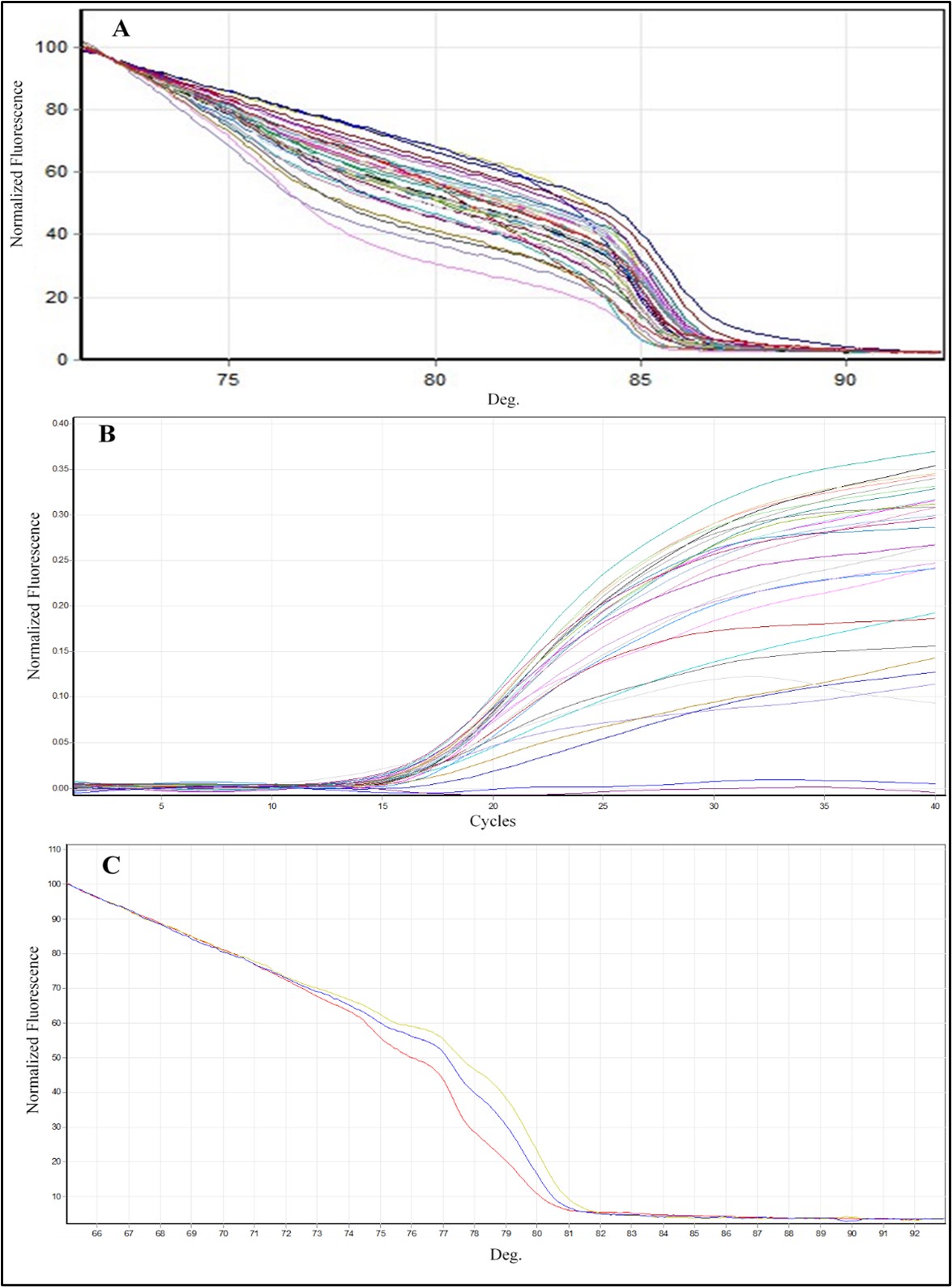
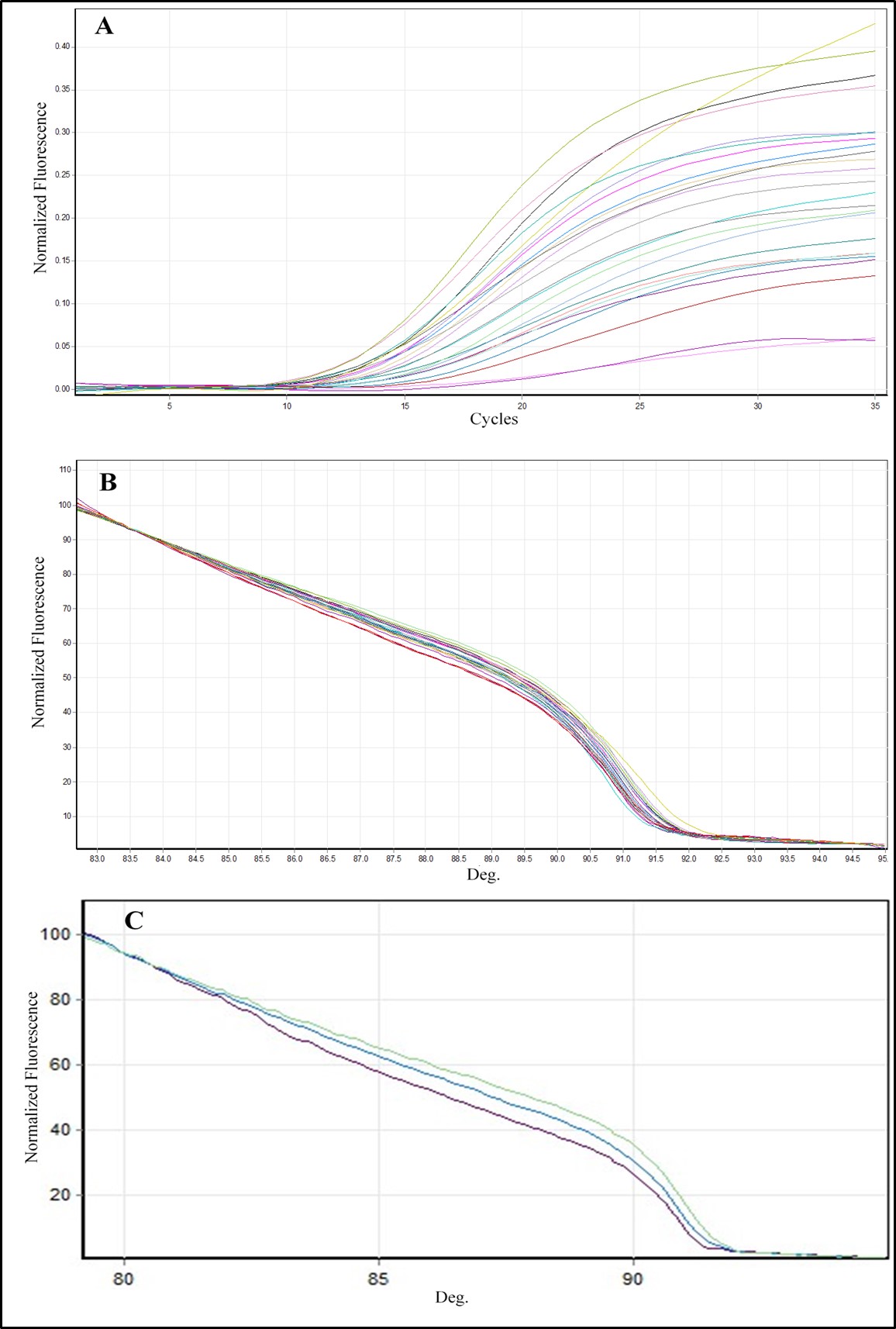
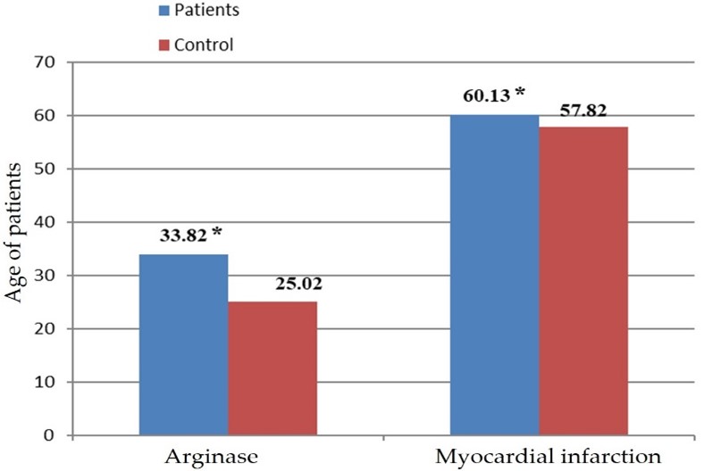
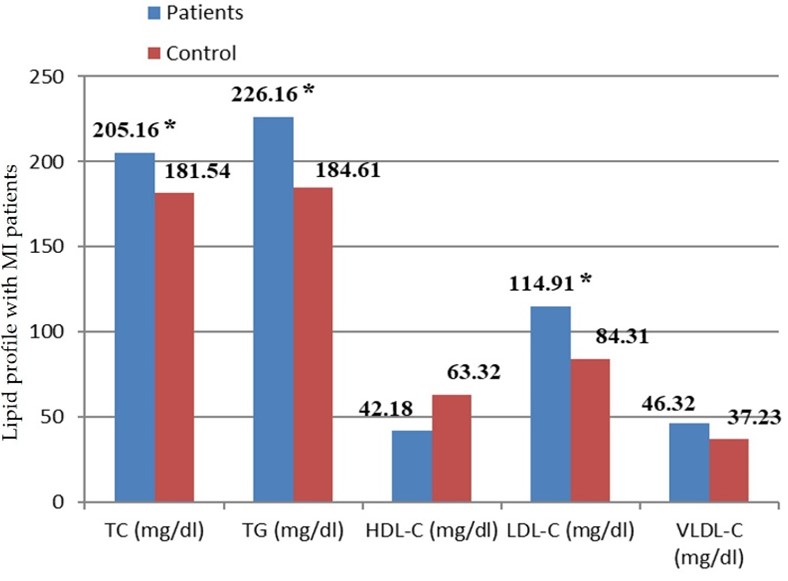
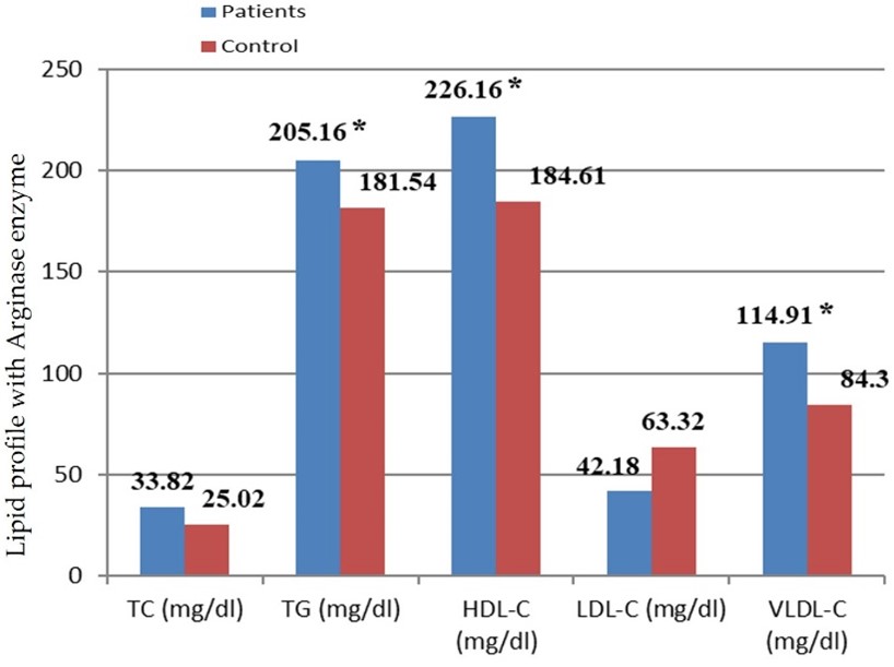
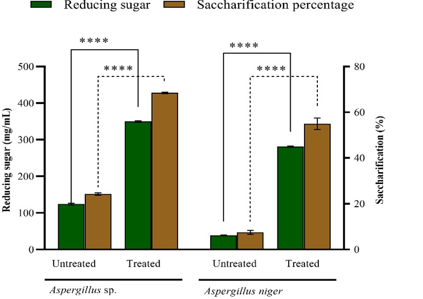
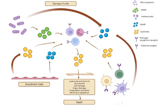
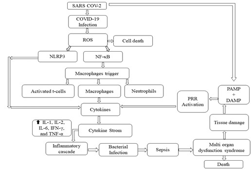
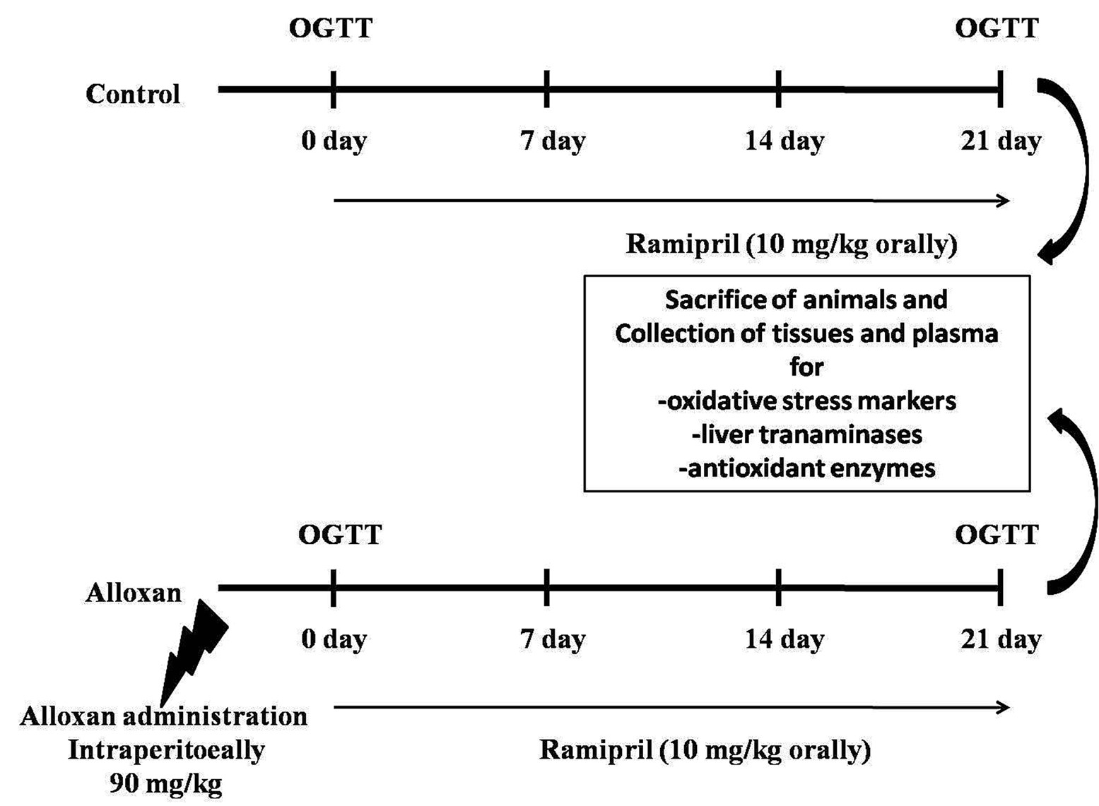
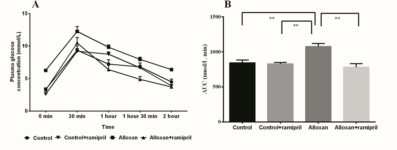
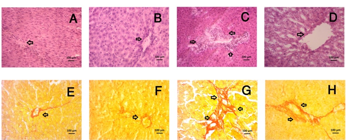
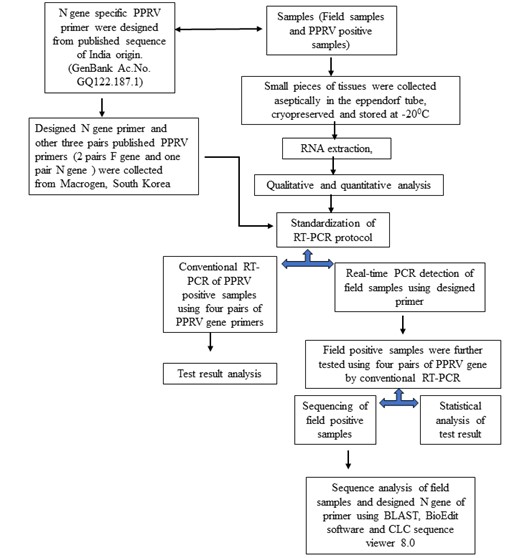
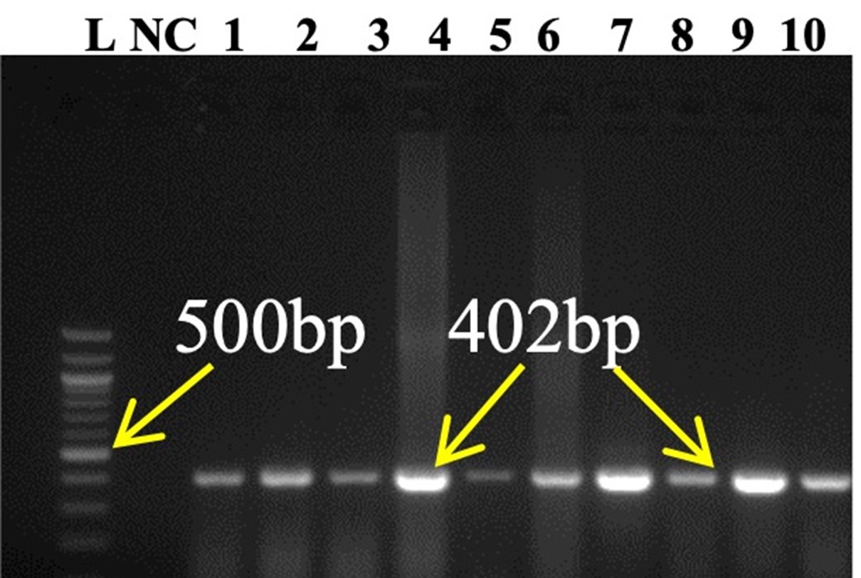
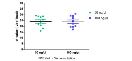
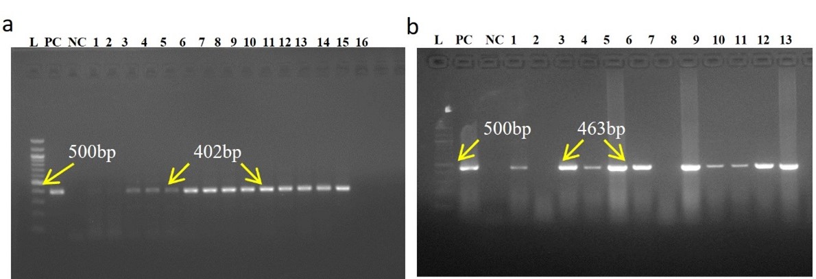
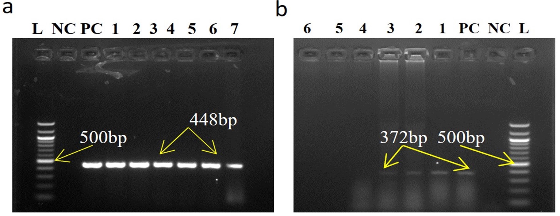
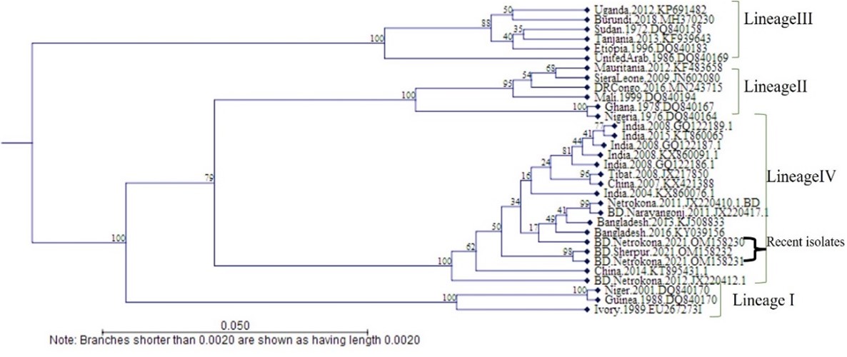
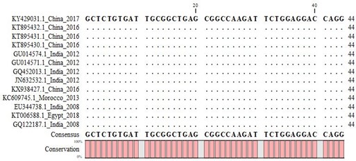
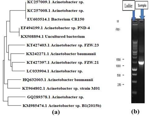
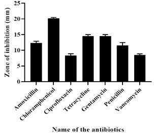
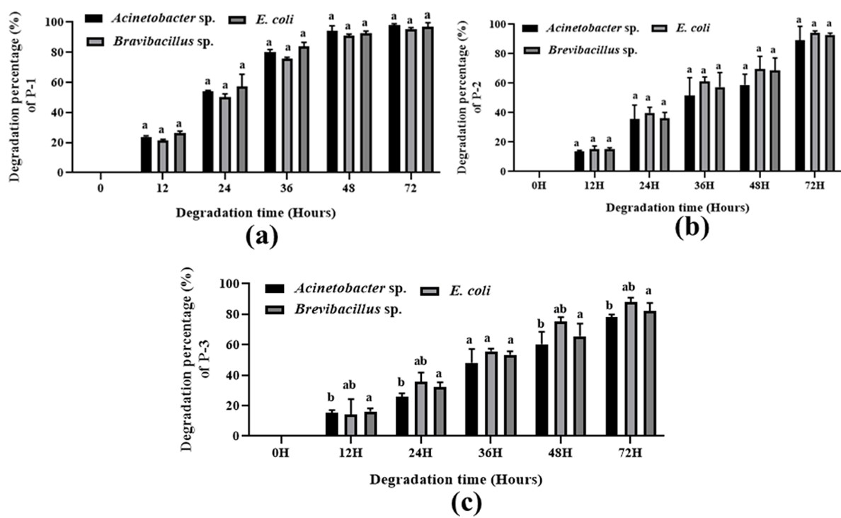
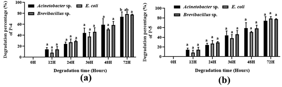
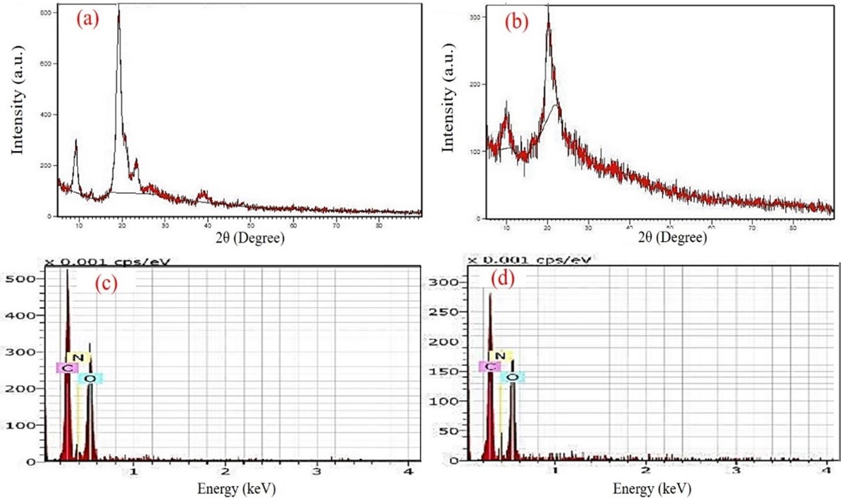
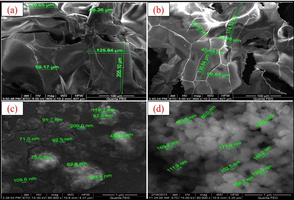
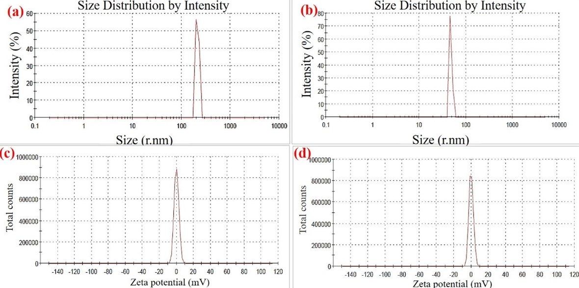
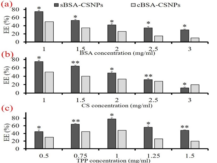
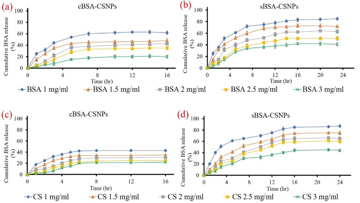
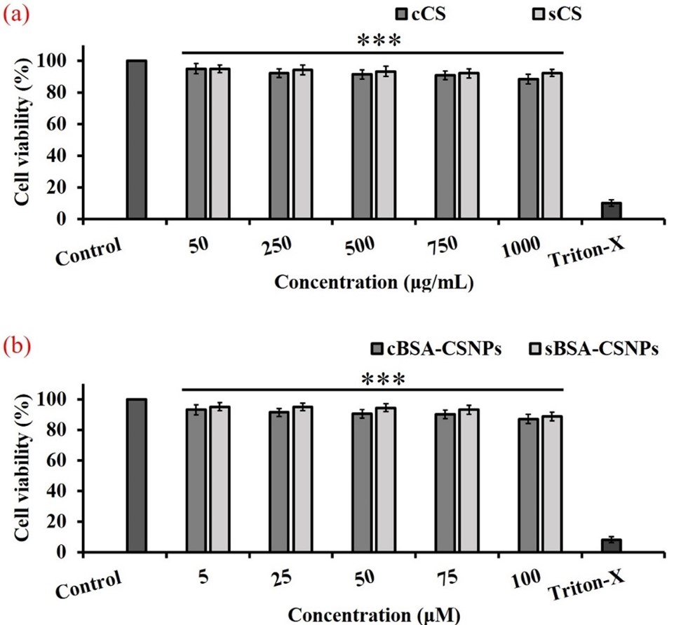
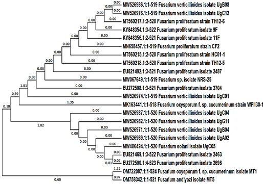
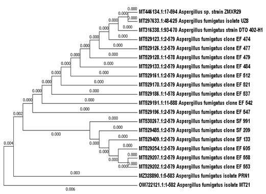
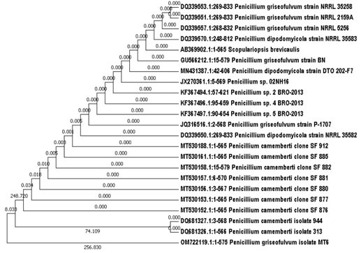
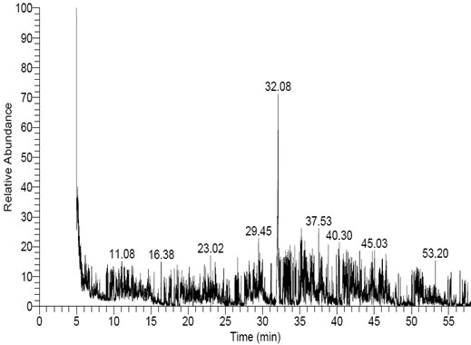
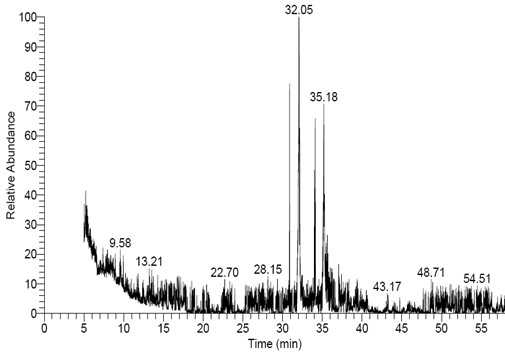
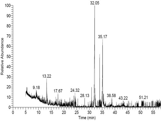
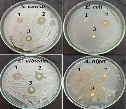
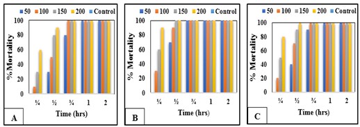
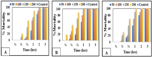