HTTP 200 OK
Allow: GET, HEAD, OPTIONS
Content-Type: application/json
Vary: Accept
{
"count": 321,
"next": "https://jabet.bsmiab.org/articles/?format=api&page=16",
"previous": "https://jabet.bsmiab.org/articles/?format=api&page=14",
"results": [
{
"id": 154,
"slug": "178-1648149750-developing-a-multiepitope-vaccine-against-dengue-virus-in-bangladesh-using-immunoinformatics-approach",
"featured": false,
"slider": false,
"issue": "Vol6 Issue1",
"type": "original_article",
"manuscript_id": "178-1648149750",
"recieved": "2022-03-26",
"revised": null,
"accepted": "2022-08-06",
"published": "2022-08-19",
"pdf_file": "https://jabet.bsmiab.org/media/pdf_file/2023/11/178-1648149750.pdf",
"title": "Developing a multiepitope vaccine against dengue virus in Bangladesh using immunoinformatics approach",
"abstract": "<p>Dengue fever is a devastating mosquito-borne illness that has claimed the lives of countless people. The virus responsible for this disease is a member of the Flaviviridae family, which produces positive-stranded RNA. Dengue fever is an exquisite viral fever caused by the bite of an Aedes mosquito carrying one of four serotypes of dengue virus. This virus is transmitted via a vertical route utilizing a comprehensively unique system. Unfortunately, no effective vaccine has yet been developed to eradicate this disease. This study employed computational methods to design and propose a multi-epitope vaccine against dengue virus in Asia. This study utilized various immunoinformatics databases to predict potent epitopes on the envelope protein of the dengue virus using in silico methods. We identified a total of 14 epitopes from the target envelope protein by assessing their ability to induce both innate and acquired immunity through T- and B-lymphocyte-mediated responses. Because dengue virus is an RNA virus, epitope conservation was considered, and all selected epitopes were 100 percent conserved. The antigenicity of the final component of the multi-epitope vaccine was 0.7055. To improve the stability of the vaccine protein, disulfide engineering was performed in a region with high mobility. Additionally, codon adaptation and in silico cloning ensure that the proposed subunit vaccine will be expressed at a higher level in <em>E. coli</em>. In order to evaluate the binding free energy and stability of the combination, the vaccine protein and TLR-4 receptor were subjected to a molecular docking simulation. In order to establish active immunity against the dengue virus, the proposed in silico vaccine must be tested for safety and immunogenicity.</p>",
"journal_reference": "J Adv Biotechnol Exp Ther. 2023; 6(1): 44-57.",
"academic_editor": "Md Jamal Uddin, PhD; ABEx Bio-Research Center, Dhaka-1230, Bangladesh",
"cite_info": "Akash SR, Hossain MI, et al. Developing a multiepitope vaccine against dengue virus in Bangladesh using immunoinformatics approach. J Adv Biotechnol Exp Ther. 2023; 6(1): 44-57.",
"keywords": [
"Epitope",
"Dengue",
"Microbiology",
"Immunoinformatics",
"Vaccine"
],
"DOI": "10.5455/jabet.2023.d105",
"sections": [
{
"section_number": 1,
"section_title": "INTRODUCTION",
"body": "<p>Dengue is a serious disease transmitted by mosquitoes that has the potential to spread as a pandemic. Endemic dengue is a causative agent and it is a grave health warning for many developing tropical countries. They have been established in all tropical regions of the planet for more than six decades [<a href=\"#r-1\">1</a>]. Southeast Asia is identified as the region with the highest prevalence of this disease in numerous tropical areas. The awe-inspiring hemorrhagic pattern of dengue fever has become the most dangerous cause of death in southeast Asia. This infection has also been documented in non-tropical regions of Asia, including East Asia and China [<a href=\"#r-2\">2</a>]. Dengue occurs irregularly in Bangladesh, where the virus caused a widespread epidemic in 2000 and persisted until 1964. At that time, we discovered dengue in Bangladesh, where it was first identified, and identified factors beneficial to future dengue hemorrhagic fever epidemics [<a href=\"#r-3\">3</a>]. Most likely, the outbreak started when a strain of the dengue virus came from a nearby country where it was common, and probably it was Thailand. In addition to the end of dichlorodiphenyltrichloroethane (DDT) spraying, the spread of disease was caused by weather, population, and lifestyle [<a href=\"#r-4\">4</a>]. Even though it increased every other year, the highest number of cases was reported in 2002. Thus, we reduce the number of notifications that may be an artifact of the surveillance system [<a href=\"#r-5\">5</a>]. Poll-based serological observation suggests that transmission of dengue is typical. Without intelligent interventions, future dengue risk will be exacerbated by unplanned urbanization, environmental decline, rising population mobility, and financial factors. Therefore, there is an urgent need to develop a vaccine for these high-risk regions. As part of this research, we need to use immunoinformatics to make a vaccine against the Asian-associated dengue virus that will work in Bangladesh. A flow chart discloses the full procedure of the study in <a href=\"#figure1\">Figure 1</a>.</p>\r\n\r\n<div id=\"figure1\">\r\n<figure class=\"image\"><img alt=\"\" height=\"598\" src=\"/media/article_images/2023/22/28/178-1648149750-Figure1.jpg\" width=\"500\" />\r\n<figcaption><strong>Figure 1.</strong> Schematic depiction of our workflow procedure.</figcaption>\r\n</figure>\r\n\r\n<p> </p>\r\n</div>"
},
{
"section_number": 2,
"section_title": "METHODS AND MATERIALS",
"body": "<p><strong>Proteome salvation and antigen extracts</strong><br />\r\nWe selected probable HCMV proteomes from the viprbrc website database in order to pick antigens [6]. On the HCMV surface membrane, spike proteins were deposited. They collaborated with this host-binding protein to enter the human genome [<a href=\"#r-7\">7</a>]. We measured the HCMV spike protein for the multiepitope vaccine strategy with the precise link between glycoproteins and disease. First, we selected the protein sequence of the dengue virus, which had been downloaded as a fasta file. The ddg-pharmfac database website was used to analyze the collected antigens having a threshold value of 0.4 [<a href=\"#r-8\">8</a>]. Lastly, the spike protein with the highest antigenic score was chosen for further investigation.</p>\r\n\r\n<p> </p>\r\n\r\n<p><strong>Forecast and evaluation of helper T-lymphocyte epitopes</strong><br />\r\nHelper T-cells (HTLs) are an essential component of adaptive immunity that recognizes different antigens and initiates B and cytotoxic T-cells, resulting in the destruction of the pathogen. Furthermore, B-cell and T-cell epitopes were predicted from dengue virus, as well as SARS coronavirus, which became disease and could be pandemic. To determine the HTL epitopes, we utilized the IEDB’s MHC class II essential allele prediction tool. The HTL epitopes were chosen using the Agreement method to support a percentile level of fifty [<a href=\"#r-7\">7</a>]. These epitopes underwent additional testing and demonstrated antigenicity using Vaxijen server version 2.0 [<a href=\"#r-9\">9</a>].</p>\r\n\r\n<p> </p>\r\n\r\n<p><strong>Forecast and evaluation of cytotoxic T-lymphocyte epitopes</strong><br />\r\nCytotoxic T lymphocytes are able to kill phagocytes via this mechanism directly [<a href=\"#r-10\">10</a>]. Therefore, we utilized the NetCTL v1.2 server [<a href=\"#r-11\">11</a>]. The collected epitopes were tested once more using the Vaxijen v2.0 [<a href=\"#r-10\">10</a>], Toxiprod [<a href=\"#r-13\">13</a>], and Allerrtop v2.0 [9] servers. All parameters are left at their defaults for all forecasts. A prediction threshold of 0.75 was established for CTL epitope identification [<a href=\"#r-13\">13</a>].</p>\r\n\r\n<p> </p>\r\n\r\n<p><strong>Forecast and evaluation of linear B lymphocyte epitopes</strong><br />\r\nB cell epitopes are needed to safely give humoral or antibody medication [<a href=\"#r-14\">14</a>]. For this purpose, the online portal iBCE-EL (http://www.thegleelab.org/iBCE-EL/) utilized this with default levels [<a href=\"#r-15\">15</a>].</p>\r\n\r\n<p> </p>\r\n\r\n<p><strong>Modeling of multi-epitope vaccine</strong><br />\r\nThe vaccine was produced using the selected CTL epitope, HTL epitope, and LBL epitopes, a complete adjuvant, and the pertinent linkers [<a href=\"#r-14\">14; 16</a>]. As an adjuvant for viral glycoprotein recognition, we used TLR4 agonist here [<a href=\"#r-17\">17; 18</a>]. Therefore, 50S ribosomal protein (NCBI ID: P9WHE3) was valued as an adjuvant to enhance the immunogenicity of the candidate vaccine. The adjuvant was linked to the linker EAAAK. In contrast, the selected CTL was linked with (AAY) linkers, the HTL was linked with (GPGPG) linkers, and the LBL was linked with (KK) linkers [<a href=\"#r-14\">14; 16</a>]. The AAY linker was utilized to affect protein equilibrium [<a href=\"#r-19\">19; 20</a>]. The linkers effectively separate two epitopes to ensure that each epitope maintains its optimal function [<a href=\"#r-13\">13</a>].</p>\r\n\r\n<p> </p>\r\n\r\n<p><strong>Physicochemical and immunological evaluation</strong><br />\r\nThe functional characteristics of the vaccine were predicted using the ProtParam database (http://web.expasy.org/protparam/) [<a href=\"#r-21\">21</a>]. ProtParam is a program that calculates the various physical and chemical parameters of a protein sequence, such as its molecular weight, theoretical pI (isoelectric point), amino acid composition, atomic composition, extinction coefficient, estimated half-life, instability index, aliphatic index, and grand average hydropathicity (GRAVY). Again, we used Vaxijen v2.0 (http://www.ddg-pharmfac.net/vaxijen/VaxiJen/VaxiJen.html) [<a href=\"#r-22\">22</a>] to evaluate the vaccine’s immune properties by measuring its MHC-1 immunogenicity [<a href=\"#r-9\">9; 11</a>] using Allertop, Biosoland, and SOLpro [<a href=\"#r-8\">8</a>].</p>\r\n\r\n<p> </p>\r\n\r\n<p><strong>Secondary construction forecast</strong><br />\r\nThe SOPMA server (https://npsa-prabi.ibcp.fr/NPSA/npsa seccons.html) and PSIPRED v4.0 server (http://bioinf.cs.ucl.ac.uk/index.php?id=779) identified the vaccine model’s two-dimensional basic characteristics, such as alpha-helix and random coils, when given the vaccine model [<a href=\"#r-23\">23</a>]. SOPMA produces a secondary structure prediction accuracy of greater than 80% [<a href=\"#r-13\">13</a>].</p>\r\n\r\n<p> </p>\r\n\r\n<p><strong>Homology modeling, 3D construction clarification and validation</strong><br />\r\nWe uploaded the created vaccine to the I-TASSER server (https://zhanggroup.org/I-TASSER) [24] in order to construct the structure prognosis. Then, to breed vaccine composition, we refine the vaccine from the Galaxyweb server (https://galaxy.seoklab.org/) [<a href=\"#r-25\">25</a>]. The complete structure was downloaded from the portal, and the chosen structure was subsequently named based on the highest RMSD rate and effectiveness number. Using the PyMOL v2.3.4 software, we could observe the refined and refined formation practiced imaginatively. The ProSA-web accessory and Procheck demonstrated the significance of the Ramachandran plot and Z-point [<a href=\"#r-26\">26</a>].</p>\r\n\r\n<p> </p>\r\n\r\n<p><strong>Molecular docking investigations</strong><br />\r\nIt highlights the key connections between protein model units and receptor units. For this docking study [<a href=\"#r-27\">27</a>], we uploaded the completed vaccine model as ligand and the TLR4 protein as a receptor molecule to the ClusPro v2.0 site. The TLR4 receptor (PDB ID: 3W3M) was picked from the PDB website.</p>\r\n\r\n<p> </p>\r\n\r\n<p><strong>Molecular dynamics simulation study</strong><br />\r\nThe simulation of molecular dynamics was used to examine the physical motions of atoms and molecules and biophysical systems. This would allow us to assess the dynamics and safety of the vaccine-receptor fear [<a href=\"#r-28\">28</a>]. The illusion had been removed from the iMODS website (<a href=\"http://imods.chaconlab.com/\">http://imods.chaconlab.com</a>).</p>\r\n\r\n<p> </p>\r\n\r\n<p><strong>Exempt protected rejoinder simulation</strong><br />\r\nThe whole construct was sent to the C-IMMSIM server (www.cbs.dtu.dk/services/C-ImmSim-10.1/) to see how the vaccine might affect the immune system [<a href=\"#r-29\">29</a>]. As mentioned, we agreed that a 30-day break between application submissions would be the minimum acceptable gap [<a href=\"#r-30\">30</a>].</p>\r\n\r\n<p> </p>\r\n\r\n<p><strong>Codon adaptation and in silico cloning technique</strong><br />\r\nAs the appearance of an alien gene in an organism is problematic, organism-specific codon optimization is required more frequently. Depending on the codon change, the JCat server released the construct. The modified course was evaluated based on the codon adaptation ratio (CAI) preference and guanine-cytosine content. In SnapGene v4.2, the body in silico cloning strategy was successfully implemented.</p>"
},
{
"section_number": 3,
"section_title": "RESULTS",
"body": "<p><strong>Best antigenic protein selection</strong><br />\r\nUsing UniProt server, different structural and non-structural protein sequences were retrieved to construct the vaccine. Based on antigenicity, the design protein scored with an antigenic point of 0.7055 (Vaxijen). Further analysis of these proteins’ amino acid sequences was done in order to determine CTL and HTL epitopes.</p>\r\n\r\n<p> </p>\r\n\r\n<p><strong>Possible HTL epitopes</strong><br />\r\nA total of 195 epitopes, consisting of 15 amino acids, were identified using the IEDB server for mouse MHC-II alleles (IAb, IAd, IAs, IEb, IEd and IEs). We only selected among top five HTL epitopes based on antigenic scores to construct the final vaccine (<a href=\"#Table-1\">Table 1</a>).</p>\r\n\r\n<div id=\"Table-1\">\r\n<p><a href=\"https://jabet.bsmiab.org/table/178-1648149750-table1/\">Table-1</a><strong>Table 1.</strong> The selected HTL epitopes for the final vaccine construction.</p>\r\n\r\n<p> </p>\r\n</div>\r\n\r\n<p><strong>Possible CTL epitopes</strong><br />\r\nFrom NetCTL 1.2, server, structural, and non-structural protein CTL epitopes were predicted on the basis of antigenicity, allergenicity, toxicity and C. score. A number of 170 epitopes, where each consist of a length of nine amino acids CTL were predicted from the sorted out of spike protein. We took top five CTL epitopes based on antigenicity score (<a href=\"#Table-2\">Table 2</a>).</p>\r\n\r\n<div id=\"Table-2\">\r\n<p><a href=\"https://jabet.bsmiab.org/table/178-1648149750-table2/\">Table-2</a><strong>Table 2.</strong> The selected CTL epitopes for the final vaccine construction.</p>\r\n\r\n<p> </p>\r\n</div>\r\n\r\n<p><strong>Possible LBL epitopes</strong><br />\r\nAll structural and non-structural proteins of linear B-cell epitopes were predicted from the iBCE-EL server, where they were shortlisted based on the prediction score. We chose only one out of a total of 36 B-cell epitopes that was shown to be antigenic, non-toxic, and non-allergenic (<a href=\"#Table-3\">Table 3</a>).</p>\r\n\r\n<div id=\"Table-3\">\r\n<p><a href=\"https://jabet.bsmiab.org/table/178-1648149750-table3/\">Table-3</a><strong>Table 3.</strong> The selected B cell epitopes for the final vaccine construction.</p>\r\n\r\n<p> </p>\r\n</div>\r\n\r\n<p><strong>Vaccine construct and fundamental premises</strong><br />\r\nThe final vaccine was constructed with the selected 11 epitopes, which belong to three different classes, for example, 5 CTL, 5 HTL, and 1 LBL (<a href=\"#figure2\">Figure 2</a>). All epitopes were joined by the AAY, GPGPG, and KK linkers. The TLR4 agonist 50S ribosome added in the beginning played the role of extra support to the immunogenicity to construct these 178 amino acid vaccines.</p>\r\n\r\n<div id=\"figure2\">\r\n<figure class=\"image\"><img alt=\"\" height=\"230\" src=\"/media/article_images/2023/22/28/178-1648149750-Figure2.jpg\" width=\"500\" />\r\n<figcaption><strong>Figure 2. </strong>Graphical outline of the expressed multi-epitope vaccine assembles where it linked with adjuvant, CTL epitopes, HTL epitopes and LBL epitopes (Left to right). Here the adjuvant and CTL were linked by EAAAK linker (pink), CTL epitopes were linked by AAK linker (Blue), HTL epitopes were linked by GPPG (Yellow) and finally LBL epitopes were linked by KK linker (Green).</figcaption>\r\n</figure>\r\n\r\n<p> </p>\r\n</div>\r\n\r\n<p><strong>Physicochemical characteristics and immunological assessment</strong><br />\r\nThe physicochemical properties of the constructed vaccine were analyzed and documented, as shown in (<a href=\"#Table-4\">Table 4</a>). The constructed vaccine’s molecular weight was 18822.22 Da. In addition to the 178 amino acids, the theoretical isoelectric point (pI) was 9.36. Simultaneously, the vaccine had the formula C<sub>834</sub>H<sub>1319</sub>N<sub>231</sub>O<sub>259</sub>S<sub>3</sub>, an instability index of 35.33, an aliphatic index of 75.67, and a grand average of hydropathicity of -0.337. In addition, the constructed vaccine’s antigenicity was 0.7055, it was non-toxic, and its solubility was 0.614 out of 1 (<a href=\"#figure3\">Figure 3</a>) indicating that it is highly soluble, and the windowed charge score and fold propensity score were depicted in <a href=\"#figure4\">Figure 4</a>.<br />\r\nThe a-helix, b-strand, and random coil secondary structures of the vaccine were evaluated using the SOPMA and PSIPRED servers (<a href=\"#Table-5\">Table 5</a> and <a href=\"#figure5\">Figure 5A</a>).</p>\r\n\r\n<div id=\"Table-4\">\r\n<p><a href=\"https://jabet.bsmiab.org/table/178-1648149750-table4/\">Table-4</a><strong>Table 4.</strong> Physicochemical characteristics of the construct.</p>\r\n</div>\r\n\r\n<p> </p>\r\n\r\n<div id=\"Table-5\">\r\n<p><a href=\"https://jabet.bsmiab.org/table/178-1648149750-table5/\">Table-5</a><strong>Table 5. </strong>The secondary structural features of designated vaccine.</p>\r\n\r\n<p> </p>\r\n</div>\r\n\r\n<div id=\"figure3\">\r\n<figure class=\"image\"><img alt=\"\" height=\"245\" src=\"/media/article_images/2023/22/28/178-1648149750-Figure3.jpg\" width=\"147\" />\r\n<figcaption><strong>Figure 3. </strong>Solubility value by protein sol server.</figcaption>\r\n</figure>\r\n</div>\r\n\r\n<div id=\"figure4\">\r\n<figure class=\"image\"><img alt=\"\" height=\"319\" src=\"/media/article_images/2023/22/28/178-1648149750-Figure4.jpg\" width=\"500\" />\r\n<figcaption><strong>Figure 4.</strong> A) Windowed charge score per amino acid. B) Windowed fold propensity score per amino acid. C) Secondary structure prediction report.</figcaption>\r\n</figure>\r\n</div>\r\n\r\n<div id=\"figure5\">\r\n<figure class=\"image\"><img alt=\"\" height=\"192\" src=\"/media/article_images/2023/22/28/178-1648149750-Figure5.jpg\" width=\"500\" />\r\n<figcaption><strong>Figure 5.</strong> A) Tertiary structure of designated vaccine. Different secondary structure of amino acids is identified as different color, like a-helix as red color, b-strand as yellow color and random coil as green color. B) Refined Tertiary structure of designated vaccine.</figcaption>\r\n</figure>\r\n</div>\r\n\r\n<p> </p>\r\n\r\n<p><strong>Tertiary structure, sophistication, and evaluation</strong><br />\r\nThe I-TASSER server (https://zhanggroup.org/I-TASSER) was used to obtain the best homology model (PDB id:01) among the top five modes. As advised by the server, we selected the lowest C-score (-3.38). <a href=\"#figure5\">Figure 5B</a> depicts the designed vaccine’s tertiary structure. The vaccine demonstrated in the Ramachandran graph that it exceeds 93.8 percent in the significant region, with a GDT- score, RMSD, MolProbity 2.047, 1.276 Clash 1.3, and rotamers score of 0.0. This was determined by refining the model that we developed (<a href=\"#figure6\">Figure 6</a>). The average Z score for the vaccine is -8.81 according to the Procheck online site, which was provided with <a href=\"#Table-6\">Table 6</a>, an additional file containing all findings, and the ProSA web server.</p>\r\n\r\n<div id=\"figure6\">\r\n<figure class=\"image\"><img alt=\"\" height=\"426\" src=\"/media/article_images/2023/22/28/178-1648149750-Figure6.jpg\" width=\"485\" />\r\n<figcaption><strong>Figure 6. </strong>Validation of the tertiary structure of the vaccine</figcaption>\r\n</figure>\r\n</div>\r\n\r\n<div id=\"Table-6\">\r\n<p><a href=\"https://jabet.bsmiab.org/table/178-1648149750-table6/\">Table-6</a><strong>Table 6.</strong> The protein structure and overall structure geometry.</p>\r\n</div>\r\n\r\n<p> </p>\r\n\r\n<p><strong>Molecular docking analysis</strong><br />\r\nWe evaluated the interaction between the refined model and immune receptor TLR-5 using a software-based simulation Clus Pro v2.0 site. It docked 30 models in various positions where the minimum energy value was determined using an additional file. Consequently, we placed model 2 in the dangling position with an energy score of -1401.2% (<a href=\"#figure7\">Figure 7</a>).</p>\r\n\r\n<div id=\"figure7\">\r\n<figure class=\"image\"><img alt=\"\" height=\"233\" src=\"/media/article_images/2023/22/28/178-1648149750-Figure7.jpg\" width=\"284\" />\r\n<figcaption><strong>Figure 7. </strong>Constructed vaccine after molecular docking.</figcaption>\r\n</figure>\r\n\r\n<p> </p>\r\n</div>\r\n\r\n<p><strong>Molecular dynamics simulation experiment</strong><br />\r\nDocked complex was subjected to molecular dynamics using the iMODS server to examine stable interactions between ligand molecule and receptor (TLR-5) at the microscopic level. The average life B- factor map, Eigenvalues, Variance, Covariance map, and Elastic network were displayed on <a href=\"#figure8\">Figure 8A-F</a>, respectively.</p>\r\n\r\n<div id=\"figure8\">\r\n<figure class=\"image\"><img alt=\"\" height=\"274\" src=\"/media/article_images/2023/22/28/178-1648149750-Figure8.jpg\" width=\"500\" />\r\n<figcaption><strong>Figure 8. </strong>Molecular dynamics simulation of the vaccine. Here, different MD simulation plots show (A) Molecular deformability on molecular dynamic simulation; (B) B-factor/mobility on molecular dynamic simulation; (C) Eigenvalues on molecular dynamic simulation; (D) Variance on molecular dynamic simulation; (E) Covariance map on molecular dynamic simulation; (F) Elastic network on molecular dynamic simulation.</figcaption>\r\n</figure>\r\n\r\n<p> </p>\r\n</div>\r\n\r\n<p><strong>Exempt rejoinder simulation</strong><br />\r\nSpecial pathogens expressed produced actual immunological aspects confirmed (<a href=\"#figure9\">Figures 9A-G</a>). Antigen and immunoglobulin values showed in Figure 9A, B lymphocytes cell; IgM and IgG in <a href=\"#figure9\">Figure 9B</a>, plasma B lymphocytes count as sub-divided per isotope (IgM, IgG1, IgG2) in <a href=\"#figure9\">Figure 9C</a>. The other value showed in <a href=\"#figure9\">Figure 9C to 9G</a>.</p>\r\n\r\n<div id=\"figure9\">\r\n<figure class=\"image\"><img alt=\"\" height=\"279\" src=\"/media/article_images/2023/22/28/178-1648149750-Figure9.jpg\" width=\"500\" />\r\n<figcaption><strong>Figure 9. </strong>Immune response stirred up by the designed vaccine where the graph shows (A) Antigen and immunoglobulins. Antibodies are divided per isotype; (B) B lymphocytes: total measure using selected epitopes; (C) Plasma B lymphocytes measure using the isotype (IgM, IgG1 and IgG2); (D) CD4 T-helper lymphocytes count. The plot shows total and memory counts; (E) CD4 T-regulatory lymphocytes count. Both total, memory and per entity-state counts are plotted here; (F) CD8 T-cytotoxic lymphocytes count. Total and memory shown; and (G) Cytokines Concentration of cytokines and interleukins. D in the inset plot is danger signal.</figcaption>\r\n</figure>\r\n\r\n<p> </p>\r\n</div>\r\n\r\n<p><strong>Codon evolution and in silico cloning</strong><br />\r\nThe codons in the created vaccine were optimized to develop their key factor according to the <em>E. coli</em> bacteria in JCat site. Lastly, the formed size of the vaccine cloning product is 5907 base pairs, and the vector was 5369, insert 546 base pairs (nucleotide base pair). <a href=\"#figure10\">Figure 10</a> depicts the cloned product that was manufactured.</p>\r\n\r\n<div id=\"figure10\">\r\n<figure class=\"image\"><img alt=\"\" height=\"347\" src=\"/media/article_images/2023/22/28/178-1648149750-Figure10.jpg\" width=\"500\" />\r\n<figcaption><strong>Figure 10.</strong> Constructed vaccine after cloning. The red section indicates the codon-optimized multi-epitope vaccine that has been introduced into the pET-28a (+) expression vector.</figcaption>\r\n</figure>\r\n\r\n<p> </p>\r\n</div>"
},
{
"section_number": 4,
"section_title": "DISCUSSION",
"body": "<p>An estimated 3.9 billion people worldwide are susceptible to dengue infection. In 129 nations, Asians shoulder the lion’s share of the burden or nearly 70 percent of the total [<a href=\"#r-31\">31</a>].<br />\r\nIn recent decades, millions of dengue cases have been reported annually, claiming the lives of a substantial section of the tropical and subtropical populations. In addition, the disease’s rapid global spread [<a href=\"#r-13\">13</a>] makes dengue an ever-worsening global problem. dengue is a virus transmitted by mosquitoes, and different DENV serotypes can cause the disease. These serotypes (DENV-1-4) exhibit immunological cross-reactivity [<a href=\"#r-31\">31</a>]. dengue treatment should stray as far as possible from “eliminating the pathogen and reducing confusion” [<a href=\"#r-32\">32</a>]. Yet, no specific vaccination has been discovered due to the lack of an effective vaccine and the goal to limit vaccine adverse effects. A vaccine must be developed immediately to eradicate dengue from the human body. Various types of candidates are already available in the field of research, but none has yet been established. In this study, however, we introduce a multi-epitope-based vaccine based on computational methods for the Asian region. This novel strategy is required to combat this life-threatening public health condition. The S protein contributes to the viral host range, infectiousness, and human-to-human transmission. Therefore, we had to design the vaccine to target the S protein to reach the virus’s surface. We selected the CTL, HTL, and LBL epitopes in this study. We determined that the HTL was associated with the production of humoral and cellular immune responses and the HTL’s role in extending immunity and eliminating virus-infected cells.<br />\r\nRefining with the highest antigenic number, we selected one B-cell epitope from the top five models between HTL and CTL epitopes. The CTL, HBL, and LBL epitopes were attached to linkers to build a suitable vaccine. The constructed vaccine consists of 178 amino acids, and its physiological properties were established utilizing web resources. As the vaccine’s solubility was measured at 0.614 on a scale of 1, it was more soluble and would easily enter the <em>E. coli</em> host. Based on the vaccine’s physiological features, the theoretical PI value was determined to be 9.16 and fundamental. In addition, the physiochemical values of several metrics showed a higher possibility of effectiveness against the dengue virus. Web-based programs were used to analyze and forecast the homology modeling and three-dimensional structure of the manufactured vaccine. Using the PROCHECK server, the PDB file containing the final 3D structure of the vaccine was validated [<a href=\"#r-31\">31</a>]. For the Ramachandran plot, we discovered a good overall predicted value of Z-score (-8.81) and the characteristics of the most preferred, accepted, and disallowed regions.<br />\r\nIt is essential to establish the molecular basis of the TLR4 receptor’s effective immune response to develop the most efficient vaccination against MHC alleles (HLA-DRB1*04-01). Molecular docking between the peptide vaccine and the virus glycoprotein-binding favorable receptor of TLR4, which had the lowest energy score of -1401.2, suggested that the vaccination may inhibit infection. Furthermore, it revealed a potential close interaction between the modeled vaccine ligand and the TLR5 receptor surface [<a href=\"#r-31\">31</a>]. Codon optimization was conducted to stabilize the built vaccine within the host for optimal multi-epitope vaccine generation [<a href=\"#r-31\">31</a>], and codon optimization was performed. In silico cloning was performed using the JCAT web service [<a href=\"#r-31\">31</a>] to optimize codons in a pET28a (+) vector to prevent codon bias. As long as we maintain the greatest degree of safety tools and procedures, we hope that the dengue virus will be eradicated if this vaccination instruction is followed in the laboratory.</p>"
},
{
"section_number": 5,
"section_title": "CONCLUSIONS",
"body": "<p>Dengue is currently one of the most significant and life-threatening diseases in the world, and it is rapidly spreading. Bangladesh is one of the countries with the highest incidence of dengue transmission. Over the past few decades, dengue fever incidence has increased at an alarming rate. There is currently no effective and permanent treatment for dengue disease. Different approaches have been taken over the past few years, but none of them has yielded a definitive solution. In this study, a computational method was utilized to create a multi-epitope-based vaccine against the dengue virus.<br />\r\nOur designed vaccine possessed a high level of immunity and could bind with the immune receptor TLR4 to produce the greatest response against the dengue virus. As a result of this study, we hope that the designed vaccine may play a significant role in eradicating this rapidly spreading virus.</p>"
},
{
"section_number": 6,
"section_title": "ACKNOWLEDGEMENT",
"body": "<p>The authors would like to thank the department of Biotechnology and Genetic Engineering, Bangabandhu Sheikh Mujibur Rahman Science and Technology University, Gopalganj, Bangladesh for supporting this research.</p>"
},
{
"section_number": 7,
"section_title": "AUTHOR CONTRIBUTIONS",
"body": "<p>SRA, MIH and MSA designed the study. SRA, MIH and MSA performed the experiments, analyzed, and interpreted the data. SRA and MIH prepared the manuscript. MSA reviewed the manuscript. All authors approved the final version of the manuscript</p>"
},
{
"section_number": 8,
"section_title": "CONFLICTS OF INTEREST",
"body": "<p>There is no conflict of interest among the authors.</p>"
}
],
"figures": [
{
"figure": "https://jabet.bsmiab.org/media/article_images/2023/22/28/178-1648149750-Figure1.jpg",
"caption": "Figure 1. Schematic depiction of our workflow procedure.",
"featured": false
},
{
"figure": "https://jabet.bsmiab.org/media/article_images/2023/22/28/178-1648149750-Figure2.jpg",
"caption": "Figure 2. Graphical outline of the expressed multi-epitope vaccine assembles where it linked with adjuvant, CTL epitopes, HTL epitopes and LBL epitopes (Left to right). Here the adjuvant and CTL were linked by EAAAK linker (pink), CTL epitopes were linked by AAK linker (Blue), HTL epitopes were linked by GPPG (Yellow) and finally LBL epitopes were linked by KK linker (Green).",
"featured": false
},
{
"figure": "https://jabet.bsmiab.org/media/article_images/2023/22/28/178-1648149750-Figure3.jpg",
"caption": "Figure 3. Solubility value by protein sol server.",
"featured": false
},
{
"figure": "https://jabet.bsmiab.org/media/article_images/2023/22/28/178-1648149750-Figure4.jpg",
"caption": "Figure 4. A) Windowed charge score per amino acid. B) Windowed fold propensity score per amino acid. C) Secondary structure prediction report.",
"featured": false
},
{
"figure": "https://jabet.bsmiab.org/media/article_images/2023/22/28/178-1648149750-Figure5.jpg",
"caption": "Figure 5. A) Tertiary structure of designated vaccine. Different secondary structure of amino acids is identified as different color, like a-helix as red color, b-strand as yellow color and random coil as green color. B) Refined Tertiary structure of designated vaccine.",
"featured": false
},
{
"figure": "https://jabet.bsmiab.org/media/article_images/2023/22/28/178-1648149750-Figure6.jpg",
"caption": "Figure 6. Validation of the tertiary structure of the vaccine.",
"featured": false
},
{
"figure": "https://jabet.bsmiab.org/media/article_images/2023/22/28/178-1648149750-Figure7.jpg",
"caption": "Figure 7. Constructed vaccine after molecular docking.",
"featured": false
},
{
"figure": "https://jabet.bsmiab.org/media/article_images/2023/22/28/178-1648149750-Figure8.jpg",
"caption": "Figure 8. Molecular dynamics simulation of the vaccine. Here, different MD simulation plots show (A) Molecular deformability on molecular dynamic simulation; (B) B-factor/mobility on molecular dynamic simulation; (C) Eigenvalues on molecular dynamic simulation; (D) Variance on molecular dynamic simulation; (E) Covariance map on molecular dynamic simulation; (F) Elastic network on molecular dynamic simulation.",
"featured": false
},
{
"figure": "https://jabet.bsmiab.org/media/article_images/2023/22/28/178-1648149750-Figure9.jpg",
"caption": "Figure 9. Immune response stirred up by the designed vaccine where the graph shows (A) Antigen and immunoglobulins. Antibodies are divided per isotype; (B) B lymphocytes: total measure using selected epitopes; (C) Plasma B lymphocytes measure using the isotype (IgM, IgG1 and IgG2); (D) CD4 T-helper lymphocytes count. The plot shows total and memory counts; (E) CD4 T-regulatory lymphocytes count. Both total, memory and per entity-state counts are plotted here; (F) CD8 T-cytotoxic lymphocytes count. Total and memory shown; and (G) Cytokines Concentration of cytokines and interleukins. D in the inset plot is danger signal.",
"featured": false
},
{
"figure": "https://jabet.bsmiab.org/media/article_images/2023/22/28/178-1648149750-Figure10.jpg",
"caption": "Figure 10. Constructed vaccine after cloning. The red section indicates the codon-optimized multi-epitope vaccine that has been introduced into the pET-28a (+) expression vector.",
"featured": false
}
],
"authors": [
{
"id": 642,
"affiliation": [
{
"affiliation": "Department of Pharmacy, Bangladesh University, Dhaka-1207, Bangladesh"
}
],
"first_name": "Sajidur Rahman",
"family_name": "Akash",
"email": null,
"author_order": 1,
"ORCID": null,
"corresponding": false,
"co_first_author": false,
"co_author": false,
"corresponding_author_info": "",
"article": 154
},
{
"id": 643,
"affiliation": [
{
"affiliation": "Department of Biotechnology and Genetic Engineering, Bangabandhu Sheikh Mujibur Rahman Science and Technology University, Gopalganj8100, Bangladesh"
}
],
"first_name": "Md Imran",
"family_name": "Hossain",
"email": null,
"author_order": 2,
"ORCID": null,
"corresponding": false,
"co_first_author": false,
"co_author": false,
"corresponding_author_info": "",
"article": 154
},
{
"id": 644,
"affiliation": [
{
"affiliation": "Department of Biotechnology and Genetic Engineering, Bangabandhu Sheikh Mujibur Rahman Science and Technology University, Gopalganj8100, Bangladesh"
}
],
"first_name": "Md Sarafat",
"family_name": "Ali",
"email": "sarafatbiotech@bsmrstu.edu.bd",
"author_order": 3,
"ORCID": null,
"corresponding": true,
"co_first_author": false,
"co_author": false,
"corresponding_author_info": "Md Sarafat Ali, PhD; Department of Biotechnology and Genetic Engineering, Bangabandhu Sheikh Mujibur Rahman Science and Technology University,\r\nGopalganj-8100, Bangladesh, e-mail: sarafatbiotech@bsmrstu.edu.bd",
"article": 154
}
],
"views": 1361,
"downloads": 227,
"references": [
{
"id": 5014,
"serial_number": 1,
"pmc": null,
"reference": "Luo H, He J, Zheng K, Li L, za LJ-Z liu xing bing xue, 2002 undefined. Analysis on the epidemiologic features of Dengue fever in Guangdong province, 1990-2000. EuropepmcOrg n.d.",
"DOI": null,
"article": 154
},
{
"id": 5015,
"serial_number": 2,
"pmc": null,
"reference": "Soni A, Chugh K, Sachdev A, Gupta D. Management of dengue fever in ICU. Indian J Pediatr 2001;68.",
"DOI": null,
"article": 154
},
{
"id": 5016,
"serial_number": 3,
"pmc": null,
"reference": "Nhan NT, Phuong CXT, Kneen R, Wills B, Van My N, Phuong NTQ. Acute Management of Dengue Shock Syndrome: A Randomized Double-Blind Comparison of 4 Intravenous Fluid Regimens in the First Hour. Clin Infect Dis. 2001;32(2):204–13.",
"DOI": null,
"article": 154
},
{
"id": 5017,
"serial_number": 4,
"pmc": null,
"reference": "Damonte E, Matulewicz M, Cerezo A. Sulfated Seaweed Polysaccharides as Antiviral Agents. Curr Med Chem 2012;11.",
"DOI": null,
"article": 154
},
{
"id": 5018,
"serial_number": 5,
"pmc": null,
"reference": "Wagatsuma Y, Chowdhury M, Ahmed TU, Ashraf Uddin M, Nazmul Sohel S, Kittayapong P. Analysis of some Socio-demographic Factors Related to DF/DHF Outbreak in Dhaka City. 2000;24.",
"DOI": null,
"article": 154
},
{
"id": 5019,
"serial_number": 6,
"pmc": null,
"reference": "Pickett BE, Sadat EL, Zhang Y, Noronha JM, Squires RB, Hunt V, et al. ViPR: an open bioinformatics database and analysis resource for virology research. Nucleic Acids Res 2012;40:D593–8.",
"DOI": null,
"article": 154
},
{
"id": 5020,
"serial_number": 7,
"pmc": null,
"reference": "Wang P, Sidney J, Kim Y, Sette A, Lund O, Nielsen M, et al. Peptide binding predictions for HLA DR, DP and DQ molecules. BMC Bioinformatics 2010;11.",
"DOI": null,
"article": 154
},
{
"id": 5021,
"serial_number": 8,
"pmc": null,
"reference": "Magnan CN, Zeller M, Kayala MA, Vigil A, Randall A, Felgner PL, et al. High-throughput prediction of protein antigenicity using protein microarray data. Bioinformatics 2010;26:2936–43.",
"DOI": null,
"article": 154
},
{
"id": 5022,
"serial_number": 9,
"pmc": null,
"reference": "Dimitrov I, Flower DR, Doytchinova I. AllerTOP – a server for in silico prediction of allergens. BMC Bioinformatics 2013;14.",
"DOI": null,
"article": 154
},
{
"id": 5023,
"serial_number": 10,
"pmc": null,
"reference": "Calis JJA, Maybeno M, Greenbaum JA, Weiskopf D, De Silva AD, Sette A, et al. Properties of MHC Class I Presented Peptides That Enhance Immunogenicity. PLoS Comput Biol 2013;9.",
"DOI": null,
"article": 154
},
{
"id": 5024,
"serial_number": 11,
"pmc": null,
"reference": "Larsen M V., Lundegaard C, Lamberth K, Buus S, Lund O, Nielsen M. Large-scale validation of methods for cytotoxic T-lymphocyte epitope prediction. BMC Bioinformatics 2007;8:1–12.",
"DOI": null,
"article": 154
},
{
"id": 5025,
"serial_number": 12,
"pmc": null,
"reference": "Gupta S, Kapoor P, Chaudhary K, Gautam A, Kumar R, Raghava GPS. In Silico Approach for Predicting Toxicity of Peptides and Proteins. PLoS One 2013;8.",
"DOI": null,
"article": 154
},
{
"id": 5026,
"serial_number": 13,
"pmc": null,
"reference": "Ali M, Pandey RK, Khatoon N, Narula A, Mishra A, Prajapati VK. Exploring dengue genome to construct a multi-epitope-based subunit vaccine by utilizing immunoinformatics approach to battle against dengue infection. Sci Rep 2017;7.",
"DOI": null,
"article": 154
},
{
"id": 5027,
"serial_number": 14,
"pmc": null,
"reference": "Nain Z, Abdulla F, Rahman MM, Karim MM, Khan MSA, Sayed S Bin, et al. Proteome-wide screening for designing a multi-epitope vaccine against emerging pathogen Elizabethkingia anophelis using immunoinformatic approaches. https://doi.org/101080/0739110220191692072 2019;38:4850–67.",
"DOI": null,
"article": 154
},
{
"id": 5028,
"serial_number": 15,
"pmc": null,
"reference": "Manavalan B, Govindaraj RG, Shin TH, Kim MO, Lee G. iBCE-EL: A New Ensemble Learning Framework for Improved Linear B-Cell Epitope Prediction. Front Immunol 2018;9:1695.",
"DOI": null,
"article": 154
},
{
"id": 5029,
"serial_number": 16,
"pmc": null,
"reference": "Dorosti H, Eslami M, Negahdaripour M, Ghoshoon MB, Gholami A, Heidari R, et al. Vaccinomics approach for developing multi-epitope peptide pneumococcal vaccine.",
"DOI": null,
"article": 154
},
{
"id": 5030,
"serial_number": 17,
"pmc": null,
"reference": "Olejnik J, Hume AJ, Mühlberger E. Toll-like receptor 4 in acute viral infection: Too much of a good thing. PLoS Pathog 2018;14:1–7.",
"DOI": null,
"article": 154
},
{
"id": 5031,
"serial_number": 18,
"pmc": null,
"reference": "Pandey, Rajan Kumar, Tarun Kumar Bhatt, and Vijay Kumar Prajapati. “Novel immunoinformatics approaches to design multi-epitope subunit vaccine for malaria by investigating anopheles salivary protein.” Scientific reports 8.1 (2018): 1-11.",
"DOI": null,
"article": 154
},
{
"id": 5032,
"serial_number": 19,
"pmc": null,
"reference": "Abdellrazeq GS, Fry LM, Elnaggar MM, Bannantine JP, Schneider DA, Chamberlin WM, et al. Simultaneous cognate epitope recognition by bovine CD4 and CD8 T cells is essential for primary expansion of antigen-specific cytotoxic T-cells following ex vivo stimulation with a candidate Mycobacterium avium subsp. paratuberculosis peptide vaccine. Vaccine 2020;38:2016–25.",
"DOI": null,
"article": 154
},
{
"id": 5033,
"serial_number": 20,
"pmc": null,
"reference": "Borthwick N, Silva-Arrieta S, Llano A, Takiguchi M, Brander C, Hanke T. Novel nested peptide epitopes recognized by CD4+ T cells induced by HIV-1 conserved-region vaccines. Vaccines 2020;8.",
"DOI": null,
"article": 154
},
{
"id": 5034,
"serial_number": 21,
"pmc": null,
"reference": "Gasteiger E, Hoogland C, Gattiker A, Duvaud S, Wilkins MR, Appel RD, et al. Protein Identification and Analysis Tools on the ExPASy Server. Proteomics Protoc Handb 2005:571–607.",
"DOI": null,
"article": 154
},
{
"id": 5035,
"serial_number": 22,
"pmc": null,
"reference": "Doytchinova IA, Flower DR. VaxiJen: A server for prediction of protective antigens, tumour antigens and subunit vaccines. BMC Bioinformatics 2007;8:1–7.",
"DOI": null,
"article": 154
},
{
"id": 5036,
"serial_number": 23,
"pmc": null,
"reference": "Buchan DWA, Minneci F, Nugent TCO, Bryson K, Jones DT. Scalable web services for the PSIPRED Protein Analysis Workbench. Nucleic Acids Res 2013;41:349–57.",
"DOI": null,
"article": 154
},
{
"id": 5037,
"serial_number": 24,
"pmc": null,
"reference": "Roy A, Kucukural A, Zhang Y. I-TASSER: A unified platform for automated protein structure and function prediction. Nat Protoc 2010;5.",
"DOI": null,
"article": 154
},
{
"id": 5038,
"serial_number": 25,
"pmc": null,
"reference": "Nugent T, Cozzetto D, Jones DT. Evaluation of predictions in the CASP10 model refinement category. Proteins Struct Funct Bioinforma 2014;82:98–111.",
"DOI": null,
"article": 154
},
{
"id": 5039,
"serial_number": 26,
"pmc": null,
"reference": "Wiederstein M, Sippl MJ. ProSA-web: Interactive web service for the recognition of errors in three-dimensional structures of proteins. Nucleic Acids Res 2007;35.",
"DOI": null,
"article": 154
},
{
"id": 5040,
"serial_number": 27,
"pmc": null,
"reference": "Kozakov, Dima, David R. Hall, Bing Xia, Kathryn A. Porter, Dzmitry Padhorny, Christine Yueh, Dmitri Beglov, and Sandor Vajda. “The ClusPro web server for protein–protein docking.” Nature protocols 12, no. 2 (2017): 255-278.",
"DOI": null,
"article": 154
},
{
"id": 5041,
"serial_number": 28,
"pmc": null,
"reference": "López-Blanco, José Ramón, José I. Aliaga, Enrique S. Quintana-Ortí, and Pablo Chacón. “iMODS: internal coordinates normal mode analysis server.” Nucleic acids research 42, no. W1 (2014): W271-W276.",
"DOI": null,
"article": 154
},
{
"id": 5042,
"serial_number": 29,
"pmc": null,
"reference": "Rapin N, Lund O, Bernaschi M, Castiglione F. Computational immunology meets bioinformatics: The use of prediction tools for molecular binding in the simulation of the immune system. PLoS One 2010;5.",
"DOI": null,
"article": 154
},
{
"id": 5043,
"serial_number": 30,
"pmc": null,
"reference": "Castiglione, Filippo, Francesca Mantile, Piergiuseppe De Berardinis, and Antonella Prisco. “How the interval between prime and boost injection affects the immune response in a computational model of the immune system.” Computational and mathematical methods in medicine 2012 (2012):842329",
"DOI": null,
"article": 154
},
{
"id": 5044,
"serial_number": 31,
"pmc": null,
"reference": "Bhatt S, Gething PW, Brady OJ, Messina JP, Farlow AW, Moyes CL, et al. The global distribution and burden of Dengue. Nature 2013;496.",
"DOI": null,
"article": 154
},
{
"id": 5045,
"serial_number": 32,
"pmc": null,
"reference": "Abdus Samad, Foysal Ahammad, Zulkar Nain, Rahat Alam, Raihan Rahman Imon, Mahadi Hasan & Md. Shahedur Rahman. Designing a multi-epitope vaccine against SARS-CoV-2: an immunoinformatics approach, Journal of Biomolecular Structure and Dynamics, 2022; 40:1, 14-30.",
"DOI": null,
"article": 154
}
]
},
{
"id": 148,
"slug": "178-1653070433-assessment-and-comparison-of-cardiovascular-disease-risk-factors-and-biochemical-parameters-among-men-and-women-a-cross-sectional-study",
"featured": false,
"slider": false,
"issue": "Vol6 Issue1",
"type": "original_article",
"manuscript_id": "178-1653070433",
"recieved": "2022-05-20",
"revised": null,
"accepted": "2022-08-04",
"published": "2022-08-12",
"pdf_file": "https://jabet.bsmiab.org/media/pdf_file/2023/50/178-1653070433.pdf",
"title": "Assessment and comparison of cardiovascular disease risk factors and biochemical parameters among men and women: A cross-sectional study",
"abstract": "<p>Cardiovascular disease (CVD) is one of the most common causes of death among men and women worldwide. It is predicted that by 2030 around 23.3 million people will die as a consequence of CVD. There are numerous risk factors for CVD. The goal of this study is to examine the cardiovascular clinical and biochemical parameters of hospitalized CVD patients, as well as to assess the most common risk factors. Seventy-two known healthy individuals were randomly allocated to the control group (male 47 and female 25). We collected data through a questionnaire from 154 CVD patients as a study group (male 108 and female 46). Cardiovascular status was assessed using clinical parameters like hypertension, chest pain, shortness of breathing, pain in the arm, and biochemical parameters like lipid profile, RBS, creatinine, and an electrolyte panel. Inter- and intra-group comparison was performed. A total of 154 hospitalized CVD patients were analyzed (male 108 female 46) The most significant age group for males was observed as 56-65 years (mean 60) and for females was 38-48 (mean 43) years. A highly statistically significant increase was observed in total cholesterol and LDL in males than in females, but a decrease in TG than in females. Changes were also observed in other cardiovascular biochemical and clinical parameters. There was a significant difference in smoking status, physical activity, lipid panel, and other biochemical parameters among males and females. It is concluded that studies investigating the observed sex differences in traditional risk factor effects would not only help us better understand the etiology of CVD but also help us understand how to prevent it.</p>",
"journal_reference": "J Adv Biotechnol Exp Ther. 2023; 6(1): 25-34.",
"academic_editor": "Md. Abdul Hannan, PhD; Bangladesh Agricultural University, Bangladesh",
"cite_info": "Islam S, Nobel FA, et al. Assessment and comparison of cardiovascular disease risk factors and biochemical parameters among men and women: A cross-sectional study. J Adv Biotechnol Exp Ther. 2023; 6(1): 25-34.",
"keywords": [
"Hypertension",
"Lipid profile",
"cardiovascular disease",
"Outpatients",
"Biochemical parameter"
],
"DOI": "10.5455/jabet.2023.d103",
"sections": [
{
"section_number": 1,
"section_title": "INTRODUCTION",
"body": "<p>Cardiovascular disease is the leading cause of death worldwide and was responsible for an estimated 16.7 million deaths worldwide in 2010, with forecasts suggesting a startling 23.3 million by 2030. The number of deaths owing to dietary inadequacies, infectious diseases, and maternal and perinatal conditions is considered equivalent to mortality rates of CVD [<a href=\"#r-1\">1</a>]. According to WHO, an estimated 17.9 million people died from cardiovascular diseases (CVDs) in 2019, representing 32% of all deaths worldwide. Roughly 7.4 million of these deaths (17.9 million) are thought to be caused by coronary heart disease, and 6.7 million by stroke [<a href=\"#r-2\">2</a>]. In Europe, more than 4 million death occurred by CVD among which 49% of death occurs in women & 40% in men [<a href=\"#r-3\">3</a>]. CVD causes more deaths in the U.S. each year than any other cause, declared to the American heart association. In the case of Asia, the central Asian countries had the highest age-adjusted mortality from CVD, followed by West Asian, South Asian, and Southeast Asian countries according to the world health organization database 2004 [<a href=\"#r-4\">4</a>]. The populations of low- and middle-income countries like Bangladesh are increasingly being affected by CVD and account for over three-quarters of all CVD deaths [<a href=\"#r-5\">5</a>]. Among CVDs, strokes and heart attacks are usually acute events, stroke is the second cause of death worldwide [<a href=\"#r-6\">6</a>]. these are mainly caused by arteriosclerosis involving the heart or brain. Strokes can also be caused by blood clots or leakage from a blood vessel in the brain.<br />\r\nSeveral risk factors, such as family history, high cholesterol, high blood pressure, and being overweight or diabetic, have been linked to the development of CVD, but a significant number of people who have few or no identified risk factors will also develop CVD. Most cardiovascular diseases can be prevented by addressing behavioral risk factors, such as tobacco use, unhealthy diet and obesity, physical inactivity, harmful use of alcohol, hypertension, diabetes, and hyperlipidemia. This fact has led researchers to look for the risk factors that might be either causing CVD or that could be used to determine lifestyle changes and/or treatments that could reduce a person’s risk. People with cardiovascular disease or who are at high cardiovascular risk (due to the presence of one or more risk factors such as hypertension, diabetes, hyperlipemia, or already established disease) need early detection and management using counseling and medicines, as appropriate.<br />\r\nIn this study, we statistically analyzed the risk factors & biochemical parameters among the patients with CVD who are hospitalized based on age, sex, smoking, diabetes, and different medical conditions. Here tried to find out any difference in biochemical parameters between males and females based on age and medical conditions and tried to identify the most common risk factors among hospitalized patients which will help to identify the disease conditions and help in the diagnosis and prognosis of the CVD.</p>"
},
{
"section_number": 2,
"section_title": "METHODS AND MATERIALS",
"body": "<p><strong>Institutional and ethical committee permission</strong><br />\r\nFor this study data were collected from hospitalized patients and outpatients through a questionnaire with written consent from Sheikh Hasina Medical, Tangail, and Chittagong Medical College, Chittagong. The study was also approved by the ethical review committee of the Department of Biochemistry and Molecular Biology, Mawlana Bhashani Science and Technology University, Santosh, Tangail-1902, Bangladesh, with the certificate number MBSTU/BMB/TEST/6/2022/152.</p>\r\n\r\n<p> </p>\r\n\r\n<p><strong>Study design</strong><br />\r\nSubjects were divided into two groups: Group 1: Healthy control (n=72), and Group 2: patients with coronary artery disease (CAD) (n=154). The subjects in group 2 were further sub-divided into two groups; male (n=108) and female (n=46) to compare the risk factors and biochemical parameters between them.</p>\r\n\r\n<p> </p>\r\n\r\n<p><strong>Selection of healthy human volunteers</strong><br />\r\nThere were 72 healthy control (Male-47, Female-25), aged <25 to 75 years, and with BMI 18-22.9 were served healthy control. These individuals attended the outpatient department for their periodical health check-up.</p>\r\n\r\n<p> </p>\r\n\r\n<p><strong>Study population and data collection</strong><br />\r\nData were collected from outpatients and from the patients who were hospitalized at the ward of the coronary unit in Chittagong Medical College, Chittagong & Sheikh Hasina Medical, Tangail for this study purpose. We collected data from 154 patients (male 108 and female 46) aged between 35 to 80 with confirmed clinical diagnoses of CAD and the data included patient demographics, medical history, symptoms on arrival, results of laboratory testing, diabetes status, and smoking history, lipid-lowering medication, etc. The laboratory testing includes lipid profile, Creatinine, RBS, an electrolyte panel, and diabetes. Data were collected by using a standard questionnaire.</p>\r\n\r\n<p> </p>\r\n\r\n<p><strong>Determination of biochemical parameters</strong><br />\r\nThe biochemical parameters which were analyzed in this study were lipid profile, creatinine, random blood sugar (RBS), and electrolytes. A standard protocol was used to collect blood samples and performed the biochemical test of the blood samples for the measurement of lipid profile, creatinine level, and RBS and electrolytes.</p>\r\n\r\n<p> </p>\r\n\r\n<p><strong>Statistical analysis</strong><br />\r\nAll data were examined using IBM SPSS (Inc., Chicago, IL, USA) and Microsoft Excel. Frequency distribution, Cross-tabulation, and Bar chart were applied for the statistical estimation of the variables. Descriptive statistics like frequencies and percentages were used to determine the risk factors of CVD.</p>"
},
{
"section_number": 3,
"section_title": "RESULTS",
"body": "<p><strong>Demographic characteristics</strong><br />\r\nThe demographic characteristics such as age, weight, height, BMI, physical activity, smoking status, sleeping hours, etc. were collected from patients (<a href=\"#Table-1\">Table 1</a>).<br />\r\nThe study population ranged in age from 25 to 75 years old (mean 60.2), with the males on average 60 years old and the females on average 43 years old. Among them 5.8% of cardiovascular patients were under the age of 25, 12.3% were 26-35 years old, 14.9 % were 36-45 years old, 18.2% were 46-55 years old, 27.9% were 56-65 years old, 13.6 % were 66-76 years old, and 7.1 % were beyond 75 years old (<a href=\"#figure1\">Figure 1</a>). The participants range from age 56-65 and are the most susceptible to CVD in this area. This category accounts for nearly one-third of all CVD patients.<br />\r\nSmoking is another CVD risk factor that is attributed to the disease either directly or indirectly since it promotes plaque formation in atherosclerosis. In our study group, about 56% of patients were connected to smoking (47% of the respondents were current smokers, 9% were ex-smokers) and 44% were non-smokers (<a href=\"#figure2\">Figure 2</a>). In the case of males 97% of patients experienced passive smoking, 63% were current smokers, 10.2%were ex-smoker, and 26.9% never did smoke. On the other side, 43.5% of female patients experienced passive smoking, 10.7% were current smokers, 4.3% were ex-smoker and 84.8% were a non-smoker (<a href=\"#Table-1\">Table 1</a>).<br />\r\nCVD patients’ physical activity, such as the morning or other sorts of modest exercise was observed. Between males and females, a substantial difference in physical activity patterns was observed. A total of 17% of males and 27% of females never performed any type of physical activity. Physical activity was performed frequently by 48% of males and 39% of females, compared to occasionally by 35% of males and 34% of females (<a href=\"#figure3\">Figure 3</a>). Females are more likely than their male counterparts to have never exercised.<br />\r\nThe average sleeping hours of CVD patients were also investigated, with 20.8 percent of patients sleeping for 0-4 hours, 36.4 percent for 4-5 hours, and 42.86 percent for 6-7 hours (<a href=\"#Table-1\">Table 1</a>).</p>\r\n\r\n<div id=\"figure1\">\r\n<figure class=\"image\"><img alt=\"\" height=\"232\" src=\"/media/article_images/2023/22/29/178-1653070433-Figure1.jpg\" width=\"500\" />\r\n<figcaption><strong>Figure 1.</strong> Represents the frequency of CVD patients based on age. The age group 56-65 accounts for the majority of CVD patients, accounting for 27% of all patients. The second major age group associated with CVD is 46-55.</figcaption>\r\n</figure>\r\n</div>\r\n\r\n<div id=\"figure2\">\r\n<figure class=\"image\"><img alt=\"\" height=\"412\" src=\"/media/article_images/2023/22/29/178-1653070433-Figure2.jpg\" width=\"500\" />\r\n<figcaption><strong>Figure 2. </strong>The percentage of patients who are connected to smoking is depicted in the pie chart. Nearly half of all patients (47%) were current smokers.</figcaption>\r\n</figure>\r\n</div>\r\n\r\n<div id=\"figure3\">\r\n<figure class=\"image\"><img alt=\"\" height=\"273\" src=\"/media/article_images/2023/22/29/178-1653070433-Figure3.jpg\" width=\"500\" />\r\n<figcaption><strong>Figure 3. </strong>Comparison of physical activity between male and female patients. The number of males who frequently perform physical exercise is greater than females, besides more females never did exercise than males.</figcaption>\r\n</figure>\r\n</div>\r\n\r\n<div id=\"Table-1\">\r\n<p><a href=\"https://jabet.bsmiab.org/table/178-1653070433-table1/\">Table-1</a><strong>Table 1.</strong> Demographic profile and medical history of the participants.</p>\r\n\r\n<p> </p>\r\n</div>\r\n\r\n<p><strong>Medical history</strong><br />\r\nMales and females in the study group had their medical histories examined for hypertension, chest pain, diabetes mellitus, fatigue and weakness, and arm pain, shortness of breathing. Among males 98% of patients had hypertension, 33% had diabetes mellitus, 80.6% had chest pain, 65.7% felt fatigued and weak, 9.3% experienced pain in the arm and, 26.9% experienced shortness of breathing. On the contrary, among females 93.5% of patients had hypertension, 43.5% had diabetes mellitus, 32.6% had chest pain, 58.7% experienced fatigue and weakness, 4.3% felt pain in the arm and, 73.9% felt shortness of breathing. Hypertension was the most common medical symptom in both males (98%) and females (93.5%), with males having a higher rate than females. The number of patients with diabetes mellitus is greater in females (43.5%) than in males (33%) (<a href=\"#figure4\">Figure 4</a>).</p>\r\n\r\n<div id=\"figure4\">\r\n<figure class=\"image\"><img alt=\"\" height=\"246\" src=\"/media/article_images/2023/22/29/178-1653070433-Figure4.jpg\" width=\"500\" />\r\n<figcaption><strong>Figure 4.</strong> Comparison of medical history between male and female patients. More males experienced chest pain and fatigue & weakness than females, shortness of breathing was higher in females.</figcaption>\r\n</figure>\r\n\r\n<p> </p>\r\n</div>\r\n\r\n<p><strong>Cardiovascular biochemical parameters</strong><br />\r\nLipid profile, creatinine, random blood sugar (RBS), and electrolyte level were the biochemical parameters examined in this study. The lipid profile such as total cholesterol, low-density lipoprotein (LDL), high-density lipoprotein (HDL), and TG of diseased males and females compared with that of control. The mean value of LDL, HDL, TG, and total cholesterol of diseased persons was higher than that of the control. The mean value of total cholesterol and HDL was higher in females (230.7 and 50.4) than that in males (218.9 and 43.6). LDL and TG mean values are greater in males (101.3 and 278) than in females (95.1 and 190) (<a href=\"#figure5\">Figure 5</a>).<br />\r\nWe compared the electrolyte level of the control group with that of diseased males and females. The diseased group had an imbalanced level of electrolyte value compared to the control group. Diseased males (138.7) and females (125) both had a lower mean value of Na than the control group (164). But the mean value of K and Cl of the control group (3.75 and 98) was slightly lower than that of the diseased male (101.1 and 4) and female (99 and 4.3) (<a href=\"#figure6\">Figure 6</a>).<br />\r\nThe creatinine and RBS level of the patients was also analyzed. The creatinine level of the diseased group was higher than that of the control group (1.08), where the female (2.1) had a higher creatinine mean value than that of the male (1.85). In the case of RBS, both diseased males (7.4) and females (7.1) had a higher mean value of RBS than that of the control group (7). Among the diseased group, the males had a slightly higher mean value of RBS than females (<a href=\"#figure7\">Figure 7</a>).</p>\r\n\r\n<div id=\"figure5\">\r\n<figure class=\"image\"><img alt=\"\" height=\"231\" src=\"/media/article_images/2023/22/29/178-1653070433-Figure5.jpg\" width=\"500\" />\r\n<figcaption><strong>Figure 5. </strong>Comparison of Lipid panel in control vs diseased male and diseased female. Cholesterol and HDL were higher in females than in males, but TG and LDL were lower.</figcaption>\r\n</figure>\r\n</div>\r\n\r\n<div id=\"figure6\">\r\n<figure class=\"image\"><img alt=\"\" height=\"291\" src=\"/media/article_images/2023/22/29/178-1653070433-Figure6.jpg\" width=\"500\" />\r\n<figcaption><strong>Figure 6. </strong>Comparison of electrolytes among control group, diseased male, and female. Na, K, and Cl levels were higher in diseased males and females.</figcaption>\r\n</figure>\r\n</div>\r\n\r\n<div id=\"figure7\">\r\n<figure class=\"image\"><img alt=\"\" height=\"211\" src=\"/media/article_images/2023/22/29/178-1653070433-Figure7.jpg\" width=\"500\" />\r\n<figcaption><strong>Figure 7. </strong>Comparison of RBS and creatinine level between control and diseased group. Creatinine level was higher in females than males, but RBS level was higher in males.</figcaption>\r\n</figure>\r\n</div>"
},
{
"section_number": 4,
"section_title": "DISCUSSION",
"body": "<p>Cardiovascular disease is one of the leading causes of death both in males and females worldwide. The majority of the burden of CVD is explained by a group of classic risk factors which influence both men and women, such as high blood pressure, smoking, being overweight or obese, diabetes, and high cholesterol [<a href=\"#r-7\">7</a>]. In this study, we tried to find out the risk factors associated with cardiovascular disease in patients who are hospitalized. We analyzed the demographic profile and biochemical parameters of the patients.<br />\r\nThe most important risk factor influencing cardiovascular homeostasis is age [<a href=\"#r-8\">8</a>]. As a consequence, one-fifth of the world’s population will be 65 or older by 2030, resulting in an exponential rise in the prevalence of CVD due to an additional 27 thousand individuals with hypertension, 8 million with coronary heart disease, 4 million with stroke, and 3 million with heart failure [<a href=\"#r-9\">9</a>]. In our study, we found the age group 56-65 is most vulnerable to CVD, more than one-fourth (27.9%) of the patients belonged to this age group. Approximately 13% of patients were in the age group 26-35, 15% were in the age group 36-45, 18.2% of the patients were in the age group 45-55 and 13.6% were in the age group 66-75. Around 75% of the patient’s ages were greater than 35 years old, CVD can be an old men’s disease. In our study, the mean age of CVD for males is 60 years and for a female is 43.6.<br />\r\nSmoking is another risk factor for CVD patients. According to epidemiological studies, cigarette smoking (CS) increases the risk of myocardial infarction (MI) and fatal CAD in both men and women [<a href=\"#r-10\">10,11</a>]. Low-tar cigarettes and smokeless tobacco have been proven to enhance the risk of cardiovascular events in smokers compared to non-smokers. [<a href=\"#r-12\">12</a>]. Even passive smoking (atmospheric tobacco exposure) with a smoke exposure one-hundredth that of active CS is correlated to a 30% increase in CAD risk, compared to an 80% increase in active smokers [<a href=\"#r-13\">13</a>]. In our study, we found approximately 56% of the patients were connected to smoking, and 75 % of males and 15% of females were connected to smoking. Among whole patients, 47% were current smokers and 9% were ex-smokers. 81% of all patients were passive smokers where 97% were male, and 43% were female (table 1). Around 90% of whole hospitalized CVD patients (49% current, 9% ex-smoker, 27.9% passive smokers) were connected to smoking<br />\r\nPhysical inactivity and a less active lifestyle have been linked to significant increases in CVD risk. Elimination of a sedentary lifestyle may reduce CVD by 15% to 39% and stroke by 33% [<a href=\"#r-14\">14</a>]. Physical activity like morning walks, running, walking, and other shorts of exercise can reduce the risk of CVD. In our study group, 27% of females and 17% of males never did physical exercise. More males (48%) did exercise frequently than females (39%).<br />\r\nThe most common medical conditions of CVD patients were hypertension, fatigue & weakness, diabetes mellitus, and shortness of breathing. Hypertension was most common among males and females. The prevalence of hypertension is broadly similar in men and women and is projected to increase with population growth and aging in both sexes. In 2000, nearly a billion adults, 27% of all men and 26% of all women, had hypertension; these estimates are projected to increase to 1.5 billion adults, 29% of men, and 30% of women, in 2025 [<a href=\"#r-15\">15</a>]. In our study, the number of females (93.5%) with hypertension was lower than males (98%). More males experienced fatigue & weakness (65.7%) than females (58.7%), on the contrary, more females (73.9%) experienced shortness of breathing than males (26.9%).<br />\r\nTotal cholesterol and LDL are key modifiable risk factors for atherosclerotic vascular disease and its clinical symptoms, according to several studies. The ratio of HDL to total cholesterol was thought to be a principal determinant of the sex difference in CHD risk. [<a href=\"#r-16\">16</a>]. Prospective epidemiological data have suggested that the relationship between LDL and CHD is log-linear, with a relative risk set at 1.0 for LDL of 40 mg/dL [<a href=\"#r-17\">17</a>]. In the present study, almost 80% of patients hospitalized with CAD have total cholesterol, LDL, and TG higher than that of the control group. The mean value of total cholesterol and LDL was higher in males than females, but HDL and TG were lower in females.<br />\r\nThe electrolyte imbalance is related to CVD; especially K imbalance is one of the causes of a heart attack. In our study, diseased males (138.7) and females (125) both had a lower mean value of Na than the control group (164). Males had higher mean values of Na and K than females, whereas females had higher mean values of Cl than males.<br />\r\nBoth RBS and creatinine level was greater in the diseased group than that in the control group. The mean value of creatinine was higher in females (2.1) than males (1.85), but the mean RBS value was lower in females (7.1) than males (7.4)</p>"
},
{
"section_number": 5,
"section_title": "CONCLUSIONS",
"body": "<p>Cardiovascular diseases remain the world’s leading cause of death and disability in both men and women but affect more women than men. The conventional risk factors account for the majority of the CVD burden. While the effects of high blood pressure, obesity, and cholesterol on cardiovascular outcomes are largely similar in men and women, long-term smoking and diabetes are much more dangerous for women. More research into the studies investigating the observed sex differences in traditional risk factor effects would not only help us better understand the etiology of CVD but would also help us understand how to prevent it. It is equally critical that women and medical professionals are aware of these disparities to lower the risk of CVD.</p>"
},
{
"section_number": 6,
"section_title": "ACKNOWLEDGEMENTS",
"body": "<p>The authors would like to express their gratitude to the authorities of Sheikh Hasina Medical College, Tangail, and Chittagong Medical College, Chittagong, as well as the entire study population for their enthusiastic engagement. This work was supported by grants of Research Cell, MBSTU from the University Grants Commission of Bangladesh and, the Ministry of Science and Technology, Bangladesh.</p>"
},
{
"section_number": 7,
"section_title": "AUTHOR CONTRIBUTIONS",
"body": "<p>MJI was involved in the conception and design of the study. AI and FAN were involved in data collection, analysis, and interpretation of data. SI and SS contributed to the analysis and manuscript writing. MJI and SS revised it critically for important intellectual content.</p>"
},
{
"section_number": 8,
"section_title": "CONFLICTS OF INTEREST",
"body": "<p>There is no conflict of interest among the authors.</p>"
}
],
"figures": [
{
"figure": "https://jabet.bsmiab.org/media/article_images/2023/22/29/178-1653070433-Figure1.jpg",
"caption": "Figure 1. Represents the frequency of CVD patients based on age. The age group 56-65 accounts for the majority of CVD patients, accounting for 27% of all patients. The second major age group associated with CVD is 46-55.",
"featured": false
},
{
"figure": "https://jabet.bsmiab.org/media/article_images/2023/22/29/178-1653070433-Figure2.jpg",
"caption": "Figure 2. The percentage of patients who are connected to smoking is depicted in the pie chart. Nearly half of all patients (47%) were current smokers.",
"featured": false
},
{
"figure": "https://jabet.bsmiab.org/media/article_images/2023/22/29/178-1653070433-Figure3.jpg",
"caption": "Figure 3. Comparison of physical activity between male and female patients. The number of males who frequently perform physical exercise is greater than females, besides more females never did exercise than males.",
"featured": false
},
{
"figure": "https://jabet.bsmiab.org/media/article_images/2023/22/29/178-1653070433-Figure4.jpg",
"caption": "Figure 4. Comparison of medical history between male and female patients. More males experienced chest pain and fatigue & weakness than females, shortness of breathing was higher in females.",
"featured": false
},
{
"figure": "https://jabet.bsmiab.org/media/article_images/2023/22/29/178-1653070433-Figure5.jpg",
"caption": "Figure 5. Comparison of Lipid panel in control vs diseased male and diseased female. Cholesterol and HDL were higher in females than in males, but TG and LDL were lower.",
"featured": false
},
{
"figure": "https://jabet.bsmiab.org/media/article_images/2023/22/29/178-1653070433-Figure6.jpg",
"caption": "Figure 6. Comparison of electrolytes among control group, diseased male, and female. Na, K, and Cl levels were higher in diseased males and females.",
"featured": false
},
{
"figure": "https://jabet.bsmiab.org/media/article_images/2023/22/29/178-1653070433-Figure7.jpg",
"caption": "Figure 7. Comparison of RBS and creatinine level between control and diseased group. Creatinine level was higher in females than males, but RBS level was higher in males.",
"featured": false
}
],
"authors": [
{
"id": 624,
"affiliation": [
{
"affiliation": "Department of Biochemistry and Molecular Biology, Mawlana Bhashani Science and Technology University, Tangail-1902, Bangladesh"
}
],
"first_name": "Saiful",
"family_name": "Islam",
"email": null,
"author_order": 1,
"ORCID": "http://orcid.org/0000-0002-8490-8619",
"corresponding": false,
"co_first_author": false,
"co_author": false,
"corresponding_author_info": "",
"article": 148
},
{
"id": 625,
"affiliation": [
{
"affiliation": "Department of Biochemistry and Molecular Biology, Mawlana Bhashani Science and Technology University, Tangail-1902, Bangladesh"
},
{
"affiliation": "Infectious Diseases Division, International Centre for Diarrheal Disease Research, Bangladesh, Dhaka-1212"
}
],
"first_name": "Fahim Alam",
"family_name": "Nobel",
"email": null,
"author_order": 2,
"ORCID": "http://orcid.org/0000-0002-7975-4555",
"corresponding": false,
"co_first_author": false,
"co_author": false,
"corresponding_author_info": "",
"article": 148
},
{
"id": 626,
"affiliation": [
{
"affiliation": "Department of Biochemistry and Molecular Biology, Mawlana Bhashani Science and Technology University, Tangail-1902, Bangladesh"
}
],
"first_name": "Saima",
"family_name": "Sabrina",
"email": null,
"author_order": 3,
"ORCID": null,
"corresponding": false,
"co_first_author": false,
"co_author": false,
"corresponding_author_info": "",
"article": 148
},
{
"id": 627,
"affiliation": [
{
"affiliation": "Department of Biochemistry and Molecular Biology, Mawlana Bhashani Science and Technology University, Tangail-1902, Bangladesh"
}
],
"first_name": "Ashekul",
"family_name": "Islam",
"email": null,
"author_order": 4,
"ORCID": "http://orcid.org/0000-0002-4808-1685",
"corresponding": false,
"co_first_author": false,
"co_author": false,
"corresponding_author_info": "",
"article": 148
},
{
"id": 628,
"affiliation": [
{
"affiliation": "Department of Biochemistry and Molecular Biology, Mawlana Bhashani Science and Technology University, Tangail-1902, Bangladesh"
}
],
"first_name": "Mohammod Johirul",
"family_name": "Islam",
"email": "johir7479@gmail.com",
"author_order": 5,
"ORCID": "http://orcid.org/0000-0002-4808-1685",
"corresponding": true,
"co_first_author": false,
"co_author": false,
"corresponding_author_info": "Mohammod Johirul Islam, PhD; Department of Biochemistry and Molecular Biology, Mawlana Bhashani Science and Technology University, Bangladesh, \r\ne-mail: johir7479@gmail.com",
"article": 148
}
],
"views": 1285,
"downloads": 212,
"references": [
{
"id": 4870,
"serial_number": 1,
"pmc": null,
"reference": "Bansilal S, Castellano JM, Fuster V. Global burden of CVD: Focus on secondary prevention of cardiovascular disease. Int J Cardiol 2015;201:S1–7.",
"DOI": null,
"article": 148
},
{
"id": 4871,
"serial_number": 2,
"pmc": null,
"reference": "Hajar R. Framingham Contribution to Cardiovascular Disease. Heart Views 2016;17:78–81.",
"DOI": null,
"article": 148
},
{
"id": 4872,
"serial_number": 3,
"pmc": null,
"reference": "Tsioufis C. Cardiovascular disease statistics: The Greek reality. Hell J Cardiol 2018;59:301–2.",
"DOI": null,
"article": 148
},
{
"id": 4873,
"serial_number": 4,
"pmc": null,
"reference": "Ohira T, Iso H. Cardiovascular disease epidemiology in Asia – An overview. Circ J 2013;77:1646–52.",
"DOI": null,
"article": 148
},
{
"id": 4874,
"serial_number": 5,
"pmc": null,
"reference": "Laslett LJ, Alagona P, Clark BA, Drozda JP, Saldivar F, Wilson SR, et al. The worldwide environment of cardiovascular disease: Prevalence, diagnosis, therapy, and policy issues: A report from the American college of cardiology. J Am Coll Cardiol 2012;60:S1–49.",
"DOI": null,
"article": 148
},
{
"id": 4875,
"serial_number": 6,
"pmc": null,
"reference": "Avan A, Digaleh H, Di Napoli M, Stranges S, Behrouz R, Shojaeianbabaei G, et al. Socioeconomic status and stroke incidence, prevalence, mortality, and worldwide burden: An ecological analysis from the Global Burden of Disease Study 2017. BMC Med 2019;17.",
"DOI": null,
"article": 148
},
{
"id": 4876,
"serial_number": 7,
"pmc": null,
"reference": "Appelman Y, van Rijn BB, ten Haaf ME, Boersma E, Peters SAE. Sex differences in cardiovascular risk factors and disease prevention. Atherosclerosis 2014;241:211–8.",
"DOI": null,
"article": 148
},
{
"id": 4877,
"serial_number": 8,
"pmc": null,
"reference": "Kovacic JC, Moreno P, Nabel EG, Hachinski V, Fuster V. Cellular senescence, vascular disease, and aging: Part 2 of a 2-part review: Clinical vascular disease in the elderly. Circulation 2011;123:1900–10.",
"DOI": null,
"article": 148
},
{
"id": 4878,
"serial_number": 9,
"pmc": null,
"reference": "Nichols M, Townsend N, Scarborough P, Rayner M. Cardiovascular disease in Europe 2014: Epidemiological update. Eur Heart J 2014;35:2950–9.",
"DOI": null,
"article": 148
},
{
"id": 4879,
"serial_number": 10,
"pmc": null,
"reference": "Jonas MA, Oates JA, Ockene JK, Hennekens CH. Statement on smoking and cardiovascular disease for health care professionals. Circulation 1992;86:1664–9.",
"DOI": null,
"article": 148
},
{
"id": 4880,
"serial_number": 11,
"pmc": null,
"reference": "Ambrose JA, Barua RS. The pathophysiology of cigarette smoking and cardiovascular disease: An update. J Am Coll Cardiol 2004;43:1731–7.",
"DOI": null,
"article": 148
},
{
"id": 4881,
"serial_number": 12,
"pmc": null,
"reference": "Negri E, Franzosi MG, La Vecchia C, Santoro L, Nobili A, Tognoni G. Tar yield of cigarettes and risk of acute myocardial infarction. Br Med J 1993;306:1567–70.",
"DOI": null,
"article": 148
},
{
"id": 4882,
"serial_number": 13,
"pmc": null,
"reference": "Glantz SA, Parmley WW. Passive Smoking and Heart Disease: Mechanisms and Risk. JAMA J Am Med Assoc 1995;273:1047–53.",
"DOI": null,
"article": 148
},
{
"id": 4883,
"serial_number": 14,
"pmc": null,
"reference": "Giada F, Biffi A, Agostoni P, Anedda A, Belardinelli R, Carlon R, et al. Exercise prescription for the prevention and treatment of cardiovascular diseases: part I. J Cardiovasc Med (Hagerstown) 2008;9:529–44.",
"DOI": null,
"article": 148
},
{
"id": 4884,
"serial_number": 15,
"pmc": null,
"reference": "Kearney PM, Whelton M, Reynolds K, Whelton PK, He J. Worldwide prevalence of hypertension: a systematic review. J Hypertens 2004;22:11–9.",
"DOI": null,
"article": 148
},
{
"id": 4885,
"serial_number": 16,
"pmc": null,
"reference": "Hada DS, Garg S, Ramteke GB, Ratre MS. Effect of Non-Surgical Periodontal Treatment on Clinical and Biochemical Risk Markers of Cardiovascular Disease: A Randomized Trial. J Periodontol 2015;86:1201–11.",
"DOI": null,
"article": 148
},
{
"id": 4886,
"serial_number": 17,
"pmc": null,
"reference": "Sachdeva A, Cannon CP, Deedwania PC, Labresh KA. Lipid levels in patients hospitalized with coronary artery disease : An analysis of 136, 905 hospitalizations in getting With The Guidelines 2009.",
"DOI": null,
"article": 148
}
]
},
{
"id": 139,
"slug": "178-1654751274-influence-of-fruits-and-vegetables-waste-pre-treatment-on-black-soldier-fly-larval-growth",
"featured": false,
"slider": false,
"issue": "Vol6 Issue1",
"type": "original_article",
"manuscript_id": "178-1654751274",
"recieved": "2022-06-10",
"revised": null,
"accepted": "2022-07-22",
"published": "2022-08-11",
"pdf_file": "https://jabet.bsmiab.org/media/pdf_file/2023/23/178-1654751274.pdf",
"title": "Influence of fruits and vegetables waste pre-treatment on black soldier fly larval growth",
"abstract": "<p><em>Hermetia illuciens</em>, commonly known as black soldier fly (BSF) can convert organic biomass into manure and insect biomass into protein and fat. Currently, in the waste management industry, BSF larvae are predominantly used to convert valorized organic substrates into fertilizer and to produce biogas. They are also alternatively used as viable protein substitutes for animal feed as well as human consumer products. The treatment of organic wastes before BSFL consumption indicated a positive stimulation in the biomass conversion rate, larval growth rate and overall larval performance parameters. The current study was designed to analyze the effect of the bio-stimulation-based pre-treatment strategy of the feed on performance parameters, such as larval weight, larval survival rate, substrate reduction by pre-treatment of biowaste for better growth, efficient performance, and good biomass composition in BSFL. In our study, we noticed significant up- and downregulation of several larval parameters by assessing substrate composition, larval growth parameters and substrate reduction rate. The bio-stimulated waste showed larval weight gain when compared to larvae grown in control feed. The heat pretreated waste was not suitable for larval growth as the parameters assessed were observed to be declined. Further investigations are needed to comprehend how BSFL reared on pretreated substrates enhances the nutritional composition of larvae.</p>",
"journal_reference": "J Adv Biotechnol Exp Ther. 2023; 6(1): 01-08.",
"academic_editor": "Md Jamal Uddin, PhD; ABEx Bio-Research Center, Dhaka-1230, Bangladesh",
"cite_info": "Humpy B, Kumar SS, et al. Influence of fruits and vegetables waste pre-treatment on black soldier fly larval growth. J Adv Biotechnol Exp Ther. 2023; 6(1): 01-08.",
"keywords": [
"Bio-stimulation",
"Black soldier fly larvae",
"Waste management",
"Pretreatment"
],
"DOI": "10.5455/jabet.2023.d100",
"sections": [
{
"section_number": 1,
"section_title": "INTRODUCTION",
"body": "<p>Owing to increased world population and urbanization two major concerns arrived one being food security and the other is sustainable management of the waste generated due to vigorous production and consumption of food. This had raised a demand for proteins and for diets rich in protein components such as meat, milk, and soy products. One of the highly adopted methods of valorizing food waste is the mass cultivation of edible insects by using the waste as a substrate [<a href=\"#r-1\">1</a>]. Entomophagy is a phenomenon that means insects as food obtained an enormous value as insects possess a high level of proteins, lipids, fats, vitamins, and minerals of which protein and lipids occupy the highest value. BSFL is an emerging efficient organism that has the potential to meet both demands and is a well-established and valued animal feed supplement [<a href=\"#r-2\">2</a>]. They can ingest and valorize the organic waste they are feeding into decomposable substances and utilize the wastes for larval growth with a high protein (40% DM) and lipid (35-38% DM) content hence BSFL can be used directly as animal feed in poultry and aquaculture. The waste residue can be used as a soil fertilizer and as a source for biogas production. Apart from protein and lipids bioactive compounds like Chitin, minerals, vitamins, AMP (antimicrobial peptides) can be obtained from the larval biomass [<a href=\"#r-3\">3</a>]. Kalemsteiner <em>et al</em>. from their experiments concluded that BSF frass served as productive fertilizer compared to commercially used NH<sub>4</sub>NO<sub>3</sub> [<a href=\"#r-4\">4</a>]. The fuel properties of biodiesel produced from the larvae were in matching rapeseed biomass as the transesterification of crude fat yielded 96% of biodiesel from pig manure, 93% from cattle manure and 30.1% from chicken manure which indicates the use of BSFL to meet the energy demand of current day [<a href=\"#r-5\">5</a>].<br />\r\nLignocellulose biomass such as vegetable and fruit peels are not easily digestible. In fact fibers rich in lignin and cellulose are nondigestible and shows low larval growth [<a href=\"#r-6\">6, 7</a>]. Pre-treatment of cellulosic biomass enables the sugar digestion and digest the complex molecules by decomposition of the organic waste by nutrient enriched microbes.<br />\r\nPre-treatment methods are profoundly used for the digestion of cellulosic biomass as they valorize and increase its yield. The different techniques in the pre-treatment used are a) Thermal pre-treatment b) enzymatic pre-treatment c) microbial pre-treatment d) mechanical pre-treatment e) acidic pre-treatment d) alkaline pre-treatment f) chemical pre-treatment [<a href=\"#r-8\">8</a>]. Pre-treatment of substrate in BSFL rearing increased the nutrient composition and insect to biomass ratio. The previous studies conducted on methods to improve the digestibility for the biomass conversion by BSFL are by mixing the substrate rice straw with sugar [<a href=\"#r-9\">9,10</a>]. Another method was on addition of microorganisms to better increase of available nutrients by decomposition of waste. These pre-treatment methods improve the bioconversion efficiency of lignocellulosic biomass [<a href=\"#r-11\">11,12</a>]. Heat pre-treatment of biomass or substrate digests the compound molecules for the organism to consume [<a href=\"#r-8\">8</a>] .<br />\r\nThe present study was aimed to improve the BSFL composting efficiency on vegetable and fruit peel using pre-treatment methods. The larval performance parameters grown on bio-stimulated, and heat pre-treated waste were evaluated.</p>"
},
{
"section_number": 2,
"section_title": "MATERIALS AND METHODS",
"body": "<p><strong>Black solider fly larvae (BSFL)</strong><br />\r\nBSFL National Accession No. NBAIR-IS-STR-01 were purchased from NBAIR Live Insect Repository (National bureau of agriculturally important insects ICAR institute Bengaluru) in the first instar larval stages. The insect larvae were reared in the lab on vegetable waste for 6-8 days until the larvae attained a mean weight of 2.5mg.</p>\r\n\r\n<p> </p>\r\n\r\n<p><strong>Substrate</strong><br />\r\nVegetable and fruit wastes were collected from SRMIST canteen, Kattankulathur, Chennai, India. The collected waste was homogenized using a kitchen blender and was stored at -20ºC until used to feed the larvae. Prior to the day of the experiment, the frozen waste was thawed and revived back to room temperature.</p>\r\n\r\n<p> </p>\r\n\r\n<p><strong>Pretreatment of substrate</strong><br />\r\nThe bio stimulation of the substrate was carried using nutrient broth. Briefly, 2 g of nutrient broth was mixed in 20 ml of distilled water. It was sprayed on the substrate to activate the microbial colony for better decomposition. Heat treatment of substrate was carried by heating the substance at 121°C with 2 bar pressure for 30 min [8].</p>\r\n\r\n<p> </p>\r\n\r\n<p><strong>Experimental setup</strong><br />\r\n80 g of solid waste was taken into five plastic containers (five experimental groups) in which one container served as control. The other three containers consisting of solid waste were bio stimulated by nutrient broth for 24, 48, and 72h, respectively and the waste in the final container consisted of heat pre-treated waste.</p>\r\n\r\n<p> </p>\r\n\r\n<p><strong>BSFL composting</strong><br />\r\n20 larvae of similar weight from the stock batch were added to each of the container and the experiment was performed in triplicates. The moisture content was checked and sprinkled with water once every 2 days.</p>\r\n\r\n<p> </p>\r\n\r\n<p><strong>Determination of proximate composition of substrate</strong><br />\r\nThe proximate analysis for substrate in control and pre-treated wastes were determined according to standard procedures. The moisture content of substrate was determined by drying the sample in hot air oven at 70°C for 2 days (AOAC 1997). Similarly, ash content and total volatile solids were measured after incineration of samples in the muffle furnace at 550°C for 5 h. Nutrient analysis was performed for the experimental substrate in order to evaluate the efficiency of BSFL composting on the pre-treated waste. Crude protein was determined by Lowry method [<a href=\"#r-13\">13</a>] and anthrone method [<a href=\"#r-14\">14</a>] was used to estimate crude sugars. The crude lipids were estimated according to ultra sonicator assisted solvent extraction method as described [<a href=\"#r-15\">15</a>].</p>\r\n\r\n<p> </p>\r\n\r\n<p><strong>Larval growth parameters</strong><br />\r\nAfter 14 days when the larvae attained its prepupal stage the larval growth parameters that observed were survival rate, larval weight and substrate reduction. All the larvae were separated from the respective containers washed thoroughly under tap water until the debris is removed and placed on paper towel for drying. The percentage Survival rate was given as the ratio of number of larvae at the end to the number of larvae in the beginning of the experiment [<a href=\"#r-16\">16</a>].<br />\r\nSurvival rate (%) = Larvae (at end)/larvae (at beginning) *100<br />\r\nThe mean larval weight was measured by using a digital weighing balance. Substrate reduction or waste reduction determines the amount of waste utilized by the larvae to increase its body mass.<br />\r\nSubstrate Reduction (%) = [W1-W2/W1] *100<br />\r\nW1: weight (g) of substrate at the start of experiment<br />\r\nW2: weight (g) of substrate at the end of experiment</p>\r\n\r\n<p> </p>\r\n\r\n<p><strong>Statistical analysis</strong><br />\r\nStatistical analysis was performed using GraphPad prism<strong>. </strong>A one-way ANOVA test was performed to give statistical significance and the results are represented as mean ± SEM. The statistical significance was set at p< 0.05.</p>"
},
{
"section_number": 3,
"section_title": "RESULTS",
"body": "<p><strong>Effect of fruits and vegetables on physio-chemical composition of substrate</strong><br />\r\nThe substrate considered was organic waste for larval feeding, therefore the humidity is maximum. Minerals from incineration were obtained more in the 24h bio stimulated waste with 6.8% DM, followed by a 48h bio-stimulated waste. The total content of volatiles in all experimental groups was over 90% as described in <a href=\"#Table-1\">Table 1</a>.</p>\r\n\r\n<div id=\"Table-1\">\r\n<p><a href=\"https://jabet.bsmiab.org/table/178-1654751274-table1/\">Table-1</a><strong>Table 1. </strong>Physio-chemical composition of control, 24, 48, and 72 h heat pretreated waste.</p>\r\n\r\n<p> </p>\r\n</div>\r\n\r\n<p><strong>Effect of fruits and vegetables on nutrients of substrate</strong><br />\r\nThe crude sugars were observed to be more in heat pre-treated waste with 27.3% followed by untreated waste as shown in the <a href=\"#Table-2\">Table 2</a>. Observed results showed that the sugar content of the waste increased with pre-treatment of the biomass, indicating free solubility, and it was evident that the digested sugars were present in the substrate for consumption by the larvae. However, the total amount of NFC carbohydrates was obtained at the 20% level in the formulated vegetable and fruit peel diet [<a href=\"#r-8\">8</a>]. The protein content was varied for each of the substrate used as shown in <a href=\"#Table-2\">Table 2</a>. The highest protein content was obtained in bio-stimulated waste for 48h. Similar results were observed in a previous work that evaluated crude protein with values of 10-20% DM of plant waste used [<a href=\"#r-17\">17</a>] .<br />\r\nAs a result of evaluating the lipids using the ultrasound sonicator method, control waste and pre-heated waste resulted 1% crude lipids. Bio stimulated waste gave 1.4%, 0.9%, and 1.3% for 24, 48, and 72h treatments, respectively (<a href=\"#Table-2\">Table 2</a>). The low lipid content may be due to the fact that the waste used are the peel of vegetables and fruits. Previous studies on <em>Terminalia catappa </em>L. used a sonication method to extract crude lipids using petroleum ether as solvent, yielded in 0.8% [<a href=\"#r-15\">15</a>].</p>\r\n\r\n<div id=\"Table-2\">\r\n<p><a href=\"https://jabet.bsmiab.org/table/178-1654751274-table2/\">Table-2</a><strong>Table 2.</strong> Nutrient analysis of control, bio-stimulated, and heat pretreated waste.</p>\r\n\r\n<p> </p>\r\n</div>\r\n\r\n<p><strong>Effect of fruits and vegetables on larval growth</strong><br />\r\nThe survival of the larvae was observed, and the results are shown in the <a href=\"#figure1\">Figure 1</a>. All the groups resulted in a survival rate above 83% with bio-stimulated waste giving maximum survivability of 85.8%. There was no significant difference between the treatment group and the control group (p<0.05). Evaluating the BSFL growth rates by pre-treatment of banana peels shown the near results with the survival rate ranging above 80% [<a href=\"#r-7\">7</a>].<br />\r\n10 larvae were collected from each experimental group, and the average larval weight was measured. The mean larval weight in the bio-stimulated waste at 72 h was 1.23g which was higher where the control larval weight was 1.01g. In all the experiments the heat pretreated waste yielded the larvae in less weight (0.70g) and the graph for the values were shown in <a href=\"#figure2\">Figure 2</a>. A significant difference (p<0.05) was found between the control group and the groups treated for 72 h and the heat pre-treatment group. The reduction of weight of larvae by heat pretreatment (0.13 g) was noted as similar to that observed in a previous study [<a href=\"#r-8\">8</a>].</p>\r\n\r\n<div id=\"figure1\">\r\n<figure class=\"image\"><img alt=\"\" height=\"415\" src=\"/media/article_images/2023/44/29/178-1654751274-Figure1.jpg\" width=\"500\" />\r\n<figcaption><strong>Figure 1.</strong> Mean survival rate of BSFL in control and treatment groups after 24, 48, and 72h of exposure along with heat pretreated samples. Statistical significance and the results are represented as mean ± SEM. The statistical significance was set at p< 0.05.</figcaption>\r\n</figure>\r\n</div>\r\n\r\n<div id=\"figure2\">\r\n<figure class=\"image\"><img alt=\"\" height=\"487\" src=\"/media/article_images/2023/44/29/178-1654751274-Figure2.jpg\" width=\"500\" />\r\n<figcaption><strong>Figure 2.</strong> Mean larval weight of BSFL observed in control and treatment groups after 24, 48, and 72h of exposure along with heat pretreated samples. Statistical significance and the results are represented as mean ± SEM. The statistical significance was set at p< 0.05.</figcaption>\r\n</figure>\r\n</div>\r\n\r\n<p> </p>\r\n\r\n<p><strong>Effect of fruits and vegetables on substrate reduction</strong><br />\r\nThe reduction in the amount of substrate in the control group was 59.2% and 53.2% in the pre-cooked group. For 24, 48, and 72h the induction waste treatment group was 49.4, 51.25, and 48.7%, respectively as shown in <a href=\"#figure3\">Figure 3</a>. Substrate reduction was significantly higher in the control group and lower in the 72h treatment group. In the one-way ANOVA test, statistical significance was observed with a significant difference (p<0.05) between control and treatment groups.</p>\r\n\r\n<div id=\"figure3\">\r\n<figure class=\"image\"><img alt=\"\" height=\"460\" src=\"/media/article_images/2023/44/29/178-1654751274-Figure3.jpg\" width=\"500\" />\r\n<figcaption><strong>Figure 3. </strong>Substrate reduction of waste analyzed in control and treatment groups after 24h, 48h, 72h of exposure along with heat pretreated samples. Statistical significance and the results are represented as mean ± SEM. The statistical significance was set at p< 0.05.</figcaption>\r\n</figure>\r\n</div>"
},
{
"section_number": 4,
"section_title": "DISCUSSION",
"body": "<p>BSFL are the voracious eaters and are known to be potential decomposers of any organic waste. They are environmentally friendly organisms which grows on the substrate and valorize the waste and its own biomass. Hence these organisms are suited for the cost-effective waste management. Even though they consume any kind of substrate nutrient imbalanced feed rich in hemicellulose and lignin will affect the bioconversion and larval biomass [<a href=\"#r-8\">8</a>]. Pre-treatment of such waste would aid for the utilization of even lignocellulosic biomass. In the present study, BSFL were grown on pretreated waste to improve the biomass nutritional efficiency for the larvae to grow. Since BSFL are voracious eaters of any organic waste, pre-treatment helps the organism to improve its feeding capacity and there by the growth and performance parameters will be enhanced.<br />\r\nStimulation of microbes in the vegetable waste by nutrient media on increasing days resulted in the increased larval weight and substrate reduction which could be a possibility of more decomposition by fungi and bacteria present in the waste. Gut microorganisms would also have been altered due to bio stimulation [<a href=\"#r-12\">12</a>] . The survival rate in all the groups with no significant changes were observed because BSFL can grow on any waste as it is its inherent character. An increase in larval weight was observed on increased days of bio stimulated pre-treatment. Even in our study heat pre-treatment didn’t end in favor for BSFL which was similar to the previous work conducted by Isibika <em>et al</em>., where the larvae grown on heat pre-treated waste resulted in low larval weight and bio mass conversion efficiency than microbial and chemical method of pre-treatment [<a href=\"#r-8\">8</a>]. The present findings revealed that microbe stimulated pre-treatment for 3 days showed a better performance parameter in the larvae. To the authors knowledge no work has been reported on pre-treatment of waste by bio stimulation but there are reports on heat pretreatment. Bio stimulation by nutrient broth considered to be a cost-efficient method as it can be practiced by farmers within economy range. However, the detailed nutritional study of the larvae on pretreated waste needs to be addressed for better justification of the current work.</p>"
},
{
"section_number": 5,
"section_title": "CONCLUSION",
"body": "<p>As BSFL are the leading converters of organic substrate and are proficient in valorization of waste the necessity to better enhance the utilization of waste is needed and pre-treatment of waste is one such strategy. From the performed study as represented in <a href=\"#figure4\">Figure 4</a>, it can be concluded that heat pre-treatment of waste is not suitable for BSFL whereas the bio-stimulation of waste by nutrient media is beneficial as the larval weight was increased predominantly. The other pre-treatment methods coupled by using of microbes and fungi, enzyme pre-treatment involves cost whereas this kind of pre-treatment is reliable and suitable with low investment as well. Further studies on the influence of pre-treatment on nutrient composition of larvae are required to incorporate this in commercial scale.</p>\r\n\r\n<div id=\"figure4\">\r\n<figure class=\"image\"><img alt=\"\" height=\"280\" src=\"/media/article_images/2023/44/29/178-1654751274-Figure4.jpg\" width=\"389\" />\r\n<figcaption><strong>Figure 4. </strong>Schematic representation of BSFL grown on heat Pre-treated waste and bioactivated waste samples.</figcaption>\r\n</figure>\r\n</div>"
},
{
"section_number": 6,
"section_title": "ACKNOWLEDGEMENT",
"body": "<p>The authors would like to acknowledge the department of Biotechnology SRMIST for the workspace provided.</p>"
},
{
"section_number": 7,
"section_title": "AUTHOR CONTRIBUTIONS",
"body": "<p>BH: conceptualization, methodology, data curation, writing original draft, software and validation. SSK: conceptualization, methodology, writing – original draft, writing -review & editing, software, validation. SSM: conceptualization, methodology, supervision, writing – review & editing.</p>"
},
{
"section_number": 8,
"section_title": "CONFLICTS OF INTEREST",
"body": "<p>There is no conflict of interest among the authors.</p>"
}
],
"figures": [
{
"figure": "https://jabet.bsmiab.org/media/article_images/2023/44/29/178-1654751274-Figure1.jpg",
"caption": "Figure 1. Mean survival rate of BSFL in control and treatment groups after 24, 48, and 72h of exposure along with heat pretreated samples. Statistical significance and the results are represented as mean ± SEM. The statistical significance was set at p< 0.05.",
"featured": false
},
{
"figure": "https://jabet.bsmiab.org/media/article_images/2023/44/29/178-1654751274-Figure2.jpg",
"caption": "Figure 2. Mean larval weight of BSFL observed in control and treatment groups after 24, 48, and 72h of exposure along with heat pretreated samples. Statistical significance and the results are represented as mean ± SEM. The statistical significance was set at p< 0.05.",
"featured": false
},
{
"figure": "https://jabet.bsmiab.org/media/article_images/2023/44/29/178-1654751274-Figure3.jpg",
"caption": "Figure 3. Substrate reduction of waste analyzed in control and treatment groups after 24h, 48h, 72h of exposure along with heat pretreated samples. Statistical significance and the results are represented as mean ± SEM. The statistical significance was set at p< 0.05.",
"featured": false
},
{
"figure": "https://jabet.bsmiab.org/media/article_images/2023/44/29/178-1654751274-Figure4.jpg",
"caption": "Figure 4. Schematic representation of BSFL grown on heat Pre-treated waste and bioactivated waste samples.",
"featured": false
}
],
"authors": [
{
"id": 579,
"affiliation": [
{
"affiliation": "Department of Biotechnology, School of Bioengineering, SRM Institute of Science and Technology, Kattankulathur-603 203, Tamil Nadu, India"
}
],
"first_name": "Bulla",
"family_name": "Humpy",
"email": null,
"author_order": 1,
"ORCID": null,
"corresponding": false,
"co_first_author": false,
"co_author": false,
"corresponding_author_info": "",
"article": 139
},
{
"id": 580,
"affiliation": [
{
"affiliation": "Department of Biotechnology, School of Bioengineering, SRM Institute of Science and Technology, Kattankulathur-603 203, Tamil Nadu, India"
}
],
"first_name": "Swetha Senthil",
"family_name": "Kumar",
"email": null,
"author_order": 2,
"ORCID": "http://orcid.org/0000-0002-5816-6674",
"corresponding": false,
"co_first_author": false,
"co_author": false,
"corresponding_author_info": "",
"article": 139
},
{
"id": 581,
"affiliation": [
{
"affiliation": "Department of Biotechnology, School of Bioengineering, SRM Institute of Science and Technology, Kattankulathur-603 203, Tamil Nadu, India"
}
],
"first_name": "Sahabudeen Sheik",
"family_name": "Mohideen",
"email": "sahabuds@srmist.edu.in",
"author_order": 3,
"ORCID": "http://orcid.org/0000-0002-9921-8039",
"corresponding": true,
"co_first_author": false,
"co_author": false,
"corresponding_author_info": "Sahabudeen Sheik Mohideen, PhD; Department of Biotechnology, School of Bioengineering, SRM Institute of Science and Technology, Kattankulathur-603203, Chengalpattu, Tamil Nadu, India, e-mail: sahabuds@srmist.edu.in",
"article": 139
}
],
"views": 1451,
"downloads": 176,
"references": [
{
"id": 4661,
"serial_number": 1,
"pmc": null,
"reference": "Wang YS, Shelomi M. Review of black soldier fly (Hermetia illucens) as animal feed and human food, Foods. 2017; 6: 10.",
"DOI": null,
"article": 139
},
{
"id": 4662,
"serial_number": 2,
"pmc": null,
"reference": "Ebeneezara S, Linga Prabu D, Tejpal CS, bJeena NS, Summaya R, Chandrasekar S et al., Nutritional evaluation, bioconversion performance and phylogenetic assessment of black soldier fly (Hermetia illucens, Linn. 1758) larvae valorized from food waste. Environ. Technol. Innov. 2021; 23: 101783.",
"DOI": null,
"article": 139
},
{
"id": 4663,
"serial_number": 3,
"pmc": null,
"reference": "Müller A, Wolf D, and Gutzeit HO. The black soldier fly, Hermetia illucens – A promising source for sustainable production of proteins, lipids and bioactive substances. Zeitschrift fur Naturforsch. – Sect. C J. Biosci. 2017 ;72: 351–363.",
"DOI": null,
"article": 139
},
{
"id": 4664,
"serial_number": 4,
"pmc": null,
"reference": "Klammsteiner T, Turan V, Juárez MFD, Oberegger S, Insam H. Suitability of black soldier fly frass as soil amendment and implication for organic waste hygienization. Agronomy. 2020; 10:10.",
"DOI": null,
"article": 139
},
{
"id": 4665,
"serial_number": 5,
"pmc": null,
"reference": "Li Q, Zheng L, Qiu N, Cai H, Tomberlin JK, Yu Z. Bioconversion of dairy manure by black soldier fly (Diptera: Stratiomyidae) for biodiesel and sugar production. Waste Manag. 2011; 31:1316–1320.",
"DOI": null,
"article": 139
},
{
"id": 4666,
"serial_number": 6,
"pmc": null,
"reference": "Wang H, Rehman KU, Liu X, Yang Q, Zheng L, Li W, et al., Insect biorefinery: A green approach for conversion of crop residues into biodiesel and protein. Biotechnol. Biofuels 2017; 10: 1–13.",
"DOI": null,
"article": 139
},
{
"id": 4667,
"serial_number": 7,
"pmc": null,
"reference": "Nyakeri EM, Ogola HJO, Ayieko MA, Amimo FA. Valorisation of organic waste material: Growth performance of wild black soldier fly larvae (Hermetia illucens) reared on different organic wastes. J. Insects as Food Feed. 2017; 3:193.",
"DOI": null,
"article": 139
},
{
"id": 4668,
"serial_number": 8,
"pmc": null,
"reference": "Isibika A, Vinnerås B, Kibazohi O, Zurbrügg C, Lalander C, Pre-treatment of banana peel to improve composting by black soldier fly (Hermetia illucens (L.), Diptera: Stratiomyidae) larvae. Waste Manag. 2019; 100: 151–160.",
"DOI": null,
"article": 139
},
{
"id": 4669,
"serial_number": 9,
"pmc": null,
"reference": "Zheng L, Hou Y, Li W, Yang S, Li Q, et al. Biodiesel production from rice straw and restaurant waste employing black soldier fly assisted by microbes. Energy 2012; 47:225–229.",
"DOI": null,
"article": 139
},
{
"id": 4670,
"serial_number": 10,
"pmc": null,
"reference": "Lievens S, Poma G. De Smet J, Van Campenhout L, Covaci A, Van Der Borght M. Chemical Safety of Black Soldier Fly Larvae (Hermetia Illucens), Knowledge Gaps and Recommendations for Future Research: a Critical Review. J. Insects as Food Feed. 2021; 7: 383–396.",
"DOI": null,
"article": 139
},
{
"id": 4671,
"serial_number": 11,
"pmc": null,
"reference": "Liu X, Chen X, Wang H, Yang Q, Ur Rehman K, Li W, et al., Dynamic changes of nutrient composition throughout the entire life cycle of black soldier fly. PLoS One.2017; 12, doi: 10.1371/journal.pone.0182601.",
"DOI": null,
"article": 139
},
{
"id": 4672,
"serial_number": 12,
"pmc": null,
"reference": "Yu G, Cheng P, Chen Y, Li Y, Yang Z, Chen Y, et al., Inoculating poultry manure with companion bacteria influences growth and development of black soldier fly (Diptera: Stratiomyidae) larvae. Environ. Entomol 2011;40:30–35.",
"DOI": null,
"article": 139
},
{
"id": 4673,
"serial_number": 13,
"pmc": null,
"reference": "Lowry OH, Rosebrough NJ, Farr AL, Randall RJ. Protein measurementwith the Folin phenol reagent. J Biol Chem. 1951;193(1):265-75.",
"DOI": null,
"article": 139
},
{
"id": 4674,
"serial_number": 14,
"pmc": null,
"reference": "Morris DL. Quantitative determination of carbohydrates with Dreywood’s anthrone reagent. Science. 1948;107(2775):254-5.",
"DOI": null,
"article": 139
},
{
"id": 4675,
"serial_number": 15,
"pmc": null,
"reference": "Khilari VJ, Sharma PP. Determination of total lipids from five underutilized wild edible fruits in Ahmednagar district, Maharashtra (India). Int J Adv Res Biol Sci. 2016 ;3:14–20.",
"DOI": null,
"article": 139
},
{
"id": 4676,
"serial_number": 16,
"pmc": null,
"reference": "Diener S, Zurbrügg C, Tockner K. Conversion of organic material by black soldier fly larvae: Establishing optimal feeding rates. Waste Manag.2009 ;27: 603–610.",
"DOI": null,
"article": 139
},
{
"id": 4677,
"serial_number": 17,
"pmc": null,
"reference": "Hopkins I, Newman LP, Gill H, Danaher J. The influence of food waste rearing substrates on black soldier fly larvae protein composition: A systematic review, Insects 2021;12(7): 608.",
"DOI": null,
"article": 139
},
{
"id": 4678,
"serial_number": 18,
"pmc": null,
"reference": "AOAC. Official Methods of Analysis, 17th ed.; Association of official analytical chemists: Arlington, VA, USA, 2000.",
"DOI": null,
"article": 139
}
]
},
{
"id": 138,
"slug": "178-1646574432-plasma-sex-hormone-binding-globulin-shbg-level-a-comparative-study-between-patients-with-gestational-diabetes-mellitus-and-healthy-pregnant-women",
"featured": false,
"slider": false,
"issue": "Vol5 Issue3",
"type": "short_communication",
"manuscript_id": "178-1646574432",
"recieved": "2022-03-06",
"revised": null,
"accepted": "2022-06-28",
"published": "2022-08-10",
"pdf_file": "https://jabet.bsmiab.org/media/pdf_file/2023/47/178-1646574432.pdf",
"title": "Plasma sex hormone-binding globulin (SHBG) level: A comparative study between patients with gestational diabetes mellitus and healthy pregnant women",
"abstract": "<p>Gestational diabetes mellitus is a key pregnancy problem worldwide. The aim of this study was to compare the level of globulin binding hormone sex (SHBG) between diabetic pregnant women and healthy pregnant women in Kerman. This descriptive cross-sectional study was performed on diabetic pregnant women where diabetes was confirmed by a 2 h blood glucose tolerance test using 50 g and 100 g glucose. After completion of demographic information and midwifery records blood samples were collected from the patients and the blood glucose levels were measured. Also, SHBG was measured and finally all collected data were analyzed using SPSS software. The serum levels of SHBG were significantly lower in diabetic pregnant women as compared to the control group. However, this level reduction of SHBG in diabetic pregnant women was not significantly associated with other underlying factors and midwifery factors. Therefore, it is suggested that the serum SHBG levels can be used as an early factor in diagnosing the gestational diabetes.</p>",
"journal_reference": "J Adv Biotechnol Exp Ther. 2022; 5(3): 721-726.",
"academic_editor": "Md Jamal Uddin, PhD; ABEx Bio-Research Center, Dhaka-1230, Bangladesh",
"cite_info": "Mokhtari A, Hasanizadeh R. Plasma sex hormone-binding globulin (SHBG) level: A comparative study between patients with gestational diabetes mellitus and healthy pregnant women. J Adv Biotechnol Exp Ther. 2022; 5(3): 721-726.",
"keywords": [
"Diabetes mellitus",
"Adhesives",
"Sex hormone-binding globulin",
"Pregnancy"
],
"DOI": "10.5455/jabet.2022.d150",
"sections": [
{
"section_number": 1,
"section_title": "INTRODUCTION",
"body": "<p>Gestational diabetes mellitus (GDM), as a major pregnancy complication, is defined as intolerance to carbohydrates with different intensities being first diagnosed or arisen during the pregnancy [<a href=\"#r-1\">1</a>].<br />\r\nDuring pregnancy, the plasma insulin level physiologically increases up to twice of its normal level. Although the exact mechanism is unknown, it could be due to elevated levels of diabetogenic hormones such as cortisol, placental lactogen and progesterone. On the other hand, the lipolysis level increment, particularly in obese pregnant women might cause this phenomenon through increasing the circulating free fatty acids [<a href=\"#r-2\">2</a>]. As evidenced by previous studies, the insulin performance in late pregnancy is lower than 50-70% of its amount in non-pregnant ones, which leads to an increment in the serum insulin level to compensate [<a href=\"#r-3\">3-5</a>].<br />\r\nGDM puts both mother and fetus at risk of some complications. It has been showed that the mother will be at risk of type 2 DM in the future [<a href=\"#r-6\">6</a>]. For the fetus, the most important issue is the high weight, mainly due to the high transfer of glucose from the mother to the fetus, which may cause delivery injuries [<a href=\"#r-7\">7</a>].<br />\r\nPrevious epidemiological studies have proposed a relationship between sex hormones, type 2 diabetes, and cardiovascular diseases [<a href=\"#r-8\">8</a>]. Some prior cross-sectional and longitudinal studies have suggested that there is a relationship between serum SHBG and metabolic syndrome [<a href=\"#r-9\">9</a>]. Different studies have also indicated the relationship between serum SHBG levels and type 2 diabetes mellitus [<a href=\"#r-10\">10-13</a>]. Based on the previous of studies on the role of the SHBG on DM, in this study we evaluate the relationship between the SHBG and GDM.</p>"
},
{
"section_number": 2,
"section_title": "MATERIALS AND METHODS",
"body": "<p>This was a cross-sectional descriptive study conducted on diabetic pregnant women whose disease was diagnosed by a 2 h blood glucose tolerance test (GTT) using 50 g or a 3 h 100 g glucose test (fasting level greater than 95, 1 h above 186, 2 h above 153 and 3 h above 140). They were referred to the Gynecology Clinic of Afzal pour Hospital in Kerman, Iraq during 2018.<br />\r\nA total of 80 patients were examined in the study, 40 (50%) participants in each group. Two groups had similar pregnancy age and parity. We matched both case (patients with GDM) and control (healthy pregnant women) groups in terms of the pre-pregnancy body mass index (BMI). Criteria for entry into the study was pregnancy and presence of GDM (for case group); while the participants having pre-pregnancy chronic diabetes, polycystic ovary syndrome (PCOS), and other metabolic disorders were excluded.<br />\r\nParticipants were asked to fill out a questionnaire that included demographic information. Blood pressure measurement was performed using the ALPK2 (Japan) sphygmomanometer cuff relationship of pre-pregnancy weight recorded by means of a German SCA scale. Fasting blood glucose, plasma SHBG and cholesterol levels were measured by the enzymatic photometric (GPO-PAP, Pars Azmun company kit), ELISA (ALPCO, Salem, NH), and photometric (CHOD-PAP, Pars Azmun company kit) methods, respectively.<br />\r\nAll subjects gave their informed consent for inclusion before they participated in the study. The study was conducted in accordance with the Declaration of Kerman University of Medical Sciences, and the protocol was approved by the ethics Committee (IR.KMU.AH.REC. 1396.1559).</p>\r\n\r\n<p> </p>\r\n\r\n<p><strong>Statistical analysis</strong><br />\r\nData was analyzed using SPSS analytical software, version 16 (IBM Inc.; Chicago; IL; USA).</p>"
},
{
"section_number": 3,
"section_title": "RESULTS",
"body": "<p>There was no significant difference between case group and control in respect of demographic information of study participants (<a href=\"#Table-1\">Table 1</a>). We found that the 75-g GTT, white blood cells (WBC) count, and family history of hypertension and DM were higher in people with GDM (<a href=\"#Table-2\">Table 2</a>). The rate of cesarean is a bit higher in patients with GDM (Table 2). History of previous GDM is more frequent in case group (<a href=\"#Table-2\">Table 2</a>). There was no significant difference between BMI and macrosomia in two groups.<br />\r\nThe serum SHBG levels are estimated as 83.5±66.4 and 142.9±65 nmol/l in the case and control groups, respectively and this difference was statistically significant (P value = 0.000) (<a href=\"#Table-2\">Table 2</a>). The multivariate analysis between the serum SHBG level in each group and investigated background factors, only the age had a weak direct significant relationship and not statistically one was observed in other cases. Then we divide the SHBG in 4 groups (quartile). Then, reanalyzed as shown in <a href=\"#Table-3\">Table 3</a>. We observed that in the group with the highest SHBG, the number of patients with GDM were the lowest.</p>\r\n\r\n<div id=\"Table-1\">\r\n<p><a href=\"https://jabet.bsmiab.org/table/178-1646574432-table1/\">Table-1</a><strong>Table 1. </strong>Demographic information of the study participants.</p>\r\n\r\n<p> </p>\r\n</div>\r\n\r\n<div id=\"Table-2\">\r\n<p><a href=\"https://jabet.bsmiab.org/table/178-1646574432-table2/\">Table-2</a><strong>Table 2. </strong>Analysis result of other parameters.</p>\r\n\r\n<p> </p>\r\n</div>\r\n\r\n<div id=\"Table-3\">\r\n<p><a href=\"https://jabet.bsmiab.org/table/178-1646574432-table3/\">Table-3</a><strong>Table 3. </strong>The level of sex hormone binding globulin.</p>\r\n</div>"
},
{
"section_number": 4,
"section_title": "DISCUSSION",
"body": "<p>Eighty patients were included in the study and it was indicated that the 75-g GTT, WBC counts and family history of HTN and DM are higher in those with GDM. However, patients in both groups were replicated in terms of other midwifery factors. The serum SHBG level in the case group is significantly lower than that of the control one. Also, in the conducted multivariate test, there was no statistically significant relationship between the serum SHBG level in each group and examined background factors.<br />\r\nIn a review study conducted by Ding et al. in 2006, it was found that SHBG level have a strong inverse relationship with the risk of type 2 DM in both males and females [<a href=\"#r-11\">11</a>]. Similar to this study, the present research also indicated that the serum level of this hormone is lower in people with diabetes. However, the difference between the present and previous studies is that the previous one has been conducted on groups with type 2 DM, including both male and female patients, while the current research has been performed on the pregnant women with GDM.<br />\r\nAnother study by Ding et al., conducted in 2009 on 359 newly diagnosed patients with type 2 DM and the same number of healthy individuals, illustrated that increasing serum SHBG levels is associated with a reduced risk of type 2 DM [<a href=\"#r-12\">12-13</a>]. In the present study, as in the previous one, it was shown that the serum level of this hormone in people with DM are lower than those of the control group. Moreover, like the previous one, the current study deals with people with type 2 DM.<br />\r\nIn a study by Daniela and colleagues in the Czech Republic, the relationship between SHBG level, type 2 DM, GDM and PCOS were examined. As in the previous studies, a lower serum SHBG level was found to lead to an increased affection of type 2 DM, GDM, and PCOS [<a href=\"#r-14\">14</a>]. The present achievements are consistent with the results of this study, as reflected by the reduced SHBG levels in people with DM. However, the difference between this study and the present research is that the control group selected in the former one contains healthy non-pregnant women being compared to patients with GDM, but this disruptive factor has been eliminated in the present study.<br />\r\nIn the study conducted by Bartha et al. in 2000 on 34 pregnant women with GDM and 32 healthy ones, it was concluded that the SHBG concentration decreases in women with GDM [<a href=\"#r-15\">15</a>]. In this study which has been conducted on pregnant women with GDM, as in the current investigation, it has been indicated that the level of the above-mentioned hormone is significantly reduced in diabetic pregnant women compared to the control group. In a study by Hedderson <em>et al.</em> 2014, it was indicated that the low serum SHBG concentration is associated with an increased risk of GDM, and this finding can be effective in the early diagnosis of gestational diabetes risk [<a href=\"#r-16\">16</a>]. In the present study, as in the previous one, the serum level of this hormone was significantly lower in people with GDM than in the control group. However, the previous study has reported that this hormone can be used as a factor for the early diagnosis of diabetes, while the current one did not take this issue into consideration.<br />\r\nAnother review study published in 2010 by Perry et al., illustrated that SHBG is involved in the etiology of creating type 2 DM [<a href=\"#r-17\">17</a>]. Also, Peter et al. in 2010 found that SHBG hormone prevents type 2 DM through affecting the influential mechanisms in raising the blood glucose level and has no impact on the insulin secretion mechanism [<a href=\"#r-18\">18</a>]. Furthermore, another study by Li et al. conducted in 2010 on 1226 male patients with metabolic syndrome, revealed that a decrease in the serum SHBG level has a strong statistical relationship with an increased prevalence of this syndrome [<a href=\"#r-18\">18</a>].<br />\r\nAnother study by Lakshman et al. in 2010 on 1709 male patients with type 2 DM, indicated that SHBG level decreases in people with this disease and this molecule has a preventative effect, although this value is not significant and further investigations are needed [<a href=\"#r-19\">19</a>]. Previous studies have also confirmed the results of this study and it seems that lower levels of this hormone are observable in people with diabetes and it can even be considered as a risk factor.<br />\r\nThe main limitation of this study was that we checked only level of SHBG is once during pregnancy, while it is preferred to check it before pregnancy and in each trimester of pregnancy.</p>"
},
{
"section_number": 5,
"section_title": "CONCLUSION",
"body": "<p>Our results indicated that the serum SHBG levels significantly decrease in people with GDM, but this decrement is not significantly associated with other background agents and midwifery factors. It is suggested that a study be performed with a larger sample size and hormone level measurement as a self-control group in order to eliminate all possible disruptive factors from the test. A step-by-step measurement of this hormone is also another suggestion to confirm the results of previous limited studies stating that this measure can be used as an early factor in diagnosing the gestational diabetes.</p>"
},
{
"section_number": 6,
"section_title": "ACKNOWLEDGEMENT",
"body": "<p>We acknowledge and thank the Institute of Basic and Clinical Physiology Science and thanks to the Kerman University of Medical Sciences, Iraq.</p>"
},
{
"section_number": 7,
"section_title": "AUTHOR CONTRIBUTIONS",
"body": "<p>AM was involved in the conception and design of the experiments. RH contributed to perform the experiments. AM and RH analyzed data. AM contributed to drafting the article. AM and RH made the final approval of the version to be published. All authors have read and agreed to the published version of the manuscript.</p>"
},
{
"section_number": 8,
"section_title": "CONFLICTS OF INTEREST",
"body": "<p>There is no conflict of interest among the authors.</p>"
}
],
"figures": [],
"authors": [
{
"id": 574,
"affiliation": [
{
"affiliation": "Endocrinology and Metabolism Research Center, Institute of Basic and Clinical Physiology Science, & Physiology Research Center, Kerman University of Medical Sciences, Kerman, I.R. Iran"
}
],
"first_name": "Abnoos",
"family_name": "Mokhtari",
"email": "abnoos.mokhtari@gmail.com",
"author_order": 1,
"ORCID": null,
"corresponding": true,
"co_first_author": false,
"co_author": false,
"corresponding_author_info": "Abnoos Mokhtari, PhD; Kerman Physiology Research Center, Kerman University of Medical Sciences, Kerman, Iran, e-mail: abnoos.mokhtari@gmail.com",
"article": 138
},
{
"id": 575,
"affiliation": [
{
"affiliation": "Endocrinology and Metabolism Research Center, Institute of Basic and Clinical Physiology Science, & Physiology Research Center, Kerman University of Medical Sciences, Kerman, I.R. Iran"
}
],
"first_name": "Roza",
"family_name": "Hasanizadeh",
"email": null,
"author_order": 2,
"ORCID": null,
"corresponding": false,
"co_first_author": false,
"co_author": false,
"corresponding_author_info": "",
"article": 138
}
],
"views": 866,
"downloads": 149,
"references": [
{
"id": 4604,
"serial_number": 1,
"pmc": null,
"reference": "Horsager R, Roberts S, Rogers V, Santiago-Muñoz P, Worley K, Hoffman B. Williams Obstetrics, Study Guide: McGraw Hill Professional; 2014.p:1361-5.",
"DOI": null,
"article": 138
},
{
"id": 4605,
"serial_number": 2,
"pmc": null,
"reference": "Lind L, Hänni A, Lithell H, Hvarfner A, Sürensen O, Ljunghall S. Vitamin D is related to blood pressure and other cardiovascular risk factors in middle-aged men. American Journal of Hypertension. 1995;8(9):894-901.",
"DOI": null,
"article": 138
},
{
"id": 4606,
"serial_number": 3,
"pmc": null,
"reference": "Saisho Y, Miyakoshi K, Ikenoue S, Kasuga Y, Matsumoto T, Minegishi K, et al. Marked decline in beta cell function during pregnancy leads to the development of glucose intolerance in Japanese women. Endocrine journal. 2013;60(4):533-9.",
"DOI": null,
"article": 138
},
{
"id": 4607,
"serial_number": 4,
"pmc": null,
"reference": "Anderson S, Cawdell-Smith A, Foote C, Boston R, Bryden W, editors. Insulin resistance and beta cell failure in late pregnant mares with a low body condition score. Journal of Equine Veterinary Science; 2013: WB Saunders.",
"DOI": null,
"article": 138
},
{
"id": 4608,
"serial_number": 5,
"pmc": null,
"reference": "Buchanan TA, Metzger BE, Freinkel N, Bergman RN. Insulin sensitivity and B-cell responsiveness to glucose during late pregnancy in lean and moderately obese women with normal glucose tolerance or mild gestational diabetes. American journal of obstetrics and gynecology. 1990;162(4):1008-14.",
"DOI": null,
"article": 138
},
{
"id": 4609,
"serial_number": 6,
"pmc": null,
"reference": "Brody SC, Harris R, Lohr K. Screening for gestational diabetes: a summary of the evidence for the US Preventive Services Task Force. Obstetrics & Gynecology. 2003;101(2):380-92.",
"DOI": null,
"article": 138
},
{
"id": 4610,
"serial_number": 7,
"pmc": null,
"reference": "McElduff A. Shared care: gestational diabetes. Australian family physician. 2003;32(3):113.",
"DOI": null,
"article": 138
},
{
"id": 4611,
"serial_number": 8,
"pmc": null,
"reference": "Reusch JE. Current concepts in insulin resistance, type 2 diabetes mellitus, and the metabolic syndrome. The American journal of cardiology. 2002;90(5):19-26.",
"DOI": null,
"article": 138
},
{
"id": 4612,
"serial_number": 9,
"pmc": null,
"reference": "Muller M, Grobbee DE, Den Tonkelaar I, Lamberts SW, Van Der Schouw YT. Endogenous sex hormones and metabolic syndrome in aging men. The Journal of Clinical Endocrinology & Metabolism. 2005;90(5):2618-23.",
"DOI": null,
"article": 138
},
{
"id": 4613,
"serial_number": 10,
"pmc": null,
"reference": "Le TN, Nestler JE, Strauss JF, Wickham EP. Sex hormone-binding globulin and type 2 diabetes mellitus. Trends in Endocrinology & Metabolism. 2012;23(1):32-40.",
"DOI": null,
"article": 138
},
{
"id": 4614,
"serial_number": 11,
"pmc": null,
"reference": "Ding EL, Song Y, Malik VS, Liu S. Sex differences of endogenous sex hormones and risk of type 2 diabetes: a systematic review and meta-analysis. Jama. 2006;295(11):1288-99.",
"DOI": null,
"article": 138
},
{
"id": 4615,
"serial_number": 12,
"pmc": null,
"reference": "Perry JR, Weedon MN, Langenberg C, Jackson AU, Lyssenko V, Sparso T, et al. Genetic evidence that raised sex hormone binding globulin (SHBG) levels reduce the risk of type 2 diabetes. Human molecular genetics. 2009:ddp522.",
"DOI": null,
"article": 138
},
{
"id": 4616,
"serial_number": 13,
"pmc": null,
"reference": "Ding EL, Song Y, Manson JE, Hunter DJ, Lee CC, Rifai N, et al. Sex hormone–binding globulin and risk of type 2 diabetes in women and men. New England Journal of Medicine. 2009;361(12):1152-63.",
"DOI": null,
"article": 138
},
{
"id": 4617,
"serial_number": 14,
"pmc": null,
"reference": "Vejrazkova D, Hill M, Vankova M, Lukasova P, Vcelak J, Bradnova O, et al. SHBG Genetic Variability and Glucose Tolerance in T2DM Patients, Gestational Diabetics, and Women with PCOS in Comparison with the Control Czech Population Sample. Journal of Steroids & Hormonal Science. 2013;2014.",
"DOI": null,
"article": 138
},
{
"id": 4618,
"serial_number": 15,
"pmc": null,
"reference": "Bartha JL, Comino-Delgado R, Romero-Carmona R, Gomez-Jaen MDC. Sex hormone-binding globulin in gestational diabetes. Acta obstetricia et gynecologica Scandinavica. 2000;79(10):839-45.",
"DOI": null,
"article": 138
},
{
"id": 4619,
"serial_number": 16,
"pmc": null,
"reference": "Hedderson MM, Xu F, Darbinian JA, Quesenberry CP, Sridhar S, Kim C, et al. Prepregnancy SHBG concentrations and risk for subsequently developing gestational diabetes mellitus. Diabetes care. 2014;37(5):1296-303.",
"DOI": null,
"article": 138
},
{
"id": 4620,
"serial_number": 17,
"pmc": null,
"reference": "Perry JR, Weedon MN, Langenberg C, Jackson AU, Lyssenko V, Sparsø T, et al. Genetic evidence that raised sex hormone binding globulin (SHBG) levels reduce the risk of type 2 diabetes. Human molecular genetics. 2009;19(3):535-44.",
"DOI": null,
"article": 138
},
{
"id": 4621,
"serial_number": 18,
"pmc": null,
"reference": "Peter A, Kantartzis K, Machann J, Schick F, Staiger H, Machicao F, et al. Relationships of circulating sex hormone–binding globulin with metabolic traits in humans. Diabetes. 2010;59(12):3167-73.",
"DOI": null,
"article": 138
},
{
"id": 4622,
"serial_number": 19,
"pmc": null,
"reference": "Lakshman KM, Bhasin S, Araujo AB. Sex hormone–binding globulin as an independent predictor of incident type 2 diabetes mellitus in men. Journals of Gerontology Series A: Biomedical Sciences and Medical Sciences. 2010;65(5):503-9.",
"DOI": null,
"article": 138
}
]
},
{
"id": 140,
"slug": "178-1653256308-histological-and-physiological-determinants-of-hypothyroidism-in-patients-and-its-relationship-with-lipid-profile",
"featured": false,
"slider": false,
"issue": "Vol6 Issue1",
"type": "original_article",
"manuscript_id": "178-1653256308",
"recieved": "2022-05-23",
"revised": null,
"accepted": "2022-07-29",
"published": "2022-08-10",
"pdf_file": "https://jabet.bsmiab.org/media/pdf_file/2023/41/178-1653256308.pdf",
"title": "Histological and physiological determinants of hypothyroidism in patients and its relationship with lipid profile",
"abstract": "<p>Hypothyroidism is a disorder in which the thyroid gland fails to produce enough thyroid hormone to maintain normal physiological function. Low thyroid hormone levels make it difficult for cells in the body to get enough thyroid hormone, resulting in a slowed metabolism. There are various reasons for the prevalence of autoimmune disease and radiation therapy. Excess or deficiency of iodine causes thyroiditis. Because lipids are hydrophobic, they cannot transport fats like cholesterol, phospholipids, and triglycerides through the bloodstream. Instead, they bind to proteins to produce lipoproteins, which carry fats such as cholesterol, triglycerides, and phospholipids throughout the body. This study evaluated the levels of triiodothyronine (T3), thyroxin (T4), and thyroid-stimulating hormone (TSH) in 25 patients and 25 healthy individuals using the EIA technique. The study found that hypothyroidism patients have higher TSH levels than healthy controls, while their T3 and T4 levels are lower than healthy controls. When hypothyroidism patients were compared to healthy controls, there was a significant increase in the mean levels of all lipids except for high-density lipoprotein. We concluded that the TSH levels in hypothyroidism patients were significantly higher than in the control group. In addition, these patients have an abnormal lipid profile, with significantly higher levels of TC, TG, LDL, and VLDL.</p>",
"journal_reference": "J Adv Biotechnol Exp Ther. 2023; 6(1): 09-16.",
"academic_editor": "Md Jamal Uddin, PhD; ABEx Bio-Research Center, Dhaka-1230, Bangladesh",
"cite_info": "Hashim AM, Harbi SJA, et al. Histological and physiological determinants of hypothyroidism in patients and its relationship with lipid profile. J Adv Biotechnol Exp Ther. 2023; 6(1): 09-16.",
"keywords": [
"Lipid profile",
"Tetraiodothyronine",
"Thyroid-stimulating hormone",
"Hypothyroidism",
"Triiodothyronine"
],
"DOI": "10.5455/jabet.2023.d101",
"sections": [
{
"section_number": 1,
"section_title": "INTRODUCTION",
"body": "<p>Hypothyroidism is a clinical syndrome characterized by a thyroid hormone deficiency, leading to a broad slowdown of metabolic processes [<a href=\"#r-1\">1</a>]. Thyroid dysfunction worsens with advancing age, particularly in women. Numerous biochemical anomalies are linked to hypothyroidism. As thyroid function deteriorates, levels of total cholesterol and low-density lipoprotein cholesterol tend to rise [<a href=\"#r-2\">2</a>].<br />\r\nThe thyroid gland generates the hormones triiodothyronine (T3) and tetraiodothyronine (T4), which are essential for human health (T4) and are required for proper energy levels and a healthy lifestyle. Thyroid hormones have long been known to play a role in maintaining the initial concentration of phospholipids in cell membranes and the fatty acid content of lipids [<a href=\"#r-1\">1</a>]. Any effect that lowers the production of T3 and T4 hormones causes hypothyroidism. Hypothyroidism-related abnormal lipid metabolism may lead to atherosclerosis, which may ultimately cause cardiovascular disease. Thyroid disorders are a list of the most frequent endocrine diseases; only diabetes mellitus comes in second [<a href=\"#r-2\">2</a>]. Thyroid disorders are characterized by changes in the number of thyroid hormone production. Hyperthyroidism is caused by excessive production, whereas hypothyroidism is caused by insufficient production [<a href=\"#r-3\">3</a>]. Hypothyroidism is one of the most prevalent thyroid diseases in the world [<a href=\"#r-4\">4</a>]. Thyroid inactivity is caused by decreased T4 and T3 secretion [<a href=\"#r-5\">5</a>]. Low thyroid hormone levels in the blood (hypothyroidism) are related to slowed metabolism, decreased weight gain, lipolysis, poor cholesterol clearance, and high blood cholesterol levels. The effects of thyroid hormone include both genetic and non-genetic [<a href=\"#r-6\">6</a>]. A decrease in T4 and T3 levels causes pituitary TSH hypersecretion and an exacerbated increase in blood TSH levels. In individuals with thyroid dysfunction, changes in lipid profiles are prevalent [<a href=\"#r-7\">7, 8</a>]. The hypothalamic-pituitary-thyroid axis is a system in which the hypothalamus, pituitary, and thyroid work together to govern thyroid hormone synthesis. The hypothalamus gland produces and stores thyrotropin-releasing hormone (TRH). TSH is produced by this hormone, which is then circulated to the thyroid gland, where it increases thyroid hormone synthesis and release [<a href=\"#r-9\">9, 10</a>]. The thyroid gland produces and secretes calcitonin and thyroid hormones. Calcitonin is a hormone that controls calcium levels in the body. Thyroid hormone affects body metabolism, brain development, and other physiological functions. T3 and T4 are often known as thyroxine. Thyroid hormone comes in two metabolically active forms [<a href=\"#r-11\">11</a>]. Hyperthyroidism causes increased metabolism symptoms like hunger, tachycardia, and fatigue.<br />\r\nIn contrast, hypothyroidism causes symptoms of a slowed metabolism, including edema, dry skin, and diarrhea. Hypothyroidism is more frequent than hyperthyroidism in thyroid disease, and subclinical thyroid dysfunction is prevalent [<a href=\"#r-12\">12, 13</a>]. There are numerous reasons for hypothyroidism or an underactive thyroid. Some causes include previous thyroid surgery, ionizing radiation exposure, autoimmune thyroiditis, chronic thyroid inflammation, iodine deficiency, a lack of enzymes required to produce thyroid hormone, and numerous medications. Thyroid dysfunction can interfere with thyroid hormone synthesis, a catabolic hormone that affects various metabolic processes, including lipid metabolism. Thyroid hormones regulate cholesterol synthesis, absorption, and metabolism, which helps to maintain a cholesterol reservoir [<a href=\"#r-14\">14</a>]. Thyroid hormones increase the activity of the common effluent treatment plant (CETP), which can influence HDL metabolism. HDL2 cholesteryl esters are switched to very-low-density lipoprotein (VLDL). VLDL and TGs, on the other hand, work in the opposite direction [<a href=\"#r-15\">15, 16</a>]. People with thyroid issues can see the link between thyroid hormones and lipid metabolism. Higher TG and TC levels are associated with overt hypothyroidism. In contrast, overt hyperthyroidism results in lower lipid levels [<a href=\"#r-17\">17</a>]. According to recent molecular biology advancements, thyroid hormones and TSH may play a role in lipid metabolism control. Hypothyroidism is the first stage of severe hypothyroidism, marked by a slight increase in TSH in patients with normal blood T4 levels. In adults, TSH has been linked to hyperlipidemia [<a href=\"#r-18\">18</a>]. Overt hypothyroidism raises total cholesterol (TC) levels in the blood and raises blood pressure (BP). Adults with low FT4 levels have a greater IR, and those with poor thyroid function have a higher risk of CD disease [<a href=\"#r-19\">19</a>]. It’s well known in thyroid conditions, particularly hyperthyroidism and overt hypothyroidism. However, a few studies have found that subclinical hypothyroidism (SCH) causes increased oxidative stress. Thyroid-stimulating hormone (TSH) levels are slightly elevated in SCH, while the rest of the thyroid hormones are normal. As it has the same cardiovascular effects as overt hypothyroidism, it has recently received great attention [<a href=\"#r-20\">20</a>]. Thyroid failure is more common in women and gets more common as you age. TC and LDLC levels are higher in hypothyroid patients [<a href=\"#r-21\">21</a>]. Secondary dyslipidemia is a frequent complication of hypothyroidism [<a href=\"#r-22\">22</a>]. Overt hypothyroidism is associated with increased TC and LDL-C levels [<a href=\"#r-23\">23</a>]. Subclinical hypothyroidism (SH) is more prevalent than overt hypothyroidism. It’s a clinical condition in which blood TSH levels are elevated FT4 yet, and FT3 values are normal. SH is predicted to be present in 4.3 percent to 9% of the general population. Women and the elderly have a greater incidence of SH [<a href=\"#r-24\">24, 25</a>]. Since hypothyroidism and hyperthyroidism are less common in the general population, researchers have increasingly focused on the relationship between thyroid hormones and lipid profiles in the euthyroid population. Free thyroxine (FT4), free triiodothyronine (FT3), TSH, and other hormones are all linked to lipid profiles in the euthyroid population regardless of gender [<a href=\"#r-27\">27</a>]. As a result, the possibility of FT4, FT3, and TSH interacting in their lipid profile link cannot be ruled out. Thyroid hormones were previously thought to mediate TSH’s effects on lipid profile [<a href=\"#r-28\">28</a>]. The current study aimed to find the relationship between lipid profile and thyroid disease as an important factor for diagnosing hypothyroidism, in addition to measuring the histological changing in the thyroid gland as a result of T3 protein expression.</p>"
},
{
"section_number": 2,
"section_title": "MATERIALS AND METHODS",
"body": "<p><strong>Sample collection</strong><br />\r\nT3, T4, and TSH levels were evaluated using 5 mL of blood samples collected in a private facility and tested using the enzyme immunoassay technique. A completely automated analyzer was used to assess the blood’s TC, Tg, HDL, LDL, and VLDL cholesterol. Thyroid dysfunction affects lipid metabolism in various ways, including lipid imbalances and carbohydrate and protein abnormalities. Statistical examination of the numerous data collected throughout the study yielded the mean, standard deviation, and P-value for each variable. Between October 2021 and June 2022, Thyroid diseases were evaluated in 25 persons (control samples) who visited a private lab in Babylon province, Iraq.</p>\r\n\r\n<p> </p>\r\n\r\n<p><strong>Ethical statement</strong><br />\r\nEvery volunteer has given written informed permission. This research received ethical approval (DSM-6631) for scientific research from the Ministry of Health (MOH) and Ministry of Higher Education and Scientific Research (MOHESR) ethics committees in Iraq.</p>\r\n\r\n<p> </p>\r\n\r\n<p><strong>Samples preparation</strong><br />\r\nFollowing an overnight fast, blood samples were taken from an arm vein with a disposable syringe and allowed to clot for one hour at room temperature. The serum was separated from the clotted blood in a centrifuge for 5 to 10 minutes at 3000 revolutions per minute (rpm). 10 ml of blood was extracted aseptically from the antecubital vein and transferred to a well-clean and metal-free test tube to prevent hemolysis. The plain test tube was centrifuged for 15 minutes at 2500 rpm after 20 minutes at room temperature. Any leftover erythrocytes were spun down using re-centrifugation, after which the serum was transferred to a polypropylene container and stored until analysis. Half of the serum was transferred to a metal-free test tube, stored at -20°C after proper labeling, and securely packed using well-clean auto pipettes.<br />\r\nA sample of the thyroid gland affected by hypothyroidism was taken and preserved in formalin solution at a concentration of 4% to preserve it from lysis and shrinking. The sample was transferred to jars containing ethanol alcohol (Sigma Aldrich, Germany) with serial concentrations (70%, 80%, 90%, 100%) to prepare the sample. Then it was transferred to other jars containing a solution of xylene (Sigma Aldrich, Germany) at a concentration of 100% to clear the sample and allow the paraffin wax to mix with its sections. After that, the tissue sample was placed in a particular template, and liquid paraffin wax was poured over it at 60°C, then left at room temperature to solidify. Finally, the tissue sections were made at a 5-micron thickness using the microtome device. The sections were loaded on a slide in preparation for the initial dyeing and making of the immunohistochemical assay.</p>\r\n\r\n<p> </p>\r\n\r\n<p><strong>Immunohistochemistry (IHC)</strong><br />\r\nThis technique was used to demonstrate hypothyroidism of protein expression. The immunohistochemistry method used for this purpose is the mouse and rabbit specific HRP/DAB detection kit (Abcam, England). This method is summarized by detecting the gene expression product (protein) in the defect T3 and normal cells via targeting the nucleoprotein using human monoclonal antibodies. The binding between primary and secondary antibodies is then detected by immunofluorescent conjugated dye (a polymer-labeled peroxidase). Finally, the positive reaction was determined by the precipitation of blue color at the binding site by the substrate DAB in the chromogen solution [<a href=\"#r-29\">29</a>].</p>\r\n\r\n<p> </p>\r\n\r\n<p><strong>Statistical analysis</strong><br />\r\nAll data were analyzed using SPSS software version 16 (SPSS I., Chicago, Illinois, USA) for one-way ANOVA to explore Duncan’s correlation, and means were compared using the L.S.D test. The levels of significance were indicated as p < 0.01 [<a href=\"#r-30\">30</a>].</p>"
},
{
"section_number": 3,
"section_title": "RESULTS",
"body": "<p><strong>Effect of thyroid hormones in hypothyroidism</strong><br />\r\nThe mean values of T3, T4, and TSH hormones in hypothyroidism and control cases have been studied (<a href=\"#figure1\">Figure 1</a>). TSH levels in hypothyroidism patients were significantly higher in patients (12.5 μIU/L) than in control subjects (1.4 μIU/L). Compared to control, patients experienced a significant drop in T3 and T4 levels.</p>\r\n\r\n<div id=\"figure1\">\r\n<figure class=\"image\"><img alt=\"\" height=\"308\" src=\"/media/article_images/2023/07/29/178-1653256308-Figure1.jpg\" width=\"500\" />\r\n<figcaption><strong>Figure 1. </strong>Distribution of thyroid hormone levels in study groups. * Significant at p ≤ 0.01.</figcaption>\r\n</figure>\r\n\r\n<p> </p>\r\n</div>\r\n\r\n<p><strong>Effect of lipid profile in hypothyroidism</strong><br />\r\nHypothyroidism patients have an abnormal lipid profile with significantly higher TC, TG, LDL, and VLDL (230, 220, 150, and 45 mg/dI, respectively) (<a href=\"#figure2\">Figure 2</a>). On the other hand, they showed significantly lower HDL activity (32 mg/dI) than control participants (40 mg/dI) (<a href=\"#figure2\">Figure 2</a>).</p>\r\n\r\n<div id=\"figure2\">\r\n<figure class=\"image\"><img alt=\"\" height=\"252\" src=\"/media/article_images/2023/07/29/178-1653256308-Figure2.jpg\" width=\"500\" />\r\n<figcaption><strong>Figure 2.</strong> Distribution of lipid parameter levels in the study group. * Significant at p ≤ 0.01.</figcaption>\r\n</figure>\r\n\r\n<p> </p>\r\n</div>\r\n\r\n<p><strong>Expression of T3 in hypothyroidism patients</strong><br />\r\n<a href=\"#figure3\">Figure 3</a> showed the expression of the T3 marker of the thyroid gland during hypothyroidism by IHC. Particularly, <a href=\"#figure3\">Figure 3A</a> showed normal follicle of the gland with stronger IHC reaction and the high signal intensity of normal tissue stained with nuclear fast red stain. While Figure 3B showed swelling of the follicle of the gland as a result of T3 protein expression with moderate IHC reaction and the moderate signal intensity of T3 in hypothyroidism.</p>\r\n\r\n<div id=\"figure3\">\r\n<figure class=\"image\"><img alt=\"\" height=\"244\" src=\"/media/article_images/2023/07/29/178-1653256308-Figure3.jpg\" width=\"500\" />\r\n<figcaption><strong>Figure 3. </strong>The thyroid gland tissue section showed the immunohistochemistry staining protein overexpression using biotinylated T3 protein antibody; stained by dab-chromogen (blue) and counterstained by nuclear fast red stain (red). a-normal tissue, b-t3 hypothyroidism dysfunction.</figcaption>\r\n</figure>\r\n</div>"
},
{
"section_number": 4,
"section_title": "DISCUSSION",
"body": "<p>The thyroid hormone regulates lipid absorption, digestion, synthesis, and catabolism in numerous ways [<a href=\"#r-31\">31</a>]. In overt hyperthyroidism, total cholesterol, LDL-C, and HDL-C (especially HDL2) levels are all reduced, although triglyceride levels are slightly elevated, normal, or decreased [<a href=\"#r-32\">32</a>]. The hypothalamic-pituitary-thyroid system regulates thyroid hormone production. The thyrotropin-releasing hormone is produced and stored by the hypothalamus gland. TSH is produced by this hormone, which is then circulated to the thyroid gland, where it increases thyroid hormone synthesis and release [<a href=\"#r-33\">33, 34</a>]. Thyroid hormone is split into two metabolically active components, T3 and T4, both of which are often referred to as thyroxine [<a href=\"#r-35\">35, 36</a>]. Thyroid function affects the synthesis, mobilization, and breakdown of lipids, among other metabolic factors.<br />\r\nThe thyroid hormone increases cholesterol production in the liver. It has a higher conversion rate to bile salts. Thyroid hormone T3 can make adipocytes more sensitive to the lipolytic activity of adrenaline, boosting fatty acid transport to the liver and enhancing hepatic triacylglycerol production indirectly [<a href=\"#r-24\">24, 25</a>]. A lipid profile is a collection of blood tests used to determine the risk of cardiovascular disease. TC, LDL, HDL, VLDL, and triglycerides are all included in this group [<a href=\"#r-21\">21</a>]. VLDL is a kind of lipoprotein that transports fats and cholesterol into circulation. VLDL consists of the most triglycerides compared to the other lipoproteins and is a harmful type of cholesterol [<a href=\"#r-18\">18, 19</a>]. HDL cholesterol was substantially lower in cases compared to controls, whereas total cholesterol, LDL cholesterol, and triglycerides were significantly higher (Figure 2). Lee et al. [<a href=\"#r-19\">19</a>] found that mean plasma total cholesterol and LDL cholesterol levels were higher in hypothyroid patients than in normal controls. The average serum total cholesterol level was higher in both primary and secondary hypothyroidism in another investigation [<a href=\"#r-16\">16, 17</a>]. Laker and Mayes found elevated triglyceride levels in hypothyroid individuals [<a href=\"#r-23\">23</a>]. As a result, the findings of our study were consistent with those of other researchers [<a href=\"#r-34\">34</a>]. The increase in HDL cholesterol concentration is mainly related to increased HDL2 particle concentration [<a href=\"#r-28\">28, 29</a>].<br />\r\nAccording to Jung and colleagues [<a href=\"#r-31\">31</a>], one of the most common thyroid illnesses in humans is hypothyroidism, a condition in which thyroid hormone production declines due to thyroid gland malfunction, which alters hormone synthesis and secretion. T3 thyroid H&E sections exhibited an increase in the breadth of the follicular epithelium, which was statistically corroborated by a substantial rise in the patients compared to the control group. This was consistent with the findings of another group of researchers, Ferreira et al. [<a href=\"#r-35\">35</a>], who found elevated T3 in the hypothyroid group. In the present study, H&E-stained sections of T3 samples (hypothyroid group) revealed an increase in perifollicular T3 concentration. This might be due to elevated T4 levels caused by increasing TSH levels. These findings were consistent with the findings of Dauth et al. [<a href=\"#r-36\">36</a>], who discovered that hypothyroidism could cause a rise in T4 levels.</p>"
},
{
"section_number": 5,
"section_title": "CONCLUSION",
"body": "<p>We concluded that the TSH levels in hypothyroidism patients were significantly higher than in the control group. In addition, these patients have an abnormal lipid profile, with significantly higher levels of TC, TG, LDL, and VLDL.</p>"
},
{
"section_number": 6,
"section_title": "ACKNOWLEDGEMENT",
"body": "<p>The authors would like to thank Dr Ameer Mezher Hadi (DNA Research Center, University of Babylon. Pune for their kind support with all laboratory equipment and provide the suitable facilities, also for drafting the manuscript to make this work done.</p>"
},
{
"section_number": 7,
"section_title": "AUTHOR CONTRIBUTIONS",
"body": "<p>Conception and design of the study: AMH and YHAM. Drafting the manuscript: SJA and MMB. Analysis and/or interpretation of data: YHAM. All authors read and approved the final version of the manuscript.</p>"
},
{
"section_number": 8,
"section_title": "CONFLICTS OF INTEREST",
"body": "<p>There is no conflict of interest among the authors.</p>"
}
],
"figures": [
{
"figure": "https://jabet.bsmiab.org/media/article_images/2023/07/29/178-1653256308-Figure1.jpg",
"caption": "Figure 1. Distribution of thyroid hormone levels in study groups. * Significant at p ≤ 0.01.",
"featured": false
},
{
"figure": "https://jabet.bsmiab.org/media/article_images/2023/07/29/178-1653256308-Figure2.jpg",
"caption": "Figure 2. Distribution of lipid parameter levels in the study group. * Significant at p ≤ 0.01.",
"featured": false
},
{
"figure": "https://jabet.bsmiab.org/media/article_images/2023/07/29/178-1653256308-Figure3.jpg",
"caption": "Figure 3. The thyroid gland tissue section showed the immunohistochemistry staining protein overexpression using biotinylated T3 protein antibody; stained by dab-chromogen (blue) and counterstained by nuclear fast red stain (red). a-normal tissue, b-t3 hypothyroidism dysfunction.",
"featured": false
}
],
"authors": [
{
"id": 582,
"affiliation": [
{
"affiliation": "DNA Research Center, University of Babylon, Hillah, Babylon state, Iraq"
}
],
"first_name": "Adil Mohammed",
"family_name": "Hashim",
"email": null,
"author_order": 1,
"ORCID": null,
"corresponding": false,
"co_first_author": false,
"co_author": false,
"corresponding_author_info": "",
"article": 140
},
{
"id": 583,
"affiliation": [
{
"affiliation": "Department of Human Anatomy, College of Medicine, University of Babylon, Hillah, Babylon state, 51001, Iraq"
}
],
"first_name": "Sawsam Jaseim Al",
"family_name": "Harbi",
"email": null,
"author_order": 2,
"ORCID": null,
"corresponding": false,
"co_first_author": false,
"co_author": false,
"corresponding_author_info": "",
"article": 140
},
{
"id": 584,
"affiliation": [
{
"affiliation": "Department of Human Anatomy, College of Medicine, University of Babylon, Hillah, Babylon state, 51001, Iraq"
}
],
"first_name": "Mariam Mohammed",
"family_name": "Burhan",
"email": null,
"author_order": 3,
"ORCID": null,
"corresponding": false,
"co_first_author": false,
"co_author": false,
"corresponding_author_info": "",
"article": 140
},
{
"id": 585,
"affiliation": [
{
"affiliation": "DNA Research Center, University of Babylon, Hillah, Babylon state, Iraq"
}
],
"first_name": "Yasir Haider Al",
"family_name": "Mawlah",
"email": "yasser.almawla@uobabylon.edu.iq",
"author_order": 4,
"ORCID": "http://orcid.org/0000-0002-2453-9567",
"corresponding": true,
"co_first_author": false,
"co_author": false,
"corresponding_author_info": "Yasir Haider Al-Mawlah, PhD; DNA Research Center, University of Babylon, Hillah, Babylon state, Iraq, e-mail: yasser.almawla@uobabylon.edu.iq",
"article": 140
},
{
"id": 586,
"affiliation": [
{
"affiliation": "Department of Human Anatomy, College of Medicine, University of Babylon, Hillah, Babylon state, 51001, Iraq"
}
],
"first_name": "Ameer Mezhir",
"family_name": "Hadi",
"email": null,
"author_order": 5,
"ORCID": null,
"corresponding": false,
"co_first_author": false,
"co_author": false,
"corresponding_author_info": "",
"article": 140
}
],
"views": 1662,
"downloads": 172,
"references": [
{
"id": 4679,
"serial_number": 1,
"pmc": null,
"reference": "Prasad R, Kumar V. Thyroid hormones increase Na+-Pi co-transport activity in intestinal brush border membrane: Role of membrane lipid composition and fluidity. Molecular and Cellular Biochemistry. 2005; 278(1): 195-202.",
"DOI": null,
"article": 140
},
{
"id": 4680,
"serial_number": 2,
"pmc": null,
"reference": "Spencer C. Laboratory support for the diagnosis and monitoring of thyroid disease. Pathology. 2009; 41(17).",
"DOI": null,
"article": 140
},
{
"id": 4681,
"serial_number": 3,
"pmc": null,
"reference": "Papadopoulos AS, Cleare AJ. Hypothalamic–pituitary–adrenal axis dysfunction in chronic fatigue syndrome. Nature Reviews Endocrinology. 2012; 8(1): 22-32.",
"DOI": null,
"article": 140
},
{
"id": 4682,
"serial_number": 4,
"pmc": null,
"reference": "Kumar DV, Mathur DS, Tuteja DR. Effects of thyroid dysfunction on lipid profile. Int J Med Biomed Stud. 2019; 3: 76-80.",
"DOI": null,
"article": 140
},
{
"id": 4683,
"serial_number": 5,
"pmc": null,
"reference": "Davis LM, Rho JM, Sullivan PG. UCP‐mediated free fatty acid uncoupling of isolated cortical mitochondria from fasted animals: correlations to dietary modulations. Epilepsia. 2008; 49: 117-119.",
"DOI": null,
"article": 140
},
{
"id": 4684,
"serial_number": 6,
"pmc": null,
"reference": "Chin KY, Ima-Nirwana S, Mohamed IN, Aminuddin A, Johari MH et al. The relationships between thyroid hormones and thyroid-stimulating hormone with lipid profile in euthyroid men. International journal of medical sciences. 2014; 11(4): 349.",
"DOI": null,
"article": 140
},
{
"id": 4685,
"serial_number": 7,
"pmc": null,
"reference": "Benseñor IM, Goulart AC, Lotufo PA, Menezes PR, Scazufca M. Prevalence of thyroid disorders among older people: results from the São Paulo Ageing & Health Study. Cadernos de saude publica. 2011; 27: 155-161.",
"DOI": null,
"article": 140
},
{
"id": 4686,
"serial_number": 8,
"pmc": null,
"reference": "Valeix P, Santos CD, Castetbon K, Bertrais S, Cousty C, et al. Thyroid hormone levels and thyroid dysfunction of French adults participating in the SU. VI. MAX study. In Annales D’endocrinologie. 2004; 65(6): 477-486).",
"DOI": null,
"article": 140
},
{
"id": 4687,
"serial_number": 9,
"pmc": null,
"reference": "Dentice M, Salvatore D. Local impact of thyroid hormone inactivation: Deiodinases: the balance of thyroid hormone. Journal of Endocrinology. 2011; 209(3): 273.",
"DOI": null,
"article": 140
},
{
"id": 4688,
"serial_number": 10,
"pmc": null,
"reference": "Crook M. Clinical biochemistry and metabolic medicine. CRC Press. 2013. ISBN: 9780429586071.",
"DOI": null,
"article": 140
},
{
"id": 4689,
"serial_number": 11,
"pmc": null,
"reference": "Rizos CV, Elisaf MS, Liberopoulos EN. Effects of thyroid dysfunction on lipid profile. The open cardiovascular medicine journal. 2011; 5: 76.",
"DOI": null,
"article": 140
},
{
"id": 4690,
"serial_number": 12,
"pmc": null,
"reference": "Lieberman M, Marks AD. Marks’ basic medical biochemistry: a clinical approach (2nd edition). Lippincott Williams & Wilkins. 2009.",
"DOI": null,
"article": 140
},
{
"id": 4691,
"serial_number": 13,
"pmc": null,
"reference": "Khan FA, Patil SKB, Thakur AS, Khan MF, Murugan K. Lipid Profile in Thyroid Dysfunction: A Study on Patients of Bastar Tiroid Fonksiyon Bozukluğu .TC, 322(51.19a). 2014; 155-85.",
"DOI": null,
"article": 140
},
{
"id": 4692,
"serial_number": 14,
"pmc": null,
"reference": "Peppa M, Betsi G, Dimitriadis G. Lipid abnormalities and cardiometabolic risk in patients with overt and subclinical thyroid disease. Journal of lipids. 2011.",
"DOI": null,
"article": 140
},
{
"id": 4693,
"serial_number": 15,
"pmc": null,
"reference": "Roos A, Bakker SJ, Links TP, Gans RO, Wolffenbuttel BH. Thyroid function is associated with components of the metabolic syndrome in euthyroid subjects. The Journal of Clinical Endocrinology & Metabolism. 2007; 92(2): 491-496.",
"DOI": null,
"article": 140
},
{
"id": 4694,
"serial_number": 16,
"pmc": null,
"reference": "Cabrera-Solé RM, Lucas CT, Nuñez AG, Luekpe EE, Cañas J, Aguilera M, et al. PO102 subclinical hypothyroidism associates with worst evolution of heart failure in older patients. Global Heart. 2018; 13(4): 407.",
"DOI": null,
"article": 140
},
{
"id": 4695,
"serial_number": 17,
"pmc": null,
"reference": "Canaris GJ, Manowitz NR, Mayor G, Ridgway EC. The Colorado thyroid disease prevalence study. Archives of internal medicine. 2000; 160(4): 526-534.",
"DOI": null,
"article": 140
},
{
"id": 4696,
"serial_number": 18,
"pmc": null,
"reference": "Luboshitzky R, Aviv A, Herer P, Lavie L. Risk factors for cardiovascular disease in women with subclinical hypothyroidism. Thyroid. 2002; 12(5): 421-425.",
"DOI": null,
"article": 140
},
{
"id": 4697,
"serial_number": 19,
"pmc": null,
"reference": "Lee WY, Suh JY, Rhee EJ, Park JS, Sung KC, et al. Plasma CRP, apolipoprotein A-1, apolipoprotein B and Lp (a) levels according to thyroid function status. Archives of medical research. 2004; 35(6): 540-545.",
"DOI": null,
"article": 140
},
{
"id": 4698,
"serial_number": 20,
"pmc": null,
"reference": "Hollowell JG, Staehling NW, Flanders WD, Hannon WH, Gunter EW, et al. Serum TSH, T4, and thyroid antibodies in the United States population (1988 to 1994): National Health and Nutrition Examination Survey (NHANES III). The Journal of Clinical Endocrinology & Metabolism. 2002; 87(2): 489-499.",
"DOI": null,
"article": 140
},
{
"id": 4699,
"serial_number": 21,
"pmc": null,
"reference": "Wang F, Tan Y, Wang C, Zhang X, Zhao Y, et al. Thyroid-stimulating hormone levels within the reference range are associated with serum lipid profiles independent of thyroid hormones. The Journal of Clinical Endocrinology & Metabolism. 2012; 97(8): 2724-2731.",
"DOI": null,
"article": 140
},
{
"id": 4700,
"serial_number": 22,
"pmc": null,
"reference": "Ruhla S, Weickert MO, Arafat AM, Osterhoff M, Isken F, et al. A high normal TSH is associated with the metabolic syndrome. Clinical endocrinology. 2010; 72(5): 696-701.",
"DOI": null,
"article": 140
},
{
"id": 4701,
"serial_number": 23,
"pmc": null,
"reference": "Cui CW, Yang SJ, Liu YP, Liu YF. Interaction between p53 and HSP70 in human hepatocellular carcinoma tissues. Chinese Journal of Cellular and Molecular Immunology. 2003; 19(2): 195-196.",
"DOI": null,
"article": 140
},
{
"id": 4702,
"serial_number": 24,
"pmc": null,
"reference": "Waheeb AA, Taher MJ, Fahad A, Sarray TR. Thyroid status effect on leptin hormone and lipid profile in female patients with thyroid dysfunction. J. Cardiovasc Disease Res. 2020; 11(4): 200-204.",
"DOI": null,
"article": 140
},
{
"id": 4703,
"serial_number": 25,
"pmc": null,
"reference": "Moon HJ, Kwak JY, Kim MJ, Son EJ, Kim EK. Can vascularity at power Doppler US help predict thyroid malignancy? Radiology. 2010; 255(1): 260-269. DOI: 10.1148/radiol.09091284.",
"DOI": null,
"article": 140
},
{
"id": 4704,
"serial_number": 26,
"pmc": null,
"reference": "Bishop ML. Clinical Chemistry: Principles, Techniques, and Correlations, Enhanced Edition: Principles, Techniques, and Correlations. Jones & Bartlett Learning 2020.",
"DOI": null,
"article": 140
},
{
"id": 4705,
"serial_number": 27,
"pmc": null,
"reference": "Yen PM. Physiological and molecular basis of thyroid hormone action. Physiological reviews. 2001; 81(3): 1097-1142.",
"DOI": null,
"article": 140
},
{
"id": 4706,
"serial_number": 28,
"pmc": null,
"reference": "Pagana KD and Pagana TJ. Mosby’s manual of diagnostic and laboratory tests-e-book. Elsevier Health Sciences. 2017. (6th edn).",
"DOI": null,
"article": 140
},
{
"id": 4707,
"serial_number": 29,
"pmc": null,
"reference": "Bais R, Michael Bishop, Edward Fody, Larry Schoeff, Baltimore. Clinical Chemistry: Principles, Procedures. Lippincott Williams & Wilkins.2005; 51(8): 1567-1567",
"DOI": null,
"article": 140
},
{
"id": 4708,
"serial_number": 30,
"pmc": null,
"reference": "George D, Mallery P. SPSS for Windows step by step: answers to selected exercises. A simple guide and reference. 2003; 63(1): 1461-1470.",
"DOI": null,
"article": 140
},
{
"id": 4709,
"serial_number": 31,
"pmc": null,
"reference": "Jung CH, Sung KC, Shin HS, Rhee EJ, Lee WY, et al. Thyroid dysfunction, and their relation to cardiovascular risk factors such as lipid profile, hsCRP, and waist hip ratio in Korea. The Korean journal of internal medicine. 2003; 18(3): 146.",
"DOI": null,
"article": 140
},
{
"id": 4710,
"serial_number": 32,
"pmc": null,
"reference": "Laker ME, Mayes PA. Effect of hyperthyroidism and hypothyroidism on lipid and carbohydrate metabolism of the perfused rat liver. Biochemical Journal. 1981; 196(1): 247-255.",
"DOI": null,
"article": 140
},
{
"id": 4711,
"serial_number": 33,
"pmc": null,
"reference": "Keyes WG, Heimberg M. Influence of thyroid status on lipid metabolism in the perfused rat liver. The Journal of clinical investigation. 1979; 64(1): 182-190.",
"DOI": null,
"article": 140
},
{
"id": 4712,
"serial_number": 34,
"pmc": null,
"reference": "Heimberg M, Olubadewo JO, Wilcox HG. Plasma lipoproteins and regulation of hepatic metabolism of fatty acids in altered thyroid states. Endocrine Reviews. 1985; 6(4): 590-607.",
"DOI": null,
"article": 140
},
{
"id": 4713,
"serial_number": 35,
"pmc": null,
"reference": "Ferreira E, Silva AE, Serakides R, Gomes AES, Cassali GD. Model of induction of thyroid dysfunctions in adult female mice. Arquivo Brasileiro de Medicina Veterinária e Zootecnia. 2007; 59: 1245-1249.",
"DOI": null,
"article": 140
},
{
"id": 4714,
"serial_number": 36,
"pmc": null,
"reference": "Dauth S, Arampatzidou M, Rehders M, Yu DMT, Führer D, et al. Thyroid cathepsin K: roles in physiology and thyroid disease. Clinical Reviews in Bone and Mineral Metabolism. 2011; 9(2): 94-106.",
"DOI": null,
"article": 140
}
]
},
{
"id": 137,
"slug": "178-1655650827-validation-study-of-c746ag-snp-of-bmpr1b-gene-associated-with-litter-size-in-sheep-genetic-resources-of-bangladesh",
"featured": false,
"slider": false,
"issue": "Vol5 Issue3",
"type": "original_article",
"manuscript_id": "178-1655650827",
"recieved": "2022-06-19",
"revised": null,
"accepted": "2022-08-05",
"published": "2022-08-10",
"pdf_file": "https://jabet.bsmiab.org/media/pdf_file/2023/17/178-1655650827.pdf",
"title": "Validation study of c.746A>G SNP of BMPR1B gene associated with litter size in sheep genetic resources of Bangladesh",
"abstract": "<p>The bone morphogenetic protein receptor type 1B (<em>BMPR1B</em>) gene is one of the major fecundity genes that have been investigated in different sheep populations worldwide for its association with prolificacy traits. The present study was performed to validate the association of c.746A>G SNP of <em>BMPR1B</em> gene with litter size trait in different sheep populations of Bangladesh. A total of 192 blood samples were collected from ewes of both farmers’ and institutional flocks comprising sheep populations of Jamuna River Basin (JRB), Barendra (BAT), Coastal (COR), Garole (GAR) and Muzaffarnagari (MUZ). Genotyping of the individuals was performed using PCR-RFLP method and single marker association analysis was carried out to evaluate the relationships between resultant genotypes and litter size trait. The prolificacy attributed homozygous FecB<sup>BB</sup> genotype frequencies were 66, 50 and 55%, respectively in JRB, BAT and GAR populations whereas it was only 0-2% in MUZ and COR populations. Association analysis revealed highly significant <em>(P<0.001)</em> association of litter size trait with genotypes and populations. The mean litter sizes of JRB, BAT, GAR, COR and MUZ were 2.17±0.15, 1.88±0.07 1.90±0.10, 1.12±0.03, and 1.02±0.02, respectively. The prolific JRB, BAT, and GAR ewes produced 0.67 to 0.83 more lambs per lambing than their wild type FecB<sup>++</sup> counterparts. Thus, this study validated the potential contribution of the investigated c.746A>G SNP that could be applied in marker assisted selection (MAS) program for identifying high prolific ewes in order to improve litter size trait at the population level.</p>",
"journal_reference": "J Adv Biotechnol Exp Ther. 2022; 5(3): 712-720.",
"academic_editor": "Hasan-Al-Faruque, PhD; University of Utah, USA.",
"cite_info": "Mony SI, Maruf TM, et al. Validation study of c.746A>G SNP of BMPR1B gene associated with litter size in sheep genetic resources of Bangladesh. J Adv Biotechnol Exp Ther. 2022; 5(3): 712-720.",
"keywords": [
"Bangladesh",
"SNP",
"Prolificacy",
"BMPR1B",
"Sheep"
],
"DOI": "10.5455/jabet.2022.d149",
"sections": [
{
"section_number": 1,
"section_title": "INTRODUCTION",
"body": "<p>Indigenous sheep is one of the important livestock species in Bangladesh primarily used for mutton production. The total heads of sheep in Bangladesh are about 3.61 million [<a href=\"#r-1\">1</a>] which are mainly concentrated in some agro-ecological zones namely Coastal regions (COR), Barind tracts (BAT) and Jamuna River basin (JRB) areas [<a href=\"#r-2\">2</a>]. Garole (GAR) is a world-famous dwarf type sheep breed available in and around the Sundarban delta region (SDR). Sheep genetic resources of Bangladesh are comprised of mostly non-descript indigenous types along with a small proportion of exotic pure breed and crossbreds [<a href=\"#r-3\">3,4</a>]. Indigenous sheep have greater adaptability under hot and humid climatic conditions with better disease resistance capability. Moreover, the capability of bi-annual lambing with multiple birth and withstand ability to utilize low quality roughage make them a suitable ruminant for small and marginal farmers [<a href=\"#r-5\">5,6</a>]. On the other hand, notable differences were observed among the individuals of within and between sheep populations of Bangladesh for production, reproduction, and morphometric traits. For example, litter size of COR sheep was significantly lower than the sheep populations of SDR and JRB [<a href=\"#r-7\">7</a>] .<br />\r\nLitter size is one of the important traits of multiparous livestock species that is directly related to the economic benefits of farms. Reproductive traits such as ovulation rate and litter size are genetically influenced by major genes known as fecundity genes [<a href=\"#r-8\">8,9</a>]. The <em>BMPR1B</em> or Booroola gene (FecB) was identified as the first major gene that has additive effects on sheep fertility and prolificacy [<a href=\"#r-10\">10,11</a>]. It is located on sheep chromosome 6 and belongs to transforming growth factor beta (TGF-β) superfamily gene [<a href=\"#r-12\">12</a>]. Davis [<a href=\"#r-13\">13</a>] reported that one copy of FecB gene increased ovulation rate by 1.5 and the two homozygous copies by 3.0 in Booroola Merino sheep. Earlier studies reported that mutation(s) of this gene were associated with prolificacy traits in different Indo-Pacific sheep populations. More particularly, the c.746A>G mutation in exon 6 of <em>BMPR1B</em> gene causing non-conservative substitution (Q249R) eventually converts the wild type allele (+) to mutant allele (B) that had significant association with higher prolificacy of ewes [<a href=\"#r-9\">9,14,15</a>]. However, a recent study depicted the prevalence of the aforesaid mutation and its association with litter size in indigenous sheep populations of Bangladesh [<a href=\"#r-16\">16</a>]. Moreover, significant association had also been reported among the locations where JRB population possessed higher prolificacy compared to other regions. Hence, validation study is needed for further confirmation of c.746A>G polymorphism of <em>BMPR1B</em> gene and its association with litter size trait considering all available sheep populations of Bangladesh before applying at the field level as a molecular marker.</p>"
},
{
"section_number": 2,
"section_title": "MATERIALS AND METHODS",
"body": "<p><strong>Blood sampling and phenotypic data collection</strong><br />\r\nAn approval was taken from the university ethical committee (No. 838/BAURES/2020 ESRC/AH/01) for conducting research as per institutional guidelines. Multiparous unrelated mature ewes those who had at least two parity records were included in this study. Blood sampling and litter size information were collected from several sheep populated regions of Bangladesh namely southern coastal region, Sundarban delta region, Jamuna River basin areas and Barind tract. Nomenclature of the respective sheep population was accomplished region basis. Sampling for MUZ and GAR populations were performed from Bangladesh Agricultural University managed and farmers’ flocks as well as from Sundarban delta regions, respectively. Blood samples from two or three individuals were collected from each flock in order to avoid related animals. On the other hand, Bangladesh Livestock Research institute (BLRI), Savar, Dhaka has been conserved all aforementioned sheep populations except Garole. Accordingly, blood sampling and phenotypic data was also collected from the BLRI managed flocks. In total, 192 blood samples (3 to 5 ml) were collected from Jugular vein using vacutainer containing EDTA and were transferred to the Animal Genetics and Genomics lab of Bangladesh Agricultural University, Mymensingh.</p>\r\n\r\n<p> </p>\r\n\r\n<p><strong>DNA extraction and PCR amplification</strong><br />\r\nDNA extraction was performed using AddPrep genomic DNA extraction kit (ADDBIO Inc., Daejeon, South Korea) according to the manufacturer’s instruction. The amount and purity of the extracted DNA was determined using spectrophotometer (Nanodrop 2000, Thermo Fisher Scientific, CA, USA). The primer pair was selected from the published paper by El-Seedy <em>et al</em>.[<a href=\"#r-17\">17</a>] for amplifying the amplicon that harbored the target c.746A>G SNP. FecB fragment of <em>BMPR1B</em> gene was amplified by using following primers with the amplicon size of 190 bp; FecB-forward: 5′-ccagaggacaatagcaaagcaaa-3′ and FecB-reverse: 5′-caagatgttttcatgcctcatcaacaggtc-3′. PCR was carried out using Swift MiniPro thermocycler (ESCO Healthcare, Singapore) in 16µl reaction volume that contains ~100 ng genomic DNA, 10 pmol of each primer and 1x PCR Add Taq master mix (ADDBIO Inc., Daejeon, South Korea). The thermal profile comprised of initial denaturation at 95°C for 10 min followed by 37 cycles of 95℃ for 30 sec., 58℃ for 30 sec., 72℃ for 45 sec. and final extraction at 72℃ for 10 min.</p>\r\n\r\n<p> </p>\r\n\r\n<p><strong>FecB genotyping</strong><br />\r\nFecB genotyping was carried out in order to validate the association between c.746A>G polymorphism of <em>BMPR1B</em> gene with litter size in different sheep populations of Bangladesh. PCR-RFLP method was employed to identify and categorize the sheep populations whether it is a Booroola carrier (BB) or non-carrier (++). The PCR product digestion was performed using <em>Ava</em>II restriction enzyme (New England Biolabs Inc., CA, USA). Digestion was done by mixing 10 µl of PCR products with 5 U (10 U/µl) restriction enzyme and then incubated for 10 minutes at 37℃. Visualization of the digested product was done by gel electrophoresis in 2.5% agarose gel stained with 5 µl (10 µg/µl) of safe gel stain dye (green) and the images were captured by GDS 200 digital gel documentation system (Sunil Bio Inc., Seoul, South Korea).</p>\r\n\r\n<p> </p>\r\n\r\n<p><strong>Statistical analysis</strong><br />\r\nGenotypic and allelic frequencies were calculated according to Falconer and Mackay [<a href=\"#r-18\">18</a>]. Single marker association analysis was carried out to evaluate the relationships between the resultant genotypes of <em>BMPR1B</em> gene polymorphism and litter size trait using agricolae package in R [<a href=\"#r-19\">19</a>]. Mean separation was tested by pastecs package in R [20]. The management system and population were considered as fixed effect on litter size where the effects were calculated according to the following model.<br />\r\nY<sub>ijkl</sub> = µ+ M<sub>i</sub> +P<sub>j</sub> + G<sub>k</sub> +I<sub>l</sub> +e<sub>ijkl</sub><br />\r\nWhere, Y<sub>ijkl</sub> = the dependent variable (litter size); µ = the overall mean; M<sub>i</sub> = the fixed effect of i<sup>th</sup> management system; P<sub>j</sub> = the fixed effect of j<sup>th</sup> population, G<sub>k</sub> = the effect of k<sup>th</sup> genotype, I<sub>l </sub>= interaction effects and e<sub>ijkl</sub> = the residual error.</p>"
},
{
"section_number": 3,
"section_title": "RESULTS",
"body": "<p><strong>PCR-RFLP genotyping of <em>BMPR1B </em>gene fragment</strong><br />\r\nPCR–RFLP genotyping was performed for 192 individuals from five different sheep populations of Bangladesh to categorize them as wild (++), carrier (+B) or mutant (BB) types based on c.746A>G polymorphism of <em>BMPR1B</em> gene. <em>Ava</em>II cleavage site (G|GACC) created three different banding patterns ++ (190 bp), +B (190, 160 and 30 bp) and BB (160 and 30 bp) in agarose gel electrophoresis. The PCR-RFLP genotyping of <em>BMPR1B</em> (FecB) gene fragment detected all three genotypes in sheep populations of Bangladesh (<a href=\"#figure1\">Figure 1</a>). The high prolific Booroola mutant digested to yield a 160 bp whereas non carrier products remain uncut (190 bp).</p>\r\n\r\n<div id=\"figure1\">\r\n<figure class=\"image\"><img alt=\"\" height=\"304\" src=\"/media/article_images/2023/54/08/178-1655650827-Figure1.jpg\" width=\"500\" />\r\n<figcaption><strong>Figure 1. </strong>Image of PCR product of the FecB fragment of <em>BMPR1B</em> gene digested with <em>AvaII</em> restriction enzyme. M = 100bp DNA marker; the wild-type genotype (AA) = 190 bp and homozygous mutant genotype (GG) = 160, 30 bp and heterozygous carrier of FecB mutation (AG) = 190, 160, 30 bp.</figcaption>\r\n</figure>\r\n\r\n<p> </p>\r\n</div>\r\n\r\n<p><strong>Genotype and allele frequencies of FecB mutation of <em>BMPR1B</em> gene</strong><br />\r\n<a href=\"#Table-1\">Table 1</a> describes the genotypic and allelic frequencies of FecB genotypes in different sheep populations of Bangladesh. Wild type genotype (FecB<sup>++</sup>) was dominant in COR (60%) and MUZ (95%) ewes whereas the proportion of mutant BB ewes were found negligible (<2%). Frequency of the FecB<sup>++</sup>, FecB<sup>+B</sup> and FecB<sup>BB</sup> genotypes in prolific JRB were 0.09, 0.25 and 0.66 respectively. Similarly, 50% of BAT ewes detected as FecB<sup>BB</sup> genotype and it was 55% in case of GAR sheep population. Among the investigated sheep samples, the prolificacy attributed BB genotype was highest in JRB (66%) while it was absent in MUZ sheep population. Accordingly, the FecB<sup>B </sup>allele frequency was predominant in JRB ewes (78%) and was lowest in MUZ individuals (2%). Considering all sheep populations, 35% (67) individuals were non-carrier having wild type (++) genotype and the frequency of heterozygous (FecB<sup>+B</sup>) and homozygous carrier (FecB<sup>BB</sup>) were 0.33 and 0.32, respectively.</p>\r\n\r\n<div id=\"Table-1\">\r\n<p><a href=\"https://jabet.bsmiab.org/table/178-1655650827-table1/\">Table-1</a><strong>Table 1. </strong>Genotypic and allelic frequencies of c.746A>G genotypes of <em>BMPR1B</em> gene in different sheep populations of Bangladesh.</p>\r\n\r\n<p> </p>\r\n</div>\r\n\r\n<p><strong>Effect of </strong><strong>c.746A>G SNP</strong><strong> genotypes on litter size trait</strong><br />\r\n<a href=\"#Table-3\">Table 2</a> represents the effects of fixed factors and their interactions with litter size trait. There were highly significant (<em>P<0.001</em>) association observed between litter size trait with population, genotype and farming systems. On the contrary, none of their interactions had significant effects on litter size trait (<em>P>0.05</em>) in the studied populations. The effects of the genotypes on mean litter size performance among the studied sheep populations are presented in <a href=\"#Table-3\">Table 3</a>. Litter size performance among the individuals of three genotypes (FecB<sup>++</sup>, FecB<sup>+B</sup>, FecB<sup>BB</sup>) were insignificant (<em>P>0.05</em>) in COR, MUZ and GAR sheep populations. However, in JRB sheep population, litter size differed significantly (<em>P<0.05</em>) among the homozygous BB (2.50±0.17), heterozygous +B (1.5±0.19) and homozygous non-carrier ++ (1.67±0.33) genotypes. Similarly, significant associations of genotypes with litter size trait were also observed in BAT ewes where the mean litter sizes of FecB<sup>++</sup>, FecB<sup>+B</sup> and FecB<sup>BB</sup> genotypes were 1.33±0.33, 1.79±0.10 and 2.06±0.06, respectively. Altogether, the FecB<sup>BB </sup>genotype had 0.67 and 1.03 more lambs than FecB<sup>+B</sup> and FecB<sup>++ </sup>genotypes. Among the FecB<sup>BB</sup> genotypes, the highest litter size was observed in JRB population. Population wise mean litter size is presented in <a href=\"#Table-4\">Table 4</a>. Comparatively better litter size was observed in JRB (2.17±0.15), GAR (1.90±0.10) and BAT (1.88±0.07) sheep populations whereas COR and MUZ ewes had lower litter size (1.12±0.03 and 1.02±0.02). The overall litter size was found 1.59±0.05 that differed significantly among those populations (<em>P<0.001</em>). JRB population had the highest litter size and MUZ occupied the lowest position among the aforementioned five populations.</p>\r\n\r\n<div id=\"Table-2\">\r\n<p><a href=\"https://jabet.bsmiab.org/table/178-1655650827-table2/\">Table-2</a><strong>Table 2. </strong>Test of significance of various factors and their interactions on litter size trait in different populations of Bangladesh.</p>\r\n\r\n<p> </p>\r\n</div>\r\n\r\n<div id=\"Table-3\">\r\n<p><a href=\"https://jabet.bsmiab.org/table/178-1655650827-table3/\">Table-3</a><strong>Table 3. </strong>Genotype wise mean litter size of <em>BMPR1B </em>c.746A>G polymorphism in different sheep populations of Bangladesh.</p>\r\n\r\n<p> </p>\r\n</div>\r\n\r\n<div id=\"Table-4\">\r\n<p><a href=\"https://jabet.bsmiab.org/table/178-1655650827-table4/\">Table-4</a><strong>Table 4. </strong>Population wise mean litter size for <em>BMPR1B </em>c.746A>G polymorphism.</p>\r\n\r\n<p> </p>\r\n</div>\r\n\r\n<p><strong>Effect of </strong><strong>management system</strong><strong> on litter size trait</strong><br />\r\nThe mean litter size in the studied sheep populations on the basis of management system (farmer’s and on station level) is given in <a href=\"#Table-5\">Table 5</a>. Even though management system had insignificant (<em>P>0.05</em>) effects in COR and MUZ sheep populations, litter size differed significantly between two production systems (<em>P<0.05</em>) in BAT (1.58±0.13 versus 2.05±0.05) and JRB (1.68±0.19 versus 2.43±0.18) populations. Moreover, litter size was comparatively better in farmer’s condition than on-station management system for these two populations. On the other hand, unavailability of litter size data for GAR sheep at institutional flocks limits the comparison between two management systems.</p>\r\n\r\n<div id=\"Table-5\">\r\n<p><a href=\"https://jabet.bsmiab.org/table/178-1655650827-table5/\">Table-5</a><strong>Table 5. </strong>Population wise mean litter size of <em>BMPR1B </em>c.746A>G polymorphism under two different management conditions.</p>\r\n</div>"
},
{
"section_number": 4,
"section_title": "DISCUSSION",
"body": "<p>Reproductive efficiency of multiparous animal is primarily determined by prolificacy attributing traits where sheep is considered as a model animal for analyzing fecundity genes. Polymorphisms in the several major genes affecting litter size traits have been investigated in different sheep populations worldwide. The FecB was the first identified major gene affecting prolificacy and also had positive effects on ovulation rate and litter size in Booroola Merino ewes [<a href=\"#r-21\">21</a>]. Fabre <em>et al</em>. [<a href=\"#r-22\">22</a>] reported that both ovulation rate and litter size was increased with each copy of FecB mutation in <em>BMPR1B</em> gene. Earlier studies reported the FecB mutation of <em>BMPR1B</em> gene in Australian Booroola Merino [<a href=\"#r-9\">9,13</a>], Indian Garole and Nilagiri sheep [<a href=\"#r-11\">11</a>,<a href=\"#r-23\">23</a>], Indonesian Javanese [<a href=\"#r-24\">24</a>], Chinese Small Tailed Han and Hu sheep [24,25] and Iranian Baluchi and Kalehkoohi sheep [<a href=\"#r-26\">26,27</a>]. Taken together, FecB mutation is predominant in different Asian sheep breeds and following this prolificacy attributed mutation was also detected in the sheep populations of Bangladesh and thus supports those previous findings.<br />\r\nThe resultant genotypes of c.746A>G SNP of <em>BMPR1B</em> gene are almost equally distributed in in Bangladeshi sheep populations where 32% of ewes were prolificacy attributed mutant (FecB<sup>BB</sup>) type and is quite lower than the findings of Chu <em>et al. </em>[<a href=\"#r-15\">15</a>] who reported the frequencies of FecB<sup>BB</sup> genotype were 56 and 68% in Chinese Small tail Han and Hu sheep, respectively. However, FecB<sup>BB</sup> genotype frequencies varied from 6 to 13% in Indonesian fat-tailed [28], Iranian Kalehkoohi [<a href=\"#r-27\">27</a>] and Luzhong mutton sheep [29] and is lower than the present findings. No wild type ewes (FecB<sup>++</sup>) were detected by Polley <em>et al.</em> [<a href=\"#r-11\">11</a>] in prolific Indian GAR sheep and likewise only 7% ewes with wild type genotypes were also found in Bangladeshi GAR population. In Luzhong mutton sheep only 5.2% ewes were mutant (FecB<sup>BB</sup>) [<a href=\"#r-29\">29</a>] which is comparable with the frequency of mutant ewes in COR sheep of this study (2%). Altogether, genotype and allele frequency are population or breed specific that varies largely due to genomic structure of the population, sample size and random genetic drift.<br />\r\nLike Bangladeshi sheep populations, there are different sheep breeds or populations worldwide reported significant association with FecB gene mutation. More particularly, Chinese Small-tailed Han sheep and Hu sheep [<a href=\"#r-15\">15</a>], Luzhong mutton ewes [<a href=\"#r-29\">29</a>], Iranian Baluchi and Kalehkoohi sheep [<a href=\"#r-26\">26,27</a>], Indian Garole × Malpura crossbred sheep [<a href=\"#r-30\">30</a>] as well as Mongolian sheep breeds [<a href=\"#r-31\">31</a>] had significant association with FecB mutation of <em>BMPR1B</em> gene and are in agreement with the present study. GAR is a highly prolific sheep breed both in India and Bangladesh having coexisting mutations in two different prolificacy attributed genes <em>BMPR1B</em> and GDF9 [<a href=\"#r-11\">11</a>] that support the present findings. According to Davis <em>et al.</em> [<a href=\"#r-32\">32</a>] FecB mutation has been fixed in some GAR populations and homozygous carrier genotype (FecB<sup>BB</sup>) is the original genotype of this breed. It was also reported that Booroola gene was introduced into Australian Booroola Merino through GAR sheep [<a href=\"#r=32\">32</a>]. However, in Indian Bonpala sheep showed non-significant association between genotypes and litter size [<a href=\"#r-33\">33</a>] that contradicts to this study. In addition, the FecB mutation was absent in Thoka, Woodlands, Olkuska, Lacaune, Belclare, and Cambridge sheep breeds [<a href=\"#r-32\">32</a>] and five other Egyptian sheep breeds (Rahmani, Ossimi, Awassi, Barki and Awassi x Barki crossbred) [34] that support the current data of Coastal and Muzaffarnagari sheep populations where incidence of FecB mutation was negligible.<br />\r\nDue to additive effect of FecB mutation on sheep litter size, carrier genotype FecB<sup>BB </sup>and FecB<sup>B+ </sup>of <em>BMPR1B </em>gene in Small Tailed Han and Hu sheep gave 0.78 and 0.58 more lambs (P<0.01) than those with wild type FecB<sup>++ </sup>genotype [<a href=\"#r-15\">15</a>]. Similar results were also found in three regional sheep populations JRB, BAT and GAR sheep of this study where mutant ewes (FecB<sup>BB</sup>) resulted in 0.83, 0.73 and 0.67 more lambs than their wild type counterparts (FecB<sup>++</sup>). Likewise, Mishra <em>et al.</em> [<a href=\"#r-30\">30</a>] found 65.6% higher litter size in ewes with mutant FecB<sup>BB</sup> genotype compared to non-carriers (FecB<sup>++</sup>) in Indian Garole × Malpura crossbred sheep. Mahdavi <em>et al.</em> [<a href=\"#r-27\">27</a>] reported that the homozygous FecB<sup>BB </sup>and carrier FecB<sup>B+</sup> genotypes possessed 0.52 and 0.35 more lambs compared to homozygous wild-type (FecB<sup>++</sup>) in Iranian Kalehkoohi sheep. Altogether, the above stated findings are in agreement with this study having various degrees effects of mutant genotypes on litter size trait in sheep.<br />\r\nThis FecB mutation exert effects not only on litter size trait but also reported to have effects on other traits related with reproductive efficiency of ewes such as fertility and embryo survival, lamb survival, lamb growth, carcass quality, wool production that supports the present findings [<a href=\"#r-35\">35</a>]. Moreover, the g.29380965A>G locus of <em>BMPR1B </em>gene found to had negative effect on the litter size of STH sheep of China, but combination with FecB genotype produce significantly (<em>P</em><0.05) higher litter size [<a href=\"#r-36\">36</a>]. On the other hand, some non-genetic factors were reported to have significant effect on litter size. Chu <em>et al.</em> [<a href=\"#r-37\">37</a>] observed significant (<em>P<0.05</em>) effect of lambing season and parity as non-genetic factors and it was also stated that improved ovulation rate may accelerate by nutrition and management system [<a href=\"#r-35\">35</a>]. Among the Bangladeshi sheep populations, the litter size was significantly better in farmers level (<em>P<0.01</em>) for JRB and BAT populations that revealed precise selection of ewes at farmers’ level for prolificacy traits. Taken together, the present findings revealed strong evidence for FecB mutation and its association with litter size trait in the regional sheep populations of Bangladesh. However, the effects of this mutation were close to zero in Coastal and Muzaffarnagari sheep due to low frequency of mutant alleles.</p>"
},
{
"section_number": 5,
"section_title": "CONCLUSION",
"body": "<p>The present study validated that c.746A>G mutation of BMPR1B gene had significant association with litter size in the sheep populations of Bangladesh. Population wise analysis depicted JRB sheep as the most prolific sheep followed by GAR and BAT sheep populations due to the additive effect of FecB mutation. This mutation could be used as a molecular marker for selecting prolificacy attributed FecBBB genotype even at earlier stage of sheep and thereby possible to employ molecular information guided breeding against the long term conventional selective breeding to accelerate genetic gain particularly for low heritable trait like litter size.</p>"
},
{
"section_number": 6,
"section_title": "ACKNOWLEDGMENT",
"body": "<p>This research was supported by a grant from BAS-UDSA (Project no. BAS-USDA PALS LS-19). The authors are thankful to Goat and Sheep Production Research Division, Bangladesh Livestock Research Institute (BLRI) for their cordial support in blood sampling and phenotypic data collection. The authors are also thankful to the sheep owners for their cooperation in sheep identification and blood sampling.</p>"
},
{
"section_number": 7,
"section_title": "AUTHOR CONTRIBUTIONS",
"body": "<p>SIM and MSAB designed of the experiments. TMM, SIM and MHP performed the experiments. NHD and SA were involved in phenotypic data recording. MMH and MHP analyzed data. SIM and TMM contributed to drafting the article. MMH and MSAB contributed to revising it critically for important intellectual content and made the final approval of the version to be published.</p>"
},
{
"section_number": 8,
"section_title": "CONFLICTS OF INTEREST",
"body": "<p>There is no conflict of interest among the authors.</p>"
}
],
"figures": [
{
"figure": "https://jabet.bsmiab.org/media/article_images/2023/54/08/178-1655650827-Figure1.jpg",
"caption": "Figure 1. Image of PCR product of the FecB fragment of BMPR1B gene digested with AvaII restriction enzyme. M = 100bp DNA marker; the wild-type genotype (AA) = 190 bp and homozygous mutant genotype (GG) = 160, 30 bp and heterozygous carrier of FecB mutation (AG) = 190, 160, 30 bp.",
"featured": false
}
],
"authors": [
{
"id": 567,
"affiliation": [
{
"affiliation": "Department of Animal Breeding and Genetics, Bangladesh Agricultural University, Mymensingh-2202, Bangladesh"
}
],
"first_name": "Sabrina Islam",
"family_name": "Mony",
"email": null,
"author_order": 1,
"ORCID": "http://orcid.org/0000-0002-5363-8687",
"corresponding": false,
"co_first_author": false,
"co_author": false,
"corresponding_author_info": "",
"article": 137
},
{
"id": 568,
"affiliation": [
{
"affiliation": "Department of Animal Breeding and Genetics, Bangladesh Agricultural University, Mymensingh-2202, Bangladesh"
}
],
"first_name": "T. M.",
"family_name": "Maruf",
"email": null,
"author_order": 2,
"ORCID": "http://orcid.org/0000-0002-8143-3398",
"corresponding": false,
"co_first_author": false,
"co_author": false,
"corresponding_author_info": "",
"article": 137
},
{
"id": 569,
"affiliation": [
{
"affiliation": "Goat and Sheep Production Research Division, Bangladesh Livestock Research Institute, Savar, Dhaka-1341, Bangladesh"
}
],
"first_name": "Nure Hasni",
"family_name": "Desha",
"email": null,
"author_order": 3,
"ORCID": "http://orcid.org/0000-0003-3221-249X",
"corresponding": false,
"co_first_author": false,
"co_author": false,
"corresponding_author_info": "",
"article": 137
},
{
"id": 570,
"affiliation": [
{
"affiliation": "Department of Animal Breeding and Genetics, Bangladesh Agricultural University, Mymensingh-2202, Bangladesh"
}
],
"first_name": "Mohammad Hasanuzzaman",
"family_name": "Pabitra",
"email": null,
"author_order": 4,
"ORCID": "http://orcid.org/0000-0003-1103-5973",
"corresponding": false,
"co_first_author": false,
"co_author": false,
"corresponding_author_info": "",
"article": 137
},
{
"id": 571,
"affiliation": [
{
"affiliation": "Goat and Sheep Production Research Division, Bangladesh Livestock Research Institute, Savar, Dhaka-1341, Bangladesh"
}
],
"first_name": "Sadek",
"family_name": "Ahmed",
"email": null,
"author_order": 5,
"ORCID": "http://orcid.org/0000-0002-9680-7372",
"corresponding": false,
"co_first_author": false,
"co_author": false,
"corresponding_author_info": "",
"article": 137
},
{
"id": 572,
"affiliation": [
{
"affiliation": "Department of Animal Breeding and Genetics, Bangladesh Agricultural University, Mymensingh-2202, Bangladesh"
}
],
"first_name": "Md. Munir",
"family_name": "Hossain",
"email": null,
"author_order": 6,
"ORCID": "http://orcid.org/0000-0002-8595-7578",
"corresponding": false,
"co_first_author": false,
"co_author": false,
"corresponding_author_info": "",
"article": 137
},
{
"id": 573,
"affiliation": [
{
"affiliation": "Department of Animal Breeding and Genetics, Bangladesh Agricultural University, Mymensingh-2202, Bangladesh"
}
],
"first_name": "Mohammad Shamsul Alam",
"family_name": "Bhuiyan",
"email": "msabhuiyan.abg@bau.edu.bd",
"author_order": 7,
"ORCID": "http://orcid.org/0000-0003-1525-2182",
"corresponding": true,
"co_first_author": false,
"co_author": false,
"corresponding_author_info": "Mohammad Shamsul Alam Bhuiyan, Department of Animal Breeding and\r\nGenetics, Bangladesh Agricultural University, Mymensingh, Bangladesh\r\ne-mail: msabhuiyan.abg@bau.edu.bd",
"article": 137
}
],
"views": 1020,
"downloads": 154,
"references": [
{
"id": 4567,
"serial_number": 1,
"pmc": null,
"reference": "DLS. Livestock Economy at a Glance (2019-20), Ministry of Fisheries and LIvestock. Dhaka, Bangladesh: 2020.",
"DOI": null,
"article": 137
},
{
"id": 4568,
"serial_number": 2,
"pmc": null,
"reference": "Pervage S, Ershaduzzaman M, Talukder M, Hasan M, Khandoker M. Phenotypic characteristics of Indigenous sheep of Bangladesh. Bang J Anim Sci. 2009;38:1-6.",
"DOI": null,
"article": 137
},
{
"id": 4569,
"serial_number": 3,
"pmc": null,
"reference": "Bhuiyan AKFH. Livestock genetic resources in Bangladesh: preservation and management. Proceedings of International Conference on Livestock Services, Beijing, China.: Chinese Acad Agril Sci. 2006, p. 38–47.",
"DOI": null,
"article": 137
},
{
"id": 4570,
"serial_number": 4,
"pmc": null,
"reference": "Deb G, Choudhury M, Kabir M, Khan M, Ershaduzzaman M, Nahar T et al. Genetic relationship among indigenous sheep population of Bangladesh. Bang J Anim Sci. 2019;48:17–22.",
"DOI": null,
"article": 137
},
{
"id": 4571,
"serial_number": 5,
"pmc": null,
"reference": "Hassan MR, Talukder MAI. Comparative performance of different regional native sheep in Bangladesh. Bang Vet. 2012;28:85-95.",
"DOI": null,
"article": 137
},
{
"id": 4572,
"serial_number": 6,
"pmc": null,
"reference": "Hailemariam F, Gebremicheal D, Hadgu H. Phenotypic characterization of sheep breeds in Gamogofa zone. Agril Food Secur. 2018;7:1-7.",
"DOI": null,
"article": 137
},
{
"id": 4573,
"serial_number": 7,
"pmc": null,
"reference": "Mousumee MA, Hossain MM, Hoque MR, Bhuiyan MSA. Genetic Diversity and Maternal Origin of Indigenous Sheep of Bangladesh Using Mitochondrial DNA. Iranian J Appl Anim. Sci. 2021;11:311-318",
"DOI": null,
"article": 137
},
{
"id": 4574,
"serial_number": 8,
"pmc": null,
"reference": "Piper LR and Bindon BM. The Booroola Merino and the performance of medium non-peppin crosses at Armidale. The Booroola Merino, Melbourne: CSIRO; 1982, p. 9-20.",
"DOI": null,
"article": 137
},
{
"id": 4575,
"serial_number": 9,
"pmc": null,
"reference": "Davis GH, Montgomery GW, Allison AJ, Kelly RW, Bray AR. Segregation of a major gene influencing fecundity in progeny of booroola sheep. New Zealand J Agril Res. 1982;25:525–9.",
"DOI": null,
"article": 137
},
{
"id": 4576,
"serial_number": 10,
"pmc": null,
"reference": "Montgomery GW, Crawford AM, Penty JM, Dodds KG, Ede AJ, Henry HM, et al. The ovine Booroola fecundity gene (FecB) is linked to markers from a region of human chromosome 4q. Nature Genetics 1993;4:410–4.",
"DOI": null,
"article": 137
},
{
"id": 4577,
"serial_number": 11,
"pmc": null,
"reference": "Polley S, De S, Brahma B, Mukherjee A. Polymorphism of BMPR1B , BMP15 and GDF9 fecundity genes in prolific Garole sheep. Trop Anim Health Prod. 2010;42:985-993",
"DOI": null,
"article": 137
},
{
"id": 4578,
"serial_number": 12,
"pmc": null,
"reference": "Regan SLP, McFarlane JR, O’Shea T, Andronicos N, Arfuso F, Dharmarajan A et al. Flow cytometric analysis of FSHR, BMRR1B, LHR and apoptosis in granulosa cells and ovulation rate in merino sheep. Reproduction 2015;150:151–63.",
"DOI": null,
"article": 137
},
{
"id": 4579,
"serial_number": 13,
"pmc": null,
"reference": "Davis GH. Fecundity genes in sheep. Anim Reprod Sci. 2004;82:247–253.",
"DOI": null,
"article": 137
},
{
"id": 4580,
"serial_number": 14,
"pmc": null,
"reference": "Mulsant P, Lecerf F, Fabre S, Schibler L, Monget P, Lanneluc I et al. Mutation in bone morphogenetic protein receptor-IB is associated with increased ovulation rate in Booroola Mérino ewes. Pro Natl Acad Sci. USA. 2001;98:5104-9.",
"DOI": null,
"article": 137
},
{
"id": 4581,
"serial_number": 15,
"pmc": null,
"reference": "Chu MX, Liu ZH, Jiao CL, He YQ, Fang L, Ye SC, et al. Mutations in BMPR-IB and BMP-15 genes are associated with litter size in Small Tailed Han sheep (Ovis aries). J Anim Sci. 2007;85:598-603.",
"DOI": null,
"article": 137
},
{
"id": 4582,
"serial_number": 16,
"pmc": null,
"reference": "Rabeya B. Screening of DNA polymorphisms in BMPRIB gene and their association with litter size in Indigenous sheep of Bangladesh. Bangladesh Agricultural University, Mymensingh, Bangladesh., 2020.",
"DOI": null,
"article": 137
},
{
"id": 4583,
"serial_number": 17,
"pmc": null,
"reference": "El-Seedy AS, Hashem NM, El-Azrak KM, Nour El-Din ANM, Ramadan TA, Taha TA et al. Genetic screening of FecB, FecXG and FecXI mutations and their linkage with litter size in Barki and Rahmani sheep breeds. Reprod Domes Anim. 2017;52:1133–7.",
"DOI": null,
"article": 137
},
{
"id": 4584,
"serial_number": 18,
"pmc": null,
"reference": "Falconer DS and Mackay TFC. Introduction to quantitative genetics. Essex, England: Longman.: 1996.",
"DOI": null,
"article": 137
},
{
"id": 4585,
"serial_number": 19,
"pmc": null,
"reference": "Mendiburu FD. Agricolae: Statistical procedures for agricultural research. R Package . 2021.",
"DOI": null,
"article": 137
},
{
"id": 4586,
"serial_number": 20,
"pmc": null,
"reference": "Grosjean P, Ibanez F EM. Pastecs: Package for analysis of space-time ecological series. R Package 2018.",
"DOI": null,
"article": 137
},
{
"id": 4587,
"serial_number": 21,
"pmc": null,
"reference": "Souza CJH, MacDougall C, Campbell BK, McNeilly AS, Baird DT. The Booroola (FecB) phenotype is associated with a mutation in the bone morphogenetic receptor type 1 B (BMPR1B) gene. J Endocrinol. 2001;169:3-8.",
"DOI": null,
"article": 137
},
{
"id": 4588,
"serial_number": 22,
"pmc": null,
"reference": "Fabre S, Pierre A, Mulsant P, Bodin L, Di Pasquale E, Persani L, et al. Regulation of ovulation rate in mammals: Contribution of sheep genetic models. Reprod Biol Endocrinol. 2006;4:1-12.",
"DOI": null,
"article": 137
},
{
"id": 4589,
"serial_number": 23,
"pmc": null,
"reference": "Sudhakar A, Rajendran R, Rahumathulla PS. Detection of Booroola (FecB) mutation in Indian sheep–Nilagiri. Small Rumin Res. 2013;113:55-7.",
"DOI": null,
"article": 137
},
{
"id": 4590,
"serial_number": 24,
"pmc": null,
"reference": "Davis GH, Balakrishnan L, Ross IK, Wilson T, Galloway SM, Lumsden BM, et al. Investigation of the Booroola (FecB) and Inverdale (FecXI) mutations in 21 prolific breeds and strains of sheep sampled in 13 countries. Anim Reprod Sci. 2006; 92:87-96.",
"DOI": null,
"article": 137
},
{
"id": 4591,
"serial_number": 25,
"pmc": null,
"reference": "Jia C, Li N, Zhao X, Zhu X, Jia Z. Association of single nucleotide polymorphisms in exon 6 region of BMPRIB gene with litter size traits in sheep. Asian-Australas J Anim Sci. 2005;18:1375-8.",
"DOI": null,
"article": 137
},
{
"id": 4592,
"serial_number": 26,
"pmc": null,
"reference": "Moradband F, Rahimi G, Gholizadeh M. Association of polymorphisms in fecundity genes of GDF9, BMP15 and BMP15-1B with litter size in iranian Baluchi sheep. Asian-Australas J Anim Sci. 2011;24:1179-83.",
"DOI": null,
"article": 137
},
{
"id": 4593,
"serial_number": 27,
"pmc": null,
"reference": "Mahdavi M, Nanekarani S, Hosseini SD. Mutation in BMPR-IB gene is associated with litter size in Iranian Kalehkoohi sheep. Anim Reprod Sci. 2014;147:93–8.",
"DOI": null,
"article": 137
},
{
"id": 4594,
"serial_number": 28,
"pmc": null,
"reference": "Maskur M, Tapaul R, Kasip L. Genetic polymorphism of bone morphogenetic protein receptor 1B (BMPR-1B) gene and its association with litter size in Indonesian fat-tailed sheep. Afri J Biotechnol. 2016;15:1315-9.",
"DOI": null,
"article": 137
},
{
"id": 4595,
"serial_number": 29,
"pmc": null,
"reference": "Wang F, Chu M, Pan L, Wang X, He X, Zhang R, et al. Polymorphism detection of gdf9 gene and its association with litter size in luzhong mutton sheep (Ovis aries). Animals 2021;11:1-11.",
"DOI": null,
"article": 137
},
{
"id": 4596,
"serial_number": 30,
"pmc": null,
"reference": "Mishra AK . Arora AL and Satish Kumar. Increasing prolificacy and ewe efficiency in sheep through FecB gene introgression. Current Sci. 2009;97:925-30.",
"DOI": null,
"article": 137
},
{
"id": 4597,
"serial_number": 31,
"pmc": null,
"reference": "Gao Y, Hao Q, Cang M, Wang J, Yu H, Liu Y, Zhang W TB. Association between novel variants in BMPR1B gene and litter size in Mongolia and Ujimqin sheep breedsitle. Reprod Domest Anim. 2021;56:1562–1571.",
"DOI": null,
"article": 137
},
{
"id": 4598,
"serial_number": 32,
"pmc": null,
"reference": "Davis GH, Galloway SM, Ross IK, Gregan SM, Ward J, Nimbkar B V., et al. DNA tests in prolific sheep from eight countries provide new evidence on origin of the Booroola (FecB) mutation. Biol Reprod 2002;66:1869-74.",
"DOI": null,
"article": 137
},
{
"id": 4599,
"serial_number": 33,
"pmc": null,
"reference": "Roy J, Polley S, De S, Mukherjee A, Batabyal S, Pan S, et al. Polymorphism of fecundity genes (fecb, fecx, and fecg) in the indian bonpala sheep. Anim Biotechnol. 2011; 22:151-62.",
"DOI": null,
"article": 137
},
{
"id": 4600,
"serial_number": 34,
"pmc": null,
"reference": "El-Hanafy. AA E-S. M. Fingerprinting of FecB gene in five Egyptian sheep breeds. Biotechnology in Animal Husbandry 2009;25:205-12.",
"DOI": null,
"article": 137
},
{
"id": 4601,
"serial_number": 35,
"pmc": null,
"reference": "Fogarty NM. A review of the effects of the Booroola gene (FecB) on sheep production. Small Rumin Res. 2009;85:75-84.",
"DOI": null,
"article": 137
},
{
"id": 4602,
"serial_number": 36,
"pmc": null,
"reference": "Wen YL, Guo XF, Ma L, Zhang XS, Zhang JL, Zhao SG CM. The expression and mutation of BMPR1B and its association with litter size in small-tail Han sheep (Ovis aries). Archives Anim Breed. 2021;64:211-221.",
"DOI": null,
"article": 137
},
{
"id": 4603,
"serial_number": 37,
"pmc": null,
"reference": "Chu M, Jia L, Zhang Y, Jin M, Chen H, Fang L, et al. Polymorphisms of coding region of BMPR-IB gene and their relationship with litter size in sheep. Mol Biol Rep. 2011;38:4071-6.",
"DOI": null,
"article": 137
}
]
},
{
"id": 147,
"slug": "178-1655358317-oxidant-and-antioxidant-status-of-erythrocytes-and-plasma-samples-in-polycystic-ovary-syndrome-patients",
"featured": false,
"slider": false,
"issue": "Vol6 Issue1",
"type": "original_article",
"manuscript_id": "178-1655358317",
"recieved": "2022-06-16",
"revised": null,
"accepted": "2022-07-29",
"published": "2022-08-10",
"pdf_file": "https://jabet.bsmiab.org/media/pdf_file/2023/27/178-1655358317.pdf",
"title": "Oxidant and antioxidant status of erythrocytes and plasma samples in polycystic ovary syndrome patients",
"abstract": "<p>Polycystic ovary syndrome (PCOS) is one of the most common types of endocrine syndromes in women, where the disease is described as chronic anovulation and hyperandrogenism, which was characterized by the formation of small cysts (fluid-filled sacs) in the ovaries. This study was carried out to introduce knowledge when plasma and erythrocytes portions were chosen to compare the antioxidant and oxidant status of PCOS patients treated or without treatment with metformin. Samples included 75 women diagnosed with PCOS according to Rotterdam criteria. The control group included healthy volunteers with regular menstrual cycles and with no symptoms of hyperandrogenism. Total antioxidants capacity (TAC), total oxidant status (TOS), and Cu\\Zn SOD enzyme activity were examined in both plasma and erythrocytes. The activity of the Cu\\Zn SOD enzyme was determined based on the ability of the enzyme to inhibit the autoxidation of pyrogallol. Body mass index (BMI), hormonal tests (LH and FSH) showed significant differences in PCOS patients as compared to control. In both plasma and erythrocyte, TAC levels were significantly decreased in PCOS patients. Though the activity of superoxide dismutase was significantly higher in plasma and erythrocytes in patients. On the other hand, PCOS patients showed significantly higher levels of TOS in plasma, while erythrocytes did not show any significant differences. In addition, metformin treatment significantly decreased levels of TAC, TOS and SOD1 activity in PCOS patients. Considering all, it is concluded that both TOS and SOD1 activity introduce the same pattern in blood samples, TAC displays a different line of responses which can be due to the crucial role of SOD1 as an antioxidant factor.</p>",
"journal_reference": "J Adv Biotechnol Exp Ther. 2023; 6(1): 17-24.",
"academic_editor": "Md Jamal Uddin, PhD; ABEx Bio-Research Center, Dhaka-1230, Bangladesh",
"cite_info": "Nawar AS, Obaid ZH, et al. Oxidant and antioxidant status of erythrocytes and plasma samples in polycystic ovary syndrome patients. J Adv Biotechnol Exp Ther. 2023; 6(1): 17-24.",
"keywords": [
"Oxidative Stress",
"PCOS",
"Erythrocytes",
"Plasma samples"
],
"DOI": "10.5455/jabet.2023.d102",
"sections": [
{
"section_number": 1,
"section_title": "INTRODUCTION",
"body": "<p style=\"text-align:justify\">Polycystic ovarian syndrome (PCOS) is one of the most widespread, frequent and complicated endocrinological disorders in adolescents and women of reproductive age [<a href=\"#r-1\">1</a>]. By an ESHRE/ASRM consensus or so-called Rotterdam criteria in 2003 [<a href=\"#r-2\">2</a>], this disease is characterized by the presence of two or more features including both reproductive and metabolic characteristics, biochemical elevated androgen, persistent oligo-ovulation or anovulation, and polycystic ovaries. Hyperandrogenism, pregnancy problems, ovarian dysfunction, and infertility are all examples of reproductive issues. Whereas insulin resistance, dyslipidemia, reduced glucose tolerance, and diabetes mellitus are among the metabolic consequences [<a href=\"#r-3\">3</a>].<br />\r\nThe development of PCOS has also been linked to oxidative stress and low-grade inflammation [<a href=\"#r-4\">4</a>]. Oxidative stress status results from an imbalance between the systemic expression of reactive oxygen species (ROS) and the biological system’s inability to quickly detoxify or repair the reactive intermediates, resulting in peroxides of lipids, DNA, and proteins [<a href=\"#r-5\">5</a>]. Increased oxidative stress and reduced antioxidant capacity are thought to be linked with more than clinical disorders, for instance, increased risk of cardiovascular disease, hypertension, central obesity and dyslipidemia, and insulin resistance [<a href=\"#r-6\">6</a>]. PCOS has been linked to oxidative stress by meta-analysis research and identified the role of the abnormal level of circulating oxidative stress markers in the pathophysiology of PCOS patients [<a href=\"#r-7\">7,8</a>]. Physical and antioxidant defenses in addition to Preventative and repair processes are among the physiological mechanisms that protect the body against the oxidant species [<a href=\"#r-9\">9</a>]. Antioxidants are substances that can neutralize, scavenge, or prevent the generation of oxidants. These antioxidants include enzymatic (superoxide dismutase (SOD), catalase, glutathione peroxidase, and paraoxonase) and non-enzymatic materials such as ascorbic acid, glutathione and vitamin E [<a href=\"#r-10\">10</a>]. SOD is responsible for catalyzing the conversion of superoxide oxygen and hydrogen peroxide by dismutation reaction, in which one molecule is reduced and the other is oxidized; in this case, two molecules of superoxide anions were transformed into hydrogen peroxide and molecular oxygen. Humans have three different types of superoxide dismutase. SOD1 is found in the cytoplasm, SOD2 is found in the mitochondria, and SOD3 is found outside the cell. The first is a dimer (two units), whereas the others are tetramers (four units) (four subunits). Copper and zinc are found in SOD1 and SOD3, but manganese is found in SOD2, a mitochondrial enzyme [<a href=\"#r-11\">11</a>]. The aim of this study is to evaluate the oxidant and antioxidant status for patients diagnosed with PCOS by quantification of total oxidant and total antioxidant particularly the antioxidant enzyme activity of Cu\\Zn SOD in plasma and erythrocyte portions.</p>"
},
{
"section_number": 3,
"section_title": "MATERIALS AND METHODS",
"body": "<p style=\"text-align:justify\"><strong>Sample collection</strong><br />\r\nThis study was performed during the period from November 2019 until October 2021, the samples were collected from seventy-five females (married and unmarried/ patients and controls) who regularly visited the AL- Sadiq Hospital and private gynaecology clinic in AL- Hillah city. The age of participants ranged from 20 to 45 years old, and their historical data were conducted by collecting information related to standard symptoms such as acne, amenorrhea, and excessive hair growth. Their residence (urban, rural), risk factors (obesity, hereditary factors), other past medical diseases (diabetes, hypertension, asthma, epilepsy) and their hormonal tests (FSH and LH) were all recorded.</p>\r\n\r\n<p style=\"text-align:justify\"> </p>\r\n\r\n<p style=\"text-align:justify\"><strong>Rotterdam criteria</strong><br />\r\nThe Rotterdam criteria were used to diagnose the symptoms of PCOS disease since all the participants were signed written consent and fully informed about the study methodology. Adrenal congenital hyperplasia, cushing syndrome, and androgen-secreting tumors, as well as other medical conditions, were excluded from the study. The women in the control group all have regular menstrual cycles lasting 4-5 days and regularity of 25-30 days, as well as no symptoms of increased androgen.</p>\r\n\r\n<p style=\"text-align:justify\"> </p>\r\n\r\n<p style=\"text-align:justify\"><strong>Ethical statement</strong><br />\r\nWritten permissions were taken by volunteers before taking samples for research and the procedures for this research were carried out under the ethical approval numbered (DSM/HO-15314) for scientific research from the ethics committees of the Ministry of Higher Education and Scientific Research and the Iraqi Ministry of Health.</p>\r\n\r\n<p style=\"text-align:justify\"> </p>\r\n\r\n<p style=\"text-align:justify\"><strong>Hormonal tests</strong><br />\r\nIn this experiment, anti-luteinizing hormone (LH) and anti-follicle stimulating hormone (FSH) monoclonal antibodies were used to label ABEI and then another monoclonal antibody was used to make the sample to be diagnosed bound to both sides of the antibodies. The sample was then incubated on the pallet at 37 °C for half an hour and then g, to form a sandwich; After sedimentation in the field of magnets Celt once with the washing solution, then, the substrate 1 and 2 were added to reach the immune-chemiluminescence reaction, finally the light signal was measured by a photomultiplier such as RLU within 3 seconds and the amount of the emitted immunofluorescence increased directly with the concentration of LH and FSH present in the samples.</p>\r\n\r\n<p style=\"text-align:justify\"> </p>\r\n\r\n<p style=\"text-align:justify\"><strong>Activity of Cu\\Zn SOD enzyme</strong><br />\r\n(Cu\\Zn) SOD enzyme activity was determined by used a simple and rapid method based on the ability of the enzyme to inhibit the autoxidation of pyrogallol. The autoxidation of pyrogallol in the presence of EDTA in pH (8.2) is 50%. The principle of this method is based on the competition between pyrogallol autoxidation by O<sub>2</sub>•¯ and the dismutation of this radical by SOD (Cu\\Zn) SOD activity is expressed as units/ml. One unit of (Cu/Zn) SOD activity is defined as the amount of enzyme required to cause 50% inhibition of pyrogallol autoxidation [<a href=\"#r-12\">12,13</a>].<br />\r\nTOS of plasma was measured using a method developed by Erel [<a href=\"#r-14\">14</a>]. Oxidants in the plasma oxidize the ferrous ion–o-dianisidine complex to ferric ion. The oxidation reaction is improved by glycerol molecules abundantly found in the reaction medium. The ferric ion creates a colored complex with xylenol orange in an acidic medium. Color intensity, which can be determined spectrophotometrically, is associated with the sample’s total amount of oxidant molecules. The assay is calibrated with hydrogen peroxide and the results are expressed in terms of micromolar hydrogen peroxide equivalent per liter (µmol H<sub>2</sub>O<sub>2</sub> Eq/L).<br />\r\nAntioxidants + Cu<sup>+2</sup> = Cu<sup>+</sup><br />\r\nCu<sup>+ </sup> + 2, 9-dimethyl-1, 10-phenanthroline > complex (λ max at 450 nm)</p>\r\n\r\n<p style=\"text-align:justify\"> </p>\r\n\r\n<p style=\"text-align:justify\"><strong>Statistical analysis</strong><br />\r\nMost of the experiments were statistically analyzed using a one-way analysis of variances (ANOVA) and the t-test. Experiments were carried out in triplicate and error bars represent the standard deviation (±) of the means. Analysis was performed using GraphPad Prism7 software. Experiments were carried out in triplicate and error bars represent the standard deviation (±) of the means. Analysis was performed using GraphPad Prism7 software [<a href=\"#r-15\">15</a>].</p>"
},
{
"section_number": 3,
"section_title": "RESULTS",
"body": "<p style=\"text-align:justify\"><strong>Distribution and characteristics of the participants</strong><br />\r\nTable 1 showed the association between PCOS and the control group according to the age, body mass index (BMI), hormonal tests (LH and FSH), history of infertility, hyperandrogenism, and menstrual cycle. Whereas BMI, FSH and LH showed significant differences (p ≤0.01) in PCOS patients as compared to control (<a href=\"#Table-1\">Table 1</a>).</p>\r\n\r\n<div id=\"Table-1\">\r\n<p style=\"text-align:justify\"><a href=\"https://jabet.bsmiab.org/table/178-1655358317-table1/\">Table-1</a><strong>Table 1.</strong> The association between PCOS patients and control group according to the age, body mass index (BMI), hormonal tests (LH and FSH), history of infertility, hyperandrogenism, and menstrual cycle. </p>\r\n\r\n<p style=\"text-align:justify\"> </p>\r\n</div>\r\n\r\n<p style=\"text-align:justify\"><strong>Level of total antioxidants capacity in PCOS</strong><br />\r\n<a href=\"#figure1\">Figure 1</a> showed the relationship of TAC in plasma and erythrocytes, where the PCOS patients showed significantly lower level (p≤0.01) of TAC in both plasma and erythrocytes.</p>\r\n\r\n<div id=\"figure1\">\r\n<figure class=\"image\"><img alt=\"\" height=\"261\" src=\"/media/article_images/2023/17/29/178-1655358317-Figure1.jpg\" width=\"500\" />\r\n<figcaption><strong>Figure 1.</strong> The total antioxidants capacity (TAC) in both PCOS patients and control group according to the plasma and erythrocytes. **** Significant difference (p ≤0.01).</figcaption>\r\n</figure>\r\n\r\n<p> </p>\r\n</div>\r\n\r\n<p style=\"text-align:justify\"><strong>Level of total oxidant status in PCOS</strong><br />\r\nFigure 2 showed the relationship of TOS in plasma and erythrocytes. PCOS patients showed significantly higher levels (p≤0.01) of TOS in plasma samples, while erythrocytes didn’t show any significant differences (<a href=\"#figure2\">Figure 2</a>).</p>\r\n\r\n<div id=\"figure2\">\r\n<figure class=\"image\"><img alt=\"\" height=\"248\" src=\"/media/article_images/2023/17/29/178-1655358317-Figure2.jpg\" width=\"500\" />\r\n<figcaption><strong>Figure 2.</strong> The total oxidant status (TOS) in both PCOS patients and control group according to the plasma and erythrocytes. **** Significant difference (p≤0.01).</figcaption>\r\n</figure>\r\n\r\n<p> </p>\r\n</div>\r\n\r\n<p style=\"text-align:justify\"><strong>Cu\\Zn superoxide dismutase enzyme activity in PCOS</strong><br />\r\n<a href=\"#figure3\">Figure 3 </a>showed the Cu\\Zn superoxide dismutase enzyme activity in both PCOS patients and control group according to the plasma and erythrocytes, where these two parameters showed significantly higher levels (p ≤0.01) of activity in PCOS patients.</p>\r\n\r\n<div id=\"figure3\">\r\n<figure class=\"image\"><img alt=\"\" height=\"256\" src=\"/media/article_images/2023/17/29/178-1655358317-Figure3.jpg\" width=\"500\" />\r\n<figcaption><strong>Figure 3. </strong>The Cu\\Zn Superoxide dismutase enzyme activity in both PCOS patients and control group according to the plasma and erythrocytes. **** Significant difference (p ≤0.01).</figcaption>\r\n</figure>\r\n\r\n<p> </p>\r\n</div>\r\n\r\n<p style=\"text-align:justify\"><strong>Effect of metformin on antioxidant and oxidant status</strong><br />\r\n<a href=\"#Table-2\">Table 2</a> showed the effect of metformin on antioxidant and oxidant status in both PCOS patients and the control group according to the plasma and erythrocytes. The plasma and erythrocytes according to the Cu/Zn SOD activity showed significant differences at 0.0196 and 0.0450, respectively, while TAC showed significant differences only in plasma at 0.0486. Finally, according to the TOS showed significant differences in both plasma and erythrocytes at 0.0308 and 0.0050, respectively.</p>\r\n\r\n<div id=\"Table-2\">\r\n<p style=\"text-align:justify\"><a href=\"https://jabet.bsmiab.org/table/178-1655358317-table2/\">Table-2</a><strong>Table 2.</strong> Effect of metformin on antioxidant and oxidant status in both PCOS patients and control group according to the plasma and erythrocytes. </p>\r\n</div>"
},
{
"section_number": 4,
"section_title": "DISCUSSION",
"body": "<p>The analysis of the total oxidant, antioxidant and SOD1 activity in patients diagnosed with PCOS in considering with treatment by metformin medication was to present valuable information that can visualize their interactions. This study presented data considering for the first time the knowledge that can be extracted when samples of erythrocytes and plasma portion have been compared for the relation between oxidative stress and Metformin drug in PCOS patients. PCOS symptoms were diagnosed in 75 patients under the supervision of a Gynecologist senior doctor. According to the values in Table 1 the BMI and LH were increased significantly in patient’s diagnostic with PCOS compared with the controls group, whereas FH level was significantly decreased. These results were agreed with the finding, which was shown to be as high as 78.7% in one research, which might be due to ovaries-thyroid and pituitary axis malfunction. PCOS affects the majority of women who have irregular menstrual periods [<a href=\"#r-17\">17</a>].<br />\r\nThe antioxidant status was investigated, and the TAC values were significantly lower in PCOS patients than in controls, the content of TAC in plasma was significantly higher than in erythrocytes when compared to the control group of the study. Antioxidants present at high concentrations in the plasma, and these include uric acid, albumin, vitamins C and E, carotene, thiol groups, tocopherol, bilirubin and antioxidant enzymes, which are sensitive to the change in the degrees of oxidative stress [<a href=\"#r-18\">18</a>].<br />\r\nTAC is considered a biomarker that measures the antioxidant potential of body fluids and has been reported to be significantly low in numerous diseases [<a href=\"#r-19\">19</a>]. A study adapted by Hilali <em>et al. </em>[<a href=\"#r-7\">7</a>] reported a deteriorating in the TAC level in patients with PCOS than the controls group. Following the oxidative stress condition, the TOS values were significantly higher in the plasma of the patient’s group than control. Moreover, the values were significantly higher in erythrocytes than in plasma within the patients and controls group. These results were agreed with previous research [<a href=\"#r-20\">20</a>] referring to an increase in oxidative stress of plasma for PCOS patients than controls.<br />\r\nThe higher level of oxidative stress that contributes in PCOS along with the reduction of antioxidants was similarly demonstrated in a study by Moti <em>et al </em>[<a href=\"#r-21\">21</a>]. The primary role of RBCs is to transport haemoglobin, that provides oxygen to each tissue. The interaction among several factors, including hemoglobin (active metal protein) that acts as an oxidase and peroxidase, higher levels of oxygen in the circulation, membrane-bound proteins, and oxygenated unsaturated fatty acids, all provide a harmful potential environment for RBCs [<a href=\"#r-22\">22</a>]. The later knowledge can explain to some extent the higher activity of enzyme superoxide dismutase in erythrocytes than in plasma. Although the TAC values were reported to be decreased in patients, The activity (U/ml) of superoxide dismutase was significantly higher in patients’ samples than the controls group. These results are agreed with previous studies that indicated an increase in the SOD1 activity in serum [<a href=\"#r-23\">23,24</a>] and erythrocytes [<a href=\"#r-25\">25</a>] and in patients with PCOS compared to control by using the pyrogallol method [<a href=\"#r-26\">26</a>].<br />\r\nIn considering with metformin effect on previous criteria as this medication is crucially dependent on PCOS patients’ treatment, this drug was significantly decreased the levels of total antioxidant and oxidant status along with superoxide dismutase activity when patients were compared with the controls group. The scavenging capacity of metformin against reactive oxygen species like hydroxyl, hydrogen peroxide (H<sub>2</sub>O<sub>2</sub>) and superoxide (O<sub>2</sub>.) radicals have been studied by Khouri <em>et al </em>[<a href=\"#r-27\">27</a>], the authors concluded that metformin does not scavenge O<sub>2</sub> radicals nor H<sub>2</sub>O<sub>2</sub> but can react with OH radicals and decrees oxidative. This study partially agreed with who found lower enzyme activity in the treated group than the untreated group, but the data were not at significant levels.</p>"
},
{
"section_number": 5,
"section_title": "CONCLUSION",
"body": "<p>In conclusion, there was statistically a significant difference according to the BMI, LH, and FSH in contrast to the other markers in PCOS patients. Also, there was an increase in the total capacity of antioxidants. The level of total oxidant status in PCOS was elevated in the plasma samples compared to the erythrocytes. Also, Cu\\Zn superoxide dismutase enzyme activity in PCOS showed statistically a significant difference in both erythrocytes and plasma samples. Finally, both TOS and SOD1 activity presented the same pattern in blood samples, and TAC values showed a different line of responses, and this may be due to the critical role of SOD1 as an antioxidant factor.</p>"
},
{
"section_number": 6,
"section_title": "ACKNOWLEDGEMENT",
"body": "<p>The authors would like to thank Dr Yasir Haider Al-Mawlah and Dr Ameer Mezher Hadi (DNA Research Center, University of Babylon, and the collage of sciences for their kind support with all laboratory equipment and providing the suitable facilities, also for drafting the manuscript to make this work done.</p>"
},
{
"section_number": 7,
"section_title": "AUTHOR CONTRIBUTIONS",
"body": "<p>Conception and design of the study; ASN. Drafting the manuscript; ZHO. Analysis and/or interpretation of data; QIS. All authors read and approved the final version of the manuscript.</p>"
},
{
"section_number": 8,
"section_title": "CONFLICTS OF INTEREST",
"body": "<p>There is no conflict of interest among the authors.</p>"
}
],
"figures": [
{
"figure": "https://jabet.bsmiab.org/media/article_images/2023/17/29/178-1655358317-Figure1.jpg",
"caption": "Figure 1. The total antioxidants capacity (TAC) in both PCOS patients and control group according to the plasma and erythrocytes. **** Significant difference (p ≤0.01).",
"featured": false
},
{
"figure": "https://jabet.bsmiab.org/media/article_images/2023/17/29/178-1655358317-Figure2.jpg",
"caption": "Figure 2. The total oxidant status (TOS) in both PCOS patients and control group according to the plasma and erythrocytes. **** Significant difference (p≤0.01).",
"featured": false
},
{
"figure": "https://jabet.bsmiab.org/media/article_images/2023/17/29/178-1655358317-Figure3.jpg",
"caption": "Figure 3. The Cu\\Zn Superoxide dismutase enzyme activity in both PCOS patients and control group according to the plasma and erythrocytes. **** Significant difference (p ≤0.01).",
"featured": false
}
],
"authors": [
{
"id": 621,
"affiliation": [
{
"affiliation": "College of Science, University of Babylon, Hilla, Babylon state, 51001, Iraq"
}
],
"first_name": "Aghras S.",
"family_name": "Nawar",
"email": "aghras.s@biotech.uoqasim.edu.iq",
"author_order": 1,
"ORCID": null,
"corresponding": true,
"co_first_author": false,
"co_author": false,
"corresponding_author_info": "Aghras S. Nawar, College of Science, University of Babylon, Hilla, Babylon state, Iraq, e-mail: aghras.s@biotech.uoqasim.edu.iq",
"article": 147
},
{
"id": 622,
"affiliation": [
{
"affiliation": "College of Science, University of Babylon, Hilla, Babylon state, 51001, Iraq"
}
],
"first_name": "Zeena H.",
"family_name": "Obaid",
"email": null,
"author_order": 2,
"ORCID": null,
"corresponding": false,
"co_first_author": false,
"co_author": false,
"corresponding_author_info": "",
"article": 147
},
{
"id": 623,
"affiliation": [
{
"affiliation": "College of Molecular Biology and Biotechnology, University of Sheffield, Sheffield city, United Kingdom"
}
],
"first_name": "Qaiser I.",
"family_name": "Sheikh",
"email": null,
"author_order": 3,
"ORCID": null,
"corresponding": false,
"co_first_author": false,
"co_author": false,
"corresponding_author_info": "",
"article": 147
}
],
"views": 1123,
"downloads": 176,
"references": [
{
"id": 4842,
"serial_number": 1,
"pmc": null,
"reference": "Azziz, R, Woods KS, Reyna R, Key TJ, Knochenhauer ES, Yildiz BO. The prevalence and features of the polycystic ovary syndrome in an unselected population. The Journal of Clinical Endocrinology & Metabolism. 2004; 89(6): 2745-2749.",
"DOI": null,
"article": 147
},
{
"id": 4843,
"serial_number": 2,
"pmc": null,
"reference": "ESHRE. The Rotterdam, and ASRM-Sponsored PCOS Consensus Workshop Group. “Revised 2003 consensus on diagnostic criteria and long-term health risks related to polycystic ovary syndrome. Fertility and sterility. 2004; 81(1): 19-25.",
"DOI": null,
"article": 147
},
{
"id": 4844,
"serial_number": 3,
"pmc": null,
"reference": "Fatima Q, Amin S, Kawa IA, Jeelani H, Manzoor S, Rizvi SM, et al. Evaluation of antioxidant defense markers in relation to hormonal and insulin parameters in women with polycystic ovary syndrome (PCOS): a case-control study. Diabetes & Metabolic Syndrome: Clinical Research & Reviews. 2019; 13(3): 1957-1961.",
"DOI": null,
"article": 147
},
{
"id": 4845,
"serial_number": 4,
"pmc": null,
"reference": "Agarwal A, Gupta S, Sikka S. The role of free radicals and antioxidants in reproduction. Current opinion in obstetrics and gynecology. 2006; 18(3): 325-332.",
"DOI": null,
"article": 147
},
{
"id": 4846,
"serial_number": 5,
"pmc": null,
"reference": "Giacco F, Brownlee M. Oxidative stress and diabetic complications. Circulation research.2010;107(9): 1058-1070.",
"DOI": null,
"article": 147
},
{
"id": 4847,
"serial_number": 6,
"pmc": null,
"reference": "Mohan SK, Priya VV. Lipid peroxidation, glutathione, ascorbic acid, vitamin E, antioxidant enzyme and serum homocysteine status in patients with polycystic ovary syndrome. Biology and Medicine.2009; 1(3): 44-49.",
"DOI": null,
"article": 147
},
{
"id": 4848,
"serial_number": 7,
"pmc": null,
"reference": "Hilali N, Vural M, Camuzcuoglu H, Camuzcuoglu A, Aksoy N. Increased prolidase activity and oxidative stress in PCOS. Clinical endocrinology. 2013; 79(1): 105-110.",
"DOI": null,
"article": 147
},
{
"id": 4849,
"serial_number": 8,
"pmc": null,
"reference": "Murri M, Luque-Ramírez M, Insenser M, Ojeda-Ojeda M, Escobar-Morreale HF. Circulating markers of oxidative stress and polycystic ovary syndrome (PCOS): a systematic review and meta-analysis. Human reproduction update. 2013; 19(3): 268-288.",
"DOI": null,
"article": 147
},
{
"id": 4850,
"serial_number": 9,
"pmc": null,
"reference": "Valko M, Leibfritz D, Moncol J, Cronin MT, Mazur M, Telser J. Free radicals and antioxidants in normal physiological functions and human disease. The international journal of biochemistry & cell biology. 2007; 39(1): 44-84.",
"DOI": null,
"article": 147
},
{
"id": 4851,
"serial_number": 10,
"pmc": null,
"reference": "Valko M, Leibfritz D, Moncol J, Cronin MT, Mazur M, Telser J. Free radicals and antioxidants in normal physiological functions and human disease. The international journal of biochemistry & cell biology. 2007; 39(1): 44-84.",
"DOI": null,
"article": 147
},
{
"id": 4852,
"serial_number": 11,
"pmc": null,
"reference": "Faraci FM, Didion SP. Vascular protection: superoxide dismutase isoforms in the vessel wall. Arteriosclerosis, thrombosis, and vascular biology. 2004; 24(8): 1367-1373.",
"DOI": null,
"article": 147
},
{
"id": 4853,
"serial_number": 12,
"pmc": null,
"reference": "Marklund S, Marklund G. Involvement of the superoxide anion radical in the autoxidation of pyrogallol and a convenient assay for superoxide dismutase. European journal of biochemistry.1974; 47(3): 469-474.",
"DOI": null,
"article": 147
},
{
"id": 4854,
"serial_number": 13,
"pmc": null,
"reference": "Apak R, Güçlü K, Özyürek M, Karademir SE. Novel total antioxidant capacity index for dietary polyphenols and vitamins C and E, using their cupric ion reducing capability in the presence of neocuproine: CUPRAC method. Journal of agricultural and food chemistry.2004; 52(26):7970-7981.",
"DOI": null,
"article": 147
},
{
"id": 4855,
"serial_number": 14,
"pmc": null,
"reference": "Erel O. A new automated colorimetric method for measuring total oxidant status. Clinical biochemistry .2005; 38(12): 1103-1111.",
"DOI": null,
"article": 147
},
{
"id": 4856,
"serial_number": 15,
"pmc": null,
"reference": "George D, Mallery P. SPSS for Windows step by step: answers to selected exercises. A simple guide and reference. 2003; 63(1): 1461-1470.1",
"DOI": null,
"article": 147
},
{
"id": 4857,
"serial_number": 16,
"pmc": null,
"reference": "Alsaadi YL, Mohamad BJ. Prevalence of hyperandrogenism in Iraqi women with polycystic ovary syndrome. Iraqi Journal of Science. 2019;2600-2608.",
"DOI": null,
"article": 147
},
{
"id": 4858,
"serial_number": 17,
"pmc": null,
"reference": "Singla R, Gupta Y, Khemani M, Aggarwal S. Thyroid disorders and polycystic ovary syndrome: An emerging relationship. Indian journal of endocrinology and metabolism. 2015; 19(1): 25.",
"DOI": null,
"article": 147
},
{
"id": 4859,
"serial_number": 18,
"pmc": null,
"reference": "Garibaldi S, Valentini S, Aragno I, Pronzato MA, Traverso N, Odetti, P. Plasma protein oxidation and antioxidant defense during aging. International journal for vitamin and nutrition research. 2001; 71(6): 332-338.",
"DOI": null,
"article": 147
},
{
"id": 4860,
"serial_number": 19,
"pmc": null,
"reference": "Morera-Fumero AL, Diaz-Mesa E, Abreu-Gonzalez P, Fernandez-Lopez L, del Rosario Cejas-Mendez M. Low levels of serum total antioxidant capacity and presence at admission and absence at discharge of a day/night change as a marker of acute paranoid schizophrenia relapse. Psychiatry research.2017; 249: 200-205.",
"DOI": null,
"article": 147
},
{
"id": 4861,
"serial_number": 20,
"pmc": null,
"reference": "Verit FF, Erel O. Oxidative stress in nonobese women with polycystic ovary syndrome: correlations with endocrine and screening parameters. Gynecologic and obstetric investigation.2008; 65(4): 233-239.",
"DOI": null,
"article": 147
},
{
"id": 4862,
"serial_number": 21,
"pmc": null,
"reference": "Moti M, Amini L, Ardakani SS, Kamalzadeh S, Masoomikarimi M. Oxidative stress and antioxidant defense system in Iranian women with polycystic ovary syndrome. Iranian journal of reproductive medicine.2015; 13(6): 373.",
"DOI": null,
"article": 147
},
{
"id": 4863,
"serial_number": 22,
"pmc": null,
"reference": "Gorressen S, Stern M, van de Sandt AM, Cortese-Krott MM, Ohlig J, Rassaf T, et al. Circulating NOS3 modulates left ventricular remodeling following reperfused myocardial infarction. PloS one.2015;10(4): e0120961.",
"DOI": null,
"article": 147
},
{
"id": 4864,
"serial_number": 23,
"pmc": null,
"reference": "Ali AL-Dahhan NA, Albdairi AJ, Hamad AJ. Assessment of Neutrophil-to-Lymphocyte Ratio, Platelet-to-Lymphocyte Ratio, Oxidative Stress and Antioxidants levels in Polycystic Ovary Syndrome Patients with Low-Grade Chronic Inflammation. Medico-Legal Update. 2021; 21(1).",
"DOI": null,
"article": 147
},
{
"id": 4865,
"serial_number": 24,
"pmc": null,
"reference": "Peskin AV, Winterbourn CC. Assay of superoxide dismutase activity in a plate assay using WST-1. Free Radical Biology and Medicine; 2017; 103: 188-191.",
"DOI": null,
"article": 147
},
{
"id": 4866,
"serial_number": 25,
"pmc": null,
"reference": "Al-Azzawie HF, Humadi EH. Oxidative Stress and the Antioxidant Mechanisms in a Sample of Iraqi Patients with Polycystic Ovary Syndrome (POS). IRAQI JOURNALOF COMMUNITY MEDICINE.2010; 23(3).",
"DOI": null,
"article": 147
},
{
"id": 4867,
"serial_number": 26,
"pmc": null,
"reference": "Sumithra NU, Lakshmi RL, Leela Menon N, Subhakumari KN, Sheejamol V S. Evaluation of oxidative stress and hsCRP in polycystic ovarian syndrome in a tertiary care hospital. Indian journal of clinical biochemistry.2015;30(2): 161-166.",
"DOI": null,
"article": 147
},
{
"id": 4868,
"serial_number": 27,
"pmc": null,
"reference": "Khouri H, Collin F, Bonnefont‐Rousselot D, Legrand A, Jore D, Gardès‐Albert M. Radical‐induced oxidation of metformin. European journal of biochemistry.2004; 271(23‐24): 4745-4752.",
"DOI": null,
"article": 147
},
{
"id": 4869,
"serial_number": 28,
"pmc": null,
"reference": "TASKÖYLÜ BY, Fenkci MS, ÖZTEKİN Ö, Yaşar EN, Topsakal S, Yaylali GF. How to Effect of Metformin Adding to Cyproterone Acetate+ Ethinyl Estradiol Treatment on Antioxidant Status in Polycystıc Ovary Syndrome?. Eurasian Journal of Medical Investigation. 2019; 3(1): 14-18.",
"DOI": null,
"article": 147
}
]
},
{
"id": 150,
"slug": "178-1654636722-investigation-of-the-relationship-between-matrix-metalloproteinase-9-and-tissue-inhibitor-of-metalloproteinase-1-with-sars-cov-2-infections",
"featured": false,
"slider": false,
"issue": "Vol6 Issue1",
"type": "original_article",
"manuscript_id": "178-1654636722",
"recieved": "2022-06-07",
"revised": null,
"accepted": "2022-07-25",
"published": "2022-08-10",
"pdf_file": "https://jabet.bsmiab.org/media/pdf_file/2023/32/178-1654636722.pdf",
"title": "Investigation of the relationship between matrix metalloproteinase-9 and tissue inhibitor of metalloproteinase-1 with SARS CoV-2 infections",
"abstract": "<p>SARS-CoV-2 stands for severe acute respiratory syndrome coronavirus 2. Matrix metalloproteinases- 9 (MMP-9) performs a crucial physiological role. In addition to its role in the molecular basis of lung fibrosis, this enzyme may also play a part in the “cytokine storm,” which may represent one of the potential scenarios during coronavirus infection. Tissue inhibitors of metalloproteinase (TIMPs) are well-known for their ability to regulate MMP activity during remodeling of the extracellular matrix. As cytokines, they are also thought of as signaling molecules that impact on a wide range of biological processes. This study aimed to investigate the link between each of MMP-9 and TIMP-1, and COVID19 disease. A total of 58 COVID-19 patients and 30 apparently healthy adults enrolled in this study. The ORF1ab, E and N genes of SARS-CoV-2 were detected using multiplex real-time PCR, while the ELISA technique was used to estimate the level of serum MMP-9, TIMP-1, and C-reactive protein (CRP). The study results demonstrated higher concentrations of MMP-9 in COVID-19 patients (2810 ± 1160 pg/ml) compared to controls (2110 ± 850 pg/ml), with non-significant differences (p=0.002). Unlike, TIMP-1, showed considerably higher levels in the patient’s group (541.53 ± 201.42 pg/ml) than in controls (276.33 ± 67.26 pg/ml) with high significant differences (p ≤ 0.001). Considering this study, TIMP-1 in COVID patients most likely play an important role in inflammatory response. Its clinical utility as a biomarker may be insufficient, but it provides a useful data in the diagnosis of COVID‐19.</p>",
"journal_reference": "J Adv Biotechnol Exp Ther. 2023; 6(1): 35-43.",
"academic_editor": "Md Jamal Uddin, PhD; ABEx Bio-Research Center, Dhaka-1230, Bangladesh",
"cite_info": "Ismael MK, Rasuol LM, et al. Investigation of the relationship between matrix metalloproteinase-9 and tissue inhibitor of metalloproteinase-1 with SARS CoV-2 infections. J Adv Biotechnol Exp Ther. 2023; 6(1): 35-43.",
"keywords": [
"ELISA",
"SARS-CoV-2",
"COVID-19",
"Diagnosis",
"MMP-9",
"TIMP-1"
],
"DOI": "10.5455/jabet.2023.d104",
"sections": [
{
"section_number": 1,
"section_title": "INTRODUCTION",
"body": "<p>An infectious disease emerged in March 2020 as a pandemic, triggers acute respiratory tract diseases in humans, later identified as SARS disease corona virus-2 and is abbreviated by SARS-CoV-2 [<a href=\"#r-1\">1</a>]. This virus, along with Middle East respiratory syndrome coronavirus (MERS-CoV) and severe acute respiratory syndrome coronavirus (SARS-CoV), can lead to aggressive respiratory consequences [<a href=\"#r-2\">2</a>]. The viral nucleic acid test is the most used diagnostic test for SARS-CoV-2. The structural proteins of SARS are spike (S), envelope (E), nucleocapsid (N) proteins, and membrane glycoprotein (M). The SARS-CoV-2 spike protein attaches to the receptor angiotensin-converting enzyme 2 (ACE2), allowing the virus to enter the host cell [<a href=\"#r-3\">3</a>]. Mutations may alter this adhesion ability that the (M) protein and (S) protein undergo, as evidence suggests that some proteinases (especially MMPs) control those processes [<a href=\"#r-4\">4</a>]. MMP-9 is a member of MMPs, a large family of 23 calcium-dependent, zinc-containing endoproteinases. These endoproteinases also degrade and remodel the extracellular matrix [<a href=\"#r-5\">5</a>] and participate in several physiological functions, including wound healing, embryogenesis, reproduction, and tissue remodeling [<a href=\"#r-6\">6</a>]. This enzyme may play a role in the “cytokine storm” that is one of the coronavirus infections features and in lung fibrosis-associated molecular mechanisms [<a href=\"#r-7\">7</a>]. MMP9 is not expressed in healthy lung tissue but is released in case of inflammation like infections and neoplastic diseases; neutrophils, lymphocytes, macrophages, and mast cells are the common producing cells [<a href=\"#r-8\">8</a>]. Since overexpression may be part of the pathology, which can lead to tissue damage and a set of pulmonary diseases, it is crucial to keep the balance between MMPs and TIMPs. TIMPs can down-regulate MMPs activities by covalently binding to the pre-or active forms of MMPs at molar equivalence [<a href=\"#r-9\">9</a>]. They have both MMP-dependent and independent biological functions [<a href=\"#r-10\">10</a>]. TIMPs can be indirectly regulate MMP-mediated processes such as cytokines processing, degradation of growth factor binding proteins, as well as the liberation of ECM-bound growth factors [<a href=\"#r-11\">11</a>]. This regulation plays a vital role in balancing MMPs and TIMPs. Several pathologies may occur in case of any disturbance in their ratio [<a href=\"#r-12\">12</a>]. TIMP-1 is one of the four investigated mammalian TIMPs, considered a soluble protein that can be secreted and vastly expressed upon inflammatory stimuli [<a href=\"#r-13\">13</a>]. Through studying TIMP-1’s cytokine-like function, several studies have recognized complex regulatory networks that include a specific surface receptor and successive signaling pathways which eventually control the cell’s fate and behavior [<a href=\"#r-14\">14</a>]. Recently, minimal data was provided about the diagnostic/therapeutic role of MMPs/ TIMPs in COVID-19 [<a href=\"#r-15\">15</a>]. The objective of the present study was to evaluate the association between MMP-9, tissue inhibitor of metalloproteinase (TIMP-1), and COVID-19, as well as the diagnostic potential of these markers.</p>"
},
{
"section_number": 2,
"section_title": "METHODS AND MATERIALS",
"body": "<p><strong>Study subjects</strong><br />\r\nSARS CoV-2 infected patients participated in this study. Blood samples were collected from patients admitted to several hospitals in Baghdad city. The Bioethics Committee approved the study (University of Baghdad- College of science- dept. of Biology Ref. no. CSEC/0222/0007). Fifty-eight COVID-19 patients who had a positive nasopharyngeal swab were selected. The control group included 30 healthy people whose nasopharyngeal was negative and matched in age and gender. Exclusion criteria included any healthy person who had a history of autoimmune disease, allergic reactions or was a pregnant woman.</p>\r\n\r\n<p> </p>\r\n\r\n<p><strong>Specimen collection and processing</strong><br />\r\nThe oropharyngeal swabs were taken and placed in a disposable viral sample tube with virus preservation solution. Specimens can be frozen and thawed up to five times for 24 hours at 2°C-8°C or three months at -70°C.<br />\r\nFor virus inactivation, the water bath was preheated to 56°C, and the sealed package containing the specimen was sprayed with 75% ethanol in a biosafety cabinet for 30 minutes prior to placing it in the water bath. The specimen was gently mixed for 10 minutes to keep the collecting tube from floating.<br />\r\nAn assay is carried out in the following manner. The qRT-PCR mix was made according to the instructions in <a href=\"#Table-1\">Table 1</a>. Two liters of internal control per test were transported to the negative control, positive control, and specimen for nucleic acid extraction.</p>\r\n\r\n<div id=\"Table-1\">\r\n<p><a href=\"https://jabet.bsmiab.org/table/178-1654636722-table1/\">Table-1</a><strong>Table 1.</strong> qRT-PCR mix preparation. </p>\r\n\r\n<p> </p>\r\n</div>\r\n\r\n<p><strong>SARS-CoV-2 detection using multiplex PCR</strong><br />\r\nSARS-CoV-2 is detected using a multiplex real-time polymerase chain reaction (RT-PCR) method that targets the virus’s <em>ORF1ab, E, and N</em> genes using particular primers and fluorescent probes. Fluorescence intensity is used to detect virus nucleic acid in real-time. Internal controls are implemented to ensure no PCR inhibitors are present in the samples, preventing false-negative results (Chengdu, China-based Maccura Biotechnology Co., Ltd.).</p>\r\n\r\n<p> </p>\r\n\r\n<p><strong>PCR analysis of the specimens</strong><br />\r\nIn a PCR reaction tube containing qRT-PCR mix, 40 L of RNA templates (nucleic acid isolated from the negative control, positive control, and specimen) were added. The plate’s lid was promptly fastened to avoid contamination.<br />\r\nAmplification through PCR (polymerase chain reaction): The negative control, positive control, and test specimen were all placed in the correct order, and the PCR reaction tube was inserted into the machine.<br />\r\nDeciding on a fluorescence route: The detection channels are FAM (Reporter: FAM, Quencher, None) and ROX (Reporter: ROX, Quencher, None); the internal control channels are HEX or VIC (Reporter, HEX\\VIC, Quencher: None).<br />\r\nThe reaction volume is 40 μl. <a href=\"#Table-2\">Table 2 </a>shows the cycling protocol. Once the first run was completed, the findings were kept. Set the baseline threshold level, start value, and end value for each channel manually: Set the start and end values to 3-15 and 5-20, respectively. The negative control (NG) should be higher than the fluorescence background. The NC’s NG, FAM, ROX, and CY5 amplification curves should be horizontal or lower than the threshold level, depending on the fluorescence background. The “analyze” button was pressed, and the results were displayed on the report screen (Maccura Biotechnology Co., Ltd.).</p>\r\n\r\n<div id=\"Table-2\">\r\n<p><a href=\"https://jabet.bsmiab.org/table/178-1654636722-table2/\">Table-2</a><strong>Table 2.</strong> PCR amplification protocol.</p>\r\n\r\n<p> </p>\r\n</div>\r\n\r\n<p><strong>Detection of</strong> <strong>MMP-3, TIMP-2, and CRP using ELISA</strong><br />\r\nSerum levels of MMP-9 (Catalogue No. SL 1157Hu), TIMP-1 (Catalogue No. SL 1711Hu), and CRP (Catalogue No. SL0535Hu) were determined using the Enzyme-linked immunosorbent assay (ELISA), this ELISA kit uses Sandwich-ELISA as the method (SunLong Biotech Co., LTD).</p>\r\n\r\n<p> </p>\r\n\r\n<p><strong>Hematological and biochemical analysis</strong><br />\r\nFresh whole blood samples were used for WBC count. Hemoglobin concentration and platelets counting was performed using the D-Cell 60 DIGON Ltd (Europe/Hungary) as a fully automated haematology analyzing device.<br />\r\nErythrocyte sedimentation rate (ESR): 1.5 mL of blood preserved in an EDTA vial was added to 0.5 mL of sodium citrate solution, and the diluted blood was mixed well and then carefully drawn into a Westergren tube, and then the tube was placed in a Westergren rack in a vertical position for 1 h. Afterward, the distance in millimeters from the bottom of the surface meniscus to the top of the sedimented red blood cells was read. The result was read in millimeters per h.<br />\r\nSerum specimens were used for glucose levels measurement using Glucose – Cobas c501/ Roche/Hitachi Cobas c systems as a fully automated device. It is an UV test using enzymatic reference method with hexokinase. Hexokinase catalyzes the phosphorylation of glucose to glucose-6-phosphate by ATP. Glucose-6-phosphate dehydrogenase oxidizes glucose-6-phosphate in the presence of NADP to gluconate-6-phosphate. No other carbohydrate is oxidized. The rate of NADPH formation during the reaction is directly proportional to the glucose concentration and is measured photometrically.<br />\r\nBody mass index (BMI) is a calculation of the relationship between weight and height, measuring the weight in kilograms divided by the height square in meters (kg/m). BMI is an indicator of body fatness. Studies show a link among body fat, BMI, and the health risk features; thus it is important detecting in BMI for evaluating the health risks and obesity [<a href=\"#r-16\">16</a>].</p>\r\n\r\n<p> </p>\r\n\r\n<p><strong>Statistical analysis</strong><br />\r\nSPSS version 24.0 software was used for the statistical analysis. Normality tests were conducted for data measurement where normal data was represented as mean and standard deviation [16], while a t-test was used for comparison between two groups to compare the differences in serum MMP-9 and TIMP-1 levels between the two groups. Correlations between MMP-9 and TIMP-1 were estimated by Pearson correlation analysis. A two-sided p < 0.05 are considered statistically significant.</p>"
},
{
"section_number": 3,
"section_title": "RESULTS",
"body": "<p><strong>Level of MMP-9 in SARS-CoV-2 infection</strong><br />\r\nThe MMP-9 serum levels in the patients and control group are shown in <a href=\"#figure1\">Figure 1</a>. The mean level of MMP-9 was 2810±1160 pg/ml in the patient group and 2110 ± 850 pg/ml in the control group, with non-significant differences between the two groups (p=0.002).</p>\r\n\r\n<div id=\"figure1\">\r\n<figure class=\"image\"><img alt=\"\" height=\"252\" src=\"/media/article_images/2023/32/29/178-1654636722-Figure1.jpg\" width=\"500\" />\r\n<figcaption><strong>Figure 1.</strong> The mean level of MMP-9 (pg\\ml) in COVID-19 patients (2810±1160 pg/ml) and the control group (2110±850 pg/ml) with non-significant differences between the two groups (p=0.002).</figcaption>\r\n</figure>\r\n\r\n<p> </p>\r\n</div>\r\n\r\n<p><strong>Level of TIMP-1 in SARS-CoV-2 infection</strong><br />\r\nThe mean level of TIMP-1 was 541.53 ± 201.42 pg/ml and 276.33 ± 67.26 pg/ml in the patients and controls, respectively. The variation was highly significant between these two groups (p ≤ 0.001), as shown in<a href=\"#figure2\"> Figure 2</a>.</p>\r\n\r\n<div id=\"figure2\">\r\n<figure class=\"image\"><img alt=\"\" height=\"265\" src=\"/media/article_images/2023/32/29/178-1654636722-Figure2.jpg\" width=\"500\" />\r\n<figcaption><strong>Figure 2. </strong>The mean level of TIMP-1 (pg\\ml) in COVID-19 patients (541.53±201.42 pg/ml) and control group (276.33±67.26 pg/ml) with highly significant variations between the two groups (p≤0.001).</figcaption>\r\n</figure>\r\n\r\n<p> </p>\r\n</div>\r\n\r\n<p><strong>Correlation of MMP-9 and TIMP-1 in SARS-CoV-2 infection</strong><br />\r\nPearson correlation analysis demonstrated that serum levels of MMP-9 were significantly (p = 0.001) and positively correlated with serum TIMP-1 (<a href=\"#figure3\">Figure 3</a>).</p>\r\n\r\n<div id=\"figure3\">\r\n<figure class=\"image\"><img alt=\"\" height=\"284\" src=\"/media/article_images/2023/32/29/178-1654636722-Figure3.jpg\" width=\"500\" />\r\n<figcaption><strong>Figure 3. </strong>Pearson correlation analysis of serum MMP-9 <em>versus </em>serum TIMP-1 (r = 0.424, p = 0.001).</figcaption>\r\n</figure>\r\n\r\n<p> </p>\r\n</div>\r\n\r\n<p><strong>Hematological and biochemical parameters in SARS-CoV-2 infection</strong><br />\r\nRegarding CRP, the results showed a notable elevation in patients than the controls, with highly significant differences between the two groups (p < 0.001) (45.12 ± 10.11 pg/ ml and 2.5 ± 0.59 pg/ ml, respectively) as shown in <a href=\"#figure4\">Figure 4</a>.<br />\r\n<a href=\"#Table-3\">Table 3</a> illustrates the hematological biochemical parameters examined in the laboratory. COVID-19 patients had a higher WBC count (12.11 ± 5.14) than the control group (5.75±1.67). The random blood sugar (RBS) and erythrocyte sedimentation rate (ESR) values of patients were significantly higher than the controls. In contrast, no significant differences in hemoglobin (Hb), platelet number, or body mass index (BMI) were detected between the two groups.</p>\r\n\r\n<div id=\"figure4\">\r\n<figure class=\"image\"><img alt=\"\" height=\"291\" src=\"/media/article_images/2023/32/29/178-1654636722-Figure4.jpg\" width=\"500\" />\r\n<figcaption><strong>Figure 4. </strong>The mean level of CRP (pg/ml) in COVID-19 patients (45.12 ± 10.11 pg/ml) and control group (2.5 ± 0.59 pg/ml) with highly significant variations between the two groups (p≤0.001).</figcaption>\r\n</figure>\r\n</div>\r\n\r\n<p> </p>\r\n\r\n<div id=\"Table-3\">\r\n<p><a href=\"https://jabet.bsmiab.org/table/178-1654636722-table3/\">Table-3</a><strong>Table 3. </strong> Hematological and biochemical parameters of COVID-19 patients and healthy groups. </p>\r\n</div>"
},
{
"section_number": 4,
"section_title": "DISCUSSION",
"body": "<p>The present study investigated MMP-9 and TIMP-1 levels in serum during SARS-CoV-2 infection. Our study enrolled 58 adult hospitalized patients. Although the MMP-9 level was higher in patients, it did not change remarkably compared to the control group (p = 0.002). The concentration of MMP-9 in normal lung tissue is at the minimum level and starts to increase with various lung illnesses such as fibrosis, chronic obstructive pulmonary disease (COPD), and asthma. Yet, the role of MMP-9 during lung inflammation is not well understood [<a href=\"#r-17\">17</a>]. In addition, the MMP-9 gene is up-regulated, and the protein contributes to cytokine recruitment in lung tissue of COVID-19 patients [<a href=\"#r-18\">18</a>]. Moreover, it is induced by WBCs like neutrophils and monocytes, which may be parallel to our findings of a notable increase in total patient<sup>’</sup>s WBC count. MMP-9 production may be disease-stage related, as a recent study found that the release of MMP-9 during COVID-19 is not an early event in 18 patients of the WHO 3 subgroup compared to controls. All WHO subgroups had higher MMP-9 serum levels after a week of hospitalization compared to admission. Moreover, increased levels of MMP-9 were recorded after one month of admission in most of the patients [<a href=\"#r-19\">19</a>]. MMP-9 levels may affect the progression of recovery from a disease. This study has some limitations. All the studied parameters were tested without making follow-up after patient admission, so no correlation between serum levels of studied biomarkers and the disease stage was assessed. Similarly, in 39 COVID-19 patients, an early increase of plasma MMP-9 was recorded, which also did not evaluate the correlation with the disease [<a href=\"#r-20\">20</a>]. Another study on 175 patients reported a gradual increase of serum MMP-9 with the severity of COVID-19 [<a href=\"#r-21\">21</a>]. These controversial levels of MMP-9s might depend on the sample type, whether serum or plasma. Therefore, higher levels of MMP-9 have been noted in sera samples as it is released during blood coagulation. Further studies with larger sample sizes and categorizing according to disease severity are required.<br />\r\nSo far, the recognized four TIMP molecules (TIMP-1, -2, -3, and -4) were found to inhibit all known MMPs. TIMPs bind reversibly in a 1:1 stoichiometric ratio to the catalytic subunit of MMPs, consequently inhibiting their function. A member of the same β-coronavirus genus as SARS-CoV-2, murine hepatitis virus (MHV; m-β-CoV) was used in a recent study. It was demonstrated that during acute infection following MHV infection, TIMP-1 induction could serve as an antiviral host response to modulate MMP activities [<a href=\"#r-22\">22</a>]. While TIMP-1 mRNA expression has been linked to viral virulence and load in earlier investigations [<a href=\"#r-23\">23</a>]. In accordance with our study results, TIMP-1 is significantly increased in COVID-19 patients; this may add further information in understanding the role of TIMP-1 as its elevation could be part of a fundamental host defense mechanism against virus-induced metalloproteases [<a href=\"#r-24\">24</a>]. MMPs substrate has expanded over time to include growth factors, chemokines, and hormones [<a href=\"#r-25\">25</a>]. MMPs work on cytokines or chemokines that have been immobilized on ECM or cell surface. It was found that MMPs can release soluble effector molecules for successful inflammatory signaling like tumor necrosis factor (TNF) and interleukine-1 (IL-1). In pulmonary infection, MMPs induce the various cytokine and chemokine signals by host immune cells [<a href=\"#r-26\">26-29</a>].<br />\r\nChanges in other blood parameters were noticed as COVID-19 individuals showed an increased level of CRP, which is considered a critical measure that turns obviously during infection [<a href=\"#r-30\">30</a>]. Moreover, an elevated count of white blood cells was recorded [<a href=\"#r-31\">31</a>], which was consistent with the current findings. A recent study found patients with increased WBC levels have a higher risk of death [<a href=\"#r-32\">32</a>]. Furthermore, the SARS coronavirus infiltrates pancreatic islets and kills them, resulting in acute hyperglycemia [<a href=\"#r-33\">33</a>].</p>"
},
{
"section_number": 5,
"section_title": "CONCLUSION",
"body": "<p>The elevation tendency of TIMP-1 in COVID‐19 patients may refer to its importance as an advantage in disease identification. Consequently, this may pave the way for adopting TIMP-1 as a useful biological marker in diagnosing COVID‐19.</p>"
},
{
"section_number": 6,
"section_title": "ACKNOWLEDGEMENTS",
"body": "<p>None</p>"
},
{
"section_number": 7,
"section_title": "AUTHOR CONTRIBUTIONS",
"body": "<p>Conceptualization: MKI; Investigation: MKI and LMR; Resources: YBQ; Writing Original Draft Preparation: MKI, LMR and YBQ; Data analysis: YBQ. All authors have read and agreed to the published version of the manuscript.</p>"
},
{
"section_number": 8,
"section_title": "CONFLICTS OF INTEREST",
"body": "<p>There is no conflict of interest among the authors.</p>"
}
],
"figures": [
{
"figure": "https://jabet.bsmiab.org/media/article_images/2023/32/29/178-1654636722-Figure1.jpg",
"caption": "Figure 1. The mean level of MMP-9 (pg\\ml) in COVID-19 patients (2810±1160 pg/ml) and the control group (2110±850 pg/ml) with non-significant differences between the two groups (p=0.002).",
"featured": false
},
{
"figure": "https://jabet.bsmiab.org/media/article_images/2023/32/29/178-1654636722-Figure2.jpg",
"caption": "Figure 2. The mean level of TIMP-1 (pg\\ml) in COVID-19 patients (541.53±201.42 pg/ml) and control group (276.33±67.26 pg/ml) with highly significant variations between the two groups (p≤0.001).",
"featured": false
},
{
"figure": "https://jabet.bsmiab.org/media/article_images/2023/32/29/178-1654636722-Figure3.jpg",
"caption": "Figure 3. Pearson correlation analysis of serum MMP-9 versus serum TIMP-1 (r = 0.424, p = 0.001).",
"featured": false
},
{
"figure": "https://jabet.bsmiab.org/media/article_images/2023/32/29/178-1654636722-Figure4.jpg",
"caption": "Figure 4. The mean level of CRP (pg/ml) in COVID-19 patients (45.12 ± 10.11 pg/ml) and control group (2.5 ± 0.59 pg/ml) with highly significant variations between the two groups (p≤0.001).",
"featured": false
}
],
"authors": [
{
"id": 635,
"affiliation": [
{
"affiliation": "Department of Biology, College of science, University of Baghdad, Baghdad, Iraq."
}
],
"first_name": "May K.",
"family_name": "Ismael",
"email": "majidalbayati2@gmail.com",
"author_order": 1,
"ORCID": "http://orcid.org/0000-0001-7662-8038",
"corresponding": true,
"co_first_author": false,
"co_author": false,
"corresponding_author_info": "May K. Ismael, Department of Biology, College of science, University of Baghdad, Baghdad, Iraq, e-mail: majidalbayati2@gmail.com",
"article": 150
},
{
"id": 636,
"affiliation": [
{
"affiliation": "Department of Biology, College of science, University of Baghdad, Baghdad, Iraq."
}
],
"first_name": "Lubna M.",
"family_name": "Rasuol",
"email": null,
"author_order": 2,
"ORCID": "http://orcid.org/0000-0001-9836-1151",
"corresponding": false,
"co_first_author": false,
"co_author": false,
"corresponding_author_info": "",
"article": 150
},
{
"id": 637,
"affiliation": [
{
"affiliation": "Department of Biology, College of science, University of Baghdad, Baghdad, Iraq."
}
],
"first_name": "Yasir B.",
"family_name": "Qaddoori",
"email": null,
"author_order": 3,
"ORCID": "http://orcid.org/0000-0002-7380-0565",
"corresponding": false,
"co_first_author": false,
"co_author": false,
"corresponding_author_info": "",
"article": 150
}
],
"views": 1400,
"downloads": 167,
"references": [
{
"id": 4980,
"serial_number": 1,
"pmc": null,
"reference": "Ben Moftah M, Eswayah A. Intricate relationship between SARS-CoV-2–induced shedding and cytokine storm generation: A signaling inflammatory pathway augmenting COVID-19. Health Sciences Review. 2022; 100011.",
"DOI": null,
"article": 150
},
{
"id": 4981,
"serial_number": 2,
"pmc": null,
"reference": "Ismael MK. Article Review: Toll-like Receptors and COVID-19. Inter J Res Appl Sci Biotech. 2022; 9(2):78-95.",
"DOI": null,
"article": 150
},
{
"id": 4982,
"serial_number": 3,
"pmc": null,
"reference": "Wrapp D, Wang N, Corbett KS, Goldsmith JA, Hsieh CL, Abiona O, et al. Cryo-EM structure of the 2019-nCoV spike in the prefusion conformation. Science. 2020; 367: 1260–1263.",
"DOI": null,
"article": 150
},
{
"id": 4983,
"serial_number": 4,
"pmc": null,
"reference": "Hardy E, Patron CF. Targeting MMP-regulation of inflammation to increase metabolic tolerance to COVID-19 pathologies. A Hypothesis Biomol. 2021; 11:390.",
"DOI": null,
"article": 150
},
{
"id": 4984,
"serial_number": 5,
"pmc": null,
"reference": "Patron CF, Kassiri Z, Leung D. Modulation of systemic metabolism by MMP-2: From MMP-2 deficiency in mice to MMP-2 deficiency in patients. Compr. Physiol. 2016; 6: 1935–1949.",
"DOI": null,
"article": 150
},
{
"id": 4985,
"serial_number": 6,
"pmc": null,
"reference": "Hao W, Li M, Zhang Y, Zhang C, Xue Y. Expressions of MMP-12, TIMP-4, and neutrophil elastase in PBMCs and exhaled breath condensate in patients with COPD and their relationships with disease Severity and acute exacerbations. J Immunol Res. 2019; 2019: 1-10.",
"DOI": null,
"article": 150
},
{
"id": 4987,
"serial_number": 7,
"pmc": null,
"reference": "Charzewski L, Krzyśko KA, Lesyng B. Structural characterization of inhibitory and non‑inhibitory MMP‑9–TIMP‑1 complexes and implications for regulatory mechanisms of MMP‑9. Scientific Reports. 2021;11:13376.",
"DOI": null,
"article": 150
},
{
"id": 4988,
"serial_number": 8,
"pmc": null,
"reference": "Bradley LM, Douglass MF, Chatterjee D, Akira S, Baaten BJG. Matrix metalloprotease 9 mediates neutrophil migration into the airways in response to influenza virus-induced toll-like receptor signaling. PLOS Pathogens. 2012; 8(4): e1002641.",
"DOI": null,
"article": 150
},
{
"id": 4989,
"serial_number": 9,
"pmc": null,
"reference": "Esa, SA, Rawy AM, El-Behissy MM, El-Bastawisy M. Study the level of sputum matrix metalloproteinase-9 and tissue inhibitor metaloprotienase-1 in patients with interstitial lung diseases. Egypt J Chest Dis Tuberc. 2016; 65: 303–309.",
"DOI": null,
"article": 150
},
{
"id": 4990,
"serial_number": 10,
"pmc": null,
"reference": "Allen JR, Ge1 L, Huang Y, Brauer R, Parimon T, Cassel S, et al. TIMP-1 promotes the immune response in influenza induced acute lung injury. Lung. 2018;196(6): 737–743.",
"DOI": null,
"article": 150
},
{
"id": 4991,
"serial_number": 11,
"pmc": null,
"reference": "Rossano R, Larocca M, Macellaro M, Bilancia D, Riccio, P. Unveiling a hidden biomarker of inflammation and tumor progression: The 65 kDa isoform of MMP-9 new horizons for therapy. Curr. Issues Mol. Biol. 2022; 44: 105–116.",
"DOI": null,
"article": 150
},
{
"id": 4992,
"serial_number": 12,
"pmc": null,
"reference": "Silva AM, Silva CI, Clemente M, Figueiredo JP, Pereira T, Gabriel A, et al. Evaluation of MMP-10 and TIMP-1 levels associated with Resveratrol supplementation. European Journal of Public Health. 2020; 30: 2.",
"DOI": null,
"article": 150
},
{
"id": 4993,
"serial_number": 13,
"pmc": null,
"reference": "Savarin C, Bergmann CC, Hinton DR, Stohlman SA. MMP independent role of TIMP-1 at the blood brain barrier during viral encephalomyelitis. ASN Neuro. 2013; 5:5.",
"DOI": null,
"article": 150
},
{
"id": 4994,
"serial_number": 14,
"pmc": null,
"reference": "Ries C. Cytokine functions of TIMP-1. Cell and Mol Life Sci. 2014; 71: 659–672.",
"DOI": null,
"article": 150
},
{
"id": 4995,
"serial_number": 15,
"pmc": null,
"reference": "Solun B, Shoenfeld Y. Inhibition of metalloproteinases in therapy for severe lung injury due to COVID-19. Med Drug Discov. 2020; 7:100052.",
"DOI": null,
"article": 150
},
{
"id": 4996,
"serial_number": 16,
"pmc": null,
"reference": "WHO 2021. Obesity and overweight. Accessed on July 28, 2022. https://www.who.int/news-room/fact-sheets/detail/obesity-and-overweight.",
"DOI": null,
"article": 150
},
{
"id": 4997,
"serial_number": 17,
"pmc": null,
"reference": "Atkinson JJ, Senior RM. Matrix metalloproteinase-9 in lung remodeling. Am. J. Respir. Cell. Mol. Biol. 2003; 28: 12–24.",
"DOI": null,
"article": 150
},
{
"id": 4998,
"serial_number": 18,
"pmc": null,
"reference": "Hazra S, Chaudhuri AG, Tiwary BK, Chakrabarti N. Matrix metallopeptidase 9 as a host protein target of chloroquine and melatonin for immunoregulation in COVID-19: A network-based meta-analysis. Life Sci. 2020; 257: 118096.",
"DOI": null,
"article": 150
},
{
"id": 4999,
"serial_number": 19,
"pmc": null,
"reference": "Gelzo M, Cacciapuoti S, Pinchera B, Rosa AD, Cernera G, Scialò F, et al. Matrix metalloproteinases (MMP) 3 and 9 as biomarkers of severity in COVID‑19 patients. Scientific Reports. 2022; 12:1212.",
"DOI": null,
"article": 150
},
{
"id": 5000,
"serial_number": 20,
"pmc": null,
"reference": "Ueland T. Distinct and early increase in circulating MMP-9 in COVID-19 patients with respiratory failure. J. Infect 2020; 81:e41–e43.",
"DOI": null,
"article": 150
},
{
"id": 5001,
"serial_number": 21,
"pmc": null,
"reference": "Abers MS, Delmonte OM, Ricotta EE, Fintzi J, Fink DL, Almeida de Jesus AA,et al. An immune-based biomarker signature is associated with mortality in COVID-19 patients. JCI Insight. 2021; 6: e144455.",
"DOI": null,
"article": 150
},
{
"id": 5002,
"serial_number": 22,
"pmc": null,
"reference": "Sengupta S, Addya S, Biswas D, Banerjee P, Sarma JD. Matrix metalloproteinases and tissue inhibitors of metalloproteinases in murine β-coronavirus-induced neuroinflammation. Virology. 2022; 566: 122–135.",
"DOI": null,
"article": 150
},
{
"id": 5003,
"serial_number": 23,
"pmc": null,
"reference": "Zhou J, Stohlman SA, Atkinson R, Hinton DR, Marten NW. Matrix metalloproteinase expression correlates with virulence following neurotropic mouse hepatitis virus infection. Virol. 2002; 76 (15): 7374–7384.",
"DOI": null,
"article": 150
},
{
"id": 5004,
"serial_number": 24,
"pmc": null,
"reference": "Cui N, Hu M, Khalil, RA. Biochemical and biological attributes of matrix metalloproteinases. Prog Mol Biol Transl Sci. 2017; 147:1-73.",
"DOI": null,
"article": 150
},
{
"id": 5005,
"serial_number": 25,
"pmc": null,
"reference": "Young D, Das N, Anowai A, Dufour A. Matrix metalloproteases as influencers of the cells’ social media. Int. J. Mol. Sci. 2019; 16: 20. 6720954.",
"DOI": null,
"article": 150
},
{
"id": 5006,
"serial_number": 26,
"pmc": null,
"reference": "Hussein SA, Ismael MK, Abdulmajeed NG. Antineutrophil cytoplasmic antibodies in patients with tuberculosis. Iraqi J Sci. 2014; 55:360-366.",
"DOI": null,
"article": 150
},
{
"id": 5007,
"serial_number": 27,
"pmc": null,
"reference": "Nasser HA, Ismael MK, Al. Halbosiy MF. The relationship between Chlamydia pneumoniae infection and TNF-α in cardiovascular disease patients. Iraqi J Sci. 2018; 59: 1836-1842.",
"DOI": null,
"article": 150
},
{
"id": 5008,
"serial_number": 28,
"pmc": null,
"reference": "Al. Halbosiy MF, Ismael MK, Nasser HA. Correlation between Chlamydia pneumoniae infection and lipid profile in patients with cardiovascular diseases. Iraqi J Sci. 2019; 60(10): 2129-2135.",
"DOI": null,
"article": 150
},
{
"id": 5009,
"serial_number": 29,
"pmc": null,
"reference": "Hafid ES, Ismael MK. Detection of serum levels of IL-17 and CCL-5 in a sample of Iraqi pulmonary tuberculosis patients. Iraqi J Sci. 2021; 62(9): 2887-2893.",
"DOI": null,
"article": 150
},
{
"id": 5010,
"serial_number": 30,
"pmc": null,
"reference": "Ali N. Elevated level of C‐reactive protein may be an early marker to predict risk for severity of COVID‐19. letter to the editor. J Med Virol. 2020; 1–3.",
"DOI": null,
"article": 150
},
{
"id": 5011,
"serial_number": 31,
"pmc": null,
"reference": "Husain-Syed F, Wilhelm J, Kassoumeh S, Birk HW, Herold S, Vada´sz I, et al. Acute kidney injury and urinary biomarkers in hospitalized patients with coronavirus disease-2019. Nephrol Dial Transplant. 2020; 35: 1271–1274.",
"DOI": null,
"article": 150
},
{
"id": 5012,
"serial_number": 32,
"pmc": null,
"reference": "Zhu B, Feng X, Jiang C, Mi S, Yang L, Zhao Z, et al. Correlation between white blood cell count at admission and mortality in COVID-19 patients: a retrospective study. BMC Infect Dis. 2021; 21(574):1-5.",
"DOI": null,
"article": 150
},
{
"id": 5013,
"serial_number": 33,
"pmc": null,
"reference": "Thomas S. the structure of the membrane protein of SARS-CoV-2 resembles the sugar transporter semi SWEET. Pathog Immun. 2020; 5(1): 342-363.",
"DOI": null,
"article": 150
}
]
},
{
"id": 133,
"slug": "178-1654441210-association-of-candidate-genes-polymorphisms-in-iraqi-patients-with-chronic-kidney-disease",
"featured": false,
"slider": false,
"issue": "Vol5 Issue3",
"type": "original_article",
"manuscript_id": "178-1654441210",
"recieved": "2022-06-05",
"revised": null,
"accepted": "2022-07-03",
"published": "2022-08-07",
"pdf_file": "https://jabet.bsmiab.org/media/pdf_file/2023/38/178-1654441210.pdf",
"title": "Association of candidate genes polymorphisms in Iraqi patients with chronic kidney disease",
"abstract": "<p>In many areas of the globe, chronic kidney disease (CKD) is a widespread health concern defined by the progressive loss of kidney function. The genetic contribution to the development of kidney disease is essential in forecasting risk variables and resolving genetic enigmas. Genetic variations are among the factors that might be linked with renal disease development. This research aims to examine the relationship between five single nucleotide polymorphisms (SNPs): rs1126616, rs35068180, rs1800247, rs4236, and rs2248359 with the risk of developing CKD among Iraqi patients. A study involved 80 subjects, divided into fifty CKD patients and thirty healthy subjects. Genotyping was identified by applying the polymerase chain reaction followed by the restriction fragment length polymorphism (PCR-RFLP) method. Compared to the control group, the recurrences of the genotyping TT (p = 0.01) and their allelic T (p = 0.02) in the rs1126616 were considerably higher in the CKD group. Regarding of rs1800247 CT and TT genotypes and (T) allele exhibited a substantial excess in the CKD patients (p = 0.01, 0.0229, < 0.03, respectively). For rs4236, the TT genotype (p = 0.01) and T (p = 0.02) allele were significantly increased in the CKD patients. The recurrences of the genotyping TT (p = 0.02) and their allelic T (p = 0.01) in the rs2248359 were considerably greater in the CKD group. Furthermore, the genotypes and alleles of rs35068180 showed no significant variations among CKD patients and healthy subjects. The results demonstrated that four genetic polymorphisms are probably biomarkers or effective factors linked to CKD patients. Also, specific genetic polymorphisms might lower or raise a patient’s renal disease risk. Differences in genetic and allelic patterns can significantly impact how a disease is treated and what medicines are used.</p>",
"journal_reference": "J Adv Biotechnol Exp Ther. 2022; 5(3): 687-701",
"academic_editor": "Md Jamal Uddin, PhD; ABEx Bio-Research Center, Dhaka-1230, Bangladesh",
"cite_info": "Darraji MNA, Saqban LH, et al. Association of candidate genes polymorphisms in Iraqi patients with chronic kidney disease. J Adv Biotechnol Exp Ther. 2022; 5(3): 687-701.",
"keywords": [
"CKD",
"MGP",
"Osteocalcin",
"CYP24A1 gene",
"Gene polymorphisms",
"MMP-3 gene"
],
"DOI": "10.5455/jabet.2022.d147",
"sections": [
{
"section_number": 1,
"section_title": "INTRODUCTION",
"body": "<p>Chronic kidney disease (CKD) is a prevalent health problem in many states that involves the gradual cessation of kidney function as waste and fluid accumulate in the body and are not excreted in the urine [<a href=\"#r-1\">1</a>]. In addition to advancing age, genetic and environmental factors, diabetes mellitus, high blood pressure, and cardiovascular disease may raise the risk of CKD, which can develop into end-stage renal failure, which can be fatal [<a href=\"#r-2\">2, 3</a>].<br />\r\nGenetic influences can be involved in determining the course of renal disease and the possibility of its development through advanced modern techniques that review the effect of genetic change on the phenotype through the course of the natural disease, susceptibility to infection, and reactions to preventive treatments [<a href=\"#r-4\">4</a>].<br />\r\nSeveral studies utilizing advanced technologies have contributed to the assessment of genetic risk factors and the identification of the roles of genetic variants associated with chronic kidney failure through the modification of the coding of some genes containing single-nucleotide polymorphisms and their crucial roles in predicting the risk of CKD [<a href=\"#r-5\">5-7</a>].<br />\r\nThere is much evidence referring to the genetic contribution to kidney disease development. Some researchers have established a link between renal failure and the modification of protein expression and genetic differences present in numerous human genes. These genes are involved in predicting risk factors and elucidating genetic conditions related to the diseases including osteopontin (OPN), matrix metallopeptidase-3 (<em>MMP-3</em>), osteocalcin (OCN), matrix gamma-carboxyglutamic acid protein gene (MGP), and 24-hydroxy [<a href=\"#r-8\">8-12</a>].<br />\r\nOPN is a glycoprotein found in many secretions, cells, and organs that plays a role in bone resorption and serves as a controller of immunological reactions. It also protects against kidney disease, oxidative stress, and nephrolithiasis [<a href=\"#r-13\">13</a>]. Furthermore, the OPN SNP rs1126616 is linked to kidney disease and has increased chronic kidney disease and kidney dysfunction [<a href=\"#r-14\">14</a>]. Moreover, a relationship was found between the recurrences of (SNPs) at the OPN gene and the nephritic complications, which may be useful as biomarkers for rejecting kidney transplants [<a href=\"#r-8\">8</a>]. On the other hand, the <em>MMP-3</em> gene or stromelysin-1 are anti-fibrotic factors involved in many physiological actions that are expressed in many tissues of the body and linked with the regulation of renal diseases that can be seen in CKD and inflamed renal tissues [<a href=\"#r-15\">15</a>]. According to mounting data, MMPs appear to have a wide range of roles in the development of new blood vessels, inflammation, proliferation, and apoptosis, which are typically caused by an immune response, as well as a variety of kidney disorders, including renal damage, CKD, and glomerulonephritis [<a href=\"#r-15\">15-17</a>].<br />\r\nHowever, nephropathy in diabetics has been associated with the rs35068180 SNPs in the gene that codes for the <em>MMP-3</em> gene [<a href=\"#r-18\">18</a>]. Its conjunction with the <em>MMP-1</em> gene is highly related to kidney fibrosis and end-stage renal failure. The <em>MMP-3</em> gene suppression can ameliorate fibrosis and damage in the renal tissues [<a href=\"#r-19\">19</a>].<br />\r\nThe OCN gene is a protein substantially more prevalent in osteocytes, dependent on potassium supplements, and associated with anticancer activity [<a href=\"#r-20\">20</a>]. It is also involved in the structure of bone matrix calcification pathways.<br />\r\nThe OCN gene may decrease intravenous calcination by influencing the vitamin D gene activity in humans. Indicating that the beneficial role of calciferol on membrane thin layer cell damage is intermediated by a lowering in the expression of (OCN) protein in calcination pathways [<a href=\"#r-21\">21</a>]. Vitamin D deficiency is common in most patients with renal disease [<a href=\"#r-22\">22</a>].<br />\r\nThe harmful interaction between the brittle skeleton and the rigidity of the arterial wall results from osteoporosis and vascular calcification developing in chronic renal failure. So, bone-related OCN proteins were given importance, and a relationship investigation between SNPs-rs1800247 of OCN coding gene, bone gamma-carboxyglutamate protein (BGLAP), and chronic renal failure development was carried out, which serve as regulators or activators and mediate between the bone and vasculature [<a href=\"#r-23\">23, 24</a>].<br />\r\nOtherwise, the MMP-3 gene, called MGP, can be found in many organs and is a well-known suppressor of calcination in the vasculature. It is abundant in the calcinated atheroma plaques in people. The potassium deficiency, linked to impaired MGP activity in the vessels, is a significant predictor of vascular calcination and osteoporosis in renal failure patients [<a href=\"#r-25\">25</a>]. Moreover, the polymorphisms of the MGP gene have been identified in patients with CKD to estimate the affinity between genotype and disease development by rs4236 SNPs related to the MGP gene that may be a biomarker for the development in end-stage patients of kidney failure [<a href=\"#r-11\">11</a>].<br />\r\nThe decreased vitamin D and phosphate levels in patients with kidney failure are associated with an increased expression of the CYP24A1 genes, which are related to the cytochrome P450 enzyme that stimulates the processing of 25-hydroxy vitamin D3 and 1,25-dihydroxy vitamin D3 to form vitamin D-24 hydroxylase [<a href=\"#r-12\">12</a>, <a href=\"#r-26\">26</a>]. In the same context, rs2248359 SNPs are located in the CYP24A1 gene that codes the 24-hydroxylase enzyme, contributing to the regulation of renal damage exacerbated [<a href=\"#r-27\">27</a>].<br />\r\nHowever, declining vitamin D can aggravate calcium and phosphate balance, leading to kidney-bone abnormalities in patients with CKD and other renal dysfunction consequences. These outcomes support a greater emphasis on suppressing CYP24A1 action to treat vitamin D insufficiency in patients with CKD [28]. For other applications, the principal objective of this research is to identify the linkage between gene polymorphisms and the risk of developing CKD, the potential for solid significance between the polymorphism of these genes and the development of CKD, and the possibility of using genes as biological biomarkers in detecting CKD.</p>"
},
{
"section_number": 2,
"section_title": "MATERIALS AND METHODS",
"body": "<p><strong>Subjects of the study</strong><br />\r\nSamples were collected from volunteer subjects at hospitals and medical clinics in Anbar Governorate, Iraq. Doctors diagnosed the samples of patients with CKD according to patient reports. The ethical clearance was done according to the Iraqi Ministry of Health and Environment (15.02.2021-15.02.2022).<br />\r\nThis study was conducted in the Molecular Genetics Laboratory, College of Science, University of Anbar. The study involved 80 subjects aged (25–70) years, divided into 50 patients with CKD (26 and 24) for men and women, respectively. It excluded the subjects with a history of acute kidney failure, liver failure, malignancy, and infectious disease. The control group involved 30 healthy subjects (19 and 11), men and women, respectively, with the absence of health disorders or any history of CKD.</p>\r\n\r\n<p> </p>\r\n\r\n<p><strong>Sampling and DNA extraction</strong><br />\r\nUnder sterile conditions, 5 ml of whole blood was drawn from each participant and placed in ethylenediaminetetraacetic acid (EDTA) tubes. Double-stranded nucleic acid (DNA) was extracted through a genomic DNA purification kit (Promega, USA), depending on the manufacturer’s procedures.</p>\r\n\r\n<p> </p>\r\n\r\n<p><strong>Genotyping and polymorphisms</strong><br />\r\nThe running investigation examined five polymorphisms: rs1126616 at the OPN gene, rs35068180 at the MMP-3 gene, rs1800247 at the OCN gene, rs4236 at the MGP gene, and rs2248359 at the CYP24A1 gene were chosen. Genotyping was identified by applying the polymerase chain reaction followed by the restriction fragment length polymorphism (PCR-RFLP) method.<br />\r\nIn addition, the five studied polymorphisms (rs1126616, rs35068180, rs1800247, and rs2248359) were evaluated by applying specific methods for each gene, which included enzymatic amplification procedures, thermocycling conditions, primer sequences, and restriction enzymes used for each gene, as shown in <a href=\"#Table-1\">Table 1</a>. These methods were based on genetic analysis methods proposed by previous research.<br />\r\nFrom <a href=\"#Table-1\">Table 1</a>, a polymerase chain reaction PCR (Thermo-Fisher-Scientific, USA) device was used for all amplification reactions. Depending on the manufacturer’s steps, five restriction enzymes specific to each gene were used to digest the products when PCR products were generated. The size of PCR-amplicons of single nucleotide polymorphisms (SNPs) rs1126616 (250 bp), rs35068180 (129 bp), rs1800247 (253 bp), rs4236 (173 bp), and rs2248359 (326 bp) was digested by <em>Alu</em>I,<em> Psy</em>I, <em>Hind</em>III, Eco477, and SacII enzyme, respectively.<br />\r\nThe PCR was used to obtain PCR and enzyme products, and electrophoresis with 2 % agarose gel was used to examine and isolate the PCR and enzyme products.</p>\r\n\r\n<div id=\"Table-1\">\r\n<p><a href=\"https://jabet.bsmiab.org/table/178-1654441210-table1/\">Table-1</a><strong>Table 1. </strong>Screening of polymorphisms genotyping with products of PCR and restriction enzymes.</p>\r\n\r\n<p> </p>\r\n</div>\r\n\r\n<p><strong>Statistical analysis</strong><br />\r\nSPSS software (modeler-18.0, United States) was used to analyze the data statistically. The associations between genetic polymorphisms and CKD were evaluated by comparing allele and genotype frequencies in patients and healthy subjects by (Chi-squared and Fisher’s Exact) tests. Evaluating the odds ratio (OR) with a 95% confidence interval (CI) and a probability (p) value that is below five percent (p = 0.05) is taken into account to be statistically meaningful.</p>"
},
{
"section_number": 3,
"section_title": "RESULTS",
"body": "<p><strong>The OPN gene and CKD</strong><br />\r\n<a href=\"#Table-2\">Table 2</a> represents the frequencies of the rs1126616 genotyping and its alleles among the CKD patients and control (healthy subjects) groups. The genotype frequency for the rs1126616 polymorphism, CC, CT, and TT, is (8%, 28%, and 64%) in the CKD group (76.66%, 20%, and 3.333%) in the control group, respectively.<br />\r\nRecurrence of the (C and T) alleles was seen in (22% and 78%) of CKD patients, compared to (86.66% and 13.33%) of controls. The recurrence of the rs1126616 TT genotype was expressively excess in kidney patients at 64% when compared to the healthy subjects at 3.33% (<em>p</em> = 0.01, <em>OR</em> = 0.01940, 95% <em>CI</em> = 0.001841-0.1329).<br />\r\nAlso, the recurrence of the (T) allele was significantly higher in the CKD group (78% compared to the healthy subjects 13.33 % (<em>p</em> = 0.02, OR = 0.04339, 95% <em>CI</em> = 0.01874 to 0.1036) as shown in<a href=\"#figure1\"> Figure 1</a>.</p>\r\n\r\n<div id=\"figure1\">\r\n<figure class=\"image\"><img alt=\"\" height=\"316\" src=\"/media/article_images/2023/13/08/178-1654441210-Figure1.jpg\" width=\"500\" />\r\n<figcaption><strong>Figure 1. </strong>Association between OPN gene rs1126616 and CKD.</figcaption>\r\n</figure>\r\n\r\n<p> </p>\r\n</div>\r\n\r\n<div id=\"Table-2\">\r\n<p><a href=\"https://jabet.bsmiab.org/table/178-1654441210-table2/\">Table-2</a><strong>Table 2.</strong> In CKD patients, genotypic and allelic frequencies (%) of OPN gene rs1126616 polymorphism.</p>\r\n</div>\r\n\r\n<p> </p>\r\n\r\n<p><strong>The MMP-3 gene and CKD</strong><br />\r\n<a href=\"#Table-3\">Table 3</a> shows how the genotypes and alleles of the <em>MMP-3</em> gene rs35068180 polymorphism were spread out among people with CKD and people who were controlled. CKD group, the frequency of the rs35068180 polymorphism genotypes consists of (5A/5A = 16%, 5A/6A = 64%, and 6A/6A = 20%), while in healthy subjects it is (5A/5A = 33.33%, 5A/6A = 46.67%, and 6A/6A = 20%). However, the frequency of the rs35068180 alleles consists of (5A = 48% and 6A = 52%) in CKD patients, whereas the allelic in healthy subjects was (5A = 56.67% and 6A = 43.33%).<br />\r\nFurthermore, the proportions of rs35068180 polymorphism genotypes and alleles among CKD patients and healthy subjects showed insignificant variations that were not statistically significant, despite the frequency of the rs35068180 5A/6A genotype being high in the CKD patients versus the healthy subjects (64% vs 46.67%). Still, the variation was also not statistically significant.</p>\r\n\r\n<div id=\"Table-3\">\r\n<p><a href=\"https://jabet.bsmiab.org/table/178-1654441210-table3/\">Table-3</a><strong>Table 3. </strong>In CKD patients, the genotypic and allelic frequencies (%) of MMP-3 gene rs35068180 polymorphism.</p>\r\n\r\n<p> </p>\r\n</div>\r\n\r\n<p><strong>The relationship between CKD and the OCN gene</strong><br />\r\nResults of CKD patients and healthy subjects on the genotyping and allelic recurrence of the OCN-rs1800247 polymorphism are described in <a href=\"#Table-4\">Table 4</a>. Regarding rs1800247 polymorphism, the frequency of the genotypes CC, CT, and TT consists of (4%, 68%, and 28%) in the CKD group and (80%, 13.33%, and 6.66%) in the control group, respectively.<br />\r\nWhile the rs1800247 allele frequencies (C and T) were seen in (38% and 62%) of CKD patients and (86.66% and 13.3%) of healthy individuals, respectively. The current data indicated that the frequency of the rs1800247 polymorphism genotypes (CT and TT) showed a significant increase in the CKD group (68% and 28%) than those of the control group (13.33% and 6.66%) (p = 0.001, 0.0229, OR = 0.07240, 0.1837, 95% CI = 0.02492-0.2447, 0.03953 to 0.8360, respectively).<br />\r\nLikewise, the allelic frequency (C) showed a significant increase in the CKD patients (62%) compared to the control group (13.33%) (p = 0.01, OR = 0.09429, 95% CI = 0.04306-0.2241) as shown in <a href=\"#figure2\">Figure 2</a>.</p>\r\n\r\n<div id=\"figure2\">\r\n<figure class=\"image\"><img alt=\"\" height=\"323\" src=\"/media/article_images/2023/13/08/178-1654441210-Figure2.jpg\" width=\"500\" />\r\n<figcaption><strong>Figure 2. </strong>Association between OCN gene rs1800247and CKD.</figcaption>\r\n</figure>\r\n\r\n<p> </p>\r\n</div>\r\n\r\n<div id=\"Table-4\">\r\n<p><a href=\"https://jabet.bsmiab.org/table/178-1654441210-table4/\">Table-4</a><strong>Table 4. </strong>In CKD patients, the genotypic and allelic frequencies (%) of OCN gene rs1800247 polymorphism.</p>\r\n\r\n<p> </p>\r\n</div>\r\n\r\n<p><strong>The MGP gene and CKD</strong><br />\r\n<a href=\"#Table-5\">Table 5</a> shows the genotyping and allelic recurrences of the MGP gene rs4236 polymorphism in people with CKD and those in control. For rs4236, the (CC, CT, and TT) genotypes make up (4%, 12%, and 78%) of the CKD patients and (83.33%, 10%, and 6.7%) of the healthy control group, respectively. The frequency of the rs4236 allele (C and T) was (13% and 87%) in CKD patients and was (88.33% and 11.67%) in control, respectively.<br />\r\nMoreover, an elevated rate of recurrence of the genotype (TT) was detected in the CKD patients at 78% when compared with the healthy control group at 6.67%; thus, this genotype of rs4236 polymorphism might be associated with an increased risk of CKD (p = 0.01, OR = 0.02015, 95% CI, 0.004498-0.09359).<br />\r\nHowever, the rs4236-T allele was remarkably excess in the CKD patients 87% compared to the healthy subjects 13%, so this allele may be associated with an increased risk of CKD (<em>p</em> = 0.02, <em>OR</em> = 0.0197, 95% <em>CI</em>, 0.008311-0.05140) as shown in <a href=\"#figure3\">Figure 3</a>.</p>\r\n\r\n<div id=\"Figure3\">\r\n<figure class=\"image\"><img alt=\"\" height=\"328\" src=\"/media/article_images/2023/13/08/178-1654441210-Figure3.jpg\" width=\"500\" />\r\n<figcaption><strong>Figure 3.</strong> Association between MGP gene rs4236 and CKD.</figcaption>\r\n</figure>\r\n\r\n<p> </p>\r\n</div>\r\n\r\n<div id=\"Table-5\">\r\n<p><a href=\"https://jabet.bsmiab.org/table/178-1654441210-table5/\">Table-5</a><strong>Table 5. </strong>The percentages of CKD patients with an MGP gene rs4236 polymorphism.</p>\r\n\r\n<p> </p>\r\n</div>\r\n\r\n<p><strong>The CYP24A1 gene and CKD</strong><br />\r\n<a href=\"#Table-6\">Table 6</a> indicates the frequencies of the CYP24A1 gene rs2248359 genotypes and their alleles between the CKD and control groups. The genotyping frequencies for the CYP24A1 gene rs2248359 polymorphism, the (CC, CT, and TT) consist of (6, 22, and 72) % in the patients with CKD group, and (86.66, 13.33, and 0) % in the control group, respectively. The frequency of the rs2248359 (C and T) alleles was found in (17 and 83) % of the CKD patients, respectively, while it was (93.33 and 6.66) % of the control group, respectively. The frequency of the rs2248359 (TT) genotype was significantly elevated in the CKD group (72%) compared to the control group (0%) (<em>p</em> = 0.02, <em>OR</em> = 0.0065, 95% <em>CI</em> = 0.01874-0.1036). The frequency of the (T) allele was significantly higher in the CKD group (83%) compared to the control group (6.66%) (<em>p</em> = 0.01, <em>OR</em> = 0.01463, 95% <em>CI</em> = 0.005342-0.04586) as shown in <a href=\"#figure4\">Figure 4</a>.</p>\r\n\r\n<div id=\"figure4\">\r\n<figure class=\"image\"><img alt=\"\" height=\"312\" src=\"/media/article_images/2023/13/08/178-1654441210-Figure4.jpg\" width=\"500\" />\r\n<figcaption><strong>Figure 4. </strong>Association between CYP24A1 gene rs2248359 and CKD.</figcaption>\r\n</figure>\r\n\r\n<p> </p>\r\n</div>\r\n\r\n<div id=\"Table-6\">\r\n<p><a href=\"https://jabet.bsmiab.org/table/178-1654441210-table6/\">Table-6</a><strong>Table 6.</strong> Genotypic and allelic frequencies (%) of CYP24A1 gene rs2248359 polymorphism in CKD patients.</p>\r\n</div>"
},
{
"section_number": 4,
"section_title": "DISCUSSION",
"body": "<p>SNPs linked to or located at genes have been extensively studied in many human disorders, including kidney, heart, pancreatic, infectious, and malignant diseases. A category of Iraq sufferers with chronic renal disease for five genes that involve OPN gene, <em>MMP-3</em> gene, OCN gene, MGP gene, and CYP24A1 gene, respectively for genotype were estimated. During the five polymorphisms analysis of the CKD patients compared to the control group, our results showed an association with four SNPs, including rs1126616, rs1800247, rs4236, and rs2248359, via observing significant elevations in their genetic and allelic frequencies. At the same time, we did not find any association with the SNPs rs35068180, and their genetic and allelic frequencies showed no significant variations.<br />\r\nMany complications and physiological disorders are associated with CKD patients, which can lead to human death. However, several studies indicate some disorders may be associated with kidney failure but are clinically not completely clear [<a href=\"#r-34\">34</a>]. Among those problems and complications, atherosclerosis, heart and blood vessel disease cases are the complications introduced by patients with renal failure [<a href=\"#r-35\">35, 36</a>].<br />\r\nNumerous studies have shown the significant impact of the OPN gene polymorphism rs1126616 in the progression of malignancies and the likelihood of cancer in people [<a href=\"#r-37\">37</a>]. The polymorphism of the OPN gene rs1126616 plays a significant role in tumorigenesis. It is linked to an elevated risk of colon cancer, and the prevalence of polymorphisms OPN gene is affected by demographic characteristics [<a href=\"#r-38\">38</a>].<br />\r\nThe outcomes showed an association in the genetic and allelic frequencies of the rs1126616 polymorphism in CKD, as there was a significant increase in the genetic (TT) and allelic (T) frequencies among kidney patients compared to the control group. These results corroborate a study conducted on lupus nephropathy patients, which revealed an increase in genotype frequencies (TT) of the OPN gene in lupus nephropathy patients compared to healthy subjects [<a href=\"#r-30\">30</a>]. Also, previous research supports the findings, which describe that the rs1126616 SNPs are linked to cases of renal failure and cardiac disease. In addition, elevated patterns of the OPN gene have been detected in patients with severe renal complications [<a href=\"#r-8\">8</a>].<br />\r\nSNP rs35068180 is essential for contributing to the pathological and physiological roles of the kidneys by inducing ameliorative effects in injured kidney tissues and other renal functions [<a href=\"#r-39\">39</a>]. <em>MMP-3</em> gene encodes an enzyme that breaks down proteins and peptides outside of a cell in a regulated manner. Some matrices patterns have been found in hepatic tissue [<a href=\"#r-40\">40</a>]. The genotype patterns of the MMPs gene regulate the development and growth of the endothelium cells and involuntary non-striated muscle, influencing the progression of the vascular endothelium, blood-vessel reorder, and maybe inducing high blood pressure [<a href=\"#r-41\">41-42</a>].<br />\r\nThe effect of the <em>MMP-3</em> gene was indistinct at the biological level in current findings due to the low genetic and allelic frequencies of SNPs rs35068180 in Iraqi CKD patients. There was a low number of genetic (TT) and allelic (T) frequencies in kidney disease patients. Nevertheless, this might be a marker or a protective signal, and previous researchers’ remarks were split about the protective functions of these genes play in kidney disease patients<br />\r\nOften, the genetic analyses do not correlate with one another on the function of rs35068180 polymorphism as indicators in the evolution of high blood pressure and may change in various communities according to the demographic origin, participant number, and methodology in the study [<a href=\"#r-43\">43</a>]. In another study, associations between three patterns of the <em>MMP-3</em> genes and frequent abortion in Iranian women were examined. Their results found that the rs35068180 genotype variation was most common in patients with miscarriages, while no association was found with polymorphisms in other patterns of MMPs genes [<a href=\"#r-44\">44</a>].<br />\r\nThe OCN gene rs1800247 polymorphism is prominent in the coding position of the OCN gene that has been linked to blood pressure and bone injuries in numerous studies [<a href=\"#r-45\">45-47</a>]. In contrast, previous studies did not find prominent roles or sufficient information for the rs1800247 polymorphism at the renal level. An association between rs1800247 and kidney patients has been found. It is unclear how or why this gene affects the start and progression of renal failure, but it could have a role because it is in the regulatory region, causes a change in the nucleotides, and may control gene expression. The findings showed an elevated frequency of the (TT) genotype and (T) allele of the MGP gene rs4236 polymorphism in the patients with CKD compared to the control group.<br />\r\nTherefore, these genotypic and allelic variants of rs4236 might be related to an increased risk of CKD. Moreover, a Ukrainian study reported the presence of the (MGP) rs4236 polymorphism in female and male CKD patients with complications of myocardial infarction, low blood flow, and blood vessel diseases. Their genetic analyses showed a significantly increased frequency of the rs4236 polymorphism (TT) genotype and the (T) allele in the CKD patients compared to the healthy group.<br />\r\nThey stated that the rs4236 genetic polymorphism in patients who show negative symptoms and poor performance of the heart and blood vessels associated with renal failure could be a biomarker of these diseases and their dangerous complications [<a href=\"#r-32\">32</a>]. Furthermore, the MGP gene can be organized by calcination in external cell matrices. It is overexpressed in many tissues, including the heart, blood vessels, renal, skeleton, and pulmonary tissues [<a href=\"#r-11\">11</a>].<br />\r\nThe MGP gene was highly expressed in the epithelium cells of kidney tissues and people with a fatty deposit on a blood vessel [<a href=\"#r-48\">48</a>]. A Chinese study was conducted on men and women patients with renal calculi to detect the genotypes of the polymorphism MGP gene rs4236. They found that rs4236 strongly correlates with renal calculi patients and may be an indicator to predict the risk of developing renal calculi. The efficiency of the MGP gene is likely linked to the inner membrane of blood vessel autonomic myocytes [<a href=\"#r-49\">49</a>].<br />\r\nHowever, the genotyping of MGP gene rs4236 polymorphism and variations in the allele can cause an alteration in the amino acid sequence and translation process of the protein, which may influence the development of non-soluble calcium synthesis in kidney patients [<a href=\"#r-50\">50</a>]. The links between the CYP24A1 gene rs2248359 polymorphism and the danger of CKD among Iraqi individuals have been investigated for genetic analysis.<br />\r\nA comparison of rs2248359 genotype frequencies in the CKD patients and control groups showed that the (TT) genotypes and (T) allele portions were linked to an elevated risk of CKD. Our findings revealed that CKD patients with high (TT) genotypes and (T) alleles had a higher chance of developing CKD. The diagnostic variables of the rs2248359 polymorphism of these results could be helpful in risk evaluation or as potential diagnostic markers for CKD.<br />\r\nThe CYP24A1 genotype has been linked to renal disease and calcium levels in genomic investigations because of its critical role in the metabolic processes of vitamin D [<a href=\"#r-13\">13</a>]. The relationship between rs2248359 variants and the metabolic pathway of vitamin D insufficiency has been identified in many communities [<a href=\"#r-51\">51</a>]. Within that investigation, we successfully amplified the CYP24A1 gene expressing hydroxylase in the biological process of vitamin D. We discovered a connection between rs2248359 SNPs and CKD patients, and our outcomes indicated that CYP24A1 gene variants might raise the risk of CKD [<a href=\"#r-50\">50, 51</a>].<br />\r\nAs very few studies have suggested rs2248359 polymorphisms with CKD, these genetic variations have been widely researched in other disorders, particularly heart disease, metabolic disorders, autoimmunity, endocrine disorders, and various malignancies [<a href=\"#r-52\">52-54</a>]. Furthermore, [<a href=\"#r-12\">12,</a> <a href=\"#r-28\">28</a>, <a href=\"#r-52\">52-60</a>] showed that CYP24A1 gene variants and their alleles are associated with people with renal disorders such as increased serum and urine calcium levels, precipitation of salts and minerals in the renal tissues, and renal sacks.<br />\r\nAs a result, more research into the role of polymorphisms in CKD patients must be promoted to fully comprehend the mechanism underlying the reported link to renal illness. For individuals still in the early stages of CKD, a better understanding of the effect of these polymorphisms might be beneficial in developing new therapies for them. Additional analyses of the genetic variations in other population categories will allow us to recognize better the linked processes of the CKD and which support or contradict our results. Moreover, the findings of this research potentially give a clear picture of gene variants in CKD patients and identify analytical, clinical, and preventive indicators that contribute to the management, early diagnosis, and treatments of many diseases.</p>"
},
{
"section_number": 5,
"section_title": "CONCLUSION",
"body": "<p>Genetic analyses in this study propose that SNPs are likely to be a biomarker or effective participator linked to CKD in the Iraqi populations, as increases in the genetic variation and allelic frequencies were detected in four SNPs included rs1126616, rs1800247, rs4236, and rs2248359. Moreover, no express variations in the genotyping and allelic recurrences of the SNPs-rs35068180 were detected between CKD patients and control people. Furthermore, appropriate genetic polymorphisms might alleviate or increase the risk of renal disease in patients, and the variations in genetic and allelic patterns can play essential roles in disease management and medication. However, it is suggested that more genetic study designs be conducted to demonstrate the similarities or differences between these findings via selecting large and different social models to investigate the role of polymorphisms in genetic associations.</p>"
},
{
"section_number": 6,
"section_title": "ACKNOWLEDGEMENTS",
"body": "<p>None.</p>"
},
{
"section_number": 7,
"section_title": "AUTHORS CONTRIBUTION",
"body": "<p>AJH, TFM; methodology, MNA, and LHS; designed and performed the experiments, MNA, LHS, and MR; analyzed and interpreted the data. MNA, and MR; prepared the manuscript. All authors have read and agreed to the published version of the manuscript.</p>"
},
{
"section_number": 8,
"section_title": "CONFLICTS OF INTEREST",
"body": "<p>There is no conflict of interest among the authors.</p>"
}
],
"figures": [
{
"figure": "https://jabet.bsmiab.org/media/article_images/2023/13/08/178-1654441210-Figure1.jpg",
"caption": "Figure 1. Association between OPN gene rs1126616 and CKD.",
"featured": false
},
{
"figure": "https://jabet.bsmiab.org/media/article_images/2023/13/08/178-1654441210-Figure2.jpg",
"caption": "Figure 2. Association between OCN gene rs1800247and CKD.",
"featured": false
},
{
"figure": "https://jabet.bsmiab.org/media/article_images/2023/13/08/178-1654441210-Figure3.jpg",
"caption": "Figure 3. Association between MGP gene rs4236 and CKD.",
"featured": false
},
{
"figure": "https://jabet.bsmiab.org/media/article_images/2023/13/08/178-1654441210-Figure4.jpg",
"caption": "Figure 4. Association between CYP24A1 gene rs2248359 and CKD.",
"featured": false
}
],
"authors": [
{
"id": 542,
"affiliation": [
{
"affiliation": "Department of Biology, Science College, University of Al-Anbar, Iraq"
}
],
"first_name": "Mustafa Nuhad Al",
"family_name": "Darraji",
"email": null,
"author_order": 1,
"ORCID": "http://orcid.org/0000-0002-2008-8922",
"corresponding": false,
"co_first_author": false,
"co_author": false,
"corresponding_author_info": "",
"article": 133
},
{
"id": 543,
"affiliation": [
{
"affiliation": "Department of Biology, College of Education For Pure Sciences, University of Karbala, Iraq"
}
],
"first_name": "Liqaa Hasson",
"family_name": "Saqban",
"email": null,
"author_order": 2,
"ORCID": null,
"corresponding": false,
"co_first_author": false,
"co_author": false,
"corresponding_author_info": "",
"article": 133
},
{
"id": 544,
"affiliation": [
{
"affiliation": "Applied Sciences Department, University of Technology- Iraq, Baghdad, Iraq"
},
{
"affiliation": "MOLTECH Anjou, Universite d’Angers/UMR CNRS 6200, 2, Bd Lavoisier, 49045 Angers, France"
}
],
"first_name": "Mohammed",
"family_name": "Rasheed",
"email": "mohammed.s.rasheed@uotechnology.edu.iq",
"author_order": 3,
"ORCID": "http://orcid.org/0000-0002-0768-2142",
"corresponding": true,
"co_first_author": false,
"co_author": false,
"corresponding_author_info": "Mohammed Rasheed, Applied Sciences Department, University of Technology-Iraq, Baghdad, Iraq, e-mail: mohammed.s.rasheed@uotechnology.edu.iq",
"article": 133
},
{
"id": 545,
"affiliation": [
{
"affiliation": "Department of Biology, College of Education For Pure Sciences, University of Al-Anbar, Iraq"
}
],
"first_name": "Aasim Jasim",
"family_name": "Hussein",
"email": null,
"author_order": 4,
"ORCID": null,
"corresponding": false,
"co_first_author": false,
"co_author": false,
"corresponding_author_info": "",
"article": 133
},
{
"id": 546,
"affiliation": [
{
"affiliation": "Department of Medical Laboratory Techniques, Al-maarif University College, Al-Anbar, Iraq"
}
],
"first_name": "Thulfiqar Fawwaz",
"family_name": "Mutar",
"email": null,
"author_order": 5,
"ORCID": null,
"corresponding": false,
"co_first_author": false,
"co_author": false,
"corresponding_author_info": "",
"article": 133
}
],
"views": 1099,
"downloads": 197,
"references": [
{
"id": 4366,
"serial_number": 1,
"pmc": null,
"reference": "Webster AC, Nagler EV, Morton RL, Masson P. Chronic kidney disease. The lancet. 2017; 389(10075): 1238-1252.",
"DOI": null,
"article": 133
},
{
"id": 4367,
"serial_number": 2,
"pmc": null,
"reference": "Eriksen BO, Stefansson VT, Jenssen TG, Mathisen UD, Schei J, Solbu MD, Wilsgaard T, Melsom T. Elevated blood pressure is not associated with accelerated glomerular filtration rate decline in the general non-diabetic middle-aged population. Kidney international. 2016; 90(2): 404-10.",
"DOI": null,
"article": 133
},
{
"id": 4368,
"serial_number": 3,
"pmc": null,
"reference": "Chen J, Budoff MJ, Reilly MP, Yang W, Rosas SE, Rahman M, Zhang X, Roy JA, Lustigova E, Nessel L, Ford V. Coronary artery calcification and risk of cardiovascular disease and death among patients with chronic kidney disease. JAMA cardiology. 2017; 2(6): 635-43.",
"DOI": null,
"article": 133
},
{
"id": 4369,
"serial_number": 4,
"pmc": null,
"reference": "Qiu C, Huang S, Park J, Park Y, Ko YA, Seasock MJ, Bryer JS, Xu XX, Song WC, Palmer M, Hill J. Renal compartment–specific genetic variation analyses identify new pathways in chronic kidney disease. Nature medicine. 2018; 24(11): 1721-1731.",
"DOI": null,
"article": 133
},
{
"id": 4370,
"serial_number": 5,
"pmc": null,
"reference": "Geddes RF, Jepson RE, Forcada Y, Elliott J, Syme HM. Associations between single nucleotide polymorphisms in the calcium sensing receptor and chronic kidney disease-mineral and bone disorder in cats. The Veterinary Journal. 2018; 235: 34-41.",
"DOI": null,
"article": 133
},
{
"id": 4371,
"serial_number": 6,
"pmc": null,
"reference": "Albert C, Kube J, Albert A, Schanze D, Zenker M, Mertens PR. Cubilin single nucleotide polymorphism variants are associated with macroangiopathy while a matrix metalloproteinase-9 single nucleotide polymorphism flip-flop may indicate susceptibility of diabetic nephropathy in type-2 diabetic patients. Nephron. 2019; 141(3): 156-65.",
"DOI": null,
"article": 133
},
{
"id": 4372,
"serial_number": 7,
"pmc": null,
"reference": "Corredor Z, Rodríguez-Ribera L, Velázquez A, Hernández A, Catalano C, Hemminki K, Coll E, Silva I, Diaz JM, Ballarin J, Vallés Prats M. Genetic variants associated with chronic kidney disease in a Spanish population. Scientific reports. 2020; 10(1): 1-1.",
"DOI": null,
"article": 133
},
{
"id": 4373,
"serial_number": 8,
"pmc": null,
"reference": "Cambray S, Galimudi RK, Bozic M, Bermúdez-López M, Rodríguez I, Valdivielso JM. The rs1126616 single nucleotide polymorphism of the osteopontin gene is independently associated with cardiovascular events in a chronic kidney disease cohort. Journal of clinical medicine. 2019; 8(5): 592.",
"DOI": null,
"article": 133
},
{
"id": 4374,
"serial_number": 9,
"pmc": null,
"reference": "Robinson C, Tsang L, Solomon A, Woodiwiss AJ, Gunter S, Millen AM, Norton GR, Fernandez-Lopez MJ, Hollan I, Dessein PH. Omentin concentrations are independently associated with those of matrix metalloproteinase-3 in patients with mild but not severe rheumatoid arthritis. Rheumatology international. 2017; 37(1): 3-11.",
"DOI": null,
"article": 133
},
{
"id": 4375,
"serial_number": 10,
"pmc": null,
"reference": "Marchelek-Mysliwiec M, Wisniewska M, Nowosiad-Magda M, Safranow K, Kwiatkowska E, Banach B, Dołegowska B, Dołegowska K, Stepniewska J, Domanski L, Pawlik A. Association between plasma concentration of klotho protein, osteocalcin, leptin, adiponectin, and bone mineral density in patients with chronic kidney disease. Hormone and Metabolic Research. 2018; 50(11): 816-821.",
"DOI": null,
"article": 133
},
{
"id": 4376,
"serial_number": 11,
"pmc": null,
"reference": "Jaminon AM, Dai L, Qureshi AR, Evenepoel P, Ripsweden J, Söderberg M, Witasp A, Olauson H, Schurgers LJ, Stenvinkel P. Matrix Gla protein is an independent predictor of both intimal and medial vascular calcification in chronic kidney disease. Scientific reports. 2020; 10(1): 1-9.",
"DOI": null,
"article": 133
},
{
"id": 4377,
"serial_number": 12,
"pmc": null,
"reference": "Hanna C, Potretzke TA, Cogal AG, Mkhaimer YG, Tebben PJ, Torres VE, Lieske JC, Harris PC, Sas DJ, Milliner DS, Chebib FT. High prevalence of kidney cysts in patients with CYP24A1 deficiency. Kidney International Reports. 2021; 6(7): 1895-903.",
"DOI": null,
"article": 133
},
{
"id": 4378,
"serial_number": 13,
"pmc": null,
"reference": "Si J, Wang C, Zhang D, Wang B, Hou W, Zhou Y. Osteopontin in bone metabolism and bone diseases. Medical science monitor: international medical journal of experimental and clinical research. 2020; 26: e919159-1.",
"DOI": null,
"article": 133
},
{
"id": 4379,
"serial_number": 14,
"pmc": null,
"reference": "Vianello E, Kalousová M, Dozio E, Tacchini L, Zima T, Corsi Romanelli MM. Osteopontin: The molecular bridge between fat and cardiac–renal disorders. International Journal of Molecular Sciences. 2020; 21(15): 5568.",
"DOI": null,
"article": 133
},
{
"id": 4380,
"serial_number": 15,
"pmc": null,
"reference": "Yazgan B, Avcı F, Memi G, Tastekin E. Inflammatory response and matrix metalloproteinases in chronic kidney failure: Modulation by adropin and spexin. Experimental Biology and Medicine. 2021; 246(17): 1917-1927.",
"DOI": null,
"article": 133
},
{
"id": 4381,
"serial_number": 16,
"pmc": null,
"reference": "Amin M, Pushpakumar S, Muradashvili N, Kundu S, Tyagi SC, Sen U. Regulation and involvement of matrix metalloproteinases in vascular diseases. Frontiers in bioscience (Landmark edition). 2016; 1(21): 89.",
"DOI": null,
"article": 133
},
{
"id": 4382,
"serial_number": 17,
"pmc": null,
"reference": "Cabral-Pacheco GA, Garza-Veloz I, Castruita-De la Rosa C, Ramirez-Acuna JM, Perez-Romero BA, Guerrero-Rodriguez JF, Martinez-Avila N, Martinez-Fierro ML. The roles of matrix metalloproteinases and their inhibitors in human diseases. International journal of molecular sciences. 2020; 21(24): 9739.",
"DOI": null,
"article": 133
},
{
"id": 4383,
"serial_number": 18,
"pmc": null,
"reference": "Srilatha Reddy G, Surekha Rani H. Matrix metalloproteases: Potential role in type 2 diabetic nephropathy. Pathophysiological Aspects of Proteases. 2017; (pp. 605-616).",
"DOI": null,
"article": 133
},
{
"id": 4384,
"serial_number": 19,
"pmc": null,
"reference": "Ay A, Alkanli N, Cevik G. Investigation of the relationship between MMP-1 (− 1607 1G/2G), MMP-3 (− 1171 5A/6A) gene variations and development of bladder cancer. Molecular Biology Reports. 2021; 48(12): 7689-7695.",
"DOI": null,
"article": 133
},
{
"id": 4385,
"serial_number": 20,
"pmc": null,
"reference": "Lee KH, Lee KJ, Kim TY, Hutomo F, Sun HJ, Cheon GJ, Park SI, Cho SW, Im SA. Circulating osteocalcin‐positive cells as a novel diagnostic biomarker for bone metastasis in breast cancer patients. Journal of Bone and Mineral Research. 2020; 35(10): 1838-1849.",
"DOI": null,
"article": 133
},
{
"id": 4386,
"serial_number": 21,
"pmc": null,
"reference": "Miyamoto T, Oguma Y, Sato Y, Kobayashi T, Ito E, Tani M, Miyamoto K, Nishiwaki Y, Ishida H, Otani T, Matsumoto H. Elevated creatine kinase and lactic acid dehydrogenase and decreased osteocalcin and uncarboxylated osteocalcin are associated with bone stress injuries in young female athletes. Scientific reports. 2018; 8(1): 1-10.",
"DOI": null,
"article": 133
},
{
"id": 4387,
"serial_number": 22,
"pmc": null,
"reference": "Kurnatowska I, Grzelak P, Masajtis-Zagajewska A, Kaczmarska M, Stefańczyk L, Vermeer C, Maresz K, Nowicki M. Plasma desphospho-uncarboxylated matrix Gla protein as a marker of kidney damage and cardiovascular risk in advanced stage of chronic kidney disease. Kidney and Blood Pressure Research. 2016; 41(3): 231-9.",
"DOI": null,
"article": 133
},
{
"id": 4388,
"serial_number": 23,
"pmc": null,
"reference": "Zhang XY, He JW, Fu WZ, Liu YJ, Zhang ZL. Associations of serum osteocalcin and polymorphisms of the osteocalcin gene with bone mineral density in postmenopausal and elderly Chinese women. Lifestyle Genomics. 2016; 9(5-6): 231-242.",
"DOI": null,
"article": 133
},
{
"id": 4389,
"serial_number": 24,
"pmc": null,
"reference": "Valls J, Cambray S, Pérez-Guallar C, Bozic M, Bermúdez-López M, Fernández E, Betriu À, Rodríguez I, Valdivielso JM. Association of candidate gene polymorphisms with chronic kidney disease: results of a case-control analysis in the nefrona cohort. Frontiers in genetics. 2019; 10: 118.",
"DOI": null,
"article": 133
},
{
"id": 4390,
"serial_number": 25,
"pmc": null,
"reference": "Barrett H, O’Keeffe M, Kavanagh E, Walsh M, O’Connor EM. Is matrix Gla protein associated with vascular calcification? A systematic review. Nutrients. 2018; 10(4): 415.",
"DOI": null,
"article": 133
},
{
"id": 4391,
"serial_number": 26,
"pmc": null,
"reference": "Meyer MB, Pike JW. Mechanistic homeostasis of vitamin D metabolism in the kidney through reciprocal modulation of Cyp27b1 and Cyp24a1 expression. The Journal of steroid biochemistry and molecular biology. 2020; 196:105500.",
"DOI": null,
"article": 133
},
{
"id": 4392,
"serial_number": 27,
"pmc": null,
"reference": "Solache-Berrocal G, Rolle-Sóñora V, Martín-Fernández N, Cambray S, Valdivielso JM, Rodríguez I. CYP24A1 and KL polymorphisms are associated with the extent of vascular calcification but do not improve prediction of cardiovascular events. Nephrology Dialysis Transplantation. 2021; 36(11): 2076-2083.",
"DOI": null,
"article": 133
},
{
"id": 4393,
"serial_number": 28,
"pmc": null,
"reference": "Allegra S, Cusato J, De Francia S, Arduino A, Longo F, Pirro E, Massano D, De Nicolò A, Piga A, D’avolio A. Role of CYP24A1, VDR and GC gene polymorphisms on deferasirox pharmacokinetics and clinical outcomes. The Pharmacogenomics Journal. 2018; 18(3): 506-15.",
"DOI": null,
"article": 133
},
{
"id": 4394,
"serial_number": 29,
"pmc": null,
"reference": "Salimi S, Noora M, Nabizadeh S, Rezaei M, Shahraki H, Milad MK, Naghavi A, Farajian-Mashhadi F, Zakeri Z, Sandoughi M. Association of the osteopontin rs1126616 polymorphism and a higher serum osteopontin level with lupus nephritis. Biomedical Reports. 2016; 4(3): 355-360.",
"DOI": null,
"article": 133
},
{
"id": 4395,
"serial_number": 30,
"pmc": null,
"reference": "Ibrahim FA, Elfeky SE, Haroun M, Ahmed MA, Elnaggar M, Ismail NA, Abd El Moneim NA. Association of matrix metalloproteinases 3 and 9 single nucleotide polymorphisms with breast cancer risk: A case‑control study. Molecular and Clinical Oncology. 2020; 13(1): 54-62.",
"DOI": null,
"article": 133
},
{
"id": 4396,
"serial_number": 31,
"pmc": null,
"reference": "Chumachenko YD, Harbuzova VY, Ataman AV. Association study between BGLAP gene hindIII polymorphism and type 2 diabetes mellitus development in ukrainian population. Journal of Diabetes Research. 2019; 2019.",
"DOI": null,
"article": 133
},
{
"id": 4397,
"serial_number": 32,
"pmc": null,
"reference": "Garbuzova VY, Gurianova VL, Stroy DA, Dosenko VE, Parkhomenko AN, Ataman AV. Association of matrix Gla protein gene allelic polymorphisms (G− 7→ A, T− 138→ C and Thr83→ Ala) with acute coronary syndrome in the Ukrainian population. Experimental & Clinical Cardiology. 2012; 17(1): 30.",
"DOI": null,
"article": 133
},
{
"id": 4398,
"serial_number": 33,
"pmc": null,
"reference": "Skalli A, Tazi-Ahnini R, Mpandzou GA, Tibar H, Bouslam N, Benomar A, Hajjout K, Yahyaoui M, El Fahime E, Bouhouche A. Association study of two polymorphisms in vitamin D pathway with multiple sclerosis in the Moroccan population. World Journal of Neuroscience. 2017; 7(4): 363-375.",
"DOI": null,
"article": 133
},
{
"id": 4399,
"serial_number": 34,
"pmc": null,
"reference": "Chironda G, Bhengu B. Contributing factors to non-adherence among chronic kidney disease (CKD) patients: a systematic review of literature. Medical & Clinical Reviews. 2016; 2(4): 29.",
"DOI": null,
"article": 133
},
{
"id": 4400,
"serial_number": 35,
"pmc": null,
"reference": "Schlieper G, Schurgers L, Brandenburg V, Reutelingsperger C, Floege J. Vascular calcification in chronic kidney disease: an update. Nephrology Dialysis Transplantation. 2016; 31(1): 31-39.",
"DOI": null,
"article": 133
},
{
"id": 4401,
"serial_number": 36,
"pmc": null,
"reference": "Valdivielso JM, Rodríguez-Puyol D, Pascual J, Barrios C, Bermúdez-López M, Sánchez-Niño MD, Pérez-Fernández M, Ortiz A. Atherosclerosis in chronic kidney disease: more, less, or just different. Arteriosclerosis, thrombosis, and vascular biology. 2019; 39(10):1938-1966.",
"DOI": null,
"article": 133
},
{
"id": 4402,
"serial_number": 37,
"pmc": null,
"reference": "Kamal A, Darwish RK, Saad S, Salama M, El-Baradie TS, Mahmoud HG, Elshiwy Y. Association of osteopontin gene polymorphisms with colorectal cancer. Cancer investigation. 2017; 35(2): 71-7.",
"DOI": null,
"article": 133
},
{
"id": 4403,
"serial_number": 38,
"pmc": null,
"reference": "Briones-Orta MA, Avendaño-Vázquez SE, Aparicio-Bautista DI, Coombes JD, Weber GF, Syn WK. Osteopontin splice variants and polymorphisms in cancer progression and prognosis. Biochimica et Biophysica Acta (BBA)-Reviews on Cancer. 2017; 1868(1): 93-108.",
"DOI": null,
"article": 133
},
{
"id": 4404,
"serial_number": 39,
"pmc": null,
"reference": "Tan RJ, Liu Y. Matrix metalloproteinases in kidney homeostasis and diseases. American journal of physiology-renal physiology. 2012; 302(11): F1351-61.",
"DOI": null,
"article": 133
},
{
"id": 4405,
"serial_number": 40,
"pmc": null,
"reference": "Shin HP, Lee JI, Jung JH, Yim SV, Kim HJ, Cha JM, Park JB, Joo KR, Hwang JS, Jang BK. Matrix metalloproteinase (MMP)-3 polymorphism in patients with HBV related chronic liver disease. Digestive diseases and sciences. 2008; 53(3): 823-829.",
"DOI": null,
"article": 133
},
{
"id": 4406,
"serial_number": 41,
"pmc": null,
"reference": "Rouis M. Matrix metalloproteinases: a potential therapeutic target in atherosclerosis. Current Drug Targets-Cardiovascular & Hematological Disorders. 2005; 5(6): 541-548.",
"DOI": null,
"article": 133
},
{
"id": 4407,
"serial_number": 42,
"pmc": null,
"reference": "Huang XY, Han LY, Huang XD, Guan CH, Mao XL, Ye ZS. Impact of 5A/6A polymorphism of matrix metalloproteinase-3 on recurrent atherosclerotic ischemic stroke in Chinese. International Journal of Neuroscience. 2016; 126(10): 936-941.",
"DOI": null,
"article": 133
},
{
"id": 4408,
"serial_number": 43,
"pmc": null,
"reference": "Munshi A, Rajeshwar K, Kaul S, Al‐Hazzani A, Alshatwi AA, Shafi G, Balakrishna N, Jyothy A. Association of tumor necrosis factor‐α and matrix metalloproteinase‐3 gene variants with stroke. European Journal of Neurology. 2011; 18(8): 1053-1059.",
"DOI": null,
"article": 133
},
{
"id": 4409,
"serial_number": 44,
"pmc": null,
"reference": "Behforouz A, Dastgheib SA, Abbasi H, Karimi-Zarchi M, Javaheri A, Hadadan A, Tabatabaei RS, Meibodi B, Neamatzadeh H. Association of MMP-2, MMP-3, and MMP-9 polymorphisms with susceptibility to recurrent pregnancy loss. Fetal and Pediatric Pathology. 2021; 40(5): 378-86.",
"DOI": null,
"article": 133
},
{
"id": 4410,
"serial_number": 45,
"pmc": null,
"reference": "Ling Y, Gao X, Lin H, Ma H, Pan B, Gao J. A common polymorphism rs1800247 in osteocalcin gene is associated with hypertension and diastolic blood pressure levels: The Shanghai Changfeng study. Journal of Human Hypertension. 2016; 30(11): 679-684.",
"DOI": null,
"article": 133
},
{
"id": 4411,
"serial_number": 46,
"pmc": null,
"reference": "Boyacioglu O, Orenay-Boyacioglu S, Yildirim H, Korkmaz M. Boron intake, osteocalcin polymorphism and serum level in postmenopausal osteoporosis. Journal of Trace Elements in Medicine and Biology. 2018; 48: 52-6.",
"DOI": null,
"article": 133
},
{
"id": 4412,
"serial_number": 47,
"pmc": null,
"reference": "Roumeliotis S, Dounousi E, Eleftheriadis T, Liakopoulos V. Association of the inactive circulating matrix Gla protein with vitamin K intake, calcification, mortality, and cardiovascular disease: a review. International journal of molecular sciences. 2019; 20(3): 628.",
"DOI": null,
"article": 133
},
{
"id": 4413,
"serial_number": 48,
"pmc": null,
"reference": "Murugesan A, Kumar L, Janarthanan P. Status of single nucleotide polymorphism of matrix gla protein gene (rs4236) in Nephrolithiasis: a preliminary study in indian population. International Journal of Applied and Basic Medical Research. 2018; 8(1): 38.",
"DOI": null,
"article": 133
},
{
"id": 4414,
"serial_number": 49,
"pmc": null,
"reference": "Petkovich M, Jones G. CYP24A1 and kidney disease. Current opinion in nephrology and hypertension. 2011; 20(4): 337-344.",
"DOI": null,
"article": 133
},
{
"id": 4415,
"serial_number": 50,
"pmc": null,
"reference": "Hibler EA, Klimentidis YC, Jurutka PW, Kohler LN, Lance P, Roe DJ, Thompson PA, Jacobs ET. CYP24A1 and CYP27B1 polymorphisms, concentrations of vitamin D metabolites, and odds of colorectal adenoma recurrence. Nutrition and cancer. 2015; 67(7): 1131-1341.",
"DOI": null,
"article": 133
},
{
"id": 4416,
"serial_number": 51,
"pmc": null,
"reference": "Colussi G, Ganon L, Penco S, De Ferrari ME, Ravera F, Querques M, Primignani P, Holtzman EJ, Dinour D. Chronic hypercalcaemia from inactivating mutations of vitamin D 24-hydroxylase (CYP24A1): implications for mineral metabolism changes in chronic renal failure. Nephrology Dialysis Transplantation. 2014; 29(3): 636-43.",
"DOI": null,
"article": 133
},
{
"id": 4417,
"serial_number": 52,
"pmc": null,
"reference": "Shen H, Bielak LF, Ferguson JF, Streeten EA, Yerges-Armstrong LM, Liu J, Post W, O’Connell JR, Hixson JE, Kardia SL, Sun YV. Association of the vitamin D metabolism gene CYP24A1 with coronary artery calcification. Arteriosclerosis, thrombosis, and vascular biology. 2010; 30(12): 2648-2654.",
"DOI": null,
"article": 133
},
{
"id": 4418,
"serial_number": 53,
"pmc": null,
"reference": "Al-Mawlah YH, Alasadi YF, Al-Darraji MN. Association between genetic polymorphisms of (Cu/ZnSOD and CAT C262T) and the risk of breast cancer. Gene Reports. 2021; 25: 101401.",
"DOI": null,
"article": 133
},
{
"id": 4419,
"serial_number": 54,
"pmc": null,
"reference": "Mohammed GK, Jumaa MN. Study of genetic variations of FTO gene and its relationship to obese in Iraqi population. Biochemical and Cellular Archives. 2020; 20(2): 6715-21.",
"DOI": null,
"article": 133
},
{
"id": 4420,
"serial_number": 55,
"pmc": null,
"reference": "Kadri E, Dhahri K, Zaafouri A, Krichen M, Rasheed M, Khirouni K, Barillé R. Ac conductivity and dielectric behavior of a− Si: H/c− Si1− yGey/p− Si thin films synthesized by molecular beam epitaxial method. Journal of Alloys and Compounds. (2017); 705, 708-713.",
"DOI": null,
"article": 133
},
{
"id": 4421,
"serial_number": 56,
"pmc": null,
"reference": "Azaza NB, Elleuch S, Rasheed M, Gindre D, Abid S, Barillé R, Ammar H. 3-(p-nitrophenyl) Coumarin derivatives: Synthesis, linear and nonlinear optical properties. Optical Materials. (2019); 96, 109328.",
"DOI": null,
"article": 133
},
{
"id": 4422,
"serial_number": 57,
"pmc": null,
"reference": "Kadri E, Krichen M, Mohammed R, Zouari A, Khirouni K. Electrical transport mechanisms in amorphous silicon/crystalline silicon germanium heterojunction solar cell: impact of passivation layer in conversion efficiency. Optical and Quantum Electronics. (2016); 48(12), 1-15.",
"DOI": null,
"article": 133
},
{
"id": 4423,
"serial_number": 58,
"pmc": null,
"reference": "Enneffati M, Rasheed M, Louati B, Guidara K, Barillé R. Morphology, UV–visible and ellipsometric studies of sodium lithium orthovanadate. Optical and Quantum Electronics. (2019); 51(9), 1-19.",
"DOI": null,
"article": 133
},
{
"id": 4424,
"serial_number": 59,
"pmc": null,
"reference": "Jalal R, Shihab S, Alhadi MA, Rasheed M. Spectral Numerical Algorithm for Solving Optimal Control Using Boubaker-Turki Operational Matrices. In Journal of Physics: Conference Series. (2020, November); (Vol. 1660, No. 1, p. 012090). IOP Publishing.",
"DOI": null,
"article": 133
},
{
"id": 4425,
"serial_number": 60,
"pmc": null,
"reference": "Rasheed M, Shihab S, Sabah OW. An investigation of the structural, electrical and optical properties of graphene-oxide thin films using different solvents. In Journal of Physics: Conference Series. (2021, March); (Vol. 1795, No. 1, p. 012052). IOP Publishing.",
"DOI": null,
"article": 133
}
]
},
{
"id": 134,
"slug": "178-1654665867-antioxidant-anticholinesterase-and-neurotrophic-potentials-of-indigenous-medicinal-herbs-of-bangladesh",
"featured": false,
"slider": false,
"issue": "Vol5 Issue3",
"type": "original_article",
"manuscript_id": "178-1654665867",
"recieved": "2022-06-08",
"revised": null,
"accepted": "2022-07-29",
"published": "2022-08-07",
"pdf_file": "https://jabet.bsmiab.org/media/pdf_file/2023/00/178-1654665867.pdf",
"title": "Antioxidant, anticholinesterase, and neurotrophic potentials of indigenous medicinal herbs of Bangladesh",
"abstract": "<p>In Ayurvedic system of medicine, a variety of medicinal herbs are being prescribed for brain disorders, including mental dysfunction, indifference, and memory impairment. Neuropharmacological mechanisms of these herbs are poorly understood. A total of nineteen indigenous medicinal herbs of Bangladesh were investigated for their neuropharmacological potentials, including antioxidant, anticholinesterase, and neurotrophic activities. The antioxidant activity of plant ethanolic extracts was determined based on their DPPH free radical scavenging capacity. Acetylcholinesterase (AChE) inhibitory activity was determined by the colorimetric assay based on Ellman’s method. The neurotrophic activity of plant extracts was measured based on their capacity to promote neurite outgrowth in a primary culture of hippocampal neurons. Of the herbs, <em>Camellia sinensis</em><em>, Terminalia chebula</em>, <em>Cinnamomum tamala</em><em>, Terminalia bellirica, Phyllanthus emblica, </em>and <em>Curcuma longa</em> exhibited remarkable antioxidant activity with IC<sub>50</sub> values of <100 µg/mL. The highest anticholinesterase activity was shown by <em>C. longa </em>(IC<sub>50 </sub>9.37 µg/mL) followed by <em>C. sinensis</em> (IC<sub>50 </sub>86.01 µg/mL) and <em>C. tamala </em>(IC<sub>50 </sub>86.37 µg/mL). Notably, <em>C. tamala</em> showed the highest neurotrophic activity (an increase in the length of primary neurites by 82% compared to control), whereas <em>C. sinensis</em> and <em>C. longa </em>showed moderate activities. The neurotrophic activity of <em>C. tamala </em>was reported for the first time. The present findings indicate that these indigenous herbs, particularly <em>C. tamala</em>, <em>C. sinensis</em>, and <em>C. longa </em>possess a remarkable neuropharmacological potential and suggest that these neuroactive herbs could be used in disease-modifying therapies for brain disorders.</p>",
"journal_reference": "J Adv Biotechnol Exp Ther. 2022; 5(3): 702-711.",
"academic_editor": "Md. Masudur Rahman, PhD; Sylhet Agricultural University, Bangladesh",
"cite_info": "Afrin T, Haque MN, et al. Antioxidant, anticholinesterase, and neurotrophic potentials of indigenous medicinal herbs of Bangladesh. J Adv Biotechnol Exp Ther. 2022; 5(3): 702-711.",
"keywords": [
"Antioxidant",
"Medicinal plants",
"Cinnamomum tamala",
"Anticholinesterase activity",
"Neurotrophic effects"
],
"DOI": "10.5455/jabet.2022.d148",
"sections": [
{
"section_number": 1,
"section_title": "INTRODUCTION",
"body": "<p>As life expectancy continues to increase, degenerative brain disorders including Alzheimer’s disease (AD) are of growing concern among elderly persons. In AD, there is a degeneration of cholinergic neurons in the basal forebrain and associated loss of cholinergic neurotransmission in the cerebral cortex and some other areas [<a href=\"#r-1\">1</a>]. Therefore, Acetylcholinesterase (AChE) inhibitors to enhance cholinergic function in cognitive disorders to prolong the availability of acetylcholine (ACh) released into the neuronal synaptic cleft could have a therapeutic role to overcome cognitive deficits in the AD. Existing AChE inhibitors are limited by their effectiveness in only relieving symptoms and failing to halt disease progression.<br />\r\nOxidative stress (OS), a pathological condition resulting from an imbalance between ROS generation and cellular antioxidant capacity is known to be implicated in AD pathobiology [<a href=\"#r-2\">2</a>]. Antioxidants that can minimize the damaging effect of ROS may be effective in preventing or at least delaying the progression of this disease [<a href=\"#r-3\">3</a>]. In addition, the region-specific neuronal degeneration and atrophy, with the reduction of neurotrophic factors level and excessive neuronal cell loss via neurite damage is a common pathogenic feature of AD [<a href=\"#r-4\">4, 5</a>]. Pharmacological intervention that may induce neuritogenesis and replenish neurotrophic factor level in the brain can be beneficial for patients. Under such conditions, therapeutic agents with combined anti-AChE, antioxidant, and neurotrophic potentials could be applied as multi-target strategies against AD onset and progression.<br />\r\nMedicinal plants have been explored for their antioxidant, anticholinesterase and neurotrophic potentials that may offer a promising solution for neurodegenerative disorders [<a href=\"#r-6\">6, 7</a>]. From the time immemorial, the Ayurvedic system of medicine has been using various herbs for the management of CNS ailments as well as to improve cognitive function [<a href=\"#r-8\">8</a>]. It is widely accepted that drugs derived from plant sources are relatively well tolerated than synthetic sources. Indigenous plants that are popular in folk medicine and often used in herbal formulas for several neurological problems remain largely underexploited for their neuropharmacological properties. Aiming to discover novel and promising sources of potential anti-AD agents, the present study was designed to evaluate the antioxidant, anticholinesterase, and neurotrophic activities of indigenous medicinal plants in Bangladesh as an effective remedy for memory and other cognitive disorders.</p>"
},
{
"section_number": 2,
"section_title": "MATERIALS AND METHODS",
"body": "<p><strong>Chemicals</strong><br />\r\n1, 1-diphenyl-2-picrylhydrazyl (DPPH), acetylthiocholine iodide (ATCI), AChE from electric eel (type VI-S lyophilized powder), bovine serum albumin (BSA), (5, 59-dithiobis [2-nitrobenzoic acid]) (DTNB), ascorbic acid and berberine were purchased from Sigma. All other reagents used were of analytical grade and obtained locally.</p>\r\n\r\n<p> </p>\r\n\r\n<p><strong>Plant sample collection and extract preparation</strong><br />\r\nRhizome of <em>Curcuma longa</em> and fresh leaves of other 18 medicinal plants were collected from the Botanical Garden of Crop Botany department, Bangladesh Agricultural University. The identification of plants and authentication of the botanical name was performed by AKMMI. The botanical names of plants have also been checked with <a href=\"http://www.theplantlist.org/\">http://www.theplantlist.org</a> (accessed in December 2018). Voucher specimens are deposited in the corresponding author’s (MAH) laboratory. Botanical names, English names, local names, and traditional uses in folk medicines for cognitive functions of these medicinal plants are summarized in <a href=\"#Table-1\">Table 1</a>.<br />\r\nThe plant samples were cleaned and air-dried at room temperature for two weeks. Samples were then pulverized using a grinder to a uniform powder and then sieved. The powdered sample was extracted twice with 95% ethanol (1:50 ratio). The mixture was kept on an orbital shaker at 200 RPM at RT for overnight. The supernatant was filtered and concentrated <em>in vacuo</em> and completely dried under a stream of nitrogen gas. The dried ethanol extract was weighed and then dissolved in EtOH or dimethyl sulfoxide (DMSO) to make an aliquot and stored in a foil-wrapped vial at -20℃ for further analysis.</p>\r\n\r\n<div id=\"Table-1\">\r\n<p><a href=\"https://jabet.bsmiab.org/table/178-1654665867-table1/\">Table-1</a><strong>Table 1.</strong> Bangladeshi medicinal herbs investigated in the current study and their traditional uses relevant to CNS function.</p>\r\n\r\n<p> </p>\r\n</div>\r\n\r\n<p><strong>Determination of antioxidant property by </strong><strong>DPPH radical scavenging </strong><strong>assay</strong><br />\r\nThe antioxidant activity of plant ethanolic extracts was determined based on their DPPH free radical scavenging capacity [<a href=\"#r-9\">9</a>]. One mL of 10 mM ethanolic stock solution of DPPH was prepared by adding 200 µL DPPH of 50 mM in 800 µL EtOH. A 40 µL ethanolic solution of DPPH (10 mM) was added to 200 µL of each extract solution of the samples with different concentrations (0.01 – 1 µg/mL). DPPH solution was freshly prepared and kept in the dark at 4℃. Ethanol (3.8 mL) was added, and the mixture was shaken vigorously. After 30 min, absorbance was measured spectrophotometrically at 517 nm. A blank sample containing the same amount of ethanol and DPPH was also prepared in each case. All determinations were performed in triplicate. The radical scavenging activities of the tested samples expressed as a percentage of inhibition were calculated according to the following equation [<a href=\"#r-10\">10</a>].<br />\r\n% inhibition of DPPH activity = [(A<sub>B</sub> – A<sub>A</sub>) / A<sub>B</sub>] × 100<br />\r\nWhere A<sub>A </sub>and A<sub>B </sub>denote the absorbance values of the test and the blank sample, respectively. The radical scavenging effect was examined and compared with natural antioxidant L-ascorbic acid (as a positive control).</p>\r\n\r\n<p> </p>\r\n\r\n<p><strong>Acetylcholinesterase inhibition assay</strong><br />\r\nAcetylcholinesterase (AChE) activity was determined using the colorimetric microplate assay based on Ellman’s method, using acetylthiocholine iodide (ATCI) as a substrate [<a href=\"#r-11\">11</a>]. AChE used in the assay was from an electric eel. DTNB and ACTI were dissolved in the buffer and deionized water, respectively. In the 96-well plates, 100 mL of 3 mM DTNB, 20 mL of 0.26 U/mL of AChE, and 40 ml of buffer (50 mM Tris pH 8.0), 20 mL of each extract in various concentrations (50, 100, 250 and 500 µg/mL) dissolved in buffer was added to the wells. After mixing, the plate was incubated for 15 min (at 25°C) and then the absorbance was measured at 412 nm in a microplate reader. The enzymatic reaction was initiated by the addition of 20 mL of 15 mM ATCI and the hydrolysis of acetylthiocholine was monitored by reading the absorbance every 5 min for 20 min. Berberine was used as a positive control. The percentage of inhibition of enzyme activity for each test solution was calculated using the following formula:<br />\r\n% inhibition of AChE activity = [(A<sub>B</sub> – A<sub>A</sub>) / A<sub>B</sub>] x 100<br />\r\nWhere A<sub>A </sub>and A<sub>B </sub>denote the absorbance values of the test and the blank sample, respectively. The AChE inhibitory activities were examined and compared with berberine (positive control). The IC<sub>50</sub> value was calculated from the % inhibition values of different concentrations of each plant extract.</p>\r\n\r\n<p> </p>\r\n\r\n<p><strong>Evaluation of neurotrophic activity</strong><br />\r\nThe neurotrophic activity of selected plant extracts was determined based on their capacity to promote neurite outgrowth in a primary culture of hippocampal neurons.</p>\r\n\r\n<p> </p>\r\n\r\n<p><em>Culture of primary hippocampal neurons and extract treatment</em><br />\r\nAll the reagents used for cell cultures were purchased from Invitrogen (Carlsbad, CA, USA) unless otherwise stated. The animal experiment was approved by the Institutional Animal Care and Use Committee of the Dongguk University College of Medicine (approval certificate number IACUC-2016-001). Time-pregnant rats (Sprague-Dawley) were ordered on the 13<sup>th</sup> day of pregnancy and housed at a controlled temperature with a light/dark cycle of 12/12 h and with access to food and water<em> ad libitum</em>. On the 19<sup>th</sup> day of pregnancy, the pregnant rat was euthanized with isofluorane and the fetuses were collected. The fetal hippocampi were then dissected from the brain and neuronal cultures were prepared as described previously [<a href=\"#r-12\">12</a>]. Briefly, the dissected hippocampi were collected in Hank’s balanced salt solution (HBSS), and the tissues were dissociated by digestion with 0.25% trypsin in HBSS for 12 min at 37°C and trituration with fire-polished graded Pasteur pipettes. The dissociated cells were counted with a hemocytometer and plated at a density of 1.0 × 10<sup>4</sup> ~ 2.0 × 10<sup>4</sup> cells/cm<sup>2</sup> onto poly-DL-lysine (PDL) (Sigma-Aldrich, St. Louis, MO, USA) -coated 12 mm glass coverslips in 24-wells culture plates. Cultures were maintained in a defined serum-free neurobasal media supplemented with B27 and incubated at 37<sup>o</sup>C under 5% CO<sub>2 </sub>and 95% air. Extracts or vehicle (DMSO, the final concentration of ≤0.375 %) was added to the media before cell plating. A normal control (media only) and vehicle control (media with DMSO) cultures were always compared with those treated with test samples.</p>\r\n\r\n<p> </p>\r\n\r\n<p><em>Image acquisition, analysis and quantification</em><br />\r\nA Leica Research Microscope DM IRE2 equipped with I3 S, N2.1 S and Y5 filter systems (Leica Microsystems AG, Wetzlar, Germany) was used for phase-contrast microscopy. Images (1,388 × 1,039 pixels) were acquired with a high-resolution CoolSNAP<sup>TM</sup> CCD camera (Photometrics Inc., Germany) under the control of a computer using Leica FW4000 software. The digital images were processed using Adobe Photoshop 7.0 software.<br />\r\nMorphometric analyses and quantification were performed with Image J (version 1.45) software with the simple neurite tracer plug-in (National Institute of Health, USA). Morphometric parameters such as the number of primary processes (neurites that originated directly from the soma), the total length of primary process (the sum of the length of primary neurites), and the length of the longest process were measured. Neurons (a minimum of 20 cells) that were not intermingled with the processes of adjacent neurons were selected for analysis. In this study, neurons of media only and vehicle (DMSO, up to 0.375%) controls exhibited very similar patterns of growth. Therefore, extract-treated cultures were always compared with vehicle control during morphometric analysis.</p>\r\n\r\n<p> </p>\r\n\r\n<p><strong>Statistical analysis</strong><br />\r\nData are expressed as mean ± standard deviation (with at least triplicate analysis). Data were analyzed using a one-way analysis of variance (ANOVA) with <em>post hoc</em> Duncan multiple comparisons, with a significant difference at <em>p </em>< 0.05 (SPSS software, version 16.0).</p>"
},
{
"section_number": 3,
"section_title": "RESULTS",
"body": "<p>A total of 19 plant extracts were evaluated for their antioxidant, anticholinesterase, and neurotrophic activities. In this study, aqueous ethanol (95%) was used as an extraction solvent as most of the compounds, both polar and non-polar, are readily soluble in this solvent. The extract yields ranged from 4 to 20% (w/w) (<a href=\"#Table-2\">Table 2</a>).</p>\r\n\r\n<div id=\"Table-2\">\r\n<p><a href=\"https://jabet.bsmiab.org/table/178-1654665867-table2/\">Table-2</a><strong>Table 2. </strong>Extract yield and antioxidant activity of plant extracts. </p>\r\n\r\n<p> </p>\r\n</div>\r\n\r\n<p><strong>Screening of medicinal plants for antioxidant activity</strong><br />\r\nA total of 19 plant extracts with four different concentrations (0.01, 0.1, 0.5, 1.0 µg/mL) were evaluated for their antioxidant activity using DPPH radical scavenging assay. Of the extracts, <em>C. sinensis</em> exhibited the highest antioxidant activity with an IC<sub>50</sub> value of 24.24 µg/mL, which was comparable to the positive control, vitamin C (IC<sub>50</sub> value of 7.44 µg/mL) (<a href=\"#Table-2\">Table 2</a>). <em>T. chebula</em><em>, C. tamala, T. bellirica, and P. emblica </em>also exhibited a significantly higher antioxidant activity with IC<sub>50</sub> values of 38.54, 45.87, 56.84 and 62.12 µg/mL, respectively. <em>C. longa</em> showed a moderate antioxidant activity with an IC<sub>50 </sub>value of 107.28 µg/mL.</p>\r\n\r\n<p> </p>\r\n\r\n<p><strong>Anticholinesterase activity of medicinal plants</strong><br />\r\nAfter initial screening, a total of six plant extracts showing promising antioxidant activities such as <em>C. sinensis</em>,<em> T. chebula, P. emblica</em>,<em> T. bellirica, C. tamala</em>, and <em>C. longa</em> were chosen to further evaluate their acetylcholinesterase inhibitory activity. As compared to positive control berberine (IC<sub>50 </sub>0.37 µg/mL), <em>C. longa</em> showed the lowest IC<sub>50</sub> value (IC<sub>50</sub> value of 9.3737 µg/mL) followed by <em>C. sinensis</em> (IC<sub>50 </sub>value of 86.0137 µg/mL) and <em>C. tamala </em>T. (IC<sub>50</sub> value of 86.3737 µg/mL) (<a href=\"#Table-3\">Table 3</a>). Anticholinesterase activities of other extracts were insignificant.</p>\r\n\r\n<div id=\"Table-3\">\r\n<p><a href=\"https://jabet.bsmiab.org/table/178-1654665867-table3/\">Table-3</a><strong>Table 3.</strong> AChE inhibitory activity of plant extracts. </p>\r\n\r\n<p> </p>\r\n</div>\r\n\r\n<p><strong>Neurotrophic activity of medicinal plants</strong><br />\r\nWe next evaluated neurite outgrowth promoting potentials of selected plant extracts that exhibited promising antioxidant and anticholinesterase activities. Morphological analysis revealed that plant extracts dose-dependently promoted neurite outgrowth in primary hippocampal neurons (<a href=\"#figure1\">Figure 1</a>). As compared to control, neurons treated <em>C. tamala </em>(at 30 μg/ml) exhibited the highest increase in primary neurites (30%), length of longest neurite (110%), and the total length of primary neurites (82%) (<a href=\"#figure1\">Figure 1</a>). Although the effects were moderate, <em>C. sinensis </em>and <em>C. longa </em>also significantly increased the indices of neurite outgrowth. These findings indicate that <em>C. tamala</em>, <em>C. sinensis, </em>and <em>C. longa </em>possess promising neurotrophic activity along with the antioxidant and anticholinesterase activities.</p>\r\n\r\n<div id=\"figure1\">\r\n<figure class=\"image\"><img alt=\"\" height=\"252\" src=\"/media/article_images/2023/45/25/178-1654665867-Figure1.jpg\" width=\"500\" />\r\n<figcaption><strong>Figure 1.</strong> Neurotrophic activity of selected medicinal herb extracts. Embryonic rat hippocampal neurons (E19) were cultured on poly-DL-lysine-coated coverslips. Cultures were treated with ethanolic extracts from three medicinal herbs at a defined concentration or vehicle (DMSO) for two days. A) Representative phase-contrast photomicrographs from a culture of the most active concentration of each treatment. Scale bar, 80 μm, applies to all images. B) Morphometric analysis for the number of primary processes (a), the total length of primary process (b), and the length of the longest process (c) demonstrates that extracts promote process/neurite development in a dose-dependent manner. Bars represent the mean ± SEM (n =20 individual neurons). Statistical significance compared to vehicle: *p < 0.05, **p < 0.01 and ***p < 0.001.</figcaption>\r\n</figure>\r\n</div>"
},
{
"section_number": 4,
"section_title": "DISCUSSION",
"body": "<p>Here, an investigation on the neuropharmacological properties of indigenous medicinal herbs revealed that <em>C. tamala, C. sinensis</em><em>, </em>and <em>C. longa</em> were among the herbs that showed significant antioxidant, anticholinesterase, and neurotrophic activities. These medicinal herbs could be valuable to explore novel neuroprotective agents that can target oxidative stress, cholinergic deficits, and neurodegeneration.<br />\r\nAs oxidative stress plays an important role in the pathobiology of neurodegenerative disorders, pharmacological targeting of this pathogenic factor by antioxidants could intervene in the disease progression. From this perspective, traditional herbs that naturally contain potential antioxidant principles could be an attractive option. A remarkable antioxidant activity of <em>C. sinensis</em> (IC<sub>50 </sub>24.24 µg/mL),<em> C. tamala </em>(IC<sub>50 </sub>45.87 µg/mL), and <em>C. longa </em>(IC<sub>50 </sub>107.12 µg/mL), which is concomitant to the antioxidant-enriched traditional medicinal herbs [<a href=\"#r-13\">13</a>], could play an important role in protecting against oxidative stress in brain disorders [<a href=\"#r-14\">14</a>].<br />\r\nIn the CNS, the cholinergic neurons innervate almost all regions of the brain [<a href=\"#r-15\">15</a>]. According to the cholinergic hypothesis, a reduction in ACh level in the brain and specific degeneration of cholinergic neurons potentially contribute to AD pathology [<a href=\"#r-16\">16</a>]. One of the potential therapeutic strategies is, therefore, to increase the ACh level in the brain by inhibiting AChE. AChE inhibitors that can increase the function of neural cells by increasing the concentration of ACh [<a href=\"#r-17\">17</a>] are currently in use to treat AD patients. An ethnopharmacological survey [<a href=\"#r-7\">7</a>] reports AChE inhibitory activity of traditionally used medicinal plants. In line with these, the present study also demonstrated that <em>C. longa </em>(IC<sub>50</sub> 9.37 µg/mL)<em>, </em><em>C. sinensis </em>(IC<sub>50</sub> 86.01 µg/mL), and <em>C. tamala </em>(IC<sub>50</sub> 86.37 µg/mL) exhibited promising anticholinesterase activity. These findings have also been supported by the previous other studies where similar anticholinesterase activities of <em>C. longa </em>[<a href=\"#r-18\">18</a>] and <em>C. tamala </em>[<a href=\"#r-19\">19</a>] were observed. Besides, when treated with 0.5% green tea (<em>C. sinensis</em>) for 8 weeks, a significant decrease in AChE activity was reported especially in cerebrum and cerebellum regions of the rat brain, ensuring a sufficient Ach level to sustain cholinergic neurotransmission [<a href=\"#r-20\">20</a>].<br />\r\nThe region-specific degeneration and atrophy of brain neurons along with the reduction of neurotrophic factors level is a common pathogenic feature of AD [<a href=\"#r-4\">4, 5</a>]. The damaged neuronal networks can be reestablished by regaining the neuritogenic capacity which can be achieved through neuropharmacological modulators. Neurite outgrowth promoting activity of<em> C. tamala</em>,<em> C.sinensis</em>, and <em>C. longa</em> indicates that these traditional herbs could be promising exogenous sources of neurotrophic factor-like substance(s), supporting the integration of neuronal networks by inducing axonal and dendritic remodeling. A line of evidence also supports neuromodulatory and neuroprotective functions of <em>C. longa </em>and its main bioactive curcumin [<a href=\"#r-18\">18</a>, <a href=\"#r-21\">21-23</a>] and <em>C. sinensis</em> [<a href=\"#r-24\">24</a>].<br />\r\nThe leaf extract of <em>C. tamala</em> (bay leaf) contains numerous polyphenolic compounds and essential oil which may underlie its antioxidant activity. In addition, the high anticholinesterase activity of <em>C. tamala</em> leaf extract in the present finding might be due to the presence of linalool, a lead bioactive component isolated from essential oils and leaf extract of <em>C. tamala,</em> showing a potent anticholinesterase activity in an <em>in vitro</em> study [<a href=\"#r-19\">19</a>]. Green tea (<em>C. sinensis) </em>is another medicinal herb that possesses various pharmacological properties, including antioxidant [<a href=\"#r-25\">25</a>] and neuroprotective [<a href=\"#r-24\">24</a>] properties. <em>C. sinensis </em>is also rich in various phytochemicals including catechins, flavonols, flavanones, and phenolic acids [26]. Evidence [<a href=\"#r-27\">27-30</a>] suggest that <em>C. sinensis </em>possesses high antioxidant activity which is in line with the current finding. In addition, due to the promising anticholinesterase activity, <em>C. sinensis</em> has the potential to enhance cholinergic function and therefore may have a role in ameliorating the cholinergic deficit in the AD and other age-related memory impairments [<a href=\"#r-31\">31</a>]. Turmeric (<em>C. longa</em>) is a popular spice in the Indian subcontinent, including Bangladesh, that possesses natural polyphenolic alkaloids such as curcumin, desmethoxycurcumin, and bis-desmethoxy curcumin having potent antioxidant activity. Turmeric has a potent ability to inhibit the level of AChE and thereby positively influence neurotransmitter production, and also increase the dendritic arborization to enhance the learning and memory functions [<a href=\"#r-18\">18</a>].<br />\r\nWith these preliminary findings, this study provides a scientific rationale for the folk use of medicinal plants as a remedy for memory impairment and other neurological disorders. Plant-derived AChE inhibitors are comparatively safer than synthetic ones. Therefore, the current findings on the indigenous medicinal plants could provide some valuable information for the development of novel, clinically effective, and safe cholinesterase inhibitors, and neuroprotective agents to be used in neurological disorders. However, further bioassay-guided isolation and identification of active compound(s) responsible for the antioxidant, anticholinesterase, and neurotrophic activities are warranted. In addition, further evaluation is required to assess the safety and bioavailability of these herbs in animal models.</p>"
},
{
"section_number": 5,
"section_title": "CONCLUSIONS",
"body": "<p>The present findings indicate that the indigenous medicinal herbs, particularly the leaves of <em>Cinnamomum tamala</em> and <em>Camellia sinensis</em><em>, </em>and the rhizome of <em>Curcuma longa</em> exhibited a remarkable antioxidant, anticholinesterase, and neurotrophic potentials, suggesting that these popular herbs could be exploited as a potential source of neuroprotective compounds to be used in the development of disease-modifying therapies for central nervous system disorders.</p>"
},
{
"section_number": 6,
"section_title": "ACKNOWLEDGEMENT",
"body": "<p>This research was co-funded by Bangladesh Agricultural University Research System (BAURES) to MAH (grant # 2015/38/AU-GC) and by the National Research Foundation of Korea (NRF) to Il Soo Moon (grant # NRF-2021R1A2C1008564). TA wishes to thank Ministry of Science and Technology, Government of The People’s Republic of Bangladesh for graduate assistance through NST fellowship.</p>"
},
{
"section_number": 7,
"section_title": "AUTHOR CONTRIBUTIONS",
"body": "<p>MAH designed outlines and drafted the manuscript. TA, MAH, MNH, MM, and RD performed the experiments and analyzed the data. TA and MAH wrote the initial draft of the manuscript. MAH, AKMMI, MGM, MJU, ISM reviewed the scientific contents described in the manuscript. All authors read and approved the final submitted version of the manuscript.</p>"
},
{
"section_number": 8,
"section_title": "CONFLICTS OF INTEREST",
"body": "<p>There is no conflict of interest among the authors.</p>"
}
],
"figures": [
{
"figure": "https://jabet.bsmiab.org/media/article_images/2023/45/25/178-1654665867-Figure1.jpg",
"caption": "Figure 1. Neurotrophic activity of selected medicinal herb extracts. Embryonic rat hippocampal neurons (E19) were cultured on poly-DL-lysine-coated coverslips. Cultures were treated with ethanolic extracts from three medicinal herbs at a defined concentration or vehicle (DMSO) for two days. A) Representative phase-contrast photomicrographs from a culture of the most active concentration of each treatment. Scale bar, 80 μm, applies to all images. B) Morphometric analysis for the number of primary processes (a), the total length of primary process (b), and the length of the longest process (c) demonstrates that extracts promote process/neurite development in a dose-dependent manner. Bars represent the mean ± SEM (n =20 individual neurons). Statistical significance compared to vehicle: *p < 0.05, **p < 0.01 and ***p < 0.001.",
"featured": false
}
],
"authors": [
{
"id": 547,
"affiliation": [
{
"affiliation": "Department of Biochemistry and Molecular Biology, Bangladesh Agricultural University, Mymensingh-2202, Bangladesh"
},
{
"affiliation": "Interdisciplinary Institute for Food Security, Bangladesh Agricultural University, Mymensingh-2202, Bangladesh"
}
],
"first_name": "Tania",
"family_name": "Afrin",
"email": null,
"author_order": 1,
"ORCID": null,
"corresponding": false,
"co_first_author": false,
"co_author": false,
"corresponding_author_info": "",
"article": 134
},
{
"id": 548,
"affiliation": [
{
"affiliation": "Dept. of Fisheries Biology and Genetics, Patuakhali Science and Technology University, Patuakhali-8602, Bangladesh"
},
{
"affiliation": "Department of Anatomy, Graduate School of Dongguk University College of Medicine, Gyeongju 38066 Korea"
}
],
"first_name": "Md. Nazmul",
"family_name": "Haque",
"email": null,
"author_order": 2,
"ORCID": null,
"corresponding": false,
"co_first_author": false,
"co_author": false,
"corresponding_author_info": "",
"article": 134
},
{
"id": 549,
"affiliation": [
{
"affiliation": "Department of Fishing and Post Harvest Technology, Sher-e-Bangla Agricultural University, Sher-e-Bangla Nagar, Dhaka-1207, Bangladesh"
},
{
"affiliation": "Seafood Research Center, Silla University, #605, Advanced Seafood Processing Complex, Wonyang-ro, Amnam-dong, Seo-gu, Busan 49277, Korea"
},
{
"affiliation": "Department of Food Biotechnology, Division of Bioindustry, College of Medical and Life Sciences, Silla University, Busan 46958, Korea"
}
],
"first_name": "Md.",
"family_name": "Mohibbullah",
"email": null,
"author_order": 3,
"ORCID": null,
"corresponding": false,
"co_first_author": false,
"co_author": false,
"corresponding_author_info": "",
"article": 134
},
{
"id": 554,
"affiliation": [
{
"affiliation": "Department of Biochemistry and Molecular Biology, Bangladesh Agricultural University, Mymensingh-2202, Bangladesh"
}
],
"first_name": "Md. Afzal",
"family_name": "Hossain",
"email": null,
"author_order": 4,
"ORCID": null,
"corresponding": false,
"co_first_author": false,
"co_author": false,
"corresponding_author_info": "",
"article": 134
},
{
"id": 555,
"affiliation": [
{
"affiliation": "Department of Biochemistry and Molecular Biology, Bangladesh Agricultural University, Mymensingh-2202, Bangladesh"
}
],
"first_name": "Md. Golam",
"family_name": "Mortuza",
"email": null,
"author_order": 5,
"ORCID": null,
"corresponding": false,
"co_first_author": false,
"co_author": false,
"corresponding_author_info": "",
"article": 134
},
{
"id": 556,
"affiliation": [
{
"affiliation": "Department of Agronomy, Bangladesh Agricultural University, Mymensingh-2202, Bangladesh"
}
],
"first_name": "A K M Mominul",
"family_name": "Islam",
"email": null,
"author_order": 6,
"ORCID": null,
"corresponding": false,
"co_first_author": false,
"co_author": false,
"corresponding_author_info": "",
"article": 134
},
{
"id": 557,
"affiliation": [
{
"affiliation": "Department of Anatomy, Graduate School of Dongguk University College of Medicine, Gyeongju 38066 Korea"
}
],
"first_name": "Raju",
"family_name": "Dash",
"email": null,
"author_order": 7,
"ORCID": null,
"corresponding": false,
"co_first_author": false,
"co_author": false,
"corresponding_author_info": "",
"article": 134
},
{
"id": 558,
"affiliation": [
{
"affiliation": "ABEx Bio-Research Center, East Azampur, Dhaka-1230, Bangladesh"
}
],
"first_name": "Md Jamal",
"family_name": "Uddin",
"email": null,
"author_order": 8,
"ORCID": "http://orcid.org/0000-0003-2911-3255",
"corresponding": false,
"co_first_author": false,
"co_author": false,
"corresponding_author_info": "",
"article": 134
},
{
"id": 559,
"affiliation": [
{
"affiliation": "Department of Anatomy, Graduate School of Dongguk University College of Medicine, Gyeongju 38066 Korea"
}
],
"first_name": "Il Soo",
"family_name": "Moon",
"email": null,
"author_order": 9,
"ORCID": null,
"corresponding": false,
"co_first_author": false,
"co_author": false,
"corresponding_author_info": "",
"article": 134
},
{
"id": 563,
"affiliation": [
{
"affiliation": "Department of Biochemistry and Molecular Biology, Bangladesh Agricultural University, Mymensingh-2202, Bangladesh"
},
{
"affiliation": "ABEx Bio-Research Center, East Azampur, Dhaka-1230, Bangladesh"
}
],
"first_name": "Md. Abdul",
"family_name": "Hannan",
"email": "hannanbmb@bau.edu.bd",
"author_order": 10,
"ORCID": "http://orcid.org/0000-0001-6640-4495",
"corresponding": true,
"co_first_author": false,
"co_author": false,
"corresponding_author_info": "Md. Abdul Hannan, PhD; Department of Biochemistry and Molecular Biology, Bangladesh Agricultural University, Mymensingh-2202, Bangladesh, e-mail: hannanbmb@bau.edu.bd",
"article": 134
}
],
"views": 1415,
"downloads": 167,
"references": [
{
"id": 4462,
"serial_number": 1,
"pmc": null,
"reference": "Francis PT, Palmer AM, Snape M, Wilcock GK. The cholinergic hypothesis of Alzheimer’s disease: a review of progress. Journal of Neurology, Neurosurgery & Psychiatry. 1999;66:137-47.",
"DOI": null,
"article": 134
},
{
"id": 4463,
"serial_number": 2,
"pmc": null,
"reference": "Hannan MA, Dash R, Sohag AAM, Haque MN, Moon IS. Neuroprotection Against Oxidative Stress: Phytochemicals Targeting TrkB Signaling and the Nrf2-ARE Antioxidant System. 2020;13.",
"DOI": null,
"article": 134
},
{
"id": 4464,
"serial_number": 3,
"pmc": null,
"reference": "Pandit MK. Neuroprotective properties of some Indian medicinal plants. Int J Pharm Biol Arch. 2011;2:1374-9.",
"DOI": null,
"article": 134
},
{
"id": 4465,
"serial_number": 4,
"pmc": null,
"reference": "Schaeffer EL, Novaes BA, da Silva ER, Skaf HD, Mendes-Neto ÁG. Strategies to promote differentiation of newborn neurons into mature functional cells in Alzheimer brain. Progress in Neuro-Psychopharmacology and Biological Psychiatry. 2009;33:1087-102.",
"DOI": null,
"article": 134
},
{
"id": 4466,
"serial_number": 5,
"pmc": null,
"reference": "Christensen DZ, Bayer TA, Wirths O. Intracellular Aβ triggers neuron loss in the cholinergic system of the APP/PS1KI mouse model of Alzheimer’s disease. Neurobiology of aging. 2010;31:1153-63.",
"DOI": null,
"article": 134
},
{
"id": 4467,
"serial_number": 6,
"pmc": null,
"reference": "Uddin MJ, Russo D, Rahman MM, Uddin SB, Halim MA, Zidorn C, et al. Anticholinesterase Activity of Eight Medicinal Plant Species: In Vitro and In Silico Studies in the Search for Therapeutic Agents against Alzheimer’s Disease. Evidence-Based Complementary and Alternative Medicine. 2021;2021:9995614.",
"DOI": null,
"article": 134
},
{
"id": 4468,
"serial_number": 7,
"pmc": null,
"reference": "Natarajan S, Shunmugiah KP, Kasi PD. Plants traditionally used in age-related brain disorders (dementia): an ethanopharmacological survey. Pharmaceutical Biology. 2013;51:492-523.",
"DOI": null,
"article": 134
},
{
"id": 4469,
"serial_number": 8,
"pmc": null,
"reference": "Rao RV, Descamps O, John V, Bredesen DE. Ayurvedic medicinal plants for Alzheimer’s disease: a review. Alzheimer’s research & therapy. 2012;4:1-9.",
"DOI": null,
"article": 134
},
{
"id": 4470,
"serial_number": 9,
"pmc": null,
"reference": "Braca A, Sortino C, Politi M, Morelli I, Mendez J. Antioxidant activity of flavonoids from Licania licaniaeflora. Journal of ethnopharmacology. 2002;79:379-81.",
"DOI": null,
"article": 134
},
{
"id": 4471,
"serial_number": 10,
"pmc": null,
"reference": "Yen GC, Duh PD. Scavenging effect of methanolic extracts of peanut hulls on free-radical and active-oxygen species. Journal of agricultural and food chemistry. 1994;42:629-32.",
"DOI": null,
"article": 134
},
{
"id": 4472,
"serial_number": 11,
"pmc": null,
"reference": "Ellman GL. A colorimetric method for determining low concentrations of mercaptans. Archives of biochemistry and Biophysics. 1958;74:443-50.",
"DOI": null,
"article": 134
},
{
"id": 4473,
"serial_number": 12,
"pmc": null,
"reference": "Hannan MA, Kang JY, Hong YK, Lee H, Choi JS, Choi IS, et al. The marine alga Gelidium amansii promotes the development and complexity of neuronal cytoarchitecture. Phytotherapy Research. 2013;27:21-9.",
"DOI": null,
"article": 134
},
{
"id": 4474,
"serial_number": 13,
"pmc": null,
"reference": "Shukla SD, Bhatnagar M, Khurana S. Critical evaluation of ayurvedic plants for stimulating intrinsic antioxidant response. Frontiers in neuroscience. 2012;6:112.",
"DOI": null,
"article": 134
},
{
"id": 4475,
"serial_number": 14,
"pmc": null,
"reference": "Behl C, Moosmann B. Antioxidant neuroprotection in Alzheimer’s disease as preventive and therapeutic approach. Free Radical Biology and Medicine. 2002;33:182-91.",
"DOI": null,
"article": 134
},
{
"id": 4476,
"serial_number": 15,
"pmc": null,
"reference": "Woolf NJ, Butcher LL. Cholinergic systems mediate action from movement to higher consciousness. Behavioural brain research. 2011;221:488-98.",
"DOI": null,
"article": 134
},
{
"id": 4477,
"serial_number": 16,
"pmc": null,
"reference": "Bartus RT, Dean Rr, Beer B, Lippa AS. The cholinergic hypothesis of geriatric memory dysfunction. Science. 1982;217:408-14.",
"DOI": null,
"article": 134
},
{
"id": 4478,
"serial_number": 17,
"pmc": null,
"reference": "Tabet N. Acetylcholinesterase inhibitors for Alzheimer’s disease: anti-inflammatories in acetylcholine clothing! Age and ageing. 2006;35:336-8.",
"DOI": null,
"article": 134
},
{
"id": 4479,
"serial_number": 18,
"pmc": null,
"reference": "Devi N, Mukkadan J. Impact of rotatory vestibular stimulation and Curcuma longa on spatial learning and memory in Wistar albino rats. Asian J Pharm Clin Res. 2016;9:167-73.",
"DOI": null,
"article": 134
},
{
"id": 4497,
"serial_number": 19,
"pmc": null,
"reference": "Dalai MK, Bhadra S, Chaudhary SK, Bandyopadhyay A, Mukherjee PK. Anti-cholinesterase potential of cinnamomum tamala (Buch.-Ham.) T. nees & eberm. leaves. Indian Journal of Traditional Knowledge. 2014;13:691–7.",
"DOI": null,
"article": 134
},
{
"id": 4498,
"serial_number": 20,
"pmc": null,
"reference": "Kaur T, Pathak C, Pandhi P, Khanduja K. Effects of green tea extract on learning, memory, behavior and acetylcholinesterase activity in young and old male rats. Brain and cognition. 2008;67:25-30.",
"DOI": null,
"article": 134
},
{
"id": 4499,
"serial_number": 21,
"pmc": null,
"reference": "Kulkarni S, Akula KK, Deshpande J. Evaluation of antidepressant-like activity of novel water-soluble curcumin formulations and St. John’s wort in behavioral paradigms of despair. Pharmacology. 2012;89:83-90.",
"DOI": null,
"article": 134
},
{
"id": 4500,
"serial_number": 22,
"pmc": null,
"reference": "Hannan MA, Dash R, Sohag AAM, Haque MN, Moon IS. Neuroprotection Against Oxidative Stress: Phytochemicals Targeting TrkB Signaling and the Nrf2-ARE Antioxidant System. Frontiers in molecular neuroscience. 2020;13:116.",
"DOI": null,
"article": 134
},
{
"id": 4501,
"serial_number": 23,
"pmc": null,
"reference": "Yang J, Song S, Li J, Liang T. Neuroprotective effect of curcumin on hippocampal injury in 6-OHDA-induced Parkinson’s disease rat. Pathology-Research and Practice. 2014;210:357-62.",
"DOI": null,
"article": 134
},
{
"id": 4502,
"serial_number": 24,
"pmc": null,
"reference": "Lee S-R, Suh S-I, Kim S-P. Protective effects of the green tea polyphenol (−)-epigallocatechin gallate against hippocampal neuronal damage after transient global ischemia in gerbils. Neuroscience letters. 2000;287:191-4.",
"DOI": null,
"article": 134
},
{
"id": 4503,
"serial_number": 25,
"pmc": null,
"reference": "Leung LK, Su Y, Chen R, Zhang Z, Huang Y, Chen Z-Y. Theaflavins in black tea and catechins in green tea are equally effective antioxidants. The Journal of nutrition. 2001;131:2248-51.",
"DOI": null,
"article": 134
},
{
"id": 4504,
"serial_number": 26,
"pmc": null,
"reference": "Pan X, Niu G, Liu H. Microwave-assisted extraction of tea polyphenols and tea caffeine from green tea leaves. Chemical Engineering and Processing: Process Intensification. 2003;42:129-33.",
"DOI": null,
"article": 134
},
{
"id": 4505,
"serial_number": 27,
"pmc": null,
"reference": "Chan EWC, Lim YY, Chew YL. Antioxidant activity of Camellia sinensis leaves and tea from a lowland plantation in Malaysia. Food Chemistry. 2007;102:1214-22.",
"DOI": null,
"article": 134
},
{
"id": 4506,
"serial_number": 28,
"pmc": null,
"reference": "Kerio LC, Wachira FN, Wanyoko JK, Rotich MK. Total polyphenols, catechin profiles and antioxidant activity of tea products from purple leaf coloured tea cultivars. Food Chemistry. 2013;136:1405-13.",
"DOI": null,
"article": 134
},
{
"id": 4507,
"serial_number": 29,
"pmc": null,
"reference": "Macedo JA, Battestin V, Ribeiro ML, Macedo GA. Increasing the antioxidant power of tea extracts by biotransformation of polyphenols. Food Chemistry. 2011;126:491-7.",
"DOI": null,
"article": 134
},
{
"id": 4508,
"serial_number": 30,
"pmc": null,
"reference": "Namal Senanayake SPJ. Green tea extract: Chemistry, antioxidant properties and food applications – A review. Journal of Functional Foods. 2013;5:1529-41.",
"DOI": null,
"article": 134
},
{
"id": 4509,
"serial_number": 31,
"pmc": null,
"reference": "Okello EJ, Savelev SU, Perry EK. In vitro anti‐β‐secretase and dual anti‐cholinesterase activities of Camellia sinensis L.(tea) relevant to treatment of dementia. Phytotherapy Research: An International Journal Devoted to Pharmacological and Toxicological Evaluation of Natural Product Derivatives. 2004;18:624-7.",
"DOI": null,
"article": 134
}
]
}
]
}
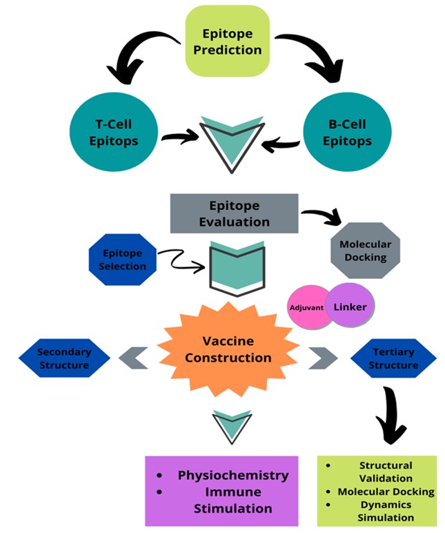
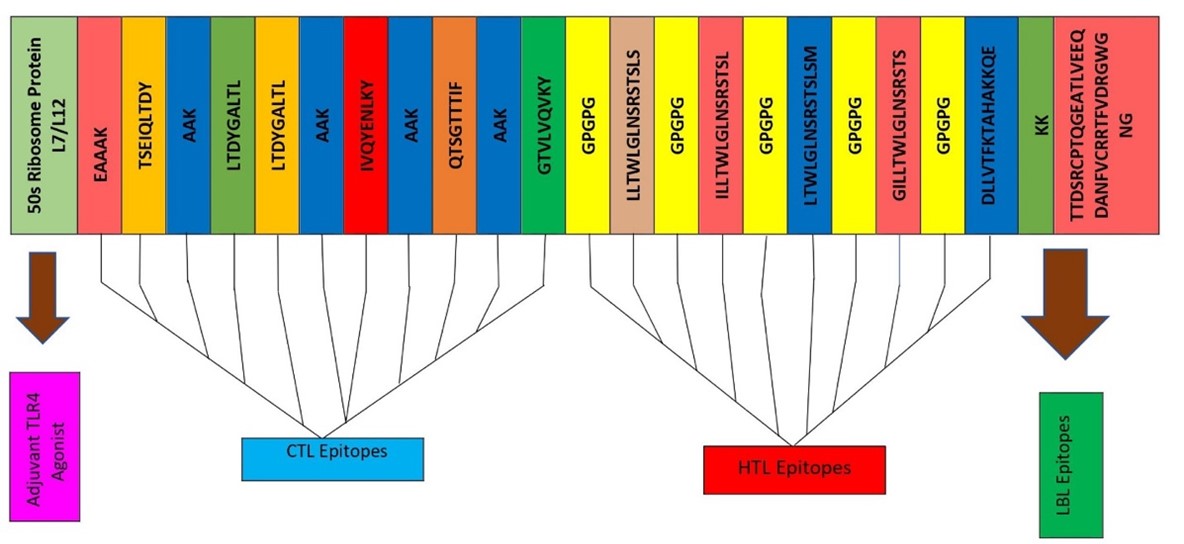
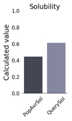
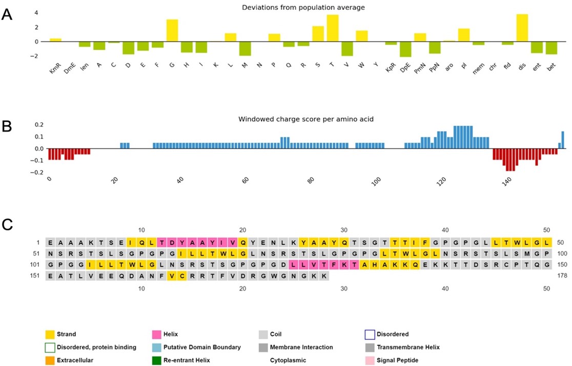
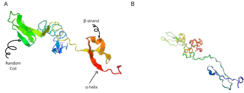
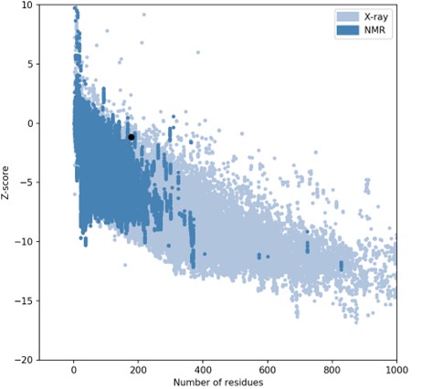
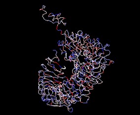
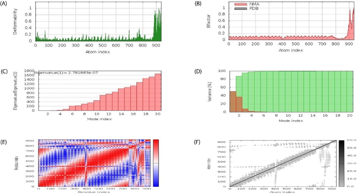
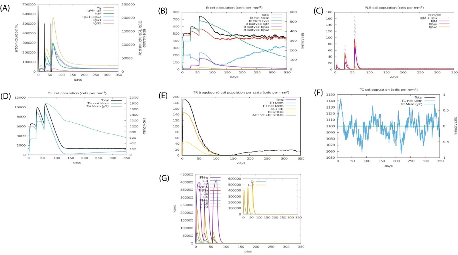
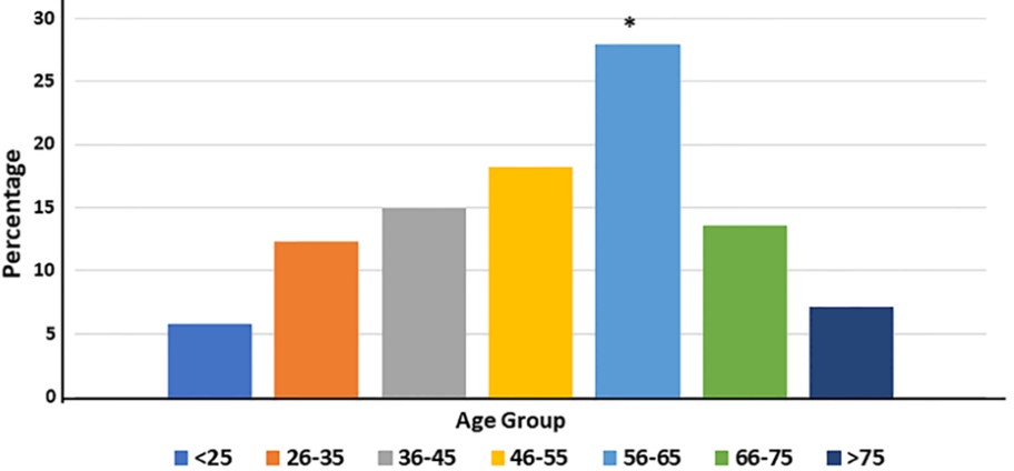
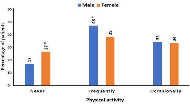
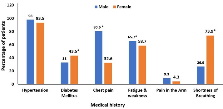
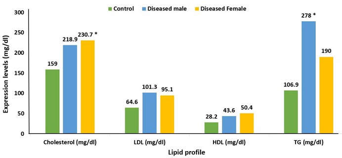
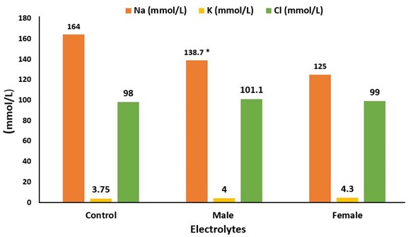
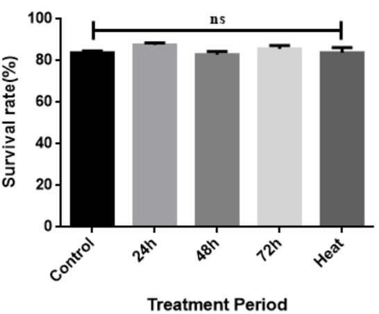
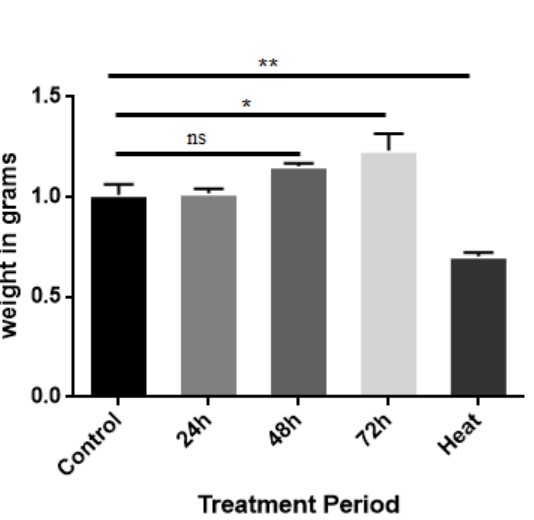
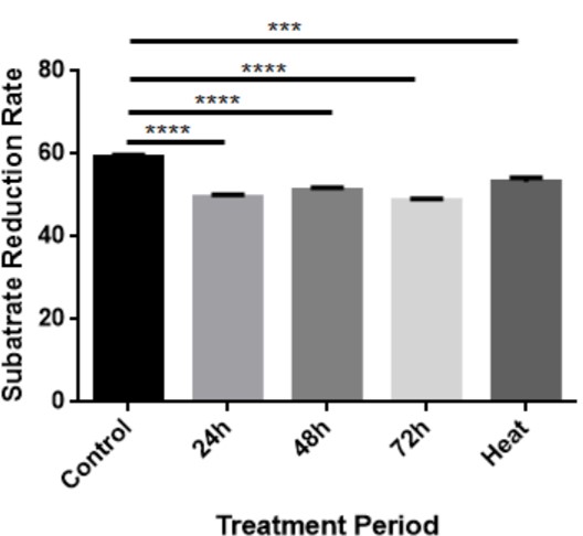
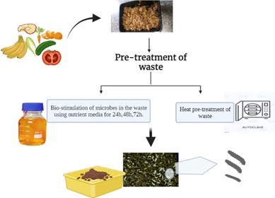
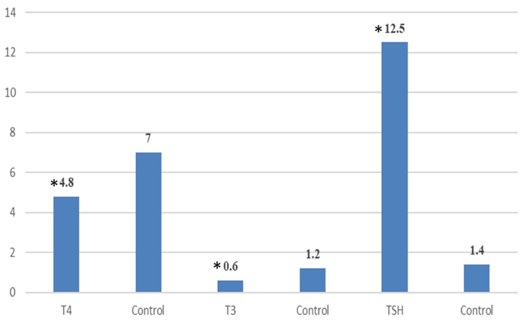
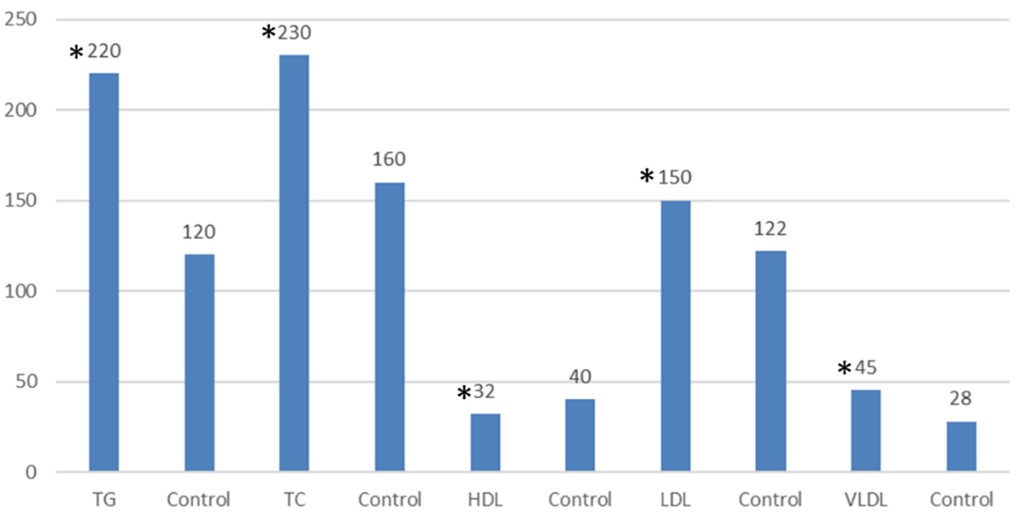
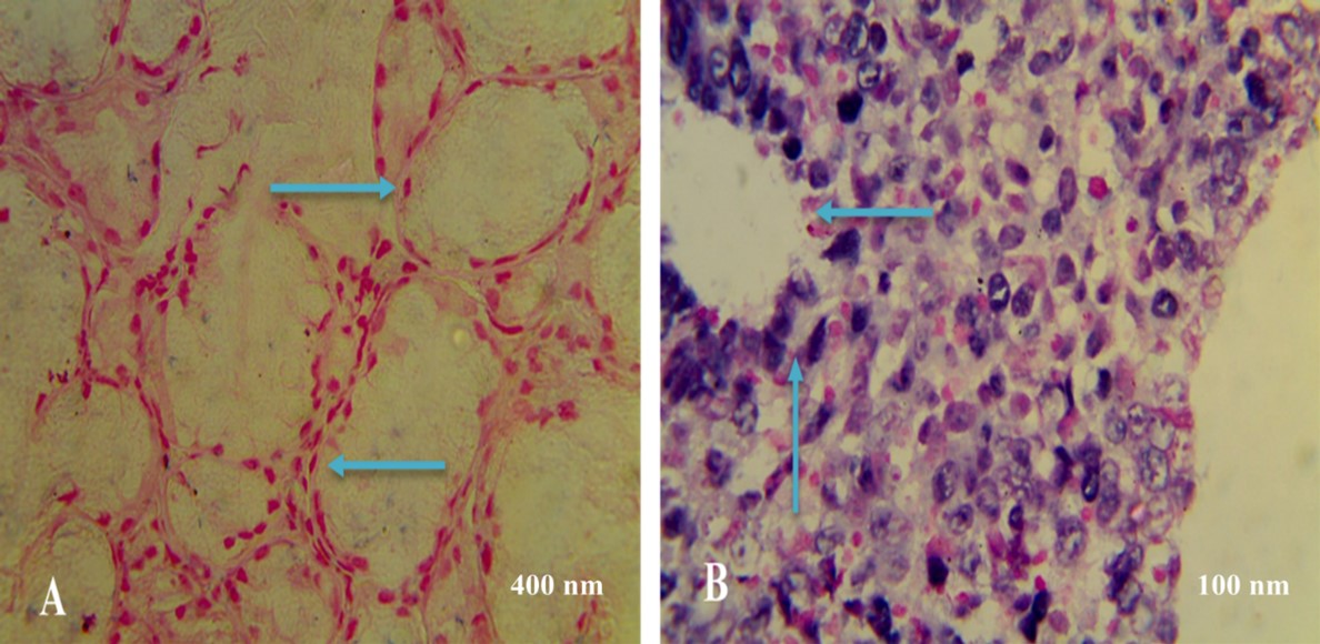
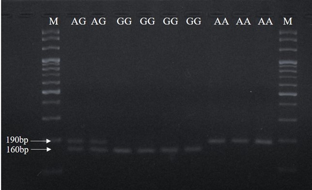
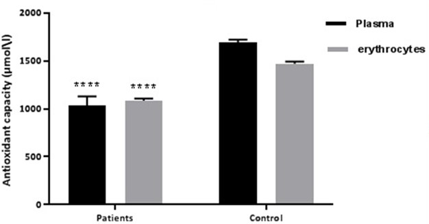
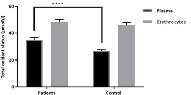
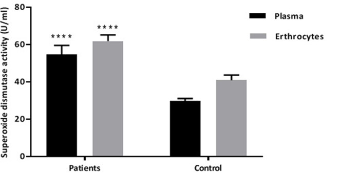
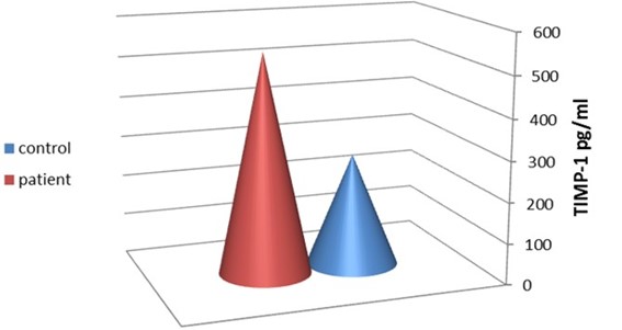
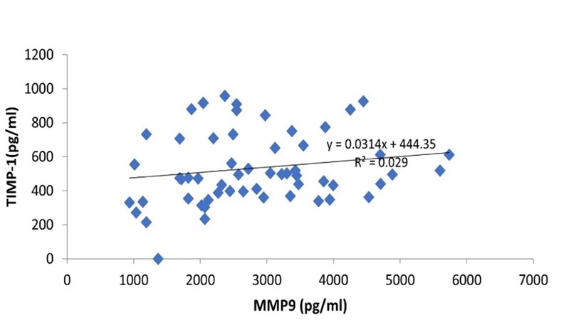
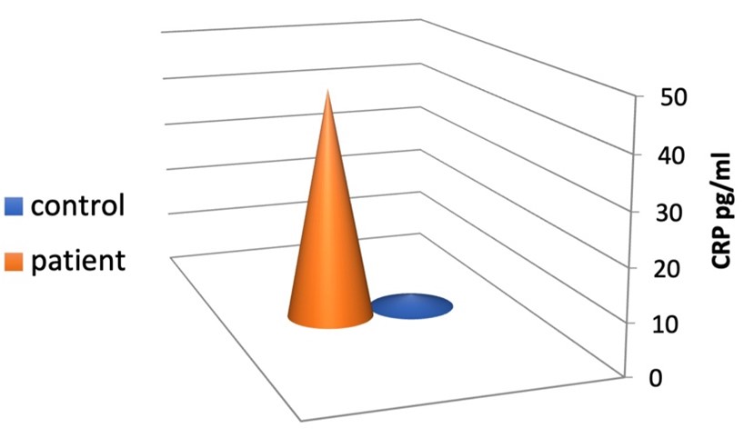
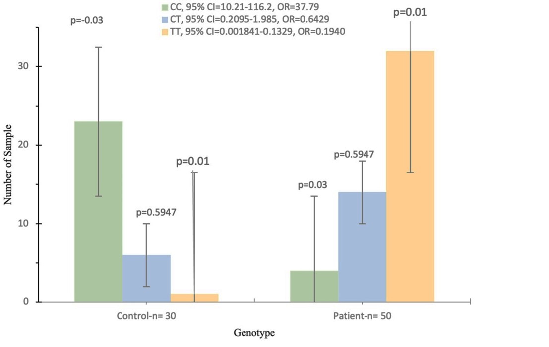
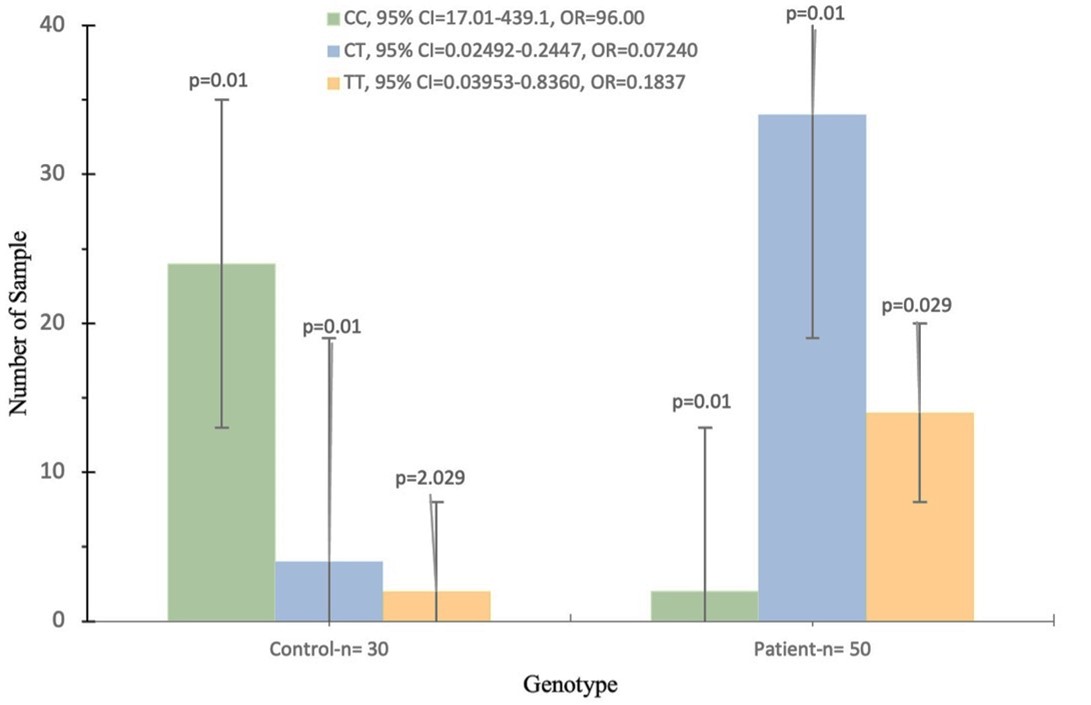
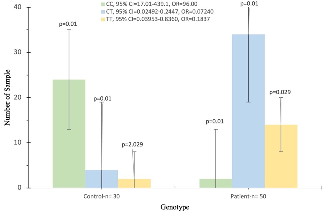
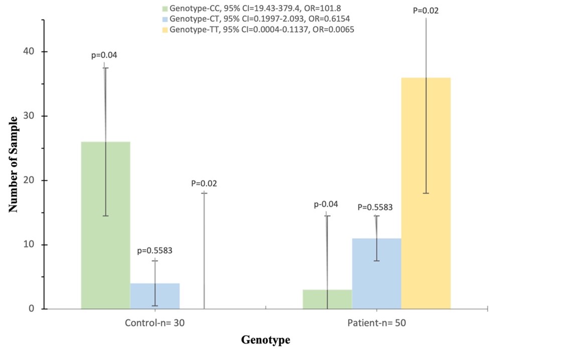
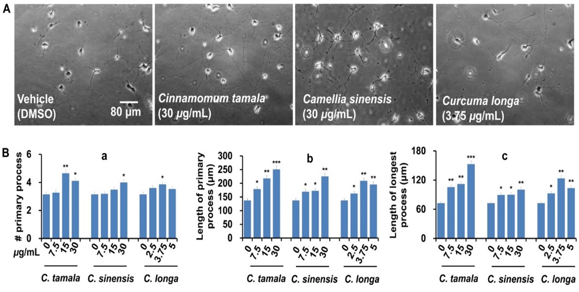