HTTP 200 OK
Allow: GET, HEAD, OPTIONS
Content-Type: application/json
Vary: Accept
{
"count": 321,
"next": "https://jabet.bsmiab.org/articles/?format=api&page=15",
"previous": "https://jabet.bsmiab.org/articles/?format=api&page=13",
"results": [
{
"id": 166,
"slug": "178-1662066505-increased-cd73-expression-is-associated-with-poorly-differentiated-gleason-score-and-tumor-size-in-prostate-cancer",
"featured": false,
"slider": false,
"issue": "Vol6 Issue1",
"type": "original_article",
"manuscript_id": "178-1662066505",
"recieved": "2022-09-04",
"revised": null,
"accepted": "2022-10-19",
"published": "2022-10-27",
"pdf_file": "https://jabet.bsmiab.org/media/pdf_file/2023/09/178-1662066505.pdf",
"title": "Increased CD73 expression is associated with poorly differentiated Gleason score and tumor size in prostate cancer",
"abstract": "<p>There are few prostate cancer prognostic biomarkers. However, clinical difficulties in distinguishing between aggressive and non-aggressive tumors have been observed. CD73 is a 70-kDa glycosylphosphatidylinositol-linked ecto-enzyme that reduces antitumor immunity in mouse models of tumor, particularly prostate cancer. It’s believed to be a promising biomarker for predicting the clinical development and prognosis of certain tumor types. Its function in prostate cancer, however, is unknown. This study aims to investigate the hypothesis that CD73 may be used as a biomarker in prostate cancer diagnosis and/or prognosis. Nuclear and cytoplasmic CD73 staining has been evaluated by immunohistochemistry using benign and malignant prostate tissues. The immunohistochemical study showed nuclear and cytoplasmic CD73 staining in cancerous and non-cancerous prostate tissues. Increased CD73 staining was shown in prostate cancer tissues compared to benign prostate tissues. A negative association between CD73 expression and Gleason scores has been observed. However, increased cytoplasmic CD73 staining was significantly associated with increasing tumor size. This finding suggests that CD73 may have a role in cancer development or aggressiveness, indicating that more research is needed to better understand its function and determine whether it might be used as a diagnostic biomarker for prostate cancer.</p>",
"journal_reference": "J Adv Biotechnol Exp Ther. 2023; 6(1): 161-171.",
"academic_editor": "Md Jamal Uddin, PhD; ABEx Bio-Research Center, Dhaka-1230, Bangladesh",
"cite_info": "Alghezi DA, Aljawher RQ, et al. Increased CD73 expression is associated with poorly differentiated Gleason score and tumor size in prostate cancer. J Adv Biotechnol Exp Ther. 2023; 6(1): 161-171.",
"keywords": [
"Gleason score",
"CD73",
"Tumor size",
"Prostate cancer"
],
"DOI": "10.5455/jabet.2023.d115",
"sections": [
{
"section_number": 1,
"section_title": "INTRODUCTION",
"body": "<p>Prostate cancer (PCa) represents one of the most serious health problems in the world, with a high fatality rate [<a href=\"#r-1\">1, 2]</a>. This disease can affect millions of men and represents the second greatest cause of cancer-related death, with an incidence of 300,000 cases/ year in the USA after skin cancer, 41,000 deaths/year after lung cancer [<a href=\"#r-3\">3</a>]. Approximately 95% of PCa cases are diagnosed with acinar adenocarcinoma which is derived from the prostate gland glandular regions [<a href=\"#r-4\">4, 5]</a>. However, there are only 5 % of PCa cases diagnosed histopathologically as a ductal adenocarcinoma which begins in the cells lining prostate gland ducts [<a href=\"#r-6\">6</a>].<br />\r\nThe Gleason grade system, which is developed in the 1960s and 1970s by Dr Donald Gleason, represents the most widely used histopathological grading scheme for measuring PCa development [<a href=\"#r-7\">7, 8</a>]. This system can be divided into five different Gleason grades (1-5) based on a review of the prostate’s histopathological architecture which specifies how much of the prostate tissue seems normal or abnormal [8]. This system is based on how closely the cancer tissue resembles normal tissue when seen under a light microscope. For example, less aggressive cancer are more likely to seem like healthy tissue, but more aggressive cancer are more likely to spread to other parts of the body and don’t look like healthy tissue [<a href=\"#r-8\">8</a>]. Because PCa is a heterogeneous disease with several histopathological patterns in the same PCa sample, a Gleason score is calculated by adding the two most common Gleason grades: primary and secondary, which are assigned separately for biopsy and prostatectomy [<a href=\"#r-8\">8</a>]. Gleason score of 10 represents the highest score in this system [<a href=\"#r-8\">8</a>]. In this system, the first number assigned is the most prevalent grade found in cancer. For instance, if it is expressed as 3+4=7, it signifies that the majority of the tumor is grade 3 and just a little portion is grade 4, and the two are added to provide a Gleason score of 7. In addition, Gleason score of 7 (4+3) means that the majority of the tumor is grade 4 and a few sections are grade 3. If all cancer sections are the same grade (for example, grade 3), the Gleason score is 3+3=6 [<a href=\"#r-8\">8</a>]. However, this system isn’t always able to distinguish between aggressive and non-aggressive tumors [9]. The tumor-node-metastasis (TNM) system, which is developed by the American Joint Committee on Cancer/International Union Against Cancer (AJCC/ UICC), is another system used to diagnose and progress PCa. This system is based on the PCa size and the extent of its dissemination [<a href=\"#r-2\">2</a>, <a href=\"#r-10\">10</a>]. This system has the benefit of being able to evaluate the prognosis of PCa patients as well as determine the expected progression of their disease [<a href=\"#r-11\">11</a>], as well as act as a guide for patient treatment planning. However, this system is unable to predict which patients would relapse following the first therapy and which will remain in remission.<br />\r\nThe evidence of a loss of basal cells is a crucial step in accurately diagnosing PCa [<a href=\"#r-12\">12</a>]. However, the H&E staining may be unable to accurately identify basal cells in prostate glands [<a href=\"#r-13\">13</a>] and because of that, it is necessary to find a biomarker that can confirm the presence of basal cells in prostate glands. Biomarkers that are expressed in PCa, rather than being lost, are also used. Rare biomarkers have been recognized for PCa diagnosis/prognosis and there are clinical difficulties in distinguishing between prostate gland disorders such as cancerous vs. non-cancerous and localized vs. metastasized PCa. Therefore, identifying new PCa biomarkers has become a priority.<br />\r\nAnti-tumor immune biomarkers may have a role in tumor diagnosis and prognosis, according to much research [<a href=\"#r-14\">14,15</a>]. One of the most well-known immunosuppressive pathways implicated in the development of tumor is the CD73–adenosinergic pathway[<a href=\"#r-16\">16</a>, <a href=\"#r-17\">17</a>]. CD73, also known as ecto-5′-nucleotidase (ecto-5′-NT, EC 3.1.3.5), is a 70-kDa glycosyl-phosphatidylinositol (GPI)-linked ecto-enzyme that reduces antitumor immunity in mouse models of tumor, particularly PCa [<a href=\"#r-18\">18</a>]. CD73, a protein that catalyzes the conversion of AMP to adenosine, is overexpressed in a variety of cancers [<a href=\"#r-19\">19</a>]. Its expression is regulated by a variety of variables and processes such as proliferation, migration, and invasion [<a href=\"#r-20\">20</a>]. It has a role in regulating cancer cell proliferation, migration, and invasion in vitro, tumor angiogenesis, and tumor immune evasion in vivo, according to much evidence [<a href=\"#r-21\">21</a>]. CD73 has been a popular therapeutic target due to its critical function in cancer. Targeted inhibition of CD73 in mouse models has recently been shown to be a promising cancer therapeutic strategy in the future. A study found that The CD73–adenosinergic pathway can be activated by tissue hypoxia and soluble factors present in the TME, such as type I IFNs, TNFα, IL1b, TGFβ, and Wnt activators [<a href=\"#r-22\">22</a>]. Another study showed that CD73 deficiency may be linked to reducing in PCa growth and an increase in CD8 T cell infiltration [<a href=\"#r-23\">23</a>] , suggesting CD73 could be linked to PCa progression and reduced antitumor immunity. Endothelial and epithelial cells, as well as a minority of lymphocytes, particularly regulatory T cells, express CD73. CD73, formerly known as a lymphocyte differentiation antigen, has been discovered to operate as a lymphocyte signaling and adhesion molecule [<a href=\"#r-24\">24</a>]. According to the previous finding, CD73 may have a key role in the developing tumor and may be linked to a poor prognosis in a variety of cancers; however, its prognostic significance in PCa remains unknown. Therefore, the current study aims to assess CD73 immunostaining in cancerous and non-cancerous prostate tissues as well as to establish if its expression correlates with prostate clinical parameters such as grade and stage.</p>"
},
{
"section_number": 2,
"section_title": "MATERIALS AND METHODS",
"body": "<p><strong>Patients and ethics statement.</strong><br />\r\nThe study was accepted by the ethics board of Al Hussein Teaching hospital, Thi-Qar governorate, Iraq (Thi-Qar 2021159 in 7/12/2022). The total number of prostate tissue samples in the study was 96. Seventy-five formalin-fixed, paraffin-embedded tissue samples from radical prostatectomy or transurethral resection of the prostate (TURP) specimens which reviewed to establish Gleason score and stage of samples by histopathologist were used in this study, whereas twenty- one benign prostate tissue samples were also used as a control. These tissue samples were obtained from Al-Hussein teaching hospital’s histopathology department, Thi-Qar city, Iraq. Tonsil tissue samples was used as a positive control for Anti CD73 antibodies. Negative control, no primary antibody add, was also used in this study. A diagnostic H&E section was prepared to identify the tissues architecture by histopathologists. Table 1 summarizes the clinical data of benign and malignant prostate samples.</p>\r\n\r\n<p> </p>\r\n\r\n<p><strong>Immunohistochemistry</strong><br />\r\nImmunohistochemistry was used to stain the benign and malignant prostate tissue sections using two independent anti-CD73 antibodies (Mouse monoclonal, dilution 1:25, Abcam, cat. number Ab3380) and anti-CD73 Rabbit polyclonal, 1:200; Abcam, cat. number Ab3380). Pretreatment steps were used before IHC. paraffin-embedded prostate tissue sections (5 μm) were cute, deparaffinized by use of Histoclear, and rehydrated through graded alcohols (100%, 95%, 70%, respectively. The tissue sections were then permeabilized 0.5% triton X-100 in PBS (phosphate buffer saline), subjected to heat-induced epitope retrieval in a citrate buffer, pH 6.0 with 0.05% Tween 20 for 30 minutes at 90°C, followed by a 20 mins cool down.<br />\r\nDrops of 3% H<sub>2</sub>O<sub>2</sub> (Dako peroxidase) were added on the tissue section in a humid chamber to block endogenous peroxidase activity. Additionally, 10% normal goat serum with 0.05 bovine serum albumin solution was prepared in PBS and then drops of the solution were added to tissue sections. The primary CD73 antibody diluted in Dako antibody diluent (Dako, Ely, UK) was added on the tissue sections and incubated overnight at 4°C and then washed three times for 10 mins each. The next day, the secondary antibody was then added on the tissue section and incubated for 30 minutes at room temperature. As a chromogen, diaminobenzidine tetrahydrochloride was used to view the reaction products using the EnVision+Kit (K400611-2 and K401011-2, Dako, Ely, UK) following the manufacturer’s instructions. Finally, hematoxylin (H-3401, Vector Laboratories, Peterborough, UK) was used to counterstain the sections. These sections were mounted in DPX (Sigma-Aldrich, Gillingham, UK). Nikon Eclipse E800 brightfield illumination was used to see stained tissues, and a Nikon Digital Sight DS-U1 CCD Digital camera was used to take pictures.</p>\r\n\r\n<p> </p>\r\n\r\n<p><strong>Immunohistochemical analysis</strong><br />\r\nTo assess the CD37 Immunostaining in prostate tissue samples, 5 random images were taken and then scored using a semi-quantitative scoring system for cytoplasmic CD73 staining. The percentage scoring of cytoplasmic CD37 immunoreactive was as follows: 0 (0%), 1 (1-25%), 2 (26-50) and 3 (>50%). The cytoplasmic CD37 intensity was scored as negative (0), weak (+1), moderate (+2), or strong (+3). The final score for each case represents the sum of the proportion and intensity scores, which ranged from 0 to 6 [<a href=\"#r-25\">25</a>].</p>\r\n\r\n<p> </p>\r\n\r\n<p><strong>Statistical analysis</strong><br />\r\nThe mean, standard error, and standard deviation data were calculated using GraphPad Prism version 8.00 for Windows, GraphPad Software, La Jolla, California, USA, www.graphpad.com. The unpaired t-test and one-way ANOVA with Tukey’s multiple comparisons tests were used for statistical analysis. P<0.05 was considered significant.</p>"
},
{
"section_number": 3,
"section_title": "RESULTS",
"body": "<p><strong>CD73 expression in benign and malignant prostate tissues</strong><br />\r\nCD37 immunostaining was examined on benign and malignant prostate tissues. The immunohistochemistry results revealed cytoplasmic CD73 staining in both groups with varying degrees of signal strength, ranging from strong and widespread (<a href=\"#figure1\">Figure 1B</a>, arrow) to moderate (<a href=\"#figure1\">Figure 1A & C</a>, arrows) to weak (<a href=\"#figure1\">Figure 1D</a>, arrow) or Negative (<a href=\"#figure1\">Figure 1E</a>, arrow). Because CD73 is located in the cytoplasm of tonsil cells, this study used normal tonsil tissues as a positive control for anti-CD73 [<a href=\"#r-26\">26</a>], and IHC revealed cytoplasmic CD73 staining in tonsil cells, as predicted (<a href=\"#figure1\">Figure 1F</a>, arrow). There was no significant background staining in prostate tissue in the negative control (NC) group, which did not utilize a primary antibody (<a href=\"#figure1\">Figure 1G</a>, arrow).</p>\r\n\r\n<div id=\"figure1\">\r\n<figure class=\"image\"><img alt=\"\" height=\"721\" src=\"/media/article_images/2023/54/22/178-1662066505-Figure1.jpg\" width=\"500\" />\r\n<figcaption><strong>Figure 1. </strong>CD73 staining in prostate tissues. A) moderate nuclear and cytoplasmic CD73 staining (arrow) was observed in BP. B) Strong membrano-cytoplasmic staining of CD73 (arrow) was shown in PCa. C) Moderate cytoplasmic staining of CD73 (arrow) was observed in PCa. D) Weak cytoplasmic staining of CD73 (arrow) was detected in PCa. E) there was no staining (arrow) for CD73 shown in Pca. F) Weak cytoplasmic staining of CD73 (arrow) was detected in tonsil. G) Negative control shows no background staining in PCa (arrow). Pca: Prostate cancer; BP: Benign prostate; NC: Negative control. Scale bars=100 μm.</figcaption>\r\n</figure>\r\n\r\n<p> </p>\r\n</div>\r\n\r\n<p><strong>Increased CD73 expression is associated with poorly differentiated Gleason score and tumor size in PCa</strong><br />\r\nQuantification of the IHC staining revealed that CD73 staining was increased significantly in PCa tissues compared to benign prostate tissues (p=0.0344) (<a href=\"#figure2\">Figure 2A</a> and <a href=\"#Table-1\">Table 1</a>). CD73 expression was negatively associated with increasing Gleason score, using an ANOVA test (p=0.0072) (Figure 2B and <a href=\"#Table-1\">Table 1</a>). When comparing PCa patients with a high Gleason score to those with a low (p=0.0081) or intermediate (p=0.0469) Gleason score, further analysis utilizing multi-comparison (Tukey) testing revealed that cytoplasmic CD73 staining was considerably reduced (<a href=\"#figure2\">Figure 2B</a> and <a href=\"#Table-2\">Table 2)</a>. In contrast, there was a positive association between cytoplasmic CD73 immunostaining and clinical stage T (T3-4 vs. T1-2) (P= 0.0144) (<a href=\"#figure2\">Figure 2C</a> and <a href=\"#Table-2\">Table 2</a>), but not associated with other clinical stage parameters, including Metastasis (M1 vs M0) and Lymph node metastasis (N1vs N0) (p=0.8191& 0.9650, respectively) (<a href=\"#Table-2\">Table 2</a>).</p>\r\n\r\n<div id=\"figure2\">\r\n<figure class=\"image\"><img alt=\"\" height=\"585\" src=\"/media/article_images/2023/54/22/178-1662066505-Figure2.jpg\" width=\"500\" />\r\n<figcaption><strong>Figure 2. </strong>Two different CD73 antibodies show the expected patterns of CD37 staining prostate tissue samples. (A&C) Weak cytoplasmic CD73 (mouse monoclonal) staining (arrows) in prostate tissue. (B&D) Weak cytoplasmic CD73 (rabbit polyclonal) staining (arrows) in prostate tissue. (E and F) Negative control tissue showed negative staining (in PCa arrows). Both CD73 antibodies revealed very similar prostate tissue staining patterns. PCa: Prostate cancer; NC: Negative control. Scale bars—100 μm with inserts at 2x zoom.</figcaption>\r\n</figure>\r\n</div>\r\n\r\n<div id=\"Table-1\">\r\n<p><a href=\"https://jabet.bsmiab.org/table/178-1662066505-table1/\">Table-1</a><strong>Table 1.</strong> Clinical data of benign and malignant prostate samples.</p>\r\n</div>\r\n\r\n<div id=\"Table-2\">\r\n<p><a href=\"https://jabet.bsmiab.org/table/178-1662066505-table2/\">Table-2</a><strong>Table 2.</strong> Cytoplasmic CD73 staining in prostate tissue samples as compared to clinical data.</p>\r\n\r\n<p> </p>\r\n</div>\r\n\r\n<p><strong>Validation of CD73 expression on prostate tissues samples</strong><br />\r\nIHC was then performed on tissue sections from identical locations of prostate samples to establish that the two independent CD73 antibodies (mouse monoclonal and rabbit polyclonal) produced a similar staining pattern. Using a mouse monoclonal CD37 antibody (<a href=\"#figure3\">Figure 3A and C</a>, arrows) and a rabbit polyclonal antibody, IHC results revealed a cytoplasmic staining pattern in PCa tissues (<a href=\"#figure3\">Figure 3B and D</a>, arrows). PCa revealed no background staining with negative control (<a href=\"#figure2\">Figure 2E</a> and<a href=\"#figure2\"> F</a>, arrows).</p>\r\n\r\n<div id=\"figure3\">\r\n<figure class=\"image\"><img alt=\"\" height=\"416\" src=\"/media/article_images/2023/54/22/178-1662066505-Figure3.jpg\" width=\"500\" />\r\n<figcaption><strong>Figure 3. </strong>Cytoplasmic CD73 staining in benign and malignant prostate tissues quantified. The percentage and intensity scores for cytoplasmic IHC staining were used to quantify CD73 staining. A) Increased cytoplasmic CD73 staining significantly in PCa compared to BP tissues (p=0.0344). B) Cytoplasmic CD73 staining showed a significant difference among different Gleason scores (p=0.0072) and multiple comparison tests (Tukey) confirmed a significant reduction with increasing Gleason score. When comparing PCa patients with a high Gleason score to those with a low (p=0.0081) or intermediate (p=0.0469) Gleason score, the decrease was statistically significant. C) Cytoplasmic CD73 staining was shown to be positively associated with primary tumor volume (p=0.0144). Prostate cancer PCa (n=75) and Benign prostate BP (n=21), low Gleason score 3 (n=16), moderate Gleason score (n=29) and high Gleason score (n=22), Tumor size T1-2 (n= 44) and T3-4 (n= 30). X axis: Prostate samples: Benign prostate or Prostate cancer tissue samples. Gleason score: Low, moderate and high. T: Tumor size (T1-2 vs T3-4). Y axis: Final score (proportion and intensity) of cytoplasmic CD73 in each case.</figcaption>\r\n</figure>\r\n</div>"
},
{
"section_number": 4,
"section_title": "DISCUSSION",
"body": "<p>The adenosine pathway has been an interesting topic in cancer research in recent years because of increasing evidence suggesting its role in the development of cancer and metastasis [<a href=\"#r-27\">27</a>]. This study examined the CD73 expression in benign and malignant prostate tissue samples using IHC as a potential biomarker for PCa diagnosis, prognosis and therapy. The current data revealed increased cytoplasmic CD73 immunostaining in PCa tissues compared to benign prostate tissues. This is consistent with other CD73 data from different types of tumors, including breast [<a href=\"#r-21\">21</a>], colorectal [<a href=\"#r-28\">28</a>], ovarian [<a href=\"#r-29\">29</a>] and salivary gland tumors [<a href=\"#r-30\">30</a>] suggesting CD73 may have an important role in cancer formation and development because of its role as a novel immunoinhibitory protein which plays an important function in tumor growth and metastasis. In addition, a previous study revealed that the major role of CD73 in normal tissues is to convert extracellular ATP to immunosuppressive adenosine in conjunction with CD39 to inhibit excessive immune response. Tumors, on the other hand, use the CD73-mediated adenosinergic pathway to defend themselves against immunological attacks [<a href=\"#r-31\">31</a>]. Other studies have found the extracellular adenosine produced by CD73 on malignant cells is enough to mediate immune evasion, allowing cancer growth and metastasis to occur [<a href=\"#r-23\">23</a>, <a href=\"#r-32\">32</a>]. In addition, it has been found that CD73 can regulate the cell cycle, apoptosis, and signaling pathways such EGFR, b-catenin/cyclin D1, VEGF, and AKT/ERK to enhance tumor cell proliferation [<a href=\"#r-33\">33</a>]. In addition, Leclerc and his colleagues found that increased expression of CD73 in the epithelial cells of the prostate can reduce CD8 T cell immunosurveillance and turn them into tumor-promoting cells [<a href=\"#r-22\">22</a>]. Another study has also found that reduction of CD73 by reprogramming Th17 cells may enhance the antitumor effects through increasing their effector function [<a href=\"#r-25\">25</a>]. Taken together, Increased CD73 expression increased may promote prostate cancer growth.<br />\r\nFurthermore, the purpose of this study was to see if there was a link between CD73 immunostaining and Gleason score. The result of this study showed that CD73 immunostaining was negatively associated with increasing Gleason score. This data has in agreement with the previous studies. For example, it has been found that CD73 is reduced in endometrial carcinoma cells of poorly differentiated and advanced-stages in compared to low-grade malignancies, suggesting the protective role of CD73-derived adenosine on epithelial integrity in normal endometrium [<a href=\"#r-34\">34</a>]. Another study on urothelial bladder cancer has found that increased CD73 immunostaining is negatively associated with poorly differentiated grades [<a href=\"#r-35\">35</a>]. In contrast, another study demonstrated that there was no significant association between CD73 immunostaining and differentiation of kinds of cancers, including prostate [<a href=\"#r-18\">18</a>]. This difference may be because of using different methods and/ or different scoring systems. Taken together, CD73 appears to be linked to tumor differentiation and the loss of CD73 on epithelial cells of prostate may encourage the progression of PCa and increasing CD73 expression in tumors may represent a good prognosis indicator for patients with PCa.<br />\r\nThis study looked at the association between CD73 immunostaining and PCa clinical stage. The current data showed a positive correlation between CD73 immunostaining and tumor size (T1-2 vs T3-4). This data was agreed with the previous studies on colorectal carcinoma [<a href=\"#r-36\">36</a>] and papillary thyroid carcinoma [20], suggesting Increased CD73 may promote the growth of kinds of cancer, including PCa. These studies suggest that CD73 stimulates the development of human cancer cells via EGFR and the ß-catenin/cyclin D1 signaling pathway, according to all of the findings [<a href=\"#r-36\">36</a>]. In contrast, this data was not agreed with another study which showed increased CD73 was significantly associated with lymph node metastasis [<a href=\"#r-18\">18</a>]. This difference may be because of using different antibodies, antigen retrievals, scoring systems or different populations. In addition, another reason which may explain these differences is that the sample size of lymph node metastatic PCa (M1 and N1) in this study was lower than non-lymph node metastatic PCa (M0 and N0) (Table 1). Taken together, this data may suggest that increased CD73 expression seems to be linked to PCa progression and prognosis and might be a useful biomarker for PCa.<br />\r\nIn conclusion, increased CD73 staining in PCa is negatively associated with Gleason score and positively associated with tumor size. This early evidence suggests that CD73 may have a role in the development and progression of PCa. CD73 might be a new potential biomarker for PCa. Further study is also needed to validate these data using a second independent antibody with a large cohort or using an RNAscope to detect the mRNA level of CD73 in cancerous and non-cancerous prostate tissues. In addition, it will be very fascinating to investigate the functional role of CD73 in prostate cell lines using tissue culture.</p>"
},
{
"section_number": 5,
"section_title": "ACKNOWLEDGEMENT",
"body": "<p>The authors would like to thank Cancer research unit in the college of Medicine at the University of Thi-Qar for providing use of imaging. The authors thank all staffs at Al-Hussein Teaching hospital for collecting samples.</p>"
},
{
"section_number": 6,
"section_title": "AUTHOR CONTRIBUTIONS",
"body": "<p>Dhafer wrote the first manuscript, did the experimental techniques, designed the entire study, and performed the statistical analysis. Collecting data was done by Dhafer and Rash. Writing, review and editing: Dhafer, Rasha, and Sada. All authors read and approved the final manuscript.</p>"
},
{
"section_number": 7,
"section_title": "CONFLICTS OF INTEREST",
"body": "<p>There is no conflict of interest among the authors.</p>"
}
],
"figures": [
{
"figure": "https://jabet.bsmiab.org/media/article_images/2023/54/22/178-1662066505-Figure1.jpg",
"caption": "Figure 1. CD73 staining in prostate tissues. A) moderate nuclear and cytoplasmic CD73 staining (arrow) was observed in BP. B) Strong membrano-cytoplasmic staining of CD73 (arrow) was shown in PCa. C) Moderate cytoplasmic staining of CD73 (arrow) was observed in PCa. D) Weak cytoplasmic staining of CD73 (arrow) was detected in PCa. E) there was no staining (arrow) for CD73 shown in Pca. F) Weak cytoplasmic staining of CD73 (arrow) was detected in tonsil. G) Negative control shows no background staining in PCa (arrow). Pca: Prostate cancer; BP: Benign prostate; NC: Negative control. Scale bars=100 μm.",
"featured": false
},
{
"figure": "https://jabet.bsmiab.org/media/article_images/2023/54/22/178-1662066505-Figure2.jpg",
"caption": "Figure 2. Two different CD73 antibodies show the expected patterns of CD37 staining prostate tissue samples. (A&C) Weak cytoplasmic CD73 (mouse monoclonal) staining (arrows) in prostate tissue. (B&D) Weak cytoplasmic CD73 (rabbit polyclonal) staining (arrows) in prostate tissue. (E and F) Negative control tissue showed negative staining (in PCa arrows). Both CD73 antibodies revealed very similar prostate tissue staining patterns. PCa: Prostate cancer; NC: Negative control. Scale bars—100 μm with inserts at 2x zoom.",
"featured": false
},
{
"figure": "https://jabet.bsmiab.org/media/article_images/2023/54/22/178-1662066505-Figure3.jpg",
"caption": "Figure 3. Cytoplasmic CD73 staining in benign and malignant prostate tissues quantified. The percentage and intensity scores for cytoplasmic IHC staining were used to quantify CD73 staining. A) Increased cytoplasmic CD73 staining significantly in PCa compared to BP tissues (p=0.0344). B) Cytoplasmic CD73 staining showed a significant difference among different Gleason scores (p=0.0072) and multiple comparison tests (Tukey) confirmed a significant reduction with increasing Gleason score. When comparing PCa patients with a high Gleason score to those with a low (p=0.0081) or intermediate (p=0.0469) Gleason score, the decrease was statistically significant. C) Cytoplasmic CD73 staining was shown to be positively associated with primary tumor volume (p=0.0144). Prostate cancer PCa (n=75) and Benign prostate BP (n=21), low Gleason score 3 (n=16), moderate Gleason score (n=29) and high Gleason score (n=22), Tumor size T1-2 (n= 44) and T3-4 (n= 30). X axis: Prostate samples: Benign prostate or Prostate cancer tissue samples. Gleason score: Low, moderate and high. T: Tumor size (T1-2 vs T3-4). Y axis: Final score (proportion and intensity) of cytoplasmic CD73 in each case.",
"featured": false
}
],
"authors": [
{
"id": 691,
"affiliation": [
{
"affiliation": "Cancer Research Unit, College of Medicine, University of Thi-Qar, Thi- Qar, Iraq"
},
{
"affiliation": "Medical Microbiology and Immunology Department, College of Medicine, University of Thi-Qar, Thi- Qar, Iraq"
}
],
"first_name": "Dhafer A.",
"family_name": "Alghezi",
"email": "dr.daf79@utq.edu.iq",
"author_order": 1,
"ORCID": null,
"corresponding": true,
"co_first_author": false,
"co_author": false,
"corresponding_author_info": "Dhafer A. Alghezi, Medical Microbiology and Immunology Department,\r\nCollege of Medicine, University of Thi-Qar, Thi- Qar, Iraq, e-mail: dr.daf79@utq.edu.iq",
"article": 166
},
{
"id": 693,
"affiliation": [
{
"affiliation": "Histopathology and Forensic Medicine Department, College of Medicine, University of Thi-Qar, Thi-Qar, Iraq"
}
],
"first_name": "Rasha",
"family_name": "Aljawher",
"email": null,
"author_order": 2,
"ORCID": null,
"corresponding": false,
"co_first_author": false,
"co_author": false,
"corresponding_author_info": "",
"article": 166
},
{
"id": 694,
"affiliation": [
{
"affiliation": "College of Dentistry, University of Thi-Qar, Thi- Qar, Iraq"
},
{
"affiliation": "College of Health and Medical technology, National University of Science and Technology, Thi- Qar, Iraq"
}
],
"first_name": "Sada Al",
"family_name": "Musawi",
"email": null,
"author_order": 3,
"ORCID": null,
"corresponding": false,
"co_first_author": false,
"co_author": false,
"corresponding_author_info": "",
"article": 166
}
],
"views": 1070,
"downloads": 128,
"references": [
{
"id": 5475,
"serial_number": 1,
"pmc": null,
"reference": "Lang SH, Frame FM, Collins AT. Prostate cancer stem cells. The Journal of pathology. 2009; 217(2): 299-306.",
"DOI": null,
"article": 166
},
{
"id": 5476,
"serial_number": 2,
"pmc": null,
"reference": "Kirby RS. Prostate cancer. 7th ed. ed. Abingdon: Abingdon : Health Press. 2012.",
"DOI": null,
"article": 166
},
{
"id": 5477,
"serial_number": 3,
"pmc": null,
"reference": "Swami U, McFarland TR, Nussenzveig R, Agarwal N. Advanced Prostate Cancer: Treatment Advances and Future Directions. Trends Cancer. 2020; 6(8): 702-715. doi: 10.1016/j.trecan.2020.04.010. Epub 2020 Jun 10. PMID: 32534790.",
"DOI": null,
"article": 166
},
{
"id": 5478,
"serial_number": 4,
"pmc": null,
"reference": "Dunn MW and Kazer MW. Prostate Cancer Overview. Seminars in Oncology Nursing. 2011, 27(4): 241-250.",
"DOI": null,
"article": 166
},
{
"id": 5479,
"serial_number": 5,
"pmc": null,
"reference": "Bagnall P. Diagnosis and treatment of prostate cancer. Nursing times. 2014; 110(9): 12-15.",
"DOI": null,
"article": 166
},
{
"id": 5480,
"serial_number": 6,
"pmc": null,
"reference": "Stajno P, Kalinowski T, Ligaj M. and Demkow T. An incidentally diagnosed prostatic ductal adenocarcinoma. Cent European J Urol. 2013; 66(2):164-167. doi:10.5173/ceju.2013.",
"DOI": null,
"article": 166
},
{
"id": 5481,
"serial_number": 7,
"pmc": null,
"reference": "Gleason, D.F. Classification of prostatic carcinomas. Cancer Chemother. 1966; Rep. 50",
"DOI": null,
"article": 166
},
{
"id": 5482,
"serial_number": 8,
"pmc": null,
"reference": "Matoso, A. and Epstein, J.I. Grading of prostate cancer: past, present, and future. Current urology reports. 2016; 17(3): 1-6.",
"DOI": null,
"article": 166
},
{
"id": 5483,
"serial_number": 9,
"pmc": null,
"reference": "Penney KL, Stampfer MJ, Jahn JL, Sinnott JA, Flavin R, Rider JR, et.al. Gleason grade progression is uncommon. Cancer research. 2013; 73(16): 5163-8.",
"DOI": null,
"article": 166
},
{
"id": 5484,
"serial_number": 10,
"pmc": null,
"reference": "Edge SB and Compton CC. The American Joint Committee on Cancer: the 7th edition of the AJCC cancer staging manual and the future of TNM. 2010; 17(6): 1471-1474.",
"DOI": null,
"article": 166
},
{
"id": 5485,
"serial_number": 11,
"pmc": null,
"reference": "Goldstein AS, Huang J, Guo C, Garraway IP & Witte ON. Identification of a cell of origin for human prostate cancer. Science (New York, N.Y.). 2010; 329(5991): 568.",
"DOI": null,
"article": 166
},
{
"id": 5486,
"serial_number": 12,
"pmc": null,
"reference": "Shah RB, Zhou M, LeBlanc M, Snyder M, Rubin MA. Comparison of the basal cell-specific markers, 34betaE12 and p63, in the diagnosis of prostate cancer. Am J Surg Pathol. 2002; 26(9):1161-8. doi: 10.1097/00000478-200209000-00006. PMID: 12218572.",
"DOI": null,
"article": 166
},
{
"id": 5487,
"serial_number": 13,
"pmc": null,
"reference": "Varma M, Lee MW, Tamboli P, Zarbo RJ, Jimenez RE, Salles PG. et al. Morphologic criteria for the diagnosis of prostatic adenocarcinoma in needle biopsy specimens. A study of 250 consecutive cases in a routine surgical pathology practice. Archives of pathology & laboratory medicine. 2002; 126(5): 554-61.",
"DOI": null,
"article": 166
},
{
"id": 5488,
"serial_number": 14,
"pmc": null,
"reference": "Basch E, Loblaw DA, Oliver TK, Carducci M, Chen RC, Frame JN, et al. Systemic therapy in men with metastatic castration-resistant prostate cancer: American Society of Clinical Oncology and Cancer Care Ontario clinical practice guideline. J Clin Oncol. 2014; 32: 3436–48.",
"DOI": null,
"article": 166
},
{
"id": 5489,
"serial_number": 15,
"pmc": null,
"reference": "Saad F, Miller K. Current and emerging immunotherapies for castration- resistant prostate cancer. Urology. 2015; 85: 976–86.",
"DOI": null,
"article": 166
},
{
"id": 5490,
"serial_number": 16,
"pmc": null,
"reference": "Allard B, Turcotte M, Stagg J. Targeting CD73 and downstream adenosine receptor signaling in triple-negative breast cancer. Expert Opin Ther Targets. 2014; 18:863–8.",
"DOI": null,
"article": 166
},
{
"id": 5491,
"serial_number": 17,
"pmc": null,
"reference": "Hatfield SM, Kjaergaard J, Lukashev D, Schreiber TH, Belikoff B, Abbott R, et al. Immunological mechanisms of the antitumor effects of supplemental oxygenation. Sci Transl Med. 2015; 7: 277.",
"DOI": null,
"article": 166
},
{
"id": 5492,
"serial_number": 18,
"pmc": null,
"reference": "Jiang T, Xu X, Qiao M, Li X, Zhao C, Zhou F, et al. Comprehensive evaluation of NT5E/CD73 expression and its prognostic significance in distinct types of cancers. BMC Cancer. 2018; 18:267. 10.1186/s12885-018-4073-7.",
"DOI": null,
"article": 166
},
{
"id": 5493,
"serial_number": 19,
"pmc": null,
"reference": "Zhang B. CD73 promotes tumor growth and metastasis. Oncoimmunology. 2012; 1(1):67-70. doi:10.4161/onci.1.1.18068.",
"DOI": null,
"article": 166
},
{
"id": 5494,
"serial_number": 20,
"pmc": null,
"reference": "Jeong YM, Cho H, Kim TM, Kim Y, Jeon S, Bychkov A and Jung C.K. CD73 Overexpression Promotes Progression and Recurrence of Papillary Thyroid Carcinoma. Cancers (Basel). 2020; 12(10):3042.. doi:10.3390/cancers12103042.",
"DOI": null,
"article": 166
},
{
"id": 5495,
"serial_number": 21,
"pmc": null,
"reference": "Chen S, Wainwright D A, Wu J D, Wan Y, Matei D E, Zhang Y et al. CD73: an emerging checkpoint for cancer immunotherapy. Immunotherapy. 2019; 11(11), 983–997. https://doi.org/10.2217/imt-2018-0200.",
"DOI": null,
"article": 166
},
{
"id": 5496,
"serial_number": 22,
"pmc": null,
"reference": "Leclerc BG, Charlebois R, Chouinard G, Allard B, Pommey S, Saad F, Stagg J. CD73 Expression Is an Independent Prognostic Factor in Prostate Cancer. Clin Cancer Res. 2016; 22(1):158-66. doi: 10.1158/1078-0432.CCR-15-1181. Epub 2015 Aug 7. PMID: 26253870.",
"DOI": null,
"article": 166
},
{
"id": 5497,
"serial_number": 23,
"pmc": null,
"reference": "Stagg J, Divisekera U, McLaughlin N, Sharkey J, Pommey S, Denoyer D, et al. Anti-cd73 antibody therapy inhibits breast tumor growth and metastasis. Proc Natl Aca Sci U S A . 2010; 107:1547-1552.",
"DOI": null,
"article": 166
},
{
"id": 5498,
"serial_number": 24,
"pmc": null,
"reference": "Zhang B.) CD73: a novel target for cancer immunotherapy. Cancer Res. 2010; 70(16):6407–6411.",
"DOI": null,
"article": 166
},
{
"id": 5499,
"serial_number": 25,
"pmc": null,
"reference": "Chatterjee S, Thyagarajan K, Kesarwani P, Song JH, Soloshchenko M, Fu J, Bailey SR, et al. Reducing cd73 expression by il1beta-programmed th17 cells improves immunotherapeutic control of tumors. Cancer Res. 2014; 74:6048-6059.",
"DOI": null,
"article": 166
},
{
"id": 5500,
"serial_number": 26,
"pmc": null,
"reference": "Sharpe B, Alghezi DA, Cattermole C, Beresford M, Bowen R, Mitchard J, Chalmers AD. A subset of high Gleason grade prostate carcinomas contain a large burden of prostate cancer syndecan-1 positive stromal cells. Prostate. 2017; 77(13):1312-1324. doi: 10.1002/pros.23391.",
"DOI": null,
"article": 166
},
{
"id": 5501,
"serial_number": 27,
"pmc": null,
"reference": "Turiello R, Pinto A, Morello S. CD73: A Promising Biomarker in Cancer Patients. Front Pharmacol. 2020; 16; 11:609931. doi: 10.3389/fphar.2020.609931. PMID: 33364969; PMCID: PMC7751688.",
"DOI": null,
"article": 166
},
{
"id": 5502,
"serial_number": 28,
"pmc": null,
"reference": "Hajizadeh F, Masjedi A, Heydarzedeh Asl S, Karoon Kiani F, Peydaveisi M, et al. Adenosine and adenosine receptors in colorectal cancer. Int Immunopharmacol. 2020; 87:106853. doi: 10.1016/j.intimp.",
"DOI": null,
"article": 166
},
{
"id": 5503,
"serial_number": 29,
"pmc": null,
"reference": "de Leve S, Wirsdörfer F and Jendrossek V. Targeting the Immunomodulatory CD73/Adenosine System to Improve the Therapeutic Gain of Radiotherapy. Front Immunol. 2019; 5;10:698. doi: 10.3389/fimmu.2019.00698.",
"DOI": null,
"article": 166
},
{
"id": 5504,
"serial_number": 30,
"pmc": null,
"reference": "Ranjbar MA, Ranjbar Z, Zahed M and Nikookar N. CD73 a novel marker for the diagnosis of benign and malignant salivary gland tumors. J Clin Exp Dent. 2019; 1;11(3):e213-e218. doi: 10.4317/jced.54918. PMID: 31001389; PMCID: PMC6461735.",
"DOI": null,
"article": 166
},
{
"id": 5505,
"serial_number": 31,
"pmc": null,
"reference": "Roh M, Wainwright DA, Wu JD, Wan Y, and Zhang B. Targeting CD73 to augment cancer immunotherapy. Curr Opin Pharmacol. 2020; 53:66-76. doi: 10.1016/j.coph.2020.07.001. Epub 2020 7. PMID: 32777746; PMCID: PMC7669683.",
"DOI": null,
"article": 166
},
{
"id": 5506,
"serial_number": 32,
"pmc": null,
"reference": "Jin D, Fan J, Wang L, Thompson LF, Liu A, Daniel BJ, et al. Cd73 on tumor cells impairs antitumor t-cell responses: a novel mechanism of tumor-induced immune suppression. Cancer Res. 2010; 70:2245-2255.",
"DOI": null,
"article": 166
},
{
"id": 5507,
"serial_number": 33,
"pmc": null,
"reference": "Gao ZW, Dong K. and Zhang HZ. The roles of CD73 in cancer. Biomed Res Int. 2014 :460654. doi: 10.1155/2014/460654.",
"DOI": null,
"article": 166
},
{
"id": 5508,
"serial_number": 34,
"pmc": null,
"reference": "Bowser JL, Blackburn MR, Shipley GL, Molina JG, Dunner K Jr and Broaddus RR. Loss of CD73-mediated actin polymerization promotes endometrial tumor progression. J Clin Invest. 2016; 126(1):220-238. doi:10.1172/JCI79380.",
"DOI": null,
"article": 166
},
{
"id": 5509,
"serial_number": 35,
"pmc": null,
"reference": "Wettstein MS, Buser L, Hermanns T, Roudnicky F, Eberli D, Baumeister P, et al. CD73 Predicts Favorable Prognosis in Patients with Nonmuscle-Invasive Urothelial Bladder Cancer. Dis Markers. 2015; 785461. doi: 10.1155/2015/785461.",
"DOI": null,
"article": 166
},
{
"id": 5510,
"serial_number": 36,
"pmc": null,
"reference": "Wu R, Chen Y, Li F, Li W, Zhou H, Yang Y et al. Effects of CD73 on human colorectal cancer cell growth in vivo and in vitro. Oncol Rep. 2016; 35(3):1750-6. doi: 10.3892/or.2015.4512. Epub 2015 Dec 23. PMID: 26708311.",
"DOI": null,
"article": 166
}
]
},
{
"id": 164,
"slug": "178-1660251743-evaluation-of-oxidative-stress-activity-and-the-levels-of-homocysteine-vitamin-b12-and-dna-methylation-among-women-with-breast-cancer",
"featured": false,
"slider": false,
"issue": "Vol6 Issue1",
"type": "original_article",
"manuscript_id": "178-1660251743",
"recieved": "2022-08-16",
"revised": null,
"accepted": "2022-10-06",
"published": "2022-10-26",
"pdf_file": "https://jabet.bsmiab.org/media/pdf_file/2023/49/178-1660251743.pdf",
"title": "Evaluation of oxidative stress activity and the levels of homocysteine, vitamin B12, and DNA methylation among women with breast cancer",
"abstract": "<p>Breast cancer (BC) is the most common malignant tumor in women and the leading cause of cancer deaths worldwide. This work was conducted to estimate the roles of oxidative stress, vitamin B12, homocysteine (HCY), and DNA methylation in BC disease progression. Sixty BC patients (age range 33–80 years) and 30 healthy controls were recruited for this study. Patients with BC were split to group 1 consisted of stage II BC women (low level), and group 2 consisted of patients in stages III and IV (high level). Malondialdehyde (MDA), glutathione peroxidase 3 (GPX3), HCY, and vitamin B12 levels in the study groups were measured. Also, the 5-methylcytosine (5mC) global DNA methylation levels were evaluated. The results showed a significant increase in HCY, and MDA in BC patients compared to healthy controls, with evident increases observed in those with advanced-stage BC (stages III and IV). They were accompanied by significantly reduced levels of 5mC, with a positive correlation between 5mC and the different stages of BC. Also, patients in advanced stages and those with a poor prognosis were exposed to low levels of vitamin B12 and GPX3 (except for the patients in stage IV, which showed increased GPX3 levels). The findings of this study suggest that the differences in global DNA methylation levels at the various phases may be used as a risk factor for developing BC, which indicates the involvement of GPX3 and HCY in BC progression.</p>",
"journal_reference": "J Adv Biotechnol Exp Ther. 2023; 6(1): 149-160.",
"academic_editor": "Md Jamal Uddin, PhD; ABEx Bio-Research Center, Dhaka-1230, Bangladesh",
"cite_info": "Rubaye RHKA; Jumaily RMKA. Evaluation of oxidative stress activity and the levels of homocysteine, vitamin B12, and DNA methylation among women with breast cancer. J Adv Biotechnol Exp Ther. 2023; 6(1): 149-160.",
"keywords": [
"Malondialdehyde",
"Homocysteine",
"Glutathione peroxidase",
"DNA methylation",
"Breast cancer"
],
"DOI": "10.5455/jabet.2023.d114",
"sections": [
{
"section_number": 1,
"section_title": "INTRODUCTION",
"body": "<p>The most frequent malignant tumor and the primary reason for cancer-related deaths in women globally is breast cancer (BC). It is anticipated that around 2.26 million new cases of BC in women will be diagnosed globally by 2020, with roughly 685,000 women dying because of the disease [<a href=\"#r-1\">1</a>]. However, the architecture of BC is extremely complicated, and transcriptional problems and gene dysregulation can manifest on many different levels. Breast cancer also appears to have genetic and epigenetic characteristics [<a href=\"#r-2\">2</a>]. Oxidative stress is the result of absence the balance between reactive oxygen species (ROS) generation and clearance [<a href=\"#r-3\">3</a>]. An increase in ROS production, a reduction in antioxidants that reduce ROS, or a combination of both might result in excess ROS. Lipid peroxidation is a process in which excess ROS assaults and damages membrane lipids containing polyunsaturated fatty acids or phospholipids with carbon-carbon double bonds [<a href=\"#r-4\">4</a>].<br />\r\nMalondialdehyde (MDA) is a three-carbon dialdehyde that is highly reactive and is formed because of peroxidation of polyunsaturated fatty acids and arachidonic acid metabolism [<a href=\"#r-5\">5</a>]. Due to its strong cytotoxicity, MDA is also regarded to be a tumor promoter and co-carcinogen [<a href=\"#r-6\">6</a>]. The oxidative stress level can be assessed by measuring the production the levels of MDA. According to earlier research [<a href=\"#r-7\">7</a>], MDA is employed as a marker in cancer. As a result, cells possess a balance system to neutralize too much ROS. This system is known as an antioxidant system, and it includes both enzyme-based antioxidants such as superoxide dismutase (SOD), catalase (CAT), and glutathione peroxidases (GPXs), as well as non-enzyme-based antioxidants that work together to reduce the oxidative state [<a href=\"#r-8\">8</a>]. The primary mechanism defense against oxidative stress is GPX3, which is also essential in maintaining cellular redox homeostasis. By converting organic peroxides and hydrogen peroxide (H<sub>2</sub>O<sub>2</sub>) into water and the appropriate alcohols, GPX3 protects cells from oxidative stress damage through an enzymatic mechanism [<a href=\"#r-9\">9</a>]. The toxic amino acid homocysteine (HCY), which contains sulfur and has no protein, is a byproduct of the interconversion of the two amino acids cysteine and methionine. There are two ways to metabolize homocysteine: re-methylation and trans-sulfuration [<a href=\"#r-10\">10</a>]. Methionine is the source of HCY in the human body. Homocysteine is transformed into cysteine via the trans-sulfuration process when methionine levels are excessive. Homocysteine is re-methylated to methionine in the presence of a negative methionine balance; this reaction necessitates the cofactors methionine synthase and vitamin B12 [<a href=\"#r-11\">11</a>].<br />\r\nNumerous reports suggest DNA methylation plays a master key in the development of tumor. A methyl donor, which is mainly produced by metabolism of one-carbon, is necessary for DNA methylation [<a href=\"#r-12\">12</a>]. All methylation reactions that take place in living organisms are thought to use S-adenosylmethionine (SAM) as their primary methyl donor [<a href=\"#r-13\">13</a>]. It can change DNA cytosine methylation and disrupt the methionine cycle by lowering intracellular SAM levels. One-carbon metabolism (OCM) is influenced by several substances, including homocysteine, methionine, folate (vitamin B9), vitamin B6, and vitamin B12. These components interact with one another in a different biochemical metabolic process. Via the intermediate impact of these molecules, one-carbon groups are transported to maintain DNA methylation, manage gene structure, and provide a basic material for different biological activities [<a href=\"#r-14\">14</a>]. Additionally, OCM is an active operation in which reduce or increase of one component can disrupt DNA methylation and integrity of genomic. Therefore, by changing how epigenetic modifications are made, how tumor suppressor and oncogene genes are balanced, and how malignant transformation is triggered [<a href=\"#r-15\">15</a>]. The frequent modification of DNA methylation in cancer is well known. In human cells, DNA methyl transferase enzymes (DNMTs) function primarily in the context of cytosine-guanine dinucleotides (CpG) to add a methyl group to the cytosine base at position 5 (5-methylcytosine; 5mC) [<a href=\"#r-16\">16</a>].<br />\r\nThis study was aimed at determining the correlation between oxidative stress, vitamin B12, homocysteine, and DNA methylation and BC risk to evaluate their role in BC disease progression.</p>"
},
{
"section_number": 2,
"section_title": "MATERIALS AND METHODS",
"body": "<p><strong>Study groups</strong><br />\r\nSixty breast cancer patients were randomly chosen from among the 90 Iraqi women who participated in the study, which was conducted at the Oncology Teaching Hospital, Medical City, and Baghdad, Iraq. The research was conducted between November 2020 and April 2021. The age of breast cancer patients ranges from 33–80 years. Additionally, 30 healthy women whose ages ranged from 32 to 75 years old were involved in this study. Patients with BC included in this study were divided into two groups: Group 1 was made up of stage II breast cancer patients (low level), whereas Group 2 was made up of stage III and IV patients (high level).</p>\r\n\r\n<p> </p>\r\n\r\n<p><strong>Collection of blood samples</strong><br />\r\nThe venous blood samples were drawn from positive BC patients at the time of diagnosis and five ml of disposable syringes were using in the sitting position. Each subject had a vein puncture to obtain 5 ml of blood. Three milliliters of blood were progressively forced into disposable serum tubes containing separating gel, while the remaining 2 ml were put into ethylene diamine tetraacetic acid (EDTA) tubes. The serum was then kept at -20˚C for subsequent use after the blood in the gel tubes had been allowed to coagulate at room temperature for 15 min. The blood in the EDTA tubes was subsequently used for DNA extraction and was kept at -20˚C until use.</p>\r\n\r\n<p> </p>\r\n\r\n<p><strong>Ethical clearance</strong><br />\r\nThe ethical committee of the department of biology at the College of Science at the University of Bagdad, Baghdad, Iraq, gave their stamp of approval to this work. The authorization with the reference number CSEC/1120/0082 was obtained on November 15, 2020.</p>\r\n\r\n<p> </p>\r\n\r\n<p><strong>Quantitative measurements of biomarkers, homocysteine, and vitamin B12 in serum samples</strong><br />\r\nThe quantitative measurements of biomarkers (MDA and GPX3), HCY, and vit. B12 in samples of human serum were performed using (ELISA) kit (Sun Long Biotech, China), following the manufacturer’s instructions. Before use, all reagents and samples were thawed and brought to room temperature. Then, 50 μl of standards (S1, S2, S3, S4, and S5) were added to wells of a 600 ml wash buffer, leaving one well empty to act as a blank control. The microtiter plate was mixed, covered, and kept there for half an hour at 37 °C. After 40 μl of sample dilution buffer and 10 μl of serum samples were added. The solution in all wells was discarded, and the washing solution was added to each well to wash it five times. After adding 50 μl of (Horseradish peroxidase) HRP-conjugate reagent to each well, mixing it, and covering the plate, the plate was incubated at 37°C for 30 minutes before being removed and washed five times as before. Following this, 50 μl of each chromogen solution was added to each well in the dark, gently mixed, and followed by 15 minutes of incubation at °C. The reaction was stopped by pouring the stop solution (50 μl) to each well, which caused the wells to become yellow. This caused the reaction to be complete. A spectrophotometric microplate reader set to a wavelength of 450 nm was used to determine the absorbance of the sample (ELISA reader, Mindray, India). The concentration was derived from the standard curve, which was used in the calculation.</p>\r\n\r\n<p> </p>\r\n\r\n<p><strong>DNA extraction</strong><br />\r\nThe DNA was extracted from the whole blood samples for both BC patients and control groups by using the gSYNC™ DNA Extraction Kit (Geneaid, Taiwan). The presence and purity of extracted DNA were confirmed using a Nanodrop spectrophotometer (Thermo, USA), which calculates DNA concentration (ng/μl) and examines DNA purity by measuring the absorbance at (260/280 nm). Staining DNA with ethidium bromide after running it on 1% agarose gels at 80 V for 30 minutes revealed any damage to the DNA.</p>\r\n\r\n<p> </p>\r\n\r\n<p><strong>Global DNA methylation procedure</strong><br />\r\nFollowing the manufacturer’s instructions, the MethylFlashTM methylated DNA quantification Kit (Epigentek, USA) was used to measure the total 5-methylcytosine (5mC) content in DNA extracted from blood samples. For each sample, 100 ng of genomic DNA was used in the assay. To begin, a standard curve was created for a methylated polynucleotide serving as a positive control that contained 50% of 5mC. This curve was created by employing the five concentrations illustrated in <a href=\"#figure1\">Figure 1</a>. Following the addition of the standard DNA and the sample DNA in their corresponding wells. Absorbance was measured using an ELISA reader at 450 nm. The standard curve as obtained by linear regression is shown in <a href=\"#figure1\">Figure 1</a>, and the formula below was used to compute the proportion of 5mC in the entire DNA.</p>\r\n\r\n<div id=\"figure1\">\r\n<figure class=\"image\"><img alt=\"\" height=\"210\" src=\"/media/article_images/2023/15/22/178-1660251743-Figure1.jpg\" width=\"500\" />\r\n<figcaption><strong>Figure 1.</strong> Standard curve for determining methylation of DNA as determined by the immunoassay. Diagrammatic representation of the linear connection between the quantity of 5-methylcytosine and its absorbance.</figcaption>\r\n</figure>\r\n\r\n<p> </p>\r\n</div>\r\n\r\n<p><strong>Statistical analysis</strong><br />\r\nStatistical analysis was performed by using the statistical package for the social sciences (SPSS) version 23. The result was stated as Mean ± SEM. Statistical comparison between groups was analyzed using an analysis of variance (ANOVA), and a p≤0.05 was considered significant [<a href=\"#r-17\">17</a>].</p>"
},
{
"section_number": 3,
"section_title": "RESULTS",
"body": "<p><strong>Oxidative stress biomarker levels in low and high stages of breast cancer</strong><br />\r\nAccording to the statistical analysis, MDA levels were significantly (p≤0.001) higher in breast cancer patients than in the control group. As shown in <a href=\"#figure2\">Figure 2</a>, the results of the current study showed a highly significant (p≤0.001) increased level of MDA in patients of BC with low stage compared to the control group. Furthermore, a highly significant (p≤0.001) increased level of MDA was observed in patients of BC with high stage compared to patients with a low stage and the control group. The levels of GPX3 in the control group and breast cancer patients did not differ significantly. A significant (p≤0.05) reduction was observed when comparing low-stage breast cancer patients to high-stage. Also, the results showed significantly (p≤0.05) reduction in the level of GPX3 in low-stage breast cancer patients comparing to control groups. Meanwhile, there was no significant difference (p≤0.05) in the GPX3 level in breast cancer patients at the high-level stage and control group (<a href=\"#figure3\">Figure 3</a>).</p>\r\n\r\n<div id=\"figure2\">\r\n<figure class=\"image\"><img alt=\"\" height=\"341\" src=\"/media/article_images/2023/15/22/178-1660251743-Figure2.jpg\" width=\"500\" />\r\n<figcaption><strong>Figure 2. </strong>Levels of malondialdehyde (MDA) in the blood of breast cancer patients and control subjects; A) MDA level in breast cancer patients compared to the control, B) MDA level in the early (low-level) and advanced (high-level) individuals with breast cancer in comparison to those who served as controls, ** Refers to significance at p≤0.001, Values represent the mean ± SEM. Low level consisted of patients in stage II, High level consisted of patients in stages III and IV.</figcaption>\r\n</figure>\r\n</div>\r\n\r\n<figure class=\"image\"><img alt=\"\" height=\"331\" src=\"/media/article_images/2023/15/22/178-1660251743-Figure3.jpg\" width=\"500\" />\r\n<figcaption><strong>Figure 3. </strong>Levels of glutathione peroxidase (GPX3) in the blood of breast cancer patients and healthy control subjects; A) GPX3 level in breast cancer patients compared to the control, B) GPX3 level in the early (low-level) and advanced (high-level) in breast cancer patients compared to the control. * Refers to significance at p≤0.05, Values represent the mean ± SEM. Low level consisted of patients in stage II, High level consisted of patients in stages III and IV.</figcaption>\r\n</figure>\r\n\r\n<p> </p>\r\n\r\n<p><strong>Serum vitamin B12 and homocysteine levels in breast cancer patients</strong><br />\r\nHomocysteine levels of BC patients significantly (p≤0.001) increased when compared to the control group. As shown in <a href=\"#figure4\">Figure 4</a>, there was also evidence of a highly significant rise (p≤0.001) in patients at the high-level stage compared to patients at the low-level stage and control groups, as well as a significant increase (p≤0.05) in the level of HCY in patients at the low-level stage compared to control. Patients with breast cancer had significantly (p≤0.001) lower vitamin B12 levels compared to the control group. In addition, the number of patients who were in the high-level stage dropped significantly (p≤0.001) when compared to the low-level stage and the control group. <a href=\"#figure5\">Figure 5</a> demonstrates that in comparison to the control group, vitamin B12 levels in patients who were at the low-level stage dropped by a statistically significant amount (p≤0.001).</p>\r\n\r\n<div id=\"figure4\">\r\n<figure class=\"image\"><img alt=\"\" height=\"333\" src=\"/media/article_images/2023/15/22/178-1660251743-Figure4.jpg\" width=\"500\" />\r\n<figcaption><strong>Figure 4.</strong> Levels of homocysteine (HCY) in the blood of breast cancer patients and healthy control subjects; A) HCY level in breast cancer patients compared to the control, B) HCY level in the early (low-level) and advanced (high-level) in breast cancer patients compared to the control. * Refers to significance at p≤0.05, ** Refers to significance at p≤0.001, Values represent the mean ± SEM. Low level consisted of patients in stage II, High level consisted of patients in stages III and IV.</figcaption>\r\n</figure>\r\n\r\n<figure class=\"image\"><img alt=\"\" height=\"306\" src=\"/media/article_images/2023/15/22/178-1660251743-Figure5.jpg\" width=\"500\" />\r\n<figcaption><strong>Figure 5.</strong> Levels of vitamin B12 in the blood of breast cancer patients and healthy control subjects; A) Vitamin B12 level in breast cancer patients compared to the control, B) Vitamin B12 level in the early (low-level) and advanced (high-level) in breast cancer patients compared to the control. ** Refers to significance at p≤0.001, Values represent the mean ± SEM. Low level consisted of patients in stage II, High level consisted of patients in stages III and IV.</figcaption>\r\n</figure>\r\n</div>\r\n\r\n<p> </p>\r\n\r\n<p><strong>Comparison of serum biomarkers in different stages of breast cancer</strong><br />\r\nBlood MDA levels were significantly (p≤0.001) higher in stage IV breast cancer patients compared to stage II and III patients. Serum MDA levels were significantly (p≤0.001) higher in stage III breast cancer patients compared to stage II patients. Glutathione peroxidase was found to be significantly (p≤0.001) higher in stage IV patients compared to stage II and III patients, but not significantly (p>0.05) different between patients in stages II and III. HCY levels were significantly (p≤0.001) higher in stage IV breast cancer patients than in stage II and III patients. In addition, there was an increase that might be considered statistically significant (p≤0.001) between phases II and III. The levels of vitamin B12 in individuals with stage IV breast cancer were shown to be statistically substantially (p≤0.001) lower when compared to those with stages II and III of the disease. According to <a href=\"#Table-1\">Table 1</a>, there were no statistically significant differences (p>0.05) between individuals diagnosed with breast cancer at stages II and III.</p>\r\n\r\n<div id=\"Table-1\">\r\n<p><a href=\"https://jabet.bsmiab.org/table/178-1660251743-table1/\">Table-1</a><strong>Table 1.</strong> Serum levels of MDA, GPX3, HCY, and vitamin B12 in stages of breast cancer groups. </p>\r\n\r\n<p> </p>\r\n</div>\r\n\r\n<p><strong>Global changes in 5-methylcytosine contents in breast cancer patients</strong><br />\r\nCompared to the control group, there was a highly significant (p≤0.001) reduction in 5mC levels in breast cancer patients. The patients with low levels of the disease showed a slightly significant (p≤0.05) decrease in 5mC levels compared to the control group. In addition, <a href=\"#figure6\">Figure 6</a> displays a highly significant (p≤0.001) lower level of 5mC in breast cancer patients at the high-level stage compared to those at the low-level stage and control.</p>\r\n\r\n<div id=\"figure6\">\r\n<figure class=\"image\"><img alt=\"\" height=\"292\" src=\"/media/article_images/2023/15/22/178-1660251743-Figure6.jpg\" width=\"500\" />\r\n<figcaption><strong>Figure 6. </strong>Changes in the global contents of 5-methylcytosine (5mC) in breast cancer patients, including: A) the level of 5mC in breast cancer patients compared to the control group, and B) the level of 5mC in early (low-level) and advanced (high-level) breast cancer patients compared to the control group. * Refers to significance at p≤0.05, ** Refers to significance at p≤0.001, Values represent the mean ± SEM. Low level consisted of patients in stage II, High level consisted of patients in stages III and IV.</figcaption>\r\n</figure>\r\n\r\n<p> </p>\r\n</div>\r\n\r\n<p><strong>Comparison of global 5-methylcytosine in different stages of breast cancer patients</strong><br />\r\nThe level of 5mC decreased significantly (p≤0.01) in stage IV (No12) breast cancer patients compared to stage II patients (No.30). However, no significant differences (p>0.05) between stage IV and III (No.18) breast cancer patients were observed. Also, there was a significant (p≤0.01) decrease in the 5mC level in stage III patients compared to stage II patients (<a href=\"#Table-2\">Table 2</a>).</p>\r\n\r\n<div id=\"Table-2\">\r\n<p><a href=\"https://jabet.bsmiab.org/table/178-1660251743-table2/\">Table-2</a><strong>Table 2.</strong> Comparison of global 5-methylcytosine (5mC) in different stages of breast cancer. </p>\r\n</div>"
},
{
"section_number": 4,
"section_title": "DISCUSSION",
"body": "<p>Breast cancer is one of the most public diseases among middle-aged and older women worldwide. Due to the increasing number the patients of breast cancer and their generally young age, breast cancer therapy and prognosis are receiving increased attention. Appropriate biomarkers aid in the prediction of prognosis and the selection of the best treatment for each patient [<a href=\"#r-18\">18, 19</a>]. The results also indicated there was a highly significant rise in the level of MDA in patients with breast cancer in the high-level stage compared to patients in the low-level stage and control group. Also, a significantly higher increase in the level of MDA in patients with breast cancer in the low-level stage compared to control group was observed. This observation agrees with the findings of Baskaran et al. [<a href=\"#r-20\">20</a>], who noted a highly significant degree of lipid peroxidation in breast cancer patients compared to controls. The results additionally demonstrated that patients in advanced stages of the disease and those with a bad prognosis were subjected to high levels of oxidative stress. The majority of ROS are produced by tumor cells, and surgical excision of malignant cells has been shown to reduce excessive MDA levels [<a href=\"#r-21\">21</a>].<br />\r\nAdditionally, a correlation between tumor growth and MDA serum levels, and rising MDA levels in the blood may signal the advancement of a tumor as it is shown by recent a study [<a href=\"#r-22\">22</a>]. The results of the present study on the GPX3 status in cancer patients revealed that both patients with stage II and stage III breast cancer had somewhat lower levels of GPX3. In contrast to those with stage II and stage III breast cancer, an unexpectedly increased GPX3 in patients with stage IV breast cancer during the same period was observed. Glutathione peroxidases are at the forefront of cellular redox balance maintenance and oxidative stress defense [<a href=\"#r-23\">23</a>]. Diverse antioxidant defense mechanisms are essential for regulating the formation of free radicals. By disrupting the pro-oxidant/antioxidant equilibrium, increased free radical production, inactivation of antioxidant enzymes, or excessive antioxidant intake all lead to oxidative damage [<a href=\"#r-24\">24</a>]. Therefore, the lower levels of GPX3 in stages II and III may be explained by increased utilization to scavenge lipid peroxides and sequestration by tumor cells [<a href=\"#r-25\">25</a>]. On the other hand, women with late-stage breast cancer had a dramatic elevation of GPX3 (stage IV). These results support previous research that demonstrated higher GPX3 expression is linked to ovarian cancer, is more common in tumors that are farther along in their development and is linked to poor patient survival [<a href=\"#r-26\">26</a>]. The outcomes of this study are in line with Jardim <em>et al</em>. that showed the elevated GPX3 expression in breast cancer was linked with shorter overall survival time breast cancer patients as well as poor response to certain treatment [<a href=\"#r-27\">27</a>]. Enhanced antioxidant enzyme activity is an adaptive response to free radical (ROS)-induced cell damage. Additionally, elevated ROS levels may activate signaling pathways that ultimately result in the production of GPX3 mRNA. The overexpression of GPX3 may detoxify hydrogen peroxides, toxins, and carcinogens [<a href=\"#r-28\">28</a>]. It is observed that between early-stage (I/II) and late-stage (III/IV)/recurrent individuals, there was a significant difference in GPX3 levels [<a href=\"#r-29\">29</a>], indicating that GPX3 could be used as a biomarker of disease progression.<br />\r\nDuring the breakdown of ingested proteins, HCY, a mediate sulfur-containing amino acid, is created from methionine. The levels of Plasma HCY are influenced by dietary nutrients such as vit. B12, B6 and folic acid, and others [<a href=\"#r-30\">30</a>]. Akilzhanova <em>et al</em>. similarly reported an increase in HCY levels in breast cancer patients in their study. According to [<a href=\"#r-31\">31</a>], who established a correlation between high plasma HCY levels and an increased risk of breast cancer, high plasma HCY levels may be a metabolic breast cancer risk factor. As a cofactor for various enzymes involved in the metabolism of HCY, vitamin B12 is necessary for the conversion of HCY to methionine [<a href=\"#r-32\">32</a>]. Therefore, HCY breakdown would be hindered by vitamin B12 shortage, as shown in this study, and lower activity of the implicated metabolic enzymes, resulting in a buildup of intracellular HCY, rapid excretion into the circulation, and finally a rise in its level [<a href=\"#r-33\">33</a>]. It has been proven that breast carcinogenesis is promoted by vitamin B12 deficiency, which increases HCY synthesis. However, the molecular signaling pathways underlying HCY-mediated mammary cancer remain unclear.<br />\r\nVitamin B12 deficiency induced HCY-mediated tumor growth in Mouse mammary tumor virus (MMTV-ErbB2) transgenic mice by activating ErbB2/MAPK/PI3K/Akt signaling pathway. S-adenosylmethionine (SAM) activity for DNA methylation and gene regulation can be negatively impacted by vitamin B12 deficiency, which in turn increases the risk of breast cancer [<a href=\"#r-34\">34</a>]. Additionally, it was discovered in this study that patients with late-stage breast cancer had significantly higher HCY levels than healthy individuals. These results are in line with other researchers who discovered considerably higher HCY in individuals whose breast cancer had spread through metastasis in advanced stages [<a href=\"#r-35\">35</a>]. Homocysteine oxidative damage to cells, pro-oxidant activity, and an increase in free radicals and hydrogen peroxide are potential associations between elevated HCY levels and cancer [<a href=\"#r-36\">36</a>].<br />\r\nFurthermore, earlier research has shown that HCY produces S-adenosylhomocysteine intracellular accumulation and promotes estrogen oxidative metabolites (catechol estrogens) [<a href=\"#r-37\">37</a>]. A lot of anomalies in DNA methylation in cancer are responsible for altered gene expression, including the inactivation of tumor suppressor genes by hypermethylation and the triggering of pro-metastatic genes by hypomethylation [<a href=\"#r-38\">38</a>]. Tumor cells exhibit two types of abnormal DNA methylation: both hypermethylation of CpG islands in promoter regions and hypomethylation of DNA throughout the whole of the cell are seen. Repetitive sequences show global DNA hypomethylation in cancer cells, but specific CpG islands near the promoters of tumor suppressor genes have been shown to have hypermethylation in cancer cells [<a href=\"#r-39\">39</a>]. The extent of DNA hypomethylation across the entire genome is estimated by the sum of 5-mC in dinucleotide CpG sites. Most 5-mC sites are found in repetitive DNA sequences, which account for more than 50% of the human genome. In healthy tissue, these repetitive DNA sequences are heavily methylated; studies have shown that cancer cells exhibit global DNA hypomethylation in repetitive sequences while exhibiting hypermethylation at specific CpG islands in the promoters of tumor suppressor genes [<a href=\"#r-40\">40</a>]. By counting the total number of 5-mC in dinucleotide CpG sites, the amount of genome-wide DNA hypomethylation can be calculated.<br />\r\nOver 50% of the human genome is made up of repetitive DNA sequences, which also make up most 5-mC sites and are heavily methylated in healthy tissue. In contrast, hypomethylation of these sequences significantly contributes to global DNA hypomethylation in cancer [40]. Hypomethylation in repetitive elements can cause genomic instability and reactivation of transposable element expression, whereas hypomethylation in gene bodies can disrupt alternative splicing [<a href=\"#r-41\">41</a>]. Patients with stage IV breast cancer showed a significant reduction in the level of 5mC. This result supported the findings of Pasha et al., who discovered that methylation levels were significantly lower in advanced tumor stages in breast cancer patients than in stages I and II [<a href=\"#r-42\">42</a>]. It was found in 75% of individuals with benign conditions and 96% of those with breast cancer, indicating that it might be utilized to detect tumorigenesis even before a physical breast examination. By activating the production of pro-metastatic genes including heparanase, MMP2 (encoding matrix metalloproteinase 2), and uPA (encoding urokinase plasminogen activator), DNA demethylation plays a critical role in cancer [<a href=\"#r-43\">43</a>]. Demethylation may play a causal role in cancer metastasis since demethylating drugs increase the invasiveness of non-metastatic breast cancer cells [<a href=\"#r-44\">44</a>]. DNA methylation is a reversible process, it has been demonstrated that reversing the un-methylation of breast cancer and liver cancer cell lines decreases the capacity of these cells to invade or metastasize [<a href=\"#r-45\">45</a>].</p>"
},
{
"section_number": 5,
"section_title": "CONCLUSION",
"body": "<p>The findings of the study reveal that there is a significant increase in MDA in patients of breast cancer compared to healthy subjects, with clear improvements shown in those with advanced-stage breast cancer. They were accompanied by significantly reduced levels in GPX3 levels (except for the patients in stage IV). Furthermore, a significantly increase in the HCY levels of breast cancer patients in advanced stages as compared to healthy subjects. Additionally, the patients in advanced stages of breast cancer and those with a poor prognosis experienced low levels of vitamin B12 and 5mC. These findings suggest that involvement of GPX3 and HCY in progression of BC, with the possibility of using variations in global DNA methylation levels at different stages as a risk factor for BC.</p>"
},
{
"section_number": 6,
"section_title": "ACKNOWLEDGMENT",
"body": "<p>The authors are highly appreciative of all the patients for their great cooperation in collecting samples; they were an important part of this research. Also, the authors wish to acknowledge the staff of the Oncology Teaching Hospital, Medical City, Baghdad, Iraq, for their help in collecting the samples.</p>"
},
{
"section_number": 7,
"section_title": "AUTHOR CONTRIBUTIONS",
"body": "<p>The experiments were designed and supervised by Rakad Mohammed Khamas (RMKh). RMKh and Rana Hanan Khudhair (RHK) performed the experiments. In addition to providing research assistance for data collection, RMKh and RHK helped with sample collection, documenting raw data, and collecting data. RMKh and RHK analyzed the data and interpreted the results. RMKh and RHK drafted and edited the manuscript. All the authors acknowledged that they shared responsibility for the work’s correctness and integrity.</p>"
},
{
"section_number": 8,
"section_title": "CONFLICTS OF INTEREST",
"body": "<p>There is no conflict of interest among the authors.</p>"
}
],
"figures": [
{
"figure": "https://jabet.bsmiab.org/media/article_images/2023/15/22/178-1660251743-Figure1.jpg",
"caption": "Figure 1. Standard curve for determining methylation of DNA as determined by the immunoassay. Diagrammatic representation of the linear connection between the quantity of 5-methylcytosine and its absorbance.",
"featured": false
},
{
"figure": "https://jabet.bsmiab.org/media/article_images/2023/15/22/178-1660251743-Figure2.jpg",
"caption": "Figure 2. Levels of malondialdehyde (MDA) in the blood of breast cancer patients and control subjects; A) MDA level in breast cancer patients compared to the control, B) MDA level in the early (low-level) and advanced (high-level) individuals with breast cancer in comparison to those who served as controls, ** Refers to significance at p≤0.001, Values represent the mean ± SEM. Low level consisted of patients in stage II, High level consisted of patients in stages III and IV.",
"featured": false
},
{
"figure": "https://jabet.bsmiab.org/media/article_images/2023/15/22/178-1660251743-Figure3.jpg",
"caption": "Figure 3. Levels of glutathione peroxidase (GPX3) in the blood of breast cancer patients and healthy control subjects; A) GPX3 level in breast cancer patients compared to the control, B) GPX3 level in the early (low-level) and advanced (high-level) in breast cancer patients compared to the control. * Refers to significance at p≤0.05, Values represent the mean ± SEM. Low level consisted of patients in stage II, High level consisted of patients in stages III and IV.",
"featured": false
},
{
"figure": "https://jabet.bsmiab.org/media/article_images/2023/15/22/178-1660251743-Figure4.jpg",
"caption": "Figure 4. Levels of homocysteine (HCY) in the blood of breast cancer patients and healthy control subjects; A) HCY level in breast cancer patients compared to the control, B) HCY level in the early (low-level) and advanced (high-level) in breast cancer patients compared to the control. * Refers to significance at p≤0.05, ** Refers to significance at p≤0.001, Values represent the mean ± SEM. Low level consisted of patients in stage II, High level consisted of patients in stages III and IV.",
"featured": false
},
{
"figure": "https://jabet.bsmiab.org/media/article_images/2023/15/22/178-1660251743-Figure5.jpg",
"caption": "Figure 5. Levels of vitamin B12 in the blood of breast cancer patients and healthy control subjects; A) Vitamin B12 level in breast cancer patients compared to the control, B) Vitamin B12 level in the early (low-level) and advanced (high-level) in breast cancer patients compared to the control. ** Refers to significance at p≤0.001, Values represent the mean ± SEM. Low level consisted of patients in stage II, High level consisted of patients in stages III and IV.",
"featured": false
},
{
"figure": "https://jabet.bsmiab.org/media/article_images/2023/15/22/178-1660251743-Figure6.jpg",
"caption": "Figure 6. Changes in the global contents of 5-methylcytosine (5mC) in breast cancer patients, including: A) the level of 5mC in breast cancer patients compared to the control group, and B) the level of 5mC in early (low-level) and advanced (high-level) breast cancer patients compared to the control group. * Refers to significance at p≤0.05, ** Refers to significance at p≤0.001, Values represent the mean ± SEM. Low level consisted of patients in stage II, High level consisted of patients in stages III and IV.",
"featured": false
}
],
"authors": [
{
"id": 689,
"affiliation": [
{
"affiliation": "Department of Biology, College of Education for Pure Sciences, Ibn Al-Haitham, University of Baghdad, Iraq"
}
],
"first_name": "Rana H. K.",
"family_name": "Al-Rubaye",
"email": "ranabio189@gmail.com",
"author_order": 1,
"ORCID": null,
"corresponding": true,
"co_first_author": false,
"co_author": false,
"corresponding_author_info": "Rana H. K. Al-Rubaye, Department of Biology, College of Education for Pure Sciences, Ibn Al-Haitham, University of Baghdad, Iraq, e-mail: ranabio189@gmail.com",
"article": 164
},
{
"id": 690,
"affiliation": [
{
"affiliation": "Department of Biology, College of science, University of Baghdad, Baghdad, Iraq."
}
],
"first_name": "Rakad M. Kh",
"family_name": "Al-Jumaily",
"email": null,
"author_order": 2,
"ORCID": null,
"corresponding": false,
"co_first_author": false,
"co_author": false,
"corresponding_author_info": "",
"article": 164
}
],
"views": 1522,
"downloads": 148,
"references": [
{
"id": 5430,
"serial_number": 1,
"pmc": null,
"reference": "Winter P, Kotowicz B, Fuksiewicz M, Kowalska M, Jagiello-Gruszfeld A, Nowecki Z. Predictive value of serum aurora a, thymidine kinase 1, and HER3/ErbB3 in breast cancer patients before neoadjuvant treatment. Research Square .2022; 12 January (V1).",
"DOI": null,
"article": 164
},
{
"id": 5431,
"serial_number": 2,
"pmc": null,
"reference": "Dai X, Cheng H, Bai Z, Li J. Breast cancer cell line classification and its relevance with breast tumor subtyping. Journal of Cancer. 2017; 8(16): 3131.",
"DOI": null,
"article": 164
},
{
"id": 5432,
"serial_number": 3,
"pmc": null,
"reference": "Marrocco I, Altieri F, Peluso I. Measurement and clinical significance of biomarkers of oxidative stress in humans. Oxidative medicine and cellular longevity. 2017.",
"DOI": null,
"article": 164
},
{
"id": 5433,
"serial_number": 4,
"pmc": null,
"reference": "Papac-Milicevic N, Busch CL, Binder CJ. Malondialdehyde epitopes as targets of immunity and the implications for atherosclerosis. Advances in Immunology. 2016; 131: 1-59.",
"DOI": null,
"article": 164
},
{
"id": 5434,
"serial_number": 5,
"pmc": null,
"reference": "Jadoon S, Malik A. A review article on the formation, mechanism and biochemistry of MDA and MDA as a biomarker of oxidative stress. International Journal of Advanced Research.2017; 5: 811-818.",
"DOI": null,
"article": 164
},
{
"id": 5435,
"serial_number": 6,
"pmc": null,
"reference": "Salzman R, Pácal L, Tomandl J, Kaňková K, Tóthová E, Gál B, Salzman P. Elevated malondialdehyde correlates with the extent of primary tumor and predicts poor prognosis of oropharyngeal cancer. Anticancer Research. 2009; 29(10): 4227-4231.",
"DOI": null,
"article": 164
},
{
"id": 5436,
"serial_number": 7,
"pmc": null,
"reference": "Cherian DA, Peter T, Narayanan A, Madhavan SS, Achammada S, Vynat GP. Malondialdehyde as a marker of oxidative stress in periodontitis patients. Journal of pharmacy and bioallied sciences. 2019; 11(Suppl 2): S297.",
"DOI": null,
"article": 164
},
{
"id": 5437,
"serial_number": 8,
"pmc": null,
"reference": "He L, He T, Farrar S, Ji L, Liu T, Ma X. Antioxidants maintain cellular redox homeostasis by elimination of reactive oxygen species. Cellular Physiology and Biochemistry. 2017; 44(2): 532-553.",
"DOI": null,
"article": 164
},
{
"id": 5438,
"serial_number": 9,
"pmc": null,
"reference": "Behnisch-Cornwell S, Wolff L, Bednarski PJ. The Effect of Glutathione Peroxidase-1 Knockout on Anticancer Drug Sensitivities and Reactive Oxygen Species in Haploid HAP-1 Cells. Antioxidants. 2020; 9(12): 1300.",
"DOI": null,
"article": 164
},
{
"id": 5439,
"serial_number": 10,
"pmc": null,
"reference": "Hasan T, Arora R, Bansal AK, Bhattacharya R, Sharma GS, Singh LR. Disturbed homocysteine metabolism is associated with cancer. Experimental and molecular medicine. 2019;51(2): 1-13.",
"DOI": null,
"article": 164
},
{
"id": 5440,
"serial_number": 11,
"pmc": null,
"reference": "Lin J, Lee IM, Song Y, Cook NR, Selhub J, Manson JE, Zhang SM. Plasma homocysteine and cysteine and risk of breast cancer in women. Cancer research. 2010 ;70(6): 2397-2405.",
"DOI": null,
"article": 164
},
{
"id": 5441,
"serial_number": 12,
"pmc": null,
"reference": "Newman AC, Maddocks OD. One-carbon metabolism in cancer. British journal of cancer2017;116(12): 1499-1504.",
"DOI": null,
"article": 164
},
{
"id": 5442,
"serial_number": 13,
"pmc": null,
"reference": "Ouyang Y, Wu Q, Li J, Sun S, Sun S. S‐adenosylmethionine: a metabolite critical to the regulation of autophagy. Cell proliferation. 2020; 53(11): e12891.",
"DOI": null,
"article": 164
},
{
"id": 5443,
"serial_number": 14,
"pmc": null,
"reference": "Shuvalov O, Petukhov A, Daks A, Fedorova O, Vasileva E, Barlev NA. One-carbon metabolism and nucleotide biosynthesis as attractive targets for anticancer therapy. Oncotarget. 2017; 8(14): 23955.",
"DOI": null,
"article": 164
},
{
"id": 5444,
"serial_number": 15,
"pmc": null,
"reference": "Yang J, Li H, Deng H, Wang Z. Association of one-carbon metabolism-related vitamins (folate, B6, B12), homocysteine and methionine with the risk of lung cancer: systematic review and meta-analysis. Frontiers in oncology. 2018;8: 493.",
"DOI": null,
"article": 164
},
{
"id": 5445,
"serial_number": 16,
"pmc": null,
"reference": "Skvortsova K, Stirzaker C, Taberlay P. The DNA methylation landscape in cancer. Essays in biochemistry. 2019; 63(6): 797-811.",
"DOI": null,
"article": 164
},
{
"id": 5446,
"serial_number": 17,
"pmc": null,
"reference": "Duncan RC, Knapp RG, Miller MC. Introductory Biostatics of health Science. John Wileyad Sons, London. 1983.",
"DOI": null,
"article": 164
},
{
"id": 5447,
"serial_number": 18,
"pmc": null,
"reference": "Kim MS, Kim SY, Kim JH, Park B, Choi HG. Depression in breast cancer patients who have undergone mastectomy: a national cohort study. PloS one. 2017;12(4):e0175395.",
"DOI": null,
"article": 164
},
{
"id": 5448,
"serial_number": 19,
"pmc": null,
"reference": "Hong W, Dong E. The past, present and future of breast cancer research in China. Cancer letters. 2014;351(1): 1-5.",
"DOI": null,
"article": 164
},
{
"id": 5449,
"serial_number": 20,
"pmc": null,
"reference": "Baskaran D, Singh AK, Prabasheela B. Antioxidant and lipid peroxidation status in pre- and post-operative breast carcinoma. International Journal of Pharma and Bio Sciences. 2011; 2 (1): 450-454.",
"DOI": null,
"article": 164
},
{
"id": 5450,
"serial_number": 21,
"pmc": null,
"reference": "Salzman R, Pácal L, Tomandl J, Kaňková K, Tóthová E, Gál B, Salzman P. Elevated malondialdehyde correlates with the extent of primary tumor and predicts poor prognosis of oropharyngeal cancer. Anticancer research. 2009;29(10): 4227-4231.",
"DOI": null,
"article": 164
},
{
"id": 5451,
"serial_number": 22,
"pmc": null,
"reference": "Lepara Z, Lepara O, Fajkić A, Rebić D, Alić J, Spahović H. Serum malondialdehyde (MDA) level as a potential biomarker of cancer progression for patients with bladder cancer. Romanian Journal of Internal Medicine. 2020; 58(3): 146-152.",
"DOI": null,
"article": 164
},
{
"id": 5452,
"serial_number": 23,
"pmc": null,
"reference": "Behnisch-Cornwell S, Wolff L, Bednarski PJ. The Effect of glutathione peroxidase-1 knockout on anticancer drug sensitivities and reactive oxygen species in haploid HAP-1 cells. Antioxidants. 2020; 9(12):1300.",
"DOI": null,
"article": 164
},
{
"id": 5453,
"serial_number": 24,
"pmc": null,
"reference": "Gupta RK, Patel AK, Kumari R, Chugh S, Shrivastav C, Mehra S, Sharma A N. Interactions between oxidative stress, lipid profile and antioxidants in breast cancer: a case control study. Asian Pacific Journal of Cancer Prevention. 2012;13(12):6295-6298.",
"DOI": null,
"article": 164
},
{
"id": 5454,
"serial_number": 25,
"pmc": null,
"reference": "Sarıkaya E, Doğan S. Glutathione Peroxidase in Health and Diseases. IntechOpen book, biochemistry, London, UK. 2020; 17: 49.",
"DOI": null,
"article": 164
},
{
"id": 5455,
"serial_number": 26,
"pmc": null,
"reference": "Worley BL, Kim YS, Mardini J, Zaman R, Leon KE, Vallur PG. Hempel N. GPX3 supports ovarian cancer progression by manipulating the extracellular redox environment. Redox biology. 2019; 25; 101051.",
"DOI": null,
"article": 164
},
{
"id": 5456,
"serial_number": 27,
"pmc": null,
"reference": "Jardim BV, Moschetta MG, Leonel C, Gelaleti GB, Regiani VR, Ferreira LC, de Campos Zuccari DA. Glutathione and glutathione peroxidase expression in breast cancer: an immunohistochemical and molecular study. Oncology reports. 2013; 30(3): 1119-1128.",
"DOI": null,
"article": 164
},
{
"id": 5457,
"serial_number": 28,
"pmc": null,
"reference": "Wang C, Yu J, Wang H, Zhang J, Wu N. Lipid peroxidation and altered antioxidant status in breast adenocarcinoma patients. Drug research. 2014; 64(12): 690-692.",
"DOI": null,
"article": 164
},
{
"id": 5458,
"serial_number": 29,
"pmc": null,
"reference": "Agnani D, Camacho-Vanegas O, Camacho C, Lele S, Odunsi K, Cohen S, Martignetti JA. Decreased levels of serum glutathione peroxidase 3 are associated with papillary serous ovarian cancer and disease progression. Journal of ovarian research. 2011; 4(1): 1-8.",
"DOI": null,
"article": 164
},
{
"id": 5459,
"serial_number": 30,
"pmc": null,
"reference": "Plazar N, Jurdana M. Hyperhomocysteinemia and the role of B vitamins in cancer. Radiology and oncology. 2010; 44(2): 79-85.",
"DOI": null,
"article": 164
},
{
"id": 5460,
"serial_number": 31,
"pmc": null,
"reference": "Akilzhanova A, Nurkina Z, Momynaliev K, Ramanculov E, Zhumadilov Z, Rakhypbekov T, Takamura N. Genetic profile and determinants of homocysteine levels in Kazakhstan patients with breast cancer. Anticancer research. 2013; 33(9): 4049-4059.",
"DOI": null,
"article": 164
},
{
"id": 5461,
"serial_number": 32,
"pmc": null,
"reference": "Froese DS, Fowler B, Baumgartner MR. Vitamin B12, folate, and the methionine remethylation cycle—biochemistry, pathways, and regulation. Journal of inherited metabolic disease. 2019 ;42(4): 673-685.",
"DOI": null,
"article": 164
},
{
"id": 5462,
"serial_number": 33,
"pmc": null,
"reference": "Xiao Z, Yao G, Liu Y, Zhao C. A Folate-and vitamin B12-deficient diet decreases the latency and increases the incidence of mammary tumors in MMTV-ErbB2 transgenic mice. Research Square.2021. 23April (V1).",
"DOI": null,
"article": 164
},
{
"id": 5463,
"serial_number": 34,
"pmc": null,
"reference": "Kim JA, Choi R, Won H, Kim S, Choi HJ, Ryu JM, Lee SY. Serum vitamin levels and their relationships with other biomarkers in Korean breast cancer patients. Nutrients. 2020;12(9): 2831.",
"DOI": null,
"article": 164
},
{
"id": 5464,
"serial_number": 35,
"pmc": null,
"reference": "Gatt A, Makris A, Cladd H, Burcombe RJ, Smith JM, Cooper P, Makris M. Hyperhomocysteinemia in women with advanced breast cancer. International Journal of Laboratory Hematology. 2007; 29(6): 421-425.",
"DOI": null,
"article": 164
},
{
"id": 5465,
"serial_number": 36,
"pmc": null,
"reference": "Chiang FF, Wang HM , Lan YC, Yang MH, Huang SC, Huang YC. High homocysteine is associated with increased risk of colorectal cancer independently of oxidative stress and antioxidant capacities.Clinical nutrition. 2014;33(6):1054-1060.",
"DOI": null,
"article": 164
},
{
"id": 5466,
"serial_number": 37,
"pmc": null,
"reference": "Naushad SM, Reddy CA, Kumaraswami K, Divyya S, Kotamraju S, Gottumukkala SR, Kutala VK. Impact of hyperhomocysteinemia on breast cancer initiation and progression: epigenetic perspective. Cell biochemistry and biophysics. 2014;68(2): 397-406.",
"DOI": null,
"article": 164
},
{
"id": 5467,
"serial_number": 38,
"pmc": null,
"reference": "Mahmood N, Arakelian A, Cheishvili D, Szyf M, Rabbani SA. S‐adenosylmethionine in combination with decitabine shows enhanced anti‐cancer effects in repressing breast cancer growth and metastasis. Journal of cellular and molecular medicine. 2020;24(18): 10322-10337.",
"DOI": null,
"article": 164
},
{
"id": 5468,
"serial_number": 39,
"pmc": null,
"reference": "Kerachian MA, Kerachian M. Long interspersed nucleotide element-1 (LINE-1) methylation in colorectal cancer. Clinica Chimica Acta. 2019; 488: 209-214.",
"DOI": null,
"article": 164
},
{
"id": 5469,
"serial_number": 40,
"pmc": null,
"reference": "Ehrlich M. DNA hypomethylation in cancer cells. Epigenomics. 2009; 1(2):239-259.",
"DOI": null,
"article": 164
},
{
"id": 5470,
"serial_number": 41,
"pmc": null,
"reference": "Chi HC, Tsai CY, Tsai MM, Lin KH. Impact of DNA and RNA methylation on radiobiology and cancer progression. International journal of molecular sciences. 2018; 19(2): 555.",
"DOI": null,
"article": 164
},
{
"id": 5471,
"serial_number": 42,
"pmc": null,
"reference": "Pasha HA, Rezk NA, Riad MA. Circulating cell free nuclear DNA, mitochondrial DNA and global DNA methylation: potential noninvasive biomarkers for breast cancer diagnosis. Cancer Investigation. 2019; 37(9): 432-439.",
"DOI": null,
"article": 164
},
{
"id": 5472,
"serial_number": 43,
"pmc": null,
"reference": "Ezeani MC, Onyenekwe CC, Meludu SC, Chianakwana GU, Anyiam DC, Emegoakor CD, Amilo GI. Assessment of DNA methylation patterns in breast tumours and apparently healthy subjects in Anambra state, Nigeria. Integrative Cancer Science and Therapeutics. 2017;4(3): 1-6.",
"DOI": null,
"article": 164
},
{
"id": 5473,
"serial_number": 44,
"pmc": null,
"reference": "Chik F, Szyf M. Effects of specific DNMT gene depletion on cancer cell transformation and breast cancer cell invasion; toward selective DNMT inhibitors. Carcinogenesis. 2011;32(2): 224-232.",
"DOI": null,
"article": 164
},
{
"id": 5474,
"serial_number": 45,
"pmc": null,
"reference": "Stefanska B, Huang J, Bhattacharyya B, Suderman M, Hallett M, Han ZG, Szyf M. Definition of the landscape of promoter DNA hypomethylation in liver cancer. Cancer research. 2011; 71(17): 5891-5903.",
"DOI": null,
"article": 164
}
]
},
{
"id": 162,
"slug": "178-1656491348-evaluation-of-phytochemical-profile-and-antimicrobial-activity-of-tragia-brevipes-extracts-against-pathogenic-bacteria",
"featured": false,
"slider": false,
"issue": "Vol6 Issue1",
"type": "original_article",
"manuscript_id": "178-1656491348",
"recieved": "2022-06-29",
"revised": null,
"accepted": "2022-10-03",
"published": "2022-10-26",
"pdf_file": "https://jabet.bsmiab.org/media/pdf_file/2023/03/178-1656491348.pdf",
"title": "Evaluation of phytochemical profile and antimicrobial activity of Tragia brevipes extracts against pathogenic bacteria",
"abstract": "<p>Infectious diseases remain a global health challenge as result of antimicrobial resistance. The use of natural products has revealed a potential source of alternative antimicrobial agents. The aim of this research was to evaluate the antibacterial activities and the phytoconstituents of <em>Tragia brevipes</em> against pathogenic bacteria. The crude extracts of leaves, roots and stems were obtained using polar and non-polar solvents and the antibacterial assay was performed using agar well diffusion and disc diffusion methods. The results showed that in the leaves and roots, flavonoids, saponins, glycosides, phenols, tannins, and resins were present whereas in the stem, flavonoids, glycosides, and resins were present. Only <em>S. aureus</em> ATCC43300, <em>E. coli</em> ATCC25922 did not show inhibition zone to all plant parts. <em>P. aeruginosa</em> ATTCC27853 showed the highest inhibition zone compared to other bacteria. It was observed that methanolic extract has a great potential antimicrobial activity, followed by distilled water extracts. The combination of plant extracts showed a marked synergic effect. The two-way ANOVA showed a statistical difference in the mean zone of inhibition obtained among bacteria and leaves (f = 2.3478, p < 0.05), roots (f = 2.3478, p < 0.05) whereas stem extracts difference was between solvents and tested bacteria (f = 3.4923, p < 0.05). <em>T. brevipes</em> could be a potential candidate in the treatment of bacterial infection as a source of novel antimicrobial agents. Further studies are recommended to isolate the active ingredients and investigate pharmacological properties of <em>T. brevipes.</em></p>",
"journal_reference": "J Adv Biotechnol Exp Ther. 2023; 6(1): 140-148.",
"academic_editor": "Md. Masudur Rahman, PhD; Sylhet Agricultural University, Bangladesh",
"cite_info": "Migabo H, Munyeshyaka E, et al. Evaluation of phytochemical profile and antimicrobial activity of Tragia brevipes extracts against pathogenic bacteria. J Adv Biotechnol Exp Ther. 2023; 6(1): 140-148.",
"keywords": [
"Antimicrobial activity",
"Phytochemical profile",
"Bioactive compounds",
"Tragia brevipes"
],
"DOI": "10.5455/jabet.2023.d113",
"sections": [
{
"section_number": 1,
"section_title": "INTRODUCTION",
"body": "<p>Traditional medicine began long ago with the use of natural products and have displayed high efficacy due to a wide spectrum of pharmacological compounds [<a href=\"#r-1\">1</a>]. According to World Health Organization, most of populations in developing countries consult traditional healers for the treatment of infectious diseases where medicinal plants are used [<a href=\"#r-2\">2</a>].<br />\r\nIt is known that antimicrobial resistance is associated with high morbidity and mortality and remains one of the global health concerns of this century. Multidrug resistance patterns in Gram-positive and Gram-negative bacteria have been reported and have presented difficulties to treat with conventional antibiotics. By 2050, the antimicrobial-resistant infections will lead to millions of deaths per year with subsequent socio-economic impacts if no appropriate actions at different levels [<a href=\"#r-3\">3</a>]. Therefore, there is a need to promote the development of alternative antimicrobial agents considering the current emergency of new strains, ineffectiveness of the existing therapies, ineffective awareness and prevention measures [<a href=\"#r-4\">4</a>].<br />\r\n<em>Tragia brevipes </em>is a species of shrub in Euphorbiaceae or Spurge family and is one of the hundreds Tragia species. Studies have reported the antimicrobial activity of <em>T. brevipes </em>by inhibiting the growth of bacteria including <em>E. coli</em>, Salmonella species<em>,</em> <em>Enterobacter aerogenes</em>, and <em>Proteus vulgaris </em>[<a href=\"#r-5\">5</a>]. In addition, the qualitative analysis of the plant has revealed the presence of bioactive compounds such as flavonoids, phenols, terpenoids, tannins, glycosides, saponins but lack alkaloids and steroids [<a href=\"#r-6\">6</a>].<br />\r\nSeveral studies have reported considerable activity against bacteria by various extracts of medicinal plant [<a href=\"#r-7\">7</a>]. Medicinal plants have been used in Rwanda for many years and remain the main therapeutic option for both rural and urban populations. However, the lack of experimental scientific studies confirming the possible antimicrobial properties from Rwandan plants is still a gap. Among the high number of medicinal plant species, only few have been studied and underwent pharmacological investigations. It has been reported that plant bioactive compounds could display antimicrobial activities against multidrug-resistant microbial pathogens by targeting specific sites other than those used by the current antibiotics [<a href=\"#r-8\">8</a>]. Thus, efficacy of many of the plant drugs including <em>Tragia brevipes </em>(vernacularly known as<em> isusa</em>) used by local traditional healers as effective anti-bacterial agent is still not well documented.<br />\r\nThe present study was initiated to evaluate the potential antimicrobial activity and screen bioactive compound of <em>T. brevipes </em>parts against selected bacteria as potential alternative antibacterial compounds.</p>"
},
{
"section_number": 2,
"section_title": "MATERIALS AND METHODS",
"body": "<p><strong>Plant sampling method</strong><br />\r\nParts of the plant, <em>T. brevipes</em> (leaves, stem and roots) were collected in natural forest in Musanze district, Rwanda.</p>\r\n\r\n<p> </p>\r\n\r\n<p><strong>Sample processing</strong><br />\r\nEach part of the plant was separated from the whole herb which resulted in 3 groups (leaves, roots and stem). The plant was washed twice using distilled water. Stem and roots were cut into pieces for the quick air dry. Each sample was thoroughly mixed and spread to dry at room temperature (23.5 <sup>o</sup>C) in the laboratory under sterile conditions in shade place.</p>\r\n\r\n<p> </p>\r\n\r\n<p><strong>Kinetic maceration technique</strong><br />\r\nDried parts of <em>T. brevipes </em>were again cut into too small pieces and passed in the oven for 15 min at 75<sup>o</sup>C. It was later ground into the fine powder using sample mill machine (electronical powder grinder, DE-200 g). Total powder of leaves, stem and roots parts was weighed. From each group, 25 grams of the powdered group part was dissolved in 3 different solvents, 250 ml of 99.8% methanol, 250 ml distilled water and 250 ml of 90% petroleum ether. The mixture was placed on stopper container and stand on frequent agitation by the help of Orbital shaker / OS-34<sup>o</sup>C at 60 rpm for 5 days. Later the mixture was filtered using a gauze and remained with a homogenous solution.</p>\r\n\r\n<p> </p>\r\n\r\n<p><strong>Extract preparation</strong><br />\r\nCrude plant extract was prepared by Hydro-distillation method, rotary vacuum evaporator (R-11) with 120 rpm and a water bath at low temperatures 45 <sup>o</sup>C was used to evaporate the solvent. The obtained extract was put in sterilized labelled tubes with a proper labeling for future use and stored at 4 <sup>o</sup>C for further antimicrobial studies [<a href=\"#r-9\">9</a>].</p>\r\n\r\n<p> </p>\r\n\r\n<p><strong>Screening of phytochemicals in the plant</strong><br />\r\nThe standard methods provided by Harborne [<a href=\"#r-10\">10</a>] were used to screen the presence of different bioactive secondary metabolites in the plant. Resins, tannins, phenols, alkaloids, glycosides, saponins and flavonoids were screened in this study.</p>\r\n\r\n<p> </p>\r\n\r\n<p><strong>Microorganism</strong><strong>s</strong><br />\r\nThe study used eleven strains of bacteria obtained from the National Reference Laboratory (Rwanda): <em>S. typhi </em>ATCCB69,<em> S. typhi </em>ATCC B71,<em> S. aureus </em>ATCC29213,<em> S. aureus </em>ATCC43300,<em> E. coli </em>ATCC35218,<em> E. coli </em>ATCC29922,<em> P. mirabilis </em>ATCC126,<em> S. pneumonia </em>ATTCC49619,<em> S. pyogene </em>ATTCC 12344,<em> K. oxytoca </em>ATTCC700524 and <em>P. aeruginosa </em>ATTCC27853.<br />\r\nAll bacteria were sub-cultured on their selective media and incubated at 37 ℃ for 24 h. Colony characterization was recorded, and Gram staining was performed followed by biochemical tests for a better identification.<br />\r\n2-5 similar colonies were lifted with a sterile wire loop and transferred into a 4 ml of normal saline contained in a sterilized tube. The bacterial suspensions were standardized to 10<sup>8</sup> CFU/ml. To adjust the turbidity of the suspension, 0.5 McFarland standard solution was used as reference.</p>\r\n\r\n<p> </p>\r\n\r\n<p><strong>Assay of antibacterial activity</strong><br />\r\nUnder aseptic conditions, a sterilized cotton swab was dipped (50 μl) in a prepared bacteria suspension and uniformly distributed on the surface of the Mueller Hinton agar (MHA) using streaking method. For agar well diffusion method, wells of 5 mm were punched in which the extract was added and for disc diffusion method, blank discs were dipped in the extract. The plates were incubated at 37 <sup>0</sup>C for 24 h. The zones of inhibition were measured in millimeter and the antimicrobial assays were interpreted according to Kirby Bauer technique [<a href=\"#r-11\">11</a>]. Each solvent was used as a negative control and duplicate assay was done on each antimicrobial test.</p>\r\n\r\n<p> </p>\r\n\r\n<p><strong>Antimicrobial study of combined parts of<em> Tragia brevipes</em></strong><br />\r\nThe triple (leaves, roots and stem) combinations of plant extracts parts were prepared by mixing equal quantities of each extract respective to the solvent. Each of the obtained mixture extracts was tested for antibacterial activity under sterile conditions. Duplicate assay was done on each antimicrobial test.</p>\r\n\r\n<p> </p>\r\n\r\n<p><strong>Statistical analysis</strong><br />\r\nThe results were analyzed depending on effect and control used. For data entry and analysis, all results were statistically analyzed using Statistical Package for the Social Sciences version 22. To compare the means mentioned in this study objective. Two-way ANOVA was useful and to find a difference between two groups, a post hoc test (Bonferroni) was run to indicate a marked difference.</p>"
},
{
"section_number": 3,
"section_title": "RESULTS",
"body": "<p><strong>Phytochemical </strong><strong>compounds</strong><br />\r\nThe phytochemical qualitative study revealed the presence of some major secondary metabolites whereas other were absent in the plant extracts (<a href=\"#Table-1\">Table 1</a>).</p>\r\n\r\n<div id=\"Table-1\">\r\n<p><a href=\"https://jabet.bsmiab.org/table/178-1656491348-table1/\">Table-1</a><strong>Table 1.</strong> Phytochemical screening of the <em>Tragia brevipes.</em></p>\r\n\r\n<p> </p>\r\n</div>\r\n\r\n<p><strong>Antimicrobial activity of <em>T. brevipes</em> against selected pathogenic bacteria</strong><br />\r\nResults obtained from the work of testing <em>T. brevipes</em> antimicrobial activity indicated that the most non inhibited strains to all plant extracts were <em>E</em><em>. coli </em>ATCC25922 and <em>S. aureus </em>ATCC43300, while other tested strains were potent to the plant extracts as shown in <a href=\"#figure1\">Figure 1</a>. It was observed that depending on the solvents used, the zones of inhibition (ZI) vary and the methanolic extract was more potent against tested bacteria compared to others.<br />\r\n<em>T. brevipes</em> stem was weak to inhibit the bacteria, leaves responded highly with methanol and petroleum ether solvents whereas roots responded highly with the extraction done by distilled water. On the other hand, the tested bacteria showed a variation in their susceptibility to the extracts as shown below in <a href=\"#figure1\">Figure 1</a>. The highest inhibition was generated against the growth of<em> P. aeroginosa </em>ATTCC27853 (ZI: 17 mm) and the lowest against <em>S. pneumonia </em>ATTCC49619 (ZI: 8 mm)<em>.</em> Both disc and agar well diffusion methods were conducted, the obtained results were almost the same as shown in the <a href=\"#figure1\">Figure 1</a>.</p>\r\n\r\n<div id=\"figure1\">\r\n<figure class=\"image\"><img alt=\"\" height=\"254\" src=\"/media/article_images/2023/48/22/178-1656491348-Figure1.jpg\" width=\"500\" />\r\n<figcaption><strong>Figure 1.</strong> Antimicrobial activity of <em>T. brevipes</em> crude extracts. A) Zone of inhibition produced using agar well diffusion method. B) Antibacterial activity screened using only solvents as negative control. C) Zone of inhibition produced using disc diffusion method.</figcaption>\r\n</figure>\r\n\r\n<p> </p>\r\n</div>\r\n\r\n<p><strong>Variance of antimicrobial activity of <em>T. brevipes</em></strong><br />\r\nThe analysis of variance indicated that there was a significant difference on the mean zones of inhibition obtained from both leaves(f = 2.3478, p < 0.05) and roots (f = 2.3478, <em>p</em> < 0.05) extracts against bacteria (<a href=\"#Table-2\">Table 2</a>). Therefore, Bonferroni correction was runned and revealed a significant difference in some bacteria as showed in <a href=\"#Table-3\">Table 3</a>. A post hoc analysis (Bonferroni correction) showed a marked difference between bacteria and the used <em>P</em> value was 0.004545 (<a href=\"#Table-3\">Table 3</a>). However, for the stem extracts, a significant difference was observed among solvents (f = 3.4923, p < 0.05) (<a href=\"#Table-2\">Table 2</a>). The bonferroni correction showed a clear difference between methanol vs. petroleum ether solvent used in stem extracts (p = 0.0028, p = 0.016667) (<a href=\"#Table-3\">Table 3</a>).</p>\r\n\r\n<div id=\"Table-2\">\r\n<p><a href=\"https://jabet.bsmiab.org/table/178-1656491348-table2/\">Table-2</a><strong>Table 2.</strong> Variance of antimicrobial activity of <em>Tragia brevipes.</em></p>\r\n</div>\r\n\r\n<div id=\"Table-3\">\r\n<p><a href=\"https://jabet.bsmiab.org/table/178-1656491348-table3/\">Table-3</a><strong>Table 3.</strong> Marked difference with post hoc test.</p>\r\n</div>\r\n\r\n<p> </p>\r\n\r\n<p><strong>Effects of combined parts of<em> T. brevipes</em> on selected pathogenic bacteria</strong><br />\r\nThe triple combinations of plant extracts parts resulted to potent activity (<a href=\"#figure2\">Figure 2</a>). Combined extract of methanol showed a marked increased activity compared with other solvents. Obtained zone of inhibition ranged between 10 to 18 mm. Generally, bacteria strains were inhibited by methanol and distilled water extracts. Petroleum ether only inhibited <em>S. aureus</em> strains. In addition, the combination showed an increase in zone of inhibition against all bacteria, highly on Gram positive bacteria compared to Gram negative bacteria.</p>\r\n\r\n<div id=\"figure2\">\r\n<figure class=\"image\"><img alt=\"\" height=\"274\" src=\"/media/article_images/2023/48/22/178-1656491348-Figure2.jpg\" width=\"500\" />\r\n<figcaption><strong>Figure 2. </strong>Antimicrobial activity of crude extracts of <em>T. brevipes</em> vs combined crude extracts. A) Zone of inhibition produced by different parts of the plant from different solvents. B) Zone of inhibition produced when different parts from the same solvent are combined.</figcaption>\r\n</figure>\r\n\r\n<p> </p>\r\n</div>\r\n\r\n<p><strong>Variance of antimicrobial effects of combined parts of <em>T. brevipes</em></strong><br />\r\nThree different extracts of <em>T. brevipes</em> were tested for antimicrobial activity using agar well diffusion method. The presence of zone of inhibition proved that plant was able to inhibit the growth of bacteria but at a different extent depending to the solvents (<a href=\"#Table-4\">Table 4</a>). The analysis of variance of two factors showed that there was significant difference on the mean zones of inhibition obtained against bacteria through different solvents (f = 3.492828, p < 0.05) (<a href=\"#Table-4\">Table 4</a>). A post hoc test using Bonferroni correction indicated that a significant difference was between methanol vs. petroleum ether solvent (<a href=\"#Table-5\">Table 5</a>).</p>\r\n\r\n<div id=\"Table-4\">\r\n<p><a href=\"https://jabet.bsmiab.org/table/178-1656491348-table4/\">Table-4</a><strong>Table 4.</strong> Comparison of the activity produced by combined parts crude extracts.</p>\r\n</div>\r\n\r\n<div id=\"Table-5\">\r\n<p><a href=\"https://jabet.bsmiab.org/table/178-1656491348-table5/\">Table-5</a><strong>Table 5.</strong> Marked difference observed between solvents.</p>\r\n</div>"
},
{
"section_number": 4,
"section_title": "DISCUSSION",
"body": "<p>Worldwide a large number of medicinal plants are harvested from the natural environment for traditional medicine [<a href=\"#r-12\">12</a>]. Thus, the natural area is very important for local populations to satisfy their primary healthcare needs. Testing the antimicrobial activity of various medicinal plants require complex methods such as sampling, air drying, grinding, maceration, extraction, and antimicrobial testing although extraction is key step in this experiment. Different solvents were used while macerating and thus different extracts were obtained. Maceration is a convenient and less costly method for small and medium research projects compared to other modern extraction methods [<a href=\"#r-13\">13</a>]. This technique softens and breaks the plant’s cell wall or other various substances to release the soluble phytochemicals as reported previously [<a href=\"#r-14\">14</a>].<br />\r\nThree solvents were used to evaluate the plant activity based on the polarity of solvents from low to high where the petroleum ether is a nonpolar solvent while methanol are distilled water polar solvents [<a href=\"#r-15\">15</a>]. Bioactive compounds represent the phytoconstituents which are synthesized as secondary metabolites in all plant cells with different concentrations depending on the plant part [<a href=\"#r-16\">16</a>]. In the present study, resins, tannins, phenols, glycosides, saponins and flavonoids were present in the leaves and roots while flavonoids, glycosides and resins were present in stem. Similar findings have been reported by Swamy et al. [<a href=\"#r-6\">6</a>] where the leaves contained all the components were present but they lacked alkaloids and steroids. Each compound has a specific target [<a href=\"#r-17\">17</a>] and the presence of the same phytochemicals in the roots and leaves could be an indication of the same potential antibacterial activity. Maobe <em>et al.</em> [<a href=\"#r-18\">18</a>] revealed that the active compounds interfere with the microorganism metabolic processes or may change their gene expression. Phenolic structures are highly active against the microorganisms and tannins have been reported as inhibitors to the growth of many fungi, yeast, bacteria and other viruses [<a href=\"#r-19\">19</a>].<br />\r\nFrom this study, the plant extracts were found to have antibacterial activity against nine of the eleven bacteria tested and the observed antibacterial activity was closely associated with the polarity of solvents [<a href=\"#r-20\">20</a>]. The experiment showed a future hope of having novel antibacterial agent as the potential antibacterial activity of <em>T. brevipes</em> recorded in this study was also confirmed by other studies. Swamy <em>et al.</em> reported that<em> T. brevipes</em> was found to inhibit the growth of <em>E. coli</em>, <em>Salmonella</em> sp., <em>E. aerogenes</em>, <em>B. cereus</em>, <em>S. liquefaciens</em> and <em>P. vulgaris </em>[<a href=\"#r-5\">5</a>].<em> </em><br />\r\nIn this study, a very high zone of inhibition was observed on <em>P. aeruginosa</em>. It has been reported that <em>P. aeruginosa</em> is highly inhibited by the plant extracts with phenolic content [<a href=\"#r-21\">21</a>]. Both <em>S. aureus </em>ATCC43300 and <em>E. coli </em>ATCC25992 were not inhibited by the plant extracts. Those bacteria have been reported among the most resistant bacteria [<a href=\"#r-22\">22</a>]. The variations between different studied strains of <em>S. aureus</em> and <em>E. coli</em> could be attributed to different factors including the isolation site and the type of prophylactic antibiotics [<a href=\"#r-23\">23</a>].<br />\r\nThe combination of the plant extracts exhibited diverse antibacterial activity against the tested microorganisms with reference to plant parts alone. Interestingly, both <em>S. aureus </em>ATCC43300 and <em>E. coli </em>ATCC25992 (which were not inhibited in the first experiments) were inhibited by the combined parts. It has been observed that the increased activity was found more on Gram positive bacteria. The sensitivity of microbes to the extracts may be due by the cell wall structure of the bacteria as reported by Rachuonyo <em>et al. </em>[<a href=\"#r-24\">24</a>]. Gram positive bacteria contain layers of peptidoglycan which make them more permeable to many molecules than the cell wall of Gram negative bacteria with the outer membrane and lipid bilayer which make them less sensitive to many extracts [<a href=\"#r-25\">25</a>].<br />\r\nBoth ANOVA and ad hock test indicated a significant difference between bacteria and within solvents. This could be explained by the fact that Gram positive respond differently to extracts compared to Gram negative and non-polar solvents react differently to polar solvents [<a href=\"#r-26\">26</a>]. To exclude the action of solvents on bacterial growth, the antibacterial activity was screened using only solvents as negative control. Interestingly, the solvents alone didn’t exhibit any activity.<br />\r\nIn additional to the antimicrobial activity studies, <em>T. brevipes </em>was found to be an anti-proliferative activity on cancer cells, relief stomach pain, purgative and in treatment of rheumatism [<a href=\"#r-27\">27</a>, <a href=\"#r-28\">28</a>]. It was reported that its leaves and roots are used to treat peptic ulcers, diabetes, local anesthetic agent and antivenom [<a href=\"#r-29\">29–31</a>].</p>"
},
{
"section_number": 5,
"section_title": "CONCLUSION",
"body": "<p>This study has revealed that the antibacterial activity of <em>T. brevipes</em> is attributed to the phytochemicals present in the plant as the result showed that leaves and roots act as best antibacterial agents than stem extract which lack many bioactive compounds. Therefore, data obtained in this study could be used as a scientific report for plants traditional use and <em>T. brevipes</em> could be a potential good candidate in bacterial infection treatment and management as a source of novel antimicrobial agents. However, antibiotics that appear potent <em>in vitro</em> are not necessarily more beneficial <em>in vivo</em> when treating or preventing disease. Therefore, further studies are needed for better understanding the plant toxicity and side effects. In addition, it is recommended to evaluate the activity against other microorganisms with several other solvents, isolate the active ingredients, and investigate the mechanisms of action.</p>"
},
{
"section_number": 6,
"section_title": "ACKNOWLEDGMENTS",
"body": "<p>The National Council for Science and Technology of Rwanda supported the work under the Excellent Research Grants theme (NCST/ERC1/03). The authors gratefully acknowledge the support of staff members of Microbiology and Chemistry laboratories of INES-Ruhengeri.</p>"
},
{
"section_number": 7,
"section_title": "AUTHOR CONTRIBUTIONS",
"body": "<p>TH and FNN conceived, designed and supervised the study. HM collected samples. HM, and EM performed the experiments. HM, CI, CY, FNN and TH analyzed the data. HM,TH, and CI prepared the manuscript. HM, EM, CI, CY, FNN and TH revised the manuscript. All authors approved the final version.</p>"
},
{
"section_number": 8,
"section_title": "CONFLICTS OF INTEREST",
"body": "<p>There is no conflict of interest among the authors.</p>"
}
],
"figures": [
{
"figure": "https://jabet.bsmiab.org/media/article_images/2023/48/22/178-1656491348-Figure1.jpg",
"caption": "Figure 1. Antimicrobial activity of T. brevipes crude extracts. A) Zone of inhibition produced using agar well diffusion method. B) Antibacterial activity screened using only solvents as negative control. C) Zone of inhibition produced using disc diffusion method.",
"featured": false
},
{
"figure": "https://jabet.bsmiab.org/media/article_images/2023/48/22/178-1656491348-Figure2.jpg",
"caption": "Figure 2. Antimicrobial activity of crude extracts of T. brevipes vs combined crude extracts. A) Zone of inhibition produced by different parts of the plant from different solvents. B) Zone of inhibition produced when different parts from the same solvent are combined.",
"featured": false
}
],
"authors": [
{
"id": 675,
"affiliation": [
{
"affiliation": "Department of Biomedical Laboratory Sciences, INES-Ruhengeri, Musanze, Rwanda"
}
],
"first_name": "Hiberte",
"family_name": "Migabo",
"email": null,
"author_order": 1,
"ORCID": null,
"corresponding": false,
"co_first_author": false,
"co_author": false,
"corresponding_author_info": "",
"article": 162
},
{
"id": 676,
"affiliation": [
{
"affiliation": "Department of Molecular Microbiology, Kagawa University Graduate School of Medicine, Japan"
}
],
"first_name": "Emmanuel",
"family_name": "Munyeshyaka",
"email": null,
"author_order": 2,
"ORCID": null,
"corresponding": false,
"co_first_author": false,
"co_author": false,
"corresponding_author_info": "",
"article": 162
},
{
"id": 677,
"affiliation": [
{
"affiliation": "Department of Biomedical Laboratory Sciences, INES-Ruhengeri, Musanze, Rwanda"
}
],
"first_name": "Cedrick",
"family_name": "Izere",
"email": null,
"author_order": 3,
"ORCID": null,
"corresponding": false,
"co_first_author": false,
"co_author": false,
"corresponding_author_info": "",
"article": 162
},
{
"id": 678,
"affiliation": [
{
"affiliation": "Department of Public Health, Kampala University, Uganda"
}
],
"first_name": "Callixte",
"family_name": "Yadufashije",
"email": null,
"author_order": 4,
"ORCID": null,
"corresponding": false,
"co_first_author": false,
"co_author": false,
"corresponding_author_info": "",
"article": 162
},
{
"id": 679,
"affiliation": [
{
"affiliation": "Department of Math, Science and PE, CE, University of Rwanda, Rwanda"
}
],
"first_name": "François N.",
"family_name": "Niyonzima",
"email": null,
"author_order": 5,
"ORCID": null,
"corresponding": false,
"co_first_author": false,
"co_author": false,
"corresponding_author_info": "",
"article": 162
},
{
"id": 680,
"affiliation": [
{
"affiliation": "Department of Biomedical Laboratory Sciences, INES-Ruhengeri, Musanze, Rwanda"
}
],
"first_name": "Thierry",
"family_name": "Habyarimana",
"email": "h.thierry@ines.ac.rw",
"author_order": 6,
"ORCID": "http://orcid.org/0000-0001-5488-5578",
"corresponding": true,
"co_first_author": false,
"co_author": false,
"corresponding_author_info": "Thierry Habyarimana, Department of Biomedical Laboratory Sciences, INESRuhengeri, Musanze, Rwanda, e-mail: h.thierry@ines.ac.rw",
"article": 162
}
],
"views": 1398,
"downloads": 207,
"references": [
{
"id": 5361,
"serial_number": 1,
"pmc": null,
"reference": "Yuan H, Ma Q, Ye L, Piao G. The traditional medicine and modern medicine from natural products. Molecules; 21. Epub ahead of print 2016.",
"DOI": null,
"article": 162
},
{
"id": 5362,
"serial_number": 2,
"pmc": null,
"reference": "Oyebode O, Kandala NB, Chilton PJ, Lilford RJ. Use of traditional medicine in middle-income countries: A WHO-SAGE study. Health Policy Plan 2016; 31: 984–991.",
"DOI": null,
"article": 162
},
{
"id": 5363,
"serial_number": 3,
"pmc": null,
"reference": "Chokshi A, Sifri Z, Cennimo D, Horng H. Global contributors to antibiotic resistance. J Glob Infect Dis 2019; 11: 36–42.",
"DOI": null,
"article": 162
},
{
"id": 5364,
"serial_number": 4,
"pmc": null,
"reference": "Frieri M, Kumar K, Boutin A. Antibiotic resistance. J Infect Public Health 2017; 10: 369–378.",
"DOI": null,
"article": 162
},
{
"id": 5365,
"serial_number": 5,
"pmc": null,
"reference": "Swamy A, Ngule MC, Jackie OK. In Vitro Antibacterial activity of Methanolic-aqua extract of Tragia brevipes Leaves. Int J Pharm Life Sci 2014; 5: 3289–3294",
"DOI": null,
"article": 162
},
{
"id": 5366,
"serial_number": 6,
"pmc": null,
"reference": "Swamy A, Ngule MC, Ngule ME, Francis R. Qualitative Analysis of Phytoconstituents in Tragia Brevipes Plant. Int J Pharm Res Anal 2013; 3: 93–98.",
"DOI": null,
"article": 162
},
{
"id": 5367,
"serial_number": 7,
"pmc": null,
"reference": "Marasini BP, Baral P, Aryal P, Ghimire KR, Neupane S, Dahal N, et al. Evaluation of antibacterial activity of some traditionally used medicinal plants against human pathogenic bacteria. Biomed Res Int; 2015. Epub ahead of print 2015.",
"DOI": null,
"article": 162
},
{
"id": 5368,
"serial_number": 8,
"pmc": null,
"reference": "Li S, Han Q, Qiao C, Song J, Cheng CL, Xu H. Chemical markers for the quality control of herbal medicines: An overview. Chin Med 2008; 3: 1–16.",
"DOI": null,
"article": 162
},
{
"id": 5369,
"serial_number": 9,
"pmc": null,
"reference": "Janvier H, Sabine I, Colores U, Bernard NJ. Antimicrobial and Antifungal Activity of Three Selected Homegrown Vegetables Consumed in Rwanda. European J Med Plants 2018; 23: 1–8.",
"DOI": null,
"article": 162
},
{
"id": 5370,
"serial_number": 10,
"pmc": null,
"reference": "Harborne JB. Phytochemical methods. a guide to modern tehniques of plant analysis. 1985. Epub ahead of print 1985.",
"DOI": null,
"article": 162
},
{
"id": 5371,
"serial_number": 11,
"pmc": null,
"reference": "Eloff JN. Which extractant should be used for the screening and isolation of antimicrobial components from plants? J Ethnopharmacol 1998; 60: 1–8.",
"DOI": null,
"article": 162
},
{
"id": 5372,
"serial_number": 12,
"pmc": null,
"reference": "Abyot E, Zewdu B, Tefera A, Mohammedberhan AW, Mulugeta F. Capacity Buildings of Traditional Medicine Practitioners? As a Primary Health Care Workers in Gondar Town, Northwest Ethiopia. J Homeopath Ayurvedic Med; 03. Epub ahead of print 2014.",
"DOI": null,
"article": 162
},
{
"id": 5373,
"serial_number": 13,
"pmc": null,
"reference": "Tambun R, Alexander V, Ginting Y. Performance comparison of maceration method, soxhletation method, and microwave-assisted extraction in extracting active compounds from soursop leaves (Annona muricata): A review. IOP Conf Ser Mater Sci Eng 2021; 1122: 012095.",
"DOI": null,
"article": 162
},
{
"id": 5374,
"serial_number": 14,
"pmc": null,
"reference": "Azwanida N. A Review on the Extraction Methods Use in Medicinal Plants, Principle, Strength and Limitation. Med Aromat Plants 2015; 04: 3–8.",
"DOI": null,
"article": 162
},
{
"id": 5375,
"serial_number": 15,
"pmc": null,
"reference": "Abubakar AR, Haque M. Methodology Used in the Study. Asian J Pharm Clin Res 2017; 7: 1–5.",
"DOI": null,
"article": 162
},
{
"id": 5376,
"serial_number": 16,
"pmc": null,
"reference": "Stéphane FFY, Jules BKJ, Batiha GE, Ali I, Bruno LN. Extraction of Bioactive Compounds from Medicinal Plants and Herbs. Nat Med Plants. Epub ahead of print 2022..",
"DOI": null,
"article": 162
},
{
"id": 5377,
"serial_number": 17,
"pmc": null,
"reference": "Ali S, Khan MR, Irfanullah, Sajid M, Zahra Z. Phytochemical investigation and antimicrobial appraisal of Parrotiopsis jacquemontiana (Decne) Rehder. BMC Complement Altern Med 2018; 18: 1–15.",
"DOI": null,
"article": 162
},
{
"id": 5378,
"serial_number": 18,
"pmc": null,
"reference": "Maobe MAG, Gitu L, Gatebe E, Rotich H. Antifungal activity of eight selected medicinal herbs used for the treatment of diabetes, malaria and pneumonia in Kisii Region, Southwest Kenya. World J Med Sci 2013; 8: 74–78.",
"DOI": null,
"article": 162
},
{
"id": 5379,
"serial_number": 19,
"pmc": null,
"reference": "Sharma K, Kumar V, Kaur J, Tanwar B, Goyal A, Sharma R, et al. Health effects, sources, utilization and safety of tannins: a critical review. Toxin Rev 2021; 40: 432–444.",
"DOI": null,
"article": 162
},
{
"id": 5380,
"serial_number": 20,
"pmc": null,
"reference": "Barchan A, Bakkali M, Arakrak A, Pagan R, Laglaoui A. The effects of solvents polarity on the phenolic contents and antioxidant activity of three Mentha species extracts. Int J Curr Microbiol Appl Sci 2014; 3: 399–412.",
"DOI": null,
"article": 162
},
{
"id": 5381,
"serial_number": 21,
"pmc": null,
"reference": "Ahmed D, Khan MM, Saeed R. Comparative analysis of phenolics, flavonoids, and antioxidant and antibacterial potential of methanolic, hexanic and aqueous extracts from Adiantum caudatum leaves. Antioxidants 2015; 4: 394–409.",
"DOI": null,
"article": 162
},
{
"id": 5382,
"serial_number": 22,
"pmc": null,
"reference": "Boss R, Overesch G, Baumgartner A. Antimicrobial resistance of Escherichia coli, enterococci, Pseudomonas aeruginosa, and Staphylococcus aureus from raw fish and seafood imported into Switzerland. J Food Prot 2016; 79: 1240–1246.",
"DOI": null,
"article": 162
},
{
"id": 5383,
"serial_number": 23,
"pmc": null,
"reference": "Aslam B, Wang W, Arshad MI, Khurshid M, Muzammil S, Rasool MH, et al. Antibiotic resistance : a rundown of a global crisis. Infect Drug Resist 2018; 11: 1645–1658.",
"DOI": null,
"article": 162
},
{
"id": 5384,
"serial_number": 24,
"pmc": null,
"reference": "Rachuonyo HO, Ogola PE, Arika WM, Wambani JR, Gatheri GW, Nyamache AK. Combined Effect of Crude Leaf Extracts of Selected Medicinal Plants against Selected Enteric Bacterial Pathogens and Candida albicans. J Antimicrob Agents 2016; 02: 1–5.",
"DOI": null,
"article": 162
},
{
"id": 5385,
"serial_number": 25,
"pmc": null,
"reference": "Silhavy TJ, Kahne D, Walker S. The Bacterial Cell Envelope. Cold Spring Harb Perspect Biol 2010; 2: 1–16.",
"DOI": null,
"article": 162
},
{
"id": 5386,
"serial_number": 26,
"pmc": null,
"reference": "Bakht J, Khan S, Shafi M. Antimicrobial potentials of fresh Allium cepa against gram positive and gram negative bacteria and fungi. Pakistan J Bot 2013; 45: 1–6.",
"DOI": null,
"article": 162
},
{
"id": 5387,
"serial_number": 27,
"pmc": null,
"reference": "Irakiza R, Vedaste M, Elias B, Nyirambangutse B, Serge NJ, Marc N. Assessment of traditional ecological knowledge and beliefs in the utilisation of important plant species: The case of Buhanga sacred forest, Rwanda. Koedoe; 58. Epub ahead of print 2016.",
"DOI": null,
"article": 162
},
{
"id": 5388,
"serial_number": 28,
"pmc": null,
"reference": "Chepng’etich J, Ngule C, Jepkorir M, Mwangangi R, Njuguna DK, Ndung’u JW, et al. Total Phenolic Content and in vitro Antiproliferative Activity of Tragia brevipes (Pax) and Tetradenia riparia (Hochst) Leaves Extract. European J Med Plants 2018; 22: 1–10.",
"DOI": null,
"article": 162
},
{
"id": 5389,
"serial_number": 29,
"pmc": null,
"reference": "Kipkore W, Wanjohi B, Rono H, Kigen G. A study of the medicinal plants used by the Marakwet Community in Kenya. J Ethnobiol Ethnomed; 10. Epub ahead of print 2014.",
"DOI": null,
"article": 162
},
{
"id": 5390,
"serial_number": 30,
"pmc": null,
"reference": "Kigen G, Kipkore W, Wanjohi B, Haruki B, Kemboi J. Medicinal Plants Used by Traditional Healers in Sangurur, Elgeyo Marakwet County, Kenya. Pharmacognosy Res 2020; 10: 24–30.",
"DOI": null,
"article": 162
},
{
"id": 5391,
"serial_number": 31,
"pmc": null,
"reference": "Omara T. Plants Used in Antivenom Therapy in Rural Kenya: Ethnobotany and Future Perspectives. J Toxicol; 2020. Epub ahead of print 2020.",
"DOI": null,
"article": 162
}
]
},
{
"id": 159,
"slug": "178-1660366351-epidemiological-burden-risk-factors-and-recent-therapeutic-advances-in-chronic-obstructive-pulmonary-disease",
"featured": false,
"slider": false,
"issue": "Vol6 Issue1",
"type": "review_article",
"manuscript_id": "178-1660366351",
"recieved": "2022-08-13",
"revised": null,
"accepted": "2022-09-24",
"published": "2022-10-25",
"pdf_file": "https://jabet.bsmiab.org/media/pdf_file/2023/46/178-1660366351.pdf",
"title": "Epidemiological burden, risk factors, and recent therapeutic advances in chronic obstructive pulmonary disease",
"abstract": "<p>Chronic obstructive pulmonary disease (COPD) is a progressive disease that is certainly preventable and treatable and is caused due to the continuous exposure to noxious substances and toxic gases and is characterized by airflow limitation and chronic inflammation in the lungs coupled with persistent symptoms in the respiratory tract leading to obstructive bronchiolitis and parenchymal emphysema. The incidence and progression of COPD is a complicated pathological phenomenon, and the overall severity is due to its exacerbations and comorbidities in individuals. Further, COPD is a major contributor to the global years of life lost and by 2030 it would be the third leading causes of mortality in the world. Also, knowledge on COPD, its associated conditions, and the clinical understanding of the disease date back to the 16th century. The prevalence, morbidity and mortality may vary across the globe based on their exposure to smoking, tobacco, occupational pollutants, indoor pollution, outdoor air pollution, gender, age, and genetic inheritance. Considering the continuous exposure to toxic substances and aging of the general population, the burden and prevalence of COPD are estimated to increase substantially in the coming years. Furthermore, COVID-19 patients with pre-existing COPD conditions suffer from severe disease progression and delay in recovery.</p>",
"journal_reference": "J Adv Biotechnol Exp Ther. 2023; 6(1): 109-122.",
"academic_editor": "Md Jamal Uddin, PhD; ABEx Bio-Research Center, Dhaka-1230, Bangladesh",
"cite_info": "Loganathan AB, Abdul NR. Epidemiological burden, risk factors, and recent therapeutic advances in chronic obstructive pulmonary disease. J Adv Biotechnol Exp Ther. 2023; 6(1): 109-122.",
"keywords": [
"Epidemiology",
"Prevalence",
"Morbidity",
"Mortality",
"COPD",
"Smoking"
],
"DOI": "10.5455/jabet.2023.d110",
"sections": [
{
"section_number": 1,
"section_title": "INTRODUCTION",
"body": "<p>Chronic obstructive pulmonary disease (COPD) is a complex disease indicated by persistent symptoms in the respiratory tract and limitation in the airflow, combined with an increased chronic inflammation in the airway tract and lungs due to continuous exposure to toxic substances, gases, and is influenced by a combination of host factors. Inhaling these toxic particles and gases from biomass fuels and cigarette smoke causes lung inflammation and further worsens by destructing the parenchymal tissues and disrupt the natural defense and repair mechanism resulting in emphysema and airway fibrosis [<a href=\"#r-1\">1</a>]. This host of pathological changes often leads to symptoms like difficulty in breathing, cough, dyspnea, and hyper production of sputum [<a href=\"#r-2\">2</a>]. It is a progressive disease which is commonly preventable and treatable, but the overall complications in individuals, morbidity and mortality are often due to the exacerbations and comorbidities [<a href=\"#r-3\">3</a>]. COPD is characterized by parenchymal destruction, i.e., emphysema and disease of the small airways i.e., obstructive bronchiolitis [<a href=\"#r-4\">4</a>].<br />\r\nThere are several potential risk factors for COPD. For some individuals, the fundamental cause is inefficient lung function in early adulthood, for others it is the accelerated loss of forced expiratory volume in the first second (FEV1) with age, and for some others it may be both. Independent of FEV1 trajectory, smoking is strongly related to the progression of disease, the development of emphysema, and a poor prognosis. In Western nations, never-smokers’ experiences with COPD are considerably more favorable. Due to factors such as biomass exposure and tuberculosis sequelae, underdeveloped nations may suffer from a kind of COPD characterized by more severe airway pathology and less emphysema than developed nations. [<a href=\"#r-5\">5</a>]. The Global Initiative for Chronic Obstructive Lung Disease (GOLD) has declared that smoking cigarettes and tobacco are high risk factors in high-income countries, with occupational exposure and indoor cooking adding to the significant risk factors in low-income countries [<a href=\"#r-6\">6</a>]. Also, genetics contribute to the complexity of COPD by determining the clinical phenotype when genetic abnormalities are combined with a specific exposure for a certain period [<a href=\"#r-7\">7</a>].<br />\r\nCOPD is the major contributor to the years of life lost globally, with significant mortality numbers, it was ranked third in the world in 2010 and is estimated to be the fourth highest cause of mortality and fifth in terms of the COPD disease burden by 2030 [<a href=\"#r-1\">1</a>,<a href=\"#r-8\">8-9</a>]. The most prevalent COPD phenotype is the result of several interconnected factors, the most prominent of this is active smoking [<a href=\"#r-10\">10</a>]. However, it is not established that all smokers acquire COPD, implying that some intrinsic or extrinsic elements have a role in the progression of clinical illness. Also, COPD patients have significant clinical heterogeneity [<a href=\"#r-11\">11</a>]. Also, it is well understood that individuals with distinct COPD traits differ distinctively [<a href=\"#r-12\">12</a>], including the disease onset, early phases, and their disease progression [<a href=\"#r-13\">13</a>]. Furthermore, the existence of comorbidities in elderly adults, such as diabetes, cardiovascular disease, lung cancer and osteoporosis has been shown to impact the course of COPD in individual patients [<a href=\"#r-14\">14</a>]. Various minor factors like the loss of pulmonary function during the early days, social and psychological aspects of a patient, as well as clinical delays have made the early diagnosis of COPD complicated [<a href=\"#r-15\">15</a>]. There has been a lot of studies worldwide about the epidemiological aspects of COPD, but they are confined to different geographical locations. This study is focused on analyzing and compiling the epidemiological burden of COPD and its risk factors at various geographical locations across the globe. This paper also reviews the recent therapeutic advancements for COPD.</p>"
},
{
"section_number": 2,
"section_title": "ORIGIN AND HISTORY OF COPD",
"body": "<p>Some of the earliest evidence to the description of the disease dates back to the 16<sup>th</sup> century when a condition of emphysema was described as the ‘voluminous lungs’ [<a href=\"#r-16\">16</a>]. 19 cases of ‘turgid lungs’ due to the air was described by Morgagni [<a href=\"#r-17\">17</a>] and Samuel Johnson’s emphysematous lungs was illustrated in 1789 by Baille [<a href=\"#r-18\">18-19</a>]. In 1814, the clinical understanding of chronic bronchitis and its association to COPD were described by Badham, who referred to the prime symptoms, chronic cough with a hyper secretion of mucus as ‘catarrh’ and referred chronic bronchitis and bronchiolitis to ‘disabling disorders’ [<a href=\"#r-20\">20</a>]. William Briscoe, during the ‘9th Aspen Emphysema Conference’ is thought to have coined and initiated the term ‘COPD’ in a discussion. COPD is now used to indicate a rapidly rising health hazard [<a href=\"#r-21\">21</a>]. ‘The Central Institute of Brackish water Aquaculture (CIBA) Guest Symposium’ held in 1959 and ‘the American Thoracic Society Committee on Diagnostic Standards’ held in 1962 [<a href=\"#r-22\">22-23</a>] are the two landmark meetings that defined COPD and its components, which led the foundation for the definitions framed thereafter.<br />\r\nThe knowledge of the pathogenesis of the disease evolved over the years and in 1993, as a part of the new national health initiative, The Lung Division of the National Heart, Lung, and Blood Institute (NHLBI) was launched and ‘The National Lung Health Education Program (NLHEP)’ was established to investigate the developments in COPD. Further, in 2001, the World Health Organization (WHO) and NHLBI jointly launched GOLD to spread awareness of the severity of the disease and to facilitate early diagnosis and treatment [<a href=\"#r-24\">24</a>]. This was followed by the formation of ‘the European Respiratory Society (ERS)’ and ‘the American Thoracic Society (ATS)’ in 2004 for COPD guidelines [<a href=\"#r-25\">25</a>].<br />\r\nCharles Fletcher [<a href=\"#r-26\">26</a>] dedicatedly studies the natural history of COPD by recognizing the risk factors of smoking and the abrupt decline rate of FEV1 in susceptible smokers in his efforts of disabling symptoms which led to the scientific foundation for the cessation of smoking at various phases of the disease [<a href=\"#r-27\">27</a>]. Also, Burrows <em>et al.,</em>[<a href=\"#r-28\">28</a>] described the phenomenon when patients with high slump having the worst prognosis as “The Horse Racing Effect”. This discovery also stressed on the significance of early detection and clinical intervention. During the early course of the disease, the complex biochemical and cellular reactions in the small airways and alveoli get damaged and lose elastic recoil [<a href=\"#r-29\">29</a>] and the lungs expand and FVC increases resulting in early physiological changes that can be easily identified with the help of Spirometry [<a href=\"#r-28\">28</a>]. It is also established that the clinical signs are often developed during the moderate and advanced phases of COPD [<a href=\"#r-30\">30</a>].</p>"
},
{
"section_number": 3,
"section_title": "BURDEN OF COPD",
"body": "<p>COPD is considered as one of the primary factors for morbidity and mortality in the world and causes an ever increasing social and economic burden [<a href=\"#r-9\">9</a>] and the key points are highlighted in <a href=\"#figure1\">Figure 1</a>. The prevalence, morbidity and mortality of COPD may vary across the globe based on the conditions the different populations are exposed to [<a href=\"#r-31\">31</a>]. Mostly, tobacco smoking is a most important cause for the prevalence of COPD although, other factors like occupational exposure, indoor and outdoor air pollution contribute significantly to many countries [<a href=\"#r-32\">32</a>]. Upon considering the continuous exposure to toxic substances and aging of the world population, the burden and prevalence of COPD is estimated to rise substantially of the coming decades [<a href=\"#r-8\">8</a>].</p>\r\n\r\n<div id=\"figure1\">\r\n<figure class=\"image\"><img alt=\"\" height=\"303\" src=\"/media/article_images/2023/08/22/178-1660366351-Figure1.jpg\" width=\"500\" />\r\n<figcaption><strong>Figure 1. </strong>Burden of COPD.</figcaption>\r\n</figure>\r\n\r\n<p> </p>\r\n</div>\r\n\r\n<p><strong>Prevalence</strong><br />\r\nCOPD’s prevalence in the globe increased by around 44.2% from 1990 to 2015 [<a href=\"#r-33\">33</a>]. In the year 2016, ‘The Global Burden of Disease Study’ reported an estimated COPD prevalence of around 251 million [<a href=\"#r-34\">34</a>]. Various meta-analysis and systematic reviews were conducted over the years which revealed a pooled COPD prevalence from 37 studies to be 7.6% and 8.9% from 26 estimated spirometrics [<a href=\"#r-35\">35</a>]. Similarly, in a study conducted in over 28 countries between the years 1990 and 2004, the COPD prevalence was found to be high among ex-smokers and smokers when compared to the non-smokers over 40 years of age. Also, it was found to be comparatively more in men than women [<a href=\"#r-35\">35</a>].<br />\r\nThe Burden of Obstructive Lung Disease (BOLD) program uses standardized pre- and post-questionnaire and retrieved data from various spirometric studies to analyze the prevalence and risk of COPD across the world population over 40 years of age spread across 38 countries of which nine countries are still under study [3]. BOLD reported a grade 2 or higher overall prevalence of 10.1%, 11.8% among male and 8.5% among the female with a prevalence of 3-11% among non-smokers [<a href=\"#r-36\">36</a>]. Among the low and middle-income countries, the prevalence of COPD was 9.2% [<a href=\"#r-37\">37</a>]. According to WHO (2021), they contribute to over 90% of the deaths due to COPD [34]. Also, meta-analysis estimates reported a prevalence of 10.6 in population of over 30 years in LMICs [<a href=\"#r-38\">38</a>]. In India, the reported number of COPD cases spiked from 28.1 million in 1990 to a striking 55.3 million in 2016 and the prevalence from 3.3% to 4.2%. Also, in 2016, the DALY rates and age standardized COPD prevalence were maximum in less developed low Epidemiological transitional level (ETL) states of Uttar Pradesh and Rajasthan in India [<a href=\"#r-39\">39</a>]. The prevalence, DALYs and severity of COPD among the world population is summarized in <a href=\"#Table-1\">Table 1</a>.</p>\r\n\r\n<div id=\"Table-1\">\r\n<p><a href=\"https://jabet.bsmiab.org/table/178-1660366351-table1/\">Table-1</a><strong>Table 1.</strong> Prevalence, DALYs and severity of COPD among the world population.</p>\r\n</div>\r\n\r\n<p>In India, a cross sectional study was conducted for the people above 30 years of age between 2000 and 2020. The data revealed that the prevalence of COPD among the Indian population aged over 30 years was over 7 percent [<a href=\"#r-43\">43</a>].</p>\r\n\r\n<p> </p>\r\n\r\n<p><strong>Morbidity and mortality</strong><br />\r\nMorbidity includes conditions of physician intervention, emergency department visits and hospitalization. Since the database from these parameters are less reliable and not readily available compared to that of the mortality data, studies indicate that COPD morbidity increases with age [<a href=\"#r-44\">44-45</a>] and COPD patients develop comorbidities at a very young age [<a href=\"#r-46\">46</a>]. Other chronic conditions like diabetes mellitus, cardiovascular diseases, etc., also contribute to the morbidity in COPD patients [<a href=\"#r-47\">47</a>].<br />\r\nIn 2011, COPD was considered as the third highest cause of overall death in the United States. COPD, on being a progressive disease in the respiratory tract accounted to 3 million deaths which was 6% of overall deaths globally in the year 2012 [<a href=\"#r-3\">3</a>], 3.2 million deaths which was 5.7% in 2017 [<a href=\"#r-48\">48</a>] and 3.23 million of55.4 million deaths or 6% of total deaths in 2019 worldwide with over 90% deaths from the low and middle-income countries. COPD was ranked as the second most prevalent cause of death in 2017 and it had already exceeded the WHO’s prediction of the disease to be the third leading cause of death in 2030 [<a href=\"#r-34\">34</a>]. With the increase in smoking in many developing countries, and the increase in the age of the population in high income countries, the COPD prevalence will increase in the upcoming 40 years and by 2060, it is predicted that there would be 5.4 million annual deaths due to COPD and its associated conditions. Further, the significant increase in the death rate due to COPD can be attributed to expanding the epidemic of smoking, reduced mortality rates due to other conditions like heart attack and stroke, increase in age of the population in high income countries and relatively low disease control measures in developing countries.</p>\r\n\r\n<p> </p>\r\n\r\n<p><strong>Social and economic burden</strong><br />\r\nIn 2016, India contributed to 32% of the overall DALYs due to chronic respiratory diseases in the world and 75.6% of the total DALYs of the respiratory disease in India were due to COPD. This disproportionately high disease burden and huge health loss in India, particularly in the low epidemiological transition level states emphasize the need for more convergent policy interventions to investigate the significant disease burden in India. Compared to the global average, the DALYs per case of COPD in India was higher by 1.7 times in 2016, where even most states in the country had higher DALY rates in comparison with most other places with similar Socio-demographic Index worldwide [<a href=\"#r-39\">39</a>].<br />\r\nIn the European Union, out of the overall annual budget for health, around 6% is estimated to be allotted for respiratory diseases and around 56% of the allotment (38.6 billion Euros) for respiratory disease is accounted for COPD [<a href=\"#r-49\">49</a>] and it is estimated to be $32 billion and $20 billion as the cost of COPD in the United States [<a href=\"#r-50\">50</a>]. Previous studies suggest that the impact in the Indian economy in terms of indirect and direct medical expenditure was reported to be significantly high (direct medical cost: approximately Rs. 29,885 ± 11,995.33, or US$300–500; direct nonmedical cost: approximately Rs. 7,441.25 ± 2,228.90, or US$90–155) and is also associated with loss of daily wages for a significant time period [<a href=\"#r-51\">51-52</a>]. Negative relations of COPD were found when using clean fuel and a better economic condition of the families [<a href=\"#r-53\">53</a>].</p>"
},
{
"section_number": 4,
"section_title": "RISK FACTORS OF COPD",
"body": "<p>COPD mortality rates have declined in recent years, and anti-smoking initiatives have succeeded in some western nations. Nevertheless, the demographic influence of ageing on the increasing world population and increase in smoke and air pollution in Asia, will make to COPD a never settling problem in the 21<sup>st</sup> century [<a href=\"#r-54\">54</a>]. In the year 2016, high risk factors were identified as air pollution which contributed to 53.7%, tobacco usage which contributed to 25.4% and occupational exposure which contributed to 16.5% of the total DALYs due to COPD in India [<a href=\"#r-39\">39</a>]. Although several longitudinal studies have been carried out to monitor the disease progression, the current understanding of the potential risk factors for COPD (<a href=\"#figure2\">Figure 2</a>) is still incomplete in many aspects. Exposure to biomass fuel, active and passive smoking, tobacco smoke, exposure to occupational dust, pollution and increasing age have all been linked to COPD among Indians [<a href=\"#r-43\">43</a>].</p>\r\n\r\n<p> </p>\r\n\r\n<p><strong>Smoking and air pollution</strong><br />\r\nThough smoking is the primary risk factor for COPD, studies have proved the prevalence of COPD and airflow limitations among non-smokers too [<a href=\"#r-36\">36</a>] but the non-smokers have comparatively lesser symptoms and milder systemic inflammation compared to smokers [<a href=\"#r-55\">55</a>]. On the other hand, even for frequent smokers, less than 50% develop COPD in their lifetime [<a href=\"#r-56\">56</a>]. Cigar, pipe, waterpipe [<a href=\"#r-57\">57</a>] and marijuana [<a href=\"#r-58\">58</a>] are other types that are equally important risk factors of COPD. Environmental tobacco smoke or passive smoking also leads to increased lung burden of the inhaled toxic gases leading to the development of COPD [<a href=\"#r-59\">59</a>]. Second-hand smoking and other factors were understood to play a major role when a study in Uttar Pradesh, India by Mahmood <em>et al.,</em>[<a href=\"#r-60\">60</a>] reported a striking 56.5% of the patients were non-smokers.<br />\r\nAccording to WHO report, outdoor air pollution affects 1 billion, biomass fuel exposure affects around 2 billion people and 1 billion people in the globe are at the risk of second-hand smoking. With reference to census 2010, over 65% of the population in India live in rural areas where biomass fuels are used in the form of wood, animal dung and crops and the effluent release in the living environment results in a significant population getting exposed to biomass fuels [<a href=\"#r-61\">61</a>]. Also, in a similar study conducted in India, over half the recorded COPD cases were from the industrial area. The industrial areas show overall high prevalence (5.6 to 9.4 cases per thousand) of COPD and non-industrial areas showed low (0 to 0.9 per thousand) prevalence [<a href=\"#r-62\">62</a>].</p>\r\n\r\n<div id=\"figure2\">\r\n<figure class=\"image\"><img alt=\"\" height=\"343\" src=\"/media/article_images/2023/08/22/178-1660366351-Figure2.jpg\" width=\"500\" />\r\n<figcaption><strong>Figure 2.</strong><a href=\"https://www.bsmiab.org/jabet/wp-content/uploads/sites/2/2022/11/178-1660366351.pdf\"> </a>Risk factors of COPD.</figcaption>\r\n</figure>\r\n\r\n<p> </p>\r\n</div>\r\n\r\n<p><strong>Age and sex</strong><br />\r\nThere has always been an increase in the clinical investigations of aged population in studies related to COPD. ‘Evaluation of COPD Longitudinally to Identify Predictive Surrogate End-points (ECLIPSE)’ and ‘COPD Gene cohorts’ data studies revealed that the patients aged over 65 years were reported to have worst lung function, needed frequent oxygen therapy due to low exercise tolerance and high risk for comorbidities compared to the younger population [<a href=\"#r-63\">63</a>]. During 2004-2011, the self-reported doctor diagnosed cases of COPD among adults aged 4 to 70 years had an overall average annual prevalence of 4.18% and the trend increased with age [<a href=\"#r-64\">64</a>]. This is also in accordance with the reports from the ‘Behavioral Risk Factor Surveillance System’ study in 2011 among US adults, which ranged from 6.6% for adults of age 45 years to 12.1% for adults aged around 70 years [<a href=\"#r-65\">65</a>]. Also, a meta-analysis of over 123 research studies across the globe reported a prevalence rate of 11.4% among adults over 30 years old [<a href=\"#r-38\">38</a>]. In a field survey conducted among a population of 44387 in Bangalore, India, there was a prevalence of 4.36% and it was found to increase with age.<br />\r\nPopulation based studies in India revealed a COPD prevalence of 2-22% among men and 1.2-19% among women [66]. Early diagnosis of COPD patients < 55 years reported 66% predominance in the female and demonstrated that it is strongly associated with maternal smoking in maternal COPD cases [<a href=\"#r-67\">67</a>]. Among the patients with critical emphysema, early onset of COPD and GOLD stage IV COPD, more female with comparatively few pack-years of smoking have radiographic emphysema compared to that of male [<a href=\"#r-68\">68</a>] and older women have more severe COPD and dyspnea than older men [<a href=\"#r-69\">69</a>]. Also, in another study conducted by Busch <em>et al.,</em>[<a href=\"#r-70\">70</a>], younger women suffer severe dyspnea and limitation in airflow with an exacerbation of 1.53 higher Odds ratio than young men. This can be attributed to high wall area percentage and low luminal area, airway thickness and internal diameter in women compared with male smokers [<a href=\"#r-71\">71</a>].<br />\r\nAdditionally, since the female COPD Gene longitudinal data suggests that they have increased risk of more acute respiratory diseases irrespective of other covariates [<a href=\"#r-72\">72</a>] women are prone to a high risk for developing COPD and exacerbations. On the other hand, low bone mineral density and vertebral fractures associated with steroid usage, pack years of smoking, age, smoking status, radiographic emphysema, and exacerbations highlight high risk than female smokers [<a href=\"#r-73\">73</a>].</p>\r\n\r\n<p> </p>\r\n\r\n<p><strong>Occupational exposure</strong><br />\r\nCOPD is characterized by airflow restriction, respiratory symptoms, and comorbidities associated with exposure to harmful particles and gases. Exposure to vapors, gases, dust, and fumes (VGDF) in the workplace is one of these factors, and hence adds to the classification of “occupational” COPD. Specifically for inorganic dust, this link has been confirmed through epidemiological investigations. In population-based investigations, occupational exposure to VGDF accounts to 14% which is an essential factor to consider [<a href=\"#r-74\">74</a>]. Numerous studies have proven the biological validity of this connection. According to Balmes et al.,[<a href=\"#r-75\">75</a>], occupational exposure attributes to around 15% of COPD cases. The increased prevalence of the disease was associated with service occupations such as health care, personal care, transportation, food preparation and service [<a href=\"#r-76\">76</a>]. Further, women in these occupations are at 2 to 4-fold more risk compared to that of men [<a href=\"#r-77\">77</a>]. Exposure to gases, fumes, dusts and vapors in various industries and occupation have been studied to be associated with COPD among the working population [<a href=\"#r-76\">76</a>][<a href=\"#r-78\">78</a>]. Prolonged exposure of silica dust and coal among the miners [<a href=\"#r-75\">75</a>][<a href=\"#r-79\">79</a>], organic and inorganic dust and other particles among agricultural workers [<a href=\"#r-80\">80</a>] result in high mortality and morbidity rates of COPD. Among women, the increased prevalence of COPD can be attributed to the biological differences, environmental tobacco smoke and occupational exposure [<a href=\"#r-81\">81</a>].<br />\r\nA study among the US workers of age between 40 and 70 years belonging to different occupational groups was carried out to determine the prevalence and odds ratio of COPD among various occupational groups. The overall prevalence was reported as 4.2% with healthcare workers with highest odds of COPD, followed by hotels and service workers. Variation by occupations further confirms the occupational exposure contributing to the disease significantly [<a href=\"#r-64\">64</a>]. Smoking-adjusted risk of COPD development was observed to be increased by occupational particle exposure, according to the study by Grahn et al., [<a href=\"#r-82\">82</a>]. The exposure-response relationships for diesel exhaust, gypsum and insulation, and welding fumes were all favorable. COPD was also associated to high levels of exposure to asphalt/bitumen and other organic substances. For men, the population attributable fraction for COPD induced by occupational particle exposure was 10.6%, whereas for women it was 6.1%. A reduction in these exposures is essential to avoid future COPD occurrences.</p>\r\n\r\n<p> </p>\r\n\r\n<p><strong>Genetic factors</strong><br />\r\nA severe genetic deficiency of alpha-1 antitrypsin (AATD) is documented as the genetic risk factor of COPD [<a href=\"#r-83\">83</a>]. Although the genetic risk factor of COPD is attributed to a very minor population, it is very crucial to understand the interaction of the environmental factors and genetics. In a systematic review conducted from 20 studies in the European population, AATD PiZZ genotype was recorded in 0.12% of the COPD patients and was prevalent 1 in 408 and 1 in 1274 in Northern and Eastern Europe respectively [<a href=\"#r-84\">84</a>]. In a study comprising 439 255 participants, it was discovered that a polygenic risk score comprised of millions of variants with low genome-wide significance had significant associations with incident COPD. People with a high genetic risk for COPD may be more susceptible to the effects of smoking [<a href=\"#r-85\">85</a>]. Among the smokers and siblings of patients with COPD, a prominent family risk of limitation in airflow was observed by [<a href=\"#r-86\">86</a>] and this proves the risk of environmental exposure in genetics. Genes encoding glutathione S-transferase and matrix metalloproteinase 12 (MMP-12) were found to be associated with reduced lung function [<a href=\"#r-87\">87</a>] and genetic loci with COPD with phenotypes FEV<sub>1</sub> or FEV<sub>1</sub>/FVC and markers near hedgehog interacting protein (HHIP), alpha-nicotine acetylcholine receptors and many other genes are under investigation on their involvement in COPD [<a href=\"#r-88\">88-89</a>].</p>"
},
{
"section_number": 5,
"section_title": "DVANCES IN DRUG DEVELOPMENT FOR COPD",
"body": "<p>Maintenance therapies for Chronic Obstructive Pulmonary disease (COPD) including various pharmacological treatments focuses on alleviating symptoms and reducing the risk of disease progression, exacerbation, and mortality. Often, pharmacological interventions can lead to variable responses from COPD patients due to the disease heterogeneity. From precision in pharmacological approach to optimizing treatment based on the information from integrated clinical and biomarker, the treatment options for COPD had evolved over the years. Studies conducted by many researchers have provided optimal evidence for using combination treatments at various levels [<a href=\"#r-90\">90</a>]. The pressing need for pharmacological treatments for exacerbations and dyspnea and symptom specific treatment approach to alleviate risks and reducing symptoms and its frequencies, improving health and exercise tolerance was recommended by the GOLD [<a href=\"#r-91\">91</a>]. On the other hand, there is no medication for COPD that can improve the lung function in the long time and there is no strong clinical evidence to support the same till date.<br />\r\nPharmacological therapy can be tailored to individuals possessing diverse pathophysiological mechanisms using biomarkers [<a href=\"#r-92\">92</a>]. Vaccinations are early preventive strategies. Pharmacological therapy is based on the severity of lung impairment and symptoms such as cough, sputum production, dyspnea, and exacerbation levels [<a href=\"#r-93\">93</a>]. Around 37 generic drugs belonging to the class of medications including Anticholinergics, Bronchodilators, Inhaled Corticosteroids (ICS), Beta2-Agonist, Antimuscarinic drugs, Methylxanthines, Phosphodiesterase-4 inhibitors, Anti-inflammatory agents and Mucolytic agents are commonly used across the world [<a href=\"#r-93\">93</a>] and some are furnished in <a href=\"#Table-2\">Table 2</a>. Though most drugs are effective in relieving symptoms, some demonstrate adverse side effects, which in turn highlight the need for more natural bioactive compounds with comparatively lesser side effects.</p>\r\n\r\n<div id=\"Table-2\">\r\n<p><a href=\"https://jabet.bsmiab.org/table/178-1660366351-table2/\">Table-2</a><strong>Table 2.</strong> Pharmacological therapies for COPD.</p>\r\n</div>\r\n\r\n<p> </p>\r\n\r\n<p><strong>Anti-COPD compounds from natural sources</strong><br />\r\nNine bioactive compounds screened from marine brown algae (<em>Ecklonia cava, Ishige foliacea, Ishige okamura, Undariopsis peteseniana </em>and<em> Hizikia fusiformis</em>) and Apo9 1 fucoxanthinoneisolated from <em>Undariopsis peteseniana </em>exhibit superior protection against the cytotoxicity induced by the cigarette smoke in cultured airway human epithelial cells by preventing cigarette smoke-induced apoptosis, DNA damage and mitochondria-derived ROS production but the activities were not demonstrated <em>in vivo </em>[<a href=\"#r-102\">102</a>]<em>. </em>Astaxanthin, a xanthophyll carotenoid with natural reddish-orange pigment widely present in abundance in marine organisms such as algae, crab, shrimp, krill, and salmon possess potent antioxidant properties and is proved to be the best among other carotenoids and vitamin E. It suppresses cigarette smoke induced emphysema in mice by increasing the Nrf2 and HO-1 expression in the lung [<a href=\"#r-103\">103</a>]. Though this compound was successful <em>in vitro</em> and in in murine model of COPD, the study had limitations like the concentration of astaxanthin in the blood of the mice and the bioavailability was not known and the optimal concentration of astaxanthin is yet to be studied for effectiveness.<br />\r\nBrevenal from a marine dinoflagellate, <em>Karenia brevis</em> reduces pro-inflammatory mediators while still preserving anti-inflammatory secretion from the cells on many cell lines which was proposed as a potential anti-inflammatory drug for mucociliary clearance [<a href=\"#r-104\">104</a>]. However, an <em>in vivo</em> trial would prove significant in proving the physiological effect of the compound without compromising on the healing response of the immune system.3,3′-dimethyellagic acid-4-O-sulfate and Jaboticabin-two polyphenols from Jaboticaba exerted anti-inflammatory activities, with a potential to treat COPD on cell lines but the studies are yet to be confirmed with animal models [<a href=\"#r-105\">105</a>]<em>. </em>Naringenin, a natural flavanone from dormant peach <em>Prunus persica</em> flower buds, consumable by human, abundantly present in citrus fruits and vegetables with diverse bioactivity, was proposed to show potential pharmacological activities against multiple pathological stages of COPD [<a href=\"#r-106\">106</a>]. Though the bioactivity of this flavanone was extensively studied <em>in vitro</em> as well as <em>in vivo</em>, it needs more clinical study to support its effect on human, which is limited by its poor aqueous solubility and bioavailability in human and needs an effective drug delivery system. The potentially promising anti-COPD compounds were predominantly studied from plant source and very few from marine algae, were more of phenolic compounds and flavanones with limited bioavailability and heavier for gastric absorption and requires stronger supporting evidence based <em>in vivo</em> studies.</p>"
},
{
"section_number": 6,
"section_title": "CONCLUSION",
"body": "<p>COPD incidence and disease progression is a complicated pathological phenomenon, and it involves a host of pathological processes. The overall severity, morbidity and mortality of the disease is due to the exacerbations and related comorbidities in individuals. COPD being a major contributor to the global years of life lost, it is projected to be the third leading cause of mortality in the world by 2030. Prevalence, morbidity, and mortality of the disease exhibit may vary across the globe based on their factors like exposure to smoking, tobacco, occupational pollutants, indoor and outdoor air pollution, age, gender, and genetic inheritance. Considering the continuous exposure to toxic substances and aging of the general population in the world, the burden and prevalence of COPD is estimated to increase substantially soon. On the other hand, it is also necessary to include research on the severe disease progression and delayed recovery in the COVID-19 patients with pre-existing COPD conditions at various levels.</p>"
},
{
"section_number": 7,
"section_title": "ACKNOWLEDGEMENT",
"body": "<p>Both authors would like to thank the management of SRM Institute of Science and Technology, Chennai, TN, India for their support.</p>"
},
{
"section_number": 8,
"section_title": "AUTHOR CONTRIBUTIONS",
"body": "<p>Formal analysis, data acquisition, writing original draft: BLA; Validation, conceptualization, writing-reviewing, editing, analysis, supervision: RAN. All authors have read and approved the final version of the manuscript.</p>"
},
{
"section_number": 9,
"section_title": "CONFLICTS OF INTEREST",
"body": "<p>There is no conflict of interest among the authors.</p>"
}
],
"figures": [
{
"figure": "https://jabet.bsmiab.org/media/article_images/2023/08/22/178-1660366351-Figure1.jpg",
"caption": "Figure 1. Burden of COPD.",
"featured": false
},
{
"figure": "https://jabet.bsmiab.org/media/article_images/2023/08/22/178-1660366351-Figure2.jpg",
"caption": "Figure 2. Risk factors of COPD.",
"featured": false
}
],
"authors": [
{
"id": 665,
"affiliation": [
{
"affiliation": "Biopharmaceuticals Lab, Department of Biotechnology, School of Bioengineering, SRM Institute of Science and Technology, Kattankulathur, Tamilnadu, India"
}
],
"first_name": "Bakthavatchalam Loganathan",
"family_name": "Ayilya",
"email": null,
"author_order": 1,
"ORCID": null,
"corresponding": false,
"co_first_author": false,
"co_author": false,
"corresponding_author_info": "",
"article": 159
},
{
"id": 666,
"affiliation": [
{
"affiliation": "Biopharmaceuticals Lab, Department of Biotechnology, School of Bioengineering, SRM Institute of Science and Technology, Kattankulathur, Tamilnadu, India"
}
],
"first_name": "Rasool Abdul",
"family_name": "Nazeer",
"email": "nazeerr@srmist.edu.in",
"author_order": 2,
"ORCID": "http://orcid.org/0000-0002-0587-3770",
"corresponding": true,
"co_first_author": false,
"co_author": false,
"corresponding_author_info": "Rasool Abdul Nazeer, PhD; Biopharmaceuticals Lab, Department of Biotechnology, School of Bioengineering, SRM Institute of Science and\r\nTechnology, Kattankulathur Tamilnadu, India, e-mail: nazeerr@srmist.edu.in",
"article": 159
}
],
"views": 2112,
"downloads": 274,
"references": [
{
"id": 5198,
"serial_number": 1,
"pmc": null,
"reference": "Vestbo J, Hurd SS, Agustí AG, Jones PW, Vogelmeier C, Anzueto A, et al. Global strategy for the diagnosis, management, and prevention of chronic obstructive pulmonary disease: GOLD executive summary. Am J Respir Crit Care Med. 2013; 187(4):347-365.",
"DOI": null,
"article": 159
},
{
"id": 5199,
"serial_number": 2,
"pmc": null,
"reference": "Riley CM, Sciurba FC. Diagnosis and outpatient management of chronic obstructive pulmonary disease: a review. JAMA. 2019; 321(8):786-797.",
"DOI": null,
"article": 159
},
{
"id": 5200,
"serial_number": 3,
"pmc": null,
"reference": "Global Initiative for Chronic Obstructive Lung Disease (GOLD). 2021. Global strategy for the diagnosis, management and prevention of COPD. Proceedings of the Global Initiative for Chronic Obstructive Lung Disease.",
"DOI": null,
"article": 159
},
{
"id": 5201,
"serial_number": 4,
"pmc": null,
"reference": "Vogelmeier CF, Criner GJ, Martinez FJ, Anzueto A, Barnes PJ, Bourbeau J, et al. Global strategy for the diagnosis, management, and prevention of chronic obstructive lung disease 2017 report. GOLD executive summary. Am J Respir Crit Care Med. 2017; 195(5):557-582.",
"DOI": null,
"article": 159
},
{
"id": 5202,
"serial_number": 5,
"pmc": null,
"reference": "Lange P, Ahmed E, Lahmar ZM, Martinez FJ, Bourdin A. Natural history and mechanisms of COPD. Respirology. 2021; 26(4): 298-321.",
"DOI": null,
"article": 159
},
{
"id": 5203,
"serial_number": 6,
"pmc": null,
"reference": "Brusselle GG, Joos GF, Bracke KR. New insights into the immunology of chronic obstructive pulmonary disease. Lancet. 2011; 378(9795):1015-1026.",
"DOI": null,
"article": 159
},
{
"id": 5204,
"serial_number": 7,
"pmc": null,
"reference": "Silverman EK, Spira A, Paré PD. Genetics and genomics of chronic obstructive pulmonary disease. Proc Am Thorac Soc. 2009; 6(6):539-542.",
"DOI": null,
"article": 159
},
{
"id": 5205,
"serial_number": 8,
"pmc": null,
"reference": "Mathers CD, Loncar D. Projections of global mortality and burden of disease from 2002 to 2030. PLoS Med. 2006; 3(11):e442. doi: 10.1371/journal.pmed.0030442.",
"DOI": null,
"article": 159
},
{
"id": 5206,
"serial_number": 9,
"pmc": null,
"reference": "Lozano R, Naghavi M, Foreman K, Lim S, Shibuya K, Aboyans V, et al. Global and regional mortality from 235 causes of death for 20 age groups in 1990 and 2010: a systematic analysis for the Global Burden of Disease Study 2010. Lancet. 2012; 380(9859):2095-2128.",
"DOI": null,
"article": 159
},
{
"id": 5207,
"serial_number": 10,
"pmc": null,
"reference": "Han MK, Agusti A, Calverley PM, Celli BR, Criner G, Curtis JL, et al. Chronic obstructive pulmonary disease phenotypes: the future of COPD. Am J Respir Crit Care Med. 2010; 182(5):598-604.",
"DOI": null,
"article": 159
},
{
"id": 5208,
"serial_number": 11,
"pmc": null,
"reference": "Dobnhoest AC. Respiratory insufficiency. Lancet, 1955; 265(6876):1185-1187.",
"DOI": null,
"article": 159
},
{
"id": 5209,
"serial_number": 12,
"pmc": null,
"reference": "Han MK, Tayob N, Murray S, Woodruff PG, Curtis JL, Kim V, et al. Association between emphysema and chronic obstructive pulmonary disease outcomes in the COPDGene and SPIROMICS cohorts: a post hoc analysis of two clinical trials. Am J Respir Crit Care Med. 2018; 198(2):265-267.",
"DOI": null,
"article": 159
},
{
"id": 5210,
"serial_number": 13,
"pmc": null,
"reference": "Labaki WW, Martinez CH, Martinez FJ, Galbán CJ, Ross BD, Washko GR, et al. The role of chest computed tomography in the evaluation and management of the patient with chronic obstructive pulmonary disease. Am J Respir Crit Care Med. 2017; 196(11):1372-1379.",
"DOI": null,
"article": 159
},
{
"id": 5211,
"serial_number": 14,
"pmc": null,
"reference": "Negewo NA, Gibson PG, McDonald VM. COPD and its comorbidities: impact, measurement and mechanisms. Respirology. 2015; 20(8):1160-1171.",
"DOI": null,
"article": 159
},
{
"id": 5212,
"serial_number": 15,
"pmc": null,
"reference": "Jones RC, Price D, Ryan D, Sims EJ, von Ziegenweidt J, Mascarenhas L, et al. Opportunities to diagnose chronic obstructive pulmonary disease in routine care in the UK: a retrospective study of a clinical cohort. Lancet Respir Med. 2014; 2(4):267-276.",
"DOI": null,
"article": 159
},
{
"id": 5213,
"serial_number": 16,
"pmc": null,
"reference": "Bonet T. Sepulchretum sive anatonia pructica ex Cadaveribus Morbo denatis, proponens Histoa’s Observations omnium pené humani corporis affectuum, ipsarcomoue Causas recorditas revelans. Geneva (Switzerland): Pierre Chouet.1679.",
"DOI": null,
"article": 159
},
{
"id": 5214,
"serial_number": 17,
"pmc": null,
"reference": "Morgagni GB. The seats and causes of disease. Investigated by anatomy; in five books, containing a great variety of dissections, with remarks. Translated by Alexander B, Miller A, Caldwell T. London: Johnson and Payne. 1769.",
"DOI": null,
"article": 159
},
{
"id": 5215,
"serial_number": 18,
"pmc": null,
"reference": "Baillie M. The morbid anatomy of some of the past important parts of the human body divided into 10 fasciculi. London: W. Blum. R. and Co.1799.",
"DOI": null,
"article": 159
},
{
"id": 5216,
"serial_number": 19,
"pmc": null,
"reference": "Bishop PJ. Samuel Johnson’s lung. Tubercle, 1959; 40:478–81.",
"DOI": null,
"article": 159
},
{
"id": 5217,
"serial_number": 20,
"pmc": null,
"reference": "Badham C. An Essay on Bronchitis: With a Supplement Containing Remarks on Simple Pulmonary Abscess, Etc. Callow.1814.",
"DOI": null,
"article": 159
},
{
"id": 5218,
"serial_number": 21,
"pmc": null,
"reference": "Briscoe WA, Nash ES. The slow space in chronic obstructive pulmonary disease. Ann N Y Acad Sci. 1965; 121(3):706-722.",
"DOI": null,
"article": 159
},
{
"id": 5219,
"serial_number": 22,
"pmc": null,
"reference": "Ciba Guest Symposium. 1959. Terminology, definitions, and classification of chronic pulmonary emphysema and related conditions. Thorax. 14:286–99",
"DOI": null,
"article": 159
},
{
"id": 5220,
"serial_number": 23,
"pmc": null,
"reference": "Donald KW. Identification of asthma. Chairman’s opening remarks. Ciba Found Study Group. 1971; 38:1-4.",
"DOI": null,
"article": 159
},
{
"id": 5221,
"serial_number": 24,
"pmc": null,
"reference": "Petty TL. The history of COPD. Int J Chron Obstruct Pulmon Dis. 2006; 1(1):3–14.",
"DOI": null,
"article": 159
},
{
"id": 5222,
"serial_number": 25,
"pmc": null,
"reference": "Celli BR, MacNee WATS, Agusti AATS, Anzueto A, Berg B, Buist AS, et al. Standards for the diagnosis and treatment of patients with COPD: a summary of the ATS/ERS position paper. Eur Respir J. 2004; 23(6):932-946.",
"DOI": null,
"article": 159
},
{
"id": 5223,
"serial_number": 26,
"pmc": null,
"reference": "Fletcher CM. The natural history of chronic bronchitis and emphysema. An eight-year study of early chronic obstructive lung disease in working men in London.1976.",
"DOI": null,
"article": 159
},
{
"id": 5224,
"serial_number": 27,
"pmc": null,
"reference": "Anthonisen NR, Skeans MA, Wise RA, Manfreda J, Kanner RE, Connett JE, Lung Health Study Research Group*. The effects of a smoking cessation intervention on 14.5-year mortality: a randomized clinical trial. Ann Intern Med. 2005; 142(4):233-239.",
"DOI": null,
"article": 159
},
{
"id": 5225,
"serial_number": 28,
"pmc": null,
"reference": "Burrows B, Knudson RJ, Camilli AE, Lyle SK, Lebowitz MD. The “horse-racing effect” and predicting decline in forced expiratory volume in one second from screening spirometry. Am Rev Respir Dis. 1987; 135(4):788-793.",
"DOI": null,
"article": 159
},
{
"id": 5226,
"serial_number": 29,
"pmc": null,
"reference": "Petty TL, Silvers GW, Stanford RE. Mild emphysema is associated with reduced elastic recoil and increased lung size but not with air-flow limitation. Am Rev Respir Dis. 1987; 136(4):867-871.",
"DOI": null,
"article": 159
},
{
"id": 5227,
"serial_number": 30,
"pmc": null,
"reference": "Mannino DM, Gagnon RC, Petty TL, Lydick E. Obstructive lung disease and low lung function in adults in the United States: data from the National Health and Nutrition Examination Survey, 1988-1994. Arch Intern Med. 2000; 160(11):1683-1689.",
"DOI": null,
"article": 159
},
{
"id": 5228,
"serial_number": 31,
"pmc": null,
"reference": "Lange P, Celli B, Agustí A, Boje Jensen G, Divo M, Faner R, et al. Lung-function trajectories leading to chronic obstructive pulmonary disease. N Engl J Med. 2015; 373(2):111-122.",
"DOI": null,
"article": 159
},
{
"id": 5229,
"serial_number": 32,
"pmc": null,
"reference": "Salvi SS, Barnes PJ. Chronic obstructive pulmonary disease in non-smokers. Lancet. 2009; 374(9691):733-743.",
"DOI": null,
"article": 159
},
{
"id": 5230,
"serial_number": 33,
"pmc": null,
"reference": "GBD Chronic Respiratory Disease Collaborators. Prevalence and attributable health burden of chronic respiratory diseases, 1990-2017: a systematic analysis for the Global Burden of Disease Study 2017. Lancet Respir Med. 2020; 8(6): 585 -596.",
"DOI": null,
"article": 159
},
{
"id": 5231,
"serial_number": 34,
"pmc": null,
"reference": "World Health Organization. Chronic Obstructive Pulmonary Disease (COPD) Fact Sheet. World Health Organization. 2021. Available at Chronic obstructive pulmonary disease (COPD) (who.int) [Accessed on 04 February, 2022]",
"DOI": null,
"article": 159
},
{
"id": 5232,
"serial_number": 35,
"pmc": null,
"reference": "Halbert RJ, Natoli JL, Gano A, Badamgarav E, Buist AS, Mannino DM. Global burden of COPD: systematic review and meta-analysis. Eur Respir J, 2006; 28(3):523–32.",
"DOI": null,
"article": 159
},
{
"id": 5233,
"serial_number": 36,
"pmc": null,
"reference": "Lamprecht B, McBurnie MA, Vollmer WM, Gudmundsson G, Welte T, Nizankowska-Mogilnicka E, et al. BOLD Collaborative Research Group. COPD in never smokers: results from the population-based burden of obstructive lung disease study. Chest. 2011; 139(4):752-763.",
"DOI": null,
"article": 159
},
{
"id": 5234,
"serial_number": 37,
"pmc": null,
"reference": "World Health Organization. 2017. Available at Chronic respiratory diseases (who.int) [Accessed 9 February 2022].",
"DOI": null,
"article": 159
},
{
"id": 5235,
"serial_number": 38,
"pmc": null,
"reference": "Adeloye D, Chua S, Lee C, Basquill C, Papana A, Theodoratou E, Nair H, et al. Global Health Epidemiology Reference Group (GHERG). Global and regional estimates of COPD prevalence: Systematic review and meta–analysis. J Glob Health. 2015; 5(2):020415.",
"DOI": null,
"article": 159
},
{
"id": 5236,
"serial_number": 39,
"pmc": null,
"reference": "India State-Level Disease Burden Initiative CRD Collaborators. The burden of chronic respiratory diseases and their heterogeneity across the states of India: the Global Burden of Disease Study 1990-2016. Lancet Glob Health. 2018; 6(12):e1363-e1374. doi: 10.1016/S2214-109X(18)30409-1.",
"DOI": null,
"article": 159
},
{
"id": 5237,
"serial_number": 40,
"pmc": null,
"reference": "Varmaghani M, Dehghani M, Heidari E, Sharifi F, Moghaddam SS, Farzadfar F. Global prevalence of chronic obstructive pulmonary disease: systematic review and meta-analysis. East Mediterra Health J. 2019;25(1): 47-57.",
"DOI": null,
"article": 159
},
{
"id": 5238,
"serial_number": 41,
"pmc": null,
"reference": "Soriano JB, Kendrick PJ, Paulson KR, Gupta V, Abrams EM, Adedoyin RA, et al. Prevalence and attributable health burden of chronic respiratory diseases, 1990–2017: a systematic analysis for the Global Burden of Disease Study 2017. The Lancet Respiratory Medicine. 2020;8(6): 585-596.",
"DOI": null,
"article": 159
},
{
"id": 5239,
"serial_number": 42,
"pmc": null,
"reference": "Adeloye D, Song P, Zhu Y, Campbell H, Sheikh A, Rudan I.et al. Global, regional, and national prevalence of, and risk factors for, chronic obstructive pulmonary disease (COPD) in 2019: a systematic review and modelling analysis. The Lancet Respiratory Medicine. 2022; 10(5):447-458.",
"DOI": null,
"article": 159
},
{
"id": 5240,
"serial_number": 43,
"pmc": null,
"reference": "Verma A, Gudi N, Yadav UN, Roy MP, Mahmood A, Nagaraja R, et al. Prevalence of COPD among population above 30 years in India: A systematic review and meta-analysis. J Glob Health. 2021; 11.",
"DOI": null,
"article": 159
},
{
"id": 5241,
"serial_number": 44,
"pmc": null,
"reference": "Quach A, Giovannelli J, Chérot-Kornobis N, Ciuchete A, Clément G, Matran R, et al. Prevalence and underdiagnosis of airway obstruction among middle-aged adults in northern France: The ELISABET study 2011–2013. Respir Med. 2015; 109(12): 1553-1561.",
"DOI": null,
"article": 159
},
{
"id": 5242,
"serial_number": 45,
"pmc": null,
"reference": "Menezes AMB, Perez-Padilla R, Jardim JB, Muiño A, Lopez MV, Valdivia G, et al. Chronic obstructive pulmonary disease in five Latin American cities (the PLATINO study): a prevalence study. Lancet. 2005; 366(9500):1875-1881.",
"DOI": null,
"article": 159
},
{
"id": 5243,
"serial_number": 46,
"pmc": null,
"reference": "Divo MJ, Celli BR, Poblador-Plou B, Calderón-Larrañaga A, de-Torres JP, Gimeno-Feliu LA, et al. EpiChron—BODE Collaborative Group. Chronic Obstructive Pulmonary Disease (COPD) as a disease of early aging: Evidence from the EpiChron Cohort. PLoS One. 2018; 13(2).",
"DOI": null,
"article": 159
},
{
"id": 5244,
"serial_number": 47,
"pmc": null,
"reference": "Chen W, Thomas J, Sadatsafavi M, FitzGerald JM. Risk of cardiovascular comorbidity in patients with chronic obstructive pulmonary disease: a systematic review and meta-analysis. Lancet Respir Med. 2015; 3(8):631-639.",
"DOI": null,
"article": 159
},
{
"id": 5245,
"serial_number": 48,
"pmc": null,
"reference": "Li X, Cao X, Guo M, Xie M, & Liu X. Trends and risk factors of mortality and disability adjusted life years for chronic respiratory diseases from 1990 to 2017: systematic analysis for the Global Burden of Disease Study 2017. BMJ. 2020; 368.",
"DOI": null,
"article": 159
},
{
"id": 5246,
"serial_number": 49,
"pmc": null,
"reference": "Gibson GJ, Loddenkemper R, Lundbäck B, Sibille Y. Respiratory health and disease in Europe: the new European Lung White Book. Eur Respir J. 2013; 42(3):559-563.",
"DOI": null,
"article": 159
},
{
"id": 5247,
"serial_number": 50,
"pmc": null,
"reference": "Guarascio AJ, Ray SM, Finch CK, Self TH. The clinical and economic burden of chronic obstructive pulmonary disease in the USA. ClinicoEconomics and outcomes research: Clinicoecon Outcomes Res. 2013; 5:235.",
"DOI": null,
"article": 159
},
{
"id": 5248,
"serial_number": 51,
"pmc": null,
"reference": "Lakiang T, Nair NS, Ramaswamy A, Singhal U. Economic impact of chronic obstructive pulmonary disease: A cross-sectional study at teaching hospital in South India. J Family Med Prim Care. 2018; 7(5):1002.",
"DOI": null,
"article": 159
},
{
"id": 5249,
"serial_number": 52,
"pmc": null,
"reference": "Londhe J, Mudliar K, Powar K, Dhadge N, Gaikwad S, Modi M, et al. Direct and indirect costs of COPD treatment in Pune city, India. 2019.",
"DOI": null,
"article": 159
},
{
"id": 5250,
"serial_number": 53,
"pmc": null,
"reference": "Roy MP. Socio-demographic factors of COPD mortality in India. Clinical Epidemiology and Global Health, 2020; 8(3), 939-942.",
"DOI": null,
"article": 159
},
{
"id": 5251,
"serial_number": 54,
"pmc": null,
"reference": "López‐Campos JL, Tan W & Soriano JB. Global burden of COPD. Respirology. 2016; 21(1):14-23.",
"DOI": null,
"article": 159
},
{
"id": 5252,
"serial_number": 55,
"pmc": null,
"reference": "Thomsen M, Nordestgaard BG, Vestbo J, Lange P. Characteristics and outcomes of chronic obstructive pulmonary disease in never smokers in Denmark: a prospective population study. Lancet Respir Med. 2013; 1(7):543-550.",
"DOI": null,
"article": 159
},
{
"id": 5253,
"serial_number": 56,
"pmc": null,
"reference": "Rennard SI, Vestbo J. COPD: the dangerous underestimate of 15%. Lancet. 2006; 367(9518):1216-1219.",
"DOI": null,
"article": 159
},
{
"id": 5254,
"serial_number": 57,
"pmc": null,
"reference": "Günen H, Tarraf H, Nemati A, Al Ghobain M, Al Mutairi S, Bacha ZA. Waterpipe tobacco smoking. Tuber Toraks, 2016; 64(1):94-96.",
"DOI": null,
"article": 159
},
{
"id": 5255,
"serial_number": 58,
"pmc": null,
"reference": "Tan WC, Lo C, Jong A, Xing L, FitzGerald MJ, Vollmer WM, et al. Marijuana and chronic obstructive lung disease: a population-based study. CMAJ. 2009; 180(8):814-820.",
"DOI": null,
"article": 159
},
{
"id": 5256,
"serial_number": 59,
"pmc": null,
"reference": "Yin P, Jiang CQ, Cheng KK, Lam TH, Lam KH, Miller MR, et al. Passive smoking exposure and risk of COPD among adults in China: the Guangzhou Biobank Cohort Study. Lancet. 2007; 370(9589):751-757.",
"DOI": null,
"article": 159
},
{
"id": 5257,
"serial_number": 60,
"pmc": null,
"reference": "Mahmood T, Singh RK, Kant S, Shukla AD, Chandra A, Srivastava RK. Prevalence and etiological profile of chronic obstructive pulmonary disease in nonsmokers. Lung India. 2017; 34(2):122.",
"DOI": null,
"article": 159
},
{
"id": 5258,
"serial_number": 61,
"pmc": null,
"reference": "Salvi S, Barnes PJ. Is exposure to biomass smoke the biggest risk factor for COPD globally? Chest. 2010; 138(1):3-6.",
"DOI": null,
"article": 159
},
{
"id": 5259,
"serial_number": 62,
"pmc": null,
"reference": "Sharma AK, Kalra OP, Saini NK, Kelkar H. Pilot study of chronic obstructive pulmonary disease in an industrial town in India. J Health Pollut. 2019; 9(21).",
"DOI": null,
"article": 159
},
{
"id": 5260,
"serial_number": 63,
"pmc": null,
"reference": "Parulekar AD, Martinez C, Tsai CL, Locantore N, Atik M, Yohannes AM, et al. Examining the effects of age on health outcomes of chronic obstructive pulmonary disease: results from the genetic epidemiology of chronic obstructive pulmonary disease study and evaluation of chronic obstructive pulmonary disease longitudinally to identify predictive surrogate endpoints cohorts. J Am Med Dir Assoc. 2017; 18(12):1063-1068.",
"DOI": null,
"article": 159
},
{
"id": 5261,
"serial_number": 64,
"pmc": null,
"reference": "Doney B, Hnizdo E, Syamlal G, Kullman G, Burchfiel C, Martin CJ, Mujuru P. Prevalence of chronic obstructive pulmonary disease among US working adults aged 40 to 70 years: National Health Interview Survey Data 2004 to 2011. J Occup Environ Med. 2014; 56(10):1088.",
"DOI": null,
"article": 159
},
{
"id": 5262,
"serial_number": 65,
"pmc": null,
"reference": "Centers for Disease Control and Prevention (CDC). Current cigarette smoking prevalence among working adults–United States, 2004-2010. MMWR Morb Mortal Wkly Rep. 2011; 60(38):1305-1309.",
"DOI": null,
"article": 159
},
{
"id": 5263,
"serial_number": 66,
"pmc": null,
"reference": "Jindal SK, Aggarwal AN, Gupta D. Disease and Its Association with Smoking. Indian J Chest Dis Allied Sci, 2001; 43:139-471.",
"DOI": null,
"article": 159
},
{
"id": 5264,
"serial_number": 67,
"pmc": null,
"reference": "Foreman MG, Zhang L, Murphy J, Hansel NN, Make B, Hokanson JE, et al. COPD Gene Investigators. Early-onset chronic obstructive pulmonary disease is associated with female sex, maternal factors, and African American race in the COPD Gene Study. Am J Respir Crit Care Med. 2011; 184(4):414-420.",
"DOI": null,
"article": 159
},
{
"id": 5265,
"serial_number": 68,
"pmc": null,
"reference": "Hardin M, Foreman M, Dransfield MT, Hansel N, Han MK, Cho MH, DeMeo D. Sex-specific features of emphysema among current and former smokers with COPD. Eur Respir J. 2016; 47(1):104-112.",
"DOI": null,
"article": 159
},
{
"id": 5266,
"serial_number": 69,
"pmc": null,
"reference": "Sørheim IC, Johannessen A, Gulsvik A, Bakke PS, Silverman EK, DeMeo DL. Gender differences in COPD: are women more susceptible to smoking effects than men? Thorax. 2010; 65(6):480-485.",
"DOI": null,
"article": 159
},
{
"id": 5267,
"serial_number": 70,
"pmc": null,
"reference": "Busch R, Han MK, Bowler RP, Dransfield MT, Wells JM, Regan EA, Hersh CP. Risk factors for COPD exacerbations in inhaled medication users: the COPD Gene study biannual longitudinal follow-up prospective cohort. BMC Pulm Med. 2016; 10:16-28.",
"DOI": null,
"article": 159
},
{
"id": 5268,
"serial_number": 71,
"pmc": null,
"reference": "Kim YI, Schroeder J, Lynch D, et al. Gender differences of airway dimensions in anatomically matched sites on CT in smokers. COPD. 2011; 8(4):285-292.",
"DOI": null,
"article": 159
},
{
"id": 5269,
"serial_number": 72,
"pmc": null,
"reference": "Bowler RP, Kim V, Regan E, Williams AA, Santorico SA, Make BJ, et al. Prediction of acute respiratory disease in current and former smokers with and without COPD. Chest. 2014; 146(4):941-950.",
"DOI": null,
"article": 159
},
{
"id": 5270,
"serial_number": 73,
"pmc": null,
"reference": "Jaramillo JD, Wilson C, Stinson DJ, Lynch DA, Bowler RP, Lutz S, et al. Reduced bone density and vertebral fractures in smokers. Men and COPD patients at increased risk. Ann Am Thorac Soc. 2015; 12(5):648-656.",
"DOI": null,
"article": 159
},
{
"id": 5271,
"serial_number": 74,
"pmc": null,
"reference": "Murgia N, Gambelunghe A. Occupational COPD – The most under‐recognized occupational lung disease? Respirology. 2022.",
"DOI": null,
"article": 159
},
{
"id": 5272,
"serial_number": 75,
"pmc": null,
"reference": "Balmes J, Becklake M, Blanc P, Henneberger P. American Thoracic Society Statement: Occupational contribution to the burden of airway disease. Am J Respir Crit Care Med. 2003; 167(5):787-97.",
"DOI": null,
"article": 159
},
{
"id": 5273,
"serial_number": 76,
"pmc": null,
"reference": "Bang KM, Syamlal G, Mazurek JM. Prevalence of chronic obstructive pulmonary disease in the US working population: an analysis of data from the 1997–2004 National Health Interview Survey. COPD. 2009; 6(5):380-387.",
"DOI": null,
"article": 159
},
{
"id": 5274,
"serial_number": 77,
"pmc": null,
"reference": "Varkey AB. Chronic obstructive pulmonary disease in women: exploring gender differences. Curr Opin Pulm Med. 2004; 10(2):98-103.",
"DOI": null,
"article": 159
},
{
"id": 5275,
"serial_number": 78,
"pmc": null,
"reference": "Blanc PD. Occupation and COPD: a brief review. J Asthma. 2012; 49(1):2-4.",
"DOI": null,
"article": 159
},
{
"id": 5276,
"serial_number": 79,
"pmc": null,
"reference": "Hnizdo E, Vallyathan V. Chronic obstructive pulmonary disease due to occupational exposure to silica dust: a review of epidemiological and pathological evidence. Occup Environ Med. 2003; 60(4):237-243.",
"DOI": null,
"article": 159
},
{
"id": 5277,
"serial_number": 80,
"pmc": null,
"reference": "Bergdahl IA, Toren K, Eriksson K, Hedlund U, Nilsson T, Flodin R, Järvholm B. Increased mortality in COPD among construction workers exposed to inorganic dust. Eur Respir J. 2004; 23(3):402-406.",
"DOI": null,
"article": 159
},
{
"id": 5278,
"serial_number": 81,
"pmc": null,
"reference": "Han MK, Postma D, Mannino DM, Giardino ND, Buist S, Curtis JL, Martinez FJ. Gender and chronic obstructive pulmonary disease: why it matters. Am J Respir Crit Care Med. 2007; 176(12):1179-1184.",
"DOI": null,
"article": 159
},
{
"id": 5279,
"serial_number": 82,
"pmc": null,
"reference": "Grahn K, Gustavsson P, Andersson T, Lindén A, Hemmingsson T, Selander J, & Wieber P. Occupational exposure to particles and increased risk of developing chronic obstructive pulmonary disease (COPD): A population-based cohort study in Stockholm, Sweden. Environ Res. 2021; 200: 111739.",
"DOI": null,
"article": 159
},
{
"id": 5280,
"serial_number": 83,
"pmc": null,
"reference": "Stoller JK, Aboussouan LS. α1-antitrypsin deficiency. Lancet, 2005; 365(9478):2225-2236.",
"DOI": null,
"article": 159
},
{
"id": 5281,
"serial_number": 84,
"pmc": null,
"reference": "Blanco I, Diego I, Bueno P, Pérez-Holanda S, Casas-Maldonado F, Miravitlles M. Prevalence of α1-antitrypsin PiZZ genotypes in patients with COPD in Europe: a systematic review. Eur Respir Rev. 2020; 29(157). doi: 10.1183/16000617.0014-2020.",
"DOI": null,
"article": 159
},
{
"id": 5282,
"serial_number": 85,
"pmc": null,
"reference": "Zhang PD, Zhang XR, Zhang A, Li ZH, Liu D, Zhang YJ & Mao C. Associations of genetic risk and smoking with incident COPD. Eur Respir J. 2022; 59(2).",
"DOI": null,
"article": 159
},
{
"id": 5283,
"serial_number": 86,
"pmc": null,
"reference": "McCloskey SC, Patel BD, Hinchliffe SJ, Reid ED, Wareham NJ, Lomas DA. Siblings of patients with severe chronic obstructive pulmonary disease have a significant risk of airflow obstruction. Am J Respir Crit Care Med. 2001; 164(8):1419-1424.",
"DOI": null,
"article": 159
},
{
"id": 5284,
"serial_number": 87,
"pmc": null,
"reference": "Ding Z, Wang K, Li J, Tan Q, Tan W, Guo G. Association between glutathione S‐transferase gene M1 and T1 polymorphisms and chronic obstructive pulmonary disease risk: a meta‐analysis. Clin Genet. 2019; 95(1):53-62.",
"DOI": null,
"article": 159
},
{
"id": 5285,
"serial_number": 88,
"pmc": null,
"reference": "Cho MH, McDonald MLN, Zhou X, Mattheisen M, Castaldi PJ, Hersh CP, et al. Risk loci for chronic obstructive pulmonary disease: a genome-wide association study and meta-analysis. Lancet Respir Med. 2014; 2(3):214-225. doi: 10.1016/S2213-2600(14)70002-5.",
"DOI": null,
"article": 159
},
{
"id": 5286,
"serial_number": 89,
"pmc": null,
"reference": "Pillai SG, Ge D, Zhu G, Kong X, Shianna K, Need A, et al. Investigators ICGN, Rennard SI, Lomas DA, Silverman EK, Goldstein DB: A genome-wide association study in chronic obstructive pulmonary disease (COPD): identification of two major susceptibility loci. PLoS Genet. 2009; 5(3):e1000421. doi: 10.1371/journal.pgen.1000421.",
"DOI": null,
"article": 159
},
{
"id": 5287,
"serial_number": 90,
"pmc": null,
"reference": "Singh D. Pharmacological treatment of stable chronic obstructive pulmonary disease. Respirology. 2021;26(7):643-651.",
"DOI": null,
"article": 159
},
{
"id": 5288,
"serial_number": 91,
"pmc": null,
"reference": "Singh D, Agusti A, Anzueto A, Barnes PJ, Bourbeau J, Celli BR, et al. Global strategy for the diagnosis, management, and prevention of chronic obstructive lung disease: the GOLD Science Committee report 2019. Eur Respir J. 2019;53:1900164.",
"DOI": null,
"article": 159
},
{
"id": 5289,
"serial_number": 92,
"pmc": null,
"reference": "Stockley RA, Halpin DMG, Celli BR, Singh D. Chronic obstructive pulmonary disease biomarkers and their interpretation. Am J Respir Crit Care Med. 2019;199:1195–204",
"DOI": null,
"article": 159
},
{
"id": 5290,
"serial_number": 93,
"pmc": null,
"reference": "Global Initiative for Chronic Obstructive Lung Disease (GOLD). 2022. Global strategy for the diagnosis, management and prevention of COPD. Proceedings of the Global Initiative for Chronic Obstructive Lung Disease.",
"DOI": null,
"article": 159
},
{
"id": 5291,
"serial_number": 94,
"pmc": null,
"reference": "Ni H, Moe S, Soe Z, Myint KT, Viswanathan KN. Combined aclidinium bromide and long‐acting beta2‐agonist for chronic obstructive pulmonary disease (COPD). Cochrane Database of Systematic Reviews.2018; 12.",
"DOI": null,
"article": 159
},
{
"id": 5292,
"serial_number": 95,
"pmc": null,
"reference": "Chapman KR, Wise RA, Scirica BM, Bhatt DL, Daoud SZ, Lythgoe D, Gil EG. Long-acting antimuscarinic therapy in patients with chronic obstructive pulmonary disease receiving beta-blockers. Respiratory research. 2021; 22(1): 1-10.",
"DOI": null,
"article": 159
},
{
"id": 5293,
"serial_number": 96,
"pmc": null,
"reference": "Shuai T, Zhang C, Zhang M, Wang Y, Xiong H, Huang Q, Liu J. Low-dose theophylline in addition to ICS therapy in COPD patients: a systematic review and meta-analysis. PloS one. 2021; 16(5): e0251348.",
"DOI": null,
"article": 159
},
{
"id": 5294,
"serial_number": 97,
"pmc": null,
"reference": "Bafadhel M, Singh D, Jenkins C, Peterson S, Bengtsson T, Wessman P, Fagerås M. Reduced risk of clinically important deteriorations by ICS in COPD is eosinophil dependent: a pooled post-hoc analysis. Respiratory research. 2020; 21(1): 1-10.",
"DOI": null,
"article": 159
},
{
"id": 5295,
"serial_number": 98,
"pmc": null,
"reference": "Suissa S, Dell’Aniello S, Ernst P. Comparative effects of LAMA-LABA-ICS vs LAMA-LABA for COPD: cohort study in real-world clinical practice. Chest. 2020; 157(4): 846-855.",
"DOI": null,
"article": 159
},
{
"id": 5296,
"serial_number": 99,
"pmc": null,
"reference": "Brusselle GG, Himpe U, Haerens M, Fievez P, Leduc D, Peche R, et al. A Prospective Non-Interventional Study in COPD Patients That Evolve to Fixed LABA/LAMA/ICS Triple Therapy (TRIVOLVE). In TP37. TP037 Physiologic assessment in chronic lung disease. American Thoracic Society. 2021: A2171-A2171",
"DOI": null,
"article": 159
},
{
"id": 5297,
"serial_number": 100,
"pmc": null,
"reference": "Phillips JE. Inhaled phosphodiesterase 4 (PDE4) inhibitors for inflammatory respiratory diseases. Frontiers in Pharmacology. 2020; 11: 259.",
"DOI": null,
"article": 159
},
{
"id": 5298,
"serial_number": 101,
"pmc": null,
"reference": "Juergens LJ, Worth H, Juergens UR. New perspectives for mucolytic, anti-inflammatory and adjunctive therapy with 1, 8-cineole in COPD and asthma: review on the new therapeutic approach. Advances in therapy. 2020; 37(5): 1737-1753.",
"DOI": null,
"article": 159
},
{
"id": 5299,
"serial_number": 102,
"pmc": null,
"reference": "ang JH, Lee JH, Chand HS, Lee LS, Lin Y, Weathington N, Mallampalli R, Jeon YJ, Nyunoya T. APO-9′-Fucoxanthinone Extracted from Undariopsis peteseniana Protects Oxidative Stress-Mediated Apoptosis in Cigarette Smoke-Exposed Human Airway Epithelial Cells. Marine Drugs. 2016; 14(7): 140.",
"DOI": null,
"article": 159
},
{
"id": 5300,
"serial_number": 103,
"pmc": null,
"reference": "Kubo H, Asai K, Kojima K, Sugitani A, Kyomoto Y, Okamoto A, Yamada K, Ijiri N, Watanabe T, Hirata K, Kawaguchi T. Astaxanthin Suppresses Cigarette Smoke-Induced Emphysema through Nrf2 Activation in Mice. Mar Drugs. 2019; 17(12): 673.",
"DOI": null,
"article": 159
},
{
"id": 5301,
"serial_number": 104,
"pmc": null,
"reference": "Keeler DM, Grandal MK, McCall JR. Brevenal, a Marine Natural Product, is Anti-Inflammatory and an Immunomodulator of Macrophage and Lung Epithelial Cells. Marine Drugs. 2019; 17(3): 184.",
"DOI": null,
"article": 159
},
{
"id": 5302,
"serial_number": 105,
"pmc": null,
"reference": "Zhao DK, Shi YN, Petrova V, Yue GGL, Negrin A, WuSB, D’Armiento JM, Lau CBS, Kennelly EJ. Jaboticabin and Related Polyphenols from Jaboticaba (Myrciaria cauliflora) with Anti-inflammatory Activity for Chronic Obstructive Pulmonary Disease. J Agric Food Chem. 2019; 67(5): 1513-1520.",
"DOI": null,
"article": 159
},
{
"id": 5303,
"serial_number": 106,
"pmc": null,
"reference": "Chen Z, Chen P, Wu H, Shi R, Su W, Wang Y, Li P. Evaluation of Naringenin as a Promising Treatment Option for COPD Based on Literature Review and Network Pharmacology. Biomolecules. 2020; 10(12): 1644.",
"DOI": null,
"article": 159
}
]
},
{
"id": 161,
"slug": "178-1660395372-the-potentials-of-pangi-leaf-extract-for-aedes-spp-mosquito-control",
"featured": false,
"slider": false,
"issue": "Vol6 Issue1",
"type": "original_article",
"manuscript_id": "178-1660395372",
"recieved": "2022-08-13",
"revised": null,
"accepted": "2022-10-03",
"published": "2022-10-25",
"pdf_file": "https://jabet.bsmiab.org/media/pdf_file/2023/56/178-1660395372.pdf",
"title": "The potentials of pangi leaf extract for Aedes spp. mosquito control",
"abstract": "<p><em>Aedes</em> spp. are the primary vectors of arboviruses such as dengue, Zika, and chikungunya. To date, neither an approved vaccine nor a drug that can definitively prevent or treat these diseases. Vector control continues to be an essential method of disease prevention. Plant-based insecticides are an alternative to chemical insecticides because they are less toxic to non-target insects and degrade more quickly. The Papuan people have used Pangi leaves for generations as a potent natural chemical against head lice and mosquito larvae. This study aimed to evaluate the potential of pangi (<em>Pangium edule</em>) leaf extract as a biolarvicide and mosquito repellent. Fresh pangi leaves were extracted with water by using a blender to pulverize the leaves. One-way ANOVA was used to examine the variance in mortality rates, and the LC<sub>50 </sub>value was calculated for probit analysis. Dead larvae were identified and counted. In addition to testing the effectiveness of the extracts as biolarvicide, the extract was also tested as potential insect repellents. The findings demonstrated that pangi leaf extract can kill mosquito larvae. The concentration of the extract affected larval mortality. Furthermore, the extract demonstrated its effectiveness as an insect repellent. Pangi leaf extract contains alkaloids, flavonoids, tannins, saponins, phenolic compounds, and cyanide compounds, with alkaloids being the most abundant. The study found that there was a direct correlation between the concentration of the insecticide and the number of dead <em>Aedes</em> spp. larvae. The LC<sub>50</sub> probit test revealed that pangi leaf extract belongs to the category of toxic substances. Several compounds, including alkaloids, flavonoids, tannins, saponins, and cyanide, may be responsible for the toxicity of pangi leaf extract. Thus, pangi leaf extract might be established as a means of overcoming numerous health issues caused by mosquito vectors.</p>",
"journal_reference": "J Adv Biotechnol Exp Ther. 2023; 6(1): 133-139.",
"academic_editor": "Md Jamal Uddin, PhD; ABEx Bio-Research Center, Dhaka-1230, Bangladesh",
"cite_info": "Kaitana Y, Kurnia N, et al. The potentials of pangi leaf extract for Aedes spp. mosquito control. J Adv Biotechnol Exp Ther. 2023; 6(1): 133-139.",
"keywords": [
"Mosquito repellent",
"Aedes spp.",
"Pangi leaf",
"Mosquito control",
"Larvicide"
],
"DOI": "10.5455/jabet.2023.d112",
"sections": [
{
"section_number": 1,
"section_title": "INTRODUCTION",
"body": "<p>Dengue fever is currently the most prevalent virus transmitted by mosquitoes in Indonesia. It is an acute febrile illness caused by the dengue virus (DENV) and transmitted by the <em>Aedes</em> spp. mosquito vector [<a href=\"#r-1\">1</a>].<em> Aedes aegypti</em> and <em>Ae. albopictus</em> are two members of the genus <em>Aedes </em>that serve as important disease vectors in Indonesia [<a href=\"#r-2\">2</a>]. One of the pathogens transmitted by <em>Aedes spp</em>. mosquitoes is a flavivirus, whose members include the yellow fever virus, dengue virus, and Zika virus [<a href=\"#r-3\">3</a>]. The dengue virus is a member of the flavivirus family that infects people and contributes to the world’s most serious health issues in terms of the quantity of patients and quality of facilities it demands [<a href=\"#r-4\">4</a>]. Dengue virus can cause both primary and secondary infections. Primary infection occurs when acute fever or dengue fever subsides approximately 7 days after the formation of a complex immune system, whereas secondary infection is more severe and causes dengue shock syndrome [<a href=\"#r-5\">5</a>].<br />\r\nControlling mosquito vectors is crucial for preventing flavivirus transmission. Widely used vector control today is derived from synthetic materials that, when used for extended periods of time, can cause mosquito resistance, human health problems, and environmental pollution [<a href=\"#r-6\">6</a>]. Previous research has demonstrated that the use of synthetic insecticides, such as coil smoke, can pose serious health risks [<a href=\"#r-7\">7</a>].<br />\r\nEliminating mosquito larvae with the help of natural substances derived from plants is a risk-free alternative method for controlling disease vectors. Plant extracts with their enhanced phytochemical constituents have the potential to replace conventional mosquito larvicides and repellents [<a href=\"#r-8\">8</a>]. Numerous previous studies have demonstrated the efficacy of plants as larvicides. For instance, crude leaf extracts of <em>Acanthospermum hispidum</em> were effective against <em>Anopheles stephensi</em>, <em>Ae. aegypti</em>, and <em>Culex quinquefasciatus</em> [<a href=\"#r-9\">9</a>].<br />\r\nPangi (<em>Pangium edule</em>) is one example of a plant species that possesses the potential to act as a natural larvicide. The development of this natural larvicide has attracted the interest of numerous researchers. Aside from being consumed as vegetable [<a href=\"#r-10\">10</a>], the Papuan people have used pangi leaves for eradicating head lice and mosquito larvae. This practice has been carried out for many generations. Therefore, researchers are strongly encouraged to conduct in-depth testing of the larvicide-potential components of the Pangi plant, and the doses required to kill mosquito larvae. So that it can later be recognized by the community as a means of overcoming numerous health issues caused by mosquito vectors.</p>"
},
{
"section_number": 2,
"section_title": "MATERIALS AND METHODS",
"body": "<p>This research was conducted in Maybrat Regency, West Papua Province, Indonesia, in collaboration with the Regional Health Laboratory of Sorong, Indonesia, to identify mosquito larvae using specific criteria.</p>\r\n\r\n<p> </p>\r\n\r\n<p><strong>Pangi leave extract preparation</strong><br />\r\nPangi leaves are sliced and crushed in water using a blender mixture until smooth, after which they are filtered and placed in a container. The filtrate was then used as a test material after being subjected to several different dilutions. The concentrations used were 5%, 10%, 25%, 50%, and 75%.</p>\r\n\r\n<p> </p>\r\n\r\n<p><strong>Preparation of mosquito larvae and adults</strong><br />\r\nOviposition traps were installed inside the house, next to the door, and next to the window. After a few days, the mosquito larvae were found inside the traps, which were then transferred into collection containers. These larvae were collected for the larvicide test’s mortality evaluation. In addition, the remaining larvae were allowed to develop into adults and were used as specimens for repellent tests. Each container was equipped with a mosquito net to prevent mosquitoes from escaping.</p>\r\n\r\n<p> </p>\r\n\r\n<p><strong>Evaluation of pangi leaf extract as a larvicide</strong><br />\r\nContainers containing pangi leaf extracts of varying concentrations (5%, 10%, 25%, 50%, and 75%) were placed in the home and yard. To serve as a control, distilled water was used instead of the extract. Observations were made by calculating the number and percentage of larval mortality within each container after 24<sup>th</sup> hours with four repetitions, referring to the Federer formula as follows:<br />\r\np(n-1)≥16<br />\r\nwhere p is the number of experiment and n is the number of repetitions. The results were analysis statistically using analysis of variance (ANOVA) test and Probit LC<sub>50</sub>. At a <em>p</em>-value less than 0.05, the results were considered to be significant [<a href=\"#r-11\">11</a>]. The level of toxicity of an extract is as follows: LC5<sub>0</sub> 30 mg/l = very toxic; LC<sub>50 </sub>1,000 mg/l = toxic; LC<sub>50 </sub>> 1000 mg/l = non-toxic [<a href=\"#r-12\">12</a>].</p>\r\n\r\n<p> </p>\r\n\r\n<p><strong>Evaluation of pangi leaf extract as a mosquito repellent</strong><br />\r\nThe effectiveness of the repellent was evaluated using adult mosquitoes. The mosquitoes were placed in cages covered with mosquito nets. Pangi leaf extracts of varying concentrations (5%, 10%, 25%, 50%, and 75%) were used in this evaluation. Each concentration was applied to the surface of the subject tests. The tests were conducted by inserting the subject’s arm alternately into the treated and control (untreated) mosquito cages and counting the number of mosquitoes that landed every 10 minutes, 20 minutes, 30 minutes, and 60 minutes. Before the repellent test, the research subjects provided informed consent indicating their willingness to participate in the study.</p>\r\n\r\n<p> </p>\r\n\r\n<p><strong>Phytochemical analysis of the pangi leaf extract</strong><br />\r\nThe pangi leaf extract was subjected to phytochemical analysis, which included testing for the presence of cyanide, alkaloids, flavonoids, tannins, saponins, and phenolics. The pangi leaves are chopped into small pieces and then dried for two to three days at 50<sup>0</sup>C. Approximately 10 grams of dried leaves are pulverized at maximum speed in a blender. After adding 100 ml of distillated water, the mixture was blended again. The filtered results were then analyzed phytochemically. The analysis was carried out by following the previous methods [<a href=\"#r-11\">11,13,14</a>].</p>"
},
{
"section_number": 3,
"section_title": "RESULTS",
"body": "<p><strong>Effect of concentration of pangi leaf extract on mosquito larvae survival</strong><br />\r\n<a href=\"#Table-1\">Table 1</a> displays the findings of an experiment that was conducted to determine the mortality rate of mosquito larvae exposed to various concentrations of solutions over the course of four separate trials. At a concentration of 5%, only 0.5 larvae died on average, with a mortality percentage of 2.5%. The highest mortality percentage (42.5%) was observed at 75% extract concentration.<br />\r\nThe relationship between mosquito larval mortality and the concentration of pangi leaf extract is displayed in <a href=\"#Table-2\">Table 2</a>. The results demonstrated that the average mortality of mosquito larvae was 14, with a standard deviation of 15.04, the lowest mortality being 2, and the highest mortality being 75. Meanwhile, the average concentration of pangi leaf extract was 30% with a standard deviation of 31.02, with 5% being the lowest and 75% being the highest.<br />\r\nThe investigation continued with probit analysis and ANOVA tests. To demonstrate the toxicity of pangi leaf extract, a probit analysis was administered. At each concentration of pangi leaf extract, an ANOVA test was performed to determine if there were differences in the mortality of larvae. <a href=\"#Table-3\">Table 3</a> displays the results of the probit test, while <a href=\"#Table-4\">Table 4</a> displays the correlation test results. The results of the probit test indicated that the LC<sub>50</sub> of pangi leaf extract was 95.39, with an interval of 70-179. According to the results of statistical tests using the ANOVA, the value of <em>p </em>in Table 4 is 0.027 (<em>p</em><0.05). Thus, it can be concluded that the mortality of larvae differs depending on the concentration of pangi leaf extract. The greater the concentration of pangi leaf extract, the greater the larval mortality.</p>\r\n\r\n<div id=\"Table-1\">\r\n<p><a href=\"https://jabet.bsmiab.org/table/178-1660395372-table1/\">Table-1</a><strong>Table 1.</strong> Mortality of mosquito larvae at various concentrations of extract.</p>\r\n</div>\r\n\r\n<div id=\"Table-2\">\r\n<p><a href=\"https://jabet.bsmiab.org/table/178-1660395372-table2/\">Table-2</a><strong>Table 2.</strong> Mortality of mosquito larvae in relation to the concentration of pangi leaf extract.</p>\r\n</div>\r\n\r\n<div id=\"Table-3\">\r\n<p><a href=\"https://jabet.bsmiab.org/table/178-1660395372-table3/\">Table-3</a><strong>Table 3.</strong> Results of probit LC<sub>50</sub> analysis. *LC<sub>50</sub> = the concentration of the extract required to kill 50% of the mosquito larvae.</p>\r\n</div>\r\n\r\n<div id=\"Table-4\">\r\n<p><a href=\"https://jabet.bsmiab.org/table/178-1660395372-table4/\">Table-4</a><strong>Table 4.</strong> Results of ANOVA test. *The obtained values were considered significant at <em>p </em>£ 0.05</p>\r\n</div>\r\n\r\n<p> </p>\r\n\r\n<p><strong>Effect of pangi leaf extract as mosquito repelent</strong><br />\r\n<a href=\"#Table-5\">Table 5</a> shows the results of the evaluation of various concentrations of pangi leaf extract as a mosquito repellent. According to the findings, the higher the concentration of pangi leaf extract, the fewer mosquitos perched.</p>\r\n\r\n<div id=\"Table-5\">\r\n<p><a href=\"https://jabet.bsmiab.org/table/178-1660395372-table5/\">Table-5</a><strong>Table 5.</strong> The number of mosquitoes that landed on the arms of volunteers after applying pangi leaf extract at varying concentrations.</p>\r\n\r\n<p> </p>\r\n</div>\r\n\r\n<p><strong>Results of phytochemical analysis of pangi leaf extract</strong><br />\r\n<a href=\"#Table-6\">Table 6</a> displays the phytochemical analysis results of the pangi leaf extract. Alkaloids, flavonoids, tannins, saponins, phenolic compounds, and cyanide compounds are all found in the extract of pangi leaf. Pangi leaf contains a greater concentration of alkaloids than other compounds.</p>\r\n\r\n<div id=\"Table-6\">\r\n<p><a href=\"https://jabet.bsmiab.org/table/178-1660395372-table6/\">Table-6</a><strong>Table 6.</strong> Qualitative phytochemical test results from pangi leaf extract.</p>\r\n</div>"
},
{
"section_number": 4,
"section_title": "DISCUSSION",
"body": "<p>Female mosquitoes are anthropophilic because they prefer the blood of humans over that of animals [<a href=\"#r-15\">15</a>]. Blood proteins provide the amino acids necessary for the synthesis of vitellogenin, which is essential for egg production [<a href=\"#r-16\">16</a>]. <em>Aedes</em> species are known to repeatedly bite their hosts until their stomachs are completely full of blood. Therefore, these mosquitoes have the high potential to act as disease-transmitting vectors.<br />\r\nSome plants have been shown to have the potential to act as natural larvicides, as was demonstrated by the findings of previous studies. There are approximately 1,200 plant species that have potential insecticidal value, and there are 344 plant species that have only shown activity against mosquitoes [<a href=\"#r-17\">17</a>].<br />\r\nThe ANOVA results of this study’s research using pangi leaf extract showed a significance value of 0.027, indicating that there are differences in the mortality of <em>Aedes</em> mosquito larvae at each concentration. The group of toxic compounds found in Pangi leaf extract is evidence that the extract has larvicide potential. The higher the concentration of pangi extract solution, the higher the mortality of larvae.<br />\r\nThe phytochemical analysis of pangi leaf extract revealed the presence of alkaloids, flavonoids, tannins, saponics, phenolics, and cyanides, with the alkaloid content being the most abundant. It has been previously reported that the alkaloid group has larvicidal activity [<a href=\"#r-18\">18,19</a>]. In addition, saponin compounds can irritate the mucosa lining of the digestive tract, rendering them toxic to mosquito larvae. Additionally, saponin compounds can damage the cell membranes of mosquito larvae [<a href=\"#r-20\">20</a>].<br />\r\nThe relationship that exists between the levels of chemical substances and the effects that they cause is the aspect of a compound that is most responsible for determining whether or not it poses a risk to human health or not. The results of cyanide testing performed on pangi leaf extract with water serving as the solvent in the first minute showed 6 ppm, while after 1×24 hours there was only 2.22 ppm remaining. This occurs as a result of cyanide’s physical properties of being water-soluble and volatile. Young leaves of the pangi plant were reported to have a higher cyanide acid content than the old leaves [<a href=\"#r-21\">21</a>]. The amount of cyanide in the pangi leaf extract is also affected by the testing duration. The longer the required time, the quicker cyanide will evaporate. It has been reported that cyanide has larvicidal properties [<a href=\"#r-22\">22</a>]. Cyanide can bind to cytochrome oxidase, an enzyme that functions in the electron transport chain [<a href=\"#r-23\">23</a>], thereby causing mosquito larvae to die. The Food and Agriculture Organization of the United Nations recommends a safe limit of 10 ppm for cyanide consumption [<a href=\"#r-24\">24</a>]. If consumed or inhaled in low concentrations, the compound can cause headaches, dizziness, nausea, and vomiting. At higher concentrations, approximately 50 to 60 mg, cyanide acid can induce paralysis or death.</p>"
},
{
"section_number": 5,
"section_title": "CONCLUSIONS",
"body": "<p>Pangi leaf extract has the potential to be a natural larvicide, the higher the concentration, the greater the mortality of the larvae. Pangi leaf extract may also be useful as a natural repellent. Nonetheless, it is suggested that further research be conducted with larger Aedes spp. samples. Furthermore, multiple solvents should be considered in the extraction of pangi leaves.</p>"
},
{
"section_number": 6,
"section_title": "ACKNOWLEDGEMENT",
"body": "<p>This research received no external funding.</p>"
},
{
"section_number": 7,
"section_title": "AUTHOR CONTRIBUTIONS",
"body": "<p>YK and JMEM were involved in conception and design of the experiments. YK and NK contributed to perform the experiments. YK and NK analyzed data. YK, MT, JSBT, TET contributed to drafting the article. JMEM and TET contributed to revising it critically for important intellectual content. TET made the final approval of the version to be published.</p>"
},
{
"section_number": 8,
"section_title": "CONFLICTS OF INTEREST",
"body": "<p>There is no conflict of interest among the authors.</p>"
}
],
"figures": [],
"authors": [
{
"id": 669,
"affiliation": [
{
"affiliation": "Entomology Study Program, Sam Ratulangi University, Manado 95115, Indonesia"
}
],
"first_name": "Yance",
"family_name": "Kaitana",
"email": null,
"author_order": 1,
"ORCID": null,
"corresponding": false,
"co_first_author": false,
"co_author": false,
"corresponding_author_info": "",
"article": 161
},
{
"id": 670,
"affiliation": [
{
"affiliation": "Entomology Study Program, Sam Ratulangi University, Manado 95115, Indonesia"
}
],
"first_name": "Nia",
"family_name": "Kurnia",
"email": null,
"author_order": 2,
"ORCID": null,
"corresponding": false,
"co_first_author": false,
"co_author": false,
"corresponding_author_info": "",
"article": 161
},
{
"id": 671,
"affiliation": [
{
"affiliation": "Entomology Study Program, Sam Ratulangi University, Manado 95115, Indonesia"
}
],
"first_name": "Max",
"family_name": "Tulung",
"email": null,
"author_order": 3,
"ORCID": null,
"corresponding": false,
"co_first_author": false,
"co_author": false,
"corresponding_author_info": "",
"article": 161
},
{
"id": 672,
"affiliation": [
{
"affiliation": "Department of Parasitology, Faculty of Medicine, Sam Ratulangi University, Manado 95115, Indonesi"
}
],
"first_name": "Josef Sem Berth",
"family_name": "Tuda",
"email": null,
"author_order": 4,
"ORCID": null,
"corresponding": false,
"co_first_author": false,
"co_author": false,
"corresponding_author_info": "",
"article": 161
},
{
"id": 673,
"affiliation": [
{
"affiliation": "Entomology Study Program, Sam Ratulangi University, Manado 95115, Indonesia"
}
],
"first_name": "Juliet Merry Eva",
"family_name": "Mamahit",
"email": null,
"author_order": 5,
"ORCID": null,
"corresponding": false,
"co_first_author": false,
"co_author": false,
"corresponding_author_info": "",
"article": 161
},
{
"id": 674,
"affiliation": [
{
"affiliation": "Department of Biology, Faculty of Mathematics and Natural Sciences, Sam Ratulangi University, Manado 95115, Indonesia"
}
],
"first_name": "Trina Ekawati",
"family_name": "Tallei",
"email": "trina_tallei@unsrat.ac.id",
"author_order": 6,
"ORCID": "http://orcid.org/0000-0002-7963-7527",
"corresponding": true,
"co_first_author": false,
"co_author": false,
"corresponding_author_info": "Trina Ekawati Tallei, PhD; Department of Biology, Faculty of Mathematics and Natural Sciences, Sam Ratulangi University, Manado-95115, Indonesia, e-mail: trina_tallei@unsrat.ac.id",
"article": 161
}
],
"views": 1048,
"downloads": 125,
"references": [
{
"id": 5337,
"serial_number": 1,
"pmc": null,
"reference": "Sasmono RT, Wahid I, Trimarsanto H, Yohan B, Wahyuni S, Hertanto M, et al. Genomic analysis and growth characteristic of dengue viruses from Makassar, Indonesia. Infect Genet Evol 2015;32:165–77.",
"DOI": null,
"article": 161
},
{
"id": 5338,
"serial_number": 2,
"pmc": null,
"reference": "Garjito TA, Widiarti W, Hidajat MC, Handayani SW, Mujiyono M, Prihatin MT, et al. Homogeneity and possible replacement of populations of the dengue vectors Aedes aegypti and Aedes albopictus in Indonesia. Front Cell Infect Microbiol 2021;11:705129.",
"DOI": null,
"article": 161
},
{
"id": 5339,
"serial_number": 3,
"pmc": null,
"reference": "Lai Z, Zhou T, Zhou J, Liu S, Xu Y, Gu J, et al. Vertical transmission of zika virus in Aedes albopictus. PLoS Negl Trop Dis 2020;14:e0008776.",
"DOI": null,
"article": 161
},
{
"id": 5340,
"serial_number": 4,
"pmc": null,
"reference": "Kotaki T, Kurosu T, Grinyo-Escuer A, Davidson E, Churrotin S, Okabayashi T, et al. An affinity-matured human monoclonal antibody targeting fusion loop epitope of dengue virus with in vivo therapeutic potency. Sci Rep 2021;11:12987.",
"DOI": null,
"article": 161
},
{
"id": 5341,
"serial_number": 5,
"pmc": null,
"reference": "Guzmán MG, Kourí G. Dengue: an update. Lancet Infect Dis 2002;2:33–42.",
"DOI": null,
"article": 161
},
{
"id": 5342,
"serial_number": 6,
"pmc": null,
"reference": "Senthil-Nathan S. A Review of resistance mechanisms of synthetic insecticides and botanicals, phytochemicals, and essential oils as alternative larvicidal agents against mosquitoes. Front Physiol 2019;10:1591.",
"DOI": null,
"article": 161
},
{
"id": 5343,
"serial_number": 7,
"pmc": null,
"reference": "Karim MR, Ghose DK, Rahman MA, Hossain M, Islam R. Evidence of health complications caused by mosquito coil smoke inhalation in mouse model. J Adv Biotechnol Exp Ther 2020;3:122–7.",
"DOI": null,
"article": 161
},
{
"id": 5344,
"serial_number": 8,
"pmc": null,
"reference": "Tehri K, Singh NK. The role of botanicals as green pesticides in integrated mosquito management – A review. Int J Mosq Res 2015;2:18–23.",
"DOI": null,
"article": 161
},
{
"id": 5345,
"serial_number": 9,
"pmc": null,
"reference": "Vivekanandhan P, Senthil-Nathan S, Shivakumar MS. Larvicidal, pupicidal and adult smoke toxic effects of Acanthospermum hispidum (DC) leaf crude extracts against mosquito vectors. Physiol Mol Plant Pathol 2018;101:156–62.",
"DOI": null,
"article": 161
},
{
"id": 5346,
"serial_number": 10,
"pmc": null,
"reference": "Tumilaar SG, Fatimawali F, Niode NJ, Effendi Y, Idroes R, Adam AA, et al. The potential of leaf extract of Pangium edule Reinw as HIV-1 protease inhibitor: A computational biology approach. J Appl Pharm Sci 2020.",
"DOI": null,
"article": 161
},
{
"id": 5347,
"serial_number": 11,
"pmc": null,
"reference": "Dhenge NF, Pakan P, Lidia K. Larvacide effectiveness of Papaya leaf extract (Carica papaya) on the mortality of larvae vector of Dengue hemorrhagic fever caused by Aedes aegypti. {IOP} Conf Ser Earth Environ Sci 2021;913:12110.",
"DOI": null,
"article": 161
},
{
"id": 5348,
"serial_number": 12,
"pmc": null,
"reference": "Meyer BN, Ferrigni NR, Putnam JE, Jacobsen LB, Nichols DE, McLaughlin JL. Brine shrimp: a convenient general bioassay for active plant constituents. Planta Med 1982;45:31–4.",
"DOI": null,
"article": 161
},
{
"id": 5349,
"serial_number": 13,
"pmc": null,
"reference": "Sanger G, Rarung KL, Wonggo D, Dotulong V, Damongilala LJ, Tallei TE. Cytotoxic activity of seaweeds from North Sulawesi marine waters against cervical cancer. J Appl Pharm Sci 2021.",
"DOI": null,
"article": 161
},
{
"id": 5350,
"serial_number": 14,
"pmc": null,
"reference": "Sumilat DA, Lintang RAJ, Undap SL, Adam AA, Tallei TE. Phytochemical, antioxidant, and antimicrobial analysis of Trichoderma asperellum isolated from ascidian Eudistoma sp. J Appl Pharm Sci 2022;12:90–5.",
"DOI": null,
"article": 161
},
{
"id": 5351,
"serial_number": 15,
"pmc": null,
"reference": "Barredo E, DeGennaro M. Not just from blood: Mosquito nutrient acquisition from nectar Ssurces. Trends Parasitol 2020;36:473–84.",
"DOI": null,
"article": 161
},
{
"id": 5352,
"serial_number": 16,
"pmc": null,
"reference": "Gaio A de O, Gusmão DS, Santos A V, Berbert-Molina MA, Pimenta PFP, Lemos FJA. Contribution of midgut bacteria to blood digestion and egg production in Aedes aegypti (diptera: culicidae) (L.). Parasit Vectors 2011;4:105.",
"DOI": null,
"article": 161
},
{
"id": 5353,
"serial_number": 17,
"pmc": null,
"reference": "Ghosh A, Chowdhury N, Chandra G. Plant extracts as potential mosquito larvicides. Indian J Med Res 2012;135:581–98.",
"DOI": null,
"article": 161
},
{
"id": 5354,
"serial_number": 18,
"pmc": null,
"reference": "Kim S-I, Ahn Y-J. Larvicidal activity of lignans and alkaloid identified in Zanthoxylum piperitum bark toward insecticide-susceptible and wild Culex pipiens pallens and Aedes aegypti. Parasit Vectors 2017;10:221.",
"DOI": null,
"article": 161
},
{
"id": 5355,
"serial_number": 19,
"pmc": null,
"reference": "Liu ZL, Liu QZ, Du SS, Deng ZW. Mosquito larvicidal activity of alkaloids and limonoids derived from Evodia rutaecarpa unripe fruits against Aedes albopictus (Diptera: Culicidae). Parasitol Res 2012;111:991–6.",
"DOI": null,
"article": 161
},
{
"id": 5356,
"serial_number": 20,
"pmc": null,
"reference": "Ardianto H, Hamidah H. Evaluasi toksisitas ekstrak metanol daun jeruk nipis (Citrus aurantifolia) terhadap kematian larva nyamuk Aedes aegypti. J Vector-Borne Dis Stud 2018;10:57–64.",
"DOI": null,
"article": 161
},
{
"id": 5357,
"serial_number": 21,
"pmc": null,
"reference": "Wiryadiputra S. Effect of picung (Pangium edule) plant extracts as a botanical pesticide on mortality of coffee berry borer (Hypothenemus hampei). Pelita Perkeb a Coffee Cocoa Res J 2014;30:220–8.",
"DOI": null,
"article": 161
},
{
"id": 5358,
"serial_number": 22,
"pmc": null,
"reference": "Ilham R, Lelo A, Harahap U, Widyawati T, Siahaan L. The effectivity of ethanolic extract from papaya leaves (Carica papaya L.) as an alternative aarvacide to Aedes spp. Open Access Maced J Med Sci 2019;7:3395–9.",
"DOI": null,
"article": 161
},
{
"id": 5359,
"serial_number": 23,
"pmc": null,
"reference": "Randi EB, Zuhra K, Pecze L, Panagaki T, Szabo C. Physiological concentrations of cyanide stimulate mitochondrial Complex IV and enhance cellular bioenergetics. Proc Natl Acad Sci U S A 2021;118.",
"DOI": null,
"article": 161
},
{
"id": 5360,
"serial_number": 24,
"pmc": null,
"reference": "Odoemelam CS, Percival B, Ahmad Z, Chang M-W, Scholey D, Burton E, et al. Characterization of yellow root cassava and food products: investigation of cyanide and β-carotene concentrations. BMC Res Notes 2020;13:333.",
"DOI": null,
"article": 161
}
]
},
{
"id": 160,
"slug": "178-1659609890-assessment-of-antiproliferative-and-toxic-effects-of-a-peptide-from-momordica-dioica-using-in-vitro-and-in-vivo-studies",
"featured": false,
"slider": false,
"issue": "Vol6 Issue1",
"type": "original_article",
"manuscript_id": "178-1659609890",
"recieved": "2022-08-17",
"revised": null,
"accepted": "2022-09-24",
"published": "2022-10-23",
"pdf_file": "https://jabet.bsmiab.org/media/pdf_file/2023/39/178-1659609890.pdf",
"title": "Assessment of antiproliferative and toxic effects of a peptide from Momordica dioica using in vitro and in vivo studies",
"abstract": "<p>Inflammation occurs during a cascade reaction to cause damage to the tissues. Increased oxidant and cytokine expression were observed in the damaged tissues. Inflammatory bowel disease (IBD) may be a chronic and lethal disease of inflammation in gastro enteric tissue characterized by intestinal inflammation. The objective of the study was to investigate the effects of the peptide from <em>Momordica dioica</em> in treating ulcerative colitis. The isolated protein was digested with trypsin enzyme and the hydrolyzed peptide were analyzed using LCMS where the peak obtained at 678.67Da at 17mins and MALDI-TOF analysis which showed similar peaks at 3388.7Da. The results from the MTT assay showed the IC<sub>50</sub> value of the peptide at 100µg/mL. Similarly, the acridine orange staining showed decreased green-fluorescent nuclei cells than red fluorescent acidic vesicular organelles indicating autophagy-dependent cell death. The peptide displayed a cell viability effect on Colo-205 cells at 100 μg/mL when compared to the control. In the acute toxicity test, the mice were orally receiving peptide at a dose of 50-250 mg/kg BW for 14 days. The significant adverse effects were evident at a dose of 250mg/kg BW indicating that the LD<sub>50</sub> value is lesser than 250 mg/kg. Based on the results obtained from this study, the peptide with a molecular mass of 3388.7Da exhibited an increased rate of cytotoxicity and autophagy induction in cancer cells at the concentration of 100 μg/mL. Therefore, the peptide can be further used for the formulation of anticancer drug for treating ulcerative colitis conditions.</p>",
"journal_reference": "J Adv Biotechnol Exp Ther. 2023; 6(1): 123-132.",
"academic_editor": "Md Jamal Uddin, PhD; ABEx Bio-Research Center, Dhaka-1230, Bangladesh",
"cite_info": "Rupachandra S, Jagadeeshwari S. Assessment of antiproliferative and toxic effects of a peptide from Momordica dioica using in vitro and in vivo studies. J Adv Biotechnol Exp Ther. 2023; 6(1): 123-132.",
"keywords": [
"inflammation",
"Acridine orange staining",
"Ulcerative colitis",
"Inflammatory bowel disease",
"Peptide"
],
"DOI": "10.5455/jabet.2023.d111",
"sections": [
{
"section_number": 1,
"section_title": "INTRODUCTION",
"body": "<p>Inflammatory bowel disease (IBD) is chronic inflammation of the gastrointestinal tract and termed for certain conditions such as Crohn’s disease [<a href=\"#r-1\">1</a>], ulcerative colitis, and other conditions [<a href=\"#r-2\">2,3</a>]. Although both the conditions have similar characteristics, they differ in the position and nature of the inflammation. Crohn’s disease is observed with inflammation in the gastrointestinal tract, while ulcerative colitis is shown by inflammation localized to the large intestine [<a href=\"#r-4\">4</a>]. The intestinal inflammation of the mucosa in IBD is characterized by pain in the abdominal region, frequent bloody and watery stools, loss of weight, and the intrusion of neutrophils and macrophages that are responsible for cytokine production, proteolytic enzymes, and free radicals that result in swelling and ulceration [<a href=\"#r-5\">5</a>]. In contrast, ulcerative colitis occurs in periodic patterns and involves the rectum and entire colon. The pathological condition of ulcerative colitis is in the submucosa and mucosa crypt abscesses [<a href=\"#r-6\">6</a>]. Inflammation is a reaction that causes damage to tissues resulting in increased expression of cytokines in the tissues. Peptide from various sources could be used as therapies to treat inflammation. An alternate treatment supported by the utilization of bioactive peptide as medicinal drug agents is being developed [<a href=\"#r-7\">7</a>]. The peptide, in relation to little molecules, possess higher efficiency and property for their targets, primarily owing to their biological diversity [<a href=\"#r-8\">8</a>]. Bioactive peptides have a sequence range of 5-20 amino acids. The major anti-inflammatory effect of the peptide is due to the high presence of hydrophobic and positively charged amino acids. The biological activity of the peptide mainly depends on the length, the molecular weight of the peptide, and the residues present at both ends of the peptide chain [<a href=\"#r-9\">9</a>]. The proteins known as autophagy-related (ATG) are necessary for the autophagy process, and some of which belong to the Atg8 (autophagy-related 8) family. The proteins of the Atg8 family have all been linked to a variety of pathways, including cellular trafficking, autophagy, and cancer [<a href=\"#r-10\">10</a>]. Improper functioning of these related proteins or dysfunction leads to various disease conditions like inflammation, homeostasis of colonic inflammation, and other disorders [<a href=\"#r-11\">11</a>].</p>"
},
{
"section_number": 2,
"section_title": "MATERIALS AND METHODS",
"body": "<p><strong>Materials</strong><br />\r\nRoswell Park memorial Institute medium 1640 (RPMI 1640),50X Antibiotic-Antimycotic solution-A002, Fetal bovine serum (FBS)-RM10432,1X Trypsin-EDTA solution-TCL007 was purchased from HIMEDIA Laboratories. Acridine orange, MTT, Trypsin enzyme, sodium dihydrogen phosphate, and disodium hydrogen phosphate were obtained from Sisco Research Laboratories. COLO 205 (human epithelial, colon cancer) cell lines were obtained from NCCS, PUNE, and cultured in RPMI Medium containing 5% FBS and 1% antibiotic solution at 370C maintained in a 5% CO2 Incubator.</p>\r\n\r\n<p> </p>\r\n\r\n<p><strong>Protein extraction</strong><br />\r\nThe seeds of <em>Momordica dioica</em> were shade dried and powdered. 10g of powdered seed was dissolved with 0.2M phosphate buffer solution (pH- 6.6) and incubated overnight in a shaker at 37<sup>0</sup>C. The solution was filtered through the muslin cloth and the filtrate was centrifuged at 12000 rpm for 10 min. Then the supernatant collected was lyophilized and stored for further analysis [<a href=\"#r-12\">12</a>].<br />\r\nThe lyophilized powder was digested with trypsin enzyme for 2.5h at 37<sup>0</sup>C with a substrate to enzyme ratio of 100:1 w/w in 0.1M sodium phosphate buffer, then the enzyme was inactivated by heating the solution at 90<sup>0</sup>C for 5min. The digested solution was centrifuged at 12,000 rpm for 10min, and the supernatant was lyophilized and stored for future use [<a href=\"#r-13\">13</a>]. The digested solution of <em>Momordica dioica</em> seed solution was fractionated through the 2KDa dialysis membrane against water for 24h in a magnetic stirrer overnight. The dialysate was then further lyophilized and stored at -20<sup>0</sup>C for further analysis.</p>\r\n\r\n<p> </p>\r\n\r\n<p><strong>HPLC-MS analysis of partially purified peptide</strong><br />\r\nThe Mass spectrometry of the peptide mixture was analyzed using Impact HD, Bruker Germany. The column used was the zorbax eclipse C18 column with 3.5-micron Agilent, USA. The mobile Phase A: 0.1%formic acid and mobile phase B: 0.1% FA in 80% acetonitrile the mobile phase B is operated at a gradient range of 2 to 35%, then increased linearly to 80%. The flow rate of the system is maintained at 0.3 mL/min. The range was set to 200-2000 m/z [<a href=\"#r-14\">14,15</a>].</p>\r\n\r\n<p> </p>\r\n\r\n<p><strong>MALDI-TOF analysis</strong><br />\r\nThe MALDI-TOF analysis was performed using MALDI TOF (UltrafleXtreme II) BRUKER Germany. The mobile phase was prepared by mixing 100mg of α-cyano-4-hydroxycinnamic acid in 10 mL of 1:1 acetonitrile and TFA. The sample for analysis was then mixed with the CHCA solution for analysis [<a href=\"#r-16\">16</a>].</p>\r\n\r\n<p> </p>\r\n\r\n<p><strong>Cell viability assay</strong><br />\r\nThe viability of the cells was measured using MTT [<a href=\"#r-17\">17</a>]. The colo205 cells were seeded at 5x 10 cells /well in 96 well plates and incubated at 37<sup>0</sup>C for 24h. After 24 h the cells were treated with different concentrations of the peptide ranging from (25-125 µg/mL) and incubated for 24h. After incubation, the overnight grown cells were treated with MTT(5mg/mL) in PBS and incubated for 3h. The formazan crystals formed were dissolved using DMSO (100 μl) solution. The absorbance was measured at 570 nm using a microtiter plate.</p>\r\n\r\n<p> </p>\r\n\r\n<p><strong>Staining of autolysosomes by acridine orange</strong><br />\r\nThe colo205 cells were seeded in 24 well plates at 5 × 10<sup>3</sup> cells/well treated with LPS(1mg/mL) and incubated for 24 h. After 24 h the LPS-induced cells were washed with 1x PBS with different concentrations of peptide ranging from (25-125 µg/mL) and cultured for 24 h growth period. Then the cells were stained with acridine orange dye for 30 min, which was then examined using a fluorescence microscope [<a href=\"#r-18\">18</a>].</p>\r\n\r\n<p> </p>\r\n\r\n<p><strong>Toxicity evaluation of peptide in-vivo</strong><br />\r\nThe experimental BALB/c mice about 8 weeks of 20-25g were purchased from TANUVAS with IAEC Approval No. SAF/200711/04. The acute toxicity study was conducted to determine the adverse effects of the peptide according to the (OECD) guideline 420 [<a href=\"#r-19\">19</a>]. The mice in Group II-VI (n=5) received peptide concentrations of (50,100,150,200 & 250) mg/kg BW respectively, while the control group (n = 5) received no treatment. The experimental animals were maintained under observation for 14 days. On day 15th, the mice were anesthetized using ketamine and xylazine and sacrificed using cervical dislocation. For histopathological examination, the tissues were fixed with 10% formalin for 6-12 h.<br />\r\nIn designing the experiments for the acute toxicity study, the animals were divided into six groups with five animals each. Group I (control group): received water at an oral dose for 14 days. Group II: Animals received peptide at an oral dose of 50 mg/kg BW. Group III: Animals received peptide at an oral dose at dose 100 mg/kg BW. Group IV: Animals received peptide at an oral dose of dose 150 mg/kg BW. Group V: Animals received peptide at an oral dose of 200 mg/kg BW. Group VI: Animals received peptide at an oral dose of 250 mg/kg BW. All dose ranges were studied for 14 days.</p>\r\n\r\n<p> </p>\r\n\r\n<p><strong>Statistical analysis</strong><br />\r\nStatistical analysis was performed using Graph-Pad Prism 6 software. The data obtained for the study in finding differences among the control and treatment groups were analyzed using one-way ANOVA analysis. All data were expressed as mean ± SD. Statistical significance **** was set at <em>P</em> < 0.0005.</p>"
},
{
"section_number": 3,
"section_title": "RESULTS",
"body": "<p><strong>Characterization of the peptide from <em>Momordica dioica</em> using HPLC-MS and MALDI-TOF analysis</strong><br />\r\nThe HPLC-MS analysis of peptide was shown in (<a href=\"#figure1\">Figure 1A</a>) which represents the peaks obtained after 15 min indicating late eluting hydrophobic peptide at 17 min. The peaks in the chromatogram (<a href=\"#figure1\">Figure 1B</a>) represent the molecular mass analysis of the peptide which was found to be 678.67 Da which confirms the tryptic digestion of peptide.<br />\r\nThe peptide from the hydrolyzed sample was identified using MALDI-TOF, and the results are shown in (<a href=\"#figure2\">Figure 2</a>) which indicates peaks obtained are found in the range of 3388.67 Da.</p>\r\n\r\n<div id=\"figure1\">\r\n<figure class=\"image\"><img alt=\"\" height=\"345\" src=\"/media/article_images/2023/57/25/178-1659609890-Figure1.jpg\" width=\"500\" />\r\n<figcaption><strong>Figure 1.</strong> A) HPLC analysis of peptide from the seeds of <em>Momordica dioica</em> showing peaks at a later elution time after 15 min, indicates hydrophobic peptide. B) LC-MS analysis of the peptide from the seeds of <em>Momordica dioica</em> at 17 min depicts the molecular mass of 678.67Da.</figcaption>\r\n</figure>\r\n\r\n<div id=\"figure2\">\r\n<figure class=\"image\"><img alt=\"\" height=\"315\" src=\"/media/article_images/2023/57/25/178-1659609890-Figure2.jpg\" width=\"500\" />\r\n<figcaption><strong>Figure 2. </strong> MALDI-TOF analysis of peptide from seeds of <em>Momordica dioica</em> shows the molecular weight of the peptide at 3388.7Da.</figcaption>\r\n</figure>\r\n\r\n<p> </p>\r\n</div>\r\n\r\n<p><strong>Anti-proliferative effect of peptide from <em>Momordica dioica</em> on Colo-205 cells</strong><br />\r\nThe MTT assay was performed to check the cell viability of the cells before and after drug treatment strategies. The peptide from <em>Momordica</em> <em>dioica</em> significantly inhibited the viability of the cells in a dose-dependent manner. When compared to control cells, a substantial decrease in cell viability was observed with the increasing concentrations of 25 to 125 µg/mL of peptide with an IC<sub>50 </sub>value of 100 µg/mL (<a href=\"#figure3\">Figure 3</a>).</p>\r\n</div>\r\n\r\n<div id=\"figure3\">\r\n<figure class=\"image\"><img alt=\"\" height=\"374\" src=\"/media/article_images/2023/57/25/178-1659609890-Figure3.jpg\" width=\"500\" />\r\n<figcaption><strong>Figure 3. </strong>Antiproliferative effect of peptide on the viability of colon cancer cells. Results are represented as mean ± SE for significant differences (<em>P</em>< 0.001) among treatment and cell control (untreated).</figcaption>\r\n</figure>\r\n\r\n<p> </p>\r\n</div>\r\n\r\n<p><strong>Autophagy induction in peptide-treated Colo-205 cells</strong><br />\r\nColo 205 cells were treated with 25-125 µg/mL of peptide for 24 h, and the results shown in Figure 4 indicated that the cells treated at different concentrations showed significant morphological autophagic changes. The nuclei are shown in green fluorescence, whereas the autolysosomes are shown in red fluorescence. The colo205 cells treated with 100 µg/mL represent a significant decrease in the green nuclei cells compared to control cells indicating higher autophagy of the cancer cells.</p>\r\n\r\n<p> </p>\r\n\r\n<p><strong>Histological examination of in-vivo samples treated with peptide</strong><br />\r\nHistological evaluations of the kidneys, liver, and colon of mice showed no changes when compared to the control. The peptide treated groups with 50-200mg/kg respectively showed no signs of toxicity or injury in colon (<a href=\"#figure5\">Figure 5</a>), kidney (<a href=\"#figure6\">Figure 6</a>), and liver (<a href=\"#figure7\">Figure 7</a>) compared to the control group. However, the liver tissue with peptide treatment (250mg/kg), examined mild periportal inflammation (<a href=\"#figure7\">Figure 7</a>).<a href=\"#Table-1\"> Table 1</a> revealed no significant differences for behavioral changes between the peptide treatment group (II-V) while a significant difference in body weight was observed for the treatment group (VI) compared to control groups.</p>\r\n\r\n<div id=\"Table-1\">\r\n<p><a href=\"https://jabet.bsmiab.org/table/178-1659609890-table1/\">Table-1</a><strong>Table 1.</strong> Behavioral responses and general parameters observed in mice treated with different doses of peptide. </p>\r\n</div>\r\n\r\n<div id=\"figure4\">\r\n<figure class=\"image\"><img alt=\"\" height=\"273\" src=\"/media/article_images/2023/57/25/178-1659609890-Figure4.jpg\" width=\"500\" />\r\n<figcaption><strong>Figure 4.</strong> Fluorescence 40X microscopic images of Colo205 cells stained with acridine orange. Control, LPS 1 μg/mL (Positive control), LPS+P25µg/mL, LPS+P50µg/mL, LPS+P75µg/mL, LPS+P100µg/mL, LPS+P125µg/mL. Magnification: 10X, P: Peptide, LPS: Lipopolysaccharide, arrow indicates nuclei; star indicates acidic vesicular organelles.</figcaption>\r\n</figure>\r\n\r\n<div id=\"figure5\">\r\n<figure class=\"image\"><img alt=\"\" height=\"277\" src=\"/media/article_images/2023/57/25/178-1659609890-Figure5.jpg\" width=\"500\" />\r\n<figcaption><strong>Figure 5.</strong> Photomicrograph of H&E staining 40X of peptide treated colon tissue (control, peptide-50mg/kg, peptide-100mg/kg, peptide-150mg/kg, peptide-200mg/kg, peptide-250mg/kg) with normal histological appearance.</figcaption>\r\n</figure>\r\n\r\n<div id=\"figure6\">\r\n<figure class=\"image\"><img alt=\"\" height=\"274\" src=\"/media/article_images/2023/57/25/178-1659609890-Figure6.jpg\" width=\"500\" />\r\n<figcaption><strong>Figure 6. </strong>Photomicrograph of H&E staining (40X) of peptide treated kidney tissue (control, peptide-50 mg/kg, peptide-100 mg/kg, peptide-150 mg/kg, peptide-200 mg/kg, peptide-250 mg/kg) with conserved tubular and glomerular structure.</figcaption>\r\n</figure>\r\n\r\n<div id=\"figure7\">\r\n<figure class=\"image\"><img alt=\"\" height=\"270\" src=\"/media/article_images/2023/57/25/178-1659609890-Figure7.jpg\" width=\"500\" />\r\n<figcaption><strong>Figure 7. </strong>Photomicrograph of H&E staining 40X of peptide treated liver (control, peptide-50 mg/kg, peptide-100 mg/kg, peptide-150 mg/kg, peptide-200 mg/kg, peptide-250 mg/kg) with normal microscopic appearance and black arrow indicates central and portal vein congestion and mild periportal inflammation.</figcaption>\r\n</figure>\r\n</div>\r\n</div>\r\n</div>\r\n</div>"
},
{
"section_number": 4,
"section_title": "DISCUSSION",
"body": "<p>The current study demonstrated the effect of the isolated peptide dialysate on the cell viability of Colo205 cells induced with Lipopolysaccharide.LC-MS analysis of our peptide was found to be in the range of 1000Da. Cao <em>et al.,</em> [<a href=\"#r-20\">20</a>] studied the trypsin hydrolysis of enzyme and obtained four peaks in the range of 3KDa with its structural characterization from the <em>Momordica </em>species. This study’s results are in accordance with a previous study that discovered that peptide of 2000 Da exhibited a greater anti-inflammatory effect in protein hydrolysates made from a combination of proteases [<a href=\"#r-21\">21</a>]. Previously reported by Xiang et.al., [<a href=\"#r-22\">22</a>] showed hydrolysates of rice drew showed bioactive peptide with a less molecular weight greater than 1KDa.<br />\r\nMALDI-TOF analysis results showed bioactive peptide with 500-4000Da. Similar peaks with masses were found to be 1246.6 Da, 1723.8 Da, 1858.6 Da, 822.4 Da, 1435.7 Da, and 3279.2 Da after chymotryptic proteolysis using the <em>Citrullus colocynthis</em> a Cucurbitaceae plant family [<a href=\"#r-23\">23</a>]. A similar study showed the MALDI-TOF of hydrolysates from <em>Tinospora cardifolia</em> produced by gastric enzyme cleavage with a mass of 1450.71 and 1023.51Da showed decreased level of oxidative stress parameters and inflammatory activity [<a href=\"#r-24\">24</a>]. Peptide from egg white using pepsin hydrolysate showed the molecular weights of 3KDa which were shown to improve the antioxidant activity [<a href=\"#r-25\">25</a>].<br />\r\nThe MTT assay analyzed the cell viability of colo205 cells treated with an increasing dose of peptide dialysate concentration in which 75μg/mL was the concentration at which viability is maintained at 70%. A previous study reported a dose-dependent reduction in viability of leukemic cells of Molt-4 treated with hydrolysate of sesame seeds by pepsin enzyme [<a href=\"#r-26\">26</a>]. Similar studies were carried out for the anticancer property of <em>Morinda Pubescens</em> peptide against human cancer cells A459 [<a href=\"#r-27\">27</a>].<br />\r\nThe autolysosome formation in the cells is a marker of autophagy stained with acridine orange dye [<a href=\"#r-28\">28</a>]. The study reveals that the peptide in the increasing concentration shows autophagic cell death of colo205 cells at 75μg/mL. The morphological features of autophagy cells in earlier studies using PC-3 cells treated with oligopeptide QPK [<a href=\"#r-29\">29</a>] and HeLa cells treated with a hexapeptide with sequence Phe-Ile-Met-Gly-Pro-Tyr extracted from <em>Raja porosa</em> [<a href=\"#r-30\">30,31</a>].<br />\r\nThe <em>in vivo</em> acute toxicity and the behavioral changes were studied with the increasing dose (50-250mg/kg BW) of the peptide for the BALB/c mice. In this study, we observed a change in the morphology of the liver with the treatment of 250 mg/kg of peptide compared to the control group that received only water. This was in line with the study by Amare [<a href=\"#r-32\">32</a>] who showed the presence of morphological change between the control group and mice treated with 200 mg/kg. A study similar to the current study showed that whey protein hydrolysate (200-600 mg/kg) in mice showed no signs of structural changes in the organ after histopathological examination [<a href=\"#r-33\">33</a>].</p>"
},
{
"section_number": 5,
"section_title": "CONCLUSION",
"body": "<p>In this study, the peptide of molecular mass 3388.7Da showed no signs of symptoms and toxicity in <em>in-vivo</em> study, which are evident from the microscopic observation of the tissues. Further, the peptide -induced cell death by showing the presence of acidic vacuolar organelles, and autolysosomes indicating autophagy induction. Therefore, the purified Peptide was shown to decrease the inflammation process through the upregulation of autophagic proteins in colon cancer cells.</p>"
},
{
"section_number": 6,
"section_title": "ACKNOWLEDGEMENT",
"body": "<p>None.</p>"
},
{
"section_number": 7,
"section_title": "AUTHOR CONTRIBUTIONS",
"body": "<p>RS designed the outline of the study and JS drafted the manuscript. JS performed the experiments and analyzed the data. JS wrote the initial draft of the manuscript. RS reviewed the scientific contents described in the manuscript. RS read and approved the final submitted version of the manuscript.</p>"
},
{
"section_number": 8,
"section_title": "CONFLICTS OF INTEREST",
"body": "<p>There is no conflict of interest among the authors.</p>"
}
],
"figures": [
{
"figure": "https://jabet.bsmiab.org/media/article_images/2023/57/25/178-1659609890-Figure1.jpg",
"caption": "Figure 1. A) HPLC analysis of peptide from the seeds of Momordica dioica showing peaks at a later elution time after 15 min, indicates hydrophobic peptide. B) LC-MS analysis of the peptide from the seeds of Momordica dioica at 17 min depicts the molecular mass of 678.67Da.",
"featured": false
},
{
"figure": "https://jabet.bsmiab.org/media/article_images/2023/57/25/178-1659609890-Figure2.jpg",
"caption": "Figure 2. MALDI-TOF analysis of peptide from seeds of Momordica dioica shows the molecular weight of the peptide at 3388.7Da.",
"featured": false
},
{
"figure": "https://jabet.bsmiab.org/media/article_images/2023/57/25/178-1659609890-Figure3.jpg",
"caption": "Figure 3. Antiproliferative effect of peptide on the viability of colon cancer cells. Results are represented as mean ± SE for significant differences (P< 0.001) among treatment and cell control (untreated).",
"featured": false
},
{
"figure": "https://jabet.bsmiab.org/media/article_images/2023/57/25/178-1659609890-Figure4.jpg",
"caption": "Figure 4. Fluorescence 40X microscopic images of Colo205 cells stained with acridine orange. Control, LPS 1 μg/mL (Positive control), LPS+P25µg/mL, LPS+P50µg/mL, LPS+P75µg/mL, LPS+P100µg/mL, LPS+P125µg/mL. Magnification: 10X, P: Peptide, LPS: Lipopolysaccharide, arrow indicates nuclei; star indicates acidic vesicular organelles.",
"featured": false
},
{
"figure": "https://jabet.bsmiab.org/media/article_images/2023/57/25/178-1659609890-Figure5.jpg",
"caption": "Figure 5. Photomicrograph of H&E staining 40X of peptide treated colon tissue (control, peptide-50mg/kg, peptide-100mg/kg, peptide-150mg/kg, peptide-200mg/kg, peptide-250mg/kg) with normal histological appearance.",
"featured": false
},
{
"figure": "https://jabet.bsmiab.org/media/article_images/2023/57/25/178-1659609890-Figure6.jpg",
"caption": "Figure 6. Photomicrograph of H&E staining (40X) of peptide treated kidney tissue (control, peptide-50 mg/kg, peptide-100 mg/kg, peptide-150 mg/kg, peptide-200 mg/kg, peptide-250 mg/kg) with conserved tubular and glomerular structure.",
"featured": false
},
{
"figure": "https://jabet.bsmiab.org/media/article_images/2023/57/25/178-1659609890-Figure7.jpg",
"caption": "Figure 7. Photomicrograph of H&E staining 40X of peptide treated liver (control, peptide-50 mg/kg, peptide-100 mg/kg, peptide-150 mg/kg, peptide-200 mg/kg, peptide-250 mg/kg) with normal microscopic appearance and black arrow indicates central and portal vein congestion and mild periportal inflammation.",
"featured": false
}
],
"authors": [
{
"id": 667,
"affiliation": [
{
"affiliation": "Department of Biotechnology, School of Bioengineering, SRM Institute of Science and Technology, Kattankulathur-603 203, India"
}
],
"first_name": "S.",
"family_name": "Rupachandra",
"email": null,
"author_order": 1,
"ORCID": null,
"corresponding": true,
"co_first_author": false,
"co_author": false,
"corresponding_author_info": "S. Rupachandra, Department of Biotechnology, School of Bioengineering, SRM Institute of Science and Technology, Kattankulathur-603 203, India, e-mail: rupachas@srmist.edu.in",
"article": 160
},
{
"id": 668,
"affiliation": [
{
"affiliation": "Department of Biotechnology, School of Bioengineering, SRM Institute of Science and Technology, Kattankulathur-603 203, India"
}
],
"first_name": "S.",
"family_name": "Jagadeeshwari",
"email": null,
"author_order": 2,
"ORCID": null,
"corresponding": false,
"co_first_author": false,
"co_author": false,
"corresponding_author_info": "",
"article": 160
}
],
"views": 906,
"downloads": 176,
"references": [
{
"id": 5304,
"serial_number": 1,
"pmc": null,
"reference": "Canavan C, Abrams KR, Mayberry J. Meta-analysis: colorectal and small bowel cancer risk in patients with Crohn’s disease. Alimentary pharmacology & therapeutics 2006; 23:1097-1104.",
"DOI": null,
"article": 160
},
{
"id": 5305,
"serial_number": 2,
"pmc": null,
"reference": "Guan Q. A comprehensive review and update on the pathogenesis of inflammatory bowel disease. Journal of immunology research 2019; 2019:1-16.",
"DOI": null,
"article": 160
},
{
"id": 5306,
"serial_number": 3,
"pmc": null,
"reference": "Vilela EG, da Gama Torres HO, Martins FP, de Abreu Ferrari MD, Andrade MM, da Cunha AS. Evaluation of inflammatory activity in Crohn’s disease and ulcerative colitis. World journal of gastroenterology: WJG 2012; 18:872-881.",
"DOI": null,
"article": 160
},
{
"id": 5307,
"serial_number": 4,
"pmc": null,
"reference": "Fakhoury M, Negrulj R, Mooranian A, Al-Salami H. Inflammatory bowel disease: clinical aspects and treatments. Journal of inflammation research 2014;7: 113-120.",
"DOI": null,
"article": 160
},
{
"id": 5308,
"serial_number": 5,
"pmc": null,
"reference": "van Leuven SI, Hezemans R, Levels JH, Snoek S, Stokkers PC, Hovingh GK, Kastelein JJ, Stroes ES, de Groot E, Hommes DW. Enhanced atherogenesis and altered high density lipoprotein in patients with Crohn’s disease. Journal of lipid research 2007; 48:2640-2646.",
"DOI": null,
"article": 160
},
{
"id": 5309,
"serial_number": 6,
"pmc": null,
"reference": "azou A, Ikonomidis I, Bartekova M, Benedek T, Makavos G, Palioura D, Cabrera Fuentes H, Andreadou I. Chronic inflammatory diseases, myocardial function and cardioprotection. British Journal of Pharmacology 2020; 177:5357-5374.",
"DOI": null,
"article": 160
},
{
"id": 5310,
"serial_number": 7,
"pmc": null,
"reference": "Mato AR, Hill BT, Lamanna N, Barr PM, Ujjani CS, Brander DM, Howlett C, Skarbnik AP, Cheson BD, Zent CS, Pu JJ. Optimal sequencing of ibrutinib, idelalisib, and venetoclax in chronic lymphocytic leukemia: results from a multicenter study of 683 patients. Annals of Oncology 2017;28:1050-1056.",
"DOI": null,
"article": 160
},
{
"id": 5311,
"serial_number": 8,
"pmc": null,
"reference": "Anderson P, Delgado M. Endogenous anti-inflammatory neuropeptide and pro‐resolving lipid mediators: a new therapeutic approach for immune disorders. Journal of cellular and molecular medicine 2008; 12:1830-1847.",
"DOI": null,
"article": 160
},
{
"id": 5312,
"serial_number": 9,
"pmc": null,
"reference": "Chakrabarti S, Guha S, Majumder K. Food-derived bioactive peptide in human health: Challenges and opportunities. Nutrients 2018;10:1-17.",
"DOI": null,
"article": 160
},
{
"id": 5313,
"serial_number": 10,
"pmc": null,
"reference": "Netea-Maier RT, Plantinga TS, van de Veerdonk FL, Smit JW, Netea MG. Modulation of inflammation by autophagy: consequences for human disease. Autophagy 2016;12:245-260.",
"DOI": null,
"article": 160
},
{
"id": 5314,
"serial_number": 11,
"pmc": null,
"reference": "Chew TS, O’Shea NR, Sewell GW, Oehlers SH, Mulvey CM, Crosier PS, Godovac-Zimmermann J, Bloom SL, Smith AM, Segal AW. Optineurin deficiency in mice contributes to impaired cytokine secretion and neutrophil recruitment in bacteria-driven colitis, Disease models & mechanisms 2015;8:817-829.",
"DOI": null,
"article": 160
},
{
"id": 5315,
"serial_number": 12,
"pmc": null,
"reference": "Varghese J, Gajbhiye A. Purification and characterization of trypsin inhibitor protein from seeds of Momordica dioica. Asian Journal of Pharmaceutical Clinical Research 2016; 3:335- 339.",
"DOI": null,
"article": 160
},
{
"id": 5316,
"serial_number": 13,
"pmc": null,
"reference": "Jabeen U, Khanum A. Isolation and characterization of potential food preservative peptide from Momordica charantia L. Arabian Journal of Chemistry 2017;10: S3982-S3989.",
"DOI": null,
"article": 160
},
{
"id": 5317,
"serial_number": 14,
"pmc": null,
"reference": "Varghese J, Gajbhiye, A., Characterization of Protein from Momordica dioica Seeds for Trypsin Inhibitory Activity and its Mass Spectrometric Analysis. journal de afrikana 2016;3: 214-224.",
"DOI": null,
"article": 160
},
{
"id": 5318,
"serial_number": 15,
"pmc": null,
"reference": "Mikulášek K, Konečná H, Potěšil D, Holánková R, Havliš J, Zdráhal Z. SP3 Protocol for Proteomic Plant Sample Preparation Prior LC-MS/MS. Frontiers in plant science 2021;12:1-10.",
"DOI": null,
"article": 160
},
{
"id": 5319,
"serial_number": 16,
"pmc": null,
"reference": "Livet S, Worbs S, Volland H, Simon S, Dorner MB, Fenaille F, Dorner BG, Becher F. Development and evaluation of an immuno-MALDI-TOF mass spectrometry approach for quantification of the abrin toxin in complex food matrices. Toxins 2021; 13:52-60.",
"DOI": null,
"article": 160
},
{
"id": 5320,
"serial_number": 17,
"pmc": null,
"reference": "Kim HJ, Park CG, Varghese R, Lee JY, Kim Y, Sung GH. In-vitro antioxidative, anti-inflammatory properties of Aurea helianthus leaf extract a Korean traditional medicinal plant. Saudi journal of biological sciences 2017; 24:1943-1947.",
"DOI": null,
"article": 160
},
{
"id": 5321,
"serial_number": 18,
"pmc": null,
"reference": "Hussain H, Ahmad S, Abd. Razak MF, Wan Mohamud WN, Bakar J, Ghazali HM. Determination of cell viability using acridine orange/propidium iodide dual-spectrofluorometric assay. Cogent Food & Agriculture 2019; 5:1-9.",
"DOI": null,
"article": 160
},
{
"id": 5322,
"serial_number": 19,
"pmc": null,
"reference": "Dash P, Rath G, Ghosh G. In Vivo Antioxidant Potential of Protein Hydrolysates of some Cucurbitaceae Seed. Journal of Drug Delivery and Therapeutics 2020;10(3):128-132",
"DOI": null,
"article": 160
},
{
"id": 5323,
"serial_number": 20,
"pmc": null,
"reference": "Cao H, Sethumadhavan K, Grimm CC, Ullah AH. Characterization of a soluble phosphatidic acid phosphatase in bitter melon (Momordica charantia). PloS one 2014; 9:1-10.",
"DOI": null,
"article": 160
},
{
"id": 5324,
"serial_number": 21,
"pmc": null,
"reference": "Ambigaipalan P, Al-Khalifa AS, Shahidi F. Antioxidant and angiotensin I converting enzyme (ACE) inhibitory activities of date seed protein hydrolysates prepared using Alcalase, Flavourzyme and Thermolysin. Journal of Functional Foods 2015; 18:1125-1137.",
"DOI": null,
"article": 160
},
{
"id": 5325,
"serial_number": 22,
"pmc": null,
"reference": "Li X, Xiong H, Yang K, Peng D, Peng H, Zhao Q. Optimization of the biological processing of rice dregs into nutritional peptide with the aid of trypsin. Journal of Food Science and Technology 2012 ;49:537-546.",
"DOI": null,
"article": 160
},
{
"id": 5326,
"serial_number": 23,
"pmc": null,
"reference": "Shahin-Kaleybar B, Niazi A, Afsharifar A, Nematzadeh G, Yousefi R, Retzl B, Hellinger R, Muratspahić E, Gruber CW. Isolation of cysteine-rich peptide from Citrullus colocynthis. Biomolecules 2020; 10:1-17.",
"DOI": null,
"article": 160
},
{
"id": 5327,
"serial_number": 24,
"pmc": null,
"reference": "Pachaiappan R, Tamboli E, Acharya A, Su CH, Gopinath SC, Chen Y, Velusamy P. Separation and identification of bioactive peptide from stem of Tinospora cordifolia (Willd.) Miers. PloS one 2018; 13:1-10.",
"DOI": null,
"article": 160
},
{
"id": 5328,
"serial_number": 25,
"pmc": null,
"reference": "Dávalos A, Miguel M, Bartolomé B, Lópezfandiño R. Antioxidant activity of peptide derived from egg white proteins by enzymatic hydrolysis. Journal of Food Protocol 2004; 67:1939–1944.",
"DOI": null,
"article": 160
},
{
"id": 5329,
"serial_number": 26,
"pmc": null,
"reference": "Deesrisak K, Yingchutrakul Y, Krobthong S, Roytrakul S, Chatupheeraphat C, Subkorn P, Anurathapan U, Tanyong D. Bioactive peptide isolated from sesame seeds inhibits cell proliferation and induces apoptosis and autophagy in leukemic cells. EXCLI journal 2021; 20:709- 716.",
"DOI": null,
"article": 160
},
{
"id": 5330,
"serial_number": 27,
"pmc": null,
"reference": "Thomas AS, Saravanakumar R, Gupta PV. Evaluation of cytotoxic activity of protein extracts from the leaves of Morinda pubescens on human cancer cell lines. Revista Brasileira de Farmacognosia 2017; 27:99-104.",
"DOI": null,
"article": 160
},
{
"id": 5331,
"serial_number": 28,
"pmc": null,
"reference": "de Francisco L, Pinto D, Rosseto H, Toledo L, Santos R, Tobaldini-Valério F, Svidzinski T, Bruschi M, Sarmento B, Oliveira MB, Rodrigues F. Evaluation of radical scavenging activity, intestinal cell viability and antifungal activity of Brazilian propolis by-product. Food Research International 2018; 105:537- 547.",
"DOI": null,
"article": 160
},
{
"id": 5332,
"serial_number": 29,
"pmc": null,
"reference": "Huang F, Yang ZS, Yu D, Wang J, Li R, Ding G. Sepia ink oligopeptide induces apoptosis in prostate cancer cell lines via caspase-3 activation and elevation of Bax/Bcl-2 ratio. Marine Drug 2012; 10:2153–2165",
"DOI": null,
"article": 160
},
{
"id": 5333,
"serial_number": 30,
"pmc": null,
"reference": "Pan X, Zhao YQ, Hu FY, Chi CF, Wang B. Anticancer activity of a hexapeptide from Skate (Raja porosa) cartilage protein hydrolysate in HeLa Cells. Marine Drugs 2016; 14:153-162.",
"DOI": null,
"article": 160
},
{
"id": 5334,
"serial_number": 31,
"pmc": null,
"reference": "Xu G, Yan X, Hu Z, Zheng L, Ding K. Glucocappasalin Induces G2/M-Phase Arrest, Apoptosis, and Autophagy Pathways by Targeting CDK1 and PLK1 in Cervical Carcinoma Cells. Frontiers in pharmacology 2012;12: 1-15.",
"DOI": null,
"article": 160
},
{
"id": 5335,
"serial_number": 32,
"pmc": null,
"reference": "Abba S, Omotoso OD, and Joseph MI. Hemorrhagic centrolobar necrosis and cytoplasmic vacuolation of the hepatocytes in Syzygium guineense chronic treated mice. International Journal of Anatomy and Applied Physiology 2018;4: 99–102.",
"DOI": null,
"article": 160
},
{
"id": 5336,
"serial_number": 33,
"pmc": null,
"reference": "Hussein FA, Chay SY, Ghanisma SBM, Zarei M, Auwal SM. et al. Toxicity study and blood pressure–lowering efficacy of whey protein concentrate hydrolysate in rat models, plus peptide characterization. Journal of dairy science 2020;103: 2053-2064.",
"DOI": null,
"article": 160
}
]
},
{
"id": 158,
"slug": "178-1660926827-evaluation-of-diagnostic-accuracy-of-ns1-antigen-and-oxidative-stress-for-dengue-virus-infection-in-bangladeshi-population",
"featured": false,
"slider": false,
"issue": "Vol6 Issue1",
"type": "original_article",
"manuscript_id": "178-1660926827",
"recieved": "2022-07-29",
"revised": null,
"accepted": "2022-09-19",
"published": "2022-09-27",
"pdf_file": "https://jabet.bsmiab.org/media/pdf_file/2023/23/178-1660926827.pdf",
"title": "Evaluation of diagnostic accuracy of NS1 antigen and oxidative stress for Dengue virus infection in Bangladeshi population",
"abstract": "<p>The dramatic rise of global incidence of dengue infection has generated an urgent yet an unmet need for an accurate and validated diagnostic test for DENV infection. A total of 81 serum samples were enlisted to dengue NS1 ICT test and dengue NS1 ELISA test and oxidative stress markers including NO, MDA and SOD activity were measured. Out of 81 samples, 41 were found positive by ELISA (DENV group; n=41) whereas 28 were found positive by ICT test. Out of 81 samples; 40 were found negative (control group; n=40) by ELISA whereas 53 were found negative by ICT test. There were 28 samples which were found positive by both ICT and ELISA test. There were 40 samples which were found negative by both ICT and ELISA. The sensitivity and specificity of ICT test were 68.29% and 100% when compared with ELISA. The positive cases of dengue were confirmed by platelet count having significantly higher level of platelet counts (P<0.0001) in dengue patients compared to that of control. Dengue patients had significantly higher levels of NO (P=0.0007), MDA (P<0.0001) and SOD activity (P<0.0001) compared to those of control. Among dengue patients, a positive correlation was found between MDA level and hematocrit percentage (R2 = 0.8107; P<0.001) and between MDA level and NO level (R2 = 0.4954; P<0.001). The study has unveiled that though the rapid ICT test is cost-effective and with satisfactory specificity, the sensitivity is not up to the mark and possibility of misdiagnosis by this rapid test kit is significant.</p>",
"journal_reference": "J Adv Biotechnol Exp Ther. 2023; 6(1): 94-108.",
"academic_editor": "Md. Masudur Rahman, PhD; Obihiro University of Agriculture & Veterinary Medicine, Japan",
"cite_info": "Rahman T, Kundu SK, et al. Evaluation of diagnostic accuracy of NS1 antigen and oxidative stress for Dengue virus infection in Bangladeshi population. J Adv Biotechnol Exp Ther. 2023; 6(1): 94-108.",
"keywords": [
"ELISA",
"Oxidative Stress",
"Dengue",
"Antigen",
"NS1",
"Platelet count",
"Rapid test"
],
"DOI": "10.5455/jabet.2023.d109",
"sections": [
{
"section_number": 1,
"section_title": "INTRODUCTION",
"body": "<p>Dengue is recognized as one of the deadliest viral diseases with the potential to cause life threatening infections in human. It is an arthropod-borne disease responsible for causing an array of clinical manifestations in humans. The <em>Aedes aegypti </em>mosquito is recognized as the most common vector of dengue. The <em>A. aegypti</em> mosquito is normally found in urban areas and unlike other mosquitoes, <em>A. aegypti </em>bites mainly during daytime and before twilight. Dengue is prevalent in hot, steamy, sweltering tropical climates worldwide especially in semi-urban and urban regions.<br />\r\nDuring 2013 to 2017, Dhaka city experienced highest number of deaths due to dengue compared to other districts in Bangladesh. In this period, 14121 dengue cases were reported throughout the country [<a href=\"#r-1\">1</a>]. Dengue virus is mainly of 4 serotypes named DENV-1, DENV-2, DENV-3, and DENV-4 which are antigenically diverse. All four serotypes have similarities since they share approximately 65% of their genomes [<a href=\"#r-2\">2</a>]. Recently, another serotype has emerged which is named DENV-5 [<a href=\"#r-3\">3</a>]. Although being antigenically different, all the serotypes exert same disease with same clinical symptoms. Among the four dengue serotypes, DENV-1 and DENV-2 are the most predominant ones found in the circulation in three metropolitan cities of Bangladesh [<a href=\"#r-4\">4</a>]. Clinical manifestation due to dengue infection is very much irrespective of dengue virus serotypes but it can vary depending on various aspects including the immune status, genetic makeup, and age of the human host [<a href=\"#r-5\">5</a>]. In the initial stage of dengue virus infection, mild and undifferentiated “flu-like” fever symptoms can be observed just like the other viruses of <em>Flaviviridae</em> family. About 20% of all infections are symptomatic and the symptoms covers a broad scale of mild to severe clinical indices [<a href=\"#r-6\">6</a>].<br />\r\nThere are different ways to diagnose dengue and several serological techniques are available. Although many methods are available, they differ in accuracy, sensitivity and specificity. It’s a major problem in identifying and measuring the severity of the disease. Sometimes patients are asymptomatic and so they remain undiagnosed. Regions where dengue is not endemic or predominant, dengue symptoms are clinically mistaken as other disease . This is one of the main reason behind the low number of reported case in Africa [<a href=\"#r-7\">7</a>]. Dengue patients are misdiagnosed as a febrile disease when they don’t have any typical symptoms or they catch dengue outside the dengue season. Combination of extensive amount of diagnosis and improvement of accuracy are therefore needed for improving surveillance on dengue.<br />\r\nMolecular methods have provided efficiency in terms of identifying DENV. Techniques like RT-PCR and nucleic acid hybridization are widely used with great success in diagnosing DENV infection. The nonstructural protein 1 (NS1), generated by all the <em>flaviviruses</em>, is a conserved glycoprotein secreted as hexamer from the cells infected by DENV [<a href=\"#r-8\">8</a>]. Since IgM, IgG takes a few days to be generated whereas NS1 is present in acute viraemic phase of infection, NS1 detection becomes highly useful for early diagnosis of dengue [<a href=\"#r-9\">9</a>]. Serum sample taken within 1-3days after the infection might show IgM negative result which is misleading but will show NS1 positive result. Characteristics like early occurrence, specificity to dengue and exuberance in sera make it a perfect candidate for rapid diagnostic test [<a href=\"#r-10\">10</a>].<br />\r\nThe Dengue NS1 Rapid Test is a qualitative, membrane strip-built immunoassay for the identification of NS1 antigen in human serum [<a href=\"#r-11\">11</a>]. The rapid test membrane is pre-antibody coated (NS1 specific antibody) on the test line region and employs a separate control to confirm assay flow and conduction. The appearance of a red line at the test line confirms the presence of NS1 antigen. The Dengue NS1 antigen ELISA is a vastly sensitive, rapid, and reliable assay. It uses sandwich-type immunoassay to detect low levels of NS1 in serum. The presence of NS1 antigen is confirmed by the colorimetric response obtained using an enzyme-conjugate and a substrate. The values obtained for the negative and positive sera serve as guidelines as to determining if a sample contains NS1 antigen.<br />\r\nOxidative Stress is a phenomenon while protective cellular responses emerge against the invasion of foreign objects into the cell [<a href=\"#r-12\">12</a>] and it occurs due to alterations in the magnitudes of reactive oxygen species (ROS) or oxidants including hydroxyl radicals (HO<sup>.</sup>), superoxide anions (O<sub>2</sub>) and hydrogen peroxide (H<sub>2</sub>O<sub>2</sub>) [<a href=\"#r-13\">13</a>]. Balancing ROS production within a specifc homeostatic level is central for the consistent upkeeping of cells, their production, and immune responses [<a href=\"#r-14\">14</a>].<br />\r\nThe capacity of DENV to trigger oxidative stress in humans has been explored [<a href=\"#r-15\">15</a>]. Like HIV and HCV, DENV creates oxidative stress through the end product NADPH oxidase (NOX). This stimulates the inflammatory cytokine response, resulting in the pathogenesis of DENV infection [<a href=\"#r-16\">16</a>]. Antioxidants are generated to restore homeostasis [<a href=\"#r-17\">17</a>]. Stimulation of IRF3/7/STAT1, an antiviral and inflammatory network mediated by NF-κB, and Nrf-2-dependent transcription of antioxidant genes are the chief actions that occur after DENV infection [<a href=\"#r-18\">18</a>]. DENV infection activates the unfolded protein response to combat ER stress and this results conditions that are appropriate for maintaining viral infection [<a href=\"#r-19\">19</a>]. In agreement with this, recent experimental findings have suggested that controlling ER stress could be a probable method to prophylactic treatment against <em>flavivirus</em> infections [<a href=\"#r-20\">20</a>].<br />\r\nSince dengue NS1 rapid ICT test kit is used more, we wanted to evaluate the efficacy of dengue NS1 rapid ICT test kit and compare dengue NS1 rapid ICT test kit with dengue NS1 ELISA kit. It would justify whether the usage of the rapid test kit for mass testing is reliable or not. We also wanted to analyse the oxidative stress observed in DENV patients and find a correlation between oxidative stress markers with various hematological parameters. It would help us to find a prognostic marker for dengue and help us to diagnose dengue patient effectively. We also tried to find a correlation between Dengue symptoms and preventive measures taken with the occurrence of Dengue fever. Our another target was to investigate whether there is any correlation between different hematological parameters and oxidative stress markers which may help design drug for dengue patients.</p>"
},
{
"section_number": 2,
"section_title": "METHODS AND MATERIALS",
"body": "<p><strong>Study subjects</strong><br />\r\nThe study comprised of a cohort of 41 patients diagnosed with DENV at Dhaka University Medical Center. A cohort of 40 individuals who had satisfactory platelet count, no fever and negative result in dengue NS1 ELISA were enrolled as controls. Dengue patients suffering from infectious disease, renal dysfunction, diabetes, impaired liver or autoimmune disease, hypo or hypertension were excluded from the study. The control subjects did not have a prior history of dengue infection and cardiovascular diseases. Full consent was obtained from both the control and the patient groups prior to blood collection and were kept enlighten about the goals of the study. The guidelines of the Ethical Review Committee of the Faculty of Biological Sciences, University of Dhaka, were stringently adopted throughout the sample collection (20190602/ERC_biosciencedu) procedure.<br />\r\nAll the study subjects completed a questionnaire before blood collection which included generalized information on age, gender, weight, fever duration, various disease symptoms and several demographic factors including knowledge, attitude and practice related to DENV infection.</p>\r\n\r\n<p> </p>\r\n\r\n<p><strong>Separation and storage of samples</strong><br />\r\nThe study was conducted from June 2019 to July 2020. About 10 mL of peripheral blood was collected from the study participants with the aid of an expert technician and the collected blood samples were allowed to stand at RT for few minutes in vacutainer tubes. The samples were then centrifuged for 10 minutes at 3,000 rpm and the resulting serum were then collected using micro pipettes so that no red blood cells could remain in it. Appropriate aliquots of serum were then stored in microcentrifuge tubes and were stored at -20<sup>0</sup>C until analyzed.</p>\r\n\r\n<p> </p>\r\n\r\n<p><strong>Dengue NS1 rapid ICT test</strong><br />\r\nThe Dengue NS1 Rapid ICT Test, which is a qualitative, membrane strip-built immunoassay for the identification of NS1 antigen in human serum was used in the study. The rapid test involved the pre-coat of the membrane with a NS1 specific antibody on the test line region and utilized a separate control to assure assay flow and performance. During testing, sample was added directly to the sample well and running buffer to the buffer well. If NS1 antigen is present, a red line would appear at the test line. The red line at the control region should always appear if the assay is performed correctly. The presence of this red line verified that proper flow had occurred, and catastrophic failure of the conjugate had not occurred.<br />\r\nThis technique was carried out according to the protocol provided with the kit (SD Bioline, Korea). At the beginning, the forehand of a study subject was cleaned with alcohol swab and blood was drawn from a vein of that part and the blood was collected in an EDTA tube (K3 EDTA tube, India). Two drops of blood was then placed in the sample well and another two drops of buffer solution was added in the buffer well of the test strip without further delay. The solution migrated upward on the membrane via capillary action to react with the anti-NS1 antibody on the membrane. The results were read subsequently after 15 minutes and were interpreted according to the position of the test line described above. The presence of the red line in the control region indicated that the test was a valid one. The presence of red line in both the control and test region indicated that the sample was dengue NS1 Ag positive. The presence of red line in the test region only marked the test result as an invalid one. No red line in any of the region also defined the test as invalid.</p>\r\n\r\n<p> </p>\r\n\r\n<p><strong>Dengue NS1 ELISA (Enzyme-linked immunosorbent assay) test</strong><br />\r\nThe Dengue NS1 antigen ELISA which is a highly sensitive, rapid, and reliable sandwich-type immunoassay (SD Biosensor, India) was utilized in the study. In this assay, controls and unknown serum samples were diluted in sample dilution buffer, containing secondary antibody, and incubated in micro wells.<br />\r\nMicrotitration plates were coated overnight at 4<sup>0</sup>C with monoclonal anti-dengue NS1 antibodies. The samples were added in the coated wells and incubated for 1 hour at RT. After washing to remove unreacted and unbound material, monoclonal antibody-HRP conjugate was added. The wells were washed further to eradicate unbound components and the bound enzyme was detected by adding substrate. The reaction was stopped after specified time with acid and the absorbance was determined for each well at 450 nm with an ELISA reader (Benchmark Scientific, USA). The cutoff value was calculated by the given formula and absorbance of all the wells were compared with the cutoff value. Any sample having absorbance more than the cutoff value was considered reactive.<br />\r\n50 μL sample dilution buffer was added to each well followed by the addition of 100 μL sample or control in different wells. The plate was incubated for 60 minutes at 37 <sup>o</sup>C after which each well was washed by filling approximately 350 µL diluted wash buffer. Next 100 µL diluted conjugate was added in each well and incubated for 30 minutes at 37 °C. Later, wells were washed with wash buffer and 100μL substrate was added in each well and incubated at room temperature for 15 minutes in dark. Then reaction was stopped by adding 100μL stop solution and the absorbance was measured at 450nm within 30 min of stopping the reaction.</p>\r\n\r\n<p> </p>\r\n\r\n<p><strong>Calculation of sensitivity, specificity and accuracy of the dengue NS1 rapid ICT test</strong><br />\r\nThe sensitivity and specificity of the NS1 rapid ICT test kit was calculated based on the NS1 ELISA test result.<br />\r\nMathematically this can be stated as:<br />\r\nSensitivity= (TP*100) / (TP+FN)<br />\r\nWhere, TP= True positive = the number of cases correctly identified as patient<br />\r\nFN= False negative = the number of cases incorrectly identified as healthy<br />\r\nSpecificity= (TN* 100) / (FN+TN)<br />\r\nWhere, TN= True negative = the number of cases correctly identified as healthy<br />\r\nFN= False negative = the number of cases incorrectly identified as healthy<br />\r\nAccuracy of a test is its ability to differentiate the patient and healthy cases correctly. Mathematically, this can be stated as:<br />\r\nAccuracy= (TP+TN) / (TP+TN+FP+FN)</p>\r\n\r\n<p> </p>\r\n\r\n<p><strong>Detection of thiobarbituric acid reactive substances</strong><br />\r\nThiobarbiturate reactive substances (TBARS) are the low-molecular-weight end products, whose main component is malondialdehyde (MDA), that are formed during the decomposition of lipid peroxidation products. These MDA react with thiobarbituric acid to form a fluorescent red adduct which was measured spectrophotometrically. The serum concentration of TBARS was used as an index of lipid peroxidation and oxidative stress. In this study, TBARS level was determined according to the method of Yagi et al [<a href=\"#r-21\">21</a>, <a href=\"#r-14\">14</a>].<br />\r\nAn aliquot of 2 mL of working TBAR reagent (Sigma-Aldrich) was added to 1mL sample (100 μL plasma + 900 μL PBS saline), followed by an addition of 30 μL of 50 mM butylated hydroxy toluene (BHT) (The Chemical Co.), which was then incubated for 15 minutes in a boiling water bath. It was then refrigerated in a stream for 15 minutes and centrifuged for 10 minutes at 3000 rpm at RT and the supernatant was collected to measure the absorbance at 535 nm. Then the MDA concentration of the sample was measured using a standard curve.</p>\r\n\r\n<p> </p>\r\n\r\n<p><strong>Estimation of nitric oxide</strong><br />\r\nEndothelium-derived NO level was determined using the Griess Reagent System. The Griess reaction is a two-step diazotization reaction in which the NO-derived dinitrogen trioxide (N<sub>2</sub>O<sub>3</sub>) generated from the acid-catalyzed formation of nitrous acid from nitrite (or autoxidation of NO) reacted with sulfanilamide to produce a diazonium ion which was then coupled to N-(1-napthyl) ethylenediamine to form a chromophoric azoproduct that absorbed light strongly at 548 nm.<br />\r\n33.3 μL of Griess Reagent (BioVision), 100 μL of the nitrite-containing sample and 865 μL of deionized water were mixed, which was then incubated for 30 minutes at RT. The final absorbance was measured at 548nm.A photometric reference sample was prepared by mixing 100 μL of Griess Reagent and 2.9 mL of deionized water which was used to construct a standard curve. Absorbance of the nitrite-containing sample was measured at 548 nm relative to the reference sample.</p>\r\n\r\n<p> </p>\r\n\r\n<p><strong>Detection of superoxide dismutase activity</strong><br />\r\nSuperoxide dismutase (SOD) is an enzymatic antioxidant, which was measured spectrophotometrically by the method established by Markland et al (1974). At an alkaline pH, pyrogallol underwent autoxidation and produced a superoxide anion as an intermediate. SOD inhibited the autoxidation of pyrogallol to o-quinone, a yellow-colored product. The rate of autoxidation inhibition was directly proportional to the level of SOD.<br />\r\nA 1.5 ml reaction mixture was prepared containing 1250μL 50mM Tris-HCl buffer (pH 8.2), 50 μL 1mM EDTA, 50 μL enzyme, 100 μL 0.4mM pyrogallol (Otto Chemie Pvt. Ltd.) and 50 μL distilled water. All reagents except pyrogallol was added into the blank and sample cuvettes (quartz) and mixed properly. The spectrophotometer (Shimadzu UV-VIS) was set to the time scan mode at 420nm. Pyrogallol solution (100 μL) was added in the sample cuvette and the absorbance was recorded at every 10 seconds interval for two minutes. Temperature of the room was maintained at 30±2 <sup>o </sup>C throughout the entire study. SOD activity was calculated by the equation (Ma et al., 2009): SOD activity (U/ml) = [(Vp-Vs)/(Vpx0.5)] x [Vt/Vs] x n</p>\r\n\r\n<p> </p>\r\n\r\n<p><strong>Statistical analysis</strong><br />\r\nData analyses were performed using GraphPad Prism for windows, version 8.0.1 software. Results were shown as the mean ± standard deviation. The Mann-Whitney U-test was used for comparisons of different types of blood cells between control and patient group (nonparametric data). Relationships between nitric oxide, SOD activity, lipid peroxidation and blood cells were evaluated using Pearson correlation and the results are expressed as coefficient of determination R<sup>2</sup>. P-value of less than 0.05 was considered statistically significant.</p>"
},
{
"section_number": 3,
"section_title": "RESULTS",
"body": "<p><strong>Baseline characteristics of the study subjects</strong><br />\r\nForty-one dengue patients (mean age of 23.78 ± 7.482 years) were included in this work. The baseline characteristics of the population are profiled in <a href=\"#Table-1\">Table 1</a>. The control group comprised of 70% male and 30% female whereas the DENV patient group comprised of 61% male and 39% female. The mean ± SD age range of the control group was 26.1 ± 11.7 years ranging from 12 years to 67 years whereas for DENV patient group it was 23.78 ± 7.482 years ranging from 16 years to 55 years. The mean weight of the control group and the DENV patient group was measured 58.4 ± 11.77 and 58.85 ± 19.48 respectively. None of the control group had any fever, whereas all the patients had fever for 2.8 days on average ranging between 1-5 days. The mean values of red blood cell were 5.172 ± 0.458 and 5.528 ± 0.984 in control and DENV patients group respectively with a P value 0.041. The control group had a hematocrit value of 41.37 ± 5.52 % with a range from 27.9 to 51.2 % whereas the DENV patient group had a hematocrit level of 49.35 ± 6.22 % with a range from 26.3 to 58.6 %. The hematocrit value was significantly higher (P <0.0001) in DENV patients compared to that of control. The mean value of white blood cell of the control group was 7664 ± 1650 cells/µL with a range from 4460 to 13210 cells/ µL while the DENV patient group showed a mean value of 5994 ± 2538 cells/µL with a range from 2880 to 13330 cells/ µL. The mean WBC value of DENV patient group was significantly lower (P=0.0005) than that of the control group. The control group had a mean neutrophil percentage of 77.61 ± 11.09 ranging from 43.6 to 92.3 %. A mean neutrophil percentage of 60.36 ± 8.186 were reported in the DENV patient group ranging from 47.2 to 80.9%. The mean neutrophil value of DENV patient group was significantly lower (P<0.0001) than that of the control group. The mean lymphocyte percentage of the control group was found 31.57 ± 7.89 with a range from 11.7 to 44.9 % whereas the DENV patient group had a mean lymphocyte percentage of 27.1 ± 12.65 with a range 5.1 to 53.1%. The mean percentage of monocyte, another member of leukocyte was found 4.929 ± 1.615 in control group with a range of 3.1 to 11.4 %. The DENV patient group had a mean monocyte value of 5.371 ± 2.002% ranging from 2.6 to 12.2 %.</p>\r\n\r\n<div id=\"Table-1\">\r\n<p><a href=\"https://jabet.bsmiab.org/table/178-1660926827-table1/\">Table-1</a><strong>Table 1.</strong> Comparison of the baseline characteristics between control subjects and dengue patients.</p>\r\n\r\n<p> </p>\r\n</div>\r\n\r\n<p><strong>General information about the dengue symptoms and preventive measures</strong><br />\r\nThe proportion of study subjects that had different types of DENV related symptoms and had taken different preventive measures are shown in <a href=\"#Table-2\">Table 2</a> which suggests that in the control group, 62.5% participants used mosquito net to protect them from encountering DENV whereas only 41.3% participants of the patient group participants used mosquito net which is significantly lower (P=0.0027) than that of the control group. Among the control group participants, 30% used insecticide spray as a preventive measure compared to 17% among DENV patient group, which is significantly lower (P=0.03) than that of the control group. Among the DENV patient group, 40.7% had sweating problems compared to 20.2% among control group participants which is significantly higher (P=0.002) than that of control group. Among the DENV patient group, 13.8% encountered rash problem and 34.5% had joint pain complication compared to 8.25% and 4% among control group participants respectively. The joint pain was significantly higher (P<0.0001) in DENV patients compared to the control group. The DENV patient group had significantly higher percentage of waist pain (P=0.0043), muscle pain (P=0.0002), back pain (P=0.0072) and eye pain (P=0.0009) compared to those of the control group.</p>\r\n\r\n<div id=\"Table-2\">\r\n<p><a href=\"https://jabet.bsmiab.org/table/178-1660926827-table2/\">Table-2</a><strong>Table 2.</strong> General information on dengue symptoms and preventive measures for study subjects.</p>\r\n\r\n<p> </p>\r\n</div>\r\n\r\n<p><strong>Platelet level in control and patients</strong><br />\r\nThe number of Platelets was counted for both control and DENV patient group and were subjected to statistical analysis. The mean platelet count for the control group was 282800 ± 58260 cells/µL ranging from 176000 cells/µL to 437000 cells/ µL. The mean platelet count for the DENV patient group was 173500 ± 84040 cells/µL with a range between 45000 cells/µL to 449000 cells/µL. The mean platelet count for the DENV patient group was significantly lower (P <0.0001) than that of the control group (<a href=\"#figure1\">Figure 1</a>).</p>\r\n\r\n<div id=\"figure1\">\r\n<figure class=\"image\"><img alt=\"\" height=\"379\" src=\"/media/article_images/2023/25/25/178-1660926827-Figure1.jpg\" width=\"500\" />\r\n<figcaption><strong>Figure 1.</strong> Comparison of the platelet count between the control and the DENV patient group. The control subject had a mean platelet count of 282800 cells/μL while the patient group had a mean platelet of 173500 cells/μL. The patient group had a significantly lower amount of platelet (P< 0.0001).</figcaption>\r\n</figure>\r\n</div>\r\n\r\n<p> </p>\r\n\r\n<p><strong>Comparison between dengue NS1 rapid ICT test kit and dengue NS1 ELISA</strong><br />\r\nAmong 81 study subjects, 28 individuals showed positive result in DENV NS1 rapid ICT test assay whereas 53 individuals showed negative result. Among 81 study subjects, 41 individuals showed positive result in DENV NS1 ELISA assay whereas 40 individuals showed negative result. 28 individuals showed positive result in both DENV NS1 rapid ICT test assay and DENV NS1 ELISA assay and 40 individuals showed negative result in both DENV NS1 rapid ICT test assay and DENV NS1 ELISA assay. 13 individuals showed positive result in DENV NS1 ELISA assay but not in NS1 rapid ICT test assay. There was no individual who showed positive result in NS1 rapid ICT test assay but not in DENV NS1 ELISA assay. <a href=\"#figure2\">Figure 2</a> summarizes a Venn diagram representing the positive, negative, and total cases in DENV NS1 rapid ICT test assay and DENV NS1 ELISA assay and the details are segregated in <a href=\"#Table-3\">Table 3</a>.</p>\r\n\r\n<div id=\"Table-3\">\r\n<p><a href=\"https://jabet.bsmiab.org/table/178-1660926827-table3/\">Table-3</a><strong>Table 3.</strong> Summary of DENV NS1 rapid ICT test assay and DENV NS1 ELISA.</p>\r\n\r\n<p> </p>\r\n</div>\r\n\r\n<div id=\"figure2\">\r\n<figure class=\"image\"><img alt=\"\" height=\"171\" src=\"/media/article_images/2023/25/25/178-1660926827-Figure2.jpg\" width=\"500\" />\r\n<figcaption><strong>Figure 2.</strong> Venn diagram showing positive, negative and total cases in DENV NS1 rapid ICT test assay and DENV NS1 ELISA assay.</figcaption>\r\n</figure>\r\n</div>\r\n\r\n<p> </p>\r\n\r\n<p><strong>Sensitivity, specificity and accuracy of the dengue NS1 rapid ICT test</strong><br />\r\nSensitivity is the proportion of true positives tests out of all DENV patients. Specificity is the percentage of true negatives out of all subjects who do not have DENV infection. Accuracy is the ability to segregate the DENV patients and healthy subjects correctly. The sensitivity and specificity of the NS1 rapid ICT test kit was calculated based on the NS1 ELISA test result.<br />\r\nThe sensitivity of NS1Rapid ICT test kit was calculated = (28*100) / (28 + 13)<br />\r\n= 68.29%<br />\r\nThe Specificity of NS1Rapid ICT test kit was calculated = (40*100) / (0+40)<br />\r\n=100%<br />\r\nThe Accuracy of NS1Rapid ICT test kit was calculated = (28+40) * 100 / (28+40+0+13)<br />\r\n= 83.95 %<br />\r\nHence, the sensitivity, specificity, and accuracy of the Dengue NS1 rapid ICT test was found 68.29%, 100% and 83.95 %, respectively.</p>\r\n\r\n<p> </p>\r\n\r\n<p><strong>Correlation between oxidative stress markers and hematological parameters in study subjects</strong><br />\r\nIn this study, DENV patients showed significantly higher levels of nitric oxide, lipid peroxidation and SOD activity compared to the control subjects. It was found that the mean nitric oxide level of the patient group was 16.37 ± 3.94 μM whereas the control group had 13.3 ± 3.9 μM (<a href=\"#figure3\">Figure 3A</a>). The thiobarbituric acid reactive substance of the control group showed a mean value of 1.18 ± 0.34 μM which is lower than the mean value of the patient group (<a href=\"#figure3\">Figure 3B</a>). The mean SOD activity in the patient group was 2.8 ± 1.19 U/mL and in the control group was 1.84 ± 0.77 U/mL (<a href=\"#figure3\">Figure 3C</a>). A positive correlation (R<sup>2 </sup>= 0.8107) was found between MDA level and hematocrit level in DENV patients (<a href=\"#figure3\">Figure 3D</a>). Another positive correlation (R<sup>2 </sup>= 0.4954) was found between nitric oxide and MDA level in DENV patients (Figure 3E). They also showed a significant negative correlation (R<sup>2</sup> = 0.2982) between the activity of superoxide dismutase (SOD) and nitric oxide level (<a href=\"#figure3\">Figure 3F</a>).</p>\r\n\r\n<div id=\"figure3\">\r\n<figure class=\"image\"><img alt=\"\" height=\"689\" src=\"/media/article_images/2023/25/25/178-1660926827-Figure3.jpg\" width=\"500\" />\r\n<figcaption><strong>Figure 3.</strong><a href=\"https://www.bsmiab.org/jabet/wp-content/uploads/sites/2/2022/09/178-1660926827.pdf\"> </a>Comparison of serum nitric oxide level (A), MDA level (B), and serum SOD activity (C) in two group of study subjects with their standard deviations within each group. The DENV patient group had significantly higher nitric oxide level (p=0.0007) (A) and higher MDA level (p<0.0001) (B), and higher level of SOD activity (p<0.0001) in serum than the control group. Correlation between malondialdehyde and hematocrit percentage (D), nitric oxide and MDA levels (E), and nitric oxide and SOD activity (F) among dengue patients with a coefficient of determination of 0.8107 (P<0.001) (D), 0.4958 (P< 0.001) (E) and 0.2982 (P=0.0002) (F), respectively.</figcaption>\r\n</figure>\r\n</div>"
},
{
"section_number": 4,
"section_title": "DISCUSSION",
"body": "<p>Bangladesh has experienced unprecedented rise in reported dengue cases in recent years especially in 2019 and it is predicted that it might become more fatal in the next two or three years by going through genetic mutation. Most cases of dengue are identified depending upon signs and symptoms alone, producing considerable ambiguity due to the broad-spectrum and with no particular symptoms. There are several methods used to identify this mosquito borne virus. It can be detected clinically and established by a range of methods, including anti-DENV antibodies, non-structural protein 1 (NS1) antigen or DENV-specific nucleic acid detection. Validation of dengue identification is useful in managing compassionate clinical care, predominantly for uncharacterized cases and plummeting the need for costly investigations and treatments for other diagnoses. However, diagnostic methods differ in their sensitivity and specificity, indicating that not all reported cases of dengue are likewise accurate. At present, various dengue NS1 Antigen rapid test kit is used frequently to identify this virus since it is very easy to use and provides results quickly. But there is always a doubt about its accuracy, sensitivity and specificity compared to other methods.<br />\r\nThe intent of the present study was to determine how accurately the clichéd dengue NS1 rapid test kit works. One of the targets of this study was to determine the sensitivity and specificity of the dengue NS1 rapid ICT test kit compared to traditional dengue NS1 ELISA. In this study 41 people were tested positive for dengue NS1 in ELISA test and only 28 of them showed positive results in dengue NS1 rapid ICT test kit. So, the rapid kit showed a sensitivity of 68.29% and a specificity of 100% while compared with NS1 antigen capture ELISA. The accuracy of the kit was 83.95% relative to DENV NS1 ELISA. Though the specificity is very satisfactory, the sensitivity is not up to the mark. Since there is still no well-known drug which works against dengue virus, it is very important to accurately identify the ones which are affected. Rapid test kits cannot perform well enough as the ELISA technique due to some limitations. As DENV rapid ICT test kit uses membrane bound antibody and depends on the lateral movement of blood sample and buffer over the membrane, sometimes this movement gets hampered resulting in failure of NS1 antigen-antibody complex formation and shows false negative result. Another drawback of this rapid ICT test kit is it cross reacts with zika virus and some other <em>flaviviruses</em> and so can show false positive results [<a href=\"#r-22\">22</a>]. On the other hand, NS1 ELISA technique is more robust, accurate and target specific and it provides a lot more reliable and credential results.<br />\r\nReports of complete blood count and differential blood count were collected, and comparative analysis was done. The mean age of the control group and the DENV patient group were close to each other 26.1 years and 23.78 years respectively. The mean weight of both the groups were even closer to each other scoring 58.4 and 58.85 kg respectively showing the even distribution of control and sample group.<br />\r\nThe DENV patient group had a mean platelet count of 173500/µL which is significantly lower (P< 0.001) than the control group’s mean platelet count of 282800/µL. This finding resembles with the observation of Chaloemwong <em>et al.</em> [<a href=\"#r-23\">23</a>] where thrombocytopenia was observed in dengue patients. Platelet consumption during ongoing coagulopathy process and activation of the complement system might be the reasons behind this thrombocytopenia [<a href=\"#r-24\">24</a>]. It has also been confirmed that DENV patients produce anti-platelet antibodies of the IgM isotype [<a href=\"#r-25\">25</a>]. Another study later confirmed that platelets from DENV-infected patients displayed characteristic signs of the intrinsic pathway of apoptosis, which comprise augmented surface phosphatidylserine exposure, mitochondrial depolarization, and caspase-9 and caspase-3 activation [<a href=\"#r-26\">26</a>]. Leucopenia was also observed in the patient group compared to the control group [<a href=\"#r-27\">27</a>]. Control group had a mean white blood cell count of 7664/µL whereas the DENV patient group had a mean white blood cell count of 5944/µL which was significantly lower than the control group (P= 0.0005). Similar observation has been reported in several other studies [<a href=\"#r-28\">28</a>, <a href=\"#r-23\">23</a>]. A hypothesis regarding the incidence of the leukopenia in the cases of dengue infection is that it is caused by the destruction or impediment of myeloid progenitor cells as the bone marrow inspection exhibited mild hypocellularity. Suppression of bone marrow region is the underlying reason behind this complexity [<a href=\"#r-28\">28</a>]. Hematocrit level of the DENV patient group was found comparatively higher than the control group which is in accordance with the finding of Chaloemwong <em>et al.,</em> 2018 [<a href=\"#r-23\">23</a>]. Neutrophil percentage was predominant in the control group compared to the DENV patient group. This result agreed with a previous study [<a href=\"#r-23\">23</a>] which showed that dengue patients encountered neutropenia. The mean value of monocyte percentage of the DENV patient group was found higher than the mean monocyte percentage of the control group. A hypothesis as to why there was an upsurge in monocytes is that monocytes and macrophages are portions of the primary immune system which perform phagocytosis of microorganisms and show the resulting carried antigen to the T helper cells [<a href=\"#r-23\">23</a>]. The DENV patient group also had a lower mean percentage of lymphocyte compared to the control group. The increased binding of neutrophils and platelets to infected endothelial cells may explain neutropenia and thrombocytopenia in dengue patients [<a href=\"#r-30\">30</a>].<br />\r\nThe general information of preventive measures and the symptoms of the DENV patients and the control subjects were collected in a preformed questionnaire. It showed that lesser proportion of DENV patients (41.3%) used mosquito net during day and nighttime compared to the percentage of the control group (62.5%). DENV patients were also more reluctant to use protective material like aerosol spray. Apart from hematological alteration, higher proportion of patients reported to suffer from symptoms like sweating, rash and pain in joint, muscle, waist, back and eye.<br />\r\nNitric oxide level of the patient group was found significantly higher than the control group. Similar result was found by Hapugaswatta <em>et al</em>. in their study [<a href=\"#r-31\">31</a>]. NO induces vasorelaxation with reduced systemic vascular resistance and therefore blood pressure [<a href=\"#r-32\">32</a>] and excessive NO production could induce shock like syndrome [<a href=\"#r-33\">33</a>]. The complex events that address the vascular leakage and hemorrhagic manifestations in DHF could be modified by NO. In this regard, several reports indicate that NO can inhibit viral replication [<a href=\"#r-34\">34</a>, <a href=\"#r-35\">35</a>] and it has been demonstrated to play a role in the transmission of dengue virus–specific suppressor signal. In addition, NO has been involved in the innate immunity against dengue-infected cells [36]. So, finding an elevated level of nitric oxide in the DENV patient group was very relevant.<br />\r\nLipid peroxidation is a multifaceted process comprising three phases including initiation, propagation, end-decomposition. Development of peroxides, particularly lipid ones, is a result of the stimulation of O<sub>2</sub>, the development of reactive species and the damage of natural system’s protection. In living environments, the most promising substrate for peroxidation is characterized by polyunsaturated fatty acids (PUFA), machineries of cell and subcellular membranes. Since aldehydes are one of the most stable products of lipid peroxidation, malondialdehyde (MDA) was measured as a marker of lipid peroxidation. The mean malondialdehyde level of the DENV patient group was found significantly higher (P< 0.0001) than that of the control group indicating that there was oxidative stress and imbalance of reactive oxygen species. This finding was very similar to the findings of Cherupanakkal <em>et al.</em> and Soundravally <em>et al.</em> [<a href=\"#r-37\">37</a>, <a href=\"#r-16\">16</a>].<br />\r\nSuperoxide dismutase (SOD) is an enzymatic antioxidant that catalyzes the superoxide anion into molecular oxygen by single electron transfer, which decreases the cellular damage from excessive concentration of ROS produced in oxidative stress of dengue. This study found the SOD activity in DENV patients to be significantly higher than in the control subjects (p<0.0001). This finding supported the observation of Anez el al [<a href=\"#r-38\">38</a>] where increased serum nitric oxide level was found in dengue patients. Since there was increased level of oxidative stress and reactive oxygen species, superoxide dismutase enzymes increased their activity to compensate with the augmented oxidative agents and balance the oxidative and anti-oxidative status.<br />\r\nIn this study, a positive correlation was found between lipid peroxidation and hematocrit percentage in DENV patient group. Due to lipid peroxidation, vascular leakage of plasma can occur resulting in high percentage of hematocrit. However, this correlation was only found significant in the DENV patient group. A similar correlation was found by Soundravally <em>et al.</em> [<a href=\"#r-39\">39</a>] reporting an increase in lipid peroxidation positively correlated with the hematocrit percentage in DENV patient serum.<br />\r\nStatistical correlation analysis shed lighter on the association between lipid peroxidation and nitric oxide level. This study showed a positive correlation between them (P< 0.001) which was significant. An amplified superoxide concentration and its downstream consequences such as hydrogen peroxide, peroxynitrite, and hydroxyl radicals appear mostly important in facilitating the augmented generation of NO and can react with superoxide to form peroxynitrite and with thiols and metal centers in proteins to form nitrosyl adducts [<a href=\"#r-40\">40</a>]. It has also been presented to impede with the disulfide-bond development and result in the buildup of misfolded proteins in the endoplasmic reticulum, resulting stress and ROS production [<a href=\"#r-41\">41</a>]. It is acknowledged that disproportionate ROS production leads to macromolecule oxidation, bringing about in a free radical attack on membrane phospholipids with resulting membrane damage via induction of lipid peroxidation. So, it supports our finding of positive correlation between lipid peroxidation and nitric oxide.<br />\r\nThe present study found a significant negative correlation between SOD activity and Nitric oxide. In the literature, no study was found analyzing these two biomarkers in dengue patients for oxidative stress analysis. In vivo development of the reaction products of NO• is retained small by its speedy elimination through reactions with oxy-hemoglobin in red blood cells and by scavenging of O<sub>2 </sub><sup>–</sup> by superoxide dismutase SOD [<a href=\"#r-42\">42</a>]. Thus, low SOD activity enhances superoxide anion levels and consequently peroxynitrite. Henrotin <em>et al.</em> [<a href=\"#r-43\">43</a>] calculated the effect of pro-inflammatory cytokines on cellular antioxidant defense mechanisms. They observed that inflammation described by TNF-α overproduction is linked to amplified oxidative stress consistent with a diminished SOD activity.<br />\r\nThe study sheds light on the efficiency and accuracy of the dengue NS1 rapid ICT test kit which are in vogue in Bangladesh. This study has found that though the specificity of the rapid test kit is satisfactory, the sensitivity is not up to the mark and possibility of misdiagnosis by this rapid test kit is significant. Although rapid test kit is much more cost-effective and takes less amount of time to produce results, misdiagnosis can lead a person’s life to a very complex situation from where it would become very tough to recover. So, attention should be given on improving its accuracy and efficiency. Several oxidative stress markers’ levels were also found higher, and more emphasis should be given on studying the correlations between hematological parameters and oxidant-antioxidant imbalance to understand the pathophysiology of the virus and invent possible medicine.</p>"
},
{
"section_number": 5,
"section_title": "ACKNOWLEDGEMENT",
"body": "<p>None.</p>"
},
{
"section_number": 6,
"section_title": "AUTHOR CONTRIBUTIONS",
"body": "<p>TR designed and supervised the overall research work; TR and SKK performed the research work; TR and SKK wrote the manuscript and analyzed the data; FS and MM revised the manuscript and generated ideas in the conduct of the lab works; TR, FS and MM critically overviewed the manuscript. All authors revised and approved the final version of the manuscript.</p>"
},
{
"section_number": 7,
"section_title": "CONFLICTS OF INTEREST",
"body": "<p>There is no conflict of interest among the authors.</p>"
}
],
"figures": [
{
"figure": "https://jabet.bsmiab.org/media/article_images/2023/25/25/178-1660926827-Figure1.jpg",
"caption": "Figure 1. Comparison of the platelet count between the control and the DENV patient group. The control subject had a mean platelet count of 282800 cells/μL while the patient group had a mean platelet of 173500 cells/μL. The patient group had a significantly lower amount of platelet (P< 0.0001).",
"featured": false
},
{
"figure": "https://jabet.bsmiab.org/media/article_images/2023/25/25/178-1660926827-Figure2.jpg",
"caption": "Figure 2. Venn diagram showing positive, negative and total cases in DENV NS1 rapid ICT test assay and DENV NS1 ELISA assay.",
"featured": false
},
{
"figure": "https://jabet.bsmiab.org/media/article_images/2023/25/25/178-1660926827-Figure3.jpg",
"caption": "Figure 3. Comparison of serum nitric oxide level (A), MDA level (B), and serum SOD activity (C) in two group of study subjects with their standard deviations within each group. The DENV patient group had significantly higher nitric oxide level (p=0.0007) (A) and higher MDA level (p<0.0001) (B), and higher level of SOD activity (p<0.0001) in serum than the control group. Correlation between malondialdehyde and hematocrit percentage (D), nitric oxide and MDA levels (E), and nitric oxide and SOD activity (F) among dengue patients with a coefficient of determination of 0.8107 (P<0.001) (D), 0.4958 (P< 0.001) (E) and 0.2982 (P=0.0002) (F), respectively.",
"featured": false
}
],
"authors": [
{
"id": 661,
"affiliation": [
{
"affiliation": "Infection and Immunology laboratory, Department of Biochemistry and Molecular Biology, University of Dhaka, Dhaka, Bangladesh"
}
],
"first_name": "Tania",
"family_name": "Rahman",
"email": "tania.rahman@du.ac.bd",
"author_order": 1,
"ORCID": "http://orcid.org/0000-0002-6398-3214",
"corresponding": true,
"co_first_author": false,
"co_author": false,
"corresponding_author_info": "Tania Rahman, PhD; Associate Professor, Department of Biochemistry and Molecular Biology, University of Dhaka, Dhaka, Bangladesh, e-mail: tania.rahman@du.ac.bd",
"article": 158
},
{
"id": 662,
"affiliation": [
{
"affiliation": "Department of Environmental Science and Management, North South University, Dhaka, Bangladesh"
}
],
"first_name": "Sourav Kumar",
"family_name": "Kundu",
"email": null,
"author_order": 2,
"ORCID": null,
"corresponding": false,
"co_first_author": false,
"co_author": false,
"corresponding_author_info": "",
"article": 158
},
{
"id": 663,
"affiliation": [
{
"affiliation": "Department of Environmental Science and Management, North South University, Dhaka, Bangladesh"
}
],
"first_name": "Md Ferdous",
"family_name": "Seraj",
"email": null,
"author_order": 3,
"ORCID": null,
"corresponding": false,
"co_first_author": false,
"co_author": false,
"corresponding_author_info": "",
"article": 158
},
{
"id": 664,
"affiliation": [
{
"affiliation": "Infection and Immunology laboratory, Department of Biochemistry and Molecular Biology, University of Dhaka, Dhaka, Bangladesh"
}
],
"first_name": "Md",
"family_name": "Mohasin",
"email": null,
"author_order": 4,
"ORCID": null,
"corresponding": false,
"co_first_author": false,
"co_author": false,
"corresponding_author_info": "",
"article": 158
}
],
"views": 1785,
"downloads": 159,
"references": [
{
"id": 5155,
"serial_number": 1,
"pmc": null,
"reference": "Kundu SK, Seraj MF, Mohasin M and Rahman T. Dengue Virus: A Review on Epidemiology, Clinical Manifestation, Diagnosis and Pathogenesis, Journal of Infectious Diseases & Travel Medicine. 2020; 4 (2): 000142.",
"DOI": null,
"article": 158
},
{
"id": 5156,
"serial_number": 2,
"pmc": null,
"reference": "Azhar EI, Hashem AM, El-Kafrawy SA, Abol-Ela S, Abd-Alla AMM, Sohrab SS, et al. Complete genome sequencing and phylogenetic analysis of dengue type 1 virus isolated from Jeddah, Saudi Arabia. Virology journal. 2015;12(1):1.",
"DOI": null,
"article": 158
},
{
"id": 5157,
"serial_number": 3,
"pmc": null,
"reference": "Mustafa MS, Rasotgi V, Jain S, Gupta V. Discovery of fifth serotype of dengue virus (DENV-5): A new public health dilemma in dengue control. Medical journal, Armed Forces India. 2015;71(1):67–70.",
"DOI": null,
"article": 158
},
{
"id": 5158,
"serial_number": 4,
"pmc": null,
"reference": "Muraduzzaman AKM, Alam AN, Sultana S, Siddiqua M, Khan MH, Akram A, et al. Circulating dengue virus serotypes in Bangladesh from 2013 to 2016. Virusdisease. 2018;29(3):303–7.",
"DOI": null,
"article": 158
},
{
"id": 5159,
"serial_number": 5,
"pmc": null,
"reference": "Zhang X-Y, Huang H-J, Zhuang D-L, Nasser MI, Yang M-H, Zhu P, et al. Biological, clinical and epidemiological features of COVID-19, SARS and MERS and AutoDock simulation of ACE2. Infectious Diseases of Poverty. 2020; 9(1):99.",
"DOI": null,
"article": 158
},
{
"id": 5160,
"serial_number": 6,
"pmc": null,
"reference": "Yacoub S, Lam PK, Vu LHM, Le TL, Ha NT, Toan TT, et al. Association of microvascular function and endothelial biomarkers with clinical outcome in dengue: An observational study. The Journal of Infectious Diseases. 2016;214(5):697–706.",
"DOI": null,
"article": 158
},
{
"id": 5161,
"serial_number": 7,
"pmc": null,
"reference": "Bhatt S, Gething PW, Brady OJ, Messina JP, Farlow AW, Moyes CL, et al. The global distribution and burden of dengue.Nature. 2013;496(7446):504–7.",
"DOI": null,
"article": 158
},
{
"id": 5162,
"serial_number": 8,
"pmc": null,
"reference": "Young PR, Hilditch PA, Bletchly C, Halloran W. An antigen capture enzyme-linked immunosorbent assay reveals high levels of the dengue virus protein NS1 in the sera of infected patients Journal of Clinical Microbiology. 2000; 38(3):1053–7.",
"DOI": null,
"article": 158
},
{
"id": 5163,
"serial_number": 9,
"pmc": null,
"reference": "Janeczko D, Czyzyk A, Kopczynski J, Krzyzanowski M. Risk factors of cardiovascular death in diabetic patients. Diabetic Medicine. 1991; 8 Spec No:S100-3.",
"DOI": null,
"article": 158
},
{
"id": 5164,
"serial_number": 10,
"pmc": null,
"reference": "Raafat N, Blacksell SD, Maude RJ. A review of dengue diagnostics and implications for surveillance and control. Transactions of the Royal Society of Tropical Medicine and Hygiene. 2019; 113(11):653–60.",
"DOI": null,
"article": 158
},
{
"id": 5165,
"serial_number": 11,
"pmc": null,
"reference": "Pal S, Dauner AL, Mitra I, Forshey BM, Garcia P, Morrison AC, et al. Evaluation of dengue NS1 antigen rapid tests and ELISA kits using clinical samples. PloS one. 2014; 9(11):e113411.",
"DOI": null,
"article": 158
},
{
"id": 5166,
"serial_number": 12,
"pmc": null,
"reference": "Sahnoun Z, Jamoussi K, Zeghal KM. Free radicals and antioxidants: human physiology, pathology and therapeutic aspects. Therapie. 1997; 52(4):251–70.",
"DOI": null,
"article": 158
},
{
"id": 5167,
"serial_number": 13,
"pmc": null,
"reference": "He L, He T, Farrar S, Ji L, Liu T, Ma X. Antioxidants maintain cellular redox homeostasis by elimination of reactive oxygen species. Cellular Physiology and Biochemistry. 2017;44(2):532–53.",
"DOI": null,
"article": 158
},
{
"id": 5168,
"serial_number": 14,
"pmc": null,
"reference": "Kamruzzaman M, Choudhury TZ, Rahman T, Islam LN. A cross-sectional study on assessment of oxidative stress in coronary heart disease patients in Bangladesh.World Journal of Cardiovascular Diseases. 2019; 09(05):331–42.",
"DOI": null,
"article": 158
},
{
"id": 5169,
"serial_number": 15,
"pmc": null,
"reference": "Rojas M, Zhang W, Xu Z, Lemtalsi T, Chandler P, Toque HA, et al. Requirement of NOX2 expression in both retina and bone marrow for diabetes-induced retinal vascular injury. Plos One. 2013;8(12):e84357.",
"DOI": null,
"article": 158
},
{
"id": 5170,
"serial_number": 16,
"pmc": null,
"reference": "Soundravally R, Hoti SL, Patil SA, Cleetus CC, Zachariah B, Kadhiravan T, et al. Association between proinflammatory cytokines and lipid peroxidation in patients with severe dengue disease around defervescence.The International Journal of Infectious Diseases. 2014;18:68–72.",
"DOI": null,
"article": 158
},
{
"id": 5171,
"serial_number": 17,
"pmc": null,
"reference": "Rahman T, Khanam A-R, Shekhar T. Recurrent Indoor Environmental Pollution and Its Impact on Health and Oxidative Stress of the Textile Workers in Bangladesh. Environmental Health Insights. 2020; 14:1178630220938393.",
"DOI": null,
"article": 158
},
{
"id": 5172,
"serial_number": 18,
"pmc": null,
"reference": "Nasirudeen AMA, Wong HH, Thien P, Xu S, Lam K-P, Liu DX. RIG-I, MDA5 and TLR3 synergistically play an important role in restriction of dengue virus infection. PLOS Neglected Tropical Diseases. 2011;5(1):e926.",
"DOI": null,
"article": 158
},
{
"id": 5173,
"serial_number": 19,
"pmc": null,
"reference": "Yu C-Y, Hsu Y-W, Liao C-L, Lin Y-L. Flavivirus infection activates the XBP1 pathway of the unfolded protein response to cope with endoplasmic reticulum stress. Journal of Virology. 2006; 80(23): 11868–80.",
"DOI": null,
"article": 158
},
{
"id": 5174,
"serial_number": 20,
"pmc": null,
"reference": "Chan KR, Gan ES, Chan CYY, Liang C, Low JZH, Zhang SL-X, et al. Metabolic perturbations and cellular stress underpin susceptibility to symptomatic live-attenuated yellow fever infection. Nature Medicine. 2019;25(8): 1218–24.",
"DOI": null,
"article": 158
},
{
"id": 5175,
"serial_number": 21,
"pmc": null,
"reference": "Yagi K. Lipid peroxides and related radicals in clinical medicine. Advances in Experimental Medicine and Biology. 1994; 366:1–15.",
"DOI": null,
"article": 158
},
{
"id": 5176,
"serial_number": 22,
"pmc": null,
"reference": "Wang SM, Sekaran SD. Early diagnosis of Dengue infection using a commercial Dengue Duo rapid test kit for the detection of NS1, IGM, and IGG. The American Journal of Tropical Medicine and Hygiene. 2010; 83(3):690–5.",
"DOI": null,
"article": 158
},
{
"id": 5177,
"serial_number": 23,
"pmc": null,
"reference": "Chaloemwong J, Tantiworawit A, Rattanathammethee T, Hantrakool S, Chai-Adisaksopha C, Rattarittamrong E, et al. Useful clinical features and hematological parameters for the diagnosis of dengue infection in patients with acute febrile illness: a retrospective study.BMC Hematology. 2018;18(1):20.",
"DOI": null,
"article": 158
},
{
"id": 5178,
"serial_number": 24,
"pmc": null,
"reference": "Simmons CP, Farrar J, Chau NVV, Wills B. Current concepts dengue. The New England Journal of Medicine. 2014; 366.",
"DOI": null,
"article": 158
},
{
"id": 5179,
"serial_number": 25,
"pmc": null,
"reference": "Honda S, Saito M, Dimaano EM, Morales PA, Alonzo MTG, Suarez L-AC, et al. Increased phagocytosis of platelets from patients with secondary dengue virus infection by human macrophages. The American journal of tropical medicine and hygiene. 2009;80(5):841–5.",
"DOI": null,
"article": 158
},
{
"id": 5180,
"serial_number": 26,
"pmc": null,
"reference": "Lin CF, Lei HY, Liu CC, Liu HS, Yeh TM, Wang ST, et al. Generation of IgM anti-platelet autoantibody in dengue patients. Journal of Medical Virology. 2001;63(2):143–9.",
"DOI": null,
"article": 158
},
{
"id": 5181,
"serial_number": 27,
"pmc": null,
"reference": "Hottz ED, Oliveira MF, Nunes PCG, Nogueira RMR, Valls-de-Souza R, Da Poian AT, et al. Dengue induces platelet activation, mitochondrial dysfunction and cell death through mechanisms that involve DC-SIGN and caspases. Journal of Thrombosis and Haemostasis. 2013;11(5):951–62.",
"DOI": null,
"article": 158
},
{
"id": 5182,
"serial_number": 28,
"pmc": null,
"reference": "Lin, SF, Liu HW, Chang CS, Yen JH, Chen TP. 1989. Hematological aspects of dengue fever. Gaoxiong Yi Xue Ke Xue Za Zhi= = The Kaohsiung journal of medical sciences. 1989. 5(1), 12-16.",
"DOI": null,
"article": 158
},
{
"id": 5183,
"serial_number": 29,
"pmc": null,
"reference": "Phan DT, Ha NT, Thuc LT, Diet NH, Phu LV, Ninh LY, et al. Some changes in immunity and blood in relation to clinical states of dengue hemorrhagic fever patients in Vietnam. Haematologia (Budap). 1991;24(1):13–21.",
"DOI": null,
"article": 158
},
{
"id": 5184,
"serial_number": 30,
"pmc": null,
"reference": "Butthep P, Bunyaratvej A, Bhamarapravati N. Dengue virus and endothelial cell: a related phenomenon to thrombocytopenia and granulocytopenia in dengue hemorrhagic fever. The Southeast Asian Journal of Tropical Medicine and Public Health. 1993;24 Suppl 1:246–9..",
"DOI": null,
"article": 158
},
{
"id": 5185,
"serial_number": 31,
"pmc": null,
"reference": "Hapugaswatta H, Wimalasekara RL, Perera SS, Premaratna R, Seneviratne KN, Jayathilaka N. Expression of nitric oxide synthase and nitric oxide levels in peripheral blood cells and oxidized low-density lipoprotein levels in saliva as early markers of severe dengue. BioMed Research International. 2021;2021:6650596.",
"DOI": null,
"article": 158
},
{
"id": 5186,
"serial_number": 32,
"pmc": null,
"reference": "Umans JG, Levi R. Nitric oxide in the regulation of blood flow and arterial pressure. Annual Review of Physiology. 1995;57(1):771–90.",
"DOI": null,
"article": 158
},
{
"id": 5187,
"serial_number": 33,
"pmc": null,
"reference": "Valero N, Espina LM, Añez G, Torres E, Mosquera JA. Short report: increased level of serum nitric oxide in patients with dengue. The American journal of tropical medicine and hygiene. 2002; 66(6):762–4.",
"DOI": null,
"article": 158
},
{
"id": 5188,
"serial_number": 34,
"pmc": null,
"reference": "Bi Z, Reiss CS. Inhibition of vesicular stomatitis virus infection by nitric oxide. Journal of Virology. 1995;69(4):2208–13.",
"DOI": null,
"article": 158
},
{
"id": 5189,
"serial_number": 35,
"pmc": null,
"reference": "Akarid K, Sinet M, Desforges B, Gougerot-Pocidalo MA. Inhibitory effect of nitric oxide on the replication of a murine retrovirus in vitro and in vivo. Journal of virology. 1995;69(11):7001–5.",
"DOI": null,
"article": 158
},
{
"id": 5190,
"serial_number": 36,
"pmc": null,
"reference": "Rigau-Pérez JG, Clark GG, Gubler DJ, Reiter P, Sanders EJ, Vorndam AV. Dengue and dengue haemorrhagic fever. Lancet. 1998;352(9132):971–7.",
"DOI": null,
"article": 158
},
{
"id": 5191,
"serial_number": 37,
"pmc": null,
"reference": "Cherupanakkal C, Samadanam DM, Muthuraman KR, Ramesh S, Venkatesan A, Balakrishna Pillai AK, et al. Lipid peroxidation, DNA damage, and apoptosis in dengue fever: DNA DAMAGE, MDA, AND APOPTOSIS IN DENGUE. IUBMB Life. 2018;70(11):1133–43..",
"DOI": null,
"article": 158
},
{
"id": 5192,
"serial_number": 38,
"pmc": null,
"reference": "0Añez G, Valero N, Mosquera J. Role of nitric oxide in the pathogenesis of dengue. Dengue Bulletin. 2007;31:118–23.",
"DOI": null,
"article": 158
},
{
"id": 5193,
"serial_number": 39,
"pmc": null,
"reference": "Soundravally R, Sankar P, Bobby Z, Hoti SL. Oxidative stress in severe dengue viral infection: association of thrombocytopenia with lipid peroxidation. Platelets. 2008;19(6):447–54.",
"DOI": null,
"article": 158
},
{
"id": 5194,
"serial_number": 40,
"pmc": null,
"reference": "Liu G, Chai C, Cui L. Fluoride causing abnormally elevated serum nitric oxide levels in chicks. Environmental Toxicology and Pharmacology. 2003;13(3):199–204.",
"DOI": null,
"article": 158
},
{
"id": 5195,
"serial_number": 41,
"pmc": null,
"reference": "Barbier O, Arreola-Mendoza L, Del Razo LM. Molecular mechanisms of fluoride toxicity. Chemico-Biological Interactions. 2010;188(2):319–33.",
"DOI": null,
"article": 158
},
{
"id": 5196,
"serial_number": 42,
"pmc": null,
"reference": "Naseem KM. The role of nitric oxide in cardiovascular diseases. Molecular Aspects of Medicine. 2005;26(1–2):33–65.",
"DOI": null,
"article": 158
},
{
"id": 5197,
"serial_number": 43,
"pmc": null,
"reference": "Henrotin YE, Bruckner P, Pujol J-PL. The role of reactive oxygen species in homeostasis and degradation of cartilage. Osteoarthritis Cartilage. 2003;11(10):747–55.",
"DOI": null,
"article": 158
}
]
},
{
"id": 1,
"slug": "178-1655275227-biofilm-formation-by-the-interaction-of-fungi-candida-tropicalis-with-various-bacteria",
"featured": false,
"slider": true,
"issue": "Vol6 Issue1",
"type": "original_article",
"manuscript_id": "178-1655275227",
"recieved": "2022-07-26",
"revised": null,
"accepted": "2022-08-25",
"published": "2022-09-05",
"pdf_file": "https://jabet.bsmiab.org/media/pdf_file/2023/14/178-1655275227.pdf",
"title": "Biofilm formation by the interaction of fungi (<span>Candida tropicalis</span>) with various bacteria",
"abstract": "<p>Biofilms are composed of more than one species and are often called polymicrobial biofilms, so research is needed on the formation of polymicrobial biofilms, especially between fungi and bacteria. This study aimed to analyze the total biomass and metabolic activity of biofilms formed from the interaction of fungi (<em>Candida tropicalis</em>) with various bacteria, including <em>Staphylococcus aureus</em>, methicillin-resistant <em>Staphylococcus aureus</em>, <em>Pseudomonas aeruginosa</em>, and <em>Acinetobacter baumannii</em>. Bacterial and fungal cultures were suspended in tryptic soy broth (TSB) medium, and biofilm was cultivated in two 96-well microplates for 48 hours at 37°C. The crystal violet assay was used to detect the total biomass biofilm, and the tetrazolium salt (3-(4,5-dimethylthiazol-2-yl)-2,5-diphenyltetrazolium bromide, or MTT) assay was used to obtain the metabolic activity of biofilm with the optical density (OD) value using an ELISA reader. The results of this study obtained significant data from both parameters. The highest average value was found in the <em>C. tropicalis</em> treatment group (2,412 ± 0.825 on the biomass test results; 1,525 ± 0.473 on the metabolic activity test results), while the lowest was seen in the <em>S. aureus</em> treatment group (0.512 ± 0.224 in the biomass test results; 0.978 ± 0.349 in the metabolic activity test results). In conclusion, it was confirmed that biofilm biomass could be generated strongly in each treatment in the total biomass parameter, while metabolic activity data suggested that each treatment could carry out cell proliferation.</p>\r\n<p> </p>",
"journal_reference": "J Adv Biotechnol Exp Ther. 2023; 6(1): 84-93.",
"academic_editor": "Md Jamal Uddin, PhD; ABEx Bio-Research Center, Dhaka 1230, Bangladesh",
"cite_info": "Asih DW, Widodo ADW, et al. Biofilm formation by the interaction of fungi (Candida tropicalis) with various bacteria. J Adv Biotechnol Exp Ther. 2023; 6(1): 84-93.",
"keywords": [
"MTT assay",
"Optical Density",
"ELISA",
"Crystal Violet assay",
"Biofilm",
"Polymicrobial"
],
"DOI": "10.5455/jabet.2023.d108",
"sections": [
{
"section_number": 1,
"section_title": "INTRODUCTION",
"body": "<p>Fungal infections with bacteria, often known as polymicrobial infections have increased. Polymicrobial infection is an interaction caused by the formation of polymicrobial biofilms, especially between fungi and bacteria [<a href=\"#r-1\">1</a>]. Polymicrobial biofilm is an interaction process of multispecies microbes to form a biofilm community [<a href=\"#r-2\">2</a>]. Polymicrobial biofilm infections have a higher risk of death (about 70%) than infections caused by a single species of microbe (23%) [<a href=\"#r-3\">3</a>]. Microbial biofilms are an important research area for two reasons. Firstly, understanding the form and existence of bacteria in multicellular communities is an intriguing basic science. Secondly, the formation of biofilms can cause problems in several fields, including medical and industrial. Additionally, biofilms can cause antimicrobial treatment resistance, decreased host response, and biocides resistance [<a href=\"#r-4\">4</a>]. Several factors affect the type of interaction between fungi and bacteria depending on the species, strain, and environmental parameters such as temperature, pH, and glucose levels [<a href=\"#r-1\">1, 5</a>]. <em>Candida tropicalis</em> is an opportunistic pathogen with a high infection rate. This microbe is one of the most common pathogens of the non-albicans candida (NAC) species and has the highest adhesion ability compared to other NAC species [<a href=\"#r-6\">6–8</a>].<br />\r\nIn polymicrobial communities, several mechanisms can occur, including antagonism mechanisms that drive competition for nutrients and cooperative mutualism synergism mechanisms that provide a mutual benefit under certain conditions [<a href=\"#r-9\">9, 10</a>]. The communication mechanism between microorganisms is often associated with quorum sensing (QS), a signaling molecule that plays a role in coordinating the expression of virulence and microbial survival [<a href=\"#r-5\">5, 11</a>]. Each cell is capable of responding to quorum-sensing molecules (QSMs) [<a href=\"#r-12\">12, 13</a>]. QSMs also play an important role in biofilm development, morphogenesis, cell population restriction, infection emergence, and dissemination processes [<a href=\"#r-11\">11, 14</a>].<br />\r\n<em>Candida spp</em>. is an example of the interaction between fungi and bacteria, since it interacts with the gram-positive bacteria <em>Staphylococcus aureus,</em> which can mutually increase the level of virulence and resistance in the host [<a href=\"#r-1\">1</a>]. Fungal and bacterial interactions can occur in various infections, for example, <em>Candida spp</em>. with <em>Pseudomonas aeruginosa</em>, <em>Acinetobacter baumannii</em>, and methicillin-resistant <em>S. aureus</em> (MRSA). The interaction between <em>Candida albicans</em> and MRSA can coexist synergistically to form biofilms [<a href=\"#r-3\">3</a>, <a href=\"#r-15\">15</a>].<br />\r\nSince polymicrobial biofilms cause severe infection, this study analyzes the effect of polymicrobial biofilms derived from interactions between the fungi <em>C. tropicalis</em> and the bacteria <em>S. aureus</em>, MRSA, <em>P. aeruginosa</em>, and <em>A. baumanii</em> on total biomass and metabolic activity of biofilm formation.</p>"
},
{
"section_number": 2,
"section_title": "MATERIALS AND METHODS",
"body": "<p><strong>Ethical approval</strong><br />\r\nThis research has received ethical approval with certificate reference number: 138/HRECC.FODM/III/2022 on March 31, 2022, by the Faculty of Dental Medicine Health Research Ethical Clearance Commission Universitas Airlangga.</p>\r\n\r\n<p> </p>\r\n\r\n<p><strong>Sample collection</strong><br />\r\nThe strains were obtained from the Microbiology Laboratory, Faculty of Medicine, Universitas Airlangga, Indonesia. Strains were stored at 4°C in saboraud dextrose agar (SDA) medium (Oxoid) for <em>Candida tropicalis</em> strain and tryptic soy agar (TSA, OXOID) medium for bacterial strains. Each strain is a clinical isolate from Dr. Soetomo General Hospital, Surabaya, Indonesia. This research was conducted at the Microbiology Laboratory, Faculty of Medicine, Universitas Airlangga, Indonesia from March to May 2022.</p>\r\n\r\n<p> </p>\r\n\r\n<p><strong>Preparation of microbial suspension</strong><br />\r\nPrior to inoculation, all strains were re-cultured from the initial culture to their respective growth medium: SDA medium for <em>Candida tropicalis</em>, and TSA medium for bacterial strains. Then all that had been re-cultured were incubated aerobically at 37°C (Binder Model BD 400) for 24 h [<a href=\"#r-16\">16</a>]. After incubation, the suspension of each strain was formed by inoculating each strain into 5 ml of tryptic soy broth (TSB) medium (OXOID, CM0129B), and incubated aerobically at 37°C for 24 h. Microbes from the suspension were vortexed and compared for turbidity with a McFarland 0.5 solution (equivalent to 1×10<sup>7</sup> CFU/ml) [<a href=\"#r-17\">17</a>].</p>\r\n\r\n<p> </p>\r\n\r\n<p><strong>Biofilm cultivation</strong><br />\r\nThe microbial suspension was used in the biofilm cultivation process on two different 96-well microplates (NEST, flat bottom TC sterile, Nest Biotechnology Co., Ltd.). The first microplate was used for the CV assay parameters and the second microplate was used for the MTT assay parameters. The biofilm cultivation step was carried out by inoculating each microbial suspension into 9 treatment wells: one well for the control group (containing only 150 µL of TSB medium), five wells given a single-species suspension of 150 µL each (<em>C. tropicalis</em>, <em>S. aureus</em>, MRSA, <em>P. aeruginosa</em>, and <em>A. baumanii</em>), and four wells were inoculated with 75 µL of mixed-species suspension to obtain a volume of 150 µL in one well. Each treatment was repeated 6 times, so there were 60 filled wells. After the process, the microplate was closed, covered with plastic wrap, and incubated at 37 degrees Celsius for 48 hours without shaking [<a href=\"#r-18\">18, 19</a>].</p>\r\n\r\n<p> </p>\r\n\r\n<p><strong>Crystal violet assay</strong><br />\r\nIn this method, the total biomass was obtained by removing the non-adherent cells from each well and rinsing the adherent cells using phosphate-buffered saline (PBS, pH 7.2) three times. Subsequently, the microplate was air-dried for approximately 30 minutes. A total of 150 µL of CV solution (0.4% (w/v), Ceristain C.I 42555) was poured into each well, allowed to stand for 15 minutes, then the microplate was rinsed under running water and air-dried at room temperature. After drying the microplate, each treatment well was fixed by adding 150 µL of 95% ethanol (Onemed, Jayamas medical industry, Indonesia). Following this procedure, the microplate was ready for reading the absorbance value with an ELISA reader (Thermo multiscan Go 1510, Thermo Fisher Scientific, Inc.) at 570 nm [<a href=\"#r-16\">16</a>, <a href=\"#r-19\">19, 20</a>].</p>\r\n\r\n<p> </p>\r\n\r\n<p><strong>MTT assay</strong><br />\r\nThis method is a colorimetric test that provides a value for metabolic activity due to the reduction of the MTT tetrazolium salt. The first step of the method was to remove non-adherent cells in each well and rinsed with PBS (pH 7.2) twice. Furthermore, 15 µL of MTT solution (3-(4,5-dimethylthiazol-2-yl)-2,5-Diphenyltetrazolium Bromide Code M6494, invitrogen<sup>TM</sup>) which had been dissolved with PBS was poured into each well including control wells. Then the microplates were incubated at room temperature 37°C for 4 hours in the dark. After incubation, 150 µL of dimethyl sulfoxide solution (DMSO, Vivatis code PC0906, Vivantis Technologies Sdn Bhd-Malaysia) was added to the wells to remove the remaining medium and dissolve the MTT formazan crystals. Furthermore, the color change from yellow to purplish in each well was observed on the microplate. After this process, the microplate was ready to read the absorbance value using an ELISA reader (BioTek 50TM, MERCK) at 540 nm [<a href=\"#r-21\">21–23</a>].</p>\r\n\r\n<p> </p>\r\n\r\n<p><strong>Data analysis for crystal violet assay</strong><br />\r\nThe data obtained from the crystal violet assay was analyzed using the microtiter plate assay method by calculating the cut-off OD (ODc) value. In this method the biomass value was obtained and grouped based on its ability to produce biofilms with the following formula format as described [<a href=\"#r-19\">19,24</a>]:</p>\r\n\r\n<p> </p>\r\n\r\n<p><strong>Data analysis for MTT assay</strong><br />\r\nThe data obtained from this method is the average value of OD in each well which was used to determine the level of metabolic activity which is characterized by the presence of cell proliferation, with the following formula as described [<a href=\"#r-22\">22,23</a>],</p>\r\n\r\n<p> </p>\r\n\r\n<p><strong>Statistical analysis</strong><br />\r\nIn this study, each microplate underwent 10 treatments with 6 repetitions. Each result obtained is presented as mean ± standard deviation. The results obtained from the CV assay and MTT assay in the form of OD values were analyzed for differences using one-way ANOVA with Tukey’s post hoc follow-up test with SPSS version 16.0 software application with 95% confidence degree (P-value <0.05 is considered to be significantly different).</p>"
},
{
"section_number": 3,
"section_title": "RESULTS",
"body": "<p><strong>Effect of biofilm formation on total biomass as measured by CV assay</strong><br />\r\nIn this study, cultivation was carried out on single-species and fungal-bacterial biofilms (<em>Candida tropicalis</em>, <em>Staphylococcus aureus</em>, MRSA, <em>Pseudomonas aeruginosa</em>, and <em>Acinetobacter baumanii</em>). The biofilms were grown in 96-well microplates and incubated for 48 h. Crystal violet staining was performed, and the absorbance value was measured three times with an ELISA reader at 570 nm. The results showed that biofilm biomass was formed in both treatments (single-species and mixed-species), except in control wells (TSB medium only), with the difference between the mean OD isolate and the mean ODc being 0.072 (data not shown). This indicates that each OD isolate value is four times greater than the ODc value (0.072). Based on the previously described formula, each treatment is included in the high biofilm-forming (HBF) category.<br />\r\nEach treatment data set was presented as mean ± SD from the highest to the lowest value, respectively. Single-species treatment of <em>Candida tropicalis</em> had the highest total biomass value (2,412 ± 0.825) and <em>S. aureus</em> had the lowest value (0.512 ± 0.224) treatment (<a href=\"#Table-1\">Table 1</a>).</p>\r\n\r\n<div id=\"Table-1\">\r\n<p><a href=\"https://jabet.bsmiab.org/table/178-1655275227-table1/\">Table-1</a><strong>Table 1.</strong> Biofilm formation for each treatment was measured by CV staining and MTT method. </p>\r\n</div>\r\n\r\n<p> </p>\r\n\r\n<p>Based on the results described in <a href=\"#Table-1\">Table 1</a>, the graph (<a href=\"#figure1\">Figure 1</a>) explains the differences in the OD-treatment values from the highest in <em>C. tropicalis</em> to the lowest in <em>S. aureus</em>. The X-axis represents the type of treatment species, while the Y-axis shows the OD-treatment value at a wavelength of 570 nm. The error bar indicates the standard deviation value. The one-way ANOVA statistical test showed a significant (P-value <0.05) difference between the single-species and mixed-species biofilm treatments.</p>\r\n\r\n<div id=\"figure1\">\r\n<figure class=\"image\"><img alt=\"\" height=\"273\" src=\"/media/article_images/2023/46/25/178-1655275227-Figure1.jpg\" width=\"500\" />\r\n<figcaption><strong>Figure 1. </strong>Total biomass by CV staining for single-species and mixed-species biofilms where OD isolate > 4xODc (0.072) = high biofilm forming. Data were obtained based on *P-value < 0.05 (compared with control) which was considered statistically significant.</figcaption>\r\n</figure>\r\n\r\n<p> </p>\r\n</div>\r\n\r\n<p><strong>Effect of biofilm formation on metabolic activity by MTT assay</strong><br />\r\nIn this method, single-species and mixed-species biofilm cultivation were carried out on <em>Candida tropicalis</em>, <em>Staphylococcus aureus</em>, MRSA, <em>Pseudomonas aeruginosa</em>, and <em>Acinetobacter baumanii</em> strains. The biofilm was grown in a 96-well microplate for 48 hours before the tetrazolium salt method test (3-(4,5-dimethylthiazol-2-yl)-2,5-Diphenyltetrazolium bromide). The presence of metabolic activity in the biofilm is indicated by the reduction or discoloration of the tetrazolium salt from yellow to blue formazan. These results were obtained by measuring the OD value three times with an ELISA reader at 540 nm, then calculating the difference between the average value of OD in each treatment with the average value of OD-control, which shows 0.079 (<a href=\"#Table-1\">table 1</a>).<br />\r\nThe results are presented as mean ± SD for each treatment, following the previously described formula regarding grouping the results of cell proliferation activity. Thus, this study found the category of increased cell proliferation (ICP) in the following treatments, <em>C. tropicalis</em> (1,525 ± 0.473), <em>C. tropicalis</em> x MRSA (1,508 ± 0.380), <em>C. tropicalis</em> x <em>S. aureus</em> (1,481 ± 0.364), and <em>C. tropicalis</em> x <em>A. baumannii</em> (1,423 ± 0.304). In addition, this study also found the results of cell proliferation which were included in the normal cell proliferation (NCP) category in the treatment of <em>P. aeruginosa</em> (1,239 ± 0.500), <em>C. tropicalis</em> x <em>P. aeruginosa</em> (1.144 ± 0.095), <em>A. baumannii</em> (1.114 ± 0.313), MRSA (0.995 ± 0.165), and <em>S. aureus</em> (0.995 ± 0.165) (<a href=\"#Table-1\">Table 1</a>).<br />\r\nAs described in<a href=\"#Table-1\"> table 1</a>, the graph (Figure 2) shows the highest OD value (treatment with ICP category for <em>C. tropicalis</em>) to the lowest OD value (treatment with NCP category for <em>S. aureus</em>). The X-axis indicates the treatment of species, while the Y-axis shows the OD value at a wavelength of 540 nm. The error bar indicates the standard deviation value (<a href=\"#figure2\">Figure 2</a>). The one-way ANOVA statistical test showed a significant (P-value <0.05) difference between the single-species and mixed-species.</p>\r\n\r\n<div id=\"figure2\">\r\n<figure class=\"image\"><img alt=\"\" height=\"275\" src=\"/media/article_images/2023/46/25/178-1655275227-Figure2.jpg\" width=\"500\" />\r\n<figcaption><strong>Figure 2.</strong> Metabolic activity by MTT assay for single-species and mixed-species biofilms where OD < 0.079=low cell proliferation, 0.079 < OD < 1.25= Normal cell proliferation, OD > 1.25= Increased cell proliferation. Data was obtained based on *P-value < 0.05 (compared with control) which was stated to be statistically significant.</figcaption>\r\n</figure>\r\n</div>"
},
{
"section_number": 4,
"section_title": "DISCUSSION",
"body": "<p>In this study, the formation of <em>in-vitro</em> biofilms showed significant results. These findings indicated that each strain was able to form biofilms. The crystal violet dye binds to negatively charged surface molecules from living and dead cells, and exopolysaccharides can be used to calculate total biomass [<a href=\"#r-25\">25</a>, <a href=\"#r-26\">26</a>]. MTT assay method reduces yellow-colored tetrazolium salt to blue-colored formazan due to the activity of mitochondria or enzymes in plasma cells such as oxidoreductase, dehydrogenase, and oxidase. So in this method cell, proliferation and viability were calculated using this method [<a href=\"#r-27\">27, 28</a>].<br />\r\nThis study demonstrates that the single-species <em>C. tropicalis</em> treatment yielded the highest absorbance value for both parameters. (<a href=\"#Table-1\">Table 1</a>). This is because the <em>C. tropicalis</em> strain is a yeast capable of producing hyphae for attachment [<a href=\"#r-29\">29</a>]. According to another study, <em>C. tropicalis</em> exhibited higher adhesion than other <em>Candida</em> species [<a href=\"#r-30\">30</a>]. Hyphae in <em>Candida</em> play a role in the preparation of exopolymer substances and blastopores, this is what causes <em>C. tropicalis</em> biofilms to form strongly [<a href=\"#r-31\">31</a>]. The <em>C. tropicalis</em> strain formed strong biofilm [<a href=\"#r-32\">32</a>].<br />\r\nBiofilms have complex structures that can adhere to both biotic and abiotic surfaces [<a href=\"#r-29\">29</a>]. In vitro biofilm formation has three important steps namely, attachment, colonization, and cell proliferation [<a href=\"#r-33\">33</a>]. Another study showed that there are five important steps in the formation of fungal and bacterial biofilms, namely, adsorption, adhesion, and formation of microcolonies along with the matrix. The maturation process is characterized by metabolic activity and the emergence of quorum sensing (QS) molecules, and the dispersion process [<a href=\"#r-1\">1</a>]. Biofilm formation is based on the concept of microbial community formation, which is mediated by a cascade of molecular mechanisms and gene expression. The quorum sensing (QS) molecular signal contained in each microbe plays an important role in communication between microbes to form a population. [<a href=\"#r-12\">12</a>,<a href=\"#r-34\">34</a>].<br />\r\nOne of the factors that influence the biofilm is cell attachment to the substrate. In <em>C. tropicalis</em>, the adhesion molecule and the regulation of the BCR1 and RBT5 genes play an important role in attachment [<a href=\"#r-29\">29</a>, <a href=\"#r-35\">35</a>]. <em>S. aureus</em> strains can form biofilms on the host or other surfaces. This attachment is mediated by recognizing adhesive matrix molecules (MSCRAMMs), regulated by the ica locus gene and hemB mutation [<a href=\"#r-36\">36</a>]. Furthermore, this strain can produce extracellular matrix factor and polysaccharide intercellular adhesin (PIA), which is regulated by the <em>Ica</em> gene and is involved in cell multiplication [<a href=\"#r-37\">37</a>].<br />\r\nIn this study, the absorbance values of the CV assay and MTT assay of mixed-species <em>C. tropicalis</em> with MRSA were higher than that of <em>C. tropicalis</em> with <em>S. aureus</em>. This was because the MRSA strain was more virulent in terms of mortality and morbidity than the S. aureus strain with MSSA [<a href=\"#r-38\">38</a>]. MRSA is encoded by the mecA gene, which binds to the PBP 2A protein and confers resistance to methicillin and beta-lactam drugs [<a href=\"#r-39\">39</a>]. This study supports previous findings that mixed-species biofilms had lower absorbance values than fungal biofilms because bacteria can prevent <em>Candida spp.</em> development [<a href=\"#r-40\">40</a>].<br />\r\nFindings of this revealed that the mixed-species treatment of <em>C. tropicalis</em> with gram-positive bacteria MRSA and <em>S. aureus</em> had a higher absorbance value than the interaction of <em>C. tropicalis</em> with gram-negative bacteria <em>P. aeruginosa</em> and <em>A. baumannii</em>. The findings of this investigation differ from prior studies on <em>Candida albicans</em> with gram-negative (<em>E. coli </em>and<em> P. aeruginosa</em>) and gram-positive (<em>S. aureus</em>) bacteria. It showed that gram-negative bacteria were able to reduce <em>C. albicans</em> biofilm compared to gram-positive bacteria [<a href=\"#r-41\">41</a>]. <em>P. aeruginosa</em> has the virulence factor flagellin which plays a role in motility and has type III secretion [<a href=\"#r-42\">42</a>]. <em>A. baumannii</em> has the polysaccharide capsule gene K1 [<a href=\"#r-43\">43</a>]. Antagonism arises in mixed-species <em>P. aeruginosa</em> because this strain can kill hyphae and biofilms of <em>C. tropicalis</em>, which are triggered by phenazine compounds that damage cell wall integrity [<a href=\"#r-44\">44, 45</a>]. However, this study is consistent with prior in vitro investigations on dual biofilms that show <em>P. aeruginosa</em> can limit the production of non-albicans Candida (NAC) biofilms such as <em>C. tropicalis</em>. This is because both microbes can release quorum sensing molecules and the presence of N- acyl homoserine lactone (AHL) in gram-negative bacteria. While in vitro conditions can decrease AHL in <em>P. aeruginosa</em>, inhibiting <em>Candida spp</em>. biofilms [<a href=\"#r-46\">46, 47</a>]<em>.</em><br />\r\nIn this experiment, single-species data on biomass or metabolic activity of bacteria exhibited a lower absorbance value than mixed species data [<a href=\"#r-3\">3</a>]. In <em>A. baumanni</em> and MRSA, the lowest absorbance value was found in <em>S. aureus</em>. This is because <em>S. aureus</em> colonizes more slowly and forms biofilms on abiotic surfaces or under monoculture conditions. The colonization of this strain increases when in mixed biofilm conditions because the microcolonies of this strain can attach to <em>Candida spp</em>. hyphae. This indicated that <em>C. tropicalis</em> was able to facilitate the growth of <em>S. aureus</em> [<a href=\"#r-48\">48</a>]. Based on the preceding description, this study found that there is a difference in total biomass value and metabolic activity between biofilm development from single-species and mixed-species treatments.</p>"
},
{
"section_number": 5,
"section_title": "CONCLUSION",
"body": "<p>Findings of the current study indicated that the aqueous leaf extracts of X. americana and P. capensis confer cardiocurative activities in rats induced with myocardial infarction. However, further studies are recommended to test and compare cardiocurative treatment effects of various phytochemicals identified in the studied extracts.</p>"
},
{
"section_number": 6,
"section_title": "ACKNOWLEDGEMENT",
"body": "<p>We acknowledge to Institute of Tropical Disease, Universitas Airlangga, Surabaya, Indonesia, and Microbiology Laboratory, Research Center Faculty of Dental Medicine, Universitas Airlangga, Surabaya, Indonesia.</p>"
},
{
"section_number": 7,
"section_title": "AUTHOR CONTRIBUTIONS",
"body": "<p>DWA did the research proceed and conceived the manuscript, ADWW and RJS planned the idea for this research, S provided direction for data analysis, and WT and MRW helped to revise this manuscript.</p>"
},
{
"section_number": 8,
"section_title": "CONFLICTS OF INTEREST",
"body": "<p>There is no conflict of interest among the authors.</p>"
}
],
"figures": [
{
"figure": "https://jabet.bsmiab.org/media/article_images/2023/46/25/178-1655275227-Figure1.jpg",
"caption": "Figure 1. Total biomass by CV staining for single-species and mixed-species biofilms where OD isolate > 4xODc (0.072) = high biofilm forming. Data were obtained based on *P-value < 0.05 (compared with control) which was considered statistically significant.",
"featured": false
},
{
"figure": "https://jabet.bsmiab.org/media/article_images/2023/46/25/178-1655275227-Figure2.jpg",
"caption": "Figure 2. Metabolic activity by MTT assay for single-species and mixed-species biofilms where OD < 0.079=low cell proliferation, 0.079 < OD < 1.25= Normal cell proliferation, OD > 1.25= Increased cell proliferation. Data was obtained based on *P-value < 0.05 (compared with control) which was stated to be statistically significant.",
"featured": false
}
],
"authors": [
{
"id": 1,
"affiliation": [
{
"affiliation": "Department of Basic Medical Science, Faculty of Medicine, Universitas Airlangga, Surabaya, Indonesia"
}
],
"first_name": "Dwi Wahyuning",
"family_name": "Asih",
"email": null,
"author_order": 1,
"ORCID": "http://orcid.org/0000-0002-3989-9266",
"corresponding": false,
"co_first_author": false,
"co_author": false,
"corresponding_author_info": "",
"article": 1
},
{
"id": 2,
"affiliation": [
{
"affiliation": "Department of Clinical Microbiology, Faculty of Medicine, Universitas Airlangga, Dr. Soetomo General Hospital, Surabaya, Indonesia"
}
],
"first_name": "Agung Dwi Wahyu",
"family_name": "Widodo",
"email": "agungimunologi@gmail.com",
"author_order": 2,
"ORCID": null,
"corresponding": true,
"co_first_author": false,
"co_author": false,
"corresponding_author_info": "Agung Dwi Wahyu Widodo, PhD; Department of Clinical Microbiology, Faculty of Medicine, Universitas Airlangga, Dr. Soetomo General Hospital, Surabaya, Indonesia, e-mail: agungimunologi@gmail.com",
"article": 1
},
{
"id": 3,
"affiliation": [
{
"affiliation": "Department of Medical Microbiology, Clinical Microbiology Specialist Program, Faculty of Medicine, Universitas Airlangga, Surabaya, Indonesia"
}
],
"first_name": "Rebekah Juniati",
"family_name": "Setiabudi",
"email": null,
"author_order": 3,
"ORCID": null,
"corresponding": false,
"co_first_author": false,
"co_author": false,
"corresponding_author_info": "",
"article": 1
},
{
"id": 576,
"affiliation": [
{
"affiliation": "Department of Public Health and Preventive Medicine, Faculty of Medicine, Universitas Airlangga, Surabaya, Indonesia"
}
],
"first_name": "Sulistiawati",
"family_name": "M",
"email": null,
"author_order": 4,
"ORCID": null,
"corresponding": false,
"co_first_author": false,
"co_author": false,
"corresponding_author_info": "",
"article": 1
},
{
"id": 577,
"affiliation": [
{
"affiliation": "Department of Veterinary Microbiology, Faculty of Veterinary Medicine, Universitas Airlangga, Surabaya, Indonesia"
}
],
"first_name": "Wiwiek",
"family_name": "Tyasningsih",
"email": null,
"author_order": 5,
"ORCID": null,
"corresponding": false,
"co_first_author": false,
"co_author": false,
"corresponding_author_info": "",
"article": 1
},
{
"id": 578,
"affiliation": [
{
"affiliation": "Department of Medical Microbiology, Clinical Microbiology Specialist Program, Faculty of Medicine, Universitas Airlangga, Surabaya, Indonesia"
}
],
"first_name": "Manik Retno",
"family_name": "Wahyunitisari",
"email": null,
"author_order": 6,
"ORCID": null,
"corresponding": false,
"co_first_author": false,
"co_author": false,
"corresponding_author_info": "",
"article": 1
}
],
"views": 2223,
"downloads": 338,
"references": [
{
"id": 1,
"serial_number": 1,
"pmc": null,
"reference": "Costa-orlandi CB, Sardi JCO, Pitangui NS, Oliveira HC De, Marcelino MY, Braz JD, et al. Fungal biofilms and polymicrobial diseases. Journal of Fungi 2017;3:1–24.",
"DOI": null,
"article": 1
},
{
"id": 2,
"serial_number": 2,
"pmc": null,
"reference": "Gabrilska RA, Rumbaugh KP. Biofilm models of polymicrobial infection. Future Microbiol 2015;10:1997–2015.",
"DOI": null,
"article": 1
},
{
"id": 3,
"serial_number": 3,
"pmc": null,
"reference": "Zago CE, Silva S, Sanitá PV, Barbugli PA, Dias MI, Lordello VB, et al. Dynamics of biofilm formation and the interaction between Candida albicans and methicillin-susceptible (MSSA) and resistant Staphylococcus aureus (MRSA). PLoS One 2015;10:1–15.",
"DOI": null,
"article": 1
},
{
"id": 4,
"serial_number": 4,
"pmc": null,
"reference": "Harmsen M, Yang L, Pamp J, Tolker-nielsen Tim. An update on Pseudomonas aeruginosa biofilm formation, tolerance, and dispersal. FEMS Immunol Med Microbiol 2010;59:2532–68.",
"DOI": null,
"article": 1
},
{
"id": 5,
"serial_number": 5,
"pmc": null,
"reference": "Roy PK, Ha AJW, Mizan MFR, Hossain MI, Ashrafudoulla M, Toushik SH, et al. Effects of environmental conditions (temperature, pH, and glucose) on biofilm formation of Salmonella enterica serotype Kentucky and virulence gene expression. Poultry Science 2021;100:101209.",
"DOI": null,
"article": 1
},
{
"id": 6,
"serial_number": 6,
"pmc": null,
"reference": "Muadcheingka T, Tantivitayakul P. Distribution of Candida albicans and non-albicans Candida species in oral candidiasis patients : Correlation between cell surface hydrophobicity and biofilm forming activities. Arch Oral Biol 2015;60:894–901.",
"DOI": null,
"article": 1
},
{
"id": 7,
"serial_number": 7,
"pmc": null,
"reference": "Hutnick MA, Pokorski JK. Polymeric interventions for microbial infections : A review. Mol Pharmaceutics 2018;15:2910–2921.",
"DOI": null,
"article": 1
},
{
"id": 8,
"serial_number": 8,
"pmc": null,
"reference": "Silva-dias A, Miranda IM, Branco J, Monteiro-soares M, Pina-vaz C, Rodrigues AG. Adhesion , biofilm formation , cell surface hydrophobicity , and antifungal planktonic susceptibility : relationship among Candida spp. Front Microbiol 2015;6:1–8.",
"DOI": null,
"article": 1
},
{
"id": 9,
"serial_number": 9,
"pmc": null,
"reference": "Peters BM, Jabra-rizk MA, May GAO, Costerton W, Shirtliff ME. Polymicrobial interactions : Impact on pathogenesis and human disease. Clin Microbiol Rev 2012;25:193–213.",
"DOI": null,
"article": 1
},
{
"id": 10,
"serial_number": 10,
"pmc": null,
"reference": "Murray JL, Connell JL, Stacy A, Turner KH, Whiteley M. Mechanisms of synergy in polymicrobial infections. Journal of Microbiology 2014;52:188–199.",
"DOI": null,
"article": 1
},
{
"id": 4623,
"serial_number": 11,
"pmc": null,
"reference": "Wongsuk T, Pumeesat P, Luplertlop N. Fungal quorum sensing molecules : Role in fungal morphogenesis and pathogenicity. J Basic Microbiol 2016;56:440–447.",
"DOI": null,
"article": 1
},
{
"id": 4624,
"serial_number": 12,
"pmc": null,
"reference": "Albuquerque P, Casadevall A. Quorum sensing in fungi – a review. Med Mycol 2012;50:337–345.",
"DOI": null,
"article": 1
},
{
"id": 4625,
"serial_number": 13,
"pmc": null,
"reference": "Cássia J De, Sardi O, Souza N De, Rodríguez-arellanes G, Lucia M, Fusco-almeida AM, et al. Highlights in pathogenic fungal biofilms. Rev Iberoam Micol 2014;31:22–29.",
"DOI": null,
"article": 1
},
{
"id": 4626,
"serial_number": 14,
"pmc": null,
"reference": "Ramage G, Saville SP, Thomas DP, López-Ribot JL. Candida biofilms: An update. Eukaryot Cell 2005;4:633–638.",
"DOI": null,
"article": 1
},
{
"id": 4627,
"serial_number": 15,
"pmc": null,
"reference": "Dixon EF, Hall RA. Noisy neighbourhoods: quorum sensing in fungal-polymicrobial infections. Cell Microbiol 2015;17:1431–1441.",
"DOI": null,
"article": 1
},
{
"id": 4628,
"serial_number": 16,
"pmc": null,
"reference": "Stepanović S, Vuković D, Hola V, Bonaventura G Di, Djukić S, Ćirković I, et al. Quantification of biofilm in microtiter plates: overview of testing conditions and practical recommendations for assessment of biofilm production by staphylococci. APMIS 2007;115:891–899.",
"DOI": null,
"article": 1
},
{
"id": 4629,
"serial_number": 17,
"pmc": null,
"reference": "Leonhard M, Zatorska B, Moser D, Schneider-Stickler B. Growth media for mixed multispecies oropharyngeal biofilm compositions on silicone. Biomed Res Int 2019;2019:1–8.",
"DOI": null,
"article": 1
},
{
"id": 4630,
"serial_number": 18,
"pmc": null,
"reference": "Tan Y, Leonhard M, Schneider-Stickler B. Evaluation of culture conditions for mixed biofilm formation with clinically isolated non-albicans Candida species and Staphylococcus epidermidis on silicone. Microb Pathog 2017;112:215–220.",
"DOI": null,
"article": 1
},
{
"id": 4631,
"serial_number": 19,
"pmc": null,
"reference": "Corte L, Casagrande Pierantoni D, Tascini C, Roscini L, Cardinali G. Biofilm specific activity: A measure to quantify microbial biofilm. Microorganisms 2019;7:1–14.",
"DOI": null,
"article": 1
},
{
"id": 4632,
"serial_number": 20,
"pmc": null,
"reference": "Capote-Bonato F, Sakita KM, de Oliveira AG, Bonfim-Mendonça P de S, Crivellenti LZ, Negri M, et al. In vitro interaction of Candida tropicalis biofilm formed on catheter with human cells. Microb Pathog 2018;125:177–182.",
"DOI": null,
"article": 1
},
{
"id": 4633,
"serial_number": 21,
"pmc": null,
"reference": "Gulati M, Lohse MB, Ennis CL, Gonzalez RE, Perry AM, Bapat P, et al. In vitro culturing and screening of candida albicans biofilms. Curr Protoc Microbiol 2018;50:e60.",
"DOI": null,
"article": 1
},
{
"id": 4634,
"serial_number": 22,
"pmc": null,
"reference": "Plumb JA. Cell Sensitivity Assays: The MTT Assay. In: Langdon S., editor. Cancer Cell Cult. Methods Mol. Med., vol. 88, Humana Press; 2004, p. 165–169.",
"DOI": null,
"article": 1
},
{
"id": 4635,
"serial_number": 23,
"pmc": null,
"reference": "van Meerloo J, Kaspers GJL, Cloos J. Cell Sensitivity Assays: The MTT Assay. In: Cree IA, editor. Cancer Cell Cult. Methods Protoc. Methods Mol. Biol., vol. 731. Second, Humana Press; 2011, p. 237–245.",
"DOI": null,
"article": 1
},
{
"id": 4636,
"serial_number": 24,
"pmc": null,
"reference": "Kırmusaoğlu S. The methods for detection of biofilm and screening antibiofilm activity of agents. antimicrob. Antibiot. Resist. Antibiofilm Strateg. Act. Methods, IntechOpen; 2019, p. 1–17.",
"DOI": null,
"article": 1
},
{
"id": 4637,
"serial_number": 25,
"pmc": null,
"reference": "Peeters E, Nelis HJ, Coenye T. Comparison of multiple methods for quantification of microbial biofilms grown in microtiter plates. J Microbiol Methods 2007;72:157–165.",
"DOI": null,
"article": 1
},
{
"id": 4638,
"serial_number": 26,
"pmc": null,
"reference": "Costa ACBP, Pereira CA, Freire F, Junqueira JC, Jorge AOC. Methods for obtaining reliable and reproducible results in studies of Candida biofilms formed in vitro. Mycoses 2013;56:614–622.",
"DOI": null,
"article": 1
},
{
"id": 4639,
"serial_number": 27,
"pmc": null,
"reference": "Wady AF, Machado AL, Foggi CC, Zamperini CA, Zucolotto V, Moffa EB, et al. Effect of a silver nanoparticles solution on Staphylococcus aureus and Candida spp. J Nanomaterials 2014;2014:1–7. https://doi.org/10.1155/2014/545279.",
"DOI": null,
"article": 1
},
{
"id": 4640,
"serial_number": 28,
"pmc": null,
"reference": "Präbst K, Engelhardt H, Ringgeler S, Hübner H. Basic Colorimetric Proliferation Assays: MTT, WST, and Resazurin. In: Gilbert DF, Friedrich O, editors. Cell Viability Assays. Methods Mol. Biol., vol. 1601, Humana Press, New York; 2017, p. 1–17.",
"DOI": null,
"article": 1
},
{
"id": 4641,
"serial_number": 29,
"pmc": null,
"reference": "Zuza-Alves DL, Silva-Rocha WP, Chaves GM. An update on Candida tropicalis based on basic and clinical approaches. Front Microbiol 2017;8:1–25.",
"DOI": null,
"article": 1
},
{
"id": 4642,
"serial_number": 30,
"pmc": null,
"reference": "Kaur R, Dhakad MS, Goyal R, Kumar R. Emergence of non-albicans Candida species and antifungal resistance in intensive care unit patients. Asian Pac J Trop Biomed 2016;6:455–460.",
"DOI": null,
"article": 1
},
{
"id": 4643,
"serial_number": 31,
"pmc": null,
"reference": "Pires RH, Montanari LB, Martins CHG, Zaia JE, Almeida AMF, Matsumoto MT, et al. Anticandidal efficacy of cinnamon oil against planktonic and biofilm cultures of Candida parapsilosis and Candida orthopsilosis. Mycopathologia 2011;172:453–64.",
"DOI": null,
"article": 1
},
{
"id": 4644,
"serial_number": 32,
"pmc": null,
"reference": "Silva S, Rodrigues CF, Araújo D, Rodrigues ME, Henriques M. Candida species biofilms’ antifungal resistance. J Fungi 2017;3:1-17.",
"DOI": null,
"article": 1
},
{
"id": 4645,
"serial_number": 33,
"pmc": null,
"reference": "Chandra J, Mukherjee PK, Leidich SD, Faddoul FF, Hoyer LL, Douglas LJ, et al. Antifungal resistance of candidal biofilms formed on denture acrylic in vitro. J Dent Res 2001;80:903–908.",
"DOI": null,
"article": 1
},
{
"id": 4646,
"serial_number": 34,
"pmc": null,
"reference": "Araújo D, Henriques M, Silva S. Portrait of candida species biofilm regulatory network genes. Trends Microbiol 2016;25:62–75.",
"DOI": null,
"article": 1
},
{
"id": 4647,
"serial_number": 35,
"pmc": null,
"reference": "Nobile CJ, Mitchell AP. Genetics and genomics of Candida albicans biofilm formation. Cell Microbiol 2006;8:1382–91.",
"DOI": null,
"article": 1
},
{
"id": 4648,
"serial_number": 36,
"pmc": null,
"reference": "Gordon RJ, Lowy FD. Pathogenesis of methicillin‐resistant Staphylococcus aureus infection. Clin Infect Dis 2008;46:S350–9.",
"DOI": null,
"article": 1
},
{
"id": 4649,
"serial_number": 37,
"pmc": null,
"reference": "Götz F. Staphylococcus and biofilms. Mol Microbiol 2002;43:1367–78.",
"DOI": null,
"article": 1
},
{
"id": 4650,
"serial_number": 38,
"pmc": null,
"reference": "Gastmeier P, Sohr D, Geffers C, Behnke M, Daschner F, Rüden H. Mortality risk factors with nosocomial Staphylococcus aureus infections in intensive care units: Results from the German nosocomial infection surveillance system (KISS). Infection 2005;33:50–5.",
"DOI": null,
"article": 1
},
{
"id": 4651,
"serial_number": 39,
"pmc": null,
"reference": "Grema HA, Geidam YA, Gadzama GB, Ameh JA, Suleiman A. Methicillin resistant staphylococcus aureus (mrsa): a review. Adv Anim Vet Sci 2015;3:79–98.",
"DOI": null,
"article": 1
},
{
"id": 4652,
"serial_number": 40,
"pmc": null,
"reference": "Hager CL, Isham N, Schrom KP, Chandra J, McCormick T, Miyagi M, et al. Effects of a novel probiotic combination on pathogenic bacterial-fungal polymicrobial biofilms. American society for microbiol 2019;10:1–11.",
"DOI": null,
"article": 1
},
{
"id": 4653,
"serial_number": 41,
"pmc": null,
"reference": "Park SJ, Han K, Park JY, Choi SJ, Lee K. Influence of bacterial presence on biofilm formation of candida albicans. Yonsei Med J 2014;55:449–58.",
"DOI": null,
"article": 1
},
{
"id": 4654,
"serial_number": 42,
"pmc": null,
"reference": "Boisvert A-A, Cheng MP, Sheppard DC, Nguyen D. Microbial biofilms in pulmonary and critical care diseases. Ann Am Thorac Soc 2016;13:1615–23.",
"DOI": null,
"article": 1
},
{
"id": 4655,
"serial_number": 43,
"pmc": null,
"reference": "Amala Reena Aa, Subramaniyan A, Kanungo R. Biofilm formation as a virulence factor of Acinetobacter baumannii: An emerging pathogen in critical care units. J Curr Res Sci Med 2017;3:74.",
"DOI": null,
"article": 1
},
{
"id": 4656,
"serial_number": 44,
"pmc": null,
"reference": "Hogan DA, Kolter R. Pseudomonas – candida interactions: An ecological role for virulence factors. Science 2002;296:2229–32.",
"DOI": null,
"article": 1
},
{
"id": 4657,
"serial_number": 45,
"pmc": null,
"reference": "Morales DK, Jacobs NJ, Rajamani S, Krishnamurthy M, Cubillos-Ruiz JR, Hogan DA. Antifungal mechanisms by which a novel Pseudomonas aeruginosa phenazine toxin kills Candida albicans in biofilms. Mol Microbiol 2010;78:1379–92.",
"DOI": null,
"article": 1
},
{
"id": 4658,
"serial_number": 46,
"pmc": null,
"reference": "Fourie, Pohl. Beyond Antagonism: The interaction between candida species and Pseudomonas aeruginosa. J Fungi 2019;5:34.",
"DOI": null,
"article": 1
},
{
"id": 4659,
"serial_number": 47,
"pmc": null,
"reference": "Navarro-Arias MJ, Hernández-Chávez MJ, Garcia-Carnero LC, Amezcua-Hernández DG, Lozoya-Pérez NE, Estrada-Mata E, et al. Differential recognition of Candida tropicalis, Candida guilliermondii, Candida krusei, and Candida auris by human innate immune cells. Infect Drug Resist 2019;12:783–94.",
"DOI": null,
"article": 1
},
{
"id": 4660,
"serial_number": 48,
"pmc": null,
"reference": "Harriott MM, Noverr MC. Importance of Candida–bacterial polymicrobial biofilms in disease. Trends Microbiol 2011;19:557–563.",
"DOI": null,
"article": 1
}
]
},
{
"id": 157,
"slug": "178-1657172458-anti-inflammatory-anti-oxidative-and-anti-microbial-activities-of-the-phytochemicals-isolated-from-various-parts-of-broccoli-wastes",
"featured": false,
"slider": false,
"issue": "Vol6 Issue1",
"type": "original_article",
"manuscript_id": "178-1657172458",
"recieved": "2022-07-07",
"revised": null,
"accepted": "2022-08-23",
"published": "2022-09-04",
"pdf_file": "https://jabet.bsmiab.org/media/pdf_file/2023/03/178-1657172458.pdf",
"title": "Anti-inflammatory, anti-oxidative, and anti-microbial activities of the phytochemicals isolated from various parts of broccoli wastes",
"abstract": "<p>Broccoli is an excellent source of vitamins, minerals, phytochemicals, and nutraceuticals. Phytochemicals, also known as phytonutrients, are chemical compounds that are present in fruits, vegetables, and other plants and can be broadly classified into carbohydrates, terpenoids, phenolics, lipids, alkaloids, and other nitrogen-containing compounds. Waste generation is a global problem and vegetable wastes that contain the same or higher amount of phytonutrients as the vegetable itself are discarded on daily basis leading to additional waste biomass. The unused portion of broccoli is considered to be waste which includes the stalk and the leaves, rotten and scraped portions of the vegetable. Only the fragile stems closest to the florets are eaten, while the lignified bottom stem of the vegetable is discarded. Approximately 30% of vegetable loss is generated at retail and consumer levels including post- harvest and processing. These wastes possess various nutrients and multiple bioactive compounds such as phytochemicals (phenolics, glycosylates, carotenoids, and flavonoids). Broccoli waste has an enormous amount of these nutrients and nutraceuticals which have a wider range of applications in food supplements, pharmaceuticals, and cosmetic industries. The phytochemicals from the leaf and stalk were extracted using the maceration process, and the stalk extract had stronger antioxidant and anti-inflammatory activity at a concentration of 500 g/ml. It also showed good anti-microbial activity at 300 μg/ml proving it to be a potential source of important bioactive compounds and implying the presence of anti-aging and anti-acne activity.</p>",
"journal_reference": "J Adv Biotechnol Exp Ther. 2023; 6(1): 67-83.",
"academic_editor": "Md Jamal Uddin, PhD; ABEx Bio-Research Center, Dhaka-1230, Bangladesh",
"cite_info": "Ramesh A, Nandi A, et al. Anti-inflammatory, anti-oxidative, and anti-microbial activities of the phytochemicals isolated from various parts of broccoli wastes. J Adv Biotechnol Exp Ther. 2023; 6(1): 67-83.",
"keywords": [
"Anti-inflammatory",
"phytochemicals",
"Antimicrobial",
"Antioxidant",
"Broccoli wastes"
],
"DOI": "10.5455/jabet.2023.d107",
"sections": [
{
"section_number": 1,
"section_title": "INTRODUCTION",
"body": "<p>Vegetables possess multiple nutrients that prove to be greatly beneficial to humankind. But so do their peels, seeds, leaves, stems, and stalks that are discarded every day from households as well as industries in large quantities. Extraction of nutritional chemical compounds from the edible parts of the vegetable, only leads to lesser available quantity to consumers and leads to more food wastage. Broccoli (<em>Brassica oleracea var. italica</em>) is a largely unused vegetable that contains numerous beneficial compounds. Phenolic compounds (flavonoids and hydroxycinnamic acids), glucosinolates, carotenoids, vitamin C, sterols, mineral elements, and fiber, all of which are incredibly important for good health, are among the key components of interest in broccoli by-products [<a href=\"#r-1\">1-4</a>]. Broccoli plant by-products are generated in large quantities since leaves and stems account for more than 95 percent of the harvested material [<a href=\"#r-5\">5</a>]. The majority of people only eat the florets portion, which contributes to about 30% of the vegetable’s biomass, resulting in a large quantity of waste, including leaves, stems, and stalks [<a href=\"#r-6\">6</a>]. During the boiling and processing of vegetable florets, the leaves and stem are eliminated, resulting in a massive loss of potential nutrients. The utilization of by-products as a possible source of important bioactive chemicals with applications for the treatment and prevention of human illnesses has been demonstrated in recent years with the aid of several research [<a href=\"#r-7\">7</a>]. In comparison to other macronutrients, these bioactive chemicals are products of the plant’s secondary metabolism and are generated in modest quantities. These rich plant components have been ascribed with a variety of helpful activities, including acting as defensive mechanisms against environmental challenges and protecting the plants, as well as biological features that have remarkable health-promoting benefits for consumers [<a href=\"#r-8\">8,9</a>].<br />\r\nBroccoli by-products are a useful source of a variety of health-promoting compounds that have a wide range of uses in the beauty sector, owing to their antioxidant properties [<a href=\"#r-10\">10,11</a>]. These by-products can be employed as supplementary additives in animal feed or as a source of nutraceuticals, reducing environmental effect while increasing economic value [<a href=\"#r-12\">12</a>]. The interest to find or develop different food resources that are not only rich in critical nutrients but also have favorable impacts on the health of the humans has encouraged the search for novel food sources as public knowledge of the healthy lifestyle paradigm has grown [<a href=\"#r-13\">13</a>].<br />\r\nFunctional foods and nutraceuticals are gaining traction as a result of these developments, since these foods have the potential to lower the risk of chronic illnesses while also improving basic nutrition, resulting in total well-being [<a href=\"#r-14\">14,15</a>]. Furthermore, numerous scientific initiatives and research organizations are concentrating on recovering the potential food wastes and reinforcing them into high-value byproducts that may be used as functional components in new product creation [<a href=\"#r-16\">16-19</a>].<br />\r\nThere are several studies on the compounds present in leaf and florets, but no overview of the beneficial properties associated with the largely discarded stalk The crude extract was obtained from using three solvents of different polarities: hexane, chloroform, and water. This paper presents the antioxidant, anti-inflammatory, anti-aging and anti-microbial properties of the Leaf and stalk in comparison, to understand the presence of beneficial compounds that could be utilized rather than extracting compounds from edible parts and generating more food waste.<br />\r\nBroccoli by-products, the leaf and the stalk extract contain beneficial bioactive compounds and has potential antioxidant, anti-inflammatory and antimicrobial activity implying anti-aging and anti-acne properties of the vegetable waste. The vegetable waste can thus be used to retrieve functional compounds to produce value added products, reducing the amount of waste generated and recycling for production of functional foods or cosmetics.<br />\r\nThe aims of the current study are a) extraction of phytochemicals from the different parts of broccoli waste, b) qualitative analysis of bioactivities of the phytochemicals extracted from broccoli waste, and c) to prepare a value-added product.</p>"
},
{
"section_number": 2,
"section_title": "MATERIALS AND METHODS",
"body": "<p><strong>Chemicals and reagents</strong><br />\r\nAnalytical grade chloroform, n-Hexane, milli-Q water, Dimethyl sulfoxide (DMSO), acetone, perchloric acid, normal saline, Bovine serum albumin (BSA), acetylsalicylic acid, trypsin, Tris-HCL, Casein ,2,2-diphenyl-1-picryl-hydrazyl-hydrate (DPPH), Ascorbic acid, FRAP reagent, Mueller Hinton (MH) agar, Azithromycin.</p>\r\n\r\n<p> </p>\r\n\r\n<p><strong>Test-strains</strong><br />\r\n<em>Staphylococcus aureus</em> (MTCC 96) was employed as the test organism for in vitro antibacterial activity. These type strains were obtained from the IMTECH, Chandigarh’s Microbial Type Culture Collection and GenBank.</p>\r\n\r\n<p> </p>\r\n\r\n<p><strong>Sample preparation</strong><br />\r\nBroccoli leaves and stalks were procured from the local market, Chennai. The by- products were surface sterilized using 70% ethanol for about 10 seconds. The extraction of plant material was performed following the method of [<a href=\"#r-20\">20</a>] with required modifications. Maceration technique was performed using three different solvents: hexane, chloroform, and water. The leaves were cut for about 1 inch and the stalks were grinded in pulsed mode. About 10 grams of the leaf and stalk were added to 150 ml of each of the solvent and placed in the orbital shaker for 48 hours. The extract was concentrated using a rotary evaporator (Z566861, IKA RV 10 rotary evaporator, basic) at a temperature below the boiling point of the different solvents after 48 hours. After that, the extracts were filtered using Whatman filter paper. A hot plate was used to remove the presence of any residual solvent and the samples obtained were stored in dry conditions and used for the further analysis.<br />\r\nAll the experiments that were performed included positive control and negative control groups to compare with the treatment group. The positive control group contained all the elements of the treatment group except for the specific waste extract, and it contained the standard drugs. Negative control group did not contain the plant extract.</p>\r\n\r\n<p> </p>\r\n\r\n<p><strong>Yield percentage and estimation of bioactive compounds</strong><br />\r\nYield percentage of the leaf and stalk samples, obtained through the three solvents was calculated. The method of [<a href=\"#r-21\">21</a>] was followed with modifications for estimation of Total Carotenoids, Chlorophyll A and Chlorophyll B. 1 ml of DMSO was added to the leaf and stalk samples of the different solvent extracts weighing 5 milligrams each. The samples were centrifuged for 10 minutes at the rate of 10,000 rpm followed by resuspending the pellet in 80% acetone, incubating for period of 24 hours, stored at 4ºC. The test samples were again centrifuged for 10 minutes at the rate of 10,000 rpm and the absorbance values for all the supernatants were read at 470, 645 and 663nm and the estimated according to [<a href=\"#r-22\">22</a>].</p>\r\n\r\n<p> </p>\r\n\r\n<p><strong>GCMS analysis</strong><br />\r\nThe chloroform leaf and hexane stalk sample were diluted in their respective HPLC- grade solvents (chloroform and hexane). 1ml of sample was given for GCMS-analysis. The HP-5 MS (30mx250x0.25m) column was used with a GC-MS (7890B GC-5977A MSD) fitted with the HP-5 MS (30mx250x0.25m) column (Agilent, Santa Clara, Calif., USA). The flow rate of the helium carrier gas was 1.0 mL/minute, and 1µL of sample was injected. The injector temperature was set to 250ºC, and the detector temperature was set at 280ºC. The oven temperature was designed to rise from 50ºC (held for 3 minutes) to 280ºC (held for 20 minutes) at a rate of 10 C/minute. At 70 eV, mass spectra were collected. All the parameters were established according to [<a href=\"#r-23\">23</a>], and the scan interval being 0.5 sec and 50 to 700 (m/z) was the mass spectral scan range. For both the samples, 44 minutes was the total running. The peaks were identified by comparing them to the National Institute of Standards and Technology’s standard library (NIST).</p>\r\n\r\n<p> </p>\r\n\r\n<p><strong>In vitro anti-inflammatory activity</strong><br />\r\n<em>Inhibition of albumin denaturation</em><br />\r\nInhibition of albumin denaturation technique was performed to study the presence of anti-inflammatory property in the Broccoli leaf and stalk following methods of [<a href=\"#r-24\">24</a>] and [<a href=\"#r-25\">25</a>] with few modifications. 1 ml of test samples (100-500 μg/ml concentrations) and 1 ml of 1% aqueous solution of BSA was the composition of the experiment mixture. Required amount of 1N HCl was mixed to attain the required pH for expected reaction. The experiment mixture was incubated for 20 minutes at room temperature and then heated to 55ºC for 20 minutes, followed by measuring the turbidity of the samples at 660 nm after cooling it down using Multiskan GO Microplate Spectrophotometer (Thermo Scientific,Finland). Experiment was conducted in duplicate.<br />\r\n% Inhibition of protein denaturation = [(A<sub>0</sub>-A<sub>S</sub>)/A<sub>0</sub>] x100<br />\r\nA<sub>0</sub> – absorbance value of the control; A<sub>S</sub> – absorbance value of the test sample.</p>\r\n\r\n<p> </p>\r\n\r\n<p><em>Antiproteinase action</em><br />\r\nThe experiment was carried out as per the method described in [<a href=\"#r-25\">25</a>] and [<a href=\"#r-26\">26</a>] with necessary modifications. 1 ml trypsin, 0.5 ml of test sample of varying concentrations (100 – 500 μg/ml) and 1 ml of 20 mM Tris HCl buffer maintained at the pH of 7.4 were included in the experiment mixture. Post 5 minutes of incubation at room temperature, 1 ml of casein [0.8% (w/v)] was added to the mixture and incubated for another 20 minutes. To stop the reaction, 0.5 mL of 70% perchloric acid was added. The samples were centrifuged, and the absorbance values of the clear solution was measured at 210 nm. The study was carried out three times.<br />\r\n% Inhibition of proteinase inhibitory activity = [(A<sub>0</sub>-A<sub>S</sub>)/A<sub>0</sub>] x100<br />\r\nA<sub>0</sub> – absorbance value of the control; A<sub>S</sub> – absorbance value of the test sample.</p>\r\n\r\n<p> </p>\r\n\r\n<p><em>Membrane stabilization</em><br />\r\nBlood was drawn from a healthy volunteer who had not taken any NSAIDs for two weeks before the experiment. The blood sample was centrifuged for 10 minutes at 3000 rpm followed by washing it three times with an equivalent volume of normal saline. The pellet was measured and reconstituted with normal saline in a 10% v/v solution [<a href=\"#r-25\">25</a>, <a href=\"#r-27\">27</a>].<br />\r\nThe reaction mixture contained 1 ml test sample of various concentrations (100-500 μg/ml) and 1 ml of 10% RBC suspension, whereas the control mixture contained 1 ml saline and 1 ml of 10% RBC suspension. For 20 minutes, all centrifuge tubes holding reaction mixture were placed in a water bath at 50°C. The tubes were cooled after the incubation period, and the reaction mixture was centrifuged for 5 minutes at 2500 rpm and the absorbance of the supernatants was measured at 560 nm. For the sample extracts, the test was done in triplicate [<a href=\"#r-25\">25, 28</a>].<br />\r\n% Inhibition of haemolysis = [(A<sub>0</sub>-A<sub>S</sub>)/A<sub>0</sub>] x100<br />\r\nA<sub>0</sub> – absorbance value of the control; A<sub>S</sub> – absorbance value of the test sample.</p>\r\n\r\n<p> </p>\r\n\r\n<p><strong>Antioxidant activity</strong><br />\r\nAntioxidant activity was evaluated using two methods: DPPH scavenging activity and the ferric reducing antioxidant power (FRAP) assays, which were modified according to [<a href=\"#r-29\">29</a>].</p>\r\n\r\n<p> </p>\r\n\r\n<p><em>DPPH assay</em><br />\r\nThe experiment was carried out by mixing 2.5 ml of 0.1 mM DPPH solution with 1 ml of leaf and stalk extracts ranging in concentrations from 100 to 500 μg/ml. The absorbance was measured at 517 nm using a Multiskan GO microplate Spectrophotometer (Thermo Scientific, Finland) after 20 minutes of incubation at room temperature and in the dark. Antioxidant activity was measured in duplicate using the DPPH assay.<br />\r\n% Radical scavenging activity = [(A<sub>0</sub>-A<sub>S</sub>)/A<sub>0</sub>] x100.<br />\r\nA<sub>0</sub> – absorbance value of the control; A<sub>S</sub> – absorbance value of the test sample.</p>\r\n\r\n<p> </p>\r\n\r\n<p><em>FRAP assay</em><br />\r\nThe FRAP reagent was typically prepared by mixing 0.3 M acetate buffer (pH 3.6), 10 mM TPTZ, and 20 mM ferrous chloride in a 10:1:1 (v/v/v) proportion. Experiments were conducted by combining 2.5 ml of FRAP reagent with 0.5 ml of test samples at increasing concentrations (100-500 μg/ml). The absorbance readings were read at 593 nm using a multiskan GO microplate spectrophotometer (Thermo Scientific,Finland) after 20 minutes of incubation in the dark at 37°C . The FRAP test was performed in duplicate to measure antioxidant activity.<br />\r\n% Radical scavenging activity = [(A<sub>0</sub>-A<sub>S</sub>)/A<sub>0</sub>] x100.<br />\r\nA<sub>0</sub> – absorbance value of the control; A<sub>S</sub> – absorbance value of the test sample.</p>\r\n\r\n<p> </p>\r\n\r\n<p><strong>Antimicrobial Activity</strong><br />\r\n<em>Determination of inhibition zone</em><br />\r\nAgar diffusion techniques with minor changes were used to test antibacterial activity according to [<a href=\"#r-30\">30</a>]. A sterile Petri plate was filled with around 25 mL of molten MH agar (Himedia, Mumbai, India). After allowing the plates to set, a sterile cotton swab was used to transfer 100 µl of 18 hours grown (OD adjusted to 0.6) bacteria onto the plate and create a culture lawn (Himedia, Mumbai, India). The wells were created using a sterile cork-borer (6 mm) after the bacteria had set for five minutes, and the test sample was poured into the wells at varying concentrations of 100 – 400 µg/mL. The negative and positive controls were filled wells with sterile water and 30 µg/mL Azithromycin, respectively. The plates were incubated for 24 hours at 37°C in a bacteriological incubator. The diameter of the zone of inhibition surrounding the well was measured using the antibiotic zone scale to estimate antibacterial activity (Himedia, Mumbai, India).</p>\r\n\r\n<p><em>Micro broth dilution assay – minimum inhibition concentration</em><br />\r\nThe 96-well microtiter plate with cover was used to test MH broth for antibacterial screening as per [<a href=\"#r-31\">31</a>] with required modifications. Plant samples (Stalk and Leaf) were developed at a concentration that was double that of the final concentration. In the first wells in row A–B, 200 μl of the produced extract (Stalk) in broth was introduced (in column 1). Rows A–B in column Using a micropipette, twofold serial dilutions were performed systematically down 2–11 contained 100 μl of broth alone, whereas rows A–B in column 12 had 200μl. columns 1–10 (from rows A–B). 100 μl was withdrawn from the starting concentrations (columns 1–10 in rows A–B) and transferred to the next column with the 100 μl broth, adequately mixed, and the operation was repeated until the last column (10) was reached, while the last 100 μl was discarded. This makes the total amount of the extract in all the test wells to 100μl, with the exception of the 12th column, which included 200 μl of broth as a sterility control. To achieve the necessary final inoculum load of 5 × 10<sup>5</sup> CFU/mL, an equivalent amount (100 μL) of 1 × 10<sup>6</sup> CFU/mL bacterial [<em>S. aureus</em>] inoculum was put into all wells except the 12th column. Column 11 was used as the growth control (extract-free). The leaf extract was applied in rows F–G in the same way, using the same technique of preparation. The extract concentrations were 2000 μg/mL to 4 μg/mL, and the microtiter plate was incubated for 24 hours at 37°C. In the broth dilutions, the Minimum Inhibitory Concentrations were calculated visually as the lowest concentrations of the extract at which no bacterial growth could be seen with the naked eye.</p>\r\n\r\n<p><em>Determination of minimum bactericidal concentrations (MBC)</em><br />\r\nThe MBC was calculated using the approach described in [<a href=\"#r-32\">32</a>]. 10 μl of culture was collected from each of the first three broth cultures that exhibited no growth in the MIC tubes and spread out on fresh nutrient agar plates. After 24 hours of incubation, the lowest concentration of extract that did not cause any bacterial growth on the solid medium was regarded as MBC values for the extracts.</p>\r\n\r\n<p> </p>\r\n\r\n<p><strong>Preparation of value-added product</strong><br />\r\nStalk extract of concentration 500 μg/ml was added to about 5 grams of organic aloe vera gel and mixed thoroughly. The product was stored in 4ºC.</p>\r\n\r\n<p> </p>\r\n\r\n<p><strong>Statistical analysis</strong><br />\r\nAll the experimental data that was obtained from the respective independent experiments represented as mean ± standard deviation. Microsoft Excel 2013 was used for graphical evaluations. Differences with p-values equal to or less than 0.05 were considered as statically significant.</p>"
},
{
"section_number": 3,
"section_title": "RESULTS",
"body": "<p><strong>Effect of solvents on the yield of different parts of broccoli</strong><br />\r\n<a href=\"#Table-1\">Table 1</a> represents the results of yield percentage which was calculated after the maceration process. It was observed that aqueous leaf extract and aqueous stalk extract had the maximum yield percentages with 2.341% and 1.973%, respectively. Whereas the lowest yield percentage was recorded in chloroform extract in the case of leaf and in hexane extract in the case of stalk.<br />\r\nMaceration is one of the best methods to obtain the yield percentage. Finding yield percentage is important in any product developmental experiment as yield percentage shows the amount of phytochemicals that can effectively be extracted from the sample material (leaf and stalk) using suitable solvents. Higher the yield percentage implies higher the amount of phytochemicals resulting in higher productivity.</p>\r\n\r\n<div id=\"Table-1\">\r\n<p><a href=\"https://jabet.bsmiab.org/table/178-1657172458-table1/\">Table-1</a><strong>Table 1.</strong> Yield of different parts of broccoli.</p>\r\n\r\n<p> </p>\r\n</div>\r\n\r\n<p><strong>Effect of broccoli on phytochemicals</strong><br />\r\n<a href=\"#figure1\">Figure 1A</a> showcases the comparison of total carotenoids, chlorophyll b and chlorophyll a content in the leaf sample using three different solvents namely aqueous, chloroform and hexane. This graph was plotted with the values that were obtained after the quantitative analysis. With the help of this graph, it is evident that all the three phytochemicals are present in higher quantities in the chloroform extract of the leaf. Contrastingly, the lowest amount of chlorophyll and total carotenoids was observed in aqueous extract and the hexane extract had the least amount of chlorophyll b. Whereas, Figure 1B showcases the comparison of total carotenoids, chlorophyll b and chlorophyll a content in the stalk sample using the same three different solvents. By observing the plotted graph, unanimously hexane extract of the stalk sample contains the higher amounts of required phytochemicals. Whereas the lowest content of these phytochemicals is seen in the chloroform extract.<br />\r\nGas chromatography-mass spectrometry is a quantitative analysis which is performed to check for the presence of beneficial compounds in a sample. As represented in <a href=\"#Table-2\">Table 2</a>, from the given leaf sample it was recorded that the sample contains compounds like Nonacosane, Tetratriacontane, n-Tetracosanol-1 and many more compounds which can exhibit antioxidant, antibacterial, antimicrobial activities, and cytotoxic properties.<br />\r\nAs represented in <a href=\"#Table-3\">Table 3</a>, the stalk extract contained compounds such as Nonacosane, Pentane, 3-bromo-3-methyl, 15-Nonacosanone which acts as preservatives and raw material in the cosmetic industry and also as recreational drugs and steroids.<br />\r\nThe significant peak in both the leaf and the stalk sample’s graph is shown by a compound called Nonacosane, with a retention time of 29.343 (<a href=\"#Table-2\">Table 2</a> and <a href=\"#figure2\">Figure 2</a>) and 29.313 (<a href=\"#Table-3\">Table 3</a> and <a href=\"#figure3\">Figure 3</a>), respectively. This significant peak signifies that the compound is present higher in quantity and eluted highly by the given sample.</p>\r\n\r\n<div id=\"figure1\">\r\n<figure class=\"image\"><img alt=\"\" height=\"231\" src=\"/media/article_images/2023/01/28/178-1657172458-Figure1.jpg\" width=\"500\" />\r\n<figcaption><strong>Figure 1. </strong>Comparison of total carotenoids, chlorophyll b, and chlorophyll a content (mg/g) in the (A) leaf sample and (B) stalk sample.</figcaption>\r\n</figure>\r\n</div>\r\n\r\n<div id=\"figure2\">\r\n<figure class=\"image\"><img alt=\"\" height=\"397\" src=\"/media/article_images/2023/01/28/178-1657172458-Figure2.jpg\" width=\"500\" />\r\n<figcaption><strong>Figure 2.</strong> GCMS results for leaf sample.<strong> </strong></figcaption>\r\n</figure>\r\n</div>\r\n\r\n<div id=\"figure3\">\r\n<figure class=\"image\"><img alt=\"\" height=\"407\" src=\"/media/article_images/2023/01/28/178-1657172458-Figure3.jpg\" width=\"500\" />\r\n<figcaption><strong>Figure 3. </strong>GCMS results for stalk sample.</figcaption>\r\n</figure>\r\n</div>\r\n\r\n<div id=\"Table-2\">\r\n<p><a href=\"https://jabet.bsmiab.org/table/178-1657172458-table2/\">Table-2</a><strong>Table 2.</strong> GCMS results for leaf sample.</p>\r\n</div>\r\n\r\n<div id=\"Table-3\">\r\n<p><a href=\"https://jabet.bsmiab.org/table/178-1657172458-table3/\">Table-3</a><strong>Table 3.</strong> GCMS results for stalk sample.</p>\r\n\r\n<p> </p>\r\n</div>\r\n\r\n<p><strong>Effect of broccoli waste on inflammatory activities</strong><br />\r\n<a href=\"#figure4\">Figure 4A</a> represents the results obtained by conducting the anti-inflammatory test such as albumin denaturation activity and it was found that supreme inhibition of 46% was seen in stalk extract and 42 % was recorded for leaf extract. Both highest peaks were observed at a concentration of 500 μg/ml. As far as the standard used is concerned, aspirin showed the maximum impediment of 37% at the same concentration. The inhibition shown by the plant extract is more when compared to the standard used which means that these plant extracts protect the structure of the protein from denaturing, hence proving that it has a better anti-inflammatory activity.<br />\r\nFrom Figure 4B, it is perceived that in both the leaf and stalk extract there is an observable exhibition of antiproteinase activity at 100 μg/ml concentration itself. But still, the maximum inhibition was recorded at the concentration of 500 μg/ml with leaf sample inhibiting around 71% and the stalk extract inhibiting around 78%. As far as the standard is concerned, aspirin was seen with a maximum inhibition of 62% in the same concentration. In this test too, the given plant sample shows higher activity than the standard used proving that the sample has a better anti-inflammatory activity.<br />\r\nThe extract was effective in inhibiting the heat induced haemolysis at different concentrations. The results showed that stalk extract at concentration 100, 200, 300, 400 and 500 µg/ml and leaf extract at concentration 500µg/ml significantly protected (p<0.05) the erythrocyte membrane against lysis induced by heat (Figure 4C). Aspirin 500µg/ml offered a significant protection against the damaging effect of heat solution. The maximum inhibition was recorded at the concentration of 500 μg/ml with leaf sample inhibiting around 58% and the stalk extract inhibiting around 95%. As far as the standard is concerned, aspirin was seen with a maximum inhibition of 93% in the same concentration. In this test, the given broccoli stalk sample shows higher activity than the standard used proving that the sample has a better anti-inflammatory activity. Aspirin was taken as a standard drug for the comparison.</p>\r\n\r\n<div id=\"figure4\">\r\n<figure class=\"image\"><img alt=\"\" height=\"349\" src=\"/media/article_images/2023/01/28/178-1657172458-Figure4.jpg\" width=\"500\" />\r\n<figcaption><strong>Figure 4. </strong>Comparison of anti-inflammatory activity of broccoli leaf and stalk extract against standard drug using (A) inhibition of albumin denaturation activity, (B) inhibition of proteinase activity, and (C) membrane stabilization activity. <em>P</em> <0.05 vs control.</figcaption>\r\n</figure>\r\n\r\n<p> </p>\r\n</div>\r\n\r\n<p><strong>Effect of broccoli waste on oxidation activities</strong><br />\r\nBoth extracts had an increasing rate of DPPH scavenging activity, which was most likely owing to the high amount of phenolic compounds. The leaf sample had the lowest antioxidant activity as measured by DPPH in each of the trials. The leaf sample showed 52% scavenging activity at 500μg concentration (<a href=\"#figure5\">Figure 5A</a>). The extract with the higher value was the stalk sample with 86% scavenging activity at 500μg concentration. This method established a significant difference of scavenging activity for the Broccoli samples with respect to the standard-Ascorbic Acid, which showed rising scavenging activity of up to 93% in concentrations 100μg, 200μg, 300μg, 400μg and 500μg respectively.<br />\r\nBroccoli leaf extract had the lowest antioxidant activity, with a 52 percent scavenging activity at 500 g concentration, according to the FRAP results (<a href=\"#figure5\">Figure 5B</a>). While, the highest antioxidant activity corresponded to the stalk extract, both with 63.8% scavenging activity at 500 μg concentration. The difference between the Broccoli extract and the standard was obtained using this method and Ascorbic Acid showed highest scavenging activity of approximately 80.54% in concentrations 100 μg, 200 μg, 300 μg, 400 μg and 500 μg, respectively</p>\r\n\r\n<div id=\"figure5\">\r\n<figure class=\"image\"><img alt=\"\" height=\"248\" src=\"/media/article_images/2023/01/28/178-1657172458-Figure5.jpg\" width=\"500\" />\r\n<figcaption><strong>Figure 5. </strong>Comparison of antioxidative properties of broccoli leaf and stalk extract against standard drug using (A) DPPH assay and (B) FRAP assay.<em> P</em> <0.05 vs control.</figcaption>\r\n</figure>\r\n\r\n<p> </p>\r\n</div>\r\n\r\n<p><strong>Effect of broccoli waste on microbial activities</strong><br />\r\nAn agar well diffusion assay was used to determine the antibacterial activity of the material. A very good antibacterial activity was observed in both the extracts against <em>S. aureus</em>. Among the extracts tested, the stalk extract showed better activity than the leaf extract (<a href=\"#figure6\">Figure 6</a>;<a href=\"#Table-4\"> Table 4</a>). At a concentration of 0.1 mg/mL, a zone of 8mm was found in the stalk extract, whereas a zone of 7mm was observed in the leaf extract at a dosage of 0.2 mg/mL. At a maximum dose of 0.4 mg/mL, stalk extract inhibited the greatest zone growth of gram-positive bacteria like <em>Staphylococcus aureus</em> (19 mm).<br />\r\nThe minimum inhibitory concentration (MIC) for leaf and stalk extracts were determined to be 0.5 mg/mL and 0.25 mg/mL, respectively (<a href=\"#figure7\">Figure 7A</a> and <a href=\"#Table-5\">Table 5</a>). At 0.25 mg/mL and 0.5 mg/mL, respectively, the minimum inhibitory zones were detected in both the stalk and leaf extracts. However, when compared to the leaf extract, the stalk extract demonstrated minimal inhibitory effect against <em>Staphylococcus aureus</em> at a lower dose (0.5 mg/mL).<br />\r\nFor leaf and stem extracts, the minimum bactericidal concentrations (MBC) were determined to be 0.5 mg/mL and 2 mg/mL, respectively (<a href=\"#figure7\">Figures 7B</a> and <a href=\"#Table-5\">Table 5</a>). At a dosage of 2 mg/mL, the antibacterial activity of broccoli plant extracts revealed minimal bactericidal activity against <em>Staphylococcus aureus</em> in the leaf extract. However, when compared to the leaf extract, the stalk extract demonstrated minimal bactericidal efficacy against <em>Staphylococcus aureus</em> at a lower dose (0.5 mg/mL).</p>\r\n\r\n<div id=\"figure6\">\r\n<figure class=\"image\"><img alt=\"\" height=\"255\" src=\"/media/article_images/2023/01/28/178-1657172458-Figure6.jpg\" width=\"500\" />\r\n<figcaption><strong>Figure 6. </strong>Antibacterial activity of test samples against <em>S. aureus: </em>a: 0 mg/mL; b: 0.1 mg/mL; c: 0.2 mg/mL; d: 0.3 mg/mL; e: 0.4 mg/mL; g: Azithromycin (0.03 mg/mL).</figcaption>\r\n</figure>\r\n</div>\r\n\r\n<div id=\"figure7\">\r\n<figure class=\"image\"><img alt=\"\" height=\"585\" src=\"/media/article_images/2023/01/28/178-1657172458-Figure7.jpg\" width=\"500\" />\r\n<figcaption><strong>Figure 7. </strong>A) Activity of test samples against <em>S. aureus</em> at Minimum Inhibitory Concentrations; Arrow indicating the No visible growth- Column 1 -10 (2 mg/mL to 0.004 m g/mL); Column 11: Extract free Control; Column 12: Sterility control; Rows A & B: Stalk Extract; Rows F & G: Leaf Extract. B) minimum bactericidal concentrations activity of test sample Stalk extract against <em>S. aureus.</em></figcaption>\r\n</figure>\r\n\r\n<p> </p>\r\n</div>\r\n\r\n<div id=\"Table-4\">\r\n<p><a href=\"https://jabet.bsmiab.org/table/178-1657172458-table4/\">Table-4</a><strong>Table 4.</strong> Antibacterial activity of test samples against <em>S. aureus.</em></p>\r\n</div>\r\n\r\n<div id=\"Table-5\">\r\n<p><a href=\"https://jabet.bsmiab.org/table/178-1657172458-table5/\">Table-5</a><strong>Table 5.</strong> Stalk extract test sample: minimum bactericidal concentrations activity against <em>S. aureus.</em></p>\r\n</div>"
},
{
"section_number": 4,
"section_title": "DISCUSSION",
"body": "<p>The impressive findings of compounds like Nonacosane, Tetratriacontane, n-Tetracosanol-1, showed the ability to exhibit antioxidant, antibacterial, antimicrobial activities, and cytotoxic properties after performing gas chromatography-mass spectrometry of the samples.<br />\r\nDenaturation of protein is a process where proteins tend to get structurally damaged. Usually, the secondary and the Tertiary structures are the ones which get disrupted by any strong compound or external stress. So, to investigate the process of anti-inflammatory activity, the potentialities of the chosen plant extract to impede the process of denaturation was studied [<a href=\"#r-24\">24</a>]. The plant extract showed remarkable results in inhibiting albumin denaturation, and it was found that the Broccoli wastes showed successful inhibition in heat induced albumin denaturation.<br />\r\nSerine proteinases are predominantly sourced from neutrophils, and these are restricted at lysosomes. It has also been formerly disclosed that these leukocyte proteinases take part in a very significant role in the progression of tissue rupture via any inflammatory reaction. To prevent further damage to the cells, a remarkable level of protection must be provided by these proteinase inhibitors [<a href=\"#r-26\">26</a>]. This test was conducted to check the antiproteinase activity of the plant sample which showed excellent anti-inflammatory activity.<br />\r\nLysosomes disintegrate and release constituent enzymes during inflammation, resulting in a range of illnesses. By blocking the release of lysosomal enzymes or stabilizing the lysosomal membrane, nonsteroidal anti-inflammatory medications (NSAIDs) have positive effects [<a href=\"#r-23\">23</a>]. Hemolysis and membrane lysis with haemoglobin oxidation occur when red blood cells (RBC) are exposed to harmful agents such as hypotonic media, heat, methyl salicylate, and phenylhydrazine. Because the human erythrocyte (HRBC) membrane resembles a lysosomal membrane component, inhibition of hypotension and heat-induced lysis of the erythrocyte membrane has been adopted as a measure of the mechanism of anti-inflammatory activity of broccoli extract. The hemolytic effect of hypotonic fluid is associated with the excessive accumulation of fluid inside the cell, leading to the rupture of its membrane. Damage to the erythrocyte membrane makes cells vulnerable to secondary damage from free radical-induced lipid peroxidation. Membrane stabilization results in the prevention of leakage of serum proteins and fluids into tissues during periods of increased permeability caused by inflammatory mediators. Broccoli stalk extract stabilizes the erythrocyte membrane by preventing the release of lytic enzymes and active inflammatory mediators [<a href=\"#r-33\">33</a>]. The human erythrocyte membrane was protected from hypotonic and heat-induced lysis by broccoli stalk extract.<br />\r\nDPPH assay is frequently used in the laboratory to determine the free radical capture capacity of purified phenolic compounds and natural plant extracts because they are fast, easy, and inexpensive. The DPPH assay measures the ability of a compound to function as a free radical scavenger or hydrogen donor and showed rising scavenging activity of up to 93% in sample leaf and stalk extract [<a href=\"#r-34\">34</a>].<br />\r\nThe FRAP assay assesses the antioxidant’s ability to reduce the ferric tripyridyltriazine (Fe3 + TPTZ) complex to yield coloured ferric tripyridyltriazine (Fe2 + TPTZ). The release of hydrogen atoms causes free radical chain breakage [<a href=\"#r-35\">35</a>]. The stalk extract showed the highest antioxidant activity with excellent scavenging activity.<br />\r\n<em>Staphylococcus aureus</em> is a facultative anaerobic, immobile, spore-free, gram-positive cocci that causes skin infections. It is the cause of various human infections, including superficial skin lesions. In this study, we evaluated the potential of broccoli plant leaf and stem extracts to treat acne. Polyphenols, which play a crucial part in the defensive system against phytopathogens, may be the source of the extract’s antibacterial activity. Gallic acid has released toxicity to numerous bacteria and exerted a significant antioxidant capacity due to its structural features, such as the hydroxyl group (OH) of gallic acid. Our study also revealed that the Broccoli stalk extract showed stronger antimicrobial effect than the leaf extract at a comparatively lower concentration of the sample [<a href=\"#r-36\">36</a>].</p>"
},
{
"section_number": 5,
"section_title": "CONCLUSION",
"body": "<p>On comparing the yield percentages of the Broccoli extracts using the three different solvents – water, hexane, and chloroform, it was observed that aqueous extract had the highest yield. It is a safe solvent with less to no toxicity and is also economical. Lack of toxicity in the aqueous extract proves its reliability to function as a cosmetic product or as a functional food. The anti-inflammatory and antioxidant activities of broccoli phytochemicals indicated anti-aging properties of the Broccoli stalk sample. Our study also revealed that the Broccoli stalk extract showed stronger antimicrobial effect than the leaf extract at a comparatively lower concentration of sample against <em>S. aureus</em>. This infers that Broccoli stalk has excellent anti-acne properties and can be used in cosmetic products. Incorporating broccoli into cosmetics at optimal concentrations not only improves physicochemical quality, but also improves nutritional value and bioactivity properties. Adding broccoli stalk extract into aloe vera gel did not have a concerning effect on the overall quality, texture or the color of the gel. Through our results we can conclude that after performing certain toxicity tests we can incorporate our research findings into creams, lotions, serums, and other cosmetic products to tackle acne, inflammation, and other concerning skin conditions.</p>"
},
{
"section_number": 6,
"section_title": "AUTHOR CONTRIBUTIONS",
"body": "<p>JL was involved in conception and design of experiments. AR, AN and SD contributed to perform the experiments and also to analyse the data. SS contributed to the drafting of the manuscript. Finally, JL approved the current script of the manuscript.</p>"
},
{
"section_number": 7,
"section_title": "ACKNOWLEDGEMENT",
"body": "<p>The authors are thankful to Prof. C. Muthamizchelvan, Pro-Vice-Chancellor, Engineering and Technology, Dr M. Vairamani, Dean, School of Bioengineering, Prof. R.A. Nazeer, HOD, Department of Biotechnology, SRM Institute of Science and Technology for providing facilities to carry out the research work.</p>"
},
{
"section_number": 8,
"section_title": "CONFLICTS OF INTEREST",
"body": "<p>There is no conflict of interest among the authors.</p>"
}
],
"figures": [
{
"figure": "https://jabet.bsmiab.org/media/article_images/2023/01/28/178-1657172458-Figure1.jpg",
"caption": "Figure 1. Comparison of total carotenoids, chlorophyll b, and chlorophyll a content (mg/g) in the (A) leaf sample and (B) stalk sample.",
"featured": false
},
{
"figure": "https://jabet.bsmiab.org/media/article_images/2023/01/28/178-1657172458-Figure2.jpg",
"caption": "Figure 2. GCMS results for leaf sample.",
"featured": false
},
{
"figure": "https://jabet.bsmiab.org/media/article_images/2023/01/28/178-1657172458-Figure3.jpg",
"caption": "Figure 3. GCMS results for stalk sample.",
"featured": false
},
{
"figure": "https://jabet.bsmiab.org/media/article_images/2023/01/28/178-1657172458-Figure4.jpg",
"caption": "Figure 4. Comparison of anti-inflammatory activity of broccoli leaf and stalk extract against standard drug using (A) inhibition of albumin denaturation activity, (B) inhibition of proteinase activity, and (C) membrane stabilization activity. P <0.05 vs control.",
"featured": false
},
{
"figure": "https://jabet.bsmiab.org/media/article_images/2023/01/28/178-1657172458-Figure5.jpg",
"caption": "Figure 5. Comparison of antioxidative properties of broccoli leaf and stalk extract against standard drug using (A) DPPH assay and (B) FRAP assay. P <0.05 vs control.",
"featured": false
},
{
"figure": "https://jabet.bsmiab.org/media/article_images/2023/01/28/178-1657172458-Figure6.jpg",
"caption": "Figure 6. Antibacterial activity of test samples against S. aureus: a: 0 mg/mL; b: 0.1 mg/mL; c: 0.2 mg/mL; d: 0.3 mg/mL; e: 0.4 mg/mL; g: Azithromycin (0.03 mg/mL).",
"featured": false
},
{
"figure": "https://jabet.bsmiab.org/media/article_images/2023/01/28/178-1657172458-Figure7.jpg",
"caption": "Figure 7. A) Activity of test samples against S. aureus at Minimum Inhibitory Concentrations; Arrow indicating the No visible growth- Column 1 -10 (2 mg/mL to 0.004 m g/mL); Column 11: Extract free Control; Column 12: Sterility control; Rows A & B: Stalk Extract; Rows F & G: Leaf Extract. B) minimum bactericidal concentrations activity of test sample Stalk extract against S. aureus.",
"featured": false
}
],
"authors": [
{
"id": 656,
"affiliation": [
{
"affiliation": "Department of Biotechnology, School of Bioengineering, SRM Institute of Science and Technology, Kattankulathur-603 203, Tamil Nadu, India"
}
],
"first_name": "Aishwarya",
"family_name": "Ramesh",
"email": null,
"author_order": 1,
"ORCID": null,
"corresponding": false,
"co_first_author": false,
"co_author": false,
"corresponding_author_info": "",
"article": 157
},
{
"id": 657,
"affiliation": [
{
"affiliation": "Department of Biotechnology, School of Bioengineering, SRM Institute of Science and Technology, Kattankulathur-603 203, Tamil Nadu, India"
}
],
"first_name": "Asmita",
"family_name": "Nandi",
"email": null,
"author_order": 2,
"ORCID": null,
"corresponding": false,
"co_first_author": false,
"co_author": false,
"corresponding_author_info": "",
"article": 157
},
{
"id": 658,
"affiliation": [
{
"affiliation": "Department of Biotechnology, School of Bioengineering, SRM Institute of Science and Technology, Kattankulathur-603 203, Tamil Nadu, India"
}
],
"first_name": "Srirangasayee",
"family_name": "Devanathan",
"email": null,
"author_order": 3,
"ORCID": null,
"corresponding": false,
"co_first_author": false,
"co_author": false,
"corresponding_author_info": "",
"article": 157
},
{
"id": 659,
"affiliation": [
{
"affiliation": "Department of Biotechnology, School of Bioengineering, SRM Institute of Science and Technology, Kattankulathur-603 203, Tamil Nadu, India"
}
],
"first_name": "Lavanya",
"family_name": "Jayaraman",
"email": "lavanyaj@srmist.edu.in",
"author_order": 4,
"ORCID": null,
"corresponding": true,
"co_first_author": false,
"co_author": false,
"corresponding_author_info": "Lavanya Jayaraman, PhD; Department of Biotechnology, School of Bioengineering, SRM Institute of Science and Technology, Kattankulathur, Tamil Nadu, India, e-mail: lavanyaj@srmist.edu.in",
"article": 157
},
{
"id": 660,
"affiliation": [
{
"affiliation": "Department of Biotechnology, School of Bioengineering, SRM Institute of Science and Technology, Kattankulathur-603 203, Tamil Nadu, India"
}
],
"first_name": "Subhashini",
"family_name": "Sivaji",
"email": null,
"author_order": 5,
"ORCID": null,
"corresponding": false,
"co_first_author": false,
"co_author": false,
"corresponding_author_info": "",
"article": 157
}
],
"views": 2116,
"downloads": 236,
"references": [
{
"id": 5119,
"serial_number": 1,
"pmc": null,
"reference": "Martinez Lopez BC. Martinez Ballesta MC, Moreno DA, Carvajal, M, Garcia VC. Growing hardier crops for better health: Salinity tolerance and the nutritional value of broccoli. J Agric Food Chem. 2009; 57: 572–578.",
"DOI": null,
"article": 157
},
{
"id": 5120,
"serial_number": 2,
"pmc": null,
"reference": "Ballesta MC, Dominguez PR, Moreno DA, Muries B, Alcaraz – Lopez C, Bastias E, Garcia-Viguera C, Carvajal M. Minerals in plant food: Effect of agricultural practices and role in human health. A review. Agron Sustain Dev. 2010; 30: 295–309.",
"DOI": null,
"article": 157
},
{
"id": 5121,
"serial_number": 3,
"pmc": null,
"reference": "Arnaiz E, Bernal J, Martin MT, Nozal MJ, Bernal JL, Toribio L. Supercritical fluid extraction of free amino acids from broccoli leaves. J Chromatogr A. 2012; 1250: 49–53.",
"DOI": null,
"article": 157
},
{
"id": 5122,
"serial_number": 4,
"pmc": null,
"reference": "Borja-Martinez M, Lozano-Sanchez J, Borras-Linares I, Pedreno MA, Sabater-Jara AB. Revalorization of Broccoli By-Products for Cosmetic Uses Using Supercritical Fluid Extraction. Antioxidants. 2020; 9(12): 1195.",
"DOI": null,
"article": 157
},
{
"id": 5123,
"serial_number": 5,
"pmc": null,
"reference": "Dominguez-Perles R, Moreno DA, Carvajal M, Garcia-Viguera C. Composition and antioxidant capacity of a novel beverage produced with green tea and minimally-processed byproducts of broccoli. Innov. Food Sci Emerg Technol. 2011; 12: 361–368.",
"DOI": null,
"article": 157
},
{
"id": 5124,
"serial_number": 6,
"pmc": null,
"reference": "Drabinska N, Ciska E, Szmatowicz B, Krupa-Kozak U. Broccoli by-products improve the nutraceutical potential of gluten-free mini sponge cakes. Food Chem. 2018; 267: 170-177.",
"DOI": null,
"article": 157
},
{
"id": 5125,
"serial_number": 7,
"pmc": null,
"reference": "Coman V, Teleky BE, Mitrea L, Martau GA, Szabo K, Calinoiu LF, Vodnar DC. Bioactive potential of fruit and vegetable wastes. Adv Food Nutr Res. 2020; 157–225.",
"DOI": null,
"article": 157
},
{
"id": 5126,
"serial_number": 8,
"pmc": null,
"reference": "Hooper L, Cassidy A. A review of the health care potential of bioactive compounds. J Sci Food Agric. 2006; 86: 1805–1813.",
"DOI": null,
"article": 157
},
{
"id": 5127,
"serial_number": 9,
"pmc": null,
"reference": "Ares AM, Nozal MJ, Bernal J. Extraction, chemical characterization and biological activity determination of broccoli health promoting compounds. J Chromatogr A. 2013; 1313: 78–95.",
"DOI": null,
"article": 157
},
{
"id": 5128,
"serial_number": 10,
"pmc": null,
"reference": "Barbulova A, Colucci G, Apone F. New trends in cosmetics: By-products of plant origin and their potential use as cosmetic active ingredients. Cosmetics. 2015; 2: 82–92.",
"DOI": null,
"article": 157
},
{
"id": 5129,
"serial_number": 11,
"pmc": null,
"reference": "Selvamuthukumaran M, Shi J. Recent advances in extraction of antioxidants from plant by-products processing industries. Food Qual Saf. 2017; 1: 61–81.",
"DOI": null,
"article": 157
},
{
"id": 5130,
"serial_number": 12,
"pmc": null,
"reference": "Ares AM, Nozal MJ, Bernal JL, Bernal J. Optimized extraction, separation and quantification of twelve intact glucosinolates in broccoli leaves. Food Chem. 2014; 152: 66-74.",
"DOI": null,
"article": 157
},
{
"id": 5131,
"serial_number": 13,
"pmc": null,
"reference": "Butkute B, Taujenis L, Norkeviciene E. Small-seeded legumes as a novel food source”. Variation of nutritional, mineral and phytochemical profiles in the chain: Raw seeds-sprouted seeds-microgreens. Molecules. 2019; 24: 133.",
"DOI": null,
"article": 157
},
{
"id": 5132,
"serial_number": 14,
"pmc": null,
"reference": "Gupta C, Prakash D, Gupta S. Relationships between bioactive food components and their health benefits” Introduction to Functional Food Science Textbook. 1st ed. Create Space Independent Publishing Platform; Scotts Valley, CA, USA; pp. 66–85. 2013.",
"DOI": null,
"article": 157
},
{
"id": 5133,
"serial_number": 15,
"pmc": null,
"reference": "Cencic A, Chingwaru W. The role of functional foods, nutraceuticals, and food supplements in intestinal health. Nutrients. 2010; 2: 611–625.",
"DOI": null,
"article": 157
},
{
"id": 5134,
"serial_number": 16,
"pmc": null,
"reference": "Le TN, Sakulsataporn N, Chiu CH. Polyphenolic Profile and Varied Bioactivities of Processed Taiwanese Grown Broccoli: A Comparative Study of Edible and Non-Edible Parts. Pharmaceuticals. 2020; 13: 82.",
"DOI": null,
"article": 157
},
{
"id": 5135,
"serial_number": 17,
"pmc": null,
"reference": "Galanakis CM, Tsatalas P, Galanakis IM. Implementation of phenols recovered from olive mill wastewater as UV booster in cosmetics. Ind Crops Prod. 2018; 111: 30–37.",
"DOI": null,
"article": 157
},
{
"id": 5136,
"serial_number": 18,
"pmc": null,
"reference": "Nagarajan J, Krishnamurthy NP, Ramanan RN, Raghunandan ME, Galanakis CM, Ooi CW. A facile water-induced complexation of lycopene and pectin from pink guava byproduct: Extraction, characterization and kinetic studies. Food Chem. 2019; 296: 47–55.",
"DOI": null,
"article": 157
},
{
"id": 5137,
"serial_number": 19,
"pmc": null,
"reference": "Galanakis CM. Phenols recovered from olive mill wastewater as additives in meat products. Trends Food Sci Technol. 2018; 79: 98–105.",
"DOI": null,
"article": 157
},
{
"id": 5138,
"serial_number": 20,
"pmc": null,
"reference": "Naviglio D, Scarano P, Ciaravolo M, Gallo M. Rapid Solid-Liquid Dynamic Extraction (RSLDE): A Powerful and Greener Alternative to the Latest Solid-Liquid Extraction Techniques. Foods. 2019; 8(7): 245.",
"DOI": null,
"article": 157
},
{
"id": 5139,
"serial_number": 21,
"pmc": null,
"reference": "Shivaji S, Dronamaraju VL. Scenedesnus rotundus isolated from the petroleum effluent employs alternate mechanisms of tolerance to elevated levels of Cadmium and Zinc. Sci Rep. 2019; 9: 8485.",
"DOI": null,
"article": 157
},
{
"id": 5140,
"serial_number": 22,
"pmc": null,
"reference": "Tripathi AK, Gautam M. Biochemical parameters of plants as indicators of air pollution. J Environ Biol. 2007; 28(1): 127–132.",
"DOI": null,
"article": 157
},
{
"id": 5141,
"serial_number": 23,
"pmc": null,
"reference": "Moon JK, Kim JR, Ahn YJ, Shibamoto T. Analysis and anti-Helicobacter activity of sulforaphane and related compounds present in broccoli (Brassica oleracea L.) sprouts. J Agric Food Chem. 2010; 58(11): 6672-6677.",
"DOI": null,
"article": 157
},
{
"id": 5142,
"serial_number": 24,
"pmc": null,
"reference": "Mizushima Y, Kobayashi M. Interaction of anti‐inflammatory drugs with serum preoteins, especially with some biologically active proteins. J of Pharma Pharmacol. 1968; 20: 169‐ 173.",
"DOI": null,
"article": 157
},
{
"id": 5143,
"serial_number": 25,
"pmc": null,
"reference": "Sakat S, Juvekar AR, Gambhire MN. In vitro antioxidant and anti-inflammatory activity of methanol extract of Oxalis corniculata Linn. Int J Pharm Pharm Sci. 2010; 2(1): 146-155.",
"DOI": null,
"article": 157
},
{
"id": 5144,
"serial_number": 26,
"pmc": null,
"reference": "Oyedepo OO, Femurewa AJ. Anti‐protease and membrane stabilizing activities of extracts of Fagra zanthoxiloides, Olax subscorpioidea and Tetrapleura tetraptera. Int J of Pharmacong. 1995; 33: 65‐69.",
"DOI": null,
"article": 157
},
{
"id": 5145,
"serial_number": 27,
"pmc": null,
"reference": "Sadique J, Al-Rqobahs WA, Bughaith, EIGindi AR. The bioactivity of certain medicinal plants on the stabilization of RBS membrane system. Fitoterapia. 1989; 60: 525-532.",
"DOI": null,
"article": 157
},
{
"id": 5146,
"serial_number": 28,
"pmc": null,
"reference": "Shinde UA, Kulkarni KR, Phadke AS, Nair AM, Dikshit VJ, Mungantiwar MN Saraf. Mast cell stabilizing and lipoxygenase inhibitory activity of Cedrus deodara (Roxb.) Loud Wood Oil. Ind J Exp Biol. 1999; 37(3): 258-261.",
"DOI": null,
"article": 157
},
{
"id": 5147,
"serial_number": 29,
"pmc": null,
"reference": "Altisent R, Plaza L, Alegre I, Vinnas I, Abadias M. Comparative study of improved vs traditional apple cultivars and their aptitude to be minimally processed as ‘ready to eat’ apple wedges. LWT ‐ Food Science and Technology. 2014; 58: 541–549.",
"DOI": null,
"article": 157
},
{
"id": 5148,
"serial_number": 30,
"pmc": null,
"reference": "Holder IA, Boyce ST. Agar well diffusion assay testing of bacterial susceptibility to various antimicrobials in concentrations non-toxic for human cells in culture. Burns. 1994; 20: 426-429.",
"DOI": null,
"article": 157
},
{
"id": 5149,
"serial_number": 31,
"pmc": null,
"reference": "Wiegand I, Hilpert K, Hancock REW. Agar and broth dilution methods to determine the minimal inhibitory concentration (MIC) of antimicrobial substances. Nature Protocols. 2008; 3(2): 163–175.",
"DOI": null,
"article": 157
},
{
"id": 5150,
"serial_number": 32,
"pmc": null,
"reference": "Olajuyigbe OO, Afolayan AJ. Antimicrobial potency of the ethanolic crude bark extract of Ziziphus mucronata Willd. subsp. mucronata Willd. Afr J Pharmacy Pharmacol. 2012; 6(10): 724-730.",
"DOI": null,
"article": 157
},
{
"id": 5151,
"serial_number": 33,
"pmc": null,
"reference": "Anosike CA, Obidoa O, Ezeanyika, LU. Membrane stabilization as a mechanism of the anti-inflammatory activity of methanol extract of garden egg (Solanum aethiopicum). Daru: Journal of Faculty of Pharmacy, Tehran University of Medical Sciences. 2012; 20(1): 76.",
"DOI": null,
"article": 157
},
{
"id": 5152,
"serial_number": 34,
"pmc": null,
"reference": "Rahman MM, Islam MB, Biswas M. In vitro antioxidant and free radical scavenging activity of different parts of Tabebuia pallida growing in Bangladesh. BMC Res Notes. 2015; 8: 621.",
"DOI": null,
"article": 157
},
{
"id": 5153,
"serial_number": 35,
"pmc": null,
"reference": "Rajurkar, Nilima S, Hande SM. Estimation of phytochemical content and antioxidant activity of some selected traditional Indian medicinal plants. Indian J Pharm sci. 2011; 73(2): 146-51.",
"DOI": null,
"article": 157
},
{
"id": 5154,
"serial_number": 36,
"pmc": null,
"reference": "Taylor TA, Unakal CG. “Staphylococcus Aureus” Treasure Island (FL): Stat Pearls Publishing, https://www.ncbi.nlm.nih.gov/books/NBK441868/#_NBK441868_pubdet. 2022.",
"DOI": null,
"article": 157
}
]
},
{
"id": 156,
"slug": "178-1656165066-detection-and-biological-control-measures-of-anthracnose-causing-fungus-isolated-from-citrus-limon-l",
"featured": false,
"slider": false,
"issue": "Vol6 Issue1",
"type": "original_article",
"manuscript_id": "178-1656165066",
"recieved": "2022-07-07",
"revised": null,
"accepted": "2022-08-12",
"published": "2022-09-01",
"pdf_file": "https://jabet.bsmiab.org/media/pdf_file/2023/46/178-1656165066.pdf",
"title": "Detection and biological control measures of anthracnose causing fungus isolated from Citrus limon (L.)",
"abstract": "<p>Anthracnose is one of the most devastating fungi causing twig dieback and postharvest fruit decay. Present study was aimed to identification of anthracnose fungus. Infected leaves of <em>Citrus limon</em> were collected and cultured on potato dextrose agar (PDA) media for pathogenic fungus isolation. The isolated fungal pure culture was characterized by physiological and morphological characterization methods. Biological control measures of the fungus were evaluated by disc diffusion methods. The highest growth and development of isolated fungus was detected in PDA media at pH 7 in fructose as the best carbon source and 0.05gm NaCl concentrations at 37°C. Pathogenicity potency of isolate was performed on lemon, orange and malta, and showed typical anthracnose symptoms after incubation at 25°C for 5 days. For antifungal activity, 200µgm/disc methanolic extract of <em>Psidium guajava </em>showed 14.33±0.66 mm inhibition zone against the isolated fungus. From the present investigations, identified anthracnose causing fungus and it’s controlling techniques may help for further research for the isolation of drugs related compound for controlling this disease.</p>",
"journal_reference": "J Adv Biotechnol Exp Ther. 2023; 6(1): 58-66.",
"academic_editor": "Md Jamal Uddin, PhD; ABEx Bio-Research Center, Dhaka-1230, Bangladesh",
"cite_info": "Hasan MF, Ferdous J, et al. Detection and biological control measures of anthracnose causing fungus isolated from Citrus limon (L.).J Adv Biotechnol Exp Ther. 2023; 6(1): 58-66.",
"keywords": [
"Fungus",
"Citrus limon",
"Biological control measures",
"Anthracnose"
],
"DOI": "10.5455/jabet.2023.d106",
"sections": [
{
"section_number": 1,
"section_title": "INTRODUCTION",
"body": "<p><em>Citrus limon </em>is an important fleshy, juicy, and edible fruits (<em>Rutaceae</em> family). Lemon is originated in Asia and now growing universally in hot, sub-tropical, warm temperate countries and the Mediterranean region [<a href=\"#r-1\">1</a>]. Lemon fruits are enriched with vitamin C, containing 64% of dietary value in 100 gm of consumption and contain numerous phytochemical and tannins [<a href=\"#r-2\">2</a>]. Lemon juice contains about 47 gm/l citric acid [<a href=\"#r-3\">3</a>]. Lemon fruits are the major source of citric acid in industries [4].<br />\r\nAnthracnose is one of the most devastating diseases of citrus causing twig dieback, premature leaf drop, dark staining on fruit and postharvest fruit decay, commonly caused by <em>Colletotrichum gloeosporioide</em>. <em>Colletotrichum </em>pathogens infect plants and develop dark, water-soaked lesions on stems, leaves, or fruits [<a href=\"#r-5\">5</a>]. Anthracnose can rot a harvested lemon in just a few days. The fungal disease overwinters in fruits and on seeds. Presence of adequate moisture influence fungus germination, the development and infection. It is spread by wind, rain, insects and garden tools [<a href=\"#r-6\">6</a>] during developmental and post-harvest stages [<a href=\"#r-7\">7</a>]. Usually, phytopathogenic fungi are controlled by artificial fungicides. However, these fungicides are harmful to human health and the environment [<a href=\"#r-8\">8</a>]. Moreover, there is no sufficient report on in vitro and in vivo controlling measures of the pathogenic fungus, studied in the present research.<br />\r\nHence, we aimed to identify anthracnose-causing fungus on <em>C. limon. </em>Moreover, to evaluate the biological control measures of the isolated fungus to provide supportive data for establishment of a proper biological control system was aimed.</p>"
},
{
"section_number": 2,
"section_title": "MATERIALS AND METHODS",
"body": "<p><strong>Plant materials</strong><br />\r\nAnthracnose disease-infected leaves of <em>C. limon</em> were used as plant materials for this present research. Anthracnose disease-infected leaves of <em>C. limon</em> were collected from the local market of Rajshahi, Bangladesh. Experiments were conducted at Microbiology and Biotechnology Lab., Department of Genetic Engineering and Biotechnology, University of Rajshahi, Rajshahi-6205, Bangladesh.</p>\r\n\r\n<p> </p>\r\n\r\n<p><strong>Isolation of pathogen</strong><br />\r\nLeaves were surface disinfected by washing them with 1% sodium hypochlorite (HIMEDIA Laboratories Pvt. Ltd., India) for 1 min, and then washed extensively with sterile distilled water. Diseased parts were cut into small pieces and transferred to potato dextrose agar, sabouraud dextrose agar and nutrient agar media (HIMEDIA Laboratories Pvt. Ltd., India) using sterile forceps. Cultured plates were incubated in a dark place at room temperature.</p>\r\n\r\n<p> </p>\r\n\r\n<p><strong>Culture of isolates</strong><br />\r\nPDA, SDA, and NA media (HIMEDIA Laboratories Pvt. Ltd., India) were used to culture of isolate. For media preparation, powdered was mixed with water according to the kit’s instructions. Ingredients of the media were suspended in 1000 ml distilled water and the pH was adjusted. Sterilization was done by autoclaving at 15 lbs pressure (121°C) for 15 minutes. The media were cooled to around 50°C and poured into Petri dishes which were covered immediately.</p>\r\n\r\n<p> </p>\r\n\r\n<p><strong>Lactophenol cotton blue staining</strong><br />\r\nIsolated fungal conidia were transferred into a clean glass slide and 2-3 drops of Lactophenol Cotton blue (HIMEDIA Laboratories Pvt. Ltd., India) stain were added. A coverslip was carefully applied on the sample. After 5 minutes of allowing the stain to penetrate through the transferred colony and images were taken under a light microscope (BioBlue, Euromex, Netharlands).</p>\r\n\r\n<p> </p>\r\n\r\n<p><strong>Determination of temperature, pH, carbohydrates, and NaCl</strong><br />\r\nGrowth profiling of isolate was studied at 4-5°C, 21-25°C, and 37°C. Isolated pathogen was studied at different pH levels such as 3.0, 5.0, 7.0 and 9.0 which were adjusted before autoclaving for maintaining the pH constant. PDA media having 5% of Fructose, glucose, lactose, and maltose (HIMEDIA Laboratories Pvt. Ltd., India) were individually used. At first, PDA media was prepared in culture vessels with 5% of glucose, lactose, maltose, and fructose. After that, cultured vessels were incubated in room temperature. Five different concentrations of NaCl (FUJIFILM Wako Pure Chemical Corp. Japan) such as 0.01, 0.05, 1, 1.5 and 2 g were fixed for the experiment and added each to the five different culture vessels. Then the cultures were kept at room temperature for 7 days. The growth characters and dry mycelia weights were recorded after 7 days of incubation through using an electrical balance (BioBlue, Euromex, Netharlands).</p>\r\n\r\n<p> </p>\r\n\r\n<p><strong>Pathogenicity test</strong><br />\r\nFor the pathogenicity test, healthy green lemon, orange, and malta were surface sterilized with 70% ethanol (FUJIFILM Wako Pure Chemical Corp. Japan), and wounds were made in each of the fruits using sterilized wooden rod. In each fruit, each wound was inoculated with mycelial plugs (3 mm) from 7 days old culture of each isolate, and one was treated with non-culture pure PDA as a control. Samples were covered by poly bags and placed at room temperature for 7 days. Fungal strains were isolated from artificially infected fruits and data were analyzed for identification.</p>\r\n\r\n<p> </p>\r\n\r\n<p><strong>Antifungal activity test</strong><br />\r\nThe antifungal activity of seven plant extracts was tested by disc diffusion method [<a href=\"#r-9\">9</a>]. Different plant extract was applied against the isolate at the concentrations of 200 μgm/disc for each. Different parts of seven plants viz., <em>Psidium guajava</em>, <em>Azadirachta indica</em>, <em>Coccinia grandis</em>, <em>Hibiscus rosa-sinensis</em>, <em>Spondias mombin</em> and <em>Cassia alata</em> were collected from different place of the Rajshahi University Campus area. They were dried under shade, crushed by mortar and pestle, and extracted with methanol (FUJIFILM Wako Pure Chemical Corp. Japan). Total 50 mg of each plant extracts were dissolved in 1 ml of solvent. Disc with methanol solvent was impregnated as negative control. The inhibitory activities of plant extracts were measured after 72 h of incubation using a millimeter (mm) scale.</p>\r\n\r\n<p> </p>\r\n\r\n<p><strong>Antagonistic activity test</strong><br />\r\nAntagonistic activity of two soil borne bacterial strains against isolated fungi was determined by disc diffusion method [<a href=\"#r-9\">9</a>]. <em>Rhizobium leguminosarum </em>and <em>Rhizobium phaseoli </em>were used for antagonistic test. For antagonistic efficiency test, previously collected soil bacteria (20 µl) were cultured and isolated fungus was applied on paper disc.</p>\r\n\r\n<p> </p>\r\n\r\n<p><strong>Statistical analysis</strong><br />\r\nAll the above investigations of the present study were conducted in triplicate and repeated thrice for consistency of results and statistical purpose. The data were expressed as mean and standard error (Mean ± SE) by using Microsoft Excel 2013 version and DMRT (Duncan Multiple Range Test) at 5% significant level was performed by SPSS-2001 software.</p>"
},
{
"section_number": 3,
"section_title": "RESULTS",
"body": "<p><strong>Characteristics of isolated fungus</strong><br />\r\nUsing cotton blue staining, it is found that the isolated fungus produced cottony white fluffy growth and were spread on the culture plate within 5 to 7 days (<a href=\"#figure1\">Figure 1a</a>). Mycelium produced pycnidia, branched mycelia and the conidia were cylindrical (<a href=\"#figure1\">Figure 1b</a>).</p>\r\n\r\n<div id=\"figure1\">\r\n<figure class=\"image\"><img alt=\"\" height=\"222\" src=\"/media/article_images/2023/01/28/178-1656165066-Figure1.jpg\" width=\"500\" />\r\n<figcaption><strong>Figure 1.</strong> Schematic represents the isolated fungus: (a) inside the culture plate 7 days after inoculation; and (b) pycnidia.</figcaption>\r\n</figure>\r\n\r\n<p> </p>\r\n</div>\r\n\r\n<p><strong>Effect of different growth factors on isolated fungus</strong><br />\r\nPDA showed the highest 2.4 gm weight of mycelium, while SDA showed the lowest 0.17 gm of dry weight (<a href=\"#figure2\">Figure 2a</a>). The optimum temperature for mycelia growth of isolated fungus was 30°C while at 37°C the maximum weight (0.37 gm) was observed (<a href=\"#figure2\">Figure 2b</a>). The optimum level of pH 5-7 was best for mycelia growth of <em>Glomerella cingulata </em>while pH was 7.0- 9.0, the growth was decreased gradually. Effects of pH are given in <a href=\"#figure2\">Figure 2c</a>. The growth of anthracnose was estimated 1.37 gm as the highest response in fructose followed by 1.26 gm in glucose and comparatively lower at 0.28 gm in lactose (<a href=\"#figure2\">Figure 2d</a>). The highest 0.55 gm weight of mycelium was found at 0.05 gm concentration of NaCl, and the lowest weight was 0.37 gm at 1.5 gm of NaCl. PDA media having 1 ml/L lactic acid was best for mycelia growth. Data are presented in <a href=\"#figure2\">Figure 2A-F</a>.</p>\r\n\r\n<div id=\"figure2\">\r\n<figure class=\"image\"><img alt=\"\" height=\"259\" src=\"/media/article_images/2023/01/28/178-1656165066-Figure2.jpg\" width=\"500\" />\r\n<figcaption><strong>Figure 2. </strong>Effect of different growth factors on fungus culture, growth, and development; (A) media, (B) Tm, (C) pH levels, (D) carbohydrates, (E) NaCl and (F) lactic acid. The mycelium weight was measured after 7 days of inoculation.</figcaption>\r\n</figure>\r\n\r\n<p> </p>\r\n</div>\r\n\r\n<p><strong>Effect of fungus on pathogenicity against the fruits</strong><br />\r\nInoculated lemon, orange and malta fruits showed typical anthracnose symptoms after incubation at 25°C for 5 days, which were sunken, circular, necrotic, and white lesions. Later, darkish mycelia developed on the lesions with subsequent dark colored conidial structures. Re-isolation of pathogen from the diseased fruits was done. Morphological as well as cultural characters of re-isolated fungi were compared with those of previously isolated fungus. Data are presented in <a href=\"#figure3\">Figure 3A-D</a>.</p>\r\n\r\n<div id=\"figure3\">\r\n<figure class=\"image\"><img alt=\"\" height=\"127\" src=\"/media/article_images/2023/01/28/178-1656165066-Figure3.jpg\" width=\"500\" />\r\n<figcaption><strong>Figure 3. </strong>The isolated fungus showed the pathogenicity against the fruits: (A) Controls; (B) Lemon; (C) Orange, and (D) Malta.</figcaption>\r\n</figure>\r\n\r\n<p> </p>\r\n</div>\r\n\r\n<p><strong>Effect plant extracts on fungal activity</strong><br />\r\nMethanol extract of <em>Psidium guajava </em>leaves showed the highest diameter of 14.33±0.66 mm of zone of inhibition followed by 11.92±0.72 mm showed by <em>Cassia alata </em>leaves at the concentration of 200 µgm/disc (Figure 4A and B). On the other hands, the lowest inhibition zone was 9.17±0.82 mm showed by <em>Coccinia grandis</em> leaves extract at the same concentration. Negative control did not show any zone of inhibition (<a href=\"#figure4\">Figure 4A and B</a>).</p>\r\n\r\n<div id=\"figure4\">\r\n<figure class=\"image\"><img alt=\"\" height=\"522\" src=\"/media/article_images/2023/01/28/178-1656165066-Figure4.jpg\" width=\"500\" />\r\n<figcaption><strong>Figure 4.</strong> (A) Antifungal activities of plant extracts (a) <em>Coccinia grandis</em>, (b) <em>Azadirachta indica</em>, (c) <em>Hibiscus rosa-sinensis</em>, (d) <em>Psidium guajava</em>, (e) <em>Cassia alata</em> and (f) <em>Spondias mombin</em>. (B) Different plants extract showing zone of inhibitions (in mm) against the isolated fungus.</figcaption>\r\n</figure>\r\n\r\n<p> </p>\r\n</div>\r\n\r\n<p><strong>Effect on antagonistic activity</strong><br />\r\n<em>Rhizobium leguminosarum </em>and <em>R. phaseoli </em>were studied against the isolated fungus at the concentration of 20 µl/disc (10<sup>8</sup> cells/ml). Both <em>R. leguminosarum </em>and <em>R. phaseoli </em>did not show any inhibition zone against the isolated fungus (<a href=\"#figure5\">Figure 5</a>).</p>\r\n\r\n<div id=\"figure5\">\r\n<figure class=\"image\"><img alt=\"\" height=\"234\" src=\"/media/article_images/2023/01/28/178-1656165066-Figure5.jpg\" width=\"500\" />\r\n<figcaption><strong>Figure 5.</strong> Antagonistic test of soil borne bacteria against the isolated fungus: (A) <em>Rhizobium leguminosarum</em>; and (B) <em>Rhizobium phaseoli</em>.</figcaption>\r\n</figure>\r\n</div>"
},
{
"section_number": 4,
"section_title": "DISCUSSION",
"body": "<p>The occurrence of <em>Colletotrichum gloeosporioides </em>and its teleomorph <em>Glomerella cingulata </em>on numerous host plants species in various climatic zones and its high pathogenicity have led to the identification of this fungus as one of the most devastating pathogenic species causing plants anthracnose [<a href=\"#r-10\">10,11,12</a>]. In our study, the fungus was identified as <em>G. cingulata </em>based on colony and conidia morphology as described previously [<a href=\"#r-13\">13</a>].<br />\r\nThe pathogen was isolated from the symptomatic infected leaves of <em>C. limon </em>on PDA under 25±2°C was similar with the study of Saroj et al. [<a href=\"#r-14\">14</a>]. The isolated fungus can grow in different culture media. Among these medium used, good to moderate colony diameter and mycelia weights was observed on PDA medium, evidence similar to the work [<a href=\"#r-15\">15; 16</a>]. Maheswari <em>et al.</em> [<a href=\"#r-17\">17</a>] also stated PDA to be the best media for mycelial growth which was consistent with our present findings. Temperature from 20°C to 28°C and high humidity facilitate fungus sporulation in infected parts of plants, conidia germination and widespread secondary infections [<a href=\"#r-18\">18</a>]. The highest growth of the fungus was found by Davis et al. [<a href=\"#r-19\">19</a>] and the range between 20-30°C. Mycelial growth was not much inhibited even at 35°C. In our study, the optimum temperature for mycelia growth of isolated fungus <em>G. cingulata </em>was 30°C while at 37°C the growth was higher which concludes that mycelial growth of the <em>Colletotrichum </em>species is good at high temperatures similar to the previous results [<a href=\"#r-20\">20,21</a>].<br />\r\nThe acid/alkaline criteria for growth of fungi have been identified in earlier studies [<a href=\"#r-22\">22</a>]. In the present study, highest mycelia growth of <em>G. cingulata </em>was found at pH 7.0 to 9.0 was higher. According to Sharma and Kulshrestha [<a href=\"#r-16\">16</a>] the growth of this pathogen found highest at pH 5.8 to 6.8 which states that the foremost acidic and alkaline pH is not best suitable for the growth of the pathogen in our study.<br />\r\nThe growth of <em>C. gloeosporioides </em>showed significant variations in manitol, followed by fructose and sucrose [<a href=\"#r-23\">23</a>]. Some previous reports showed best growth effect by sucrose on <em>Colletotrichum </em>[<a href=\"#r-24\">24,25,26</a>]. Glucose was reported as carbon source for the growth of <em>C. gloeosporioides </em>[<a href=\"#r-27\">27,28</a>]. Hasan <em>et al.</em> [<a href=\"#r-29\">29</a>] observed best response of fungal stain in PDA medium having fructose as the best carbon source which supports our present investigation.<br />\r\nNaCl was suggested as the best salt for the growth of <em>Aspergillus </em>[<a href=\"#r-30\">30,31</a>]. Highest 0.55 gm/dish weight of mycelium was found at 0.05 gm concentration of NaCl and the lowest weight was 0.37 gm at 1.5 gm of NaCl in the present study concluding that the growth of anthracnose is higher in low NaCl condition while the growth is lower in high NaCl condition, which was similar with the previous results [<a href=\"#r-32\">32</a>].<br />\r\nPathogenicity tests with <em>Colletotrichum </em>species on healthy green lemon, orange, malta, apple and pomegranate fruits showed virulence on the susceptible with similar morphological characteristics of anthracnose symptoms. In the present study, tested fruits were susceptible on isolated fungus.<br />\r\nIn the present study, different controlling techniques were evaluated against the <em>C. gloeosporioides </em>fungus. Various strains of <em>Rhizobium spp. </em>and <em>Trichoderma spp. </em>have been described as effective biological control agents antagonistic to many plant pathogens [<a href=\"#r-33\">33,34</a>]. A variety of traits are considered responsible for the action of <em>Rhizobium </em>strains as biological control agents articulated synchronously or in a controlled sequence in antagonistic studies [<a href=\"#r-35\">35,36</a>]. The antagonistic assay was determined using <em>R. phaseoli </em>and <em>R. leguminosarum.</em> Thus, the fungus is proved to be fully resistant against these bacteria. Also, the antibiotic effects were proved to be resistant showing no zone of inhibition while evaluated with six different antibiotics in our study.<br />\r\nHafeez <em>et al.</em> [<a href=\"#r-37\">37</a>] and Aqil <em>et al.</em> [<a href=\"#r-38\">38</a>] reported some medicinal plant extracts as effective antifungal agents. Methanol extracts of <em>Psidium guajava </em>leaves showed the highest 14.33±0.66 mm diameter of zone of inhibition against the isolated fungus at the concentration of 200 µgm/disc. This concentration could be used as a natural controlling agent.</p>"
},
{
"section_number": 5,
"section_title": "CONCLUSION",
"body": "<p>The present investigation showed evidence of characterization and biological control technique of anthracnose disease on<em> C. limon. </em>Different biological control techniques were studied against the isolated fungal stain. <em>P. guajava </em>leaves extract showed efficient inhibition of <em>G. cingulata. </em>Thus, this study provided information regarding the prevalence and the alternative for chemical fungicide for the isolate from anthracnose disease of <em>C. limon</em>. The present data may help to provide information on appropriate natural fungicide to controlling the devastating anthracnose disease of citrus fruits in agriculture sector.</p>"
},
{
"section_number": 6,
"section_title": "ACKNOWLEDGEMENTS",
"body": "<p>All authors are grateful to the Faculty of Biological Sciences, University of Rajshahi, Bangladesh (Grant no.101/5/52/RU/life16/20-21) for providing financial support during this research work.</p>"
},
{
"section_number": 7,
"section_title": "AUTHOR CONTRIBUTIONS",
"body": "<p>MFH; Conceptualization, methodology, writing-review and editing: JF; Investigations and writing-original draft preparation, AKD, FBM, SK; Data analysis, article writing and reviewing: BS; Project administration, funding acquisition and supervision.</p>"
},
{
"section_number": 8,
"section_title": "CONFLICTS OF INTEREST",
"body": "<p>There is no conflict of interest among the authors.</p>"
}
],
"figures": [
{
"figure": "https://jabet.bsmiab.org/media/article_images/2023/01/28/178-1656165066-Figure1.jpg",
"caption": "Figure 1. Schematic represents the isolated fungus: (a) inside the culture plate 7 days after inoculation; and (b) pycnidia.",
"featured": false
},
{
"figure": "https://jabet.bsmiab.org/media/article_images/2023/01/28/178-1656165066-Figure2.jpg",
"caption": "Figure 2. Effect of different growth factors on fungus culture, growth, and development; (A) media, (B) Tm, (C) pH levels, (D) carbohydrates, (E) NaCl and (F) lactic acid. The mycelium weight was measured after 7 days of inoculation.",
"featured": false
},
{
"figure": "https://jabet.bsmiab.org/media/article_images/2023/01/28/178-1656165066-Figure3.jpg",
"caption": "Figure 3. The isolated fungus showed the pathogenicity against the fruits: (A) Controls; (B) Lemon; (C) Orange, and (D) Malta.",
"featured": false
},
{
"figure": "https://jabet.bsmiab.org/media/article_images/2023/01/28/178-1656165066-Figure4.jpg",
"caption": "Figure 4. (A) Antifungal activities of plant extracts (a) Coccinia grandis, (b) Azadirachta indica, (c) Hibiscus rosa-sinensis, (d) Psidium guajava, (e) Cassia alata and (f) Spondias mombin. (B) Different plants extract showing zone of inhibitions (in mm) against the isolated fungus.",
"featured": false
},
{
"figure": "https://jabet.bsmiab.org/media/article_images/2023/01/28/178-1656165066-Figure5.jpg",
"caption": "Figure 5. Antagonistic test of soil borne bacteria against the isolated fungus: (A) Rhizobium leguminosarum; and (B) Rhizobium phaseoli.",
"featured": false
}
],
"authors": [
{
"id": 650,
"affiliation": [
{
"affiliation": "Department of Microbiology, University of Rajshahi, Rajshahi-6205, Bangladesh"
}
],
"first_name": "Md Faruk",
"family_name": "Hasan",
"email": "faruk_geb@ru.ac.bd",
"author_order": 1,
"ORCID": "http://orcid.org/0000-0003-2746-5994",
"corresponding": false,
"co_first_author": false,
"co_author": false,
"corresponding_author_info": "",
"article": 156
},
{
"id": 651,
"affiliation": [
{
"affiliation": "Department of Genetic Engineering and Biotechnology, University of Rajshahi, Rajshahi-6205, Bangladesh"
}
],
"first_name": "Jannatul",
"family_name": "Ferdous",
"email": "jannatulfkhushi@gmail.com",
"author_order": 2,
"ORCID": null,
"corresponding": false,
"co_first_author": false,
"co_author": false,
"corresponding_author_info": "",
"article": 156
},
{
"id": 652,
"affiliation": [
{
"affiliation": "Department of Microbiology, University of Rajshahi, Rajshahi-6205, Bangladesh"
}
],
"first_name": "Amit Kumar",
"family_name": "Dutta",
"email": "amitdutta_81@yahoo.com",
"author_order": 3,
"ORCID": null,
"corresponding": false,
"co_first_author": false,
"co_author": false,
"corresponding_author_info": "",
"article": 156
},
{
"id": 653,
"affiliation": [
{
"affiliation": "Department of Genetic Engineering and Biotechnology, University of Rajshahi, Rajshahi-6205, Bangladesh"
}
],
"first_name": "Fahmida Begum",
"family_name": "Mina",
"email": "fahmidamina10@gmail.com",
"author_order": 4,
"ORCID": null,
"corresponding": false,
"co_first_author": false,
"co_author": false,
"corresponding_author_info": "",
"article": 156
},
{
"id": 654,
"affiliation": [
{
"affiliation": "Department of Genetic Engineering and Biotechnology, University of Rajshahi, Rajshahi-6205, Bangladesh"
}
],
"first_name": "Sumon",
"family_name": "Karmakar",
"email": "sumonkr6@gmail.com",
"author_order": 5,
"ORCID": null,
"corresponding": false,
"co_first_author": false,
"co_author": false,
"corresponding_author_info": "",
"article": 156
},
{
"id": 655,
"affiliation": [
{
"affiliation": "Department of Microbiology, University of Rajshahi, Rajshahi-6205, Bangladesh"
}
],
"first_name": "Biswanath",
"family_name": "Sikdar",
"email": "bisw69@ru.ac.bd",
"author_order": 6,
"ORCID": null,
"corresponding": true,
"co_first_author": false,
"co_author": false,
"corresponding_author_info": "Biswanath Sikdar, PhD; Department of Microbiology, University of Rajshahi, Rajshahi 6205, Bangladesh, e-mail: bisw69@ru.ac.bd",
"article": 156
}
],
"views": 1380,
"downloads": 130,
"references": [
{
"id": 5081,
"serial_number": 1,
"pmc": null,
"reference": "Paliyath G, Murr DP. Common fruits, vegetables, flowers, and their quality characteristics. Postharvest Biology and Technology of Fruits, Vegetables, and Flowers. Wiley-Blackwell, 2008, 8:498.",
"DOI": null,
"article": 156
},
{
"id": 5082,
"serial_number": 2,
"pmc": null,
"reference": "Rauf A, Uddin G, Ali J. Phytochemical analysis and radical scavenging profile of juices of Citrus sinensis, Citrus anrantifolia, and Citrus limonum. Organic and Medicinal Chemistry Letters, 2014, 4(1):1–3.",
"DOI": null,
"article": 156
},
{
"id": 5083,
"serial_number": 3,
"pmc": null,
"reference": "Penniston KL, Nakada SY, Holmes RP, Assimos DG. Quantitative assessment of citric acid in lemon juice, lime juice, and commercially available fruit juice products. J Endourol 2008, 22(3):567–570.",
"DOI": null,
"article": 156
},
{
"id": 5084,
"serial_number": 4,
"pmc": null,
"reference": "Hofrichter M, Ullrich R, Pecyna MJ, Liers C, Lundell T. New and classic families of secreted fungal heme peroxidases. Applied Microbiology and Biotechnology, 2010, 87(3): 871–897.",
"DOI": null,
"article": 156
},
{
"id": 5085,
"serial_number": 5,
"pmc": null,
"reference": "Kumar AG. Colletotrichum gloeosporioides: biology, pathogenicity and management in India. J Plant Physiol Pathol 2014, 2(2):2–11.",
"DOI": null,
"article": 156
},
{
"id": 5086,
"serial_number": 6,
"pmc": null,
"reference": "Sarkar AK. Anthracnose diseases of some common medicinally important fruit plants. J Med Plants Stud 2016, 4(3):233–236.",
"DOI": null,
"article": 156
},
{
"id": 5087,
"serial_number": 7,
"pmc": null,
"reference": "Ribera AE, Zuñiga G. Induced plant secondary metabolites for phytopatogenic fungi control: a review. J Soil Sci and Plant Nutrit 2012, 12(4): 893–911.",
"DOI": null,
"article": 156
},
{
"id": 5088,
"serial_number": 8,
"pmc": null,
"reference": "Harris CA, Renfrew MJ, Woolridge MW. Assessing the risks of pesticide residues to consumers: recent and future developments. Food Additives & Contaminants, 2001, 18(12):1124–1129.",
"DOI": null,
"article": 156
},
{
"id": 5089,
"serial_number": 9,
"pmc": null,
"reference": "Hasan MF, Sikdar B. Screening of antimicrobial, cytotoxic and pesticidal activities of Coccinia grandis (L.) Voigt. J Microbiol Biotech Food Sci 2016, 5(6):584-588.",
"DOI": null,
"article": 156
},
{
"id": 5090,
"serial_number": 10,
"pmc": null,
"reference": "Lenné JM. Colletotrichum diseases of legumes. CAB International Wallingford UK, 1992, 134–166.",
"DOI": null,
"article": 156
},
{
"id": 5091,
"serial_number": 11,
"pmc": null,
"reference": "Farr DF, Aime MC, Rossman AY, Palm ME. Species of Colletotrichum on agavaceae. Mycological Research 2006, 110(12):1395–1408.",
"DOI": null,
"article": 156
},
{
"id": 5092,
"serial_number": 12,
"pmc": null,
"reference": "Medeiros LV, Maciel DB, Medeiros VV, Kido LMH, Oliveira NT. pelB gene in isolates of Colletotrichum gloeosporioides from several hosts. Genetics and Molecular Research 2010, 9(2):661–673.",
"DOI": null,
"article": 156
},
{
"id": 5093,
"serial_number": 13,
"pmc": null,
"reference": "Punithalingam E, Booth C, Waller JM. CMI descriptions of pathogenic fungi and bacteria. Commonwealth Mycological Institute Kew UK, 1981, 71:701-710.",
"DOI": null,
"article": 156
},
{
"id": 5094,
"serial_number": 14,
"pmc": null,
"reference": "Saroj A, Kumar A, Qamar N, Alam M, Singh HN, Khaliq A. First report of wet rot of Withania somnifera caused by Choanephora cucurbitarum in India. Plant Disease 2012, 96(2):293.",
"DOI": null,
"article": 156
},
{
"id": 5095,
"serial_number": 15,
"pmc": null,
"reference": "Sharma G. Influence of culture media on growth, colony character and sporulation of fungi isolated from decaying vegetable wastes. J Yeast and Fungal Research 2010, 1(8):157–164.",
"DOI": null,
"article": 156
},
{
"id": 5096,
"serial_number": 16,
"pmc": null,
"reference": "Sharma M, Kulshrestha S. Colletotrichum gloeosporioides: an anthracnose causing pathogen of fruits and vegetables. Biosciences Biotechnology Research Asia 2015, 12(2):1233–1246.",
"DOI": null,
"article": 156
},
{
"id": 5097,
"serial_number": 17,
"pmc": null,
"reference": "Maheswari SK, Singh DV, Sahu AK. Effect of several nutrient media on the growth and sporulation of Alternaria alternata. J Mycopathol Res 1999, 37(1):21–24.",
"DOI": null,
"article": 156
},
{
"id": 5098,
"serial_number": 18,
"pmc": null,
"reference": "Frencel IM. Report on first detection of anthracnose (Colletotrichum gloeosporioides) on lupins in Poland. Plant Disease 1998, 82(3): 350.",
"DOI": null,
"article": 156
},
{
"id": 5099,
"serial_number": 19,
"pmc": null,
"reference": "Davis RD, Irwin JAG, Cameron DF, Shepherd RK. Epidemiological studies on the anthracnose diseases of Stylosanthes spp. caused by Colletotrichum gloeosporioides in North Queensland and pathogenic specialization within the natural fungal populations. Australian Journal of Agricultural Research 1987, 38(6):1019–1032.",
"DOI": null,
"article": 156
},
{
"id": 5100,
"serial_number": 20,
"pmc": null,
"reference": "Chakravarty T. Anthracnose of banana (Gloeosporium musarum Cke. & Massee) with special reference to latent infection in storage. Transactions of the British Mycological Society 1957, 40(3):337.",
"DOI": null,
"article": 156
},
{
"id": 5101,
"serial_number": 21,
"pmc": null,
"reference": "Stover RH, Simmonds NW. Bananas. Longman Scientific & Technical. New York: Wiley, 1987, 3.",
"DOI": null,
"article": 156
},
{
"id": 5102,
"serial_number": 22,
"pmc": null,
"reference": "Pardo E, Marin S, Ramos AJ, Sanchis V. Ecophysiology of ochratoxigenic Aspergillus ochraceus and Penicillium verrucosum isolates. Predictive models for fungal spoilage prevention–a review. Food Additives and Contaminants 2006, 23(4):398–410.",
"DOI": null,
"article": 156
},
{
"id": 5103,
"serial_number": 23,
"pmc": null,
"reference": "Sangeetha CG, Rawal RD. Nutritional Studies of Colletotrichum gloeosporioides (Penz.) Penz. and Sacc. The Incitant of Mango Anthracnose. World Journal of Agricultural Sciences 2008, 4(6):717-720.",
"DOI": null,
"article": 156
},
{
"id": 5104,
"serial_number": 24,
"pmc": null,
"reference": "Durairaj V. Growth of Colletotrichum capsici in pure culture. J Indian Bot Soc 1956, 35:409–413.",
"DOI": null,
"article": 156
},
{
"id": 5105,
"serial_number": 25,
"pmc": null,
"reference": "Verma ML. Effect of sucrose concentration on growth and sporulation of three species of Colletotrichum pathogenic on chillies. Indian Journal of Mycology and Plant Pathology 1979, 9(1):130-131.",
"DOI": null,
"article": 156
},
{
"id": 5106,
"serial_number": 26,
"pmc": null,
"reference": "Babureddy PN. Studies on morphological, cultural and pathogenic variations among the isolates of colletotrichum gloeosporioides (penz.) penz. and sacc. of some sub-tropical fruits. University of Agricultural Science, M.Sc. Thesis. Submitted to University of Agricultural Sciences, Bangalore, 2000, Pp: 69.",
"DOI": null,
"article": 156
},
{
"id": 5107,
"serial_number": 27,
"pmc": null,
"reference": "Chandra S, Tandon RN. The Effect of Sorbose on the Utilization of Different Monosaccharides by Certain Fungi Causing “Leaf‐Spot” Diseases. J Phytopathol 1962, 45(2):130–137.",
"DOI": null,
"article": 156
},
{
"id": 5108,
"serial_number": 28,
"pmc": null,
"reference": "Jeffries P, Dodd JC, Jeger MJ, Plumbley RA. The biology and control of Colletotrichum species on tropical fruit crops. Plant Pathology 1990, 39(3):343–366.",
"DOI": null,
"article": 156
},
{
"id": 5109,
"serial_number": 29,
"pmc": null,
"reference": "Hasan MF, Islam MA, Sikdar B. Characterization and Antagonistic Control Measures of Pichia kudriavzevii Yeast like Fungus in Brown Rot Disease of Citrus sinensis (L.) Osbeck. Indian Journal of Applied Microbiology 2019, 22(1):31-48.",
"DOI": null,
"article": 156
},
{
"id": 5110,
"serial_number": 30,
"pmc": null,
"reference": "Spielvogel A, Findon H, Arst Jr HN, Araújo-Bazán L, Hernández-Ortíz P, Stahl et al. Two zinc finger transcription factors, CrzA and SltA, are involved in cation homoeostasis and detoxification in Aspergillus nidulans. Biochemical Journal 2008, 414(3):419–429.",
"DOI": null,
"article": 156
},
{
"id": 5111,
"serial_number": 31,
"pmc": null,
"reference": "Shantappa S, Dhingra S, Hernández-Ortiz P, Espeso EA, Calvo AM. Role of the zinc finger transcription factor SltA in morphogenesis and sterigmatocystin biosynthesis in the fungus Aspergillus nidulans. PLoS One 2013, 8(7): e68492.",
"DOI": null,
"article": 156
},
{
"id": 5112,
"serial_number": 32,
"pmc": null,
"reference": "Dubey AK, Barad S, Luria N, Kumar D, Espeso EA, Prusky DB. Cation-stress-responsive transcription factors SltA and CrzA regulate morphogenetic processes and pathogenicity of Colletotrichum gloeosporioides. PLoS One 2016, 11(12): e0168561.",
"DOI": null,
"article": 156
},
{
"id": 5113,
"serial_number": 33,
"pmc": null,
"reference": "Verma M, Brar SK, Tyagi RD, Surampalli RY, Valero JR. Antagonistic fungi, Trichoderma spp.: panoply of biological control. Biochemical Engineering J, 2007, 37(1):1–20.",
"DOI": null,
"article": 156
},
{
"id": 5114,
"serial_number": 34,
"pmc": null,
"reference": "Arfaoui A, Sifi B, Boudabous A, Hadrami IE, Cherif M. Identification of Rhizobium isolates possessing antagonistic activity against Fusarium oxysporum f. sp. ciceris, the causal agent of Fusarium wilt of chickpea. J Plant Pathology 2006, 67–75.",
"DOI": null,
"article": 156
},
{
"id": 5115,
"serial_number": 35,
"pmc": null,
"reference": "Nirmala J, Gaur YD. Detection of bacteriocinogenic strains of Cicer-Rhizobium by modified simultaneous antagonism method. Current Science 2000, 79(3):286–287.",
"DOI": null,
"article": 156
},
{
"id": 5116,
"serial_number": 36,
"pmc": null,
"reference": "Duraipandiyan V, Ignacimuthu S. Antibacterial and antifungal activity of Flindersine isolated from the traditional medicinal plant, Toddalia asiatica (L.) Lam. J Ethnopharmacol, 2009, 123(3):494–498.",
"DOI": null,
"article": 156
},
{
"id": 5117,
"serial_number": 37,
"pmc": null,
"reference": "Hafeez FY, Naeem FI, Naeem R, Zaidi AH, Malik KA. Symbiotic effectiveness and bacteriocin production by Rhizobium leguminosarum bv. viciae isolated from agriculture soils in Faisalabad. Environmental and Experimental Botany 2005, 54(2):142–147.",
"DOI": null,
"article": 156
},
{
"id": 5118,
"serial_number": 38,
"pmc": null,
"reference": "Aqil F, Zahin M, Ahmad I, Owais M, Khan MSA, Bansal SS, Farooq S. Antifungal activity of medicinal plant extracts and phytocompounds: a review. Combating Fungal Infections, 2010, 449–484.",
"DOI": null,
"article": 156
}
]
}
]
}
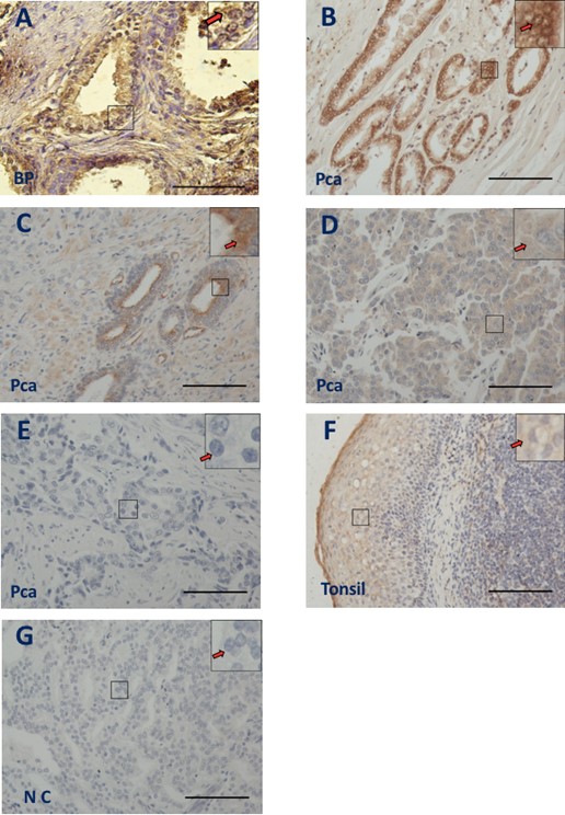
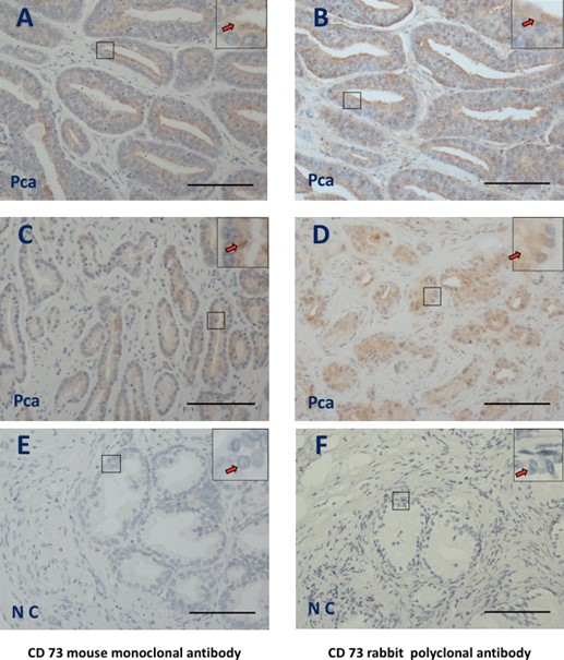
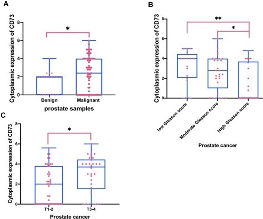
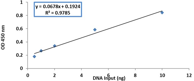
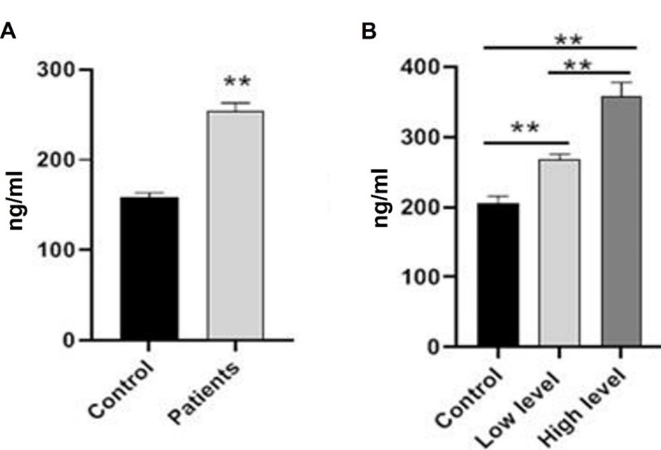
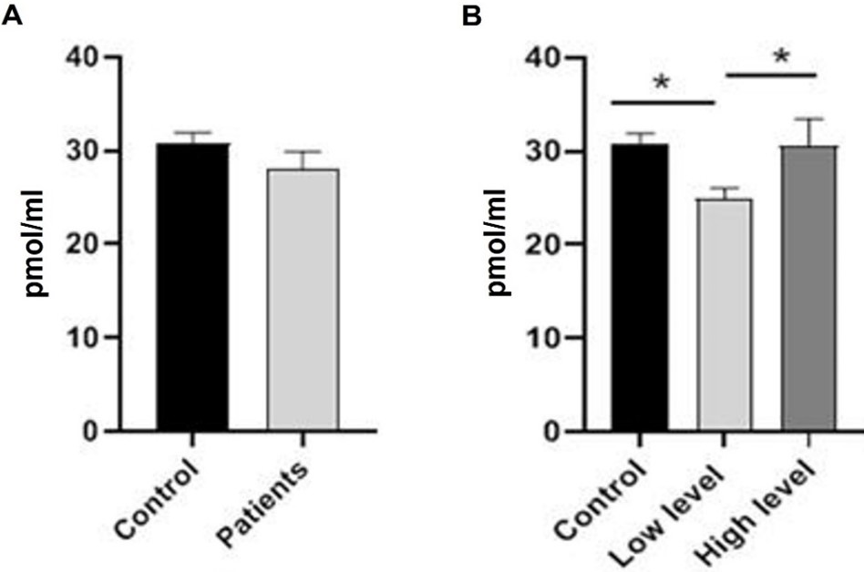
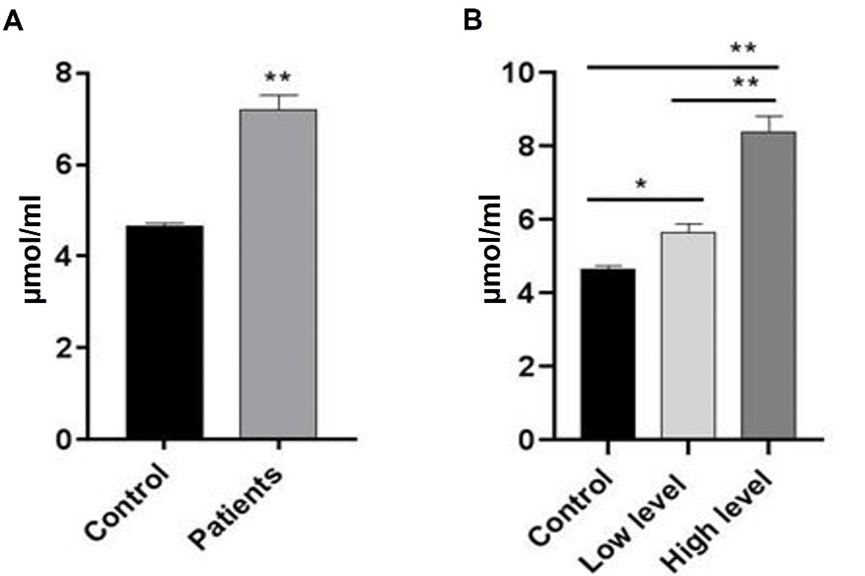
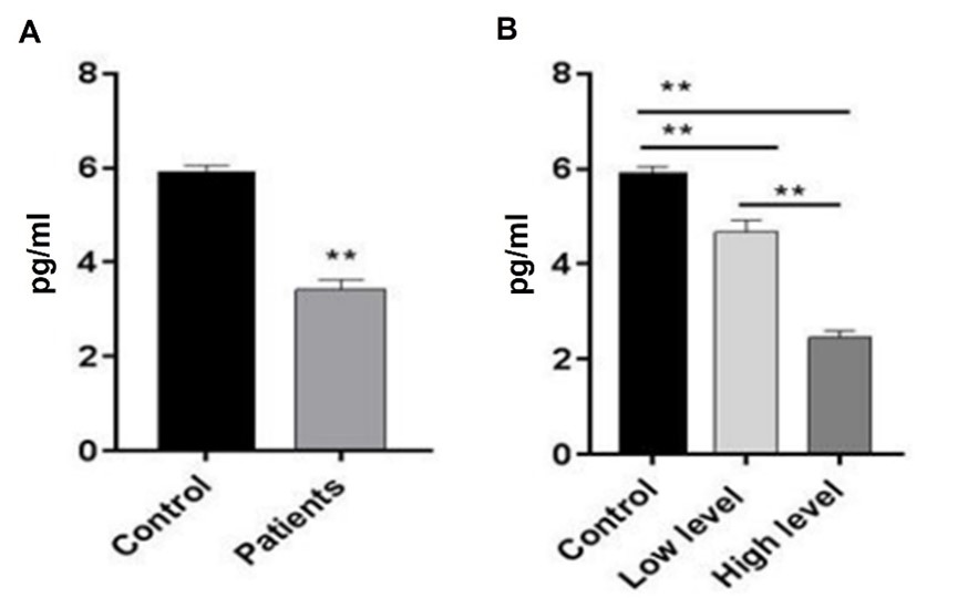
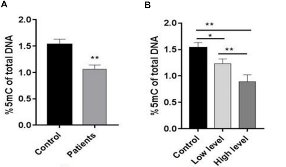
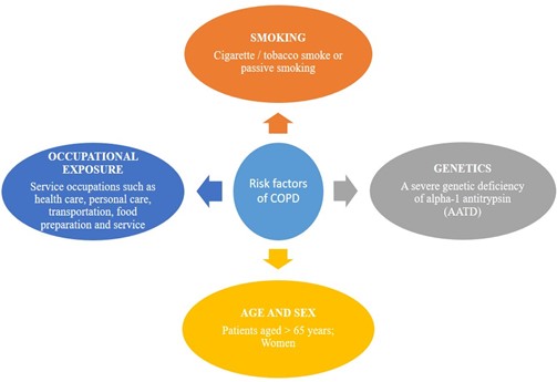
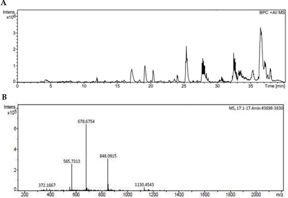
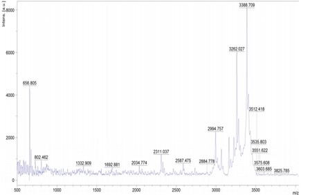
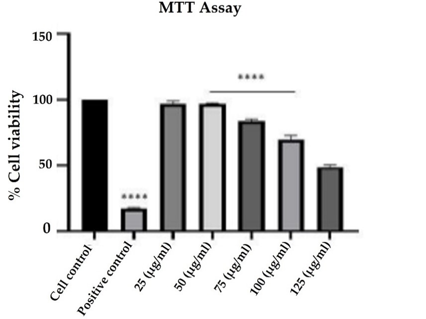
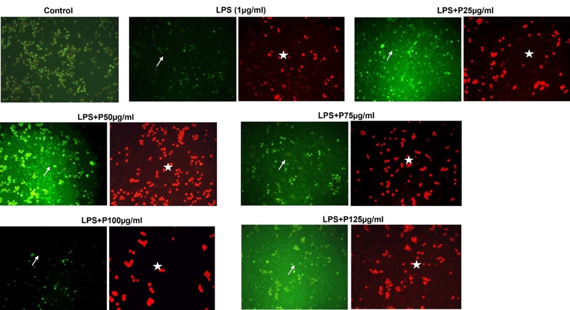
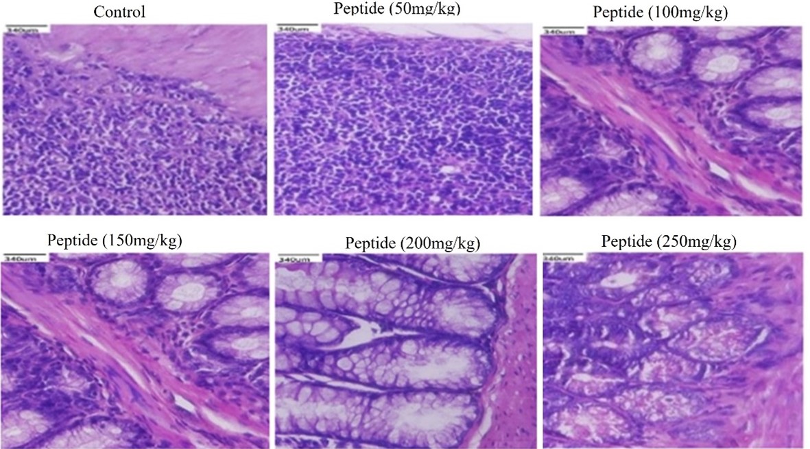
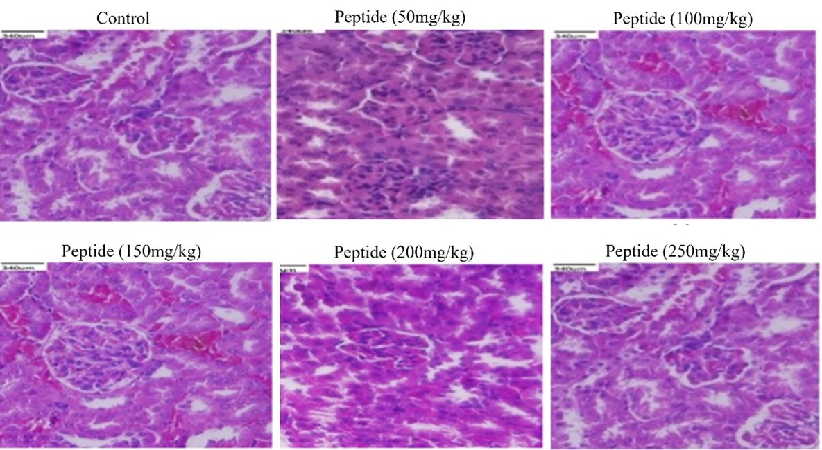
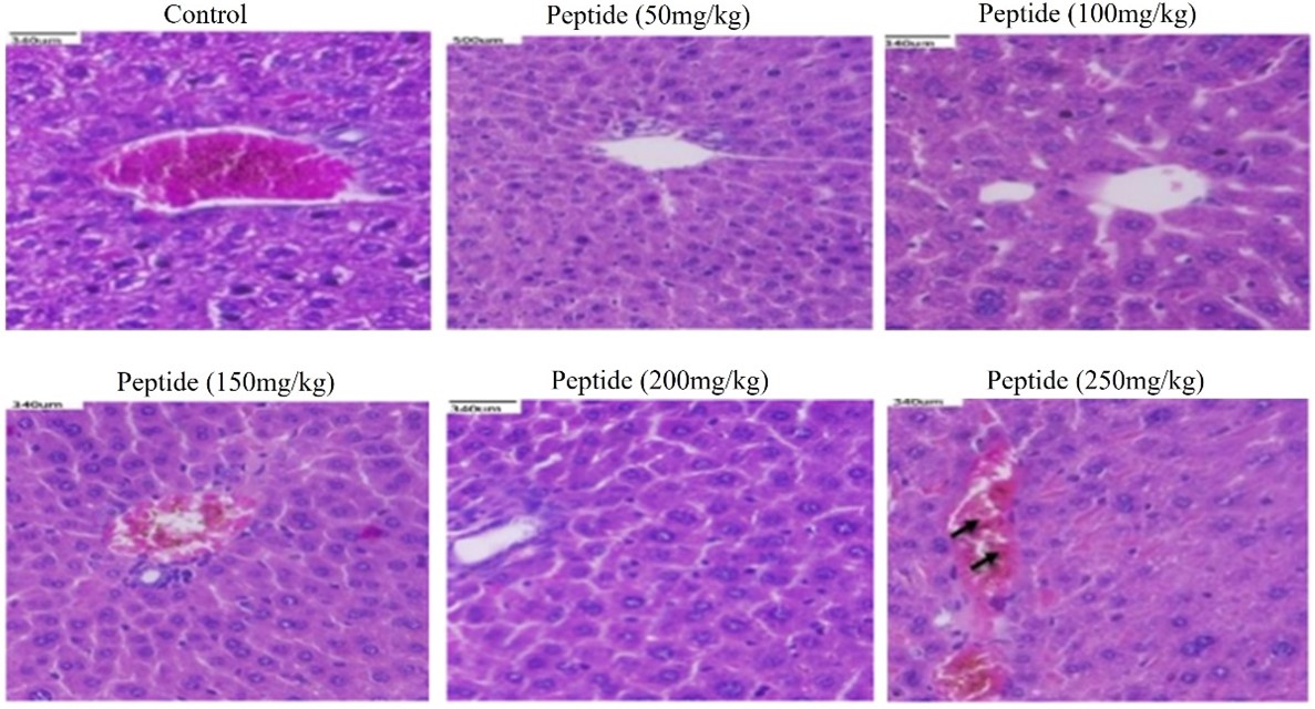
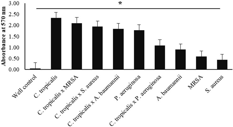
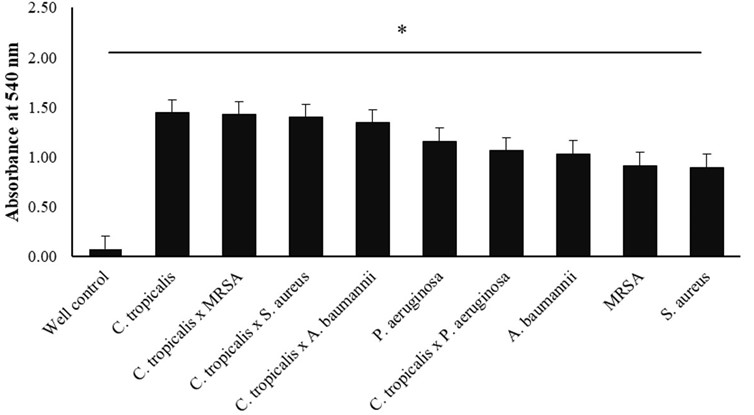
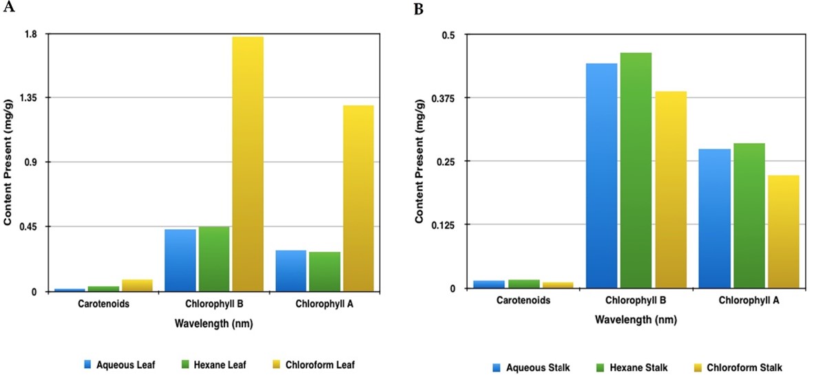
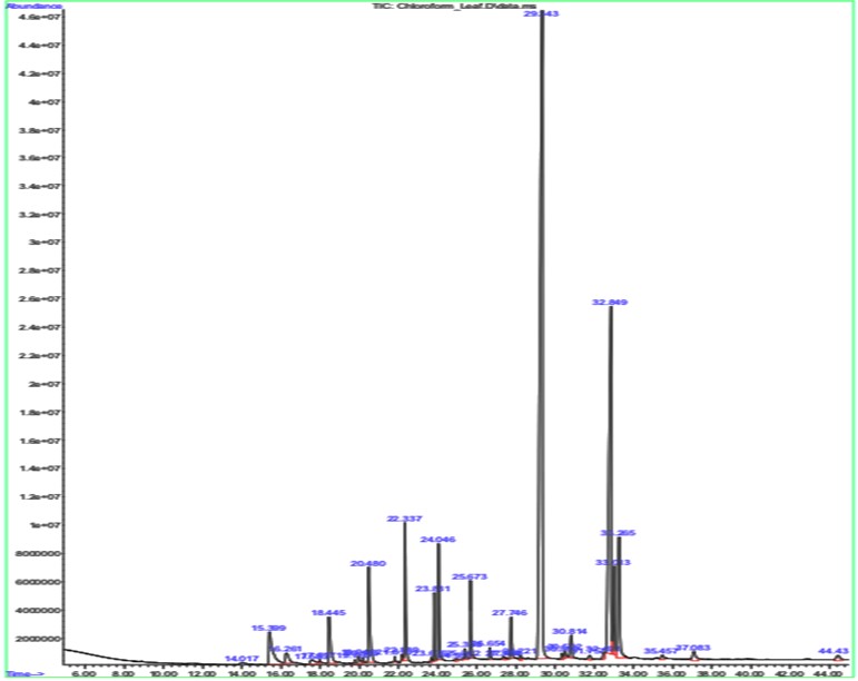
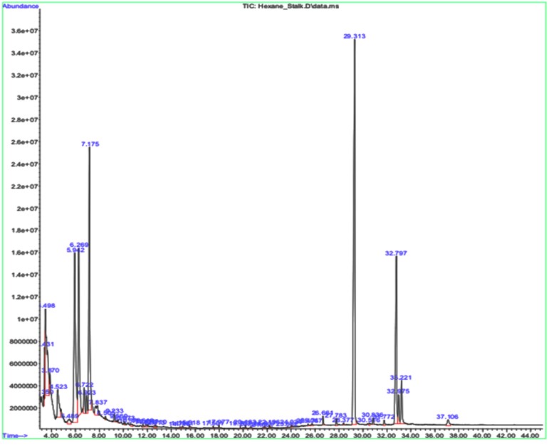
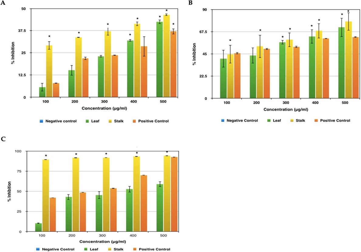
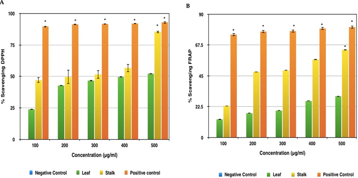
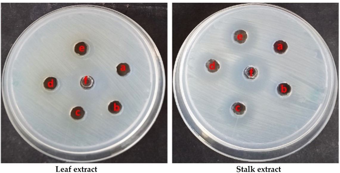
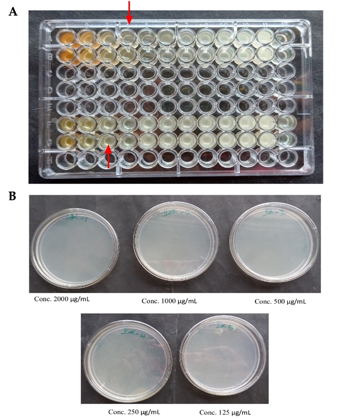
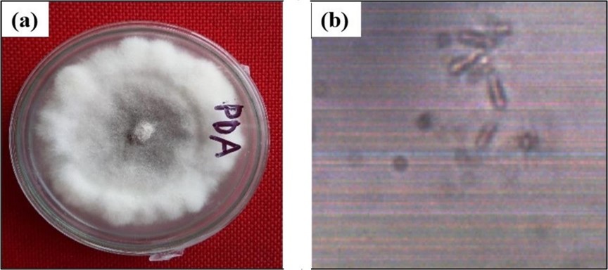
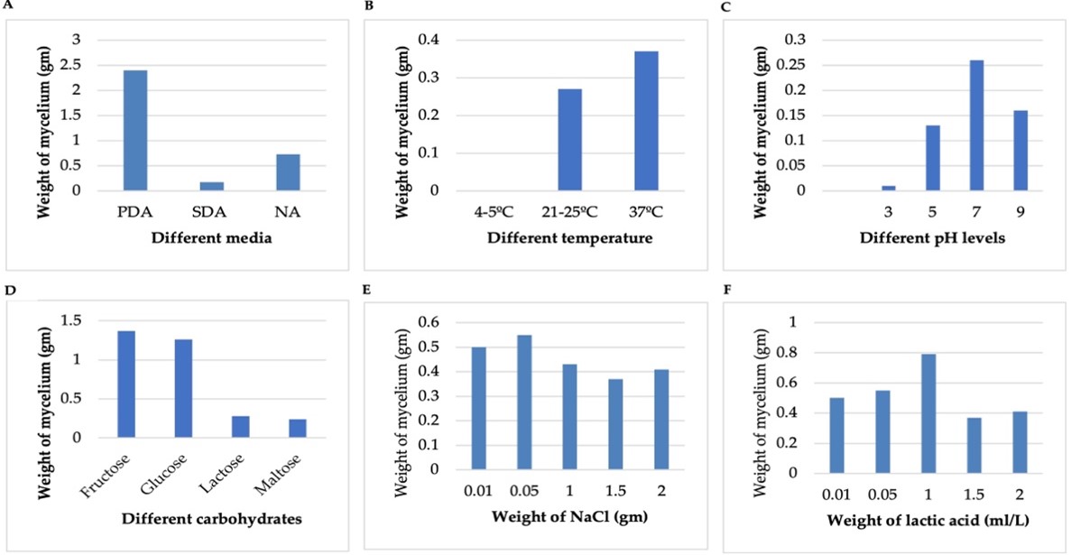
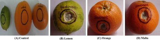
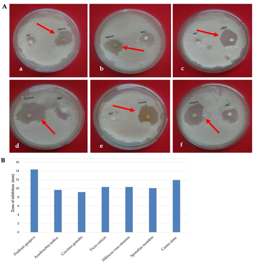