HTTP 200 OK
Allow: GET, HEAD, OPTIONS
Content-Type: application/json
Vary: Accept
{
"count": 319,
"next": "https://jabet.bsmiab.org/articles/?format=api&page=22",
"previous": "https://jabet.bsmiab.org/articles/?format=api&page=20",
"results": [
{
"id": 236,
"slug": "178-1624073760-antinociceptive-potential-of-methanol-leaf-extracts-of-cissampelos-parreira-linn-lantana-camara-linn-and-ocimum-gratissimum-african-basil",
"featured": false,
"slider": false,
"issue": "Vol4 Issue3",
"type": "original_article",
"manuscript_id": "178-1624073760",
"recieved": "2021-05-17",
"revised": null,
"accepted": "2021-07-17",
"published": "2021-07-29",
"pdf_file": "https://jabet.bsmiab.org/media/pdf_file/2023/50/178-1624073760.pdf",
"title": "Antinociceptive potential of methanol leaf extracts of Cissampelos parreira (Linn), Lantana camara (Linn) and Ocimum gratissimum (African basil)",
"abstract": "<p>The effects of <em>Cissampelos pareira </em>(Linn)<em>, Lantana camara </em>(Linn) and<em> Ocimum gratissimum</em> (African basil) leaf extracts on pain have not been biologically determined despite their frequent traditional use in pain management. The present study evaluated the effects of methanol leaf extracts of these three plants on formalin-induced pain in Swiss albino mice. Leaves of <em>C. pareira, L. camara,</em> and <em>O. gratissimum</em> were harvested, cleaned, shade dried, crushed, extracted in absolute methanol and concentrated to dryness. The quantitative phytochemical screening of the three plant extracts was first carried out. Then, the pain assay tests constituted eight groups of five mice each: normal control group, positive control group, negative control group and experimental groups of 50, 100, 150, 200 and 250 mg/kg bw extracts. The animals were administered with various treatments thirty minutes before induction of pain through injection of 0.01 ml of 2.5% formalin solution into the sub-plantar region of the left hind paw. Paracetamol at the dose of 50 mg/kg bw and 5% dimethyl sulfoxide were used as the positive and negative controls respectively. The plant extracts were administered intraperitoneally and orally. Data was analyzed using one-way analysis of variance and unpaired t-test. Phytochemical screening on separate extracts of <em>C. pareira, L. camara, </em>and <em>O. gratissimum</em> revealed fatty acids, phenols, flavonoids and terpenoids. The different dosages of methanol leave extracts of <em>C. pareira, L. camara, </em>and <em>O. gratissimum</em> reduced pain significantly (p˃0.05) in mice. The significant reduction of pain was associated with fatty acids, phenols, flavonoids and terpenoids revealed in the plant extracts.</p>",
"journal_reference": "J Adv Biotechnol Exp Ther. 2021; 4(3): 349-364.",
"academic_editor": "Md Jamal Uddin, PhD; Ewha Womans University, Seoul, South Korea",
"cite_info": "Kimuni SN, Gitahi SM, et al. Antinociceptive potential of methanol leaf extracts of Cissampelos parreira (Linn), Lantana camara (Linn) and Ocimum gratissimum (African basil). J Adv Biotechnol Exp Ther. 2021; 4(3): 349-364.",
"keywords": [
"phytochemicals",
"Antinociception",
"Ocimum gratissimum",
"Cissampelos pareira",
"Lantana camara"
],
"DOI": "10.5455/jabet.2021.d135",
"sections": [
{
"section_number": 1,
"section_title": "INTRODUCTION",
"body": "<p>Pain is a common manifestation of many diseases afflicting millions of people worldwide [<a href=\"#r-1\">1</a>]. It is a phenomenon experienced during disease processes and it runs an acute and chronic course, also referred to as phase one and two or early and late phases, respectively. Acute pain is mild and can last for just a moment, or it might be severe and last for weeks or months. It serves a protective purpose, warning the body of impending danger by activating the “fight or flight” response of the sympathetic nervous system, which often produces signs such as elevated blood pressure, dilated pupils and tenseness of skeletal muscles [<a href=\"#r-2\">2</a>].<br />\r\nAcute pain may also be exhibited through behavioral signs such as crying, moaning, or guarding painful body areas. However, physiologic and behavioral signs are not always evident in acute pain and if present, may last for a short period. With longer exposures to pain, physiologic adaptation may occur, and the body may change its behavior to appear unresponsive because of exhaustion [<a href=\"#r-3\">3</a>].<br />\r\nOn the other hand, chronic pain is normally continuous, carries on for at least six months and habitually grows insidiously. It goes on past the course of an acute disease or after tissue healing is over. It has no biological function because it is not a sign of a disease process as it is a disease process itself [<a href=\"#r-4\">4</a>]. The cause of chronic pain is often unknown. Important behavioral and psychological alterations that are seen as a result of chronic pain include depression, sleeping disorders and a tendency to deny pain. The most common types of chronic pain include persistent low back pain, pain associated with cancer, neuralgias, hemiagnosia and phantom limb pain [<a href=\"#r-5\">5</a>].<br />\r\nConventional approaches used in pain management include acetaminophen (paracetamol) and Non-Steroidal Anti-Inflammatory Drugs (NSAIDs) such as diclofenac and aspirin. Acetaminophen is believed to attenuate pain by inhibiting the activity of the cyclooxygenase enzyme [<a href=\"#r-6\">6</a>]. However, these drugs are accompanied by side effects such as nausea, constipation, skin allergies, liver and renal impairment and cardiovascular disorders [<a href=\"#r-7\">7</a>]. Other alternative methods used for pain relief include plant extracts, decoctions, faith, and acupuncture. The use of botanicals in managing painful conditions has gained first-class awareness, arguably because they are culturally acceptable, give much less or no adverse outcomes, easily accessible and affordable [<a href=\"#r-8\">8</a>].<br />\r\nThe use of medicinal plants for thousands of years by indigenous populations has attracted research on their empirical knowledge. Therefore, medicinal plants demand adequate evaluation of their efficacy, safety and mechanism of action [<a href=\"#r-9\">9</a>]. Some herbal extracts that are used as a folklore remedy for pain have been scientifically confirmed. They include <em>Lonchocarpus eriocalyx </em>[<a href=\"#r-10\">10</a>], <em>diphenyl diselenide </em>[<a href=\"#r-11\">11</a>], <em>Clinacanthus nutans </em>[<a href=\"#r-12\">12</a>], and <em>Borreria verticillata</em> [<a href=\"#r-13\">13</a>] among others<em>.</em><br />\r\nThe present study evaluated the antinociceptive effects of methanol extracts of <em>Cissampelos pareira </em>(Linn)<em>, Lantana camara </em>(Linn) and<em> Ocimum gratissimum</em> (African basil)<em>. Cissampelos pareira </em>is a climber with heart-shaped leathery leaves, 3m tall and has orange fruits. The flowers are small in size with threadlike pedicels. It is common in deciduous bushland and near rock outcrops [<a href=\"#r-14\">14</a>]. <em>Lantana camara</em> is a perennial erect hardy shrub with a powerful odour and grows up to 1 to 3 meters. It occurs in riparian zones and forest edges [<a href=\"#r-15\">15</a>]. On the other hand, <em>O. gratissimum</em> is a local shrub with an erected small plumb and many barnacles, with a height between 1-3 meters high. It is well distributed around villages and along streams [<a href=\"#r-16\">16</a>].<br />\r\nLiterature has shown that <em>C. pareira, L. camara</em> and <em>O. gratissimum</em> have traditionally been used in the management of pain [<a href=\"#r-15\">15</a>]. In South America, <em>C. pareira</em> is referred to as “Midwife’s herb since it is used to manage menstrual cramps, uterine hemorrhages after childbirth, eases childbirth and postpartum, because of its intense relaxant effect on smooth muscle. It is prepared as a decoction and administered orally [<a href=\"#r-18\">18; 19</a>]. On the other hand, dried leaves of <em>L. camara</em> are boiled in water and the decoction is used to manage hemoptysis and pulmonary tuberculosis [<a href=\"#r-15\">15</a>]. Finally, leaves of <em>O. gratissimum</em> mixed with hot water have been applied to eyes that are hurting, treat gout and colon pain [<a href=\"#r-20\">20</a>; <a href=\"#r-17\">17</a>]. The selection of the three plants was based on ethnobotanical information from local herbalists, who acknowledged their use as a folklore remedy for pain among communities living in Embu County, Kenya.<br />\r\nHowever, the continued use of these plants has not been subjected to scientific evaluation to validate their traditional use as therapeutic agents against pain. Against this background, this study was conceived and designed to determine the antinociceptive potential of the methanol leaf extracts of <em>C. pareira, L. camara</em> and <em>O. gratissimum</em> in mice.</p>"
},
{
"section_number": 2,
"section_title": "MATERIALS AND METHODS",
"body": "<p><strong>Plant samples collection and extraction</strong><br />\r\nThis study obtained a research permit from the National Commission of Science, Technology and Innovation (NACOSTI), Kenya (Re. No: NACOSTI/P/20/5273). Collection of fresh leaves of <em>C. pareira, L. camara </em>and<em> O. gratissimum</em> was done from their natural habitat in Mbeere North Sub-County, Embu County, Kenya, with the assistance of a local herbalist. The plant samples were dusted, packed in khaki bags and transported to Kenyatta University, Department of Biochemistry, Microbiology and Biotechnology, where the study was undertaken. The botanical identification of the plant samples was performed by an acknowledged taxonomist in the same university Mr. Stephen Mwangi. The respective voucher specimens of the plants were then deposited in the Plant Sciences Departmental herbarium. Specimen voucher numbers were assigned as <em>C. pareira </em>(SNK001)<em>, L. camara </em>(SNK002) and <em>O. gratissimum</em> (SNK003), respectively.<br />\r\nBefore drying, the plant materials were cleaned using distilled water. Next, they were separately shade-dried at room temperature for three weeks. They were then separately ground into fine powders using an electric mill (Christy and Norris Model 8) and stored at room temperature in well labelled khaki bags awaiting extraction. The collection, processing and extraction of the plant materials were done as per the guidelines formulated by World Health Organization on good herbal processing practices for herbal medicines [<a href=\"#r-21\">21</a>].<br />\r\nPowdered plant samples weighing 800 grams were separately soaked in 1800ml of analytical grade methanol (CAS number 67-56-1; Sigma Aldrich; Germany), agitated and left to stand for 24 hours in a well labelled 2-litre conical flask (Borosil®) and corked. The residue was immersed in methanol again after decantation of the mixture and permitted to stand for another 24 hours at room temperature before decanting again. The obtained mixture was filtered using Whatman No.1 paper [<a href=\"#r-22\">22</a>]. The steps were repeated twice, and the menstruum portions put together. Each of the extracts was then concentrated under reduced pressure and controlled temperature of between 40℃-50℃ using a rotary evaporator (Stuart RE400, Stuart, Germany) [<a href=\"#r-23\">23</a>]. The concentrate was allowed to dry up and thereafter placed in pre-weighed well labelled and sealed specimen bottles. The percentage yields of the plant extracts were determined using the formula described by [<a href=\"#r-24\">24</a>]:<br />\r\nWhere, W<sub>2</sub> is the weight of the extract and the container, W<sub>1</sub> is the weight of the container alone and W<sub>0</sub> is the weight of the initial dried sample.<br />\r\nThe extracts were then refrigerated at 4℃, awaiting use in bioassay studies and phytochemical analysis.</p>\r\n\r\n<p> </p>\r\n\r\n<p><strong>Liquid chromatography-mass spectrometry (LC-MS) analysis</strong><br />\r\nThe quantitative phytochemical screening of the three extracts was done at the International Centre for Insect Physiology and Ecology (ICIPE) laboratories, Kenya. The identification of phytochemicals was performed using Agilent MSD 6120-Triple Quadruple LC-MS with an electrospray source (Palo Alto, CA). The system was controlled using Chemstation software (Hewlett-Parkard). Reversed-phase liquid chromatography was performed on an Agilent Technologies 1200 infinite series, Eclipse plus C<sub>18</sub> column, 4.6 x 50 mm, 3.5um (Agilents CA814112) using the following gradient program 0 min, 5% B; 0-5min, 5-50% B; 5-10min, 50-80% B; 10-15min, 80-100% B; 15-25min, 100% B; 25-30min, 5% B; 30-35min, 5% B. The flow rate was constant at 1ml/min; the injection volume was 1.0Ul and data was acquired in a full-scan negative ion mode using a 100 to 1500<em>m/z</em> scan range. The dwell time for each ion was 50ms. The mass spectrometer parameters were as follows: capillary voltage 3.0kV; cone voltage 70V; extract voltage 5V; RF voltage 0.5V; source temperature 110℃; nitrogen gas temperature for desolvation, 380℃; and nitrogen gas flow for desolvation 400L/h. The LC-MS analyzed serial dilutions of Kaempferol standard (1-100 ng/µL) in full scan mode to generate linear calibration curve (peak area verses concentration) with the following equation: [<em>y=6008.9x -5250.3 (R<sup>2</sup> = 0.9987)</em>] which served as a basis for external quantification. Most of the compounds in the MS exhibited abundance (M-H) in negative ion mode. The single point external standards analyte response was assumed to be linear over concentration. All the peaks for the analyte were subjected to the same method of quantification. LC-MS in full scan mode generated a linear calibration curve (peak area verse concentration) with the following equation: [<em>y=6008.9x-5250.3 (R<sup>2</sup> = 0.9987)</em>] which served as a basis for external quantification. A mass of 1mg/mL of each sample was reconstituted in LC-MS methanol from Tedia. Inc USA and 20µl injected into as aforementioned. Pentacyclic triterpenoids present in the extracts were identified using METLIN metabolite data base and literature precedent and quantified using griseofulvin internal standard.</p>\r\n\r\n<p> </p>\r\n\r\n<p><strong>Gas chromatography-mass spectrometry (GC-MS) analysis</strong><br />\r\nOne milligram of the dried crude methanol leaf extracts of <em>C. pareira, L. camara </em>and<em> O. gratissimum </em>were dissolved in 1mL dichloromethane (sigma Aldrich GC-grade). The samples were vortexed for 30 seconds and sonicated in an ultra-bath for 15 minutes before centrifugation at 14,000 rpm for 5 minutes. The supernatants were passed through anhydrous Na<sub>2</sub>SO<sub>4</sub> to remove moisture. The resultant stock solutions (1mg/mL) were used to prepare experimental samples whose final concentrations were 100ng/μL. Each sample was prepared in triplicate.<br />\r\nEach sample was analyzed using GC-MS (7890/5975 Agilent Technologies, Inc., Beijing, China), consisting of a Gas Chromatography interfaced to a Mass Spectrometer instrument. The GC-MS was equipped with an HP-5MS (5% phenyl methyl siloxane) low bleed capillary column of 30m length, 0.25mm diameter and 0.25μm film thickness. The GC-MS used a detection with ionization energy of 70Ev. The carrier gas used was helium (99.99%) at a constant flow rate of 1.25ml/min in split mode. The injector and mass transfer line temperature were set at 250℃ and 200℃, respectively, and an injection volume of 1μl was employed. The oven temperature was programmed from 35°C for 5 minutes, with an increase of 10°C/min to 280°C for 10.5 minutes, then 50°C/min to 285°C for 29.9 minutes with a run time of 70 minutes. The mass spectrometry operating parameters were as follows: ionization energy, 70eV; ion source temperature, 230°C; solvent cut time, 3.3 min; relative detector gain mode; scan speed, 1666μ/sec; scan range of 40-550m/z and the interface temperature of 250°C. Identification was based on the molecular structure, molecular mass and calculated fragments. Interpretation on mass spectrum GC-MS was conducted using the database of National Institute of Standard and Technology (NIST) database, which has more than 62,000 patterns. The name, molecular weight and structure of the components of the test materials were ascertained. Library-MS searches using NIST Mass spectral library NIST 05, Chemecol.l, NIST 11 and Adams 2.l databases. NIST mass spectral search program Version 2.0 was used for characterization purposes in the GC-MS data system. The area was used for quantification based on the amount of internal standard added.</p>\r\n\r\n<p> </p>\r\n\r\n<p><strong>Experimental animals</strong><br />\r\nThe present study used 240 Swiss albino mice of either sex, aged between 6-8 weeks and weighing 20-25 grams. The mice were obtained from Kenya Medical Research Institute (KEMRI) Nairobi, Kenya, upon ethical approval by the Egerton University Research Ethics Committee, Kenya approval number EUREC/APP/101/2020. The mice were housed in the animal breeding facility in the Department of Biochemistry, Microbiology and Biotechnology at Kenyatta University. The animals were kept in standard cages throughout the study, maintained under room temperature and humidity (55±5%) with 12 hours’ daylight for seven days to acclimatize with the laboratory conditions [<a href=\"#r-25\">25</a>].<br />\r\nThe mice were fed on standard rodent pellet diet and were allowed access to water <em>ad libitum</em> [<a href=\"#r-26\">26</a>]. Before conducting the bioassays, the mice fasted for 12 hours. Bio-screening was conducted according to the internationally accepted procedures and ethical guidelines for evaluating efficacy and safety of herbal medicines in animal models [<a href=\"#r-27\">27; 28</a>].</p>\r\n\r\n<p> </p>\r\n\r\n<p><strong>Experimental design and bioassay</strong><br />\r\nThis study adopted a completely randomized controlled study design, from which an experimental design was formulated. First acute oral toxicity assay was carried out according to OECD protocol [29]. The acute toxicities of <em>C. pareira, L. camara </em>and<em> O. gratissimum</em> used doses of 1000 and 2000mg/kg bw. After administration of the extract, the animals were under close observation continuously for 1 hour and intermittently for 4 hours and thereafter once every 24 hours for the 14 days. During this period, observations such as mortality, neurological behavior, reduced locomotion and dullness were noted.<br />\r\nFive dose levels were selected for antinociceptive assays (50, 100, 150, 200 and 250mg/kg bw) of the studied plant extracts based on a prior pilot study. Thereafter, Swiss albino mice were randomly divided into eight groups of five mice each (n=5). Group I (Normal control) mice were intraperitoneally administered with 5%DMSO solution only. Group II (Negative control) mice were intraperitoneally administered with 5% DMSO solution 30 minutes prior to pain induction and no treatment was administered. Group III (Positive control) mice were intraperitoneally administered with reference drug (Paracetamol) at a dose of 50mg/kg bw 30 minutes prior to pain induction. Groups IV, V, VI, VII and VIII mice were intraperitoneally treated with extract dose levels of 50, 100, 150, 200 and 250mg/kg bw respectively, 30 minutes before induction of pain. The same experimental setup was done with orally administered treatments.</p>\r\n\r\n<p> </p>\r\n\r\n<p><strong>Antinociceptive activity assay</strong><br />\r\nThe antinociceptive effects of the three methanol extracts were determined using formalin-induced paw licking model in mice as described by [<a href=\"#r-30\">30</a>]. Half an hour after administering the reference drug (Paracetamol) and methanol extracts, the pain was initiated by administering 0.05ml of 2.5% formalin into the sub-plantar region of the left hind paw and the pain behavior observed and recorded. In addition, the time in seconds that the mouse spent lifting, licking or biting the injected paw was counted using a digital stopwatch (3D Max 2011 Model) and recorded as the measure of pain [<a href=\"#r-31\">31</a>]. In all groups, the mice were individually placed in a transparent plexiglass observation chamber with mirrors placed on two sides of the chamber to observe the nociceptive behavior from all angles. All injections were done using 30-gauge needles.<br />\r\nThe nociceptive behavior was monitored and recorded according to the response pattern described by [<a href=\"#r-31\">31</a>]. Two distinct periods of nociceptive behavior were identified and scored separately as early phase and late phase. The early phase was recorded 1-5 minutes after formalin injection. The late phase was recorded 15-30 minutes after formalin injection. The fifth and the fifteenth minute duration was the remission period with minimal nociceptive behavior [<a href=\"#r-32\">32</a>]. The percentage paw licking inhibition was determined using the formula described by [<a href=\"#r-33\">33</a>].<br />\r\nWhere, N- The negative control group value for each phase, T-The treated group value for each phase</p>\r\n\r\n<p> </p>\r\n\r\n<p><strong>Statistical analysis</strong><br />\r\nQuantitative experimental data on paw licking, biting, or lifting time obtained from all the animals in various treatment groups were recorded and tabulated in a Microsoft Excel spreadsheet and transferred to a Minitab spreadsheet. The data were subjected to descriptive statistics and presented as Mean ± Standard Error of Mean (SEM) and then checked for normality using the Kolomogorov-Smirnov test. Analysis was done using One-Way Analysis of Variance (ANOVA) followed by Tukey’s post hoc for pairwise separation and comparison of means. In addition, unpaired Student t-test was used to compare the antinociceptive activities of orally and intraperitoneally administered treatments. Analysis of the data was done using Minitab<sup>®</sup> Version 17 software. Statistical significance was set at 95% confidence interval. Results were presented in tables and figures.</p>"
},
{
"section_number": 3,
"section_title": "RESULTS",
"body": "<p><strong>Percentage yield of the plant extracts</strong><br />\r\nThe final yields of <em>L camara</em> and <em>C. pareira</em> were blue-black pastes while that of <em>O. gratissimum </em>was a brown, black solid. The percentage yield from <em>C. pareira, L. camara and O. gratissimum </em>leaf extracts were 21, 24 and 23%, respectively.</p>\r\n\r\n<p> </p>\r\n\r\n<p><strong>Quantitative phytochemical analysis</strong><br />\r\nThe LC-MS analysis of the leaf extracts of <em>C. pareira</em>, <em>L. camara</em> and <em>O. gratissimum</em> revealed the presence of phytoconstituents such as caffeic acid, cissamine, quercetin, luteolin, apigenin, among others (<a href=\"#Table-1\">Table 1</a>). On the other hand, the GC-MC screening of the three plant extracts demonstrated the presence of α-pinene, cymene, limonene, oleic acid, among others (<a href=\"#Table-1\">Table 1</a>).</p>\r\n\r\n<div id=\"Table-1\">\r\n<p><a href=\"https://jabet.bsmiab.org/table/178-1624073760-table1/\">Table-1</a><strong>Table 1. </strong>Phytochemicals revealed in the methanol leaf extracts through LC-MS and GC-MS analysis.</p>\r\n\r\n<p> </p>\r\n</div>\r\n\r\n<p><strong>Acute toxicity</strong><br />\r\nNo deaths were reported following oral administration of the studied plant extracts at the dose of 1000 and 2000mg/kg bw. In addition, no clinically abnormal signs were observed.</p>\r\n\r\n<p> </p>\r\n\r\n<p><strong><em>In vivo</em></strong><strong> antinociceptive activities of oral and intraperitoneal administration of methanol leaf extract of <em>Cissampelos pareira</em></strong><br />\r\nIn general, both orally and intraperitoneally administered methanol leaf extract of <em>C. pareira </em>significantly reduced paw licking time in a dose-independent fashion in the early and late phases following formalin-induced nociception in mice. In addition, the antinociceptive activities exhibited a decrease in paw licking time or raised percent paw licking inhibition following formalin-induced pain in mice (<a href=\"#Table-2\">Table 2</a> and <a href=\"#Table-3\">3</a>). It was noted that the highest analgesic activity in both phases was at the extract dose level of 150mg/kg bw (<a href=\"#Table-2\">Table 2</a>). At this dose, the antinociceptive effect was statistically similar to that of the reference drug (paracetamol) (p>0.05; <a href=\"#Table-2\">Table 2</a>). The extract exhibited the least antinociceptive effect at the dose of 250mg/kg bw in both phases (<a href=\"#Table-2\">Table 2</a>).<br />\r\nIn the early phase, the range of pain inhibition of the orally administered methanol leaf extract of <em>C. pareira </em>at the five dose levels was between 31.75 and 61.02%. It was noted that the analgesic effect of the extract dose levels of 50, 100, 150, 200 and 250mg/kg bw was statistically similar to that of the standard drug (p>0.05; <a href=\"#Table-2\">Table 2</a>). Further, it was observed that there was no significance difference in the antinociceptive activity of the extract dosages of 100, 150 and 200mg/kg bw (p>0.05; <a href=\"#Table-2\">Table 2</a>). In addition, the effect of the extract at the doses of 50, 100, and 200mg/kg bw was statistically similar (p>0.05; <a href=\"#Table-2\">Table 2</a>).<br />\r\nIn the late phase, the five extract dose levels of 50, 100, 150, 200 and 250mg/kg bw reduced the formalin-induced pain in mice by 27.51, 37.21, 40.92, 26.63 and 10.68% respectively (Table 2). The percent paw licking inhibition of the extract at dose levels of 50, 100, 150 and 200mg/kg bw were not significantly different and comparable to that of the standard drug (p>0.05; <a href=\"#Table-2\">Table 2</a>). In addition, the efficacy of the extract at the doses of 50, 200 and 250mg/kg bw was statistically similar to each other (p>0.05; <a href=\"#Table-2\">Table 2</a>).</p>\r\n\r\n<p>In the early phase, the intraperitoneally administered methanol leaf extract of <em>C. pareira </em>inhibited pain by 59.73, 70.90, 64.73, 40.21 and 35.98% at the extract dose levels of 50, 100, 150, 200 and 250mg/kg bw respectively (<a href=\"#Table-3\">Table 3</a>). Furthermore, it was observed that the highest analgesic effect was observed at the extract dosage of 100mg/kg bw while the least effect was at the extract dose of 250mg/kg bw in both phases (Table 3). Notably, the extract dose levels of 50, 100 and 150mg/kg bw as well as the reference drug evoked nociceptive inhibitions that were comparable to each other (p>0.05; <a href=\"#Table-3\">Table 3</a>). On the other hand, the efficacy exhibited by the extract dosages of 200 and 250mg/kg bw was statistically similar to each other (p>0.05; <a href=\"#Table-3\">Table 3</a>).<br />\r\nIn the late phase, the range of antinociceptive action of the <em>C. pareira </em>extract at the five dose levels ranged between 16.23 and 64.90% (<a href=\"#Table-3\">Table 3</a>). It was noted that the extract doses of 50, 200 and 250mg/kg bw caused statistically similar analgesic effect (p>0.05; <a href=\"#Table-3\">Table 3</a>), but the effect was significantly lower than that of the standard drug (p<0.05; <a href=\"#Table-3\">Table 3</a>). Further, the percent inhibitions of nociception of the extract dose levels of 100 and 150 mg/kg bw were comparable to the effects caused by the reference drug (p>0.05; <a href=\"#Table-3\">Table 3</a>).</p>\r\n\r\n<div id=\"Table-2\">\r\n<p><a href=\"https://jabet.bsmiab.org/table/178-1624073760-table2/\">Table-2</a><strong>Table 2. </strong>Antinociceptive effect of orally administered methanol leaf extract of C. pareira on formalin-induced pain in mice.</p>\r\n</div>\r\n\r\n<div id=\"Table-3\">\r\n<p><a href=\"https://jabet.bsmiab.org/table/178-1624073760-table3/\">Table-3</a><strong>Table 3. </strong>Antinociceptive effect of intraperitoneally administered the leaf extract of<em> C. pareira </em>on formalin-induced pain in mice.</p>\r\n</div>\r\n\r\n<p> </p>\r\n\r\n<p><strong>Comparison between antinociceptive effects of orally and intraperitoneally administered <em>C. pareira </em>extract.</strong><br />\r\nUpon pairwise comparison of the orally and intraperitoneally administration of methanol leaf extract of <em>C. pareira,</em> it was observed that the potency of the intraperitoneally administered extract was significantly higher than that of orally administered extract in the early phase at the extract dose of 50 and 100mg/kg bw (p<0.05; <a href=\"#figure1\">Figure 1</a>).<br />\r\nOn the other hand, the effects of the orally and intraperitoneally administered extract in the late phase were statistically similar (p>0.05) except at the doses of 100 and 250mg/kg bw (p<0.05; <a href=\"#figure2\">Figure 2</a>).</p>\r\n\r\n<div id=\"figure1\">\r\n<figure class=\"image\"><img alt=\"\" height=\"262\" src=\"/media/article_images/2024/19/06/178-1624073760-Figure1.jpg\" width=\"500\" />\r\n<figcaption><strong>Figure 1. </strong>Comparison between antinociceptive effects of oral and intraperitoneal administration of C. pareira in early phase.</figcaption>\r\n</figure>\r\n</div>\r\n\r\n<div id=\"figure2\">\r\n<figure class=\"image\"><img alt=\"\" height=\"266\" src=\"/media/article_images/2024/19/06/178-1624073760-Figure2.jpg\" width=\"500\" />\r\n<figcaption><strong>Figure 2.</strong> Comparison between antinociceptive effects of oral and intraperitoneal administration of <em>C. pareira</em> in late phase.Caption</figcaption>\r\n</figure>\r\n\r\n<p> </p>\r\n</div>\r\n\r\n<p><strong><em>In vivo</em></strong><strong> antinociceptive activities of oral and intraperitoneal administration of methanol leaf extract of <em>L. camara</em></strong><br />\r\nOverall, the orally and intraperitoneally administered methanol leaf extract of <em>L. camara</em> inhibited nociception in a dose-independent manner in both the early and late phases following formalin-induced nociception in mice (<a href=\"#Table-4\">Table 4</a> and <a href=\"#Table-5\">5</a>). In addition, the analgesic effects were demonstrated by reduced paw licking time or increased percent paw licking inhibitions on formalin-induced pain in mice.</p>\r\n\r\n<p>In both phases, the orally administered extract revealed the highest pain inhibition at the extract dose of 100mg/kg bw, while the lowest pain inhibition was at the extract dose of 250mg/kg bw (<a href=\"#Table-4\">Table 4</a>). In the early phase, the oral administration of the extract reduced pain by between 39.86 and 67.02% (<a href=\"#Table-4\">Table 4</a>). The potency of the extract doses of 50, 150, 200 and 250 was statistically similar to that of the reference drug (paracetamol) (p>0.05; <a href=\"#Table-4\">Table 4</a>).<br />\r\nIn the late phase, the extract dose levels of 50, 100, 150, 200 and 250mg/kg bw inhibited formalin-induced pain in mice by 34.22, 41.27, 36.51, 27.16 and 22.05% respectively (<a href=\"#Table-4\">Table 4</a>). Remarkably, the five extract dose levels and the standard drug evoked efficacies that were statistically similar to each other (p>0.05; <a href=\"#Table-4\">Table 4</a>).<br />\r\nIn the early phase, the five doses (50, 100, 150, 200 and 250mg/kg bw) of intraperitoneally administered extract of <em>L. camara</em> reduced the formalin-induced pain by 62.12, 73.72, 79.89, 71.43 and 64.73% respectively (<a href=\"#Table-5\">Table 5</a>). The highest percentage inhibition of pain was observed at 150mg/kg bw, while the dose of 50mg/kg bw revealed the least inhibition (<a href=\"#Table-5\">Table 5</a>). It was further noted that the effect of the five extract doses was statistically similar compared to that of paracetamol (reference drug) (p>0.05; <a href=\"#Table-5\">Table 5</a>).<br />\r\nIn phase two of the test period, the inhibitions of nociception by the five intraperitoneally administered extract doses of <em>L. camara</em> ranged between 21.69 and 62.05% (Table 5). In this phase, the lowest analgesic activity was observed at the extract dose level of 250mg/kg bw. At the same time, the extract dosage of 100mg/kg bw generated the greatest potency, which was comparable to the effect of the standard drug (p>0.05; <a href=\"#Table-5\">Table 5</a>). Notably, the effect of the extract at the dose levels of 150, 200 and 250mg/kg bw showed no significant difference (p>0.05; <a href=\"#Table-5\">Table 5</a>). Furthermore, the extract’s efficacy at the dose levels of 50, 150, 200 and 250mg/kg bw was significantly lower than that of the standard drug (p<0.05; <a href=\"#Table-5\">Table 5</a>).</p>\r\n\r\n<div id=\"Table-4\">\r\n<p><a href=\"https://jabet.bsmiab.org/table/178-1624073760-table4/\">Table-4</a><strong>Table 4.</strong> Antinociceptive effect of orally administered methanol leaf extract of L. camara on formalin-induced pain in mice.</p>\r\n</div>\r\n\r\n<div id=\"Table-5\">\r\n<p><a href=\"https://jabet.bsmiab.org/table/178-1624073760-table5/\">Table-5</a><strong>Table 5.</strong> Antinociceptive effect of intraperitoneally administered leaf extract of L. camara on formalin-induced pain in mice.</p>\r\n\r\n<p> </p>\r\n</div>\r\n\r\n<p><strong>Comparison between antinociceptive effects of orally and intraperitoneally administered <em>L. camara </em>extract.</strong><br />\r\nIn phase one, it was observed that the potency of the intraperitoneally administered extract was significantly higher compared to that of orally administered extract at the doses of 150 and 250mg/kg bw (p<0.05; <a href=\"#figure3\">Figure 3</a>).<br />\r\nNevertheless, in the second phase, the effects of the intraperitoneally and orally administered extracts were comparable to each other (p>0.05; <a href=\"#figure4\">Figure 4</a>) except at the extract dose of 150mg/kg bw (p<0.05; <a href=\"#figure4\">Figure 4</a>).</p>\r\n\r\n<div id=\"figure3\">\r\n<figure class=\"image\"><img alt=\"\" height=\"263\" src=\"/media/article_images/2024/19/06/178-1624073760-Figure3.jpg\" width=\"500\" />\r\n<figcaption><strong>Figure 3. </strong>Comparison between antinociceptive effects of oral and intraperitoneal administration of L. camara in late phase.</figcaption>\r\n</figure>\r\n</div>\r\n\r\n<div id=\"figure4\">\r\n<figure class=\"image\"><img alt=\"\" height=\"264\" src=\"/media/article_images/2024/19/06/178-1624073760-Figure4.jpg\" width=\"500\" />\r\n<figcaption><strong>Figure 4. </strong>Comparison between antinociceptive effects of oral and intraperitoneal administration of L. camara in late phase.</figcaption>\r\n</figure>\r\n</div>\r\n\r\n<p> </p>\r\n\r\n<p><strong><em>In vivo</em></strong><strong> antinociceptive activities of oral and intraperitoneal administration of methanol leaf extracts of <em>O. gratissimum</em></strong><br />\r\nGenerally, the potency of the orally and intraperitoneally administered methanol leaf extracts of <em>O. gratissimum</em> was dose-independent in the first and second phases following formalin-induced nociception in mice (<a href=\"#Table-6\">Table 6</a> and <a href=\"#Table-7\">7</a>).<br />\r\nThe extract inhibited pain in the early phase between 35.27 and 51.50% (<a href=\"#Table-6\">Table 6</a>). It was noted that the least percentage inhibition of pain was at the dose of 250mg/kg bw while the highest percentage inhibition of pain was attained at the dose of 150mg/kg bw. It was further observed that the effects of all the extract dose levels, as well as the reference drug, were comparable to each other (p>0.05; <a href=\"#Table-6\">Table 6</a>).<br />\r\nIn the second phase, the extract dose levels of 50, 100, 150, 200 and 250mg/kg bw decreased formalin-induced pain in mice by 11.89, 22.22, 28.22, 20.11 and 14.29% respectively (<a href=\"#Table-6\">Table 6</a>). The highest analgesic effect was seen at the extract dosage of 150mg/kg bw, while the extract dose of 50mg/kg bw had the lowest antinociceptive activities (<a href=\"#Table-6\">Table 6</a>). Furthermore, the effect of the extract doses of 50, 100, 200 and 250mg/kg bw was not significantly different (p>0.05; <a href=\"#Table-6\">Table 6</a>). However, the effect of the standard drug was significantly higher than that of the extract at the five dose levels tested (p<0.05; <a href=\"#Table-6\">Table 6</a>).<br />\r\nThe intraperitoneally administered <em>O. gratissimum</em> extract at the doses of 50, 100, 150, 200 and 250mg/kg bw significantly reduced formalin-induced nociception by 37.21, 58.20, 53.26, 49.21 and 40.21% respectively, in the first phase of the test period (<a href=\"#Table-7\">Table 7</a>). The least efficacy was observed at the extract dose of 50mg/kg bw in both early and late phases. It was noted that the peak of analgesic effect was observed at the extract dose of 100mg/kg bw (<a href=\"#Table-7\">Table 7</a>). Additionally, it was observed that the efficacy of the extract dose levels of 50, 200 and 250mg/kg bw was not statistically significant (p>0.05; <a href=\"#Table-7\">Table 7</a>). The effect of the extract at the five dose levels was significantly lower than that of the reference drug (p<0.05; <a href=\"#Table-7\">Table 7</a>).<br />\r\nIn the second phase, the range of pain inhibition caused by the extracts of <em>O. gratissimum</em> was between 16.26 and 38.25% (<a href=\"#Table-7\">Table 7</a>). The extract dose of 150mg/kg bw attained the highest analgesic effect (<a href=\"#Table-7\">Table 7</a>). It was observed that the potency of the extract at the dose levels of 150, 200 and 250mg/kg bw was not statistically significant (p>0.05; <a href=\"#Table-7\">Table 7</a>). However, the effect of the extract at the doses of 50, 100 and 150 was significantly different (p<0.05; <a href=\"#Table-7\">Table 7</a>). Interestingly, all the extract dosages demonstrated antinociceptive effects that were significantly lower than the effects caused by the standard drug (p<0.05; <a href=\"#Table-7\">Table 7</a>).</p>\r\n\r\n<div id=\"Table-6\">\r\n<p><a href=\"https://jabet.bsmiab.org/table/178-1624073760-table6/\">Table-6</a><strong>Table 6.</strong> Antinociceptive effect of orally administered leaf extract of O. gratissimum on formalin-induced pain in mice.</p>\r\n</div>\r\n\r\n<div id=\"Table-7\">\r\n<p><a href=\"https://jabet.bsmiab.org/table/178-1624073760-table7/\">Table-7</a><strong>Table 7.</strong> Antinociceptive effect of intraperitoneally injected leaf extract of O. gratissimum on formalin-induced pain in mice.</p>\r\n\r\n<p> </p>\r\n</div>\r\n\r\n<p><strong>Comparison between antinociceptive effects of orally and intraperitoneally administered <em>O. gratissimum </em>extract.</strong><br />\r\nIt was observed that the efficacies of the orally and intraperitoneally administered extract were statistically similar (p>0.05) in the first phase of the experimental period at all the tested dose levels except at the dose of 100mg/kg bw (p<0.05; <a href=\"#figure5\">Figure 5</a>).</p>\r\n\r\n<p>Likewise, it was noted that in the late phase, the effects of the orally and intraperitoneally administered extract were comparable to each other at the extract doses of 50, 100, 150 and 200mg/kg bw (p>0.05; <a href=\"#figure6\">Figure 6</a>).</p>\r\n\r\n<div id=\"figure5\">\r\n<figure class=\"image\"><img alt=\"\" height=\"246\" src=\"/media/article_images/2024/19/06/178-1624073760-Figure5.jpg\" width=\"500\" />\r\n<figcaption><strong>Figure 5. </strong>Comparison between antinociceptive effects of oral and intraperitoneal administration of O. gratissimum in early phase.</figcaption>\r\n</figure>\r\n</div>\r\n\r\n<div id=\"figure6\">\r\n<figure class=\"image\"><img alt=\"\" height=\"263\" src=\"/media/article_images/2024/19/06/178-1624073760-Figure6.jpg\" width=\"500\" />\r\n<figcaption><strong>Figure 6. </strong>Comparison between antinociceptive effects of oral and intraperitoneal administration of O. gratissimum in late phase.</figcaption>\r\n</figure>\r\n\r\n<p> </p>\r\n</div>\r\n\r\n<p><strong>Comparison of antinociceptive effects of methanol leaf extracts of <em>C. pareira, L. camara </em>and <em>O. gratissimum</em></strong><br />\r\nIt was observed that the effects of the three orally administered methanol extracts of <em>C. pareira, L. camara</em> and <em>O. gratissimum</em> were comparable to each other at the dose levels of 150, 200 and 250mg/kg bw in the early phase (p>0.05; <a href=\"#figure7\">Figure 7</a>). Notably, in the same phase, the effect of oral administration of <em>L. camara</em> extract at the doses of 50 and 100mg/kg bw the <em>L. camara</em> extract was significantly higher compared to those of <em>C. pareira</em> and <em>O. gratissimum</em> (p<0.05; <a href=\"#figure7\">Figure 7</a>).<br />\r\nIn the second phase, the analgesic effects of the orally administered methanol extracts were statistically similar at the dose levels of 100, 150, 200 and 250mg/kg bw (p>0.05; Figure 8). Nevertheless, the effect of <em>L. camara</em> extract at the dose level of 50mg/kg bw was significantly higher than that of <em>O. gratissimum </em>extract (p<0.05; <a href=\"#figure8\">Figure 8</a>).</p>\r\n\r\n<p>On the other hand, intraperitoneally administered extract of <em>O. gratissimum </em>at the dose of 50mg/kg bw, caused analgesic effects that were significantly lower than those of <em>L. camara</em> and <em>C. pareira</em> in the early phase (p<0.05; Figure 9)<em>. </em>It was also noted that antinociceptive effect of <em>L. camara</em> extracts at the dose of 100mg/kg bw was significantly higher than that of <em>O. gratissimum</em> (p<0.05; <a href=\"#figure9\">Figure 9</a>). Besides, the <em>O. gratissimum</em> extract at the dose of 200mg/kg bw exhibited higher antinociceptive effect than that of <em>C. pareira </em>extract (p<0.05; <a href=\"#figure9\">Figure 9</a>)<em>.</em> Further, effect of <em>L. camara</em> extract at the doses of 150 and 250mg/kg bw was significantly higher than those of <em>C. pareira </em>and<em> O. gratissimum </em>extracts (p<0.05; <a href=\"#figure9\">Figure 9</a>)<em>.</em></p>\r\n\r\n<p>In the late phase, the antinociceptivevity of <em>L. camara</em> extract at the dose level of 50mg/kg bw was significantly higher than those of <em>C. pareira </em>and<em> O. gratissimum </em>extracts (p<0.05; <a href=\"#figure10\">Figure 10</a>)<em>. </em>It was also observed that at the extract dose of 100mg/kg bw the potency of <em>O. gratissimum </em>extracts was significantly lower than those of <em>L. camara</em> and <em>C. pareira</em> extracts (p<0.05; <a href=\"#figure10\">Figure 10</a>). However, at the extract dose of 250mg/kg bw, the effect of <em>O. gratissimum </em>extract was significantly higher than those of <em>L. camara</em> and <em>C. pareire</em> extracts (p<0.05; <a href=\"#figure10\">Figure 10</a>). In addition, it was observed that at the extract dose level of 150mg/kg bw, <em>C. pareira </em>extracts exhibited a higher antinociceptive effects than <em>L. camara </em>extract (p<0.05; <a href=\"#figure5\">Figure 5c</a>). Further, at the dose level of 200mg/kg the effects of the three studied extracts bw were statistically similar (p>0.05; <a href=\"#figure10\">Figure 10</a>).</p>\r\n\r\n<div id=\"figure7\">\r\n<figure class=\"image\"><img alt=\"\" height=\"293\" src=\"/media/article_images/2024/19/06/178-1624073760-Figure7.jpg\" width=\"500\" />\r\n<figcaption><strong>Figure 7. </strong>Comparison of antinociceptive effects of orally administrated C. pareira, L. camara and O. gratissimum extracts in the early phase.</figcaption>\r\n</figure>\r\n</div>\r\n\r\n<div id=\"figure8\">\r\n<figure class=\"image\"><img alt=\"\" height=\"291\" src=\"/media/article_images/2024/19/06/178-1624073760-Figure8.jpg\" width=\"500\" />\r\n<figcaption><strong>Figure 8. </strong>Comparison of antinociceptive effects of orally administrated C. pareira, L. camara and O. gratissimum extracts in the late phase.</figcaption>\r\n</figure>\r\n</div>\r\n\r\n<div id=\"figure9\">\r\n<figure class=\"image\"><img alt=\"\" height=\"291\" src=\"/media/article_images/2024/19/06/178-1624073760-Figure9.jpg\" width=\"500\" />\r\n<figcaption><strong>Figure 9. </strong>Comparison of antinociceptive effects of intraperitoneally administrated C. pareira, L. camara and O. gratissimum extracts in the early phase.</figcaption>\r\n</figure>\r\n</div>\r\n\r\n<div id=\"figure10\">\r\n<figure class=\"image\"><img alt=\"\" height=\"286\" src=\"/media/article_images/2024/19/06/178-1624073760-Figure10.jpg\" width=\"500\" />\r\n<figcaption><strong>Figure 10. </strong>Comparison of antinociceptive effects of intraperitoneally administrated C. pareira, L. camara and O. gratissimum extracts in the late phase.</figcaption>\r\n</figure>\r\n</div>"
},
{
"section_number": 4,
"section_title": "DISCUSSION",
"body": "<p>The current study was designed to evaluate the analgesic potential of methanol leaf extracts of <em>C. pareira, L. camara</em> and <em>O. gratissimum</em> in mice. Based on the acute toxicity results the studied plant extracts were found to be safe at the tested doses. The oral and intraperitoneal administration of the three methanol leaf extracts exhibited potent analgesic effects <em>in vivo</em>. The antinociceptive activities of the extracts was manifested by reduction in paw licking time, which was expressed as percent paw licking inhibition. These findings correlated with earlier studies undertaken on medicinal plants using animal models.<br />\r\nA study by [<a href=\"#r-34\">34</a>] on dichloromethane root bark extract of <em>Carissa edulis </em>reported antinociceptive activity in early and late phases of formalin-induced pain in mice model. In yet another study, [<a href=\"#r-35\">35</a>] demonstrated analgesic effect of <em>Caesalpinia volkensii </em>(Harms) in the early and late phases of formalin-induced nociception in mice models.<br />\r\nIn the current study, methanol extracts of <em>C. pareira, L. camara</em> and <em>O. gratissimum</em> caused antinociceptive effects of between 31.75 and 79.89% in the early phase, and between 10.68 and 64.90% in the late phase. These findings are consistent with those reported on <em>Harrisonia abyssinica</em>, extract which exhibited analgesic activity of between 39.73 and 81.13% in the early phase, and between 15.92 and 69.84% in the late phase in mice [<a href=\"#r-36\">36</a>].<br />\r\nThe extract dose ranges used in the current study were within the scale used by previous studies on analgesic effects of medicinal plants in animal models. For example, a study by [<a href=\"#r-37\">37</a>] on analgesic activity of dichloromethane root extract of <em>Clutia abyssinica </em>in Swiss albino mice, used doses of 50, 100 and 150mg/kg bw. Similarly, a study by [<a href=\"#r-38\">38</a>] on the analgesic potential of dichloromethane leaf extracts of <em>Eucalyptus globulus</em> and <em>Senna didymobotrya</em> in mice used doses of 25, 50, 100, 150, 200 and 250mg/kg bw.<br />\r\nIn the present study, GC-MS and LC-MS analysis were performed. This was informed by the fact that, GC-MS is useful for monitoring highly hydrophobic and volatile compounds. On the other hand, LC-MS is suitable for the analysis of large, ionic, thermally unstable and non-volatile compounds. It also allows the analysis of polar compounds in water samples [<a href=\"#r-39\">39</a>].<br />\r\nIt was noted that the intraperitoneally administered extracts were significantly more effective than the orally administered extracts. The higher analgesic activities caused by the intraperitoneally administered extracts may have been due to high concentrations of bioactive compounds in the peritoneal cavity which bypassed the fast pass metabolic effect in the liver or gut. According to [<a href=\"#r-40\">40</a>], when a drug is orally administered, it passes through the fast pass effect in the liver or gut, where it is absorbed, metabolized and distributed before it reaches the systemic circulation. A previous study performed on <em>Rheedia longifolia</em> LAE reported that it was effective when administered orally and intraperitoneally, though the intraperitoneally administered extract had the highest analgesic effect [<a href=\"#r-41\">41</a>].<br />\r\nThe three methanol leaf extracts caused a non-dose-dependent antinociceptive effect. Both phases of formalin-induced pain imply that the highest analgesic effects were observed at lower doses, after which the activities decreased steadily at higher dose levels. This scenario could be ascribed to the saturation of bioactive compounds at the binding sites of the receptors involved in antinociceptive mechanisms at lower extract doses. The phenomenon may also be linked to antagonistic action of the antinociceptive phytocompounds with other phytoconstituents present in the methanol leaf extracts. This possibly suggests that at higher doses, the activity of phytocompounds not associated with pain may have been antagonistic to those with antinociceptive properties.<br />\r\nThese results correlated with previous findings, which reported non-dose-dependent antinociceptive effects. For example, a study by [<a href=\"#r-42\">42</a>] while evaluating antinociceptive effects of <em>Acacia mellifera</em> extracts showed non-dose-dependent response. Similarly, a study by [<a href=\"#r-43\">43</a>] on antinociceptive effects of <em>Maytenus obscura </em>demonstrated a non-dose-dependent response.<br />\r\nIn addition, the least antinociceptive activity of <em>O. gratissimum</em> extract was recorded at the dose level of 50 mg/kg bw. Therefore, it was possible that, at lower concentrations the bio-active principles in the extracts may have been metabolized, cleared or inactivated at a faster rate. Furthermore, the strength of the bioactive molecules, at lower dose levels, may have had inadequate pharmacological action [<a href=\"#r-44\">44</a>].<br />\r\nThere was pain inhibition during the early phase as well as the late phase of formalin-induced pain. However, the pain inhibition in both phases was not equal; the three methanol extracts had higher analgesic effects in the early phase than in the late phase. This suggests that the plant extracts contain centrally and/or peripherally acting analgesic bioactive compounds. The activities in the early phase may be attributed to fast diffusion of the active principles across the cell membranes into the peritoneal cavity, thereby causing the faster onset of analgesic effects. Besides, the phytocompounds contained in the methanol extracts of <em>C. pareira<strong>, </strong>L. camara</em> and <em>O. gratissimum, </em>might have applied antinociceptive action first-hand in the absence of biotransformation. Additionally, the methanol plant extracts used in this study may have had high concentrations of opioid-like analgesic phytoconstituents that exerted a central analgesic effect that was observed in the early phase.<br />\r\nThe peripheral analgesic effects (inflammatory pain) may compare to NSAIDs, which produce their effect by blocking the synthesis of prostaglandins [<a href=\"#r-45\">45</a>]. On the other hand, the knowledge of the mechanism of action of paracetamol is scarce. Still, it is believed that it acts by blocking prostaglandins’ production, hence exerting consequent effects [<a href=\"#r-46\">46</a>]. Alternative mechanisms of action of paracetamol include inhibition of L-arginine-nitric oxide (NO) pathway mediated through substance P or N-methyl-D-aspartate (NMDA), reinforcement of descending inhibitory serotonergic pain pathways and active paracetamol metabolites that have effects on cannabinoid receptors [<a href=\"#r-46\">46</a>].<br />\r\nStudies have shown that various medicinal plant species exert anti-nociceptive effects through the phytochemicals they possess such as alkaloids, flavonoids, terpenoids and saponins [<a href=\"#r-47\">47</a>]. In this study, the LC-MS and GC-MS analysis revealed different categories of phytochemicals such as phenylpropanoid compounds, flavonoids, terpenoids, fatty acids and alkaloids. These phytocompounds included luteolin, caffeic acid, quercetin, apigenin, salvigenin, vitexin, eugenol, limonene and oleanolic acid. These phytocompounds, present in the studied plant extracts, could have mimicked the mode of action of paracetamol as well as NSAIDs to confer their analgesic potential.<br />\r\nEarlier research has attributed antinociceptive properties of the extracts to the presence of phytocompounds. A study conducted on extracts and fractions of <em>Astragalus hamosus </em>revealed flavonoids, saponins, terpenes, alkaloids, tannins and phenols. The analgesic activity of the extract was attributed to these phytocompounds [<a href=\"#r-48\">48</a>]. The terpenoid α-pinene was revealed through the GC-MS analysis of the plant extracts used in this study. A related study on evaluating the antinociceptive potential of α-pinene in rats and mice showed that its effects are equivalent to those of naloxone [<a href=\"#r-49\">49</a>].<br />\r\nPhytochemical screening of the plant extracts in this study showed the presence of limonene. The terpenoid limonene has been reported to have a significant antinociceptive effect in different models of nociception without opioid receptor stimulation [49]. However, the antinociceptive effect of limonene may be related to this compound’s appreciable anti-inflammatory activity, which can decrease cell migration and cytokine release, besides possessing a potent antioxidant effect [<a href=\"#r-50\">50</a>].<br />\r\nEucalyptol (1,8-cineol) was another compound present in <em>L. camara</em> extracts. Studies have shown that low doses of 1,8-cineol were almost equivalent to morphine in rats, displaying antinociceptive action at spinal and supraspinal levels, whereas in mice a poor supraspinal effect was exhibited [<a href=\"#r-49\">49</a>]. It has been reported that the potent anti-inflammatory action of eucalyptol can be associated with the excellent peripheral analgesic effect. Several studies have shown that eucalyptol confers its analgesic effect through the inhibition of the cyclooxygenase (COX) and suppression of arachidonic acid metabolism and cytokine production such as tumor necrosis factor-α and interleukin-1β (TNF-α and IL-1β) [<a href=\"#r-49\">49</a>].<br />\r\nThe analgesic activities of the three studied extracts could have been due to the mechanism of action of cymene. A previous study evaluating antinociceptive effects of cymene, reported that it had both peripheral and central antinociceptive [<a href=\"#r-51\">51</a>]. Research has further shown that cymene lowers TNF-α; this requires the involvement of the descending pain-inhibitory mechanism through the opioid system activation [<a href=\"#r-51\">51</a>]. Furthermore, it has been found that cymene is metabolized to carvacrol and hydroxy-carvacrol in some rodent species [<a href=\"#r-52\">52</a>]. Carvacrol possesses antinociceptive activity associated with the inhibition of prostaglandin synthesis [<a href=\"#r-53\">53</a>]. Thus, cymene could act as a pro-drug that, when bio-transformed into carvacrol, may have its antinociceptive effects enhanced.<br />\r\nThe phytochemical screening of methanol extracts of <em>L. camara</em> and <em>O. gratissimum</em> revealed camphor as one of the phytocompounds. A previous study aimed at exploring analgesic effects of camphor, found that it significantly decreased the pain threshold of rats and reduced macrophage and lymphocyte infiltration. In addition, camphor was reported to inhibit interleukin -6 (IL-6), interleukin-8 (IL-8), TNF-α, and IL-1β expression by repressing nuclear factor kabba beta cells (NFκβ) stimulation [<a href=\"#r-54\">54</a>].<br />\r\nThe GC-MS analysis of <em>C. pareira </em>and <em>O. gratissimum</em> extracts in the current study also revealed essential fatty acids such as oleic, palmitic and linoleic acids. Previous studies conducted on fatty acids have revealed that their antinociceptive mechanism occurs through inhibition of both first and second phases of pain equally as well as inhibition of prostaglandin synthesis [<a href=\"#r-55\">55</a>]. It has also been reported that essential fatty acids are associated with changes in nitric oxide (NO) production, neutrophil influx and expression of pro-inflammatory proteins such as the inducible nitric oxide synthase (iNOS) and cyclooxygenase-2 (COX 2) [<a href=\"#r-55\">55</a>].<br />\r\nLinoleic acid has been shown to inhibit the formation of prostaglandins and leukotrienes from arachidonic acid, while palmitic acid and derivatives decrease thermal nociception in animal models [<a href=\"#r-56\">56</a>]. Further, it has been shown that palmitic acid, through granulation in mast cells, reduces the release of histamine from the mast and reduced release of nitric oxide and inhibits the expression of the cyclooxygenase-2 enzyme, thereby resulting in analgesic effects [<a href=\"#r-57\">57</a>]. Studies have also shown that oleic acid inhibits the release of calcium from intracellular resources and protects the brain against inflammatory cytokine injuries (TNF-α, IL-6, IL-1β) as well as inflammatory eicosanoids (PGE<sub>2</sub>) [<a href=\"#r-58\">58</a>].</p>\r\n\r\n<p>The LC-MS analysis of <em>L. camara</em> extract revealed the presence of caffeic acid and martynoside. These phenylethanoid and phenylpropanoid compounds have been shown to influence on the metabolism of arachidonic acid and histamine release [<a href=\"#r-59\">59</a>]. Such metabolites can inhibit lysosomal enzyme secretion and arachidonic acid release from membranes by inhibiting lipoxygenase, cyclooxygenase and phospholipase A<sub>2</sub>. Further, arachidonic acid suppression by inflamed cells could reduce endoperoxides, prostaglandins, prostacyclin, and thromboxanes from the lipoxygenase pathway as well as leukotrienes from the cyclooxygenase pathway [<a href=\"#r-60\">60</a>].<br />\r\nIn the present study, the three methanol leaf extracts were shown to contain apigenin and luteolin, while quercetin was shown in <em>O. gratissimum</em> extract. A previous study aimed at exploring antinociceptive activities of <em>Salvia officinalis </em>revealed apigenin, luteolin and quercetin as some of its major constituents. The results showed that the extract significantly decreased the pain threshold in animal models. Furthermore, apigenin and luteolin were reported to reduce inflammatory pain receptors by inhibiting the accumulation of receptors and signaling cascades [<a href=\"#r-61\">61</a>]. On the other hand, quercetin was reported to inhibit lipoxygenase and cyclooxygenase activities [<a href=\"#r-62\">62</a>]. The LC-MS analysis of the methanol leaf extract of <em>C. pareira </em>revealed the presence of cissamine. This alkaloid has been reported to possess anti-nociceptive properties through inhibition of prostaglandin-like mediators. In addition, the analgesic activities of alkaloids have been attributed to the reduction in membrane phospholipase activity and synthesis of pain and inflammation mediators such as cytokines, eicosanoids, leukotrienes and prostaglandins. Further, antinociceptive effects of alkaloids cause inhibition of both the spinal reflex and supraspinal centers [<a href=\"#r-63\">63</a>].<br />\r\nIt was apparent that the methanol leaf extract of <em>L. camara</em> showed the most effective analgesic effects compared to <em>C. pareira </em>and<em> L. camara</em> extracts. Notably, the methanol leaf extract of <em>L. camara</em> was found to be endowed with eucalyptol and cymene, which have been reported to possess immense analgesic properties as earlier aforementioned.<br />\r\nThe present study revealed that the three methanol leaf extracts contained flavonoids, phenolic acids, terpenoids, fatty acids and alkaloids that have been shown to possess analgesic activity. This observation suggests that the analgesic activity of the extract could have been as a result of the action of one of the bioactive compounds present in the extract or the interaction of the different compounds, which have been shown to have different mechanisms of action [<a href=\"#r-34\">34</a>].</p>"
},
{
"section_number": 5,
"section_title": "CONCLUSION",
"body": "<p>The studied plant extracts of <em>C. pareira, L. camara</em> and <em>O. gratissimum</em> exerted substantial analgesic effects in mice. The analgesic effects of the three extracts were attributed to the phytochemical present. Therefore, this study recommends that the methanol extracts of the three plants be used as potential alternative antinociceptive agents. In addition, <em>C. pareira, L. camara</em> and <em>O. gratissimum</em> plants have shown prospects for future drug development studies.</p>"
},
{
"section_number": 6,
"section_title": "ACKNOWLEDGMENT",
"body": "<p>Authors wish to thank Kenyatta University for permitting the use of their laboratories for experimentation as well as herbarium facilities. The authors also appreciate Enock Wambugu’s technical support during experimentation at the Biochemistry, microbiology and biotechnology Departmental laboratories, Kenyatta University. In addition, the authors are grateful to Kenya Medical Research Institute (KEMRI) for availing experimental animals, Kenyan Government Chemist and the International Centre of Insect Physiology and Ecology (ICIPE) for allowing us to use their laboratories to carry out GC-MS and LC-MS analysis of the plant extracts.</p>"
},
{
"section_number": 7,
"section_title": "AUTHOR CONTRIBUTIONS",
"body": "<p>Kimuni Susan Nyawira, Gitahi Stephen Maina, Njagi Eliud Mwaniki and Ngugi Mathew Piero were involved in the conception and design of the experiments. Kimuni Susan Nyawira and Gitahi Stephen Maina collected the samples. Kimuni Susan Nyawira, Gitahi Stephen Maina, Njagi Eliud Mwaniki and Ngugi Mathew Piero performed experiments and analyzed data. Kimuni Susan Nyawira contributed to drafting the article. Gitahi Stephen Maina, Njagi Eliud Mwaniki and Ngugi Mathew Piero contributed to revising it critically for important intellectual content. Ngugi Mathew Piero made the final approval of the version to be published.</p>"
},
{
"section_number": 8,
"section_title": "CONFLICTS OF INTEREST",
"body": "<p>There is no conflict of interest among the authors.</p>"
}
],
"figures": [
{
"figure": "https://jabet.bsmiab.org/media/article_images/2024/19/06/178-1624073760-Figure1.jpg",
"caption": "Figure 1. Comparison between antinociceptive effects of oral and intraperitoneal administration of C. pareira in early phase.",
"featured": false
},
{
"figure": "https://jabet.bsmiab.org/media/article_images/2024/19/06/178-1624073760-Figure2.jpg",
"caption": "Figure 2. Comparison between antinociceptive effects of oral and intraperitoneal administration of C. pareira in late phase.",
"featured": false
},
{
"figure": "https://jabet.bsmiab.org/media/article_images/2024/19/06/178-1624073760-Figure3.jpg",
"caption": "Figure 3. Comparison between antinociceptive effects of oral and intraperitoneal administration of L. camara in late phase.",
"featured": false
},
{
"figure": "https://jabet.bsmiab.org/media/article_images/2024/19/06/178-1624073760-Figure4.jpg",
"caption": "Figure 4. Comparison between antinociceptive effects of oral and intraperitoneal administration of L. camara in late phase.",
"featured": false
},
{
"figure": "https://jabet.bsmiab.org/media/article_images/2024/19/06/178-1624073760-Figure5.jpg",
"caption": "Figure 5. Comparison between antinociceptive effects of oral and intraperitoneal administration of O. gratissimum in early phase.",
"featured": false
},
{
"figure": "https://jabet.bsmiab.org/media/article_images/2024/19/06/178-1624073760-Figure6.jpg",
"caption": "Figure 6. Comparison between antinociceptive effects of oral and intraperitoneal administration of O. gratissimum in late phase.",
"featured": false
},
{
"figure": "https://jabet.bsmiab.org/media/article_images/2024/19/06/178-1624073760-Figure7.jpg",
"caption": "Figure 7.\tComparison of antinociceptive effects of orally administrated C. pareira, L. camara and O. gratissimum extracts in the early phase.",
"featured": false
},
{
"figure": "https://jabet.bsmiab.org/media/article_images/2024/19/06/178-1624073760-Figure8.jpg",
"caption": "Figure 8. Comparison of antinociceptive effects of orally administrated C. pareira, L. camara and O. gratissimum extracts in the late phase.",
"featured": false
},
{
"figure": "https://jabet.bsmiab.org/media/article_images/2024/19/06/178-1624073760-Figure9.jpg",
"caption": "Figure 9. Comparison of antinociceptive effects of intraperitoneally administrated C. pareira, L. camara and O. gratissimum extracts in the early phase.",
"featured": false
},
{
"figure": "https://jabet.bsmiab.org/media/article_images/2024/19/06/178-1624073760-Figure10.jpg",
"caption": "Figure 10. Comparison of antinociceptive effects of intraperitoneally administrated C. pareira, L. camara and O. gratissimum extracts in the late phase.",
"featured": false
}
],
"authors": [
{
"id": 1090,
"affiliation": [
{
"affiliation": "Department of Biochemistry, Microbiology and Biotechnology, Kenyatta University, Nairobi, Kenya"
}
],
"first_name": "Susan Nyawira",
"family_name": "Kimuni",
"email": "susankimuni@gmail.com",
"author_order": 1,
"ORCID": null,
"corresponding": true,
"co_first_author": false,
"co_author": false,
"corresponding_author_info": "Susan Nyawira Kimuni, PhD; Department of Biochemistry, Microbiology and Biotechnology, Kenyatta University, Nairobi, Kenya, e-mail: susankimuni@gmail.com",
"article": 236
},
{
"id": 1091,
"affiliation": [
{
"affiliation": "Department of Biochemistry, Microbiology and Biotechnology, Kenyatta University, Nairobi, Kenya"
}
],
"first_name": "Stephen Maina",
"family_name": "Gitahi",
"email": null,
"author_order": 2,
"ORCID": null,
"corresponding": false,
"co_first_author": false,
"co_author": false,
"corresponding_author_info": "",
"article": 236
},
{
"id": 1092,
"affiliation": [
{
"affiliation": "Department of Biochemistry, Microbiology and Biotechnology, Kenyatta University, Nairobi, Kenya"
}
],
"first_name": "Eliud Mwaniki",
"family_name": "Njagi",
"email": null,
"author_order": 3,
"ORCID": null,
"corresponding": false,
"co_first_author": false,
"co_author": false,
"corresponding_author_info": "",
"article": 236
},
{
"id": 1093,
"affiliation": [
{
"affiliation": "Department of Biochemistry, Microbiology and Biotechnology, Kenyatta University, Nairobi, Kenya"
}
],
"first_name": "Mathew Piero",
"family_name": "Ngugi",
"email": null,
"author_order": 4,
"ORCID": null,
"corresponding": false,
"co_first_author": false,
"co_author": false,
"corresponding_author_info": "",
"article": 236
}
],
"views": 1098,
"downloads": 174,
"references": [
{
"id": 7837,
"serial_number": 1,
"pmc": null,
"reference": "Rang H, Dale M, Ritter J, Flower R, and Henderson G. Pharmacology, 7th edition, Elsevier Churchill Livingstone, Edinburgh, UK; 2011.",
"DOI": null,
"article": 236
},
{
"id": 7838,
"serial_number": 2,
"pmc": null,
"reference": "Caspersen C, Powell K, and Christenson G. Physical activity, exercise, and physical fitness: definitions and distinctions for health-related research. Public health rep 1985; 100 (2), 126-131.",
"DOI": null,
"article": 236
},
{
"id": 7839,
"serial_number": 3,
"pmc": null,
"reference": "Polomano, R., Fillman, M., Giordano, N., Vallerand, A., Nicely, K., and Jungquist, C. Multimodal analgesia for acute postoperative and trauma-related pain. AJN The American Journal of Nursing, 2017; 117(3), S12-S26.",
"DOI": null,
"article": 236
},
{
"id": 7840,
"serial_number": 4,
"pmc": null,
"reference": "Phillips, K., and Clauw, D. Central pain mechanisms in chronic pain states–maybe it is all in their head. Best practice and research Clinical rheumatology, 2011; 25(2), pp.141-154.",
"DOI": null,
"article": 236
},
{
"id": 7841,
"serial_number": 5,
"pmc": null,
"reference": "Woessner, J. (2006). Overview of pain: Classification and concepts. Weiner’s Pain Management: A Practical Guide for Clinicians. 35-48.",
"DOI": null,
"article": 236
},
{
"id": 7842,
"serial_number": 6,
"pmc": null,
"reference": "Jóźwiak-Bebenista, M., and Nowak, J.. Paracetamol: mechanism of action, applications and safety concern. Acta poloniae pharmaceutica, 2014; 71(1) 11-23.",
"DOI": null,
"article": 236
},
{
"id": 7843,
"serial_number": 7,
"pmc": null,
"reference": "Hylands-White, N., Duarte, R., and Raphael, J. An overview of treatment approaches for chronic pain management. Rheumatology International, 2017; 37.",
"DOI": null,
"article": 236
},
{
"id": 7844,
"serial_number": 8,
"pmc": null,
"reference": "Olufunmilayo, O., Adeyemi, S., and Okpo, O. The analgesic effect of the methanol extract of Acanthus montanus; Journal of ethnopharmacology 2004; 90 (1) 45–48.",
"DOI": null,
"article": 236
},
{
"id": 7845,
"serial_number": 9,
"pmc": null,
"reference": "Quintans-Júnior, L., Moreira, J., Pasquali, M., Rabie, S., Pires, A., Schröder, R., Rabelo, T., Santos, J., Lima, P., Cavalcanti, S., et al. Antinociceptive Activity and Redox Profile of the Monoterpenes (+)-Camphene, p-Cymene, and Geranyl Acetate in Experimental Models. ISRN Toxicol. 2013.",
"DOI": null,
"article": 236
},
{
"id": 7846,
"serial_number": 10,
"pmc": null,
"reference": "Moriasi, G., Ireri, A., Nelson, E., and Ngugi, M.. In vivo anti-inflammatory, anti-nociceptive, and in vitro antioxidant efficacy, and acute oral toxicity effects of the aqueous and methanolic stem bark extracts of Lonchocarpus eriocalyx (Harms.) Heliyon, 2021; 7(5), e07145.",
"DOI": null,
"article": 236
},
{
"id": 7847,
"serial_number": 11,
"pmc": null,
"reference": "Nogueira, C., Quinhones, E., Jung, E., Zeni, G., and Rocha, J. Anti-inflammatory and antinociceptive activity of diphenyl diselenide. Inflammation research, 2003; 52(2), 56-63.",
"DOI": null,
"article": 236
},
{
"id": 7848,
"serial_number": 12,
"pmc": null,
"reference": "Zakaria, Z., Abdul Rahim, M., Roosli, R., Mohd Sani, M., Omar, M., Othman, F., … and Abdul Kadir, A. Antinociceptive activity of methanolic extract of Clinacanthus nutans leaves: Possible mechanisms of action involved. Pain Research and Management, 2018.",
"DOI": null,
"article": 236
},
{
"id": 7849,
"serial_number": 13,
"pmc": null,
"reference": "Silva, R., Lima, N., Lopes, A., Vasconcelos, C., de Mesquita, J., de Mesquita, L., … and Garcia, J. Antinociceptive activity of Borreria verticillata: In vivo and in silico studies. Frontiers in pharmacology, 2017; 8, 283.",
"DOI": null,
"article": 236
},
{
"id": 7850,
"serial_number": 14,
"pmc": null,
"reference": "Qureshi, S. Ethnomedicinal Uses, Phytochemistry and Pharmacology of Cissampelos pareira: A Review. 2017.",
"DOI": null,
"article": 236
},
{
"id": 7851,
"serial_number": 15,
"pmc": null,
"reference": "Kokwaro, J. Medicinal plants of east Africa. University of Nairobi press. 2009.",
"DOI": null,
"article": 236
},
{
"id": 7852,
"serial_number": 16,
"pmc": null,
"reference": "Vierra, R., and Simon J. Chemical characterization of Ocimum gratissimum found in the market and used in Traditional medicine in Brazil. Journal of Economic Botany, 2000; 20, 5–6.",
"DOI": null,
"article": 236
},
{
"id": 7853,
"serial_number": 17,
"pmc": null,
"reference": "Nweze, E., Okafor, J., and Njoku, O. Antimicrobial activities of methanol extracts of Trema guineensis (Schumm and Thorn) and Morinda Lucida Benth used in Nigeria herbal medicinal practice. Journal of Biological Research and Biotechnology. 2004; 2, 36–39.",
"DOI": null,
"article": 236
},
{
"id": 7854,
"serial_number": 18,
"pmc": null,
"reference": "Vardhanabhuti, B., and Ikeda, S. Isolation and characterization of hydrocolloids from monoi (Cissampelos pareira) leaves. Food hydrocolloids, 2006; 2, 885-891.",
"DOI": null,
"article": 236
},
{
"id": 7855,
"serial_number": 19,
"pmc": null,
"reference": "Pérez, C., and Anesini, C. In vitro antibacterial activity of Argentine folk medicinal plants against Salmonella typhi. J Ethnopharmacol, 1994; 44, 41-46.",
"DOI": null,
"article": 236
},
{
"id": 7856,
"serial_number": 20,
"pmc": null,
"reference": "Matasyoh, L., Matasyoh, J., Wachira, F., Kinyua, M., Muigai, A., and Mukiama, T. Antimicrobial activity of essential oils of Ocimum gratissimum L. From different populations of Kenya. African journal of traditional, complementary, and alternative medicines: AJTCAM, 2008; 5(2), 187–193.",
"DOI": null,
"article": 236
},
{
"id": 7857,
"serial_number": 21,
"pmc": null,
"reference": "World Health Organization. World Health Organization on good herbal processing practices (GHPP) for herbal medicines. 2017.",
"DOI": null,
"article": 236
},
{
"id": 7858,
"serial_number": 22,
"pmc": null,
"reference": "Abubakar, A., and Haque, M. Preparation of Medicinal Plants: Basic Extraction and Fractionation Procedures for Experimental Purposes. Journal of pharmacy & bioallied sciences, 2020; 12(1), 1–10.",
"DOI": null,
"article": 236
},
{
"id": 7859,
"serial_number": 23,
"pmc": null,
"reference": "Ali Karagöz, F., Artun, G., Gülay, M., Sezin, A., Şükran, K., and Nurhayat S. In vitro evaluation of antioxidant activity of some plant methanol extracts, Biotechnology and Biotechnological Equipment, 2015; 29 (6), 1184-1189.",
"DOI": null,
"article": 236
},
{
"id": 7860,
"serial_number": 24,
"pmc": null,
"reference": "Anokwuru, C., Anyasor, G., Ajibaye, O., Fakoya, O., and Okebugwu, P. Effect of extraction solvents on phenolic, flavonoid and antioxidant activities of three nigerian medicinal plants. Nature and Science, 2011; 9(7), 53-61.",
"DOI": null,
"article": 236
},
{
"id": 7861,
"serial_number": 25,
"pmc": null,
"reference": "National Research Council. (2010). Guide for the Care and Use of Laboratory Animals. National Academies Press.",
"DOI": null,
"article": 236
},
{
"id": 7862,
"serial_number": 26,
"pmc": null,
"reference": "Vogel, H. Drug discovery and evaluation pharmacological assays. Springer-Verlag Berlin Heidelberg New York, 2002; 1408: 2-716.",
"DOI": null,
"article": 236
},
{
"id": 7863,
"serial_number": 27,
"pmc": null,
"reference": "World Health Organization. General guidelines for methodologies on research and evaluation of traditional medicines. World Health Organization, Geneva, Switzerland, 2000.",
"DOI": null,
"article": 236
},
{
"id": 7864,
"serial_number": 28,
"pmc": null,
"reference": "Lilienblum, W., Dekant, W., Foth, H., Gebel, T., Hengstler, J., Kahl, R. and Wollin, K. Alternative methods to safety studies in experimental animals: role in the risk assessment of chemicals under the new European Chemicals Legislation (REACH). Archives of toxicology, 2008; 82(4), 211-236.",
"DOI": null,
"article": 236
},
{
"id": 7865,
"serial_number": 29,
"pmc": null,
"reference": "Oecd, T. Acute Oral Toxicity: Up-and-Down Procedure. OECD Guidelines for the Testing of Chemicals, Section, 2008; 4, 1-27.",
"DOI": null,
"article": 236
},
{
"id": 7866,
"serial_number": 30,
"pmc": null,
"reference": "Gupta, S., George, M., Singhal, M., Sharma, G. N., and Garg, V. Leaf extract of murraya koenigii linn for anti-inflammatory and analgesic activity in animal models. Journal of advanced pharmaceutical technology and research, 2010; 1(1), 68–77.",
"DOI": null,
"article": 236
},
{
"id": 7867,
"serial_number": 31,
"pmc": null,
"reference": "Munene, M., Mwaniki, E., and Piero, M. Antinociceptive effects of methanolic extracts of Pistacia aethiopica and Warbugia ugandensis in mice. J Phytopharmacol 2020; 9 (5) 300 -307.",
"DOI": null,
"article": 236
},
{
"id": 7868,
"serial_number": 32,
"pmc": null,
"reference": "Hunskaar S., and Hole K. The formalin test in mice: dissociation between inflammatory and non-inflammatory pain. Pain. 1987; 30:103-114.",
"DOI": null,
"article": 236
},
{
"id": 7869,
"serial_number": 33,
"pmc": null,
"reference": "Couto, V., Vilela, F., Dias, D., Dos Santos, M., Soncini, R., Nascimento, C., and Giusti-Paiva A. Antinociceptive effect of extract of Emilia sonchifolia in mice. J Ethnopharmacol. 2011; 24 (2),348-53.",
"DOI": null,
"article": 236
},
{
"id": 7870,
"serial_number": 34,
"pmc": null,
"reference": "Maina, S., Kelvin, K., Maina, B., Muriithi, J., Kiambi, J., Umar, A., and Piero, M. Antinociceptive properties of dichloromethane: methanol leafand root bark extracts of Carissa edulis in rats. J Phytopharmacol, 2015; 4(2), 106-112.",
"DOI": null,
"article": 236
},
{
"id": 7871,
"serial_number": 35,
"pmc": null,
"reference": "Mworia, J., Gitahi, S., Juma, K., Njagi, J., Mwangi, B., Aliyu, U., … and Ngeranwa, J. Analgesic potential of acetone leaf extract of Caesalpinia volkensii Harms in mice. Pharm Anal Acta, 2015; 6, 450.",
"DOI": null,
"article": 236
},
{
"id": 7872,
"serial_number": 36,
"pmc": null,
"reference": "Nthiga, P., Kamau, J., Safari, V., Ngugi, M and Mburu, D. Antinociceptive Activity of Methanol Stem Bark Extracts of Harrisonia Abyssinica Oliv and Landolphia Buchananii (Hallier F.) Stapf in Mice Model. Journal of Pain and Relief 2016; 5: 253.",
"DOI": null,
"article": 236
},
{
"id": 7873,
"serial_number": 37,
"pmc": null,
"reference": "Koech, S., Odhiambo, R., Michael, N., Ireri, M., Ngugi, P., and Njagi. M., Analgesic Activity of Dichloromethanolic Root Extract of Clutia abyssinica in Swiss Albino Mice. Natural Products Chemistry and Research. 2017; 05.",
"DOI": null,
"article": 236
},
{
"id": 7874,
"serial_number": 38,
"pmc": null,
"reference": "Mworia, J., Kibiti, C., Ngeranwa, J., and Ngugi, M. Analgesic potential of dichloromethane leaf extracts of Eucalyptus globulus (Labill) and Senna didymobotrya (Fresenius) in mice models. Journal of Herbmed Pharmacology, 2020; 9(4), 391-399.",
"DOI": null,
"article": 236
},
{
"id": 7875,
"serial_number": 39,
"pmc": null,
"reference": "He, P., and Aga, D. Comparison of GC-MS/MS and LC-MS/MS for the analysis of hormones and pesticides in surface waters: Advantages and pitfalls. Analytical Methods. 2019.",
"DOI": null,
"article": 236
},
{
"id": 7876,
"serial_number": 40,
"pmc": null,
"reference": "Pond, S., Tozer, T. First-Pass Elimination Basic Concepts and Clinical Consequences. Clin Pharmacokinet, 1984; 9, 1–25.",
"DOI": null,
"article": 236
},
{
"id": 7877,
"serial_number": 41,
"pmc": null,
"reference": "Frutuoso, V., Monteiro, M., Amendoeira, F., Almeida, A., Nascimento, D., Bérenger, A., … and Castro-Faria-Neto, H. Analgesic and anti-inflammatory activity of the aqueous extract of Rheedia longifolia Planch and Triana. Memorias do instituto oswaldo cruz, 2007; 102(1), 91-96.",
"DOI": null,
"article": 236
},
{
"id": 7878,
"serial_number": 42,
"pmc": null,
"reference": "Veronica, S., Cheruiyot, K., Bosibori, M., Munene, I., Murugi, J., and Piero, M. Antiinflammatory, analgesic and antipyretic effects of dichloromethane stem bark extract of Acacia mellifera. J. Phytopharmacol, 2017; 6(4), 239-246.",
"DOI": null,
"article": 236
},
{
"id": 7879,
"serial_number": 43,
"pmc": null,
"reference": "Maina, B., Maina, S., Muriithi, J., Kiambi, J., Umar, A., et al. Antinociceptive Properties of Dichloromethane: Methanolic Leaf Extracts of Caesalpinia volkensii and Maytenus obscura in Animal Models. J Pain Relief, 2015; 4,191.",
"DOI": null,
"article": 236
},
{
"id": 7880,
"serial_number": 44,
"pmc": null,
"reference": "Safari, V., Kamau, J., Nthiga, P., Ngugi, M., Orinda, G., et al. Antipyretic, Anti-inflammatory and Analgesic Activities of Aqueous Leaf Extract of Aloe volkensii in Albino Mice. Med Aromat Plants 2016; 5, 240.",
"DOI": null,
"article": 236
},
{
"id": 7881,
"serial_number": 45,
"pmc": null,
"reference": "Ong, H., Mohamad, A., Makhtar, N., Khalid, M., Khalid S. and Perimal, E. Antinociceptive activity of methanol extract of Acmella uliginosa (Sw.) Cass. 2011; 12,4-6.",
"DOI": null,
"article": 236
},
{
"id": 7882,
"serial_number": 46,
"pmc": null,
"reference": "Anderson, B., Paracetamol (acetaminophen): mechanisms of action. Paediatr Anaesth, 2008; 18(10):915–921.",
"DOI": null,
"article": 236
},
{
"id": 7883,
"serial_number": 47,
"pmc": null,
"reference": "Tanko, Y., Magaji, G., Yerima, M., Magaji, R., and Mohammed, A. Anti-nociceptive and anti-inflammatory activities of aqueous leaves extract of Ocimum Gratissimum (Labiate) in Rodents. African Journal of Traditional, Complementary and Alternative Medicines, 2008; 5(2), 141-146.",
"DOI": null,
"article": 236
},
{
"id": 7884,
"serial_number": 48,
"pmc": null,
"reference": "Shojaii, A., Motaghinejad, M., Norouzi, S., and Motevalian, M. Evaluation of Anti-inflammatory and Analgesic Activity of the Extract and Fractions of Astragalus hamosus in Animal Models. Iran J Pharm Res, 2015; 14(1):263-269.",
"DOI": null,
"article": 236
},
{
"id": 7885,
"serial_number": 49,
"pmc": null,
"reference": "Liapi, C., Anifantis, G., Chinou, I., Kourounakis, A., Theodosopoulos, S., and Galanopoulou, P. Antinociceptive properties of 1, 8-cineole and β-pinene, from the essential oil of Eucalyptus camaldulensis leaves, in rodents. Planta medica, 2007; 73(12), 1247-1254.",
"DOI": null,
"article": 236
},
{
"id": 7886,
"serial_number": 50,
"pmc": null,
"reference": "Guimarães, A., Quintans, J., and Quintans‐Júnior, L. Monoterpenes with analgesic activity—a systematic review. Phytotherapy research, 2013; 27(1), 1-15.",
"DOI": null,
"article": 236
},
{
"id": 7887,
"serial_number": 51,
"pmc": null,
"reference": "de Santana, M., Guimarães, A., Chaves, D., Silva, J., Bonjardim, L., Lucca Júnior, W., … and Villarreal, C. The anti-hyperalgesic and anti-inflammatory profiles of p-cymene: Evidence for the involvement of opioid system and cytokines. Pharmaceutical Biology, 2015; 53(11), 1583-1590.",
"DOI": null,
"article": 236
},
{
"id": 7888,
"serial_number": 52,
"pmc": null,
"reference": "Bonjardim, L., Cunha, E., Guimarães A, et al. Evaluation of the anti-inflammatory and antinociceptive properties of p-cymene in mice. Z Naturforsch CJ Biosci, 2012; 67(1-2):15-21.",
"DOI": null,
"article": 236
},
{
"id": 7889,
"serial_number": 53,
"pmc": null,
"reference": "Hotta, M., Nakata R., Katsukawa, M., Hori, K., Takahashi, S., and Inoue H. Carvacrol, a component of thyme oil, activates PPARalpha and gamma and suppresses COX-2 expression. J Lipid Res, 2010; 51(1):132-139.",
"DOI": null,
"article": 236
},
{
"id": 7890,
"serial_number": 54,
"pmc": null,
"reference": "Ji, J., Zhang, R, Li, H., Zhu, J., Pan, Y., and Guo, Q. Analgesic and anti-inflammatory effects and mechanism of action of borneol on photodynamic therapy of acne. Environ Toxicol Pharmacol, 2020; 75:103329.",
"DOI": null,
"article": 236
},
{
"id": 7891,
"serial_number": 55,
"pmc": null,
"reference": "Monteiro, É., Chibli, L., Yamamoto, C., Pereira, M., Vilela, F., Rodarte, M., de Oliveira Pinto, M., da Penha Henriques do Amaral, M., Silvério, M., de Matos Araújo, A. and da Luz André de Araújo, A., Antinociceptive and anti-inflammatory activities of the sesame oil and sesamin. Nutrients, 2014; 6(5), pp.1931-1944.",
"DOI": null,
"article": 236
},
{
"id": 7892,
"serial_number": 56,
"pmc": null,
"reference": "Singh, S., Taneja, M., and Majumdar, D. Biological activities of Ocimum sanctum L. fixed oil—An overview. Indian Journal of Experimental Biology, 2007; 45, 403–412.",
"DOI": null,
"article": 236
},
{
"id": 7893,
"serial_number": 57,
"pmc": null,
"reference": "Emin, J., Oliveria, A., and Lappa, A., Pharmacological evaluation of anti-inflammatoyr activity of citrus bioflavonoid, hespridin and isoflavonoids duartinand claussequinone in rats and mice. J pharm pharmacology, 1994; 46118-122.",
"DOI": null,
"article": 236
},
{
"id": 7894,
"serial_number": 58,
"pmc": null,
"reference": "Alcaraz, M., and Houli R. Action of flavonoids and the novel anti-inflammatory flavone, Hyperlactin-8-Glucoside on prostaglandin biosynthesis and inactivation. Journal of biochemistry pharmacology, 1985; 34(14) 2477-82.",
"DOI": null,
"article": 236
},
{
"id": 7895,
"serial_number": 59,
"pmc": null,
"reference": "Tjølsen, A., and Hole, K. Animal models of analgesia. In The pharmacology of pain. Springer, Berlin, Heidelberg, 1997; pp. 1-20.",
"DOI": null,
"article": 236
},
{
"id": 7896,
"serial_number": 60,
"pmc": null,
"reference": "Aouey, B., Samet, A., Fetoui, H., Simmonds, M., and Bouaziz, M. Antioxidant, anti-inflammatory, analgesic and antipyretic activities of grapevine leaf extract (Vitis vinifera) in mice and identification of its active constituents by LC–MS/MS analyses. Biomedicine and pharmacotherapy, 2016; 84 1088-1098.",
"DOI": null,
"article": 236
},
{
"id": 7897,
"serial_number": 61,
"pmc": null,
"reference": "Mansourabadi, A., Sadeghi, H., Razavi, N., and Rezvani, E. Anti-inflammatory and analgesic properties of salvigenin, Salvia officinalis flavonoid extracted. Advanced Herbal Medicine, 2016; 2(1), 31-41.",
"DOI": null,
"article": 236
},
{
"id": 7898,
"serial_number": 62,
"pmc": null,
"reference": "Nijveldt, R., Van Nood, E., Van Hoorn, D., Boelens, P., Van Norren, K., and Van Leeuwen, PA. Flavonoids: a review of probable mechanisms of action and potential applications. The American journal of clinical nutrition, 2001; 74(4), 418-425.",
"DOI": null,
"article": 236
},
{
"id": 7899,
"serial_number": 63,
"pmc": null,
"reference": "Goyal, M., Ghosh, M., Nagori, B., and Sasmal, D. Analgesic and anti-inflammatory studies of cyclopeptide alkaloid fraction of leaves of Ziziyphus nummularia. Saudi journal of biological sciences, 2013; 20(4), 365-371.",
"DOI": null,
"article": 236
}
]
},
{
"id": 235,
"slug": "178-1621847987-in-vivo-antidiabetic-efficacy-mineral-element-composition-and-qualitative-phytochemistry-of-the-aqueous-leaf-extracts-of-pentas-zanzibarica-klotzsch-vatke-and-olea-europaea-subspecies-africana-mill",
"featured": false,
"slider": false,
"issue": "Vol4 Issue3",
"type": "original_article",
"manuscript_id": "178-1621847987",
"recieved": "2021-05-24",
"revised": null,
"accepted": "2021-07-13",
"published": "2021-07-18",
"pdf_file": "https://jabet.bsmiab.org/media/pdf_file/2023/36/178-1621847987.pdf",
"title": "In vivo antidiabetic efficacy, mineral element composition, and qualitative phytochemistry of the aqueous leaf extracts of Pentas zanzibarica (Klotzsch.) Vatke and Olea europaea subspecies africana (Mill.)",
"abstract": "<p>Persistent hyperglycemia is the hallmark of diabetes and is accountable for the devastating complications, which cause high morbidity and mortality. Conventional anti-diabetic agents are only palliative and characterized with limited efficacy, adverse effects, high costs, inaccessibility, prompting the need for better alternatives. Therefore, we investigated the <em>in vivo </em>hypoglycemic activities, elemental composition, and qualitative phytochemistry of the aqueous leaf extracts of <em>P. zanzibarica </em>and <em>O. europaea </em>subspecies <em>africana </em>as potential sources of affordable, safer, accessible, and potent anti-diabetic therapies. <em>In vivo </em>hypoglycemic activities of the studied plant extracts were evaluated at three dose levels of 50, 100, and 150 mg/kg body weight (bw) in Alloxan-induced hyperglycemic Swiss albino mice. The elemental composition of the plant extracts was analyzed using the energy dispersive x-ray fluorescence spectroscopy (EDXRF System) and the atomic absorption spectrometry (AAS). Qualitative phytochemical screening was done following standard procedures. In this study, the aqueous leaf extract of <em>P. zanzibarica</em>, significantly (p<0.05) reduced alloxan-induced hyperglycemia in mice from 163.26±2.24 mg/dL (at 0 h) to 52.12±1.16 mg/dL (at the 4<sup>th</sup> h) at a dose of 50 mg/kg body weight, 166.98±1.56 mg/dL (at 0 h) to 48.90±1.40 mg/dL (at 4<sup>th</sup> h) at a dose of 100 mg/kg body weight, and 168.64±2.96 mg/dL (at 0 h) to 42.46±1.80 mg/dL (at 4<sup>th</sup> h) at a dose of 150 mg/kg body weight, respectively. Similarly, the aqueous leaf extract of <em>O. europaea </em>subspecies <em>africana </em>significantly (p<0.05) reduced hyperglycemia from 165.74±3.66 mg/dL (at 0 h) to 65.26±1.46 mg/dL (at the 4<sup>th</sup> h) at a dose of 50 mg/kg body weight, 158.14±3.49 mg/dL (at 0 h) to 53.68±1.48 mg/dL (at the 4<sup>th</sup> h) at a dose of 100 mg/kg body weight, and from 161.66±2.19 mg/dL (at 0 h) to 44.48±1.35 mg/dL (at the 4<sup>th</sup> h) at a dose of 150 mg/kg body weight, respectively. Furthermore, the extracts contained chromium (Cr), zinc (Zn), magnesium (Mg), among other elements, and phytochemicals like phenols, flavonoids, and alkaloids, among others. Generally, the studied plant extracts exhibited significant hypoglycemic efficacy in alloxan-induced hyperglycemic mice, indicating their antidiabetic potential, and possess pharmacologically active phytochemicals and valuable minerals.</p>",
"journal_reference": "J Adv Biotechnol Exp Ther. 2021; 4(3): 334-348.",
"academic_editor": "Md Jamal Uddin, PhD; Ewha Womans University, South Korea",
"cite_info": "Moriasi GA, Kibiti CM, et al. In vivo antidiabetic efficacy, mineral element composition, and qualitative phytochemistry of the aqueous leaf extracts of Pentas zanzibarica (Klotzsch.) Vatke and Olea europaea subspecies africana (Mill.).J Adv Biotechnol Exp Ther. 2021; 4(3): 334-348.",
"keywords": [
"Alloxan",
"Hyperglycaemi",
"Traditional medicine",
"Pentas zanzibarica",
"Olea europaea subspecies africana"
],
"DOI": "10.5455/jabet.2021.d134",
"sections": [
{
"section_number": 1,
"section_title": "INTRODUCTION",
"body": "<p>Diabetes mellitus (DM) is a broad term referring to a group of metabolic defects that are caused by abnormalities in insulin secretion, insensitivity of target tissues to insulin, or both [<a href=\"#r-1\">1,2</a>]. Inherent genetic defects and acquired factors initiate and promote the advancement of DM in affected subjects [<a href=\"#r-3\">3</a>]. The hallmark feature of DM is uncontrolled hyperglycemia, which causes severe complications, including ketosis, neuropathy, nephropathy, retinopathy, polydipsia, polyphagia, polyuria, cardiovascular impairment, among others [<a href=\"#r-2\">2,4</a>]. Death of the affected patients ensues when diabetes is not adequately controlled due to metabolic shock and multiple organ failure [<a href=\"#r-2\">2</a>].<br />\r\nThe number of people suffering from DM is rapidly increasing worldwide. It is estimated that 578 million people will be affected by the year 2030, over 240 million by the year 2040 and over 700 million will be diabetic by the year 2050 [<a href=\"#r-5\">5,6</a>]. The low- and middle-income countries, especially in the African continent, bear over 80 % of the diabetes burden [<a href=\"#r-6\">6–8</a>]. Worryingly, the International Diabetes Federation (IDF) projects over a 143 % increase in DM incidences in Africa, from the current 19 million to over 47 million by the year 2045 [<a href=\"#r-5\">5,6</a>,<a href=\"#r-9\">9</a>].<br />\r\nUnfortunately, the management of DM is a global conundrum due to the lack of curative therapies [<a href=\"#r-10\">10</a>]. Several drugs such as biguanides, inhibitors of α-glucosidase, thiazolidinediones, sulfonylureas, non-sulfonylureas secretagogues, and insulin that are currently prescribed for DM are palliative in nature with a limited usage timeframe. Furthermore, the costs of anti-diabetic drugs are beyond the reach of many diabetic persons living in rural regions of low-income countries [<a href=\"#r-11\">11</a>]. Worse still, the synthetic oral hypoglycemic agents cause adverse side effects like brain atrophy, anorexia nervosa, fatty liver, among others, thus further exacerbating diabetes complications in affected patients [<a href=\"#r-12\">12,13</a>]. Therefore, the search for novel, accessible, affordable, and safe remedies for DM is warranted as an urgency [<a href=\"#r-8\">8</a>].<br />\r\nRecently, the search for novel anti-diabetic drugs has focused on medicinal plants due to their easy availability, accessibility, affordability, cultural acceptability, efficacy, and fewer side effects associated with their use [<a href=\"#r-14\">14–20</a>]. Despite the longstanding utilization of medicinal plants or their products to manage DM, only a few of them have been scientifically scrutinized for their anti-diabetic activities. As a result, the World Health Organization recommends the scientific investigation of ethnomedically utilized plants for the management of DM [<a href=\"#r-21\">21–23</a>].<br />\r\nTraditional medicine practitioners of Kenya use various plants such as <em>Pentas zanzibarica </em>(Klotzsch) Vatke and <em>Olea europaea </em>subspecies <em>africana </em>(Mill.) to treat DM [<a href=\"#r-24\">24–28</a>]. <em>Pentas zanzibarica </em>is a herb of the Rubiaceae family that grows up to 2.6 m tall along forest edges, grassland, especially in sub-Saharan Africa [<a href=\"#r-29\">29</a>]. Locally, <em>P. zanzibarica </em>is known as ‘<em>Mdimi</em>‘ among the Shambaa and ‘<em>Mdobe</em>‘ in the Digo community [<a href=\"#r-30\">30</a>]. Traditionally, the juice of the pound leaves of <em>P. zanzibarica </em>is mixed with little water and as a drastic purgative. Additionally, a leaf decoction of <em>P. zanzibarica</em> is taken to manage the blood sugar level [<a href=\"#r-30\">30</a>]. On the other hand, a root decoction is taken as a remedy for gonorrhoea and syphilis. Besides, a tea preparation of roots and leaves is given to children as a tonic (the decoction of the roots may be mixed with skimmed milk) [<a href=\"#r-30\">30</a>]. Besides, the root is used with other plants in the treatment of cerebral malaria, headaches, rheumatic pain, gonorrhoea, and [<a href=\"#r-31\">31</a>]. Previously, the ethanolic and leaf extracts of <em>Pentas spp. </em>have been demonstrated to possess inhibitory properties against hyaluronidase, phospholipase A<sub>2</sub>, and protease, antiplasmodial, and antifungal activities [<a href=\"#r-32\">32,33</a>].<em>Olea europaea </em>subspecies<em> africana </em>(Mill.) is a shrub or tree in the Oleaceae family, which grows 3-24 m tall [<a href=\"#r-34\">34</a>]. It is found in dry upland evergreen forests, in woodlands, and on lava flows. In Kenya, it is known as ‘<em>Mutamaiyu</em>‘ or ‘<em>Mutero</em>‘ in the Agikugu, ‘<em>Ol-Orien’</em> among the Maasai, ‘<em>Muthata</em>‘ in Ameru, ‘<em>Molialundi</em>‘ in the Akamba, and ‘<em>Lorien</em>‘ among the Dorobo communities [<a href=\"#r-30\">30</a>,<a href=\"#r-35\">35</a>]. Traditionally, the bark infusion or decoction of <em>O. europaea </em>subspecies <em>africana</em> is drunk as an antihelminth against tapeworm infestation. The bark decoction is used in a steam bath while some are drunk for the treatment of itchy rash. On the other hand, the leaf decoction is taken to treat hepatic diseases and control blood sugars [<a href=\"#r-30\">30</a>]. Previous studies show that <em>O. europaea </em>subspecies <em>africana</em> has antihypertensive, anticancer, anti-inflammatory, antibacterial, and antioxidant activities [<a href=\"#r-36\">36,37</a>]. Despite the ethnomedical usage of <em>P. zanzibarica </em>and <em>O. europaea </em>subspecies <em>africana </em>in the management of blood sugar levels among other complications associated with diabetes, these plants have not been empirically evaluated, hence the present study.</p>"
},
{
"section_number": 2,
"section_title": "MATERIALS AND METHODS",
"body": "<p><strong>Collection and processing of plant materials</strong><br />\r\nThe fresh leaves of <em>P. zanzibarica </em>and <em>O. europaea </em>subspecies <em>africana </em>were harvested from Gachoka Division, Embu County, Kenya, in their natural habitat, based on their ethnomedical usage. A taxonomist at the Department of Plant Sciences, Kenyatta University, authenticated the collected plant samples and assigned them voucher specimen numbers (<em>Pentas zanzibarica</em> – PZC-002.03/2004; Olea europoea subspecies <em>africana</em> – OEC-003.01/2004). Voucher specimens were prepared, and duplicates were deposited for future reference. The collected leaves were separately spread on a bench at the Biochemistry Laboratory and regularly grabbled to dry uniformly for two weeks. The dried plant materials were separately ground using an electric plant mill (Christy and Norris Ltd., England) into powders, and stored in well-labelled khaki envelopes awaiting aqueous extraction.</p>\r\n\r\n<p> </p>\r\n\r\n<p><strong>Aqueous extraction procedure</strong><br />\r\nThe method described by Harborne [<a href=\"#r-38\">38</a>] was followed. Briefly, 100 g of each powdered plant material was indirectly heated for 60 minutes, in 1 liter of distilled water in a water bath set at 60<sup>o</sup>C for 1 hour. Then, the mixtures were cooled to room temperature, decanted carefully, and filtered through folded cotton gauzes three times into dry conical flasks. The filtrates were transferred into clean flasks and freeze-dried using a Modulyo freeze dryer (Edwards, England) for 48 hours. The dry-lyophilized extracts were weighed and stored in clean vials at -20°C awaiting biological and chemical investigations.</p>\r\n\r\n<p> </p>\r\n\r\n<p><strong>Preparation of appropriate doses the aqueous leaf extracts of <em>P. zanzibarica </em>and<em> O. europaea</em> subspecies <em>africana</em></strong><br />\r\nThree dose levels (50 mg/kg body weight,100 mg/kg body weight, and 150 mg/kg body weight) of the studied plant extracts were selected for the determination of anti-diabetic activity after a pilot study. The experimental dose preparation guidelines of Erhierhie <em>et al.</em> [<a href=\"#r-39\">39</a>]. Briefly, 125 mg, 250 mg, and 375 mg of respective extracts were dissolved in 10 mL of physiological saline to obtain 50 mg/kg body weight,100 mg/kg body weight, and 150 mg/kg body weight, respectively, for administration into mice. Besides, 25 insulin units of insulin were diluted in 100 mL of normal saline to give 1IU/kg body weight for administration. The studied plant extracts were administered orally (<em>p.o</em>) while insulin was administered intraperitoneally (<em>i.p</em>) at a volume of 0.1mL into mice.</p>\r\n\r\n<p> </p>\r\n\r\n<p><strong>Experimental animals</strong><br />\r\nIn this study, the experimental animals were 3-4 weeks old male Swiss albino mice with an average body weight of 24±2 g. The mice were bred and kept at 25 ºC, with a 12-hour/12-hour day/night cycle in the animal house of Biochemistry, Microbiology, and Biotechnology Department of Kenyatta University. The mice were offered standard rodent pellets and water <em>ad libitum. </em>The experimental mice were acclimatized for 72 hours before the assay.<br />\r\nThe experimental animals were fasted for 12 hours but allowed offered drinking water <em>ad libitum</em> before inducing hyperglycemia. Alloxan monohydrate (2,4,5,6 tetraoxypyrimidine; 5-6-dioxyuracil; 10 %) (Sigma, Steinheim, Switzerland), at a dose of 150 mg/kg body weight, was intraperitoneally administered into all mice except to those of the normal control group to induce diabetes (hyperglycemia). The normal control mice were administered with physiologic saline at a dose level of 10 mL/kg body weight orally. Hyperglycemia was confirmed in experimental mice on the fourth day after alloxan administration by the fasting blood glucose levels of ≥150 mg/dL, measured using a glucometer (Hypoguard, Woodbridge, England) [<a href=\"#r-40\">40</a>].<br />\r\nThis study was approved by the Department of Biochemistry, Microbiology and Biotechnology ethical review committee of Kenyatta University and research permit was granted by the Kenya National Commission for Science, Technology and Innovation (NACOSTI/P/16/7212/132765), and did not involve human subjects. Appropriate protocols for experimental animal handling, use, and disposal [<a href=\"#r-46\">46</a>] were followed. Furthermore, all the chemicals and reagents used in this study were handled and disposed of according to the set procedures and manufacturers’ material safety data sheets (MSDS) and instructions.</p>\r\n\r\n<p> </p>\r\n\r\n<p><strong>Experimental design</strong><br />\r\nIn this study, a completely controlled randomized study design was adopted from which an experimental design was derived. Mice were randomly assigned into six treatment groups, each consisting of 5 mice. The normal control group mice were administered with physiologic saline only. The negative (diabetic) control group mice were administered with alloxan and physiologic saline. The positive control group mice received alloxan and insulin. Experimental groups 1, 2 and 3 were administered with 50 mg/kg body weight, 100 mg/kg body weight, and 150 mg/kg body weight, respectively, of the respective studied plant extracts. This experimental design is summarized in <a href=\"#Table-1\">Table 1</a>.</p>\r\n\r\n<div id=\"Table-1\">\r\n<p><a href=\"https://jabet.bsmiab.org/table/178-1621847987-table1/\">Table-1</a><strong>Table 1. </strong>Experimental design for the determination of the hypoglycemic activity of the aqueous leaf extracts of <em>P. zanzibarica </em>and <em>O. europaea </em>Subspecies <em>africana </em>in Alloxan-induced diabetic mice.</p>\r\n</div>\r\n\r\n<p> </p>\r\n\r\n<p><strong>Energy dispersive x-ray fluorescence spectroscopy (EDXRF System) analysis</strong><br />\r\nThe previously described method of Hdyhska [<a href=\"#r-41\">41</a>] using the EDXRF system comparing an X-ray spectrometer and a radioisotope excitation source, was adopted to determine the concentration of various elements (manganese (Mn), copper (Cu), iron (Fe), strontium (Sr), nickel (Ni), molybdenum (Mo), zinc (Zn), and Lead (Pb)) in the aqueous leaf extracts of <em>P. zanzibarica </em>and <em>O. europaea </em>Subspecies <em>africana</em>. Briefly, one gram of each studied plant extract was accurately weighed and pressed using a press pellet machine to obtain 2-3 pellets of 300-1000 mg/cm<sup>2 </sup>for analysis The radiation from the radioactive source, [Cd<sup>109 </sup>(T<sub>1/2 </sub>= 453 days, and activity =10 mCi)] was incident on the sample emitting distinct X-rays, which were were distinguished by a Si (Li) detector (EG&G Ortec, 30mm<sup>2</sup>×10mm sensitive volume, 25 µm Be window) with an energy resolution of 200 eV at 5.9keV Mn K<sub>α </sub>– line. The spectral data for analysis were collected using a personal computer-based Canberra S-100 multichannel analyser (MCA). The acquisition time for EDXRF quantification was set at 1000 seconds. The X-ray spectral analysis and quantification were done using IAEA QXAS software (QXAS 1992), based on the Fundamental Parameters Method (FPM) [<a href=\"#r-42\">42,43</a>].<br />\r\nThe atomic absorption spectrophotometric (AAS) technique was used to quantify the concentration of magnesium (Mg), chromium (Cr), and Vanadium (V) in the studied plant extracts using the AAS equipment (Model: 210VGP; Scientific Equipment) [<a href=\"#r-44\">44,45</a>].<br />\r\nThe glassware (beakers, funnels, volumetric flasks, measuring cylinders, pipettes, and burettes) used in this study, were meticulously cleaned in a mixture of concentrated nitric acid, concentrated hydrochloric acid, and detergent in deionized triple-distilled water. They were then rinsed and dried in a hot-air oven maintained at 105 <sup>0</sup>C for 2 hours, then were removed and kept in a clean and dry place awaiting use. Cleaning and drying of glassware were done routinely before elemental analysis.<br />\r\nStandard stock solutions of Cr and Mg (1000 ppm) were used as provided by the manufacturer (Aldrich Chemical Co., Inc). Besides, 1.7852 g of Vanadium pentoxide was accurately weighed and dissolved in a small amount of concentrated sulphuric acid, and then gently heated for complete dissolution. The solution was cooled and transferred into a clean volumetric flask and made up to 1 liter with triple-distilled deionized water. This gave a Vanadium stock solution of 1000 ppm for use.<br />\r\nNew standard stock solutions were prepared freshly before each analysis. Briefly, appropriate volumes of the standard stock solutions of the analyzed elements were transferred into clean volumetric flasks (100 mL capacity). The flasks’ contents were made up to the mark with triple triple-distilled deionized water and mixed well. Furthermore, blank solutions comprising all the reagents except the target elements were prepared and used to control for background noise.<br />\r\nLanthanum chloride (12.6263 g) was dissolved in triple-distilled water to obtain a working solution of 50 mg/mL. After that, triple-distilled water was added into the prepared solution in a 250 mL volumetric flask, thoroughly mixed and used for quantification of magnesium in the studied plant extracts.</p>\r\n\r\n<p> </p>\r\n\r\n<p><strong>Preparation of the aqueous leaf extracts of <em>P. zanzibarica </em>and <em>O. europaea </em>subspecies <em>africana</em> for V, Mg, and Cr</strong><strong> determination</strong><br />\r\nThe wet oxidation method was used to solubilize the studied plant extracts. Briefly, 1 g of each extract were separately transferred into Pyrex beakers (100 mL capacity), after which concentrated nitric acid (10 mL) was added. The contents were mixed well, and three milliliters of 60 % perchloric acid was added. The mixtures were initially warmed up to rid frothing and later evaporated to remove the nitric acid. Upon charring, the mixtures were brought to room temperature, and after the addition of concentrated nitric acid (10 mL), the solutions were heated further till the appearance of white fumes of perchloric acid. The resultant solutions were cooled, and then 25 % hydrochloric acid (25 mL) was added and filtered through Whatman filter papers into 100mL-volumetric flasks. Triple-distilled water was then added into each volumetric flask to the mark and then mixed well. The procedure was repeated thrice for each studied plant extract, and analysis was done immediately.</p>\r\n\r\n<p> </p>\r\n\r\n<p><strong>Determination of Cr, V, and Mg in the studied plant extracts by atomic absorption spectroscopy</strong> <strong>(AAS)</strong><br />\r\nThe solutions of the digested extracts were aspirated at a volumes 1mL of into volumetric flasks (100 mL capacity) and topped up to the mark using triple-distilled water. Similarly, 5 mL of lanthanum solution was transferred into a 100 mL flask and made up to the mark with triple-distilled deionized water. Besides, the digested sample solutions were analyzed for Cr and V without further dilution. The appropriate analysis conditions for each studied element were set in the AAS instrument, and the standard stock solutions and the samples were aspirated in turns into the flame whereby respective absorbances were measured. A minimum of four standard solutions were aspirated after 6-10 sample analyses to monitor and maintain the stability of the assay conditions. The flame was always flushed using triple-distilled deionized to re-establish the baseline (zero absorbance). The obtained absorbances were converted to concentrations, which were then corrected by multiplying with respective dilution factors. The amounts of the analyzed elements were expressed as µg/g dry matter of sample.</p>\r\n\r\n<p> </p>\r\n\r\n<p><strong>Qualitative phytochemical screening</strong><br />\r\nThe qualitative phytochemical composition of the aqueous leaf extracts of <em>P. zanzibarica </em>and <em>O. europaea </em>subspecies <em>africana </em>was determined as per the standard phytochemical screening methods described by Harborne [<a href=\"#r-38\">38</a>].</p>\r\n\r\n<p><em>Test for Alkaloids</em><br />\r\nAbout 2 g of the studied plant extracts were transferred into boiling tubes into which 2 mL of 1 % aqueous concentrated hydrochloric acid (HCL). The solutions were heated in a boiling water bath for 10 minutes and then filtered while hot. The filtrates were treated with Dragendorff’s reagent. The appearance of turbidity or precipitation is an indication of the presence of alkaloids.</p>\r\n\r\n<p> </p>\r\n\r\n<p><em>Test for Sterols and Terpenoids</em><br />\r\nApproximately 2 g of the studied extracts were washed with <em>n</em>-hexane, after which the resultant residues were extracted with 2mL of dichloromethane and dehydrated over anhydrous sodium sulphate. Afterward, 0.5mL acetic acid anhydride was added, followed by the addition of two drops of concentrated sulphuric acid. The appearance of green to blue colour is indicative of the presence of sterols, whereas a colour change from pink to purple indicates the presence of terpenoids.</p>\r\n\r\n<p> </p>\r\n\r\n<p><em>Test for Saponins</em><br />\r\nAbout 1g of the studied plant extracts were transferred into clean test tubes. Then, 2 mL of distilled water were added, and the mixture was shaken vigorously for 30 seconds. The existence of persistent frothing (≥30 minutes) is an indication of the presence of saponins in the test sample.</p>\r\n\r\n<p> </p>\r\n\r\n<p><em>Test for </em><em>Flavonoids</em><br />\r\nAbout 1g of the studied plant extracts were washed in 5 mL thrice of <em>n</em>-hexane to remove fat and then in 4 mL 80% methanol before filtering. The resultant filtrates were divided into two portions and treated were treated as follows: To the first portion of the filtrate (2 mL), 1 mL of 1% aluminum chloride (prepared in methanol) was added and gently shaken. The occurrence of yellow colour indicates the presence of flavanols, flavones, and chalcones in the test sample. Into the second portion (2 mL), 1mL of 1% potassium hydroxide was added, and the contents were shaken gently. The appearance of a dark yellow colour indicates the presence of flavonoids in the test sample.</p>\r\n\r\n<p> </p>\r\n\r\n<p><em>Test for Tannins</em><br />\r\nThe studied plant extracts (1g each) were transferred into clean test tubes, and 2 mL of distilled water was added. The mixtures were shaken and filtered through Whatman filter paper No 1. Afterwards, three drops of 5 % ferric chloride were added into the filtrates and swirled. The occurrence of a blue-black to green precipitate indicates the presence of tannins in the sample.</p>\r\n\r\n<p> </p>\r\n\r\n<p><em>Test for Anthraquinones</em><br />\r\nAbout 2 g of the studied plant extracts were extracted with 10mL of benzene and filtered. Into the respective filtrates, 5 mL of 10 % ammonium hydroxide was added and mixed well. The appearance of violet colour in the ammoniacal phase indicates the presence of free anthraquinones in the sample. Similarly, 2 g of benzene-extracted samples were boiled with 10 mL of 1 % HCl and filtered while hot. Then, 10 mL of benzene were added, and the contents were mixed before separating the benzene layer. Afterwards, 5 mL of 10 % ammonium hydroxide was added and gently mixed. The occurrence of a violet colour indicates the presence of bound anthraquinones in the test sample.</p>\r\n\r\n<p> </p>\r\n\r\n<p><strong>Data management and statistical analysis</strong><br />\r\nAnti-diabetic activity and elemental analysis data were first tabulated on an Excel spreadsheet (Microsoft 365) and then exported to Minitab v19.2 statistical software for analysis. The data were subjected to descriptive statistics, after which the results were presented as . One-Way ANOVA was performed to determine significant differences among means, followed by Tukey’s test for pairwise comparison and separation of means. The comparisons between the two extracts were done using the unpaired student t-test statistic. In each case, <em>p<0.05 </em>was considered statistically significant. Qualitative phytochemical screening data were only tabulated.</p>"
},
{
"section_number": 3,
"section_title": "RESULTS",
"body": "<p><strong>Effects of the aqueous leaf extract of <em>P. zanzibarica </em>and <em>O. europaea </em>subspecies <em>africana </em>on blood glucose levels in alloxan-induced diabetic mice</strong><br />\r\nThe blood glucose levels in the normal control mice significantly reduced between zero hour (0 h) and the first hour (1<sup>st</sup> h) (p<0.05); however, no significant differences in blood glucose levels were observed after the second hour (2<sup>nd</sup> h) through to the fourth hour (4<sup>th</sup> h) (p>0.05; <a href=\"#Table-2\">Tables 2</a> and <a href=\"#Table-3\">3</a>). Besides, the negative control mice recorded significantly higher blood glucose levels than those recorded in all the other experimental mice (p<0.05), and significantly increased from 0 h through to the 4<sup>th</sup> h (p<0.05; <a href=\"#Table-2\">Tables 2</a> and <a href=\"#Table-3\">3</a>). Additionally, the blood glucose levels in the positive control group mice significantly decreased in a time-dependent manner from 0 h through to the third hour (3<sup>rd</sup> h) (p<0.05; <a href=\"#Table-2\">Tables 2</a> and <a href=\"#Table-3\">3</a>); however, the blood glucose levels in the positive control group mice recorded at the 2<sup>nd</sup> h and those recorded in the 4<sup>th</sup> h were not significantly different (p>0.05; <a href=\"#Table-2\">Tables 2</a> and <a href=\"#Table-3\">3</a>).<br />\r\nNotably, the fasting glucose levels of mice that were administered with the aqueous leaf extract of <em>P. zanzibarica</em> significantly decreased in a dose-dependent manner at each hour across the four-hour experimental period (p<0.05; <a href=\"#Table-2\">Table 2</a>), except at 0 h where the differences in blood glucose levels were not significantly different (p>0.05; <a href=\"#Table-2\">Table 2</a>). Moreover, the recorded blood glucose levels in mice treated with the aqueous leaf extract of <em>P. zanzibarica </em>significantly decreased in a time-dependent fashion from 0 h through to the 4<sup>th</sup> h (p<0.05; <a href=\"#Table-2\">Table 2</a>).<br />\r\nBesides, the positive control group mice recorded significantly lower blood glucose levels than the experimental mice that were treated with the aqueous leaf extract of <em>O. europaea </em>Subspecies <em>africana</em> and those in the negative and normal control groups at the 1<sup>st</sup> h, 2<sup>nd</sup> h, and 3<sup>rd</sup> h, respectively (p<0.05; <a href=\"#Table-3\">Table 3</a>). No significant differences in blood glucose levels of experimental mice that were administered with the aqueous leaf extract of<em> O. europaea </em>Subspecies <em>africana, </em>all three dose levels, and <em> </em>those in the positive and negative control group were observed at 0 h (p>0.05; <a href=\"#Table-3\">Table 3</a>).<br />\r\nNotably, dose-dependent reductions in blood glucose levels were observed in experimental mice that were administered with the aqueous leaf extract at the 1<sup>st</sup> h, 2<sup>nd</sup> h, 3<sup>rd</sup> h, and 4<sup>th</sup> h, respectively (p<0.05; <a href=\"#Table-3\">Table 3</a>). Additionally, the experimental mice that received the aqueous leaf extract of <em>O. europaea </em>Subspecies <em>africana </em>had significant reductions in blood glucose levels in a time dependent manner from the 1<sup>st</sup> h through to the 4<sup>th</sup> h (p<0.05; <a href=\"#Table-3\">Table 3</a>).</p>\r\n\r\n<div id=\"Table-2\">\r\n<p><a href=\"https://jabet.bsmiab.org/table/178-1621847987-table2/\">Table-2</a><strong>Table 2. </strong>Effects of the aqueous leaf extract of <em>P. zanzibarica </em>on blood glucose levels in alloxan-induced diabetic mice.</p>\r\n</div>\r\n\r\n<div id=\"Table-3\">\r\n<p><a href=\"https://jabet.bsmiab.org/table/178-1621847987-table3/\">Table-3</a><strong>Table 3. </strong>Effects of the aqueous root extract of <em>O. europaea </em>Subspecies <em>africana </em>on blood glucose levels in alloxan-induced diabetic mice.</p>\r\n</div>\r\n\r\n<p> </p>\r\n\r\n<p><strong>Comparisons between the effects of the aqueous leaf extracts of <em>P. zanzibarica </em>and <em>O. europaea </em>subspecies <em>africana </em>on blood glucose levels in Alloxan-induced diabetic mice</strong><br />\r\nUpon comparison, the results revealed no significant difference in blood glucose levels in mice that were treated with 50 mg/kg body weight of the aqueous leaf extracts of the the two studied plants at 0 h (p>0.05; <a href=\"#figure1\">Figure 1</a>). However, at the same time (0 h), the mice that received the aqueous leaf extract of <em>O. europaea </em>subspecies <em>africana </em>at doses of 100 mg/kg body weight and 150 mg/kg body weight, respectively, recorded significantly lower blood glucose levels, than those that were administered with similar doses of the aqueous leaf extract of <em>P. zanzibarica </em>(p<0.05; <a href=\"#figure1\">Figure 1</a>). Besides, at the 1<sup>st</sup> h, the mice that were treated with the aqueous leaf extract of <em>O. europaea </em>subspecies <em>africana, </em>had significantly lower blood glucose levels than those treated with the aqueous leaf extract of <em>P. zanzibarica</em> at all the three studied dose levels (p<0.05; <a href=\"#figure1\">Figure 1</a>).<br />\r\nAt the 3<sup>rd</sup> h, differences in blood glucose levels recorded in mice that administered with the two studied plants at a dose of 50 mg/kg body weight were not significant (p>0.05; <a href=\"#figure1\">Figure 1</a>). However, at dose levels of 100 mg/kg body weight and 150 mg/kg body weight, the mice that received the aqueous leaf extract of <em>P. zanzibarica, </em>had significantly lower blood glucose levels than those treated with similar doses of the aqueous leaf extract of <em>O. europaea </em>subspecies <em>africana, </em>in the 3<sup>rd</sup> h (p<0.05; <a href=\"#figure1\">Figure 1</a>). On the other hand, the mice that received the aqueous leaf extract of <em>P. zanzibarica </em>at dose levels of 100 mg/kg body weight and 150 mg/kg body weight recorded significantly lower blood glucose levels than their counterparts that were administered with similar doses of the aqueous leaf extract of <em>O. europaea </em>subspecies <em>africana </em>in the 4<sup>th</sup> h (p<0.05; <a href=\"#figure1\">Figure 1</a>). However, no significant differences in blood glucose levels were observed in nice that received 150 mg/kg body weight of the two studied plant extracts at the 4<sup>th</sup> h (p>0.05; <a href=\"#figure1\">Figure 1</a>).</p>\r\n\r\n<div id=\"figure1\">\r\n<figure class=\"image\"><img alt=\"\" height=\"353\" src=\"/media/article_images/2024/07/06/178-1621847987-Figure1.jpg\" width=\"500\" />\r\n<figcaption><strong>Figure 1. </strong>Comparison between the blood glucose levels in experimental mice treated with the aqueous leaf extracts of <em>P. zanzibarica</em> and <em>O. europaea</em> subspecies <em>africana</em> measured at different periods. Bars with the same letter within the same dose level at each hour are not significantly different (p>0.05; Unpaired student t-test).</figcaption>\r\n</figure>\r\n\r\n<p> </p>\r\n</div>\r\n\r\n<p><strong>Elemental composition of the aqueous leaf extracts <em>P. zanzibarica</em> and<em> O. europaea </em>subspecies<em> africana</em></strong><br />\r\nElemental analysis revealed significantly higher concentrations of Cr, Cu, and Fe in the aqueous leaf extract of <em>P. zanzibarica </em>than in the aqueous leaf extract of <em>O. europaea </em>subspecies <em>africana </em>(<a href=\"#Table-4\">Table 4</a>). Conversely, the concentrations of Mg and Mn were significantly higher in the aqueous leaf extract of <em>O. europaea </em>subspecies <em>africana </em>than in the aqueous leaf extract of <em>P. zanzibarica </em>(Table 4). However, Ni, Sr, and V were undetectable in both extracts of the studied plant extracts, while Mo was below the detection limit in the aqueous leaf extract of <em>P. zanzibarica </em>(<a href=\"#Table-4\">Table 4</a>).</p>\r\n\r\n<div id=\"Table-4\">\r\n<p><a href=\"https://jabet.bsmiab.org/table/178-1621847987-table4/\">Table-4</a><strong>Table 4. </strong>Elemental composition of the aqueous leaf extract of <em>P. zanzibarica </em>and <em>O. europaea </em>subspecies <em>Africana.</em></p>\r\n\r\n<p> </p>\r\n</div>\r\n\r\n<p><strong>Qualitative phytochemical composition of the aqueous leaf extracts of <em>P. zanzibarica </em>and <em>O. europaea </em>subspecies <em>africana</em></strong><br />\r\nQualitative phytochemical screening revealed the presence of saponins, flavonoids, sterols, terpenoids, tannins, and phenols, and the absence of alkaloids and anthraquinones in the aqueous leaf extract of <em>P. zanzibarica </em>(<a href=\"#Table-5\">Table 5</a>). On the other hand, Alkaloids, flavonoids, sterols, terpenoids, phenols, and anthraquinones were detected in the aqueous leaf extract of <em>O. europaea </em>subspecies <em>africana</em> (Table 5). However, tannins and saponins were absent in the aqueous leaf extract of <em>O. europaea </em>subspecies <em>africana </em>(<a href=\"#Table-5\">Table 5</a>).</p>\r\n\r\n<div id=\"Table-5\">\r\n<p><a href=\"https://jabet.bsmiab.org/table/178-1621847987-table5/\">Table-5</a><strong>Table 5. </strong>Qualitative phytochemical composition of the aqueous leaf extracts of <em>P. zanzibarica </em>and<em> O. europaea </em>subspecies <em>Africana.</em></p>\r\n</div>"
},
{
"section_number": 4,
"section_title": "DISCUSSION",
"body": "<p>The complications of diabetes mellitus cause devastating morbidity and mortality, financial strain, and poor quality of life [<a href=\"#r-47\">47–49</a>]. Furthermore, persistent hyperglycemia damages blood vessels, nerves, kidneys, eyes, among other organs and machinery of the body, resulting in devastating sequelae [<a href=\"#r-48\">48</a>]. The currently available anti-diabetic therapies, including insulin and synthetic oral hypoglycemic drugs such as biguanides, sulfonylureas, and glinides [<a href=\"#r-12\">12</a>], are inaccessible, unaffordable, palliative, and cause serious side effects [<a href=\"#r-48\">48</a>,<a href=\"#r-50\">50–52</a>]. Consequently, the search for potent, curative, safer, accessible, and affordable alternative antidiabetics is warranted. In light of this, we investigated the hypoglycemic activities, mineral element composition, and qualitative phytochemical phytochemistry of the aqueous leaf extracts of <em>P. zanzibarica </em>and <em>O. europaea </em>subspecies <em>africana </em>as potential sources of accessible, affordable, curative, and safe anti-diabetic drugs.<br />\r\nHyperglycemia (diabetes) was experimentally induced in Swiss albino mice using alloxan [<a href=\"#r-40\">40</a>]. This chemical causes oxidative damage to the β-cells of the pancreas, thereby impairing insulin secretion and action resulting in hyperglycemia, which is characteristic in diabetes [<a href=\"#r-40\">40</a>]. As a result, alloxan is widely used to screen for anti-diabetic efficacies of plant extracts and other drug agents [<a href=\"#r-53\">53–57</a>]. Research has established that when alloxan (10 %) is administered intraperitoneally at a dose of 150 mg/kg body weight , into experimental animals, especially laboratory rodents, sufficiently causes hyperglycemia (≥150 mg/dL) [<a href=\"#r-40\">40</a>,<a href=\"#r-55\">55–57</a>]. Consequently, experimental drugs/ plant extracts that can deter or reverse hyperglycemia to normoglycemia are potential anti-diabetic therapies.<br />\r\nIn this study, 10 % alloxan (150 mg/kg body weight) successfully and sufficiently induced hyperglycemia in experimental mice, as witnessed by the persistently high fasting blood glucose levels in the negative control group mice. Conversely, insulin effectively reversed hyperglycemia, as evidenced by significantly reduced blood glucose levels in the positive control group mice. Besides, the aqueous leaf extracts of <em>P. zanzibarica </em>and <em>O. europaea </em>subspecies <em>africana </em>ameliorated alloxan-induced hyperglycemia in experimental mice in a dose- and time-dependent manner, demonstrating their anti-diabetic efficacy.<br />\r\nAdditionally, the aqueous leaf extract of <em>O. europaea </em>subspecies exhibited better hypoglycemic efficacy between zero hours and the second hour, while the aqueous leaf extract of <em>P. zanzibarica</em> demonstrated higher hypoglycemic efficacy after the second hour. Perhaps, the mode of action of the aqueous leaf extract of <em>O. europaea </em>subspecies <em>africana </em>could be like that of thiazolidinediones. In contrast, the mode of action of the aqueous leaf extract of <em>P. zanzibarica </em>could be related to that of biguanides and other similar antidiabetics. These findings corroborate those of earlier reports that indicate the appreciable hypoglycemic potential of various medicinal plants at various time points [<a href=\"#r-15\">15</a>,<a href=\"#r-54\">54</a>,<a href=\"#r-57\">57–60</a>].<br />\r\nThe anti-diabetic efficacy of medicinal plants is attributable to the presence of various bioactive principles, which protect the pancreatic cells against damage, foster the regeneration of the pancreatic β-cells, or modulate insulin secretion, activity, and sensitivity [<a href=\"#r-61\">61</a>]. These bioactive principles could be specific mineral elements or phytochemicals that modulate various metabolic pathways at the cellular and molecular levels, thereby preventing, controlling, or reversing diabetes in affected patients. The present study’s findings revealed the presence of antidiabetic-associated mineral elements in the aqueous leaf extracts of the studied plants, which may have played significant roles in averting alloxan-induced hyperglycemia in mice [<a href=\"#r-62\">62</a>]. Research has indicated that Zn modulates the metabolism of proteins, carbohydrates, and lipids by targeting affecting various molecules and pathways [<a href=\"#r-63\">63</a>]. For instance, Zn modulates the activity of glyceraldehyde-3-phosphate dehydrogenase in the glycolytic pathway to optimize energy metabolism [<a href=\"#r-64\">64,65</a>].<br />\r\nAlso, Zn is a critical regulator of insulin synthesis by the pancreatic β-cells and modulates its action on the responsive cells and tissues [<a href=\"#r-65\">65</a>]. Furthermore, Zn quenches toxic free radicals and amends oxidative stress-associated damage in the pancreatic β-cells, thus promoting regeneration and proper functioning [<a href=\"#r-65\">65,66</a>]. Our study’s findings suggest that the concentrations of Zn in the studied plant were sufficient to thwart alloxan-induced oxidative damage to the pancreatic β-cells or to modulate insulin activity and metabolism in experimental mice.<br />\r\nBesides, Mg and Mn are cofactors of different oxidation and phosphorylation enzymes that mediate glucose metabolism in the body [<a href=\"#r-67\">67</a>]. These minerals also facilitate insulin synthesis, proper binding on its cell-surface receptor, and its activity [<a href=\"#r-68\">68</a>]. Moreover, studies have demystified the role of Mg and Mn in preventing and averting hyperglycemia by modulating the synthesis, secretion, and activity of insulin in the body [<a href=\"#r-67\">67,69</a>]. As a result, their deficiency has been implicated in insulin resistance and impaired metabolism, which are consequences of diabetes [<a href=\"#r-67\">67</a>]. Therefore, the Mg and Mn quantities in the aqueous leaf extracts of the studied plants are partly accountable for the hypoglycemic properties reported herein.<br />\r\nResearch has indicated that Cr enhances the expression of insulin receptors on the surface of target cells, thereby facilitating the binding of insulin to its receptor. This modulates the uptake of glucose by responsive cells, thereby deterring hyperglycemia [<a href=\"#r-70\">70</a>]. Accordingly, previous studies show that Cr supplementation forestalls various symptoms associated with diabetes, such as polyuria, hyperglycemia, and fatigue [<a href=\"#r-71\">71,72</a>]. Undeniably, the Cr concentrations in the aqueous leaf extracts of the studied plants may have played significant roles in either preventing or reversing alloxan-induced hyperglycemia in mice.<br />\r\nFurthermore, Cu facilitates the regeneration of pancreatic β cells following an assault, promotes lipogenesis, and regulates blood glucose levels [<a href=\"#r-73\">73</a>]. Also, Cu has been shown to exhibit insulin-mimetic properties and promotes the activity of various enzymes involved in the metabolism, transportation, and utilization of glucose [<a href=\"#r-74\">74</a>]. However, elevated amounts of Cu cause oxidative damage to cellular components and have been associated with hypercholesterolemia [<a href=\"#r-75\">75</a>]. This study demonstrates that the concentrations of Cu in the aqueous leaf extracts of the studied plants were optimal and may have significantly contributed to their hypoglycemic efficacy without eliciting toxicity.<br />\r\nOn the other hand, Fe overload causes hemochromatosis, thus inhibiting glucose metabolism and proper insulin functioning in the body [<a href=\"#r-73\">73</a>]. Hemochromatosis damages the liver and impedes insulin’s ability to regulate gluconeogenesis, glycogenolysis, and glucogenesis resulting in hyperglycemia [<a href=\"#r-76\">76,77</a>]. Therefore, the quantities of Fe in the aqueous leaf extracts of the two studied plants did not limit their hypoglycemic effects.<br />\r\nBesides, medicinal plants synthesize secondary metabolites to counter biotic and abiotic stresses [<a href=\"#r-78\">78,79</a>]. The medicinal value of plants is attributable to these secondary metabolites (phytochemicals) whose pharmacologic effects have been demonstrated [<a href=\"#r-80\">80,81</a>]. Upon consuming medicinal plants or their products, the phytoactive principles are delivered into the subject, thereby providing dietary and medicinal benefits [<a href=\"#r-78\">78</a>]. In the present study, qualitative phytochemical screening of the aqueous leaf extracts of studied plants revealed the presence of hypoglycemic-associated phytochemicals, which may have partly contributed to the hypoglycemic efficacy. Alkaloids are nitrogen-containing secondary metabolites produced by medicinal plants and are widely known for their analgesic and anticancer properties. Research has indicated that berberine alkaloids drive glucose-stimulated insulin secretion in experimental diabetic rats by modulating the activity of nuclear factor 4α [<a href=\"#r-82\">82,83</a>]. Besides, catharanthine, uropyranose, kinenoside, schulzeines, mahanimbine, among other alkaloidal compounds, have been shown to inhibit the activity of α-glucosidase. Furthermore, these alkaloids exhibit hypolipidemic, antihyperglycemic activities and promote the insulin hormone’s proper functioning [<a href=\"#r-84\">84</a>]. Indeed, the presence of alkaloids in the aqueous leaf extract of <em>O. europaea </em>subspecies <em>africana </em>played a critical role in the amelioration of hyperglycemia in experimental mice. However, the hypoglycemic activity of the aqueous leaf extract of <em>P. zanzibarica </em>may not be associated with alkaloids as they were absent.<br />\r\nPhenolic phytocompounds account for the antioxidant capacity of plants and exert broad pharmacologic effects by interacting with cellular machinery [<a href=\"#r-85\">85</a>]. Consumption of polyphenolic-rich plants or their products has been shown to regulate the body’s glycemic index by modulating the α-amylase activity [<a href=\"#r-86\">86</a>]. Notably, gallic acid, betulic acid, chlorogenic acid, 4-hydroxy-3-methoxycinnamic acid, among other phenolics, possess antihyperglycemic and anti-diabetic activities when consumed [<a href=\"#r-86\">86–89</a>]. Therefore, the presence of phenols in the aqueous leaf extracts of <em>P. zanzibarica </em>and <em>O. europaea </em>subspecies <em>africana </em>could be responsible for their hypoglycemic effects.<br />\r\nLike phenols, flavonoids comprise polyphenolic compounds, which are made up of 15 carbons and two benzene rings linked by a 3-carbon linear chain [<a href=\"#r-90\">90</a>]. Flavonoids possess antioxidant, antidementia, anti-inflammatory, anti-cancer, and anti-diabetic properties [<a href=\"#r-90\">90–92</a>]. Previous research shows that quercetin, a flavonoid, modulates insulin secretion, promotes insulin action on target tissues, enhances glucose uptake, and facilitates the regeneration of pancreatic β-cells following alloxan-induced damage in experimental rodents [<a href=\"#r-93\">93,94</a>]. Other flavonoid compounds like silymarin, 6-hydroxyapigennin, chrysin, myricetin, among others, possess hypoglycemic activity and demonstrable antihyperglycemic activities and boost the synthesis and functioning of insulin [<a href=\"#r-90\">90</a>,<a href=\"#r-92\">92</a>,<a href=\"#r-95\">95</a>]. Thus, the presence of flavonoids in the aqueous leaf extracts of the studied plants may have greatly contributed to their hypoglycemic efficacies in alloxan-induced hyperglycemic mice.<br />\r\nResearch has also shown that terpenoids, glycosides, and sterols were detected in the aqueous leaf extracts of <em>P. zanzibarica </em>and <em>O. europaea </em>subspecies <em>africana </em>and possess anti-diabetic activities by modulating insulin production, binding, and activity. Moreover, these phytoconstituents inhibit the α-glucosidase enzyme’s activity, regulate glycemic index, and facilitate pancreatic β-cell recovery following Alloxan-induced oxidative injury [<a href=\"#r-55\">55,56</a>,<a href=\"#r-96\">96,97</a>]. Therefore, a combination of the anti-diabetic associated phytochemicals and mineral elements in the studied plant extracts could be responsible for the hypoglycemic effects we report for the first time in our study.<br />\r\nOwing to the ethnomedical usage of <em>P. zanzibarica </em>and <em>O. europaea </em>subspecies <em>africana </em>in the management of diabetes complications, especially hyperglycemia [<a href=\"#r-30\">30,31</a>], the findings of our study demonstrate that indeed the aqueous leaf extracts of the two studied plants are potential sources of efficacious hypoglycemic agents. However, further empirical investigations should be conducted to fully establish their antidiabetic potential, safety and possibly develop potent, safe, accessible, and affordable anti-diabetic therapies.</p>"
},
{
"section_number": 5,
"section_title": "CONCLUSIONS",
"body": "<p>Considering this study’s findings, the aqueous leaf extracts of <em>P. zanzibarica </em>and <em>O. europaea </em>subspecies <em>africana </em>possess appreciable hypoglycemic activities in alloxan-induced hyperglycemic swiss albino mice. The aqueous leaf extracts of the studied plants contain phytoactive constituents and mineral elements associated with hypoglycemic activity. Further studies aimed at establishing the specific modes(s) through which the studied plant extracts exert hypoglycemic effects should be done. Moreover, isolation and characterization of the pure hypoglycemic bioactive molecules should be conducted for possible development. Additionally, studies focused on extensive toxicological studies and safety evaluation of the studied plant extracts should be performed.</p>"
},
{
"section_number": 6,
"section_title": "ACKNOWLEDGMENTS",
"body": "<p>We acknowledge the technical staff of the Department of Biochemistry, Microbiology, and Biotechnology of Kenyatta University, more especially Mr. Daniel Gitonga, for the technical assistance accorded throughout this study. We did not receive any grant/ funding from any agency/corporation/organization in the public or private sector for this study.</p>"
},
{
"section_number": 7,
"section_title": "AUTHOR CONTRIBUTIONS",
"body": "<p>Gervason Moriasi conceived the research idea, performed the experiments, and wrote the manuscript under the close guidance and supervision of Mathew Ngugi and Cromwell Kibiti. All the authors read, reviewed, and approved the final manuscript for publication.</p>"
},
{
"section_number": 8,
"section_title": "CONFLICTS OF INTEREST",
"body": "<p>There is no conflict of interest among the authors.</p>"
}
],
"figures": [
{
"figure": "https://jabet.bsmiab.org/media/article_images/2024/07/06/178-1621847987-Figure1.jpg",
"caption": "Figure 1. Comparison between the blood glucose levels in experimental mice treated with the aqueous leaf extracts of P. zanzibarica and O. europaea subspecies africana measured at different periods. Bars with the same letter within the same dose level at each hour are not significantly different (p>0.05; Unpaired student t-test).",
"featured": false
}
],
"authors": [
{
"id": 1087,
"affiliation": [
{
"affiliation": "Mount Kenya University, Department of Medical Biochemistry, PO Box 342-01000, Thika-Kenya"
}
],
"first_name": "Gervason Apiri",
"family_name": "Moriasi",
"email": "gmoriasi@outlook.com",
"author_order": 1,
"ORCID": null,
"corresponding": true,
"co_first_author": false,
"co_author": false,
"corresponding_author_info": "Gervason Apiri Moriasi, Mount Kenya University, Department of Medical\r\nBiochemistry, PO Box 342-01000, Thika-Kenya, e-mail: gmoriasi@outlook.com",
"article": 235
},
{
"id": 1088,
"affiliation": [
{
"affiliation": "Technical University of Mombasa, Department of Pure and Applied Sciences, PO Box 90420-80100, Mombasa-Kenya"
}
],
"first_name": "Cromwell Mwiti",
"family_name": "Kibiti",
"email": null,
"author_order": 2,
"ORCID": null,
"corresponding": false,
"co_first_author": false,
"co_author": false,
"corresponding_author_info": "",
"article": 235
},
{
"id": 1089,
"affiliation": [
{
"affiliation": "Kenyatta University, Department of Biochemistry, Microbiology, and Biotechnology, PO Box 43844-00100, Nairobi-Kenya"
}
],
"first_name": "Mathew Piero",
"family_name": "Ngugi",
"email": null,
"author_order": 3,
"ORCID": null,
"corresponding": false,
"co_first_author": false,
"co_author": false,
"corresponding_author_info": "",
"article": 235
}
],
"views": 300,
"downloads": 67,
"references": [
{
"id": 7900,
"serial_number": 1,
"pmc": null,
"reference": "Starup-Linde J. Diabetes, biochemical markers of bone turnover, diabetes control, and bone. Front Endocrinol (Lausanne) 2013. https://doi.org/10.3389/fendo.2013.00021.",
"DOI": null,
"article": 235
},
{
"id": 7901,
"serial_number": 2,
"pmc": null,
"reference": "Jean-Marie E. Diagnosis and classification of diabetes mellitus. Encycl. Endocr. Dis., 2018. https://doi.org/10.1016/B978-0-12-801238-3.65822-1.",
"DOI": null,
"article": 235
},
{
"id": 7902,
"serial_number": 3,
"pmc": null,
"reference": "Skyler JS, Bakris GL, Bonifacio E, Darsow T, Eckel RH, Groop L, et al. Differentiation of diabetes by pathophysiology, natural history, and prognosis. Diabetes 2017;66:241–55. https://doi.org/10.2337/db16-0806.",
"DOI": null,
"article": 235
},
{
"id": 7903,
"serial_number": 4,
"pmc": null,
"reference": "Ullah A. Diabetes mellitus and oxidative stress –– A concise review. Saudi Pharm J 2016;24:547–53. https://doi.org/10.1016/j.jsps.2015.03.013.",
"DOI": null,
"article": 235
},
{
"id": 7904,
"serial_number": 5,
"pmc": null,
"reference": "WHO. Global Report on Diabetes. Isbn 2016;978:6–86.",
"DOI": null,
"article": 235
},
{
"id": 7905,
"serial_number": 6,
"pmc": null,
"reference": "International Diabetes Federation. IDF Diabetes Atlas, 9th edn. Brussels, Belgium. 2019.",
"DOI": null,
"article": 235
},
{
"id": 7906,
"serial_number": 7,
"pmc": null,
"reference": "Zimmet P, Alberti KG, Magliano DJ, Bennett PH. Diabetes mellitus statistics on prevalence and mortality: Facts and fallacies. Nat Rev Endocrinol 2016. https://doi.org/10.1038/nrendo.2016.105.",
"DOI": null,
"article": 235
},
{
"id": 7907,
"serial_number": 8,
"pmc": null,
"reference": "Ministry of Health. Kenya National Diabetes Strategy. Moh 2015.",
"DOI": null,
"article": 235
},
{
"id": 7908,
"serial_number": 9,
"pmc": null,
"reference": "International Diabetes Federation [IDF]. Eighth edition 2017. IDF Diabetes Atlas, 8th Ed 2017. https://doi.org/http://dx.doi. org/10.1016/S0140-6736(16)31679-8.",
"DOI": null,
"article": 235
},
{
"id": 7909,
"serial_number": 10,
"pmc": null,
"reference": "Meneses M, Silva B, Sousa M, Sá R, Oliveira P, Alves M. Antidiabetic Drugs: Mechanisms of Action and Potential Outcomes on Cellular Metabolism. Curr Pharm Des 2015. https://doi.org/10.2174/1381612821666150710145753.",
"DOI": null,
"article": 235
},
{
"id": 7910,
"serial_number": 11,
"pmc": null,
"reference": "Pérez-Díaz I. Diabetes mellitus. Gac Med Mex 2016. https://doi.org/10.1055/s-0037-1620045.",
"DOI": null,
"article": 235
},
{
"id": 7911,
"serial_number": 12,
"pmc": null,
"reference": "Chaudhury A, Duvoor C, Reddy Dendi VS, Kraleti S, Chada A, Ravilla R, et al. Clinical Review of Antidiabetic Drugs: Implications for Type 2 Diabetes Mellitus Management. Front Endocrinol (Lausanne) 2017;8. https://doi.org/10.3389/fendo.2017.00006.",
"DOI": null,
"article": 235
},
{
"id": 7912,
"serial_number": 13,
"pmc": null,
"reference": "Hossain MA, Pervin R. Current Antidiabetic Drugs. Second Edi. Elsevier Inc.; 2018. https://doi.org/10.1016/b978-0-12-812019-4.00034-9.",
"DOI": null,
"article": 235
},
{
"id": 7913,
"serial_number": 14,
"pmc": null,
"reference": "Shakya AK. Medicinal plants : Future source of new drugs Medicinal plants : Future source of new drugs 2016. https://doi.org/10.13140/RG.2.1.1395.6085.",
"DOI": null,
"article": 235
},
{
"id": 7914,
"serial_number": 15,
"pmc": null,
"reference": "Hammeso WW, Emiru YK, Getahun KA, Kahaliw W. Antidiabetic and Antihyperlipidemic Activities of the Leaf Latex Extract of Aloe megalacantha Baker (Aloaceae) in Streptozotocin-Induced Diabetic Model. Evidence-Based Complement Altern Med 2019;2019. https://doi.org/10.1155/2019/8263786.",
"DOI": null,
"article": 235
},
{
"id": 7915,
"serial_number": 16,
"pmc": null,
"reference": "Ik N, Mr G, Enm N. In Vivo Antidiabetic Activity of Aqueous Extract of Psidium Quajava in Alloxanised Diabetic Mice. J Med Biomed Appl Sci 2017;5:2017.",
"DOI": null,
"article": 235
},
{
"id": 7916,
"serial_number": 17,
"pmc": null,
"reference": "Alema NM, Periasamy G, Sibhat GG, Tekulu GH, Hiben MG. Antidiabetic activity of extracts of terminalia brownii fresen. Stem bark in mice. J Exp Pharmacol 2020;12:61–71. https://doi.org/10.2147/JEP.S240266.",
"DOI": null,
"article": 235
},
{
"id": 7917,
"serial_number": 18,
"pmc": null,
"reference": "Sabu MC, Kuttan R. Anti-diabetic activity of medicinal plants and its relationship with their antioxidant property. J Ethnopharmacol 2002;81:155–60. https://doi.org/10.1016/S0378-8741(02)00034-X.",
"DOI": null,
"article": 235
},
{
"id": 7918,
"serial_number": 19,
"pmc": null,
"reference": "Oyagbemi AA, Salihu M, Oguntibeju OO, Esterhuyse AJ, O. Farombi E. Some Selected Medicinal Plants with Antidiabetic Potentials. Antioxidant-Antidiabetic Agents Hum Heal 2014. https://doi.org/10.5772/57230.",
"DOI": null,
"article": 235
},
{
"id": 7919,
"serial_number": 20,
"pmc": null,
"reference": "Patel DK, Prasad SK, Kumar R, Hemalatha S. An overview on antidiabetic medicinal plants having insulin mimetic property. Asian Pac J Trop Biomed 2012;2:320–30. https://doi.org/10.1016/S2221-1691(12)60032-X.",
"DOI": null,
"article": 235
},
{
"id": 7920,
"serial_number": 21,
"pmc": null,
"reference": "WHO. WHO. Global status report on noncommunicable diseases 2010: description of the global burden of NCDs, their risk factors and determinants. New Dir Youth Dev 2013. https://doi.org/10.1002/yd.20093.",
"DOI": null,
"article": 235
},
{
"id": 7921,
"serial_number": 22,
"pmc": null,
"reference": "Kasole R, Martin HD, Kimiywe J. Traditional medicine and its role in the management of diabetes mellitus: “patients’’ and herbalists’ perspectives”.” Evidence-Based Complement Altern Med 2019;2019. https://doi.org/10.1155/2019/2835691.",
"DOI": null,
"article": 235
},
{
"id": 7922,
"serial_number": 23,
"pmc": null,
"reference": "Oliver J. Guidelines for the Prevention, Management and Care of Diabetes Mellitus. vol. 53. 2013. https://doi.org/10.1017/CBO9781107415324.004.",
"DOI": null,
"article": 235
},
{
"id": 7923,
"serial_number": 24,
"pmc": null,
"reference": "Mutie FM, Gao LL, Kathambi V, Rono PC, Musili PM, Ngugi G, et al. An Ethnobotanical Survey of a Dryland Botanical Garden and Its Environs in Kenya: The Mutomo Hill Plant Sanctuary. Evidence-Based Complement Altern Med 2020;2020. https://doi.org/10.1155/2020/1543831.",
"DOI": null,
"article": 235
},
{
"id": 7924,
"serial_number": 25,
"pmc": null,
"reference": "Kamau LN, Mbaabu PM, Karuri PG, Mbaria JM, Kiama SG. Medicinal plants used in the management of diabetes by traditional healers of Narok County, Kenya. Tang [Humanitas Med 2017;7:10.1-10.10. https://doi.org/10.5667/tang.2017.0008.",
"DOI": null,
"article": 235
},
{
"id": 7925,
"serial_number": 26,
"pmc": null,
"reference": "Kamau LN, Mbaabu MP, Mbaria JM, Karuri GP, Kiama SG. Knowledge and demand for medicinal plants used in the treatment and management of diabetes in Nyeri County, Kenya. J Ethnopharmacol 2016;189:218–29. https://doi.org/10.1016/j.jep.2016.05.021.",
"DOI": null,
"article": 235
},
{
"id": 7926,
"serial_number": 27,
"pmc": null,
"reference": "Piero NM, N Joan M. Hypoglycemic Activity of Some Kenyan Plants Traditionally used to Manage Diabetes Mellitus in Eastern Province. J Diabetes Metab 2011;02. https://doi.org/10.4172/2155-6156.1000155.",
"DOI": null,
"article": 235
},
{
"id": 7927,
"serial_number": 28,
"pmc": null,
"reference": "Kareru PG, Kenji GM, Gachanja AN, Keriko JM, Mungai G. Traditional medicines among the Embu and Mbeere peoples of Kenya. African J Tradit Complement Altern Med 2007;4:75–86.",
"DOI": null,
"article": 235
},
{
"id": 7928,
"serial_number": 29,
"pmc": null,
"reference": "Pentas zanzibarica (Klotzsch) Vatke | Plants of the World Online | Kew Science n.d. http://www.plantsoftheworldonline.org/taxon/urn:lsid:ipni.org:names:760323-1 (accessed September 11, 2020).",
"DOI": null,
"article": 235
},
{
"id": 7929,
"serial_number": 30,
"pmc": null,
"reference": "Kokwaro J. Medicinal Plants of East Africa: Third Edition. Nairobi 2009:478.",
"DOI": null,
"article": 235
},
{
"id": 7930,
"serial_number": 31,
"pmc": null,
"reference": "Chhabra SC, Mahunnah RLA. Plants used in traditional medicine by hayas of the Kagera region, Tanzania. Econ Bot 1994;48:121–9. https://doi.org/10.1007/BF02908198.",
"DOI": null,
"article": 235
},
{
"id": 7931,
"serial_number": 32,
"pmc": null,
"reference": "Sweelam H tollah M, Abd-Alla HI, Abdelwahab AB, Gabr MM, Kirsch G. Secondary metabolites and biological activity of Pentas species: A minireview. J Adv Res 2018;10:21–30. https://doi.org/10.1016/j.jare.2017.12.003.",
"DOI": null,
"article": 235
},
{
"id": 7932,
"serial_number": 33,
"pmc": null,
"reference": "Karou SD, Tchacondo T, Ilboudo DP, Simpore J. Sub-Saharan Rubiaceae: A Review of Their Traditional Uses, Phytochemistry and Biological Activities. Pakistan J Biol Sci 2011;14:149–69. https://doi.org/10.3923/pjbs.2011.149.169.",
"DOI": null,
"article": 235
},
{
"id": 7933,
"serial_number": 34,
"pmc": null,
"reference": "Olea europaea subsp. africana | PlantZAfrica n.d. http://pza.sanbi.org/olea-europaea-subsp-africana (accessed September 11, 2020).",
"DOI": null,
"article": 235
},
{
"id": 7934,
"serial_number": 35,
"pmc": null,
"reference": "Henk B. Kenya trees, shrubs, and lianas / H.J. Beentje ; [paintings by Joy Adamson ; drawings by Dhan Bhanderi … et al.]. – Trove 1994. https://trove.nla.gov.au/work/31736408 (accessed September 11, 2020).",
"DOI": null,
"article": 235
},
{
"id": 7935,
"serial_number": 36,
"pmc": null,
"reference": "Msomi NZ, Simelane MBC. Olea europaea subsp. africana (Oleaceae). Act Ingredients from Aromat Med Plants 2017. https://doi.org/10.5772/65725.",
"DOI": null,
"article": 235
},
{
"id": 7936,
"serial_number": 37,
"pmc": null,
"reference": "Long HS, Tilney PM, Van Wyk BE. The ethnobotany and pharmacognosy of Olea europaea subsp. africana (Oleaceae). South African J Bot 2010;76:324–31. https://doi.org/10.1016/j.sajb.2009.12.005.",
"DOI": null,
"article": 235
},
{
"id": 7937,
"serial_number": 38,
"pmc": null,
"reference": "Harborne JB. Phytochemical Methods A Guide To Modern Tecniques Of Plant Analysis, Third Edition. 1998. https://doi.org/10.1017/CBO9781107415324.004.",
"DOI": null,
"article": 235
},
{
"id": 7938,
"serial_number": 39,
"pmc": null,
"reference": "Erhierhie, Oghenesuvwe E, Ekene NE, Daniel Lotanna A. Guidelines on dosage calculation and stock solution preparation in experimental animals’ studies. J Nat Sci Res Www 2014;4:2225–921.",
"DOI": null,
"article": 235
},
{
"id": 7939,
"serial_number": 40,
"pmc": null,
"reference": "Szkudelski T. The mechanism of alloxan and streptozotocin action in B cells of the rat pancreas. Physiol Res 2001;50:537–46.",
"DOI": null,
"article": 235
},
{
"id": 7940,
"serial_number": 41,
"pmc": null,
"reference": "Hdyhska B. Sampling and Sample Preparation in EDXRS* 1993;22:192–8.",
"DOI": null,
"article": 235
},
{
"id": 7941,
"serial_number": 42,
"pmc": null,
"reference": "Sparks CJ. X-Ray Fluorescence Microprobe for Chemical Analysis. Synchrotron Radiat Res 1980:459–512. https://doi.org/10.1007/978-1-4615-7998-4_14.",
"DOI": null,
"article": 235
},
{
"id": 7942,
"serial_number": 43,
"pmc": null,
"reference": "Robert D. Giauque, Fred S. Goulding, Joseph M. Jaklevic and RHP. Trace Element Determination with Semiconductor Detector wecimen 1973:671–81.",
"DOI": null,
"article": 235
},
{
"id": 7943,
"serial_number": 44,
"pmc": null,
"reference": "Akman S, Demirata-Ozturk B, Tokman N. Atomic absorption spectroscopy. Food Toxicants Anal., 2007. https://doi.org/10.1016/B978-044452843-8/50018-3.",
"DOI": null,
"article": 235
},
{
"id": 7944,
"serial_number": 45,
"pmc": null,
"reference": "Millikan M. Nutritional Metals in Foods by AAS. At. Absorpt. Spectrosc., 2012. https://doi.org/10.5772/27556.",
"DOI": null,
"article": 235
},
{
"id": 7945,
"serial_number": 46,
"pmc": null,
"reference": "Council NR. Committee for the update of the guide for the care and use of laboratory animals. Guide for the care and use of laboratory animals. 2011. https://doi.org/10.1163/1573-3912_islam_DUM_3825.",
"DOI": null,
"article": 235
},
{
"id": 7946,
"serial_number": 47,
"pmc": null,
"reference": "Lenhard MJ, Maser RE, Patten BC, DeCherney GS. The new diagnosis and classification of diabetes mellitus. Del Med J 1998;70:355–9.",
"DOI": null,
"article": 235
},
{
"id": 7947,
"serial_number": 48,
"pmc": null,
"reference": "Omer H, Rothman-Kabir Y. 1 Diabetes. Parent Vigilant Care 2018:118–23. https://doi.org/10.4324/9781315624976-9.",
"DOI": null,
"article": 235
},
{
"id": 7948,
"serial_number": 49,
"pmc": null,
"reference": "Daya R, Bayat Z, Raal FJ. Effects of diabetes mellitus on health-related quality of life at a tertiary hospital in South Africa: A cross-sectional study. South African Med J 2016;106:918–28. https://doi.org/10.7196/SAMJ.2016.v106i9.9899.",
"DOI": null,
"article": 235
},
{
"id": 7949,
"serial_number": 50,
"pmc": null,
"reference": "Meneses M, Silva B, Sousa M, Sá R, Oliveira P, Alves M. Antidiabetic Drugs: Mechanisms of Action and Potential Outcomes on Cellular Metabolism. Curr Pharm Des 2015;21:3606–20. https://doi.org/10.2174/1381612821666150710145753.",
"DOI": null,
"article": 235
},
{
"id": 7950,
"serial_number": 51,
"pmc": null,
"reference": "Manandhar Shrestha JT, Shrestha H, Prajapati M, Karkee A, Maharjan A. Adverse Effects of Oral Hypoglycemic Agents and Adherence to them among Patients with Type 2 Diabetes Mellitus in Nepal. J Lumbini Med Coll 2017;5:34. https://doi.org/10.22502/jlmc.v5i1.126.",
"DOI": null,
"article": 235
},
{
"id": 7951,
"serial_number": 52,
"pmc": null,
"reference": "Campbell I. Oral antidiabetic drugs: their properties and recommended use. Prescriber 2007;18:56–74. https://doi.org/10.1002/psb.48.",
"DOI": null,
"article": 235
},
{
"id": 7952,
"serial_number": 53,
"pmc": null,
"reference": "Atawodi SE. Evaluation of the Hypoglycemic, Hypolipidemic and Antioxidant Effects of Methanolic Extract of “Ata-Ofa” Polyherbal Tea (APolyherbal) in Alloxan-Induced Diabetic Rats. Drug Invent Today 2001;3:270–6. https://doi.org/1.",
"DOI": null,
"article": 235
},
{
"id": 7953,
"serial_number": 54,
"pmc": null,
"reference": "Njogu SM, Arika WM, Machocho AK, Ngeranwa JJN, Njagi ENM. In Vivo Hypoglycemic Effect of Kigelia africana (Lam): Studies With Alloxan-Induced Diabetic Mice. J Evidence-Based Integr Med 2018;23:1–10. https://doi.org/10.1177/2515690X18768727.",
"DOI": null,
"article": 235
},
{
"id": 7954,
"serial_number": 55,
"pmc": null,
"reference": "Tafesse TB, Hymete A, Mekonnen Y, Tadesse M. Antidiabetic activity and phytochemical screening of extracts of the leaves of Ajuga remota Benth on alloxan-induced diabetic mice. BMC Complement Altern Med 2017;17:1–9. https://doi.org/10.1186/s12906-017-1757-5.",
"DOI": null,
"article": 235
},
{
"id": 7955,
"serial_number": 56,
"pmc": null,
"reference": "Ben Younes A, Ben Salem M, El Abed H, Jarraya R. Phytochemical screening and antidiabetic, antihyperlipidemic, and antioxidant properties of anthyllis henoniana (Coss.) flowers extracts in an alloxan-induced rats model of diabetes. Evidence-Based Complement Altern Med 2018;2018. https://doi.org/10.1155/2018/8516302.",
"DOI": null,
"article": 235
},
{
"id": 7956,
"serial_number": 57,
"pmc": null,
"reference": "Gupta A, Kumar R, Pandey AK. Antioxidant and antidiabetic activities of Terminalia bellirica fruit in alloxan induced diabetic rats. South African J Bot 2020;130:308–15. https://doi.org/10.1016/j.sajb.2019.12.010.",
"DOI": null,
"article": 235
},
{
"id": 7957,
"serial_number": 58,
"pmc": null,
"reference": "Nagappa AN, Thakurdesai PA, Rao NV, Singh J. Antidiabetic activity of Terminalia catappa Linn fruits. J Ethnopharmacol 2003;88:45–50. https://doi.org/10.1016/S0378-8741(03)00208-3.",
"DOI": null,
"article": 235
},
{
"id": 7958,
"serial_number": 59,
"pmc": null,
"reference": "Kameswara Rao B, Renuka Sudarshan P, Rajasekhar MD, Nagaraju N, Appa Rao C. Antidiabetic activity of Terminalia pallida fruit in alloxan induced diabetic rats. J Ethnopharmacol 2003;85:169–72. https://doi.org/10.1016/S0378-8741(02)00396-3.",
"DOI": null,
"article": 235
},
{
"id": 7959,
"serial_number": 60,
"pmc": null,
"reference": "Yongchaiyudha S, Rungpitarangsi V, Bunyapraphatsara N, Chokechaijaroenporn O. Antidiabetic activity of Aloe vera L. juice. I. Clinical trial in new cases of diabetes mellitus. Phytomedicine 1996;3:241–3. https://doi.org/10.1016/S0944-7113(96)80060-2.",
"DOI": null,
"article": 235
},
{
"id": 7960,
"serial_number": 61,
"pmc": null,
"reference": "Salehi B, Ata A, Kumar NVA, Sharopov F, Ramírez-Alarcón K, Ruiz-Ortega A, et al. Antidiabetic potential of medicinal plants and their active components. vol. 9. 2019. https://doi.org/10.3390/biom9100551.",
"DOI": null,
"article": 235
},
{
"id": 7961,
"serial_number": 62,
"pmc": null,
"reference": "WM A, PE O. Mineral Elements Content of Selected Kenyan Antidiabetic Medicinal Plants. Adv Tech Biol Med 2015;04:1–5. https://doi.org/10.4172/2379-1764.1000160.",
"DOI": null,
"article": 235
},
{
"id": 7962,
"serial_number": 63,
"pmc": null,
"reference": "Alkaladi A, Abdelazim AM, Afifi M. Antidiabetic activity of zinc oxide and silver nanoparticles on streptozotocin-induced diabetic rats. Int J Mol Sci 2014;15:2015–23. https://doi.org/10.3390/ijms15022015.",
"DOI": null,
"article": 235
},
{
"id": 7963,
"serial_number": 64,
"pmc": null,
"reference": "Nazarizadeh A, Asri-Rezaie S. Comparative Study of Antidiabetic Activity and Oxidative Stress Induced by Zinc Oxide Nanoparticles and Zinc Sulfate in Diabetic Rats. AAPS PharmSciTech 2016;17:834–43. https://doi.org/10.1208/s12249-015-0405-y.",
"DOI": null,
"article": 235
},
{
"id": 7964,
"serial_number": 65,
"pmc": null,
"reference": "Rehana D, Mahendiran D, Kumar RS, Rahiman AK. In vitro antioxidant and antidiabetic activities of zinc oxide nanoparticles synthesized using different plant extracts. Bioprocess Biosyst Eng 2017;40:943–57. https://doi.org/10.1007/s00449-017-1758-2.",
"DOI": null,
"article": 235
},
{
"id": 7965,
"serial_number": 66,
"pmc": null,
"reference": "Maret W. Zinc in pancreatic islet biology, insulin sensitivity, and diabetes. Prev Nutr Food Sci 2017;22:1–8. https://doi.org/10.3746/pnf.2017.22.1.1.",
"DOI": null,
"article": 235
},
{
"id": 7966,
"serial_number": 67,
"pmc": null,
"reference": "Barbagallo M. Magnesium and type 2 diabetes. World J Diabetes 2015;6:1152. https://doi.org/10.4239/wjd.v6.i10.1152.",
"DOI": null,
"article": 235
},
{
"id": 7967,
"serial_number": 68,
"pmc": null,
"reference": "Crook M, Couchman S, Tutt P, Amiel S, Swaminathan R. Erythrocyte, plasma total, ultrafiltrable and platelet magnesium in type 2 (non-insulin dependent) diabetes mellitus. Diabetes Res (Edinburgh, Lothian) 1994;27:73–9.",
"DOI": null,
"article": 235
},
{
"id": 7968,
"serial_number": 69,
"pmc": null,
"reference": "Solati M, Ouspid E, Hosseini S, Soltani N, Keshavarz M, Dehghani M. Oral magnesium supplementation in type II diabetic patients. Med J Islam Repub Iran 2014;28.",
"DOI": null,
"article": 235
},
{
"id": 7969,
"serial_number": 70,
"pmc": null,
"reference": "Ulas M, Orhan C, Tuzcu M, Ozercan HH, Sahin N, Gencoglu H, et al. Anti-diabetic potential of chromium histidinate in diabetic retinopathy rats. BMC Complement Altern Med 2015;15:1–8. https://doi.org/10.1186/s12906-015-0537-3.",
"DOI": null,
"article": 235
},
{
"id": 7970,
"serial_number": 71,
"pmc": null,
"reference": "Cefalu WT, Hu FB. Role of chromium in human health and in diabetes. Diabetes Care 2004;27:2741–51. https://doi.org/10.2337/diacare.27.11.2741.",
"DOI": null,
"article": 235
},
{
"id": 7971,
"serial_number": 72,
"pmc": null,
"reference": "Król E, Krejpcio Z, Okulicz M, Śmigielska H. Chromium(III) Glycinate Complex Supplementation Improves the Blood Glucose Level and Attenuates the Tissular Copper to Zinc Ratio in Rats with Mild Hyperglycaemia. Biol Trace Elem Res 2020;193:185–94. https://doi.org/10.1007/s12011-019-01686-7.",
"DOI": null,
"article": 235
},
{
"id": 7972,
"serial_number": 73,
"pmc": null,
"reference": "Weksler-Zangen S, Jörns A, Tarsi-Chen L, Vernea F, Aharon-Hananel G, Saada A, et al. Dietary copper supplementation restores β-cell function of Cohen diabetic rats: A link between mitochondrial function and glucose-stimulated insulin secretion. Am J Physiol – Endocrinol Metab 2013;304:1023–34. https://doi.org/10.1152/ajpendo.00036.2013.",
"DOI": null,
"article": 235
},
{
"id": 7973,
"serial_number": 74,
"pmc": null,
"reference": "Jamdade DA, Rajpali D, Joshi KA, Kitture R, Kulkarni AS, Shinde VS, et al. Gnidia glauca – And Plumbago zeylanica -Mediated Synthesis of Novel Copper Nanoparticles as Promising Antidiabetic Agents. Adv Pharmacol Sci 2019;2019. https://doi.org/10.1155/2019/9080279.",
"DOI": null,
"article": 235
},
{
"id": 7974,
"serial_number": 75,
"pmc": null,
"reference": "Tanaka A, Kaneto H, Miyatsuka T, Yamamoto K, Yoshiuchi K, Yamasaki Y, et al. Role of copper ion in the pathogenesis of type 2 diabetes. Endocr J 2009;56:699–706. https://doi.org/10.1507/endocrj.K09E-051.",
"DOI": null,
"article": 235
},
{
"id": 7975,
"serial_number": 76,
"pmc": null,
"reference": "Swaminathan S, Fonseca VA, Alam MG, Shah S V. The role of iron in diabetes and its complications. Diabetes Care 2007;30:1926–33. https://doi.org/10.2337/dc06-2625.",
"DOI": null,
"article": 235
},
{
"id": 7976,
"serial_number": 77,
"pmc": null,
"reference": "Liu Q, Sun L, Tan Y, Wang G, Lin X, Cai L. Role of Iron Deficiency and Overload in the Pathogenesis of Diabetes and Diabetic Complications. Curr Med Chem 2008;16:113–29. https://doi.org/10.2174/092986709787002862.",
"DOI": null,
"article": 235
},
{
"id": 7977,
"serial_number": 78,
"pmc": null,
"reference": "Moriasi GA, Ireri AM, Ngugi MP. In Vivo Cognitive-Enhancing, Ex Vivo Malondialdehyde-Lowering Activities and Phytochemical Profiles of Aqueous and Methanolic Stem Bark Extracts of Piliostigma thonningii (Schum.). Int J Alzheimers Dis 2020;2020:1–15. https://doi.org/10.1155/2020/1367075.",
"DOI": null,
"article": 235
},
{
"id": 7978,
"serial_number": 79,
"pmc": null,
"reference": "Moriasi G, Ireri A, Ngugi M. Cognitive-Enhancing, Ex Vivo Antilipid Peroxidation and Qualitative Phytochemical Evaluation of the Aqueous and Methanolic Stem Bark Extracts of Lonchocarpus eriocalyx (Harms.). Biochem Res Int 2020;2020:1–16. https://doi.org/10.1155/2020/8819045.",
"DOI": null,
"article": 235
},
{
"id": 7979,
"serial_number": 80,
"pmc": null,
"reference": "Olela B, Mbaria J, Wachira T, Moriasi G. Acute Oral Toxicity and Anti-inflammatory and Analgesic Effects of Aqueous and Methanolic Stem Bark Extracts of Piliostigma thonningii ( Schumach .) 2020;2020. https://doi.org/https://doi.org/10.1155/2020/5651390.",
"DOI": null,
"article": 235
},
{
"id": 7980,
"serial_number": 81,
"pmc": null,
"reference": "Kurmukov AG. Phytochemistry of medicinal plants. Med. Plants Cent. Asia Uzb. Kyrg., 2013. https://doi.org/10.1007/978-1-4614-3912-7_4.",
"DOI": null,
"article": 235
},
{
"id": 7981,
"serial_number": 82,
"pmc": null,
"reference": "Soliman AT, De Sanctis V, Yassin M, Soliman N. Iron deficiency anemia and glucose metabolism. Acta Biomed 2017;88:112–8. https://doi.org/10.23750/abm.v88i1.6049.",
"DOI": null,
"article": 235
},
{
"id": 7982,
"serial_number": 83,
"pmc": null,
"reference": "Wang ZQ, Lu FE, Leng SH, Fang XS, Chen G, Wang ZS, et al. Facilitating effects of berberine on rat pancreatic islets through modulating hepatic nuclear factor 4 alpha expression and glucokinase activity. World J Gastroenterol 2008;14:6004–11. https://doi.org/10.3748/wjg.14.6004.",
"DOI": null,
"article": 235
},
{
"id": 7983,
"serial_number": 84,
"pmc": null,
"reference": "Singh R, Arif T, Khan I, Sharma P. Phytochemicals in antidiabetic drug discovery. J Biomed Ther Sci 2014;1:1–33.",
"DOI": null,
"article": 235
},
{
"id": 7984,
"serial_number": 85,
"pmc": null,
"reference": "Moriasi G, Ireri A, Ngugi MP. In Vitro Antioxidant Activities of the Aqueous and Methanolic Stem Bark Extracts of Piliostigma thonningii ( Schum .) 2020;25:1–9. https://doi.org/10.1177/2515690X20937988.",
"DOI": null,
"article": 235
},
{
"id": 7985,
"serial_number": 86,
"pmc": null,
"reference": "Naz D, Muhamad A, Zeb A, Shah I. In vitro and in vivo Antidiabetic Properties of Phenolic Antioxidants From Sedum adenotrichum. Front Nutr 2019;6:1–7. https://doi.org/10.3389/fnut.2019.00177.",
"DOI": null,
"article": 235
},
{
"id": 7986,
"serial_number": 87,
"pmc": null,
"reference": "Wojdyło A, Nowicka P, Carbonell-Barrachina ÁA, Hernández F. Phenolic compounds, antioxidant and antidiabetic activity of different cultivars of Ficus carica L. fruits. J Funct Foods 2016;25:421–32. https://doi.org/10.1016/j.jff.2016.06.015.",
"DOI": null,
"article": 235
},
{
"id": 7987,
"serial_number": 88,
"pmc": null,
"reference": "He S, Tang M, Zhang Z, Liu H, Luo M, Sun H. Hypoglycemic effects of phenolic compound-rich aqueous extract from water dropwort (: Oenanthe javanica DC.) on streptozotocin-induced diabetic mice. New J Chem 2020;44:5190–200. https://doi.org/10.1039/c9nj05533a.",
"DOI": null,
"article": 235
},
{
"id": 7988,
"serial_number": 89,
"pmc": null,
"reference": "Vinayagam R, Jayachandran M, Xu B. Antidiabetic Effects of Simple Phenolic Acids: A Comprehensive Review. Phyther Res 2016;30:184–99. https://doi.org/10.1002/ptr.5528.",
"DOI": null,
"article": 235
},
{
"id": 7989,
"serial_number": 90,
"pmc": null,
"reference": "Sarian MN, Ahmed QU, Mat So’Ad SZ, Alhassan AM, Murugesu S, Perumal V, et al. Antioxidant and antidiabetic effects of flavonoids: A structure-activity relationship based study. Biomed Res Int 2017;2017. https://doi.org/10.1155/2017/8386065.",
"DOI": null,
"article": 235
},
{
"id": 7990,
"serial_number": 91,
"pmc": null,
"reference": "Moriasi GA, Ireri AM, Nelson EM, Ngugi MP. In vivo anti-inflammatory, anti-nociceptive, and in vitro antioxidant efficacy, and acute oral toxicity effects of the aqueous and methanolic stem bark extracts of Lonchocarpus eriocalyx (Harms.). Heliyon 2021;7:e07145. https://doi.org/10.1016/j.heliyon.2021.e07145.",
"DOI": null,
"article": 235
},
{
"id": 7991,
"serial_number": 92,
"pmc": null,
"reference": "Chen J, Mangelinckx S, Adams A, Wang ZT, Li WL, De Kimpe N. Natural flavonoids as potential herbal medication for the treatment of diabetes mellitus and its complications. Nat Prod Commun 2015;10:187–200. https://doi.org/10.1177/1934578×1501000140.",
"DOI": null,
"article": 235
},
{
"id": 7992,
"serial_number": 93,
"pmc": null,
"reference": "Al-Ishaq RK, Abotaleb M, Kubatka P, Kajo K, Büsselberg D. Flavonoids and their anti-diabetic effects: Cellular mechanisms and effects to improve blood sugar levels. Biomolecules 2019;9. https://doi.org/10.3390/biom9090430.",
"DOI": null,
"article": 235
},
{
"id": 7993,
"serial_number": 94,
"pmc": null,
"reference": "Obafemi TO, Akinmoladun AC, Olaleye MT, Agboade SO, Onasanya AA. Antidiabetic potential of methanolic and flavonoid-rich leaf extracts of Synsepalum dulcificum in type 2 diabetic rats. J Ayurveda Integr Med 2017;8:238–46. https://doi.org/10.1016/j.jaim.2017.01.008.",
"DOI": null,
"article": 235
},
{
"id": 7994,
"serial_number": 95,
"pmc": null,
"reference": "Marella S. Flavonoids-The Most Potent Poly-phenols as Antidiabetic Agents: An Overview. Mod Approaches Drug Des 2017;1:2–6. https://doi.org/10.31031/madd.2017.01.000513.",
"DOI": null,
"article": 235
},
{
"id": 7995,
"serial_number": 96,
"pmc": null,
"reference": "Firdous SM. Phytochemicals for treatment of diabetes. EXCLI J 2014;13:451–3. https://doi.org/10.17877/DE290R-15666.",
"DOI": null,
"article": 235
},
{
"id": 7996,
"serial_number": 97,
"pmc": null,
"reference": "B. Gaikwad S, Krishna Mohan G, Rani MS. Phytochemicals for Diabetes Management. Pharm Crop 2014;5:11–28. https://doi.org/10.2174/2210290601405010011.",
"DOI": null,
"article": 235
}
]
},
{
"id": 230,
"slug": "178-1623169610-an-updated-review-on-lumpy-skin-disease-a-perspective-of-southeast-asian-countries",
"featured": false,
"slider": false,
"issue": "Vol4 Issue3",
"type": "review_article",
"manuscript_id": "178-1623169610",
"recieved": "2021-05-17",
"revised": null,
"accepted": "2021-06-21",
"published": "2021-06-30",
"pdf_file": "https://jabet.bsmiab.org/media/pdf_file/2023/08/178-1623169610.pdf",
"title": "An updated review on lumpy skin disease: a perspective of Southeast Asian countries",
"abstract": "<p>Recently, Lumpy Skin Disease (LSD) has been portrayed as a terrifying threat to cattle in Southeast Asia. A lump like nodules in the external skin and mucous membrane with fever and swollen lymph nodes are the preliminary noticeable clinical signs of this devastating disease. It is commonly an arthropod-borne contagious illness, correspondingly the non-vector spreading through body discharge and infected fomites. The incubation period ranges from one to four weeks leading to viremia. A pronounced socio-economic collapse is driven by reduced quantity and quality of milk, udder infection, thinness, low quality hides, loss of draught power, abortion, infertility, limitation to meat ingestion, higher morbidity, etc. Animals of any age and gender are susceptible to the disease. The morbidity rate varies according to the immune status of animals and frequency of mechanical vectors. Primarily the disease was endemic in most Sub-Saharan regions of Africa, consequently extent to Middle East, Europe, and Asia. In the South-Eastern part of Asia, the disease has first been introduced in Bangladesh in July 2019 followed by China, India, Nepal, Bhutan, Vietnam, Hong Kong and Myanmar. Bangladesh recorded the maximum attack rate in Chattogram whereas at Cuttack in India. Particular vulnerable locations of other countries are yet to be confirmed. There is no epidemiological proceeding considering the present LSD situation report from rest of Asia. Strict quarantine, vector control, and prophylactic vaccine might be the best remedy for limiting the risk factors of the disease. Future studies should be directed towards determining the true burden of LSD on livestock and its potential risk factors with the perspective of geographic distributions.</p>",
"journal_reference": "J Adv Biotechnol Exp Ther. 2021; 4(3): 322-333.",
"academic_editor": "Md. Abdul Hannan, PhD; Bangladesh Agricultural University, Bangladesh",
"cite_info": "Das M, Chowdhury MSR, et al. An updated review on lumpy skin disease: a perspective of Southeast Asian countries. J Adv Biotechnol Exp Ther. 2021; 4(3): 322-333.",
"keywords": [
"Bangladesh",
"Cattle",
"Vaccine",
"Southeast Asia",
"LSDV",
"LSD"
],
"DOI": "10.5455/jabet.2021.d133",
"sections": [
{
"section_number": 1,
"section_title": "INTRODUCTION",
"body": "<p>Lumpy Skin Disease (LSD) is an infectious disease in cattle caused by Lumpy Skin Disease Virus (LSDV) under the family Poxviridae. Currently the disease has been emerged as a devastating threat for the large domesticated ruminants in Asia, Europe and the Middle East [<a href=\"#r-1\">1</a>]. The disease is enlisted by the OIE due to its capacity for fast trans-boundary spread [<a href=\"#r-2\">2,3</a>].<br />\r\nIn endemic areas, LSD is a re-emerging transmissible infection that results significant socio-economic impairment to small-scale and courtyard agrarians [<a href=\"#r-4\">4</a>]. Considering the disease burden, morbidity and mortality cattle are found as more sensitive to the illness compared to buffalos and other ruminants [<a href=\"#r-5\">5</a>]. Despite the practice of mixed herd farming in many countries consisting of cattle, sheep, and goats, it is not yet evidenced that small ruminants act as reservoirs for LSDV except for few laboratory experimental inoculation reports [<a href=\"#r-2\">2</a>,<a href=\"#r-5\">5</a>]. Nodular dermatitis is a common feature of LSD in high yielding cattle and Asian water buffalos in comparison to aboriginal Asian and African ruminants [<a href=\"#r-2\">2</a>,<a href=\"#r-6\">6</a>]. The disease is devastating because it causes a dramatic decline in milk yield, abortion, poor coat condition and sterility in bulls [<a href=\"#r-7\">7</a>]. LSDV can spread large distance, even from one continent to another, if infected animals are moved across farms and quarantine protocols are eased [<a href=\"#r-8\">8</a>]. Notably, there are no epidemiological evidence that the disease is zoonotic [<a href=\"#r-9\">9</a>]. Until 1988, the disease was cramped into greater Africa with a gradual spread to the Middle-East, then Eastern Europe, and the Federation of Russia afterward [<a href=\"#r-10\">10</a>]. The outbreak then spread further, with new cases being reported in South and East Asia in 2019 [<a href=\"#r-1\">1</a>,<a href=\"#r-11\">11</a>]. According to an OIE report, Bangladesh was identified as the first hotspot in South Asia, with the first incident occurring on July 14, 2019 [<a href=\"#r-12\">12</a>]. However, during the current study, there is no existing scientific case report of LSD in buffalo in the country. Later, a considerable number of LSD cases has been reported subsequently in China, India, Nepal, Bhutan, Vietnam, Hong Kong and Myanmar [<a href=\"#r-13\">13</a>].<br />\r\nDespite the economic importance of LSD, limited number of studies are accessible on this extremely devastating arthropod-borne disease in South and East Asian states [<a href=\"#r-11\">11</a>,<a href=\"#r-14\">14</a>]. Recurrent outbreak and re-appearance of the disease in various parts of the world pointed out the importance of re-evaluation of the disease biology, viral transmission mechanism and updated preventive and adaptive control techniques. Considering the above-mentioned facts, a systematic review on LSD has been conducted, focusing predominantly on the South-Eastern part of Asia.</p>"
},
{
"section_number": 2,
"section_title": "MATERIALS AND METHODS",
"body": "<p>This review was attempted during the concurrent outbreaks of LSD in South-East Asian states. Newly affected countries were often monitored, and the reported data were immediately incorporated with our repository. To retrieve data, a comprehensive investigation of recently published scientific literatures was performed through PubMed and Web of Science databases using different key words like LSDV, LSD, Southeast Asia, Bangladesh, cattle, vaccine. In addition to this, more information’s regarding LSD epidemics in recent days were documented based on the OIE situation report of this zone. However, the study did not consider the reported statistics of local newspapers due to lack of laboratory validations.</p>"
},
{
"section_number": 3,
"section_title": "BIOLOGY OF LSDV",
"body": "<p>The virus that causes LSD is an enveloped, linear, ovoid, double-stranded DNA virus under the family Poxviridae and genus Capripoxvirus [<a href=\"#r-15\">15</a>]. The sole serotype of LSDV; “Neethling” was first identified in South Africa and represented similar antigenic properties with goat and sheep pox virus [<a href=\"#r-16\">16</a>]. The virus is characteristically impervious to many physical and chemical agents and remains constant between pH 6.6 and 8.6, but is predisposed to higher alkaline environment [<a href=\"#r-16\">16</a>]. It undergoes an exclusive survival capability in necrotic skin nodules (33 days), desiccated crusts (35 days), sunlight protected infected tissue (6 months) and air-dried hides at room temperature (minimum 18 days) [<a href=\"#r-17\">17</a>]. Resistance to heat is flexible but most isolates are disabled at 55ºC for couple of hours, or 65°C for 30 minutes [<a href=\"#r-18\">18</a>]. The virus is susceptible to highly alkaline or acidic solutions, and detergents containing lipid solvents [<a href=\"#r-19\">19</a>]. The organism becomes defenseless in daylight while inactivated with ultraviolet rays and at 55 °C for one hour [<a href=\"#r-20\\\">20</a>]. Moreover, LSDV shows susceptibility to 20% chloroform, 1% formalin, ether, 2% phenol, 2–3% sodium hypochlorite, 0.5% quaternary ammonium compounds, iodine compounds dilution and the detergents containing lipid solvents [<a href=\"#r-21\">21</a>].</p>"
},
{
"section_number": 4,
"section_title": "EPIDEMIOLOGY OF LSD",
"body": "<p><strong>Geographic distribution</strong><br />\r\nLSD has been reported in a wide range of locations around the world. It was initially discovered in Zambia in 1929, but it went unnoticed [<a href=\"#r-17\">17</a>]. The disease was considered as a case of poisoning or hypersensitivity reaction for insect bites as per the abundance of biting insects at that time of year. The degree of infectiousness was first documented when it struck Zimbabwe, Botswana, and the Republic of South Africa from 1943 to 1945 [<a href=\"#r-19\">19</a>]. The disease was constrained to Sub-Saharan Africa till 1986. Outside this region, the first LSD outbreak occurred in Egypt in 1988, followed by Israel in 1989 [<a href=\"#r-22\">22</a>]. The disease hit the Middle Eastern countries since 1990 including Kuwait (1991), Lebanon (1993), Yemen (1995), United Arab Emirates (2000), Bahrain (2003), and Oman (2010) [<a href=\"#r-11\">11</a>,<a href=\"#r-19\">19</a>]. Subsequently, outbreaks were reported in Jordan, Iraq, and Turkey in the year 2013, and Iran, Cyprus, and Azerbaijan in 2014 [<a href=\"#r-23\">23</a>]. In 2016, along with Saudi Arabia, Russia, Armenia, Georgia, and Kazakhstan, LSD was also pronounced in South-Eastern European countries, namely Greece, Bulgaria, North Macedonia, Serbia, Kosovo, Albania and Montenegro [<a href=\"#r-24\">24</a>]. In Russia, LSD appeared for the first time in 2015 and continued until 2019. Recently devastating effects of the disease has been reported in significant number of Asian countries and the initial source of the virus spread has yet to be determined.</p>\r\n\r\n<p> </p>\r\n\r\n<p><strong>Contemporary state of LSD in Southeast Asia</strong><br />\r\nCurrently, a substantial part of South-East Asian animal is becoming affected at a fast pace by the highly contagious disease, LSD. The first land in the continent of Asia to report an occurrence of LSD was Bangladesh. According to the situation report of OIE and recent scientific articles, there are eight countries in this defined region reporting the outbreak of the disease including Bangladesh, China, India, Nepal, Bhutan, Vietnam, Hong Kong and Myanmar until the investigation is conducted. The Republic of Bangladesh is the eighth most populated state in the world, and is terrestrially encircled by India from the east, west, and north, the Bay-of-Bengal from the south and Myanmar from the south-east. Approximately 24 million cattle along with 1.5 million buffaloes are documented in this land [<a href=\"#r-25\">25</a>]. On a regular basis, a great number of animals are imported from India and travelled inland to supply the high demand for beef in the country, as well as in China. In addition, import of zoo animals from different parts of the world may make LSDV easier to enter the country. Because of its first emergence in three upazillas, Anowara, Karnophuli, and Patia in Chattogram in July 2019, LSD has been classified an exotic disease in Bangladesh (<a href=\"#figure1\">Figure 1</a>). There were initially 66 cattle identified among 360 susceptible (18.33%) from these regions on 22<sup>nd</sup> July, 2019 presenting with external clinical signs suggestive of LSD [<a href=\"#r-12\">12</a>]. Later a true scenario of LSD outbreak had been revealed by Central Disease Investigation Laboratory (CDIL), DLS on 3<sup>rd</sup> December 2019, while performed real-time PCR. Chattogram has still been found as the highest prevalent area in Bangladesh reporting 23% morbidity among cattle. The study also claimed 1.42%, 0.87%, 0.21%, 0.06% and 0.05% morbidity in cattle in Gazipur, Naryanganj, Dhaka, Satkhira and Pabna respectively (<a href=\"#Table-1\">Table 1</a>).<br />\r\nMoreover, phylogenetic analysis exposed the existing strain of LSDV in Bangladesh closely related to LSDV NI-2490, LSDV KSGP-0240, and LSDV Kenya [<a href=\"#r-26\">26</a>]. Another piece of recent molecular study from Chattogram just been reported 10% overall farm level prevalence of LSD that proposed the addition of newly purchased animal into herd as an important risk factor [<a href=\"#r-27\">27</a>]. Besides, the morbidity rates of 41.06% and 21% in Dinajpur Sadar and Barishal were also documented depending on the external clinical signs and skin scrapping [<a href=\"#r-28\">28,29</a>].<br />\r\nOn the 3rd of August 2019, China became the second country in Southeast Asia to have an epidemic. There were 65 animals infected in the Ili Kazak region, which is located in the northwestern Xinjiang province bordering Kazakhstan and is home to 4 million cattle, as proven by QPCR [<a href=\"#r-1\">1</a>]. Since then, a total of nine discrete outbreaks have been documented throughout seven provinces of China that figured out the rate of morbidity 19.5% (156 out of 801) and mortality 0.9% (7 out of 801) [<a href=\"#r-30\">30</a>]. The spread of disease has tremendously increased from western to eastern part of China within a year and even beyond the continental to Taiwan Island.<br />\r\nAccording to the OIE, India faced three primary outbreaks of LSD at Mayurbhanj district in the state of Odisha, followed by one incursion each at four more districts, bringing the total number of outbreaks in the Eastern share of the country. There were 182 clinically affected among 2539 susceptible animals accounted for the apparent morbidity rate 7.1% with no recorded mortalities. In terms of districts affected, Cuttack displayed the highest morbidity rate of 38.34%, and Kendrapara showed 0.75% [<a href=\"#r-11\">11</a>]. Almost after a year pause, Nepal encountered its first outbreak of LSDV at June, 2020 in some adjoin cattle farms at Morang bordered by India. Consequently, few other districts were affected throughout July. All the external nodule samples (34 samples) reacted positive to RT-PCR and no information available of animal death [<a href=\"#r-31\">31</a>].<br />\r\nBased on OIE situation portal, four more states in South-East Asia namely Bhutan, Vietnam, Hong Kong and Myanmar had been attacked by the LSDV. No scientific publications are available regarding the specific affected locality, morbidity and mortality in these lands except the OIE situation reports. A scant of incomplete information’s are gathered in <a href=\"#Table-1\">Table 1</a>.</p>\r\n\r\n<div id=\"figure1\">\r\n<figure class=\"image\"><img alt=\"\" height=\"579\" src=\"/media/article_images/2024/28/06/178-1623169610-Figure1.jpg\" width=\"500\" />\r\n<figcaption><strong>Figure 1. </strong>Map showing LSD outbreaks in South-East Asia. A) Bangladesh and neighboring countries; B) Map showing LSD outbreaks in Bangladesh: 1-Dhaka, 2-Mymensingh, 3-Chattogram, 4-Barishal, 5-Khulna, 6-Rangpur, 7-Rajshahi, 8-Sylhet, C) Map showing LSD outbreaks in India: 1-Cuttak, 2-Bhadrak, 3-Mayurbhanj, 4-Balasore, 5-Kendrapara, and D) Map showing LSD outbreaks in China: 1-Xinjiang.</figcaption>\r\n</figure>\r\n</div>\r\n\r\n<div id=\"Table-1\">\r\n<p><a href=\"https://jabet.bsmiab.org/table/178-1623169610-table1/\">Table-1</a><strong>Table 1. </strong>Courses and extents of Lumpy Skin Disease in Cattle at Southeast Asian countries from 2019 to 2020.</p>\r\n</div>\r\n\r\n<div id=\"Table-2\">\r\n<p><a href=\"https://jabet.bsmiab.org/table/178-1623169610-table2/\">Table-2</a><strong>Table 2. </strong>Potential risk factors of Lumpy Skin Disease.</p>\r\n\r\n<p> </p>\r\n</div>\r\n\r\n<p><strong>Risk factors</strong><br />\r\nThe risk factors for the severity of LSD are identified in 3 basic categories. All the factors along with their states are listed in <a href=\"#Table-2\">Table 2</a>.</p>\r\n\r\n<p><em>Host associated factors</em><br />\r\nLSD is a host-specific disease affecting severely the cattle and Asian water buffalos (<em>Bubalus bubalis</em>) [<a href=\"#r-32\">32</a>]. Buffalo have a substantially lower morbidity rate than cattle [<a href=\"#r-22\">22</a>]. Cattle of both sexes are susceptible to the virus, regardless of their age. The degree of disease severity is determined by the hosts’ susceptibility and immunological condition [<a href=\"#r-33\">33</a>]. Indigenous (<em>Bos indicus</em>) breeds are less vulnerable to clinical disease compared to the <em>Bos Taurus </em>[<a href=\"#r-11\">11</a>,<a href=\"#r-17\">17</a>]<em>. </em>Moreover, young animals exhibited higher susceptibility and severity than the aged cattle [<a href=\"#r-17\">17</a>]. The role of wildlife as a possible viral reservoirs must be clarified [<a href=\"#r-34\">34</a>]. Giraffe (<em>Giraffa camelopardalis</em>) and impala (<em>Aepyceros melampus</em>) showed susceptibility to LSDV in experimental inoculations [<a href=\"#r-22\">22</a>].</p>\r\n\r\n<p> </p>\r\n\r\n<p><em>Agent related factors</em><br />\r\nLSDV is remarkably stable under varying environmental conditions. It is resistant to drying and inactivation, can survive in desiccated scabs and also withstand icing and thawing [<a href=\"#r-19\">19</a>]. The virus was reported to be shed in nasal, lachrymal, and pharyngeal exudations of diseased animals, and likewise in saliva, blood, milk, and semen. In the infectious cattle blood, the virus has been isolated within around 8.8 days and viral DNA within 16.3 days [<a href=\"#r-35\">35</a>]. It can last for up to 22 days in semen and 11 days in saliva in a suitable environment [<a href=\"#r-11\">11</a>,<a href=\"#r-36\">36</a>]. Existence for a longer time in fomites, clothing, and equipment has been proved but no indication has been found in insects exceeding four days [<a href=\"#r-17\">17</a>].</p>\r\n\r\n<p> </p>\r\n\r\n<p><em>Environment and management factors</em><br />\r\nLSDV can infect, persist, and develop within susceptible host while gets a proper environment. Warm and humid climatic conditions that favor higher proliferation of mosquitoes, flies, and ticks are reported as important environmental risk factors [<a href=\"#r-22\">22</a>]. The disease is mostly seen during wet seasons when there is an abundance of blood-sucking insects in surroundings [<a href=\"#r-11\">11</a>,<a href=\"#r-33\">33</a>]. Few studies reported the higher morbidity in intensive large farms compared to the backyard small farms [<a href=\"#r-11\">11</a>,<a href=\"#r-37\">37</a>]. Common grazing and watering points may facilitate virus circulation through the transmission of vectors [<a href=\"#r-33\">33</a>]. Moreover, the entry of new animals in herds without observing proper quarantine periods was reported as risk factor for LSD [<a href=\"#r-17\">17</a>,<a href=\"#r-22\">22</a>,<a href=\"#r-33\">33</a>].</p>\r\n\r\n<p> </p>\r\n\r\n<p><strong>Transmission of LSDV</strong><br />\r\nThe mechanism of LSDV transmission is useful in evaluating the epidemiology of the virus, thus contribute towards progressive control strategy and extinction of the disease [<a href=\"#r-1\">1</a>,<a href=\"#r-38\">38</a>]. An epitome of possible modes of transmission of LSDV is shown in Figure 2.</p>\r\n\r\n<div id=\"figure2\">\r\n<figure class=\"image\"><img alt=\"\" height=\"266\" src=\"/media/article_images/2024/28/06/178-1623169610-Figure2.jpg\" width=\"500\" />\r\n<figcaption><strong>Figure 2. </strong>Epitome of possible modes of transmission of LSDV. LSD infected cattle may affect non-infected cattle through vector or non-vector transmission.</figcaption>\r\n</figure>\r\n\r\n<p> </p>\r\n</div>\r\n\r\n<p><em>Non-vector transmission</em><br />\r\nAlthough ineffective, non-vectored LSD transmission happens when clinically afflicted animals come into contact with contaminated materials, without the need of biological or mechanical vectors. Infectious LSDV is excreted in saliva, nasal and ocular discharges, contaminating communal eating and drinking areas and spreading the disease [<a href=\"#r-17\">17</a>,<a href=\"#r-32\">32</a>,<a href=\"#r-39\">39</a>]. Transmission through contaminated needles during vaccination, dispersion through infected semen during coitus, ingestion of milk, and intrauterine transmission may also act as a sources of infection [<a href=\"#r-17\">17</a>,<a href=\"#r-36\">36</a>,<a href=\"#r-40\">40</a>].</p>\r\n\r\n<p> </p>\r\n\r\n<p><em>Vector transmission</em><br />\r\nThe role of arthropod vectors in the transmission of this virus was experimentally confirmed [<a href=\"#r-41\">41,42</a>]. Several blood-sucking hard ticks, for instance, <em>Rhipicephalus appendiculatus </em>(brown ear tick), <em>Rhipicephalus decoloratus </em>(blue tick), and <em>Amblyomma </em><em>hebraeum</em>, mosquito <em>Aedes aegypti </em>and flies <em>Stomoxys calcitran, </em><em>Haematobia irritans </em>and <em>Musca domestica </em>have been implicated in the spreading of LSDV in sub-Saharan Africa [<a href=\"#r-38\">38–40</a>]. In the tick host, LSDV is trans-stadially [<a href=\"#r-41\">41,42</a>] and transovarially transmitted during cold temperatures [<a href=\"#r-43\">43,44</a>]. The virus may spread in short distances of a few kilometers [<a href=\"#r-45\">45</a>], and even cover longer-distance due to unrestricted animal movements across international borders [<a href=\"#r-20\">20</a>,<a href=\"#r-33\">33</a>].</p>"
},
{
"section_number": 5,
"section_title": "PATHOGENESIS",
"body": "<p>LSD is manifested by prompt explosion of multiple circumscribed cutaneous nodules and accompanied by a febrile reaction [<a href=\"#r-46\">46</a>]. The spread of viral particles takes place through blood and form generalized lymphadenitis [<a href=\"#r-47\">47</a>]. Viremia occurs after the early febrile condition for almost 4 days. Following skin lesions due to the replication of the virus in certain cells such as fibroblasts, pericytes, and, endothelial cells of lymphatic and blood vessels lesions are produced in those sites [<a href=\"#r-16\">16</a>,<a href=\"#r-20\">20</a>]. Histopathological changes in acute skin injuries include lymphangitis, vasculitis, thrombosis, infarction, edema and necrosis [<a href=\"#r-19\">19</a>]. Nodules might be found in subcutaneous tissues and muscle fascia [<a href=\"#r-20\">20</a>]. Neighboring tissue of epidermis, dermis, and core musculature reveal hemorrhages, congestion, and edema with distended lymph nodes [<a href=\"#r-22\">22</a>]. A special structure called ‘sit-fasts’ (necrotic cores detached from the adjacent skin) [<a href=\"#r-17\">17</a>] is usually seen indifferent parts of the body, which may ulcerate [<a href=\"#r-48\">48</a>]. The host immunological status exposes the lower rate of lymphocyte diffusion and phagocytic motion during the subsequent fourteen days of post infection [<a href=\"#r-49\">49</a>].</p>"
},
{
"section_number": 6,
"section_title": "CLINICAL SIGNS",
"body": "<p>The incubation period of the disease varies from 1 to 4 weeks and then develop fever and downheartedness after viral entry, which continues about 4 to 14 days [<a href=\"#r-15\">15</a>]. The clinical courses of LSD may vary, and these are acute, sub-acute, or in-apparent. Typical LSD is characterized by high body temperature (>40.5<sup>0</sup>C) and skin nodules (10-50 mm diameter) that usually undergo necrosis, affecting the cranium, internal ear, eyelids, muzzle, neck, udder, limbs, perineum, genitalia, and so on [<a href=\"#r-20\">20</a>]. Additional clinical signs comprise lachrymation and nasal expulsion, enlarged subscapular and pre-femoral lymph nodes, and reduced milk yield [<a href=\"#r-17\">17</a>]. Moreover, abortion, prolonged fever, infertility, emaciation, and lameness, may occur in infected animals.</p>"
},
{
"section_number": 7,
"section_title": "HEALTH AND ECONOMIC IMPACT",
"body": "<p>The socio-economic impact of LSD can be direct or indirect and has been registered by several major sectors and industries. The sharp drop in milk production is the fast and foremost visible effect directly associated with LSD in the South-Asian region which harbored 21% of the world’s dairy farm animals [<a href=\"#r-50\">50</a>]. According to a Turkish investigation, an impacted cow’s average milk yield fell by 159L each lactation [<a href=\"#r-51\">51</a>]. However, meat from LSD infected cattle is not prohibited from entering the food chain, despite the possibility of the meat having secondary bacterial infection. An estimated 1.2% and 6.2% reduction in beef production per annum among local breeds and Friesian cattle was reported in Ethiopia respectively, due to LSDV infection [<a href=\"#r-52\">52</a>]. Besides, any breaches, scars, or lesions in the raw cattle hides or skin may deteriorate the value of leather, as in the case of severely LSD affected animal hides [<a href=\"#r-53\">53</a>]. Bangladeshi leather is highly admired for its good quality and 56% of leather is generated from cattle [<a href=\"#r-54\">54</a>], that contributed 3.5% of the country’s annual exports [<a href=\"#r-55\">55</a>]. Similarly, having the global exporting position of ninth, India earns annual revenue of US$ 8,500 million for its leather and leather products [<a href=\"#r-56\">56</a>]. Pyrexia and lameness hamper the use of animals for draught purposes. LSD can be transmitted to breeding stock through artificial insemination with infected bull semen, resulting in a lower rate of pregnancy [<a href=\"#r-36\">36</a>]. What is more, several health complications including mastitis, orchitis, abortion, and infertility in bulls also cause huge economic losses for farm owners.<br />\r\nThe indirect economic impact of LSD is counted for trade restriction, immunization, quarantine and treatment costs, feed and labor costs, stamping out, maintenance of farm biosecurity, etc. Farm owners need to pay additional cost of feed supplement for sick animals during the period of recovery along with the prolonged duration for fattening [<a href=\"#r-57\">57</a>]. The expenses for LSD in Jordan that involved medication of the affected cattle with broad-spectrum antibiotic and anti-inflammatory drugs was estimated at US$ 35.04 [<a href=\"#r-58\">58</a>]. Sometimes a large number of affected animals have to be stamped out, as was done in Greece [<a href=\"#r-59\">59</a>] and Bulgaria where Bulgaria faced the highest economic disaster of around US$ 8000 per herd [<a href=\"#r-60\">60</a>]. As a trans-boundary infectious disease, the probability of rapid spread of LSD by means of production and marketing channel is high [<a href=\"#r-61\">61</a>]. A risk assessment study for LSD conducted on an Ethiopian bull market estimated the financial loss of US$ 6,67,785.6 considering the culling rates, and the sum of bulls at risk [<a href=\"#r-62\">62</a>]. In a peripheral farming scheme, it is not always rational to adopt quarantine cost-effectively. An estimation figure of quarantine budget in USA including manual labor, feedstuff, diagnostic testing, discarding test positives, and other apprehensive expenses accounted for $145,000 (2010 US$) [<a href=\"#r-63\">63</a>]. Israel paid nearly US$ 750,000 for controlling the initial outbreak of LSD by discarding every suspected animals in the locality and executing the ring vaccination [<a href=\"#r-64\">64,65</a>].</p>"
},
{
"section_number": 8,
"section_title": "DIAGNOSIS OF LSD",
"body": "<p>Clinical history, clinical signs, and symptoms of infected animals can be used to make a presumptive LSD diagnosis. During the nodular skin lesion appearance stage, a confirmatory laboratory diagnosis is conducted. There is no diagnostic test tool on the market [<a href=\"#r-22\">22</a>]. The confirmatory tests are mostly in the form of conventional or real-time polymerase chain reaction (PCR) specific for Capri poxvirus [<a href=\"#r-66\">66</a>]. Primers used to diagnose LSD in South-East Asian countries are listed in <a href=\"#Table-3\">Table 3</a>. Samples obtained from the skin lesions yield more positive results in PCR than the blood or those collected from septic viscera due to the greater load of viral particles sheltered in the nodule [<a href=\"#r-34\">34</a>]. Fluids like saliva, nasal swab, or whole blood can be collected from clinically infested animals for viral isolation and molecular testing [<a href=\"#r-67\">67</a>].<br />\r\nAdditionally, the disease can be detected using serological tests using Enzyme-linked Immunosorbent Assay (ELISA), Indirect Fluorescent Antibody test (IFAT), Indirect Immunofluorescence test, Virus Neutralization Test (VNT) and Serum Neutralization Test (SNT) [<a href=\"#r-68\">68,69</a>]. However, the ELISA has been confirmed experimentally showing higher sensitivity and specificity in comparison with IFTA or VNT [<a href=\"#r-70\">70</a>]. A fairly new assay called Immuno-peroxidase Monolayer Assay (IPMA) has been identified for potential use in LSD diagnosis. It is a cheap and convenient test, adapted to low biosafety levels, and has higher sensitivity and specificity than VNT and commercial ELISA [<a href=\"#r-71\">71</a>]. In autopsy, small nodules alike pox knob can be noticed in the mucous membrane of multiple viscera and cavities such as tongue, oro-nasal cavities, trachea, pharynx, lungs, testis, and urinary bladder, etc. [<a href=\"#r-16\">16</a>].</p>\r\n\r\n<div id=\"Table-3\">\r\n<p><a href=\"https://jabet.bsmiab.org/table/178-1623169610-table3/\">Table-3</a><strong>Table 3. </strong>PCR primers available to diagnose LSD in Southeast Asian countries.</p>\r\n</div>"
},
{
"section_number": 9,
"section_title": "DIFFERENTIAL DIAGNOSIS",
"body": "<p>In animals, LSD is identified by lumpy nodules on the external body coat, mouth, tongue, cornea, oral, and ocular mucus membrane. Almost identical clinical indications have been seen in other disorders, leading to LSD suspicions. Although it has a shorter clinical course, Pseudo-Lumpy Skin Disease, occurred due to the bovine alpha herpes virus, creates nodule-like skin swellings and can be confused with LSD [<a href=\"#r-72\">72</a>]. Allergic symptoms like urticaria and bug bites can resemble bovine LSD in some situations. Pseudocowpox, besnoitiosis, demodicosis, vaccinia virus, bovine papular stomatitis, dermatophilosis, vesicular stomatitis, cutaneous tuberculosis, photosensitization, onchocercosis, and ringworm are all deliberated as the differential diagnoses for LSD [<a href=\"#r-17\">17</a>].</p>"
},
{
"section_number": 10,
"section_title": "TREATMENT AND CONTROL STRATEGIES",
"body": "<p>Prophylactic actions of LSD is hardly attempted in epidemic situations other than the symptomatic and supportive treatment like wound repair sprays and antibiotic drugs to restrain the secondary bacterial infections of the skin abrasions [<a href=\"#r-2\">2</a>,<a href=\"#r-73\">73</a>]. Anti-inflammatory drugs and intravenous fluid therapy might be administered to upsurge the appetite although it has no prolific feedback [<a href=\"#r-2\">2</a>]. Literally, no precise antiviral drugs are available for the treatment of LSD, thus prevention through vaccination is the only effective way of restraining the disease [<a href=\"#r-2\">2</a>].<br />\r\nProphylactic immunization with homologous (Neethling strain) or heterologous live attenuated vaccine (Sheep/Goat pox vaccine) is the best medical prophylaxis for LSD [<a href=\"#r-19\">19</a>,<a href=\"#r-32\">32</a>]. Recently, Bangladesh procured “Lumpyvax”, a commercially available vaccine from MSD Animal Health (<a href=\"https://www.msd-animal-health.co.za/products/lumpyvax/020%20product_details.aspx\">https://www.msd-animal-health.co.za/products/lumpyvax/020 product_details.aspx</a>.) for immediate control of the current and seemingly rampant LSD outbreaks in the country. In addition to medical prophylaxis, several other zoo sanitary prophylactic measures are helpful in the control of LSD in domestic animals. These include movement control, restricted grazing [<a href=\"#r-2\">2</a>,<a href=\"#r-32\">32</a>], stamping out of severely affected animals, apposite disposal of infected carcass [<a href=\"#r-74\">74</a>], washing with disinfectant of contaminated premises [<a href=\"#r-75\">75</a>], use of pest repellents [<a href=\"#r-2\">2</a>], strict quarantine [<a href=\"#r-17\">17</a>] and finally, disease awareness campaigns targeting veterinary students and professionals, farmers, herdsmen, animal traders, truck drivers, and artificial inseminators.</p>"
},
{
"section_number": 11,
"section_title": "CONCLUSIONS AND RECOMMENDATIONS",
"body": "<p>To recapitulate, this review summarizes eight virgin hotspots and their extent for the Lumpy Skin Disease (LSD) in South-East Asian cattle. The disease has become an extreme threat for marginal farmers. Until nineteenth century, the disease was endemic in greater Africa, which then outstretched into the Middle East, Eastern Europe, and the Russian Federation and recently in Asia. The recurrent assault by LSD in vulnerable areas has stricken the attention of the scientific community. Hence, it is needless to say, this is the high time to anticipate emergency preparedness to limit this trans-boundary disease from spreading enormously. Attention should be concentrated on vector control, movement restriction, harsh quarantine, improved vaccination programs, proper veterinary care, and overall farm sanitary management to avoid incursion and spread of the contagion. Thus, the study encourages future scholars to focus on identifying the source of infection, molecular detection and characterization of the causal agent, and finally, the epidemiology and ecology of LSDV in Southeast Asia.</p>"
},
{
"section_number": 12,
"section_title": "ACKNOWLEDGEMNENTS",
"body": "<p>The authors are thankful to Shobhan Das, Faculty of Veterinary, Animal and Biomedical Sciences, Sylhet Agricultural University, Sylhet-3100, Bangladesh for generating the map.</p>"
},
{
"section_number": 13,
"section_title": "AUTHOR CONTRIBUTIONS",
"body": "<p>Conceptualization, M.D. and M.M.R.; Methodology, M.D and M.M.R.; Formal analysis, M.D., S.A. and M.M.R.; Data curation, M.D., M.M.R. and M.M.R.; Writing-original draft preparation, M.D., M.S.R.C, S.A. and A.K.M.; Writing- review and editing, M.D, M.S.R.C., S.A., A.K.M., M.J.U, M.M.R. and M.M.R.; Visualization, M.D., M.M.R, M.J.U, and M.M.R.; Supervision, M.M.R. and M.M.R.; Critical revisions and writing, M.M.R. and M.M.R. All authors have read and agreed to the published version of the manuscript.</p>"
},
{
"section_number": 14,
"section_title": "CONFLICTS OF INTEREST",
"body": "<p>The authors declare no conflict of interest.</p>"
}
],
"figures": [
{
"figure": "https://jabet.bsmiab.org/media/article_images/2024/28/06/178-1623169610-Figure1.jpg",
"caption": "Figure 1. Map showing LSD outbreaks in South-East Asia. A) Bangladesh and neighboring countries; B) Map showing LSD outbreaks in Bangladesh: 1-Dhaka, 2-Mymensingh, 3-Chattogram, 4-Barishal, 5-Khulna, 6-Rangpur, 7-Rajshahi, 8-Sylhet, C) Map showing LSD outbreaks in India: 1-Cuttak, 2-Bhadrak, 3-Mayurbhanj, 4-Balasore, 5-Kendrapara, and D) Map showing LSD outbreaks in China: 1-Xinjiang.",
"featured": false
},
{
"figure": "https://jabet.bsmiab.org/media/article_images/2024/28/06/178-1623169610-Figure2.jpg",
"caption": "Figure 2. Epitome of possible modes of transmission of LSDV. LSD infected cattle may affect non-infected cattle through vector or non-vector transmission.",
"featured": false
}
],
"authors": [
{
"id": 1057,
"affiliation": [
{
"affiliation": "Department of Epidemiology and Public Health, Sylhet Agricultural University, Sylhet-3100, Bangladesh"
}
],
"first_name": "Moumita",
"family_name": "Das",
"email": null,
"author_order": 1,
"ORCID": null,
"corresponding": false,
"co_first_author": false,
"co_author": false,
"corresponding_author_info": "",
"article": 230
},
{
"id": 1058,
"affiliation": [
{
"affiliation": "Department of Medicine, Sylhet Agricultural University, Sylhet-3100, Bangladesh"
}
],
"first_name": "Md. Shahidur Rahman",
"family_name": "Chowdhury",
"email": null,
"author_order": 2,
"ORCID": null,
"corresponding": false,
"co_first_author": false,
"co_author": false,
"corresponding_author_info": "",
"article": 230
},
{
"id": 1059,
"affiliation": [
{
"affiliation": "Department of Epidemiology and Public Health, Sylhet Agricultural University, Sylhet-3100, Bangladesh"
}
],
"first_name": "Sharmin",
"family_name": "Akter",
"email": null,
"author_order": 3,
"ORCID": null,
"corresponding": false,
"co_first_author": false,
"co_author": false,
"corresponding_author_info": "",
"article": 230
},
{
"id": 1060,
"affiliation": [
{
"affiliation": "Department of Physiology, Sylhet Agricultural University, Sylhet-3100, Bangladesh"
}
],
"first_name": "Apurbo Kumar",
"family_name": "Mondal",
"email": null,
"author_order": 4,
"ORCID": null,
"corresponding": false,
"co_first_author": false,
"co_author": false,
"corresponding_author_info": "",
"article": 230
},
{
"id": 1061,
"affiliation": [
{
"affiliation": "Graduate School of Pharmaceutical Sciences, College of Pharmacy, Ewha Womans University, Seoul, Korea"
},
{
"affiliation": "ABEx Bio-Research Center, East Azampur, Dhaka-1230, Bangladesh"
}
],
"first_name": "Md Jamal",
"family_name": "Uddin",
"email": null,
"author_order": 5,
"ORCID": null,
"corresponding": false,
"co_first_author": false,
"co_author": false,
"corresponding_author_info": "",
"article": 230
},
{
"id": 1062,
"affiliation": [
{
"affiliation": "ABEx Bio-Research Center, East Azampur, Dhaka-1230, Bangladesh"
},
{
"affiliation": "Department of Pathology, Sylhet Agricultural University, Sylhet-3100, Bangladesh"
}
],
"first_name": "Md. Masudur",
"family_name": "Rahman",
"email": null,
"author_order": 6,
"ORCID": null,
"corresponding": false,
"co_first_author": false,
"co_author": false,
"corresponding_author_info": "",
"article": 230
},
{
"id": 1063,
"affiliation": [
{
"affiliation": "Department of Medicine, Sylhet Agricultural University, Sylhet-3100, Bangladesh"
}
],
"first_name": "Md Mahfujur",
"family_name": "Rahman",
"email": "mahfuj.vetmed@sau.ac.bd",
"author_order": 7,
"ORCID": null,
"corresponding": true,
"co_first_author": false,
"co_author": false,
"corresponding_author_info": "Md Mahfujur Rahman, PhD; Department of Medicine, Sylhet Agricultural University, Sylhet3100, Bangladesh, e-mail: mahfuj.vetmed@sau.ac.bd",
"article": 230
}
],
"views": 2229,
"downloads": 329,
"references": [
{
"id": 7718,
"serial_number": 1,
"pmc": null,
"reference": "Calistri P, De Clercq K, Gubbins S, Klement E, Stegeman A, Cortiñas Abrahantes J, et al. Lumpy skin disease epidemiological report IV: data collection and analysis. EFSA Journal. 2020;18(2):6010.",
"DOI": null,
"article": 230
},
{
"id": 7719,
"serial_number": 2,
"pmc": null,
"reference": "Tuppurainen ESM, Babiuk S, Klement E. Lumpy skin disease. Springer international Publishing, USA, 2018, pp 47-51.",
"DOI": null,
"article": 230
},
{
"id": 7720,
"serial_number": 3,
"pmc": null,
"reference": "Swiswa S, Masocha M, Pfukenyi DM, Dhliwayo S, Chikerema SM. Long-term changes in the spatial distribution of lumpy skin disease hotspots in Zimbabwe. Tropical Animal Health and Production. 2017;49:195–199.",
"DOI": null,
"article": 230
},
{
"id": 7721,
"serial_number": 4,
"pmc": null,
"reference": "Negesso G, Hadush T, Tilahun A, Teshale A. Trans-Boundary Animal Disease and Their Impacts on International Trade: A Review. Academic Journal of Animal Diseases. 2016;5:53–60.",
"DOI": null,
"article": 230
},
{
"id": 7722,
"serial_number": 5,
"pmc": null,
"reference": "Kardjadj M. Capripoxviruses : Transboundary Animal Diseases of Domestic Ruminants. Annals of Virology and Research. 2016;2(3):1024.",
"DOI": null,
"article": 230
},
{
"id": 7723,
"serial_number": 6,
"pmc": null,
"reference": "Borisevich S V., Sizikova TE, Petrov AA, Karulin A V., Lebedev VN. Nodular Dermatitis: Emergence of Novel Poxviral Infection in Russia. Problems of Particularly Dangerous Infections. 2018;1:5–11.",
"DOI": null,
"article": 230
},
{
"id": 7724,
"serial_number": 7,
"pmc": null,
"reference": "Angelova T, Yordanova D, Krastanov J, Miteva D, Kalaydhziev G, Karabashev V, et al. Quantitative and qualitative changes in milk yield and cheese-making properties of milk in cows vaccinated against lumpy skin disease. Macedonian Journal of Animal Science. 2018;8(2):89-95.",
"DOI": null,
"article": 230
},
{
"id": 7725,
"serial_number": 8,
"pmc": null,
"reference": "Sameea Yousefi P, Mardani K, Dalir-Naghadeh B, Jalilzadeh-Amin G. Epidemiological Study of Lumpy Skin Disease Outbreaks in North-western Iran. Transboundary and Emerging Diseases. 2017;64:1782–1789.",
"DOI": null,
"article": 230
},
{
"id": 7726,
"serial_number": 9,
"pmc": null,
"reference": "Gelaye E, Lamien CE. Sheep and goat pox. Transboundary Animal Diseases in Sahelian Africa and Connected Regions, Springer International Publishing, USA, 2019, pp 289–303.",
"DOI": null,
"article": 230
},
{
"id": 7727,
"serial_number": 10,
"pmc": null,
"reference": "Calistri P, DeClercq K, Gubbins S, Klement E, Stegeman A, Cortiñas Abrahantes J, et al. Lumpy skin disease: III. Data Collection and Analysis. EFSA Journal. 2019;17(3):5638.",
"DOI": null,
"article": 230
},
{
"id": 7728,
"serial_number": 11,
"pmc": null,
"reference": "Sudhakar SB, Mishra N, Kalaiyarasu S, Jhade SK, Hemadri D, Sood R, et al. Lumpy skin disease (LSD) outbreaks in cattle in Odisha state, India in August 2019: Epidemiological features and molecular studies. Transboundary and Emerging Diseases. 2020;67:2408-2422.",
"DOI": null,
"article": 230
},
{
"id": 7729,
"serial_number": 12,
"pmc": null,
"reference": "OIE World Animal Health Information System Event summary : Lumpy skin disease, Bangladesh. 2020;3:31742.",
"DOI": null,
"article": 230
},
{
"id": 7730,
"serial_number": 13,
"pmc": null,
"reference": "FAO/OIE (2020). GF-TADs Lumpy Skin Disease Regional Situation Update. https://rr-asia.oie.int/wp-content /uploads/2021/01/agenda-presentations-1.pdf.",
"DOI": null,
"article": 230
},
{
"id": 7731,
"serial_number": 14,
"pmc": null,
"reference": "DLS (2019). Situation Report: Lumpy Skin Disease in Bangladesh Background 3–4. https://fscluster.org/sites/ default/files/documents/sitrep_lsd_20191210.pdf.",
"DOI": null,
"article": 230
},
{
"id": 7732,
"serial_number": 15,
"pmc": null,
"reference": "Yilmaz H. Lumpy Skin Disease: Global and Turkish Perspectives. Approaches in Poultry, Dairy & Veterinary Sciences 2017;1:11-15.",
"DOI": null,
"article": 230
},
{
"id": 7733,
"serial_number": 16,
"pmc": null,
"reference": "Abdulqa HY, Rahman HS, Dyary HO, Othman HH. Lumpy Skin Disease. Reproductive Immunology: Open Access. 2016;01:1–6.",
"DOI": null,
"article": 230
},
{
"id": 7734,
"serial_number": 17,
"pmc": null,
"reference": "Ali A, Gumbe F. Review on lumpy skin disease and its economic impacts in Ethiopia. 2018;7:39–46.",
"DOI": null,
"article": 230
},
{
"id": 7735,
"serial_number": 18,
"pmc": null,
"reference": "Kreindel S, Masiulis M, Skrypnyk A, Zdravkova A, Escher M, Raizman E. Emergence of lumpy skin disease in Asia and Europe. FAO. 2016;360:24–26.",
"DOI": null,
"article": 230
},
{
"id": 7736,
"serial_number": 19,
"pmc": null,
"reference": "Aber Z, Degefu H, Gari G, Ayana Z. Review on Epidemiology and Economic Importance of Lumpy Skin Disease. International Journal of Basic and Applied Virology. 2015;4:8–21.",
"DOI": null,
"article": 230
},
{
"id": 7737,
"serial_number": 20,
"pmc": null,
"reference": "Hailu B, Tolosa T, Gari G, Teklue T, Beyene B. Estimated prevalence and risk factors associated with clinical Lumpy skin disease in north-eastern Ethiopia. Preventive Veterinary Medicine. 2014;115:64–68.",
"DOI": null,
"article": 230
},
{
"id": 7738,
"serial_number": 21,
"pmc": null,
"reference": "Mulatu E, Feyisa A. Review: Lumpy Skin Disease. Journal of Veterinary Science & Technology. 2018;09:1-8.",
"DOI": null,
"article": 230
},
{
"id": 7739,
"serial_number": 22,
"pmc": null,
"reference": "Tuppurainen ESM, Oura CAL. Review: Lumpy Skin Disease: An Emerging Threat to Europe, the Middle East and Asia. Transboundary and Emerging Diseases. 2012;59:40–48.",
"DOI": null,
"article": 230
},
{
"id": 7740,
"serial_number": 23,
"pmc": null,
"reference": "Panel E, Health A. Urgent advice on lumpy skin disease. EFSA Journal 2016;14:4573.",
"DOI": null,
"article": 230
},
{
"id": 7741,
"serial_number": 24,
"pmc": null,
"reference": "EFSA 2017. Lumpy skin disease: I. Data collection and analysis. EFSA Journal. 2017;15(4):4773.",
"DOI": null,
"article": 230
},
{
"id": 7742,
"serial_number": 25,
"pmc": null,
"reference": "Rahman AKMA, Islam SKS, Sufian MA, Talukder MH, Ward MP, Martínez-López B. Foot-and-mouth disease space-time clusters and risk factors in cattle and buffalo in Bangladesh. Pathogens. 2020;9:423.",
"DOI": null,
"article": 230
},
{
"id": 7743,
"serial_number": 26,
"pmc": null,
"reference": "Badhy SC, Chowdhury MGA, Settypalli TBK, Cattoli G, Lamien CE, Fakir MAU, et al. Molecular characterization of lumpy skin disease virus (LSDV) emerged in Bangladesh reveals unique genetic features compared to contemporary field strains. BMC Veterinary Research. 2021;17:61.",
"DOI": null,
"article": 230
},
{
"id": 7744,
"serial_number": 27,
"pmc": null,
"reference": "Hasib FMY, Islam MS, Das T, Rana EA, Uddin MH, Bayzid M, et al. Lumpy skin disease outbreak in cattle population of Chattogram, Bangladesh. Veterinary Medicine and Science. 2021;00:1-9.",
"DOI": null,
"article": 230
},
{
"id": 7745,
"serial_number": 28,
"pmc": null,
"reference": "Sarkar S, Meher MM, Parvez MMM, Akther M. Occurrences of Lumpy skin disease (LSD) in cattle in Dinajpur sadar of Bangladesh. Research in Agriculture Livestock and Fisheries. 2020;7:445–455.",
"DOI": null,
"article": 230
},
{
"id": 7746,
"serial_number": 29,
"pmc": null,
"reference": "Khalil M, Sarker MFR, Hasib Y. Outbreak investigation of lumpy skin disease in dairy farms at Barishal, Bangladesh. Turkish Journal of Agriculture – Food Science and Technology. 2021;9:205-209.",
"DOI": null,
"article": 230
},
{
"id": 7747,
"serial_number": 30,
"pmc": null,
"reference": "Lu G, Xie J, Luo J, Shao R, Jia K, Li S. Lumpy skin disease outbreaks in China, since 3 August 2019. Transboundary and Emerging Diseases. 2021;68:216–9.",
"DOI": null,
"article": 230
},
{
"id": 7748,
"serial_number": 31,
"pmc": null,
"reference": "Acharya KP, Subedi D. First outbreak of lumpy skin disease in Nepal. Transboundary and Emerging Diseases 2020;67:2280-2281.",
"DOI": null,
"article": 230
},
{
"id": 7749,
"serial_number": 32,
"pmc": null,
"reference": "OIE (2021). Lumpy skin Disease (LSD). Situation in Bhutan https://rr-asia.oie.int/wp content/uploads/2021/01/1-presentation-on-lsd-outbreaks-in-bhutan-2020.pdf.",
"DOI": null,
"article": 230
},
{
"id": 7750,
"serial_number": 33,
"pmc": null,
"reference": "OIE (2021).Technical meeting on lumpy skin disease (LSD). LSD situation in Viet Nam. https://rr-asia.oie.int/wp-content/uploads/2021/01/4-201221_lsd_vietnam_update_oie_meeting.pdf.",
"DOI": null,
"article": 230
},
{
"id": 7751,
"serial_number": 34,
"pmc": null,
"reference": "OIE (2021). Lumpy skin disease in Hong Kong. https://rr-asia.oie.int/wp-content/uploads/2021/01/3-lumpy-skin-disease-in-hong-kong-18-12-2020-afcd.pdf.",
"DOI": null,
"article": 230
},
{
"id": 7752,
"serial_number": 35,
"pmc": null,
"reference": "OIE (2021). Current LSD outbreak situation and control measures implemented in Myanmar. https://rr-asia.oie.int/wp-content/uploads/2021/01/2-lsd ppt_20122020_mtmmyanmar.pdf.",
"DOI": null,
"article": 230
},
{
"id": 7753,
"serial_number": 36,
"pmc": null,
"reference": "Tuppurainen, E., Alexandrov, T. & Beltrán-Alcrudo, D. Lumpy skin disease field manual – A manual for veterinarians. FAO Animal Production and Health Manual. 2017;20:1-60.",
"DOI": null,
"article": 230
},
{
"id": 7754,
"serial_number": 37,
"pmc": null,
"reference": "Gari G, Waret-Szkuta A, Grosbois V, Jacquiet P, Roger F. Risk factors associated with observed clinical lumpy skin disease in Ethiopia. Epidemiology and Infection 2010;138:1657–1666.",
"DOI": null,
"article": 230
},
{
"id": 7755,
"serial_number": 38,
"pmc": null,
"reference": "Zeynalova S, Asadov K, Guliyev F, Vatani M, Aliyev V. Epizootology and molecular diagnosis of lumpy skin disease among livestock in Azerbaijan. Frontiers in Microbiology 2016;7:1022.",
"DOI": null,
"article": 230
},
{
"id": 7756,
"serial_number": 39,
"pmc": null,
"reference": "Gubbins S. Using the basic reproduction number to assess the risk of transmission of lumpy skin disease virus by biting insects. Transboundary and Emerging Diseases. 2019;66:1873–1883.",
"DOI": null,
"article": 230
},
{
"id": 7757,
"serial_number": 40,
"pmc": null,
"reference": "Annandale CH, Holm DE, Ebersohn K, Venter EH. Seminal transmission of lumpy skin disease virus in heifers. Transboundary and Emerging Diseases. 2014;61:443–448.",
"DOI": null,
"article": 230
},
{
"id": 7758,
"serial_number": 41,
"pmc": null,
"reference": "Kasem S, Saleh M, Qasim I, Hashim O, Alkarar A, Abu-Obeida A, et al. Outbreak investigation and molecular diagnosis of Lumpy skin disease among livestock in Saudi Arabia 2016. Transboundary and Emerging Diseases. 2018;65:e494–500.",
"DOI": null,
"article": 230
},
{
"id": 7759,
"serial_number": 42,
"pmc": null,
"reference": "Sprygin A, Pestova Y, Wallace DB, Tuppurainen E, Kononov A V. Transmission of lumpy skin disease virus: A short review. Virus Research. 2019;269:197637.",
"DOI": null,
"article": 230
},
{
"id": 7760,
"serial_number": 43,
"pmc": null,
"reference": "Ali H, Ali AA, Atta MS, Cepica A. Common, Emerging, Vector-Borne and Infrequent Abortogenic Virus Infections of Cattle. Transboundary and Emerging Diseases. 2012;59:11–25.",
"DOI": null,
"article": 230
},
{
"id": 7761,
"serial_number": 44,
"pmc": null,
"reference": "Rouby S, Aboulsoud E. Evidence of intrauterine transmission of lumpy skin disease virus. Veterinary Journal. 2016;209:193–195.",
"DOI": null,
"article": 230
},
{
"id": 7762,
"serial_number": 45,
"pmc": null,
"reference": "Lubinga JC, Tuppurainen ESM, Stoltsz WH, Ebersohn K, Coetzer JAW, Venter EH. Detection of lumpy skin disease virus in saliva of ticks fed on lumpy skin disease virus-infected cattle. Experimental and Applied Acarology. 2013;61:129–138.",
"DOI": null,
"article": 230
},
{
"id": 7763,
"serial_number": 46,
"pmc": null,
"reference": "Lubinga JC, Tuppurainen ESM, Coetzer JAW, Stoltsz WH, Venter EH. Evidence of lumpy skin disease virus over-wintering by transstadial persistence in Amblyomma hebraeum and transovarial persistence in Rhipicephalus decoloratus ticks. Experimental and Applied Acarology. 2014;62:77–90.",
"DOI": null,
"article": 230
},
{
"id": 7764,
"serial_number": 47,
"pmc": null,
"reference": "Lubinga JC, Tuppurainen ESM, Mahlare R, Coetzer JAW, Stoltsz WH, Venter EH. Evidence of transstadial and mechanical transmission of lumpy skin disease virus by Amblyomma hebraeum ticks. Transboundary and Emerging Diseases. 2015;62:174–82.",
"DOI": null,
"article": 230
},
{
"id": 7765,
"serial_number": 48,
"pmc": null,
"reference": "Lubinga JC, Tuppurainen ESM, Coetzer JAW, Stoltsz WH, Venter EH. Transovarial passage and transmission of LSDV by Amblyomma hebraeum, Rhipicephalus appendiculatus and Rhipicephalus decoloratus. Experimental and Applied Acarology. 2014;62:67–75.",
"DOI": null,
"article": 230
},
{
"id": 7766,
"serial_number": 49,
"pmc": null,
"reference": "Hendrickx G, Gilbert M, Staubach C, Elbers A, Mintiens K, Gerbier G, et al. A wind density model to quantify the airborne spread of Culicoides species during north-western Europe bluetongue epidemic, 2006. Preventive Veterinary Medicine. 2008;87:162–81.",
"DOI": null,
"article": 230
},
{
"id": 7767,
"serial_number": 50,
"pmc": null,
"reference": "Vorster JH. Pathology Lumpy skin disease. vol. 1. 2008.",
"DOI": null,
"article": 230
},
{
"id": 7768,
"serial_number": 51,
"pmc": null,
"reference": "Chihota CM, Rennie LF, Kitching RP, Mellor PS. Mechanical transmission of lumpy skin disease virus by Aedes aegypti (Diptera: Culicidae). Epidemiology & Infection. 2001;126:317–21.",
"DOI": null,
"article": 230
},
{
"id": 7769,
"serial_number": 52,
"pmc": null,
"reference": "RADOSTITS O, GAY C, Gay C, Hinchcliff K, Constable P. A Textbook of the Diseases of Cattle, Horses, Sheep, Pigs and Goats: veterinary medicine. Veterinary Medicine. 2007;10:2045–50.",
"DOI": null,
"article": 230
},
{
"id": 7770,
"serial_number": 53,
"pmc": null,
"reference": "Neamat-Allah ANF. Immunological, hematological, biochemical, and histopathological studies on cows naturally infected with lumpy skin disease. Veterinary World. 2015;8: 1131–6.",
"DOI": null,
"article": 230
},
{
"id": 7771,
"serial_number": 54,
"pmc": null,
"reference": "Siddiky M. Dairying in South Asian region: opportunities, challenges and way forward. SAARC Journal of Agriculture. 2017;15: 173–87.",
"DOI": null,
"article": 230
},
{
"id": 7772,
"serial_number": 55,
"pmc": null,
"reference": "Şevik M, Doğan M. Epidemiological and Molecular Studies on Lumpy Skin Disease Outbreaks in Turkey during 2014-2015. Transboundary and Emerging Diseases. 2017;64: 1268–79.",
"DOI": null,
"article": 230
},
{
"id": 7773,
"serial_number": 56,
"pmc": null,
"reference": "Gari G, Bonnet P, Roger F, Waret-Szkuta A. Epidemiological aspects and financial impact of lumpy skin disease in Ethiopia. Preventive Veterinary Medicine. 2011;102: 274–83.",
"DOI": null,
"article": 230
},
{
"id": 7774,
"serial_number": 57,
"pmc": null,
"reference": "Green HF. Lumpy skin disease: its effect on hides and leather and a comparison on this respect with some other skin diseases. Bull Epizootic Dis of Africa. 1959;7:63–79.",
"DOI": null,
"article": 230
},
{
"id": 7775,
"serial_number": 58,
"pmc": null,
"reference": "Paul HL, Antunes APM, Covington AD, Evans P, Philips PS. Bangladeshi leather industry: An overview of recent sustainable developments. Journal of the Society of Leather Technologists and Chemists. 2013;97:25–32.",
"DOI": null,
"article": 230
},
{
"id": 7776,
"serial_number": 59,
"pmc": null,
"reference": "Hong SC. Developing the leather industry in Bangladesh. Asian Development Bank. 2018;102:1–8.",
"DOI": null,
"article": 230
},
{
"id": 7777,
"serial_number": 60,
"pmc": null,
"reference": "Dwivedi A, Agrawal D, Madaan J. Sustainable manufacturing evaluation model focusing leather industries in India: A TISM approach. Journal of Science and Technology Policy Management. 2019;10: 319–59.",
"DOI": null,
"article": 230
},
{
"id": 7778,
"serial_number": 61,
"pmc": null,
"reference": "Tadesse Degu BM, Fesseha H. Epidemiological Status and Economic Impact of Lumpy Skin Disease-Review. Int J Rec Biotech. 2020;8:1–15.",
"DOI": null,
"article": 230
},
{
"id": 7779,
"serial_number": 62,
"pmc": null,
"reference": "Abutarbush SM, Hananeh WM, Ramadan W, Al Sheyab OM, Alnajjar AR, Al Zoubi IG, et al. Adverse Reactions to Field Vaccination Against Lumpy Skin Disease in Jordan. Transboundary and Emerging Diseases. 2016;63:213–219.",
"DOI": null,
"article": 230
},
{
"id": 7780,
"serial_number": 63,
"pmc": null,
"reference": "Agianniotaki EI, Tasioudi KE, Chaintoutis SC, Iliadou P, Mangana-Vougiouka O, Kirtzalidou A, et al. Lumpy skin disease outbreaks in Greece during 2015–16, implementation of emergency immunization and genetic differentiation between field isolates and vaccine virus strains. Veterinary Microbiology. 2017;201:78–84.",
"DOI": null,
"article": 230
},
{
"id": 7781,
"serial_number": 64,
"pmc": null,
"reference": "Casal J, Allepuz A, Miteva A, Pite L, Tabakovsky B, Terzievski D, et al. Economic cost of lumpy skin disease outbreaks in three Balkan countries: Albania, Bulgaria and the Former Yugoslav Republic of Macedonia (2016-2017). Transboundary and Emerging Diseases. 2018;65:1680–1688.",
"DOI": null,
"article": 230
},
{
"id": 7782,
"serial_number": 65,
"pmc": null,
"reference": "Rossiter PB, Hammadi N Al. Living with transboundary animal diseases (TADs). Tropical Animal Health and Production. 2009;41:999–1004.",
"DOI": null,
"article": 230
},
{
"id": 7783,
"serial_number": 66,
"pmc": null,
"reference": "Alemayehu G, Zewde G, Admassu B. Risk assessments of lumpy skin diseases in Borena bull market chain and its implication for livelihoods and international trade. Tropical Animal Health and Production. 2013;45:1153–1159.",
"DOI": null,
"article": 230
},
{
"id": 7784,
"serial_number": 67,
"pmc": null,
"reference": "Peck D, Bruce M. The economic efficiency and equity of government policies on brucellosis: Comparative insights from Albania and the United States of America. OIE Revue Scientifique et Technique. 2017;36: 291–302.",
"DOI": null,
"article": 230
},
{
"id": 7785,
"serial_number": 68,
"pmc": null,
"reference": "Molla W, de Jong MCM, Gari G, Frankena K. Economic impact of lumpy skin disease and cost effectiveness of vaccination for the control of outbreaks in Ethiopia. Preventive Veterinary Medicine. 2017;147: 100–107.",
"DOI": null,
"article": 230
},
{
"id": 7786,
"serial_number": 69,
"pmc": null,
"reference": "Brenner J, Haimovitz M, Oren E, Stram Y, Fridgut O, Bumbarov V, et al. Lumpy skin disease (LSD) in a large dairy herd in Israel, June 2006. Israel Journal of Veterinary Medicine. 2006;61:73.",
"DOI": null,
"article": 230
},
{
"id": 7787,
"serial_number": 70,
"pmc": null,
"reference": "Alexander S, Olga B, Svetlana K, Valeriy Z, Yana P, Pavel P, et al. A real-time PCR screening assay for the universal detection of lumpy skin disease virus DNA. BMC Research Notes. 2019;12:1-5.",
"DOI": null,
"article": 230
},
{
"id": 7788,
"serial_number": 71,
"pmc": null,
"reference": "Bedeković T, Šimić I, Krešić N, Lojkić I. Detection of lumpy skin disease virus in skin lesions, blood, nasal swabs and milk following preventive vaccination. Transboundary and Emerging Diseases. 2018;65: 491–496.",
"DOI": null,
"article": 230
},
{
"id": 7789,
"serial_number": 72,
"pmc": null,
"reference": "Möller J, Moritz T, Schlottau K, Krstevski K, Hoffmann D, Beer M, et al. Experimental lumpy skin disease virus infection of cattle: comparison of a field strain and a vaccine strain. Archives of Virology. 2019;164: 2931–2941.",
"DOI": null,
"article": 230
},
{
"id": 7790,
"serial_number": 73,
"pmc": null,
"reference": "Babiuk S, Bowden TR, Parkyn G, Dalman B, Manning L, Neufeld J, et al. Quantification of lumpy skin disease virus following experimental infection in cattle. Transboundary and Emerging Diseases. 2008;55: 299–307.",
"DOI": null,
"article": 230
},
{
"id": 7791,
"serial_number": 74,
"pmc": null,
"reference": "Milovanović M, Dietze K, Milicévić V, Radojičić S, Valčić M, Moritz T, et al. Humoral immune response to repeated lumpy skin disease virus vaccination and performance of serological tests. BMC Veterinary Research. 2019;15:1-9.",
"DOI": null,
"article": 230
},
{
"id": 7792,
"serial_number": 75,
"pmc": null,
"reference": "Haegeman A, De Leeuw I, Mostin L, Van Campe W, Aerts L, Vastag M, et al. An Immunoperoxidase Monolayer Assay (IPMA) for the detection of lumpy skin disease antibodies. Journal of Virological Methods 2020;277: 113800.",
"DOI": null,
"article": 230
}
]
},
{
"id": 229,
"slug": "178-1613826663-prevalence-and-population-biology-of-mastitis-causing-streptococcus-uberis-using-an-mlst-based-approach",
"featured": false,
"slider": false,
"issue": "Vol4 Issue3",
"type": "original_article",
"manuscript_id": "178-1613826663",
"recieved": "2021-02-20",
"revised": null,
"accepted": "2021-06-19",
"published": "2021-06-24",
"pdf_file": "https://jabet.bsmiab.org/media/pdf_file/2023/46/178-1613826663.pdf",
"title": "Prevalence and population biology of mastitis-causing Streptococcus uberis using an MLST based approach",
"abstract": "<p><em>Streptococcus uberis</em> is a predominant causative agent of both clinical and subclinical varieties of bovine mastitis, an economically significant infection affecting dairy industries around the world. Yet, the genetic and evolutionary relationships among <em>S. uberis</em> strains from different countries are poorly understood. In this study, we used <em>S. uberis</em>’s multilocus sequence typing (MLST) method of genotyping to decipher country-wise prevalence of sequence types (STs) and ST complexes and to delineate genetic relationships among them. Dataset collected from PubMLST database for <em>S. uberis</em> was subjected to clonal cluster and phylogenetic analyses using BURST, globally optimized eBURST (goeBURST) and neighborhood joining algorithm tools, respectively. Whereas certain countries showed clear trends of strain prevalence, others had a more equally weighted, diverse population. Occurrence of different severities of disease varied among countries and displayed no direct correlation with ST. Clonal cluster and phylogenetic analyses predicted the ancestral roles of certain prominent STs and indicated possible strain migration and reticulate and convergent evolution occurring within the <em>S. uberis </em>population at a global scale. Furthermore, genome comparison of selected strains revealed the absence of SUB0822-SUB0826 response regulator proteins from ST-5 and ST-6 indicating their preference for contagious transmission. Information generated from this study would be crucial for monitoring infection outbreaks and directing further genomic investigations.</p>",
"journal_reference": "J Adv Biotechnol Exp Ther. 2021; 4(3): 311-321.",
"academic_editor": "Md. Abdul Hannan, PhD; Bangladesh Agricultural University, Bangladesh",
"cite_info": "Rahman A, Bhattacharjee A, et al. Prevalence and population biology of mastitis-causing Streptococcus uberis using an MLST based approach. J Adv Biotechnol Exp Ther. 2021; 4(3): 311-321.",
"keywords": [
"Phylogenetic analysis",
"Streptococcus uberis",
"MLST",
"Evolutionary biology",
"Comparative genomics",
"Mastitis"
],
"DOI": "10.5455/jabet.2021.d132",
"sections": [
{
"section_number": 1,
"section_title": "INTRODUCTION",
"body": "<p>Bovine mastitis is a significant and ubiquitous infectious disease plaguing dairy cattle, thus affecting productivity and profitability of dairy industries worldwide [<a href=\"#r-1\">1–4</a>]. <em>Streptococcus uberis</em> is one of the most commonly identified pathogenic agents responsible for both clinical and subclinical (symptomless) bovine mastitis [<a href=\"#r-3\">3,5,6</a>]. A common constituent of bovine gut flora, <em>S. uberis</em> can become an opportunistic pathogen in hosts with compromised immune systems [<a href=\"#r-7\">7</a>]. Countries, especially those with large scale dairy industries continue to be challenged by mastitis-causing <em>S. uberis, </em>with a recent study estimating up to 6.6% loss in milk yield depending on time and type of infection [<a href=\"#r-2\">2</a>,<a href=\"#r-7\">7–9</a>].<br />\r\nDuring its first identification in animal beddings in the 1970s, <em>S. uberis</em> was principally classified as an environmental causative agent of bovine mastitis, but since then, several findings have indicated <em>S. uberis</em> to be responsible for both environmental and contagious (cow-to-cow) forms of infection depending on its strain type [<a href=\"#r-7\">7</a>,<a href=\"#r-10\">10,11</a>]. While different risk mitigation strategies such as improved milking practices, post-milking teat disinfection and antimicrobial administration have led to greatly reduced incidences of mastitis derived from contagious pathogens, environmental strains of <em>S. uberis</em> continue to be a major threat owing to their high degree of genetic heterogeneity, diversified pattern of prevalence and pathogenicity [<a href=\"#r-12\">12</a>]. The outcome of genetic heterogeneity within <em>S. uberis</em> populations is that vaccines designed against a particular strain show poor efficacy against others [<a href=\"#r-13\">13,14</a>]. This, coupled with the complex nature of its infection and transmission, makes the development of suitable treatment and control strategies a challenge. Hence, a clear knowledge about country specific strain prevalence is necessary for implementation of effective control measures.<br />\r\nSince mid-2000s, multi locus sequence typing (MLST) has emerged as an efficient method of genotyping pathogens. Over time, different flavors of MLST schemes have evolved, each focusing on different sets of genetic factors such as ribosomal genes (rMLST), core genes (cgMLST) and whole genome (wgMLST) [<a href=\"#r-15\">15–17</a>]. The MLST scheme, originally developed by Coffey <em>et al. </em>in 2006, assigns Sequence Types (STs) to <em>S. uberis</em> isolates based on the allelic profiles of its seven house-keeping genes: carbamate kinase (<em>arcC</em>), D-alanine-D-alanine ligase (<em>ddl</em>), glucose kinase (gki), transketolase (<em>recP</em>), thymidine kinase (<em>tdk</em>), triosephosphate isomerase (<em>tpi</em>) and acetyl-coA acetyltransferase (<em>yqiL</em>), which were selected due to their low propensity towards undergoing mutations [<a href=\"#r-16\">16,18</a>]. Since its development, this MLST scheme has emerged as the gold standard for typing <em>S. uberis</em> isolates, and is widely implemented in research for understanding of <em>S. uberis</em> epidemiology and genetic diversity in local and national outbreaks [<a href=\"#r-8\">8</a>,<a href=\"#r-19\">19–21</a>].<br />\r\nWhile many studies have focused on <em>S. uberis</em> isolates found in specific countries [<a href=\"#r-7\">7–9</a>], our paper utilizes the MLST scheme to draw inferences on global trends in prevalence patterns and evolutionary relationships among STs and ST complexes, enabling us to understand the population biology of <em>S. uberis</em>-mediated mastitis infections around the globe [<a href=\"#r-16\">16</a>,<a href=\"#r-20\">20</a>]. Information generated from this study will aid in tracking infection outbreak, guiding genomic investigations and thus help in the implementation of country-specific preventive measures.</p>"
},
{
"section_number": 2,
"section_title": "MATERIALS AND METHODS",
"body": "<p><strong>Database and prevalence analysis</strong><br />\r\nPublicly available datasets of 1176 <em>S. uberis</em> isolates from 17 countries were collected from University of Oxford’s MLST database, PubMLST (<a href=\"https://pubmlst.org/\">https://pubmlst.org/</a>)[<a href=\"#r-22\">22</a>]<em>.</em>The strains were sorted by their source of isolation, infection level in the host (clinical vs subclinical), and country of origin to determine prevalence of STs and ST complexes by country and disease type.</p>\r\n\r\n<p> </p>\r\n\r\n<p><strong>Phylogenetic and clustering analyses</strong><br />\r\nTo decipher how STs group into different clonal clusters (ST complexes), the BURST (Based Upon Related Sequence Types) tool of PubMLST was exploited using the default value for group definition whereby STs sharing at least 5 out of 7 housekeeping alleles with one other member of the group were clustered together [23]. To trace the lineage of STs, the clustering software, goeBURST (globally optimized eBURST) was used with a more stringent definition of group whereby strains showing only up to one locus variance i.e. single locus variants (SLV) were clustered together [<a href=\"#r-24\">24</a>]. The in-built neighbor-joining (NJ) algorithm of MLST website’s was used to generate a phylogenetic tree of the 1176 strains, based on the allelic profiles of the seven housekeeping genes used in the MLST scheme, and the resulting tree was visualized using iTOL v3 [<a href=\"#r-25\">25</a>].</p>\r\n\r\n<p> </p>\r\n\r\n<p><strong><em>In silico</em></strong><strong> analysis of genes and genomes</strong><br />\r\nThe number of polymorphic sites and alleles generated from each housekeeping gene was obtained from the polymorphic sites analysis tool of PubMLST where each gene was aligned with a reference sequence from an external database. The genome comparator tool of the server was used to compare strains of different prevalence and infection capacity [<a href=\"#r-18\">18</a>]. The reference strain used was the ST-1 strain, <em>S. uberis</em> 0140J (NCBI Reference Sequence: NC_012004.1) [<a href=\"#r-26\">26</a>]. ST-5 (C6344) and ST-6 (C5072) were chosen to represent highly prevalent strains while the others were less common strains. Some strains collected from subclinical samples of the UK {ST-3 (B190), ST- 25 (C5388) and ST- 11 (AB71)} and Canada {ST- 287 (PE54) and ST- 233 (PE58)} were also compared. All STs selected for comparison belonged to ST complex 5. The SUB0822-SUB0826 gene set was analyzed using STRING v11 (www.string-db.org) to predict protein-protein interactions (PPIs) [<a href=\"#r-27\">27</a>]. For this, the SUB0822-SUB0826 protein sequences derived from NCBI were fed into the server with <em>S. uberis</em> strain 0140J used as the reference organism. The types of interactions were indicated with different colors by the STRING server.</p>"
},
{
"section_number": 3,
"section_title": "RESULTS",
"body": "<p><strong>Prevalence pattern of <em>S. uberis</em> STs vary by country and infection level</strong><br />\r\nA total of 1176 <em>S. uberis</em> isolates available on PubMLST were analyzed and sorted by their source, disease state and country of isolation (<a href=\"#Table-1\">Table 1</a>). The 1176 isolates were categorized into a total of 587 different STs. The major source of isolates was cattle (n=1016) while the rest were retrieved from different environmental sources such as dirt and animal bedding or from other species such as goat. Inspection of disease background of the isolates revealed that six countries (Canada, Switzerland, Portugal, India, Thailand and China) had only isolates from subclinical mastitis, while the 3 isolates of Denmark were taken from healthy cows. The remaining countries had isolates from both clinical and subclinical varieties of mastitis.<br />\r\nCertain countries showed a visible ST prevalence pattern, with specific STs having higher incidences among that country’s isolates (<a href=\"#figure1\">Figure 1a</a>). For the UK, ST-6, ST-5, ST-20, ST-24 and ST-26 contributed to a significantly large percentage (13%, 10.2%, 9.8%, 4.3% and 3.8%, respectively) of mastitis in the country. Portugal’s three most prevalent STs accounted for ~63% (n = 19) of the country’s disease-causing isolates. On the contrary, some countries such as Sweden and Switzerland harbored a more diverse population of STs. Moreover, no particular strain showed worldwide prevalence. ST-5 and ST-20 for example, although highly prevalent in the UK, were absent in other countries while strains like ST-91 and ST-184 that are commonly found in New Zealand were never isolated in the UK. <a href=\"#figure1\">Figure 1b</a> depicts the frequency of STs by disease background. It can be concluded that no clear correlation appears to exist between a particular strain type and infection level. ST-5 and ST-6 exemplify this, as they have been shown to be prevalent in clinical and subclinical mastitis infections as well as in healthy cattle.<br />\r\nThe high number of STs that were generated from 1176 isolates meant that very few strains were shared between two countries. <a href=\"#Table-2\">Table 2</a> is a list of STs that were common between two or more countries. Strains that were shared between countries from two different continents such as ST-233 (shared among UK, Canada and Sweden) give further insights into the evolution and global dissemination of certain <em>S. uberis</em> strains.</p>\r\n\r\n<div id=\"figure1\">\r\n<figure class=\"image\"><img alt=\"\" height=\"320\" src=\"/media/article_images/2024/06/07/178-1613826663-Figure1.jpg\" width=\"500\" />\r\n<figcaption><strong>Figure 1. </strong>a) Frequency of most prevalent <em>S. uberis</em> STs by country. Only countries with 30 or more isolates have been represented. b) Frequency of most prevalent <em>S. uberis</em> STs by disease state. (Note: For each country or disease state, up to 5 most prevalent STs have been depicted. The numbers in brackets “{}” at the top represent the total number of isolates for each group).</figcaption>\r\n</figure>\r\n</div>\r\n\r\n<div id=\"Table-1\">\r\n<p><a href=\"https://jabet.bsmiab.org/table/178-1613826663-table1/\">Table-1</a><strong>Table 1. </strong>Statistical overview of the disease background, source and country of <em>S. uberis</em> isolates collected from PubMLST.<strong> </strong></p>\r\n</div>\r\n\r\n<div id=\"Table-2\">\r\n<p><a href=\"https://jabet.bsmiab.org/table/178-1613826663-table2/\">Table-2</a><strong>Table 2. </strong>List of STs shared between two or more countries.<strong> </strong></p>\r\n\r\n<p> </p>\r\n</div>\r\n\r\n<p><strong><em>S. uberis</em></strong><strong> ST-5 and ST-143 complexes are more prevalent in mastitis infections</strong><br />\r\nIn the MLST database, strains are either classified into ST-5 complex, ST-86 complex or ST-143 complex. While 35.78% of the STs available in PubMLST were categorized into one of these three groups, a high number of STs were not assigned an ST complex (<a href=\"#Table-3\">Table 3</a>). ST-5 complex housed the most prevalent STs in the UK barring ST-20, which probably originated from a different lineage. ST-143 complex on the other hand carried ST-85, ST-91 and ST-184 all of which are prevalent in New Zealand. ST-86 complex accounted for a lesser number of STs and only had one prominent ST (ST-105) which in fact was isolated only from non-mastitis cows. This indicates ST-5 and ST-143 complexes are more dominant producers of mastitis infection in comparison to ST-86 complex.<br />\r\n<a href=\"#Table-4\">Table 4 </a>which demonstrates the distribution of bovine isolates from various countries by clinical background and ST complexes further demonstrates that ST complex 86 accounted for a lower number of mastitis cases of both clinical and subclinical varieties.<br />\r\nHowever, since all three ST complexes had isolates from both clinical and subclinical infections, it can be stated that no direct correlation exists between a type of ST complex and severity of disease. Overall, a large fraction of isolates was not classified into any of the three ST complexes, denoting the existence of other complexes that are yet to be characterized.</p>\r\n\r\n<div id=\"Table-3\">\r\n<p><a href=\"https://jabet.bsmiab.org/table/178-1613826663-table3/\">Table-3</a><strong>Table 3. </strong>Number of STs and isolates categorized into ST-5, ST-86 and ST-143 complex.</p>\r\n</div>\r\n\r\n<div id=\"Table-4\">\r\n<p><a href=\"https://jabet.bsmiab.org/table/178-1613826663-table4/\">Table-4</a><strong>Table 4. </strong>Frequency of strains belonging to three major ST complexes, sorted by disease state and country of isolation.</p>\r\n\r\n<p> </p>\r\n</div>\r\n\r\n<p><strong>Clonal clusters originating from different countries share distant variants</strong><br />\r\nBURST analysis of strains from different countries predicted predominant clones and their ancestral STs. The results revealed isolates from the UK and New Zealand to form multiple clonal complexes with a central or primitive ST: the UK isolates formed three clonal complexes with ST-5, ST-6 and ST-233 at the center, while New Zealand formed five clonal complexes (the highest for any country) with ST-86, ST-91, ST-112 and ST-143 at the center (<a href=\"#figure2\">Figure 2</a>). The ST-241 complex belonged to Sweden. Other countries did not form any clonal complex of their own and therefore were not depicted in <a href=\"#figure2\">Figure 2</a>. Lines joining ST-5, ST-6 and ST-233 complexes of the UK support the common lineage shared by strains under these three clonal complexes, while ST-233 complex connecting with ST-241 from Sweden demonstrates cross-country dissemination of strains. In contrast, only two clonal complexes from New Zealand (ST-86 and ST-162 clusters) formed a linkage. BURST output also revealed that triple locus variants (TLVs) are sometimes shared between clonal clusters originating from two different geographical regions (<a href=\"#figure4\">Figure 4</a>); ST-6 complex and ST-86 complex, predominant in the UK and New Zealand, respectively, share common distant variants. This observation combined with possible strain migration over history, underpins the likelihood of reticulate as well as convergent evolution occurring within <em>S. uberis</em> population worldwide.<br />\r\nThe goeBURST results predicted evolutionary linkage between different ST complexes (<a href=\"#figure3\">Figure 3</a>). The primary chain of clusters consisted of ST-6, ST-5 and ST-233 complexes on one side and ST-184, ST-91 and ST-143 complexes on the other. This created two topologically distinct zones of clusters, one containing STs commonly found in the UK and other European countries and another containing STs predominant in the Oceanic Countries- New Zealand and Australia (<a href=\"#figure3\">Figure 3</a>). Some anomalous clustering trends such as ST-60 of Australia branching from the European ST-233 complex and ST-851 of Canada clustering with Oceanic STs were also observed in the tree.</p>\r\n\r\n<div id=\"figure2\">\r\n<figure class=\"image\"><img alt=\"\" height=\"485\" src=\"/media/article_images/2024/06/07/178-1613826663-Figure2.jpg\" width=\"500\" />\r\n<figcaption><strong>Figure 2. </strong>Clonal clusters of strains from the UK, New Zealand and Sweden. Key: grey circle= ancestral ST; red circle= single locus variant (SLV); blue circle= double locus variant (DLV), outside circle= triple locus variant (TLV). TLVs shared between distinct clonal clusters are marked by red circles. The figure was generated using PubMLST’s BURST tool.</figcaption>\r\n</figure>\r\n</div>\r\n\r\n<div id=\"figure3\">\r\n<figure class=\"image\"><img alt=\"\" height=\"260\" src=\"/media/article_images/2024/06/07/178-1613826663-Figure3.jpg\" width=\"500\" />\r\n<figcaption><strong>Figure 3.</strong> goeBURST analysis showing predicted evolutionary linkage between strains from different geographical regions. It predicts an evolutionary pathway leading from ST-5, -6 and -233 predominantly found in the UK to ST-184 and ST-91 that frequently occur in New Zealand. Two distinct zones of cluster linkages have been indicated. A few STs clustering with a foreign ancestral ST has been labeled by country name.</figcaption>\r\n</figure>\r\n\r\n<p> </p>\r\n</div>\r\n\r\n<p><strong>Certain prevalent <em>S. uberis</em> STs are ancestral to other STs</strong><br />\r\nThe NJ tree (<a href=\"#figure4\">Figure 4</a>) exhibited interspersing of strains from different regions and thus no clear divide could be visualized between ST groups of different countries. The positions of highly prevalent STs from the UK (ST-5, ST-6, ST-20, ST-24 and ST-26) and New Zealand (ST-84, ST-91, ST-114 and ST-184) exhibit this. ST-20 for example is situated farther from the UK’s other prevalent STs, thus again denoting its different origin from other ST-5 complex strains. ST-6, ST-5 and ST-20 are positioned closer to the root in comparison to other strains indicating their primitive nature (<a href=\"#figure4\">Figure 4</a>).</p>\r\n\r\n<div id=\"figure4\">\r\n<figure class=\"image\"><img alt=\"\" height=\"497\" src=\"/media/article_images/2024/06/07/178-1613826663-Figure4.jpg\" width=\"500\" />\r\n<figcaption><strong>Figure 4. </strong>A neighbor-joining (NJ) tree of all <em>S. uberis</em> strains visualized through iTOL. Locations of prevalent strains from countries with high number of isolates have been indicated (ST-5, -6, -20, -24 and -26 are prevalent in the UK while ST-85, ST-91, ST-114 and -184 are prevalent in New Zealand).</figcaption>\r\n</figure>\r\n\r\n<p> </p>\r\n</div>\r\n\r\n<p><strong>Genome comparison showed prevalent strains to be devoid of regulatory genes</strong><br />\r\nAnalysis of polymorphic sites within each of the seven housekeeping genes showed <em>arcC</em> and <em>yqiL</em> carrying disproportionately high numbers of polymorphic sites with far fewer numbers of alleles, thus indicating simultaneous occurrence of multiple mutations in any given allele (<a href=\"#figure5\">Figure 5</a>). The other five genes, in comparison, had allele numbers greater than or equal to the number of polymorphic sites present in them.<br />\r\nGenomic comparison of the eight aforementioned strains revealed that ST-5 and ST-6 did not carry a set of genes (SUB0822, SUB0823, SUB0824/malP, SUB0825 and SUB0826) which is involved in regulating a cell’s response to environmental changes. Genome comparison amongst clinical STs and subclinical STs of UK and Canada did not point towards any genetic markers that might be associated with disease states. However, high variability (i.e., loci showing different allele types for each strain) was observed for many proteins related to replication (e.g. DNA gyrase and topoisomerases) as well as for those associated with a wide range of metabolic processes.</p>\r\n\r\n<div id=\"figure5\">\r\n<figure class=\"image\"><img alt=\"\" height=\"412\" src=\"/media/article_images/2024/06/07/178-1613826663-Figure5.jpg\" width=\"500\" />\r\n<figcaption><strong>Figure 5. </strong>Frequency of polymorphic sites and alleles for the 7 housekeeping genes involved in the MLST scheme of <em>S. uberis.</em></figcaption>\r\n</figure>\r\n\r\n<p> </p>\r\n</div>\r\n\r\n<p><strong>SUB0822-SUB0826 form a valid PPI network</strong><br />\r\nThe string server showed a network of protein interactions with their function (<a href=\"#figure6\">Figure 6</a>). The green and yellow lines between SUB0826 and SUB0825 and SUB0823 represent interactions based on gene neighborhood and text mining. The PPI network thus clearly showed that SUB0822-SUB0826 generate proper signals via response regulator (SUB0822) and sensor kinase (SUB0823) which are necessary to function sodium-dependent malate transporter (SUB0824/malP) and NAD-dependent malic enzyme (SUB0825) which needed to produce NADH. SUB0825 interacts with another set of proteins: Idh, ppdK, pdhB, pyk and pta. Combinations of these proteins are involved in glycolysis, citric acid cycle and propanoate metabolism and a subset of them (ldh, ppdK, pyk and pta) have been confirmed to control microbial metabolism in diverse environments. Several virulence proteins of <em>S. uberis</em> have been identified in recent studies [<a href=\"#r-24\">24</a>]. Significant interactions between SUB0822-SUB0826 proteins and virulent proteins were not observed except SUB0826 (putative surface-anchored subtilase family protein) (<a href=\"#figure6\">Figure 6</a>).</p>\r\n\r\n<div id=\"figure6\">\r\n<figure class=\"image\"><img alt=\"\" height=\"456\" src=\"/media/article_images/2024/06/07/178-1613826663-Figure6.jpg\" width=\"500\" />\r\n<figcaption><strong>Figure 6. </strong>Protein-protein interaction of SUB822, SUB0823, SUB0824 (malP), SUB0825 with each other and virulent protein SUB0826. The functions of proteins interacting with SUB0825 (Idh, ppdK, pdhB, pyk and pta) have been indicated.</figcaption>\r\n</figure>\r\n</div>"
},
{
"section_number": 4,
"section_title": "DISCUSSION",
"body": "<p>Bovine pathogens like<em> S uberis </em>pose a great threat to the economy of dairy industries worldwide and outbreak of this detrimental organism could cause thousands of dairy farmers to incur devastating losses. Therefore, it is essential to study the origins of <em>S. uberis</em> strains and to monitor their clonal propagations periodically. As shown in this study, MLST can be a useful tool for dissecting prevalence patterns, and evolutionary history of <em>S. uberis</em> strains. Such studies could provide crucial information for monitoring disease outbreaks and would aid in driving large-scale comparative genomic studies, the results of which could be utilized to design more discriminative and effective treatments.<br />\r\nOur finding of highly prevalent STs within mastitis infections in the UK and Canada is consistent with recent findings showing the same STs to be more frequent in infected cattle of the respective countries [<a href=\"#r-7\">7</a>,<a href=\"#r-28\">28</a>].. As most other countries showed a far more diverse population of STs, this geographical difference in prevalence patterns is indicative of different mechanisms of infection transmission. Studies in the UK have previously revealed that cattle within the same herd tend to carry one particular ST [<a href=\"#r-7\">7</a>,<a href=\"#r-10\">10</a>] indicating contagious transmission and explaining high incidences of certain strains over others in cows. On the contrary, strains from countries that have a more genetically diverse population of <em>S. uberis</em> likely infect cattle from environmental reservoirs such as pastures and animal beddings [<a href=\"#r-29\">29</a>].<br />\r\nSince no apparent correlation could be established between disease severity (clinical vs subclinical) and ST or ST complex, this study further confirms the combinatorial effect of strain genetics and confounding factors (such as environmental and host factors), on disease outcome. A recent finding demonstrated that Indian strains ST-439 and ST-475 derived from subclinical infections elicit different inflammatory responses in bovine hosts, demonstrating that immune responses can largely vary from one strain to another at any given infection level [<a href=\"#r-30\">30</a>].<br />\r\nBURST and goeBURST analyses emphasized the likelihood of the UK’s most dominant strains sharing a common lineage whereas some of New Zealand’s clonal clusters could have emerged from several distinct ancestors. In addition, the presence of common TLVs between ST complexes originating from two different continents underpins the possibility of the occurrence of convergent evolution within <em>S. uberis</em> population across the world, whereby selection of beneficial genes/alleles as a result of environmental pressures has led to the emergence of strains that can be evolutionarily linked to ancestors from different geographical regions. Despite major ST complexes of two different continents exhibiting spatial separation, the clustering of foreign isolates such as those from Canada with both European and Oceanic clonal clusters, as observed in goeBURST analysis, underpin the complex evolutionary forces as mentioned above. Strain migration and subsequent gathering of mutations could also explain this genetic relatedness to geographically separated groups of strains.<br />\r\nComparative genomics approaches have revealed diverse virulence factors in <em>S. uberis </em>[<a href=\"#r-31\">31</a>]. The SUB0822-SUB0826 family of genes that were absent from the UK’s prevalent STs formed a valid PPI network, indicating that they are both genetically and functionally related. Interactions were predicted by gene neighborhood, and thus it can be concluded that all the genes in the network are topologically proximate, while the interactions predicted by text mining depict potential protein-protein binding sites. As these proteins have been characterized to be essential for adapting to environmental changes, it is possible that their absence renders ST-5 and ST-6 incapable of surviving outside the host system, making them reliant on cow-to-cow transmission. On the contrary, STs possessing this machinery can adapt to changing environments outside the host and can therefore be held in environmental reservoirs.<br />\r\nThe lack of any stark difference in genetic content between the subclinical and clinical strains, however, further reinforces the complex nature of <em>S. uberis </em>pathogenesis and the involvement of other confounding factors in determining disease severity. Nevertheless, because of having wgMLST information of only a few isolates available, it is difficult to draw a definite conclusion regarding the significance of this set of proteins in dictating transmission route. In future, large scale comparative genomics studies are needed to excavate insights regarding the importance of gene sets that are vital for the virulence, fitness and transmission of <em>S. uberis</em> from both environmental and bovine reservoirs.</p>"
},
{
"section_number": 5,
"section_title": "CONCLUSION",
"body": "<p>The results of this study demonstrate that the MLST method of genotyping and clonal cluster analyses of STs can effectively predict prevalence, evolution and migration activity of <em>S. uberis </em>isolates<em>.</em> Information from these results can produce inferences regarding transmission route of strains, altogether resulting in the implementation of more discriminative control strategies to prevent future outbreaks. In conclusion, it is pertinent to pave way for large scale genomic studies to draw any statistically significant results regarding the association of a gene with a particular mode of infection (environmental or contagious) and disease severity (clinical or subclinical). In addition to that, structural analyses of proteins encoded by these relevant genes may aid in the design and development of treatments to combat clinical and subclinical mastitis in different geographical regions.</p>"
},
{
"section_number": 6,
"section_title": "ACKNOWLEDGEMENT",
"body": "<p>No specific funds were received for this study. The manuscript does not contain clinical studies or patient data.</p>"
},
{
"section_number": 7,
"section_title": "AUTHOR CONTRIBUTIONS",
"body": "<p>AR and AB were involved in conceptualization and design of the study and original draft preparation. AR, AB and TT did the data analysis. AR and MAI edited the draft. MH supervised the study and reviewed the paper. All authors have read and approved the final version of the manuscript.</p>"
},
{
"section_number": 8,
"section_title": "CONFLICTS OF INTEREST",
"body": "<p>There is no conflict of interest among the authors.</p>"
}
],
"figures": [
{
"figure": "https://jabet.bsmiab.org/media/article_images/2024/06/07/178-1613826663-Figure1.jpg",
"caption": "Figure 1. a) Frequency of most prevalent S. uberis STs by country. Only countries with 30 or more isolates have been represented. b) Frequency of most prevalent S. uberis STs by disease state. (Note: For each country or disease state, up to 5 most prevalent STs have been depicted. The numbers in brackets “{}” at the top represent the total number of isolates for each group).",
"featured": false
},
{
"figure": "https://jabet.bsmiab.org/media/article_images/2024/06/07/178-1613826663-Figure2.jpg",
"caption": "Figure 2. Clonal clusters of strains from the UK, New Zealand and Sweden. Key: grey circle= ancestral ST; red circle= single locus variant (SLV); blue circle= double locus variant (DLV), outside circle= triple locus variant (TLV). TLVs shared between distinct clonal clusters are marked by red circles. The figure was generated using PubMLST’s BURST tool.",
"featured": false
},
{
"figure": "https://jabet.bsmiab.org/media/article_images/2024/06/07/178-1613826663-Figure3.jpg",
"caption": "Figure 3. goeBURST analysis showing predicted evolutionary linkage between strains from different geographical regions. It predicts an evolutionary pathway leading from ST-5, -6 and -233 predominantly found in the UK to ST-184 and ST-91 that frequently occur in New Zealand. Two distinct zones of cluster linkages have been indicated. A few STs clustering with a foreign ancestral ST has been labeled by country name.",
"featured": false
},
{
"figure": "https://jabet.bsmiab.org/media/article_images/2024/06/07/178-1613826663-Figure4.jpg",
"caption": "Figure 4. A neighbor-joining (NJ) tree of all S. uberis strains visualized through iTOL. Locations of prevalent strains from countries with high number of isolates have been indicated (ST-5, -6, -20, -24 and -26 are prevalent in the UK while ST-85, ST-91, ST-114 and -184 are prevalent in New Zealand).",
"featured": false
},
{
"figure": "https://jabet.bsmiab.org/media/article_images/2024/06/07/178-1613826663-Figure5.jpg",
"caption": "Figure 5. Frequency of polymorphic sites and alleles for the 7 housekeeping genes involved in the MLST scheme of S. uberis.",
"featured": false
},
{
"figure": "https://jabet.bsmiab.org/media/article_images/2024/06/07/178-1613826663-Figure6.jpg",
"caption": "Figure 6. Protein-protein interaction of SUB822, SUB0823, SUB0824 (malP), SUB0825 with each other and virulent protein SUB0826. The functions of proteins interacting with SUB0825 (Idh, ppdK, pdhB, pyk and pta) have been indicated.",
"featured": false
}
],
"authors": [
{
"id": 1052,
"affiliation": [
{
"affiliation": "Department of Biochemistry and Microbiology, North South University, Baridhara, Basundhara, Dhaka 1229, Bangladesh"
},
{
"affiliation": "NSU Genome Research Institute (NGRI), North South University, Baridhara, Basundhara, Dhaka 1229, Bangladesh"
}
],
"first_name": "Aura",
"family_name": "Rahman",
"email": null,
"author_order": 1,
"ORCID": null,
"corresponding": false,
"co_first_author": false,
"co_author": false,
"corresponding_author_info": "",
"article": 229
},
{
"id": 1053,
"affiliation": [
{
"affiliation": "NSU Genome Research Institute (NGRI), North South University, Baridhara, Basundhara, Dhaka 1229, Bangladesh"
}
],
"first_name": "Arittra",
"family_name": "Bhattacharjee",
"email": null,
"author_order": 2,
"ORCID": null,
"corresponding": false,
"co_first_author": false,
"co_author": false,
"corresponding_author_info": "",
"article": 229
},
{
"id": 1054,
"affiliation": [
{
"affiliation": "Department of Biochemistry and Microbiology, North South University, Baridhara, Basundhara, Dhaka 1229, Bangladesh"
},
{
"affiliation": "NSU Genome Research Institute (NGRI), North South University, Baridhara, Basundhara, Dhaka 1229, Bangladesh"
}
],
"first_name": "Tahmina",
"family_name": "Tabassum",
"email": null,
"author_order": 3,
"ORCID": null,
"corresponding": false,
"co_first_author": false,
"co_author": false,
"corresponding_author_info": "",
"article": 229
},
{
"id": 1055,
"affiliation": [
{
"affiliation": "Department of Microbiology, Jagannath University, Dhaka 1100, Bangladesh"
}
],
"first_name": "Mohammad Ariful",
"family_name": "Islam",
"email": null,
"author_order": 4,
"ORCID": null,
"corresponding": false,
"co_first_author": false,
"co_author": false,
"corresponding_author_info": "",
"article": 229
},
{
"id": 1056,
"affiliation": [
{
"affiliation": "Department of Biochemistry and Microbiology, North South University, Baridhara, Basundhara, Dhaka 1229, Bangladesh"
},
{
"affiliation": "NSU Genome Research Institute (NGRI), North South University, Baridhara, Basundhara, Dhaka 1229, Bangladesh"
}
],
"first_name": "Maqsud",
"family_name": "Hossain",
"email": "muhammad.maqsud@northsouth.edu",
"author_order": 5,
"ORCID": null,
"corresponding": true,
"co_first_author": false,
"co_author": false,
"corresponding_author_info": "Maqsud Hossain, PhD; NSU Genome Research Institute (NGRI), North South University, Baridhara, Bashundhara, Dhaka-1229, Bangladesh, e-mail:\r\nmuhammad.maqsud@northsouth.edu",
"article": 229
}
],
"views": 865,
"downloads": 146,
"references": [
{
"id": 7460,
"serial_number": 1,
"pmc": null,
"reference": "Halasa T, Huijps K, Østerås O, Hogeveen H. Economic effects of bovine mastitis and mastitis management: A review. Vet Q 2007;29:18–31.",
"DOI": null,
"article": 229
},
{
"id": 7461,
"serial_number": 2,
"pmc": null,
"reference": "Heikkilä AM, Liski E, Pyörälä S, Taponen S. Pathogen-specific production losses in bovine mastitis. J Dairy Sci 2018;101:9493–504.",
"DOI": null,
"article": 229
},
{
"id": 7462,
"serial_number": 3,
"pmc": null,
"reference": "Keane OM. Symposium review: Intramammary infections—Major pathogens and strain-associated complexity. J Dairy Sci 2019;102:4713–4726.",
"DOI": null,
"article": 229
},
{
"id": 7463,
"serial_number": 4,
"pmc": null,
"reference": "Aghamohammadi M, Haine D, Kelton DF, Barkema HW, Hogeveen H, Keefe GP, et al. Herd-Level Mastitis-Associated Costs on Canadian Dairy Farms. Front Vet Sci 2018;5:100.",
"DOI": null,
"article": 229
},
{
"id": 7464,
"serial_number": 5,
"pmc": null,
"reference": "Zadoks RN, Middleton JR, McDougall S, Katholm J, Schukken YH. Molecular Epidemiology of Mastitis Pathogens of Dairy Cattle and Comparative Relevance to Humans. J Mammary Gland Biol Neoplasia 2011;16:357–372.",
"DOI": null,
"article": 229
},
{
"id": 7465,
"serial_number": 6,
"pmc": null,
"reference": "Jayarao BM, Gillespie BE, Lewis MJ, Dowlen HH, Oliver SP. Epidemiology of Streptococcus uberis intramammary infections in a dairy herd. J Vet Med Ser B 1999;46:433–442.",
"DOI": null,
"article": 229
},
{
"id": 7466,
"serial_number": 7,
"pmc": null,
"reference": "Davies PL, Leigh JA, Bradley AJ, Archer SC, Emes RD, Green MJ. Molecular Epidemiology of Streptococcus uberis Clinical Mastitis in Dairy Herds: Strain Heterogeneity and Transmission. J Clin Microbiol 2016;54:68–74.",
"DOI": null,
"article": 229
},
{
"id": 7467,
"serial_number": 8,
"pmc": null,
"reference": "Reyes Vélez J, Cameron M, Rodríguez-Lecompte JC, Xia F, Heider LC, Saab M, et al. Whole-Genome Sequence Analysis of Antimicrobial Resistance Genes in Streptococcus uberis and Streptococcus dysgalactiae Isolates from Canadian Dairy Herds. Front Vet Sci 2017;22:63.",
"DOI": null,
"article": 229
},
{
"id": 7468,
"serial_number": 9,
"pmc": null,
"reference": "Vezina B, Al-harbi H, Ramay HR, Soust M, Moore RJ, Olchowy TWJ, et al. Sequence characterisation and novel insights into bovine mastitis-associated Streptococcus uberis in dairy herds. Sci Rep 2021;11:1–16.",
"DOI": null,
"article": 229
},
{
"id": 7469,
"serial_number": 10,
"pmc": null,
"reference": "Zadoks RN, Gillespie BE, Barkema HW, Sampimon OC, Oliver SP, Schukken YH. Clinical, epidemiological and molecular characteristics of Streptococcus uberis infections in dairy herds. Epidemiol Infect 2003;130:335–349.",
"DOI": null,
"article": 229
},
{
"id": 7470,
"serial_number": 11,
"pmc": null,
"reference": "Wente N, Klocke D, Paduch JH, Zhang Y, Seeth M tho, Zoche-Golob V, et al. Associations between Streptococcus uberis strains from the animal environment and clinical bovine mastitis cases. J Dairy Sci 2019;102:9360–9369.",
"DOI": null,
"article": 229
},
{
"id": 7471,
"serial_number": 12,
"pmc": null,
"reference": "Klaas IC, Zadoks RN. An update on environmental mastitis: Challenging perceptions. Transbound Emerg Dis 2018;65:165–188.",
"DOI": null,
"article": 229
},
{
"id": 7472,
"serial_number": 13,
"pmc": null,
"reference": "Finch JM, Winter A, Walton AW, Leigh JA. Further studies on the efficacy of a live vaccine against mastitis caused by Streptococcus uberis. Vaccine 1997;15:1138–1143.",
"DOI": null,
"article": 229
},
{
"id": 7473,
"serial_number": 14,
"pmc": null,
"reference": "Collado R, Montbrau C, Sitjà M, Prenafeta A. Study of the efficacy of a Streptococcus uberis mastitis vaccine against an experimental intramammary infection with a heterologous strain in dairy cows. J Dairy Sci 2018;101:P10290-10302..",
"DOI": null,
"article": 229
},
{
"id": 7474,
"serial_number": 15,
"pmc": null,
"reference": "Perez-Losada M, Porter ML, Viscidi RP, Crandall KA. Multilocus Sequence Typing of Pathogens. Genet Evol Infect Dis 2011:503–521.",
"DOI": null,
"article": 229
},
{
"id": 7475,
"serial_number": 16,
"pmc": null,
"reference": "Coffey TJ, Pullinger GD, Urwin R, Jolley KA, Wilson SM, Maiden MC, et al. First Insights into the Evolution of Streptococcus uberis: a Multilocus Sequence Typing Scheme That Enables Investigation of Its Population Biology. Appl Environ Microbiol 2006;72:1420–1428.",
"DOI": null,
"article": 229
},
{
"id": 7476,
"serial_number": 17,
"pmc": null,
"reference": "Pullinger GD, López-Benavides M, Coffey TJ, Williamson JH, Cursons RT, Summers E, et al. Application of Streptococcus uberis multilocus sequence typing: Analysis of the population structure detected among environmental and bovine isolates from New Zealand and the United Kingdom. Appl Environ Microbiol 2006;72:1429–1436.",
"DOI": null,
"article": 229
},
{
"id": 7477,
"serial_number": 18,
"pmc": null,
"reference": "Jolley KA, Maiden MCJ. BIGSdb : Scalable analysis of bacterial genome variation at the population level. BMC Bioinformatics 2010;11:595.",
"DOI": null,
"article": 229
},
{
"id": 7478,
"serial_number": 19,
"pmc": null,
"reference": "Käppeli N, Morach M, Zurfluh K, Corti S, Nüesch-Inderbinen M, Stephan R. Sequence types and antimicrobial resistance profiles of streptococcus uberis isolated from bovine mastitis. Front Vet Sci 2019;6:234.",
"DOI": null,
"article": 229
},
{
"id": 7479,
"serial_number": 20,
"pmc": null,
"reference": "Phuektes P, Mansell PD, Dyson RS, Hooper ND, Dick JS, Browning GF. Molecular Epidemiology of Streptococcus uberis Isolates from Dairy Cows with Mastitis. J Clin Microbiol 2001;39:1460–1466.",
"DOI": null,
"article": 229
},
{
"id": 7480,
"serial_number": 21,
"pmc": null,
"reference": "Shome BR, Bhuvana M, Mitra S Das, Krithiga N, Shome R, Velu D, et al. Molecular characterization of Streptococcus agalactiae and Streptococcus uberis isolates from bovine milk. Trop Anim Health Prod 2012;44:1981–1992.",
"DOI": null,
"article": 229
},
{
"id": 7481,
"serial_number": 22,
"pmc": null,
"reference": "Jolley KA, Bray JE, Maiden MCJ. Open-access bacterial population genomics: BIGSdb software, the PubMLST.org website and their applications. Wellcome Open Res 2018;3.",
"DOI": null,
"article": 229
},
{
"id": 7482,
"serial_number": 23,
"pmc": null,
"reference": "Feil EJ, Li BC, Aanensen DM, Hanage WP, Spratt BG. eBURST: inferring patterns of evolutionary descent among clusters of related bacterial genotypes from multilocus sequence typing data. J Bacteriol 2004;186:1518–30.",
"DOI": null,
"article": 229
},
{
"id": 7483,
"serial_number": 24,
"pmc": null,
"reference": "Francisco AP, Bugalho M, Ramirez M, Carriço JA. Global optimal eBURST analysis of multilocus typing data using a graphic matroid approach 2009;10:152.",
"DOI": null,
"article": 229
},
{
"id": 7484,
"serial_number": 25,
"pmc": null,
"reference": "Letunic I, Bork P. Interactive tree of life (iTOL) v3: an online tool for the display and annotation of phylogenetic and other trees. Nucleic Acids Res 2016;44:W242–W245.",
"DOI": null,
"article": 229
},
{
"id": 7485,
"serial_number": 26,
"pmc": null,
"reference": "Ward PN, Holden MTG, Leigh JA, Lennard N, Bignell A, Barron A, et al. Evidence for niche adaptation in the genome of the bovine pathogen Streptococcus uberis. BMC Genomics 2009;10:54.",
"DOI": null,
"article": 229
},
{
"id": 7486,
"serial_number": 27,
"pmc": null,
"reference": "Szklarczyk D, Gable AL, Lyon D, Junge A, Wyder S, Huerta-Cepas J, et al. STRING v11: Protein-protein association networks with increased coverage, supporting functional discovery in genome-wide experimental datasets. Nucleic Acids Res 2019;47:607–613.",
"DOI": null,
"article": 229
},
{
"id": 7487,
"serial_number": 28,
"pmc": null,
"reference": "Reyes Velez J, Rodriguez-Lecompte JC, Sanchez J, Zadoks R. Use of whole genome sequence to identify new sequence types of Streptococcus uberis from dairy cattle, 2017. 4: 63",
"DOI": null,
"article": 229
},
{
"id": 7488,
"serial_number": 29,
"pmc": null,
"reference": "Wald R, Baumgartner M, Gutschireiter J, Bazzanella B, Lichtmannsperger K, Wagner M, et al. Comparison of the population structure of Streptococcus uberis mastitis isolates from Austrian small-scale dairy farms and a Slovakian large-scale farm. J Dairy Sci 2020;103:1820–1830.",
"DOI": null,
"article": 229
},
{
"id": 7489,
"serial_number": 30,
"pmc": null,
"reference": "Mitra S Das, Shome BR, Bhuvana M, Velu D, Banerjee A, Bankar K, et al. Streptococcus uberis ST439 and ST475 induce differential inflammatory responses in a mouse intramammary infection model. Gene 2016;585:247–255.",
"DOI": null,
"article": 229
},
{
"id": 7490,
"serial_number": 31,
"pmc": null,
"reference": "Hossain M, Egan SA, Coffey T, Ward PN, Wilson R, Leigh JA, et al. Virulence related sequences; insights provided by comparative genomics of Streptococcus uberis of differing virulence. BMC Genomics 2015;16:334.",
"DOI": null,
"article": 229
}
]
},
{
"id": 228,
"slug": "178-1620556156-aberrant-methylation-of-cdkn2a-rassf1a-and-wif1-in-sporadic-adenocarcinomatous-colorectal-cancer-associations-with-clinicopathological-features",
"featured": false,
"slider": false,
"issue": "Vol4 Issue3",
"type": "short_communication",
"manuscript_id": "178-1620556156",
"recieved": "2021-05-09",
"revised": null,
"accepted": "2021-06-15",
"published": "2021-06-20",
"pdf_file": "https://jabet.bsmiab.org/media/pdf_file/2023/28/178-1620556156.pdf",
"title": "Aberrant methylation of CDKN2A, RASSF1A and WIF1 in sporadic adenocarcinomatous colorectal cancer: Associations with clinicopathological features",
"abstract": "<p>Accumulating evidence support that aberrant methylation of various cancer-related genes plays an important role in the initiation and progression of colorectal cancer (CRC). This study aims to validate the accuracy of methylation specific polymerase chain reaction (MSP) to assess frequency and distribution of <em>GSTP1</em>, <em>CDKN2A</em>, <em>RASSF1A</em>, and <em>WIF1</em> methylation and analyse their correlation with clinicopathological variables in sporadic adenocarcinomatous CRC. Of the 248 CRC tissues, methylation was identified in 7.7% for <em>GSTP1</em>, 22.2% for <em>CDKN2A</em>, 33.1% for <em>RASSF1A</em>, and 54.4% for <em>WIF1</em>. Hypermethylation of <em>CDKN2A</em>, <em>RASSF1A</em>, and <em>WIF1</em> was significantly associated with adenocarcinoma (<em>p</em>< 0.001), mucinous adenocarcinoma (<em>p</em>< 0.001), and signet-ring cell adenocarcinoma subtypes (<em>p</em> = 0.017), respectively. Both <em>CDKN2A</em> and <em>WIF1</em> methylations were more common in stage II (<em>p</em> = 0.012 for <em>CDKN2A</em> and <em>p</em> = 0.010 for <em>WIF1</em>) and absence of lymph node metastasis (<em>p</em> = 0.011 for <em>CDKN2A </em>and <em>p</em> = 0.012 for <em>WIF1</em>) but were less common in stage III (<em>p</em> = 0.016 for <em>CDKN2A</em> and <em>p</em> = 0.010 for <em>WIF1</em>). <em>RASSF1A</em> methylation was associated with moderate differentiation (<em>p</em> = 0.038). These findings suggest that methylation of <em>CDKN2A</em>, <em>RASSF1A</em>, and <em>WIF1 </em>may significantly contribute to CRC pathogenesis and may be considered as valuable biomarkers for accessing the development and progression of particular subtypes of colorectal cancer.</p>",
"journal_reference": "J Adv Biotechnol Exp Ther. 2021; 4(3): 305-310.",
"academic_editor": "Md Jamal Uddin, PhD; Ewha Womans University, Seoul, South Korea.",
"cite_info": "Vuong LD, Nguyen HV, et al.Aberrant methylation of CDKN2A, RASSF1A and WIF1 in sporadic adenocarcinomatous colorectal cancer: Associations with clinicopathological features. J Adv Biotechnol Exp Ther. 2021; 4(3): 305-310.",
"keywords": [
"Colorectal cancer",
"RASSF1A",
"WIF1 methylation",
"GSTP1",
"CDKN2A"
],
"DOI": "10.5455/jabet.2021.d131",
"sections": [
{
"section_number": 1,
"section_title": "INTRODUCTION",
"body": "<p>Colorectal cancer (CRC) is a common malignant cancer as well as a leading cause of cancer mortality worldwide, and still has poor prognosis. Although the exact pathologic mechanism has not been understood fully, it is widely accepted that CRC development is resulted from the accumulation of multiple genetic and epigenetic alterations [<a href=\"#r-1\">1</a>]. DNA promoter methylation, one of the main mechanisms of epigenetic modifications, is to be associated with development and progression of human cancers [<a href=\"#r-1\">1, 2</a>]. Aberrant DNA promoter methylation, which is characterized by covalent addition of a methyl group to the 5’ position on Cytosine residues of CpG islands, often occurs in the earliest precursor lesion (aberrant crypt foci), and in the early stage of colorectal carcinogenesis [<a href=\"#r-3\">3, 4</a>]. Promoter CpG island DNA hypermethylation of cancer-related genes leads to transcriptional gene silencing and importantly contributes to colorectal tumorigenesis [<a href=\"#r-5\">5</a>].<br />\r\nCyclin dependent kinase inhibitor 2A (<em>CDKN2A</em>), Ras association domain family 1 isoform A (<em>RASSF1A</em>), and Wnt inhibitory factor 1 (<em>WIF1</em>) genes function as important tumor suppressors, and their activation results in cell cycle arrest, senescence, and apoptosis [<a href=\"#r-2\">2</a>, <a href=\"#r-6\">6, 7</a>]. Glutathione S-transferase pi 1(<em>GSTP1</em>) is proposed to act as a “caretaker” gene that detoxifies reactive electrophilic intermediates/carcinogenic compounds [<a href=\"#r-8\">8</a>]. <em>GSTP1</em>, <em>CDKN2A</em>, <em>RASSF1A</em>, and <em>WIF1</em> promoter methylations are frequent epigenetic events in various human cancers, including CRC, and crucial mechanisms leading to cell overgrowth, uncontrolled cell proliferation, tumor development and progression [<a href=\"#r-6\">6</a>, <a href=\"#r-8\">8, 9</a>].<br />\r\nAlthough <em>GSTP1</em>, <em>CDKN2A</em>, <em>RASSF1A</em>, and <em>WIF1</em> inactivation by aberrant DNA methylation has been widely studied in CRC, associations between <em>GSTP1</em>, <em>CDKN2A</em>, <em>RASSF1A</em>, or <em>WIF1</em> and clinicopathological features of CRC remain controversial. Therefore, the present study was conducted to elucidate the frequency of <em>GSTP1</em>, <em>CDKN2A</em>, <em>RASSF1A</em>, and <em>WIF1</em> methylation and the correlation of each with clinicopathological data.</p>"
},
{
"section_number": 2,
"section_title": "MATERIALS AND METHODS",
"body": "<p><strong>Patients and tissue specimens</strong><br />\r\nA total of 248 tumors of sporadic adenocarcinomatous CRC were collected for analysis in the present study. Clinical data of the patients were collected from the hospital records. Written consent was obtained from all patients was approved by the Ethnic Committee of National Cancer Hospital K (Circular No.04/2008/TT-BYT). Diagnostic pathology were evaluated by more than two pathologists based on the World Health Organization (WHO) classification (WHO, 2019) guidelines.</p>\r\n\r\n<p> </p>\r\n\r\n<p><strong>DNA extraction and bisulfite modification</strong><br />\r\nGenomic DNA was extracted from 5 sections of 10 μm thickness of macro-dissected colorectal tumor tissues using QIAamp DNA FFPE Tissue Kit (Qiagen, Valencia, CA, USA) (containing at least 30% tumor cells). To evaluate the quality of DNA specimens, Polymerase Chain Reaction (PCR) for single-copy gene <em>β-globin</em> was carried out. DNA samples were then introduced to sodium bisulfite conversion using EpiTect Bisulfite Kit (Qiagen, Valencia, CA, USA).</p>\r\n\r\n<p> </p>\r\n\r\n<p><strong>Methylation specific polymerase chain reaction (MSP)</strong><br />\r\nFor each sample, methylation status of <em>GSTP1</em>, <em>CDKN2A</em>, <em>RASSF1A</em>, and <em>WIF1</em> were evaluated by using methylation specific polymerase chain (MSP). Sodium bisulfite-treated DNA samples were used as templates for PCR with specific primers, which were designed to be specific to either the methylated or unmethylated sequence of each gene. The reluctant PCR products were separated on 10% Poly-acrylamide gel. Each MSP was performed at least twice. Primer sequences for each gene are listed in the <a href=\"#Table-1\">Table 1</a>.</p>\r\n\r\n<div id=\"Table-1\">\r\n<p><a href=\"https://jabet.bsmiab.org/table/178-1620556156-table1/\">Table-1</a><strong>Table 1. </strong>Primer sequences.</p>\r\n\r\n<p> </p>\r\n</div>\r\n\r\n<p><strong>Statistical analysis</strong><br />\r\nStatistical analysis was performed using SPSS software (IBM Corporation, New York, NY, USA). Fisher’s exact test or χ<sup>2</sup> test was used to determine the association of variables properly. A p-value less than 0.05 (typically ≤ 0.05) is statistically significant.</p>"
},
{
"section_number": 3,
"section_title": "RESULTS",
"body": "<p><strong>Patient characteristics</strong><br />\r\n<a href=\"#Table-2\">Table 2</a> summarizes clinicopathological characteristics of 248 patients. The median age at diagnosis was 60 years (range, 26-90 years). Histological analysis revealed 75.4% adenocarcinomas, 21.8% mucinous adenocarcinomas, and 2.8% signet ring cell adenocarcinomas. Most tumors were moderately differentiated (64.5%), and there were only 4.8% of tumors being well differentiated and 6.0% poorly differentiated (excepting for 61 cases without tumor differentiation evaluation). The differentiation criteria used in this study according to the WHO’s classification [<a href=\"#r-10\">10</a>]. The majority of patients (91.9%) had local disease at initial diagnosis (<a href=\"#Table-2\">Table 2</a>).</p>\r\n\r\n<div id=\"Table-2\">\r\n<p><a href=\"https://jabet.bsmiab.org/table/178-1620556156-table2/\">Table-2</a><strong>Table 2. </strong>Clinicopathological characteristics of the patients with CRC.<strong> </strong></p>\r\n\r\n<p> </p>\r\n</div>\r\n\r\n<p><strong>Associations between <em>GSTP1, CDKN2A, RASSF1A,</em> or <em>WIF1</em> and clinicopathological features of CRC</strong><br />\r\nAberrant promoter methylation of <em>GSTP1</em>, <em>CDKN2A</em>, <em>RASSF1A</em>, and <em>WIF1</em> was detected in 19 (7.7%), 55 (22.2%), 82 (33.1%), and 135 (54.4%) in a total of 248 colorectal tumors, respectively (<a href=\"#figure1\">Figure 1</a>). <em>GSTP1</em> methylation tended to be associated with male patients (<em>p</em> = 0.053) and moderate tumor differentiation (<em>p</em> = 0.082), yet <em>GSTP1 </em>hypermethylation did not significantly correlate with any clinicopathological feature. <em>CDKN2A</em> methylation was more common in adenocarcinoma (<em>p</em>< 0.001) but less common in mucinous adenocarcinoma (<em>p</em>< 0.001); in contrast, <em>RASSF1A</em> methylation was more frequent in mucinous adenocarcinoma (<em>p</em>< 0.001) but less frequent in adenocarcinoma (<em>p</em> = 0.002). Aberrant promoter methylation of <em>WIF1 </em>occurred frequently in signet-ring cell adenocarcinoma (<em>p</em> =0.017) but rarely in mucinous adenocarcinoma (<em>p</em> = 0.009). A statistically significant correlation between methylation and pathologic stage was observed, where both <em>CDKN2A </em>and <em>WIF1 </em>methylations were more common in stage II (<em>p</em> = 0.012 for <em>CDKN2A</em> and <em>p</em> = 0.010 for <em>WIF1</em>) and less common in stage III (<em>p</em> = 0.016 for <em>CDKN2A</em> and <em>p</em> = 0.011 for <em>WIF1</em>). Moreover, <em>CDKN2A </em>and <em>WIF1 </em>methylations were associated with the absence of lymph node metastasis (<em>p </em>= 0.014 and <em>p </em>= 0.012, respectively). <em>RASSF1A </em>hypermethylation significantly correlated with moderate tumor differentiation (<em>p</em> = 0.038) and had tendencies to be less common in stage III (<em>p</em> = 0.056) and in lymph node metastasis (<em>p</em> = 0.072) (<a href=\"#Table-3\">Table 3</a>).</p>\r\n\r\n<div id=\"figure1\">\r\n<figure class=\"image\"><img alt=\"\" height=\"200\" src=\"/media/article_images/2024/28/07/178-1620556156-Figure1.jpg\" width=\"500\" />\r\n<figcaption><strong>Figure 1. </strong>Representative analysis of MSP products amplified from bisulfite treated DNA with the primer sets of GSTP1 (A), RASSF1A (B), WIF1 (C) and CDKN2A (D). L: 100bp DNA ladder. (-): Negative control without DNA templates. S: colorectal cancer samples.</figcaption>\r\n</figure>\r\n</div>\r\n\r\n<div id=\"Table-3\">\r\n<p><a href=\"https://jabet.bsmiab.org/table/178-1620556156-table3/\">Table-3</a><strong>Table 3. </strong><em>GSTP1, CDKN2A, RASSF1A</em>, and <em>WIF1</em> methylations and correlations with clinicopathological features.</p>\r\n</div>"
},
{
"section_number": 4,
"section_title": "DISCUSSION",
"body": "<p>Identifying molecular abnormalities has been not only essential to understand the pathogenesis of the disease better, but also valuable in diagnosis, prognosis, selection of optimal therapeutic regimens, and discovery of risk factors associated with a particular subtype [<a href=\"#r-1\">1</a>]. Multiple studies on methylation of tumor suppressor genes, such as <em>CDKN2A</em>, <em>RASSF1A</em>, and <em>WIF1,</em> have been reported in CRC; however, their results are inconsistent. In fact, epigenetic patterns are modulated by both endogenous and exogenous factors, including aging, ethnicity, gender, dietary habits, lifestyles, environmental factors, and medications [<a href=\"#r-11\">11, 12</a>]. This study showed the frequency and relationship of <em>GSTP1</em>, <em>CDKN2A</em>, <em>RASSF1A</em>, and <em>WIF1</em> methylation with clinicopathological features specific to the Vietnamese CRC population.<br />\r\nIn the present study, <em>GSTP1</em>, <em>CDKN2A</em>, <em>RASSF1A</em>, and <em>WIF1</em> promoter methylation was found in 7.7%, 22.2%, 33.1%, and 54.5% of CRC tumors, respectively. This shows that <em>WIF1</em> is a commonly methylated gene, while <em>GSTP1</em> methylation seems to be a rare event in Vietnamese CRC patients. Through extensively screening reports into CRC, the rate of <em>CDKN2A </em>(22.2%) and <em>RASSF1A</em> methylation (33.1%) was approximately the same average frequency as reported in meta-analyses [<a href=\"#r-6\">6, 7</a>]. Whereas, our methylation frequency of 54.5% for <em>WIF1</em> is slightly lower than that in an earlier literature, which indicated frequency as high as 80.6% [<a href=\"#r-13\">13</a>]. Generally, the frequency of <em>WIF1</em> methylation has been found to be relatively high in CRC [<a href=\"#r-14\">14</a>], suggesting that <em>WIF1 </em>hypermethylation is a frequent event in CRC. Considering the differences in genetic and environmental factors related to CRC, it is possible that prevalence of epigenetic alterations varies among studied population. Recent evidence has shown that DNA methylation is incompatible in distinct races and ethnicities [<a href=\"#r-15\">15</a>].<br />\r\nOur study revealed that <em>CDKN2A</em> methylation frequently occurred in adenocarcinoma but rarely in mucinous adenocarcinoma, whereas <em>RASSF1A</em> methylation was more common in mucinous adenocarcinoma but less common in adenocarcinoma. Frequency of <em>WIF1 </em>hypermethylation was positively associated with singlet-ring cell adenocarcinoma and inversely associated with mucinous adenocarcinoma. Although correlation between <em>CDKN2A</em>, <em>RASSF1A</em>, or <em>WIF1</em> methylation and histologic subtypes remains unknown, our previous study also showed a significant association between <em>RASSF1A </em>hypermethylation and mucinous adenocarcinoma in CRC [<a href=\"#r-16\">16</a>]. These observations clearly indicated that <em>CDKN2A</em>, <em>RASSF1A</em>, and <em>WIF1</em> methylation targets different histologic subtypes of CRC. However, this study is limited by the small sample size in histologic subtypes. Thus, further studies with a larger sample size are essential to confirm this hypothesis.<br />\r\nOur analysis showed that <em>CDKN2A </em>hypermethylation was significantly associated with several clinicopathological characteristics toward a good prognosis. <em>CDKN2A</em> promoter methylation was found frequently in cases with early-stage and absence of lymph node metastasis. These results suggest that <em>CDKN2A</em> methylation plays a crucial role in the initiation of CRC. In contrast, several reports showed that <em>CDKN2A</em> promoter hypermethylation frequently occurred in more malignant CRC phenotype, which was associated with advanced stage and lymph node metastasis [<a href=\"#r-6\">6</a>]. This discrepancy may be attributed to sample size, sample selection, and method used.<br />\r\nSimilar to <em>CDKN2A</em> methylation, aberrant methylation of <em>WIF1</em> was significantly associated with tumor stage, in which <em>WIF1 </em>hypermethylation frequently occurred in stage II but rarely in stage III. In addition, <em>WIF1</em> promoter methylation was found commonly in cases without lymph node metastasis. These results are consistent with a previous study, which showed a relatively high frequency of <em>WIF1</em> methylation (up to 74%) in patients with stage I and II sporadic CRC compared with 2% in healthy individuals [<a href=\"#r-14\">14</a>]. The increased level of <em>WIF1</em> methylation and the down-regulation of WIF1 expression have been observed in colorectal adenoma tissues [<a href=\"#r-17\">17, 18</a>]. Based on these observations, aberrant promoter methylation of <em>WIF1</em> may be related to tumor initiation.<br />\r\nMethylation status of <em>RASSF1A</em> was obviously correlated with moderate differentiation, which is consistent with a previous report [<a href=\"#r-15\">15</a>]. Correlation between <em>RASSF1A</em> methylation and pathologic stage varies across various studies; some reports observed a higher level of <em>RASSF1A</em> methylation in early-stage of CRC while others reported more frequent <em>RASSF1A</em> methylation on later-stage [<a href=\"#r-19\">19, 20</a>]. Although not significant, <em>RASSF1A </em>hypermethylation was found rarely in stage III CRC and lymph node metastasis in the present study.<br />\r\nIn conclusion, this study reports presence of <em>GSTP1</em>, <em>CDKN2A</em>, <em>RASSF1A</em>, and <em>WIF1</em> methylation in the Vietnamese CRC population, and their correlations with clinicopathological characteristics. These observations suggest that aberrant methylation of <em>CDKN2A</em>, <em>RASSF1A</em>, and <em>WIF1</em> may be related to tumor initiation but not to tumor progression. <em>CDKN2A</em>, <em>RASSF1A</em>, and <em>WIF1 </em>methylations are considered as valuable diagnostic and prognostic markers in accessing the development and progression of particular subtypes of colorectal cancer.</p>"
},
{
"section_number": 5,
"section_title": "ACKNOWLEDGEMENT",
"body": "<p>The authors received no financial support for this project.</p>"
},
{
"section_number": 6,
"section_title": "AUTHOR CONTRIBUTIONS",
"body": "<p>L.D.V. and Q.N.N.: Conception and Design of the experiments. H.V.N.: Methodology and Data analysis, V-.L.T.: Data curation and Writing – original draft, L.D.V: Writing – review and editing. Q.N.N.: Supervision. All authors reviewed the manuscript.</p>"
},
{
"section_number": 7,
"section_title": "CONFLICTS OF INTEREST",
"body": "<p>There is no conflict of interest among the authors.</p>"
}
],
"figures": [
{
"figure": "https://jabet.bsmiab.org/media/article_images/2024/28/07/178-1620556156-Figure1.jpg",
"caption": "Figure 1. Representative analysis of MSP products amplified from bisulfite treated DNA with the primer sets of GSTP1 (A), RASSF1A (B), WIF1 (C) and CDKN2A (D). L: 100bp DNA ladder. (-): Negative control without DNA templates. S: colorectal cancer samples.",
"featured": false
}
],
"authors": [
{
"id": 1048,
"affiliation": [
{
"affiliation": "Pathology and Molecular Biology Center, National Cancer Hospital K, 30 Cau Buou Street, Thanh Tri, Hanoi, Vietnam"
}
],
"first_name": "Linh Dieu",
"family_name": "Vuong",
"email": null,
"author_order": 1,
"ORCID": null,
"corresponding": false,
"co_first_author": false,
"co_author": false,
"corresponding_author_info": "",
"article": 228
},
{
"id": 1049,
"affiliation": [
{
"affiliation": "Medical Image Technology Laboratory, Department of Computer Engineering, College of Engineering, Inje University, Gimhae 50834,\r\nSouth Korea"
}
],
"first_name": "Hung Viet",
"family_name": "Nguyen",
"email": null,
"author_order": 2,
"ORCID": null,
"corresponding": false,
"co_first_author": false,
"co_author": false,
"corresponding_author_info": "",
"article": 228
},
{
"id": 1050,
"affiliation": [
{
"affiliation": "Department of Smart Food and Drug, College of BNIT, Inje University, Gimhae 50834, South Korea"
}
],
"first_name": "Van-Long",
"family_name": "Truong",
"email": null,
"author_order": 3,
"ORCID": null,
"corresponding": false,
"co_first_author": false,
"co_author": false,
"corresponding_author_info": "",
"article": 228
},
{
"id": 1051,
"affiliation": [
{
"affiliation": "Pathology and Molecular Biology Center, National Cancer Hospital K, 30 Cau Buou Street, Thanh Tri, Hanoi, Vietnam"
}
],
"first_name": "Quang Ngoc",
"family_name": "Nguyen",
"email": "quangk8s@gmail.com",
"author_order": 4,
"ORCID": null,
"corresponding": true,
"co_first_author": false,
"co_author": false,
"corresponding_author_info": "Quang Ngoc Nguyen, PhD; Pathology and Molecular Biology Center, National Cancer Hospital K, 30 Cau Buou Street, Thanh Tri, Hanoi, Vietnam, e-mail: quangk8s@gmail.com",
"article": 228
}
],
"views": 553,
"downloads": 127,
"references": [
{
"id": 7440,
"serial_number": 1,
"pmc": null,
"reference": "Hinoue T, Weisenberger DJ, Lange CP, Shen H, Byun HM, Van Den Berg D, et al. Genome-scale analysis of aberrant DNA methylation in colorectal cancer. Genome research. 2012;22:271-282.",
"DOI": null,
"article": 228
},
{
"id": 7441,
"serial_number": 2,
"pmc": null,
"reference": "Hu H, Li B, Zhou C, Ying X, Chen M, Huang T, et al. Diagnostic value of WIF1 methylation for colorectal cancer: a meta-analysis. Oncotarget. 2018;9:5378-5386.",
"DOI": null,
"article": 228
},
{
"id": 7442,
"serial_number": 3,
"pmc": null,
"reference": "Chan AO, Broaddus RR, Houlihan PS, Issa JP, Hamilton SR, Rashid A. CpG island methylation in aberrant crypt foci of the colorectum. The American journal of pathology. 2002;160:1823-1830.",
"DOI": null,
"article": 228
},
{
"id": 7443,
"serial_number": 4,
"pmc": null,
"reference": "Ashktorab H, Brim H. DNA Methylation and Colorectal Cancer. Current colorectal cancer reports. 2014;10:425-30.",
"DOI": null,
"article": 228
},
{
"id": 7444,
"serial_number": 5,
"pmc": null,
"reference": "Patai ÁV, Molnár B, Kalmár A, Schöller A, Tóth K, Tulassay Z. Role of DNA Methylation in Colorectal Carcinogenesis. Digestive Diseases. 2012;30:310-315.",
"DOI": null,
"article": 228
},
{
"id": 7445,
"serial_number": 6,
"pmc": null,
"reference": "Xing X, Cai W, Shi H, Wang Y, Li M, Jiao J, et al. The prognostic value of CDKN2A hypermethylation in colorectal cancer: a meta-analysis. British journal of cancer. 2013;108:2542-2548.",
"DOI": null,
"article": 228
},
{
"id": 7446,
"serial_number": 7,
"pmc": null,
"reference": "Hu H, Zhou C, Li B, Chen Y, Dai J, Mao Y, et al. Diagnostic value of RASSF1A hypermethylation in colorectal cancer: a meta-analysis. Pathology, research and practice. 2018;214:1572-1578.",
"DOI": null,
"article": 228
},
{
"id": 7447,
"serial_number": 8,
"pmc": null,
"reference": "Meiers I. Glutathione S-Transferase pi (GSTP1). Atlas Genet Cytogenet Oncol Haematol. 2010;14:1181-5.",
"DOI": null,
"article": 228
},
{
"id": 7448,
"serial_number": 9,
"pmc": null,
"reference": "Samowitz WS, Albertsen H, Herrick J, Levin TR, Sweeney C, Murtaugh MA, et al. Evaluation of a large, population-based sample supports a CpG island methylator phenotype in colon cancer. Gastroenterology. 2005;129:837-845.",
"DOI": null,
"article": 228
},
{
"id": 7449,
"serial_number": 10,
"pmc": null,
"reference": "Nagtegaal ID, Odze RD, Klimstra D, Paradis V, Rugge M, Schirmacher P, et al. The 2019 WHO classification of tumours of the digestive system. Histopathology. 2020;76:182-188.",
"DOI": null,
"article": 228
},
{
"id": 7450,
"serial_number": 11,
"pmc": null,
"reference": "Lao VV, Grady WM. Epigenetics and colorectal cancer. Nature reviews Gastroenterology & hepatology. 2011;8:686-700.",
"DOI": null,
"article": 228
},
{
"id": 7451,
"serial_number": 12,
"pmc": null,
"reference": "Heyn H, Esteller M. DNA methylation profiling in the clinic: applications and challenges. Nature reviews Genetics. 2012;13:679-692.",
"DOI": null,
"article": 228
},
{
"id": 7452,
"serial_number": 13,
"pmc": null,
"reference": "Yamaoka S, Yamamoto H, Nosho K, Taniguchi H, Adachi Y, Sasaki S, et al. Genetic and epigenetic characteristics of gastric cancers with JC virus T-antigen. World journal of gastroenterology. 2009;15:5579-5585.",
"DOI": null,
"article": 228
},
{
"id": 7453,
"serial_number": 14,
"pmc": null,
"reference": "Lee BB, Lee EJ, Jung EH, Chun HK, Chang DK, Song SY, et al. Aberrant methylation of APC, MGMT, RASSF2A, and Wif-1 genes in plasma as a biomarker for early detection of colorectal cancer. Clinical cancer research : an official journal of the American Association for Cancer Research. 2009;15:6185-6191.",
"DOI": null,
"article": 228
},
{
"id": 7454,
"serial_number": 15,
"pmc": null,
"reference": "Zhang FF, Cardarelli R, Carroll J, Fulda KG, Kaur M, Gonzalez K, et al. Significant differences in global genomic DNA methylation by gender and race/ethnicity in peripheral blood. Epigenetics. 2011;6:623-629.",
"DOI": null,
"article": 228
},
{
"id": 7455,
"serial_number": 16,
"pmc": null,
"reference": "Ta TV, Nguyen QN, Chu HH, Truong VL, Vuong LD. RAS/RAF mutations and their associations with epigenetic alterations for distinct pathways in Vietnamese colorectal cancer. Pathology, research and practice. 2020;216:152898.",
"DOI": null,
"article": 228
},
{
"id": 7456,
"serial_number": 17,
"pmc": null,
"reference": "Taniguchi H, Yamamoto H, Hirata T, Miyamoto N, Oki M, Nosho K, et al. Frequent epigenetic inactivation of Wnt inhibitory factor-1 in human gastrointestinal cancers. Oncogene. 2005;24:7946-7952.",
"DOI": null,
"article": 228
},
{
"id": 7457,
"serial_number": 18,
"pmc": null,
"reference": "Belshaw NJ, Elliott GO, Foxall RJ, Dainty JR, Pal N, Coupe A, et al. Profiling CpG island field methylation in both morphologically normal and neoplastic human colonic mucosa. British journal of cancer. 2008;99:136-142.",
"DOI": null,
"article": 228
},
{
"id": 7458,
"serial_number": 19,
"pmc": null,
"reference": "Fernandes MS, Carneiro F, Oliveira C, Seruca R. Colorectal cancer and RASSF family–a special emphasis on RASSF1A. International journal of cancer. 2013;132:251-258.",
"DOI": null,
"article": 228
},
{
"id": 7459,
"serial_number": 20,
"pmc": null,
"reference": "Coppedè F, Migheli F, Lopomo A, Failli A, Legitimo A, Consolini R, et al. Gene promoter methylation in colorectal cancer and healthy adjacent mucosa specimens: correlation with physiological and pathological characteristics, and with biomarkers of one-carbon metabolism. Epigenetics. 2014;9:621-633.",
"DOI": null,
"article": 228
}
]
},
{
"id": 215,
"slug": "178-1618847123-comparison-of-different-chronic-respiratory-diseases-in-terms-of-cigarette-smoking-a-hospital-based-case-control-study-on-population-of-bangladesh",
"featured": false,
"slider": false,
"issue": "Vol4 Issue3",
"type": "original_article",
"manuscript_id": "178-1618847123",
"recieved": "2021-04-19",
"revised": null,
"accepted": "2021-06-10",
"published": "2021-06-13",
"pdf_file": "https://jabet.bsmiab.org/media/pdf_file/2023/07/178-1618847123.pdf",
"title": "Comparison of different chronic respiratory diseases in terms of cigarette smoking: A hospital-based case control study on population of Bangladesh",
"abstract": "<p>Cigarette smoking is considered as one of the main factors of developing chronic respiratory diseases (CRDs), but very few studies had been performed showing comparative association between different CRDs and cigarette smoking. This study attempts to determine and compare the association of cigarette smoking with different CRDs including chronic obstructive pulmonary disease (COPD), chronic bronchitis (CB), and asthma altogether in the context of Bangladesh. This case-control study was conducted among 406 subjects, where 297 were cases of chronic respiratory illness and 109 were controls. The diagnosis of each respiratory diseases (either asthma or CB or COPD) was confirmed by registered physicians. Of 98 COPD patients, 100 CB patients, 99 asthma patients and 109 controls, current smokers were found as 67.3%, 58%, 41.4%, and 33.9%, respectively. Current smokers with Brinkman Index (BI)≥ 400 were 43.9%, 39.7%, 26.9% and 8.2% for COPD, CB, asthma, and control group, sequentially. Regarding unadjusted association between cigarette smoking and different CRDs, current smokers with BI≥ 400 had 4.55, 21.75 and 13.14 times significantly higher chances of developing asthma, COPD and CB respectively than non-smokers. Logistic analysis revealed that after adjustment with age, body mass index (BMI) and gender, current smokers with BI≥ 400 had 6.01, 15.63 and 33.47 times more likelihood of developing asthma, CB, and COPD respectively than non-smokers; and these findings were significant as well. Thus, it can be concluded that cigarette smoking affected COPD more pronouncedly followed by CB and asthma among the study population.</p>",
"journal_reference": "J Adv Biotechnol Exp Ther. 2021; 4(3): 298-304.",
"academic_editor": "Md Jamal Uddin, PhD; Ewha Womans University, Seoul, South Korea.",
"cite_info": "Das A, Sumit AF, et al. Comparison of different chronic respiratory diseases in terms of cigarette smoking: A hospital-based case control study on population of Bangladesh. J Adv Biotechnol Exp Ther. 2021; 4(3): 298-304.",
"keywords": [
"COPD",
"Asthma",
"Cigarette smoking",
"Chronic Bronchitis"
],
"DOI": "10.5455/jabet.2021.d130",
"sections": [
{
"section_number": 1,
"section_title": "INTRODUCTION",
"body": "<p>Chronic respiratory diseases (CRDs), mainly known for its long-term effects on the airways and other anatomies of the lung, include chronic bronchitis (CB), chronic obstructive pulmonary disease (COPD), asthma, emphysema, lung fibrosis etc. [<a href=\"#r-1\">1,2</a>]. Globally 4 million people face premature death from CRDs every year [<a href=\"#r-3\">3</a>]. Approximately, 544.9 million people had CRDs worldwide in 2017, indicating 39.8% increase from 1990 and thus became the third major cause of death [<a href=\"#r-4\">4</a>]. This burden is higher among the low-and middle-income countries like Bangladesh where public policies to tackle the prime risk factors of CRDs are nearly absent [<a href=\"#r-4\">4</a>].<br />\r\nAmong all the CRDs, asthma is the most prevalent which has a direct effect to 14% of the children worldwide [<a href=\"#r-5\">5</a>]. Around 235 million people have asthma around the globe [<a href=\"#r-6\">6</a>] and it is rising gradually [<a href=\"#r-7\">7</a>]. In Bangladesh, the overall prevalence of asthma was reported around 10.7% with slightly higher prevalence among the male population [<a href=\"#r-8\">8</a>]. COPD was the second most leading cause of CRD-attributable deaths around the world in 2017, where 55.1% men and 54.8% women were suffering from it [<a href=\"#r-4\">4</a>]. In Bangladesh, the pooled prevalence of COPD among the adult population was 12.5% [<a href=\"#r-9\">9</a>], which is substantially higher than many countries (Malaysia, India, Hong Kong etc.) in Asia [<a href=\"#r-9\">9</a>]. Regarding CB, the prevalence has been reported around 2.6-16% among general population in different population-based studies [<a href=\"#r-10\">10, 11</a>] and around 7.4-53% among the COPD patients [<a href=\"#r-11\">11</a>]. There are several risk factors of CRDs including biomass fuel usage, outdoor and indoor air pollution, occupational exposures, and so on [<a href=\"#r-3\">3</a>, <a href=\"#r-12\">12</a>]. However, cigarette smoking is considered as the prime risk factors of developing COPD, asthma, and CB [<a href=\"#r-7\">7</a>, <a href=\"#r-13\">13, 14</a>].<br />\r\nIn Bangladesh, cigarette accounts for nearly half of all the tobacco smoked [<a href=\"#r-15\">15</a>]. More than 50% of Bangladeshi men >25 years of age smoke either cigarettes or bidis [<a href=\"#r-15\">15, 16</a>]. According to an earlier study, 172 men died of CRDs from 2003 to 2010 in Bangladesh, of which 84.9% were ever-smokers [<a href=\"#r-15\">15</a>]. In case of COPD in Bangladesh, smoking history (both current and former) had been a significant predictor, where the duration of smoking has also been emerged an independent risk factor [<a href=\"#r-15\">15</a>]. Furthermore, the study based on the rural community of Bangladesh showed that out of 362 current smokers, 11.6% had CB and 9.7% had asthma [<a href=\"#r-17\">17</a>]. However, there has been no study conducted in Bangladesh showing how the effect of cigarette smoking varies among different CRDs while taking either individual or altogether into account. Therefore, this study aimed to determine and compare the association of cigarette smoking with different CRDs like COPD, CB, asthma in the context of Bangladesh.</p>"
},
{
"section_number": 2,
"section_title": "MATERIALS AND METHODS",
"body": "<p><strong>Study site, subjects, and procedures</strong><br />\r\nThis case-control study was performed among 406 subjects from December 2019 to August 2020 at the Department of Respiratory Medicine of Dhaka Medical College Hospital, Dhaka, Bangladesh. Both case and control subjects were selected randomly, and their ages were between 23 and 72 years. Data were collected based on structured close-ended questionnaire including demographic information and history of cigarette smoking. The presence of each type of respiratory illness (either asthma or CB or COPD) was confirmed through medical diagnosis performed by registered physicians; and the researchers noted that immediately at the examination room. Shortly, asthma was confirmed if the subjects showed positive bronchodilator reversibility in spirometry test, and if there was any presence of at least one asthmatic symptom (wheeze, coughing, phlegm, short breathlessness, morning cough, chest tightness, etc.) [<a href=\"#r-18\">18</a>]. CB was defined if the subjects’ chronic chough and sputum production persisted for at least three months for two consecutive years [<a href=\"#r-19\">19</a>]. COPD was confirmed by spirometry testing, that is when the ratio of forced expiratory volume (FEV1) to forced vital capacity (FVC) was found less than 0.70 [FEV1/FVC<0.70] at post-bronchodilator spirometry according to GOLD criteria [<a href=\"#r-12\">12</a>]. Subjects who had asthma or CB with airways obstruction (FEV1/FVC<0.70 at post-bronchodilator spirometry) were considered as COPD in present study. Age-matched apparently healthy subjects were chosen as control who had no past history of any kind of respiratory disease.</p>\r\n\r\n<p> </p>\r\n\r\n<p><strong>Sample size calculation</strong><br />\r\nSample size (n) was calculated using this formula:<br />\r\nn= (z<sup>2</sup> p(1-p))/d²; where:<br />\r\nz = Standard normal deviate at 95% confidence level<br />\r\np = Prevalence of respiratory disease among the population of Bangladesh<br />\r\nd = Margin of error<br />\r\nNow, there is a variation in prevalence for different respiratory diseases of Bangladesh; like for asthma and COPD, it is 6.9% and 12.5% respectively [<a href=\"#r-20\">20, 21</a>]. So, if z = 1.96, d = 5% =0.05, then at 95% confidence interval, sample size n becomes 98 and 168 for prevalence (p) 6.9% and 12.5% separately. However, to capture the holistic dimension of hospital going patients suffering from respiratory illness in Bangladesh, we initially thought of 250 cases in total. And adding 20% non-response, it becomes 300. So, our final cases became 297 depending on participants’ availability and consent. And approximately for every 3 cases, we found 1 control and thus we got 109 controls in total. As a result, our final sample size accumulated a total of (297 cases + 109 controls) = 406 respondents.</p>\r\n\r\n<p> </p>\r\n\r\n<p><strong>Ethical consideration</strong><br />\r\nThe study was performed according to the ethical standards of human experimentation as per Helsinki Declaration. The aim and purpose of the study were explained to the participants and written consent was taken from everyone. There was no predilection for gender, race, and ethnicity. Anonymity, confidentiality, and voluntary participation were ensured for each respondent. Participants’ safety was ensured, taking into consideration that no physical or mental stress was caused to them. The study was also approved from the institution review board, University of Dhaka (Ref. no. 89/Biol.Scs.).</p>\r\n\r\n<p> </p>\r\n\r\n<p><strong>Statistical analysis</strong><br />\r\nData analysis was done using SPSS program version 24 software (SPSS Inc., Chicago, USA). Categorical variables were shown in numbers and percentages. Since the data followed normal distributions; for analyzing categorical variables, Pearson’s χ2 (chi-square) test was performed and for continuous variables, student’s t-test was performed. The level of significance for each result was set at P≤0.05. All statistical tests were 2-sided. Binary logistic regression analysis was also performed to determine the adjusted associations between cigarette smoking and each type of respiratory illness.</p>"
},
{
"section_number": 3,
"section_title": "RESULTS",
"body": "<p><strong>Baseline characteristics of the study subjects</strong><br />\r\nAmong 406 subjects in total, asthma, CB, COPD and control subjects represented 24.75% (n=99), 25% (n=100), 24.5% (n=98) and 27.25% (n=109), respectively (<a href=\"#Table-1\">Table 1</a>). The mean ± SD for the age of the control group was 48.5±11.5 years; and that for the asthma, CB and COPD group were 46.9±12.4 years, 48.6±12.5 years and 49.6±11.3 years, sequentially. On the other hand, the mean ± SD for BMI of the control group was found 22.3±3.8 kg/m<sup>2</sup>; and that for the asthma, CB and COPD group were 21.6±4.1 kg/m2, 21.7±4.0 kg/m2 and 22.2±4.0 kg/m<sup>2</sup> respectively. The proportion of female subjects was found slightly higher than male subjects in control and asthma group only. The majority of the respondents belonged to age≥ 40 years, normal BMI and primary level education for both case and control groups. No significant differences were observed regarding subjects’ age, BMI, gender and education for all groups (<a href=\"#Table-1\">Table 1</a>).</p>\r\n\r\n<div id=\"Table-1\">\r\n<p><a href=\"https://jabet.bsmiab.org/table/178-1618847123-table1/\">Table-1</a><strong>Table 1. </strong>Baseline characteristics of the study subjects.</p>\r\n\r\n<p><strong> </strong></p>\r\n</div>\r\n\r\n<p><strong>The habit of cigarette smoking significantly differed among the study subjects</strong><br />\r\n<a href=\"#Table-2\">Table 2</a> presented cigarette smoking habit of the study subjects. Of the total 109 control subjects, 66.1% (n=72) were found with no smoking habit, whereas that for the asthma, CB and COPD group were 58.6% (n=58), 42% (n=42) and 32.7% (n=32), one by one. The Brinkman index (BI) among the current smokers was from 15 to 1000. BI≥400 was found among 8.2% of control subjects, 26.9% of asthma subjects, 39.7% of CB subjects and 43.9% of COPD subjects, and that significantly differed when compared with subjects of BI<400 (<a href=\"#Table-2\">Table 2</a>).</p>\r\n\r\n<div id=\"Table-2\">\r\n<p><a href=\"https://jabet.bsmiab.org/table/178-1618847123-table2/\">Table-2</a><strong>Table 2. </strong>Distribution of cigarette smoking habit among control, asthma, CB, and COPD subjects<span style=\"font-size:10.8333px\">.</span></p>\r\n\r\n<p> </p>\r\n</div>\r\n\r\n<p><strong>Association of cigarette smoking with asthma, CB, and COPD</strong><br />\r\nTo observe the association of cigarette smoking with asthma, CB and COPD, the BI of the current smokers of each respiratory disease group was divided into two categories: BI<400 and BI≥ 400 and were compared with non-smokers (BI=0) (<a href=\"#figure1\">Figure 1</a>). It was derived from the result that current smokers with BI<400 had 1.09 times higher chance of developing asthma, 2.44 times higher chance of developing COPD and 1.76 times higher chance of developing CB than non-smokers. The risks of developing COPD and CB for the current smokers with BI<400 was also significant compared to non-smokers. On the other hand, current smokers with BI≥ 400 had 4.55 times higher chance of developing asthma, 21.75 times higher chance of developing COPD and 13.14 times higher chance of developing CB compared to non-smokers and all these differences were significant too (<a href=\"#figure1\">Figure 1</a>).</p>\r\n\r\n<div id=\"figure1\">\r\n<figure class=\"image\"><img alt=\"\" height=\"353\" src=\"/media/article_images/2024/41/07/178-1618847123-Figure1.jpg\" width=\"476\" />\r\n<figcaption><strong>Figure 1. </strong>Association of cigarette smoking with asthma, COPD and CB. The odds ratio (ORs) with 95% confidence interval (CI) of having asthma, COPD and CB among the non-smokers (BI=0) and current smokers (BI<400 and BI≥ 400) were shown. Current smokers with BI≥ 400 showed significantly higher ORs for asthma, COPD and CB compared to non-smokers, while current smokers with BI< 400 showed significantly higher ORs for COPD only compared to non-smokers (BI=0).</figcaption>\r\n</figure>\r\n\r\n<p> </p>\r\n</div>\r\n\r\n<p><strong>Cigarette smoking showed significant effects on asthma, CB, and COPD after adjustments</strong><br />\r\nFinally, we performed binary logistic regression analysis to observe the adjusted odds of having asthma, CB, and COPD after considering age, gender, BMI, and smoking habit (<a href=\"#Table-3\">Table 3</a>). Here, all independent variables were categorized considering age<40 years, male gender, normal BMI, and non-smokers as reference group. We found that after adjustments with age, BMI and gender, current smokers with BI<400 had increased the chance of having asthma, CB, and COPD as 1.12, 1.62 and 3.39 times higher, respectively than non-smokers. Here, only COPD vs Control group showed significance. On the other hand, after adjustments with age, BMI and gender, current smokers with BI≥ 400 had significantly increased the chance of having asthma, CB, and COPD as 6.01, 15.63 and 33.47 times higher, respectively than the non-smokers (<a href=\"#Table-3\">Table 3</a>).</p>\r\n\r\n<div id=\"Table-3\">\r\n<p><a href=\"https://jabet.bsmiab.org/table/178-1618847123-table3/\">Table-3</a><strong>Table 3. </strong>Binary logistic regression analysis.</p>\r\n</div>"
},
{
"section_number": 4,
"section_title": "DISCUSSION",
"body": "<p>This study illustrated the comparative association between cigarette smoking and CRDs from different gradients, which is a unique kind of study done in the perspective of Bangladesh. Bangladesh is facing an epidemic of respiratory diseases where 1,035 people die every year of COPD, and 7 million people are attacked by asthma each year [<a href=\"#r-22\">22</a>]. So, this study is very important for addressing the comprehensive effect of smoking being one of the major risk factors of CRDs.<br />\r\nOur study showed that 67.3% of COPD patients, 41.4% of asthma patients and 58% of CB patients were current smokers. These findings were higher compared to previous studies [<a href=\"#r-21\">21,23.24</a>] that showed the prevalence of COPD with current smoking habit were 24.8% in Bangladesh [<a href=\"#r-23\">23</a>] and 18.1% in China [<a href=\"#r-24\">24</a>]; and the prevalence of asthma patients with current smoking habit were 5.2% for male and 5.9% for female smokers [<a href=\"#r-21\">21</a>]. On the other hand, regarding the prevalence of CB among the current smokers, our study showed similar finding to a previous study of South Korea which showed 39.45% current smokers among 127 were CB patients [<a href=\"#r-25\">25</a>]. However, the habit of smoking stood up as a crucial predisposing factor for CRDs, for which further analysis was done to determine the dimension of its effect on various CRDs.<br />\r\nThe present study depicted that the cumulative dose of smoking significantly related to the different CRDs. The assessment of cumulative dose of smoking was evaluated based on BI which was also performed in several studies earlier [<a href=\"#r-26\">26, 27</a>]. According to present study, BI≥ 400 was found among 43.9%, 39.7% and 26.9% of COPD, CB, and asthma patients respectively that significantly differed than the COPD, CB and asthma patients with BI<400 respectively. Our finding was in accord with earlier studies which also showed that cumulative dose of smoking was significantly related to COPD 27 and asthma [<a href=\"#r-20\">20</a>].<br />\r\nThe present study also tried to determine the magnitude of association between cigarette smoking and different CRDs. Interestingly, we found that cigarette smoking had significant influence on developing asthma, COPD and CB. We showed that current smokers with BI ≥400 had 4.55, 21.75 and 13.14 times significantly higher chances of developing asthma, COPD and CB sequentially than the control subjects which supported previous Indian study [<a href=\"#r-28\">28</a>] that showed cigarette smokers had 1.82- and 2.35 times more influence to having asthma and CB, independently than non-smokers. Thus, we can conclude that smoking habit has very good amplitude of association with CRDs, and most profoundly associated with COPD followed by CB and asthma.<br />\r\nLastly, the adjusted association between cigarette smoking and CRDs was shown in the present study; and after adjusting age, BMI and gender, current smokers had significantly 6.01, 15.63 and 33.47 times more likelihood of getting asthma, CB, and COPD than non-smokers, respectively. Our findings supported previous studies where COPD was shown accounted for the adjusted ORs of 5.5 in the previously stated Bangladeshi study [<a href=\"#r-21\">21</a>]; and as 2.63 in the Chinese study [<a href=\"#r-24\">24</a>]. In terms of asthma, earlier study showed that current smoking habit with 15+ pack-years history had 2.37 times greater chance of developing asthma than non-smokers while adjusted for other variables 20; and for CB, the adjusted OR was significantly 1.03 in the South Korean study [<a href=\"#r-25\">25</a>].<br />\r\nThe present study had gone through several limitations. First, data could not be collected from hospitals of other divisions except Dhaka due to the unavailability of time and resources. Furthermore, this hospital-based study might not reflect the global picture of the whole country. But as our study site was a public hospital, so all classes of participants had been found there. Besides, most of the socio-demographic data were self-reported; there was no scope to check their authenticity. Moreover, for determining the association between cumulative dose of cigarette smoking and CRD, we considered BI< 400 and BI≥ 400 which was also considered in other study [<a href=\"#r-26\">26</a>]. In case of some extreme poor and vulnerable people from remote areas, they might show negative attitude in most responses due to inaccessibility or unaffordability of available health facilities or commodities; anyhow, special attentions or supports were given to them during the research.</p>"
},
{
"section_number": 5,
"section_title": "CONCLUSIONS",
"body": "<p>This study will guide to the implementation of strategies to reduce the profound effect of chronic respiratory diseases through awareness building on the harmful effects of tobacco smoking, as well as strengthening anti-tobacco policies. However, further studies are needed to fully establish the mechanism regarding the association of smoking with all kinds of chronic respiratory diseases; and thus, to mitigate the national burden of mortality from respiratory illness.</p>"
},
{
"section_number": 6,
"section_title": "ACKNOWLEDGEMENT",
"body": "<p>Authors acknowledge the registered physician, technician and staff of the Department of Respiratory Medicine of Dhaka Medical College Hospital, Dhaka, Bangladesh for their valuable support during data collection. The authors are also thankful to the participants who gave written consent to participate in this study having confidence and trust.</p>"
},
{
"section_number": 7,
"section_title": "AUTHOR CONTRIBUTIONS",
"body": "<p>Sumit AF and Das A were involved in the conception and design of the experiments. DAS A, Sumit AF, and Bhowmick D contributed to perform the experiments. Sumit AF analyzed data. DAS A contributed to drafting the article. DAS A and Sumit AF contributed to revising it critically for important intellectual content. Sumit AF made the final approval of the version to be published. Authorship must be limited to those who have contributed substantially to the work reported.</p>"
},
{
"section_number": 8,
"section_title": "CONFLICTS OF INTEREST",
"body": "<p>The authors declare no conflict of interest.</p>"
}
],
"figures": [
{
"figure": "https://jabet.bsmiab.org/media/article_images/2024/41/07/178-1618847123-Figure1.jpg",
"caption": "Figure 1. Association of cigarette smoking with asthma, COPD and CB. The odds ratio (ORs) with 95% confidence interval (CI) of having asthma, COPD and CB among the non-smokers (BI=0) and current smokers (BI<400 and BI≥ 400) were shown. Current smokers with BI≥ 400 showed significantly higher ORs for asthma, COPD and CB compared to non-smokers, while current smokers with BI< 400 showed significantly higher ORs for COPD only compared to non-smokers (BI=0).",
"featured": false
}
],
"authors": [
{
"id": 979,
"affiliation": [
{
"affiliation": "Department of Genetic Engineering and Biotechnology, University of Dhaka, Dhaka-1000"
}
],
"first_name": "Anindya",
"family_name": "Das",
"email": null,
"author_order": 1,
"ORCID": null,
"corresponding": false,
"co_first_author": false,
"co_author": false,
"corresponding_author_info": "",
"article": 215
},
{
"id": 980,
"affiliation": [
{
"affiliation": "Department of Genetic Engineering and Biotechnology, University of Dhaka, Dhaka-1000"
}
],
"first_name": "Ahmed Faisal",
"family_name": "Sumit",
"email": "ahmedfaisalsumit@du.ac.bd",
"author_order": 2,
"ORCID": null,
"corresponding": true,
"co_first_author": false,
"co_author": false,
"corresponding_author_info": "Ahmed Faisal Sumit, Department of Genetic Engineering and Biotechnology, University of Dhaka, Bangladesh. Email: ahmedfaisalsumit@du.ac.bd",
"article": 215
},
{
"id": 981,
"affiliation": [
{
"affiliation": "Department of Respiratory Medicine, Dhaka Medical College Hospital, Dhaka, Bangladesh"
}
],
"first_name": "Debasish",
"family_name": "Bhowmick",
"email": null,
"author_order": 3,
"ORCID": null,
"corresponding": false,
"co_first_author": false,
"co_author": false,
"corresponding_author_info": "",
"article": 215
}
],
"views": 1105,
"downloads": 173,
"references": [
{
"id": 7390,
"serial_number": 1,
"pmc": null,
"reference": "Labaki WW, Han MK. Chronic respiratory diseases: a global view. Lancet Respir Med. 2020;8(6):531-33.",
"DOI": null,
"article": 215
},
{
"id": 7391,
"serial_number": 2,
"pmc": null,
"reference": "Navarro-Torné A, Vidal M, Trzaska DK, Passante L, Crisafulli A, Laang H, et al. Chronic respiratory diseases and lung cancer research: a perspective from the European Union. Eur Respir J. 2015; 46(5): 1270-80.",
"DOI": null,
"article": 215
},
{
"id": 7392,
"serial_number": 3,
"pmc": null,
"reference": "Khaltaev N, Axelrod S. Chronic respiratory diseases global mortality trends, treatment guidelines, life-style modifications, and air pollution: preliminary analysis. J Thorac Dis. 2019;11(6):2643-55.",
"DOI": null,
"article": 215
},
{
"id": 7393,
"serial_number": 4,
"pmc": null,
"reference": "Soriano JB, Kendrick PJ, Paulson KR, Gupta V, Abrams EM, Adedoyin RA, et al. Prevalence and attributable health burden of chronic respiratory diseases, 1990–2017: a systematic analysis for the Global Burden of Disease Study 2017. Lancet Respir Med. 2020; 8(6):585-96.",
"DOI": null,
"article": 215
},
{
"id": 7394,
"serial_number": 5,
"pmc": null,
"reference": "Pearce N, Aït-Khaled N, Beasley R, Mallol J, Keil U, Mitchell E, et al. Worldwide trends in the prevalence of asthma symptoms: phase III of the International Study of Asthma and Allergies in Childhood (ISAAC). Thorax. 2007; 62(9):758-66.",
"DOI": null,
"article": 215
},
{
"id": 7395,
"serial_number": 6,
"pmc": null,
"reference": "Kumar P, Ram U. Patterns, factors associated and morbidity burden of asthma in India. PloS One. 2017;12(10): e0185938.",
"DOI": null,
"article": 215
},
{
"id": 7396,
"serial_number": 7,
"pmc": null,
"reference": "Ferkol T, Schraufnagel D. The global burden of respiratory disease. Ann Am Thorac Soc. 2014; 11(3):404-6.",
"DOI": null,
"article": 215
},
{
"id": 7397,
"serial_number": 8,
"pmc": null,
"reference": "Mamun MM, Salauddin AS, Hossain MF, Afrin M, Asaduzzaman M, Kabir ME, et al. Prevalence of asthma and its associated factors among the undergraduate students of Bangladesh Agricultural University. Int J Nat Soc Sci. 2016;3(1):32-6.",
"DOI": null,
"article": 215
},
{
"id": 7398,
"serial_number": 9,
"pmc": null,
"reference": "Sutradhar I, Gupta RD, Hasan M, Wazib A, Sarker M. Prevalence and risk factors of chronic obstructive pulmonary disease in Bangladesh: a systematic review. Cureus. 2019; 11(1): e3970.",
"DOI": null,
"article": 215
},
{
"id": 7399,
"serial_number": 10,
"pmc": null,
"reference": "de Oca MM, Halbert RJ, Lopez MV, Perez-Padilla R, Tálamo C, Moreno D, et al. The chronic bronchitis phenotype in subjects with and without COPD: the PLATINO study. Eur Respir J. 2012; 40(1): 28–36.",
"DOI": null,
"article": 215
},
{
"id": 7400,
"serial_number": 11,
"pmc": null,
"reference": "Mejza F, Gnatiuc L, Buist AS, Vollmer WM, Lamprecht B, Obaseki DO, et al. Prevalence and burden of chronic bronchitis symptoms: results from the BOLD study. Eur Respir J. 2017;50(5): 1700621.",
"DOI": null,
"article": 215
},
{
"id": 7401,
"serial_number": 12,
"pmc": null,
"reference": "Sumit AF, Das A, Miraj IH, Bhowmick D. Association between chronic obstructive pulmonary disease (COPD) and occupational exposures: A hospital based quantitative cross-sectional study among the Bangladeshi population. PLoS One. 2020; 15(9): e0239602.",
"DOI": null,
"article": 215
},
{
"id": 7402,
"serial_number": 13,
"pmc": null,
"reference": "Jha P, Ramasundarahettige C, Landsman V, Rostron B, Thun M, Anderson RN, et al. 21st-century hazards of smoking and benefits of cessation in the United States. New Eng J Med. 2013; 368(4):341-50.",
"DOI": null,
"article": 215
},
{
"id": 7403,
"serial_number": 14,
"pmc": null,
"reference": "Varmaghani M, Farzadfar F, Sharifi F, Rashidain A, Moin M, Moradi LM, et al. Prevalence of asthma, COPD, and chronic bronchitis in Iran: a systematic review and meta-analysis. Iran J Allergy Asthma Immunol. 2016; 15(2): 93-104.",
"DOI": null,
"article": 215
},
{
"id": 7404,
"serial_number": 15,
"pmc": null,
"reference": "Alam DS, Jha P, Ramasundarahettige C, Streatfield PK, Niessen LW, Chowdhury MA, et al. Smoking-attributable mortality in Bangladesh: proportional mortality study. Bull World Health Organ. 2013; 91(10):757-64.",
"DOI": null,
"article": 215
},
{
"id": 7405,
"serial_number": 16,
"pmc": null,
"reference": "Wu F, Chen Y, Parvez F, Segers S, Argos M, Islam T, et al. A prospective study of tobacco smoking and mortality in Bangladesh. PloS One. 2013;8(3): e58516.",
"DOI": null,
"article": 215
},
{
"id": 7406,
"serial_number": 17,
"pmc": null,
"reference": "Sultana N, Kabir MH, Sayeed MA. Prevalence of Chronic Bronchitis, Asthma and Allergic Rhinitis in a Rural Community of Bangladesh. Chest Heart J. 2014; 38(1):11-16.",
"DOI": null,
"article": 215
},
{
"id": 7407,
"serial_number": 18,
"pmc": null,
"reference": "Quirt J, Hildebrand K, Mazza J, Noya F, Kim H. Asthma. Allergy Asthma Clin Immunol. 2018; 14 (Supp 2):50.",
"DOI": null,
"article": 215
},
{
"id": 7408,
"serial_number": 19,
"pmc": null,
"reference": "Kim V, Criner GJ. Chronic bronchitis and chronic obstructive pulmonary disease. Am J Respir Crit Care Med. 2013; 187(3):228-37.",
"DOI": null,
"article": 215
},
{
"id": 7409,
"serial_number": 20,
"pmc": null,
"reference": "Hassan MR, Kabir AL, Mahmud AM, Rahman F, Hossain MA, Bennoor KS, et al. Self-reported asthma symptoms in children and adults of Bangladesh: findings of the National Asthma Prevalence Study. Int J Epidemiol. 2002; 31(2):483-8.",
"DOI": null,
"article": 215
},
{
"id": 7410,
"serial_number": 21,
"pmc": null,
"reference": "Alam DS, Chowdhury MA, Siddiquee AT, Ahmed S, Clemens JD. Prevalence and determinants of chronic obstructive pulmonary disease (COPD) in Bangladesh. COPD. 2015; 12(6):658–67.",
"DOI": null,
"article": 215
},
{
"id": 7411,
"serial_number": 22,
"pmc": null,
"reference": "Bartlett E, Parr J, Lindeboom W, Khanam MA, Koehlmoos TP. Sources and prevalence of self-reported asthma diagnoses in adults in urban and rural settings of Bangladesh. Glob Public Health. 2013;8(1):79-89.",
"DOI": null,
"article": 215
},
{
"id": 7412,
"serial_number": 23,
"pmc": null,
"reference": "Bishwajit G, Tang S, Yaya S, Feng Z. Burden of asthma, dyspnea, and chronic cough in South Asia. Int J Chron Obstruct Pulmon Dis. 2017; 12:1093-99.",
"DOI": null,
"article": 215
},
{
"id": 7413,
"serial_number": 24,
"pmc": null,
"reference": "Zha Z, Leng R, Xu W, Bao H, Chen Y, Fang L, et al. Prevalence and risk factors of chronic obstructive pulmonary disease in Anhui Province, China: a population-based survey. BMC Pulm Med. 2019; 19(1):102.",
"DOI": null,
"article": 215
},
{
"id": 7414,
"serial_number": 25,
"pmc": null,
"reference": "Choi JY, Yoon HK, Park SJ, Park YB, Shin KC, Na JO, Yoo KH, Jung KS, Kim YK, Rhee CK. Chronic bronchitis is an independently associated factor for more symptom and high-risk groups. IntJ Chron Obstruct Pulmon Dis. 2016; 11:1335-41.",
"DOI": null,
"article": 215
},
{
"id": 7415,
"serial_number": 26,
"pmc": null,
"reference": "Kojima S, Sakakibara H, Motani S, Hirose K, Mizuno F, Ochiai M, et al. Incidence of chronic obstructive pulmonary disease, and the relationship between age and smoking in a Japanese population. Journal of epidemiology. 2007;17(2):54-60.",
"DOI": null,
"article": 215
},
{
"id": 7416,
"serial_number": 27,
"pmc": null,
"reference": "Kojima S, Sakakibara H, Motani S, Hirose K, Mizuno F, Ito M, et al. Effects of smoking and age on chronic obstructive pulmonary disease in Japan. J Epidemiol. 2005; 15(4):113-7.",
"DOI": null,
"article": 215
},
{
"id": 7417,
"serial_number": 28,
"pmc": null,
"reference": "Jindal SK, Aggarwal AN, Gupta D, Agarwal R, Kumar R, Kaur T, et al. Indian study on epidemiology of asthma, respiratory symptoms and chronic bronchitis in adults (INSEARCH). Int J Tuberc Lung Dis. 2012; 16(9):1270-7.",
"DOI": null,
"article": 215
}
]
},
{
"id": 212,
"slug": "178-1618595762-management-of-mental-health-during-covid-19-pandemic-possible-strategies",
"featured": false,
"slider": false,
"issue": "Vol4 Issue3",
"type": "review_article",
"manuscript_id": "178-1618595762",
"recieved": "2021-04-20",
"revised": null,
"accepted": "2021-05-27",
"published": "2021-06-06",
"pdf_file": "https://jabet.bsmiab.org/media/pdf_file/2023/57/178-1618595762.pdf",
"title": "Management of mental health during COVID-19 pandemic: possible strategies",
"abstract": "<p>In the 21<sup>st</sup> century, any pandemic, especially, SARS-CoV-2 is a global burden due to high incidence, mortality, and mutation rate. Although several techniques have already been identified to control the pandemic or treat patients and causes of adverse impact on mental health, relatively only, fewer researchers have little concern about finding effective mitigation strategies to improve mental health. Therefore, this study aimed to find some common and unique approaches to manage mental health during a pandemic. Some strategies for the better management of mental health induced by SARS-CoV-2 infections are required for all classes of peoples. Early management is vital, and those must be associated with frontline workers and people staying at home, particularly in isolation centers and already identified as active cases. Experts have pointed out the need to pay specific attention to proper daily life. To manage abnormal mental conditions, such as anxiety, mood, personality, and psychotic disorder during the pandemic; social media, meditation, and psychological motivation with adequate diet, exercise, and sleep have significant roles in regulating some biological mechanism, incredibly immune, hormonal, and neural process. Management of mental health is mandatory for all at the time of the SARS-CoV-2 pandemic. We can consider all of the strategies mentioned above to treat mental health during and after the COVID-19 pandemic condition.</p>",
"journal_reference": "J Adv Biotechnol Exp Ther. 2021; 4(3): 276-289.",
"academic_editor": "Md Jamal Uddin, PhD; Ewha Womans University, Seoul, South Korea.",
"cite_info": "Sohel M, Hossain MA, et al. Management of mental health during COVID-19 pandemic: possible strategies. J Adv Biotechnol Exp Ther. 2021; 4(3): 276-289.",
"keywords": [
"SARS-CoV-2",
"COVID-19",
"Pandemic",
"Mental health",
"Management strategy"
],
"DOI": "10.5455/jabet.2021.d128",
"sections": [
{
"section_number": 1,
"section_title": "INTRODUCTION",
"body": "<p>The outbreak of SARS-CoV-2 is considered both epidemic and pandemic in the world. Currently, people all over the world have been affected by SARS-CoV-2, which is the fifth pandemic after the 1918 flu pandemic [<a href=\"#r-1\">1</a>]. COVID-19 is turning into COVID-21 by crossing COVID-20, but infections and death are increasing with generating several problems.<strong> </strong>The outbreak caused by SARS-CoV-2 likely to cause an increase in global issues like economic burden, poor healthcare infrastructure [<a href=\"#r-2\">2</a>], social issues, [<a href=\"#r-3\">3</a>] political problems, hunger or malnourishment, and lastly, the most magnitude is the mental health problem in both developed and developing countries [<a href=\"#r-4\">4, 5</a>]. Accumulating evidence suggests that SARS‐CoV‐2 mediated outbreak can damage psychological or mental health during a pandemic which is not something new as people have faced a similar crisis in the past year for every pandemic like the Ebola virus disease outbreak [<a href=\"#r-6\">6</a>], SARS, MERS [<a href=\"#r-7\">7</a>] influenza outbreak [<a href=\"#r-8\">8, 9</a>]. Longtime epidemics can negatively impact people in several ways, such as feelings of personal danger of infection, concern for relatives as well as family [<a href=\"#r-10\">10</a>]. Furthermore, losing jobs, decreased earnings, unpayable loans due to low income are indicative causes of mental stress [<a href=\"#r-11\">11</a>]. Shammi (2020) reported that longtime lockdown leads to unemployment, demotion, and hunger problem due to the food shortage of poor people that ultimately contribute to panic and mental stress in people [<a href=\"#r-12\">12</a>]. Zhai and Du (2020) conclude that inadequate healthcare facilities in some countries, i.e., limited testing that is also confined to the urban area and inadequate treatment facilities, negatively impact public mental health [<a href=\"#r-13\">13</a>]. A recent study by Magson <em>et al.</em> summarized that COVID-19 related worries, teething troubles of learning online, and raised family disagreement were also associated with the greater psychological problem [<a href=\"#r-14\">14</a>]. Widespread rumors and misinformation in social media and indirect exposure to continuous news may have had a wide variety of psychological effects, including stress-related disorders [<a href=\"#r-15\">15</a>]. Moreover, some additional factors, including inadequate material supplies, businesses failure, travel shut down, mutation of the virus, and conflicting vaccination, may also cause psychological problems. As a consequence, these factors may trigger feelings of anger, anxiety, grief, loneliness, boredom and may lead to severe mental health problem [<a href=\"#r-16\">16, 17</a>]. Moreover, this mental health crisis may also increase other problems like xenophobia, suicide, heart problem, insomnia, stroke, chronic pain, threatening behavior, erratic or unusual behavior, self-injury, paranoid thinking, and drug addiction among the people [<a href=\"#r-18\">18, 19</a>]. The previous studies conclude that pandemic induced mental problems result in post-traumatic stress disorder (PTSD) symptoms, including nightmares, hyperarousal, detachment, numbing, and the risk of infections such as pneumonia [<a href=\"#r-20\">20, 21</a>]. The meta-Analysis by Pappa <em>et al</em>. amalgamated thirteen studies with a total of 33,062 participants and reported that 12 reviews were anxiety positive which is accounted for 23·2%, whereas depression was consistent in 10 studies, with an incidence rate of 22·85% [<a href=\"#r-22\">22</a>]. Insomnia was another prominent mental health issue with having an incidence rate of 38·9% per 5 reports by the same author. Furthermore, descriptive analyzes have been performed and found that 18.7% of the sample revealed depressive behavior, 21.6% anxiety, and 15.8% PTSD symptoms [<a href=\"#r-23\">23</a>]. Data about mental health status caused by SARS-CoV-2 is available globally, but the proposed mechanism for managing this mental health-related problem is rare. It may be due to lack of available information. Therefore, in this review, the authors proposed few <em>possible strategies</em> to manage mental health during COVID-19 pandemic.</p>"
},
{
"section_number": 2,
"section_title": "MANAGEMENT OF MENTAL HEALTH DURING COVID -19 PANDEMIC",
"body": "<p>Psychological distress, i.e., insomnia, alcohol/drug misuse, and symptoms of posttraumatic stress disorder (PTSD), depression, anxiety, burnout, anger, and higher perceived stress, are normal during disasters like the COVID-19 pandemic. People, particularly doctors as well as other health care practitioners (hospital staff members, receptionists, cleaners, and caterers), are susceptible to the adverse effects of psychological disorders because they seek to balance the responsibility of caring for patients, their families, and friends. It’s essential to improve people’s mental and physical well-being to minimize distress and slow the occurrence of serious problems such as depression or anxiety. There are good evidence-based guidelines in this section that can reduce the negative impact and promote well-being. Following are the possible ways to manage mental health during this pandemic.</p>\r\n\r\n<p> </p>\r\n\r\n<p><strong>Exercise</strong><br />\r\nThe COVID-19 pandemic made people fixed at home (isolation and quarantine), and physical and social activity was decreased which led to increased emotional or behavioral disorders like fear, terror, depression, anxiety. The results from previous performed observational and experimental studies demonstrated that regular physical exercise could suggestively progress mental health and reduce depression, nervousness, and stress symptoms [<a href=\"#r-24\">24-29</a>].<br />\r\nIn recent research on physical activity, physical exercise is recommended for both indoors and outdoors people during the COVID-19 outbreak as it is beneficial for boosting up the immune system to relieve anxiety and depression [<a href=\"#r-29\">29-32</a>]. Studies have shown that several types of exercise effectively lower blood pressure, enhance cardiovascular health, and prevent various cancers, as well as diabetes, osteoporosis, hypertension, obesity, and Alzheimer’s disease [<a href=\"#r-25\">25-28</a>]. Studies on animal models have also shown that exercise positively affects depression by controlling neurotransmitters, neurogenesis, neurotrophic factors, and cerebral blood flow. <em>In vivo</em> experiment conducted by Sleiman SF <em>et, al</em>. reported that exercise is a factor that appears to raise the amount of brain-derived neurotrophic factor (BDNF) in the hippocampus, which is shown to combat sadness and anxiety in mice by storing an endogenous molecule, D-β-hydroxybutyrate (DBHB). Besides, regular physical activity will reduce BDNF levels and provide neuroprotection and neurotrophic impact in a similar model [<a href=\"#r-33\">33</a>]. A study from Gokdemir <em>et al.</em> and Daniele <em>et al.</em> reported that exercise could help increase norepinephrine (NE) levels and serotonin metabolites in the mouse brain to prevent depressive behavior and neurochemical shift. [<a href=\"#r-34\">34, 35</a>].<br />\r\nLong-term research carried out by Wu <em>et al.</em> on TgF344-AD transgenic, and wild rats concluded that exercise reduced anxious-depressed behaviors and enhanced fear-preventing action by controlling amyloid-β-deposition, hyperphosphorylation, microgliosis, inflammatory cytokines release, and oxidative damage [<a href=\"#r-24\">24</a>]. A clinical study in twenty-one participants by Passos <em>et al.</em> summarized that long-term moderate aerobic exercise training improved sleep, lowered depression and cortisol, and enhanced significant changes in immunologic variables like increased plasma apolipoprotein A and decreased CD4 and CD8 [<a href=\"#r-36\">36</a>]. In another study, Woods <em>et al.</em> summed up that routine exercise protects against neural diseases such as dementia and depression through potential pathways including weight loss, declines in macrophage accumulation in adipose tissue, macrophage modifications, exercise-induced muscle development of IL-6, for the effect of anti-inflammatory activities [<a href=\"#r-37\">37</a>]. Exercise is compared to antidepressant medication as a first-line treatment for mild to moderate depression. For instance, animal models show that daily aerobic activity improves brain serotonergic and noradrenergic levels similar to antidepressants effects. [<a href=\"#r-38\">38</a>]. Anxiety is the most prevalent type of mental illness, causing disturbed sleep, mood changes, and difficulty completing ordinary tasks [<a href=\"#r-39\">39</a>]. Vogelzangs and Michopoulos reported that exercise could regulate pro-inflammatory cytokines and C-reactive protein (CRP) via the inflammatory response, which improves anxiety symptoms [<a href=\"#r-40\">40</a>]. A study conducted by China on college students showed that regular physical activity could alleviate stress and anxiety induced by COVID-19 or improve mental health behavior [<a href=\"#r-5\">5</a>]. Adlard <em>et al.</em> reported that physical activity was found to accumulate BDNF at pre-stress levels, meaning that the exercise protects from stress-related lower BDNF levels [<a href=\"#r-41\">41</a>]. Furthermore, a recent study stated that relaxation and breathing strategies were recommended as potential measures to improve sleep quality, anxiety, and depression in COVID-19 patients [<a href=\"#r-42\">42</a>].<br />\r\nAccording to Liu <em>et al.,</em> the anxiolytic effect is due to enhancing hippocampal neurogenesis and restoring neurotransmission of neuropeptide Y (NPY), while Lopresti <em>et al</em>. described in his literature that exercise helped to up-regulate hypothalamic-pituitary-adrenal (HPA) axis in animal- and human-based studies [<a href=\"#r-43\">43, 44</a>]. Exercise can help to minimize inflammation and lead to improved mental health outcomes for patients with inflammatory disorders. For instance, a study in eighty patients by Abd El-Kader <em>et al.</em> showed that exercise might minimize inflammation through reducing necrosis factor-alpha (TNF-α), interleukin-4 (IL-4), interleukin-6 (IL-6), and C-reactive protein (CRP) [<a href=\"#r-45\">45</a>]. In a meta-analysis study by Heyn <em>et al.</em>, exercise results in older people with cognitive disability and dementia showed that practice instruction increases fitness, physical performance, cognitive function, and constructive actions [<a href=\"#r-46\">46</a>].<br />\r\nKeeping mentally and physically active during the COVID-19 outbreak will help reduce the negative impact on mental health following the COVID- plague. As a whole, further investigation is needed to prove the efficiency of exercise as a first-line or active treatment. But we all can agree that exercise is an effective way of decreasing stress, depression, and anxiety based on the data and previous research. Overall, exercise can be a therapeutic way to manage mental health-related disorders during an emergency situation, depicted in<a href=\"#figure1\"> Figure 1</a>.</p>\r\n\r\n<div id=\"figure1\">\r\n<figure class=\"image\"><img alt=\"\" height=\"166\" src=\"/media/article_images/2024/20/07/178-1618595762-Figure1.jpg\" width=\"261\" />\r\n<figcaption><strong>Figure 1. </strong>Possible mechanistic way of managing mental health by exercise during pandemic. Neural development is possible by regular and proper exercise through upregulating BDNF, DBHB, neurotransmitter and regulating neurodegenerative diseases related to protein aggregation. Furthermore, exercise enhances immune response, sleep, and hormone-related brain functions, leading to improved physical fitness and cognitive and positive behaviors. BDNF<strong>–</strong>Brain-derived neurotrophic factor; DBHB-β-hydroxybutyrate; NE-Norepinephrine; CD4 &8-Cluster of differentiation 4& 8; TNF-α-Tumor necrosis factor alpha; IL-4 & 6-Interleukins-4&6; CRP- C-reactive protein; Apo A- Apolipoprotein A.</figcaption>\r\n</figure>\r\n</div>\r\n\r\n<p> </p>\r\n\r\n<p><strong>Diet</strong><br />\r\nA proper diet during the COVID-19 pandemic is vital. Although no food or nutritional supplements can prevent infection with SARS-CoV-2, good diets are essential to promote immune systems and improve psychological conduct. According to the World Health Organization (WHO), eating healthy, frozen, and unrefined foods every day to get the body’s needs in terms of vitamins, minerals, dietary fiber, protein, and antioxidants is also a safe way with better immune systems and a minor risk of chronic conditions and infectious illnesses [<a href=\"#r-47\">47</a>]. This section provides scientific information and evidence on the importance of food and bioactive ingredients in health management.</p>\r\n\r\n<p> </p>\r\n\r\n<p><em>Carbohydrate</em><br />\r\nCarbohydrate intake induces insulin release into the body, and this insulin can cause blood sugar to enter cells to provides energy, activates tryptophan into the brain, and affects neurotransmitter levels [<a href=\"#r-48\">48</a>].<br />\r\nRao <em>et al.</em> reported that water-soluble polysaccharides and the complexes of polysaccharide proteins might enhance and trigger immune responses in macrophages and activate the system by binding the natural killer T-cells (iNKTs) on antigen cells to CD1 receptors. Furthermore, carbohydrate is capable of inhibiting inflammation by reducing IL-4, IL-6, CRP production while increasing IFN-β production [<a href=\"#r-49\">49</a>].<br />\r\nRodríguez-Valentín <em>et al.</em> reported in their <em>in vitro</em> study that mushroom polysaccharides were associated with anti-HIV activity through suppressed virus replication and upregulation of some antiviral chemokines, including macrophage inflammatory protein (MIP-1α/β) and stromal cell-derived factor-1 alpha (SDF-1α) [<a href=\"#r-50\">50</a>]. Some carbohydrates significantly impact memory development during short, stressful times by releasing cortisol [<a href=\"#r-51\">51</a>]. Several studies have shown that carbohydrate consumption enhances the ability to focus, response speed, processing ability, mood, and memory, while disturbances in carbohydrates raise the risk of cognitive memory disorders [<a href=\"#r-52\">52</a>]. Randomized Controlled Trial by Owen <em>et al</em>. suggests that higher carbohydrate doses can affect memory in both hippocampal and non-hippocampal brain regions [<a href=\"#r-53\">53</a>]. A systematic review conducted by Hoyland (2009) summarized that eating a regular breakfast or consuming glucose can help enhance mental efficiency by increasing memory effectiveness with attention task capability than no breakfast group [<a href=\"#r-54\">54</a>]. Clinical data suggest that a low glycaemic index (GI) breakfast allows healthier cognitive activities in rats and humans [<a href=\"#r-55\">55</a>].</p>\r\n\r\n<p> </p>\r\n\r\n<p><em>Protein</em><br />\r\nDietary protein and individual amino acids like tryptophan and tyrosine consumption help improve cognitive development and brain function discussed in observational and interventional studies by van de Rest (2013) [<a href=\"#r-56\">56</a>]. High-quality protein foods (meats, milk, and others, eggs) improve the brain’s functioning and mental health by producing neurotransmitters like dopamine and serotonin from amino acid tyrosine and tryptophan [<a href=\"#r-48\">48</a>]. An experimental study conducted by Khaliq <em>et al.</em> reported that a high dose of tryptophan (50 -100 mg/kg) and serotonin or their metabolites improve cognitive performance in the bred albino Wister rats (150-160 g) model [<a href=\"#r-57\">57</a>]. Based on a previous study, it can be suggested that foods rich in protein can help manage mental health induced by the SARS-CoV-2 infection; also, WHO suggests that consuming protein from animal sources, including fish, meat, milk, and eggs as high calorie and high protein diets are considered necessary to prevent or attenuate the loss of lean muscle in COVID-19 patients [<a href=\"#r-58\">58</a>]. In a study, Muscogiuri <em>et al.</em> highlighted that protein-rich foods such as milk, yoghurt, seeds, and nuts could be a better source of tryptophan (an amino acid and serotonin precursor) during the quarantine period of COVID-19 to manage mental health [<a href=\"#r-59\">59</a>].</p>\r\n\r\n<p> </p>\r\n\r\n<p><em>Essential fatty acids</em><br />\r\nThe fundamental role of fat in immunity has been established in the literature; for instance, monounsaturated and polyunsaturated fatty acids have a beneficial immune modulated impact. [<a href=\"#r-60\">60</a>]. These bioactive lipids also exhibit macrophage phagocytic ability and have a beneficial effect in treating a viral infection [<a href=\"#r-61\">61</a>]. There are a lot of essential fatty acids, like omega-6 and omega-3, may be effective in preventing and controlling SARS-CoV-2 infection and other enveloped viruses with inhibiting angiotensin-converting enzymes (ACE, an ACE-2 precursor), decreasing the supply of SARS-CoV-2 receptors and subsequently making unable to reach the target cell [<a href=\"#r-62\">62, 63</a>]. Furthermore, <em>in vivo</em> studies in the patients suspected of being COVID- 19 showed that lipids including fatty acids, phytosterols, and carotenoids improve immune response, anti-inflammatory activity, decrease the risk of cardiovascular disease by inhibiting ACE 4 receptors as well as limiting virus ability to reach cells [<a href=\"#r-64\">64, 65</a>].<br />\r\nThe omega-3 fatty acids, especially alpha-linolenic acid (ALA), are essential for the brain’s structure and function. It’s been assumed that sufficient polyunsaturated fatty acids (PUFAs), especially Docosahexaenoic acid (DHA), inhibit the development of depression [<a href=\"#r-48\">48</a>].</p>\r\n\r\n<p> </p>\r\n\r\n<p><em>Vitamin</em><br />\r\nVitamin is a crucial participant in the army of immunity. Carr <em>et al.</em> conclude that vitamin supplements can benefit those vulnerable to respiratory virus infections; for example, vitamin C is a potent source of antioxidants and caused reactive oxygen species (ROS) mediated phagocytosis and chemo taxis to destroy the viral pathogen [<a href=\"#r-66\">66</a>]. The main comorbidities of COVID-19 patients are common cold, diabetics, cardiovascular, kidney, cancer, the microbial infection can be prevented with the help of consuming antioxidant vitamin C through promoting immune functions, autophagy inducing, and reducing inflammation and oxidative stress by lowering NF-kB, CRP, and ILs [<a href=\"#r-67\">67</a>]. McCartney <em>et al.</em> reported that vitamin D shortage is thought to down regulate cluster of differentiation 2 (CD26), interleukin-6 (IL-6), and interferon-gamma (IFNγ) those are considered molecule for COVID-19 host cell defense system [<a href=\"#r-68\">68</a>]. Baksi <em>et al.</em> confirmed that severe vitamin D shortage in young rats could elevate catecholamine levels in the brain [<a href=\"#r-69\">69</a>]. Furthermore, a study conducted by Benton <em>et al</em>., supplementation of nine B-complex vitamins especially, vitamin B2 and B6 status, ten times over standard recommended dietary allowance (RDA) for the 1-year improved mood in both men and women [<a href=\"#r-70\">70</a>]. Clinical studies have shown that vitamin B12 prevents the onset of dementia symptoms. The addition of cobalamin stimulates older people’s brain and cognitive functions; It also supports frontal lobe factors [<a href=\"#r-71\">71</a>]. During the COVID-19 pandemic, sufficient zinc and vitamin C and D intake may be a promising pharmacological tool for preventing starting the inflammatory process [<a href=\"#r-72\">72</a>].</p>\r\n\r\n<p> </p>\r\n\r\n<p><em>Minerals</em><br />\r\nMinerals are essential micronutrients used in DNA synthesis and cell proliferation, regulating both innate and adaptive immune systems like the production of immune cells [<a href=\"#r-73\">73</a>]. Velthuis <em>et al.</em> reported that increasing the intracellular zinc with pyrithione can be a potential inhibitor for RNA viruses replication as well as SARS coronavirus (SARS‐CoV) at low concentrations [<a href=\"#r-74\">74</a>]. Furthermore, Golub <em>et al.</em>, summarized from some of the experimental animal (rodents, monkeys, human) study, have shown clearly that Zinc deficiency, mainly at the time of pregnancy, leading the loss of neurons and reduced brain volume [<a href=\"#r-75\">75</a>]. Selenium is a significant mineral and has important roles in the humoral system through the antioxidant mechanism to induce NK cell and leukocyte function [<a href=\"#r-76\">76</a>]. Copper is an essential trace element that can prevent scavenger free-radical and builds up at inflammatory sites, which is important for IL-2 production [<a href=\"#r-77\">77, 78</a>]. Furthermore, it can help T cell proliferation, antibody production and maintain immunity [<a href=\"#r-78\">78, 79</a>]. The iodine helps to energy metabolism in the cerebral cell, and iodine deficiency at the time of pregnancy induces severe cerebral malfunction resulted in cretinism [<a href=\"#r-80\">80</a>].</p>\r\n\r\n<p> </p>\r\n\r\n<p><em>Dietary phytochemicals</em><br />\r\nPhytochemicals use anti-SARS agents due to the ability to inhibit several mechanisms either at the viral entry point or the replication stages or via immunomodulation potentials [<a href=\"#r-81\">81</a>]. Honey is a significant phytochemicals source, and its mechanisms of anti-viral properties and its very vast and unknown [<a href=\"#r-82\">82</a>]. Abedi <em>et al.</em> summarized that honey and its main components inhibit the entry of the virus into the host cell and its replication and modulate the inflammatory cascade [<a href=\"#r-83\">83</a>]. Islam <em>et al.</em> summarized in their literature review that several phytochemicals from <em>N. sativa</em> could be an alternative therapy to combat SARS-CoV-2 infection [<a href=\"#r-84\">84</a>] through induction of IL‐8 [<a href=\"#r-85\">85</a>], increasing CD4+ T and IFN‐γ number with activities [<a href=\"#r-86\">86</a>]. A recent clinical study with 200 patients showed that <em>Nigella sativa</em> administration recovered patients from COVID-19 related symptoms [<a href=\"#r-87\">87</a>]. The curcumin-based placebo-controlled trial was carried out on COVID-19 patients and revealed that curcumin reduces the inflammatory markers, i.e., IFN-γ, IL-17, Il-4, TGF-β, GATA-3, t-bet, ROR- γT, and FoxP3to of COVID-19 patients [<a href=\"#r-88\">88</a>]. In a clinical study with melatonin (hormone-like compound and COVID-19 patients found that melatonin increased the recovery rate through reducing oxygen saturation, significant symptoms, and improvement of tumor necrosis factor-alpha (TNF-ɑ), cytokines like C-reactive protein (CRP), 1β IL-1β, and interleukin- IL-6 within ranging seven days of randomization [<a href=\"#r-89\">89</a>]. Similarly, several other phytochemicals are continuously testing to treat COVID-19 patients. In summary, the possible beneficial role of diet against mental health caused by SARS-CoV-2 is indicated in <a href=\"#figure2\">Figure 2</a>.</p>\r\n\r\n<div id=\"figure2\">\r\n<figure class=\"image\"><img alt=\"\" height=\"289\" src=\"/media/article_images/2024/20/07/178-1618595762-Figure2.jpg\" width=\"296\" />\r\n<figcaption><strong>Figure 2. </strong>Management of mental health during COVID -19 pandemic by diet. Several dietary components boost immune functions, i,e upregulate iNKTs, T cell, IFN-β, and downregulate IL-2,4, release neuro-transmitter, improves viral phagocytosis, regulate viral entry by controlling ACE-2 receptor, and improves antioxidant defense system. All of this component combinedly improves the mental health-related problem. [Trp- Tryptophan; ACE-2- Angiotensin-converting enzyme 2; NK-Natural killer cell; iNKT- Invariant natural killer T; IFNγ-Interferon gamma; IL-4-Interleukins-4.</figcaption>\r\n</figure>\r\n\r\n<p> </p>\r\n</div>\r\n\r\n<p><strong>Sleep</strong><br />\r\nPsychological distress and signs of mental illness are associated with severe outbreaks of COVID-19 infections [<a href=\"#r-90\">90</a>]. Scientists, on the whole, are trying to manage mental health-related disorders caused by COVID-19, where proper sleep can be a significant candidate. Recent studies outlined by Franceschini <em>et al. </em>(2020) suggest that central Italian people who died due to COVID-19 dramatically altered sleep-wake patterns marked by usual early or delayed bedtime average faster waking time decreased naps in the afternoon [<a href=\"#r-91\">91</a>]. According to the world health organization (WHO), they recommended that people in isolation take regular sleep routines to manage mental health [<a href=\"#r-92\">92</a>]. Melatonin is a pineal hormone that responds to darkness during <em>sleep time, and </em>recent studies outlined by Zhang <em>et al.</em> (2020) suggest that melatonin supports critical care patients in the COVID-19 pandemic by reducing anxiety and improving sleep quality [<a href=\"#r-94\">94, 95</a>]. Sleep regulates major antioxidants enzyme-like catalase, glutathione peroxidase, and accumulation of reactive oxygen species (ROS) of oxidative stress-associated disease [<a href=\"#r-96\">96, 97</a>]. Mirmiran <em>et al.</em> in their <em>in vitro</em> study, found that sleep deprivation can decrease brain mass, neuronal cell death, and increase the risk of eventual behavioral problems during development [<a href=\"#r-98\">98</a>], while proper sleep or rapid eye movement (REM) sleep is required in neonate for appropriate neural development termed the ontogenetic REM sleep hypothesis [<a href=\"#r-99\">99</a>]. The synaptic homeostasis hypothesis (SHY) proposed by sleep researchers, where sleep is thought to reduce synaptic strength between neurons and sleep promote synaptic weakening. This will help to recover energy stores and relieve cellular stress [<a href=\"#r-100\">100</a>].<br />\r\nSleep has a direct impact on the maintenance of immunity and the immunological response linked to the psychological problems imposed by COVID-19. Milrad <em>et al</em>. demonstrate that pro-inflammatory molecules, including interleukin (IL-1ꞵ, IL-6), TNF-α, and CRP, raised due to poor sleep in people, and a higher level of pro-inflammatory cytokines causes called “cytokine storm” related to mortality in COVID-19 patients [<a href=\"#r-93\">93, 94</a>]. Crosstalk between sleep and immune system by Besedovsky <em>et al</em>. reported that sleep less than 6 hours declines the level of T lymphocytes, the activity of natural killer cells (NK), and shorter telomere length of T-cells and sleep disturbance is associated with a decline in response to vaccines leading to increase susceptibility to infectious diseases. [<a href=\"#r-95\">95, 96</a>]. Lallukka, T. & Sivertsen (2017) summarized that insomnia therapy has potential roles in improving general mental health [<a href=\"#r-105\">105</a>], so sleep improvement might help manage and prevent mental health disorders. Teker (2017) reported that sleep deprivation is linked to increased anxiety, whereas better sleep quality is connected to decreased stress and anxiety through improving psychosocial support [<a href=\"#r-106\">106</a>]. Random sampling from 240 schools children aged 6-11 years of Nahavand city in Iran and found that sleep dysfunction is closely correlated with the psychological issue of their mental health problem, but sleeping education for parents and school trainers will help to boost the children’s mental health by reforming proper sleep patterns [<a href=\"#r-107\">107</a>]. Ojio (2016) has estimated that the night sleep period correlated with the lowest risk of depression/anxiety for teenagers aged 7-12. For men, the lowest depression/anxiety may be related to sleeping 8.5 h or more, while for females, the period was reported at 7.5 h or more [<a href=\"#r-108\">108</a>]. An observational study with a total of 2,631 participants by Supartini <em>et al. </em>demonstrated that depressive symptoms and eventually suicidal ideation are linked with low sleep quality, while enhancing the standard of sleep will avoid the occurrence of depression and decrease the risk of suicide [<a href=\"#r-109\">109</a>].</p>\r\n\r\n<p> </p>\r\n\r\n<p><strong>Motivations</strong><br />\r\nMotivation is the psychological counselling that encourages someone to work on a problem; those are the direct link to stress and physical health while enlightening individuals’ activity in a work context [<a href=\"#r-102\">102-105</a>]. So motivations can be an alternative therapy and may become an adjunctive for the management and treatment of the mental health of COVID-19 infected patients by SARS-CoV-2. Motivation is essential for the quarantine people, possible symptoms of suspected case and positive case so that these people keep feet in mentally during the pandemic. The best motivation during a pandemic is that people believe that maintaining quarantine and social distance interventions, including home-stay, have been introduced worldwide to minimize virus transmission [<a href=\"#r-114\">114</a>]. The WHO recommended that motivation from knowing about fact and clear evidence about reducing the risk of infection is needed to manage mental health. Furthermore, they suggested getting motivated from regular contact with beloved ones through e-mail, social media, telephone, or video conference to manage mental health induced by SARS-CoV-2 infections [<a href=\"#r-92\">92</a>]. Self-confidence is an immune-modifying psychotherapeutic intervention that can potentially benefit COVID-19 patients. Hannan <em>et al.</em> summarized that improving self-confidence through multiple ways like social supports, psychological interventions, food habits, spiritual connections, health supports, and positive thinking can potentially suppress psychological stress, thereby consolidating immune functions. Furthermore, they also report that self-motivation can help improve non-communicable diseases such as neurodegenerative and inflammatory bowel diseases. Consequently, self-confidence enables an individual to overcome COVID-19 and reduces the risk of contracting the disease [<a href=\"#r-93\">93</a>]. Firth <em>et al.</em> summarized in their review, professional support has the capability to tackle psychological barriers by improving physical activity [<a href=\"#r-115\">115</a>]. Motivation can help change our thinking, feeling, and behaving, whereas lack of motivation suppresses functioning and well-being. Simpson and Balsam suggest that motivation can be an alternative way to treat several psychological and neurological disorders through improving the endocrine system, circadian system, neurotransmitter function [<a href=\"#r-116\">116</a>]. During a pandemic, impulsive motivation increases student learning capacity by creating a neural network between the motivation (reward) and the brain memory systems. Rodgers summarized that motivation itself, as well as its complex role, can help to regulate behavior change and maintenance in a critical situation [<a href=\"#r-117\">117</a>]. A well-motivated person can help others like suspected to consume a balanced diet, proper sleep, and regularly perform a physical exercise; those will help to mitigate the mental health-related problems during the pandemic crisis. Although the higher incidence rate in the COVID-19 compared to the previous pandemic, the case fertility rate is rare with a higher recovery rate. So, it should not be panic and worried about the ongoing pandemics.</p>\r\n\r\n<p> </p>\r\n\r\n<p><strong>Social media</strong><br />\r\nSARS-CoV-2 infections turned the world into uncertainty, and the continuous news from social media about the severity of coronavirus infection and mortality rate made people in a puzzle. This kind of information negatively impacts people’s mental health [<a href=\"#r-20\">20</a>, <a href=\"#r-110\">110</a>, <a href=\"#r-111\">111</a>]. According to some research, social media can be a definite cause of depression. For instance, I. Pantic (2014) summarized that prolonged use of social networking sites (SNS), such as Facebook, may be related to signs and symptoms of depression [<a href=\"#r-14\">14</a>]. But being alarmed about the COVID-19 related news is understandable; individuals can handle current mental health issues, such as limiting the information and being concerned about what one has read, listening to, and seeing. Anyone may feel nervous or upset by a nearly continuous influx of news stories concerning an epidemic. During some hours of the day, check out health providers for detailed alerts and realistic advice to avoid sharing infection-related rumors responsible for nervousness. According to the World Health Organization (WHO), social networks protect mental health during the coronavirus epidemic, but mental health protection depends on personal behavior. Watching, reading, or hearing COVID-19 related news that makes people feel too nervous or upset should be reduced. Information can only be searched from reliable sources so that people will bring their plans into motion to defend themselves and members of their families. At particular times during the day, media can update their records once or twice. Anyone may be anxious about the unexpected and near-constant influx of news coverage about an epidemic. People should care about getting the facts by avoiding anecdotes and propaganda and assemble data from the World Health Organization’s website at regular intervals, and local health authority platforms also help you to differentiate facts from rumors [<a href=\"#r-92\">92</a>]. Some news about spraying disinfectants from social media causes panic in general people [<a href=\"#r-120\">120</a>], thus, it is essential to get actual information from authentic sources to manage the mental health-related problem.<br />\r\nIn COVID-19 patients, social networking offers an interchange of encouragement and makes individuals feel less depressed because they can contact friends and family and feel community support. These will prevent one from boredom, loneliness, and monotonous situations. Social media gives information within a short time rather than moving outside, avoiding the speeding of SARS-CoV-2 infection. Viewers report to feel chills, excitement, and thrills; television is essential to compensate for the painful day-to-day routine during a pandemic situation. Positive newspaper coverage of COVID-19 related news help to prevent mental illness [<a href=\"#r-121\">121</a>]. Ahmad (2020) suggests that authentic social media consumers on what constitutes good and reliable can play an essential role through younger since they use social media information to distribute it to their families and friends [<a href=\"#r-118\">118</a>]. So in the case of the COVID-19 pandemic, educational facilities would be perfect locations to design classes and symposiums that will help students and teachers search, identify and analyze clinical knowledge that can mitigate mental health. To manage the mental health-related problem, special attention should be paid to depression and anxiety; various platforms like a hotline, online appointment, online course, and outpatient consultation can include mental health services by each country’s government.</p>\r\n\r\n<p> </p>\r\n\r\n<p><strong>Meditation</strong><br />\r\nMeditation techniques are being used to treat and avoid a variety of mental disorders as it is a known and common method for controlling stress and mental health with well-being and change in both cognitive and memory functions, regulate social and emotional behavior and help to improve in various, cardiovascular, neurological, autoimmune pathologies [<a href=\"#r-114\">114, 115</a>]. Meditation can be an alternative therapy and may become an adjunctive for treating and managing mental health patients infected with SARS-CoV-2. The effects of meditation are based on endocrine system regulation, including the renin-angiotensin-aldosterone system (RAAS), the hypothalamic-pituitary-adrenal, and the thyroid axis with energy homeostasis [<a href=\"#r-123\">123</a>]. Besides, mindfulness meditation can increase anti-inflammatory glucocorticoid receptor activity and induce type I interferon signaling called transcription factor associated with SARS-CoV-2 treatment [<a href=\"#r-124\">124</a>]. Meditative practices can regulate the immune system that can help to mitigate stress. Several studies reported that meditation decreased activity of pro-inflammatory cytokines including C-reactive protein (CRP) and interleukin ( IL-6, IL-12), nuclear factor kappa B (NF-kB), natural killer (NK), and production of NK and T cell cytokine with promoting anti-inflammatory molecules such as IL-10 activity [<a href=\"#r-117\">117 – 122</a>]. Furthermore, yoga or therapies are directly linked with the expression of antimicrobial peptides, including β-defensin and HBD-2. These two mentioned antimicrobial peptides are usually expressed in the lungs’ epithelial cells associated with COVID-19 infections [<a href=\"#r-123\">123, 124</a>]. The expression of the anti-apoptotic genes BCL-2, COX-2, and HSP-70, a stress response gene, increased dramatically and prevented stress-related apoptosis by meditation [<a href=\"#r-133\">133</a>]. Meditation is related to epigenetic control, and pilot studies indicate that insight meditation training can reduce inflammation, cell ageing, and depression linked to the epigenetic pathways [<a href=\"#r-134\">134</a>]. The practice of meditation triggers neurotransmitters that modulate psychological disorders such as anxiety. For instance, meditation increased GABA and serotonin levels, while decreased norepinephrine (NE) levels help to decrease anxiety levels [<a href=\"#r-135\">135</a>]. A Comparative Study by Sharma <em>et al.</em> reported that meditation could regulate antioxidant enzymes like dismutase (SOD), catalase, and glutathione enzyme production those could alleviate oxidative stress-mediated psychosocial stress [<a href=\"#r-128\">128, 129</a>]. The application of meditation apps to alleviate anxiety with simultaneously improving wellbeing at the time of COVID-19 pandemic can be an alternative way to manage mental health. A randomized, controlled trial was designed to make relationship between daily use of a mindfulness app on measures of participant anxiety and feeling of wellbeing as well as perception of the situations during the COVID-19 pandemic [<a href=\"#r-138\">138</a>].<br />\r\nMeditation may potentially enhance melatonin activity, which has been tested for possible treatment against COVID-19 [<a href=\"#r-95\">95</a>]. In a clinical trial, 60 patients with COVID-19 patients, melatonin recovered some significant clinical symptoms and oxygen saturation and improved serum inflammatory molecules [<a href=\"#r-89\">89</a>]. Matiz <em>et al.</em> reported that a study in Italy among 66 patients, through mindfulness-based training might effectively mitigate the negative psychological impact of the COVID-19 outbreak and improve wellbeing in the most vulnerable individuals [<a href=\"#r-139\">139</a>]. Behan (2020) reported that the introduction of mindfulness and meditation practice during the COVID-19 pandemic could complement treatment and provide support with suppressing anxiety for all [<a href=\"#r-140\">140</a>]. A cross-sectional study was done by Jiménez (2020) with 412 participants and found that self-compassion was related to better cohabitation during confinement with improving mental health and COVID-19 epidemic [<a href=\"#r-141\">141</a>]. According to a review on mindfulness meditation among 47 trials with 3515 participants based study, mindfulness meditation programs had moderate evidence of improved anxiety, depression, and pain [<a href=\"#r-142\">142</a>]. In a Randomized Controlled Trial by Hoge <em>et al.</em> (2013), mindfulness-based stress reduction (MBSR) contributes to decreasing anxiety-like symptoms such as anxiety disorder (GAD) with improving stress reactivity [<a href=\"#r-143\">143</a>].</p>"
},
{
"section_number": 3,
"section_title": "CONCLUSIONS",
"body": "<p>The COVID-19 pandemic is widely regarded as one of the most severe threats to global health systems in the twenty-first century. The latest coronavirus has developed immense psychological challenges beyond the physical and geographical problem in different subpopulations around every country, especially industrialized countries. Several factors can be attributed to deal with the negative effect of mental health during the disease outbreak. This narrative research discovered that individuals’ characteristics might significantly mitigate psychological behavior, including depression, panic attack, stress, emotional disturbance and somatization, suicidal behavior, sleep disorders, anxiety, post-traumatic stress disorder (PTSD) symptoms, and many additional mental health issues. Physical activity and the maintenance of safe sleeping habits may be beneficial. These are considered mitigation techniques for average citizens forced to remain at home, both cost-effective and realistic.<br />\r\nTo decrease quarantine-related situational stress, immuno-supportive nutrients play an essential function in improving the brain’s negative effect. Furthermore, during the time of the pandemic, motivation from a family member as well as others can improve confidence level and support to the management of the psychological problem, especially health workers. Social media can play another vital role in the teenager by publishing authentic news that will mitigate depression and anxiety in individuals. In COVID-19 patients, meditation works to mitigate psychological behavior by enhancing resilience and improving well-being during an emergency crisis. Overall, our study’s knowledge to be valuable, meaningful, and appropriate to cope with mental stress caused by SARS-CoV-2. We recommend all people follow the complete package of guidance, including exercise, diet, proper sleep, motivation, social media, and meditation, to manage mental health during this COVID-19 crisis.</p>"
},
{
"section_number": 4,
"section_title": "ACKNOWLEDGEMENT",
"body": "<p>None.</p>"
},
{
"section_number": 5,
"section_title": "AUTHOR CONTRIBUTIONS",
"body": "<p>MS, MAH, and MKH were involved in conception and design of the experiments. AH and MSH contributed to drafting the article. MNA and TBE contributed to revising it critically for important intellectual content. All authors made the final approval of the version to be published.</p>"
},
{
"section_number": 6,
"section_title": "CONFLICTS OF INTEREST",
"body": "<p>There is no conflict of interest among the authors.</p>"
}
],
"figures": [
{
"figure": "https://jabet.bsmiab.org/media/article_images/2024/20/07/178-1618595762-Figure1.jpg",
"caption": "",
"featured": false
},
{
"figure": "https://jabet.bsmiab.org/media/article_images/2024/20/07/178-1618595762-Figure2.jpg",
"caption": "",
"featured": false
}
],
"authors": [
{
"id": 952,
"affiliation": [
{
"affiliation": "Department of Biochemistry and Molecular Biology, Mawlana Bhashani Science and Technology University, Tangail-1902, Bangladesh"
}
],
"first_name": "Md",
"family_name": "Sohel",
"email": null,
"author_order": 1,
"ORCID": null,
"corresponding": false,
"co_first_author": false,
"co_author": false,
"corresponding_author_info": "",
"article": 212
},
{
"id": 953,
"affiliation": [
{
"affiliation": "Department of Biotechnology and Genetic Engineering, Mawlana Bhashani Science and Technology University, Tangail-1902, Bangladesh"
}
],
"first_name": "Md. Arju",
"family_name": "Hossain",
"email": null,
"author_order": 2,
"ORCID": null,
"corresponding": false,
"co_first_author": false,
"co_author": false,
"corresponding_author_info": "",
"article": 212
},
{
"id": 954,
"affiliation": [
{
"affiliation": "Department of Biotechnology and Genetic Engineering, Mawlana Bhashani Science and Technology University, Tangail-1902, Bangladesh"
}
],
"first_name": "Mohammad Kamrul",
"family_name": "Hasan",
"email": null,
"author_order": 3,
"ORCID": null,
"corresponding": false,
"co_first_author": false,
"co_author": false,
"corresponding_author_info": "",
"article": 212
},
{
"id": 955,
"affiliation": [
{
"affiliation": "Bangabandhu Sheikh Mujib Medical College, University of Dhaka, Faridpur 7800, Bangladesh"
}
],
"first_name": "Anonna",
"family_name": "Haque",
"email": null,
"author_order": 4,
"ORCID": null,
"corresponding": false,
"co_first_author": false,
"co_author": false,
"corresponding_author_info": "",
"article": 212
},
{
"id": 956,
"affiliation": [
{
"affiliation": "Department of Pharmacy, Islamic University, Kushtia7003, Bangladesh"
}
],
"first_name": "Md. Shoriful",
"family_name": "Islam",
"email": null,
"author_order": 5,
"ORCID": null,
"corresponding": false,
"co_first_author": false,
"co_author": false,
"corresponding_author_info": "",
"article": 212
},
{
"id": 957,
"affiliation": [
{
"affiliation": "Department of Pharmacy, Atish Dipankar University of Science and Technology, Dhaka-1230. Bangladesh"
}
],
"first_name": "Md. Shahadat",
"family_name": "Hossain",
"email": null,
"author_order": 6,
"ORCID": null,
"corresponding": false,
"co_first_author": false,
"co_author": false,
"corresponding_author_info": "",
"article": 212
},
{
"id": 958,
"affiliation": [
{
"affiliation": "Department of Pharmacy, Atish Dipankar University of Science and Technology, Dhaka-1230. Bangladesh"
},
{
"affiliation": "Pratyasha Health Biomedical Research Center, Dhaka-1230, Bangladesh"
}
],
"first_name": "Mohammad Nurul",
"family_name": "Amin",
"email": "amin.pharma07@gmail.com",
"author_order": 7,
"ORCID": null,
"corresponding": true,
"co_first_author": false,
"co_author": false,
"corresponding_author_info": "Mohammad Nurul Amin, Department of Pharmacy, Atish Dipankar University of Science and Technology, Dhaka-1230. Bangladesh. Email: amin.pharma07@gmail.com",
"article": 212
},
{
"id": 959,
"affiliation": [
{
"affiliation": "Department of Pharmacy, BGC Trust University Bangladesh, Chittagong-4381, Bangladesh"
}
],
"first_name": "Talha Bin",
"family_name": "Emran",
"email": "talhabmb@bgctub.ac.bd",
"author_order": 8,
"ORCID": null,
"corresponding": true,
"co_first_author": false,
"co_author": false,
"corresponding_author_info": "Talha Bin Emran, PhD; Department of Pharmacy, BGC Trust University Bangladesh, Chittagong-4381, Bangladesh. Email: talhabmb@bgctub.ac.bd",
"article": 212
}
],
"views": 2196,
"downloads": 148,
"references": [
{
"id": 7243,
"serial_number": 1,
"pmc": null,
"reference": "Liu YC, Kuo RL, Shih SR. COVID-19: The first documented coronavirus pandemic in history. Biomed J 2020;43:328–33.",
"DOI": null,
"article": 212
},
{
"id": 8886,
"serial_number": 2,
"pmc": null,
"reference": "Zheng J. SARS-coV-2: An emerging coronavirus that causes a global threat. Int J Biol Sci 2020;16:1678–85.",
"DOI": null,
"article": 212
},
{
"id": 8887,
"serial_number": 3,
"pmc": null,
"reference": "Sotgiu G, Dobler CC. Social stigma in the time of coronavirus disease 2019. Eur Respir J 2020;56.",
"DOI": null,
"article": 212
},
{
"id": 8888,
"serial_number": 4,
"pmc": null,
"reference": "Talevi D, Socci V, Carai M, Carnaghi G, Faleri S, Trebbi E, et al. Mental health outcomes of the covid-19 pandemic. Riv Psichiatr 2020;55:137–44.",
"DOI": null,
"article": 212
},
{
"id": 8889,
"serial_number": 5,
"pmc": null,
"reference": "Zhang Y, Zhang H, Ma X, Di Q. Mental health problems during the COVID-19 pandemics and the mitigation effects of exercise: A longitudinal study of college students in China. Int J Environ Res Public Health 2020;17.",
"DOI": null,
"article": 212
},
{
"id": 8890,
"serial_number": 6,
"pmc": null,
"reference": "Shultz JM, Cooper JL, Baingana F, Oquendo MA, Espinel Z, Althouse BM, et al. The Role of Fear-Related Behaviors in the 2013–2016 West Africa Ebola Virus Disease Outbreak. Curr Psychiatry Rep 2016;18:104.",
"DOI": null,
"article": 212
},
{
"id": 8891,
"serial_number": 7,
"pmc": null,
"reference": "De Brier N, Stroobants S, Vandekerckhove P, De Buck E. Factors affecting mental health of health care workers during coronavirus disease outbreaks (SARS, MERS & COVID-19): A rapid systematic review. PLoS One 2020;15:e0244052.",
"DOI": null,
"article": 212
},
{
"id": 8892,
"serial_number": 8,
"pmc": null,
"reference": "Pfefferbaum B, Schonfeld D, Flynn BW, Norwood AE, Dodgen D, Kaul RE, et al. The H1N1 crisis: A case study of the integration of mental and behavioral health in public health crises. Disaster Med Public Health Prep 2012;6:67–71.",
"DOI": null,
"article": 212
},
{
"id": 8893,
"serial_number": 9,
"pmc": null,
"reference": "Khan S, Siddique R, Xiaoyan W, Zhang R, Nabi G, Sohail Afzal M, et al. Mental health consequences of infections by coronaviruses including severe acute respiratory syndrome coronavirus 2 (SARS-CoV-2). Brain Behav 2021;11:e01901.",
"DOI": null,
"article": 212
},
{
"id": 8894,
"serial_number": 10,
"pmc": null,
"reference": "Li S, Wang Y, Xue J, Zhao N, Zhu T. The impact of covid-19 epidemic declaration on psychological consequences: A study on active weibo users. Int J Environ Res Public Health 2020;17.",
"DOI": null,
"article": 212
},
{
"id": 8895,
"serial_number": 11,
"pmc": null,
"reference": "Frasquilho D, Matos MG, Salonna F, Guerreiro D, Storti CC, Gaspar T, et al. Mental health outcomes in times of economic recession: A systematic literature review Health behavior, health promotion and society. BMC Public Health 2016;16.",
"DOI": null,
"article": 212
},
{
"id": 8896,
"serial_number": 12,
"pmc": null,
"reference": "Shammi M, Bodrud-Doza M, Towfiqul Islam ARM, Rahman MM. COVID-19 pandemic, socioeconomic crisis and human stress in resource-limited settings: A case from Bangladesh. Heliyon 2020;6.",
"DOI": null,
"article": 212
},
{
"id": 8897,
"serial_number": 13,
"pmc": null,
"reference": "Zhai Y, Du X. Addressing collegiate mental health amid COVID-19 pandemic. Psychiatry Res 2020;288.",
"DOI": null,
"article": 212
},
{
"id": 8898,
"serial_number": 14,
"pmc": null,
"reference": "Magson NR, Freeman JYA, Rapee RM, Richardson CE, Oar EL, Fardouly J. Risk and Protective Factors for Prospective Changes in Adolescent Mental Health during the COVID-19 Pandemic. J Youth Adolesc 2021;50:44–57.",
"DOI": null,
"article": 212
},
{
"id": 8899,
"serial_number": 15,
"pmc": null,
"reference": "Neria Y, Sullivan GM. Understanding the mental health effects of indirect exposure to mass trauma through the media. JAMA – J Am Med Assoc 2011;306:1374–5.",
"DOI": null,
"article": 212
},
{
"id": 8900,
"serial_number": 16,
"pmc": null,
"reference": "Chew QH, Wei KC, Vasoo S, Chua HC, Sim K. Narrative synthesis of psychological and coping responses towards emerging infectious disease outbreaks in the general population: Practical considerations for the COVID-19 pandemic. Singapore Med J 2020;61:350–6.",
"DOI": null,
"article": 212
},
{
"id": 8901,
"serial_number": 17,
"pmc": null,
"reference": "Fardin MA. Covid-19 and anxiety: A review of psychological impacts of infectious disease outbreaks. Arch Clin Infect Dis 2020;15.",
"DOI": null,
"article": 212
},
{
"id": 8902,
"serial_number": 18,
"pmc": null,
"reference": "Goodwin GM. Depression and associated physical diseases and symptoms. Dialogues Clin Neurosci 2006;8:259–65.",
"DOI": null,
"article": 212
},
{
"id": 8903,
"serial_number": 19,
"pmc": null,
"reference": "Mamun MA, Griffiths MD. First COVID-19 suicide case in Bangladesh due to fear of COVID-19 and xenophobia: Possible suicide prevention strategies. Asian J Psychiatr 2020;51.",
"DOI": null,
"article": 212
},
{
"id": 8904,
"serial_number": 20,
"pmc": null,
"reference": "Gao J, Zheng P, Jia Y, Chen H, Mao Y, Chen S, et al. Mental health problems and social media exposure during COVID-19 outbreak. PLoS One 2020;15.",
"DOI": null,
"article": 212
},
{
"id": 8905,
"serial_number": 21,
"pmc": null,
"reference": "Seminog OO, Goldacre MJ. Risk of pneumonia and pneumococcal disease in people with severe mental illness: English record linkage studies. Thorax 2013;68:171–6.",
"DOI": null,
"article": 212
},
{
"id": 8906,
"serial_number": 22,
"pmc": null,
"reference": "Pappa S, Ntella V, Giannakas T, Giannakoulis VG, Papoutsi E, Katsaounou P. Prevalence of depression, anxiety, and insomnia among healthcare workers during the COVID-19 pandemic: A systematic review and meta-analysis. Brain Behav Immun 2020;88:901–7.",
"DOI": null,
"article": 212
},
{
"id": 8907,
"serial_number": 23,
"pmc": null,
"reference": "González-Sanguino C, Ausín B, Castellanos MÁ, Saiz J, López-Gómez A, Ugidos C, et al. Mental health consequences during the initial stage of the 2020 Coronavirus pandemic (COVID-19) in Spain. Brain Behav Immun 2020;87:172–6.",
"DOI": null,
"article": 212
},
{
"id": 8908,
"serial_number": 24,
"pmc": null,
"reference": "Wu C, Yang L, Li Y, Dong YAN, Yang B, Tucker LD, et al. Effects of Exercise Training on Anxious-Depressive-like Behavior in Alzheimer Rat. Med Sci Sports Exerc 2020;52:1456–69.",
"DOI": null,
"article": 212
},
{
"id": 8909,
"serial_number": 25,
"pmc": null,
"reference": "Stonerock GL, Hoffman BM, Smith PJ, Blumenthal JA. Exercise as Treatment for Anxiety: Systematic Review and Analysis. Ann Behav Med 2015;49:542–56.",
"DOI": null,
"article": 212
},
{
"id": 8910,
"serial_number": 26,
"pmc": null,
"reference": "Wegner M, Helmich I, Machado S, Nardi A, Arias-Carrion O, Budde H. Effects of Exercise on Anxiety and Depression Disorders: Review of Meta- Analyses and Neurobiological Mechanisms. CNS Neurol Disord – Drug Targets 2014;13:1002–14.",
"DOI": null,
"article": 212
},
{
"id": 8911,
"serial_number": 27,
"pmc": null,
"reference": "Harvey SB, Overland S, Hatch SL, Wessely S, Mykletun A, Hotopf M. Exercise and the prevention of depression: Results of the HUNT cohort study. Am J Psychiatry 2018;175:28–36.",
"DOI": null,
"article": 212
},
{
"id": 8912,
"serial_number": 28,
"pmc": null,
"reference": "Carek PJ, Laibstain SE, Carek SM. Exercise for the treatment of depression and anxiety. Int J Psychiatry Med 2011;41:15–28.",
"DOI": null,
"article": 212
},
{
"id": 8913,
"serial_number": 29,
"pmc": null,
"reference": "Jurak G, Morrison SA, Leskošek B, Kovač M, Hadžić V, Vodičar J, et al. Physical activity recommendations during the coronavirus disease-2019 virus outbreak. J Sport Heal Sci 2020;9:325–7.",
"DOI": null,
"article": 212
},
{
"id": 8914,
"serial_number": 30,
"pmc": null,
"reference": "Gleeson M. Immune function in sport and exercise. J Appl Physiol 2007;103:693–9.",
"DOI": null,
"article": 212
},
{
"id": 8915,
"serial_number": 31,
"pmc": null,
"reference": "Simpson RJ, Kunz H, Agha N, Graff R. Exercise and the Regulation of Immune Functions. Prog. Mol. Biol. Transl. Sci., vol. 135, 2015, p. 355–80.",
"DOI": null,
"article": 212
},
{
"id": 8916,
"serial_number": 32,
"pmc": null,
"reference": "Song M, Chan AT. The Potential Role of Exercise and Nutrition in Harnessing the Immune System to Improve Colorectal Cancer Survival. Gastroenterology 2018;155:596–600.",
"DOI": null,
"article": 212
},
{
"id": 8917,
"serial_number": 33,
"pmc": null,
"reference": "Sleiman SF, Henry J, Al-Haddad R, El Hayek L, Haidar EA, Stringer T, et al. Exercise promotes the expression of brain derived neurotrophic factor (BDNF) through the action of the ketone body β- hydroxybutyrate. Elife 2016;5.",
"DOI": null,
"article": 212
},
{
"id": 8918,
"serial_number": 34,
"pmc": null,
"reference": "Daniele TM da C, de Bruin PFC, Rios ERV, de Bruin VMS. Effects of exercise on depressive behavior and striatal levels of norepinephrine, serotonin and their metabolites in sleep-deprived mice. Behav Brain Res 2017;332:16–22.",
"DOI": null,
"article": 212
},
{
"id": 8919,
"serial_number": 35,
"pmc": null,
"reference": "Patki G, Li L, Allam F, Solanki N, Dao AT, Alkadhi K, et al. Moderate treadmill exercise rescues anxiety and depression-like behavior as well as memory impairment in a rat model of posttraumatic stress disorder. Physiol Behav 2014;130:47–53.",
"DOI": null,
"article": 212
},
{
"id": 8920,
"serial_number": 36,
"pmc": null,
"reference": "Passos GS, Poyares D, Santana MG, Teixeira AADS, Lira FS, Youngstedt SD, et al. Exercise improves immune function, antidepressive response, and sleep quality in patients with chronic primary insomnia. Biomed Res Int 2014;2014.",
"DOI": null,
"article": 212
},
{
"id": 8921,
"serial_number": 37,
"pmc": null,
"reference": "Woods JA, Vieira VJ, Keylock KT. Exercise, Inflammation, and Innate Immunity. Immunol Allergy Clin North Am 2009;29:381–93.",
"DOI": null,
"article": 212
},
{
"id": 8922,
"serial_number": 38,
"pmc": null,
"reference": "Anderson E, Shivakumar G. Effects of exercise and physical activity on anxiety. Front Psychiatry 2013;4.",
"DOI": null,
"article": 212
},
{
"id": 8923,
"serial_number": 39,
"pmc": null,
"reference": "Mogg K, Bradley BP. Anxiety and Threat-Related Attention: Cognitive-Motivational Framework and Treatment. Trends Cogn Sci 2018;22:225–40",
"DOI": null,
"article": 212
},
{
"id": 8924,
"serial_number": 40,
"pmc": null,
"reference": "Michopoulos V, Powers A, Gillespie CF, Ressler KJ, Jovanovic T. Inflammation in Fear-and Anxiety-Based Disorders: PTSD, GAD, and beyond. Neuropsychopharmacology 2017;42:254–70.",
"DOI": null,
"article": 212
},
{
"id": 8925,
"serial_number": 41,
"pmc": null,
"reference": "Adlard PA, Cotman CW. Voluntary exercise protects against stress-induced decreases in brain-derived neurotrophic factor protein expression. Neuroscience 2004;124:985–92.",
"DOI": null,
"article": 212
},
{
"id": 8926,
"serial_number": 42,
"pmc": null,
"reference": "Khawam E, Khouli H, Pozuelo L. Treating acute anxiety in patients with COVID-19: Posted april 26, 2020. Cleve Clin J Med 2020;87:1–4.",
"DOI": null,
"article": 212
},
{
"id": 8927,
"serial_number": 43,
"pmc": null,
"reference": "Liu PZ, Nusslock R. Exercise-mediated neurogenesis in the hippocampus via BDNF. Front Neurosci 2018;12.",
"DOI": null,
"article": 212
},
{
"id": 8928,
"serial_number": 44,
"pmc": null,
"reference": "Lopresti AL, Hood SD, Drummond PD. A review of lifestyle factors that contribute to important pathways associated with major depression: Diet, sleep and exercise. J Affect Disord 2013;148:12–27.",
"DOI": null,
"article": 212
},
{
"id": 8929,
"serial_number": 45,
"pmc": null,
"reference": "Abd El-Kader SM, Al-Jiffri OH. Exercise alleviates depression related systemic inflammation in chronic obstructive pulmonary disease patients. Afr Health Sci 2016;16:1078–88.",
"DOI": null,
"article": 212
},
{
"id": 8930,
"serial_number": 46,
"pmc": null,
"reference": "Heyn P, Abreu BC, Ottenbacher KJ. The effects of exercise training on elderly persons with cognitive impairment and dementia: A meta-analysis. Arch Phys Med Rehabil 2004;85:1694–704.",
"DOI": null,
"article": 212
},
{
"id": 8931,
"serial_number": 47,
"pmc": null,
"reference": "World Health Organization. WHO EMRO | Nutrition advice for adults during the COVID-19 outbreak | Nutrition-infocus | Nutrition. Nutr Advice Adults Dur COVID-19 Outbreak 2020. http://www.emro.who.int/nutrition/nutrition-infocus/nutrition-advice-for-adults-during-the-covid-19-outbreak.html (accessed January 16, 2021).",
"DOI": null,
"article": 212
},
{
"id": 8932,
"serial_number": 48,
"pmc": null,
"reference": "athyanarayana Rao T, Asha M, Ramesh B, Jagannatha Rao K. Understanding nutrition, depression and mental illnesses. Indian J Psychiatry 2008;50:77.",
"DOI": null,
"article": 212
},
{
"id": 8933,
"serial_number": 49,
"pmc": null,
"reference": "Nieman DC. Influence of carbohydrate on the immune response to intensive, prolonged exercise. Exerc Immunol Rev 1998:64–76.",
"DOI": null,
"article": 212
},
{
"id": 8934,
"serial_number": 50,
"pmc": null,
"reference": "Rodríguez-Valentín M, López S, Rivera M, Ríos-Olivares E, Cubano L, Boukli NM. Naturally derived anti-HIV polysaccharide peptide (PSP) triggers a toll-like receptor 4-dependent antiviral immune response. J Immunol Res 2018;2018.",
"DOI": null,
"article": 212
},
{
"id": 8935,
"serial_number": 51,
"pmc": null,
"reference": "Hou YC, Liou KT, Chern CM, Wang YH, Liao JF, Chang S, et al. Preventive effect of silymarin in cerebral ischemia-reperfusion-induced brain injury in rats possibly through impairing NF-κB and STAT-1 activation. Phytomedicine 2010;17:963–73.",
"DOI": null,
"article": 212
},
{
"id": 8936,
"serial_number": 52,
"pmc": null,
"reference": "Ciok J, Dolna A. Wȩglowodany a sprawność umysłowa – Znaczenie indeksu glikemicznego. Pol Merkur Lek 2006;20:367–70.",
"DOI": null,
"article": 212
},
{
"id": 8937,
"serial_number": 53,
"pmc": null,
"reference": "Owen L, Finnegan Y, Hu H, Scholey AB, Sünram-Lea SI. Glucose effects on long-term memory performance: Duration and domain specificity. Psychopharmacology (Berl) 2010;211:131–40.",
"DOI": null,
"article": 212
},
{
"id": 8938,
"serial_number": 54,
"pmc": null,
"reference": "Hoyland A, Dye L, Lawton CL. A systematic review of the effect of breakfast on the cognitive performance of children and adolescents. Nutr Res Rev 2009;22:220–43.",
"DOI": null,
"article": 212
},
{
"id": 8939,
"serial_number": 55,
"pmc": null,
"reference": "Benton D, Ruffin MP, Lassel T, Nabb S, Messaoudi M, Vinoy S, et al. The delivery rate of dietary carbohydrates affects cognitive performance in both rats and humans. Psychopharmacology (Berl) 2003;166:86–90.",
"DOI": null,
"article": 212
},
{
"id": 8940,
"serial_number": 56,
"pmc": null,
"reference": "Van De Rest O, Van Der Zwaluw NL, De Groot LCPGM. Literature review on the role of dietary protein and amino acids in cognitive functioning and cognitive decline. Amino Acids 2013;45:1035–45.",
"DOI": null,
"article": 212
},
{
"id": 8941,
"serial_number": 57,
"pmc": null,
"reference": "Khaliq S, Haider S, Ahmed SP, Perveen T, Haleem DJ. Relationship of brain tryptophan and serotonin in improving cognitive performance in rats. Pak J Pharm Sci 2006;19:11–5.",
"DOI": null,
"article": 212
},
{
"id": 8942,
"serial_number": 58,
"pmc": null,
"reference": "Wycherley TP, Moran LJ, Clifton PM, Noakes M, Brinkworth GD. Effects of energy-restricted high-protein, low-fat compared with standard-protein, low-fat diets: A meta-analysis of randomized controlled trials. Am J Clin Nutr 2012;96:1281–98.",
"DOI": null,
"article": 212
},
{
"id": 8943,
"serial_number": 59,
"pmc": null,
"reference": "Muscogiuri G, Barrea L, Savastano S, Colao A. Nutritional recommendations for CoVID-19 quarantine. Eur J Clin Nutr 2020;74:850–1.",
"DOI": null,
"article": 212
},
{
"id": 8944,
"serial_number": 60,
"pmc": null,
"reference": "Hunsche C, Hernandez O, Gheorghe A, Díaz LE, Marcos A, De la Fuente M. Immune dysfunction and increased oxidative stress state in diet-induced obese mice are reverted by nutritional supplementation with monounsaturated and n-3 polyunsaturated fatty acids. Eur J Nutr 2018;57:1123–35.",
"DOI": null,
"article": 212
},
{
"id": 8945,
"serial_number": 61,
"pmc": null,
"reference": "Laskin DL, Sunil VR, Gardner CR, Laskin JD. Macrophages and tissue injury: Agents of defense or destruction? Annu Rev Pharmacol Toxicol 2011;51:267–88.",
"DOI": null,
"article": 212
},
{
"id": 8946,
"serial_number": 62,
"pmc": null,
"reference": "Das UN. Response to: Bioactive Lipids and Coronavirus (COVID-19)-further Discussion. Arch Med Res 2020;51:445–9.",
"DOI": null,
"article": 212
},
{
"id": 8947,
"serial_number": 63,
"pmc": null,
"reference": "Das UN. Can Bioactive Lipids Inactivate Coronavirus (COVID-19)? Arch Med Res 2020;51:282–6.",
"DOI": null,
"article": 212
},
{
"id": 8948,
"serial_number": 64,
"pmc": null,
"reference": "Zhou P, Yang X Lou, Wang XG, Hu B, Zhang L, Zhang W, et al. A pneumonia outbreak associated with a new coronavirus of probable bat origin. Nature 2020;579:270–3.",
"DOI": null,
"article": 212
},
{
"id": 8949,
"serial_number": 65,
"pmc": null,
"reference": "Hoffmann M, Kleine-Weber H, Schroeder S, Krüger N, Herrler T, Erichsen S, et al. SARS-CoV-2 Cell Entry Depends on ACE2 and TMPRSS2 and Is Blocked by a Clinically Proven Protease Inhibitor. Cell 2020;181:271-280.e8.",
"DOI": null,
"article": 212
},
{
"id": 8950,
"serial_number": 66,
"pmc": null,
"reference": "Carr AC, Maggini S. Vitamin C and immune function. Nutrients 2017;9.",
"DOI": null,
"article": 212
},
{
"id": 8951,
"serial_number": 67,
"pmc": null,
"reference": "Farjana M, Moni A, Sohag AAM, Hasan A, Hannan MA, Hossain MG, et al. Repositioning vitamin c as a promising option to alleviate complications associated with COVID-19. Infect Chemother 2020.",
"DOI": null,
"article": 212
},
{
"id": 8952,
"serial_number": 68,
"pmc": null,
"reference": "McCartney DM, Byrne DG. Optimisation of vitamin d status for enhanced immuno-protection against covid-19. Ir Med J 2020;113.",
"DOI": null,
"article": 212
},
{
"id": 8953,
"serial_number": 69,
"pmc": null,
"reference": "Baksi SN, Hughes MJ. Chronic vitamin D deficiency in the weanling rat alters catecholamine metabolism in the cortex. Brain Res 1982;242:387–90.",
"DOI": null,
"article": 212
},
{
"id": 8954,
"serial_number": 70,
"pmc": null,
"reference": "Benton D, Haller J, Fordy J. Vitamin supplementation for 1 year improves mood. Neuropsychobiology 1995;32:98–105.",
"DOI": null,
"article": 212
},
{
"id": 8955,
"serial_number": 71,
"pmc": null,
"reference": "Bourre JM. Effects of nutrients (in food) on the structure and function of the nervous system: Update on dietary requirements for brain. Part 2: Macronutrients. J Nutr Heal Aging 2006;10:386–99.",
"DOI": null,
"article": 212
},
{
"id": 8956,
"serial_number": 72,
"pmc": null,
"reference": "Name JJ, Souza ACR, Vasconcelos AR, Prado PS, Pereira CPM. Zinc, Vitamin D and Vitamin C: Perspectives for COVID-19 With a Focus on Physical Tissue Barrier Integrity. Front Nutr 2020;7:295.",
"DOI": null,
"article": 212
},
{
"id": 8957,
"serial_number": 73,
"pmc": null,
"reference": "Wessels I, Maywald M, Rink L. Zinc as a gatekeeper of immune function. Nutrients 2017;9.",
"DOI": null,
"article": 212
},
{
"id": 8958,
"serial_number": 74,
"pmc": null,
"reference": "te Velthuis AJW, van den Worml SHE, Sims AC, Baric RS, Snijder EJ, van Hemert MJ. Zn2+ inhibits coronavirus and arterivirus RNA polymerase activity in vitro and zinc ionophores block the replication of these viruses in cell culture. PLoS Pathog 2010;6.",
"DOI": null,
"article": 212
},
{
"id": 8959,
"serial_number": 75,
"pmc": null,
"reference": "Golub MS, Keen CL, Gershwin ME, Hendrickx AG. Developmental zinc deficiency and behavior. J. Nutr., vol. 125, 1995.",
"DOI": null,
"article": 212
},
{
"id": 8960,
"serial_number": 76,
"pmc": null,
"reference": "Maggini S, Pierre A, Calder PC. Immune function and micronutrient requirements change over the life course. Nutrients 2018;10.",
"DOI": null,
"article": 212
},
{
"id": 8961,
"serial_number": 77,
"pmc": null,
"reference": "Alpert PT. The Role of Vitamins and Minerals on the Immune System. Home Heal Care Manag Pract 2017;29:199–202.",
"DOI": null,
"article": 212
},
{
"id": 8962,
"serial_number": 78,
"pmc": null,
"reference": "Saeed F, Nadeem M, Ahmed RS, Tahir Nadeem M, Arshad MS, Ullah A. Studying the impact of nutritional immunology underlying the modulation of immune responses by nutritional compounds – a review. Food Agric Immunol 2016;27:205–29.",
"DOI": null,
"article": 212
},
{
"id": 8963,
"serial_number": 79,
"pmc": null,
"reference": "Maggini S, Wintergerst ES, Beveridge S, Hornig DH. Selected vitamins and trace elements support immune function by strengthening epithelial barriers and cellular and humoral immune responses. Br. J. Nutr., vol. 98, 2007. https://doi.org/10.1017/S0007114507832971.",
"DOI": null,
"article": 212
},
{
"id": 8964,
"serial_number": 80,
"pmc": null,
"reference": "Sankar R, Rai B, Pulger T, Sankar G, Srinivasan T, Srinivasan L, et al. Intellectual and motor functions in school children from severely iodine deficienct region in Sikkim. Indian J Pediatr 1994;61:231–6.",
"DOI": null,
"article": 212
},
{
"id": 8965,
"serial_number": 81,
"pmc": null,
"reference": "Hossain KS, Hossain MG, Moni A, Rahman MM, Rahman UH, Alam M, et al. Prospects of honey in fighting against COVID-19: pharmacological insights and therapeutic promises. Heliyon 2020.",
"DOI": null,
"article": 212
},
{
"id": 8966,
"serial_number": 82,
"pmc": null,
"reference": "Zareie PP. Honey as an antiviral agent against respiratory syncytial virus 2011.",
"DOI": null,
"article": 212
},
{
"id": 8967,
"serial_number": 83,
"pmc": null,
"reference": "Abedi F, Ghasemi S, Farkhondeh T, Azimi-Nezhad M, Shakibaei M, Samarghandian S. Possible Potential Effects of Honey and Its Main Components Against Covid-19 Infection. Dose-Response 2021.",
"DOI": null,
"article": 212
},
{
"id": 8968,
"serial_number": 84,
"pmc": null,
"reference": "Islam MN, Hossain KS, Sarker PP, Ferdous J, Hannan MA, Rahman MM, et al. Revisiting pharmacological potentials of Nigella sativa seed: A promising option for COVID-19 prevention and cure. Phyther Res 2021.",
"DOI": null,
"article": 212
},
{
"id": 8969,
"serial_number": 85,
"pmc": null,
"reference": "Ulasli M, Gurses SA, Bayraktar R, Yumrutas O, Oztuzcu S, Igci M, et al. The effects of Nigella sativa (Ns), Anthemis hyalina (Ah) and Citrus sinensis (Cs) extracts on the replication of coronavirus and the expression of TRP genes family. Mol Biol Rep 2014.",
"DOI": null,
"article": 212
},
{
"id": 8970,
"serial_number": 86,
"pmc": null,
"reference": "Forouzanfar F, Fazly Bazzaz BS, Hosseinzadeh H. Black cumin (Nigella sativa) and its constituent (thymoquinone): A review on antimicrobial effects. Iran J Basic Med Sci 2014.",
"DOI": null,
"article": 212
},
{
"id": 8971,
"serial_number": 87,
"pmc": null,
"reference": "Koshak AE, Koshak EA, Mobeireek AF, Badawi MA, Wali SO, Malibary HM, et al. Nigella sativa supplementation to treat symptomatic mild COVID-19: A structured summary of a protocol for a randomised, controlled, clinical trial. Trials 2020;21:703.",
"DOI": null,
"article": 212
},
{
"id": 8972,
"serial_number": 88,
"pmc": null,
"reference": "Hassaniazad M, Inchehsablagh BR, Kamali H, Tousi A, Eftekhar E, Jaafari MR, et al. The clinical effect of Nano micelles containing curcumin as a therapeutic supplement in patients with COVID-19 and the immune responses balance changes following treatment: A structured summary of a study protocol for a randomised controlled trial. Trials 2020;21:1–3.",
"DOI": null,
"article": 212
},
{
"id": 8973,
"serial_number": 89,
"pmc": null,
"reference": "Ziaei A, Davoodian P, Dadvand H, Safa O, Hassanipour S, Omidi M, et al. Evaluation of the efficacy and safety of Melatonin in moderately ill patients with COVID-19: A structured summary of a study protocol for a randomized controlled trial. Trials 2020;21.",
"DOI": null,
"article": 212
},
{
"id": 8974,
"serial_number": 90,
"pmc": null,
"reference": "Bao Y, Sun Y, Meng S, Shi J, Lu L. 2019-nCoV epidemic: address mental health care to empower society. Lancet 2020;395:e37–8.",
"DOI": null,
"article": 212
},
{
"id": 8975,
"serial_number": 91,
"pmc": null,
"reference": "Franceschini C, Musetti A, Zenesini C, Palagini L, Scarpelli S, Quattropani MC, et al. Poor sleep quality and its consequences on mental health during the COVID-19 lockdown in Italy. Front Psychol 2020;11:1–15.",
"DOI": null,
"article": 212
},
{
"id": 8976,
"serial_number": 92,
"pmc": null,
"reference": "WHO/2019-nCoV/MentalHealth/2020.1. Mental Health and Psychosocial Considerations During COVID-19 Outbreak. World Heal Organ 2020;1:1–6.",
"DOI": null,
"article": 212
},
{
"id": 8977,
"serial_number": 93,
"pmc": null,
"reference": "Hannan M, Islam M, Uddin M. Self-confidence as an immune-modifying psychotherapeutic intervention for COVID-19 patients and understanding of its connection to CNS-endocrine-immune axis. J Adv Biotechnol Exp Ther 2020",
"DOI": null,
"article": 212
},
{
"id": 8978,
"serial_number": 94,
"pmc": null,
"reference": "Seithikurippu R AM. Melatonin, the Hormone of Darkness: From Sleep Promotion to Ebola Treatment. Brain Disord Ther 2015;04.",
"DOI": null,
"article": 212
},
{
"id": 8979,
"serial_number": 95,
"pmc": null,
"reference": "Zhang R, Wang X, Ni L, Di X, Ma B, Niu S, et al. COVID-19: Melatonin as a potential adjuvant treatment. Life Sci 2020;250.",
"DOI": null,
"article": 212
},
{
"id": 8980,
"serial_number": 96,
"pmc": null,
"reference": "Everson CA, Laatsch CD, Hogg N. Antioxidant defense responses to sleep loss and sleep recovery. Am J Physiol – Regul Integr Comp Physiol 2005;288.",
"DOI": null,
"article": 212
},
{
"id": 8981,
"serial_number": 97,
"pmc": null,
"reference": "Vaccaro A, Kaplan Dor Y, Nambara K, Pollina EA, Lin C, Greenberg ME, et al. Sleep Loss Can Cause Death through Accumulation of Reactive Oxygen Species in the Gut. Cell 2020;181:1307-1328.e15.",
"DOI": null,
"article": 212
},
{
"id": 8982,
"serial_number": 98,
"pmc": null,
"reference": "Mirmiran M, Scholtens J, Van de Poll NE, Uylings HBM, Van der Gugten J, Boer GJ. Effects of experimental suppression of active (REM) sleep during early development upon adult brain and behavior in the rat. Dev Brain Res 1983;7:277–86.",
"DOI": null,
"article": 212
},
{
"id": 8983,
"serial_number": 99,
"pmc": null,
"reference": "Frank MG, Heller HC. The ontogeny of mammalian sleep: A reappraisal of alternative hypotheses. J Sleep Res 2003;12:25–34.",
"DOI": null,
"article": 212
},
{
"id": 8984,
"serial_number": 100,
"pmc": null,
"reference": "Tononi G, Cirelli C. Sleep and the Price of Plasticity: From Synaptic and Cellular Homeostasis to Memory Consolidation and Integration. Neuron 2014;81:12–34.",
"DOI": null,
"article": 212
},
{
"id": 8985,
"serial_number": 101,
"pmc": null,
"reference": "Ragab D, Salah Eldin H, Taeimah M, Khattab R, Salem R. The COVID-19 Cytokine Storm; What We Know So Far. Front Immunol 2020;11.",
"DOI": null,
"article": 212
},
{
"id": 8986,
"serial_number": 102,
"pmc": null,
"reference": "Milrad SF, Hall DL, Jutagir DR, Lattie EG, Ironson GH, Wohlgemuth W, et al. Poor sleep quality is associated with greater circulating pro-inflammatory cytokines and severity and frequency of chronic fatigue syndrome/myalgic encephalomyelitis (CFS/ME) symptoms in women. J Neuroimmunol 2017;303:43–50.",
"DOI": null,
"article": 212
},
{
"id": 8987,
"serial_number": 103,
"pmc": null,
"reference": "Besedovsky L, Lange T, Haack M. The sleep-immune crosstalk in health and disease. Physiol Rev 2019;99:1325–80.",
"DOI": null,
"article": 212
},
{
"id": 8988,
"serial_number": 104,
"pmc": null,
"reference": "Miller GE, Cohen S, Pressman S, Barkin A, Rabin BS, Treanor JJ. Psychological Stress and Antibody Response to Influenza Vaccination: When Is the Critical Period for Stress, and How Does It Get Inside the Body? Psychosom Med 2004;66:215–23.",
"DOI": null,
"article": 212
},
{
"id": 8989,
"serial_number": 105,
"pmc": null,
"reference": "Lallukka T, Sivertsen B. Sleep improvement by internet-based cognitive behavioural therapy might help prevent mental health disorders. The Lancet Psychiatry 2017;4:734–5.",
"DOI": null,
"article": 212
},
{
"id": 8990,
"serial_number": 106,
"pmc": null,
"reference": "Teker AG. Sleep Quality and Anxiety Level in Employees. North Clin Istanbul 2017.",
"DOI": null,
"article": 212
},
{
"id": 8991,
"serial_number": 107,
"pmc": null,
"reference": "Shamsaei F, Daraei MM, Aahmadinia H, Khalili A, Seif M. The relationship between sleep habits and mental health in Iranian elementary school children. Sleep Sci 2019;12:94–9.",
"DOI": null,
"article": 212
},
{
"id": 8992,
"serial_number": 108,
"pmc": null,
"reference": "Ojio Y, Nishida A, Shimodera S, Togo F, Sasaki T. Sleep duration associated with the lowest risk of depression/anxiety in adolescents. Sleep 2016;39:1555–62.",
"DOI": null,
"article": 212
},
{
"id": 8993,
"serial_number": 109,
"pmc": null,
"reference": "Supartini A, Honda T, Basri NA, Haeuchi Y, Chen S, Ichimiya A, et al. The Impact of Sleep Timing, Sleep Duration, and Sleep Quality on Depressive Symptoms and Suicidal Ideation amongst Japanese Freshmen: The EQUSITE Study. Sleep Disord 2016;2016:1–10.",
"DOI": null,
"article": 212
},
{
"id": 8994,
"serial_number": 110,
"pmc": null,
"reference": "Morgan GA, Harmon RJ, Maslin-Cole CA. Mastery motivation: Definition and measurement. Early Educ Dev 1990;1:318–39.",
"DOI": null,
"article": 212
},
{
"id": 8995,
"serial_number": 111,
"pmc": null,
"reference": "Karatepe OM, Uludag O. Conflict, exhaustion, and motivation: A study of frontline employees in Northern Cyprus hotels. Int J Hosp Manag 2007;26:645–65.",
"DOI": null,
"article": 212
},
{
"id": 8996,
"serial_number": 112,
"pmc": null,
"reference": "Tsutsumi A, Nagami M, Morimoto K, Kawakami N. Motivation, overcommitment and psychological health at work: A path analytic approach. J UOEH 2008;30:279–92.",
"DOI": null,
"article": 212
},
{
"id": 8997,
"serial_number": 113,
"pmc": null,
"reference": "Wegge J, Van Dick R, Fisher G, Wecking C, Moltzen K. Work motivation, organisational identification, and well-being in call centre work. Work Stress 2006;20:60–83.",
"DOI": null,
"article": 212
},
{
"id": 8998,
"serial_number": 114,
"pmc": null,
"reference": "West R, Michie S, Rubin GJ, Amlôt R. Applying principles of behaviour change to reduce SARS-CoV-2 transmission. Nat Hum Behav 2020;4:451–9.",
"DOI": null,
"article": 212
},
{
"id": 8999,
"serial_number": 115,
"pmc": null,
"reference": "Firth J, Rosenbaum S, Stubbs B, Gorczynski P, Yung AR, Vancampfort D. Motivating factors and barriers towards exercise in severe mental illness: A systematic review and meta-analysis. Psychol Med 2016;46:2869–81.",
"DOI": null,
"article": 212
},
{
"id": 9000,
"serial_number": 116,
"pmc": null,
"reference": "Simpson EH, Balsam PD. The behavioral neuroscience of motivation: An overview of concepts, measures, and translational applications. Curr Top Behav Neurosci 2016;27:1–12.",
"DOI": null,
"article": 212
},
{
"id": 9001,
"serial_number": 117,
"pmc": null,
"reference": "Rodgers WM, Loitz CC. The role of motivation in behavior change: How do we encourage our clients to be active? vol. 13. 2009.",
"DOI": null,
"article": 212
},
{
"id": 9002,
"serial_number": 118,
"pmc": null,
"reference": "Ahmad AR, Murad HR. The impact of social media on panic during the COVID-19 pandemic in iraqi kurdistan: Online questionnaire study. J Med Internet Res 2020;22.",
"DOI": null,
"article": 212
},
{
"id": 9003,
"serial_number": 119,
"pmc": null,
"reference": "Ni MY, Yang L, Leung CMC, Li N, Yao XI, Wang Y, et al. Mental health, risk factors, and social media use during the COVID-19 epidemic and cordon sanitaire among the community and health professionals in wuhan, China: Cross-sectional survey. JMIR Ment Heal 2020;7.",
"DOI": null,
"article": 212
},
{
"id": 9004,
"serial_number": 120,
"pmc": null,
"reference": "Md Sohel -, Mahmod MR. Spraying Disinfectants to Combat SARS-CoV-2 Transmission: Is it Fruitless Rather Than Fruitful? Bangladesh J Infect Dis 2021;7:113–4.",
"DOI": null,
"article": 212
},
{
"id": 9005,
"serial_number": 121,
"pmc": null,
"reference": "Rhydderch D, Krooupa AM, Shefer G, Goulden R, Williams P, Thornicroft A, et al. Changes in newspaper coverage of mental illness from 2008 to 2014 in England. Acta Psychiatr Scand 2016;134:45–52.",
"DOI": null,
"article": 212
},
{
"id": 9006,
"serial_number": 122,
"pmc": null,
"reference": "Pascoe MC, Thompson DR, Ski CF. Meditation and Endocrine Health and Wellbeing. Trends Endocrinol Metab 2020;31:469–77.",
"DOI": null,
"article": 212
},
{
"id": 9007,
"serial_number": 123,
"pmc": null,
"reference": "Jindal V, Gupta S, Das R. Molecular mechanisms of meditation. Mol Neurobiol 2013;48:808–11.",
"DOI": null,
"article": 212
},
{
"id": 9008,
"serial_number": 124,
"pmc": null,
"reference": "Bower JE, Crosswell AD, Stanton AL, Crespi CM, Winston D, Arevalo J, et al. Mindfulness meditation for younger breast cancer survivors: A randomized controlled trial. Cancer 2015;121:1231–40.",
"DOI": null,
"article": 212
},
{
"id": 9009,
"serial_number": 125,
"pmc": null,
"reference": "Yoga, meditation counter gene expression changes that cause stress n.d. https://www.medicalnewstoday.com/articles/317946?fbclid=IwAR1HO1fbDxDDbpYzRmys0nIqX5IGtewxyXPkyGoYNrxF6NBv79vc3dpcIqE (accessed February 3, 2021).",
"DOI": null,
"article": 212
},
{
"id": 9010,
"serial_number": 126,
"pmc": null,
"reference": "Witek-Janusek L, Albuquerque K, Chroniak KR, Chroniak C, Durazo-Arvizu R, Mathews HL. Effect of mindfulness based stress reduction on immune function, quality of life and coping in women newly diagnosed with early stage breast cancer. Brain Behav Immun 2008;22:969–81.",
"DOI": null,
"article": 212
},
{
"id": 9011,
"serial_number": 127,
"pmc": null,
"reference": "Carlson LE, Speca M, Patel KD, Goodey E. Mindfulness-based stress reduction in relation to quality of life, mood, symptoms of stress, and immune parameters in breast and prostate cancer outpatients. Psychosom Med 2003;65:571–81.",
"DOI": null,
"article": 212
},
{
"id": 9012,
"serial_number": 128,
"pmc": null,
"reference": "Jang JH, Park HY, Lee US, Lee KJ, Kang DH. Effects of mind-body training on cytokines and their interactions with catecholamines. Psychiatry Investig 2017;14:483–90.",
"DOI": null,
"article": 212
},
{
"id": 9013,
"serial_number": 129,
"pmc": null,
"reference": "Cahn BR, Goodman MS, Peterson CT, Maturi R, Mills PJ. Yoga, meditation and mind-body health: Increased BDNF, cortisol awakening response, and altered inflammatory marker expression after a 3-month yoga and meditation retreat. Front Hum Neurosci 2017;11.",
"DOI": null,
"article": 212
},
{
"id": 9014,
"serial_number": 130,
"pmc": null,
"reference": "Jedel S, Hoffman A, Merriman P, Swanson B, Voigt R, Rajan KB, et al. A randomized controlled trial of mindfulness-based stress reduction to prevent flare-up in patients with inactive ulcerative colitis. Digestion 2014;89:142–55.",
"DOI": null,
"article": 212
},
{
"id": 9015,
"serial_number": 131,
"pmc": null,
"reference": "Eda N, Shimizu K, Suzuki S, Tanabe Y, Lee E, Akama T. Effects of yoga exercise on salivary beta-defensin 2. Eur J Appl Physiol 2013;113:2621–7.",
"DOI": null,
"article": 212
},
{
"id": 9016,
"serial_number": 132,
"pmc": null,
"reference": "Kim J, Yang YL, Jang SH, Jang YS. Human β-defensin 2 plays a regulatory role in innate antiviral immunity and is capable of potentiating the induction of antigen-specific immunity. Virol J 2018;15.",
"DOI": null,
"article": 212
},
{
"id": 9017,
"serial_number": 133,
"pmc": null,
"reference": "Saatcioglu F. Regulation of gene expression by yoga, meditation and related practices: A review of recent studies. Asian J Psychiatr 2013;6:74–7.",
"DOI": null,
"article": 212
},
{
"id": 9018,
"serial_number": 134,
"pmc": null,
"reference": "Kaliman P. Epigenetics and meditation. Curr Opin Psychol 2019;28:76–80.",
"DOI": null,
"article": 212
},
{
"id": 9019,
"serial_number": 135,
"pmc": null,
"reference": "Krishnakumar D, Hamblin MR, Lakshmanan S. Meditation and Yoga can Modulate Brain Mechanisms that affect Behavior and Anxiety- A Modern Scientific Perspective. Anc Sci 2015;2:13.",
"DOI": null,
"article": 212
},
{
"id": 9020,
"serial_number": 136,
"pmc": null,
"reference": "Donnelly GF. The relaxation response revisited. vol. 21. Morrow New York; 2007.",
"DOI": null,
"article": 212
},
{
"id": 9021,
"serial_number": 137,
"pmc": null,
"reference": "Sharma H, Sen S, Singh A, Bhardwaj NK, Kochupillai V, Singh N. Sudarshan Kriya practitioners exhibit better antioxidant status and lower blood lactate levels. Biol Psychol 2003;63:281–91.",
"DOI": null,
"article": 212
},
{
"id": 9022,
"serial_number": 138,
"pmc": null,
"reference": "O’Donnell KT, Dunbar M, Speelman DL. Effectiveness of using a meditation app in reducing anxiety and improving well-being during the COVID-19 pandemic: A structured summary of a study protocol for a randomized controlled trial. Trials 2020;21.",
"DOI": null,
"article": 212
},
{
"id": 9023,
"serial_number": 139,
"pmc": null,
"reference": "Matiz A, Fabbro F, Paschetto A, Cantone D, Paolone AR, Crescentini C. Positive impact of mindfulness meditation on mental health of female teachers during the COVID-19 outbreak in Italy. Int J Environ Res Public Health 2020;17:1–22.",
"DOI": null,
"article": 212
},
{
"id": 9024,
"serial_number": 140,
"pmc": null,
"reference": "Behan C. The benefits of meditation and mindfulness practices during times of crisis such as COVID-19. Ir J Psychol Med 2020;37:256–8.",
"DOI": null,
"article": 212
},
{
"id": 9025,
"serial_number": 141,
"pmc": null,
"reference": "Jiménez Ó, Sánchez-Sánchez LC, García-Montes JM. Psychological impact of COVID-19 confinement and its relationship with meditation. Int J Environ Res Public Health 2020;17:1–14.",
"DOI": null,
"article": 212
},
{
"id": 9026,
"serial_number": 142,
"pmc": null,
"reference": "Goyal M, Singh S, Sibinga EMS, Gould NF, Rowland-Seymour A, Sharma R, et al. Meditation programs for psychological stress and well-being: A systematic review and meta-analysis. JAMA Intern Med 2014;174:357–68.",
"DOI": null,
"article": 212
},
{
"id": 9027,
"serial_number": 143,
"pmc": null,
"reference": "Hoge EA, Bui E, Marques L, Metcalf CA, Morris LK, Robinaugh DJ, et al. Randomized controlled trial of mindfulness meditation for generalized anxiety disorder: Effects on anxiety and stress reactivity. J Clin Psychiatry 2013;74:786–92.",
"DOI": null,
"article": 212
}
]
},
{
"id": 211,
"slug": "178-1615318554-evaluation-of-optimum-dietary-inclusion-level-of-probiotics-for-potential-benefits-on-intestinal-histomorphometry-microbiota-and-ph-in-japanese-quails",
"featured": false,
"slider": false,
"issue": "Vol4 Issue3",
"type": "original_article",
"manuscript_id": "178-1615318554",
"recieved": "2021-03-09",
"revised": null,
"accepted": "2021-05-28",
"published": "2021-06-06",
"pdf_file": "https://jabet.bsmiab.org/media/pdf_file/2023/15/178-1615318554.pdf",
"title": "Evaluation of optimum dietary inclusion level of probiotics for potential benefits on intestinal histomorphometry, microbiota, and pH in Japanese Quails",
"abstract": "<p>Among the alternative options of antibiotics as growth promoters (AGP) to reduce the antimicrobial resistance, probiotics are the attractive alternative which needs to compare at different doses with AGP on the intestinal health of Japanese quail. For this, a total 75 Japanese quails were equally assigned to five treatment groups having three replicates in each group (n=5). In addition to basal diet (control), four other groups were supplemented by AGP and probiotics at the dose of 0.015 gm/bird, 0.03 gm/bird, and 0.045 gm/bird. The results revealed, 0.03 gm/bird probiotics group had significantly (p<0.05) lower mean on gizzard and intestine relative weights (gm/kg) of 23.68 and 35.61; and the relative length (cm/kg) of duodenum, jejunum and ileum were 51.06, 137.30 and 101.95, respectively. Additionally, the villus height (VH) of jejunum and ileum had significantly (p<0.01) higher mean in 0.03 gm/bird probiotics group of 599.25 and 417.25 µm, respectively. Although, there was a quadratic relationship in VH of jejunum (p<0.001) and ileum (p<0.01), CD (p<0.01) and VH:CD (p<0.05) of duodenum with the probiotics dose, but only VH of jejunum and ileum (p<0.001) showed a linear interaction. The enumeration of intestinal bacteria was lower in AGP group but did not differ significantly (p>0.05) with 0.03 gm/bird probiotics group in which the <em>E. coli</em>, <em>Salmonella, Staphylococcus</em> and TBC mean was 5.160 log<sub>10</sub>, 4.440 log<sub>10</sub>, 2.923 log<sub>10</sub> and 6.972 log<sub>10</sub> CFU/gm, respectively. However, the highest pH was recorded in ileum in each group without any significant differences. In a short of, probiotics are effective substitute to AGP and having the potential effects on intestinal health especially for 0.03 gm/bird.</p>",
"journal_reference": "J Adv Biotechnol Exp Ther. 2021; 4(3): 265-275.",
"academic_editor": "Md Nabiul Islam, PhD; Yamaguchi University, Japan",
"cite_info": "Afrin M, Sachi MS, et al. Evaluation of optimum dietary inclusion level of probiotics for potential benefits on intestinal histomorphometry, microbiota, and pH in Japanese Quails. J Adv Biotechnol Exp Ther. 2021; 4(3): 265-275.",
"keywords": [
"Growth promoters",
"Microflora",
"Probiotics",
"Japanese quail",
"Morphometric"
],
"DOI": "10.5455/jabet.2021.d127",
"sections": [
{
"section_number": 1,
"section_title": "INTRODUCTION",
"body": "<p>Quail (<em>Coturnix coturnix japonica</em>) farming is the lucrative addition in the poultry industry that the fastest growing sector across the world. During the last few decades, antibiotics as growth promoters (AGP) had been used in the poultry industry to improve feed efficiency and reduce mortality [<a href=\"#r-1\">1</a>]. However, the successive infliction of AGP has driven towards the acquired resistance and residual agents are now one of the major growing concerns [<a href=\"#r-2\">2</a>]. Therefore, embargoes on the use of AGP in many countries have created a gap in preventing poultry against the common pathogens [<a href=\"#r-3\">3</a>]. Consequently, the poultry researchers focused on alternative approaches to improve broiler performance and optimize intestinal health [<a href=\"#r-4\">4</a>]. Among the available options, probiotics are the attractive alternatives which markedly improve performance in comparison to diets without AGP [<a href=\"#r-5\">5</a>]. Probiotics are single or mixed cultures of live microorganisms which balance the intestinal flora as well as leave no residues in animal originated food therefore have no antimicrobial resistance (AMR) properties [<a href=\"#r-6\">6</a>]. The probiotics are mainly composed of some beneficial microorganisms and the most common probiotics containing micro-organisms are <em>Lactobacillus acidophilus, Lactobacillus casei, Bifidobactuium bifidum</em>, <em>Aapergillus oryzae</em> and <em>Torulopsis sp. </em>[<a href=\"#r-7\">7</a>]. Interestingly, these probiotics bacteria prevent the colonization in the gut by reducing the load of various harmful pathogens, such as <em>Escherichia coli, Salmonella</em> spp, <em>Streptococcus</em> spp. and <em>Staphylococcus</em> spp [<a href=\"#r-8\">8</a>]. Besides this, probiotics decrease the GIT pH level and release bacteriocins that hinder the growth of these harmful pathogens [<a href=\"#r-9\">9</a>]. Afterwards, the activity of probiotics improves digestibility of dry and organic matters in the diets [<a href=\"#r-10\">10</a>] and this diet of poultry greatly enhance the development of intestinal morphometric [<a href=\"#r-11\">11</a>]. The morphology of intestinal villi and epithelial cells are related to intestinal functions and the huge villi height indicate active intestinal functions [<a href=\"#r-12\">12</a>]. However, several studies also determined the impact of probiotics on intestinal histology and also on intestinal microbial population but limited were in Japanese quail along with pH measurements of the small intestine [<a href=\"#r-13\">13</a>].<br />\r\nTherefore, update information is always necessary to enhance the performance of poultry. Hence, this study was designed to assess the effects of market available probiotics at different doses in comparison to AGP and basal diets on small intestinal histomorphology, microbial counts and pH of Japanese quail.</p>"
},
{
"section_number": 2,
"section_title": "MATERIALS AND METHODS",
"body": "<p><strong>Ethical approval</strong><br />\r\nIn this study, all efforts were made to minimize the suffering of the experimental birds in considering with animal welfare policies. Therefore, this study was approved by the Animal Welfare and Ethical Committee, Faculty of Veterinary Science, Bangladesh Agricultural University and the approval number is AWEEC/BAU/2020(29).</p>\r\n\r\n<p> </p>\r\n\r\n<p><strong>Study site, experimental birds, and management</strong><br />\r\nThis study was conducted at the Department of Anatomy and Histology, Bangladesh Agricultural University during the period of 3rd January to 2nd February 2019 and Microbial study was performed at the laboratory of department of Microbiology and Hygiene of the same university. In this study, a total of 75 overtly healthy one-day-old Japanese quails (Coturnix coturnix japonica) were purchased from a local hatchery located in Mymensingh Sadar of Bangladesh. After purchase, the birds were transferred to the experimental house under the department of Anatomy and Histology, Bangladesh Agricultural University. This experimental house has 16 hours continuous light facilities for birds both of natural and artificial (light on from 6 a.m. to 10 p.m.). The birds rose with ad-libitum of safe drinking water and mash feed. The basal diet was containing the ingredients without probiotics and antibiotics as per described by Razee et al., in 2016 [<a href=\"#r-14\">14</a>]. Moreover, proper hygiene and sanitation were maintained.</p>\r\n\r\n<p> </p>\r\n\r\n<p><strong>Experimental design</strong><br />\r\nA total of 75, day-old quail chicks irrespective of sex were randomly assigned to 5 experimental groups with the similar average body weight. All experimental groups included 3 replicates with 5 quail chicks in each. Among the five groups, the first was control and maintained only with basal diet but the 2nd, 3rd and 4th groups were supplemented by probiotics additionally with the basal diet at the dose rate of 0.015 gm/bird, 0.03 gm/bird and 0.045 gm/bird, respectively. And the last, 5th group was supplemented by antibiotics growth promoter (AGP) with the basal diet. The commercially available probiotic having the strength of minimum 5×l0<sup>12</sup> colony forming units (CFU)/gm were supplemented. This product is a freeze dried preparation containing the following live viable strains of naturally occurring microorganisms:</p>\r\n\r\n<table style=\"width:100%\">\r\n\t<tbody>\r\n\t\t<tr>\r\n\t\t\t<td>Bacteria</td>\r\n\t\t\t<td>CFU/gm</td>\r\n\t\t\t<td>Active ingredient (%)</td>\r\n\t\t</tr>\r\n\t\t<tr>\r\n\t\t\t<td><em>Lactobacillus plantarum</em></td>\r\n\t\t\t<td>1.26×10<sup>8</sup></td>\r\n\t\t\t<td>6.3</td>\r\n\t\t</tr>\r\n\t\t<tr>\r\n\t\t\t<td><em>Lactobacillus bulgaricus</em></td>\r\n\t\t\t<td>2.06×10<sup>8</sup></td>\r\n\t\t\t<td>10.3</td>\r\n\t\t</tr>\r\n\t\t<tr>\r\n\t\t\t<td><em>Lactobacillus acidophilus</em></td>\r\n\t\t\t<td>2.06×10<sup>8</sup></td>\r\n\t\t\t<td>10.3</td>\r\n\t\t</tr>\r\n\t\t<tr>\r\n\t\t\t<td><em>Lactobacillus rhamnosus</em></td>\r\n\t\t\t<td>2.06×10<sup>8</sup></td>\r\n\t\t\t<td>10.3</td>\r\n\t\t</tr>\r\n\t\t<tr>\r\n\t\t\t<td><em>Bifidobacterium bifidum</em></td>\r\n\t\t\t<td>3.69×10<sup>8</sup></td>\r\n\t\t\t<td>10.0</td>\r\n\t\t</tr>\r\n\t\t<tr>\r\n\t\t\t<td><em>Pediococcus </em>sp.</td>\r\n\t\t\t<td>3.69×10<sup>8</sup></td>\r\n\t\t\t<td>18.48</td>\r\n\t\t</tr>\r\n\t\t<tr>\r\n\t\t\t<td><em>Enterococcus faecium</em></td>\r\n\t\t\t<td>5.32×10<sup>7</sup></td>\r\n\t\t\t<td>29.00</td>\r\n\t\t</tr>\r\n\t</tbody>\r\n</table>\r\n\r\n<p>The AGP was sourced from commercially available broad-spectrum antibiotic (Ciprofloxacin) @ 0.5 gm per 50 kg basal feed. At the day of 49, all birds were weighed individually and recorded. All the birds of each replicate belong to the different groups were maintained under similar management and housing conditions.</p>\r\n\r\n<p> </p>\r\n\r\n<p><strong>Sample collection</strong><br />\r\nOn the day 49 (Seven weeks) of study, two quails per replication of each group were randomly selected and killed by cervical dislocation. Then the digestive organs were separated for further morphometric and histological study. Before removing the ingesta from the digestive tract, samples were taken from intestinal content for microbial analysis.</p>\r\n\r\n<p> </p>\r\n\r\n<p><strong>Intestinal morphometric study</strong><br />\r\nIn order to estimate the intestinal morphometry of the quail at 49 days of age in each treatment groups, the gastrointestinal tracts were collected, and its contents were removed and cleaned immediately for recording the empty gizzard weight (without fat), intestinal weight along with the length of small intestinal segments (duodenum, jejunum and ileum). The weights were expressed relative to live body weight (BW) (gm/kg) and similarly, the relative lengths to live BW (cm/kg) were measured [<a href=\"#r-15\">15</a>]. A measuring tape and RADWAG balance and scales (Model AS 220.R2) were used to measure the lengths and weights, respectively.</p>\r\n\r\n<p> </p>\r\n\r\n<p><strong>Intestinal histological analysis</strong><br />\r\nTwo tissue Samples approximately 2cm from the duodenum (midpoint of the gizzard to bile duct), jejunum (midpoint of the bile ducts to Meckel’s diverticulum) and ileum (midway of the Meckel’s diverticulum to the ileo–caecal junction) of each group per replication were taken for microscopic assessment according to the methods described by Afrin et al., in 2016 [<a href=\"#r-16\">16</a>]. The histological indices from these segments were prepared where the 10% formalin fixed tissues were dehydrated in the series of ascending grade of alcohol. The clearing was done by several changes of xylene and immersing samples into it for 2 hours. Then, different graded of melted paraffin (60 °C and 62 °C) were used to impregnation of the tissues at 30 minutes interval. The samples were thoroughly embedded with 62 °C melted paraffin into a block and finally the tissues were sectioned at 6µm thickness using sliding microtome (MIC 509, Euromex, Japan). The sections were allowed to spread on warm water bath (45°C). The sections were taken on the glass slides after putting 1-2 drops adhesive (Mayer’s egg albumin) onto the slides. After that the slides were allowed to dry at 40°C on a slide warmer for 8 hours. The sections were then stained using Mayer’s Hematoxylin and Eosin (H & E). Finally, a cover slip was mounted over the tissue samples on the slide, using optical grade non-aqueous mounting medium DPX. Necessary photographs were taken with light microscope (Olympus, BX 51, Japan) under low (×10) and high (×40) magnifications. Then the biometric measurements of different histological structures of the intestinal tissues were performed. Villus height (VH) and crypt depths (CD) of the segments were measured by using calibrated eyepiece micrometer (Olympus, U-OCMC10/ 100XY, Japan). Then the villus height and crypt depths ratio (VH: CD) was calculated [<a href=\"#r-17\">17</a>].</p>\r\n\r\n<p> </p>\r\n\r\n<p><strong>Enumeration of intestinal bacteria</strong><br />\r\nTo investigate the bacteriological status, 1 gm of intestinal content was collected immediately after sacrificing the two quails from each replication of all experimental groups and processed immediately for microbiological studies. The intestinal contents were serially diluted, whereby 0.1 ml per dilution were inoculated on standard plate count agar for total bacterial count (TBC), Mac Conkey agar for E. coli, Salmonella Shigella (SS) agar for Salmonella and Mannitol salt agar for Staphylococci. After 24 hours incubation, the total numbers of bacterial colonies were counted by a digital colony counter. All the bacteriological culture was maintained according to the methods described by Merchant and Packer in 1967 [<a href=\"#r-18\">18</a>]. Before statistical analysis, the number of bacteria was transferred to Log<sub>10</sub> numbers and expressed as arithmetical means ± SE (Log<sub>10 </sub>CFU/gm).</p>\r\n\r\n<p> </p>\r\n\r\n<p><strong>Intestinal pH measurement</strong><br />\r\nAt the end of the study two quails from each group per replication were randomly chosen, slaughtered and pH values of the intestinal segments (duodenum, jejunum, and ilium) were measured by direct probe of pH meter (Lutron PH-208). The intestinal pH measurement was taken five minutes after placing the electrode.</p>\r\n\r\n<p> </p>\r\n\r\n<p><strong>Statistical analysis</strong><br />\r\nThe statistical Package for Social Sciences (SPSS) version 25.0 was used for analysis of data. The obtained data from the treatments were subjected to One-Way Analysis of Variance (ANOVA) and followed by Duncan’s multiple range tests was applied to determine the statistically significant differences among the treatment groups. Additionally, the orthogonal polynomial contrast test was considered to ascertain the linear and quadratic effects of increasing probiotics supplementation (gm) in diets on each parameter. The intestinal pH level was analyzed through a boxplot to show the distribution and the level in different treatment groups. However, before considering the statistical test, the assumptions of the performed statistical test were assessed and none found violated. The significant value of the entire test was set, p ≤ 0.05.</p>"
},
{
"section_number": 3,
"section_title": "RESULTS",
"body": "<p><strong>Intestinal morphometric study</strong><br />\r\nCommercial strains of probiotic at different doses and AGP were used for experimental challenges in Japanese quails. The results (<a href=\"#Table-1\">Table 1</a>) showed that dietary inclusion of commercial probiotic @ 0.03g/bird had significantly increased the relative weights of gizzard (p<0.01) and intestine (p<0.05) over the control (16.94 and 29.09 gm/kg) and AGP (17.43 and 30.27 gm/kg) groups respectively. However, AGP and among the probiotic groups the relative length of intestinal segments (duodenum, jejunum, and ileum) was not statistically significant (p˃0.01) than that of those fed only basal diet (control). Interestinly, the increasing dose of probiotics had a linear effect on relative weight of intestine (p<0.01) and relative length of the duodenum (p<0.05) of Japanese quail. On the other hand, quadratic effect was on relative weight of gizzard (p<0.01) and intestine (p<0.05) of Japanese quail (<a href=\"#Table-1\">Table 1</a>).</p>\r\n\r\n<div id=\"Table-1\">\r\n<p><a href=\"https://jabet.bsmiab.org/table/178-1615318554-table1/\">Table-1</a><strong>Table 1.</strong> Effects on digestive tract morphometric indices (Mean ± SEM) of control, probiotics and AGP groups in Japanese Quail (<em>Coturnix coturnix japonica</em>). </p>\r\n\r\n<p> </p>\r\n</div>\r\n\r\n<p><strong>Intestinal histoarchitecture study</strong><br />\r\n<a href=\"#Table-2\">Table 2</a> shows, the mean data of different treatment groups on intestinal histology, where the villus height (VH), Crypt depth (CD) and VH:CD were measured. In duodenum, the CD had significant (p<0.01) difference among the dietary treatment groups where highest mean was 117.45 µm in group treated with 0.03 gm/bird of probiotics (<a href=\"#figure1\">Figure 1</a>) but had no significant difference with 0.045 gm/bird probiotics diet. Likewise, the VH:CD of duodenum had found significant (p<0.05) difference among all of the groups but the highest value was noted in control group (11.59 µm). Villus height of the jejunum in control and treated groups, the mean had significant (p<0.01) difference and highest was 599.25 µm in diet containing 0.03 gm/bird of probiotics (<a href=\"#figure2\">Figure 2</a>). The last segments of small intestine i.e. ileum of Japanese quails of different treatment groups had significant (p<0.01) difference on VH where the highest was 417.25 µm recorded in group fed by 0.03 gm/bird of probiotics (<a href=\"#figure3\">Figure 3</a>). Although, the quadratic relationship was observed in VH of jejunum (p<0.001) and ileum (p<0.01), CD (p<0.01) and VH:CD (p<0.05) of duodenum with the probiotics dose, but a linear interaction was found only in VH of jejunum (p<0.001) and ileum (p<0.001).</p>\r\n\r\n<div id=\"figure1\">\r\n<figure class=\"image\"><img alt=\"\" height=\"268\" src=\"/media/article_images/2024/56/07/178-1615318554-Figure1.png\" width=\"500\" />\r\n<figcaption><strong>Figure 1.</strong> Histological representation of duodenal mucosa of control and 0.03 mg/bird probiotic treated groups in Japanese quails. The yellow lines and red lines represent villus height, and crypt depth respectively (H&E, ×10). VH: villus height; CD: crypt depth. Scale bar = 200 µm. The blue arrow showing duodenal villi (H&E, ×40; Scale bar = 50 µm) in control and 0.03 mg/bird probiotic treated groups of Japanese quails.</figcaption>\r\n</figure>\r\n</div>\r\n\r\n<div id=\"figure2\">\r\n<figure class=\"image\"><img alt=\"\" height=\"303\" src=\"/media/article_images/2024/56/07/178-1615318554-Figure2.jpg\" width=\"500\" />\r\n<figcaption><strong>Figure 2. </strong>Histological representation of jejunum mucosa of control and 0.03 mg/bird probiotic treated groups in Japanese quails. The yellow lines and red lines represent villus height and crypt depth respectively (H&E, ×40). VH: villus height; CD: crypt depth. Scale bars, 50 µm.</figcaption>\r\n</figure>\r\n</div>\r\n\r\n<div id=\"figure3\">\r\n<figure class=\"image\"><img alt=\"\" height=\"241\" src=\"/media/article_images/2024/56/07/178-1615318554-Figure3.jpg\" width=\"500\" />\r\n<figcaption><strong>Figure 3.</strong> Histological representation of ileum mucosa of control and 0.03 mg/bird probiotic treated groups in Japanese quails. The yellow lines and red lines represent villus height and crypt depth respectively (H&E, ×40). VH: villus height; CD: crypt depth. Scale bars, 50 µm.</figcaption>\r\n</figure>\r\n</div>\r\n\r\n<div id=\"Table-2\">\r\n<p><a href=\"https://jabet.bsmiab.org/table/178-1615318554-table2/\">Table-2</a><strong>Table 2. </strong>Effects on small intestine histological indices (Mean ± SEM) of control, probiotics, and AGP groups in Japanese Quail (Coturnix coturnix japonica). </p>\r\n\r\n<p> </p>\r\n</div>\r\n\r\n<p><strong>Enumeration of intestinal microbiota</strong><br />\r\nThe enumeration of intestinal microbial population (E. coli, Salmonella spp and Staphylococcus spp) in Japanese quails of different treatment groups were performed and is presented in <a href=\"#Table-3\">Table 3</a>. The count of E. coli, Salmonella spp Staphylococcus spp and total bacterial count (TBC) in Japanese quails of 0.03 gm/bird probiotics supplemented group was found lower mean of 5.160 log<sub>10</sub>, 4.440 log<sub>10</sub>, 2.923 log<sub>10</sub> and 6.972 log<sub>10</sub> CFU/gm respectively while significantly (p<0.01) lower mean was also found in AGP treated group but had no significant (p>0.05) difference to 0.03 gm/bird supplemented probiotics group. Interestingly, the higher reduction rate was observed in 0.03 gm/bird probiotics treated group than the other doses of probiotics and control groups. Nevertheless, the highest count of this certain bacteria was in quails of control group, had no additional dietary supplementation. Both linear (p<0.001) and quadratic (p<0.05) effect of probiotics supplementation was observed on intestinal microbial count.</p>\r\n\r\n<div id=\"Table-3\">\r\n<p><a href=\"https://jabet.bsmiab.org/table/178-1615318554-table3/\">Table-3</a><strong>Table 3. </strong>Effects on intestinal microbial populations (log<sub>10 </sub>CFU/gm of intestinal content) of control, probiotics and AGP groups (Mean ± SEM) in Japanese Quail (<em>Coturnix coturnix japonica)</em>. </p>\r\n\r\n<p> </p>\r\n</div>\r\n\r\n<p><strong>Intestinal pH assessment</strong><br />\r\nThe pH of intestinal segments (duodenum, jejunum and ileum) is reported in boxplot and presented in <a href=\"#figure4\">Figure 4</a>. The boxplot indicated that a higher trend of pH observed in the groups of ileum and lower trend in duodenum. The highest median of pH was found in jejunum of AGP treated quails (6.80) whereas the lowest median was in duodenum at different doses of probiotics supplemented quails i.e. approximately 6.10. However, the pH of duodenum, jejunum and ileum of several groups had no significant (p>0.05) differences.</p>\r\n\r\n<div id=\"figure4\">\r\n<figure class=\"image\"><img alt=\"\" height=\"235\" src=\"/media/article_images/2024/56/07/178-1615318554-Figure4.jpg\" width=\"341\" />\r\n<figcaption><strong>Figure 4. </strong>The pH values of Duodenum, Jejunum and Ileum of control, probiotics and AGP groups in Japanese quails. Data are presented in boxplot. Each boxplot represents the average value for all the data points (control and challenged groups, n = 30). Significant differences were determined using One-Way Analysis of Variance (ANOVA) with Duncan’s multiple range tests. A higher trend of pH was observed in ileum and lower trend in duodenum of all treated and control groups. pH level of duodenum, jejunum and ileum of control and challenged groups had no significant (p>0.05) differences.</figcaption>\r\n</figure>\r\n</div>"
},
{
"section_number": 4,
"section_title": "DISCUSSION",
"body": "<p>The study revealed that the dietary supplementation of probiotics in comparison to AGP had some extent of potential effects on intestinal properties of Japanese quails. Although Inborr in 2000 [<a href=\"#r-19\">19</a>] reported that probiotics and antibiotics markedly improved the general health status of the poultry. But the effects of antibiotics as growth promoter have been omitted regarding to its toxicity, residues in food and transferable antibiotic resistance after long term administration at low doses in Japanese quails.<br />\r\nThe significant positive impacts of probiotics on poultry performance and health were well established. The main assumed health benefits of probiotic bacteria include improving the balance of commensal and pathogenic gut microflora, immunomodulation, producing digestive enzymes, enhancing nutrients bioavailability and digestibility as well as carcass yield and quality of Japanese quails [<a href=\"#r-20\">20</a>]<br />\r\nIn our study, the relative weight of gizzard and GIT were significantly higher in 0.03 gm/bird probiotics supplemented group while Hetland <em>et al</em>., in 2005 claimed that the more muscular and enlarged gizzard can improve digestion and unable to affect the digesta movements when lacking feed stimuli [<a href=\"#r-21\">21</a>]. The present study is in agreement with previous studies where the gizzard [<a href=\"#r-22\">22</a>] and intestinal [<a href=\"#r-23\">23</a>] weight of broiler was higher in probiotics treated group but without any significant differences. In contrast, Awad <em>et al</em>., in 2006 [<a href=\"#r-24\">24</a>] observed that the probiotics had no significant effects on gizzard and intestine weight of broiler. These variations could be for differences in sample sizes and species. In case of, the relative length of individual segments of the small intestine (duodenum, jejunum, ileum), Stęczny and Kokoszyński in 2020 [<a href=\"#r-25\">25</a>] found no significant differences in broiler raised with or without probiotics supplement, which suggest our findings in Japanese quails.<br />\r\nThe histological study of the intestinal mucosa and the state of microscopic structures can be good indicators for determining the health status of quail provided by active substances in feed [<a href=\"#r-26\">26</a>]. It is assumed that the optimum development and the morphology of the intestinal tract are dependable on the first exposure of microbiota as long as the host matures [<a href=\"#r-27\">27</a>]. The use of probiotics to quail feed is one of the key strategies to enhance the intestinal health for digestion and absorption of nutrients can be assessed by measuring the VH and CD [<a href=\"#r-28\">28</a>]. In our study, the probiotics treatment had the trends to have longer villus in both jejunum and ileum which is in accordance with findings of Hidayat <em>et al</em>., in 2018 [<a href=\"#r-29\">29</a>] who stated that the broiler fed by probiotics can significantly increase the VH of jejunum and ileum other than the duodenum. Subsequently, the inreased ileal villus height was observed with addition of <em>E. faecium</em> [<a href=\"#r-30\">30</a>], and also increased jejunal villus height was found with addition of a probiotic containing <em>Lactobacilli</em>, <em>B. thermophilum</em>, and <em>E. faecium </em>in broiler diet [<a href=\"#r-31\">31</a>]. Longer VH reflected the better gut epithelial cell proliferation after probiotic supplement [<a href=\"#r-32\">32</a>]. Specifically, the longer intestinal villi indicate an increased surface area for enhancing capability of intestine to absorb nutrients [<a href=\"#r-24\">24</a>] and also associated with activated cell mitosis [<a href=\"#r-33\">33</a>]. It is agreed that greater villus height is a sign that the function of intestinal villi is activated [<a href=\"#r-34\">34</a>]. Additionally, the concentrations of amylase in broiler intestine were increased after supplementation of diet with either a single strain of <em>Lactobacillus acidophilus </em>or a mixture of <em>Lactobacillus </em>strains which is responsible for longer villi [<a href=\"#r-35\">35</a>]. However, amylase concentrations were not estimated in the present study and further experiments are needed to verify this effect. In the duodenum of 0.045 gm/bird probiotics supplemented group, the significantly higher value on crypt depth (CD) and some extent longer VH was reflected. Deeper intestinal villi crypts allow the renewal of the intestinal villi by rapid metabolism of tissue when its regeneration is required [<a href=\"#r-36\">36</a>]. Thereafter, a decrease in intestinal villi height or shorter CD may diminish the absorption of nutrients and/or increase the energy requirement to maintain the functions of the intestine [<a href=\"#r-36\">36</a>]. Furthermore, it is assumed that the epithelium of intestinal villi can act as a natural barrier against commonly existent infectious bacteria and toxic substances in the intestinal lumen [<a href=\"#r-37\">37</a>].<br />\r\nProbiotics are living bacteria having the beneficial effects on the host body by improving the balance of intestinal microflora [<a href=\"#r-6\">6</a>]. In our findings, the supplementation of 0.03 gm/bird probiotics had higher beneficial effects on quails by increasing the reduction of intestinal microbial population (<em>E. coli</em>, <em>Salmonella </em>spp, <em>Staphylococcus</em> spp and TBC) which is in agreement with the findings of Manafi <em>et al</em>., in 2016 [<a href=\"#r-38\">38</a>] who narrated that feeding of <em>Bacillus subtilis</em> in Japanese quails markedly reduced the populations of <em>Salmonella, </em>coliforms and <em>E. coli.</em> Similarly, the other authors Siadati <em>et al</em>., in 2017 [<a href=\"#r-39\">39</a>] reported the lowest <em>E. coli</em> populations in Japanese quails under the treatments of the native probiotics. On the other side, though Strompfova <em>et al</em>., in 2012 [<a href=\"#r-40\">40</a>] noted that there were no significant differences in the counts of <em>Staphylococci </em>spp, but the count was lower in caecal content in probiotics treated group than control. This is in line with some studies representing that the administration of probiotics do appear to have only minor and temporary measurable effects on fecal microbiota as assessed by qPCR assays or sequencing of 16S rRNA genes [<a href=\"#r-41\">41</a>]. Surprisingly, the lower count of potentially pathogenic bacteria indicated that the probiotics suppressed the growth of several harmful bacterial species [<a href=\"#r-42\">42</a>]. Those effects are caused by activating the metabolism of one or a partial number of health-promoting bacteria or by collectively stimulating their growth, which improved the health of the host, or both [<a href=\"#r-43\">43</a>]. Particularly, the probiotics that contain <em>Lactobacillus</em> spp. accomplished to produce lactic acid and antimicrobial compounds (organic acids, hydrogen peroxide, diacetyl and bacteriocin) that can kill pathogenic bacteria [<a href=\"#r-44\">44</a>]<strong>.</strong> Moreover, the probiotic <em>Lactobacillus casei</em> hinders the increase in paracellular permeability normally induced by enteropathogen <em>E. coli</em> [<a href=\"#r-45\">45</a>]. Subsequently, supplementation of probiotic does favor the enterocytes to act as phagocytic cells or as antigen-presenting cells in the same way as M-cells in higher vertebrates that possess degraded bacteria within phagolysosome-like vesicles in its cytoplasm [<a href=\"#r-46\">46</a>]. Hansen and Olafsen in 1999 [<a href=\"#r-47\">47</a>] also observed endocytosis of bacteria by enterocytes in herring (<em>Clupea harengus L</em>.) larvae. The reason behind this has not been interpreted yet. To some extent, this might be attributable to that the pathogen outcompeting the probiotic bacteria due to administration of appropriate levels of probiotic bacteria in quail diets.<br />\r\nOn the other side, in our study, the pH values of small intestinal contents did not differ significantly. Likewise, the authors Fonseca <em>et al</em>., in 2010 [<a href=\"#r-48\">48</a>] did not found any significant differences in intestinal pH between the broilers of supplemented and not supplemented with probiotics groups. Additionally, Alam and Ferdaushi in 2019 [<a href=\"#r-22\">22</a>] also observed that pH values of broiler breast meat did not differ significantly among the probiotics, antibiotics, and control groups. However, Eizaguirre <em>et al</em>., in 2002 [<a href=\"#r-49\">49</a>] stated that probiotics reduced intestinal pH in humans, improving the absorption of minerals by enhancing their solubility. Probably, a quantitative proportion of L (+) lactate isomer and D (–) lactate isomer produced by the applied strain influence the pH values [<a href=\"#r-40\">40</a>].<br />\r\nThe present study concerning the dose and exposure protocol of probiotics for uttermost protective shield on the intestine of Japanese quails. This study elicited that supplementation of probiotics in quail’s diet can increase the relative weight of gizzard and GIT as well as increase the VH of Jejunum, ileum, and also duodenum CD as well as VH:CD significantly compared to control and AGP. Moreover, the effects of probiotics especially at 0.03 gm/bird reflected on pathogenic intestinal bacteria count where a decrease count was observed in relation to AGP. Although the lactobacillus containing probiotics were supplemented but no significant changes appeared in intestinal pH. However, the aforementioned findings of this study imply that the probiotics, especially at the dose rate of 0.03 gm/bird offers a good alternative to AGP to improve intestinal health in Japanese quail (<a href=\"#figure5\">Figure 5</a>). Furthermore, in future, the probiotics performance in Japanese quails can be assessed in consideration to the larger sample size along with haematological, biochemical, and molecular analysis.</p>\r\n\r\n<div id=\"figure5\">\r\n<figure class=\"image\"><img alt=\"\" height=\"243\" src=\"/media/article_images/2024/56/07/178-1615318554-Figure5.jpg\" width=\"330\" />\r\n<figcaption><strong>Figure 5. </strong>A schematic summary showing probiotics as effective substitute to AGP in a dose dependent manner. In experiment, Japanese quails (n=75) were randomly allocated to five treatment groups having three replicates in each group (n=5). In addition to basal diet (control), four other groups were supplemented by AGP (0.5 gm/50kg) and probiotics at the dose of 0.015 gm/bird, 0.03 gm/bird, and 0.045 gm/bird. After 49 days of study, birds were sacrificed for pH measurement, enumeration of intestinal microbiota and intestinal histomorphometric analysis. The present study demonstrates the alteration of intestinal pH which are not accompanied by the probiotic supplementation, whereas dietary inclusion of probiotics in quails can increase the VH of Jejunum, ileum, and also CD of duodenum significantly compared to control and AGP. Moreover, probiotics at 0.03 gm/bird decreased pathogenic intestinal bacteria count in relation to AGP. Hence, our findings denote that the probiotics inclusion, especially @ 0.03 gm/bird, outweigh AGP to enhance intestinal health in Japanese quails.</figcaption>\r\n</figure>\r\n</div>"
},
{
"section_number": 5,
"section_title": "ACKNOWLEDGEMENT",
"body": "<p>The authors are gratefully acknowledged to the financially support of the BAURES of Bangladesh Agricultural University (BAU), Mymensingh-2202, Bangladesh (Project Number: 2018/559/AU-GC). The authors are also grateful to Professor Dr. Sukumar Saha, Department of Microbiology and Hygiene, BAU for his advice and technical supports during microbiological study.</p>"
},
{
"section_number": 6,
"section_title": "AUTHOR CONTRIBUTIONS",
"body": "<p>MA and MS were involved in conception and design of the experiments. MA and NJ contributed to perform the experiments. MMM analyzed data statistically and contributed to drafting the article. MA and MMM contributed to revising it critically for important intellectual content. All authors read the article and approved the final version to be published.</p>\r\n\r\n<p> </p>"
},
{
"section_number": 7,
"section_title": "CONFLICTS OF INTEREST",
"body": "<p>There is no conflict of interest among the authors.</p>"
}
],
"figures": [
{
"figure": "https://jabet.bsmiab.org/media/article_images/2024/56/07/178-1615318554-Figure1.png",
"caption": "Figure 1. Histological representation of duodenal mucosa of control and 0.03 mg/bird probiotic treated groups in Japanese quails. The yellow lines and red lines represent villus height, and crypt depth respectively (H&E, ×10). VH: villus height; CD: crypt depth. Scale bar = 200 µm. The blue arrow showing duodenal villi (H&E, ×40; Scale bar = 50 µm) in control and 0.03 mg/bird probiotic treated groups of Japanese quails.",
"featured": false
},
{
"figure": "https://jabet.bsmiab.org/media/article_images/2024/56/07/178-1615318554-Figure2.jpg",
"caption": "Figure 2. Histological representation of jejunum mucosa of control and 0.03 mg/bird probiotic treated groups in Japanese quails. The yellow lines and red lines represent villus height and crypt depth respectively (H&E, ×40). VH: villus height; CD: crypt depth. Scale bars, 50 µm.",
"featured": false
},
{
"figure": "https://jabet.bsmiab.org/media/article_images/2024/56/07/178-1615318554-Figure3.jpg",
"caption": "Figure 3. Histological representation of ileum mucosa of control and 0.03 mg/bird probiotic treated groups in Japanese quails. The yellow lines and red lines represent villus height and crypt depth respectively (H&E, ×40). VH: villus height; CD: crypt depth. Scale bars, 50 µm.",
"featured": false
},
{
"figure": "https://jabet.bsmiab.org/media/article_images/2024/56/07/178-1615318554-Figure4.jpg",
"caption": "Figure 4. The pH values of Duodenum, Jejunum and Ileum of control, probiotics and AGP groups in Japanese quails. Data are presented in boxplot. Each boxplot represents the average value for all the data points (control and challenged groups, n = 30). Significant differences were determined using One-Way Analysis of Variance (ANOVA) with Duncan’s multiple range tests. A higher trend of pH was observed in ileum and lower trend in duodenum of all treated and control groups. pH level of duodenum, jejunum and ileum of control and challenged groups had no significant (p>0.05) differences.",
"featured": false
},
{
"figure": "https://jabet.bsmiab.org/media/article_images/2024/56/07/178-1615318554-Figure5.jpg",
"caption": "Figure 5. A schematic summary showing probiotics as effective substitute to AGP in a dose dependent manner. In experiment, Japanese quails (n=75) were randomly allocated to five treatment groups having three replicates in each group (n=5). In addition to basal diet (control), four other groups were supplemented by AGP (0.5 gm/50kg) and probiotics at the dose of 0.015 gm/bird, 0.03 gm/bird, and 0.045 gm/bird. After 49 days of study, birds were sacrificed for pH measurement, enumeration of intestinal microbiota and intestinal histomorphometric analysis. The present study demonstrates the alteration of intestinal pH which are not accompanied by the probiotic supplementation, whereas dietary inclusion of probiotics in quails can increase the VH of Jejunum, ileum, and also CD of duodenum significantly compared to control and AGP. Moreover, probiotics at 0.03 gm/bird decreased pathogenic intestinal bacteria count in relation to AGP. Hence, our findings denote that the probiotics inclusion, especially @ 0.03 gm/bird, outweigh AGP to enhance intestinal health in Japanese quails.",
"featured": false
}
],
"authors": [
{
"id": 948,
"affiliation": [
{
"affiliation": "Department of Anatomy and Histology, Faculty of Veterinary Science, Bangladesh Agricultural University, Mymensingh-2202, Bangladesh."
}
],
"first_name": "Marya",
"family_name": "Afrin",
"email": "marya.afrin@bau.edu.bd",
"author_order": 1,
"ORCID": null,
"corresponding": true,
"co_first_author": false,
"co_author": false,
"corresponding_author_info": "Ms. Marya Afrin, Department of Anatomy and Histology, Faculty of Veterinary Science, Bangladesh Agricultural University, Mymensingh-2202, Bangladesh. Email: marya.afrin@bau.edu.bd",
"article": 211
},
{
"id": 949,
"affiliation": [
{
"affiliation": "Department of Pharmacology, Faculty of Veterinary Science, Bangladesh Agricultural University, Mymensingh-2202, Bangladesh"
}
],
"first_name": "Md. Sabbya",
"family_name": "Sachi",
"email": null,
"author_order": 2,
"ORCID": null,
"corresponding": false,
"co_first_author": false,
"co_author": false,
"corresponding_author_info": "",
"article": 211
},
{
"id": 950,
"affiliation": [
{
"affiliation": "Department of Microbiology and Public Health, Faculty of Veterinary Medicine and Animal Science, Bangabandhu Sheikh Mujibur Rahman Agricultural University, Gazipur-1706, Bangladesh"
}
],
"first_name": "Mirza Mienur",
"family_name": "Meher",
"email": null,
"author_order": 3,
"ORCID": null,
"corresponding": false,
"co_first_author": false,
"co_author": false,
"corresponding_author_info": "",
"article": 211
},
{
"id": 951,
"affiliation": [
{
"affiliation": "Department of Physiology, Faculty of Veterinary Science, Bangladesh Agricultural University, Mymensingh-2202, Bangladesh."
}
],
"first_name": "Nusrat",
"family_name": "Jahan",
"email": null,
"author_order": 4,
"ORCID": null,
"corresponding": false,
"co_first_author": false,
"co_author": false,
"corresponding_author_info": "",
"article": 211
}
],
"views": 1001,
"downloads": 103,
"references": [
{
"id": 7194,
"serial_number": 1,
"pmc": null,
"reference": "Abudabos AM, Alyemni AH, Dafalla YM, Khan RU. The effect of phytogenic feed additives to substitute in-feed antibiotics on growth traits and blood biochemical parameters in broiler chicks challenged with Salmonella typhimurium. Environ Sci Pollut Res. 2016; 23(23): 24151–7.",
"DOI": null,
"article": 211
},
{
"id": 7195,
"serial_number": 2,
"pmc": null,
"reference": "Bajpai VK, Baek KH, Kang SC. Control of Salmonella in foods by using essential oils: A review. Food Res Int. 2012; 45(2): 722–34.",
"DOI": null,
"article": 211
},
{
"id": 7196,
"serial_number": 3,
"pmc": null,
"reference": "Tehseen M, Tahir M, Khan RU, Jabbar A, Ahmad B, Ahsan T, et al. The Philippine Agricultural Scientists. vol. 99. 2016.",
"DOI": null,
"article": 211
},
{
"id": 7197,
"serial_number": 4,
"pmc": null,
"reference": "Markowiak P, Ślizewska K. The role of probiotics, prebiotics and synbiotics in animal nutrition. Gut Pathog. 2018; 10(1).",
"DOI": null,
"article": 211
},
{
"id": 7198,
"serial_number": 5,
"pmc": null,
"reference": "Faria DE, Borges Cam DM, Alfonso-To KA, Serpa Viei B, Rosa PS, Vaz AM, et al. Protein Levels for Heat-Exposed Broilers: Performance, Nutrients Digestibility, and Energy and Protein Metabolism. Int J Poult Sci. 2007; 6(3): 187–94.",
"DOI": null,
"article": 211
},
{
"id": 7199,
"serial_number": 6,
"pmc": null,
"reference": "Fuller AR. Probiotics in man and animals. J Appl Bacteriol. 1989; 66(5): 365–78.",
"DOI": null,
"article": 211
},
{
"id": 7200,
"serial_number": 7,
"pmc": null,
"reference": "Mohan B, Kadirvel R, Bhaskaran M, Natarajan A. Effect of probiotic supplementation on serum/yolk cholesterol and on egg shell thickness in layers. Br Poult Sci. 1995; 36(5): 799–803.",
"DOI": null,
"article": 211
},
{
"id": 7201,
"serial_number": 8,
"pmc": null,
"reference": "Duke GE. Alimentary Canal: Secretion and Digestion, Special Digestive Functions, and Absorption (4th ed.). Springer: New York, 1986.",
"DOI": null,
"article": 211
},
{
"id": 7202,
"serial_number": 9,
"pmc": null,
"reference": "Rolfe RD. The role of probiotic cultures in the control of gastrointestinal health. J Nutr. 2000; 130(2): 396S-402S.",
"DOI": null,
"article": 211
},
{
"id": 7203,
"serial_number": 10,
"pmc": null,
"reference": "Ahmed ST, Islam MM, Mun HS, Sim HJ, Kim YJ, Yang CJ. Effects of Bacillus amyloliquefaciens as a probiotic strain on growth performance, cecal microflora, and fecal noxious gas emissions of broiler chickens. Poult Sci. 2014; 93(8): 1963–71.",
"DOI": null,
"article": 211
},
{
"id": 7204,
"serial_number": 11,
"pmc": null,
"reference": "Fallah R, Saghafi M, Rezaei H, Parvar R. Effect of bioplus 2B and protoxin probiotics supplementation on growth performance, small intestinal morphology and carcass characteristics of broiler chickens. Br J Poult Sci. 2013; 2(2): 11–5.",
"DOI": null,
"article": 211
},
{
"id": 7205,
"serial_number": 12,
"pmc": null,
"reference": "Ruttanavut J, Yamauchi K, Goto H, Erikawa T. Effects of Dietary Bamboo Charcoal Powder Including Vinegar Liquid on Growth Performance and Histological Intestinal Change in Aigamo Ducks. Int J Poult Sci. 2009; 8(3): 229–36.",
"DOI": null,
"article": 211
},
{
"id": 7206,
"serial_number": 13,
"pmc": null,
"reference": "De-Souza LFA, Araújo DN, Stefani LM, Giometti IC, Cruz-Polycarpo VC, Polycarpo G, et al. Probiotics on performance, intestinal morphology and carcass characteristics of broiler chickens raised with lower or higher environmental challenge. Austral J Vet Sci. 2018; 50(1): 35–41.",
"DOI": null,
"article": 211
},
{
"id": 7207,
"serial_number": 14,
"pmc": null,
"reference": "Razee A, Mahbub ASM, Miah MY, Hasnath MR, Hasan MK, Uddin MN, et al. Performance of Japanese Quails (Coturnix coturnix japonica) on Floor and Cage Rearing System in Sylhet, Bangladesh: Comparative Study. Iran J Appl Anim Sci. 2016; 6(4): 931–6.",
"DOI": null,
"article": 211
},
{
"id": 7208,
"serial_number": 15,
"pmc": null,
"reference": "Rezaei M, Karimi Torshizi MA, Wall H, Ivarsson E. Body growth, intestinal morphology and microflora of quail on diets supplemented with micronised wheat fibre. Br Poult Sci. 2018; 59(4): 422–9.",
"DOI": null,
"article": 211
},
{
"id": 7209,
"serial_number": 16,
"pmc": null,
"reference": "Afrin M, Amin T, Karim R, R IM. Effects of formaldehyde intoxication on liver of Swiss albino mice. IOSR J Agric Vet Sci. 2016; 9(9): 76–81.",
"DOI": null,
"article": 211
},
{
"id": 7210,
"serial_number": 17,
"pmc": null,
"reference": "Iji PA. The impact of cereal non-starch polysaccharides on intestinal development and function in broiler chickens. Worlds Poult Sci J. 1999; 55(4): 383–7.",
"DOI": null,
"article": 211
},
{
"id": 7211,
"serial_number": 18,
"pmc": null,
"reference": "Merchant IA, Packer RA. Veterinary Bacteriology and Virology (7th ed.). The Iowa State University Press: Ames, lowa, USA, 1967.",
"DOI": null,
"article": 211
},
{
"id": 7212,
"serial_number": 19,
"pmc": null,
"reference": "Inborr J. Swedish poultry production without ýn-feed antiobiotics – a testing ground or a model for the future? Australian Poultry Science Symposium (APSS). 2000; 12: 1–9.",
"DOI": null,
"article": 211
},
{
"id": 7213,
"serial_number": 20,
"pmc": null,
"reference": "Alagawany M, Abd El-Hack Me, Farag Mr, Sachan S, Karthik K, Dhama K. The use of probiotics as eco-friendly alternatives for antibiotics in poultry nutrition. Environ Sci Poll Res. 2018; 25(11): 10611-10618.",
"DOI": null,
"article": 211
},
{
"id": 7214,
"serial_number": 21,
"pmc": null,
"reference": "Hetland H, Svihus B, Choct M. Role of Insoluble Fiber on Gizzard Activity in Layers. J Appl Poult Res. 2005; 14(1): 38–46.",
"DOI": null,
"article": 211
},
{
"id": 7215,
"serial_number": 22,
"pmc": null,
"reference": "Alam M, Ferdaushi Z. Use of probiotics instead of antibiotics in broiler production. Progress Agric. 2019; 29(4): 359–70.",
"DOI": null,
"article": 211
},
{
"id": 7216,
"serial_number": 23,
"pmc": null,
"reference": "Aristizabal-Gutierrez D, Narváez-Solarte W, Giraldo-Carmona J. Evaluation of a probiotic preparation in broilers under commercial breeding conditions. Vet y Zootec. 2016; 10(2): 53–61.",
"DOI": null,
"article": 211
},
{
"id": 7217,
"serial_number": 24,
"pmc": null,
"reference": "Awad WA, Böhm J, Razzazi-Fazeli E, Ghareeb K, Zentek J. Effect of addition of a probiotic microorganism to broiler diets contaminated with deoxynivalenol on performance and histological alterations of intestinal villi of broiler chickens. Poult Sci. 2006; 85(6): 974–9.",
"DOI": null,
"article": 211
},
{
"id": 7218,
"serial_number": 25,
"pmc": null,
"reference": "Stęczny K, Kokoszyński D. Effect of probiotic preparations (EM) and sex on morphometric characteristics of the digestive system and leg bones, and caecal microflora in broiler chickens. J Appl Anim Res. 2020; 48(1): 45–50.",
"DOI": null,
"article": 211
},
{
"id": 7219,
"serial_number": 26,
"pmc": null,
"reference": "Munyaka PM, Echeverry H, Yitbarek A, Camelo-Jaimes G, Sharif S, Guenter W, et al. Local and systemic innate immunity in broiler chickens supplemented with yeast-derived carbohydrates. Poult Sci. 2012; 91(9): 2164–72.",
"DOI": null,
"article": 211
},
{
"id": 7220,
"serial_number": 27,
"pmc": null,
"reference": "Hooper LV, Gordon JI. Commensal host-bacterial relationships in the gut, Science. 2001; 292(5519): 1115–1118.",
"DOI": null,
"article": 211
},
{
"id": 7221,
"serial_number": 28,
"pmc": null,
"reference": "Awad WA, Ghareeb K, Abdel-Raheem S, Böhm J. Effects of dietary inclusion of probiotic and synbiotic on growth performance, organ weights, and intestinal histomorphology of broiler chickens. Poult Sci. 2009; 88(1): 49–56.",
"DOI": null,
"article": 211
},
{
"id": 7222,
"serial_number": 29,
"pmc": null,
"reference": "Hidayat MN, Malaka R, Agustina L, Pakiding W. Effect of Lactobacillus sp. probiotics on intestinal histology, Escherichia coli in excreta and broiler performance. J Indones Trop Anim Agric. 2018; 43(4): 445.",
"DOI": null,
"article": 211
},
{
"id": 7223,
"serial_number": 30,
"pmc": null,
"reference": "Samli HE, Senkoylu N, Koc F, Kanter M, Agma A. Effects of Enterococcus faecium and dried whey on broiler performance, gut histomorphology and microbiota. Arch Anim Nutr. 2007; 61(1): 42–49.",
"DOI": null,
"article": 211
},
{
"id": 7224,
"serial_number": 31,
"pmc": null,
"reference": "Chichlowski M, Croom WJ, Edens FW, MacBride BW, Qiu R, Chiang CC, Daniel LR, Havenstein GB, and Koci MD. Microarchitecture and spatial relationship between bacteria and ileal, cecal and colonic epithelium in chicks fed a direct-fed microbial, PrimaLac, and salinomycin. Poult Sci. 2007; 86(6):1121–1132.",
"DOI": null,
"article": 211
},
{
"id": 7225,
"serial_number": 32,
"pmc": null,
"reference": "Kim JS, Ingale SL, Kim YW, Kim KH, Sen S, Ryu MH, et al. Effect of supplementation of multi-microbe probiotic product on growth performance, apparent digestibility, cecal microbiota and small intestinal morphology of broilers. J Anim Physiol Anim Nutr (Berl). 2012; 96(4): 618–26.",
"DOI": null,
"article": 211
},
{
"id": 7226,
"serial_number": 33,
"pmc": null,
"reference": "Samanya M, Yamauchi K. Histological alterations of intestinal villi in chickens fed dried Bacillus subtilis var. natto. Comp. Biochem Physiol. 2002; 133(1): 95–104.",
"DOI": null,
"article": 211
},
{
"id": 7227,
"serial_number": 34,
"pmc": null,
"reference": "Shamoto K, Yamauchi K. Recovery responses of chick intestinal villus morphology to different refeeding procedures. Poult Sci. 2000; 79(5): 718–723.",
"DOI": null,
"article": 211
},
{
"id": 7228,
"serial_number": 35,
"pmc": null,
"reference": "Jin LZ, Ho HW, Abdullah N, Jalaludin S. Digestive and bacteria enzyme activities in broilers fed diets supplemented with Lactobacillus cultures. Poult Sci. 2000; 79(6): 886–891.",
"DOI": null,
"article": 211
},
{
"id": 7229,
"serial_number": 36,
"pmc": null,
"reference": "Hamedi S, Rezaian M, Shomali T. Histological changes of small intestinal mucosa of cocks due to sunflower meal single feeding. Am J Anim Vet Sci. 2011; 6(4): 171–5.",
"DOI": null,
"article": 211
},
{
"id": 7230,
"serial_number": 37,
"pmc": null,
"reference": "Pelicano E, Souza P, Souza H, Figueiredo D, Boiago M, Carvalho S, et al. Intestinal mucosa development in broiler chickens fed natural growth promoters. Rev Bras Ciência Avícola. 2005; 7(4): 221–9.",
"DOI": null,
"article": 211
},
{
"id": 7231,
"serial_number": 38,
"pmc": null,
"reference": "Manafi M, Khalaji S, Hedayati M. Assessment of a probiotic containing bacillus subtilis on the performance and gut health of laying Japanese quails (coturnix coturnix japonica). Rev Bras Cienc Avic. 2016; 18(4): 599–606.",
"DOI": null,
"article": 211
},
{
"id": 7232,
"serial_number": 39,
"pmc": null,
"reference": "Siadati SA, Ebrahimnezhad Y, Salehi Jouzani G, Shayegh J. Evaluation of probiotic potential of some native lactobacillus strains on the growth performance and serum biochemical parameters of Japanese quails (Coturnix Coturnix Japonica) during rearing period. Rev Bras Cienc Avic. 2017; 19(3): 399–408.",
"DOI": null,
"article": 211
},
{
"id": 7233,
"serial_number": 40,
"pmc": null,
"reference": "Strompfova V, Marcinakova M, Gancarcikova S, Jonecova Z, Scirankova L, Guba P, et al. New probiotic strain Lactobacillus fermentum AD1 and its effect in Japanese quail. Vet Med (Praha). 2012; 50(9): 415–20.",
"DOI": null,
"article": 211
},
{
"id": 7234,
"serial_number": 41,
"pmc": null,
"reference": "Garcia-Mazcorro JF, Lanerie DJ, Dowd SE, Paddock CG, Grutzner N, et al. Effect of a multi-species synbiotic formulation on fecal bacterial microbiota of healthy cats and dogs as evaluated by pyrosequencing. FEMS Microbiol Ecol. 2011; 78(3): 542–554.",
"DOI": null,
"article": 211
},
{
"id": 7235,
"serial_number": 42,
"pmc": null,
"reference": "Vicente JL, Torres-Rodriguez A, Higgins SE, Pixley C, Tellez G, Donoghue AM, et al. Effect of a selected Lactobacillus spp.-based probiotic on Salmonella enterica serovar enteritidis-infected broiler chicks. Avian Dis. 2008; 52(1): 143–6.",
"DOI": null,
"article": 211
},
{
"id": 7236,
"serial_number": 43,
"pmc": null,
"reference": "Gibson GR, Roberfroid MB. Dietary modulation of human colonic microbiota: Introducing the concept of prebiotic. J Nutr. 1995; 125(6):1401–1412.",
"DOI": null,
"article": 211
},
{
"id": 7237,
"serial_number": 44,
"pmc": null,
"reference": "Abdelbasset M, Djamila K. Antimicrobial activity of autochthonous lactic acid bacteria isolated from Algerian traditional fermented milk Raib. African J Biotechnol. 2008; 7(16): 2908–14. doi: 10.5897/AJB07.753.",
"DOI": null,
"article": 211
},
{
"id": 7238,
"serial_number": 45,
"pmc": null,
"reference": "Parassol N, Freitas M, Thoreaux K, Dalmasso G, Bourdet-Sicard R, Rampal P. Lactobacillus casei DN-114001 inhibits the increase in paracellular permeability of enteropathogenic Escherichia coli-infected T84 cells. Res Microbiol. 2005; 156(2): 256–262.",
"DOI": null,
"article": 211
},
{
"id": 7239,
"serial_number": 46,
"pmc": null,
"reference": "Ringø E, Salinas I, Olsen RE, Nyhaug A, Myklebust R, Mayhew TM. Histological changes in intestine of Atlantic salmon (Salmo salar L.) following in vitro exposure to pathogenic and probiotic bacterial strains. Cell Tissue Res. 2007; 328(1):109-16.",
"DOI": null,
"article": 211
},
{
"id": 7240,
"serial_number": 47,
"pmc": null,
"reference": "Hansen GF, Olafsen JA. Bacterial interactions in early life stages of marine cold water fish. Microb Ecol. 1999; 38(1): 1–26.",
"DOI": null,
"article": 211
},
{
"id": 7241,
"serial_number": 48,
"pmc": null,
"reference": "Fonseca BB, Beletti ME, da Silva MS, da Silva PL, Duarte IN, Rossi DA. Microbiota cecal, morfometria do íleo, pH do inglúvio e desempenho zootécnico de frangos de corte sob suplementação com probióticos. Rev Bras Zootec. 2010; 39(8): 1756–60.",
"DOI": null,
"article": 211
},
{
"id": 7242,
"serial_number": 49,
"pmc": null,
"reference": "Eizaguirre I, Urkia NG, Asensio A, Zubillaga I, Zubillaga P, Vidales C, et al. Probiotic supplementation reduces the risk of bacterial translocation in experimental short bowel syndrome. J Pediatr Surg. 2002; 37 (5): 699-702.",
"DOI": null,
"article": 211
}
]
},
{
"id": 214,
"slug": "178-1620111160-antiviral-effect-of-honey-extract-camelyn-against-sars-cov-2",
"featured": false,
"slider": false,
"issue": "Vol4 Issue3",
"type": "original_article",
"manuscript_id": "178-1620111160",
"recieved": "2021-04-27",
"revised": null,
"accepted": "2021-06-01",
"published": "2021-06-06",
"pdf_file": "https://jabet.bsmiab.org/media/pdf_file/2023/28/178-1620111160.pdf",
"title": "Antiviral effect of honey extract Camelyn against SARS-CoV-2",
"abstract": "<p>This study aimed to evaluate the potential antiviral effects of honey extract “Camelyn” against Severe Acute Respiratory Syndrome coronavirus 2 (SARS-CoV-2). The baby hamster kidney cell line 21 (BHK‐21), bone marrow-derived hematopoietic stem cells (HSCs), and splenic cells were used for Camelyn cytotoxicity assay. After the isolation procedures, cell viability was assessed by trypan blue dye exclusion under a microscope using a hemocytometer. The in vitro cell growth rate was carried out using the cell counting Kit 8 (CCK-8) assay. The cells were seeded in growth media with various Camelyn concentrations (35 μg, 50 μg, 70 μg, 100 μg, 150 μg, and 200 μg). The absorbance at 450 nm was determined by the multiplate reader. The antiviral effect was assessed by plaque reduction assay for the determination of drug susceptibility against SARS-CoV-2. Serial dilution of the selected compounds was pre-incubated with 40 to 100 plaque-forming units (PFUs) of SARS-CoV-2. The pre-incubated mix of Camelyn and SARS-CoV-2 was then added to the confluent Vero E6 cells after incubation cells were fixed and stained and the number of PFUs was counted under an inverted microscope and plotted against the logarithm of antiviral concentrations. Our study showed that Camelyn is not cytotoxic, has a stimulatory effect on cell proliferation, and has an inhibitory effect against SARS-CoV-2 with EC50 (half-maximal effective concentration) from 85.7 μg/mL to 192.4 μg/mL depending on product concentration and viral plaque per cell.</p>",
"journal_reference": "J Adv Biotechnol Exp Ther. 2021; 4(3): 290-297.",
"academic_editor": "Md Jamal Uddin, PhD; Ewha Womans University, Seoul, South Korea",
"cite_info": "Kalediene L, Baz M, et al. Antiviral effect of honey extract Camelyn against SARS-CoV-2. J Adv Biotechnol Exp Ther. 2021; 4(3): 290-297.",
"keywords": [
"Cytotoxicity",
"SARS-CoV-2",
"Honey",
"Camelyn",
"Antiviral effects"
],
"DOI": "10.5455/jabet.2021.d129",
"sections": [
{
"section_number": 1,
"section_title": "INTRODUCTION",
"body": "<p>The current pandemic shows a great demand for every possible treatment and prevention approach against COVID-19 including existing natural products. Various organic compounds including bee honey, propolis, royal jelly, curcumin, resveratrol are extensively studied and utilized as potential treatment options for different infections [<a href=\"#r-1\">1</a>]. Despite the critique of modern medicine in recent years, honey has got great attention due to its wide range of therapeutic properties including antimicrobial, anti-inflammatory, and antiviral activity [<a href=\"#r-2\">2, 3</a>]. Researchers have described various phytochemical factors such as hydrogen peroxide, volatile organic acids, lysozyme, glucose oxidase, catalase as effective antibacterial factors [<a href=\"#r-4\">4</a>]. The beeswax, pollen, and propolis are important chemical compounds that provide antimicrobial properties to honey [<a href=\"#r-5\">5, 6</a>]. Honey also contains oligosaccharides in small quantities related to the growth inhibition of various microbes, such as intestinal bacteria [<a href=\"#r-7\">7</a>]. Phenolic compounds, including flavonoids, of honey, propolis, and royal jelly are attributed to biologically active molecules that demonstrate antimicrobial effects [<a href=\"#r-8\">8, 9</a>]. These physical and chemical factors give honey unique properties. It is determined that honey eliminates wound infections, provides minimization of scarring, suppresses inflammation, stimulates angiogenesis and epithelium growth [<a href=\"#r-10\">10</a>]. The honey’s anti-inflammatory activity showed an inhibition of the expression of cytokines [<a href=\"#r-11\">11</a>]. It is known that honey can improve the proliferation of T and B lymphocytes, stimulates phagocytosis, and regulates the production of cytokine.es from monocytes, such as tumor necrosis factor (TNF), interleukin 1 beta (IL-1β), and IL-6 [<a href=\"#r-12\">12</a>]. It was determined that honey and its several components block the cell cycle of colon cancer cell lines in the G0/G1 phase [<a href=\"#r-13\">13, 14</a>].<br />\r\nIn vitro studies have shown the antiviral activity of honey against different types of viruses [<a href=\"#r-15\">15-17</a>]. The antiviral effect of honey is attributed to its various ingredients, for instance, copper, which is a trace element part of honey inactivates viruses. The phenolic compounds, such as flavonoids, ascorbic acid, or hydrogen peroxide cause viral growth inhibition by interrupting viral transcription, translation, and replication [<a href=\"#r-1\">1</a>], [<a href=\"#r-18\">18</a>], [<a href=\"#r-19\">19</a>]. To elucidate the possible action of honey, plaque inhibition assays were used in Watanabe<em> et al.</em> study [<a href=\"#r-20\">20</a>]. It was reported that Manuka honey efficiently inhibited influenza virus replication (EC50 = 3.6 ± 1.2 mg/mL), which is related to its antiviral effects. In the presence of 3.13 mg/mL Manuka honey, the EC50 of zanamivir or oseltamivir was reduced to nearly 1/1000th of their single-use. The results showed that honey has a strong inhibitory activity against the influenza virus, and demonstrated a possible medicinal value it may have.<br />\r\nThe different kinds of honey from eight floral sources were analyzed to evaluate their anti-HIV-1 activities as well as their effects on lymphocyte proliferation. The anti-HIV-1 activity of eight different kinds of honey was performed by quantitative polymerase chain reaction (PCR) assay. The study revealed that monofloral (the same plant species) kinds of honey had anti-HIV-1 activity depended on plant sources and the amount of methylglyoxal in these plants biomass [<a href=\"#r-21\">21</a>].<br />\r\nThe study of Abedi <em>et al.</em> [<a href=\"#r-22\">22</a>] provided some evidence of the potential effects of honey and its compounds against the coronavirus due to their ability to regulate the attachment and entry of the virus into the host cell and RNA replication. Honey and its components may also regulate cellular signaling pathways including oxidative stress, inflammation, and apoptosis. One mechanism of the anti-viral action is inhibition of the viral proteins necessary for attachment and entry into host cells [<a href=\"#r-23\">23</a>]. It has been pointed out that honey can affect the disulfide bonds in hemagglutinin protein HA receptors, which prevents the attachment of the influenza virus to the host cell surface. The coronavirus spike protein belongs to the same class of protein family [<a href=\"#r-24\">24</a>]. It has been reported that [<a href=\"#r-25\">25,26</a>] honey compounds such as quercetin, chrysin, kaempferol, galangin, and caffeic acid have anti-viral activity against COVID-19 through strong binding affinity to main protease and viral replication. The main compounds of honey, such as kaempferol, galangin, and caffeic acid can inhibit virus adsorption, invasion, and replication. Chrysin can prevent virus entry into the host cells and virus replication. Guercerin can inhibit virus coating, invasion, and replication [<a href=\"#r-27\">27–29</a>]. The recent studies and the review article of the potential pharmacological effects of honey [<a href=\"#r-30\">30</a>] indicate that honey and its main components have potential implications for the prevention and treatment of coronavirus infection, including COVID-19.<br />\r\nAlthough the antimicrobial activities of honey have been well studied against many bacteria and fungi [<a href=\"#r-31\">31</a>] [<a href=\"#r-32\">32</a>], its antiviral activities still need extensive investigations that it can be used as prevention and treatment of various viral infections. This study aimed to evaluate the antiviral effects of honey extract Camelyn against Severe Acute Respiratory Syndrome coronavirus 2 (SARS-CoV-2).</p>"
},
{
"section_number": 2,
"section_title": "MATERIALS AND METHODS",
"body": "<p>The commercial product Camelyn ampoules, obtained from JSC “Silicon Biotechnology”, is made from a selected honey extract. The contents of the ampoule consist of 35% of Camelyn, and 65% water for injection. Camelyn contains ketones, ethers, bioorganic acids, phenols, aldehydes, and furfural. For cytotoxicity and antiviral assays, Camelyn was diluted to a final concentration from 10 μg/mL to 2500 μg/mL.</p>\r\n\r\n<p> </p>\r\n\r\n<p><strong>Experimental animals</strong><br />\r\nSix-week-old BALB/c mice (n=3) were bred and housed in a breeding facility at the State Research Institute Centre for Innovative Medicine (Lithuania). All procedures were carried out under the institutional guidelines of the European Union and were approved by the Lithuanian Ethics Committee on the Use of the Laboratory Animals under the State Veterinary Service No. G2–124 (2019.07.11). Animals were maintained in an environment of controlled temperature (23 ± 1 °C). Food and water were provided <em>ad libitum</em>.</p>\r\n\r\n<p> </p>\r\n\r\n<p><strong>Cells preparation</strong><br />\r\nThe baby hamster kidney cell line 21 (BHK‐21) was obtained from Vilnius University Life Sciences Center (Lithuania). Parental BHK-21 cells were seeded in high glucose Dulbecco’s Modified Eagle Medium (DMEM) (4.5 g/L) (Life Technologies, USA), supplemented with 10% FBS (Lonza, Switzerland) and 1% antibiotics (penicillin and streptomycin 10.000 U (Lonza, Switzerland). Cultures were maintained at 37 °C and 5% CO<sub>2</sub> atmosphere. The cell monolayer was dispersed using a 0.25% trypsin – EDTA (Lonza, Switzerland) mixture.<br />\r\nBone marrow-derived hematopoietic stem cells (HSCs) were isolated by flushing femur and tibiae of BALB/c mice as previously described by Juppperi<em> et al.</em> [<a href=\"#r-33\">33</a>] with some modifications. Splenic cells were isolated by gentle pressure-dissociation of tissue using PBS and then passed through a 70 µm sterile cell strainer. Collected HSCs and splenic cell suspensions were washed with PBS and then fractionated in a density gradient using Lympholyte M (Cedarlane, USA) media according to the manufacturer’s recommendations. The isolated HSCs and splenic cells were washed three times in Roswell Park Memorial Institute RPMI-1640 media containing 10% FBS (Lonza, Switzerland), and 1% antibiotics (penicillin and streptomycin 10.000 U (Lonza, Switzerland), centrifuged for 10 minutes at 300 × g, resuspended, and counted. After the isolation procedures, cell viability was assessed by trypan blue dye (0.4%, w/v) exclusion under Nikon ECLIPSE 50i (Nikon, Japan) microscope using a hemocytometer.</p>\r\n\r\n<p> </p>\r\n\r\n<p><strong>Camelyn cytotoxicity assay</strong><br />\r\nThe in vitro cell growth rate was carried out using the cell counting Kit-8 (CCK-8) assay (Dojindo Laboratories, Japan) according to the recommendation by the manufacturer. Two x 10<sup>5</sup> BHK-21, HSCs, and 5 x 10<sup>5</sup> splenic cells were seeded in growth media into 96-well plates and for 72 hours incubated with control and various Camelyn concentrations (35 μg, 50 μg, 70 μg, 100 μg,150 μg, and 200 μg) at 37 °C in a 5% CO<sub>2</sub> atmosphere. The absorbance at 450 nm was determined by the multiplate reader Sunrise (Tecan, Austria). The viability of the Camelyn treated cells was compared to control cells (untreated) and treated with DMSO (positive control). All assays were performed in three independent experiments.</p>\r\n\r\n<p> </p>\r\n\r\n<p><strong>SARS-CoV-2 plaque reduction assay</strong><br />\r\nThe compound was assessed by plaque reduction assay for the determination of drug susceptibility against SARS-CoV-2/Quebec City/21697/2020. The selected compounds were assessed by plaque reduction assay, the gold standard phenotypic method for the determination of drug susceptibility against SARS-CoV-2. Briefly, confluent Vero E6 cells were seeded at 1 × 10<sup>5</sup> cells/well into 6 well plates. Serial dilution of the selected compounds was pre-incubated with 40 to 100 plaque-forming units (PFUs) of SARS-CoV-2 for 60 min at 37 °C in a 5% CO<sub>2</sub> atmosphere. The pre-incubated mix of compound and SARS-CoV-2 was then added to the confluent Vero E6 cells and incubated for 60 minutes at 37 °C in a 5% CO<sub>2</sub> atmosphere. Then inoculum was removed and the infected cells were incubated for three days (without the compound) in Minimum Essential Medium (MEM) (Merck, Germany) with 2% fetal bovine serum (Thermo Fisher Scientific, USA) containing 0.6% SeaPlaque agarose (Lonza, Switzerland). Cells were fixed and stained and the number of PFUs was counted under an inverted microscope and plotted against the logarithm of antiviral concentrations. The EC50 values then were calculated. In parallel, the antiviral drugs favipiravir and remdesivir were assessed by plaque reduction assay according to a standard protocol (no pre-incubation of a virus with drugs) for the determination of drug susceptibility against SARS-CoV-2.</p>\r\n\r\n<p> </p>\r\n\r\n<p><strong>Statistical analysis</strong><br />\r\nStatistical analyses were done using Microsoft Excel and IBM SPSS Statistics software package 25. Spearman rank correlations were used to calculate relationships between variables. A probability level of p-value < 0.05 was taken as statistically significant.</p>"
},
{
"section_number": 3,
"section_title": "RESULTS",
"body": "<p><strong>Cytotoxicity assay</strong><br />\r\nCytotoxicity assay is a quantitative determination of the difference between cell death and cell growth and has been used in our experiments to evaluate the possible effect of the honey product on cell growth and proliferation. Each ampoule of honey extract Camelyn contained 2 ml of an amber-colored solution for injection. The content of active compounds was 0.035g/mL. None of the Camelyn concentrations (35 μg, 50 μg, 70 μg, 100 μg,150 μg, and 200 μg) tested had an adverse cytotoxic effect (<a href=\"#figure1\">Figure 1a, 1b, 1c</a>). The higher Camelyn concentration significantly increased the hematopoietic stem cell amount. Compared with control, the number of HSCs ranged from 114%, when Camelyn concentration was 70 μg/mL, to 206% when concentration reached 200 μg/mL. Similar trends were observed in cell line BKH-21 assay (<a href=\"#figure1\">Figure 1a, 1b</a>). The number of cells increased from 116% to 203% when the concentration increased from 70 μg/mL to 200 μg/mL. The stimulatory effect of Camelyn was even more notable for spleen cell growth. Starting from Camelyn concentration of 50 μg/mL the growth of spleen cells was increased, and at the end of the experiment, the average number of treated cells was more than 3 times higher compared with control (<a href=\"#figure1\">Figure 1c</a>).</p>\r\n\r\n<div id=\"figure1\">\r\n<figure class=\"image\"><img alt=\"\" height=\"691\" src=\"/media/article_images/2024/13/07/178-1620111160-Figure1.jpg\" width=\"500\" />\r\n<figcaption><strong>Figure 1. </strong>Influence of different concentrations of honey extract Camelyn on baby hamster kidney cell line 21 (BHK‐21) (a), hematopoietic stem cells (HSCs) (b), and spleen cells (c) growth; black horizontal line – a value of control OD.</figcaption>\r\n</figure>\r\n\r\n<p> </p>\r\n</div>\r\n\r\n<p><strong>SARS-CoV-2 plaque reduction assay</strong><br />\r\nTo assess the antiviral activity of the honey product Camelyn against SARS-CoV-2, its half-maximal effective concentration (EC50) was determined. Confluent Vero E6 cells were seeded in 6-well plates. Two-fold serial dilutions of Camelyn were pre-incubated with about 50–100 plaque-forming units (PFUs) of SARS-CoV-2/Quebec City/21697/2020 for 60 minutes and used to infect cells. After 3 days of incubation (without Camelyn) cells were fixed and stained with crystal violet. The number of PFUs was counted under an inverted microscope and plotted against the logarithm of antiviral concentrations to obtain the EC50.<br />\r\nConcentrations of honey extract Camelyn from 9.08 μg/mL to 72.6 μg/mL, had an insignificant effect on virus plaque reduction when cells were infected with 100 PFU. The number of virus plaques decreased by 4.5–13.5 %. The higher concentration of 145.3 μg/mL has reduced the number of virus plaques to 53.85% (<a href=\"#figure2\">Figure 2</a>). The EC50 was 192.4 μg/mL.<br />\r\nWhen virus inoculum was reduced to 25–30 PFU, Camelyn concentrations from 9.08 μg/mL to 36.3 μg/mL decreased virus plaque number 30–33% (<a href=\"#figure2\">Figure 2</a>). Starting from 72.6 μg/mL concentration, the number of virus plaques was reduced significantly compared to control. This assay revealed, the concentration of honey extract Camelyn has shown inhibitory activity with EC50 of 85.7 μg/mL. An additional test was done using the concentrate product, Camelyn tablets, to verify the similarity of the profile of inhibition. The number of virus plaque had a similar effect on Camelyn tablet inhibition. However, the dilutions of the concentrated Camelyn tablet’s solution have shown a stronger inhibitory effect with EC50 of 116.27 ± 73.39 μg/mL when infected with 100 PFU. Our results showed that Camelyn extract seems to have an inhibitory activity at the beginning of the replicative cycle of SARS-CoV-2.<br />\r\nIn comparison, the antiviral drugs favipiravir and remdesivir activities were found to have EC50 of 15.71 μg/mL (100 μM) and 0.616 μg/mL (1.16μM), respectively. Wang et al. [<a href=\"#r-34\">34</a>] identified favipiravir to have activity in vitro against SARS-CoV-2, albeit requiring a high concentration compared with remdesivir (EC50 = 61.80 μM). Notably, remdesivir potently blocked virus infection at low-micromolar concentration (EC50 = 0.77 μM) [<a href=\"#r-35\">35</a>].</p>\r\n\r\n<div id=\"figure2\">\r\n<figure class=\"image\"><img alt=\"\" height=\"196\" src=\"/media/article_images/2024/13/07/178-1620111160-Figure2.jpg\" width=\"500\" />\r\n<figcaption><strong>Figure 2. </strong>Effect of different Camelyn concentrations on SARS-CoV-2 plaque reduction in VERO E6 cells: blue line – average virus plaque per well was 100 (Spearman rank correlations between virus plaque per well and the concentration is rs1 = -1.000, and significant at the 0.01 level (2-tailed)).; red line – average virus plaque per well was 30 (Spearman rank correlations between virus plaque per well and the concentration rs2 = -0.952, are significant at the 0.01 level (2-tailed)); grey line – half-maximal effective concentration (EC50).</figcaption>\r\n</figure>\r\n</div>"
},
{
"section_number": 4,
"section_title": "DISCUSSION",
"body": "<p>Honey is known for its medicinal benefits and receiving attention as natural medicine. The growing number of scientific and clinical reports suggest that honey could be used not only for home treatment but also for wound healing and tissue repair [<a href=\"#r-36\">36</a>], [<a href=\"#r-37\">37</a>], [<a href=\"#r-38\">38</a>]. The beneficial effects of honey on wound healing mostly were attributed to antibacterial activity. High sugar content, which leads to high osmotic pressure, and low pH cause bacterial cell dehydration and cell wall disruption. Studies show that the antimicrobial activities of honey are related to increases in reactive oxygen species (ROS) hydrogen peroxide activity [<a href=\"#r-39\">39</a>]. The antioxidant effect of honey is correlating with its anti-inflammatory and wound healing activity [<a href=\"#r-40\">40</a>]. The honey samples may release hydrogen peroxide that is produced by the enzyme glucose oxidase and is responsible for the antimicrobial effect [<a href=\"#r-39\">39</a>]. The harmful oxidizing effect of hydrogen peroxide is not observed on skin cells due to the honey polyphenolic component, which can antagonize the action of this ROS. Some researchers have noted the potential of honey to induce stem cell proliferation, stimulate hematopoietic stem cell migration, and also mediate the healing process by increasing tissue blood flow. On the other hand, the honey exhibited inhibitory effects on cellular growth by reducing the proliferation ability, inducing cell apoptosis, and inhibiting the cell cycle in a dose-dependent manner [<a href=\"#r-41\">41</a>].<br />\r\nIn many instances, honey should be used as it is. However, as needed, the active ingredients of honey, such as small peptides, amino acids, polyphenols, sugars, vitamins, can be extracted from honey. Camelyn is an original honey extract that was received from the special sort of honey by a patented extraction method and is a mixture of sugars, proteins, polyphenols, vitamins, minerals, and free amino acids. The data showed that Camelyn is not cytotoxic, and has a strong stimulatory effect on cell proliferation. Due to this property, Camelyn would be useful in wound healing as other honey products.<br />\r\nThe data of Bakradze<em> et al.</em> clinical study of inflammatory diseases of parodentium revealed that Camelyn possesses immunostimulation, anti-inflammatory action, activates regeneration process has an analgesic effect. 56 patients with various forms of the disease (gingivitis, parodontitis) were under clinical observation. The study results have confirmed clinical appropriateness to use Camelyn for gingivitis, parodontitis, and periodontitis in combined treatment [<a href=\"#r-42\">42</a>]. The research of Chumburidze <em>et al.</em> [<a href=\"#r-43\">43</a>] showed an excellent regenerating and healing effect of Camelyn on damaged tissues. The features of Camelyn for treatment of different types of infections and tumors and pharmacokinetics of Camelyn in rat’s plasma were described in this study. The minimum inhibitory concentration (MIC) of Camelyn was determined against some of the bacterial and fungal strains in the study of Maglakelidze<em> et al.</em> [<a href=\"#r-44\">44</a>]. Camelyn was seen to possess powerful inhibitory action (0,012-0,150 μg/mL) against most test bacteria in vitro studies. Camelyn exhibited potent in vitro activities against fluconazole-resistant strains of Candida albicans, Candida glabrata, Candida tropicalis, Candida parapsilosis, and Candida krusei, with MICs at which 90% of isolates were inhibited of 0.012 μg/ml, respectively.<br />\r\nAs far as is known, no published scientific or clinical studies have observed the effects of honey on SARS-CoV-2. To date, four registered clinical trials estimate the efficacy of honey and its active compounds in patients with COVID-19 (NCT04323345, NCT04345549, NCT04347382, NCT04468139) [<a href=\"#r-45\">45</a>]. Several studies that predicted the binding affinity of Manuka honey polyphenolic compounds to SARS-CoV-2 viral proteins have been conducted. Most of these studies investigated a possible antiviral effect of polyphenols based on the predicted binding to SARS-CoV-2 main protease (Mpro) [<a href=\"#r-28\">28</a>] [<a href=\"#r-46\">46</a>]. The study of Heba<em> et al.</em> [<a href=\"#r-28\">28</a>] screened the biological activity of six compounds present in honeybee and propolis against the COVID-19. The study revealed that four compounds have strong binding affinity and may inhibit the COVID-19 virus replication. Polyphenols are among these bioactive compounds and are currently under phase 3 of clinical investigation as a treatment of COVID-19 patients [<a href=\"#r-46\">46</a>]. Results from Watanabe<em> et al.</em> study [<a href=\"#r-20\">20</a>] showed that honey, and particularly Manuka honey, has potent inhibitory activity against the influenza virus, demonstrating a potential medicinal value. Manuka honey efficiently inhibited influenza virus replication (EC50 = 3.6 ± 1.2 mg/mL). To compare with Camelyn inhibitory activity, this concentration is 10–20 times higher.<br />\r\nBased on scientific studies review, Hossain<em> et al.</em> [<a href=\"#r-30\">30</a>] summarized that honey might be useful for COVID-19 patients by several major mechanisms; direct antiviral properties, regulating/boosting host immune signaling pathways, and curing and/or improving comorbid conditions. The use of drugs faces several problems such as bacterial multidrug resistance and possible side effects. This makes to think about new therapeutic alternatives such as honey and honey products.</p>"
},
{
"section_number": 5,
"section_title": "CONCLUSIONS",
"body": "<p>The research has shown that honey extract Camelyn has no cytotoxic effect, is safe, and has demonstrated antiviral properties against SARS-CoV-2 as well. However, the molecular studies of the Camelyn effect on virus replication or immune system need to be done in detail in the future. There is no doubt that Camelyn doesn’t work in the same way as other investigational drugs currently used to treat COVID 19, however, given the current emergency caused by the COVID-19 pandemic and the limited therapeutic options, Camelyn is presented as a promising and relevant therapeutic option that is safe, easy to administrate orally, and is readily available as a natural supplement.</p>"
},
{
"section_number": 6,
"section_title": "ACKNOWLEDGMENTS",
"body": "<p>Materials used for experiments provided by JSC “Silicon Biotechnology”. This research was funded by The Agency for Science, Innovation and Technology, Republic of Lithuania (MITA) (grant no. 01.2.1-MITA-T-851-02-0248 to P.J.) and from the Canadian Institutes of Health Research (grant no. 170629 to M.B.).</p>"
},
{
"section_number": 7,
"section_title": "AUTHOR CONTRIBUTIONS",
"body": "<p>Conceptualization, LK; methodology, MB, LK, and GB; formal analysis, IG; investigation, MB and AL; writing – original draft preparation, LK; writing – review and editing, MB, AL, NJ; visualization, IG; supervision, RB; funding acquisition, PJ. All authors have read and agreed to the published version of the manuscript.</p>"
},
{
"section_number": 8,
"section_title": "CONFLICTS OF INTEREST",
"body": "<p>There is no conflict of interest among the authors.</p>"
}
],
"figures": [
{
"figure": "https://jabet.bsmiab.org/media/article_images/2024/13/07/178-1620111160-Figure1.jpg",
"caption": "Figure 1. Influence of different concentrations of honey extract Camelyn on baby hamster kidney cell line 21 (BHK‐21) (a), hematopoietic stem cells (HSCs) (b), and spleen cells (c) growth; black horizontal line – a value of control OD.",
"featured": false
},
{
"figure": "https://jabet.bsmiab.org/media/article_images/2024/13/07/178-1620111160-Figure2.jpg",
"caption": "Figure 2. Effect of different Camelyn concentrations on SARS-CoV-2 plaque reduction in VERO E6 cells: blue line – average virus plaque per well was 100 (Spearman rank correlations between virus plaque per well and the concentration is rs1 = -1.000, and significant at the 0.01 level (2-tailed)).; red line – average virus plaque per well was 30 (Spearman rank correlations between virus plaque per well and the concentration rs2 = -0.952, are significant at the 0.01 level (2-tailed)); grey line – half-maximal effective concentration (EC50).",
"featured": false
}
],
"authors": [
{
"id": 969,
"affiliation": [
{
"affiliation": "Department of Microbiology and Biotechnology of Life Sciences Centre of Vilnius University, Lithuania"
}
],
"first_name": "Lilija",
"family_name": "Kalediene",
"email": null,
"author_order": 1,
"ORCID": null,
"corresponding": false,
"co_first_author": false,
"co_author": false,
"corresponding_author_info": "",
"article": 214
},
{
"id": 970,
"affiliation": [
{
"affiliation": "Research Center in Infectious Diseases, CHU of Québec-Laval University, Québec City, QC, Canada"
}
],
"first_name": "Mariana",
"family_name": "Baz",
"email": null,
"author_order": 2,
"ORCID": null,
"corresponding": false,
"co_first_author": false,
"co_author": false,
"corresponding_author_info": "",
"article": 214
},
{
"id": 971,
"affiliation": [
{
"affiliation": "Pharmacy Center, Institute of Biomedical Sciences, Faculty of Medicine, University of Vilnius, Lithuania"
},
{
"affiliation": "Department of Biomodels of State Research Institute Centre for Innovative Medicine, Vilnius, Lithuania"
}
],
"first_name": "Ausra",
"family_name": "Liubaviciute",
"email": null,
"author_order": 3,
"ORCID": null,
"corresponding": false,
"co_first_author": false,
"co_author": false,
"corresponding_author_info": "",
"article": 214
},
{
"id": 972,
"affiliation": [
{
"affiliation": "Department of Biomodels of State Research Institute Centre for Innovative Medicine, Vilnius, Lithuania"
}
],
"first_name": "Gene",
"family_name": "Biziuleviciene",
"email": null,
"author_order": 4,
"ORCID": null,
"corresponding": false,
"co_first_author": false,
"co_author": false,
"corresponding_author_info": "",
"article": 214
},
{
"id": 973,
"affiliation": [
{
"affiliation": "Department of Physics, Mathematics and Biophysics of Lithuanian University of Health Sciences, Kaunas, Lithuania"
}
],
"first_name": "Ingrida",
"family_name": "Grabauskyte",
"email": null,
"author_order": 5,
"ORCID": null,
"corresponding": false,
"co_first_author": false,
"co_author": false,
"corresponding_author_info": "",
"article": 214
},
{
"id": 974,
"affiliation": [
{
"affiliation": "UAB \"Silicio Biotechnologijos\", Vilnius, Lithuania"
}
],
"first_name": "Ruta",
"family_name": "Bieliauskiene",
"email": null,
"author_order": 6,
"ORCID": null,
"corresponding": false,
"co_first_author": false,
"co_author": false,
"corresponding_author_info": "",
"article": 214
},
{
"id": 975,
"affiliation": [
{
"affiliation": "UAB \"Silicio Biotechnologijos\", Vilnius, Lithuania"
}
],
"first_name": "Paulius",
"family_name": "Jovaisas",
"email": null,
"author_order": 7,
"ORCID": null,
"corresponding": false,
"co_first_author": false,
"co_author": false,
"corresponding_author_info": "",
"article": 214
},
{
"id": 976,
"affiliation": [
{
"affiliation": "UAB “Guruma”, Kaunas, Lithuania"
}
],
"first_name": "Nidas",
"family_name": "Jurjonas",
"email": "nidas.jurjonas@guruma.eu",
"author_order": 8,
"ORCID": null,
"corresponding": true,
"co_first_author": false,
"co_author": false,
"corresponding_author_info": "Nidas Jurjonas, UAB “Guruma”, Kaunas, Lithuania, Phone: +370-37-328 877. Email: nidas.jurjonas@guruma.eu",
"article": 214
}
],
"views": 4857,
"downloads": 167,
"references": [
{
"id": 7284,
"serial_number": 1,
"pmc": null,
"reference": "Ali AM, Kunugi H. Propolis, Bee Honey, and Their Components Protect against Coronavirus Disease 2019 (COVID-19): A Review of In Silico, In Vitro, and Clinical Studies. Molecules 2021;26:1231.",
"DOI": null,
"article": 214
},
{
"id": 7285,
"serial_number": 2,
"pmc": null,
"reference": "Molan P. Why honey is effective as a medicine: 2. The scientific explanation of its effects. Bee World 2001;82:22–40.",
"DOI": null,
"article": 214
},
{
"id": 7286,
"serial_number": 3,
"pmc": null,
"reference": "Olaitan PB, Adeleke OE, Ola IO. Honey: a reservoir for microorganisms and an inhibitory agent for microbes. Afr Health Sci 2007;7:159–65.",
"DOI": null,
"article": 214
},
{
"id": 7287,
"serial_number": 4,
"pmc": null,
"reference": "Bansal V, Medhi B, Pandhi P. Honey – A remedy rediscovered and its therapeutic utility. Kathmandu Univ Med J (KUMJ) 2005;3:305–9.",
"DOI": null,
"article": 214
},
{
"id": 7288,
"serial_number": 5,
"pmc": null,
"reference": "Estevinho L, Pereira AP, Moreira L, Dias LG, Pereira E. Antioxidant and antimicrobial effects of phenolic compounds extracts of Northeast Portugal honey. Food Chem Toxicol 2008;46:3774–9.",
"DOI": null,
"article": 214
},
{
"id": 7289,
"serial_number": 6,
"pmc": null,
"reference": "Al-Hatamleh MAI, Boer JC, Wilson KL, Plebanski M, Mohamud R, Mustafa MZ. Antioxidant-Based Medicinal Properties of Stingless Bee Products: Recent Progress and Future Directions. Biomolecules 2020;10:1–28.",
"DOI": null,
"article": 214
},
{
"id": 7290,
"serial_number": 7,
"pmc": null,
"reference": "Shin HS, Ustunol Z. Carbohydrate composition of honey from different floral sources and their influence on growth of selected intestinal bacteria: An in vitro comparison. Food Research International 2005;38:721–8.",
"DOI": null,
"article": 214
},
{
"id": 7291,
"serial_number": 8,
"pmc": null,
"reference": "Cianciosi D, Forbes-Hernández TY, Afrin S, Gasparrini M, Reboredo-Rodriguez P, Manna PP, et al. Phenolic compounds in honey and their associated health benefits: A review. Molecules 2018;23:1–20.",
"DOI": null,
"article": 214
},
{
"id": 7292,
"serial_number": 9,
"pmc": null,
"reference": "Viuda-Martos M, Ruiz-Navajas Y, Fernández-López J, Pérez-Álvarez JA. Functional properties of honey, propolis, and royal jelly. J Food Sci 2008;73:117–24.",
"DOI": null,
"article": 214
},
{
"id": 7293,
"serial_number": 10,
"pmc": null,
"reference": "Molan PC. The evidence supporting the use of honey as a wound dressing. Lower Extremity Wounds 2006;5:40–54.",
"DOI": null,
"article": 214
},
{
"id": 7294,
"serial_number": 11,
"pmc": null,
"reference": "Miguel MG, Antunes MD, Faleiro ML. Honey as a complementary medicine. Integr Med Insights 2017;12:1–15.",
"DOI": null,
"article": 214
},
{
"id": 7295,
"serial_number": 12,
"pmc": null,
"reference": "Tonks AJ, Cooper RA, Jones KP, Blair S, Parton J, Tonks A. Honey stimulates inflammatory cytokine production from monocytes. Cytokine 2003;21:242–7.",
"DOI": null,
"article": 214
},
{
"id": 7296,
"serial_number": 13,
"pmc": null,
"reference": "Jaganathan SK. Honey constituents and their apoptotic effect in colon cancer cells. J ApiProduct and ApiMed Sci 2009;1:29–36.",
"DOI": null,
"article": 214
},
{
"id": 7297,
"serial_number": 14,
"pmc": null,
"reference": "Pichichero E, Cicconi R, Mattey M, Muzi MG, Canini A. Acacia honey and chrysin reduce proliferation of melanoma cells through alterations in cell cycle progression. Int J Oncol 2010;37:973–81.",
"DOI": null,
"article": 214
},
{
"id": 7298,
"serial_number": 15,
"pmc": null,
"reference": "Shahzad A, Cohrs RJ. In vitro antiviral activity of honey against varicella zoster virus (VZV): A translational medicine study for potential remedy for shingles. Transl Biomed 2012;3:2.",
"DOI": null,
"article": 214
},
{
"id": 7299,
"serial_number": 16,
"pmc": null,
"reference": "Al-Waili NS. Topical honey application vs. acyclovir for the treatment of recurrent herpes simplex lesions. Med Sci Monit 2004;10:94–9.",
"DOI": null,
"article": 214
},
{
"id": 7300,
"serial_number": 17,
"pmc": null,
"reference": "Berretta AA, Silveira MAD, Cóndor Capcha JM, de Jong D. Propolis and its potential against SARS-CoV-2 infection mechanisms and COVID-19 disease: Running title: Propolis against SARS-CoV-2 infection and COVID-19. Biomed Pharmacother 2020;131.",
"DOI": null,
"article": 214
},
{
"id": 7301,
"serial_number": 18,
"pmc": null,
"reference": "Kwakman PHS, te Velde AA, de Boer L, Vandenbroucke-Grauls CMJE, Zaat SAJ. Two major medicinal honeys have different mechanisms of bactericidal activity. PLoS ONE 2011;6:3–9.",
"DOI": null,
"article": 214
},
{
"id": 7302,
"serial_number": 19,
"pmc": null,
"reference": "Yao L, Jiang Y, D’Arcy B, Singanusong R, Datta N, Caffin N, et al. Quantitative High-Performance Liquid Chromatography Analyses of Flavonoids in Australian Eucalyptus Honeys. J Agric Food Chem 2004;52:210–4.",
"DOI": null,
"article": 214
},
{
"id": 7303,
"serial_number": 20,
"pmc": null,
"reference": "Watanabe K, Rahmasari R, Matsunaga A, Haruyama T, Kobayashi N. Anti-influenza Viral Effects of Honey In Vitro: Potent High Activity of Manuka Honey. Arch Med Res 2014;45:359–65.",
"DOI": null,
"article": 214
},
{
"id": 7304,
"serial_number": 21,
"pmc": null,
"reference": "Behbahani M, Khan RH. Anti-HIV-1 Activity of Eight Monofloral Iranian Honey Types. PLoS ONE 2014;9:e108195.",
"DOI": null,
"article": 214
},
{
"id": 7305,
"serial_number": 22,
"pmc": null,
"reference": "Abedi F, Ghasemi S, Farkhondeh T, Azimi-Nezhad M, Shakibaei M, Samarghandian S. Possible Potential Effects of Honey and Its Main Components Against Covid-19 Infection. Dose-Response 2021;19:1–13.",
"DOI": null,
"article": 214
},
{
"id": 7306,
"serial_number": 23,
"pmc": null,
"reference": "Münstedt K. Bee products and the treatment of blister-like lesions around the mouth, skin and genitalia caused by herpes viruses—A systematic review. Complement Ther Med 2019;43:81–4.",
"DOI": null,
"article": 214
},
{
"id": 7307,
"serial_number": 24,
"pmc": null,
"reference": "Belouzard S, Millet JK, Licitra BN, Whittaker GR. Mechanisms of Coronavirus Cell Entry Mediated by the Viral Spike Protein. Viruses 2012;4:1011–33.",
"DOI": null,
"article": 214
},
{
"id": 7308,
"serial_number": 25,
"pmc": null,
"reference": "Anand K, Ziebuhr J, Wadhwani P, Mesters JR, Hilgenfeld R. Coronavirus main proteinase (3CLpro) structure: basis for design of anti-SARS drugs. Science 2003;300:1763–7.",
"DOI": null,
"article": 214
},
{
"id": 7309,
"serial_number": 26,
"pmc": null,
"reference": "Hilgenfeld R. From SARS to MERS: crystallographic studies on coronaviral proteases enable antiviral drug design. FEBS J 2014;281:4085–96.",
"DOI": null,
"article": 214
},
{
"id": 7310,
"serial_number": 27,
"pmc": null,
"reference": "Choi HJ, Kim JH, Lee CH, Ahn YJ, Song JH, Baek SH, et al. Antiviral activity of quercetin 7-rhamnoside against porcine epidemic diarrhea virus. Antiviral Res 2009;81:77–81.",
"DOI": null,
"article": 214
},
{
"id": 7311,
"serial_number": 28,
"pmc": null,
"reference": "Hashem HE. IN Silico Approach of Some Selected Honey Constituents as SARS-CoV-2 Main Protease (COVID-19) Inhibitors. EJMO 2020;4:196–200.",
"DOI": null,
"article": 214
},
{
"id": 7312,
"serial_number": 29,
"pmc": null,
"reference": "Mehla R, Bivalkar-Mehla S, Chauhan A. A flavonoid, luteolin, cripples HIV-1 by abrogation of Tat function. PLoS ONE 2011;6:e27915.",
"DOI": null,
"article": 214
},
{
"id": 7313,
"serial_number": 30,
"pmc": null,
"reference": "Hossain KS, Hossain MG, Moni A, Rahman MM, Rahman UH, Alam M, et al. Prospects of honey in fighting against COVID-19: pharmacological insights and therapeutic promises. Heliyon 2020;6:e05798.",
"DOI": null,
"article": 214
},
{
"id": 7314,
"serial_number": 31,
"pmc": null,
"reference": "Israili Z h. Antimicrobial properties of honey. Am J Ther 2014;21:304–23.",
"DOI": null,
"article": 214
},
{
"id": 7315,
"serial_number": 32,
"pmc": null,
"reference": "Albaridi NA. Antibacterial potency of honey. Int J Microbiol 2019;1:1–10.",
"DOI": null,
"article": 214
},
{
"id": 7316,
"serial_number": 33,
"pmc": null,
"reference": "Juopperi TA, Schuler W, Yuan X, Collector MI, Dang C v., Sharkis SJ. Isolation of bone marrow-derived stem cells using density-gradient separation. Exp Hematol 2007;35:335–41.",
"DOI": null,
"article": 214
},
{
"id": 7317,
"serial_number": 34,
"pmc": null,
"reference": "Wang M, Cao R, Zhang L, Yang X, Liu J, Xu M, et al. Remdesivir and chloroquine effectively inhibit the recently emerged novel coronavirus (2019-nCoV) in vitro. Cell Res 2020;30:269–71.",
"DOI": null,
"article": 214
},
{
"id": 7318,
"serial_number": 35,
"pmc": null,
"reference": "Coomes EA, Haghbayan H. Favipiravir, an antiviral for COVID-19? J Antimicrob Chemother 2020;75:2013–4.",
"DOI": null,
"article": 214
},
{
"id": 7319,
"serial_number": 36,
"pmc": null,
"reference": "Saikaly SK, Khachemoune A. Honey and Wound Healing: An Update. Am J Clin Dermatol 2017;18:237–51.",
"DOI": null,
"article": 214
},
{
"id": 7320,
"serial_number": 37,
"pmc": null,
"reference": "Martinotti S, Bucekova M, Juraj Majtan, Ranzato E. An Effective Regenerative Medicine Product in Wound Management. Curr Med Chem 2019;26:5230–40.",
"DOI": null,
"article": 214
},
{
"id": 7321,
"serial_number": 38,
"pmc": null,
"reference": "Martinotti S, Laforenza U, Patrone M, Moccia F, Ranzato E. Honey-mediated wound healing: H 2 O 2 entry through AQP3 determines extracellular Ca 2+ influx. Int J of Mol Sci 2019;20:764.",
"DOI": null,
"article": 214
},
{
"id": 7322,
"serial_number": 39,
"pmc": null,
"reference": "Nooh HZ, Nour-Eldien NM. The dual anti-inflammatory and antioxidant activities of natural honey promote cell proliferation and neural regeneration in a rat model of colitis. Acta Histochem 2016;118:588–95.",
"DOI": null,
"article": 214
},
{
"id": 7323,
"serial_number": 40,
"pmc": null,
"reference": "Manjunatha HD, Chua LS. The anti-inflammatory and wound healing properties of honey. Eur Food Res Technol 2014;239:1003–14.",
"DOI": null,
"article": 214
},
{
"id": 7324,
"serial_number": 41,
"pmc": null,
"reference": "Afrin S, Giampieri F, Gasparrini M, Forbes-Hernández TY, Cianciosi D, Reboredo-Rodriguez P, et al. The inhibitory effect of Manuka honey on human colon cancer HCT-116 and LoVo cell growth. Part 1: the suppression of cell proliferation, promotion of apoptosis and arrest of the cell cycle. Food Funct 2018.",
"DOI": null,
"article": 214
},
{
"id": 7325,
"serial_number": 42,
"pmc": null,
"reference": "Bakradze MS, Chantladze VG, Shoniia NO. Using Camelyn in stomatology, results and vistas. Georgian Med News 2009;171:24–7.",
"DOI": null,
"article": 214
},
{
"id": 7326,
"serial_number": 43,
"pmc": null,
"reference": "Chumburidze TB, Murtazashvili TG, Kunchuliia LS, Nemsitsveridze NG. Analysis of pharmacokinetics of kamelin in rat’s blood plasma. Georgian Med News 2009;167:96–8.",
"DOI": null,
"article": 214
},
{
"id": 7327,
"serial_number": 44,
"pmc": null,
"reference": "Maglakelidze B, Abasshidze G, Dedashidze I, Mshvildadze V, Pichete A. Evaluation of in vitro and in vivo antibacterial and antifungal activity of “CAMELYN M”. Science end Technology Against Microbial Pathogens. Proceedings of the International Conference on Antimicrobial Research, 2011, p. 94–9.",
"DOI": null,
"article": 214
},
{
"id": 7328,
"serial_number": 45,
"pmc": null,
"reference": "Tantawy MA. The Efficacy of Natural Honey in Patients Infected with Novel Coronavirus (COVID-19): A Randomized, Controlled, Single Masked, Investigator Initiated, Multi-center Trial. 2020; NCT04323345.",
"DOI": null,
"article": 214
},
{
"id": 7329,
"serial_number": 46,
"pmc": null,
"reference": "Mustafa MZ, Shamsuddin SH, Sulaiman SA, Abdullah JM. Anti-inflammatory properties of stingless bee honey may reduce the severity of pulmonary manifestations in COVID-19 infections. Malays J Med Sci 2020;27:165–9.",
"DOI": null,
"article": 214
}
]
},
{
"id": 199,
"slug": "178-1618063128-prevalence-of-multidrug-resistance-patterns-of-escherichia-coli-from-suspected-urinary-tract-infection-in-mymensingh-city-bangladesh",
"featured": false,
"slider": false,
"issue": "Vol4 Issue3",
"type": "original_article",
"manuscript_id": "178-1618063128",
"recieved": "2021-04-10",
"revised": null,
"accepted": "2021-05-28",
"published": "2021-06-05",
"pdf_file": "https://jabet.bsmiab.org/media/pdf_file/2023/17/178-1618063128.pdf",
"title": "Prevalence of multidrug resistance patterns of Escherichia coli from suspected urinary tract infection in Mymensingh city, Bangladesh",
"abstract": "<p>Uropathogenic <em>Escherichia coli </em>(UPEC) is considered as one of the major bacteria causing urinary tract infection (UTI) affecting millions of people worldwide. UPEC rates of high resistance towards antibiotics have increased dramatically in recent years and made treatment difficult in Bangladesh. The study intended to determine the prevalence and antibiotic resistance pattern of <em>E. coli</em> from suspected urinary tract infections in Bangladeshi patients. A single cross-sectional retrospective observation was carried out in the Department of Microbiology at the Popular Diagnostic Centre, Mymensingh city, Bangladesh from August 2019 to August 2020. We collected data on urine culture from diagnostic reports of 4000 patients from which positive urine culture data were analyzed using SPSS software. During the study period, 453 positive urine cultures were identified from 4000 suspected UTI patients. Among them, 300 (66.2%) were female and 153 (33.8%) were male with their mean age of 45.50 (47.94 for male and 44.27 for female). According to the findings, <em>Escherichia coli</em> was the only uropathogenic bacterial species found in the patient’s urine culture. The highest antimicrobial resistance was seen among patients aged between 41 and 50 years. In an antimicrobial susceptibility test, 99% of isolates were resistant to at least one antibiotic, and 92% were multidrug-resistant (≥3 classes).</p>",
"journal_reference": "J Adv Biotechnol Exp Ther. 2021; 4(3): 256-264.",
"academic_editor": "Md. Masudur Rahman, PhD; Sylhet Agricultural University, Bangladesh",
"cite_info": "Nobel FA, Akter S, et al. Prevalence of multidrug resistance patterns of Escherichia coli from suspected urinary tract infection in Mymensingh city, Bangladesh. J Adv Biotechnol Exp Ther. 2021; 4(3): 256-264.",
"keywords": [
"Escherichia coli",
"Antibiotic susceptibility",
"Urinary tract infection",
"Multidrug resistance"
],
"DOI": "10.5455/jabet.2021.d126",
"sections": [
{
"section_number": 1,
"section_title": "INTRODUCTION",
"body": "<p>Urinary tract infection (UTI) is the most severe bacterial infection constituting a common cause of emergency department visits, which results in getting antibiotics and hospitalization [<a href=\"#r-1\">1,2</a>]. Increasing multidrug-resistant pathogens (especially resistant to the commonly used antimicrobials) is threatening our ability to treat these kinds of infections and increasingly being a concern [<a href=\"#r-3\">3, 4</a>].<br />\r\nUTI is one of the major global health problems affecting almost 150 million people every year and responsible for about 8.1 million visits to health care providers each year [5-7]. The presence of microbial pathogens within the urinary tract is referred to as urinary tract infection, and the site of infection usually classifies it as the bladder (cystitis), kidney (pyelonephritis), or urine (bacteriuria). If the UTI is left untreated, it often results in serious complications leading to a rise in treatment costs and mortality [<a href=\"#r-8\">8</a>]. The diagnosis of UTI must be based on positive urine culture. Urine serves as one of the important cultured ingredients for the growth of major pathogenic bacteria responsible for UTI [<a href=\"#r-6\">6</a>]. UTI develops when a significant number of microorganisms (>10<sup>5 </sup>cfu/ml) are found in urine [<a href=\"#r-9\">9</a>]. UTI affects patients of all age groups and both sexes [<a href=\"#r-10\">10</a>]. However, women are at higher risk of developing UTI than men. A short length of the urethra and its proximity to the anus as well as pregnancy and sexual activity, are some of the significant factors of UTI affecting women. The incidence of this infection in women is about 30% compared to only 1% in men [<a href=\"#r-11\">11-13</a>].<br />\r\nAccording to the previous reports, <em>Escherichia coli</em> is the most common uropathogen causing UTI, which occurs in more than 80% of cases [<a href=\"#r-10\">10</a>, <a href=\"#r-14\">14, 15</a>]. Primarily, based on the symptomatology and microbiological confirmations, acute UTI is treated with antibiotics. The excessive use and misuse of antimicrobials are considered significant factors in the rise of Multidrug-resistance (MDR) uropathogenic bacteria [<a href=\"#r-15\">15, 16</a>]. The resistance pattern of uropathogens is changing significantly with time, specifically in developing countries, like Bangladesh [<a href=\"#r-10\">10</a>]. The multidrug-resistant pathogens are making UTI treatment more difficult, increasing morbidity as well as mortality [<a href=\"#r-5\">5</a>]. The emergence of multidrug-resistant <em>E. coli </em> causing UTIs is increasing, according to reports from the USA, Japan, China, India, Saudi Arabia, Brazil, and Nepal [<a href=\"#r-17\">17-22</a>]. However, antibiotic resistance varies significantly between countries. Since most UTIs are treated based on observation early diagnosis, proper antimicrobial treatment is needed to reduce mortality and other complications [<a href=\"#r-23\">23</a>]. Moreover, the prescribed antimicrobial agents should be determined based on the most likely pathogens and their expected resistance pattern in a geographic area. Regular monitoring is needed for the causative agents of UTI and their resistance patterns [<a href=\"#r-24\">24</a>].<br />\r\nAntibiotic resistance is a consequence of bacterial adaptation to an antibiotic. Nowadays, a few antibiotics are used to treat a huge number of bacterial infections that favor the development of resistance [<a href=\"#r-5\">5</a>]. Moreover, antibiotic resistance patterns may have significant variations in gender, age, and region. Therefore, a regional study from different periods is needed for a better understanding of the disease, its treatment, and prevention of complications. This study was aimed to analyze the MDR patterns to antibiotics of <em>E. coli</em> causing UTI among the people visiting a Diagnostic Centre at Mymensingh city in Bangladesh to facilitate better treatment and management of this common infectious disease.</p>"
},
{
"section_number": 2,
"section_title": "MATERIALS AND METHODS",
"body": "<p><strong>Study design and location</strong><br />\r\nThis study was conducted in Mymensingh city, Bangladesh, and the data were collected routinely with their written consent from the Microbiology Department of the Popular Diagnostic Centre from August 2019 to August 2020. The study was also approved by the ethical review committee of the Department of Biochemistry and Molecular Biology, Mawlana Bhashani Science and Technology University, Santosh, Tangail-1902, Bangladesh, with the certificate number MBSTU/BMB/TEST/2014/06(1).</p>\r\n\r\n<p> </p>\r\n\r\n<p><strong>Sample collection and laboratory analyses of bacterial culture</strong><br />\r\nUrine samples were collected from sterile wide-mouthed urine cups. Each urine sample was then inoculated onto a Blood Agar base (Oxoid, Basingstoke, Hampshire, UK) with 10 % sheep blood, MacConkey agar (Oxoid), and HiCrome UTI Agar (HiMedia) in the Biosafety Cabinet Class II. The inoculated plates were then streaked using a calibrated loop and the plates were incubated at 37⁰c into an incubator for 18-24 hours for the growth of bacteria. The plates yielding colony counts more than or equal 100, 000 colonies/ml (≥10<sup>5 </sup>/ ml) of urine are regarded as significant bacterial growth [<a href=\"#r-1\">1</a>]. Bacterial isolates were identified and characterized considering colony morphology, microscopic examination, and biochemical tests using Triple Sugar Iron (Oxoid) agar, Motility Indole Urea (Oxoid), and Simmons Citrate agar (Oxoid) following standard methods [<a href=\"#r-5\">5</a>].</p>\r\n\r\n<p> </p>\r\n\r\n<p><strong>Antimicrobial susceptibility test (AST)</strong><br />\r\nAntibiotic susceptibilities were performed for 453 <em>E. coli</em> isolates against 16 different antibiotic discs (Oxoid, UK) by standard disk diffusion technique as reported by Clinical and Laboratory Standards Institute (CLSI) using the Kirby-Bauer disk diffusion test on Mueller-Hinton agar [<a href=\"#r-25\">25, 26</a>]. Different classes of antibiotics were used as follows: amoxicillin (penicillin), cephradine, ceftriaxone, and ceftazidime (cephalosporins); imipenem (carbapenems); erythromycin (macrolides), Co-trimoxazole (sulfonamides); moxifloxacin, nalidixic acid, ciprofloxacin, and levofloxacin (quinolones); gentamicin, netilmicin, and amikacin (aminoglycosides); nitrofurantoin (nitrofurantoin); aztreonam (monobactams). The antibiotic concentrations are as follows: amoxicillin (20 μg, Oxoid), cephradine (30 μg, Oxoid), ceftriaxone (30μg, Oxoid), ceftazidime (30 μg, Oxoid), imipenem (10 μg, Oxoid), erythromycin (15μg, Oxoid), Co-trimoxazole (25 μg, Oxoid), moxifloxacin (5μg, Oxoid), nalidixic acid (30μg, Oxoid), ciprofloxacin (5μg, Oxoid), levofloxacin (5μg, Oxoid), gentamicin (10μg, Oxoid), netilmicin (30μg, Oxoid), amikacin (30 μg, Oxoid), nitrofurantoin (300 μg, Oxoid) and aztreonam (30 μg, Oxoid). MDR was defined as resistance to at least three antimicrobial classes as suggested by Magiorakos <em>et al.</em> [<a href=\"#r-27\">27</a>]. After measuring the zone of inhibition, results were determined compared to the Clinical and Laboratory Standards Institute (CLSI) guidelines [<a href=\"#r-25\">25</a>]. The recommended reference strain of <em>E. coli</em> ATCC 25922 was used as a control to the antimicrobial test [<a href=\"#r-26\">26</a>].</p>\r\n\r\n<p> </p>\r\n\r\n<p><strong>Statistical analysis</strong><br />\r\nStatistical Package for Social Science (SPSS) version 24 was used for the data analysis (SPSS Inc., Chicago, IL, USA). Frequency distribution, Cross-tabulation, Chi-square test, Fisher exact test, Pie chart and Bar chart were applied for the statistical estimation of the variables.</p>"
},
{
"section_number": 3,
"section_title": "RESULTS",
"body": "<p><strong>Gender distribution of the total study subjects</strong><br />\r\n<a href=\"#figure1\">Figure 1</a> shows the gender distribution of the total study subjects. A total of 4000 urine samples from different ages and sex of suspected UTI patients were processed to understand the resistance patterns of several antibiotics. During this study period, 11.32% (n=453) samples were found as growth positive; among them, 66.2% (n=300) were from female patients and 33.8% (n=153) from male patients with an approximate ratio of 2:1.</p>\r\n\r\n<div id=\"figure1\">\r\n<figure class=\"image\"><img alt=\"\" height=\"461\" src=\"/media/article_images/2024/43/07/178-1618063128-Figure1.jpg\" width=\"500\" />\r\n<figcaption><strong>Figure 1. </strong>Pie diagram shows the gender distribution of male and female patients. From 453 patients, 153 (33.8%) were male and 300 (66.2%) were female patients.</figcaption>\r\n</figure>\r\n\r\n<p> </p>\r\n</div>\r\n\r\n<p><strong>Gender distribution </strong><strong>of total </strong><strong>subjects</strong><strong> according to age category</strong><br />\r\n<a href=\"#figure2\">Figure 2</a> shows the gender distribution of the total UTI patients according to their age category. The mean age of the patients was 45.50 (47.94 for male and 44.27 for female). Based on the age of the patients, the highest rate of infection was observed in females except for the age groups of 1-10 and 71-80 years. In these age groups, males were found more infected than females on average. The maximum number of bacterial growths was observed in the age group of 41-50 years for both male and female.</p>\r\n\r\n<div id=\"figure2\">\r\n<figure class=\"image\"><img alt=\"\" height=\"303\" src=\"/media/article_images/2024/43/07/178-1618063128-Figure2.jpg\" width=\"500\" />\r\n<figcaption><strong>Figure 2. </strong>The number of male & female patients according to their age groups.</figcaption>\r\n</figure>\r\n\r\n<p> </p>\r\n</div>\r\n\r\n<p><strong>Overall antibiotic susceptibility pattern of <em>E. coli</em></strong><br />\r\nOverall susceptibility profiles for Gram-negative bacteria with their percentages are summarized in Figure 3. In our study, <em>E. coli</em> was found to be the only causative agent in the sample of patients with urinary tract infection. In the AST, different types of antibiotic discs have been used.</p>\r\n\r\n<p> </p>\r\n\r\n<p><strong>Antibiotic susceptibility pattern of <em>E. coli</em> according to the age group of total patients</strong><br />\r\n<a href=\"#Table-1\">Table 1</a> shows the susceptibility pattern of <em>E. coli</em> against antibiotics according to patients ages group. Among 453 isolates, 99% of isolates were found resistant to at least one antibiotic, and 92% were found resistant to 3 or more classes of antibiotics, thus classified as MDR. <em>E. coli</em> was found to be most resistant to erythromycin (98%), followed by amoxicillin (89.4%), nalidixic acid (79.5%), co-trimoxazole (78.1%), cephradine (69.5%), ceftriaxone (60.9%), ciprofloxacin (59.6%), aztreonam (58.3%), levofloxacin (54.3%). Resistance of <em>E. coli</em> was less prevalent (<50%) to imipenem (7.3%), nitrofurantoin (15.9%), amikacin (18.5%), netilmicin (20.5%), gentamicin (28.5%), ceftazidime (46.4%). Those aged between 41 to 50 years shows the highest resistance pattern, followed by other age categories of the patients. Since <em>E. coli</em> is the only uropathogenic bacteria found in our study, the resistance profile of this Gram-negative bacteria is presented in more detail.</p>\r\n\r\n<div id=\"Table-1\">\r\n<p><a href=\"https://jabet.bsmiab.org/table/178-1618063128-table1/\">Table-1</a><strong>Table 1. </strong>Susceptibility pattern of E. coli against antibiotics according to patient age group.</p>\r\n</div>\r\n\r\n<p> </p>\r\n\r\n<p><strong>Antibiotic susceptibility pattern of <em>E. coli</em> among male and female patients</strong><br />\r\nIn <a href=\"#Table-1\">table 2</a>, the distribution of the resistance pattern and its relation to gender are summarized. A statistically significant trend in the rate of antibiotic resistance according to the male and female was also assessed using chi-square and Fisher exact test for the following antibiotics: imipenem, gentamicin, nalidixic acid, levofloxacin, cephradine, aztreonam, erythromycin, ciprofloxacin, and nitrofurantoin. In our study, we found a significant difference in the resistance and sensitivity patterns of <em>E coli.</em> among the male and female patients. Here we observed <em>E. coli</em> was found to be the most resistant (99%) to erythromycin among the female patients, while the percentage was slightly less (96.1%) in the male patients. Furthermore, 91% of the female patients were resistant to amoxicillin, 77% to co-trimoxazole. In contrast, 86.3% male resistant to amoxicillin, 80.4% to co-trimoxazole. In case of sensitivity, imipenem had shown a significant sensitivity to <em>E. coli, </em>and the percentage was about 92% for females and 90.2% for males. Moreover, 85% of females and 78.4% of males were being sensitive to nitrofurantoin.</p>\r\n\r\n<div id=\"Table-2\">\r\n<p><a href=\"https://jabet.bsmiab.org/table/178-1618063128-table2/\">Table-2</a><strong>Table 2. </strong>Antibiotic susceptibility pattern of E. coli in male and female patients.</p>\r\n</div>"
},
{
"section_number": 4,
"section_title": "DISCUSSION",
"body": "<p>Antibiotic resistance is an alarming threat to our life due to its misuse and immoderate use. So, it is crucial for clinicians to be aware of the regional antibiotic resistance rates before prescribing any kind of antibiotics for UTI patients. Our study assessed the antibiotic resistance pattern of <em>E. coli</em> causing UTI among the people visiting the Popular Diagnostic Centre in Mymensingh city, Bangladesh. From our data, it is confirmed that <em>E. coli</em> is still the most common single microorganism causing UTI in patients of all age groups. MDR was observed, and surprisingly, <em>E. coli</em> being resistant to two-thirds of the antimicrobials of distinct classes. The second and third most common microorganisms differ significantly from region to region and from one study to another. A similar scenario is observed in our country as well [<a href=\"#r-28\">28-32</a>]. According to our study, from 4000 urine samples, we found significant growth in 11.32% (n=453) clinical samples. The frequency of the isolated sample is close to the incidence reported by Ahmed, Avasarala (2008), and Begum <em>et al</em>. (2006), which was about 12.7% and 16.4%, respectively [<a href=\"#r-33\">33</a>].<br />\r\nIn the case of 11.32% (n=453) uropathogenic samples, the majority 66.2% (n=300) were from female and 33.8% (n=153) were from males. This value indicates that women are more likely to develop UTI than men, and at the age of 41 to 50, patients are more affected. Our results, in this regard, are significantly co-related to other studies [<a href=\"#r-34\">34-36</a>]. Because of their anatomical and physiological changes, women are more prone to develop UTI than men. The reason is that the drier environment in the urethra of males prevents the optimal growth of bacteria. The antimicrobial activity of the prostate secretions and the long distance between the anus and urethra is one of the major factors that creates the differences in the prevalence of UTI between the two genders [<a href=\"#r-37\">37-39</a>].<br />\r\nRepetitive unreasonable use of antibiotics changes the environment for the intestinal flora and leads to bacterial resistance [<a href=\"#r-40\">40</a>]. In developing countries, antibiotic resistance has now become a public health concern, especially in countries like Bangladesh. Based on different studies in Bangladeshi population, most of the antibiotics here are found resistant to the uropathogen. Similar findings have also been reported in other parts of the world [<a href=\"#r-40\">40-41</a>].<br />\r\nOur main prospect from this study was to focus on the uropathogenic <em>E. coli</em> and their resistance patterns to different groups of antibiotics commonly administered to treat the infection over the last years. Many factors may be involved, yet the abuse or misuse of antibiotics is the primary reason for the development of antibiotic resistance [<a href=\"#r-11\">11</a>]. For first-line empirical treatment, the resistance pattern of antibiotics should not exceed 20% [<a href=\"#r-43\">43</a>]. Our data reveals that most of the antibiotics have already crossed their safety limits and are unable to be treated as a first-line treatment for the suspected patients. Although the resistance patterns of netilmicin are about 20.5% (n=93), it is considered a sensitive antibiotic for Gram-negative bacteria.<br />\r\nIn another study, it is noted that the level of antibiotic resistance profile should be 10% or less is suitable for empiric therapy [<a href=\"#r-26\">26</a>]. If so, then our entire tested antibiotics are no longer appropriate for empiric management of UTI except imipenem. There is a significant discrepancy in these two studies. Therefore, a nationwide study is needed for the exact level of antibiotic resistance patterns because day by day, it has become an international threat to health. Not only uropathogenic but also other pathogens are now developing resistance to several antibiotics.<br />\r\nSultana <em>et al.</em> (2018) [<a href=\"#r-9\">9</a>] conducted a study in Dhaka, clearly shown that <em>E. coli</em> is 45% resistant against imipenem, 59% resistant to amikacin, 87% to gentamicin, and 66% to nitrofurantoin [<a href=\"#r-9\">9</a>]. However, in our study, these antibiotics have shown a significant sensitivity to <em>E. coli, </em>which is conducted in Mymensingh city. There is an evident regional variation in the resistance pattern of antimicrobials to <em>E. coli. </em> It has been observed that the resistance of antimicrobial agents varies according to age, gender, and regional distribution. For the appropriate treatment, proper information is necessary regarding these resistance patterns of the current bacteria to give an effective antibiotic on time. In that case, health care providers should be aware of the resistance patterns of different uropathogenic bacteria.<br />\r\nWe tried to explore the overall resistance patterns of several antibiotics against UTI infection in Mymensingh city, but it may not represent the whole situation in Bangladesh. So, a well-organized study is needed to find out the real scenarios of the multidrug-resistant uropathogenic bacteria among the general population of Bangladesh.</p>"
},
{
"section_number": 5,
"section_title": "LIMITATIONS OF THE STUDY",
"body": "<p>The major limitation of the present study is the lack of clinical information to confirm whether urinary tract infections were in hospital or community-acquired and complicated or uncomplicated. The second thing was that the study might not represent the whole population in the Mymensingh city because only those who come to the diagnostic centre either by referral or as patients were included in this study. We emphasized those antibiotics that were only used for regional health purposes. Hence, our study was unable to talk about all the other types of antibiotics profiles in clinical practice. Also, we could not monitor the patient’s health patterns and information about the outcomes or any further diagnostic tests they performed.</p>"
},
{
"section_number": 6,
"section_title": "CONCLUSION",
"body": "<p>MDR is widespread among uropathogenic bacteria. Due to high resistance to commonly used antibiotics, urinary tract infections, especially caused by <em>E. coli</em>, are now very difficult to treat empirically. Proper knowledge of local antimicrobial resistance patterns is essential for prescribing effective antibiotics. Our study may help in choosing a better treatment for urinary tract infection (UTI) patients in Bangladesh.</p>"
},
{
"section_number": 7,
"section_title": "ACKNOWLEDGEMENTS",
"body": "<p>The authors would like to acknowledge the authority of Popular Diagnostic Centre, Mymensingh, Bangladesh, for their wholehearted support of this work.</p>"
},
{
"section_number": 8,
"section_title": "AUTHOR CONTRIBUTIONS",
"body": "<p>MJI conceived the development and designed the study. FAN, SAZ, and MRH contributed to the collection and assembly of the data. FAN, SA, TCS, RAJ contributed to the data entry. MMR and FAN contributed to the statistical analysis. FAN, SA, TCS, RAJ, and SAZ performed the first draft of the manuscript. MJI, KI, SS, AI, NA and FAN revised the manuscript and prepared the final draft of the manuscript.</p>"
},
{
"section_number": 9,
"section_title": "CONFLICT OF INTEREST",
"body": "<p>Declaring no conflict of interest.</p>"
}
],
"figures": [
{
"figure": "https://jabet.bsmiab.org/media/article_images/2024/43/07/178-1618063128-Figure1.jpg",
"caption": "Figure 1. Pie diagram shows the gender distribution of male and female patients. From 453 patients, 153 (33.8%) were male and 300 (66.2%) were female patients.",
"featured": false
},
{
"figure": "https://jabet.bsmiab.org/media/article_images/2024/43/07/178-1618063128-Figure2.jpg",
"caption": "Figure 2. The number of male & female patients according to their age groups.",
"featured": false
},
{
"figure": "https://jabet.bsmiab.org/media/article_images/2024/43/07/178-1618063128-Figure3.jpg",
"caption": "Figure 3. Orange, aqua, and purple color bars show the percentages of resistant, intermediate and sensitivity patterns of antibiotics against E. coli, respectively.",
"featured": false
}
],
"authors": [
{
"id": 936,
"affiliation": [
{
"affiliation": "Department of Biochemistry and Molecular Biology, Mawlana Bhashani Science and Technology University, Tangail-1902, Bangladesh"
}
],
"first_name": "Fahim Alam",
"family_name": "Nobel",
"email": null,
"author_order": 1,
"ORCID": null,
"corresponding": false,
"co_first_author": true,
"co_author": false,
"corresponding_author_info": "",
"article": 199
},
{
"id": 937,
"affiliation": [
{
"affiliation": "Department of Biochemistry and Molecular Biology, Mawlana Bhashani Science and Technology University, Tangail-1902, Bangladesh"
}
],
"first_name": "Sharmin",
"family_name": "Akter",
"email": null,
"author_order": 2,
"ORCID": null,
"corresponding": false,
"co_first_author": true,
"co_author": false,
"corresponding_author_info": "",
"article": 199
},
{
"id": 938,
"affiliation": [
{
"affiliation": "Department of Biochemistry and Molecular Biology, Mawlana Bhashani Science and Technology University, Tangail-1902, Bangladesh"
}
],
"first_name": "Ruksana Akter",
"family_name": "Jebin",
"email": null,
"author_order": 3,
"ORCID": null,
"corresponding": false,
"co_first_author": false,
"co_author": false,
"corresponding_author_info": "",
"article": 199
},
{
"id": 939,
"affiliation": [
{
"affiliation": "Department of Biochemistry and Molecular Biology, Mawlana Bhashani Science and Technology University, Tangail-1902, Bangladesh"
}
],
"first_name": "Titash Chandra",
"family_name": "Sarker",
"email": null,
"author_order": 4,
"ORCID": null,
"corresponding": false,
"co_first_author": false,
"co_author": false,
"corresponding_author_info": "",
"article": 199
},
{
"id": 940,
"affiliation": [
{
"affiliation": "Department of Statistics, Mawlana Bhashani Science and Technology University, Tangail-1902, Bangladesh"
}
],
"first_name": "Md. Mizanur",
"family_name": "Rahman",
"email": null,
"author_order": 5,
"ORCID": null,
"corresponding": false,
"co_first_author": false,
"co_author": false,
"corresponding_author_info": "",
"article": 199
},
{
"id": 941,
"affiliation": [
{
"affiliation": "Department of Biotechnology and Genetic Engineering, Mawlana Bhashani Science and Technology University, Tangail-1902, Bangladesh"
}
],
"first_name": "Saad Al",
"family_name": "Zamane",
"email": null,
"author_order": 6,
"ORCID": null,
"corresponding": false,
"co_first_author": false,
"co_author": false,
"corresponding_author_info": "",
"article": 199
},
{
"id": 942,
"affiliation": [
{
"affiliation": "Department of Biochemistry and Molecular Biology, Mawlana Bhashani Science and Technology University, Tangail-1902, Bangladesh"
}
],
"first_name": "Khairul",
"family_name": "Islam",
"email": null,
"author_order": 7,
"ORCID": null,
"corresponding": false,
"co_first_author": false,
"co_author": false,
"corresponding_author_info": "",
"article": 199
},
{
"id": 943,
"affiliation": [
{
"affiliation": "Department of Biochemistry and Molecular Biology, Mawlana Bhashani Science and Technology University, Tangail-1902, Bangladesh"
}
],
"first_name": "Saima",
"family_name": "Sabrina",
"email": null,
"author_order": 8,
"ORCID": null,
"corresponding": false,
"co_first_author": false,
"co_author": false,
"corresponding_author_info": "",
"article": 199
},
{
"id": 944,
"affiliation": [
{
"affiliation": "Department of Biochemistry and Molecular Biology, Mawlana Bhashani Science and Technology University, Tangail-1902, Bangladesh"
}
],
"first_name": "Nishat",
"family_name": "Akther",
"email": null,
"author_order": 9,
"ORCID": null,
"corresponding": false,
"co_first_author": false,
"co_author": false,
"corresponding_author_info": "",
"article": 199
},
{
"id": 945,
"affiliation": [
{
"affiliation": "Department of Biochemistry and Molecular Biology, Mawlana Bhashani Science and Technology University, Tangail-1902, Bangladesh"
}
],
"first_name": "Ashekul",
"family_name": "Islam",
"email": null,
"author_order": 10,
"ORCID": null,
"corresponding": false,
"co_first_author": false,
"co_author": false,
"corresponding_author_info": "",
"article": 199
},
{
"id": 946,
"affiliation": [
{
"affiliation": "Popular Diagnostic Centre, Mymensingh, Bangladesh"
}
],
"first_name": "Md. Rakibul",
"family_name": "Hasan",
"email": null,
"author_order": 11,
"ORCID": null,
"corresponding": false,
"co_first_author": false,
"co_author": false,
"corresponding_author_info": "",
"article": 199
},
{
"id": 947,
"affiliation": [
{
"affiliation": "Department of Biochemistry and Molecular Biology, Mawlana Bhashani Science and Technology University, Tangail-1902, Bangladesh"
}
],
"first_name": "Mohammod Johirul",
"family_name": "Islam",
"email": "johir7479@gmail.com",
"author_order": 12,
"ORCID": null,
"corresponding": true,
"co_first_author": false,
"co_author": false,
"corresponding_author_info": "Mohammod Johirul Islam, PhD; Department of Biochemistry and Molecular Biology, Mawlana Bhashani Science and Technology University, Tangail-1902, Bangladesh. Email: johir7479@gmail.com",
"article": 199
}
],
"views": 1457,
"downloads": 209,
"references": [
{
"id": 7151,
"serial_number": 1,
"pmc": null,
"reference": "Kalaitzidou I, Ladomenou F, Athanasopoulos E, Anatoliotaki M, Vlachaki G. Susceptibility patterns of uropathogens identified in hospitalized children. Pediatr Int. 2019;61(3):246–251.",
"DOI": null,
"article": 199
},
{
"id": 7152,
"serial_number": 2,
"pmc": null,
"reference": "Mutters NT, Mampel A, Kropidlowski R, Biehler K, Günther F, Bălu I, et al. Treating urinary tract infections due to MDR E. coli with Isothiocyanates – a phytotherapeutic alternative to antibiotics? Fitoterapia. 2018;129:237–240.",
"DOI": null,
"article": 199
},
{
"id": 7153,
"serial_number": 3,
"pmc": null,
"reference": "Fagan M, Lindbæk M, Grude N, Reiso H, Romøren M, Skaare D, et al. Antibiotic resistance patterns of bacteria causing urinary tract infections in the elderly living in nursing homes versus the elderly living at home: an observational study. BMC Geriatr. 2015;15(1):98.",
"DOI": null,
"article": 199
},
{
"id": 7154,
"serial_number": 4,
"pmc": null,
"reference": "Mathai E, Grape M, Kronvall G. Integrons and multi-drug resistance among Escherichia coli causing community-acquired urinary tract infection in southern India. APMIS. 2004;112(3):159–164.",
"DOI": null,
"article": 199
},
{
"id": 7155,
"serial_number": 5,
"pmc": null,
"reference": "Alam J, Juliana FM, Rahimgir M, Hossain MN, Fatema B, Asaduzzaman M. Resistance Pattern of Ciprofloxacin against common Uropathogens in Selected Area of Dhaka city , Bangladesh. IOSR J Nurs Heal Sci. 2017;6(5):52–57.",
"DOI": null,
"article": 199
},
{
"id": 7156,
"serial_number": 6,
"pmc": null,
"reference": "Soni T, Kumar P, Prabhakar, Hussain S. Increasing Antibiotic Resistance in the Uropathogens. Asian J Pharm. 2019;13(1):1–4.",
"DOI": null,
"article": 199
},
{
"id": 7157,
"serial_number": 7,
"pmc": null,
"reference": "Bitew A, Molalign T, Chanie M. Species distribution and antibiotic susceptibility profile of bacterial uropathogens among patients complaining urinary tract infections. BMC Infect Dis. 2017;17(1):1–8.",
"DOI": null,
"article": 199
},
{
"id": 7158,
"serial_number": 8,
"pmc": null,
"reference": "Alzohairy M. Frequency and Antibiotic Susceptibility Pattern of Uro-Pathogens Isolated from Community and Hospital-Acquired Infections in Saudi Arabia – A Prospective Case Study. Br J Med Med Res. 2011;1(2):45–56.",
"DOI": null,
"article": 199
},
{
"id": 7159,
"serial_number": 9,
"pmc": null,
"reference": "Sultana R, Sarkar P, Khan J, Datta S. Prevalence of Multi-drug Resistance Pattern of Escherichia coli in Different Ages and Gender of Urinary Tract Infected Patients. Microbiol Res J Int. 2018;24(2):1–10.",
"DOI": null,
"article": 199
},
{
"id": 7160,
"serial_number": 10,
"pmc": null,
"reference": "Nahar A, Hasnat S, Akhter H, Begum N. Evaluation of antimicrobial resistance pattern of uropathogens in a tertiary care hospital in Dhaka city, Bangladesh. South East Asia J Public Heal. 2018;7(2):12–18.",
"DOI": null,
"article": 199
},
{
"id": 7161,
"serial_number": 11,
"pmc": null,
"reference": "Alam MJ, Asma R, Chowdury SS, Rahimgir M. Sensitivity pattern of cefotaxime against common uropathogens in vitro in Dhaka, Bangladesh. Drugs Ther Perspect. 2019;35(3):145–149.",
"DOI": null,
"article": 199
},
{
"id": 7162,
"serial_number": 12,
"pmc": null,
"reference": "Parveen R, Rahim I. Study of Bacterial Pathogens in Urinary Tract Infection and their Antimicrobial Sensitivity Pattern. Bangladesh J Infect Dis. 2018;4(2):40–44.",
"DOI": null,
"article": 199
},
{
"id": 7163,
"serial_number": 13,
"pmc": null,
"reference": "Lalhmangaihzuali FE, . Z, Varte Z, Laldinmawii G. Antibiotic resistance pattern of uropathogens in urinary tract infections in children at State Referral Hospital, Falkawn, Mizoram, India. Int J Contemp Pediatr. 2018;5(6):2108.",
"DOI": null,
"article": 199
},
{
"id": 7164,
"serial_number": 14,
"pmc": null,
"reference": "Córdoba G, Holm A, Hansen F, Hammerum AM, Bjerrum L. Prevalence of antimicrobial resistant Escherichia coli from patients with suspected urinary tract infection in primary care, Denmark. BMC Infect Dis. 2017;17(1):670.",
"DOI": null,
"article": 199
},
{
"id": 7165,
"serial_number": 15,
"pmc": null,
"reference": "Dehbanipour R, Rastaghi S, Sedighi M, Maleki N, Faghri J. High prevalence of multidrug-resistance uropathogenic Escherichia coli strains, Isfahan, Iran. J Nat Sci Biol Med. 2016;7(1):22.",
"DOI": null,
"article": 199
},
{
"id": 7166,
"serial_number": 16,
"pmc": null,
"reference": "Linhares I, Raposo T, Rodrigues A, Almeida A. Incidence and Diversity of Antimicrobial Multidrug Resistance Profiles of Uropathogenic Bacteria. Biomed Res Int. 2015;2015:1–11.",
"DOI": null,
"article": 199
},
{
"id": 7167,
"serial_number": 17,
"pmc": null,
"reference": "Sahm DF, Thornsberry C, Mayfield DC, Jones ME, Karlowsky JA. Multidrug-Resistant Urinary Tract Isolates ofEscherichia coli: Prevalence and Patient Demographics in the United States in 2000. Antimicrob Agents Chemother. 2001;45(5):1402–1406.",
"DOI": null,
"article": 199
},
{
"id": 7168,
"serial_number": 18,
"pmc": null,
"reference": "Niranjan V, Malini A. Antimicrobial resistance pattern in Escherichia coli causing urinary tract infection among inpatients. Indian J Med Res. 2014;139(6):945–948.",
"DOI": null,
"article": 199
},
{
"id": 7169,
"serial_number": 19,
"pmc": null,
"reference": "Ishikawa K, Hayakawa S, Miyakawa S, Kusaka M, Shiroki R, Hoshinaga K. Survey of the susceptibility of urinary isolates to antibacterial agents in 2003. J Infect Chemother. 2005;11(1):44–47.",
"DOI": null,
"article": 199
},
{
"id": 7170,
"serial_number": 20,
"pmc": null,
"reference": "Ho P-L, Wong RCW, Yip K-S, Loke S-L, Leung MST, Mak GC, et al. Antimicrobial resistance in Escherichia coli outpatient urinary isolates from women: emerging multidrug resistance phenotypes. Diagn Microbiol Infect Dis. 2007;59(4):439–445.",
"DOI": null,
"article": 199
},
{
"id": 7171,
"serial_number": 21,
"pmc": null,
"reference": "Rafay AM, Nsanze HN. Multi-drug resistance of Escherichia coli from the urinary tract. Saudi Med J. 2003;24(3):261–264.",
"DOI": null,
"article": 199
},
{
"id": 7172,
"serial_number": 22,
"pmc": null,
"reference": "Kumari N, Ghimire G, Magar JKG, Mohapatra TM, Rai A. Antibiogram pattern of isolates from UTI cases in Eastern part of Nepal. Nepal Med Coll J. 2005;7(2):116–118.",
"DOI": null,
"article": 199
},
{
"id": 7173,
"serial_number": 23,
"pmc": null,
"reference": "Gunduz S, Uludağ Altun H. Antibiotic resistance patterns of urinary tract pathogens in Turkish children. Glob Heal Res Policy. 2018;3(1):10.",
"DOI": null,
"article": 199
},
{
"id": 7174,
"serial_number": 24,
"pmc": null,
"reference": "Ramírez-Castillo FY, Moreno-Flores AC, Avelar-González FJ, Márquez-Díaz F, Harel J, Guerrero-Barrera AL. An evaluation of multidrug-resistant Escherichia coli isolates in urinary tract infections from Aguascalientes, Mexico: cross-sectional study. Ann Clin Microbiol Antimicrob. 2018;17(1):34.",
"DOI": null,
"article": 199
},
{
"id": 7175,
"serial_number": 25,
"pmc": null,
"reference": "Institut C and LS. Clinical and Laboratory Standards Institute. In: CLSI document M07-A9. 2019. p. 604.",
"DOI": null,
"article": 199
},
{
"id": 7176,
"serial_number": 26,
"pmc": null,
"reference": "Abejew AA, Denboba AA, Mekonnen AG. Prevalence and antibiotic resistance pattern of urinary tract bacterial infections in Dessie area, North-East Ethiopia. BMC Res Notes. 2014;7(1):1–7.",
"DOI": null,
"article": 199
},
{
"id": 7177,
"serial_number": 27,
"pmc": null,
"reference": "Magiorakos A-P, Srinivasan A, Carey RB, Carmeli Y, Falagas ME, Giske CG, et al. Multidrug-resistant, extensively drug-resistant and pandrug-resistant bacteria: an international expert proposal for interim standard definitions for acquired resistance. Clin Microbiol Infect. 2012;18(3):268–281.",
"DOI": null,
"article": 199
},
{
"id": 7178,
"serial_number": 28,
"pmc": null,
"reference": "Yüksel S, Öztürk B, Kavaz A, Özçakar ZB, Acar B, Güriz H, et al. Antibiotic resistance of urinary tract pathogens and evaluation of empirical treatment in Turkish children with urinary tract infections. Int J Antimicrob Agents. 2006;28(5):413–416.",
"DOI": null,
"article": 199
},
{
"id": 7179,
"serial_number": 29,
"pmc": null,
"reference": "Ipek IO, Bozaykut A, Arman DC, Sezer RG. Antimicrobial resistance patterns of uropathogens among children in Istanbul, Turkey. Southeast Asian J Trop Med Public Health. 2011;42(2):355–362.",
"DOI": null,
"article": 199
},
{
"id": 7180,
"serial_number": 30,
"pmc": null,
"reference": "Yolbaş I, Tekin R, Kelekci S, Tekin A, Okur MH, Ece A, et al. Community-acquired urinary tract infections in children: pathogens, antibiotic susceptibility and seasonal changes. Eur Rev Med Pharmacol Sci. 2013;17(7):971–976.",
"DOI": null,
"article": 199
},
{
"id": 7181,
"serial_number": 31,
"pmc": null,
"reference": "Bean DC, Krahe D, Wareham DW. Antimicrobial resistance in community and nosocomial Escherichia coli urinary tract isolates, London 2005 – 2006. Ann Clin Microbiol Antimicrob. 2008;7(1):13.",
"DOI": null,
"article": 199
},
{
"id": 7182,
"serial_number": 32,
"pmc": null,
"reference": "Akram M, Shahid M, Khan AU. Etiology and antibiotic resistance patterns of community-acquired urinary tract infections in J N M C Hospital Aligarh, India. Ann Clin Microbiol Antimicrob. 2007;6(1):4.",
"DOI": null,
"article": 199
},
{
"id": 7183,
"serial_number": 33,
"pmc": null,
"reference": "Ahmed SM, Avasarala AK. Urinary tract infection (UTI) among adolescent girls in rural Karimnagar district. AP-K.A.P. Indian J. 2009;40(1):1–2.",
"DOI": null,
"article": 199
},
{
"id": 7184,
"serial_number": 34,
"pmc": null,
"reference": "Deshpande KD, Pichare AP, Suryawanshi NM, Davane MS, Article O. Antibiogram of Gram Negative Uropathogens in Hospitalized. Int J Recent Trends Sci Technol. 2011;1(2):56–60.",
"DOI": null,
"article": 199
},
{
"id": 7185,
"serial_number": 35,
"pmc": null,
"reference": "Gn M, Prakash R, Annam V, Shetty K, Res. IJBM. Changing trends in the spectrum of antimicrobial drug resistance pattern of uropathogens isolated from hospitals and community patients with urinary tract infections in tumkur and bangalore. 2011;2(2):504–507.",
"DOI": null,
"article": 199
},
{
"id": 7186,
"serial_number": 36,
"pmc": null,
"reference": "Hooton TM. Pathogenesis of urinary tract infections: an update. J Antimicrob Chemother. 2000;46(90001):1–7.",
"DOI": null,
"article": 199
},
{
"id": 7187,
"serial_number": 37,
"pmc": null,
"reference": "Kibret M, Abera B. Prevalence and antibiogram of bacterial isolates from urinary tract infections at Dessie Health Research Laboratory, Ethiopia. Asian Pac J Trop Biomed. 2014;4(2):164–168.",
"DOI": null,
"article": 199
},
{
"id": 7188,
"serial_number": 38,
"pmc": null,
"reference": "State N, Durowade K. Prevalence of urinary tract infections (UTI) among patients attending Dalhatu Araf Specialist Hospital ,. J Med Med Sci. 2009;1(5):163–167.",
"DOI": null,
"article": 199
},
{
"id": 7189,
"serial_number": 39,
"pmc": null,
"reference": "Prasada S, Bhat A, Bhat S, Shenoy Mulki S, Tulasidas S. Changing antibiotic susceptibility pattern in uropathogenic Escherichia coli over a period of 5 years in a tertiary care center. Infect Drug Resist. 2019;Volume 12:1439–1443.",
"DOI": null,
"article": 199
},
{
"id": 7190,
"serial_number": 40,
"pmc": null,
"reference": "Costa D, Vinué L, Poeta P, Coelho AC, Matos M, Sáenz Y, et al. Prevalence of extended-spectrum beta-lactamase-producing Escherichia coli isolates in faecal samples of broilers. Vet Microbiol. 2009;138(3–4):339–344.",
"DOI": null,
"article": 199
},
{
"id": 7191,
"serial_number": 41,
"pmc": null,
"reference": "Alemu A, Moges F, Shiferaw Y, Tafess K, Kassu A, Anagaw B, et al. Bacterial profile and drug susceptibility pattern of urinary tract infection in pregnant women at University of Gondar Teaching Hospital, Northwest Ethiopia. BMC Res Notes. 2012;5(1):197.",
"DOI": null,
"article": 199
},
{
"id": 7192,
"serial_number": 42,
"pmc": null,
"reference": "Assefa A, Asrat D, Woldeamanuel Y, G/Hiwot Y, Abdella A, Melesse T. Bacterial profile and drug susceptibility pattern of urinary tract infection in pregnant women at Tikur Anbessa Specialized Hospital Addis Ababa, Ethiopia. Ethiop Med J. 2008;46(3):227—235.",
"DOI": null,
"article": 199
},
{
"id": 7193,
"serial_number": 43,
"pmc": null,
"reference": "Gupta K, Hooton TM, Naber KG, Wullt B, Colgan R, Miller LG, et al. International Clinical Practice Guidelines for the Treatment of Acute Uncomplicated Cystitis and Pyelonephritis in Women: A 2010 Update by the Infectious Diseases Society of America and the European Society for Microbiology and Infectious Diseases. Clin Infect Dis. 2011;52(5):e103–120.",
"DOI": null,
"article": 199
}
]
}
]
}
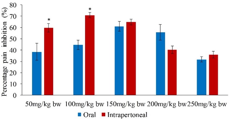
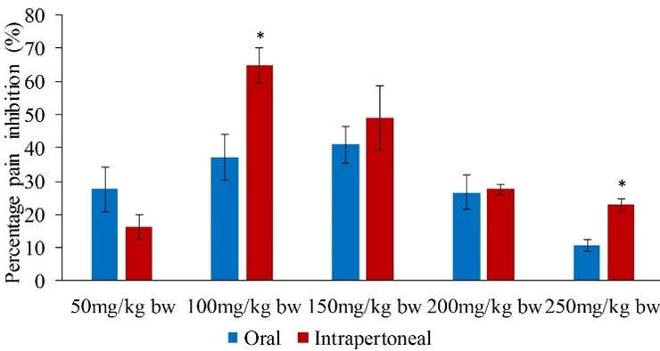
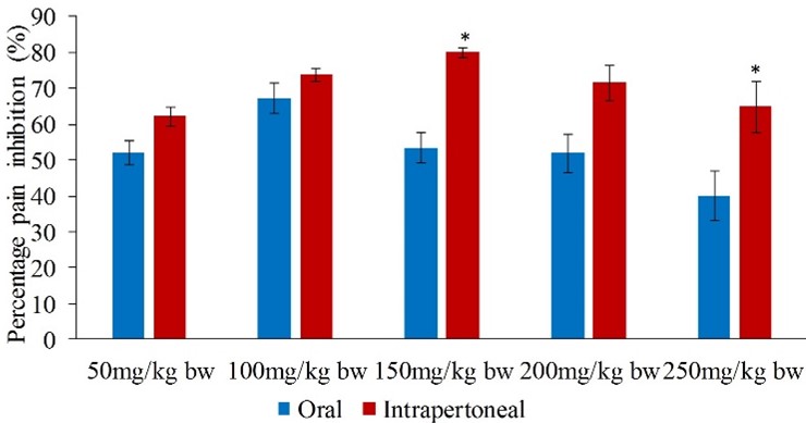
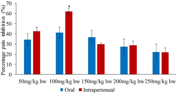
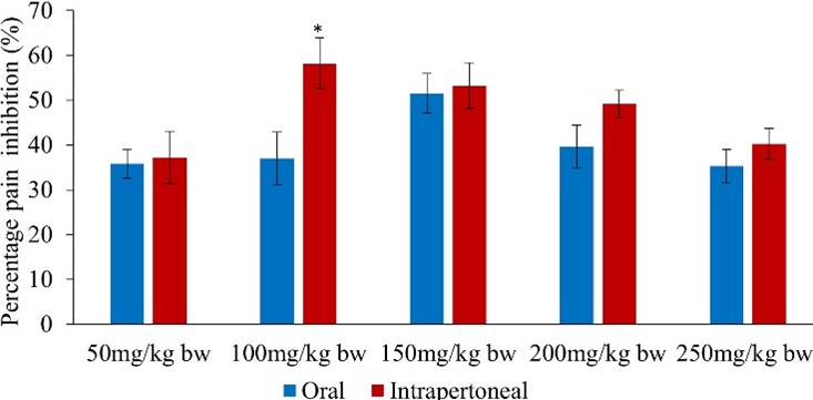
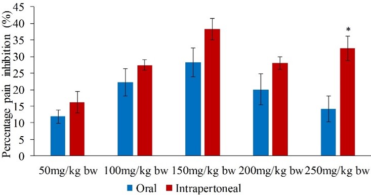
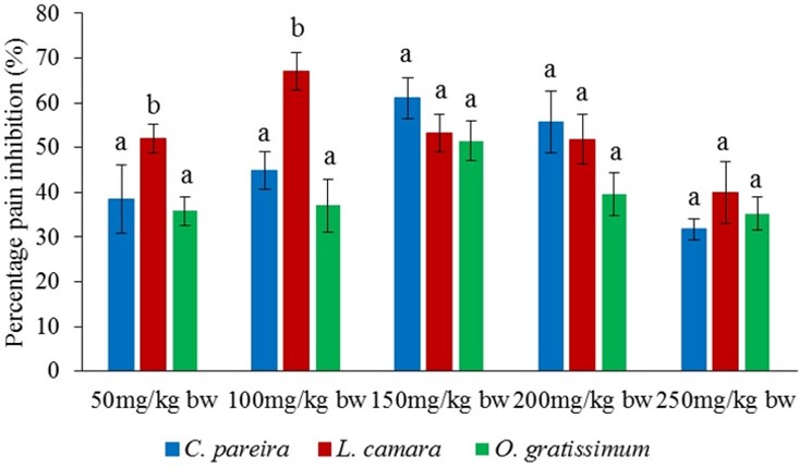
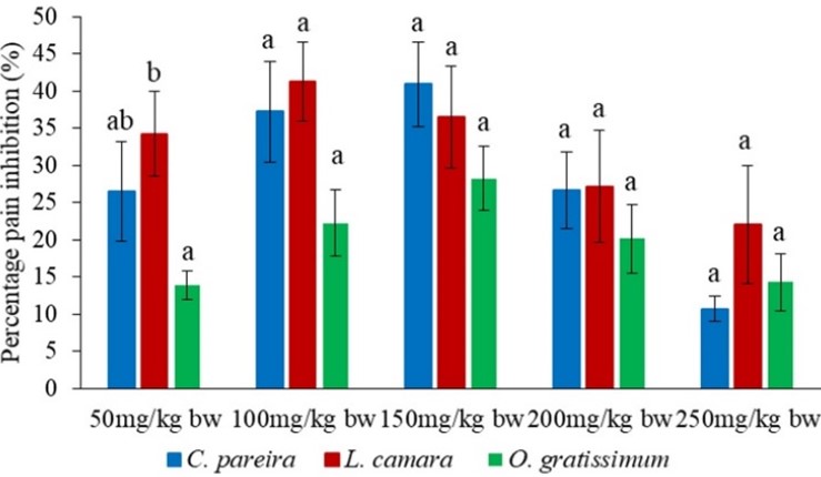
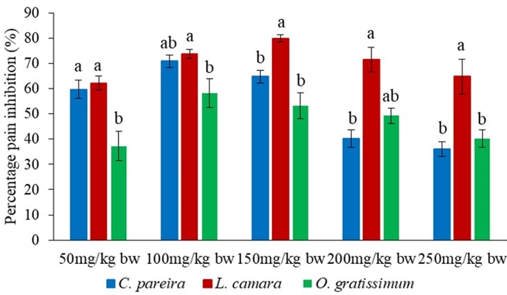
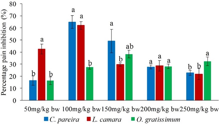
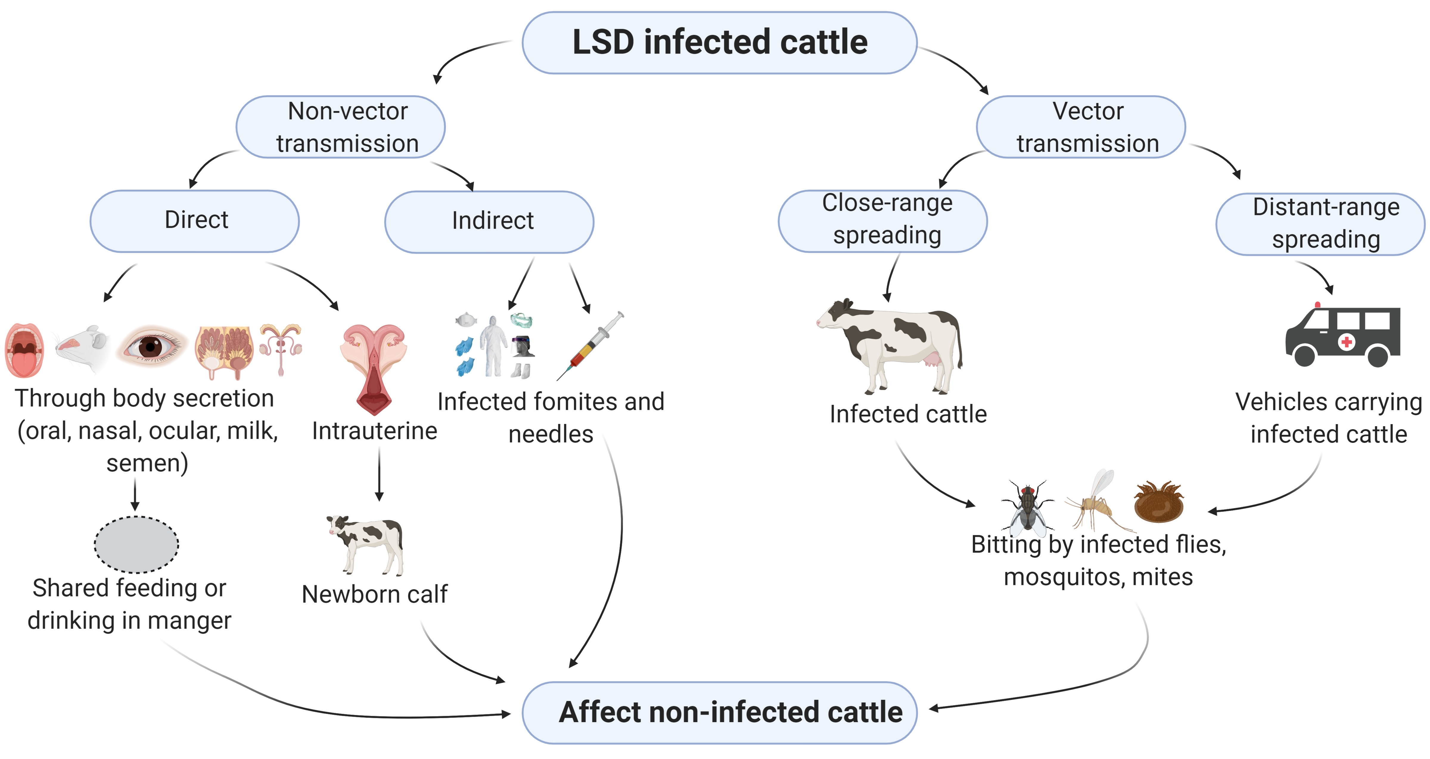
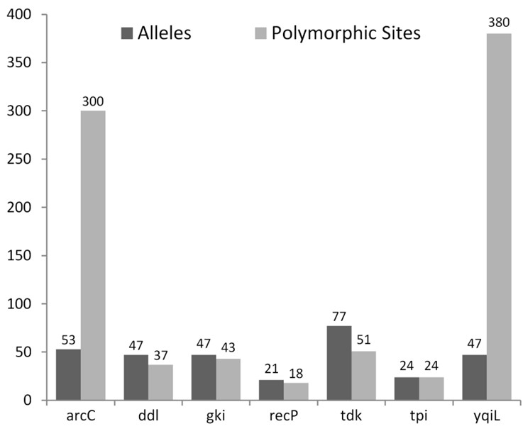
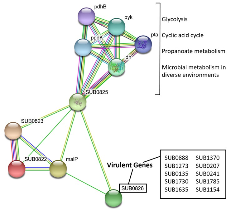
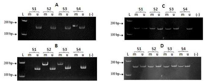
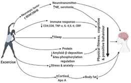